Putative palytoxin and its new analogue, ovatoxin-a, in Ostreopsis ovata collected along the...
10
Putative Palytoxin and Its New Analogue, Ovatoxin-a, in Ostreopsis ovata Collected Along the Ligurian Coasts During the 2006 Toxic Outbreak Patrizia Ciminiello, a Carmela Dell’Aversano, a Ernesto Fattorusso, a Martino Forino, a Luciana Tartaglione, a Claudio Grillo, b and Nunzia Melchiorre b a Dipartimento di Chimica delle Sostanze Naturali, Università degli Studi di Napoli Federico II, Napoli, Italy b Agenzia Regionale per la Protezione dell’Ambiente Ligure (ARPAL), Dipartimento La Spezia, La Spezia, Italy In this article we report on the liquid chromatography tandem mass spectrometry (LC-MS) investigation of plankton samples collected in the summer of 2006 along the Ligurian coasts, coinciding with a massive bloom of the tropical microalga Ostreopsis ovata. LC-MS analyses indicated the occurrence of putative palytoxin along with a much more abundant palytoxin- like compound never reported so far, which we named ovatoxin-a. On the basis of molecular formula, fragmentation pattern, and chromatographic behavior, the structure of ovatoxin-a appeared to be strictly related to that of palytoxin. We report also on the analysis of cultured O. ovata, which was necessary to unequivocally demonstrate that putative palytoxin and ovatoxin-a contained in field samples were actually produced by O. ovata itself. (J Am Soc Mass Spectrom 2008, 19, 111–120) © 2008 American Society for Mass Spectrometry T he first referenced evidence of the presence of the tropical microalga Ostreopsis spp. in the Mediter- ranean seawater is dated back to the late 1990s [1]. Since then, these algae have been continuously detected along most of the Mediterranean coastline [2]. They have only occasionally caused cases of human sufferings, until, in summer 2005, a massive prolifera- tion of O. ovata broke out with alarming proportion in Liguria, nearby Genoa, Italy. This phenomenon caught the attention of both national and international media, which delivered breaking news on the event, as hun- dreds of people required medical attention after expo- sure to marine aerosol. The symptoms shown by all the patients included fever associated with serious respira- tory distress and, in some cases, conjunctivitis. The toxic outbreak reached its climax in coincidence with the highest concentration of O. ovata in seawater and dis- appeared as the population of this microalga started fading away. We collected a concentrated plankton sample off Genoa coasts during the toxic outbreak and analyzed it for the most common phycotoxins found in the Medi- terranean sea, such as okadaic acid, spirolides, azaspi- racids, yessotoxins, paralytic shellfish poisoning toxins, and domoic acid [3, 4]. We also investigated the occur- rence of brevetoxins which, although never reported in the Mediterranean sea, are usually associated with poisonings due to marine aerosols [5]. None of the above toxins was detected in the plankton sample [4]. Some Ostreopsis species produce palytoxin (Figure 1) [6–8], a complex polyhydroxylated compound and one of the most potent marine biotoxins, or palytoxin ana- logues such as ostreocin-D [9 –11] and mascarenotoxins [12]. Structure of ostreocin-D was assigned to 42-hy- droxy-3,26-didemethyl-19,44-dideoxypalytoxin, based on NMR data [9, 10], and verified by negative-ion fast-atom bombardment collision-induced dissociation tandem mass spectrometry [11]. The charge-remote fragmentations were facilitated by a negative charge introduced to the terminal amino group or to a hy- droxyl group at the other terminus of the molecule by reaction of pure ostreocin-D with 2-sulfobenzoic acid cyclic anhydride. Mascarenotoxins [12] were purified and identified as palytoxin-like compounds based on comparison of their mass spectrum profiles and frag- mentation patterns with those of a reference standard of palytoxin. Other analogues of palytoxin, namely homo-, bishomo-, neo-, and deoxy-palytoxin were isolated from the soft coral Palythoa tuberculosa [13] but they were not yet proven to be produced by Ostreopsis spp. To investigate the presence of palytoxin in the plank- ton sample collected during the Genoa 2005 outbreak, we set up a new method based on liquid chromatogra- phy tandem mass spectrometry (LC-MS) for sensitive Address reprint requests to Professor E. Fattorusso, Dipartimento di Chimica delle Sostanze Naturali, Università degli Studi di Napoli Federico II, via D. Montesano 49, 80131, Napoli, Italy. E-mail: [email protected] Published online November 7, 2007 © 2008 American Society for Mass Spectrometry. Published by Elsevier Inc. Received July 27, 2007 1044-0305/08/$32.00 Revised October 31, 2007 doi:10.1016/j.jasms.2007.11.001 Accepted November 1, 2007
Transcript of Putative palytoxin and its new analogue, ovatoxin-a, in Ostreopsis ovata collected along the...
Putative Palytoxin and Its New Analogue,Ovatoxin-a, in Ostreopsis ovata CollectedAlong the Ligurian Coasts During the 2006Toxic Outbreak
Patrizia Ciminiello,a Carmela Dell’Aversano,a Ernesto Fattorusso,a
Martino Forino,a Luciana Tartaglione,a Claudio Grillo,b andNunzia Melchiorreb
a Dipartimento di Chimica delle Sostanze Naturali, Università degli Studi di Napoli Federico II, Napoli, Italyb Agenzia Regionale per la Protezione dell’Ambiente Ligure (ARPAL), Dipartimento La Spezia, La Spezia,Italy
In this article we report on the liquid chromatography tandem mass spectrometry (LC-MS)investigation of plankton samples collected in the summer of 2006 along the Ligurian coasts,coinciding with a massive bloom of the tropical microalga Ostreopsis ovata. LC-MS analysesindicated the occurrence of putative palytoxin along with a much more abundant palytoxin-like compound never reported so far, which we named ovatoxin-a. On the basis of molecularformula, fragmentation pattern, and chromatographic behavior, the structure of ovatoxin-aappeared to be strictly related to that of palytoxin. We report also on the analysis of culturedO. ovata, which was necessary to unequivocally demonstrate that putative palytoxin andovatoxin-a contained in field samples were actually produced by O. ovata itself. (J Am SocMass Spectrom 2008, 19, 111–120) © 2008 American Society for Mass Spectrometry
The first referenced evidence of the presence of thetropical microalga Ostreopsis spp. in the Mediter-ranean seawater is dated back to the late 1990s
[1]. Since then, these algae have been continuouslydetected along most of the Mediterranean coastline [2].They have only occasionally caused cases of humansufferings, until, in summer 2005, a massive prolifera-tion of O. ovata broke out with alarming proportion inLiguria, nearby Genoa, Italy. This phenomenon caughtthe attention of both national and international media,which delivered breaking news on the event, as hun-dreds of people required medical attention after expo-sure to marine aerosol. The symptoms shown by all thepatients included fever associated with serious respira-tory distress and, in some cases, conjunctivitis. The toxicoutbreak reached its climax in coincidence with thehighest concentration of O. ovata in seawater and dis-appeared as the population of this microalga startedfading away.
We collected a concentrated plankton sample offGenoa coasts during the toxic outbreak and analyzed itfor the most common phycotoxins found in the Medi-terranean sea, such as okadaic acid, spirolides, azaspi-racids, yessotoxins, paralytic shellfish poisoning toxins,and domoic acid [3, 4]. We also investigated the occur-
Address reprint requests to Professor E. Fattorusso, Dipartimento di
Chimica delle Sostanze Naturali, Università degli Studi di Napoli FedericoII, via D. Montesano 49, 80131, Napoli, Italy. E-mail: [email protected]© 2008 American Society for Mass Spectrometry. Published by Elsevie1044-0305/08/$32.00doi:10.1016/j.jasms.2007.11.001
rence of brevetoxins which, although never reported inthe Mediterranean sea, are usually associated withpoisonings due to marine aerosols [5]. None of theabove toxins was detected in the plankton sample [4].
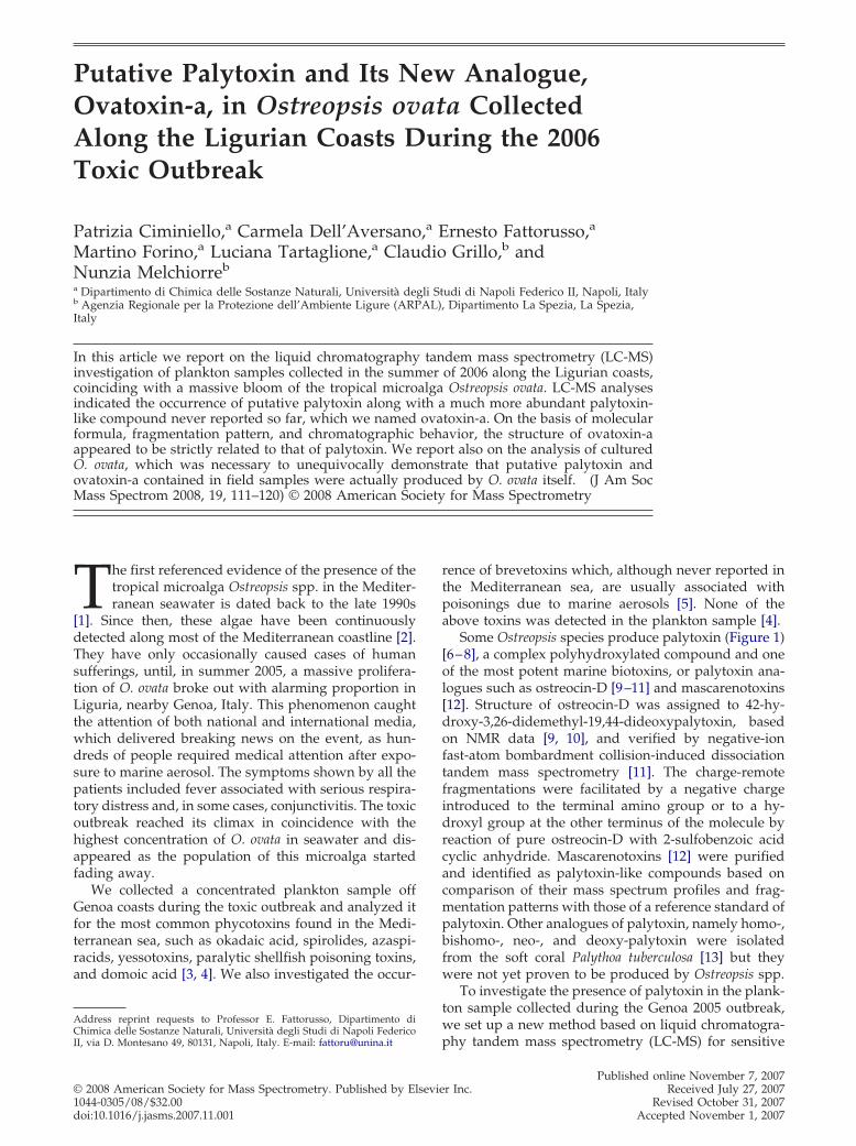
Some Ostreopsis species produce palytoxin (Figure 1)[6–8], a complex polyhydroxylated compound and oneof the most potent marine biotoxins, or palytoxin ana-logues such as ostreocin-D [9–11] and mascarenotoxins[12]. Structure of ostreocin-D was assigned to 42-hy-droxy-3,26-didemethyl-19,44-dideoxypalytoxin, basedon NMR data [9, 10], and verified by negative-ionfast-atom bombardment collision-induced dissociationtandem mass spectrometry [11]. The charge-remotefragmentations were facilitated by a negative chargeintroduced to the terminal amino group or to a hy-droxyl group at the other terminus of the molecule byreaction of pure ostreocin-D with 2-sulfobenzoic acidcyclic anhydride. Mascarenotoxins [12] were purifiedand identified as palytoxin-like compounds based oncomparison of their mass spectrum profiles and frag-mentation patterns with those of a reference standard ofpalytoxin. Other analogues of palytoxin, namely homo-,bishomo-, neo-, and deoxy-palytoxin were isolatedfrom the soft coral Palythoa tuberculosa [13] but theywere not yet proven to be produced by Ostreopsis spp.
To investigate the presence of palytoxin in the plank-ton sample collected during the Genoa 2005 outbreak,we set up a new method based on liquid chromatogra-
phy tandem mass spectrometry (LC-MS) for sensitivePublished online November 7, 2007r Inc. Received July 27, 2007
Revised October 31, 2007Accepted November 1, 2007

ent.
112 CIMINIELLO ET AL. J Am Soc Mass Spectrom 2008, 19, 111–120
and selective detection of palytoxin. Relying on thenewly developed method, we succeeded in disclosingthe involvement of a putative palytoxin in the Genoa2005 outbreak and assessing its occurrence in the Med-iterranean seawater for the very first time [4].
Following this event, in 2006 the presence of O. ovatawas monitored along the Ligurian coasts to prevent fur-ther human sufferings. Indeed, a remarkable proliferationof O. ovata was detected in the Mediterranean Sea fromJuly through August 2006. Bathing was forbidden inseveral Italian coastal areas and, thus, the number ofpeople suffering from the toxic outbreak was significantlylimited in comparison with the 2005 event [2].
During the 2006 event, we collected field samples ofplankton along the Ligurian coasts, where O. ovatabloomed more abundantly. The collected samples wereinvestigated for the presence of palytoxin by the newlydeveloped method and used to create in vitro culturesof the dinoflagellate. In this study, we report on theLC-MS analyses of the plankton samples collected inthe summer of 2006, which indicated the presence ofputative palytoxin together with a palytoxin-like mole-cule never reported so far, which we named ovatoxin-a.Furthermore, we report on the analyses of cultured O.ovata, which was necessary to unequivocally demon-
O
OH
O
O
OO
O
HN
O
NH
H2N
Me
Me
H
OHOH
OH Me OHMe
Me
HO8
9
A moiety
B
Figure 1. Structure of palytoxin. Cleavage betmoieties. [M�H�B moiety-H2O]�ion appears aof palytoxin carried out on the triple quadrupoappears at m/z 2318.2749 in the HR product ion spout on the linear ion trap hybrid FTMS instrum
strate that putative palytoxin and ovatoxin-a contained
in field samples were actually produced by O. ovataitself.
Experimental
Reagents
All organic solvents were of distilled-in-glass grade(Carlo Erba, Milan, Italy). Water was distilled andpassed through a MilliQ water purification system(Millipore Ltd., Bedford, MA). Glacial acetic acid (lab-oratory grade) was purchased from Carlo Erba. Analyt-ical standard of palytoxin was purchased from WakoChemicals GmbH (Neuss, Germany) and dissolved inmethanol/water (1:1, vol/vol). Matrix-matched stan-dards of palytoxin were prepared as reported previ-ously [4].
Collection and Identification of the Plankton
The harmful algal bloom occurred in late July throughAugust 2006 along the rocky coasts of Liguria, whichpresent pebbly seabed colonized by macroalgae. Seawa-ter samples were collected by operators of the AgenziaRegionale per la Protezione dell’Ambiente Ligure (AR-
OH
O
OH
OH
OHMe
O
HO
O
HOMe
O
OH
OHHO
O
OH
HO
OH
OH
OH
OH
OH
OH
OH
OH
OHOHOH
HO
OH
OH
OH
OH
OH
OH
OH
OH
OH
OH
OH
iety
carbons 8 and 9 originates A and B structural327.1 in the full scan MS spectrum (DP � 50 V)S instrument. [M � H � A moiety-H2O]� ionm of the mono-charged ion of palytoxin carried
HO
O
mo
weent m/zle Mectru
PAL) and used to determine the qualitative and quan-

atched out
113J Am Soc Mass Spectrom 2008, 19, 111–120 PUTATIVE PALYTOXIN AND OVATOXIN-A IN O. ovata
titative composition of phytoplankton. The phytoceno-ses were characterized after treatment with Lugolsolution and sedimentation in Uthermöhl tubes [14].Analysis on the whole sedimentation chamber withinverted microscope showed that O. ovata [15] was themajor species present in the sample, together with a fewcells of diatoms (Coscinodiscus spp.) and other poten-tially toxic dinoflagellates, namely Coolia monotis, Pro-rocentrum lima, and Amphidinium sp. Since O. ovata is abenthonic dinoflagellate, five samples of macroalgae(Rhodophyta, Chlorophyta, and Phaeophyta) covered by aplankton film, were collected and closed underwater insampling bags (PBI type) in the area of Genoa (samples1 and 2) and La Spezia (samples 3 and 4). The macroal-gal samples were separately transferred to hermeticallysealed vessels and the sampling bags were rinsed withfiltered (0.22 �m mesh) seawater collected at the sam-pling sites. The rinsing water was added to the vesselsshaking for 5 min and the cell suspension was filteredthrough a plankton net (20 �m mesh). The procedurewas repeated several times to obtain a plankton pelletthat was suspended again in filtered seawater. Analiquot of each sample was treated with Lugol solutionand used for cell identification and counting by in-verted microscope. The remaining aliquot for eachsample was centrifuged using a fixed-angle rotor (rmax
9.5 cm) at 5000 rpm for 20 min to separate pellet fromseawater, and shipped to the Dipartimento di Chimicadelle Sostanze Naturali, Università degli Studi diNapoli Federico II, for chemical analyses.
Cultures of O. ovata
Cultures of O. ovata were obtained from sample 1 collectedat the site most affected by the phenomenon of algalbloom on July 24, 2006. Single cells were isolated bymicromanipulation at the inverted microscope and trans-ferred in sterile slabs equipped with traps (1.5 cm diame-ter). They were grown in conditions close to the naturalones, as reported in literature for Ostreopsidaceae cultures(temperature 25 °C, luminous existence 2000 l�, 16:8hlight-to-dark cycle) [16]. The standard K-Keller mediumwas used, which supplies NaNO3 and NH4Cl as nitrogensources, and �-glycerophosphate in place of NaH2PO4.
Table 1. Amounts of putative palytoxin and ovatoxin-aa containLigurian coasts in the summer of 2006
Sample Collected onCell number
(x106)
1 July 24 412 July 24 243 August 22 224 August 23 14
aOn account of the structural similarities between palytoxin and ovaovatoxin-a content was determined by direct comparison to a matrix-mexperimental conditions. Both SIM and MRM experiments were carrie
Gradually, cultures were transferred to larger flasks, up to
500 mL. The algal pellets (16 � 106 cell) were collectedduring late exponential growth phases, on 14th day, by adisposable vacuum filtration system (Stericup; Millipore)on 0.22 �m cellulose filters. Both pellets and growthmedium were stored at �20 °C until shipment.
Extraction
Each field (or cultured) pellet sample was sonicatedwith 8 mL of a methanol/water (1:1, vol/vol) solutionfor 3 min in pulse mode, while cooling in ice bath. Themixture was centrifuged at 5500 rpm for 30 min, thesupernatant was decanted, and the pellet was washedtwice with 8 mL of methanol/water (1:1, vol/vol). Theextracts were combined and the volume was adjusted to30 mL with extracting solvent. The obtained mixturewas analyzed directly by LC-MS (5 �L injected). Eachseawater (or growth medium) sample was extractedtwice with an equal volume of butanol. The butanollayer was evaporated to dryness, dissolved in 5 mL ofmethanol/water (1:1, vol/vol), and analyzed directlyby LC-MS (5 �L injected).
Liquid Chromatography-Mass Spectrometry
LC-MS analyses were accomplished by using a 3 �mGemini C18 (150 � 2.00 mm) column (Phenomenex,Torrance, CA) maintained at room temperature andeluted at 0.2 mL min�1 with water (Eluent A) and 95%acetonitrile/water (Eluent B), both containing 30 mMacetic acid. The following gradient systems were used:a fast gradient elution (20% to 100% B over 10 min andhold 5 min), which allowed elution of palytoxin at 6.60min, and a slow gradient elution (20% to 50% B over 20min, 50% to 80% B over 10 min, 80% to 100% B in 1 min,and hold 5 min), which allowed chromatographic sep-aration between palytoxin (Rt � 11.08 min) and ova-toxin-a (Rt � 11.48 min). Mass spectral experiments atunit resolution (FWHM � 0.7 � 0.1 Th) were carried outon the following LC-MS systems: (1) an Agilent 1100 LCbinary system (Palo Alto, CA) coupled to a PE-SCIEXAPI 2000 triple-quadrupole MS equipped with a Tur-bospray source (Concorde, ON, Canada); (2) a SurveyorMS plus LC quaternary system coupled to an LCQ
pellets and seawater extracts of O. ovata collected along the
Putative palytoxin Ovatoxin-a
(�g) pg/cell (�g) pg/cell
16.5 0.40 127.6 3.117.0 0.29 36.4 1.511.9 0.09 27.8 1.263.1 0.22 31.6 2.26
-a, we assumed that their molar responses were quite similar. Thed standard of palytoxin [4] at similar concentration injected in the same(see Experimental section).
ed in
toxin
DECA XP ion trap MS equipped with an OPTON ESI

114 CIMINIELLO ET AL. J Am Soc Mass Spectrom 2008, 19, 111–120
m/z
Time, min
884.
1
890.
1
896.
1
0 5 10 15 20
TICm/z 300-1400
5
XICm/z 327
5.44
6.55
7.99
c)
a)
6.552.611.64
327.
1
1335
.7
1343
.7
1324
.71315
.7
1306
.7
1297
.7
1288
.7
1279
.7
1280 1300 1320 1340
b)
Time, min
880 900 920
327.
1 [M
+H
-B m
oiet
y-H
2O]+
300 320 340 360
d)
300 320 340 360m/z
860 880 900 920
894.
1 [M
+2H
+N
a-H
2O]3+
901.
1 [M
+2H
+N
a]3+
906.
1 [M
+2H
+K
]3+
1280 1300 1320 1340 1360
1359
.7 [M
+H
+K
]2+
1351
.7 [M
+H
+N
a]2+
1340
.7 [M
+2H
]2+
1331
.7 [M
+2H
-H2O
]2+
1322
.7 [M
+2H
-2H
2O]2+
1313
.7 [M
+2H
-3H
2O]2+
1304
.7 [M
+2H
-4H
2O]2+
1295
.7 [M
+2H
-5H
2O]2+
1286
.7 [M
+2H
-6H
2O]2+
1277
.7 [M
+2H
-7H
2O]2+
Figure 2. (a) Total ion chromatogram (TIC) of sample 1; (b) related extracted ion chromatogram(XIC) at m/z 327.1; (c) full scan MS spectrum associated with the peak eluting at 6.55 min; (d) full scanMS spectrum of palytoxin standard. LC-MS analyses of sample 1 and palytoxin standard wereacquired on the triple quadrupole MS instrument in positive full scan MS mode (DP � 50 V), by usingthe following LC conditions: a 3 �m Gemini C18 (150 � 2.00 mm) column eluted at 0.2 mL min�1 withA � water and B � 95% acetonitrile/water, both eluents containing 30 mM acetic acid. A fast gradientelution (20% to 100% B over 10 min and hold 5 min) was used. Assignment of bi-charged andtri-charged ion clusters was based on high-resolution MS data, since the low-resolution settings on themass spectrometer and annotation of the centroids of unresolved multiply charged ions could lead to
misleading compositions [4].
ions
115J Am Soc Mass Spectrom 2008, 19, 111–120 PUTATIVE PALYTOXIN AND OVATOXIN-A IN O. ovata
source (Thermo-Fisher, San Jose, CA). High-resolution(HR) MS and product ion spectra were acquired byusing a Surveyor MS plus LC quaternary system cou-pled to a linear ion trap LTQ Orbitrap XL hybridFourier Transform MS (FTMS) equipped with an ESI
m/z
901.
1 [M
+2H
+N
a]3+
906.
1 [M
+2H
+K
]3+
1331
.7 [M
+2H
- H2O
]2+
134 0
.7 [
M+
2H]2+
a)
1000 1500
1000 1500m
896.
1 [M
+2H
+K
]3+
1324
.6 [M
+2H
]2+
1315
.6 [
M+
2H-H
2O]2+
b)
Figure 3. (a) Full scan MS spectrum of palytoeluting at 6.55 min in the LC-MS analysis of saminstrument in positive ion mode by using thebi-charged and tri-charged ion clusters was bresolution settings on the mass spectrometer ancharged ions could lead to misleading composit
ION MAX source (Thermo-Fisher, San Jose, CA).
Triple Quadrupole MS Experiments
Full scan MS experiments in the range m/z 300–1400were carried out in positive ion mode by using thefollowing source settings: a turbogas temperature of
X 6
2680
.6 [M
+H
]+
2318
.6 [
M+
H- 3
62]+
2300
.6 [
M+
H-3
62-H
2O]+
2282
.6 [M
+H
-362
-2H
2O]+
2264
.6 [M
+H
-362
-3H
2O]+
2000 2500 3000
2000 2500 3000
2648
.5 [
M+
H]+
2268
.5 [
M+
H-3
62-H
2O]+
2250
.5 [
M+
H-3
62-2
H2O
]+22
32.5
[M
+H
-362
-3H
2O]+
2286
.5 [
M+
H-3
62]+
andard; (b) full scan MS spectrum of the peak. The spectra were acquired on the ion trap MSLC conditions as in Figure 2. Assignment ofon high-resolution MS data, since the low-
otation of the centroids of unresolved multiply[4].
/z
xin stple 1sameased
d ann
300 °C, an ionspray voltage of 5500 V, a declustering

116 CIMINIELLO ET AL. J Am Soc Mass Spectrom 2008, 19, 111–120
potential (DP) of 8 V (50 V in the in-source fragmentationexperiments), a focusing potential (FP) of 350 V, and anentrance potential (EP) of 11 V. Multiple reaction mon-itoring (MRM) experiments were carried out by using acollision energy of 50 eV, a cell exit potential (CXP) of 10V, and monitoring the transitions m/z 1340.7 ¡ 327.1(DP � 8 V), 1331.7 ¡ 327.1 (DP � 8 V), and 906.1 ¡327.1 (DP � 50 V) for palytoxin and the transitions m/z1324.7 ¡ 327.1 (DP � 8 V), 1315.7 ¡ 327.1 (DP � 8 V),896.1¡ 327.1 (DP � 50 V) for ovatoxin-a. The dwelltime was set to achieve a total scan time of 1 s. Thetransitions m/z 1340.7 ¡ 327.1 and 1324.7 ¡ 327.1 wereused for quantitation of putative palytoxin and ova-toxin-a, respectively. Quantitative results were obtainedby direct comparison of peak areas to matrix-matchedstandard solutions of palytoxin at similar concentrationinjected in the same experimental conditions [4]. MRMquantitative results obtained for ovatoxin-a were con-firmed by selected ion monitoring (SIM) experiments.The following ions were monitored for ovatoxin-a m/z890.1, 896.1, 1297.7, 1306.7, 1315.7, 1324.7, 1335.7, 1343.7whereas the following ions were monitored for paly-toxin m/z 906.1, 901.1, 1313.7, 1322.7, 1331.7, 1340.7,1351.7, 1359.7. The obtained peak areas for ovatoxin-awere summed and compared with ions sum of standardsolutions of palytoxin injected in the same experimentalconditions.
Ion Trap MS Experiments
Full scan MS experiments in the range m/z 700–3000were carried out in positive ion mode by using a spray
264
7.4
868
264
8.4
896
264
9.4
954
2650
.496
4
2651
.49
73
m/z2645 2646 2647 2648 2649 2650 2651 2652 2653
monoisotopic peakC129 H224N3O52
(∆ = -3.918 ppm)
Figure 4. Mass scale expansion of the m/z 2645–2653 region of theHRMS spectrum of ovatoxin-a acquired on the linear ion traphybrid FTMS instrument in positive ion mode by using the sameLC conditions as in Figure 2.
voltage of 7.5 kV, a capillary temperature of 200 °C, a
capillary voltage of 39 V, a sheath gas and an auxiliarygas flow of 50 and 10 (arbitrary units), respectively.
Linear Ion Trap Hybrid FTMS Experiments
HRMS experiments (positive ions) were acquired in therange m/z 600–3000 at the 100,000 resolving powersetting by using the following source settings: a sprayvoltage of 3.8 kV, a capillary temperature of 260 °C, acapillary voltage of 49 V, a sheath gas and an auxiliarygas flow of 50 and 10 (arbitrary units), respectively.High-resolution product ion spectra were acquired atthe 60,000 resolving power setting by fragmenting the[M�H]� ion of both palytoxin (m/z 2680.5) and ova-toxin-a (m/z 2648.5) with a collision energy of 25%, anactivation Q of 0.250, and an activation time of 30 ms.Calculation of elemental formula of ovatoxin-a and itsfragments contained in the product ion spectrum wasperformed by using the mono-isotopic ion peak of eachion cluster.
Results and Discussion
Four plankton samples were collected in summer 2006along the Ligurian coasts in the area of Genoa (1 and 2)and La Spezia (3 and 4). Analyses with inverted micro-scope showed that O. ovata was the dominant species inthe collected samples [15], while just a few cells ofdiatoms (Coscinodiscus spp.) and other potentially toxicdinoflagellates, namely Coolia monotis, Prorocentrumlima, and Amphidinium sp., were present in seawater.
The collected plankton samples were independentlycentrifuged to separate pellets from seawater, whichwere extracted as reported in the Experimental section.The crude extracts were directly analyzed by the newlydeveloped method for detection of palytoxin [4] usingan ESI-triple quadrupole MS instrument operating inthe mass range 0–1800. Chromatographic separationwas accomplished on a reversed-phase column byusing a mobile phase containing water/acetonitrile, 30mM acetic acid and a fast gradient elution, whichallowed the early elution of palytoxin standard at 6.60min. Multiple reaction monitoring (MRM) experimentswere carried out by selecting the transition (precursorion ¡ product ion) reported for palytoxin in ourprevious paper [4] (m/z 1340.7 ¡ 327.1) together withtwo additional transitions at m/z 1331.7 ¡ 327.1 and906.1 ¡ 327.1, for further confirmation. The product ionat m/z 327.1 arises from the cleavage between thecarbons 8 and 9 of palytoxin [8] and the additional lossof a water molecule; it can be assigned to the [M � H �B moiety-H2O]� ion (Figure 1). This ion dominates theproduct ion spectrum of palytoxin, whatever the pre-cursor ion used, either the [M�2H]2� ion at m/z 1340.7,the [M�2H�H2O]2� ion at m/z 1331.7, or the[M�2H�K]3� ion at m/z 906.1.
Results of MRM analyses of the plankton collected in2006 paralleled those obtained on the 2005 plankton
samples. The presence of putative palytoxin was shown
117J Am Soc Mass Spectrom 2008, 19, 111–120 PUTATIVE PALYTOXIN AND OVATOXIN-A IN O. ovata
in all the samples, 1–4, by peaks eluting at 6.55 min,which closely matched those of a reference sample ofpalytoxin in retention time, fragmentation, and ionratios. The amounts of putative palytoxin contained insamples 1–4 are reported in Table 1. The toxin contentwas determined by direct comparison with matrix-matched standards of palytoxin at similar concentra-tion. The use of matrix-matched standards is requiredfor accurate quantitation since palytoxin ionization suf-fers from some matrix suppression effect, which isanalyte concentration-dependent [4].
Basing on MRM quantitative results, sample 1 ap-peared to contain a relatively high amount of putativepalytoxin, which allowed a more detailed and compre-hensive MS investigation. To look for additional paly-toxin analogues in the sample, we carried out a full scanMS experiment on the triple quadrupole MS in therange 300–1400 u by favouring the in-source formationof the fragment ion [M�HB moiety-H2O]� at m/z 327.1.This ion is generated in the fragmentation experimentsof palytoxin itself as well as in those of some otherpalytoxin-like compounds [12, 13]. Preliminary experi-
2230 2240 2250 2260 2270 2280 2290
2286
.286
4 [M
+H
-A m
oiet
y-H
2O]+
2268
.276
8 [M
+H
-A m
oiet
y-2H
2O]+
2250
.266
4 [M
+H
-A m
oiet
y-3H
2O]+
2232
.256
2 [M
+H
-A m
oiet
y-4H
2O]+
X 10
2285
.282
5
2267
.272
4
2248
.246
3
2231
.251
8
Figure 5. Mass scale expansions of the m/z 2220ion spectrum of ovatoxin-a acquired on the lin[M�H]� ion at m/z 2648.5 as precursor and the sathe mono-isotopic ion peaks.
ments showed that in-source formation of ion at m/z
327.1 depends basically on front end potentials used,mostly declustering potential (DP), whereas it is notaffected by variation in turbo ion spray temperature.Thus a DP of 50 V and a spray temperature of 300 °Cwere used.
The total ion chromatogram (TIC) of sample 1 (Fig-ure 2a) appeared very complex, which was not surpris-ing since the analyzed sample was a crude extract, notsubjected to any clean-up. Thus, we extracted the ion atm/z 327.1 by obtaining an extracted ion chromatogram(XIC) (Figure 2b) dominated by a peak at an elutiontime (6.55 min) close to that of the palytoxin standard(6.60 min). The associated mass spectrum (Figure 2c)contained tri-charged and bi-charged ion clusters in therange 850–930 and 1270–1370, respectively, togetherwith the base peak at m/z 327.1. An accurate comparisonbetween this MS spectrum and that of the palytoxinstandard (Figure 2d) indicated that, although bothspectra contained the ion peak at m/z 327.1, they pre-sented similar tri-charged and bi-charged ion clusters,which basically differed only for m/z absolute values.
2612
.467
8 [M
+H
-2H
2O]+
2570 2580 2590 2600 2610 2620 2630 2640
2611
.463
8
2593
.454
1
2576
.447
5
2594
.457
4 [M
+H
-3H
2O]+
2576
.447
5 [ M
+H
-4H
2O]+
0 and m/z 2560–2650 regions of the HR producton trap hybrid FTMS instrument by using theC conditions as in Figure 2. Dotted lines indicate
m/z
–230ear ime L
This evidence pointed to the presence in sample 1 of a

118 CIMINIELLO ET AL. J Am Soc Mass Spectrom 2008, 19, 111–120
palytoxin-like molecule that didn’t correspond to paly-toxin itself. We named it ovatoxin-a.
Additional information was obtained by full scan MSexperiments run on an ESI-ion trap-MS instrumentoperating in the mass range 50–4000 u. Such experi-ments were carried out on both sample 1 extract andpalytoxin standard. Under the used ionization condi-tions, the MS spectrum of palytoxin (Figure 3a) wasdominated by the [M�2H]2� ion peak at m/z 1340.7,while only one water molecule was lost from the[M�2H]2�, and the fragment ion at m/z 327.1 wascompletely lacking. Tri-charged ions [M � 2H � Na]3�
and [M�2H�K]3� were present at m/z 901.1 and 906.1,respectively. In the upper region of the MS spectrum,the mono-charged [M�H]� ion of palytoxin waspresent at m/z 2680.6 and an additional ion peak waspresent at m/z 2318.6, followed by ion peaks attributableto subsequent losses of three water molecules. The ionat m/z 2318.6 was likely due to an in-source fragmenta-tion of the [M�H]� ion of palytoxin by loss of 362 u.The different degree of front end fragmentation ofpalytoxin observed on turbospray triple quadrupoleMS and ESI ion trap MS could be likely due to differentgeometry of ionization sources and ionization parame-ters used.
The TIC of sample 1 showed a chromatographic peakat a retention time close to that of palytoxin. Theassociated MS spectrum (Figure 3b), interpreted in thelight of that of palytoxin (Figure 3a), presented amono-charged ion at m/z 2648.5, which was assigned to
11.48
11.08
Time, min6 8 10 12 14
x 5x 5
m/z 1315.7 327.1m/z 1324.7 327.1
m/z 1331.7 327.1m/z 1340.7 327.1
Figure 6. LC-MS analysis of sample 1 acquired on the triplequadrupole MS instrument in positive MRM mode. The MRMtransitions m/z 1340.7 ¡ 327.1 and 1331.7 ¡ 327.1 were used todetect palytoxin. The MRM transitions m/z 1324.7 ¡ 327.1 and1315.1 ¡ 327.1 were selected for detection of ovatoxin-a. Thefollowing LC conditions were used: a 3 �m Gemini C18 (150 �2.00 mm) column eluted at 0.2 mL min�1 with A � water and B �95% acetonitrile/water, both eluents containing 30 mM acetic acid.A slow gradient elution (20%to 50% B over 20 min, 50% to 80% Bover 10 min, 80% to 100% B in 1 min, and hold 5 min) was used.Under the used LC conditions, putative palytoxin eluted at 11.08min, while ovatoxin-a eluted at 11.48 min.
the [M�H]� ion, a dominant bi-charged ion peak at m/z
1324.6, [M�2H]2�, a [M�2H�H2O]2� ion at m/z 1315.6,and a tri-charged ion at m/z 896.1, [M�2H�K]3�. Sim-ilarly to palytoxin, the fragment ion at m/z 327.1 waslacking in the spectrum but an additional ion peak waspresent at m/z 2286.5, corresponding to a 362 u neutralloss from the [M�H]� ion, together with ion peaks dueto subsequent losses of three water molecules. Thesedata, particularly the m/z values of tri-charged andbi-charged ions, which corresponded to ion peaks ob-served in MS spectrum obtained on the triple quadru-pole MS system (Figure 2c), indicated that the com-pound was actually ovatoxin-a.
The next step was to establish the exact mass ofovatoxin-a and its fragments to assign the molecularformula and to gain additional information on thestructure of ovatoxin-a. Thus, LC-MS and product ionMS experiments were run for both palytoxin standardand sample 1 on a linear ion trap hybrid FTMS instru-ment in the mass range 600–3000 u at a resolving powerof 100,000. The mono-charged ion cluster in the HRMSspectrum of ovatoxin-a presented a mono-isotopic ionpeak [M�H]� at m/z 2647.4868 (Figure 4), which al-lowed to infer the molecular formula C129H223N3O52 (�� �3.918 ppm) to the molecule. Further confirmationfor such elemental composition was provided by theexact masses of bi-charged (mono-isotopic ion peak atm/z 1335.2429 for C129H224N3NaO52, � � �0.174 ppm)and tri-charged (mono-isotopic ion peak at m/z 890.4937for C129H225N3NaO52, � � �4.657 ppm) ions. So, ova-toxin-a presents two oxygen atoms less than palytoxin(C129H223N3O54).
We also recorded an LC-HR product ion spectrumon sample 1 by selecting the mono-charged ion ofovatoxin-a as precursor ion. The obtained spectrum(Figure 5) contained abundant peaks at m/z 2612.4678,2594.4574, and 2576.4475, which corresponded to sub-sequent losses of two, three, and four water moleculesfrom the [M�H]� ion, respectively. In addition, astructurally interesting fragment ion was present at m/z2286.2864 (mono-isotopic ion at m/z 2285.2825), whichwas followed by three ion peaks attributable to subse-quent losses of water molecules. The ion at m/z2286.2864 was due to the loss of 362.2043 u from theprecursor ion, corresponding to a C16H30N2O7 (� ��2.765 ppm) neutral fragment.
In the same experimental conditions, we recorded anLC-HR product ion spectrum of palytoxin standard byusing the mono-charged ion of palytoxin as precursorion. The obtained spectrum paralleled that of ova-toxin-a in the presence of ions (m/z 2644.4552, 2626.4452,and 2608.4343) due to loss of two, three, and four watermolecules, respectively, and an ion (m/z 2318.2749) dueto neutral loss of 362.2036 u (C16H30N2O7) accompaniedby three losses of water molecules. The neutral frag-ment C16H30N2O7 originates from cleavage between car-bons 8 and 9 and corresponds to A moiety of palytoxinwith an additional molecule of water (Figure 1).
Comparison of MS data of ovatoxin-a and palytoxin
suggests that the two molecules share the same part
119J Am Soc Mass Spectrom 2008, 19, 111–120 PUTATIVE PALYTOXIN AND OVATOXIN-A IN O. ovata
structure A as indicated by (1) the common loss of aC16H30N2O7 part structure from the mono-charged ionsemerging from the experiments performed on ion trapMS and linear ion trap hybrid FTMS instruments; (2)fragment ion at m/z 327.1 contained in MS spectra ofboth compounds run on the triple quadrupole MSinstrument. Thus, structural differences between ova-toxin-a and palytoxin likely lie in the part structure B(Figure 1).
To gain information about the relative abundance ofovatoxin-a and putative palytoxin in the plankton sam-ples, we set up an LC-MS experiment by using a slowgradient elution, which allowed to chromatographicallyseparate the two compounds (see the Experimentalsection). MS detection was accomplished in MRM modeon the triple quadrupole MS instrument by monitoringthe transitions m/z 1340.7 ¡ 327.1, 1331.7 ¡ 327.1 forputative palytoxin and m/z 1324.7 ¡ 327.1, 1315.7 ¡327.1 for ovatoxin-a. The best results are shown inFigure 6. Under the chromatographic conditions used,putative palytoxin eluted at the same retention time asthat of the palytoxin standard, namely 11.08 min, whileovatoxin-a eluted at 11.48 min. On account of theevident structural similarities between the two com-pounds, we assumed that their molar responses werequite similar. However, since differences in adductformation could greatly affect relative intensities ofdifferent adduct ions, MRM quantitative results ob-tained for ovatoxin-a were corroborated by SIM exper-iments. All significant ions contained in full scan MSspectra of ovatoxin-a were monitored, the obtainedpeak areas for ovatoxin-a were summed, and comparedwith ion sum of corresponding ions monitored forpalytoxin. SIM results were in full agreement withMRM data. The calculated amount of ovatoxin-a insamples 1–4 are reported in Table 1.
Comparison between putative palytoxin and ova-toxin-a contents suggested that this latter was by far thepredominant palytoxin-like compound in the 2006plankton.
Cultures of O. ovata were obtained from shares ofmicroalgal pellets coming from the washing of macroal-gae collected in correspondence of the site interested bythe phenomenon of algal bloom in July 2006. Cells werecultured at 25 °C using a light-to-dark regimen of 16:8h. They were collected on 14th day, during the lateexponential growth phase.
Pellet and growth medium were separated bycentrifugation and extracted separately. LC-MS anal-yses were performed in MRM mode by using theslow gradient elution mentioned above. A chromato-graphic peak at 11.08 min in the ion traces at m/z1340.7 ¡ 327.1 and 1331.7 ¡ 327.1 evidenced thepresence of putative palytoxin as well as a chromato-graphic peak at 11.48 min in the ion traces at m/z1324.7 ¡ 327.1 and 1315.7 ¡ 327.1 highlighted thepresence of ovatoxin-a. These data were conclusive inindicating O. ovata as the producing organism of both
compounds detected in natural plankton. Quantita-tive analyses for pellet and growth medium extractsof O. ovata cultures afforded a putative palytoxin andan ovatoxin-a content of 0.55 pg/cell and 3.85 pg/cell, respectively. Interestingly, the two compoundsappeared to be produced approximately in the sameratio observed in natural plankton.
Conclusions
The present paper provides further insights into thetoxin profile of the Mediterranean O. ovata. The re-ported results confirmed the presence of putative paly-toxin in the Mediterranean Sea and evidenced theoccurrence of the novel ovatoxin-a. This latter com-pound was believed to be the major toxin produced byO. ovata during the 2006 outbreak. On the basis ofmolecular formula (ovatoxin-a presents two oxygenatoms less than palytoxin), fragmentation pattern, andchromatographic behavior, the structure of ovatoxin-aappeared to be strictly related to that of palytoxin.However, even slight structural differences could sig-nificantly affect and differentiate the toxicology of thetwo compounds on the account of data reported onbio-activity of palytoxin analogues [10, 17]. Thus, isola-tion of ovatoxin-a is necessary both to elucidate itschemical structure and evaluate its toxicology.
AcknowledgmentsThis work is a result of a research supported by BIOTOX marin2005. LC-MS experiments on the triple quadrupole MS and on theion trap MS were performed at Centro di Servizio Interdiparti-mentale di Analisi Strumentale, Università degli Studi di NapoliFederico II, and at Centro Regionale di Competenza in Diagnos-tica e Farmaceutica Molecolari (Regione Campania), respectively.The authors gratefully acknowledge the assistance of the staff. Theauthors also thank Thermo Fisher Scientific for LC-MS experi-ments on the linear ion trap hybrid FTMS.
References1. Sansoni, G.; Borghini, B.; Camici, G.; Casotti, M.; Righini, P.; Rustighi, C.
Fioriture algali di Ostreopsis ovata (Gonyaulacales: Dinophyceae): unproblema emergente. Biologia Ambientale 2003, 17, 17–23.
2. Di Girolamo, I.; Fattorusso, E.; Funari, E.; Gramaccioni, L.; Grillo, C.;Icardi, G.; Mattei, D.; Poletti, R.; Scardala, S.; Testai, E. Linee guida-Gestione del Rischio associato alle Fioriture di Ostreopsis ovata nellecoste italiane. Enacted by Consiglio Superiore di Sanità-Ministero dellaSalute-May 24th 2007 �http://www.ministerosalute.it/imgs/C_17_pubblicazioni_641_allegato/pdf�.
3. Ciminiello, P.; Fattorusso, E. Shellfish Toxins—Chemical Studies onNorthern Adriatic Mussels. Eur. J. Org. Chem. 2004, 12, 2533–2551.
4. Ciminiello, P.; Dell’Aversano, C.; Fattorusso, E.; Forino, M.; Magno,G. S.; Tartaglione, L.; Grillo, C.; Melchiorre, N. The Genoa 2005Outbreak. Determination of Putative Palytoxin in Mediterranean. Os-treopsis ovata by a New Liquid Chromatography Tandem Mass Spec-trometry Method. Anal. Chem. 2006, 78, 6153–6159.
5. Furey, A.; Garcia, J.; O’Callaghan, K.; Lehane, M.; Amandi, M. F.; James,K. J. Brevetoxins: Structures, Toxicology, and Origin. In Phycotoxins,Botana, L. M., Eds., Blackwell Publishing Professional: Ames, IA, 2007;p. 19–46.
6. Moore, R. E.; Bartolini, G. Structure of Palytoxin. J. Am. Chem. Soc. 1981,103, 2491–2494.
7. Cha, J. K.; Christ, W. J.; Finan, J. M.; Fujioka, H.; Kishi, Y.; Klein, L. L.;Ko, S. S.; Leder, J.; McWhorter, W. W., Jr.; Pfaff, K.-P.; Yonaga, M.;Uemura, D.; Hirata, Y. Stereochemistry of Palytoxin. Part 4. CompleteStructure. J. Am. Chem. Soc. 1982, 104, 7369–71.
8. Taniyama, S.; Osamu, A.; Masamitsu, T.; Sachio, N.; Tomohiro, T.;
Yahia, M.; Tamao, N. Ostreopsis sp., a Possible Origin of Palytoxin (PTX)in Parrotfish Scarus ovifrons. Toxicon. 2003, 42, 29–33.
120 CIMINIELLO ET AL. J Am Soc Mass Spectrom 2008, 19, 111–120
9. Usami, M.; Satake, M.; Ishida, S.; Inoue, A.; Kan, Y.; Yasumoto, T.Palytoxin Analogs from the Dinoflagellate Ostreopsis siamensis. J. Am.Chem. Soc. 1995, 117, 5389–5390.
10. Ukena, T.; Satake, M.; Usami, M.; Oshima, Y.; Naoki, H.; Fujita, T.; Kan,Y.; Yasumoto, T. Structure Elucidation of Ostreocin D, a PalytoxinAnalog Isolated from the Dinoflagellate Ostreopsis siamensis. Biosci.Biotechnol. Biochem. 2001, 65, 2585–2588.
11. Ukena, T.; Satake, M.; Usami, M.; Oshima, Y.; Fujita, T.; Naoki, H.;Yasumoto, T. Structural Confirmation of Ostreocin-D by Application ofNegative-Ion Fast-Atom Bombardment Collision-Induced DissociationTandem Mass Spectrometric methods. Rapid Commun. Mass Spectrom.2002, 16, 2387–2393.
12. Lenoir, S.; Ten-Hage, L.; Turquet, J.; Quod, J. P.; Bernard, C.; Hennion,M. C. First Evidence of Palytoxin Analogues from an Ostreopsis mas-
carenensis (Dinophyceae) Benthic Bloom in Southwestern Indian Ocean. J.Phycol. 2004, 40, 1042–1051.
13. Uemura, D.; Hirata, Y.; Iwashita, T.; Naoki, H. Studies on Palytoxins.Tetrahedron 1985, 41, 1007–1017.
14. Uthermöhl, H. Zur Vervolkommnung der quantitativen PhytoplanktonMethodik. Mitt. Int. Ver. Theor. Angew. Limnol. 1958, 9, 1–38.
15. Penna, A.; Bertozzini, E.; Battocchi, C.; Galluzzi, L.; Giacobbe, M. G.;Vila, M.; Garces, E.; Luglie, A.; Magnani, M. Monitoring of HAB Speciesin the Mediterranean Sea Through Molecular Methods. J. Plankton. Res.2007, 29, 19–38.
16. Keller, M. D.; Selvin, R. C.; Claus, W.; Guillard, R. R. L. Media for theCulture of Oceanic Ultraphytoplankton. J. Phycol. 1987, 23, 633–638.
17. Uemura, D. Bioorganic Studies on Marine Natural Products—DiverseChemical Structures and Bioactivities. Chem. Record 2006, 6, 235–248.