Structure of a fucoidan from the brown seaweed Fucus serratus L
Upload
independentCategory
view
0download
0
265
Biochimica et Biophysica Acta, 556 (1979) 265--277 © Elsevier/North-Holland Biomedical Press
BBA 78482
PURIFICATION AND PARTIAL CHARACTERIZATION OF A PROCARYOTIC GLYCOPROTEIN FROM THE PLASMA MEMBRANE OF THERMOPLASMA ACIDOPHILUM
LI L. YANG and A. HAUG
MSU-DOE Plant Research Laboratory, Michigan State University, East Lansing MI 48824 (U.S.A.) (Received January 12th, 1979)
Key words: Prokaryotic glycoprotein; Plasma membrane; Adaptation; (Mass spectrometry, Thermo plasma acidoph ilum )
Summary
The obligate, thermophilic, acidophilic mycoplasma, Thermoplasma acido- philum, grows optimally at 56°C and pH 2.0. Its plasma membrane possessed 21--22 protein bands that were resolved by polyacrylamide gel electrophoresis. One major membrane protein, molecular weight 152 000, which stained for carbohydrate with periodic acid-Schiff reagent, accounted for 32% (w/w) of the total membrane proteins. It was isolated and further purified by concanavalin A affinity chromatography. The carbohydrate content amounted to less than 10% (w/w) compared to that of the entire glycoprotein. The carbo- hydrate moiety consisted mainly of mannose residues with branched ~ 1 -~ 2 linkages at the non-reducing ends of the glycopeptide as determined by per- methylation followed by gas chromatography-mass spectrometry analysis. The reducing end was an N-glycosidic linkage between asparagine and N-acetyl- glucosamine. The amino acid composition of this glycoprotein showed 62 mol% hydrophobic residues, while the acidic amino acid content contrib- uted 9 mol% more than that of the basic amino acids. The existence of mem- brane glycoproteins in the procaryotic, wall-less T. acidophilum may provide a protective coat for the plasma membrane. The stereochemistry and the con- formation of the carbohydrate chains, in conjunction with water turgor, may contribute to the rigidity of the membrane and the cation binding.
Introduction
One of the major modifications that a protein may undergo after synthesis of its peptide chains is the attachment of sugar residues. A large number of
A b b r e v i a t i o n : SDS, sodium dodecyl sulfate.

266
proteins of diverse origin and biological function are known to contain such covalently linked carbohydrates and are called glycoproteins. Glycoproteins are widely distributed in eucaryotes, occurring not only in vertebrate and invertebrate animals, but also in plants and unicellular organisms. In particular, carbohydrate components of plasma membranes, which separate the cell from its external environment, appear to play a crucial role in active transport of molecules, serve as receptors for viruses, hormones, and antibodies, and take part in intercellular recognition and adhesion [ 1 ].
Glycoproteins, though quite common in eucaryotes, are rare in procaryotes [2,3 ], and the search for glycoproteins in mycoplasma membranes is still in its initial stage [4]. A thermophilic mycoplasma, viz. Thermoplasma acidophilum, grows optimally at pH 2 and 56°C [5]. The organism is totally devoid of a cell wall, only its plasma membrane encounters the harsh environment. Besides structurally unique glycerol ether membrane lipids [6,7], we discovered the existence of membrane glycoproteins [8] which may play a role in the survival of T. acidophilum. This primitive microorganism may serve as a model to explore the complex interactions between cell and its extreme environment. In this communicat ion, we are specifically concerned ,with the elucidation of structural and functional relationships of membrane glycoproteins. The experi- mental results will also provide a bet ter comprehension of the mechanism of resistance of cells that are exposed to environmental stress.
Materials and Methods
Growth of cells T. acidophilum was originally obtained from the American Type Culture
Collection (ATCC 25905). Cells were grown and harvested as described previ- ously [9].
Mere brane prepara tion Cells were lysed by 1 M glycine buffer, pH 9.3, 22°C, at a protein concentra-
tion higher than 10 mg/ml. Membranes were collected and purified on a discon- t inuous sucrose gradient, followed by three water washes. The purity of the membrane preparation was ascertained by electron microscopy, absence of DNA, and absence of cytoplasmic contaminants [9 ].
Buffer system for SDS gel electrophoresis and sample preparation The discontinuous SDS buffer system of Laemmli [10] was used. Separating
gels usually contained 8% acrylamide, while stacking gels contained 5% acryl- amide; they were prepared from the same stock solution. The final concentra- tion of SDS was 0.1% in both kinds of gel and in the electrode buffer. Sample preparation was performed as described [10], except that samples were boiled for 2 min.
Isolation and purification of membrane glycoprotein By preparative slab gel electrophoresis. The buffer system of Laemmli [10]
was used to run slab gels. The slab gel apparatus (Bio-Rad, Richmond, CA) was operated at 30 mA with a slab thickness of 3 mm. After electrophoresis, three

267
strips of gels were cut vertically and stained with periodic acid-Schiff and Coomassie blue. The gel was reconstructed after staining and glycoprotein bands Were cut, and eluted with buffer. SDS was removed by dialysis.
By phenol extraction. Membranes were extracted with 50% phenol solution at 70°C for 15 min. Material at the interface was collected and dialysed.
By Sepharose 4B column chromatography. Membranes were solubilized by 1% SDS and loaded onto a 90 X 1.5 cm (inner diameter) Sepharose 4B (Pharmacia, Uppsala, Sweden) column. Fractions of 3 ml were collected and assayed for protein and carbohydrate.
By concanavalin A-Sepharose column chromatography. Glycoproteins iso- lated by the above three methods can be further purified by concanavalin A- Sepharose (Sigma, St. Louis, MO) column. Glycoprotein was loaded on the column and eluted first with a solution of 0.5% Triton X-100, 50 mM Tris, pH 8.0, 0.5 M NaC1, and then with 0.5% Triton X-100, 50 mM Tris, pH 8.0, 0.2 M a-methylmannoside. Triton X-100 was removed by dialysis.
Methanolysis of carbohydrates Methanolysis of glycoprotein to s tudy the carbohydrate portion was
performed by the method of Chambers and Clamp [11]. Trimethylsilylated methyl glycosides were analyzed on a Perkin-Elmer gas chromatograph, Model 910, with a 12 ft, 3% SP-2100 column {80--100 mesh, on Supelcoport) with an initial hold of 4 min at 120°C, then programmed at 0.5°C/min to 185°C, and held at 185°C for 20 min.
Permethylation for linkage studies Permethylat ion of carbohydrates was performed by the method of
Hakomori [12]. All the permethylat ion operations were carried out under dry nitrogen. After acid hydrolysis, borohydr ide reduction, and acetylation, the partially methylated alditol acetates in chloroform were ready for analyses by GLC or GLC-MS. Column packings used for these analyses were 3% OV-210 on Gas Chrom Q ( 8 0 - 1 0 0 mesh) or 3% OV-225 on Gas Chrom Q ( 8 0 - 100 mesh). The mass spectra were interpreted according to BjSrndal et al. [13].
Anhydrous hydrogen fluoride deglycosylation The reaction vessel containing freeze-dried sample was cooled in a liquid
nitrogen bath. 10 ml of HF was distilled over from the reservoir. The reaction vessel was allowed to warm up to room temperature. At the end of the reaction, the reaction vessel was evacuated via a calcium oxide trap. To ensure complete removal of HF, the line was evacuated for an additional hour after removal of visible HF. The sample was then taken up in water and passed through an Amicon ultrafiltration cell to separate the carbohydrate and the protein [14].
Amino acid analysis 5.7 N HC1 was added to the purified glycoprotein. Hydrolysis was complete
after 18 h at l l 0 ° C . The suspension was taken to dryness under nitrogen and resuspended in 0.01 N HC1. Amino acid analysis was performed on a single column accelerated flow Technicon system modified by Dr. D.T.A. Lamport , Plant Research Laboratory, Michigan State University.

268
a-Mannosidase digestion. Reaction mixtures containing approximately 5 mg of purified glycoprotein in 1 ml of 0.1 M citrate buffer, pH 4.5, were incubated at 37°C with 2--3 units of a-mannosidase. Incubation was continued until no additional release of mannose was observed. The reaction was terminated by introducing 2 ml of 0.2 M borate buffer, pH 9.8. The reaction mixtures were then passed through an ultrafiltration cell to separate the free carbohydrates from the rest of the glycoproteins.
fl-Glucosidase digestion. The same procedure for a-mannosidase was used for fi-glucosidase.
[J-Galactosidase digestion. The same procedure for a-mannosidase was used; only the buffer (0.05 M phosphate, pH 7) was different. ~-Galactosidase has an opt imum pH at 7.0. Mannosidase, glucosidase and galactosidase were purchased from Sigma Chemical Co. (St. Louis, MO).
Endoglycosidase H digestion. Glycopept ide in 1 ml of 0.1 M acetate buffer, pH 5.0, was incubated with one unit of endoglycosidase H for 16 h at 37°C. The reaction was stopped by heating for 1 min in a boiling water bath and the reaction mixture ultrafiltrated. Endoglycosidase H (Miles, Elkhart, IN) from Streptomyces griseus hydrolyzes the ~l i -N-acetylchi tobiose structure in asparagine-linked sugar chains of glycopeptides.
Results
SDS-polyacrylamide gel electrophoresis of membrane proteins showed 21-- 22 protein bands (Fig. 1). Two bands which have the apparent molecular weights of 180 000 and 152 000 (1/4 ratio, w/w) were periodic acid-Schiff positive (Fig. 1).
Isolation and purification of the glycoprotein Several methods were used to isolate the glycoproteins. The three most
satisfactory ones are listed in Materials and Methods. Using slab gel electro- phoresis, the quantities of the isolated glycoproteins are rather small. Since 1 mg of membrane proteins could be maximally loaded onto a slab to get good resolution, the glycoproteins recovered from the gel eluent were in the range of 200 pg. This was a rather small quanti ty for structural analysis. Therefore, phenol extract ion was used for the first step in glycoprotein isolation. Glyco- proteins were recovered at the interface between water and phenol. After dialysis to remove phenol, the glycoproteins were solubilized in 1% SDS, then loaded onto a 90 by 1.5 cm (inner diameter) Sepharose 4B column to separate glycoproteins. Further purification could be achieved by application to a concanavalin A-Sepharose column (Fig. 2). A Thermoplasma glycoprotein (apparent molecular weight 152 000) was isolated and purified by the proce- dure listed above (Fig. 3). The purified glycoprotein was homogeneous showing a single band by gel electrophoresis in three different concentrations of acryl- amide (5%, 7%, and 9%); staining was performed with Coomassie blue and periodate-Schiff reagents.
Since many glycoproteins stain very poorly with Coomassie blue, it will be inaccurate to estimate the glycoprotein content in the membrane by staining intensity. Membrane proteins and isolated glycoproteins were determined by

o9 I -
:3
uJ (,J z
t n
o
m
t.2
1.0
0.8
0 .6
0.4
0.2
1.0
0 .8
0 .6
0 .4
0.2
,.o i
B
M I GRAT I ON (cm)
Fig. 1. S o d i u m d o d e e y l s u l f a t e - p o l y a c r y l a m i d e gel e l e c t x o p h o r e s i s p a t t e r n o f p l a s m a m e m b r a n e p r o t e i n s f r o m T. acidophi lum. (A) The gel w a s s t a i n e d w i t h C o o m a s s i e b lue a n d s c a n n e d a t 5 5 0 n m . (B) The gel w a s s t a i n e d w i t h p e r i o d i c a c i d - S c h i f f r e a g e n t a n d s c a n n e d a t 5 4 0 n m .
O05M O-MM' 02M a-MM' 0.5M NaCl
~ 0 5
?
269
2 0 !
5 25 35 4 0 45
FRACTION NUMBER
Fig. 2. E l u t i o n p a t t e r n o f p l a s m a m e m b r a n e g l y c o p r o t e i n f r o m c o n c a n a v a l i n A - S e p h a r o s e c o l u m n . G l y c o - p r o t e i n i s o l a t e d f r o m T. acidophilum m e m b r a n e w a s l o a d e d o n t h e c o n c a n a v a l i n A - S e p h a r o s e c o l u m n ( 1 0 b y 0 . 5 era , i n n e r d i a m e t e r ) a n d e l u t e d f i r s t w i t h a s o l u t i o n o f 0 .5% T r i t o n X - 1 0 0 , 50 m M Tris , p H 8 .0 , 0 .5 M NaCI, a n d t h e n w i t h 0 .5% T r i t o n X - 1 0 0 , 5 0 m M Tris , p H 8 .0 , 0 . 0 5 M ~ - m e t h y l m a n n o s i d e (~-MM') , t h e n w i t h 0 .5% T r i t o n X - 1 0 0 , 5 0 m M Tris , p H 8 .0 , 0 .2 M ~ -MM' , a n d f ina l ly w i t h 0 .5% T r i t o n - X - 1 0 0 , 5 0 m M Tris , p H 8 . 0 , 0 . 5 M NaCI. T h e a r r o w s i n d i c a t e c h a n g e s o f e i n t i o n b u f f e r s .

270
I.O
09 i - 0.8
w ,o 0.6 z r'n
0.4 o co rn '~ 0.2_
2 .00 0 0 0 "
H 1 5 0 O 0 0 - -r
',D I 0 0 0 0 0 w
i'Y
-J 5 0 0 0 0 ::::) cD W
O ~E
2 0 0 0 0
v
~ o BSA
~ ~ g g M I G R A T I O N (cm)
Fig. 3. S o d i u m d o d e c y l s u l f a t e - p o l y a c r y l a m i d e gel e l ec trophores i s pat tern o f the purif ied g l y c o p r o t e i n f r o m concanava l in A - S e p h ar ose c o l u m n and the m o l e c u l a x w e i g h t d e t e r m i n a t i o n . The gels w e r e s ta ined w i t h C o o m a s s i e blue . Molecu lar w e i g h t s tandards are: f l '-subunit o f R N A p o l y m e r a s e f r o m Escher ich ia
co l i (165 000), ~-subunit o f R N A p o l y m e r a s e f r o m E. co i l (155 000), bov ine serum a lbumin (BSA) (68 000), C~-subunit o f R N A p o l y m e r a s e f r o m E. co l i (39 000) and trypsin inhibi tor (TI ) (21 500).
the method of Wang and Smith [15] with Triton X-100. The two membrane glycoproteins accounted for approximately 40% (w/w) of the total membrane proteins. Since the glycoprotein with an apparent molecular weight of 152 000 seems to be the major membrane glycoprotein, the following analysis deals with this membrane component .
Pronase digestion of the glycoprotein Proteolytic digestion of this purified glycoprotein was performed with
pronase (Calbiochem) in 0.01 M Tris buffer, pH 7.8, at an enzyme-to-protein ratio of 1 : 10. Mixtures were incubated at 37°C for 48 h with 0.02% sodium azide added to prevent bacterial growth. After digestion, the sample was loaded onto a 90 by 1.5 cm {inner diameter) Sephadex G-100 column. The anthrone- positive fraction was then loaded onto a 90 by 1.5 cm (inner diameter) Sephadex G-75 column to give further purification of the glycopeptides. Gel electrophoresis o f the purified glycopeptides showed a single band with an apparent molecular weight of 15 000. Molecular weight estimations by gel elec-
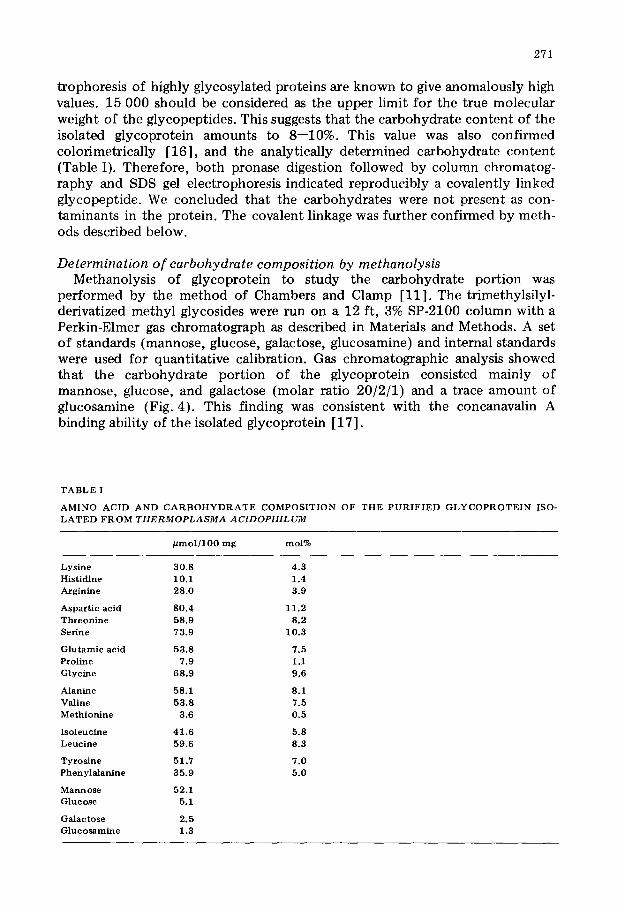
2 7 1
trophoresis of highly glycosylated proteins are known to give anomalously high values. 15 000 should be considered as the upper limit for the true molecular weight of the glycopeptides. This suggests that the carbohydrate content of the isolated glycoprotein amounts to 8--10%. This value was also confirmed colorimetrically [16], and the analytically determined carbohydrate content (Table I). Therefore, both pronase digestion followed by column chromatog- raphy and SDS gel electrophoresis indicated reproducibly a covalently linked glycopeptide. We concluded that the carbohydrates were not present as con- taminants in the protein. The covalent linkage was further confirmed by meth- ods described below.
Determination of carbohydrate composition by methanolysis Methanolysis of giycoprotein to study the carbohydrate portion was
performed by the method of Chambers and Clamp [11]. The trimethylsilyl- derivatized methyl glycosides were run on a 12 ft, 3% SP-2100 column with a Perkin-Elmer gas chromatograph as described in Materials and Methods. A set of standards (mannose, glucose, galactose, glucosamine) and internal standards were used for quantitative calibration. Gas chromatographic analysis showed that the carbohydrate portion of the glycoprotein consisted mainly of mannose, glucose, and galactose (molar ratio 20/2/1) and a trace amount of glucosamine (Fig. 4). This finding was consistent with the concanavalin A binding ability of the isolated glycoprotein [ 17 ].
T A B L E I
A M I N O A C I D A N D C A R B O H Y D R A T E C O M P O S I T I O N OF T H E P U R I F I E D G L Y C O P R O T E I N ISO- L A T E D F R O M T H E R M O P L A S M A A C I D O P H I L U M
p m o l ] 1 0 0 rag mol%
Lysine 30.8 4.3 Histidine 1 O.1 1.4 Arginine 28.0 3.9
Aspart ic acid 80.4 11.2 Threon ine 58.9 8.2 Serine 73.9 10.3
Glutamic acid 53.8 7.5 Proline 7.9 1.1 Glycine 68.9 9.6
Alanine 58.1 8.1 Valine 53 .8 7.5 Methionine 3.6 0.5
Isoleucine 41 .6 5.8 Leucine 59.6 8.3
"ryroslne 51.7 7.0 Phenylalanine 35.9 5.0
Mannose 52.1 Glucose 5.1
Galactose 2.5 Glucosamine 1.3

272
mort
gal ok: l S qlcNac
I0 20 3() 4() 50 6'0 "?0 RETENTION TIME (min)
Fig. 4. Gas- l iquid ch roma tog raphy o f t r imethy isL ly la ted m e t h y l giycosides o f the pu r i f i ed g lycopept ide f r o m the plasma membrane o f T. acldophilum. Analys is was p e r f o r m e d on a 12 f t , 3% SP-2100 co lumn (80 - -100 mesh, on Supe leopor t ) . Marmi to ] was used as in te rna l standard (I.S.). The peak eluted at about 43 min was n o t ident i f ied .
Amino acid analysis of the glycoprotein (Table I) The amino acid composi t ion of the glycoprotein was analyzed as described
in Materials and Methods. Hydrophobic amino acids, as defined by Tanford [18], comprised at least 62 mol% of the protein portion, while acidic amino acids amounted to 9 mol% more than the basic amino acids.
Glycosidic linkage studies of the glycoprotein The purified glycoprotein resisted alkaline hydrolysis [19]; this suggested
that the carbohydrates were N-glycosidically linked to the peptide. Exposure of glycoproteins to anhydrous hydrogen fluoride cleaves all the
linkages of neutral and acidic sugars while leaving peptide bonds and glyco- peptide linkages of amino sugars intact [14]. Following HF treatment, the pro- tein port ion was separated from the carbohydrate moiety by dialysis. Methano- lysis was carried out on the protein portion. Glucosamine was the only sugar detected.
Digestions of glycopeptide by exo- and endoglycosidases The purified glycoprotein was first incubated with a-mannosidase. This
enzyme was selected since the glycoprotein can be purified by concanavalin A affinity chromatography. Also from the carbohydrate content (Fig. 4), mannose accounted for 85%. After 72 h of incubation (Materials and Methods), 70% (w/w) of the mannose was released. Further digestion with a-mannosidase in the presence of SDS (1%) for another 48 h did not remove any more mannose. The glycopeptide recovered from a-mannosidase digestion was further treated with ~-glucosidase and a-mannosidase. This reaction enabled the release of 62% of the glucose and 90% of the mannose residues (Table II). Further digestion with fl-galactosidase, ~-glucosidase and a-mannosidase resulted in virtually total removal o f the galactose and glucose residues

2 7 3
T A B L E II
M O L A R R A T I O S OF C A R B O H Y D R A T E R E S I D U E S F R O M P U R I F I E D G L Y C O P R O T E I N A N D AF- T E R T R E A T M E N T S W I T H G L Y C O S I D A S E
Molar ra t ios were ca lcu la ted by set t ing the value for g lucosamine as 1.0. Th e g lucosamine c o n t e n t r e m a i n e d c o n s t a n t a f t e r p ro longe d hydrolys is . The values in p a r en th e se s were ca lcu la ted as tool c a rbohy- d r a t e ] g l y c o p r o t e i n .
G l y e o p r o t e i n A f t e r d iges t ion w i t h :
a-Mannos idase a -Marmosidase ~-Mannosidase a -Mannos idase 13-glue osidase ~-glucosidase ~-glue osidase
f3-galaetosidase ~3-galaetosidase endog lycos idase
Mannose 40.1 (80 .2 ) 11 .8 3.9 0.9 - - Glucose 3.9 (7 .8) 3.9 1.5 t race - - Galac tose 1.9 (3 .8) 1.9 1.9 t race - - G l u c o s a m i n e 1.0 (2 .0) 1.0 1.0 1.0 (1 .0)
(Table II). From the experiments on exoglycosidase digestions, it was con- cluded that the carbohydrate portion of the glycoprotein had a long chain of a-mannose (55--56 residues) at its non-reducing end, followed by a region con- taining six residues of fl-glucose, and 16 of a-mannose, followed by another region containing four residues of ~-galactose, three residues of/3-glucose, and six residues of a-mannose. After the exoglycosidase digestion, the glycopeptide was treated with endoglycosidase H. Since this enzyme removed all the remaining carbohydrates except one glucosamine residue (Table II), this indi- cated that the reducing end comprised two N-acetylgiucosamine residues (chitobiose), fl-linked, followed by mannose (1--2 residues). The inability to remove the last trace (1--2 residues) of mannose with a-mannosidase, suggested that at least one of the mannose residues was/3-1inked.
The glycosidases were used without further purification. Nevertheless, results from permethylation experiments, followed by mass spectrometry, supported all the information derived from studies with glycosidases.
Permethylation studies of the glycopeptides To determine linkages, glycoproteins were first treated with pronase and
glycosidases, then subjected to permethylation [12]. Partially methylated acetates were analyzed by combined gas-liquid chromatography-mass spectro- metry and the data were interpreted as described [13]. Partially methylated derivatives of mannose, glucose, galactose, and glucosamine were identifed both by their retention times in the gas chromatograms and by mass spectrometry by their fragmentation patterns, and were further compared with known refer- ence standards. Permethylation and mass spectral analyses of the glycopeptides gave evidence for the presence of 2,3,4,6-tetra-O-methylmannitol, 3,4,6-tri-O- methylmannitol, 2,4,6-tri-O-methylmannitol, 2,3,4-tri-O-methylmannitol, 2,4- di-O-methylmannitol, 2,3,6-tri-O-methylglucitol, 2,4,6-tri-O-methylgalactitol, and 3,6-di-O-methylglucosaminitol (Table III). The presence of 2,3,4,6-tetra- O-methylmannitol as the only tetramethylated carbohydrate, indicated that there were eight non-reducing terminal mannoses. The presence of an abundant

2 7 4
T A B L E III
M O L A R R A T I O S A N D R E T E N T I O N T I M E S O F P A R T I A L L Y M E T H Y L A T E D A L D I T O L A C E T A T E S O F M A N N O S E , G L U C O S E , G A L A C T O S E A N D G L U C O S A M I N E O B T A I N E D BY P E R M E T H Y L A T I O N O F T H E P U R I F I E D G L Y C O P E P T I D E
M o l a r r a t i o s we re c a l c u l a t e d us ing 1 , 4 , 5 - t r i - O - a c e t y l - 3 , 6 - d i - O - m e t h y i g l u c o s a m i n i t o l as 1 .0 . The va lues in p a r e n t h e s e s we re c a l c u l a t e d as m o l c a r b o h y d r a t e / g l y c o p r o t e i n . R e t e n t i o n t i m e ana lys i s was p e r f o r m e d o n a 6 ft , 3%, O V - 2 2 5 ( 8 0 - - 1 0 0 m e s h o n S y p c l c o p o r t ) a n d o p e r a t e d i s o t h e r m a l l y for 50 ra in a t 1 7 0 ° C , t h e n p r o g r a m m e d f r o m 1 7 0 ° C to 2 5 0 ° C a t 5 ° C / r a i n , w i t h h e l i u m as ca r r i e r gas. Q u a n t i t a t i v e e v a l u a t i o n o f t he c h r o m a t o g r a m s was p e r f o r m e d w i t h a H e w l e t t - P a c k a r d i n t e g r a t o r , m o d e l 3 3 7 0 B .
M o l a r r a t i o s R e t e n t i o n t i m e ( m i n )
B e f o r e A f t e r ~ - m a n n o s i d a s e ~ - m a n n o s i d a s e d ige s t i on d iges t i on
1 , 5 - D i - O - a c e t y l - 2 , 3 , 4 , 6 - t e t r a - O - m e t h y l m a n n i t o l 4 .1 (8 .2 ) 0 1 , 2 , 5 - T r i - O - a c e t y l - 3 , 4 , 6 - t r i - O - m e t h y l m a n n i t o l 18 .4 ( 3 6 . 8 ) t r a c e 1 , 3 , 5 - T r i - O - a c e t y l - 2 , 4 , 6 - t r i - O - m e t h y l m a n n i t o l 6.1 ( 1 2 . 2 ) 6 .3 1 , 5 , 6 -T t i -O-ace t y l - 2 , 3 , 4 - t r i - O - m e t h y l m ann i t o ] 5.1 ( 1 0 . 2 ) 5.1 1 , 3 , 5 , 6 - T e t r a - O - a c e t y l - 2 , 4 - d i - O - m e t h y l m a n n i t o l 3 .5 (7 .0 ) t r a c e 1 ,4 ,5 -Tr i -O -ace ty l -2 ,3 ,60- t r i -O-m e t h y l g l u c i t o l 3.1 (6 .2 ) 2 .4 1 , 3 , 5 - T r i - O - a c e t y l - 2 , 4 , 6 - t r i - O - m e t h y l g a l a c t i to l 1 .5 (3 .0 ) 1.7 N-Acetyl-N-methyl-l,4,5-tri-O-acetyl-3,6-di-O- 1.0 (2 .0 ) 1 .0
m e t h y l g l u c o s a m i n i t o l
12 .2 22 .3 23 .2 26 .7 52.1 28 .3 24 .7 61 .4
amount of 3,4,6-tri49-methylmannitols (1,2-substituted) was consistent with the glycoprotein's capacity to bind concanavalin A. The presence of 2,4-di-O- methylmannitol (1,3,6-substituted) was indicative of a branched glycopeptide. From the molar ratios (Table III), it was obvious that the glycopeptide branched at seven locations. 2,4,6-Tri-O-methylmannitol (1,3-substituted) and 2,3,4-tri-O-methylmannitol (1,6-substituted) seemed to occur in about equal amounts. After e-mannosidase digestion, the 2,4<li-O-methylmannitol and 3,4,6-tri-O-methylmannitol derivatives were drastically reduced (Table III). This indicated that the branching was entirely at the outer mannose residues which amounted to 70% (w/w) (Table II) and which were linked predomi- nantly via ~ 1 -* 2 linkages. The middle inner core of the carbohydrate moiety
081404 SCAN
1 0 0 116 >" X 5 CH2OAc
t- 80 ; '16 H PI M/Me 1 5 8 i~e Y " ~ A c . . . . . . . -1 i z
P" 60 ~" . . . . . . - - _ HCOAc ,~ Z % 255
_ 40" 142 CH2OMe
< 20 • 129 202 J
" ' II 50 1 O0 150 200 250 300
m/e Fig . ,5. Mass s p e c t r u m of p a r t i a l l y m e t h y l a t e d a ld i t o l a c e t a t e i d e n t i f i e d as N-acetyl-N-methyl-l,4,5-tri-O. a c e t y l - 3 , 6 - d i - O - m e t h y l g l u c o s a m i n i t o l f r o m the p u r i f i e d g l y c o p e p t i d e o f t h e T. acidophnum p l a s m a m e m - b r a n e .

2 7 5
[Man (o, l -2) la-Mon (~1-3) _ • , IMan (col 2)j b Man (o:1 6) ~ Man (o:1-2)- lMan (o: l°2) ] i -Man(al"6).
- . N~ Man (a l -2 ) - Man (a 1-2) m-Man(o:1-6)• IMan (a 1-2)] c -Man (o:l-S) ~ Man(o: l*2)- [Man (a 1-2)] j -Man ( a l - 3 ) / ' J \ J Man (a 1-2)1d -Man (o: 1-3) ) M a n (o: 1-4)'-/ IMon ( o : l - 2 ) ; e - M o , , ( o : 1 - 6 ) • • / Man (a 1-2)- ~Man (o: 1-2)[k-Man (al-3) Man cE1-2 f - M a n o:1-3 " ~ (a l -2) - IMan (rxl-2)]n- Mort (a 1-3) Man
IMan (a 1-2)]g-Mon (a 1"3) ~> Mon {o:1-2)- IMan (o:1~2)lt-Mort (o:1-6) , /
Man (o: l -2) lh-Mon (o:1-6)
I GIc (/~ 1-4)11~ [Man (o:1-6)]¢~ IMon (al-3)]F [Gal (,6'l-3}]~[Man {o:1-3}]r GIc(D 1-4},-" Man (~ 1-4)'-GIc Noc (,81 *4)-GIcNoc {~ 1)-A rm - - PEP'rIDE
Fig . 6. P ~ o p o s e d s t r u c t u r e o f t h e T. acidophnura p l a s m a m e m b r a n e g l y c o p r o t e i n , a + b + c + d + e + f +
g + h + i + j + k + 1 + m + n = 3 9 r e s i d u e s ; q + r + t = 2 0 r e s i d u e s ; p = f i v e r e s i d u e s ; s ~ t h r e e r e s i d u e s . T h e
s e q u e n c e o f t h e r e s i d u e s i n s e g m e n t I a n d I I i s n o t c e r t a i n .
consisted of ].,4-substituted glucose, 1,3-substituted galactose, 1,2-, 1,3-, and 1,6-substituted mannose. The innermost section of the carbohydrate region comprised 1,4-substituted glucosamine residues (Fig. 5). Because a-mannosidase (Table II) was incapable of removing the last trace of mannose, this suggested to us that at least one mannose residue was ~-linked to the distal N-acetyl- glucosamine residue. A proposed structure of the glycopeptide is shown in Fig. 6.
Discussion
The purified membrane glycoprotein from T. acidophilum had an apparent molecular weight of 152 000 with less than 10% carbohydrate content (w/w). The carbohydrate moiety consisted mainly of mannose residues with branched a 1 -* 2 linkages at the non-reducing ends of the glycopeptide. The reducing end probably was an N-glycosidic linkage between the amino acid asparagine and N-acetylglucosamine, as evidenced by resistance to alkaline hydrolysis, cleavage by anhydrous HF, and endoglycosidase reactions. At least one mannose residue was not a-linked. The glycoprotein accounted for 40% (w/w) of the total membrane proteins. The non-reducing ends of the glycopeptide showed a highly branched pattern (Fig. 6) which may extend over the entire cell surface. This was supported by electron microscopic studies on Thermo- plasma membranes labelled with concanavalin A [20].
N-Glycosidically linked, mannose-rich glycoproteins have been found in animal systems [21], fungi [22], yeast [22], and plants [23]. It appears that we have demonstrated for the first time a N-giycosidically linked, mannose- rich procaryotic glycoprotein. Preliminary experiments carried out in our laboratory appear to indicate that glycosylation of certain membrane proteins may contribute to the survival ability of T. acidophilum at extreme environ- ments. We supplied growing cultures of T. acidophilum with bacitracin. There seems to exist a correlation of growth at increasing pH values and the reduced amount of carbohydrates in membrane glycoproteins. Bacitracin is an anti-

276
biotic which blocks peptidogiycan biosynthesis in bacteria by complexmg with the lipid pyrophosphate released after transfer of the lipid-linked subunit to the growing peptidoglycan chain [24 ].
The amino acid composit ion of the glycoprotein showed 62 mol% hydro- phobic residues, while the acidic amino acid content contr ibuted 9 mol% more than that of the basic amino acids. The amount of chargeable groups (at most 28 mol%) may be diminished further at the low optimal pH value because of the protonat ion of free carboxyl groups which predominate over the number of free amino groups. A high degree of hydrophobic i ty is of survival value since it possesses advantages for thermophily as well as acidophily. Hydrophobic interactions have a maximum stability around 60°C [25] which is very close to the opt imum growth temperature of T. acidophilum.
The native structure of proteins is stabilized by a combination of hydrogen bonding, electrostatic and hydrophobic interactions [26].
In the Thermoplasma membrane, 40% of the proteins consist of a mannose- rich glycoprotein. As far as interactions between the carbohydrate moiety and the membrane proteins are concerned, hydrophobic interactions probably play an important role [26]. Consequently the effect of carbohydrates on proteins depends on the influence of sugars upon water structure. Constructing Pauling- Corey spacefilling models of the carbohydrate portion of the Thermoplasma glycoprotein, one easily visualizes the extreme flexibility of the 1,6-linkage between the corresponding [27] carbohydrate sections. Furthermore, sugar conformations may be visualized capable of generating polysaccharide surfaces which are virtually hydrophobic . The mutual hydroxyl orientations play obviously a considerable role. The array of hydroxyl groups participates in the formation of hydrogen bonds with water.
Interactions between regular periodic mannose portions of glycoproteins may generate periodic conformations such as a helix or a r ibbon [28]. Thus it is conceivable that the Thermoplasma surface is surrounded by a mesh-like carbohydrate coat where water molecules are immobilized. The resulting water turgor and the interactions between carbohydrates provide perhaps additional factors for the rigidity of the limiting plasma membrane. Indeed, electron para- magnetic resonance studies [29] have shown that the T. acidophilum mem- brane is the most rigid membrane reported hitherto.
The capacity of neutral or anionic carbohydrates [28] to form complexes may further contr ibute to the stability of the membrane. Given the appropriate coordination geometry, cations may link carbohydrates with the underlying protein-lipid framework. The rigidity of the Thermoplasma membrane is actually enhanced in the presence of Ca 2÷ or Al 3÷ [30,31] . It appears worth while to mention that rather limited information is available regarding struc- tural and thermodynamic properties of 1,2-1inked polysaccharides [32].
In conclusion, the existence of a mannose-rich glycoprotein in the wall-less T. acidophilum provides presumably a protective coat towards the harsh environment. The stereochemistry and conformation of the carbohydrate net- work, in conjunction with an ensuing hydroskeleton, may contr ibute to the stability of the membrane and the cation binding.

2 7 7
Acknowledgements
The authors wish to thank Dr. D.T.A. Lamport for his personal support and critical evaluation of this work. This work was supported by the US Depart- ment of Energy Contract No. EY-76-C-02-1338.
References
1 Kornfe ld , R. a n d Kornfe ld , S. (1976) Annu . Rev. Biochem. 45, 2 1 7 - - 2 3 7 2 Razin, S. (1975) Prog. Surf. Membrane ScL 9, 2 5 7 - - 3 1 2 3 Mescher, M.F. a n d S t rominger , J .L. (1976) Proc. Natl . Acad. Sci. U.S. 73, 2 6 8 7 - - 2 6 9 1 4 Razin, S. (1978) Microbiol . Rev. 42, 4 1 4 - - 4 7 0 5 Dar land, G., Brock, T., Samsonof f , W. and Conti , S.F. (1970) Science 170, 1 4 1 6 - - 1 4 1 8 6 Yang, L.L. and Haug, A. (1977) Biophys . J . 17, 96a 7 L a n g w o r t h y , T.A. (1977) Biochim. Biophys . Ac ta 487 , 37---50 8 Yang, L.L. and Haug, A. (1975) Biophys. J. 2 1 , 2 0 6 a 9 Ruwar t , M.J. a n d Haug, A. (1975) Biochemis t ry 1 4 , 8 6 0 - - 8 6 6
10 Laemmli , U.K. (1970) Nature 2 7 7 , 6 8 0 - - 6 8 5 11 Chambers , R.E. and Clamp, J .R. (1971) Biochem. J. 125, 1 0 0 9 - - 1 0 1 8 12 H a k o m o r i , S.-I. (1964) J . Biochem. ( To k y o ) 55, 2 0 5 - - 2 0 8 13 Bj6rndal , H., Hellerqvist, C.G. , Lindberg , B. and Svensson, S. (1970) Angew. Chem. Int. Edn. 8 , 6 1 0 - -
619 14 Mort , A.J . a n d L a m p o r t , D.T.A. (1977) Anal. Chem. 82, 2 8 9 - - 3 0 9 15 Wang, C.-S. a n d Smith , R.L. (1975) Anal. Biochem. 63, 4 1 4 - - 4 1 7 16 Roe, J.H. (1955) J . Biol. Chem. 2 1 2 , 3 3 5 - - - 3 4 3 17 Goldstein , I.J. (1976) in Coneanaval in A as a Tool (Bittiger, H. and Schnebl i , H.P., eds.), pp. 55- -57 ,
Wiley Publ. , New York 18 Tanfo rd , C. (1973) in The H y d r o p h o b i c Effect , pp. 120 - -121 , Wiley Publ. , New York 19 Spiro, R.G. (1971) Methods Enzymol . 28B, 3 - 4 3 20 Mayber ry-Carson , K.J . , Jewell , M.J. and Smith , P.P. 0 9 7 8 ) J . Bacteriol . 133, 1 5 1 0 - - 1 5 1 3 21 Montreui l , J . (1975) Pure Appl . Chem, 42, 4 3 1 - 4 7 7 22 Lucas, J~I., Waechter , C.J. and Lermaxz, W~I. (1976) J . Biol. Chem. 250 , 1 9 9 2 - - 2 0 0 2 23 Lis, H. a n d Sharon , N. (1978) J. Biol. Chem. 253 , 3 4 6 8 - - 3 4 7 6 24 S to rm, D.R. and S t rominger , J .L. (1973) J. Biol. Chem. 248, 3 9 4 0 - - 3 9 4 5 25 Scheraga, H. (1963) in The Proteins (Neura th , H., ed.), pp. 4 7 7 - - 6 9 4 , Academic Press, New York 26 Lakshmi , T.S. and Nandi , P.K. (1976) J . Phys. Chem. 80, 2 4 9 - - 2 5 2 27 Rees, D.A. (1972) Binchem. J. 126, 2 5 7 - - 2 7 3 28 Rees, D.A. (1975) in Biochemis t ry of Ca rbohydra t e s , MTP In te rna t iona l Review of Science (Whelan,
W.J., ed.) , Vol. 5, pp. 1- -42 , Universi ty Park Press, Bal t imore, MD 29 Smith , G.G., Ruwar t , M~I. and Haug, A. (1974) FEBS Lett . 45, 96 - -98 30 WeBer, H.G. a n d Haug, A. (1977) J . Gen. Mierobiol. 99, 3 7 9 - - 3 8 2 31 Vierstra, R. and Haug, A. (1978) Biochem. Biophys. Res. C o m m u n . 8 4 , 1 3 8 - - 1 4 3 32 Sugget t , A. (1975) in Water (Franks , F., ed.) , Vol. 4, pp. 5 1 9 - - 5 6 7 , Plenum Press, New York
Copyright © 2022 FDOKUMEN