
Modeling and Experimental Studies on Polymer Melting and ...

Nuclear Instruments and Methods in Physics Research B 344 (2015) 70–75
Contents lists available at ScienceDirect
Nuclear Instruments and Methods in Physics Research B
journal homepage: www.elsevier .com/locate /n imb
Proton-induced nanorod melting in a coating obtained from the pulsedlaser ablation of W2B5=B4C
http://dx.doi.org/10.1016/j.nimb.2014.12.0210168-583X/� 2014 Elsevier B.V. All rights reserved.
⇑ Corresponding author at: Department of Electrical, Electronics and ComputerEngineering, French South African Institute of Technology/Cape Peninsula Univer-sity of Technology, Bellville Campus, PO Box 1906, Bellville 7530, South Africa.
E-mail address: [email protected] (I. Tadadjeu Sokeng).
I. Tadadjeu Sokeng a,b,⇑, B.D. Ngom c,d,e, F. Cummings b, L. Kotsedi c,d, M. Msimanga f, M. Maaza c,d,R.R. Van Zyl a
a Department of Electrical, Electronics and Computer Engineering, French South African Institute of Technology/Cape Peninsula University of Technology, Bellville Campus,PO Box 1906, Bellville 7530, South Africab Electron Microscopy Unit, University of the Western Cape, Private bag x17, Bellville 7535, South Africac Nanosciences African Network (NANOAFNET), iThemba LABS-National Research Foundation, 1 Old Faure Road, 7129, PO Box 722, Somerset West, Western Cape Province, South Africad UNESCO-UNISA Africa Chair in Nanosciences/Nanotechnology, College of Graduate Studies, University of South Africa (UNISA), Muckleneuk Ridge, PO Box 392, Pretoria, South Africae Laboratoire de Photonique et de Nanofrabrication, Groupes de physique du Solide et Sciences des Matriaux (GPSSM), Facult des sciences et Techniques, Universit Cheikh Anta Diopde Dakar (UCAD), B.P. 25114 Dakar-Fann, Dakar, Senegalf iThemba LABS Gauten, Private Bag 11, WITS 2050, Johannesburg, South Africa
a r t i c l e i n f o a b s t r a c t
Article history:Received 29 September 2014Received in revised form 1 December 2014Accepted 5 December 2014
Keywords:Pulsed laser depositionW2B5=B4CProton irradiationNanorod meltingSpace application
Coatings obtained from pulsed laser ablated W2B5=B4C were irradiated with 900 keV protons at fluencesranging from about 1 � 1015 protons=cm2 to about 4 � 1015 protons=cm2. Scanning Electron Micros-copy (SEM) and Energy Dispersive Spectroscopy (EDS) were used to study the resulting structural effects.Clusters of nanorods were observed to disperse and reduce in number with increase in proton fluence.The atomic percentage of constituent elements were observed to vary with proton fluence, both withinthe nanorods and the film floor. Our results show that the structural effect of proton irradiation on thecoating is lateral dispersion of matter.
� 2014 Elsevier B.V. All rights reserved.
1. Introduction
Boron Carbide based ceramics are used for bullet-proof applica-tions because of their high hardness (about 30 GPa), high meltingpoint (2427 �C) and low density (2:52 g cm�3) [1–3]. They are alsoused for neutron absorption applications due to their high NeutronMass Absorption (2:4 m2 Kg�1, corresponding to about 600 barn)[3]. Monolithic B4C, however, has a low strength (< 300 MPa)and low fracture toughness (about 2:2 MPa m1=2) [4]. The additionof a W2B5 phase was reported to result in Boron Carbide basedcomposite ceramics with increased flexure strength and fracturetoughness [3,4]. The W2B5 phase was also used by Cai and Nan[5] to enhance the electric and thermal conductivity of B4C.
Coatings obtained from laser ablation of W2B5=B4C are promis-ing for space applications at low earth orbit. One of the targetedapplications of this coating is the shielding of low energy space
radiations, among which are low energy protons. Protons withenergies < 10 MeV are the most probable in low earth orbit [6].To investigate the structural effects of low energy protons on thesecoatings, they were irradiated with 900 keV protons at fluencesranging from about 1 � 1015 protons=cm2 to about 4 � 1015
protons=cm2. Therefore, this work is consistent with similar protonirradiation investigations towards the space applicability of othernano-materials (such as Zinc oxide (ZnO), Vanadium dioxide(VO2) and fullerene nanorods) in low earth orbit [7–9].
2. Material and methods
The target used to make the coating was a B4C=W2B5 pellet inthe shape of a regular hexagon. Its diameter was 25 mm, its heightwas 6 mm, and it weighed 6:23 g. Before being used for the depo-sition of the coatings, its crystal structure was investigated. Thiswas done by X-ray Diffraction (XRD) using BRUKER AXS equipmenthaving a D8 Advance diffractometer, a Cu� Ka radiation tube(kKa1 ¼ 1:5406 Å) and a PSD Vantec-1 detector. The intensity ofthe direct beam is obtained using a variable tube current of40 kV and 40 mA with Cu radiation of wavelengths Ka1 length.

Fig. 1. XRD spectrum of the W2B5=B4C target.Fig. 2. Heavy Ion ERDA depth profile of the target.
I. Tadadjeu Sokeng et al. / Nuclear Instruments and Methods in Physics Research B 344 (2015) 70–75 71
The goniometer comprises a set up with N �1 Vantec detector andfilter 4 degrees of Soller slit and providing narrow symmetricalprofiles instrumental (PI) to the desired angular range. The spectraof X-ray diffraction of the samples were recorded in the range2h ¼ 12� 80� using in each case a step size of 0:02� and a counttime of 20 s per step without moving the sample. The instrumentalprofile was obtained experimentally by collecting diffraction dataof a standard Al2O3 powder by fitting with pseudo-Voigt functionsand setting changes on the value of the width at half height. Aqualitative phase analysis (search match) was carried out usingthe PANAlytical X’pert Highscore plus software employing theICDD PDF database. The B4C phases were identified by comparisonwith the Powder Diffraction File (PDF) card No JCPDS000350798and the W2B5 phases were identified using the fileJCPDS000381365. Fig. 1 shows the XRD spectrum of the target.The target was confirmed to be a B4C=W2B5 (B4C rhombohedral lat-tice structure, and W2B5 hexagonal lattice structure) composite.Fig. 2 shows a 10% oxygen content revealed by Heavy Ion ElasticRecoil Detection Analysis (Heavy Ion ERDA) [10].
The substrates on which the coatings were deposited were cutfrom corning glass (approximately 1 cm by 1 cm in dimension).They were cleaned using a BRANSONIC ultrasonic cleaner(70 W;42 kHz �6%). During the cleaning process, they wereimmersed in methanol, acetone, trichloroethylene, distilled waterand methanol again for 5 min each in an ultrasound water bath.Although for the targeted applications the films are to be depositeddirectly on integrated circuit components, glass was used as sub-strate for characterisation purposes.
The pulsed laser deposition was done using a Q-switched 3rdharmonic Nd� YAG (Spectra Physics) laser with a wavelength of355 nm and a frequency of 10 Hz. The laser fluence was18:05 J=cm2. The target was 3:7 cm away from the substrate in avacuum chamber at 4 � 10�5 mbar. The deposition was carriedout at room temperature and the deposition time was 15 min persample, for 5 different identical samples.
The samples were irradiated with a focused proton beam ema-nating from a standard Van De Graaf accelerator. The protonenergy was set to 900 keV, the beam diameter was 6 mm and thebeam current was maintained at 14 nA. The proton flux was3:095 � 1011 protons=cm2=s and 4 samples were irradiated atfluences ranging from 1 � 1015 protons=cm2 to 4 � 1015
protons=cm2 at room temperature in a vacuum chamber at5 � 10�6 mbar. The 5th sample was not irradiated and was usedas control sample.
Scanning Electron Microscopy (SEM) was done with a ZeissAuriga field emission gun SEM (FEG-SEM) operated at 5 kV for
secondary electron imaging, using an inlens detector; and 20 kVfor Energy Dispersive Spectroscopy (EDS) analysis. The EDS spectrawere collected using an Oxford Instruments X-Max solid-state sil-icon drift detector.
3. Results and discussion
Fig. 3(a–e) shows the SEM images of the coating as depositedand irradiated at different fluences. These fluences were1 � 1015 protons=cm2;2 � 1015 protons=cm2;3 � 1015 protons=cm2
and 4 � 1015 protons=cm2. The droplets that are characteristic ofPLD are observed on every sample. The as-deposited coatingshows clustered nanostructures of no particular shape, as shownin Fig. 3(a). These structures are referred to as rods in this workbecause they look like many rods ‘‘glued’’ together, despite theirirregularity in shapes. These clusters appear to have formed inthe later stages of the film growth, as they are on top of thesurface of the film floor, from which they are quite distinct. Thissuggests that either the growth was entirely of Volmer–Webertype (favoured by the high laser fluence during deposition)[11], or it was of Stranski–Krastanov type (favoured by thethermal expansion coefficient mismatch between the substrateand the coating: 4:6 � 10�6=�C for corning glass [12], and2:6 � 10�8=
�C for W2B5 [13]) [14]. A close observation ofFig. 3(b–d), in particular on the 200 nm scale, shows an increas-ing number of features merged with the film floor. These fea-tures are absent in Fig. 3(a) and (e). One can also observe thatthe rod shapes are not regular, except for those in Fig. 3(e),which are uniformly shaped rods about 405 nm long and90 nm wide. These rods are longer than those in Fig. 3(d) (about320 nm long for the more regular looking rods). This suggeststhat the clusters melt as the proton fluence increases from1 � 1015 protons=cm2 to 3 � 1015 protons=cm2. At 4 � 1015
protons=cm2, no further melting occurs, but instead, the rodswhich are not molten seem to grow.
Fig. 4(a) shows the average cluster area as a function of irradi-ation fluence. This figure shows a slight increase from 0:29 lm2
at 3 � 1015 protons=cm2 to 0:38 lm2 at 4 � 1015 protons=cm2.The average cluster size at fluences of 3 � 1015 protons=cm2 and4 � 1015 protons=cm2 correspond to the average area of individualrods, since at those fluences there are little or no agglomerations.In terms of the number of clusters per unit area, averagingover several spots showed a distribution of about 0:07 clusters=lm2;1:48 clusters=lm2;6:72 clusters=lm2;0:23 clusters=lm2, and
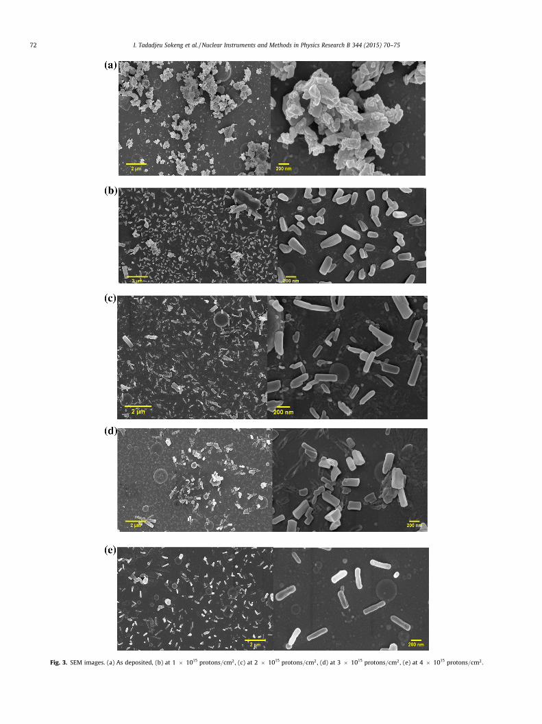
Fig. 3. SEM images. (a) As deposited, (b) at 1 � 1015 protons=cm2, (c) at 2 � 1015 protons=cm2, (d) at 3 � 1015 protons=cm2, (e) at 4 � 1015 protons=cm2.
72 I. Tadadjeu Sokeng et al. / Nuclear Instruments and Methods in Physics Research B 344 (2015) 70–75

Fig. 4. (a) Average cluster area as a function of irradiation fluence, (b) average roddimensions as a function of irradiation fluence.
Table 1Summary of EDS analysis on rods and film floor.
Structure Boron Carbon Tungsten Oxygen
As depositedRod 0.7060969 0.1758401 0.0085717 0.1094913Floor 0.3460639 0.5262948 0.0277557 0.0998856
At 1 � 1015 protons=cm2
Rod 0.6069097 0.2345462 0.0027392 0.1558049Floor 0.4861638 0.4126669 0.0069280 0.0942413
At 2 � 1015 protons=cm2
Rod 0.5407147 0.2323496 0.0015777 0.2253580Floor 0.5890330 0.3667203 0.0089517 0.0352950
At 3 � 1015 protons=cm2
Rod 0.4909736 0.3135224 0.0013592 0.1941448Floor 0.5353494 0.3770116 0.0438195 0.0438195
At 4 � 1015 protons=cm2
Rod 0.4060836 0.4568133 0.0041864 0.1329167Floor 0.5438454 0.3189551 0.0048654 0.1323341
I. Tadadjeu Sokeng et al. / Nuclear Instruments and Methods in Physics Research B 344 (2015) 70–75 73
0:60 clusters=lm2, respectively for the sample as deposited and foreach irradiation fluence. This is normal as the fewer large clustersmelt down to many smaller clusters as their constituent nanorods
melt. These clusters in turn reduce in amount per unit area as therods melt further. Fig. 4(b) shows the average rod dimensions as afunction of irradiation fluence. It should be noted that this analysiswas only considered for irradiated samples. This is because the levelof agglomeration in the as-deposited sample did not allow for ameaningful rod distinction. Of interest is the observation that at4 � 1015 protons=cm2, there is a considerable increase in averagerod length. The corresponding average rod width, however, doesnot increase as much, but does not decrease either. This is consis-tent with the slight increase in cluster average area from0:29 lm2 at 3 � 1015 protons=cm2 to 0:38 lm2 at4 � 1015 protons=cm2. These observations support the propositionthat after 3 � 1015 protons=cm2, the melting stops and a wellaligned rod growth starts, and this growth can be observed at4 � 1015 protons=cm2.
EDS measurements were made on a glass substrate and on thesamples. This was to eliminate uncertainties related to the compo-sition of the coatings. The glass and the coating samples were firstcoated with about 10 nm of Au/Pd to avoid charging. Indeed, themeasurements on glass revealed O, Si, Na, Mg, Sn, Ca, Au and Pd,and the measurements on the samples revealed O, Si, Na, Mg, Sn,Ca, Au, Pd, B, C and W. The glass measurements and the evidenceof Oxygen presence in the target from Fig. 2 suggest that the coat-ings contain B, C, W and O. The O:Si ratio in glass was used todetermine the amount of Oxygen attributed to the coatings only,given that the amount of Silicon measured in the samples wasattributable only to the glass substrate. To obtain the compositionof the coatings, the amount of Oxygen attributable to the coatingswas calculated as ðmeasured O in coating sampleÞ � ððmeasuredSi in coating sampleÞ � ðO : Si ratio in glass substrateÞÞ. Then, theSi, Na, Mg, Sn, Ca, Au and Pd measured in the samples were ignoredand the content of B, C, W and O were re-scaled as½B C W O�=ð
Pð½B C W O�ÞÞ. The results are shown in Table 1. While
the measured EDS values did not exceed 2 decimal places, theresults in Table 1 are not measured but are computed using themeasured EDS data. They were therefore not rounded down toavoid misinterpretations during the result analysis. Fig. 5(a–d)illustrates the content of Table 1. It shows the atomic percentagesof each element in the coating as a function of proton fluence, bothin the rods and in the film floor. These graphs show an inverse rela-tionship between the atomic percentage of each element in therods and in the floor. In Fig. 5(a), Boron is seen to steadily reducefrom the rods while at the same time increasing in the floor asthe proton fluence increases. In Fig. 5(b), Carbon increases steadilyin the rods as it decreases from the floor at the same time withincrease in proton fluence. In Fig. 5(c), Oxygen increases in the rodsand then drops after 2 � 1015 protons=cm2 while following theinverse trend in the floor. In Fig. 5(d), Tungsten decreases steadilyfrom the rods and then increases at 4 � 1015 protons=cm2, whilefollowing the inverse trend in the floor. Empirical derivationsshow a stoichiometry of the form WaxBxC1�½ð1þaÞxþy�Oy wherea�½0:002;0:1�. This is true when the atomic percentages areconsidered cumulatively in the rods and in the floor. It should benoted that this stoichiometry is simply an estimate of elementalproportions rather than an expression of chemical states.
It is clear from the above observations that the energy that theprotons lose to the coating goes to melt the nanorods on the latterup to a certain point, then it contributes to the growth of uniformlyshaped rods. This is consistent with the known interactions of pro-tons with matter, and the tendency of Tungsten to catalyse nano-rod growth when thermally excited. Indeed, protons lose energythrough non-ionising processes such as the thermal excitation ofatoms, and in ionising processes such as inelastic collisions withatomic electrons [15]. The average energy required to free anelectron from an atom is two to four times greater than the actualionisation potential because of the energy lost in non-ionising

Fig. 5. Atomic percentage distribution per element as a function of proton fluence. (a) Boron atomic percentage distribution, (b) Carbon atomic percentage distribution, (c)Oxygen atomic percentage distribution, (d) Tungsten atomic percentage distribution.
Fig. 6. Light Microscopy image and surface 3D representation of an irradiated sample showing the wave front of laterally displaced matter.
74 I. Tadadjeu Sokeng et al. / Nuclear Instruments and Methods in Physics Research B 344 (2015) 70–75
processes [15]. Furthermore, thermal excitation has been reportedto provide appropriate conditions for growth of nanorods in thepresence of Zinc [16] and Tungsten [12] as catalysts. This meansthat the energy from the incident protons was lost to the coatingas thermal energy and melted the clusters down to individual rods.These rods in turn were sufficiently thermally excited that theirTungsten content catalysed their growth into nanorods of uniformshape. In fact, these rods are very similar in size and shape to WCnanorods synthesised by different processes where WO3 nanorodsand thermal treatment played a key role [17–20].
The interactions described and discussed so far confirm that thecoating deposited in this work transfers the energy from incidentprotons laterally across its surface rather than normally throughitself. This lateral transfer of energy translates into lateraldisplacement of matter as illustrated in Fig. 6. This figure showsa zoomed light microscope image of the sample irradiated at4 � 1015 protons=cm2 and its surface 3D representation. Clearlyvisible are the displaced matter wave front and the depression atthe irradiated spot from where matter is displaced across thecoating. Indeed, the melting of its surface nanorod clusters, the

I. Tadadjeu Sokeng et al. / Nuclear Instruments and Methods in Physics Research B 344 (2015) 70–75 75
resulting cluster dispersion and the subsequent growth ofuniformly shaped nanorods are key effects making this coatingsuitable for space applications.
4. Conclusion
The pulsed laser deposition of W2B5=B4C onto corning glass sub-strates produced a coating that exhibits lateral diffusion of energywhen irradiated with 900 keV protons at fluences ranging fromabout 1 � 1015 protons=cm2 to about 4 � 1015protons=cm2. Thisenergy is laterally diffused through the melting of the surfaceclustered nanorods and the subsequent growth of uniformlyshaped nanorods. The laterally diffused energy translates intolateral displacement of matter across the coating. This lateraldiffusion of energy and displacement of matter makes the coatingsparticularly promising for space applications. Our results alsoshow further evidence that Tungsten is indeed a catalyst for theformation of nanorods.
Acknowledgement
We thank the French South African Institute of Technology/Cape Peninsula University of Technology and iThemba LABS-National Research Foundation for providing the necessary sponsor-ship and experimental facilities for this work.
References
[1] Z. Wang, Fabrication and applications progress on boron carbon for bulletproofceramics, China Powder Sci. Technol. 14 (3) (2008) 56–59.
[2] Z. Wang, Research progress of boron carbon for bulletproof ceramic, Bull. Chin.Ceram. Soc. 27 (1) (2008) 132–135.
[3] J. Yin, Z. Huang, X. Liu, Z. Zhang, D. Jiang, Microstructure, mechanical andthermal properties of in situ toughened boron carbide-based ceramiccomposites co-doped with tungsten carbide and pyrolytic carbon, J. Eur.Ceram. Soc. 33 (10) (2013) 1647–1654.
[4] G. Wen, S. li, B. Zhang, Z. Guo, Processing of in situ toughened B�W � Ccomposites by reaction hot pressing of B4C and WC, Scr. Mater. 43 (9) (2000)853–857.
[5] K. Cai, C. Nan, The influence of W2B5 addition on microstructure andthermoelectric properties of B4C ceramic, Ceram. Int. 26 (5) (2000) 523–527.
[6] M. Mayanbari, Y. Kasesaz, Design and analyse space radiation shielding for ananosatellite in Low Earth Orbit (LEO), in: 5th International Conference onRecent Advances in Space Technologies, 2011, pp. 487–493.
[7] T. Sechogela, L. Kotsedi, M. Nkosi, C. Sandt, R. Madjoe, W. Przybylowicz, K.Bharuth-Ram, M. Maaza, 2 MeV proton irradiation effects on ZnO singlecrystal, Surf. Rev. Lett. 21 (1) (2014).
[8] L. Mathevula, Deep space radiations-like effects on VO2 smart nano-coatingsfor heat management in small satellite (Master’s thesis), University of SouthAfrica, 2014.
[9] C. Mtshali, L. Kotsedi, B. Ngom, C. Ndlangamandla, O. Ndwandwe, M. Maaza,Structural investigation of 2 MeV proton-irradiated fullerene nanorods, Nucl.Instrum. Methods Phys. Res. Sect. B 296 (0) (2013) 22–25.
[10] M. Msimanga, D. Wamwangi, C. Comrie, C. Pineda-Vargas, M. Nkosi, T.Hlatshwayo, The new heavy ion {ERDA} set up at ithemba {LABS} gauteng:multilayer thin film depth profiling using direct calculation and monte carlosimulation codes, Nucl. Instrum. Methods Phys. Res. Sect. B 296 (0) (2013) 54–60, http://dx.doi.org/10.1016/j.nimb.2012.11.015. URL <http://www.science-direct.com/science/article/pii/S0168583X12007380>.
[11] M. Volmer, A. Weber, Keimbildung in bersttigten gebilden, Z. Phys. Chem. 119(1926) 277–301.
[12] B. Ngom, O. Sakho, N. Manyala, J. Kana, N. Mlungisi, L. Guerbous, A. Fasasi,M. Maaza, A. Beye, Structural, morphological and photoluminescenceproperties of w-doped ZnO nanostructures, Appl. Surf. Sci. 255 (2009)7314–7318.
[13] Y. Lv, G. Wen, T. Lei, Friction and wear behavior of c-based composites in situreinforced with fW2B5g, J. Eur. Ceram. Soc. 26 (15) (2006) 3477–3486, http://dx.doi.org/10.1016/j.jeurceramsoc.2005.09.031. <http://www.sciencedirect.com/science/article/pii/S095522190500823X>.
[14] N. Stranski, L. Krastanov, Theory of orientation separation of ionic crystals,Sitzungsberichte/ Akademie der Wissenschaften in Wien, Mathematisch-Naturwissenschaftliche Klasse Abteilung IIB 146 (1938) 797810.
[15] W. Shockley, Problems related to p-n junctions in silicon, Czech. J. Phys. B 11(1961) 81–121.
[16] N. Wang, Y. Cai, R. Zhang, Growth of nanowires, Mater. Sci. Eng. R 60 (1–6)(2008) 1–51.
[17] Y. Yan, L. Zhang, X. Qi, H. Song, J. Wang, H. Zhang, X. Wang, Template-freepseudomorphic synthesis of tungsten carbide nanorods, Small 8 (21) (2012)3350–3356.
[18] S. Shanmugam, D. Jacob, A. Gedanken, Solid state synthesis of tungsten carbidenanorods and nanoplatelets by a single-step pyrolysis, J. Phys. Chem. 109 (41)(2005) 19056–19059.
[19] N. Li, Y. Yan, B.-Y. Xia, J.-Y. Wang, X. Wang, Novel tungsten carbide nanorods:an intrinsic peroxidase mimetic with high activity and stability in aqueous andorganic solvents, Biosens. Bioelectron. 54 (2014) 521–527, http://dx.doi.org/10.1016/j.bios.2013.11.040. URL <http://www.sciencedirect.com/science/article/pii/S0956566313008269>.
[20] Z. Yan, M. Cai, P. Shen, Nanosized tungsten carbide synthesized by a novelroute at low temperature for high performance electrocatalysis, Sci. Rep. 3(2013) 1646.
Copyright © 2022 FDOKUMEN