Protective role of Chitosan against lead acetate induced liver damage in rats
16
J. of Biological Chem. Env. Res., Vol. 6 No. 2, 431 – 444 (2011). 1 Protective role of Chitosan against lead acetate induced liver damage in rats Hoda E. A., Farid; Medhat M., Abozid; S. M., EL-Sayed Biochemistry Department, Faculty of Agriculture, Menofia University, Shibin El- Kom, Egypt ABSTRACT Evaluation of protective role of chitosan against the toxicity induced by lead acetate in liver of male rats was investigated. Eighteen male albino rats were divided into three groups and treated as follow: Control, Lead (500mg/L in drinking water), Lead + chitosan (500mg/L in drinking water and 3% chitosan in diet). Treatments were expanded for six weeks. Biochemical investigations revealed that, lead acetate induced significant increase of triglycerides, total cholesterol, LDL-cholesterol levels in plasma, while HDL- cholesterol level was decreased. On the other hand a significant elevation in the activity of plasma alanine aminotransferase (ALT), aspartate aminotransferase (AST), alkaline phosphatase (ALP) and gamma glutamyl transferase (GGT) were recorded, but albumin level was decreased. Histopathological examination revealed degeneration of hepatocytes of rat liver treated with lead acetate. Chitosan supplement improve the detrimental effects of lead acetate in rats. KEYWORDS: Chitosan, Lead acetate, Lipid profile, Liver damage. 1- INTRODUCTION Heavy metal pollutants are of significant ecological concern because they are not biodegradable and have long half-lives in the soil (Ram et al., 2000). According to Okunola et al. (2007), these metals also get accumulated when plants and crops cultivated along major roads are consumed by man and animals. Lead (Pb), a highly toxic heavy metal, is widely distributed in nature. Pb is the most ancient poison known to man. Contamination of soil, water and air has become increasingly widespread through mining, refining and smelting operations. The concentrations of Pb in liver and bone of red deer and wild boar were higher in the mining area than in the control area (Reglero et al., Print to PDF without this message by purchasing novaPDF (http://www.novapdf.com/)
Transcript of Protective role of Chitosan against lead acetate induced liver damage in rats

J. of Biological Chem. Env. Res., Vol. 6 No. 2, 431 – 444 (2011).
1
Protective role of Chitosan against lead acetate induced liver damage in rats
Hoda E. A., Farid; Medhat M., Abozid; S. M., EL-Sayed
Biochemistry Department, Faculty of Agriculture, Menofia University, Shibin El-Kom, Egypt
ABSTRACT Evaluation of protective role of chitosan against the toxicity induced by
lead acetate in liver of male rats was investigated. Eighteen male albino rats
were divided into three groups and treated as follow: Control, Lead (500mg/L
in drinking water), Lead + chitosan (500mg/L in drinking water and 3%
chitosan in diet). Treatments were expanded for six weeks. Biochemical
investigations revealed that, lead acetate induced significant increase of
triglycerides, total cholesterol, LDL-cholesterol levels in plasma, while HDL-
cholesterol level was decreased. On the other hand a significant elevation in
the activity of plasma alanine aminotransferase (ALT), aspartate
aminotransferase (AST), alkaline phosphatase (ALP) and gamma glutamyl
transferase (GGT) were recorded, but albumin level was decreased.
Histopathological examination revealed degeneration of hepatocytes of rat
liver treated with lead acetate. Chitosan supplement improve the detrimental
effects of lead acetate in rats. KEYWORDS: Chitosan, Lead acetate, Lipid profile, Liver damage.
1- INTRODUCTION
Heavy metal pollutants are of significant ecological concern because they
are not biodegradable and have long half-lives in the soil (Ram et al., 2000).
According to Okunola et al. (2007), these metals also get accumulated when
plants and crops cultivated along major roads are consumed by man and
animals.
Lead (Pb), a highly toxic heavy metal, is widely distributed in nature. Pb is
the most ancient poison known to man. Contamination of soil, water and air
has become increasingly widespread through mining, refining and smelting
operations. The concentrations of Pb in liver and bone of red deer and wild
boar were higher in the mining area than in the control area (Reglero et al.,
Print to PDF without this message by purchasing novaPDF (http://www.novapdf.com/)

J. of Biological Chem. Env. Res., Vol. 6 No. 2, 431 – 444 (2011).
2
2009). The inhibitory effect of dietary Pb on digestive enzyme activities was
dietary Pb concentration dependent (Dai et al., 2009). Lead also is a
ubiquitous environmental and industrial pollutant that has been detected in
almost all phases of environmental and biological systems. This heavy use
has caused local and global contamination of air, dust and soil (CDC, Atlanta, 2002).
Chitosan a polyglucosamine derived from chitin is a cellulose-like polymer
located mainly in the exoskeletons of arthropods, such as carbs, shrimps,
lobsters and insects (Razdan and Pettersson 1994) it can be defined both
chemically and physiologically as a dietary fiber since it is a polysaccharide,
which cannot be digested by digestive enzymes of humans (Razdan and
Pettersson 1996). Chitin and its derivatives (chitosan, chitin oligosaccharide,
chitooligosaccharides) have characteristics that set them apart from other
polysaccharides and are used in medicine, pharmaceutics, biochemistry,
sewage treatment, agriculture and other areas (Sandford and Hutchings,
1987).
Chitosan is economically attractive, since it can be obtained from the
deacetylation of chitin, and chitin is the second most abundant biopolymer in
nature, next to cellulose (Annadurai et al., 2007). In nature, the main sources
of chitin/chitosan are from the animal and plant kingdoms, including the shells
of crustaceans and mollusks, the algae commonly known as marine diatoms,
and the cell walls of fungal species.
It is natural and nontoxic and growing evidence indicate that it exhibits a
marked hypolipidemic activity that would reduce the risk of cardiovascular
diseases (Zhou et al., 2006). It has exhibited a potent hypocholesterolemic
activity in rats (Simunek and Bartonova, 2005 and Liu et al., 2008).
Various studies of metal ion adsorption by chitosan have been undertaken in
recent years, such as the removal of Cu2+ ions from aqueous solution onto
chitosan and cross-linked chitosan beads (Wan et al., 2002, and Wan et al.,
2004). In addition, chitosan can be used to achieve adsorption of chromium
(Sag and Aktay 2002), cadmium (Evans et al., 2002), iron (Wan et al.,
2005), nickel (Pradhan et al., 2005 and Paulino et al., 2007) and lead
(Paulino et al., 2007) ions from aqueous solution.
Print to PDF without this message by purchasing novaPDF (http://www.novapdf.com/)
J. of Biological Chem. Env. Res., Vol. 6 No. 2, 431 – 444 (2011).
3
Therefore, the objectives of this study are: (i) to preparation of two type of
chitosan from shrimp waste and (ii) to determine whether chitosan would
reduce/prevent the liver damage, serum hyperlipidemia and hyperlipidemic
atherosclerosis induced by lead acetate.
2. MATERIALS AND METHODS 2.1. Production of chitosan
The production of chitosan involved the demineralization (DM),
deproteinization (DP), DC, and DA steps (No et al., 2003). Shrimp shell was
demineralized with 1N HCl for 30 min at ambient temperature with a
solid/solvent ratio of 1:15 (w/v). Following the DM step, the demineralized
shell was collected on a 100-mesh sieve, washed to neutrality in running tap
water, rinsed with deionized water, and filtered to remove excess moisture.
The DP step was accomplished by treating the demineralized shell with 3%
NaOH for 15min at 15 psi/121 °C and a solid/solvent ratio of 1:10 (w/v). The
residue was then washed and filtered as mentioned above. For the DC step,
the resulting chitin residue was bleached with 10% sodium hypochlorite
solution for 5min with a solid/solvent ratio of 1:10 (w/v). The bleached chitin
was collected, washed as mentioned above, and dried at 60 °C for 4 h in a
forced-air oven or by sun drying (approximately at 23 °C) for 4 h. The DA step
was achieved by treating chitin under conditions of 15 psi/121 °C with 45%
NaOH for 30min and a solid/solvent ratio of 1:10 (w/v). The resulting chitosan
was collected, washed as mentioned above, and dried at 60 °C for 4 h in a
forced-air oven or by sun drying (approximately at 23 °C) for 4 h.
2.2. Detremination of deacetylation degree of chitosan
The deacetylation degree of chitosan was determined by the
potentiometric titration method described by Broussignac, reported by
(Tolaimate et al., 2000). Chitosan was dissolved in a known excess of
hydrochloric acid. From the titration of this solution with a 0.1 M sodium
hydroxide solution, a curve with two inflexion points was obtained. The
difference between the volumes of these two inflexion points corresponded to
Print to PDF without this message by purchasing novaPDF (http://www.novapdf.com/)
J. of Biological Chem. Env. Res., Vol. 6 No. 2, 431 – 444 (2011).
4
the acid consumption for the salification of amine groups and permitted the
determination of chitosan’s acetylation degree, through Eq.
% NH2 = 16.1 ( V2 - V1 ) x Mb /W
where (V1) and (V2) are the base volumes referred to first and second
inflexion points, respectively, in mL, (Mb) is the base molarity in g/mol, and
(W) is the original weight of the polymer in g.
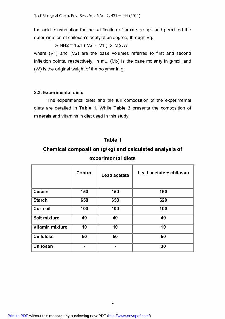
2.3. Experimental diets
The experimental diets and the full composition of the experimental
diets are detailed in Table 1. While Table 2 presents the composition of
minerals and vitamins in diet used in this study.
Table 1
Chemical composition (g/kg) and calculated analysis of
experimental diets
Control
Lead acetate
Lead acetate + chitosan
Casein 150 150 150
Starch 650 650 620
Corn oil 100 100 100
Salt mixture 40 40 40
Vitamin mixture 10 10 10
Cellulose 50 50 50
Chitosan - - 30
Print to PDF without this message by purchasing novaPDF (http://www.novapdf.com/)
J. of Biological Chem. Env. Res., Vol. 6 No. 2, 431 – 444 (2011).
5
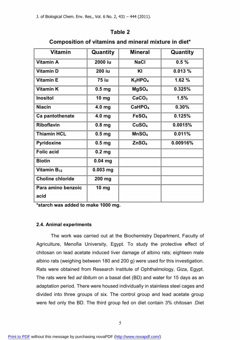
Table 2
Composition of vitamins and mineral mixture in diet*
Vitamin Quantity Mineral Quantity Vitamin A 2000 iu NaCl 0.5 %
Vitamin D 200 iu KI 0.013 %
Vitamin E 75 iu K2HPO4 1.62 %
Vitamin K 0.5 mg MgSO4 0.325%
Inositol 10 mg CaCO3 1.5%
Niacin 4.0 mg CaHPO4 0.30%
Ca pantothenate 4.0 mg FeSO4 0.125%
Riboflavin 0.8 mg CuSO4 0.0015%
Thiamin HCL 0.5 mg MnSO4 0.011%
Pyridoxine 0.5 mg ZnSO4 0.00916%
Folic acid 0.2 mg
Biotin 0.04 mg
Vitamin B12 0.003 mg
Choline chloride 200 mg
Para amino benzoic acid
10 mg
*starch was added to make 1000 mg.
2.4. Animal experiments
The work was carried out at the Biochemistry Department, Faculty of
Agriculture, Menofia University, Egypt. To study the protective effect of
chitosan on lead acetate induced liver damage of albino rats; eighteen male
albino rats (weighing between 180 and 200 g) were used for this investigation.
Rats were obtained from Research Institute of Ophthalmology, Giza, Egypt.
The rats were fed ad libitum on a basal diet (BD) and water for 15 days as an
adaptation period. There were housed individually in stainless steel cages and
divided into three groups of six. The control group and lead acetate group
were fed only the BD. The third group fed on diet contain 3% chitosan .Diet
Print to PDF without this message by purchasing novaPDF (http://www.novapdf.com/)
J. of Biological Chem. Env. Res., Vol. 6 No. 2, 431 – 444 (2011).
6
intake was monitored daily. The first group (C) was used as controls and
received tape water as drinking water. The other two groups; received tape
water containing lead acetate (C2H3O2)2Pb at a dose of 500 mg/L, in
drinking water, daily for six weeks. All rats fasted before blood sampling. The
blood samples were drawn from eye plexuses, after 6 weeks , the rats were
anesthetized using diethyl ether. The weight gain of the rats was recorded on
a weekly basis.
2.5. Blood sampling and analysis
Blood samples were collected after six weeks in tubes contain heparin
as an anticoagulant from the eye plexuses under diethyl ether anesthesia and
then centrifuged at 3000 rpm for 20 min. to obtain plasma, which was kept
frozen until analysis. The total cholesterol was analyzed calorimetrically
according Richmond, (1973). HDL - cholesterol was determined according to
Lopez et al. (1977). Acording to Demacker et al. (1984) LDL – cholesterol
was calculated as the difference between total cholesterol and HDL –
cholesterol. The triglycerides were analyzed according to Fossati and Prencipe (1982). The both of alanine-aminotransferase (ALT) and aspartate-
aminotransferase (AST) activities were measured according to the method
described by Reitman and Frankel (1957). Gamma glutamyl transferase
(GGT) activity was measured kinetic method of Szasz (1969). Alkaline
phosphatase (ALP) activity was measured by Hausamen et al., (1967). And
albumin was determined according Doumas et al. (1971).
2.6. Histopathology
Liver from the experimental groups were immediately fixed in 10%
formalin, then treated with conventional grades of alcohol and xylol,
embedded in paraffin and sectioned at 4–6 lm thickness. The sections were
stained with Hematoxylin and Eosin (H&E) stain for studying the
histopathological changes (Lillie, 1965).
2.7. Statistical analysis
The results of the animal experiments were expressed as the Mean ±
SD and they were analyzed statistically using the one-way analysis of
Print to PDF without this message by purchasing novaPDF (http://www.novapdf.com/)
J. of Biological Chem. Env. Res., Vol. 6 No. 2, 431 – 444 (2011).
7
variance ANOV A followed by Duncan’s test. In all cases p<0.05 was used as
the criterion of statistical significance.
3. RESULTS AND DISCUSSION
3.1. The degree of N-deacetylation of chitosan
The degree of N-deacetylation and the molecular weight of the
chitosan were determined by titration as described in our previous work. The
final deacetylation degree was 85%.
3.2. Effect of chitosan supplementation on lipid profile of rats intoxicated with lead acetate.
The expressed data in Table (3) revealed that intoxication with lead
acetate induced significant increases in triglycerides, total cholesterol, and
LDL-cholesterol levels in plasma of male albino rats. Meanwhile HDL-
cholesterol level was decreased. Our finding of elevated total cholesterol and lipoproteins supports previous
reports (El-Gazzar et al., 1989; and Newairy and Abdou 2009).
The association between lead exposure and high serum lipid levels is
biologically plausible and could be due to either increased synthesis or
decreased removal of lipoproteins. Lead nitrate-mediated development of
hepatic hypercholesterolaemia involves the activation of cholesterol
biosynthetic enzymes (i.e., 3-hydroxy- 3methyglutaryl-CoA reductase, farnesyl
diphosphate synthase, squalene synthase, lanosterol 14xdemethylase) and
the simultaneous suppression of cholesterol-catabolic enzymes such as 7a-
hydroxylase (Kojima et al., 2002).
However, supplementation with chitosan improved effect of lead acetate on
these parameters. This data are in line with Xu et al., (2007) ; and Osman et
al., (2010) who reported that chitosan could decrease levels of total
cholesterol, low density lipoprotein cholesterol in plasma. The protective
effects of chitosan could be attributed to its metal chelating properties. This
data is in line with (Qin et al., 2006) who reported chitosan can absorb a
significant amount of heavy metal ions.
Deuchi et al., (1994) reported that chitosan had potent ability to increase lipid
excretion into the feces. Some recent in vitro studies (Sugano et al., 1980;
Print to PDF without this message by purchasing novaPDF (http://www.novapdf.com/)
J. of Biological Chem. Env. Res., Vol. 6 No. 2, 431 – 444 (2011).
8
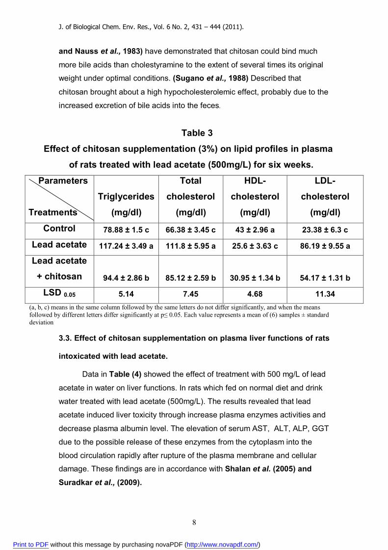
and Nauss et al., 1983) have demonstrated that chitosan could bind much
more bile acids than cholestyramine to the extent of several times its original
weight under optimal conditions. (Sugano et al., 1988) Described that
chitosan brought about a high hypocholesterolemic effect, probably due to the
increased excretion of bile acids into the feces.
Table 3
Effect of chitosan supplementation (3%) on lipid profiles in plasma
of rats treated with lead acetate (500mg/L) for six weeks.
LDL-
cholesterol
mg/dl)(
HDL-
cholesterol
mg/dl)(
Total
cholesterol
mg/dl)(
Triglycerides
mg/dl)(
Parameters
Treatments
23.38 ± 6.3 c 43 ± 2.96 a 66.38 ± 3.45 c 78.88 ± 1.5 c Control
86.19 ± 9.55 a 25.6 ± 3.63 c 111.8 ± 5.95 a 117.24 ± 3.49 a Lead acetate
54.17 ± 1.31 b 30.95 ± 1.34 b 85.12 ± 2.59 b 94.4 ± 2.86 b
Lead acetate + chitosan
11.34 4.68 7.45 5.14 LSD 0.05 (a, b, c) means in the same column followed by the same letters do not differ significantly, and when the means followed by different letters differ significantly at p≤ 0.05. Each value represents a mean of (6) samples ± standard deviation
3.3. Effect of chitosan supplementation on plasma liver functions of rats
intoxicated with lead acetate.
Data in Table (4) showed the effect of treatment with 500 mg/L of lead
acetate in water on liver functions. In rats which fed on normal diet and drink
water treated with lead acetate (500mg/L). The results revealed that lead
acetate induced liver toxicity through increase plasma enzymes activities and
decrease plasma albumin level. The elevation of serum AST, ALT, ALP, GGT
due to the possible release of these enzymes from the cytoplasm into the
blood circulation rapidly after rupture of the plasma membrane and cellular
damage. These findings are in accordance with Shalan et al. (2005) and
Suradkar et al., (2009).
Print to PDF without this message by purchasing novaPDF (http://www.novapdf.com/)
J. of Biological Chem. Env. Res., Vol. 6 No. 2, 431 – 444 (2011).
9
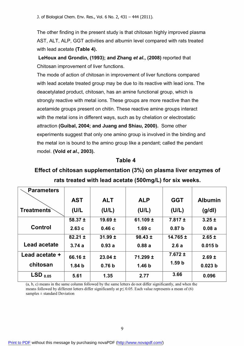
The other finding in the present study is that chitosan highly improved plasma
AST, ALT, ALP, GGT activities and albumin level compared with rats treated
with lead acetate (Table 4).
LeHoux and Grondin, (1993); and Zhang et al., (2008) reported that
Chitosan improvement of liver functions.
The mode of action of chitosan in improvement of liver functions compared
with lead acetate treated group may be due to its reactive with lead ions. The
deacetylated product, chitosan, has an amine functional group, which is
strongly reactive with metal ions. These groups are more reactive than the
acetamide groups present on chitin. These reactive amine groups interact
with the metal ions in different ways, such as by chelation or electrostatic
attraction (Guibal, 2004; and Juang and Shiau, 2000). Some other
experiments suggest that only one amino group is involved in the binding and
the metal ion is bound to the amino group like a pendant; called the pendant
model. (Vold et al., 2003).
Table 4 Effect of chitosan supplementation (3%) on plasma liver enzymes of
rats treated with lead acetate (500mg/L) for six weeks.
Albumin
(g/dl)
GGT
(U/L)
ALP
(U/L)
ALT
(U/L)
AST
U/L(
Parameters
Treatments 3.25 ±
0.08 a
7.817 ±
0.87 b
61.109 ±
1.69 c
19.69 ±
0.46 c
58.37 ±
2.63 c Control 2.65 ±
0.015 b 14.765 ±
2.6 a 98.43 ± 0.88 a
31.99 ± 0.93 a
82.21 ± 3.74 a Lead acetate
2.69 ± 0.023 b
7.672 ± 1.59 b
71.299 ± 1.46 b
23.04 ± 0.76 b
66.16 ± 1.84 b
Lead acetate + chitosan
0.096 3.66 2.77 1.35 5.61 LSD 0.05 (a, b, c) means in the same column followed by the same letters do not differ significantly, and when the means followed by different letters differ significantly at p≤ 0.05. Each value represents a mean of (6) samples ± standard Deviation
Print to PDF without this message by purchasing novaPDF (http://www.novapdf.com/)
J. of Biological Chem. Env. Res., Vol. 6 No. 2, 431 – 444 (2011).
10
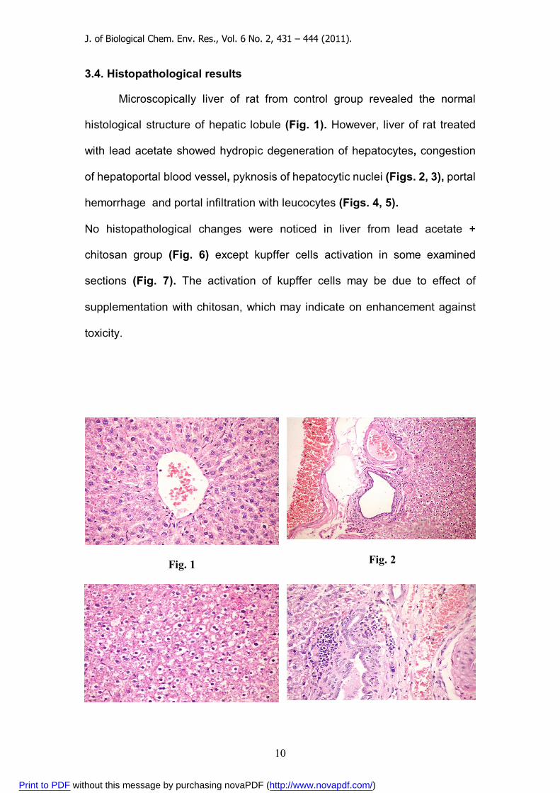
3.4. Histopathological results
Microscopically liver of rat from control group revealed the normal
histological structure of hepatic lobule (Fig. 1). However, liver of rat treated
with lead acetate showed hydropic degeneration of hepatocytes, congestion
of hepatoportal blood vessel, pyknosis of hepatocytic nuclei (Figs. 2, 3), portal
hemorrhage and portal infiltration with leucocytes (Figs. 4, 5).
No histopathological changes were noticed in liver from lead acetate +
chitosan group (Fig. 6) except kupffer cells activation in some examined
sections (Fig. 7). The activation of kupffer cells may be due to effect of
supplementation with chitosan, which may indicate on enhancement against
toxicity.
Fig. 1
Fig. 2
Print to PDF without this message by purchasing novaPDF (http://www.novapdf.com/)

J. of Biological Chem. Env. Res., Vol. 6 No. 2, 431 – 444 (2011).
11
Fig. 3 Fig. 4
Fig. 5
Fig. 6
Fig. 7
REFRENCES
Annadurai, G., Ling L. Y., Lee, J. F., 2007. “Adsorption of reactive dye from
an aqueous solution by chitosan: isotherm, kinetic and thermodynamic
analysis”, Journal of Hazardous Materials, accepted 3 July 2007.
CDC, Atlanta, 2002. Centers for disease control and prevention.
Developmental assessment and interventions. In: Managing Elevated Blood
Lead Levels Among Young Children: Recommendations from the Advisory
Committee on Childhood Lead Poisoning Prevention, pp. 79–95 (Chapter 5).
Dai, W., Du, H., Fu, L., Jin, C., Xu. Z., Liu, H., 2009. Effects of dietary Pb on
accumulation, histopathology, and digestive enzyme activities in the digestive
system of tilapia (Oreochromis niloticus). Biological Trace Element Research
127:124-31.
Print to PDF without this message by purchasing novaPDF (http://www.novapdf.com/)
J. of Biological Chem. Env. Res., Vol. 6 No. 2, 431 – 444 (2011).
12
Demacker AG, Hijmans BG, Brenninkmeijer AP, Jansen JS, and van't Laar A. 1984. Five methods for determining low-density lipoprotein
cholesterol compared. Clinic. Chem. 30:1797-1800.
Deuchi K., Kanauchi O., Imasato Y., and Kobayashi E., 1994. Decreasing
Effect of Chitosan on the Apparent Fat Digestibility by Rats Fed on a High-Fat
Diet. Biosci. Biotech. Biochem. 58 (9), 1613-1616.
Doumas B. T., Watson W. A., Biggs H. G., 1971. Albumin standards
and measurement of serum albumin with bromcresol green. Clin Chim
Acta 31: 87–96. El-Gazzar, R.M., El-Hen, S.A., Noweir, K.H., Shamy, M.Y., 1989. Study of
the lipoprotein pattern among workers exposed to lead. J. Egypt Public Health
Assoc. 64, 571–585.
Evans, J.R., Davids, W.G., MacRae, J.D., Amirbahman, A., 2002. Kinetics
of cadmium uptake by chitosan-based crab shells. Water Res. 36: 3219-3226.
Fossati P, Prencipe L. 1982. Serum triglycerides determined colorimetrically
with an enzyme that produces hydrogen peroxide. Clinic. Chem. 28: 2077–
2080.
Guibal, E. 2004. Interactions of metal ions with chitosan-based sorbents: a
review. Separation and Purification Technology. 38, 43-74.
Hausamen, T. V., Helger, R., Rick, W., and Gross, W., 1967. Optimal
conditions for the determination of serum alkaline phosphatase by a new
kinetic method. Clin. Chem. Acta., 15: 241 – 245.
Juang, R., and Shiau, R. 2000. Metal removal from aqueous solutions using
chitosan enhanced membrane ultrafiltration. Journal of Membrane Science.
165, 159-167.
Kojima M, Nemoto K, Murai U, Yoshimura N, Ayabe Y, and Degawa M., 2002. Altered gene expression of hepatic lanosterol 14xdemethylase
(CYP51) in lead nitrate-treated rats. Arch Toxicol; 76 : 398-403.
LeHoux, J.G. and F. Grondin, 1993. Some effects of chitosan on liver
function in the rat. Endocrinol., 132: 1078-1084.
Lillie, R.D., 1965. Histopathological technique and practical histochemistry,
third ed. Published by the Blakistar Division of McGraw-Hill Book Co., New
York, Toronto, London.
Print to PDF without this message by purchasing novaPDF (http://www.novapdf.com/)
J. of Biological Chem. Env. Res., Vol. 6 No. 2, 431 – 444 (2011).
13
Liu, J., J. Zhang and W. Xia, 2008. Hypocholesterolaemic effects of different
chitosan samples in vitro and in vivo. Food Chem., 107: 419-425.
Lopez,M.F; Stone,S.; Ellis,S. and Collwell,J.A., 1977. Cholesterol
determination in high density lipoproteins separated by three different
methods Clin. Chem., 23: 882 – 886.
Nauss J. L., Thompson J. L., Nagyvary J., 1983. The Binding of Micellar
Lipids to Chitosan. Lipids 18, 714-719.
Newairy A., A., and Abdou H., M., 2009. Protective role of flax lignans
against lead acetate induced oxidative damage and hyperlipidemia in rats. J.
Food and Chemical Toxicology, 47, 813 – 818.
No, H. K., Lee, S. H., Park, N. Y., and Meyers, S. P., 2003. Comparison of
physicochemical, binding, and antibacterial properties of chitosans prepared
without and with deproteinization process. Journal of Agricultural and Food
Chemistry, 51, 7659 - 7663.
Okunola, O.J., Uzairu, A., Ndukwe, G., 2007. Levels of trace metals in soil
and vegetation along major and minor roads in Metropolitan city of Kaduna,
Nigeria. African J. Biotechnol., 6, 1703–1709.
Osman, M., Fayed, S.A., Ghada I. Mahmoud and Romeilah, R.M. 2010.
Protective Effects of Chitosan, Ascorbic Acid and Gymnema Sylvestre Against
Hypercholesterolemia in Male Rats. Aust. J. Basic & Appl. Sci., 4(1): 89-98.
Paulino, A.T., Guilherme, M. R., Reis, A. V., Tambourgi, E. B., Nozaki, J., Muniz, E.C., 2007. Capacity of adsorption of Pb2+ and Ni2+ from aqueous
solutions by chitosan produced from silkworm chrysalides in different degrees
of deacetylation. J. Hazard. Mater, 147(1-2): 139-147.
Pradhan, S., Shukla, S.S., Dorris, K.L., 2005. Removal of nickel from
aqueous solutions using crab shells, J. Hazard. Mater. B125: 201-204.
Qin Y., Shi B., and Liu J., 2006. Application of chitosan and alginate in
treating waste water containing heavy metal ions. Indian J. of Chemical
Technology, 13, 464 – 469.
Ram, M.S., Singh, L., Suryanarayana, M.V., Alam, S.T., 2000. Effect of Iron,
Nickel, Cobalt on bacterial activity and dynamics during anaerobic oxidation of
organic matter. Water Air Soil Pollut., 117, 305–312.
Print to PDF without this message by purchasing novaPDF (http://www.novapdf.com/)

J. of Biological Chem. Env. Res., Vol. 6 No. 2, 431 – 444 (2011).
14
Razdan, A., and Pettersson, D., 1994. Effect of chitin and chitosan on
nutrient digestibility and plasma lipid concentration in broiler chickens. British
J. of Nutr., 72: 277 – 288.
Razdan, A., and Pettersson, D., 1996. Hypolipidameic, gastrointestinal and
related responses of broiler chickens to chitosan of different viscosity. British
J. of Nutr., 76: 387 – 397. Reglero, M. M, Taggart, M. A., Monsalve-González, L., Mateo, R., 2009.
Heavy metal exposure in large game from a lead mining area: effects on
oxidative stress and fatty acid composition in liver. Environmental Pollution
157:1388-95.
Reitman S. and Frankel S., 1957. Colorimetric determination of GOT and
GPT Am.J.Clin.Path. 28:56.
Richmond W. 1973. Preparation and properties of a cholesterol oxidase from
Nocardia sp. and its application to the enzymatic assay of total cholesterol in
serum. Clinic. Chem. 19:1350–1356.
Sag, Y., and Aktay Y., 2002. Kinetic studies on sorption of Cr (VI) and Cu (II)
ions by chitin, chitosan and Rhizopus arrhizus. Biochem. Eng. J. 12: 143-153,
2002.
Sandford, P.A. and Hutchings, G.P., 1987. : Chitosan-A natural, cationic
biopolymer. Polysaccharides: Gentic Engineering, Structure/Property
Relations and Applications, Yalpani, M (Ed) Elsevier Science Publishers,
Amsterdam, p363.
Shalan M..G., Mostafa, M.S., Hasouna M.M., Nabi S.E. and Refaie A., 2005. Amelioration of lead toxicity on rat liver with vitamin C and silymarin
supplements. Toxicology. 206:1-15.
Simunek, J., and H. Bartonova, 2005. Effect of dietary chitin and chitosan
on cholesterolemic of rats. Acta Vet. Brno, 74: 491-499.
Sugano M., Fujikawa T., Hiratsuji Y., Nakashima K., Fukuda N., and
Hasegawa Y., 1980. A novel use of chitosan as a hypocholesterolemic agent
in rats Am. J. Clin. Nut., 33,787-793.
Print to PDF without this message by purchasing novaPDF (http://www.novapdf.com/)
J. of Biological Chem. Env. Res., Vol. 6 No. 2, 431 – 444 (2011).
15
Sugano M., Watanabe S., Kishi A., Izume M., and Ohtakara A., 1988.
Hypocholesterolemic action of chitosans with different viscosity in rat, Lipids
23: 187 – 191.
Suradkar, S. G., Ghodasara, D.J., Vihol, P., Patel, J., Jaiswal V., and Prajapati, K.S., 2009. Haemato-Biochemical Alterations induced by lead
acetate toxicity in Wistar Rats.Veterinary World, 2:429-431.
Szasz, G., 1969. A kinetic photometric method for serum gamma glutamyl
transferase. Clin. Chem., 15: 124 – 136.
Tolaimate, A., Desbrie`res, J., Rhazi, M., Alagui, A., Vincendon, M., and Vottero, P., 2000. On the influence of deacetylation process on the
physicochemical characteristics of chitosan from squid chitin. Polymer, 41:
2463–2469.
Vold, I.M.N., Varum, K.M., Guibal, E., Smidsrød, O. 2003. Binding of ions to
chitosan – selectivity studies. Carbohydrate Polymers. 54, 471-477.
Wan Ngah, W.S., Ab Ghani, S., Kamari, A., 2005. Adsorption behavior of
Fe(II) and Fe(III) ions in aqueous solution on chitosan and cross-linked
chitosan beads. Biores. Technol. 96: 443-450.
Wan Ngah, W.S., Endud, C.S., Mayanar, R., 2002. Removal of copper (II)
ions from aqueous solution onto chitosan and cross-linked chitosan beads.
React. Funct. Polym. 50: 181-190.
Wan, M.W., Petrisor, I.G., Lai, H.T., Kim, D., Yen, T. F., 2004. Cupper
Adsorption through Chitosan Immobilized on Sand to Demonstrate the
Feasibility for In-Situ Field Decontamination Studies. Carbohydrates
Polymers, 55(3): 249-254.
Xu G., Huang X., Qiu L., Wu J.,and Hu Y., 2007. Mechanism study of
chitosan on lipid metabolism in hyperlipidemic rats. Asia Pac J Clin Nutr 16
(Suppl 1):313-317.
Zhang, J., J. Liu, L. Li, and E. Xia, 2008. Dietary chitosan improves
hypercholesterolemia in rats fed high fat diets. Nutr. Res., 28: 383-390.
Zhou, K., W. Xia, C. Zhang and L. Yu, 2006. In vitro binding of bile acids and
triglycerides by selected chitosan preparations and their physicochemical
properties. LWT-Food Sci. Technol., 39: 1087-1092.
Print to PDF without this message by purchasing novaPDF (http://www.novapdf.com/)
J. of Biological Chem. Env. Res., Vol. 6 No. 2, 431 – 444 (2011).
16
ةأثیر الوقائي للكیتوزان المضاد لتأثیر خالت الرصاص المسببالت
لتلف الكبد في الفئران ، مدحت مصطفي أبوزيد ، صالح منصور عبد الجواد السید أحمد فريدھدي
مصر– جامعة المنوفیة – كلیة الزراعة –قسم الكیمیاء الحیوية
الملخص العربي
لكیتوزان المضاد لتأثیر خالت الرصاص ل الوقائيتأثیرالتھدف ھذه الدراسة إلي معرفة
فأر تجارب إلي ثالث ١٨ تم تقسیم وقد .السام علي كبد ذكور فئران التجارب
المجموعة األولي وھي الكونترول أما المجموعة الثانیة فتم بمعاملتھا : مجموعات
لتر في ماء الشرب، في حین تم معاملة المجموعة / ملجم ٥٠٠بخالت الرصاص بتركیز
ن الثالثة بنفس التركیز من خالت الرصاص في ماء الشرب ولكنھا تم امدادھا بالكیتوزا
ودلت النتائج المتحصل .مرت التجربة لمدة ستة أسابیعتواس% ٣في العلیقة بنسبة
–الجلسريدات الثالثیة (علیھا علي حدوث زيادة معنوية في مستوي دھنیات الدم
وحدوث انخفاض معنوي في ) الكولسترول المنخفض الكثافة–الكولسترول الكلي
ي المجموعة المعاملة بخالت الرصاص في مستوي الكولسترول المرتفع الكثافة وذلك ف
ث زيادة معنوية في مستوي إنزيمات األالنین أمینو ترانسفیريز و ، وكذلك حدماء الشرب
في فوسفاتیز والجاماجلوتامیل ترانسفیريز ت أمینوترانسفیريز واأللكالینیألسبارتوا
. حدث انخفاض معنوي في مستوي األلبیومینبینما ،بالزماال
. فحص النسیجي تحطم خاليا الكبد في المجموعة المعاملة بخالت الرصاصوأظھر ال
وقد حسنت المجموعة المغذاة علي الكیتوزان كل المؤشرات البیوكیمیائیة السابقة
. تحسینا معنويا مقللة بذلك التأثیر السام الناتج من خالت الرصاص علي فئرن التجارب
Print to PDF without this message by purchasing novaPDF (http://www.novapdf.com/)