Investigation of amino-tail translocation by the conserved YidC ...
Upload
washingtonCategory
view
2download
0
Proteasome inhibitor-induced apoptosis is mediated by positivefeed-back amplification of PKCδ proteolytic activation andmitochondrial translocation
Faneng Sun, Arthi Kanthasamy, Chunjuan Song, Yongjie Yang, Vellareddy Anantharam, andAnumantha G. Kanthasamy*Parkinson’s Disorder Research Laboratory, Iowa Center for Advanced Neurotoxicology,Department of Biomedical Sciences, Iowa State University, Ames, IA 50011-1250, USA
AbstractEmerging evidences implicate impaired protein degradation by the ubiquitin proteasome system(UPS) in Parkinson’s disease; however, cellular mechanisms underlying dopaminergic degenerationduring proteasomal dysfunction are yet to be characterized. In the present study, we identified thatthe novel PKC isoform PKCδ plays a central role in mediating apoptotic cell death following UPSdysfunction in dopaminergic neuronal cells. Inhibition of proteasome function by MG-132 indopaminergic neuronal cell model (N27 cells) rapidly depolarized mitochondria independent of ROSgeneration to activate the apoptotic cascade involving cytochrome c release, and caspase-9 andcaspase-3 activation. PKCδ was a key downstream effector of caspase-3 because the kinase wasproteolytically cleaved by caspase-3 following exposure to proteasome inhibitors MG-132 orlactacystin, resulting in a persistent increase in the kinase activity. Notably, MG-132 treatmentresulted in translocation of proteolytically cleaved PKCδ fragments to mitochondria in a time-dependent fashion, and the PKCδ inhibition effectively blocked the activation of caspase-9 andcaspase-3, indicating that the accumulation of the PKCδ catalytic fragment in the mitochondrialfraction possibly amplifies mitochondria-mediated apoptosis. Overexpression of the kinase activecatalytic fragment of PKCδ (PKCδ-CF) but not the regulatory fragment (RF), or mitochondria-targeted expression of PKCδ-CF triggers caspase-3 activation and apoptosis. Furthermore, inhibitionof PKCδ proteolytic cleavage by a caspase-3 cleavage-resistant mutant (PKCδ-CRM) or suppressionof PKCδ expression by siRNA significantly attenuated MG-132-induced caspase-9 and -3 activationand DNA fragmentation. Collectively, these results demonstrate that proteolytically activatedPKCδ has a significant feedback regulatory role in amplification of the mitochondria-mediatedapoptotic cascade during proteasome dysfunction in dopaminergic neuronal cells.
IntroductionUbiquitin-proteasome system (UPS) is one of the major intracellular proteolysis systemsresponsible for degradation of damaged or misfolded proteins and proteins involved in variouscellular processes including apoptosis. Polyubiquitination of target proteins, which is essentialfor their recognition and degradation by the 26S proteasome complex, involves a cascade ofubiquitinating enzymes including ubiquitin activating enzyme, ubiquitin conjugating enzyme,and ubiquitin ligase [1].
*Corresponding author: Dr. Anumantha G. Kanthasamy, Professor and Eugene & Linda Lloyd Chair in Neurotoxicology, Parkinson’sDisorder Research Laboratory, Iowa Center for Advanced Neurotoxicology, Department of Biomedical Sciences, 2062 VeterinaryMedicine Building, Iowa State University, Ames, IA 50011, Phone: (515) 294-2516; Fax: (515) 294-2315; Email: [email protected].
NIH Public AccessAuthor ManuscriptJ Cell Mol Med. Author manuscript; available in PMC 2010 October 20.
Published in final edited form as:J Cell Mol Med. 2008 December ; 12(6A): 2467–2481. doi:10.1111/j.1582-4934.2008.00293.x.
NIH
-PA Author Manuscript
NIH
-PA Author Manuscript
NIH
-PA Author Manuscript
Parkinson’s disease (PD) is the most common neurodegenerative movement disorder, affectingover 4 million people worldwide, and is becoming more prevalent each year. The disease ischaracterized by the selective and progressive loss of nigral dopaminergic neurons, with theunderlying neuronal death remaining elusive [2]. Lines of evidence for pathogenic roles ofdysfunctional UPS in PD include reduced proteasomal activities, selective loss of proteasomesubunits in substantia nigra of patients with sporadic PD, and mutation of several genesinvolved in the UPS degradation pathway in familial PD [2–4]. Accumulation of ubiquitinatedproteins in Lewy bodies, presumably due to failure of the clearance of target proteins by UPS,is indicative of impaired UPS function in PD. Exposure to pharmacological inhibitors of theproteasome replicates some biochemical and pathological characteristics of PD cell cultureand animal models. Proteasome inhibition has been previously shown to result in α-synucleinprotein aggregation and cell death in various cell models including mesencephalicdopaminergic neurons [2]. The Parkinsonian toxin MPTP has been shown to cause UPSdysfunction and protein aggregation in the substantia nigra [5,6]. Additionally, otherneurotoxic pesticides linked to PD such as rotenone and dieldrin cause proteasome inhibitionand protein aggregation [2]. Systemically administered proteasome inhibitors produceinconsistent results in producing Parkinsonian-like pathology in rodents [7–12] Recently, weand others demonstrated that microinjection of proteasome inhibitors into substantia nigra orstriatum effectively reproduces a nigrostriatal dopamine degeneration [13–15]. Despiteextensive observations of defective UPS degradation in PD pathogenesis, the cellular andmolecular mechanisms leading to dopamine neuronal death following proteasomal dysfunctionremain to be characterized. In the present study, we report for the first time that proteolyticactivation and mitochondrial translocation of PKCδ play a critical role in apoptotic cell deathduring proteasome dysfunction in dopaminergic neuronal cells.
Materials and methodsCell culture and treatment paradigm
The immortalized rat mesencephalic dopaminergic cell line (N27 cells) was grown in RPMI1640 medium containing 10% fetal bovine serum, 2 mM L-glutamine, 50 units penicillin, and50 μg/ml streptomycin in a humidified atmosphere of 5% CO2 at 37 °C [16,17]. Cells weretreated with different concentrations of MG-132 or lactacystin dissolved in dimethyl sulfoxide(0.1% DMSO final concentration) for the indicated duration in the experiments. Control groupswere treated with 0.1% DMSO.
Mitochondria depolarization assayThe cationic lipophilic fluorescent dye JC-1 accumulates in the matrix of healthy mitochondriathrough a membrane potential-dependent manner, and thus fluoresce red. However, JC-1cannot accumulate in mitochondria with collapsed membrane potential, and thus exists incytoplasm at low concentration as a monomer, which fluoresces green. The intensity of redand green fluorescence provides a reliable measurement of mitochondria membrane potential.N27 cells grown in 6-well plates were treated with MG-132 prior to incubation with JC-1 dye(Invitrogen, Carlsbad, CA) for 20 min at a final concentration of 2 μg/ml. Red and greenfluorescence were determined for the treated cells using a flow cytometer with a setting of“double-bandpass” filter Ex/Em 485/535 nm for green fluorescence and Ex/Em 590/610 nmfor red fluorescence, and the ratio between red/green was used as an indicator of mitochondriapotential.
ROS assayFlow cytometric analysis of reactive oxygen species in N27 cells was performed usingdihydroethidine, as described previously [18–21]. In cytosol, blue fluorescent dihydroethidiumcan be dehydrogenated by superoxide (O2
−) to form ethidium bromide, which subsequently
Sun et al. Page 2
J Cell Mol Med. Author manuscript; available in PMC 2010 October 20.
NIH
-PA Author Manuscript
NIH
-PA Author Manuscript
NIH
-PA Author Manuscript
produces a bright red fluorescence (620 nm). N27 cells were collected by trypsinization andresuspended in Earle’s balanced salt solution (EBSS) with 2 mM calcium at a density of 1.0 ×10⁶ cells/mL. The cell suspension then was incubated with 10 μM hydroethidine at 37 °C inthe dark. Following addition of MG-132, ROS generation in N27 cells was measured at 0, 20,40, and 60 min in a flow cytometer (Em/Ex 488/585 nm with 42-nm bandpass). Treatmentwith H2O2 was used as positive control. ROS levels were normalized as percentage of time-matched control. ROS generation was also examined by monitoring fluorescence usingmicroscopic analysis.
Microscopic assays of ROS production were conducted using dihydroethidium or CM-H2DCFDA by following the protocols suggested by the manufacturers. Briefly, 24 hr aftercells were in culture, N27 cells in 96-well plate were then treated with MG-132 (1, 3 or 5μM)for 1, 3 or 6 hours. Incubation of the treated cells with 5μM dihydroethidium or 10μM CM-H2DCFDA for an additional 15 min was performed, and the cells were then visualized underfluorescence microscopy with Em at 620 nm and 535nm dihydroethidium or CM-H2DCFDA,respectively.
Caspase enzymatic activity assayCaspase activities were assessed as described in our publications [17,18]. Cells were lysed with10 μM digitonin in Tris buffer (50 mM Tris-HCl, 1 mM EDTA, 10 mM EGTA). Thesupernatants (14000 × g, 5 min) of the lysates were incubated with 50 μM of the fluorogenicsubstrates Ac-DEVD-AFC, Ac-IETD-AFC and Ac-LEHD-AFC (Biomol International,Plymouth Meeting, PA) for determination of caspase-3, -8 and -9 activities, respectively.Levels of cleaved substrate (active caspase) were monitored using a fluorescence plate reader(Molecular Devices Corporation, Ex/Em 400/505 nm). Protein concentration was determinedby the Bradford method and used for normalization of caspase activity.
Subcellular fractionation, preparation of cell lysate, and Western blotMitochondria isolation was conducted, as described previously [22] with modification. Cellswere resuspended in homogenization buffer (pH 7.5, 20 mM HEPES, 10 mM KCl, 1.5 mMMgCl2, 1 mM EDTA, 1 mM EGTA, 250 mM sucrose, 1 mM DTT, 0.1 mM PMSF, and proteaseinhibitors), and homogenized with a glass Dounce homogenizer. Unlysed cells, cell debris andnuclei were removed by centrifugation at 1,000 × g for 10 min. The supernatant was furthercentrifuged at 10,000 × g for 25 min to obtain supernatant fraction and pellet as cytosolic andmitochondria fractions. For whole cell lysates, cells were homogenized by sonication inhomogenization buffer [pH 8.0, 20 mM Tris, 2 mM EDTA, 10 mM EGTA, 2 mM DTT, 1 mMPMSF, protease inhibitor cocktail (AEBSF•HCl, aprotinin, bestatin, E-64, leupeptin, pepstatin;Pierce Biotechnology, Rockford, IL, catalog #78430) and then centrifuged at 16,000 × g for40 min. For Western blot, samples were resolved on SDS-PAGE and then transferred tonitrocellulose membranes for immunoblotting with antibodies recognizing PKCδ (Santa CruzBiotechnology Inc., Santa Cruz, CA, 1:2000), V5 (Invitrogen, Carlsbad, CA, 1:5000),cytochrome c (BD Pharmingen, San Jose, CA, 1:500), Smac (ProSci, Poway, CA 1:500) orCOX IV (Invitrogen, Carlsbad, CA, 1:1500).
In vitro mitochondria release assayIsolated mitochondria were resuspended in the same isolation buffer at a concentration of 2.0mg/ml. For the release assay [22], 40 μL mitochondria suspension was incubated with MG-132at 30 °C for 60 min. Triton X-100 (0.2%, v/v) was included as positive control to releasecytochrome c. After incubation, mitochondria were spun down and the supernatant wascollected for the SDS-PAGE and immunoblotted for cytochrome c (BD Pharmingen, San Jose,CA, 1:500).
Sun et al. Page 3
J Cell Mol Med. Author manuscript; available in PMC 2010 October 20.
NIH
-PA Author Manuscript
NIH
-PA Author Manuscript
NIH
-PA Author Manuscript
PKCδ kinase assayThe enzymatic activity of PKCδ was measured with an immunoprecipitation kinase assay, asdescribed previously [23]. Cells were lysed with lysis buffer (25 mM HEPES pH 7.5, 20 mMβ-glycerophosphate, 0.1 mM sodium orthovanadate, 0.1% Triton X-100, 0.3 M NaCl, 1.5 mMMgCl2, 0.2 mM EDTA, 0.5 mM DTT, 10 mM NaF, 4 μg/ml aprotinin, and 4 μg/ml leupeptin).The cell lysate was centrifuged at 10,000 × g for 20 min to obtain the supernatant as cytosolicfraction. Cytosolic protein (500 μg) was immunoprecipitated with 2 μg PKCδ antibody. Theimmunoprecipitates were washed 3 times with 2X kinase buffer (40 mM Tris pH 7.4, 20 mMMgCl2, 20 μM ATP, and 2.5 mM CaCl2), and resuspended in 20 μL of the same buffer. Thereaction was initiated by adding 20 μL of reaction buffer (0.4 mg Histone H1, 50 μg/mLphosphatidylserine, 4.1 μM dioleoylglycerol, and 5 μCi of [γ-32P] ATP) to the resuspendedimmunoprecipitates. After 10 min incubation, samples were separated on 12% SDS-PAGE.The radioactively labeled histone H1 was detected using a Phosphoimager system (PersonalMolecular Imager, FX model, Bio-Rad Laboratories) and analyzed with Quantity One 4.2.0software.
Plasmid construction and siRNA synthesisFull-length wild-type (wt) PKCδ-GFP and PKCδD327A-GFP in pEGFP-N1 vector wereobtained from Dr. Mary Reyland (University of Colorado, Boulder, CO). Full-length (PKCδ-FL), the regulatory fragment (PKCδ-RF), and the catalytic fragment (PKCδ-CF) of PKCδ wereamplified from wt-PKCδ-GFP in the pEGFP-N1 vector, and PKCδD327A (caspase-3 cleavage-resistant mutant, PKCδ-CRM) was amplified from PKCδD327A-GFP in pEGFP-N1 vector byPCR. The PCR product was then cloned into the plenti6/V5-D-TOPO expression vector byfollowing the procedure provided by the manufacturer (Invitrogen, Carlsbad, CA). The primersused were: 5′-CACCATGGCACCCTTCCTGCTC3′ (forward primer for PKCδ-FL, PKCδ-CRM and PKCδ-RF) and 5′-AATGTCCAGGAATTGCTCAAAC-3′ (reverse primer forPKCδ-FL, PKCδ-CRM and PKCδ-CF), 5′-ACTCCCAGAGACTTCTGGCTT-3′ (reverseprimer for PKCδ-RF), and 5′-CACCATGAACAACGGGACCTGTGGCAA-3′ (forwardprimer for PKCδ-CF). To achieve mitochondria-targeted expression, PKCδ-RF and PKCδ-CFwere cloned into the pCMV/Myc/Mito vector (Invitrogen) at Sal I and Not I sites by followingstandard cloning procedure. LacZ was cloned into the same vector to serve as a control. Theprimers used include: 5′-ATATGGGTCGACATGGCACCCTTCCTGCGCA-3′ (forwardprimer for PKCδ-RF), 5′-ATATATGTCGACATGAACAACGGGACCTATGGCAAGA-3′(forward primer for PKCδ-CF), 5′ATATAGCGGCCGCAATGTCCAGGAATTGCTCAAAC 3′ (reverse primer for PKCδ-FLand PKCδ-CF), and 5′-ATATATGCGGCCGCACTCCCAGAGACTTCTGGCT-3′ (reverseprimer for PKCδ-RF).
Synthesis of siRNA duplex specifically targeting PKCδ and a nonspecific siRNA wasconducted as described in our previous publications [16,24]. Chemically synthesized sense andantisense transcription templates contain a leader sequence complementary to the T7 promoterprimer and an encoding sequence for siRNA. Following the annealing of the transcriptiontemplates with T7 promoter primer, hybridized DNA oligonucleotides were extended usingKlenow DNA polymerase to form double-stranded transcription templates. An in vitrotranscription reaction was then conducted with antisense or sense templates using T7 RNApolymerase, and the resulting RNA transcripts were annealed to form siRNA duplexes beforeremoval of the leader sequence with single-strand-specific ribonuclease.
Cell transfectionThe expression vectors (pLenti-PKCδ-CRM and pLenti-LacZ) were cotransfected withpackaging plasmids provided by the manufacturer into 293 FT cells using Lipofectamine™
Sun et al. Page 4
J Cell Mol Med. Author manuscript; available in PMC 2010 October 20.
NIH
-PA Author Manuscript
NIH
-PA Author Manuscript
NIH
-PA Author Manuscript
2000 reagent for virus production (Invitrogen, Carlsbad, CA). The lentiviruses derived fromthe transfected 293 FT cells were used for transfection of pLenti-PKCδ-CRM and pLenti-LacZin N27 cells. For stable expression, cells were selected with blasticidin (10.0 μg/ml).
Transient transfection was conducted using either AMAXA Nucleofector reagent (Amaxa Inc.,Gaithersburg, MD) or the jetPEI™ DNA in vitro transfection reagent (Polyplus-transfectionInc., New York, NY). For AMAXA electroporation, approximately 2 × 10⁶ cells weresuspended in 100 μl prepared Nucleofector™ solution V, and then mixed well with expressionvectors (PKCδ-CF or PKCδ-RF), or siRNA (nonspecific (NS) siRNA, or PKCδ siRNA) beforeelectroporation. Based on our previous studies [16], 25 nM siRNA-PKCδ and siRNA-NS wereused for transfection. This concentration effectively suppresses PKCδ in N27 cells [16,24].Transfection efficiency was determined by pmaxGFP transfection, which was used as a controlgroup for the caspase-3 assay. For transfection of mitochondria-targeted vectors, plasmids(2.0μg) were first mixed with 100 μL sterile sodium chloride (150 mM) to make the plasmidsolution. The jetPEI™ solution was made by mixing 4.0 μL jetPEI™ reagent with 100 μLsterile sodium chloride. The jetPEI™ solution was then added to the plasmid solution andmixed well, and 200 μl jetPEI™/DNA mixture was incubated at room temperature for 25 minbefore being added into culture wells. Transfected cells were viewed using confocalmicroscopy.
DNA fragmentation assayDNA fragmentation was measured using a Cell Death Detection ELISA Plus Assay Kit (RocheApplied Science, Indianapolis, IN) as previously described [18]. Briefly, cells wereresuspended with the lysis buffer provided in the assay kit. The lysate was centrifuged at 200× g, and 20 μl of supernatant was incubated for 2 h with the mixture of HRP-conjugatedantibody cocktail that recognizes histones, and single and double-stranded DNA. After washingaway the unbound components, the final reaction product was measured colorimetrically, withABTS as an HRP substrate using a spectrophotometer at 405 nm (490 nm as reference).
Immunocytochemistry and TUNEL stainingImmunofluorescence staining was conducted, as described previously [25]. Briefly, 24 h afterplasmid transfection, N27 cells cultured on coverslips pre-coated with poly-L-lysine werewashed with PBS, and fixed with 4% paraformaldehyde. After permeabilization with 0.2%Triton X-100, cells were incubated with blocking buffer (5% BSA, 5% goat serum in PBS) tominimize nonspecific binding. For double staining, cells were incubated overnight withantibodies recognizing Myc tag (Abcam, Mouse monoclonal Ab 1:200) and cleaved caspase-3(Cell signaling, Rabbit monoclonal Ab, 1:100). Then Myc tagged fusion proteins and cleavedcaspase-3 were visualized with Cy3 conjugated anti-mouse and Alexa 488-conjugated anti-rabbit secondary antibodies, respectively. The images were analyzed using Nikon C1 confocalmicroscopy.
TUNEL staining for the transfected cells was conducted by following the protocol describedby the manufacturer (Roche Applied Science, Indianapolis, IN). Immunostaining with the Myctag antibody was performed as described. The images were analyzed with Nikon invertedfluorescence microscopy (Model TE-2000U).
Data analysisResults are presented as mean ± S.E.M., and Prism 4.0 software (GraphPad software, SanDiego) was used for data analysis. P-values were determined using Student’s t-test for singlecomparisons of two samples. One-way ANOVA followed by Dunnett’s post test was used tocompare all groups with the control group or Bonferroni’s test for comparison of selectedgroups. A significant difference between groups was defined as p<0.05.
Sun et al. Page 5
J Cell Mol Med. Author manuscript; available in PMC 2010 October 20.
NIH
-PA Author Manuscript
NIH
-PA Author Manuscript
NIH
-PA Author Manuscript
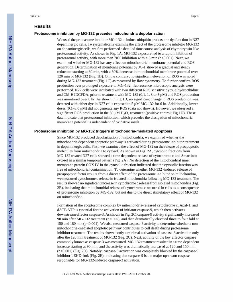
ResultsProteasome inhibition by MG-132 precedes mitochondria depolarization
We used the proteasome inhibitor MG-132 to induce ubiquitin proteasome dysfunction in N27dopaminergic cells. To systematically examine the effect of the proteasome inhibitor MG-132on dopaminergic cells, we first performed a detailed time course analysis of chymotrypsin-likeproteasomal activity. As shown in Fig. 1A, MG-132 exposure led to a rapid inhibition ofproteasomal activity, with more than 70% inhibition within 5 min (p<0.001). Next, weexamined whether MG-132 has any effect on mitochondrial membrane potential and ROSgeneration. Determination of membrane potential by JC-1 showed a gradual and steadyreduction starting at 30 min, with a 50% decrease in mitochondrial membrane potential over120 min of MG-132 (Fig. 1B). On the contrary, no significant elevation of ROS was notedduring MG-132 treatment (Fig. 1C) as measured by flow cytometry. To further confirm ROSproduction over prolonged exposure to MG-132, fluorescence microscopic analysis wereperformed. N27 cells were incubated with two different ROS sensitive dyes, dihydroethidineand CM-H2DCFDA, prior to treatment with MG-132 (0.1, 1, 3 or 5 μM) and ROS productionwas monitored over 6 hr. As shown in Fig 1D, no significant change in ROS production wasdetected with either dye in N27 cells exposed to 5 μM MG-132 for 6 hr. Additionally, lowerdoses (0.1–3.0 μM) did not generate any ROS (data not shown). However, we observed asignificant ROS production in the 50 μM H2O2 treatment (positive control; Fig 1D). Thesedata indicate that proteasomal inhibition, which precedes the dissipation of mitochondriamembrane potential is independent of oxidative insult.
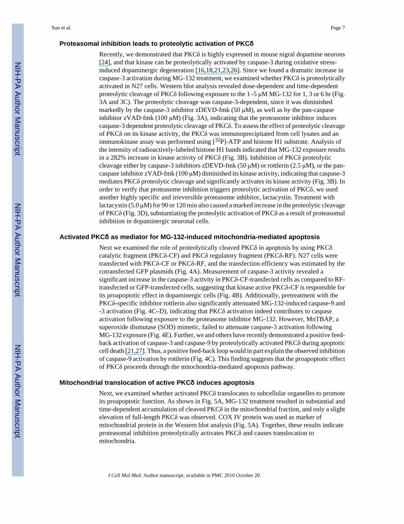
Proteasome inhibition by MG-132 triggers mitochondria-mediated apoptosisSince MG-132 produced depolarization of mitochondria, we examined whether themitochondria dependent apoptotic pathway is activated during proteasome inhibitor treatmentin dopaminergic cells. First, we examined the effect of MG-132 on the release of proapoptoticmolecules from mitochondria to cytosol. As shown in Fig. 2A, cytosolic fractions fromMG-132 treated N27 cells showed a time dependent release of cytochrome c and Smac intocytosol in a similar temporal pattern (Fig. 2A). No detection of the mitochondrial innermembrane protein COX IV in the cytosolic fraction indicated that the cytosolic fraction wasfree of mitochondrial contamination. To determine whether MG-132 -induced release ofproapoptotic factor results from a direct effect of the proteasome inhibitor on mitochondria,we measured cytochrome c release in isolated mitochondria following MG-132 treatment. Theresults showed no significant increase in cytochrome c release from isolated mitochondria (Fig.2B), indicating that mitochondrial release of cytochrome c occurred in cells as a consequenceof proteasome inhibition by MG-132, but not due to the direct stimulatory effect of MG-132on mitochondria.
Formation of the apoptosome complex by mitochondria-released cytochrome c, Apaf-1, anddATP/ATP is essential for the activation of initiator caspase-9, which then activatesdownstream effector caspase-3. As shown in Fig. 2C, caspase-9 activity significantly increased90 min after MG-132 treatment (p<0.05), and then dramatically elevated three to four fold at150 and 180 min (p<0.001). We also measured caspase-8 activity to determine whether a non-mitochondria-mediated apoptotic pathway contributes to cell death during proteasomeinhibitor treatment. The results showed only a minimal activation of caspase-8 activation onlyafter the 120 min treatment of MG-132 (Fig. 2C). Next, activity of the key effector caspasecommonly known as caspase-3 was measured. MG-132 treatment resulted in a time-dependentincrease starting at 90 min, and the activity was dramatically increased at 120 and 150 min(p<0.001) (Fig. 2D). Notably, caspase-3 activation was completely blocked by the caspase-9inhibitor LEHD-fmk (Fig. 2E), indicating that caspase-9 is the major upstream caspaseresponsible for MG-132-induced caspase-3 activation.
Sun et al. Page 6
J Cell Mol Med. Author manuscript; available in PMC 2010 October 20.
NIH
-PA Author Manuscript
NIH
-PA Author Manuscript
NIH
-PA Author Manuscript
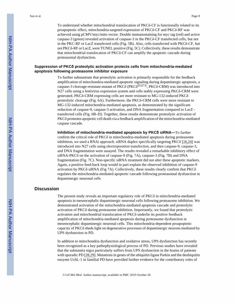
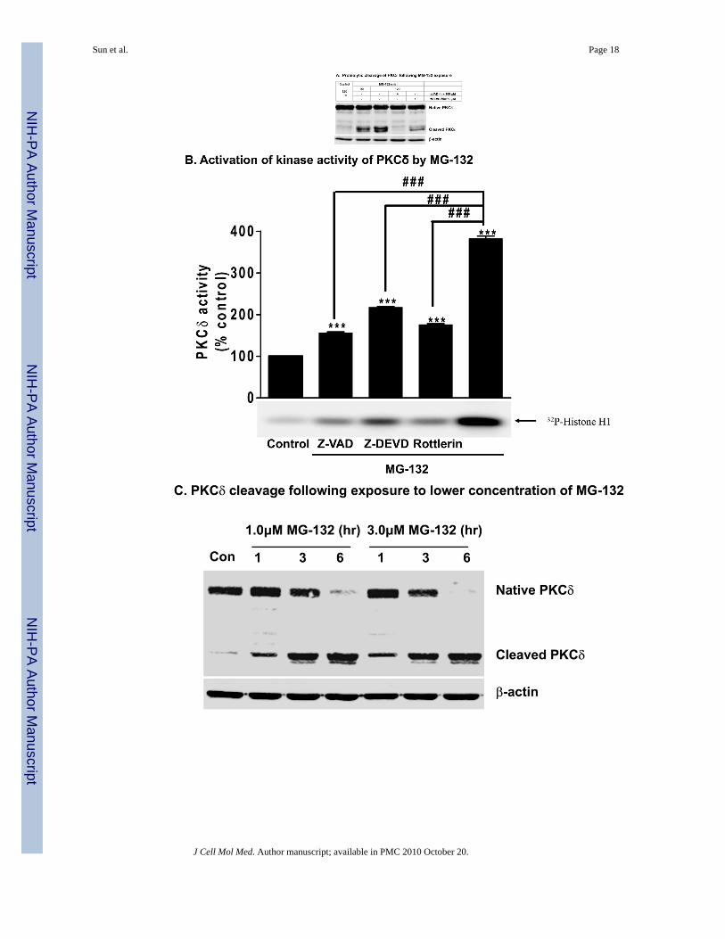
Proteasomal inhibition leads to proteolytic activation of PKCδRecently, we demonstrated that PKCδ is highly expressed in mouse nigral dopamine neurons[24], and that kinase can be proteolytically activated by caspase-3 during oxidative stress-induced dopaminergic degeneration [16,18,21,23,26]. Since we found a dramatic increase incaspase-3 activation during MG-132 treatment, we examined whether PKCδ is proteolyticallyactivated in N27 cells. Western blot analysis revealed dose-dependent and time-dependentproteolytic cleavage of PKCδ following exposure to the 1–5 μM MG-132 for 1, 3 or 6 hr (Fig.3A and 3C). The proteolytic cleavage was caspase-3-dependent, since it was diminishedmarkedly by the caspase-3 inhibitor zDEVD-fmk (50 μM), as well as by the pan-caspaseinhibitor zVAD-fmk (100 μM) (Fig. 3A), indicating that the proteasome inhibitor inducescaspase-3 dependent proteolytic cleavage of PKCδ. To assess the effect of proteolytic cleavageof PKCδ on its kinase activity, the PKCδ was immunoprecipitated from cell lysates and animmunokinase assay was performed using [32P]-ATP and histone H1 substrate. Analysis ofthe intensity of radioactively-labeled histone H1 bands indicated that MG-132 exposure resultsin a 282% increase in kinase activity of PKCδ (Fig. 3B). Inhibition of PKCδ proteolyticcleavage either by caspase-3 inhibitors zDEVD-fmk (50 μM) or rottlerin (2.5 μM), or the pan-caspase inhibitor zVAD-fmk (100 μM) diminished its kinase activity, indicating that caspase-3mediates PKCδ proteolytic cleavage and significantly activates its kinase activity (Fig. 3B). Inorder to verify that proteasome inhibition triggers proteolytic activation of PKCδ, we usedanother highly specific and irreversible proteasome inhibitor, lactacystin. Treatment withlactacystin (5.0 μM) for 90 or 120 min also caused a marked increase in the proteolytic cleavageof PKCδ (Fig. 3D), substantiating the proteolytic activation of PKCδ as a result of proteasomalinhibition in dopaminergic neuronal cells.
Activated PKCδ as mediator for MG-132-induced mitochondria-mediated apoptosisNext we examined the role of proteolytically cleaved PKCδ in apoptosis by using PKCδcatalytic fragment (PKCδ-CF) and PKCδ regulatory fragment (PKCδ-RF). N27 cells weretransfected with PKCδ-CF or PKCδ-RF, and the transfection efficiency was estimated by thecotransfected GFP plasmids (Fig. 4A). Measurement of caspase-3 activity revealed asignificant increase in the caspase-3 activity in PKCδ-CF-transfected cells as compared to RF-transfected or GFP-transfected cells, suggesting that kinase active PKCδ-CF is responsible forits proapoptotic effect in dopaminergic cells (Fig. 4B). Additionally, pretreatment with thePKCδ-specific inhibitor rottlerin also significantly attenuated MG-132-induced caspase-9 and-3 activation (Fig. 4C–D), indicating that PKCδ activation indeed contributes to caspaseactivation following exposure to the proteasome inhibitor MG-132. However, MnTBAP, asuperoxide dismutase (SOD) mimetic, failed to attenuate caspase-3 activation followingMG-132 exposure (Fig. 4E). Further, we and others have recently demonstrated a positive feed-back activation of caspase-3 and caspase-9 by proteolytically activated PKCδ during apoptoticcell death [21,27]. Thus, a positive feed-back loop would in part explain the observed inhibitionof caspase-9 activation by rottlerin (Fig. 4C). This finding suggests that the proapoptotic effectof PKCδ proceeds through the mitochondria-mediated apoptosis pathway.
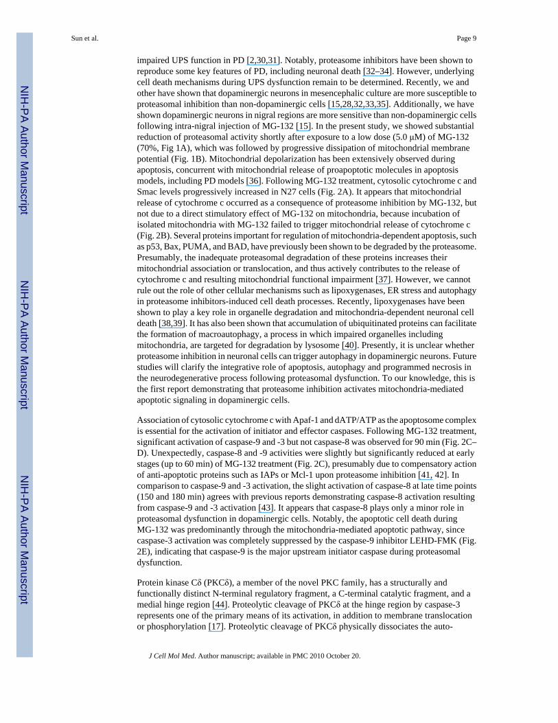
Mitochondrial translocation of active PKCδ induces apoptosisNext, we examined whether activated PKCδ translocates to subcellular organelles to promoteits proapoptotic function. As shown in Fig. 5A, MG-132 treatment resulted in substantial andtime-dependent accumulation of cleaved PKCδ in the mitochondrial fraction, and only a slightelevation of full-length PKCδ was observed. COX IV protein was used as marker ofmitochondrial protein in the Western blot analysis (Fig. 5A). Together, these results indicateproteasomal inhibition proteolytically activates PKCδ and causes translocation tomitochondria.
Sun et al. Page 7
J Cell Mol Med. Author manuscript; available in PMC 2010 October 20.
NIH
-PA Author Manuscript
NIH
-PA Author Manuscript
NIH
-PA Author Manuscript
To understand whether mitochondrial translocation of PKCδ-CF is functionally related to itsproapoptotic effect, mitochondria-targeted expression of PKCδ-CF and PKCδ-RF wasachieved using pCMV/myc/mito vector. Double immunostaining for myc tag (red) and activecaspase-3 (green) revealed activation of caspase-3 in the PKCδ-CF transfected cells, but notin the PKC-RF or LacZ transfected cells (Fig. 5B). Also, cells transfected with PKCδ-CF, butnot PKCδ-RF or LacZ, were TUNEL positive (Fig. 5C). Collectively, these results demonstratethat mitochondrial translocation of PKCδ-CF can amplify the apoptotic cascade duringproteasomal dysfunction.
Suppression of PKCδ proteolytic activation protects cells from mitochondria-mediatedapoptosis following proteasome inhibitor exposure
To further substantiate that proteolytic activation is primarily responsible for the feedbackamplification of mitochondria-mediated apoptotic signaling during dopaminergic apoptosis, acaspase-3 cleavage-resistant mutant of PKCδ (PKCδD327A, PKCδ-CRM) was introduced intoN27 cells using a lentivirus expression system and cells stably expressing PKCδ-CRM weregenerated. PKCδ-CRM expressing cells are more resistant to MG-132-induced PKCδproteolytic cleavage (Fig. 6A). Furthermore, the PKCδ-CRM cells were more resistant toMG-132-induced mitochondria-mediated apoptosis, as demonstrated by the significantreduction of caspase-9, caspase-3 activation, and DNA fragmentation compared to LacZ -transfected cells (Fig. 6B–D). Together, these results demonstrate proteolytic activation ofPKCδ promotes apoptotic cell death via a feedback amplification of the mitochondria-mediatedcaspase cascade.
Inhibition of mitochondria-mediated apoptosis by PKCδ siRNA—To furtherconfirm the critical role of PKCδ in mitochondria-mediated apoptosis during proteasomeinhibition, we used a RNAi approach. siRNA duplex specifically targeting PKCδ [16,24] wasintroduced into N27 cells using electroporation transfection, and then caspase-9, caspase-3,and DNA fragmentation were assayed. The results revealed a remarkable inhibitory effect ofsiRNA-PKCδ on the activation of caspase-9 (Fig. 7A), caspase-3 (Fig. 7B) and DNAfragmentation (Fig. 7C). Non-specific siRNA treatment did not alter these apoptotic markers.Again, a positive feed-back loop would in part explain the observed inhibition of caspase-9activation by PKCδ siRNA (Fig 7A). Collectively, these results clearly confirm that PKCδregulates the mitochondria-mediated apoptotic cascade following proteasomal dysfunction indopaminergic neuronal cells
DiscussionThe present study reveals an important regulatory role of PKCδ in mitochondria-mediatedapoptosis in mesencephalic dopaminergic neuronal cells following proteasome inhibition. Wedemonstrated activation of the mitochondria-mediated apoptosis cascade and proteolyticactivation of PKCδ during proteasome inhibition. Importantly, we found that proteolyticactivation and mitochondrial translocation of PKCδ underlie its positive feedbackamplification of mitochondria-mediated apoptosis during proteasome dysfunction inmesencephalic dopaminergic neuronal cells. This mitochondria-dependent proapoptoticcapacity of PKCδ sheds light on degenerative processes of dopaminergic neurons mediated byUPS dysfunction in PD.
In addition to mitochondria dysfunction and oxidative stress, UPS dysfunction has recentlybeen recognized as a key pathophysiological process of PD. Previous studies have revealedthat the substantia nigra particularly suffers from UPS dysfunction in the brains of patientswith sporadic PD [28,29]. Mutations in genes of the ubiquitin ligase Parkin and the deubiquitinenzyme UchL-1 in familial PD have provided further evidence for the contributory roles of
Sun et al. Page 8
J Cell Mol Med. Author manuscript; available in PMC 2010 October 20.
NIH
-PA Author Manuscript
NIH
-PA Author Manuscript
NIH
-PA Author Manuscript
impaired UPS function in PD [2,30,31]. Notably, proteasome inhibitors have been shown toreproduce some key features of PD, including neuronal death [32–34]. However, underlyingcell death mechanisms during UPS dysfunction remain to be determined. Recently, we andother have shown that dopaminergic neurons in mesencephalic culture are more susceptible toproteasomal inhibition than non-dopaminergic cells [15,28,32,33,35]. Additionally, we haveshown dopaminergic neurons in nigral regions are more sensitive than non-dopaminergic cellsfollowing intra-nigral injection of MG-132 [15]. In the present study, we showed substantialreduction of proteasomal activity shortly after exposure to a low dose (5.0 μM) of MG-132(70%, Fig 1A), which was followed by progressive dissipation of mitochondrial membranepotential (Fig. 1B). Mitochondrial depolarization has been extensively observed duringapoptosis, concurrent with mitochondrial release of proapoptotic molecules in apoptosismodels, including PD models [36]. Following MG-132 treatment, cytosolic cytochrome c andSmac levels progressively increased in N27 cells (Fig. 2A). It appears that mitochondrialrelease of cytochrome c occurred as a consequence of proteasome inhibition by MG-132, butnot due to a direct stimulatory effect of MG-132 on mitochondria, because incubation ofisolated mitochondria with MG-132 failed to trigger mitochondrial release of cytochrome c(Fig. 2B). Several proteins important for regulation of mitochondria-dependent apoptosis, suchas p53, Bax, PUMA, and BAD, have previously been shown to be degraded by the proteasome.Presumably, the inadequate proteasomal degradation of these proteins increases theirmitochondrial association or translocation, and thus actively contributes to the release ofcytochrome c and resulting mitochondrial functional impairment [37]. However, we cannotrule out the role of other cellular mechanisms such as lipoxygenases, ER stress and autophagyin proteasome inhibitors-induced cell death processes. Recently, lipoxygenases have beenshown to play a key role in organelle degradation and mitochondria-dependent neuronal celldeath [38,39]. It has also been shown that accumulation of ubiquitinated proteins can facilitatethe formation of macroautophagy, a process in which impaired organelles includingmitochondria, are targeted for degradation by lysosome [40]. Presently, it is unclear whetherproteasome inhibition in neuronal cells can trigger autophagy in dopaminergic neurons. Futurestudies will clarify the integrative role of apoptosis, autophagy and programmed necrosis inthe neurodegenerative process following proteasomal dysfunction. To our knowledge, this isthe first report demonstrating that proteasome inhibition activates mitochondria-mediatedapoptotic signaling in dopaminergic cells.
Association of cytosolic cytochrome c with Apaf-1 and dATP/ATP as the apoptosome complexis essential for the activation of initiator and effector caspases. Following MG-132 treatment,significant activation of caspase-9 and -3 but not caspase-8 was observed for 90 min (Fig. 2C–D). Unexpectedly, caspase-8 and -9 activities were slightly but significantly reduced at earlystages (up to 60 min) of MG-132 treatment (Fig. 2C), presumably due to compensatory actionof anti-apoptotic proteins such as IAPs or Mcl-1 upon proteasome inhibition [41, 42]. Incomparison to caspase-9 and -3 activation, the slight activation of caspase-8 at late time points(150 and 180 min) agrees with previous reports demonstrating caspase-8 activation resultingfrom caspase-9 and -3 activation [43]. It appears that caspase-8 plays only a minor role inproteasomal dysfunction in dopaminergic cells. Notably, the apoptotic cell death duringMG-132 was predominantly through the mitochondria-mediated apoptotic pathway, sincecaspase-3 activation was completely suppressed by the caspase-9 inhibitor LEHD-FMK (Fig.2E), indicating that caspase-9 is the major upstream initiator caspase during proteasomaldysfunction.
Protein kinase Cδ (PKCδ), a member of the novel PKC family, has a structurally andfunctionally distinct N-terminal regulatory fragment, a C-terminal catalytic fragment, and amedial hinge region [44]. Proteolytic cleavage of PKCδ at the hinge region by caspase-3represents one of the primary means of its activation, in addition to membrane translocationor phosphorylation [17]. Proteolytic cleavage of PKCδ physically dissociates the auto-
Sun et al. Page 9
J Cell Mol Med. Author manuscript; available in PMC 2010 October 20.
NIH
-PA Author Manuscript
NIH
-PA Author Manuscript
NIH
-PA Author Manuscript
inhibitory regulatory fragment from its catalytic fragment, thus permanently activating itskinase activity. Tyr-311 phosphorylation of PKCδ is important for its caspase-3-mediatedproteolytic cleavage [45]. Previously, we showed that proteolytically activated PKCδ plays akey role in mediating oxidative stress-induced apoptosis in dopaminergic neuronal cells [16,18,21,46]. The possible mechanisms of positive feedback activation of caspase-3 indopaminergic neuronal cells are yet to be identified. Although phosphorylation of caspase-3by full-length PKCδ has been shown to increase enzymatic activity of caspase-3 in monocytes[47], we did not find caspase-3 phosphorylation by PKCδ in dopaminergic cells (unpublishedobservations). It appears that PKCδ amplifies caspase-3 activation via distinct mechanismswhen it is proteolytically activated in dopaminergic neuronal cells. Proteolytic activation ofPKCδ in dopaminergic cells appears to depend on caspase-3 activation in N27 cells exposedto proteasome inhibitors (Fig. 3A–C). The direct proapoptotic effect of PKCδ-CF is manifestedby the elevation of caspase-3 activity in PKCδ-CF transfected cells (Fig. 4B), consistent withapoptotic function of PKCδ proteolytic activation. In addition, PKCδ likely enhances activationof caspase-9, which further activates the downstream effector caspase-3, since the PKCδ-specific inhibitor rottlerin attenuates activation of caspase-3 and upstream initiator caspase-9,at least in our study (Fig. 4C–D). Emerging evidence has indicated that rottlerin, a previouslywell-accepted specific inhibitor for PKCδ, actually possesses some biological function otherthan PKCδ inhibition, such as uncoupling mitochondria [48]. Rottlerin has been shown to bea potent inhibitor of PKCδ, with an IC50 of 3–6 μM, whereas the Ki for other PKC isoformsare 5–10 times higher. Other studies have shown rottlerin may inhibit other kinases such asCaM kinase III, PRAK and MAPKAP-K2 as well as other cellular targets [48–52]. Sincerottlerin is not a highly specific inhibitor for PKCδ, we used multiple approaches includingsiRNA, dominant-negative mutant and cleavage-resistant mutants in our studies. In the presentstudy, effective suppression of mitochondria-mediated apoptosis either by PKCδ cleavage-resistant mutant or by PKCδ siRNA clearly suggests PKCδ activation truly underlies its pro-apoptotic capacity following MG-132 exposure.
Induction of ROS generation was observed during prolonged exposure to proteasome inhibitorsBortezomib [53], MG-132, lactacystin [54], and PS-341 [55] in several non-neuronal cell lines.Oxidative stress has been demonstrated to activate caspase-3 and PKCδ in N27 cells [18,23].In an attempt to determine whether dopaminergic apoptosis following MG-132 exposureinvolves oxidative stress, ROS generation was measured; no significant increase in ROSgeneration was noted (Fig. 1C–D). Additionally, the antioxidant MnTBAP, which has beenpreviously shown to effectively inhibit caspase-3 activation during oxidative stress in N27 cells[18], failed to attenuate caspase-3 activation induced by MG-132 (Fig. 4E). Our data suggestthat ROS generation plays a negligible role in apoptotic cell death following proteasomeinhibition in mesencephalic dopaminergic neuronal cells.
The mitochondria-dependent proapoptotic capacity of active PKCδ, as indicated bysuppression of caspase-9 activation by rottlerin, was accompanied by mitochondrialtranslocation of PKCδ. This translocation of proteolytically activated PKCδ followingproteasome inhibition is consistent with the existing hypothesis that the mitochondrion is amajor target organelle that determines the fate of cell survival and death [56]. PKCδ has beenshown to accumulate in nuclei [57], golgi [58], and endoplasmic reticulum [59] in various celltypes. In an attempt to determine whether mitochondrial translocation of proteolyticallyactivated PKCδ underlies its proapoptotic effect, marked activation of caspase-3 was noted inthe N27 cells expressing mitochondria targeted PKCδ-CF (Fig. 5B), but not PKCδ-RF or LacZ.This indicates that mitochondrial translocation of PKCδ-CF possibly underlies its feedbackamplification of caspase activation. Considering the multiple pathways that lead to PKCδactivation, we conducted additional experiments to verify that PKCδ proteolytic activationmediates its mitochondria-dependent proapoptotic effect. Expression of a caspase-3 cleavage-resistant mutant of PKCδ (PKCδ-CRM), which effectively inhibited the proteolytic cleavage
Sun et al. Page 10
J Cell Mol Med. Author manuscript; available in PMC 2010 October 20.
NIH
-PA Author Manuscript
NIH
-PA Author Manuscript
NIH
-PA Author Manuscript
of endogenous PKCδ (Fig. 6A), significantly attenuated the activation of mitochondria-mediated apoptosis triggered by MG-132 (Fig. 6B–D). These findings are consistent with arecent study showing that PKCδ-CRM reduces the mitochondrial release of cytochrome c inUV-challenged keratinocytes [46]. Phosphorylation of mitochondrial resident proteins byactive PKCδ likely underlies its effect on mitochondria-mediated apoptosis. Severalmitochondrial proteins have been characterized as candidate substrates of PKCδ, includingphospholipid scramblase [60] and pyruvate dehydrogenase kinase [61]. Likely, elevated levelsof BAD, due to its incomplete degradation by the proteasome, also participates in themitochondria-dependent proapoptotic effect of PKCδ in dopaminergic cells, given the previousfinding that PKCδ mitochondrial translocation is accompanied by an increase in the level ofBAD following cardiac ischemia [62]. We also recently reported that systemic administrationof PKCδ inhibitor alleviates the nigrostriatal dopaminergic degeneration in an MPTP-inducedanimal model of PD; PKCδ inhibitor has been previously shown to activate dopaminergicapoptosis via proteolytic activation of PKCδ in the N27 cells [18,24]. Therefore, we believethat PKCδ inhibition is expected to exert a similar neuroprotection in an MG-132 in vivo model.We are currently investigating the role of PKCδ using RNAi-based gene knockdown andPKCδ-knockout animals. Future studies should focus on identifying the key target moleculein mitochondria that contributes to amplification of apoptotic cell death following proteasomedysfunction.
As summarized in Fig. 8, the present study demonstrates that proteolytic activation andmitochondrial translocation of PKCδ underlies its feedback activation of mitochondria-mediated apoptosis during proteasome dysfunction in mesencephalic dopaminergic neuronalcells. The high expression of PKCδ in nigral dopaminergic neurons and the convergence ofproteasomal and mitochondrial dysfunction at the level of PKCδ demonstrate that the kinaseis a crucial proapoptotic signaling molecule in dopaminergic degenerative processes. Thisknowledge advances our understanding of the pathogenesis of nigrostriatal degeneration andvalidates PKCδ as potential target for therapeutic intervention of PD.
AcknowledgmentsThis work was supported by National Institute of Health (NIH) grants NS38644, ES10586 and NS45133. W. Eugeneand Linda Lloyd Endowed Chair to AGK is also acknowledged. The authors acknowledge Ms. Keri Henderson forher assistance in the preparation of this manuscript.
References1. Glickman MH, Ciechanover A. The ubiquitin-proteasome proteolytic pathway: destruction for the sake
of construction. Physiol Rev 2002;82:373–428. [PubMed: 11917093]2. Sun F, Kanthasamy A, Anantharam V, Kanthasamy AG. Environmental neurotoxic chemicals-induced
ubiquitin proteasome system dysfunction in the pathogenesis and progression of Parkinson’s disease.Pharmacol Ther 2007;114:327–44. [PubMed: 17521740]
3. McNaught KS, Olanow CW. Proteolytic stress: a unifying concept for the etiopathogenesis ofParkinson’s disease. Ann Neurol 2003;53(Suppl 3):S73–84. discussion S84–6. [PubMed: 12666100]
4. Dauer W, Przedborski S. Parkinson’s disease: mechanisms and models. Neuron 2003;39:889–909.[PubMed: 12971891]
5. Fornai F, Schluter OM, Lenzi P, Gesi M, Ruffoli R, Ferrucci M, Lazzeri G, Busceti CL, Pontarelli F,Battaglia G, Pellegrini A, Nicoletti F, Ruggieri S, Paparelli A, Sudhof TC. Parkinson-like syndromeinduced by continuous MPTP infusion: convergent roles of the ubiquitin-proteasome system andalpha-synuclein. Proc Natl Acad Sci U S A 2005;102:3413–8. [PubMed: 15716361]
6. Zeng BY, Iravani MM, Lin ST, Irifune M, Kuoppamaki M, Al-Barghouthy G, Smith L, Jackson MJ,Rose S, Medhurst AD, Jenner P. MPTP treatment of common marmosets impairs proteasomal enzymeactivity and decreases expression of structural and regulatory elements of the 26S proteasome. Eur JNeurosci 2006;23:1766–74. [PubMed: 16623833]
Sun et al. Page 11
J Cell Mol Med. Author manuscript; available in PMC 2010 October 20.
NIH
-PA Author Manuscript
NIH
-PA Author Manuscript
NIH
-PA Author Manuscript
7. Manning-Bog AB, Reaney SH, Chou VP, Johnston LC, McCormack AL, Johnston J, Langston JW,Di Monte DA. Lack of nigrostriatal pathology in a rat model of proteasome inhibition. Ann Neurol2006;60:256–60. [PubMed: 16862576]
8. Schapira AH, Cleeter MW, Muddle JR, Workman JM, Cooper JM, King RH. Proteasomal inhibitioncauses loss of nigral tyrosine hydroxylase neurons. Ann Neurol 2006;60:253–5. [PubMed: 16862591]
9. McNaught KS, Olanow CW. Proteasome inhibitor-induced model of Parkinson’s disease. Ann Neurol2006;60:243–7. [PubMed: 16862580]
10. McNaught KS, Perl DP, Brownell AL, Olanow CW. Systemic exposure to proteasome inhibitorscauses a progressive model of Parkinson’s disease. Ann Neurol 2004;56:149–62. [PubMed:15236415]
11. Bove J, Zhou C, Jackson-Lewis V, Taylor J, Chu Y, Rideout HJ, Wu DC, Kordower JH, PetrucelliL, Przedborski S. Proteasome inhibition and Parkinson’s disease modeling. Ann Neurol2006;60:260–4. [PubMed: 16862585]
12. Kordower JH, Kanaan NM, Chu Y, Suresh Babu R, Stansell J 3rd, Terpstra BT, Sortwell CE, Steece-Collier K, Collier TJ. Failure of proteasome inhibitor administration to provide a model ofParkinson’s disease in rats and monkeys. Ann Neurol 2006;60:264–8. [PubMed: 16862579]
13. McNaught KS, Bjorklund LM, Belizaire R, Isacson O, Jenner P, Olanow CW. Proteasome inhibitioncauses nigral degeneration with inclusion bodies in rats. Neuroreport 2002;13:1437–41. [PubMed:12167769]
14. Miwa H, Kubo T, Suzuki A, Nishi K, Kondo T. Retrograde dopaminergic neuron degenerationfollowing intrastriatal proteasome inhibition. Neurosci Lett 2005;380:93–8. [PubMed: 15854758]
15. Sun F, Anantharam V, Latchoumycandane C, Zhang D, Kanthasamy A, Kanthasamy AG. ProteasomeInhibitor MG-132 Induces Dopaminergic Degeneration in Cell Culture and Animal Models.Neurotoxicology 2006;27:807–15. [PubMed: 16870259]
16. Yang Y, Kaul S, Zhang D, Anantharam V, Kanthasamy AG. Suppression of caspase-3-dependentproteolytic activation of protein kinase C delta by small interfering RNA prevents MPP+-induceddopaminergic degeneration. Mol Cell Neurosci 2004;25:406–21. [PubMed: 15033169]
17. Kanthasamy AG, Anantharam V, Zhang D, Latchoumycandane C, Jin H, Kaul S, Kanthasamy A. Anovel peptide inhibitor targeted to caspase-3 cleavage site of a proapoptotic kinase protein kinase Cdelta (PKCdelta) protects against dopaminergic neuronal degeneration in Parkinson’s diseasemodels. Free Radic Biol Med 2006;41:1578–89. [PubMed: 17045926]
18. Kaul S, Kanthasamy A, Kitazawa M, Anantharam V, Kanthasamy AG. Caspase-3 dependentproteolytic activation of protein kinase C delta mediates and regulates 1-methyl-4-phenylpyridinium(MPP+)-induced apoptotic cell death in dopaminergic cells: relevance to oxidative stress indopaminergic degeneration. Eur J Neurosci 2003;18:1387–401. [PubMed: 14511319]
19. Narayanan PK, Goodwin EH, Lehnert BE. Alpha particles initiate biological production of superoxideanions and hydrogen peroxide in human cells. Cancer Res 1997;57:3963–71. [PubMed: 9307280]
20. Andrews ZB, Horvath B, Barnstable CJ, Elsworth J, Yang L, Beal MF, Roth RH, Matthews RT,Horvath TL. Uncoupling protein-2 is critical for nigral dopamine cell survival in a mouse model ofParkinson’s disease. J Neurosci 2005;25:184–91. [PubMed: 15634780]
21. Anantharam V, Kitazawa M, Wagner J, Kaul S, Kanthasamy AG. Caspase-3-dependent proteolyticcleavage of protein kinase Cdelta is essential for oxidative stress-mediated dopaminergic cell deathafter exposure to methylcyclopentadienyl manganese tricarbonyl. J Neurosci 2002;22:1738–51.[PubMed: 11880503]
22. Luo X, Budihardjo I, Zou H, Slaughter C, Wang X. Bid, a Bcl2 interacting protein, mediatescytochrome c release from mitochondria in response to activation of cell surface death receptors. Cell1998;94:481–90. [PubMed: 9727491]
23. Kitazawa M, Anantharam V, Kanthasamy AG. Dieldrin induces apoptosis by promoting caspase-3-dependent proteolytic cleavage of protein kinase Cdelta in dopaminergic cells: relevance to oxidativestress and dopaminergic degeneration. Neuroscience 2003;119:945–64. [PubMed: 12831855]
24. Zhang D, Kanthasamy A, Yang Y, Anantharam V. Protein kinase Cdelta negatively regulates tyrosinehydroxylase activity and dopamine synthesis by enhancing protein phosphatase-2A activity indopaminergic neurons. J Neurosci 2007;27:5349–62. [PubMed: 17507557]
Sun et al. Page 12
J Cell Mol Med. Author manuscript; available in PMC 2010 October 20.
NIH
-PA Author Manuscript
NIH
-PA Author Manuscript
NIH
-PA Author Manuscript
25. Sun F, Anantharam V, Latchoumycandane C, Kanthasamy A, Kanthasamy AG. Dieldrin inducesubiquitin-proteasome dysfunction in alpha-synuclein overexpressing dopaminergic neuronal cellsand enhances susceptibility to apoptotic cell death. J Pharmacol Exp Ther 2005;315:69–79. [PubMed:15987830]
26. Latchoumycandane C, Anantharam V, Kitazawa M, Yang Y, Kanthasamy A, Kanthasamy AG.Protein kinase Cdelta is a key downstream mediator of manganese-induced apoptosis indopaminergic neuronal cells. J Pharmacol Exp Ther 2005;313:46–55. [PubMed: 15608081]
27. Reyland ME, Anderson SM, Matassa AA, Barzen KA, Quissell DO. Protein kinase C delta is essentialfor etoposide-induced apoptosis in salivary gland acinar cells. J Biol Chem 1999;274:19115–23.[PubMed: 10383415]
28. McNaught KS, Belizaire R, Jenner P, Olanow CW, Isacson O. Selective loss of 20S proteasomealpha-subunits in the substantia nigra pars compacta in Parkinson’s disease. Neurosci Lett2002;326:155–8. [PubMed: 12095645]
29. McNaught KS, Jenner P. Proteasomal function is impaired in substantia nigra in Parkinson’s disease.Neurosci Lett 2001;297:191–4. [PubMed: 11137760]
30. Olanow CW, McNaught KS. Ubiquitin-proteasome system and Parkinson’s disease. Mov Disord2006;21:1806–23. [PubMed: 16972273]
31. Moore DJ, West AB, Dawson VL, Dawson TM. Molecular pathophysiology of Parkinson’s disease.Annu Rev Neurosci 2005;28:57–87. [PubMed: 16022590]
32. McNaught KS, Mytilineou C, Jnobaptiste R, Yabut J, Shashidharan P, Jennert P, Olanow CW.Impairment of the ubiquitin-proteasome system causes dopaminergic cell death and inclusion bodyformation in ventral mesencephalic cultures. J Neurochem 2002;81:301–6. [PubMed: 12064477]
33. Rideout HJ, Lang-Rollin IC, Savalle M, Stefanis L. Dopaminergic neurons in rat ventral midbraincultures undergo selective apoptosis and form inclusions, but do not up-regulate iHSP70, followingproteasomal inhibition. J Neurochem 2005;93:1304–13. [PubMed: 15934949]
34. Rideout HJ, Larsen KE, Sulzer D, Stefanis L. Proteasomal inhibition leads to formation of ubiquitin/alpha-synuclein-immunoreactive inclusions in PC12 cells. J Neurochem 2001;78:899–908.[PubMed: 11520910]
35. Reaney SH, Johnston LC, Langston WJ, Di Monte DA. Comparison of the neurotoxic effects ofproteasomal inhibitors in primary mesencephalic cultures. Exp Neurol 2006;202:434–40. [PubMed:16920101]
36. Ly JD, Grubb DR, Lawen A. The mitochondrial membrane potential (deltapsi(m)) in apoptosis; anupdate. Apoptosis 2003;8:115–28. [PubMed: 12766472]
37. Zhang HG, Wang J, Yang X, Hsu HC, Mountz JD. Regulation of apoptosis proteins in cancer cellsby ubiquitin. Oncogene 2004;23:2009–15. [PubMed: 15021888]
38. Grullich C, Duvoisin RM, Wiedmann M, van Leyen K. Inhibition of 15-lipoxygenase leads to delayedorganelle degradation in the reticulocyte. FEBS Lett 2001;489:51–4. [PubMed: 11231012]
39. van Leyen K, Arai K, Jin G, Kenyon V, Gerstner B, Rosenberg PA, Holman TR, Lo EH. Novellipoxygenase inhibitors as neuroprotective reagents. J Neurosci Res. 2007 in press.
40. Shacka JJ, Roth KA, Zhang J. The autophagy-lysosomal degradation pathway: role inneurodegenerative disease and therapy. Front Biosci 2008;13:718–36. [PubMed: 17981582]
41. Yang Y, Fang S, Jensen JP, Weissman AM, Ashwell JD. Ubiquitin protein ligase activity of IAPsand their degradation in proteasomes in response to apoptotic stimuli. Science 2000;288:874–7.[PubMed: 10797013]
42. Nijhawan D, Fang M, Traer E, Zhong Q, Gao W, Du F, Wang X. Elimination of Mcl-1 is requiredfor the initiation of apoptosis following ultraviolet irradiation. Genes Dev 2003;17:1475–86.[PubMed: 12783855]
43. Viswanath V, Wu Y, Boonplueang R, Chen S, Stevenson FF, Yantiri F, Yang L, Beal MF, AndersenJK. Caspase-9 activation results in downstream caspase-8 activation and bid cleavage in 1-methyl-4-phenyl-1,2,3,6-tetrahydropyridine-induced Parkinson’s disease. J Neurosci 2001;21:9519–28.[PubMed: 11739563]
44. Steinberg SF. Distinctive activation mechanisms and functions for protein kinase Cdelta. Biochem J2004;384:449–59. [PubMed: 15491280]
Sun et al. Page 13
J Cell Mol Med. Author manuscript; available in PMC 2010 October 20.
NIH
-PA Author Manuscript
NIH
-PA Author Manuscript
NIH
-PA Author Manuscript
45. Kaul S, Anantharam V, Yang Y, Choi CJ, Kanthasamy A, Kanthasamy AG. Tyrosine phosphorylationregulates the proteolytic activation of protein kinase Cdelta in dopaminergic neuronal cells. J BiolChem 2005;280:28721–30. [PubMed: 15961393]
46. D’Costa AM, Denning MF. A caspase-resistant mutant of PKC-delta protects keratinocytes from UV-induced apoptosis. Cell Death Differ 2005;12:224–32. [PubMed: 15618968]
47. Voss OH, Kim S, Wewers MD, Doseff AI. Regulation of monocyte apoptosis by the protein kinaseCdelta-dependent phosphorylation of caspase-3. J Biol Chem 2005;280:17371–9. [PubMed:15716280]
48. Soltoff SP. Rottlerin: an inappropriate and ineffective inhibitor of PKCdelta. Trends Pharmacol Sci2007;28:453–8. [PubMed: 17692392]
49. Davies SP, Reddy H, Caivano M, Cohen P. Specificity and mechanism of action of some commonlyused protein kinase inhibitors. Biochem J 2000;351:95–105. [PubMed: 10998351]
50. Gschwendt M, Muller HJ, Kielbassa K, Zang R, Kittstein W, Rincke G, Marks F. Rottlerin, a novelprotein kinase inhibitor. Biochem Biophys Res Commun 1994;199:93–8. [PubMed: 8123051]
51. Samokhin GP, Jirousek MR, Ways DK, Henriksen RA. Effects of protein kinase C inhibitors onthromboxane production by thrombin-stimulated platelets. Eur J Pharmacol 1999;386:297–303.[PubMed: 10618482]
52. Soltoff SP. Rottlerin is a mitochondrial uncoupler that decreases cellular ATP levels and indirectlyblocks protein kinase Cdelta tyrosine phosphorylation. J Biol Chem 2001;276:37986–92. [PubMed:11498535]
53. Ling YH, Liebes L, Zou Y, Perez-Soler R. Reactive oxygen species generation and mitochondrialdysfunction in the apoptotic response to Bortezomib, a novel proteasome inhibitor, in human H460non-small cell lung cancer cells. J Biol Chem 2003;278:33714–23. [PubMed: 12821677]
54. Wu HM, Chi KH, Lin WW. Proteasome inhibitors stimulate activator protein-1 pathway via reactiveoxygen species production. FEBS Lett 2002;526:101–5. [PubMed: 12208513]
55. Fribley A, Zeng Q, Wang CY. Proteasome inhibitor PS-341 induces apoptosis through induction ofendoplasmic reticulum stress-reactive oxygen species in head and neck squamous cell carcinomacells. Mol Cell Biol 2004;24:9695–704. [PubMed: 15509775]
56. Denning MF, Wang Y, Tibudan S, Alkan S, Nickoloff BJ, Qin JZ. Caspase activation and disruptionof mitochondrial membrane potential during UV radiation-induced apoptosis of human keratinocytesrequires activation of protein kinase C. Cell Death Differ 2002;9:40–52. [PubMed: 11803373]
57. Cross T, Griffiths G, Deacon E, Sallis R, Gough M, Watters D, Lord JM. PKC-delta is an apoptoticlamin kinase. Oncogene 2000;19:2331–7. [PubMed: 10822384]
58. Kajimoto T, Shirai Y, Sakai N, Yamamoto T, Matsuzaki H, Kikkawa U, Saito N. Ceramide-inducedapoptosis by translocation, phosphorylation, and activation of protein kinase Cdelta in the Golgicomplex. J Biol Chem 2004;279:12668–76. [PubMed: 14715667]
59. Zrachia A, Dobroslav M, Blass M, Kazimirsky G, Kronfeld I, Blumberg PM, Kobiler D, Lustig S,Brodie C. Infection of glioma cells with Sindbis virus induces selective activation and tyrosinephosphorylation of protein kinase C delta. Implications for Sindbis virus-induced apoptosis. J BiolChem 2002;277:23693–701. [PubMed: 11927579]
60. He Y, Liu J, Grossman D, Durrant D, Sweatman T, Lothstein L, Epand RF, Epand RM, Lee RM.Phosphorylation of mitochondrial phospholipid scramblase 3 by protein kinase C-delta induces itsactivation and facilitates mitochondrial targeting of tBid. J Cell Biochem 2007;101:1210–21.[PubMed: 17226776]
61. Churchill EN, Murriel CL, Chen CH, Mochly-Rosen D, Szweda LI. Reperfusion-inducedtranslocation of deltaPKC to cardiac mitochondria prevents pyruvate dehydrogenase reactivation.Circ Res 2005;97:78–85. [PubMed: 15961716]
62. Murriel CL, Churchill E, Inagaki K, Szweda LI, Mochly-Rosen D. Protein kinase Cdelta activationinduces apoptosis in response to cardiac ischemia and reperfusion damage: a mechanism involvingBAD and the mitochondria. J Biol Chem 2004;279:47985–91. [PubMed: 15339931]
Sun et al. Page 14
J Cell Mol Med. Author manuscript; available in PMC 2010 October 20.
NIH
-PA Author Manuscript
NIH
-PA Author Manuscript
NIH
-PA Author Manuscript
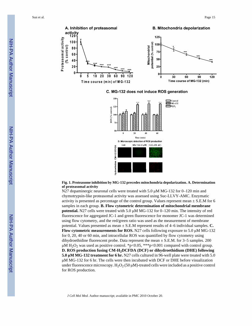
Fig. 1. Proteasome inhibition by MG-132 precedes mitochondria depolarization. A. Determinationof proteasomal activityN27 dopaminergic neuronal cells were treated with 5.0 μM MG-132 for 0–120 min andchymotrypsin-like proteasomal activity was assessed using Suc-LLVY-AMC. Enzymaticactivity is presented as percentage of the control group. Values represent mean ± S.E.M for 6samples in each group. B. Flow cytometric determination of mitochondrial membranepotential. N27 cells were treated with 5.0 μM MG-132 for 0–120 min. The intensity of redfluorescence for aggregated JC-1 and green fluorescence for monomer JC-1 was determinedusing flow cytometry, and the red/green ratio was used as the measurement of membranepotential. Values presented as mean ± S.E.M represent results of 4–6 individual samples. C.Flow cytometric measurements for ROS. N27 cells following exposure to 5.0 μM MG-132for 0, 20, 40 or 60 min, and intracellular ROS was quantified by flow cytometry usingdihydroethidine fluorescent probe. Data represent the mean ± S.E.M. for 3–5 samples. 200μM H2O2 was used as positive control. *p<0.05, ***p<0.001 compared with control group.D. ROS production fusing CM-H2DCFDA (DCF) or dihydroethidium (DHE) following5.0 μM MG-132 treatment for 6 hr. N27 cells cultured in 96-well plate were treated with 5.0μM MG-132 for 6 hr. The cells were then incubated with DCF or DHE before visualizationunder fluorescence microscopy. H2O2 (50 μM)-treated cells were included as a positive controlfor ROS production.
Sun et al. Page 15
J Cell Mol Med. Author manuscript; available in PMC 2010 October 20.
NIH
-PA Author Manuscript
NIH
-PA Author Manuscript
NIH
-PA Author Manuscript
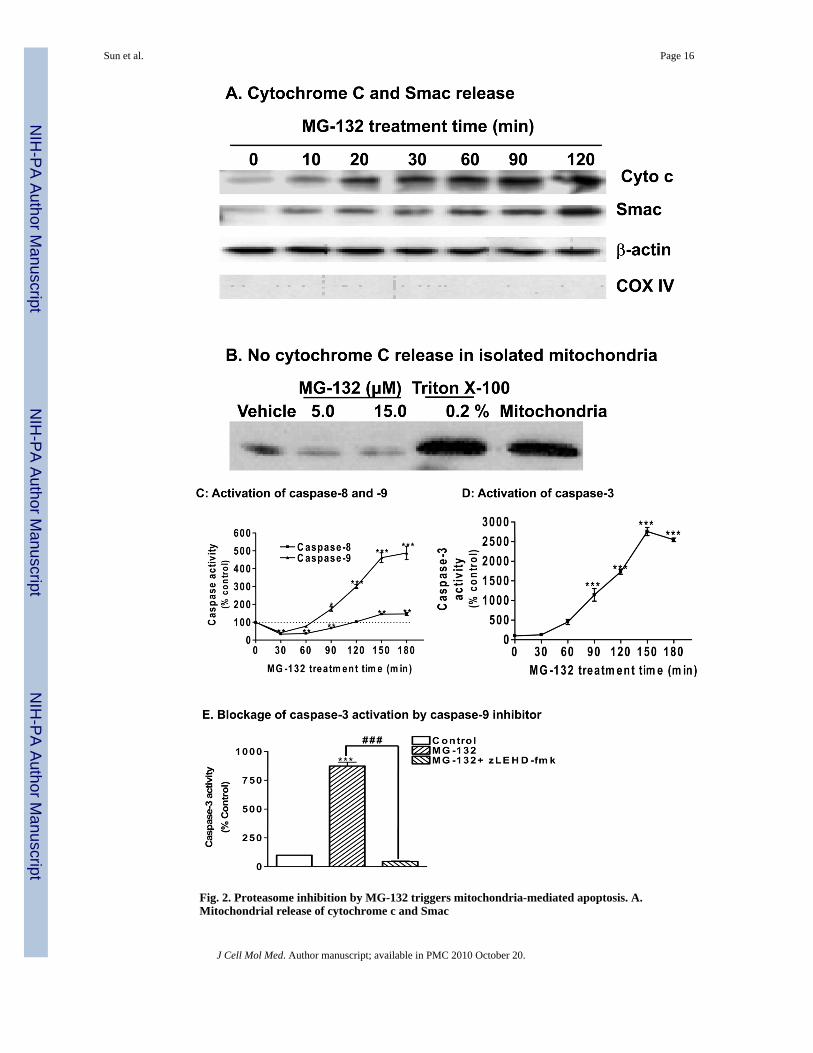
Fig. 2. Proteasome inhibition by MG-132 triggers mitochondria-mediated apoptosis. A.Mitochondrial release of cytochrome c and Smac
Sun et al. Page 16
J Cell Mol Med. Author manuscript; available in PMC 2010 October 20.
NIH
-PA Author Manuscript
NIH
-PA Author Manuscript
NIH
-PA Author Manuscript
N27 cells were treated with 5.0 μM MG-132 for 0–120 min. The cytosolic fractions preparedfrom the cells were resolved on 15% SDS-PAGE and blotted with antibodies againstcytochrome c, Smac, β-actin, or COX IV. B. Cytochrome c release from isolatedmitochondria. Mitochondria were isolated from N27 cells and resuspended in the isolationbuffer at 2.0 mg/ml. An equal amount of mitochondrial suspension was incubated with 5.0(lane 2) or 15.0 μM (lane 3) MG-132 for 1 hr, or with 0.2% Triton X-100 as positive controlto release cyto c (lane 4). Lane 5 is input of isolated mitochondria. C and D. Activation ofcaspase-8, -9, and -3. Cells were treated with 5.0 μM MG-132 for 30, 60, 90, 120, or 180 min.The caspase-8, -9, and -3 activities were determined using fluorogenic substrates as describedin the Materials and Methods. Data are presented as mean ± S.E.M for 8 samples. E. Inhibitionof caspase-3 activation by caspase-9 inhibitor LEHD-FMK. N27 cells were pretreated withLEHD-FMK (50 μM) for 40 min before treatment with 5.0 μM MG-132 for an additional 120min. The cells were collected for caspase-9 assay. Values represent mean ± S.E.M from 6individual samples. *p<0.05, **p<0.01 and ***p<0.001 vs control group.
Sun et al. Page 17
J Cell Mol Med. Author manuscript; available in PMC 2010 October 20.
NIH
-PA Author Manuscript
NIH
-PA Author Manuscript
NIH
-PA Author Manuscript
Sun et al. Page 18
J Cell Mol Med. Author manuscript; available in PMC 2010 October 20.
NIH
-PA Author Manuscript
NIH
-PA Author Manuscript
NIH
-PA Author Manuscript
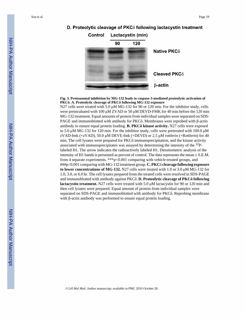
Fig. 3. Proteasomal inhibition by MG-132 leads to caspase-3-mediated proteolytic activation ofPKCδ. A. Proteolytic cleavage of PKCδ following MG-132 exposureN27 cells were treated with 5.0 μM MG-132 for 90 or 120 min. For the inhibitor study, cellswere preincubated with 100 μM ZVAD or 50 μM DEVD-FMK for 40 min before the 120 minMG-132 treatment. Equal amounts of protein from individual samples were separated on SDS-PAGE and immunoblotted with antibody for PKCδ. Membranes were reprobed with β-actinantibody to ensure equal protein loading. B. PKCδ kinase activity. N27 cells were exposedto 5.0 μM MG-132 for 120 min. For the inhibitor study, cells were pretreated with 100.0 μMzVAD-fmk (+zVAD), 50.0 μM DEVE-fmk (+DEVD) or 2.5 μM rottlerin (+Rottlerin) for 40min. The cell lysates were prepared for PKCδ immunoprecipitation, and the kinase activityassociated with immunoprecipitates was assayed by determining the intensity of the 32P-labeled H1. The arrow indicates the radioactively labeled H1. Densitometric analysis of theintensity of H1 bands is presented as percent of control. The data represents the mean ± S.E.M.from 4 separate experiments. ***p<0.001 comparing with vehicle-treated groups, and###p<0.001 comparing with MG-132 treatment group. C. PKCδ cleavage following exposureto lower concentrations of MG-132. N27 cells were treated with 1.0 or 3.0 μM MG-132 for1.0, 3.0, or 6.0 hr. The cell lysates prepared from the treated cells were resolved in SDS-PAGEand immunoblotted with antibody against PKCδ. D. Proteolytic cleavage of PKCδ followinglactacystin treatment. N27 cells were treated with 5.0 μM lactacystin for 90 or 120 min andthen cell lysates were prepared. Equal amount of protein from individual samples wereseparated on SDS-PAGE and immunoblotted with antibody for PKCδ. Reprobing membranewith β-actin antibody was performed to ensure equal protein loading.
Sun et al. Page 19
J Cell Mol Med. Author manuscript; available in PMC 2010 October 20.
NIH
-PA Author Manuscript
NIH
-PA Author Manuscript
NIH
-PA Author Manuscript
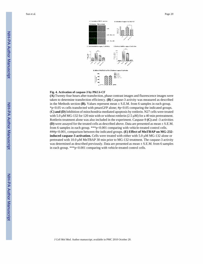
Fig. 4. Activation of caspase-3 by PKCδ-CF(A) Twenty-four hours after transfection, phase contrast images and fluorescence images weretaken to determine transfection efficiency. (B) Caspase-3 activity was measured as describedin the Methods section (B). Values represent mean ± S.E.M. from 6 samples in each group.*p<0.05 vs cells transfected with pmaxGFP alone; #p<0.05 comparing the indicated groups.(C) and (D) Inhibition of mitochondria-mediated apoptosis by rottlerin. N27 cells were treatedwith 5.0 μM MG-132 for 120 min with or without rottlerin (2.5 μM) for a 40 min pretreatment.Rottlerin treatment alone was also included in the experiment. Caspase-9 (C) and -3 activities(D) were assayed for the treated cells as described above. Data are presented as mean ± S.E.M.from 6 samples in each group. ***p<0.001 comparing with vehicle-treated control cells.###p<0.001, comparison between the indicated groups. (E) Effect of MnTBAP on MG-232-induced caspase-3 activation. Cells were treated with either with 5.0 μM MG-132 alone orpretreated with 10.0 μM MnTBAP 30 min prior to MG-132 treatment. The caspase-3 activitywas determined as described previously. Data are presented as mean ± S.E.M. from 6 samplesin each group. ***p<0.001 comparing with vehicle-treated control cells.
Sun et al. Page 20
J Cell Mol Med. Author manuscript; available in PMC 2010 October 20.
NIH
-PA Author Manuscript
NIH
-PA Author Manuscript
NIH
-PA Author Manuscript
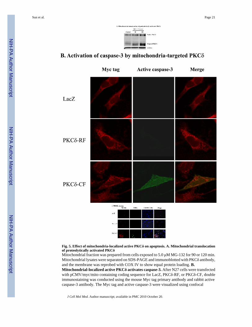
Fig. 5. Effect of mitochondria-localized active PKCδ on apoptosis. A. Mitochondrial translocationof proteolytically activated PKCδMitochondrial fraction was prepared from cells exposed to 5.0 μM MG-132 for 90 or 120 min.Mitochondrial lysates were separated on SDS-PAGE and immunoblotted with PKCδ antibody,and the membrane was reprobed with COX IV to show equal protein loading. B.Mitochondrial-localized active PKCδ activates caspase-3. After N27 cells were transfectedwith pCMV/myc/mito containing coding sequence for LacZ, PKCδ-RF, or PKCδ-CF, doubleimmunostaining was conducted using the mouse Myc tag primary antibody and rabbit activecaspase-3 antibody. The Myc tag and active caspase-3 were visualized using confocal
Sun et al. Page 21
J Cell Mol Med. Author manuscript; available in PMC 2010 October 20.
NIH
-PA Author Manuscript
NIH
-PA Author Manuscript
NIH
-PA Author Manuscript

microscopy using Cy3 conjugated anti-mouse (red) and Alexa-488 conjugated anti-rabbit(green) secondary antibodies. C. TUNEL staining in the transfected cells. After transfectionfor 24 hr, cells were subjected to TUNEL staining (green) and immunostaining with Myc tagantibody (red). The images were analyzed with fluorescence microscopy. The inserts showhigher magnification pictures (600X).
Sun et al. Page 22
J Cell Mol Med. Author manuscript; available in PMC 2010 October 20.
NIH
-PA Author Manuscript
NIH
-PA Author Manuscript
NIH
-PA Author Manuscript
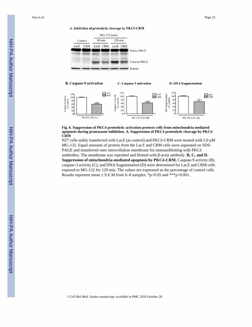
Fig. 6. Suppression of PKCδ proteolytic activation protects cells from mitochondria mediatedapoptosis during proteasome inhibition. A. Suppression of PKCδ proteolytic cleavage by PKCδ-CRMN27 cells stably transfected with LacZ (as control) and PKCδ-CRM were treated with 5.0 μMMG-132. Equal amounts of protein from the LacZ and CRM cells were separated on SDS-PAGE and transferred onto nitrocellulose membrane for immunoblotting with PKCδantibodies. The membrane was reprobed and blotted with β-actin antibody. B, C, and D.Suppression of mitochondria-mediated apoptosis by PKCδ-CRM. Caspase-9 activity (B),caspase-3 activity (C), and DNA fragmentation (D) were determined for LacZ and CRM cellsexposed to MG-132 for 120 min. The values are expressed as the percentage of control cells.Results represent mean ± S.E.M from 6–8 samples. *p<0.05 and ***p<0.001.
Sun et al. Page 23
J Cell Mol Med. Author manuscript; available in PMC 2010 October 20.
NIH
-PA Author Manuscript
NIH
-PA Author Manuscript
NIH
-PA Author Manuscript
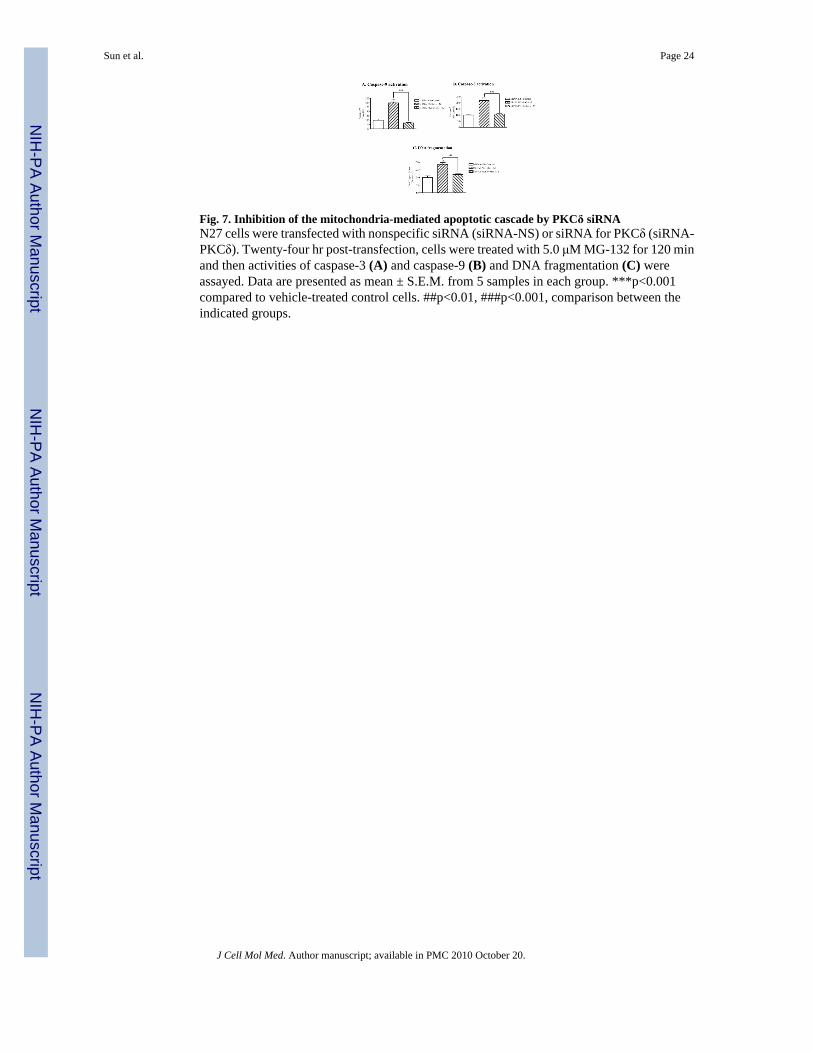
Fig. 7. Inhibition of the mitochondria-mediated apoptotic cascade by PKCδ siRNAN27 cells were transfected with nonspecific siRNA (siRNA-NS) or siRNA for PKCδ (siRNA-PKCδ). Twenty-four hr post-transfection, cells were treated with 5.0 μM MG-132 for 120 minand then activities of caspase-3 (A) and caspase-9 (B) and DNA fragmentation (C) wereassayed. Data are presented as mean ± S.E.M. from 5 samples in each group. ***p<0.001compared to vehicle-treated control cells. ##p<0.01, ###p<0.001, comparison between theindicated groups.
Sun et al. Page 24
J Cell Mol Med. Author manuscript; available in PMC 2010 October 20.
NIH
-PA Author Manuscript
NIH
-PA Author Manuscript
NIH
-PA Author Manuscript
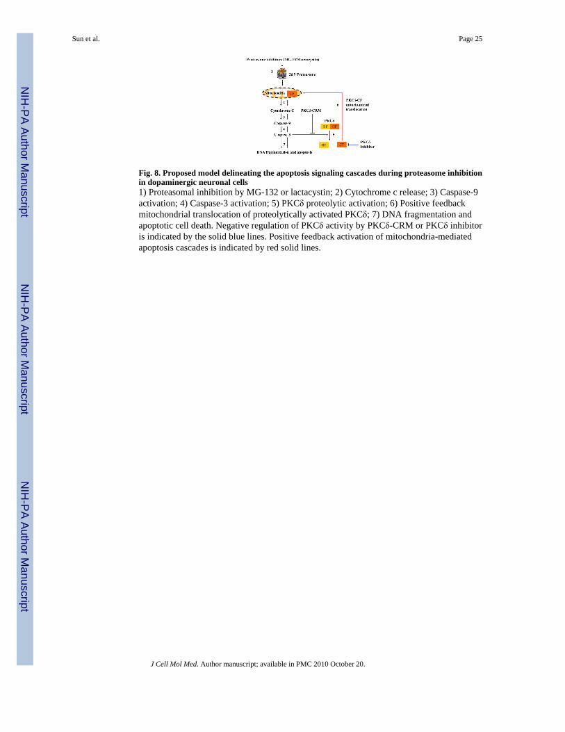
Fig. 8. Proposed model delineating the apoptosis signaling cascades during proteasome inhibitionin dopaminergic neuronal cells1) Proteasomal inhibition by MG-132 or lactacystin; 2) Cytochrome c release; 3) Caspase-9activation; 4) Caspase-3 activation; 5) PKCδ proteolytic activation; 6) Positive feedbackmitochondrial translocation of proteolytically activated PKCδ; 7) DNA fragmentation andapoptotic cell death. Negative regulation of PKCδ activity by PKCδ-CRM or PKCδ inhibitoris indicated by the solid blue lines. Positive feedback activation of mitochondria-mediatedapoptosis cascades is indicated by red solid lines.
Sun et al. Page 25
J Cell Mol Med. Author manuscript; available in PMC 2010 October 20.
NIH
-PA Author Manuscript
NIH
-PA Author Manuscript
NIH
-PA Author Manuscript
Copyright © 2022 FDOKUMEN