Proposal of Lysinibacillus boronitolerans gen. nov. sp. nov., and transfer of Bacillus fusiformis to...
13
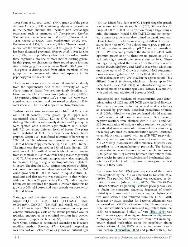
Proposal of Lysinibacillus boronitolerans gen. nov. sp. nov., and transfer of Bacillus fusiformis to Lysinibacillus fusiformis comb. nov. and Bacillus sphaericus to Lysinibacillus sphaericus comb. nov. Iftikhar Ahmed, 1,2 Akira Yokota, 3 Atsushi Yamazoe 4 and Toru Fujiwara 1,5 Correspondence Iftikhar Ahmed [email protected] Toru Fujiwara [email protected] 1 Biotechnology Research Center, University of Tokyo, Yayoi 1-1-1, Bunkyo-ku, Tokyo 113-8657, Japan 2 National Agricultural Research Centre, Park Road, Islamabad – 45500, Pakistan 3 Institute of Molecular and Cellular Biosciences, University of Tokyo, Yayoi 1-1-1, Bunkyo-ku, Tokyo 113-8657, Japan 4 Research Center for Water Environmental Technology, University of Tokyo, Hongo 7-3-1, Bunkyo-ku, Tokyo 113-8657, Japan 5 SORST, JST, Chiyoda-ku, Tokyo, Japan Three strains of a spore-forming, Gram-positive, motile, rod-shaped and boron-tolerant bacterium were isolated from soil. The strains, designated 10a T , 11c and 12B, can tolerate 5 % (w/v) NaCl and up to 150 mM boron, but optimal growth was observed without addition of boron or NaCl in Luria–Bertani agar medium. The optimum temperature for growth was 37 6C (range 16–45 6C) and the optimum pH was 7.0–8.0 (range pH 5.5–9.5). A comparative analysis of the 16S rRNA gene sequence demonstrated that the isolated strains were closely related to Bacillus fusiformis DSM 2898 T (97.2 % similarity) and Bacillus sphaericus DSM 28 T (96.9 %). DNA–DNA relatedness was greater than 97 % among the isolated strains and 61.1 % with B. fusiformis DSM 2898 T and 43.2 % with B. sphaericus IAM 13420 T . The phylogenetic and phenotypic analyses and DNA–DNA relatedness indicated that the three strains belong to the same species, that was characterized by a DNA G+C content of 36.5–37.9 mol%, MK-7 as the predominant menaquinone system and iso-C 15 : 0 (32 % of the total) as a major cellular fatty acid. In contrast to the type species of the genus Bacillus, the strains contained peptidoglycan with lysine, aspartic acid, alanine and glutamic acid. Based on the distinctive peptidoglycan composition, phylogenetic analyses and physiology, the strains are assigned to a novel species within a new genus, for which the name Lysinibacillus boronitolerans gen. nov., sp. nov. is proposed. The type strain of Lysinibacillus boronitolerans is strain 10a T (=DSM 17140 T =IAM 15262 T =ATCC BAA-1146 T ). It is also proposed that Bacillus fusiformis and Bacillus sphaericus be transferred to this genus as Lysinibacillus fusiformis comb. nov. and Lysinibacillus sphaericus comb. nov., respectively. Boron is an essential micronutrient for plants (Warington, 1923) and possibly also for animals (Rowe et al., 1998; Rowe & Eckhert, 1999). In bacteria, Chen et al. (2002) presented evidence for the biological role of boron in quorum sensing. On the other hand, boron is toxic to living cells when present above a certain threshold. Because of its toxic effects at high concentrations for micro-organisms, boron has been used as a food preservative (Nielsen, 2004), and it has also been used as an insecticide against cockroaches (Cochran, 1995). However, only recently, Bacillus boroniphilus, Chimaereicella boritolerans and Gracilibacillus boraciitolerans have been reported to tolerate more than 450, 300 and 450 mM boron, respectively, and/or require boron for growth (Ahmed et al., 2007a, b, c). Over the last decade, the genus Bacillus has been dissected taxonomically into several new genera based on compre- hensive analyses of 16S rRNA gene sequences and other chemotaxonomic data (Wisotzkey et al., 1992; Ash et al., 1993; Shida et al., 1996; Heyndrickx et al., 1998; Wainø et al., The GenBank/EMBL/DDBJ accession numbers for the 16S rRNA gene sequences of strains 10a T , 11c and 12B are respectively AB199591–AB199593. Results of growth experiments in the presence of boron, photomicro- graphs of cells of strain 10a T and results of separation of polar lipids of strain 10a T and related type strains are available as supplementary material in IJSEM Online. 63867 G 2007 IUMS Printed in Great Britain 1117 International Journal of Systematic and Evolutionary Microbiology (2007), 57, 1117–1125 DOI 10.1099/ijs.0.63867-0
-
Upload
independent -
Category
Documents
-
view
0 -
download
0
Transcript of Proposal of Lysinibacillus boronitolerans gen. nov. sp. nov., and transfer of Bacillus fusiformis to...
Proposal of Lysinibacillus boronitolerans gen. nov.sp. nov., and transfer of Bacillus fusiformis toLysinibacillus fusiformis comb. nov. and Bacillussphaericus to Lysinibacillus sphaericus comb. nov.
Iftikhar Ahmed,1,2 Akira Yokota,3 Atsushi Yamazoe4 and Toru Fujiwara1,5
Correspondence
Iftikhar Ahmed
Toru Fujiwara
1Biotechnology Research Center, University of Tokyo, Yayoi 1-1-1, Bunkyo-ku,Tokyo 113-8657, Japan
2National Agricultural Research Centre, Park Road, Islamabad – 45500, Pakistan
3Institute of Molecular and Cellular Biosciences, University of Tokyo, Yayoi 1-1-1, Bunkyo-ku,Tokyo 113-8657, Japan
4Research Center for Water Environmental Technology, University of Tokyo, Hongo 7-3-1,Bunkyo-ku, Tokyo 113-8657, Japan
5SORST, JST, Chiyoda-ku, Tokyo, Japan
Three strains of a spore-forming, Gram-positive, motile, rod-shaped and boron-tolerant bacterium
were isolated from soil. The strains, designated 10aT, 11c and 12B, can tolerate 5 % (w/v)
NaCl and up to 150 mM boron, but optimal growth was observed without addition of boron or NaCl
in Luria–Bertani agar medium. The optimum temperature for growth was 37 6C (range 16–45 6C)
and the optimum pH was 7.0–8.0 (range pH 5.5–9.5). A comparative analysis of the 16S rRNA
gene sequence demonstrated that the isolated strains were closely related to Bacillus fusiformis
DSM 2898T (97.2 % similarity) and Bacillus sphaericus DSM 28T (96.9 %). DNA–DNA
relatedness was greater than 97 % among the isolated strains and 61.1 % with B. fusiformis DSM
2898T and 43.2 % with B. sphaericus IAM 13420T. The phylogenetic and phenotypic analyses
and DNA–DNA relatedness indicated that the three strains belong to the same species, that
was characterized by a DNA G+C content of 36.5–37.9 mol%, MK-7 as the predominant
menaquinone system and iso-C15 : 0 (32 % of the total) as a major cellular fatty acid. In contrast to
the type species of the genus Bacillus, the strains contained peptidoglycan with lysine, aspartic
acid, alanine and glutamic acid. Based on the distinctive peptidoglycan composition, phylogenetic
analyses and physiology, the strains are assigned to a novel species within a new genus, for which
the name Lysinibacillus boronitolerans gen. nov., sp. nov. is proposed. The type strain of
Lysinibacillus boronitolerans is strain 10aT (=DSM 17140T=IAM 15262T=ATCC BAA-1146T).
It is also proposed that Bacillus fusiformis and Bacillus sphaericus be transferred to this genus as
Lysinibacillus fusiformis comb. nov. and Lysinibacillus sphaericus comb. nov., respectively.
Boron is an essential micronutrient for plants (Warington,1923) and possibly also for animals (Rowe et al., 1998; Rowe& Eckhert, 1999). In bacteria, Chen et al. (2002) presentedevidence for the biological role of boron in quorum sensing.On the other hand, boron is toxic to living cells when presentabove a certain threshold. Because of its toxic effects at high
concentrations for micro-organisms, boron has been usedas a food preservative (Nielsen, 2004), and it has also beenused as an insecticide against cockroaches (Cochran, 1995).However, only recently, Bacillus boroniphilus, Chimaereicellaboritolerans and Gracilibacillus boraciitolerans have beenreported to tolerate more than 450, 300 and 450 mM boron,respectively, and/or require boron for growth (Ahmed et al.,2007a, b, c).
Over the last decade, the genus Bacillus has been dissectedtaxonomically into several new genera based on compre-hensive analyses of 16S rRNA gene sequences and otherchemotaxonomic data (Wisotzkey et al., 1992; Ash et al.,1993; Shida et al., 1996; Heyndrickx et al., 1998; Wainø et al.,
The GenBank/EMBL/DDBJ accession numbers for the 16S rRNAgene sequences of strains 10aT, 11c and 12B are respectivelyAB199591–AB199593.
Results of growth experiments in the presence of boron, photomicro-graphs of cells of strain 10aT and results of separation of polar lipids ofstrain 10aT and related type strains are available as supplementarymaterial in IJSEM Online.
63867 G 2007 IUMS Printed in Great Britain 1117
International Journal of Systematic and Evolutionary Microbiology (2007), 57, 1117–1125 DOI 10.1099/ijs.0.63867-0
1999; Yoon et al., 2001, 2002). rRNA group 2 of the genusBacillus (Ash et al., 1991) containing L-lysine or D-ornithinein the peptide subunit also includes non-Bacillus-typeorganisms such as members of Caryophanon, Kurthia,Sporosarcina, Planococcus and Filibacter (Clausen et al.,1985; Keddie & Shaw, 1986; Stackebrandt et al., 1987;Pechman et al., 1976; Farrow et al., 1994). There is a need tore-evaluate the taxonomic status of this group. Although ithas been discussed previously (Farrow et al., 1994; Rheimset al., 1999), no proposal has yet been put forward to transferthese organisms into one or more new or existing genera.In this paper, we characterize three round-spore-formingstrains belonging to a new genus in Bacillus RNA group 2.This genus is distinguished from other members of thisgroup by the presence of lysine and aspartate in thepeptidoglycan of the cell wall.
The three strains were isolated from soil sampled randomlyfrom the experimental field of the University of Tokyo(Yayoi campus), Japan. We used previously described iso-lation and enrichment procedures (Ahmed et al., 2007a).The purified isolates (strains 10aT, 11c and 12B) were main-tained on agar medium, and also stored as glycerol (35 %,w/v) stocks at 280 uC, and subjected to characterization.
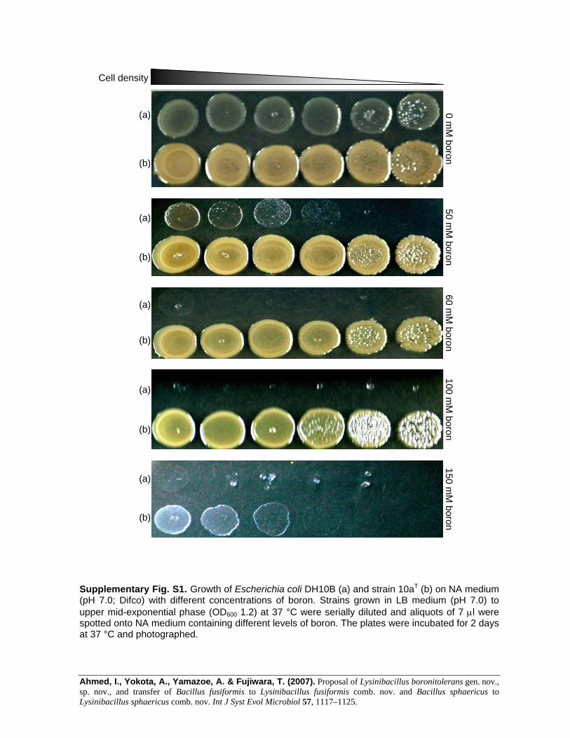
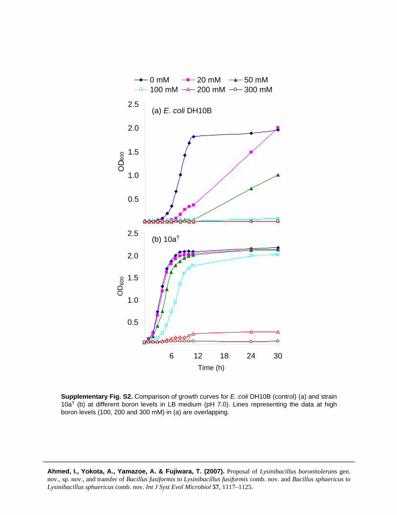
To demonstrate boron tolerance, strain 10aT and Escherichiacoli DH10B (control) were grown up to upper mid-exponential phase (OD600 1.2) at 37 uC, with vigorousshaking. The cultures were serially diluted and 7 ml aliquotswere spotted onto nutrient agar medium (NA; Difco)(pH 7.0) containing different levels of boron. The plateswere incubated at 37 uC for 2 days before being photo-graphed. Strain 10aT manifested tolerance of boron up to100 mM on NA, whereas weak growth was observed at150 mM boron (Supplementary Fig. S1 in IJSEM Online).The strain was also cultured in 150 ml Luria–Bertani (LB)medium (pH 7.0) with different levels of boron rangingfrom 0 (control) to 300 mM, while being shaken vigorouslyat 30 uC. After every 60 min, samples were taken asepticallyto measure OD600 using a spectrophotometer (HitachiU-1800). The data for OD600 against time plotted at differ-ent boron concentrations demonstrated that strain 10aT
could grow with 0–100 mM boron in liquid culture (LBmedium) and that growth was equivalent to that withoutaddition of boron (Supplementary Fig. S2), indicating thatboron was not required for growth. However, there was nogrowth at 200 mM boron and weak growth was observed at150 mM boron.
Sporangia and the sizes of cells grown on NA withMgSO4.7H2O (1.01 mM), KCl (13.4 mM), FeSO4
(0.001 mM), Ca(NO3)2 (1.0 mM) and MnCl2 (0.01 mM)at pH 7.0 for 6 days at 30 uC were examined under phase-contrast microscopy. Cells of the strains produced oval orspherical endospores in a terminal position in a swollensporangium (Supplementary Fig. S3). Cells of the strainswere Gram-positive as determined according to Hucker’smodified method (Cowan, 1974). Colonial morphologywas observed on isolated colonies grown on nutrient agar
(pH 7.0, Difco) for 2 days at 30 uC. The pH range for growthwas determined in tryptic soya broth (TSB; Difco) with a pHrange of 4.0 to 10.0 at 30 uC by monitoring OD600 using amini-photometer (model 518R; TAITEC) and the temper-ature range for growth was determined on tryptic soya agar(TSA; Difco) (pH 7.0) by incubating at different temper-atures from 4 to 50 uC. The isolated strains grew at pH 5.5–9.5 with optimum growth at pH 7.5 and no growth atpH 5.0. We observed growth of the strains at 16–45 uC withoptimum growth at 37 uC; there was no growth at ¢50 uCand only slight growth after several days at 16 uC. Thesefindings distinguished the strains from the closely relatedspecies Bacillus fusiformis and Bacillus sphaericus, which canonly grow up to 40 uC. Growth at various NaCl concentra-tions was investigated on TSA (pH 7.0) at 30 uC. The novelstrains tolerated 0–5 % (w/v) NaCl in the agar medium. Thisdiffered from B. fusiformis, which can tolerate up to 7 %(w/v) NaCl (Priest et al., 1988). We also observed growth ofthe novel strains on marine agar 2216 (Difco), TSA and NAwith and without addition of boron or NaCl.
Physiological and biochemical characteristics were deter-mined using API 20E and API 50CH galleries (bioMerieux).The strains were positive for oxidase and catalase activitiesas assessed by previously described procedures (Ahmedet al., 2007a). Motility was also confirmed with M medium(bioMerieux) in addition to microscopy. Since mainlynegative reactions were obtained with API 50CH and API20E for utilization of various carbon sources, we analysedan extended array of metabolic features of the strains usingthe Biolog GP2 and GN2 characterization system. Resistanceto antibiotics was assessed with an ATB-VET strip (bio-Merieux) and enzyme activities were determined with anAPI ZYM strip (bioMerieux). All commercial kits were usedaccording to the manufacturers’ protocols. The isolatedstrains exhibited many features that were similar to those ofB. fusiformis and B. sphaericus; however, they differed fromthese species in certain physiological and biochemical char-acteristics (Table 1). All three novel strains gave identicalresults in these tests.
Nearly complete 16S rRNA gene sequences of the strainswere amplified by the PCR as described by Katsivela et al.(1999). The purified PCR product was sequenced as de-scribed previously (Ahmed et al., 2007a). The DNASIS Pro(Hitachi Software Engineering) software package was usedto obtain the consensus sequence. Sequences of closelyrelated type strains used for constructing the phylogenetictree were selected and retrieved from the DDBJ/EMBLdatabases by BLAST searches for bacteria. Alignment wasperformed with CLUSTAL X (version 1.8w; Thompson et al.,1997) and evolutionary distances and Knuc values (Kimura,1980) were generated. BioEdit software (Hall, 1999) wasused to remove gaps and ambiguous bases in the alignments.A phylogenetic tree was constructed from 1294 unambig-uously aligned nucleotides using the neighbour-joiningmethod (Saitou & Nei, 1987) contained in the PHYLIP soft-ware package (Felsenstein, 2005) and plotted with NJPlot
1118 International Journal of Systematic and Evolutionary Microbiology 57
I. Ahmed and others
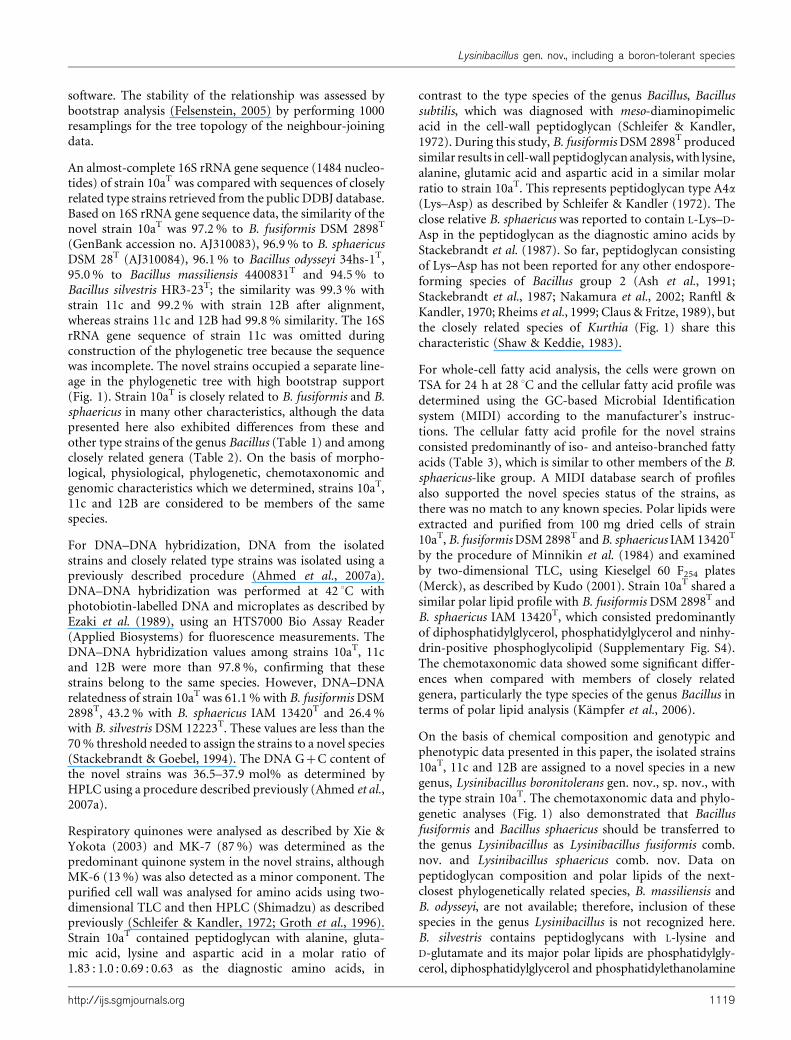
software. The stability of the relationship was assessed bybootstrap analysis (Felsenstein, 2005) by performing 1000resamplings for the tree topology of the neighbour-joiningdata.
An almost-complete 16S rRNA gene sequence (1484 nucleo-tides) of strain 10aT was compared with sequences of closelyrelated type strains retrieved from the public DDBJ database.Based on 16S rRNA gene sequence data, the similarity of thenovel strain 10aT was 97.2 % to B. fusiformis DSM 2898T
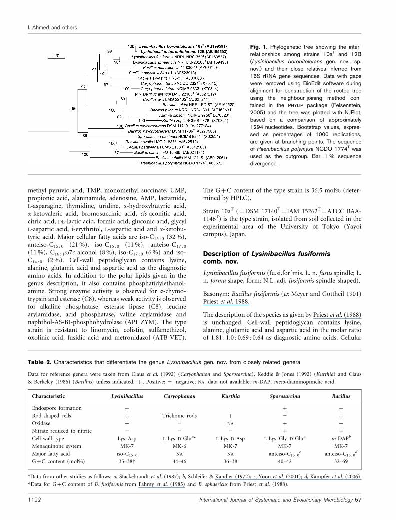
(GenBank accession no. AJ310083), 96.9 % to B. sphaericusDSM 28T (AJ310084), 96.1 % to Bacillus odysseyi 34hs-1T,95.0 % to Bacillus massiliensis 4400831T and 94.5 % toBacillus silvestris HR3-23T; the similarity was 99.3 % withstrain 11c and 99.2 % with strain 12B after alignment,whereas strains 11c and 12B had 99.8 % similarity. The 16SrRNA gene sequence of strain 11c was omitted duringconstruction of the phylogenetic tree because the sequencewas incomplete. The novel strains occupied a separate line-age in the phylogenetic tree with high bootstrap support(Fig. 1). Strain 10aT is closely related to B. fusiformis and B.sphaericus in many other characteristics, although the datapresented here also exhibited differences from these andother type strains of the genus Bacillus (Table 1) and amongclosely related genera (Table 2). On the basis of morpho-logical, physiological, phylogenetic, chemotaxonomic andgenomic characteristics which we determined, strains 10aT,11c and 12B are considered to be members of the samespecies.
For DNA–DNA hybridization, DNA from the isolatedstrains and closely related type strains was isolated using apreviously described procedure (Ahmed et al., 2007a).DNA–DNA hybridization was performed at 42 uC withphotobiotin-labelled DNA and microplates as described byEzaki et al. (1989), using an HTS7000 Bio Assay Reader(Applied Biosystems) for fluorescence measurements. TheDNA–DNA hybridization values among strains 10aT, 11cand 12B were more than 97.8 %, confirming that thesestrains belong to the same species. However, DNA–DNArelatedness of strain 10aT was 61.1 % with B. fusiformis DSM2898T, 43.2 % with B. sphaericus IAM 13420T and 26.4 %with B. silvestris DSM 12223T. These values are less than the70 % threshold needed to assign the strains to a novel species(Stackebrandt & Goebel, 1994). The DNA G+C content ofthe novel strains was 36.5–37.9 mol% as determined byHPLC using a procedure described previously (Ahmed et al.,2007a).
Respiratory quinones were analysed as described by Xie &Yokota (2003) and MK-7 (87 %) was determined as thepredominant quinone system in the novel strains, althoughMK-6 (13 %) was also detected as a minor component. Thepurified cell wall was analysed for amino acids using two-dimensional TLC and then HPLC (Shimadzu) as describedpreviously (Schleifer & Kandler, 1972; Groth et al., 1996).Strain 10aT contained peptidoglycan with alanine, gluta-mic acid, lysine and aspartic acid in a molar ratio of1.83 : 1.0 : 0.69 : 0.63 as the diagnostic amino acids, in
contrast to the type species of the genus Bacillus, Bacillussubtilis, which was diagnosed with meso-diaminopimelicacid in the cell-wall peptidoglycan (Schleifer & Kandler,1972). During this study, B. fusiformis DSM 2898T producedsimilar results in cell-wall peptidoglycan analysis, with lysine,alanine, glutamic acid and aspartic acid in a similar molarratio to strain 10aT. This represents peptidoglycan type A4a
(Lys–Asp) as described by Schleifer & Kandler (1972). Theclose relative B. sphaericus was reported to contain L-Lys–D-Asp in the peptidoglycan as the diagnostic amino acids byStackebrandt et al. (1987). So far, peptidoglycan consistingof Lys–Asp has not been reported for any other endospore-forming species of Bacillus group 2 (Ash et al., 1991;Stackebrandt et al., 1987; Nakamura et al., 2002; Ranftl &Kandler, 1970; Rheims et al., 1999; Claus & Fritze, 1989), butthe closely related species of Kurthia (Fig. 1) share thischaracteristic (Shaw & Keddie, 1983).
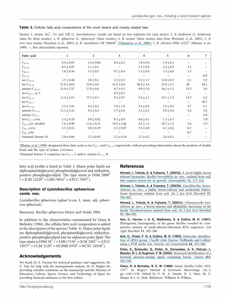
For whole-cell fatty acid analysis, the cells were grown onTSA for 24 h at 28 uC and the cellular fatty acid profile wasdetermined using the GC-based Microbial Identificationsystem (MIDI) according to the manufacturer’s instruc-tions. The cellular fatty acid profile for the novel strainsconsisted predominantly of iso- and anteiso-branched fattyacids (Table 3), which is similar to other members of the B.sphaericus-like group. A MIDI database search of profilesalso supported the novel species status of the strains, asthere was no match to any known species. Polar lipids wereextracted and purified from 100 mg dried cells of strain10aT, B. fusiformis DSM 2898T and B. sphaericus IAM 13420T
by the procedure of Minnikin et al. (1984) and examinedby two-dimensional TLC, using Kieselgel 60 F254 plates(Merck), as described by Kudo (2001). Strain 10aT shared asimilar polar lipid profile with B. fusiformis DSM 2898T andB. sphaericus IAM 13420T, which consisted predominantlyof diphosphatidylglycerol, phosphatidylglycerol and ninhy-drin-positive phosphoglycolipid (Supplementary Fig. S4).The chemotaxonomic data showed some significant differ-ences when compared with members of closely relatedgenera, particularly the type species of the genus Bacillus interms of polar lipid analysis (Kampfer et al., 2006).
On the basis of chemical composition and genotypic andphenotypic data presented in this paper, the isolated strains10aT, 11c and 12B are assigned to a novel species in a newgenus, Lysinibacillus boronitolerans gen. nov., sp. nov., withthe type strain 10aT. The chemotaxonomic data and phylo-genetic analyses (Fig. 1) also demonstrated that Bacillusfusiformis and Bacillus sphaericus should be transferred tothe genus Lysinibacillus as Lysinibacillus fusiformis comb.nov. and Lysinibacillus sphaericus comb. nov. Data onpeptidoglycan composition and polar lipids of the next-closest phylogenetically related species, B. massiliensis andB. odysseyi, are not available; therefore, inclusion of thesespecies in the genus Lysinibacillus is not recognized here.B. silvestris contains peptidoglycans with L-lysine andD-glutamate and its major polar lipids are phosphatidylgly-cerol, diphosphatidylglycerol and phosphatidylethanolamine
http://ijs.sgmjournals.org 1119
Lysinibacillus gen. nov., including a boron-tolerant species

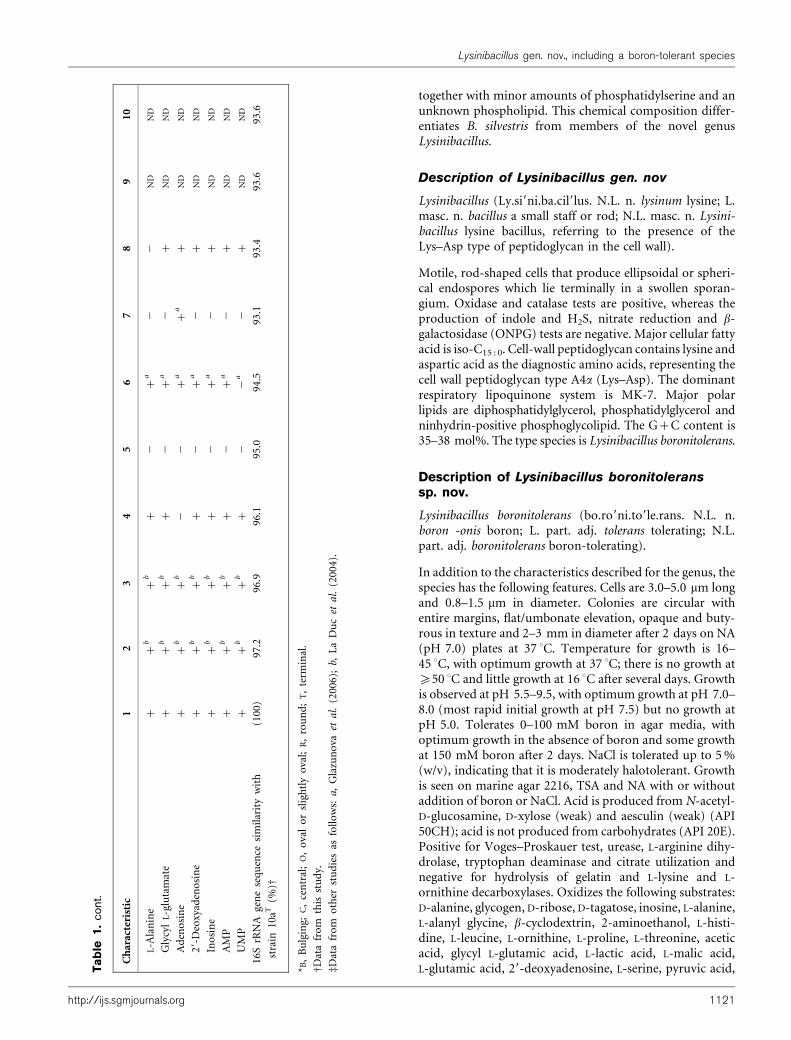
Table 1. Characteristics that differentiate the novel strains (Lysinibacillus boronitolerans) from closely related type strains
Strains: 1, strains 10aT, 11c and 12B (Lysinibacillus boronitolerans gen. nov., sp. nov.) (data from this study); 2, B. fusiformis DSM 2898T (Lysinibacillus fusiformis) (data from Priest et al.,
1988); 3, B. sphaericus IAM 13420T (Lysinibacillus sphaericus) (Claus & Berkeley, 1986); 4, B. odysseyi ATCC PTA-4993T (La Duc et al., 2004); 5, B. massiliensis CIP 108446T (Glazunova
et al., 2006); 6, B. silvestris DSM 12223T (Rheims et al., 1999); 7, B. pycnus JCM 11075T (Nakamura et al., 2002); 8, B. neidei JCM 11075T (Nakamura et al., 2002); 9, B. arenosi LMG
22166T (Heyrman et al., 2005); 10, B. arvi LMG 22165T (Heyrman et al., 2005). All type strains produced positive results for motility, growth without NaCl and catalase activity and
negative results for H2S production and indole production. ND, No data available; V, variable results; +, positive; W, weakly positive; 2, negative.
Characteristic 1 2 3 4 5 6 7 8 9 10
Cell size (mm) (length6diameter) 3.0–5.060.8–1.5
ND 1.5–5.060.6–1.0
4.0–5.061.0
1.5–4.060.3–0.5
0.9–2.060.5–0.7
3.0–5.061.0–1.5
3.0–5.061.0
3.0–8.060.8–1.0
3.0–8.060.8–1.0
Spore shape and position* R/O, T, B R, C/T, B R, T, B R, T, B ND R, T, B R, B R, B R, T, B R, T, B
Growth temperature range (uC) (optimum) 16–45
(35–37)
17–40 10–40 25–42
(30–35)
25–45
(30–37)
10–40
(20–30)
5–45
(28–30)
5–45
(28–30)
(20–30) (20–30)
pH range (optimum) 5.5–9.5
(7.0–8.0)
6.0–9.5 6.0–9.5 6.0–10.0
(7.0)
ND ND ND ND (7.0–9.0) (7.0–9.0)
Growth in 7 % (w/v) NaCl 2 + 2 2 2 2 2 ND 2 2
Boron tolerance (mM) 150 60D 75D ND ND 60D ND ND ND ND
Voges–Proskauer test + 2 2 ND + 2 ND ND 2 2
Nitrate reduction 2 2 2 2 ND 2 2 2 + 2
Oxidase + + + 2 + 2 ND ND 2 2
L-Arginine dihydrolase + ND ND 2 + 2 ND ND 2 2
L-Lysine and L-ornithine decarboxylase 2 ND ND + + ND ND ND 2 2
Tryptophan deaminase + ND 2ad 2a + 2a 2a 2aND ND
Hydrolysis of:
Urea + + 2 2 + +a 2a +a 2 +
Gelatin 2 + + 2 2 2 +a +a +W 2
Aesculin +W 2 2 ND ND 2 ND ND 2 2
Acid production from
Sucrose 2 V 2 2 2 + ND ND 2 2
D-Xylose +W 2 2 2 2 2 ND ND 2 2
N-Acetyl-D-glucosamine + ND ND 2 ND 2 ND ND 2 W
Resistance to (mg ml21):
Chloramphenicol (8) 2 + +W ND 2 ND 2 2 ND ND
Erythromycin (1) 2 + +W ND ND ND 2 2 ND ND
Rifampicin (0.25) 2 2 +W ND 2 ND ND ND ND ND
Streptomycin (8) 2 +W + ND 2 ND 2 2 ND ND
Tetracycline (2) 2 2 + ND 2 ND 2 2 ND ND
Oxidation of:
Pyruvate + +b +b + + +a + 2 ND ND
a-Hydroxybutyrate + 2b 2b + 2 2a 2aND ND ND
b-Hydroxybutyrate 2 2b 2b + 2 +a + 2 ND ND
11
20
International
Journal
of
System
aticand
Evo
lutionary
Micro
bio
logy
57
I.A
hmed
andothers
together with minor amounts of phosphatidylserine and anunknown phospholipid. This chemical composition differ-entiates B. silvestris from members of the novel genusLysinibacillus.
Description of Lysinibacillus gen. nov
Lysinibacillus (Ly.si9ni.ba.cil9lus. N.L. n. lysinum lysine; L.masc. n. bacillus a small staff or rod; N.L. masc. n. Lysini-bacillus lysine bacillus, referring to the presence of theLys–Asp type of peptidoglycan in the cell wall).
Motile, rod-shaped cells that produce ellipsoidal or spheri-cal endospores which lie terminally in a swollen sporan-gium. Oxidase and catalase tests are positive, whereas theproduction of indole and H2S, nitrate reduction and b-galactosidase (ONPG) tests are negative. Major cellular fattyacid is iso-C15 : 0. Cell-wall peptidoglycan contains lysine andaspartic acid as the diagnostic amino acids, representing thecell wall peptidoglycan type A4a (Lys–Asp). The dominantrespiratory lipoquinone system is MK-7. Major polarlipids are diphosphatidylglycerol, phosphatidylglycerol andninhydrin-positive phosphoglycolipid. The G+C content is35–38 mol%. The type species is Lysinibacillus boronitolerans.
Description of Lysinibacillus boronitoleranssp. nov.
Lysinibacillus boronitolerans (bo.ro9ni.to9le.rans. N.L. n.boron -onis boron; L. part. adj. tolerans tolerating; N.L.part. adj. boronitolerans boron-tolerating).
In addition to the characteristics described for the genus, thespecies has the following features. Cells are 3.0–5.0 mm longand 0.8–1.5 mm in diameter. Colonies are circular withentire margins, flat/umbonate elevation, opaque and buty-rous in texture and 2–3 mm in diameter after 2 days on NA(pH 7.0) plates at 37 uC. Temperature for growth is 16–45 uC, with optimum growth at 37 uC; there is no growth at¢50 uC and little growth at 16 uC after several days. Growthis observed at pH 5.5–9.5, with optimum growth at pH 7.0–8.0 (most rapid initial growth at pH 7.5) but no growth atpH 5.0. Tolerates 0–100 mM boron in agar media, withoptimum growth in the absence of boron and some growthat 150 mM boron after 2 days. NaCl is tolerated up to 5 %(w/v), indicating that it is moderately halotolerant. Growthis seen on marine agar 2216, TSA and NA with or withoutaddition of boron or NaCl. Acid is produced from N-acetyl-D-glucosamine, D-xylose (weak) and aesculin (weak) (API50CH); acid is not produced from carbohydrates (API 20E).Positive for Voges–Proskauer test, urease, L-arginine dihy-drolase, tryptophan deaminase and citrate utilization andnegative for hydrolysis of gelatin and L-lysine and L-ornithine decarboxylases. Oxidizes the following substrates:D-alanine, glycogen, D-ribose, D-tagatose, inosine, L-alanine,L-alanyl glycine, b-cyclodextrin, 2-aminoethanol, L-histi-dine, L-leucine, L-ornithine, L-proline, L-threonine, aceticacid, glycyl L-glutamic acid, L-lactic acid, L-malic acid,L-glutamic acid, 29-deoxyadenosine, L-serine, pyruvic acid,
L-A
lan
ine
++
b+
b+
2+
a2
2N
DN
D
Gly
cyl
L-g
luta
mat
e+
+b
+b
+2
+a
2+
ND
ND
Ad
eno
sin
e+
+b
+b
22
+a
+a
+N
DN
D
29-
Deo
xyad
eno
sin
e+
+b
+b
+2
+a
2+
ND
ND
Ino
sin
e+
+b
+b
+2
+a
2+
ND
ND
AM
P+
+b
+b
+2
+a
2+
ND
ND
UM
P+
+b
+b
+2
2a
2+
ND
ND
16
SrR
NA
gen
ese
qu
ence
sim
ilar
ity
wit
h
stra
in1
0aT
(%)D
(10
0)
97
.29
6.9
96
.19
5.0
94
.59
3.1
93
.49
3.6
93
.6
*B,
Bu
lgin
g;C
,ce
ntr
al;
O,
ova
lo
rsl
igh
tly
ova
l;R
,ro
un
d;
T,
term
inal
.
DD
ata
fro
mth
isst
ud
y.
dD
ata
fro
mo
ther
stu
die
sas
foll
ow
s:a
,G
lazu
no
vaet
al.
(20
06
);b,
La
Du
cet
al.
(20
04
).
Ta
ble
1.
cont
.
Ch
arac
teri
stic
12
34
56
78
91
0
http://ijs.sgmjournals.org 1121
Lysinibacillus gen. nov., including a boron-tolerant species
methyl pyruvic acid, TMP, monomethyl succinate, UMP,propionic acid, alaninamide, adenosine, AMP, lactamide,L-asparagine, thymidine, uridine, a-hydroxybutyric acid,a-ketovaleric acid, bromosuccinic acid, cis-aconitic acid,citric acid, DL-lactic acid, formic acid, gluconic acid, glycylL-aspartic acid, i-erythritol, L-aspartic acid and a-ketobu-tyric acid. Major cellular fatty acids are iso-C15 : 0 (32 %),anteiso-C15 : 0 (21 %), iso-C16 : 0 (11 %), anteiso-C17 : 0
(11 %), C16 : 1v7c alcohol (8 %), iso-C17 : 0 (6 %) and iso-C14 : 0 (2 %). Cell-wall peptidoglycan contains lysine,alanine, glutamic acid and aspartic acid as the diagnosticamino acids. In addition to the polar lipids given in thegenus description, it also contains phosphatidylethanol-amine. Strong enzyme activity is observed for a-chymo-trypsin and esterase (C8), whereas weak activity is observedfor alkaline phosphatase, esterase lipase (C8), leucinearylamidase, acid phosphatase, valine arylamidase andnaphthol-AS-BI-phosphohydrolase (API ZYM). The typestrain is resistant to linomycin, colistin, sulfamethizol,oxolinic acid, fusidic acid and metronidazol (ATB-VET).
The G+C content of the type strain is 36.5 mol% (deter-mined by HPLC).
Strain 10aT (=DSM 17140T=IAM 15262T=ATCC BAA-1146T) is the type strain, isolated from soil collected in theexperimental area of the University of Tokyo (Yayoicampus), Japan.
Description of Lysinibacillus fusiformiscomb. nov.
Lysinibacillus fusiformis (fu.si.for9mis. L. n. fusus spindle; L.n. forma shape, form; N.L. adj. fusiformis spindle-shaped).
Basonym: Bacillus fusiformis (ex Meyer and Gottheil 1901)Priest et al. 1988.
The description of the species as given by Priest et al. (1988)is unchanged. Cell-wall peptidoglycan contains lysine,alanine, glutamic acid and aspartic acid in the molar ratioof 1.81 : 1.0 : 0.69 : 0.64 as diagnostic amino acids. Cellular
Fig. 1. Phylogenetic tree showing the inter-relationships among strains 10aT and 12B(Lysinibacillus boronitolerans gen. nov., sp.nov.) and their close relatives inferred from16S rRNA gene sequences. Data with gapswere removed using BioEdit software duringalignment for construction of the rooted treeusing the neighbour-joining method con-tained in the PHYLIP package (Felsenstein,2005) and the tree was plotted with NJPlot,based on a comparison of approximately1294 nucleotides. Bootstrap values, expres-sed as percentages of 1000 replications,are given at branching points. The sequenceof Paenibacillus polymyxa NCDO 1774T wasused as the outgroup. Bar, 1 % sequencedivergence.
Table 2. Characteristics that differentiate the genus Lysinibacillus gen. nov. from closely related genera
Data for reference genera were taken from Claus et al. (1992) (Caryophanon and Sporosarcina), Keddie & Jones (1992) (Kurthia) and Claus
& Berkeley (1986) (Bacillus) unless indicated. +, Positive; 2, negative; NA, data not available; m-DAP, meso-diaminopimelic acid.
Characteristic Lysinibacillus Caryophanon Kurthia Sporosarcina Bacillus
Endospore formation + 2 2 + +
Rod-shaped cells + Trichome rods + 2 +
Oxidase + 2 NA + +
Nitrate reduced to nitrite 2 2 2 + +
Cell-wall type Lys–Asp L-Lys–D-Glua* L-Lys–D-Asp L-Lys–Gly–D-Glua m-DAPb
Menaquinone system MK-7 MK-6 MK-7 MK-7 MK-7
Major fatty acid iso-C15 : 0 NA NA anteiso-C15 : 0c anteiso-C15 : 0
d
G+C content (mol%) 35–38D 44–46 36–38 40–42 32–69
*Data from other studies as follows: a, Stackebrandt et al. (1987); b, Schleifer & Kandler (1972); c, Yoon et al. (2001); d, Kampfer et al. (2006).
DData for G+C content of B. fusiformis from Fahmy et al. (1985) and B. sphaericus from Priest et al. (1988).
1122 International Journal of Systematic and Evolutionary Microbiology 57
I. Ahmed and others
fatty acid profile is listed in Table 3. Major polar lipids arediphosphatidylglycerol, phosphatidylglycerol and ninhydrin-positive phosphoglycolipid. The type strain is DSM 2898T
(=JCM 12229T=LMG 9816T=ATCC 7055T).
Description of Lysinibacillus sphaericuscomb. nov.
Lysinibacillus sphaericus (sphae9ri.cus. L. masc. adj. sphaer-icus spherical).
Basonym: Bacillus sphaericus Meyer and Neide 1904.
In addition to the characteristics summarized by Claus &Berkeley (1986), the cellular fatty acid composition is addedto the description of the species (Table 3). Major polar lipidsare diphosphatidylglycerol, phosphatidylglycerol, ninhydrin-positive phosphoglycolipid and an unknown polar lipid. Thetype strain is DSM 28T (=LMG 7134T=JCM 2502T=ATCC14577T=CCM 2120T=NCIMB 9370T=NCTC 10338T).
Acknowledgements
We thank Dr A. Nozawa for technical guidance and suggestions, MrX. Tian for help with the menaquinone analysis, Dr N. Bughio forproviding valuable comments on the manuscript and the Ministry ofEducation, Culture, Sports, Science, and Technology of Japan forproviding financial assistance to the first author.
References
Ahmed, I., Yokota, A. & Fujiwara, T. (2007a). A novel highly boron
tolerant bacterium, Bacillus boroniphilus sp. nov., isolated from soil,
that requires boron for its growth. Extremophiles 11, 217–224.
Ahmed, I., Yokota, A. & Fujiwara, T. (2007b). Gracilibacillus boraci-
itolerans sp. nov., a highly boron-tolerant and moderately haloto-
lerant bacterium isolated from soil. Int J Syst Evol Microbiol 57,
796–802.
Ahmed, I., Yokota, A. & Fujiwara, T. (2007c). Chimaereicella bori-
tolerans sp. nov., a boron-tolerant and alkaliphilic bacterium of the
family Flavobacteriaceae isolated from soil. Int J Syst Evol Microbiol
57, 986–992.
Ash, C., Farrow, J. A. E., Wallbanks, S. & Collins, M. D. (1991).Phylogenetic heterogeneity of the genus Bacillus revealed by com-
parative analysis of small-subunit-ribosomal RNA sequences. Lett
Appl Microbiol 13, 202–206.
Ash, C., Priest, F. G. & Collins, M. D. (1993). Molecular identifica-
tion of rRNA group 3 bacilli (Ash, Farrow, Wallbanks and Collins)
using a PCR probe test. Antonie van Leeuwenhoek 64, 253–260.
Chen, X., Schauder, S., Potier, N., Dorsselaer, A. V., Pelczer, I.,Bassler, B. L. & Hughson, F. M. (2002). Structural identification of a
bacterial quorum-sensing signal containing boron. Nature 415,
545–549.
Claus, D. & Berkeley, R. C. W. (1986). Genus Bacillus Cohn 1872,
174AL. In Bergey’s Manual of Systematic Bacteriology, vol. 2,
pp. 1105–1139. Edited by P. H. A. Sneath, N. S. Mair, M. E.
Sharpe & J. G. Holt. Baltimore: Williams & Wilkins.
Table 3. Cellular fatty acid compositions of the novel strains and closely related taxa
Strains: 1, strains 10aT, 11c and 12B (L. boronitolerans) (results are based on two replicates for each strain); 2, B. fusiformis (L. fusiformis)
(data for three strains); 3, B. sphaericus (L. sphaericus) (three strains); 4, B. arenosi (three strains; data from Heyrman et al., 2005); 5, B.
arvi (two strains; Heyrman et al., 2005); 6, B. massiliensis CIP 108446T (Glazunova et al., 2006); 7, B. silvestris DSM 12223T (Rheims et al.,
1999). –, Not detected/not reported.
Fatty acid 1 2 3 4 5 6 7
C14 : 0 0.4±0.05 1.0±0.04 0.4±0.2 1.8±0.4 1.9±0.1 – –
C15 : 0 0.5±0.07 1.1±0.1 – 3.7±0.9 2.2±0.5 3.2 –
C16 : 0 1.8±0.36 3.5±0.2 0.7±0.4 1.5±0.4 1.3±0.0 1.5 –
C17 : 0 – – – – – – 0.9
iso-C14 : 0 1.7±0.48 2.8±0.1 2.3±0.3 5.2±1.7 12.8±0.3 3.1 2.9
iso-C15 : 0 31.8±4.63 23.9±0.6 41.9±0.4 46.2±3.4 23.4±2.1 48 44.2
anteiso-C15 : 0 21.4±2.57 17.9±0.6 4.7±0.2 9.8±1.0 16.2±1.1 15.3 5.6
iso-C15 : 1 at 5 – – 0.5±0.1 – – – –
iso-C16 : 0 11.2±2.61 15.7±0.3 8.2±0.7 5.4±2.1 15.1±1.2 13.5 6.2
iso-C16 : 1 – – – – – – 18.7
iso-C17 : 0 5.5±1.01 4.4±0.2 7.0±1.8 3.4±0.5 1.9±0.2 3.7 5.1
anteiso-C17 : 0 11.1±3.22 9.2±0.4 1.7±0.4 3.1±0.1 5.0±0.4 5.6 3.0
anteiso-C17 : 1 – – – – – – 2.8
iso-C17 : 1v10c 1.3±0.29 0.9±0.02 8.3±0.9 4.9±0.1 1.3±0.1 – 7.8*
C16 : 1v7c alcohol 7.6±0.90 11.6±0.31 18.3±2.66 4.3±1.2 10.7±1.2 2.6 3.1*
C16 : 1v11c 2.7±0.52 5.8±0.19 3.3±0.07 7.3±0.8 6.1±0.4 0.7 –
C18 : 1v9c – – – – – 0.7 –
Summed feature 4D 2.8±0.66 2.2±0.03 2.2±0.10 2.7±0.2 2.4±0.1 – –
*Rheims et al. (1999) designated these fatty acids as iso-C17 : 1 and C16 : 1, respectively, without providing information about the position of double
bond and the type of isomer (cis/trans).
DSummed feature 4 comprises iso-C17 : 1 I and/or anteiso-C17 : 1 B.
http://ijs.sgmjournals.org 1123
Lysinibacillus gen. nov., including a boron-tolerant species
Claus, D. & Fritze, D. (1989). Taxonomy of Bacillus. In Bacillus (Bio-
technology Handbooks vol. 2), pp. 5–26. Edited by C. R. Harwood.
New York: Plenum.
Claus, D., Fritze, D. & Kocur, M. (1992). Genera related to the genus
Bacillus – Sporolactobacillus, Sporosarcina, Planococcus, Filibacter and
Caryophanon. In The Prokaryotes: a Handbook on the Biology of
Bacteria: Ecophysiology, Isolation, Identification, Applications, 2nd edn,
vol. 2, pp. 1769–1791. Edited by A. Balows, H. G. Truper,
M. Dworkin, W. Harder & K. H. Schleifer. New York: Springer.
Clausen, V., Jones, J. G. & Stackebrandt, E. (1985). 16S ribosomal
RNA analysis of Filibacter limicola indicates a close relationship to
the genus Bacillus. J Gen Microbiol 131, 2659–2663.
Cochran, D. G. (1995). Toxic effects of boric acid on the German
cockroach. Experientia 51, 561–563.
Ezaki, T., Hashimoto, Y. & Yabuuchi, E. (1989). Fluorometric deoxy-
ribonucleic acid-deoxyribonucleic acid hybridization in microdilu-
tion wells as an alternative to membrane filter hybridization in which
radioisotopes are used to determine genetic relatedness among
bacterial strains. Int J Syst Bacteriol 39, 224–229.
Fahmy, F., Flossdorf, J. & Claus, D. (1985). The DNA base com-
position of the type strains of the genus Bacillus. Syst Appl Microbiol
6, 60–65.
Farrow, J. A. E., Wallbanks, S. & Collins, M. D. (1994). Phylogenetic
interrelationships of round-spore-forming bacilli containing cell walls
based on lysine and the non-spore-forming genera Caryophanon,
Exiguobacterium, Kurthia, and Planococcus. Int J Syst Bacteriol 44,
74–82.
Felsenstein, J. (2005). PHYLIP (Phylogeny Inference Package) version
3.6. Distributed by the author. Department of Genome Sciences,
University of Washington, Seattle, USA.
Glazunova, O. O., Raoult, D. & Roux, V. (2006). Bacillus massiliensis
sp. nov., isolated from cerebrospinal fluid. Int J Syst Evol Microbiol
56, 1485–1488.
Groth, I., Schumann, P., Weiss, N., Martin, K. & Rainey, F. A. (1996).Agrococcus jenensis gen. nov., sp. nov., a new genus of actinomycetes
with diaminobutyric acid in the cell wall. Int J Syst Bacteriol 46,
234–239.
Hall, T. A. (1999). BioEdit: a user-friendly biological sequence align-
ment editor and analysis program for Windows 95/98/NT. Nucleic
Acids Symp Ser 41, 95–98.
Heyndrickx, M., Lebbe, L., Kersters, K., De Vos, P., Forsyth, G. &Logan, N. A. (1998). Virgibacillus: a new genus to accommodate
Bacillus pantothenticus (Proom and Knight 1950). Emended
description of Virgibacillus pantothenticus. Int J Syst Bacteriol 48,
99–106.
Heyrman, J., Rodrıguez-Dıaz, M., Devos, J., Felske, A., Logan, N. A.& De Vos, P. (2005). Bacillus arenosi sp. nov., Bacillus arvi sp. nov.
and Bacillus humi sp. nov., isolated from soil. Int J Syst Evol
Microbiol 55, 111–117.
Kampfer, P., Rossello-Mora, R., Falsen, E., Busse, H.-J. & Tindall,B. J. (2006). Cohnella thermotolerans gen. nov., sp. nov., and classi-
fication of ‘Paenibacillus hongkongensis’ as Cohnella hongkongensis
sp. nov. Int J Syst Evol Microbiol 56, 781–786.
Katsivela, E., Bonse, D., Kruger, A., Strompl, C., Livingston, A. &Ittich, R.-M. (1999). An extractive membrane biofilm reactor for
degradation of 1,3-dichloropropene in industrial wastewater. Appl
Microbiol Biotechnol 52, 853–862.
Keddie, R. M. & Jones, D. (1992). The genus Kurthia. In The Prokar-
yotes: a Handbook on the Biology of Bacteria: Ecophysiology, Isolation,
Identification, Applications, 2nd edn, vol. 2, pp. 1654–1662. Edited by
A. Balows, H. G. Truper, M. Dworkin, W. Harder & K. H. Schleifer.
New York: Springer.
Keddie, R. M. & Shaw, S. (1986). Genus Kurthia. In Bergey’s Manual
of Systematic Bacteriology, vol. 2, pp. 1255–1258. Edited by P. H. A.
Sneath, N. Mair, M. E. Sharpe & J. G. Holt. Baltimore: Williams &
Wilkins.
Kimura, M. (1980). A simple method for estimating evolutionary
rates of base substitutions through comparative studies of nucleotide
sequences. J Mol Evol 16, 111–120.
Kudo, T. (2001). Phospholipids. In Identification Manual of Bacteria:
Molecular Genetics and Molecular Biological Methods, pp. 135–144.
Edited by K. Suzuki, A. Hiraishi & A. Yokota. Tokyo: Springer.
La Duc, M. T., Satomi, M. & Venkateswaran, K. (2004). Bacillus
odysseyi sp. nov., a round-spore-forming bacillus isolated from the
Mars Odyssey spacecraft. Int J Syst Evol Microbiol 54, 195–201.
Minnikin, D. E., O’Donnell, A. G., Goodfellow, M., Alderson, G.,
Athalye, M., Schaal, A. & Parlett, J. H. (1984). An integrated
procedure for the extraction of bacterial isoprenoid quinones and
polar lipids. J Microbiol Methods 2, 233–241.
Nakamura, L. K., Shida, O., Takagi, H. & Komagata, K. (2002).Bacillus pycnus sp. nov. and Bacillus neidei sp. nov., round-spored
bacteria from soil. Int J Syst Evol Microbiol 52, 501–505.
Pechman, K. J., Lewis, B. J. & Woese, C. R. (1976). Phylogenetic
status of Sporosarcina ureae. Int J Syst Bacteriol 26, 305–310.
Priest, F. G., Goodfellow, M. & Todd, C. (1988). A numerical classi-
fication of the genus Bacillus. J Gen Microbiol 134, 1847–1882.
Ranftl, H. & Kandler, O. (1970). D-Aspartyl-L-alanin als Interpeptid-
brucke im Murein von Bacillus pasteurii Migula. Z Naturforsch [C]28, 4–8 (in German).
Rheims, H., Fruhling, A., Schumann, P., Rohde, M. & Stackebrandt,E. (1999). Bacillus silvestris sp. nov., a new member of the genus
Bacillus that contains lysine in its cell wall. Int J Syst Bacteriol 49,
795–802.
Rowe, R. I. & Eckhert, C. D. (1999). Boron is required for zebrafish
embryogenesis. J Exp Biol 202, 1649–1654.
Rowe, R. I., Bouzan, C., Nabili, S. & Eckhert, C. D. (1998). The
response of trout and zebrafish embryos to low and high boron
concentrations is U-shaped. Biol Trace Elem Res 66, 261–270.
Saitou, N. & Nei, M. (1987). The neighbor-joining method: a new
method for reconstructing phylogenetic trees. Mol Biol Evol 4,
406–425.
Schleifer, K. H. & Kandler, O. (1972). Peptidoglycan types of
bacterial cell walls and their taxonomic implications. Bacteriol Rev
36, 407–477.
Shaw, S. & Keddie, R. M. (1983). A numerical taxonomic study of the
genus Kurthia with a revised description of Kurthia zopfii and a
description of Kurthia gibsonii sp. nov. Syst Appl Microbiol 4, 253–276.
Shida, O., Takagi, H., Kadowaki, K. & Komagata, K. (1996). Proposal
for two new genera, Brevibacillus gen. nov. and Aneurinibacillus
gen. nov. Int J Syst Bacteriol 46, 939–946.
Stackebrandt, E. & Goebel, B. M. (1994). Taxonomic note: a place
for DNA-DNA reassociation and 16S rRNA sequence analysis in the
present species definition in bacteriology. Int J Syst Bacteriol 44,
846–849.
Stackebrandt, E., Ludwig, W., Weizenegger, M., Dorn, S., McGill,T. J., Fox, G. E., Woese, C. E., Schubert, W. & Schleifer, K.-H. (1987).
Comparative 16S rRNA oligonucleotide analyses and murein types of
round-spore-forming bacilli and non-sporeforming relatives. J Gen
Microbiol 133, 2523–2529.
Thompson, J. D., Gibson, T. J., Plewniak, F., Jeanmougin, F. &Higgins, D. G. (1997). The CLUSTAL_X windows interface: flexible
strategies for multiple sequence alignment aided by quality analysis
tools. Nucleic Acids Res 25, 4876–4882.
1124 International Journal of Systematic and Evolutionary Microbiology 57
I. Ahmed and others
Wainø, M., Tindall, B. J., Schumann, P. & Ingvorsen, K. (1999).Gracilibacillus gen. nov., with description of Gracilibacillus halotoleransgen. nov., sp. nov.; transfer of Bacillus dipsosauri to Gracilibacillusdipsosauri comb. nov., and Bacillus salexigens to the genus Salibacillusgen. nov., as Salibacillus salexigens comb. nov. Int J Syst Bacteriol 49,821–831.
Warington, K. (1923). The effect of boric acid and borax on thebroad bean and certain other plants. Ann Bot 37, 629–672.
Wisotzkey, J. D., Jurtshuk, P., Jr, Fox, G. E., Deinhard, G. & Poralla,K. (1992). Comparative sequence analyses on the 16S rRNA (rDNA)of Bacillus acidocaldarius, Bacillus acidoterrestris, and Bacilluscycloheptanicus and proposal for creation of a new genus,Alicyclobacillus gen. nov. Int J Syst Bacteriol 42, 263–269.
Xie, C. & Yokota, A. (2003). Phylogenetic analysis of Lampropediahyalina based on the 16S rRNA gene sequence. J Gen Appl Microbiol49, 345–349.
Yoon, J.-H., Weiss, N., Lee, K.-C., Lee, I.-S., Kang, K. H. & Park, Y.-H.(2001). Jeotgalibacillus alimentarius gen. nov., sp. nov., a novelbacterium isolated from jeotgal with L-lysine in the cell wall, andreclassification of Bacillus marinus Ruger 1983 as Marinibacillusmarinus gen. nov., comb. nov. Int J Syst Evol Microbiol 51, 2087–2093.
Yoon, J. H., Kang, K. H. & Park, Y. H. (2002). Lentibacillus salicampigen. nov., sp. nov., a moderately halophilic bacterium isolatedfrom a salt field in Korea. Int J Syst Evol Microbiol 52, 2043–2048.
http://ijs.sgmjournals.org 1125
Lysinibacillus gen. nov., including a boron-tolerant species
Ahmed, I., Yokota, A., Yamazoe, A. & Fujiwara, T. (2007). Proposal of Lysinibacillus boronitolerans gen. nov., sp. nov., and transfer of Bacillus fusiformis to Lysinibacillus fusiformis comb. nov. and Bacillus sphaericus to Lysinibacillus sphaericus comb. nov. Int J Syst Evol Microbiol 57, 1117–1125.
Cell density
(a)
(b)
0 mM
boron
(a)
(b)
50 mM
boron
(a)
(b)
60 mM
boron
(a)
(b)
100 mM
boron
(a)
(b)
150 mM
boron
Supplementary Fig. S1. Growth of Escherichia coli DH10B (a) and strain 10aT (b) on NA medium (pH 7.0; Difco) with different concentrations of boron. Strains grown in LB medium (pH 7.0) to upper mid-exponential phase (OD600 1.2) at 37 °C were serially diluted and aliquots of 7 μl were spotted onto NA medium containing different levels of boron. The plates were incubated for 2 days at 37 °C and photographed.
(a) E. coli DH10B
0.5
1.0
1.5
2.0
2.5
OD6
00
0 mM 20 mM 50 mM100 mM 200 mM 300 mM
(b) 10aT
0.5
1.0
1.5
2.0
2.5
6 12 18 24 30Time (h)
OD
600
Supplementary Fig. S2. Comparison of growth curves for E. coli DH10B (control) (a) and strain 10aT (b) at different boron levels in LB medium (pH 7.0). Lines representing the data at high boron levels (100, 200 and 300 mM) in (a) are overlapping.
Ahmed, I., Yokota, A., Yamazoe, A. & Fujiwara, T. (2007). Proposal of Lysinibacillus boronitolerans gen. nov., sp. nov., and transfer of Bacillus fusiformis to Lysinibacillus fusiformis comb. nov. and Bacillus sphaericus to Lysinibacillus sphaericus comb. nov. Int J Syst Evol Microbiol 57, 1117–1125.
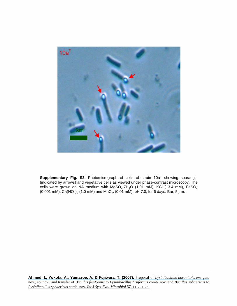
Supplementary Fig. S3. Photomicrograph of cells of strain 10aT showing sporangia (indicated by arrows) and vegetative cells as viewed under phase-contrast microscopy. The cells were grown on NA medium with MgSO4.7H2O (1.01 mM), KCl (13.4 mM), FeSO4(0.001 mM), Ca(NO3)2 (1.0 mM) and MnCl2 (0.01 mM), pH 7.0, for 6 days. Bar, 5 μm.
Ahmed, I., Yokota, A., Yamazoe, A. & Fujiwara, T. (2007). Proposal of Lysinibacillus boronitolerans gen. nov., sp. nov., and transfer of Bacillus fusiformis to Lysinibacillus fusiformis comb. nov. and Bacillus sphaericus to Lysinibacillus sphaericus comb. nov. Int J Syst Evol Microbiol 57, 1117–1125.
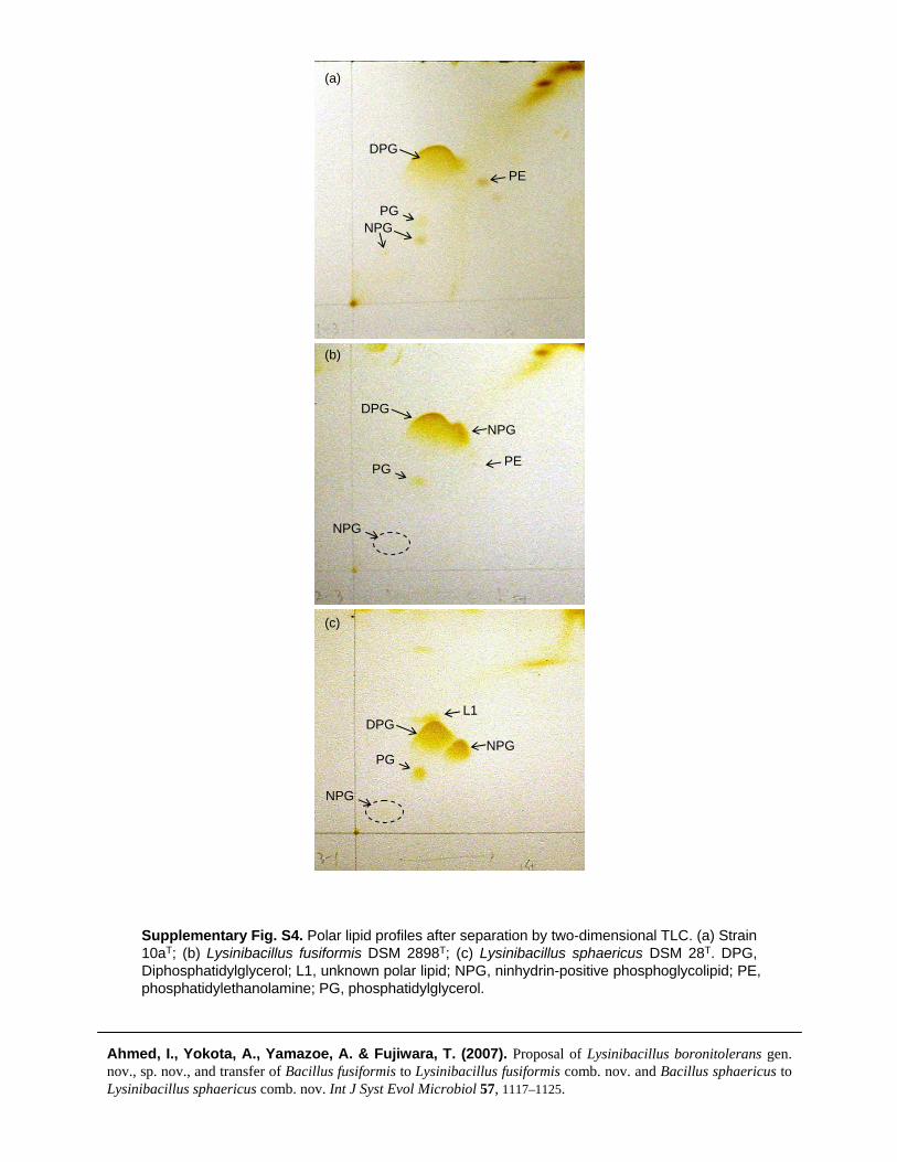
Supplementary Fig. S4. Polar lipid profiles after separation by two-dimensional TLC. (a) Strain 10aT; (b) Lysinibacillus fusiformis DSM 2898T; (c) Lysinibacillus sphaericus DSM 28T. DPG, Diphosphatidylglycerol; L1, unknown polar lipid; NPG, ninhydrin-positive phosphoglycolipid; PE, phosphatidylethanolamine; PG, phosphatidylglycerol.
Ahmed, I., Yokota, A., Yamazoe, A. & Fujiwara, T. (2007). Proposal of Lysinibacillus boronitolerans gen. nov., sp. nov., and transfer of Bacillus fusiformis to Lysinibacillus fusiformis comb. nov. and Bacillus sphaericus to Lysinibacillus sphaericus comb. nov. Int J Syst Evol Microbiol 57, 1117–1125.
(a)
(b)
(c)
DPG
PE
PGNPG
DPG
PEPG
NPG
NPG
DPGL1
PGNPG
NPG