Stable and dynamic cortical electrophysiology of induction and emergence with propofol anesthesia
Upload
taipeimedicalCategory
view
0download
0
168
Ann. N.Y. Acad. Sci. 1042: 168–176 (2005). © 2005 New York Academy of Sciences.doi: 10.1196/annals.1338.019
Propofol Specifically Inhibits Mitochondrial Membrane Potential but Not Complex I NADH Dehydrogenase Activity, Thus Reducing Cellular ATP Biosynthesis and Migration of Macrophages
GONG-JHE WU,a,d,f YU-TING TAI,b,f TA-LIANG CHEN,c LI-LING LIN,d
YUNE-FANG UENG,e AND RUEI-MING CHENb,d
aDepartment of Anesthesiology, Shin Kong Wu Ho-Su Memorial Hospital, Taipei, TaiwanbDepartment of Anesthesiology, Wan-Fang Hospital, College of Medicine, Taipei Medical University, Taipei, TaiwancTaipei City Hospital and Taipei Medical University, Taipei, TaiwandGraduate Institute of Medical Sciences, College of Medicine, Taipei Medical University, Taipei, TaiwaneNational Research Institute of Chinese Medicine, Taipei, Taiwan
ABSTRACT: Propofol is a widely used intravenous anesthetic agent. Our previ-ous study showed that a therapeutic concentration of propofol can modulatemacrophage functions. Mitochondria play critical roles in the maintenance ofmacrophage activities. This study attempted to evaluate further the effects ofmitochondria on the propofol-induced suppression of macrophage functionsusing mouse macrophage-like Raw 264.7 cells as the experimental model. Mac-rophages were exposed to a clinically relevant concentration of propofol for 1,6, and 24 h. Analysis by the Trypan blue exclusion method revealed that pro-pofol was not cytotoxic to macrophages. Exposure of macrophages to propofoldid not affect mitochondrial NADH dehydrogenase activity of complex I. How-ever, analysis of flow cytometry showed that propofol significantly decreasedthe mitochondrial membrane potential of macrophages. Cellular levels of ATPin macrophages were significantly reduced after propofol administration. Inparallel with the dysfunction of mitochondria, the chemotactic analysis showedthat exposure to propofol significantly inhibited the migration of macrophages.This study shows that a therapeutic concentration of propofol can specificallyreduce the mitochondrial membrane potential, but there is no such effect oncomplex I NADH dehydrogenase activity. Modulation of the mitochondrialmembrane potential may decrease the biosynthesis of cellular ATP and thus re-duce the chemotactic activity of macrophages. This study provides in vitro data
fGong-Jhe Wu and Yu-Ting Tai contributed equally to this paper.Address for correspondence: Ruei-Ming Chen, Ph.D., Graduate Institute of Medical Sciences,
College of Medicine, Taipei Medical University. No. 250, Wu-Hsing St. Taipei 110, Taiwan.Voice: +886-2-29307930 ext. 2159; fax: +886-2-86621119.

169WU et al.: PROPOFOL MODULATES MITOCHONDRIAL FUNCTIONS
to validate mitochondrial dysfunction as a possible critical cause for propofol-induced immunosuppression of macrophage functions.
KEYWORDS: propofol; macrophages; mitochondrial membrane potential;ATP; cell migration
INTRODUCTION
Propofol is an intravenous anesthetic agent used for induction and maintenanceof anesthesia in surgical procedures.1 Previous studies have demonstrated that pro-pofol has immunomodulatory effects on activities of human neutrophils and leuko-cytes.2,3 Macrophages play important roles in cellular host defense against infectionand tissue injury.4 Dysfunction of macrophages can decrease a host’s nonspecificcell-mediated immunity.5 An ex vivo study showed that anesthesia with propofol orisofluorene intraoperatively decreased phagocytotic and microbicidal activities ofalveolar macrophages.6 Our previous study further showed that propofol has immu-nosuppressive effects on macrophage functions via suppression of cell migration,phagocytotic activity, oxidative ability, and cytokine production.7
Macrophages are activated in response to stimulation. Adenosine triphosphate(ATP), synthesized by the mitochondrial respiration and oxidative phosphorylationsystem, is required for maintenance of macrophage activities. In murine polymicro-bial sepsis, a decrease in cellular ATP levels is positively correlated with the markedsuppression of lymphocyte and macrophage functions.8 Therefore, mitochondria,important energy-producing organelles, may participate in macrophage activation.9
The integrities of the mitochondrial membrane potential and NADH dehydrogenaseactivity of complex I are two typical factors involved in ATP biosynthesis. In thisstudy, we attempted to evaluate the role of mitochondria in propofol-induced mac-rophage dysfunction from the viewpoints of cell viability, NADH dehydrogenase ac-tivity, mitochondrial membrane potential, ATP synthesis, and cell migration ability.
MATERIALS AND METHODS
Cell Culture and Drug Treatment
Murine macrophage-like Raw 264.7 cells were used in this study as the experi-mental model. Macrophages were cultured in RPMI-1640 medium (Gibco-BRL,Grand Island, NY) supplemented with 10% fetal calf serum, L-glutamine, penicillin(100 IU/mL), and streptomycin (100 µg/mL) in 75-cm2 flasks at 37oC in a humidi-fied atmosphere of 5% CO2. Cells were grown to confluence before propofol admin-istration. Propofol, donated by Zeneca Limited (Macclesfield, Cheshire, UK), wasstored under nitrogen, protected from light, and freshly prepared by dissolving it indimethyl sufoxide (DMSO) for each independent experiment. DMSO in the mediumwas kept to less than 0.1% to avoid the toxicity of this solvent to macrophages. Pro-pofol at 50 µM, which corresponds to a clinical plasma concentration, was chosento be the dosage used in this study.10

170 ANNALS NEW YORK ACADEMY OF SCIENCES
Assay of Cell Viability
For evaluating the cytotoxicity of propofol to macrophages, cell viability was de-termined by the Trypan blue exclusion method. In brief, macrophages (2 × 10⁵) werecultured in 24-well tissue culture plates. After propofol administration, macrophageswere trypsinized with 0.1% trypsin-EDTA (Gibco-BRL). After centrifugation andwashing, macrophages were suspended in phosphate-buffered saline (0.14 M NaCl,2.6 mM KCl, 8 mM Na2HPO4, and 1.5 mM KH2PO4) and then stained with an equalvolume of Trypan blue dye (Sigma, St. Louis, MO). Dead cells, blue in appearance,were counted under a reverse-phase microscope.
Assay of Mitochondrial NADH Dehydrogenase Activity of Complex I
Activity of NADH dehydrogenase of complex I was determined by a colorimetric3-(4,5-dimethylthiazol-2-yl)-2,5-diphenyltetrazolium bromide assay.11 In brief,macrophages (2 × 10⁴) were seeded in 96-well tissue culture clusters overnight.After drug treatment, cells were renewed with medium containing 0.5 mg/mL 3-(4,5-dimethylthiazol-2-yl)-2,5-diphenyltetrazolium bromide for another 3 h. Theblue formazan product in cells was dissolved in DMSO and measured spectrophoto-metrically at a wavelength of 570 nm.
Determination of the Mitochondrial Membrane Potential
The mitochondrial membrane potential in macrophages was determined accord-ing to the method of Chen.12 In brief, macrophages (5 × 10⁵) were seeded in 12-welltissue culture plates overnight and then treated with propofol for different time in-tervals. After propofol administration, macrophages were harvested and incubatedwith 3,3′-dihexyloxacarbocyanine (DiOC6(3)), a positively charged dye, at37°C for 30 min in a humidified atmosphere of 5% CO2. After washing and centrif-ugation, cell pellets were suspended in phosphate-buffered saline. The intracellularfluorescence intensities were quantified using a flow cytometer (FACS Calibur,Becton Dickinson, San Jose, CA).
Quantification of Cellular Adenosine Triphosphate Levels
Cellular ATP levels in macrophages were determined by a bioluminescence assayas described previously.7 This assay was based on luciferase’s requirement for ATPin producing emission light according to the protocol provided in the MolecularProbes ATP determination kit (Molecular Probes, Eugene, OR). A wavelength (560nm) emitted after a luciferase-mediated reaction of ATP with luciferin was detected us-ing a WALLAC VICTOR 2 1420 multilabel counter (Welch Allyn, Turku, Finland).
Assay of Chemotactic Activity
The migrating capacity of macrophages was determined using the Costar Tran-swell cell culture chamber inserts, pore size 8 µm, according to the application guideprovided by Corning Costar (Cambridge, MA). Rich RPMI-1640 medium (1.5 mL)was first added to 12-well tissue cluster plates (Corning Costar), and the transwells
171WU et al.: PROPOFOL MODULATES MITOCHONDRIAL FUNCTIONS
were inserted into the plates. Macrophages (1 × 10⁵) suspended with propofol in 0.5mL of rich medium were added to the inside of transwells and cultured at 37oC for1, 6, and 24 h in an atmosphere of 5% CO2. Macrophages that migrated to the bottomsurface of the polycarbonate filters were counted in each field and averaged for threefields with the aid of a crosshair micrometer (Nikon, Tokyo, Japan).
Statistical Analysis
Statistical differences between groups were considered significant when the Pvalue of the Duncan’s multiple range test was less than 0.05. Statistical analysis be-tween groups over time was conducted by two-way ANOVA.
RESULTS
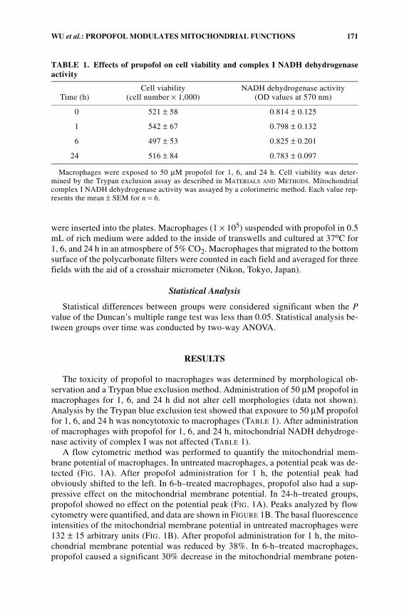
The toxicity of propofol to macrophages was determined by morphological ob-servation and a Trypan blue exclusion method. Administration of 50 µM propofol inmacrophages for 1, 6, and 24 h did not alter cell morphologies (data not shown).Analysis by the Trypan blue exclusion test showed that exposure to 50 µM propofolfor 1, 6, and 24 h was noncytotoxic to macrophages (TABLE 1). After administrationof macrophages with propofol for 1, 6, and 24 h, mitochondrial NADH dehydroge-nase activity of complex I was not affected (TABLE 1).
A flow cytometric method was performed to quantify the mitochondrial mem-brane potential of macrophages. In untreated macrophages, a potential peak was de-tected (FIG. 1A). After propofol administration for 1 h, the potential peak hadobviously shifted to the left. In 6-h–treated macrophages, propofol also had a sup-pressive effect on the mitochondrial membrane potential. In 24-h–treated groups,propofol showed no effect on the potential peak (FIG. 1A). Peaks analyzed by flowcytometry were quantified, and data are shown in FIGURE 1B. The basal fluorescenceintensities of the mitochondrial membrane potential in untreated macrophages were132 ± 15 arbitrary units (FIG. 1B). After propofol administration for 1 h, the mito-chondrial membrane potential was reduced by 38%. In 6-h–treated macrophages,propofol caused a significant 30% decrease in the mitochondrial membrane poten-
TABLE 1. Effects of propofol on cell viability and complex I NADH dehydrogenaseactivity
Time (h)Cell viability
(cell number × 1,000)NADH dehydrogenase activity
(OD values at 570 nm)
0 521 ± 58 0.814 ± 0.125
1 542 ± 67 0.798 ± 0.132
6 497 ± 53 0.825 ± 0.201
24 516 ± 84 0.783 ± 0.097
Macrophages were exposed to 50 µM propofol for 1, 6, and 24 h. Cell viability was deter-mined by the Trypan exclusion assay as described in MATERIALS AND METHODS. Mitochondrialcomplex I NADH dehydrogenase activity was assayed by a colorimetric method. Each value rep-resents the mean ± SEM for n = 6.

172 ANNALS NEW YORK ACADEMY OF SCIENCES
tial. However, administration of propofol in macrophages for 24 h did not change themitochondrial membrane potential (FIG. 1B).
To evaluate the effects of propofol on ATP synthesis, levels of intracellular ATPwere determined by a bioluminescence assay. In untreated macrophages, averagelevels of cellular ATP were 35 ± 11 pmol (FIG. 2). After administration of propofolfor 1 h, cellular ATP levels were significantly decreased by 43%. In 6-h–treated-macrophages, propofol decreased levels of macrophage ATP by 31%. Levels of cel-lular ATP were not affected after propofol administration for 24 h.
Chemotactic activities of macrophages were determined by a transwell test to val-idate effects of propofol on cell migration. In untreated macrophages, cells could mi-grate to the bottom layer of the transwell (FIG. 3A). After propofol administrationfor 1 h, macrophages migrating to the bottom layer were decreased. In 6-h–treatedmacrophages, propofol reduced the migrating capacity of macrophages to the bot-tom layer. Administration of propofol in macrophages for 24 h did not influence cellmigration (FIG. 3A). Fractions of macrophages that migrated to the bottom layers oftranswells were counted, and data are shown in FIGURE 3B. In 1-h–treated macroph-ages, propofol caused a significant 48% decrease in chemotactic activity. After pro-pofol administration for 6 h, the chemotactic activity of macrophages was reduced
FIGURE 1. Effects of propofol on the mitochondrial membrane potential. Macro-phages were exposed to 50 µM propofol for 1, 6, and 24 h. The mitochondrial membranepotential of macrophages was quantified by a flow cytometric method (A). The area of eachpeak was calculated and statistically analyzed (B). Each value represents the mean ± SEMfor n = 6. *Values significantly differ from the respective control, P < 0.05.

173WU et al.: PROPOFOL MODULATES MITOCHONDRIAL FUNCTIONS
by 40% (FIG. 3B). However, when macrophages were exposed to propofol for 24 h,cell migration was not affected.
DISCUSSION
This study shows that propofol significantly decreases the mitochondrial mem-brane potential of macrophages. In parallel with modulation of the mitochondrialmembrane potential, propofol reduced macrophage ATP levels. However, the activ-ity of mitochondrial NADH dehydrogenase of complex I was not affected after pro-pofol administration. The concentration of propofol used in this study was 50 µMwhich is within the range of clinical plasma concentrations.10 Thus, this study pro-vides in vitro data to validate that a therapeutic concentration of propofol can de-crease ATP biosynthesis in macrophages through a reduction in the mitochondrialmembrane potential, but not via modulation of NADH dehydrogenase activity ofcomplex I.
In parallel with suppression of the mitochondrial membrane potential and ATPproduction, propofol significantly decreased the chemotactic activity of macrophag-es. Previous studies reported that a decrease in cellular ATP levels can modulatelymphocyte and macrophage functions.8,13 Cellular ATP contributes to the regulationof cell migration.14,15 Thus, one of the critical reasons that explains the propofol-induced suppression of macrophage chemotactic activity is the reduction of ATP bio-synthesis. Data from the Trypan blue exclusion assay revealed that a therapeutic con-centration of propofol did not affect the integrity of the macrophage plasmamembranes. Determination of the integrity of plasma membranes can validatewhether cells have experienced an insult. According to the current data, we suggest
FIGURE 2. Effects of propofol on ATP biosynthesis. Macrophages were exposed to 50µM propofol for 1, 6, and 24 h. Levels of cellular ATP were determined by a biolumines-cence assay. Each value represents the mean ± SEM for n = 6. *Values significantly differfrom the respective control, P < 0.05.

174 ANNALS NEW YORK ACADEMY OF SCIENCES
FIGURE 3. Effects of propofol on chemotactic activities. Macrophages were exposedto 50 µM propofol for 1, 6, and 24 h. Chemotactic activity was determined by the transwellmethod (A). The number of cells that migrated to the bottom layer was counted and statis-tically analyzed (B). Each value represents the mean ± SEM for n = 6. *Values significantlydiffer from the respective control, P < 0.05.

175WU et al.: PROPOFOL MODULATES MITOCHONDRIAL FUNCTIONS
that the mechanism of propofol-induced suppression of macrophage functions doesnot occur via the death mechanism.
Balancing the mitochondrial membrane potential is important for ATP syn-thesis.16 Depolarization of the transmembrane potential will lead to disruption of therespiratory chain function and ATP synthesis. A previous study showed that depo-larization of the mitochondrial membrane potential increases the release of apoptoticfactors, including cytochrome c and reactive oxygen species from mitochondria tocytoplasm, which induces cell apoptosis.17 This study reveals that propofol can re-duce the mitochondrial membrane potential. Therefore, modulation of the mitochon-drial membrane potential is one of critical factors involved in the propofol-inducedsuppression of ATP synthesis and macrophage functions. Propofol is a hydrophobicanesthetic agent. One possible reason that may explain the suppressive effects ofpropofol on the mitochondrial membrane potential is the lipophilic characteristic ofthis anesthetic agent. Propofol may accumulate in the mitochondrial membrane anddepolarize the membrane potential through interference with the integrity of mito-chondrial membranes.
Propofol significantly reduced the mitochondrial membrane potential, ATP synthe-sis, and cell migration. However, these suppressive effects decreased with time. In 1-h–treated macrophages, propofol had the greatest inhibitory effects. After administra-tion of propofol in macrophages for 6 and 24 h, these suppressive effects partially orcompletely disappeared. The major explanation for the time-dependent decrease in theinhibitory effects of propofol is that this anesthetic agent is progressively decomposedafter exposure to visible light and aerobic conditions.1 Both cytochrome P450–depen-dent monooxygenases and uridine diphosphate glucuronosyltransfease contribute tothe metabolism of propofol into 4-hydroypropofol.18 These endogenous enzymes aredetectable in Raw 264.7 cells.19,20 Therefore, the metabolism of propofol in macroph-ages by these endogenous enzymes may be another reason to explain the decreasingeffects of this anesthetic agent on the mitochondrial membrane potential, ATP synthe-sis, and cell migration.
In conclusion, this study has shown that at therapeutic concentrations propofolcan specifically reduce the mitochondrial membrane potential without affectingNADH dehydrogenase activity of complex I. Modulation of the mitochondrial mem-brane potential leads to a decrease in cellular ATP levels. In parallel with mitochon-drial dysfunction, propofol significantly inhibits macrophage migration. Therefore,the mechanism of propofol-induced suppression of chemotactic activity is throughmodulation of the mitochondrial membrane potential and ATP synthesis, but notthrough the death effect.
ACKNOWLEDGMENTS
This study was supported by the Shin Kong Wu Ho-Su Memorial Hospital (SKH-TMU-92-10) and the Wan-Fang Hospital (93TMU-WAH-11), Taipei, Taiwan.
REFERENCES
1. SEBEL, P.S. & J.D. LOWDON. 1989. Propofol: a new intravenous anesthetic.Anesthesiology 71: 260–277.

176 ANNALS NEW YORK ACADEMY OF SCIENCES
2. MIKAWA, K., H. AKAMATSU, K. NISHINA, et al. 1998. Propofol inhibits human neutro-phil functions. Anesth. Analg. 87: 695–700.
3. HOFBAUER, R., M. FRASS, H. SALFINGER, et al. 1999. Propofol reduces the migration ofhuman leukocytes through endothelial cell monolayers. Crit. Care Med. 27: 1843–1847.
4. NATHAN, C.F. 1987. Neutrophil activation on biological surfaces. Massive secretion ofhydrogen peroxide in response to products of macrophages and lymphocytes. J. Clin.Invest. 80: 1550–1560.
5. LANDER, H.M. 1997. An essential role for free radicals and derived species in signaltransduction. FASEB J. 11: 118–124.
6. KOTANI, N., H. HASHIMOTO, D.I. SESSLER, et al. 1998. Intraoperative modulation ofalveolar macrophage function during isoflurane and propofol anesthesia. Anesthesi-ology 89: 1125–1132.
7. CHEN, R.M., C.H. WU, H.C. CHANG, et al. 2003. Propofol suppresses macrophagefunctions and modulates mitochondrial membrane potential and cellular adenosinetriphosphate levels. Anesthesiology 98: 1178–1185.
8. AYALA, A. & I.H. CHAUDRY. 1996. Immune dysfunction in murine polymicrobial sep-sis: mediators, macrophages, lymphocytes and apoptosis. Shock 6: S27–S38.
9. DIEHL, A.M. & J.B. HOEK. 1999. Mitochondrial uncoupling: role of uncoupling proteinanion carriers and relationship to thermogenesis and weight control “the benefits oflosing control.” J. Bioenerg. Biomembr. 31: 493–506.
10. GEPTS, E., F. CAMU, I.D. COCKSHOTT, et al. 1987. Disposition of propofol administeredas constant rate intravenous infusions in humans. Anesth. Analg. 66: 1256–1263.
11. WU, C.H., T.L. CHEN, T.G. CHEN, et al. 2002. Nitric oxide modulates pro- and anti-inflammatory cytokines in lipopolysaccharide-activated macrophages. J. Trauma 55:540–545.
12. CHEN, L.B. 1988. Mitochondria membrane potential in living cells. Annu. Rev. CellBiol. 4: 155–181.
13. OSHIMI, Y., S. MIYAZAKI & S. ODA. 1999. ATP-induced Ca2+ response mediated byP2U and P2Y purinoceptors in human macrophages: signalling from dying cells tomacrophages. Immunology 98: 220–227.
14. FREDHOLM, B.B. 1997. Purines and neutrophil leukocytes. Gen. Pharmacol. 28: 345–350.
15. DI VIRGILIO, F., P. CHIOZZI, D. FERRARI, et al. 2001. Nucleotide receptors: an emergingfamily of regulatory molecules in blood cells. Blood 97: 587–600.
16. LEE, I., E. BENDER, S. ARNOLD, et al. 2001. New control of mitochondrial membranepotential and ROS formation—a hypothesis. Biol. Chem. 382: 1629–1636.
17. LY, J.D., D.R. GRUBB & A. LAWEN. 2003. The mitochondrial membrane potential inapoptosis; an update. Apoptosis 8: 115–128.
18. SIMONS, P.J., I.D. COCKSHOTT, E.J. DOUGLAS, et al. 1988. Disposition in male volun-teers of a subanaesthetic intravenous dose of an oil in water emulsion of 14C-propofol. Xenobiotica 18: 429–440.
19. NAKAMURA, M., S. IMAOKA, F. AMANO, et al. 1998. P450 isoforms in a murine mac-rophage cell line, RAW264.7, and changes in the levels of P450 isoforms by treat-ment of cells with lipopolysaccharide and interferon-γ. Biochem. Biophys. Acta1385: 101–106.
20. GANOUSIS, L.G., D. GOON, T. ZYGLEWSKA, et al. 1992. Cell-specific metabolism inmouse bone marrow stroma: studies of activation and detoxification of benzenemetabolites. Mol. Pharmacol. 42: 1118–1125.
Copyright © 2022 FDOKUMEN