Prolonged physical inactivity leads to a drop in toe skin temperature during local cold stress
6
ARTICLE Prolonged physical inactivity leads to a drop in toe skin temperature during local cold stress Michail E. Keramidas, Roger Kölegård, Ola Eiken, and Igor B. Mekjavic Abstract: The purpose was to examine the effects of a prolonged period of recumbency on the toe temperature responses during cold-water foot immersion. Ten healthy males underwent 35 days of horizontal bed rest. The right foot of the subjects was assigned as the experimental (EXP) foot. To prevent bed rest-induced vascular deconditioning in the left control foot (CON), a sub-atmospheric vascular pressure countermeasure regimen was applied on the left lower leg for 4 × 10 min every second day. On the first (BR-1) and the last (BR-35) day of the bed rest, subjects performed two 30 min foot immersion tests in 8 °C water, one with the EXP foot and the other with the CON foot. The tests were conducted in counter-balanced order and separated by at least a 15 min interval. At BR-35, the average skin temperature of the EXP foot was lower than at BR-1 (–0.8 °C; P = 0.05), a drop that was especially pronounced in the big toe (–1.6 °C; P = 0.05). In the CON foot, the average skin temperature decreased by 0.6 °C in BR-35, albeit the reduction was not statistically significant (P = 0.16). Moreover, the pressure countermeasure regimen ameliorated immersion-induced thermal discomfort for the CON foot (P = 0.05). Present findings suggest that severe physical inactivity exaggerates the drop in toe skin temperature during local cold stress, and thus might constitute a potential risk factor for local cold injury. Key words: bed rest, CIVD, cold vasoconstriction, cold injury, cold tolerance, inactivity. Résumé : Cette étude se propose d’examiner les effets d’une période prolongée en décubitus dorsal sur les réponses thermiques de l’orteil au cours d’une immersion du pied en eau froide. Dix hommes en bonne santé passent 35 jours couchés a ` l’horizontale sur un lit. On assigne le pied droit a ` la condition expérimentale (EXP). Pour éviter le déconditionnement vasculaire du pied gauche (CON) causé par l’alitement, on applique a ` titre de contremesure au pied gauche un protocole de pression vasculaire subatmosphérique, et ce, a ` raison de 4 × 10 min tous les deux jours. Au premier (BR-1) et au dernier jour (BR-35) de la période d’alitement, les sujets participent a ` deux séances d’immersion en eau froide (8 °C) d’une durée de 30 min chacune, l’une pour le pied EXP et l’autre, pour le pied CON. On passe les tests selon un ordre contrebalancé et avec au moins 15 min d’intervalle. Au BR-35, la température cutanée moyenne du pied EXP est plus basse qu’au BR-1 (−0,8 °C; P = 0,05); on enregistre une baisse passablement importante au gros orteil (−1,6 °C; P = 0,05). Pour ce qui est du pied CON, la température cutanée moyenne diminue au BR-35 de 0,6 °C, mais cette diminution n’est pas significative (P = 0,16). En outre, la contremesure procurée par le protocole de pression améliore le confort thermique du pied CON en immersion (P = 0,05). D’après ces observations, l’inactivité physique extrême amplifie la baisse de la température de la peau du gros orteil au cours d’un stress local au froid, ce qui constitue un facteur de risque potentiel de lésion due au froid. [Traduit par la Rédaction] Mots-clés : alitement, vasoconstriction par le froid, lésion due au froid, tolérance au froid, inactivité. Introduction Exposure of an extremity to a cold environment mediates a vasoconstrictor response, causing a drop in peripheral blood flow, and resulting in a decrease in skin temperature. During sustained cold exposure, the initial general vasoconstrictive drive is com- monly followed by “paradoxical” increases in the digits’ circula- tion (Lewis 1930; Folkow et al. 1963), which may describe several patterns (Mekjavic et al. 2008), the most common one being an oscillatory digit skin temperature. This cold-induced vasodila- tation (CIVD) response, whose origin is not fully understood (Daanen and Layden 2010; Flouris and Cheung 2010), has been suggested to constitute a cryoprotective mechanism, with large responses predicting low susceptibility to cold injury (Yoshimura and Iida 1950; Daanen and van der Struijs 2005) and small or no responses predicting high susceptibility to cold injury (Mathew et al. 1974; Van der Struijs et al. 2008). Enhanced aerobic capacity has emerged as an essential non- thermal factor that potentiates the CIVD response (Keramidas et al. 2010). This could presumably be attributed to the exercise training-induced cardiovascular adaptations, including central (Grassi et al. 1994) and (or) peripheral (Lenasi and Strucl 2004) components. However, despite the compelling evidence of detri- mental effects of physical inactivity on health, especially on car- diovascular function (Mountjoy et al. 2011), it remains uncertain whether low cardiorespiratory fitness attenuates the peripheral vasomotor responses to cold, and hence whether it should be considered a potential risk factor for cold injury. In this context, sustained bed rest is considered a unique model to investigate the physiological responses to long-term physical inactivity in Received 9 July 2013. Accepted 7 October 2013. M.E. Keramidas. Department of Environmental Physiology, School of Technology and Health, Royal Institute of Technology, Berzelius väg 13, SE-171 65, Stockholm, Sweden; Department of Automation, Biocybernetics and Robotics, Jozef Stefan Institute, Ljubljana, Slovenia. R. Kölegård and O. Eiken. Department of Environmental Physiology, School of Technology and Health, Royal Institute of Technology, Berzelius väg 13, SE-171 65, Stockholm, Sweden. I.B. Mekjavic. Department of Automation, Biocybernetics and Robotics, Jozef Stefan Institute, Ljubljana, Slovenia. Corresponding author: Michail E. Keramidas (e-mail: [email protected]). Pagination not final (cite DOI) / Pagination provisoire (citer le DOI) 1 Appl. Physiol. Nutr. Metab. 39: 1–6 (2014) dx.doi.org/10.1139/apnm-2013-0315 Published at www.nrcresearchpress.com/apnm on 10 October 2013. Appl. Physiol. Nutr. Metab. Downloaded from www.nrcresearchpress.com by KAROLINSKA INSTITUTE on 01/27/14 For personal use only.
-
Upload
independent -
Category
Documents
-
view
0 -
download
0
Transcript of Prolonged physical inactivity leads to a drop in toe skin temperature during local cold stress

ARTICLE
Prolonged physical inactivity leads to a drop in toe skintemperature during local cold stressMichail E. Keramidas, Roger Kölegård, Ola Eiken, and Igor B. Mekjavic
Abstract: The purpose was to examine the effects of a prolonged period of recumbency on the toe temperature responses duringcold-water foot immersion. Ten healthy males underwent 35 days of horizontal bed rest. The right foot of the subjects wasassigned as the experimental (EXP) foot. To prevent bed rest-induced vascular deconditioning in the left control foot (CON), asub-atmospheric vascular pressure countermeasure regimen was applied on the left lower leg for 4 × 10 min every second day.On the first (BR-1) and the last (BR-35) day of the bed rest, subjects performed two 30 min foot immersion tests in 8 °C water, onewith the EXP foot and the other with the CON foot. The tests were conducted in counter-balanced order and separated by at leasta 15 min interval. At BR-35, the average skin temperature of the EXP foot was lower than at BR-1 (–0.8 °C; P = 0.05), a drop that wasespecially pronounced in the big toe (–1.6 °C; P = 0.05). In the CON foot, the average skin temperature decreased by 0.6 °C in BR-35,albeit the reduction was not statistically significant (P = 0.16). Moreover, the pressure countermeasure regimen amelioratedimmersion-induced thermal discomfort for the CON foot (P = 0.05). Present findings suggest that severe physical inactivityexaggerates the drop in toe skin temperature during local cold stress, and thus might constitute a potential risk factor for localcold injury.
Key words: bed rest, CIVD, cold vasoconstriction, cold injury, cold tolerance, inactivity.
Résumé : Cette étude se propose d’examiner les effets d’une période prolongée en décubitus dorsal sur les réponses thermiquesde l’orteil au cours d’une immersion du pied en eau froide. Dix hommes en bonne santé passent 35 jours couchés a l’horizontalesur un lit. On assigne le pied droit a la condition expérimentale (EXP). Pour éviter le déconditionnement vasculaire du piedgauche (CON) causé par l’alitement, on applique a titre de contremesure au pied gauche un protocole de pression vasculairesubatmosphérique, et ce, a raison de 4 × 10 min tous les deux jours. Au premier (BR-1) et au dernier jour (BR-35) de la périoded’alitement, les sujets participent a deux séances d’immersion en eau froide (8 °C) d’une durée de 30 min chacune, l’une pour lepied EXP et l’autre, pour le pied CON. On passe les tests selon un ordre contrebalancé et avec au moins 15 min d’intervalle. AuBR-35, la température cutanée moyenne du pied EXP est plus basse qu’au BR-1 (−0,8 °C; P = 0,05); on enregistre une baissepassablement importante au gros orteil (−1,6 °C; P = 0,05). Pour ce qui est du pied CON, la température cutanée moyenne diminueau BR-35 de 0,6 °C, mais cette diminution n’est pas significative (P = 0,16). En outre, la contremesure procurée par le protocole depression améliore le confort thermique du pied CON en immersion (P = 0,05). D’après ces observations, l’inactivité physiqueextrême amplifie la baisse de la température de la peau du gros orteil au cours d’un stress local au froid, ce qui constitue unfacteur de risque potentiel de lésion due au froid. [Traduit par la Rédaction]
Mots-clés : alitement, vasoconstriction par le froid, lésion due au froid, tolérance au froid, inactivité.
IntroductionExposure of an extremity to a cold environment mediates a
vasoconstrictor response, causing a drop in peripheral blood flow,and resulting in a decrease in skin temperature. During sustainedcold exposure, the initial general vasoconstrictive drive is com-monly followed by “paradoxical” increases in the digits’ circula-tion (Lewis 1930; Folkow et al. 1963), which may describe severalpatterns (Mekjavic et al. 2008), the most common one being anoscillatory digit skin temperature. This cold-induced vasodila-tation (CIVD) response, whose origin is not fully understood(Daanen and Layden 2010; Flouris and Cheung 2010), has beensuggested to constitute a cryoprotective mechanism, with largeresponses predicting low susceptibility to cold injury (Yoshimuraand Iida 1950; Daanen and van der Struijs 2005) and small or no
responses predicting high susceptibility to cold injury (Mathewet al. 1974; Van der Struijs et al. 2008).
Enhanced aerobic capacity has emerged as an essential non-thermal factor that potentiates the CIVD response (Keramidaset al. 2010). This could presumably be attributed to the exercisetraining-induced cardiovascular adaptations, including central(Grassi et al. 1994) and (or) peripheral (Lenasi and Strucl 2004)components. However, despite the compelling evidence of detri-mental effects of physical inactivity on health, especially on car-diovascular function (Mountjoy et al. 2011), it remains uncertainwhether low cardiorespiratory fitness attenuates the peripheralvasomotor responses to cold, and hence whether it should beconsidered a potential risk factor for cold injury. In this context,sustained bed rest is considered a unique model to investigatethe physiological responses to long-term physical inactivity in
Received 9 July 2013. Accepted 7 October 2013.
M.E. Keramidas. Department of Environmental Physiology, School of Technology and Health, Royal Institute of Technology, Berzelius väg 13,SE-171 65, Stockholm, Sweden; Department of Automation, Biocybernetics and Robotics, Jozef Stefan Institute, Ljubljana, Slovenia.R. Kölegård and O. Eiken. Department of Environmental Physiology, School of Technology and Health, Royal Institute of Technology, Berzeliusväg 13, SE-171 65, Stockholm, Sweden.I.B. Mekjavic. Department of Automation, Biocybernetics and Robotics, Jozef Stefan Institute, Ljubljana, Slovenia.Corresponding author: Michail E. Keramidas (e-mail: [email protected]).
Pagination not final (cite DOI) / Pagination provisoire (citer le DOI)
1
Appl. Physiol. Nutr. Metab. 39: 1–6 (2014) dx.doi.org/10.1139/apnm-2013-0315 Published at www.nrcresearchpress.com/apnm on 10 October 2013.
App
l. Ph
ysio
l. N
utr.
Met
ab. D
ownl
oade
d fr
om w
ww
.nrc
rese
arch
pres
s.co
m b
y K
AR
OL
INSK
A I
NST
ITU
TE
on
01/2
7/14
For
pers
onal
use
onl
y.
healthy individuals. It is well established that a prolonged periodof recumbency leads to cardiovascular deconditioning in terms ofhypovolemia (Greenleaf 1984), impaired autonomic regulation ofcardiovascular functions (Convertino 1990), and vascular remod-eling and dysfunction (Eiken et al. 2008; Kolegard et al. 2009).
Accordingly, the purpose of the present study was to examinethe effect of a 35 day horizontal bed rest on the toe temperatureresponses of healthy males during a local cold-water foot immer-sion test. Based on our previous finding that a 4 week aerobicexercise training enhanced the CIVD response (Keramidas et al.2010), we hypothesized that a prolonged period of physical inac-tivity would lead to a greater cold-induced vasoconstrictive re-sponse of the toes. In addition, a countermeasure was introducedto prevent the vascular deconditioning in one leg during the35 day bed rest. Namely, a sub-atmospheric vascular pressurecountermeasure (LVPC) was applied on the subjects’ left lower leg,to mimic, in the vasculature of this leg segment, the local trans-mural pressure perturbations associated with a normal ambula-tory life in erect posture. Therefore, the left leg was assigned as acontrol (CON) leg. Such a LVPC-response is limited in the anatom-ical site at which the pressure is applied, and no cross-transfereffect is transpired between the exposed and the non-exposedlimb (Eiken and Kolegard 2011; Kolegard and Eiken 2011). In thismanner, we also tested the hypothesis that the LVPC-induced pre-vention of vascular deconditioning in the CON leg would be re-flected in a vasomotor response of the toes during local cold stressthat was unaltered by bed rest.
Materials and methodsThe present work was part of a larger study investigating the
effect of a 35 day horizontal bed rest on several functions of thecardiovascular, musculoskeletal, and thermoregulatory systems.
SubjectsTen healthy males (age: 24 ± 2 years) participated in the present
study (Table 1). Prior to the bed rest, subjects had a thoroughphysical examination, and their participation was subject to aphysician’s approval. All subjects were non-smokers and had nohistory of any cardiovascular, hematological, or pulmonary dis-ease. They were physically active on a recreational basis and hadno, or very limited, previous experience with cold exposure ex-periments. The subjects were informed in detail about the ex-perimental procedures before giving their written consent toparticipate and were aware that they could terminate the experi-ment and their participation in the bed rest study at any time. Theexperimental protocol was approved by the National Committeefor Medical Ethics at the Ministry of Health of the Republic ofSlovenia and conformed to the Declaration of Helsinki.
Experimental protocol
Bed restThe bed rest protocol was conducted at the Orthopedic Hospital
Valdotra (Ankaran, Slovenia). Subjects commenced their bed restat 8:00 a.m. and finished it 35 days later at 08:00 a.m. Throughoutthe 35 day period, subjects were confined to strict horizontal po-sition and were not allowed to perform any exercise nor strenu-ous muscle contractions; they were allowed to move their arms inand above the horizontal plane and to lean on their elbows duringeating, personal hygiene, and transfer between bed and gurney.Also, they were allowed to use one pillow for head support at alltimes. Subjects were under 24 hour care, and their daily well-being was monitored by the hospital staff.
Lower leg vascular pressure countermeasure (LVPC)During the bed rest period, the left (CON) leg of the subjects was
intermittently exposed to a sub-atmospheric pressure of 11.9 kPa(90 mm Hg). Specifically, the CON leg was positioned in a custom-made box that was hermetically sealed proximally to the knee
with a short self-sealing rubber sleeve. The box was connected toa vacuum pump that applied a continuous pressure of 11.9 kPa.The LVPC was performed every second day; each session consistedof four 10 min pressure exposures, with intervening 5 min pauses.The subjects remained in a supine position at all times. The con-tralateral (right) leg of the subjects was referred to as the experi-mental (EXP) leg.
Maximal oxygen uptake (VO2max) testingBefore and after the bed rest, subjects performed an incre-
mental exercise test to exhaustion on a mechanical flywheelcycle-ergometer (Monark 818E, Stockholm, Sweden) to deter-mine their VO2max and peak power output (PPO). A metaboliccart (SensorMedics Vmax29c, Bilthoven, The Netherlands) wasused to acquire the breath-by-breath respiratory responses duringthe test. Attainment of VO2max, defined as the highest VO2 aver-aged over 20 s, was confirmed according to the following criteria:(i) severe fatigue or exhaustion resulting in an inability to main-tain exercise at a given work rate (cycling cadence lower than60 rpm) and (ii) a subjective rating of perception of effort at or nearmaximal.
Cold-water foot immersion testApproximately 5–7 h after the bed rest period had started (BR-1),
subjects conducted two cold-water immersion tests in a counter-balanced order and separated by at least a 15 min interval, onewith the EXP foot, and the other with the CON foot. On the last dayof bed rest (BR-35), subjects repeated the cold-water immersiontests. All tests were performed in the summer (BR-1 in July andBR-35 in August) and at the same time of the day (early afternoon).
Prior to the start of the test, subjects were acclimated to thethermoneutral conditions of the laboratory for �20 min. Themean temperature, relative humidity, and barometric pressureduring the tests were 25.6 ± 0.5 °C, 45 ± 4% and 10.1 ± 0.3 kPa,respectively. Throughout the test, subjects remained in a supineposition on their bed and were dressed in a T-shirt and shorttrousers. The tested foot was covered with a thin plastic bag (thick-ness of 0.025 mm) that was sealed with air permeable tape to theskin, �10 cm above the lateral malleolus. Special care was takento remove all the air from the bag.
Each test commenced with a 5 min baseline phase, duringwhich subjects rested with both feet on the bed. Thereafter, thetested foot was immersed �5 cm above the malleolus in a tankfilled with warm water (35 °C) for 5 min. Thereafter, subjects im-mersed the same foot in a different tank with cold water (8 °C; cf.Mekjavic et al. 2013) for 30 min (CWI). The temperature of thewater was maintained with an active cooling system (Haake,Germany), and a small impeller continuously stirred the waterinside the tank. After the completion of the CWI phase, the footwas removed from the water, dried with a towel, if it was wet, anda 5 min passive rewarming period ensued with both feet on thebed.
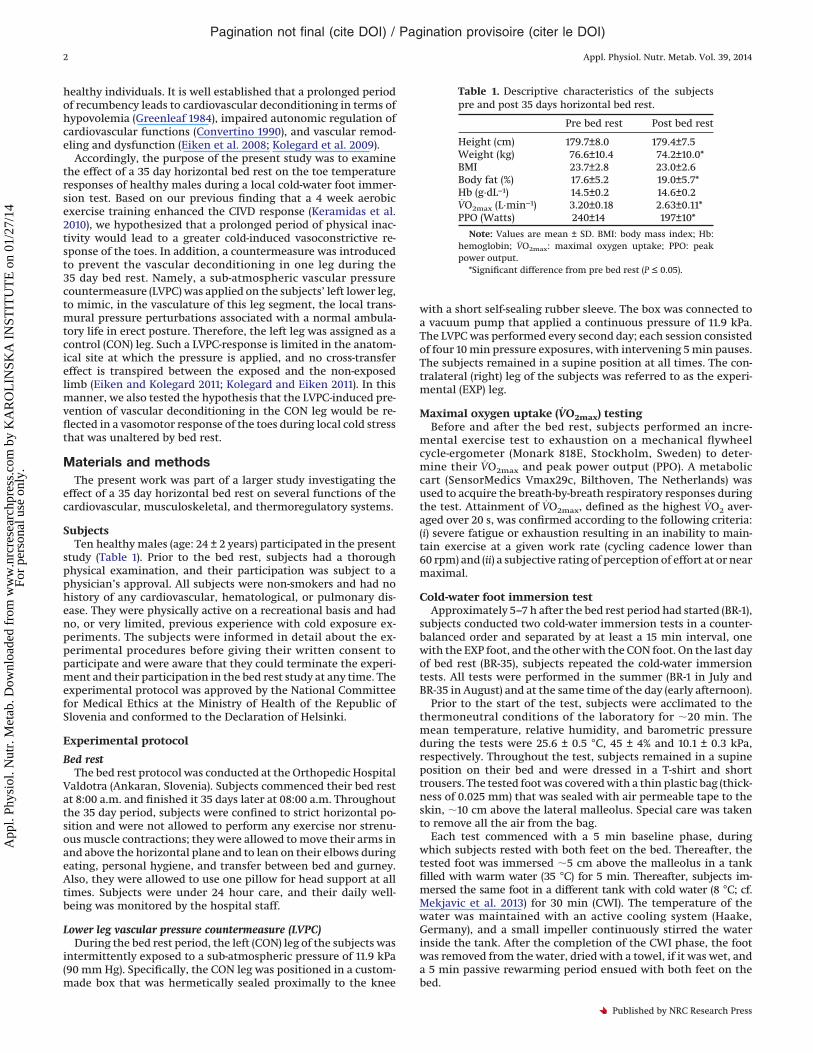
Table 1. Descriptive characteristics of the subjectspre and post 35 days horizontal bed rest.
Pre bed rest Post bed rest
Height (cm) 179.7±8.0 179.4±7.5Weight (kg) 76.6±10.4 74.2±10.0*BMI 23.7±2.8 23.0±2.6Body fat (%) 17.6±5.2 19.0±5.7*Hb (g·dL−1) 14.5±0.2 14.6±0.2VO2max (L·min−1) 3.20±0.18 2.63±0.11*PPO (Watts) 240±14 197±10*
Note: Values are mean ± SD. BMI: body mass index; Hb:hemoglobin; VO2max: maximal oxygen uptake; PPO: peakpower output.
*Significant difference from pre bed rest (P ≤ 0.05).
Pagination not final (cite DOI) / Pagination provisoire (citer le DOI)
2 Appl. Physiol. Nutr. Metab. Vol. 39, 2014
Published by NRC Research Press
App
l. Ph
ysio
l. N
utr.
Met
ab. D
ownl
oade
d fr
om w
ww
.nrc
rese
arch
pres
s.co
m b
y K
AR
OL
INSK
A I
NST
ITU
TE
on
01/2
7/14
For
pers
onal
use
onl
y.

Instrumentation
Temperature measurementsDuring the test, the skin temperature of the toes of the im-
mersed foot was measured with copper-constantan (T-type)thermocouple (each conductor was 0.2 mm in diameter) probes(Physitemp Instruments Inc., Clifton, N.J., USA), which were at-tached directly to the plantar side of the distal phalanx of each toewith thin air-permeable tape (Tegaderm, 3M, Healthcare, St. Paul,Minn., USA). The primary insulation material of the thermocou-ples was PTFE (insulation rating –75 to +250 °C); the non-insulatedwelded junctions of the thermocouple were attached directly tothe skin.
All toe-skin temperatures were recorded every second by anAlmemo data acquisition system (Almemo 5290-9 V5, Ahlborn,Holzkirchen, Germany) and stored in a PC for further analysis.Following a manual check of the raw data, a computer programwritten in Matlab language (MathWorks Inc., USA) was used todetermine the average temperature (Tavg) of each toe obtainedduring the baseline, warm-water immersion, and CWI phases, andthe minimum temperature (Tmin) reached during the CWI phase.The same program was used to detect any CIVD response, whichwas indicated by a local skin-temperature wave (N) that was de-fined as a 0.5 °C increase lasting for a minimum duration of 3 min(cf. Keramidas et al. 2010). The temperature amplitude (�T), whichwas the difference between the lowest temperature recorded justbefore the CIVD and the highest temperature reached during theCIVD, was also calculated.
Moreover, two thermocouple probes (Physitemp InstrumentsInc.) were placed on the right side of the chest (Tchest) and thelateral aspect of the calf of the non-immersed foot (Tcalf). Tchest andTcalf were recorded continuously using the aforementioned dataacquisition system (Almemo).
During the baseline phase and at 5 min intervals during theCWI phase, infrared tympanic temperature (Ttympanic) was mea-sured using a commercially available infrared thermometer(ThermoScan IRT 3020, Braun, Kronberg, Germany). Three consec-utive measurements were obtained each time, and the highest ofthe three values was used for subsequent analysis.
Hemodynamic variablesHeart rate (HR) was recorded using a heart-rate monitor (S800CX;
Polar, Kempele, Finland). Systolic (SAP) and diastolic (DAP) arterialpressure was measured with a non-invasive auscultatory method(300B, Speidel & Keller, Germany) on the left arm at 5 min inter-vals during the test. The mean arterial pressure (MAP) was calcu-lated by the equation: DAP + 1/3 (SAP-DAP).
Psychometric response scalesDuring the baseline, warm-water, CWI (at minutes 1, 2, 3, 5, and
every 5 min thereafter), and rewarming phases, subjects wererequested to provide ratings of their thermal sensation on a10-point scale (TS; from 0-unbearably cold to 9-very hot) and thermalcomfort on a 5-point scale (TC; from 1-comfortable to 5-extremelyuncomfortable). Both scales were explained to the subjects by thesame investigator prior to each test.
Statistical analysesStatistical analyses were performed using Statistica 5.0 (StatSoft,
Inc., Tulsa, Okla., USA). All data are reported as mean ± SD, unlessotherwise indicated. A three-way ANOVA for repeated measureswas used for the hemodynamic variables, Ttympanic, Tchest, and Tcalfobtained during the test [foot (CON/EXP) × testing period (BR-1/BR-35) × time (5 min intervals)]. Likewise, a three-way ANOVA forrepeated measures was used for toes Tavg, Tmin, and �T [foot(CON/EXP) × testing period (BR-1/BR-35) × toe (5 toes)]. The Tukeypost hoc test was employed to identify specific differences be-tween means when ANOVAs revealed significant F-ratio for maineffects. Differences in N, TS, and TC were evaluated with a Wilcoxon
matched pairs non-parametric test. The alpha level of significancewas set a priori at 0.05.
ResultsAll subjects completed the 35 day horizontal bed rest. VO2max
and PPO were significantly reduced after bed rest (Table 1). Theseresults have been presented in detail in a previous study (Porcelliet al. 2010).
Temperature measurementsThere was no significant difference (P > 0.05) in the Tavg of the
toes during the baseline (EXP foot: BR-1 = 29.6 ± 3.4 °C, BR-35 =29.6 ± 3.4 °C; CON foot: BR-1 = 29.0 ± 2.5 °C, BR-35 = 29.3 ± 3.0 °C)and warm-water (EXP foot: BR-1 = 34.0 ± 0.8 °C, BR-35 = 33.4 ± 1.1 °C;CON foot: BR-1 = 33.6 ± 0.9 °C, BR-35 = 33.0 ± 1.4 °C) phases betweenthe tests. Tavg of both feet during the CWI phase in BR-1 and BR-35are presented in Fig. 1. The EXP foot was colder at BR-35 than atBR-1 (P = 0.05); the drop in Tavg was especially pronounced in thebig toe (–1.6 °C; P = 0.002). Tavg of the CON foot was slightly reduced(–0.6 °C) at BR-35 compared with BR-1, albeit the difference wasnot statistically significant, nor was there any bed rest-inducedtemperature difference in any of the individual toes (P = 0.16).There were no differences between the EXP and CON foot in BR–35(P > 0.05). Tmin during the CWI phase did not differ between thefeet (P = 0.48); however, in both feet there was a tendency (P = 0.06)for lower Tmin in BR–35 than in BR-1 (EXP foot: BR-1 = 8.9 ± 0.9 °C,BR-35 = 8.4 ± 0.9 °C; CON foot: BR-1 = 9.0 ± 0.9 °C, BR-35 = 8.5 ±0.7 °C).
Apart from the big toe, the toes did not present any CIVD re-sponse during the CWI phase. Namely, with regard to the CIVDparameters of the big toe, N [EXP foot: BR-1 = 1 (0–2), BR-35 = 1 (0–2);CON foot: BR-1 = 1 (0–4), BR-35 = 0 (0–2)] and �T (EXP foot: BR-1 =1.4 ± 1.4 °C, BR-35 = 0.9 ± 1.2 °C; CON foot: BR-1 = 0.6 ± 0.8 °C, BR-35 =0.7 ± 1.2 °C) were not different between the feet or the testingperiods (P > 0.05).
There were no differences in Ttympanic, Tchest, and Tcalf betweenthe feet or the testing periods (Table 2).
Hemodynamic responsesHR was significantly higher throughout the cold-water immer-
sion test in BR-35 than in BR-1, regardless of the testing foot(Table 2). No differences between feet or testing periods wereobserved for SAP, DAP, and MAP (Table 2).
Psychometric responsesThere were no differences in TS between the feet or the testing
periods (Table 2). LVPC improved TC in the CON foot, as indicatedby the lower values (less discomfort; P = 0.05) in BR-35 than in BR-1(Fig. 2). Furthermore, TC was significantly better (lower; P = 0.01) inthe CON foot than in the EXP foot in BR-35 (Fig. 2).
DiscussionThe main finding of the present study is that a 35 day horizontal
bed rest led to a greater cutaneous vasoconstrictor response, asindicated by the lower Tavg reached during the CWI phase. Alongwith previous evidence that regular physical training enhancesthe CIVD response (Keramidas et al. 2010), current results furthersubstantiate the influence of daily activity and training on localcold tolerance. Thus, it appears that severe physical inactivityconstitutes another unhealthy lifestyle factor that, along withsmoking and alcohol consumption for instance (cf. Daanen 2003),can critically diminish the peripheral vasomotor function, andwhich may hence predispose individuals to cold injury.
A minimal presence of a CIVD response observed in the BR-1tests is in agreement with previously reported findings (Cheungand Mekjavic 2007; Dobnikar et al. 2009). It is noteworthy thatdespite a weak CIVD response of the toes already at the onset ofthe bed rest, after 35 days bed rest the cold-induced vasoconstric-
Pagination not final (cite DOI) / Pagination provisoire (citer le DOI)
Keramidas et al. 3
Published by NRC Research Press
App
l. Ph
ysio
l. N
utr.
Met
ab. D
ownl
oade
d fr
om w
ww
.nrc
rese
arch
pres
s.co
m b
y K
AR
OL
INSK
A I
NST
ITU
TE
on
01/2
7/14
For
pers
onal
use
onl
y.

tion was greater. The underlying mechanism for such a vasomotorresponse is difficult to discern from the current results. Presum-ably, the bed rest-induced cardiovascular deconditioning, that ishypovolemia (Greenleaf 1984) and (or) impaired autonomic regu-lation (Convertino 1990), contributed to an overall sympatheticexcitation and thus to a reduction in the foot circulation duringthe cold stress. It should be noted, however, that a previous studyhas suggested that exercise-induced hypovolemia does not bluntthe finger CIVD response (O’Brien and Montain 2003).
That the LVPC regimen moderated, to some extent, the degreeof cold vasoconstriction in the CON foot, even though no differ-ences were detected on the magnitude of markers of sympatheticdischarge between the tests judging from the similar HR and ar-terial pressure responses at BR-35 in both tests, might favor a localrather than a neurogenic origin of the exaggerated cutaneousvasoconstriction during BR-35. There is evidence to suggest thatlong-term bed rest reduces vasomotor responsiveness in precapillary-dependent blood vessels. Specifically, it has been shown that35 days of bed rest reduces pressure resistance in lower leg arter-ies and arterioles, effects that appear to be local and mainly at-tributable to reduced vascular wall responsiveness to eitherreceptor- or nonreceptor-mediated vasoconstrictors (Eiken et al.2008). There are also indications that prolonged bed rest reducesflow-mediated and nitroglycerin mediated conduit artery dilatoryresponses (Shoemaker et al. 1998; Bleeker et al. 2005). Besides, inthe hands and feet, thermoregulation is to large extent deter-mined by blood flow through arteriovenous anastomoses (AVAs),in particular CIVD responses might reflect AVA flow (Bergersen
et al. 1999). As yet, no evidence exists on the effect of bed rest onthe AVAs. Ergo, the mechanisms underlying bed rest-induced re-ductions of cutaneous temperature during CWI may be multifac-torial and need to be further investigated.
Notably, the LVPC regimen alleviated the thermal discomfort inthe CON foot during the BR-35 cold-water immersion test, indicat-ing that the habituation effect to pressure stimulus remainedduring the cold stimulus. It is well established that repeated ex-posures to moderately increased local intravascular pressures re-duce pain provoked by markedly elevated intravascular pressures(Eiken and Kolegard 2011; Kolegard and Eiken 2011). Such painreduction has mainly been attributed to habituation-induced re-duction of pressure distension of veins, and perhaps also of arter-ies and arterioles, resulting in reduced stimulation of perivascularunmyelinated and thinly myelinated nerve endings (Eiken andKolegard 2011; Kolegard and Eiken 2011). However, it cannot beexcluded that LVPC may reduce pressure-induced perivascular no-ciception by way of other habituation mechanisms. Upon immer-sion of a foot in an 8 °C bath, thermal discomfort and pain isevoked by stimulation of cutaneous cold and pain receptors. Dur-ing sustained cold stress, the temperature may also drop in deepertissue layers, hence stimulating perivascular nociceptors, whichare known to be triggered by thermal stimuli (Fruhstorfer andLindblom 1983; Klement and Arndt 1992). It is noteworthy thatin the present experiments, LVPC regimen appeared to reducethermal discomfort only during the latter portion of the CWI(Fig. 2). Regardless of the underlying transfer mechanisms be-tween the LVPC regimen and the thermal comfort response, a
Fig. 1. Average temperature (Tavg) in the five toes of the right-experimental (EXP) and left-control (CON) foot during the 30 min cold-waterimmersion phase (CWI) performed on the first (BR-1) and last (BR-35) day of the 35 day horizontal bed rest. *Significant difference from BR-1(P ≤ 0.05).
Table 2. Heart rate (HR), systolic arterial pressure (SAP), diastolic arterial pressure (DAP), mean arterial pressure (MAP), infrared tympanictemperature (Ttympanic), chest skin temperature (Tchest), calf skin temperature of the non-immersed foot (Tcalf), and thermal sensation (TS) obtainedduring the baseline and the 30 min cold-water immersion (CWI) of the right-experimental (EXP) and left-control (CON) foot performed at the first(BR-1) and last (BR-35) day of the 35 day horizontal bed rest.
EXP foot CON foot
BR-1 BR-35 BR-1 BR-35
Baseline CWI Baseline CWI Baseline CWI Baseline CWI
HR (beats·min−1) 64±7 67±8 72±8* 76±7* 64±10 69±7 73±9* 81±8*SAP (mmHg) 123±10 125±10 126±10 130±11 127±9 129±6 127±12 133±11DAP (mmHg) 75±7 79±7 74±7 76±5 78±9 79±5 74±8 78±7MAP (mmHg) 91±8 94±8 91±7 94±5 94±8 95±6 92±6 97±6Ttympanic (°C) 37.1±0.3 37.3±0.3 37.0±0.3 36.9±0.3 37.1±0.3 37.2±0.2 37.1±0.3 37.0±0.2Tchest (°C) 33.4±0.8 33.5±0.9 33.3±0.5 33.6±0.7 33.5±0.6 33.5±0.7 33.8±1.0 33.7±0.9Tcalf (°C) 33.4±0.8 33.2±1.1 33.5±0.5 33.1±1.0 33.4±0.6 32.8±0.7 33.7±0.8 33.1±1.0TS (0–9) 5 (5–6) 2 (1–3) 5 (5–5) 2 (1–3) 5 (5–5) 2 (2–3) 5 (5–5) 3 (1–4)
Note: Values are mean ± SD for HR, SAP, DAP, Ttympanic, Tchest, and Tcalf. Values are median (range) for TS (from 0-unbearably cold to 9-very hot).HR, SAP, DAP, MAP, and TS during CWI were significantly different from their baseline values (P < 0.001).*Significant difference from BR-1 (P ≤ 0.05).
Pagination not final (cite DOI) / Pagination provisoire (citer le DOI)
4 Appl. Physiol. Nutr. Metab. Vol. 39, 2014
Published by NRC Research Press
App
l. Ph
ysio
l. N
utr.
Met
ab. D
ownl
oade
d fr
om w
ww
.nrc
rese
arch
pres
s.co
m b
y K
AR
OL
INSK
A I
NST
ITU
TE
on
01/2
7/14
For
pers
onal
use
onl
y.

discrepancy between the actual and the perceived temperature inthe CON foot is entailed, given that the ameliorated thermal dis-comfort was not accompanied by higher values of Tavg; as hasbeen suggested previously (Klement and Arndt 1992; Geurts et al.2006; Daanen et al. 2012), this might impose a greater risk for coldinjury due to inappropriate behavioural thermoregulation.
Methodological considerationsTo minimize the confounding effect of the body position on the
circulation, especially after the bed rest when the subjects re-turned to the erect posture (Fortney et al. 1996), all the cold-waterimmersion tests were performed in a supine position. Still, itremains to be determined whether the effect of the bed rest on thelocal thermoregulatory responses in cold will be alike in the up-right or sitting posture as in the supine.
The two consecutive cold-water foot immersion tests were sep-arated by a 15 min interval that might be considered a short timeperiod for full recovery from the overall sympathetic activationcaused by the initial cold stimulus. However, the Tavg of the toeswas not different between the first (10.6 ± 0.8 °C) and the second(10.7 ± 1.0 °C) CWI phase of the cold tests (P = 0.74), thereby indi-cating a non-order effect. Moreover, the baseline cardiovascular
values (arterial pressure and HR) and the general thermal status ofthe subjects were similar in the two tests. In addition, the coun-terbalanced crossover design of the experimental protocol andthe same sequence of the tests in both testing periods minimizedany possible hysteresis effect and bias in the subjects’ perception.
Whether the bed rest would have equally influenced the handcold responses needs to be further investigated. In view of theprevious finding that leg-cycling training enhanced the CIVD re-sponse of the non-exercised hand (Keramidas et al. 2010), it cannotbe excluded that a 35 day bed rest would affect the hand responsesas well. Yet, given the non-homogeneous response of the extrem-ities to local cold stimulus (Cheung and Mekjavic 2007) and thedifferential bed rest-induced adaptations on each vascular bed(Zhang 2001), it is difficult to draw any firm conclusions.
Practical perspectivesTaking into account that �30% of the overall adult population is
insufficiently active (World Health Organization 2012), and con-sidering the number of individuals that are exposed to cold envi-ronments, viz. military personnel, elderly, homeless, alcoholics,and sportsmen, we reasoned that it might be of interest to exam-ine the effects of physical inactivity on local cold tolerance. Pro-
Fig. 2. Median values of thermal comfort (TC) obtained during the 30 min cold-water immersion of the right-experimental (EXP) and left-control (CON) foot performed on the first (BR-1) and last (BR-35) day of the 35 day horizontal bed rest. Data in all conditions were significantlydifferent from the baseline values. *, Significant difference from BR-1 (P ≤ 0.05); †, significant difference between EXP and CON foot in BR-35(P ≤ 0.05). Ranges are not depicted; at the 15th min of CWI, the range for the EXP foot was: BR-1 = 1 to 4, BR-35 = 1 to 4; and for the CON foot:BR-1 = 1.5 to 4.5, BR-35 = 1 to 4. Abscissa definitions: B, a 5 min baseline phase prior to the warm-water foot immersion; W, a 5 min warm-water (35 °C) foot immersion; RW, a 5 min passive rewarming phase following the cold-water foot immersion.
Pagination not final (cite DOI) / Pagination provisoire (citer le DOI)
Keramidas et al. 5
Published by NRC Research Press
App
l. Ph
ysio
l. N
utr.
Met
ab. D
ownl
oade
d fr
om w
ww
.nrc
rese
arch
pres
s.co
m b
y K
AR
OL
INSK
A I
NST
ITU
TE
on
01/2
7/14
For
pers
onal
use
onl
y.

longed bed rest is a unique model to investigate the physiologicalresponses to long-term physical inactivity in healthy individuals.Hence, present results support the notion that regular exerciseenhances peripheral vasomotor responses to cold stress.
In closing, the findings of the present study demonstrate that aprolonged period of physical inactivity induces an exaggeratedcutaneous vasoconstriction during local cold stress in healthy in-dividuals. Although our study does not establish a cause and effectrelationship between long-term inactivity and local cold injury, inview of the premise that the CIVD response has a cryoprotectivefunction (Lewis 1930; Daanen and van der Struijs 2005) and pre-dicts the vulnerability to cold injury (Mathew et al. 1974; Van derStruijs et al. 2008), the present findings place severe physical in-activity on the list of potential risk factors for local cold injuries.
AcknowledgementsThe current study was supported, in part by the Ministries of
Defence and of Science of the Republic of Slovenia. We are grate-ful to all the subjects for their participation. Also, we would like tothank Stylianos N. Kounalakis and Mojca Amon for their helpduring the experiments. The authors state that there are no per-sonal or financial conflicts of interest in the present study.
ReferencesBergersen, T.K., Hisdal, J., and Walloe, L. 1999. Perfusion of the human finger
during cold-induced vasodilatation. Am. J. Physiol. 276: R731–R737. PMID:10070133.
Bleeker, M.W., De Groot, P.C., Rongen, G.A., Rittweger, J., Felsenberg, D.,Smits, P., and Hopman, M.T. 2005. Vascular adaptation to deconditioning andthe effect of an exercise countermeasure: results of the Berlin Bed Rest study.J. Appl. Physiol. 99: 1293–1300. doi:10.1152/japplphysiol.00118.2005. PMID:15932956.
Cheung, S.S., and Mekjavic, I.B. 2007. Cold-induced vasodilatation is not homog-enous or generalizable across the hand and feet. Eur. J. Appl. Physiol. 99:701–705. doi:10.1007/s00421-006-0383-6. PMID:17219169.
Convertino, V.A. 1990. Physiological adaptations to weightlessness: effects onexercise and work performance. Exerc. Sport Sci. Rev. 18: 119–166. doi:10.1249/00003677-199001000-00007. PMID:2192891.
Daanen, H.A. 2003. Finger cold-induced vasodilation: a review. Eur. J. Appl.Physiol. 89: 411–426. doi:10.1007/s00421-003-0818-2. PMID:12712346.
Daanen, H.A., and Layden, J.D. 2010. Reply to A.D. Flouris and S.S. Cheung replyletter regarding “cold-induced vasodilation”. Eur. J. Appl. Physiol. 108: 215–216. doi:10.1007/s00421-009-1241-0. PMID:19820960.
Daanen, H.A., and van der Struijs, N.R. 2005. Resistance Index of Frostbite as apredictor of cold injury in arctic operations. Aviat. Space Environ. Med. 76:1119–1122. PMID:16370261.
Daanen, H.A., Koedam, J., and Cheung, S.S. 2012. Trainability of cold inducedvasodilatation in fingers and toes. Eur. J. Appl. Physiol. 112: 2595–2601. doi:10.1007/s00421-011-2233-4. PMID:22081047.
Dobnikar, U., Kounalakis, S.N., and Mekjavic, I.B. 2009. The effect of exercise-induced elevation in core temperature on cold-induced vasodilatation responsein toes. Eur. J. Appl. Physiol. 106: 457–464. doi:10.1007/s00421-009-1035-4. PMID:19319561.
Eiken, O., and Kolegard, R. 2011. Repeated exposures to moderately increasedintravascular pressure increases stiffness in human arteries and arterioles.J. Hypertens. 29: 1963–1971. doi:10.1097/HJH.0b013e32834ae3ab. PMID:21873885.
Eiken, O., Kolegard, R., and Mekjavic, I.B. 2008. Pressure-distension relationshipin arteries and arterioles in response to 5 wk of horizontal bed rest. Am. J.Physiol. Heart Circ. Physiol. 295: H1296–H1302. doi:10.1152/ajpheart.00576.2008. PMID:18660441.
Flouris, A.D., and Cheung, S.S. 2010. On the origins of cold-induced vasodilation.Eur. J. Appl. Physiol. 108: 1281–1282. doi:10.1007/s00421-009-1324-y. PMID:20020307.
Folkow, B., Fox, R.H., Krog, J., Odelram, H., and Thoren, O. 1963. Studies on the
Reactions of the Cutaneous Vessels to Cold Exposure. Acta Physiol. Scand. 58:342–354. doi:10.1111/j.1748-1716.1963.tb02656.x. PMID:14078652.
Fortney, S.M., Scneider, V.S., and Greenleaf, J.E. 1996. The physiology of bed rest.In Handbook of Physiology, Sect. IV, Environmental Physiology, Vol. II. Editedby M.J. Fregly and C.M. Blatteis. Oxford University Press. pp. 889–939.
Fruhstorfer, H., and Lindblom, U. 1983. Vascular participation in deep cold pain.Pain, 17: 235–241. doi:10.1016/0304-3959(83)90096-9. PMID:6657284.
Geurts, C.L.M., Sleivert, G.G., and Cheung, S.S. 2006. Central and peripheralfactors in thermal, neuromuscular, and perceptual adaptation of the hand torepeated cold exposures. Appl. Physiol. Nutr. Metab. 31(2): 110–117. doi:10.1139/h05-007. PMID:16604128.
Grassi, G., Seravalle, G., Calhoun, D.A., and Mancia, G. 1994. Physical trainingand baroreceptor control of sympathetic nerve activity in humans. Hyper-tension, 23: 294–301. doi:10.1161/01.HYP.23.3.294. PMID:8125553.
Greenleaf, J.E. 1984. Physiology of fluid and electrolyte responses during inac-tivity: water immersion and bed rest. Med. Sci. Sports Exerc. 16: 20–25. doi:10.1249/00005768-198401000-00006. PMID:6708776.
Keramidas, M.E., Musizza, B., Kounalakis, S.N., and Mekjavic, I.B. 2010. Enhance-ment of the finger cold-induced vasodilation response with exercise training.Eur. J. Appl. Physiol. 109: 133–140. doi:10.1007/s00421-010-1374-1. PMID:20135142.
Klement, W., and Arndt, J.O. 1992. The role of nociceptors of cutaneous veins inthe mediation of cold pain in man. J. Physiol. 449: 73–83. PMID:1522527.
Kolegard, R., and Eiken, O. 2011. Distensibility in human veins as affected by5 weeks of repeated elevations of local transmural pressure. Eur. J. Appl.Physiol. 111: 3119–3125. doi:10.1007/s00421-011-1939-7. PMID:21461927.
Kolegard, R., Mekjavic, I.B., and Eiken, O. 2009. Increased distensibility in de-pendent veins following prolonged bed rest. Eur. J. Appl. Physiol. 106: 547–554. doi:10.1007/s00421-009-1044-3. PMID:19347352.
Lenasi, H., and Strucl, M. 2004. Effect of regular physical training on cutaneousmicrovascular reactivity. Med. Sci. Sports Exerc. 36: 606–612. doi:10.1249/01.MSS.0000121948.86377.51. PMID:15064588.
Lewis, T. 1930. Observations upon the reactions of the vessels of the human skinto cold. Heart, 15: 177–208.
Mathew, L., Talwar, J.R., Purkayastha, S.S., and Mahotra, M.S. 1974. Prediction ofsusceptibility to cold injury in monkeys. In Selected Topics in EnvironmentalBiology. Edited by B. Bhatia, G.S. Chhina, and B. Singh. New Delhi, India:Interprint Publications. pp. 191–195.
Mekjavic, I.B., Dobnikar, U., Kounalakis, S.N., Musizza, B., and Cheung, S.S. 2008.The trainability and contralateral response of cold-induced vasodilatation inthe fingers following repeated cold exposure. Eur. J. Appl. Physiol. 104: 193–199. doi:10.1007/s00421-008-0727-5. PMID:18408950.
Mekjavic, I.B., Dobnikar, U., and Kounalakis, S.N. 2013. Cold-induced vasodilata-tion response in the fingers at 4 different water temperatures. Appl. Physiol.Nutr. Metab. 38(1): 14–20. doi:10.1139/apnm-2012-0118. PMID:23368823.
Mountjoy, M., Andersen, L.B., Armstrong, N., Biddle, S., Boreham, C.,Bedenbeck, H.P., et al. 2011. International Olympic Committee consensusstatement on the health and fitness of young people through physical activ-ity and sport. Br. J. Sports Med. 45: 839–848. doi:10.1136/bjsports-2011-090228.PMID:21836168.
O’Brien, C., and Montain, S.J. 2003. Hypohydration effect on finger skin temper-ature and blood flow during cold-water finger immersion. J. Appl. Physiol. 94:598–603. doi:10.1063/1.1574179. PMID:12391076.
Porcelli, S., Marzorati, M., Lanfranconi, F., Vago, P., Pisot, R., and Grassi, B. 2010.Role of skeletal muscles impairment and brain oxygenation in limiting oxi-dative metabolism during exercise after bed rest. J. Appl. Physiol. 109: 101–111. doi:10.1152/japplphysiol.00782.2009. PMID:20395541.
Shoemaker, J.K., Hogeman, C.S., Silber, D.H., Gray, K., Herr, M., and Sinoway, L.I.1998. Head-down-tilt bed rest alters forearm vasodilator and vasoconstrictorresponses. J. Appl. Physiol. 84: 1756–1762. PMID:9572827.
Van der Struijs, N.R., Van Es, E.M., Raymann, R.J., and Daanen, H.A. 2008. Fingerand toe temperatures on exposure to cold water and cold air. Aviat. SpaceEnviron. Med. 79: 941–946. doi:10.3357/ASEM.2258.2008. PMID:18856183.
World Health Organization. 2012. Prevalence of insufficient physical activity. Avail-able from http://www. who. int/gho/ncd/risk_factors/physical_activity/en/.
Yoshimura, H., and Iida, Y. 1950. Studies on the reactivity of skin vessels toextreme cold I. A point test on the resistance against frostbite. Jpn. J. Physiol.1: 147–159.
Zhang, L.F. 2001. Vascular adaptation to microgravity: what have we learned?J. Appl. Physiol. 91: 2415–2430. PMID:11717201.
Pagination not final (cite DOI) / Pagination provisoire (citer le DOI)
6 Appl. Physiol. Nutr. Metab. Vol. 39, 2014
Published by NRC Research Press
App
l. Ph
ysio
l. N
utr.
Met
ab. D
ownl
oade
d fr
om w
ww
.nrc
rese
arch
pres
s.co
m b
y K
AR
OL
INSK
A I
NST
ITU
TE
on
01/2
7/14
For
pers
onal
use
onl
y.