Synergistic Effects of Amiodarone and Fluconazole on Candida albicans

Clin. exp. Immunol. (1991) 85, 485-492
Proliferative and cytotoxic responses to mannoproteins ofCandida albicans by peripheral blood lymphocytes of HIV-infected subjects
I. QUINTI, C. PALMA*, E. C. GUERRA, M. J. GOMEZ*, I. MEZZAROMA, F. AIUTI & A. CASSONE*Department of Allergy and Clinical Immunology, University of Rome La Sapienza, and
*Department of Bacteriology and Medical Mycology, Istituto Superiore di Sanita, Rome, Italy
(Acceptedfor publication 4 March 1991)
SUMMARY
Mucosal candidiasis is one of the first opportunistic diseases in HIV-infected subjects. In order tounderstand the relationship between this disease and immunodeficiency to chemically defined,immunodominant Candida antigens, a mannoprotein fraction from C. albicans cell wall (GMP) was
used to analyse proliferative and non-MHC-restricted cytotoxic responses of peripheral bloodmononuclear cells (PBMC) from normal and HIV-infected subjects. In the former, GMP inducedextensive blastogenesis, generation of powerful cytotoxicity against a tumour cell line (K562), andproduction of substantial amounts of interferon-gamma (IFN-y). Cultured PBMC from HIV-infected subjects manifested an early decreased ability for proliferative as well as differentiativecytotoxic responses to the candidal mannoproteins. This inability became clearly evident in subjectswith stage III (CDC) of the disease, was total in CDC stage IV and occurred even in some subjectswith a normal number of CD4+ cells. Low or absent response to GMP correlated with lack ofresponse to tetanus toxoid. In contrast, both lymphoproliferative and cytotoxic responses toexogeneous IL-2 was highly preserved at all stages of infection. The production of IFN-y in GMP-stimulated PBMC cultures critically fell to negligible values in most of the subjects in CDC stages II
and III. Thus, the lowered or absent cell-mediated immune responses to candidal mannoprotein maybe one factor to explain the early, elevated susceptibility of HIV-infected subjects to mucosalcandidiasis. This study also shows that our mannoprotein preparation may be used as a probe todetect the overall efficiency of T cell responses in the above subjects.
Keywords HIV infection immune responses Candida albicans mannoproteins
INTRODUCTION
Candida albicans is an opportunistic pathogen which possessesdistinctive properties of a biological response modifier (BRM)(Cassone et al., 1981; Domer et al., 1988). These BRMproperties are in part mediated by mannoproteins, an importantantigenic and immunomodulatory constituent of candidal cellwall (Cassone, 1989). A mannoprotein-rich extract was highlyeffective in activating natural killer (NK) cells and macrophagesin mouse peritoneal cavity (Scaringi et al., 1988). It alsostimulated lymphoproliferation and induced non-MHC-res-tricted cytotoxicity in in vitro cultured human peripheral bloodmononuclear cells (PBMC) (Ausiello et al., 1989; Torosantucciet al., 1990b). Other immunomodulatory effects, includingimmunosuppressive activities on B and T cells, have also beenascribed to mannoproteins or mannan of C. albicans (Domer et
Correspondence: Dr A. Cassone, Department of Bacteriology andMedical Mycology, Istitito Superiore di Saniti, Viale Regina Elena 299,00161 Rome, Italy.
al., 1986; Durandy et al., 1987; Carrow & Domer, 1988).Because of the widespread human commensalism of this fungusand consequent natural immunization with it (Cassone, 1989),antigenic extracts of C. albicans have been used to assay the stateof cell-mediated immunity in HIV-infected subjects. A goodcorrelation was found with diminished reactivity to candidalantigenic extracts and/or tetanus toxoid (TT), and diseaseprogression.
This immunodeficiency, which has been associated clinicallywith oral and oesophageal candidasis, may be mediated by thefunctional loss of the T cell receptor (TCR)/CD3 complex(Fauci, 1988; Hofmann et al., 1989; Gruters et al., 1990; Peter etal., 1990). However, this has not been proven, as the candidalantigens tested previously have been poorly defined. As differ-ent cell populations might respond to different components ofcrude candidal extracts, studies with antigens of better-charac-terized composition should be made.
It is well demonstrated that oral and oesophageal candidia-sis represent one of the first and more common opportunistic
485
ADONIS 000991049100265J

486 I. Quinti et al.
Table 1. Clinical data of patients studied
Patient AgeCDC stage no. Sex (years) Risk category CD4+/pl
II 1 M 31 Former i.v. drug user 561II 2 F 33 Former i.v. drug user 600II 3 F 29 Former i.v. drug user 691II 4 M 28 Former i.v. drug user 1106II S M 27 Bisexual 1200II 6 F 26 Heterosexual 377II 7 F 26 IV. drug user 566II 8 F 30 Heterosexual 410II 9 M 43 Homosexual 1079
III 1 F 23 Former i.v. drug user 629III 2 M 30 Bisexual/i.v. drug user 543III 3 M 33 Former i.v. drug user 1044III 4 M 29 Former i.v. drug user 270III 5 M 14 Blood product 432III 6 M 28 Heterosexual 347III 7 M 31 Bisexual/Former i.v. drug user 240III 8 M 25 Former i.v. drug user 622III 9 M 31 Homosexual 337III 10 F 27 Former i.v. drug user 234III 11 F 33 Former i.v. drug user 532III 12 M 26 Former i.v. drug user 410III 13 M 49 Heterosexual 527III 14 M 26 Former i.v. drug user 900III 15 F 23 Former i.v. drug user 1200III 16 M 25 Former i.v. drug user 900
IV.CI 1 M 28 Former i.v. drug user 30IV.C2 2 M 28 Iv. drug user 263IV.C2 3 M 30 Former i.v. drug user 100IV.C2 4 M 33 Heterosexual 462IV.C1 5 M 28 Former i.v. drug user 200IV.Cl 6 M 29 Former i.v. drug user 357IV.C1 7 M 37 Former i.v. drug user 166IV.C2 8 M 38 Homosexual 315IV.C2 9 M 49 Heterosexual 367IV.C2 10 F 25 Heterosexual 340IV.C1 11 F 35 Blood product 101
infection in AIDS patients (Klein et al., 1984; Holmberg & SUBJECTS AND METHODSMeyer, 1986; Brawner & Cutler, 1989; Dalgleish & Malkovsky,1989). It would be important to know at which stage of infection Patientscell-mediated responses to well-defined immunodominant anti- Thirty-six subjects (25 men and I I women) with HIV- I infectiongens of Candida surface are impaired. were subgrouped according to their clinical stage (Centers for
We have purified and characterized a mannoprotein fraction Disease Control, 1987). The number of subjects in each stageof C. albicans which is capable ofinducing lymphoproliferation, and their clinical characteristics are given in Table 1. Mean ageIL-2 and interferon-gamma (IFN-y) production, and generation was 30 years (range 14-49). All stage IV patients were underof wide-spectrum cytotoxic effector cells in cultures of human treatment with AZT for at least 6 months, and two of themPBMC from all healthy donors tested (Ausiello et al., 1989; (patients 1 and 7) were affected by oral candidiasis. In studiesTorosanctucci et al., 1990b). Here we report the results of the with PBMC, blood samples from age- and sex-matched healthyproliferative and cytotoxic responses ofPBMC from subjects at donors were used as controls.different stages of HIV infection, as defined by the Centers for Diagnosis of HIV infection was made by ELISA andDisease Control (CDC), to the mannoprotein fraction. TT- or confirmed by Western blot (Du Pont de Nemours, Bruxelles,IL-2-induced cell-mediated immune responses were also studied Belgium).in comparison, in order to evaluate the potential role of purifiedmannoprotein antigens for early diagnosis ofimmune deficiency Candida antigenand possible early chemotherapeutic treatments. GMP is a mannoprotein-rich extract from the cell wall of the

Immune responses to candidal mannoproteins in HIV
Fig. 1. Electron microscopic visualization by immunogold cyto-chemistry ofGMP constitutents in section of yeast (Y) or mycelial (M)cells of C. albicans. Head arrows indicate the cell wall. (For technicaldetails, see Cassone, 1989.)
yeast form C. albicans (strain BP). The methods for itspreparation and chemical characterization have been describedelsewhere (Scaringi et al., 1988; Cassone, 1989; Ausiello et al.,1989; Torosantucci et al., 1990a). In the experiments reportedhere, a GMP preparation was used containing > 80% mannan
(detected as mannose in gas-liquid chromatography), and 8-5%protein (Torosantucci et al., 1990a). Mannan and proteins ofthis extract are present in the form of distinct mannoproteinconstituents, mostly of high mol. wt (> 200 kD), as detected bySDS-PAGE 5-10% gel gradients and stained with concanavalinA (ConA)/peroxidase or in immunoblots with mAbAFI, a
murine monoclonal antibody raised against GMP (Cassone etal., 1988; Torosantucci et al., 1990a). The localization ofmAbAF I -reactive mannoprotein constituents on the cell wall ofC. albicans has been studied with immunogold electron micro-scope cytochemistry (Cassone, 1989) and is shown in Fig. 1.mAbAF1-reactive epitopes ofGMP span the entire wall in theyeast form, but are present only in the inner layers of themycelial form (see also Cassone et al., 1988; Torosantucci et al.,1990a). The preparation was negative at the Limulus lysategelification test for endotoxin contamination.
Other stimulantsTetanus toxoid was kindly provided by Wyeth (Marietta, PA).It was used at a final dilution of 1-5 LF/well.
Recombinant IL-2 (rIL-2) (107 U/mg) was obtained fromBiogen (Geneva, Switzerland).
LymphocytesCD4+ lymphocytes were detected by direct immunofluores-cence with FITC-conjugated OKT4 (Ortho, Raritan, NJ) usinga laser analyser (Ortho Cytoron). The absolute numbers ofCD4+ cells were calculated by leucocyte differential count.
PBMC preparation and proliferation assayHeparinized venous peripheral blood samples were obtainedfrom HIV-1-infected subjects or healthy donors. PBMC wereisolated by centrifugation on density gradient (Lymphoprep;Nyegaard, Oslo, Norway), washed in phosphate-buffed saline(PBS) and resuspended in RPMI 1640 medium (GIBCO, GrandIsland, NY) supplemented with 5% pooled human AB serumand antibiotics (penicillin 100 U/ml, streptomycin 0 1 mg/ml:GIBCO). Both at this stage and after culture with the variousstimulants (see below), cell viability was assessed by the dyeexclusion method: > 95% of cells were viable in each experimentperformed, regardless ofthe source of the blood sample (controlor HIV-infected subject). PBMC (1 x 10⁶) in 0-2 ml of completemedium in 96 flat-bottomed microwell trays (Nunc, Glostrup,Denmark), were cultured with GMP (final concentration 50 pg/ml), TT (final concentration 1[5 LF/ml), or rIL-2 (final concen-tration 100 U/ml) in triplicate. Plates were incubated in 5% CO2at 370C and harvested on day 7 of culture, according topreviously established optimal conditions (Ausiello et al., 1989;Torosantucci et al., 1990b).
Eighteen hours before harvesting, radiolabelled thymidine(Amersham International, Amersham, UK) was added at a finalconcentration of 0-5 pCi/well. Results were expressed as ct/min(the 3H-thymidine incorporation of unstimulated wells, usuallyless than 1000 ct/min, was subtracted to the 3H-thymidineincorporation of the test sample).
Generation of cytotoxic effector cellsPBMC were incubated at a concentration of 1 x 10⁶/ml in 24flat-bottomed microwell plates (Nunc) in 1 ml in the presence ofindicated amount of GMP or rIL-2, for 7 days in 5% C02, at37°C, then harvested, washed in RPMI 1640 and resuspended incomplete medium. The human erythroblastoid cell line K562was used as target. Target cells were labelled with 150 pCi of aNa2- 5CrO4 solution (0 1 mCi; specific activity 298 08 mCi/ml)in a final volume of 0 4 ml for 1 h at 37°C. After labelling, thetarget cells were washed again, resuspended in RPMI 1640 andincubated for 30 min at 37°C. Cytotoxicity assays were per-formed in round-bottomed, 96-microwell plates, as alreadyreported (Ausiello et al., 1989). Briefly, 5 x 10³ target cells wereadded to suspension ofeffector cells at three different concentra-tions, in a total volume of 0 I ml, in triplicate. Plates were thencentrifuged and incubated at 37°C in 5% CO2 for 4 h, thencentrifuged again, and supernatants harvested by a supernatantcollection system (Skatron, Oslo, Norway). Radioactivity wasassessed by a gamma counter (Compugamma; LKB, Pharma-cia, Uppsala, Sweden) and the percentage of specific lysis wascalculated as follows:
Experimental -Spontaneous release 100Maximum -Spontaneous release
where the spontaneous release was that determined in theabsence of effector cells and the maximum release was the totalct/min from artificially lysed cultures.
The values are reported as mean of triplicate samples foreach effector: target cell ratio. Standard errors, which neverexceeded 5%, have been omitted.
IFN-y productionIFN-y production was determined by a radioimmunosorbentassay (Centocor, Malven, PA) on supernatants collected after
487
4L Quinti et al.
7 days of cultures of PBMC stimulated with the indicatedconcentrations of GMP and rIL-2.
Statistical analysisComparisons amongst the different groups of HIV-infected or
healthy subjects were performed by the non-parametric Mann-Whitney U-test. Comparisons between the means of cytotoxi-city were done with Student's t-test. Associations betweenlymphoproliferative responses to GMP and TT and the numberof CD4+ cells were evaluated by Pearson's correlation coeffi-cient.
RESULTS
Proliferative response to mannoprotein extract, TT and IL-2PBMC from 20 healthy donors and 36 subjects at differentstages of HIV infection were assayed for their proliferativeresponses to the mannoprotein extract of C. albicans. Severalsubjects from each of the HIV-infected groups were also testedfor response to the TT and IL-2 (Table 2). As shown in Fig. 2,the PBMC from HIV-infected subjects had a markedly de-creased proliferative response to GMP, compared with theresponse of PBMC from healthy controls. The decrease ofGMP-induced lymphoproliferation appeared to parallel thestage of infection; in fact, PBMC from CDC stage IV subjectsresponded lower (P < 0 05) than those from CDC stage III, andthe response of these latter was significantly lower (P<0 01)than that of CDC stage II subjects. The median value oflymphoproliferation in the PBMC from this latter group alsowas lower than median PBMC proliferation of control subjects,although the difference did not reach the statistical significance.Despite the low proliferation of PBMC from CDC stage III
subjects as a whole, three subjects (12, 14 and 15; Table 1) hadPBMC proliferating extensively in response to the candidalmannoprotein. Only two of them (subjects 12 and 15) were
examined for PBMC response to TT, that was the highest of thegroup (Table 2). Interestingly, one of these subjects had a lownumber of CD4+ cells (subject 12; 410 CD4+/p1; Table 1), anddid not produce detectable quantities of IFN-y (Table 3). Therewere no exceptions to the very low or totally suppressedlymphoproliferative PBMC response to GMP by CDC stage IVsubjects (Fig. 2). Overall, the degree of lymphoproliferativeresponse to GMP in HIV-infected subjects of the CDC stages II
and III was significantly related to the absolute number ofCD4+ lymphocytes, although the correlation degree was
moderate (r= 0 5, P< 0-02) (Fig. 3). Noticeably, some patientsof the above CDC stages showed low or negligible proliferativeresponse to GMP even if their CD4+ lymphocyte number fellwell within the normal range (900 + 210 CD4+ cells/pl) (Fig. 3).
When the PBMC from HIV-infected subjects were stimu-lated with TT, a similar, although not overlapping, pattern ofproliferative response was observed. The response to thisantigen was clearly diminished, or almost absent, in PBMCfrom CDC stages III or stage IV, respectively (Table 2).Responses to GMP and TT were highly correlated (r=0 85,P < 0-00 1). In contrast, all HIV-infected subjects responded tostimulation of their PBMC proliferation with IL-2, and no
statistically significant difference in this proliferative response
was noticed between the controls and CDC stage IV subjects(Table 2). However, PBMC from subjects at CDC stages II or
III were significantly less responsive to IL-2 than those fromcontrols or those from CDC stage IV subjects (Table 2). Theelevated level of lymphoproliferative response shown by CDCstage IV patients to stimulation with IL-2 demonstrates that the
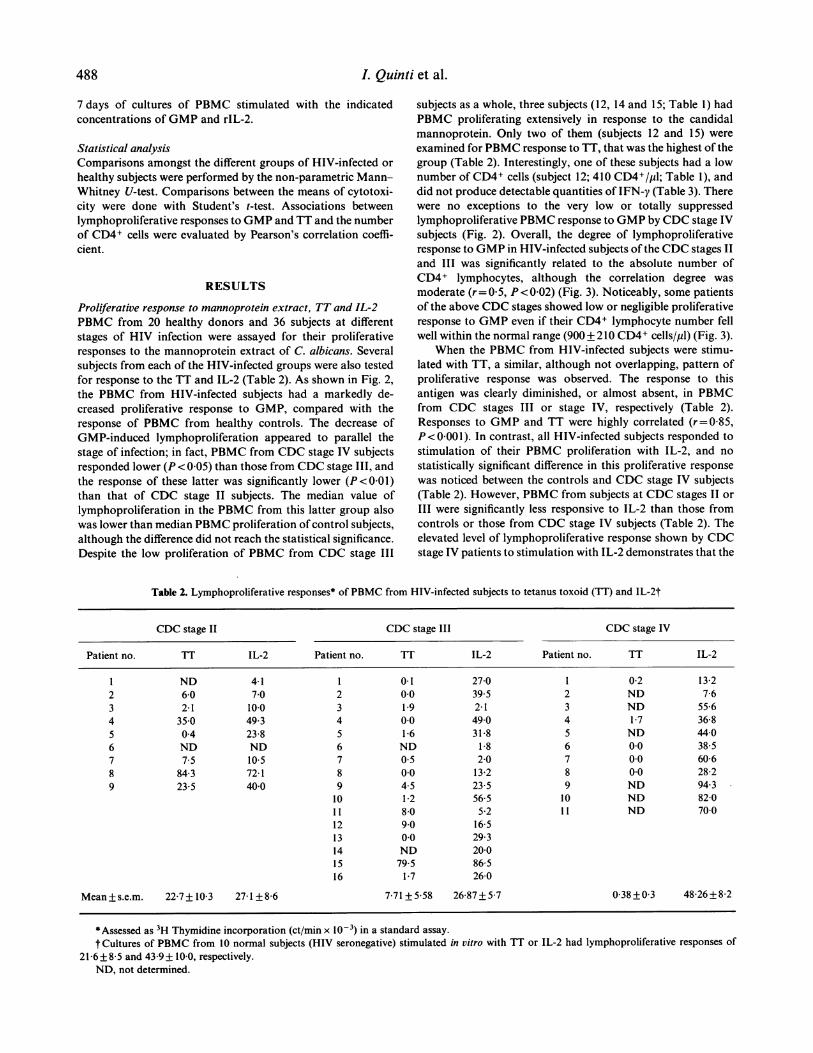
Table 2. Lymphoproliferative responses* of PBMC from HIV-infected subjects to tetanus toxoid (TT) and IL-2t
CDC stage II CDC stage III CDC stage IV
Patient no. 1T IL-2 Patient no. TT IL-2 Patient no. TT IL-2
1 ND 41 1 01 270 1 02 1322 60 70 2 00 395 2 ND 763 2 1 10.0 3 1 9 2-1 3 ND 5564 35-0 493 4 00 490 4 1 7 3685 04 23-8 5 1-6 31-8 5 ND 44-06 ND ND 6 ND 1-8 6 00 38-57 7.5 105 7 05 20 7 00 6068 84 3 72 1 8 0.0 13-2 8 0.0 28-29 23-5 40-0 9 4-5 23-5 9 ND 94.3
10 12 56-5 10 ND 82-011 80 52 11 ND 70012 9 0 16-513 00 29-314 ND 20015 79.5 86516 1 7 260
Mean + se.m. 22-7+ 10-3 27 1 +86 7 71 + 558 26 87 + 5.7 0 38 +0 3 48-26+8 2
*Assessed as 3H Thymidine incorporation (ct/min x 10-3) in a standard assay.t Cultures of PBMC from 10 normal subjects (HIV seronegative) stimulated in vitro with TT or IL-2 had lymphoproliferative responses of
21-6+8 5 and 43 9+ 10 0, respectively.ND, not determined.
488

Immune responses to candidal mannoproteins in HIV
8
0
x
.C
E
0
7
6
5
4
3
2
- tI
I
0
-0
0
0
8
7Ce 60
> 5Clp 4010
.u 30
6 2C
0 IC
00
0
Control CDC I CDC m CDC
Fig. 2. Lymphocyte proliferative response to mannoprotein (GMP)extract of C. albicans. PBMC (105 wells) from controls (HIV negative,healthy subjects) or HIV-infected subjects (at the indicated stage ofinfection) were incubated with GMP (50 Mg/ml), and 3H-thymidineincorporation was determined on day 7 of culture. Statistically signifi-cant differences exist between control (and CDD stage II) and CDCstage III (P<0-01) as well as between and CDC stages III and stage IV(P < 0 05) according to the Mann-Whitney U-test. Cumulating all dataof responses at stages 11+111, a statistically significant difference(P < 0 05) also exists between these subjects and healthy controls. Thelines indicate the median values.
50
40of0
'C
N.0
30
20
10
0
0~~~~~~~~~~~~~~~
I~~~~
-lo0 I I0-2 0.4 0*6 0-8 10 1-2 14 1-6 1-8 2.0
CD4-/mm3
Fig. 3. Correlation between the proliferative response to GMP and theabsolute number of CD4+ lymphocytes. Lymphoproliferation was
assessed by 3H-thymidine incorporation in a standard assay.
lack of PBMC response to the antigenic stimulants (GMP andTT) was rather specific and was not due to generic metabolicdefects ofT cells in HIV-infected subjects. This conclusion was
also confirmed by the experiments on cytotoxicity generationdescribed below.
Generation ofanti-K562 cytotoxic activity by GMP and IL-2One remarkable property of the GMP extract is its ability toinduce non MHC-restricted, LAK-like anti-tumour cytotoxi-city in PBMC from healthy donors (Ausiello et al., 1989); we
therefore tested the PBMC from HIV-infected subjects for theircapacity of killing a usual target of non-MHC-restrictedcytotoxicity such as the K562 cells. As a comparison, we alsoexamined whether the response to a powerful activator of this
(b)
(a)
Control CDCI CDCIf CDC]XControl CDCHf CDCJEE CDC X
Fig. 4. Generation ofcytolytic activity in cultures ofPBMC from controland HIV-infected subjects at different stage of infection followingstimulation with GMP (a) or IL-2 (b). A statistically significantdifference (P<0 01) exists between the cytotoxic response of PBMCfrom CDC stage II and PBMC from CDC stage III subjects in GMP-stimulated, but not in IL-2-stimulated cultures. Results are expressed aspercentage of cytolytic activity at E: T ratio of 10: 1 (0) and 30: 1 (E).
Table 3. Production of IFN-y (U/ml) by PBMC of HIV-infected subjects (CDC stages II and III) in response to
GMP or IL-2 stimulation
CDC stage II CDC stage III
Patient Patientno. GMP IL-2 no. GMP IL-2
2 8 14 1 76 3553 5 224 2 4 254 0 480 3 2 ND5 2 855 7 7 576 8 40 8 12 80
9 <2 19610 162 1011 0 18012 <2 3013 <2 <215 220 16016 8 120
The production of IFN-y in both GMP- and IL-2-stimulated PBMC cultures of 10 control subjects was
always > 100 U/ml.ND, not done.
kind of cytotoxicity, i.e. IL-2, was preserved in the PBMC fromHIV-infected subjects. The result of these experiments areshown in Fig. 4. Mannoprotein-induced, anti-K562 cytotoxicitywas generated in PBMC from CDC stage II subjects asefficiently as it was in controls. In contrast, PBMC fromsubsequent stages of infection were almost refractory to GMPinduction of cytotoxicity, in particular no anti-K562 activitywas generated in PBMC from CDC stage IV subjects. Wheninduced to cytotoxicity generation by IL-2, efficient, high-levelanti-K562 lytic activity was observed in cultures ofPBMC fromsubjects at each stage of HIV-infection (Fig. 4b). In particular,the subjects belonging to CDC stage IV did not show a
489
%.
0 th9

L Quinti et al.
decreased response to cytotoxicity generation by IL-2, com-
pared with all other HIV-uninfected or infected subjects.
Production of IFN-y by PBMC stimulated with GMP or IL-2Since the GMP extract is a powerful inducer of IFN-yproduction by PBMC of healthy subjects (Ausiello et al., 1989),we wanted to compare IFN production following GMP or IL-2stimulation in HIV-infected subjects. We particularly investi-gated the subjects with low but still detectable responses toGMP as those belonging to CDC groups II and III.
Table 3 shows that, with few exceptions (e.g. subjects 1, 10and 15, CDC stage III), the PBMC of HIV-infected subjectsproduced < 10 U of IFN-y after GMP stimulation, and most ofthem did not produce any detectable amount of the cytokine. Incontrast, PBMC from three out of five CDC stage II subjectsand those of five out of 11 CDC stage III subjects producedelevated amounts (> 100 U) of IFN-y after stimulation with IL-2. Interestingly, of the two patients (10 and 15) whose cellsproduced more IFN-y after stimulation with GMP than afterstimulation with IL-2 (Table 3), one (patient 10) had a lownumber of CD4+ cells (Table 1) and her PBMC proliferatedextensively in response to IL-2, but not in response to GMP.Only few subjects in CDC stage IV were studied for IFN-yproduction in PBMC cultures stimulated with GMP or IL-2.The production of this cytokine in these subjects was very low or
absent regardless of the stimulant used (data not shown).
DISCUSSION
We have demonstrated that PBMC from HIV-infected subjectsmanifest an early loss of proliferative as well as differentiativecytotoxic responses to a mannoprotein-rich extract of C.albicans. As emphasized by several investigators (Hofmann etal., 1989; Gruters et al., 1990; Peter et al., 1990), infection withHIV brings about a marked depression of T cell responsesmediated through TCR-CD3, which seems to precede thehallmark of HIV pathology, i.e. the diminution of CD4+ cells(Fauci, 1988). In this context, the response to microbial antigenswas found to be depressed with progression of the infection(Fauci, 1988; Hofmann et al., 1989). In addition to TT, crudeantigenic extracts or even whole Candida cells have been used inHIV-infected subjects (Hofmann et al., 1989; Heagy et al., 1989;Pedersen et al., 1989). These kind of candidal extracts, besidesbeing chemically undefined and ofhighly variable response, maycontain fractions which are by themselves suppressive of theantigenic response (Domer et al., 1986; Carrow & Domer, 1988)or constituents that act more as polyclonal agents than as
antigens (Tollemar, Ringden & Holmberg, 1989). These draw-backs have been eliminated in this investigation, as the micro-bial extract used here (GMP) is chemically well defined andmolecularly characterized (Scaringi et al., 1988; Ausiello et al.,1989). It is devoid of suppressive activity and has been shown toinduce antigenic stimulation of lymphoproliferation of PBMCfrom healthy subjects (Torosantucci et al., 1990b). The extract ismade of about 80% of high molecular weight mannoproteinsand its small contaminating RNA fraction was devoid of anyimmunological influence (Torosantucci et al., 1990b). Theexistence of a significant correlation between the proliferativeresponses to GMP and those to TT noticed in this studystrengthens the previous evidence for the antigenic, not polyclo-nal nature of the lymphoproliferation induced by GMP and
distinguishes between this complex and other chemically similarmaterials, acting as polyclonal stimulators (Tollemar et al.,1989).
Although our data do not rule out that at least some of thefully asymptomatic HIV+ subjects may have a reduced PBMCproliferation in response to GMP (see the difference in themedian values of lymphoproliferation between control andCDC stage II subjects, Fig. 2), clear-cut deficiency in theproliferative response to the antigen becomes evident in CDCstage III subjects. The overall deficiency in the response to GMPby CDC stages II + III subjects correlated moderately with thenumber of CD4+ cells, and some of the proliferative responses
to GMP were markedly low even in the presence of a normalnumber of CD4+ lymphocytes. Conversely, a low number ofCD4+ cells in these subjects did not preclude a high proliferativeresponse of the PBMC from some of them to candidalmannoprotein as well as to other microbial antigens (Fig. 3,Table 2). All this is in agreement with the concept of selectivefunctional deletions of antigen-specific T cells, already demon-strated in vitro in TT-specific T cell responses with the progres-
sion of HIV infection (Manca, Habeshaw & Dalgleish, 1990).Our data support the concept ofT cell-specific immunosuppres-sion, provoked by HIV or its products (gpl20) in asymptomaticsubjects, as the early hallmark of HIV disease (Fauci, 1988; Viaet al., 1990).
PBMC from HIV-infected subjects also showed a clearlydiminished ability to mount a cytotoxic, anti-K562 response on
stimulation with GMP. Interestingly, the loss of cytotoxicityinduction by GMP in CDC stage IV subjects is not due to a totallack of NK cells, which are advocated to be the precursors ofGMP-activated killer cells (Ausiello et al., 1989), asNK activitywas totally (CDC stage III) or partially (CDC stage IV)preserved in our HIV-infected subjects (Fontana et al., 1986;Sirianni, Tagliaferi & Aiuti, 1990; other data not shown). Thefall of proliferative differentiative responses to antigen stimula-tion may cause the inability of PBMC from HIV-infectedsubjects to produce one or more cytokines responsible for thecomplex cascade of events leading to proliferation/differentia-tion ofcytotoxic effector cells (Fauci, 1988; Via et al., 1990). Onesuch putative cytokine is IL-2, the production of which has beendemonstrated to be diminished in PBMC from HIV-infectedsubjects stimulated by various inducers (Heagy et al., 1989; Viaet al., 1990). The IL-2 production was not measured here, but wehave shown that HIV-infected subjects have PBMC with highlypreserved responsiveness to exogenous IL-2, that is not inhi-bited by HIV or gpl20 (Manca et al., 1990). IL-2-inducedproliferation and K562 cytotoxicity were markedly elevatedalso in those subjects in CDC stage IV who showed very lownumbers of CD4+ cells. This may also be explained by thepresence ofnormal number and functions ofCD8 + T cells (datanot shown).
Previously, we showed that IFN-y is produced abundantly(100 to > 1000 U/ml) by PBMC of normal subjects cultured inthe presence of whole Candida or GMP (Ausiello et al., 1989).However, most ofour subjects at the critical stage ofthe immuneresponse to GMP (CDC stage III) did not produce detectableIFN-y. This is in keeping with previous results by others (Heagyet al., 1989; Via et al., 1990) using other stimulants. Heagy et al.(1989) have recently shown that IFN-y enhances in vitro theproliferative response to whole Candida cells, but only whenCD4+ cells are not critically diminished. In one of our CDC
490

Immune responses to candidal mannoproteins in HIV 491
stage III subjects, an appreciable quantity of IFN-y wasproduced uponGMP stimulation, but the number ofCD4+ cellswas low, and no PBMC proliferation was induced by GMP(patient 10, Tables 1-3). Interestingly, the PBMC of this subjectproduced a low, if not insignificant amount of IFN-y followingstimulation with IL-2. It is clear that marked individualvariations in both lymphoproliferation and cytokine produc-tion are present among subjects in CDC stage III group,regardless of the nature of the PBMC stimulant (microbialantigen or IL-2), and it is possible that this clinical group islargely heterogeneous as far as functional lymphocyte responsesare concerned.
Here we have demonstrated that immune responses tomannoproteins of C. albicans are rather early imparied in HIV-infected subjects, an impairment which largely precludes andprobably explains the elevated susceptibility of these subjects tomucosal candidiasis (Klein et al., 1984; Holmberg & Meyer,1986).
The data also suggest that our antigenic preparation (GMP)may be employed as a probe to detect the overall efficiency ofT cell responses in the above subjects, at least as satisfactorily asother widely used microbial antigens, e.g. TT. Candidal manno-proteins are expressed on the cell surface of this humancommensal micro-organism, and the immune response to them,unlike that to the TT, does not depend on artificial vaccination.
ACKNOWLEDGMENTS
This research was supported by the Ministero della Sanitd, IstitutoSuperiore di Sanitd, Progetto Nazionale AIDS, 1988-1989 Contracts4206 01 and 5206 001 (A.C. and F.A.). We are grateful to Mrs A. M.Marella for help in the preparation of the manuscript.
REFERENCES
AUSIELLO, C.M., PALMA, C., SPAGNOLI, G.C., PIAZZA, A., CASCIANI,C.U. & CASSONE, A. (1989) Cytotoxic effectors in human peripheralblood mononuclear cells induced by a mannoprotein complex ofCandida albicans: a comparison with interleukin-2-activated killercells. Cell. Immunol. 121, 349.
BRAWNER, D.L. & CUTLER, J.M. (1989) Oral Candida albicans isolatesfrom nonhospitalized normal carriers, immunocompetent hospital-ized patients, and immunocompromised patients with or withoutacquired immunodeficiency syndrome. J. clin. Microbiol. 27, 1335.
CARROW, E.M. & DOMER, J.E. (1988) Immunoregulation in experi-mental murine candidiasis: specific suppression induced by Candidaalbicans cell wall glycoprotein. Infect. Immun. 49, 172.
CASSONE, A. (1989) Cell wall of Candida albicans, its functions and itsimpact on the host. Cur. top. Med. Mycol. 3, 249.
CASSONE, A., MARCONI, P., BISTONI, F., MAlTIA, E., SBARAGLIA, G.,GARACI, E. & BONMASSAR, E. (1981) Immunoadjuvant effect ofCandida albicans and its cell wall fractions in a mouse lymphomamodel. Cancer Immunol. Immunother. 10, 181.
CASSONE, A., TOROSANTUCCI, A., BOCCANERA, M., PELLEGRINI, G.,PALMA, C. & MALAVASI, F. (1988) Production and characterization ofa monoclonal antibody to a cell surface, glucomannoprotein consti-tuent of Candida albicans and other pathogenic Candida species. J.med. Microbiol. 27, 233.
CENTERS FOR DISEASE CONTROL (1987) Revision ofthe CDC surveillancecase definition for acquired immunodeficiency syndrome. MMWR,36, (Suppl. IS), 35.
CUFF, C.F., PACKER, B.J. & ROGERS, T.J. (1989) A further characteriza-tion of Candida albicans-induced suppressor B-cell activity. Immuno-logy, 68, 80.
DALGLEISH, A.G. & MALKOVSKY, M. (1989) Advances in HumanRetroviruses (ed. by P. Weinhouse & G. Klein) p. 307. Raven Press,New York.
DOMER, J., ELKINS, K., ENNIST, D. & BAKER, P. (1988) Modulation ofimmune responses by surface polysaccharides of Candida albicans.Rev. infect. Dis. 105, 419.
DOMER, J.E., STASHAK, P.W., ELKINS, K., PRESCOTT, B., CALDES, G. &BAKER, P.J. (1986) Separation of immunomodulatory effects ofmannan from Candida albicans into stimulatory and suppressivecomponents. Cell. Immunol. 101, 403.
DURANDY, A., FISHER, A., LE DEIST, F., DROUHET, E. & GRISCELLI, C.(1987) Mannan-specific and mannan-induced T-cell suppressiveactivity in patients with chronic mucocutaneous candidosis. J. clin.Immunol. 7, 400.
FAUCI, A.S. (1988) The human immunodeficiency virus: infectivity andmechanisms of pathogenesis. Science, 239, 617.
FONTANA, L., SIRIANNI, M.C., DE SANCTIS, G., CARBONARI, M., ENSOLI,B. & AIUTI, F. (1986) Deficiency of natural killer activity, but not ofnatural killer binding in patients with lymphadenopathy syndromepositive for antibodies to HTLV-III. Immunobiology, 171, 425.
GRUTERS, R.A., TERPSTRA, F.G., DE JONG, R., VAN NOESEL, C.J.M., VANLIER, R.A.W. & MIEDEMA, F. (1990) Selective loss ofT cell functionsin different stages of HIV infection. Eur. J. Immunol. 20, 1039.
HEAGY, W., STROM, T.B., KELLEY, V.E., COLELLA, J., CRUMPACKER, C.,WILLIAMS, J.M., SHAPIRO, H.M., LANBENSTEIN, L. & FINBERG, R.(1989) Recombinant Human Gamma Interferon enhances in vitroactivation of lymphocytes isolated from patients with acquiredimmunodeficiency syndrome. Infect. Immun. 57, 3619.
HOFMANN, B., JAKOBSEN, K.D., ODUM, N., DICKMEISS, E., PLATZ, P.,RYDER, L.P., PEDERSEN, C., MATHIESEN, L., BYGBJERG, I., FABER, V. &SVEJGAARD, A. (1989) Relatively preserved phytohemagglutinin as
opposed to decreased pokeweed mitogen responses may be due topossibly preserved responses via CD2/phytohemagglutinin pathway.J. Immunol. 142, 1874.
HOLMBERG, K. & MEYER, R.D. (1986) Fungal infections in patients withAIDS and AIDS-related complex. Scand. J. infect. Dis. 18, 179.
KLEIN, R.S., HARRIS, C.A., BUTKUS SMALL, C., MOLL, B., LESSER M. &FRIEDLAND G.H. (1984) Oral candidiasis in high risk patients as theinitial manifestation of the acquired immunodeficiency syndrome. N.Engl. J. Med. 311, 354.
MANCA, F., HABESHAW, J.A. & DALGLEISH A.G. (1990) HIV envelopeglicoprotein, antigen specific T-cell responses, and soluble CD4.Lancet, 335, 811.
PEDERSEN, M., PERMIN, H., NORM, S. & STAHL SKOV, P. (1989)Determination of immunoglobulins responsible for histamine releaseinduced by microbial antigens in AIDS patients. Allergy, 44, 460.
PETER, T.H., SCHELLEKENS, A., Roos, M.T.L., DE WOLF, F., LANGE,J.M.A. & MIEDEMA F. (1990) Low T-cell responsiveness to activationvia CD3/TCR is a prognostic marker for acquired immunodeficiencysyndrome (AIDS) in human immunodeficiency virus-I (HIV-l)-infected men. J. clin. Immunol. 2, 121.
SCARINGI, L., MARCONI, P. BOCCANERA, M., TISSI, L., BISTONI, F. &CASSONE, A. (1988) Cell wall components of Candida albicans asimmunomodulators. Induction of natural killer and macrophage-mediated peritoneal cell cytotoxicity in mice by mannoprotein andglucan fractions. J. gen. Microbiol. 134, 1265.
SIRIANNI, M.C., TAGLIAFERRI, F. & AIUTI, F. (1990) Pathogenesis of thenatural killer cell deficiency in AIDS. Immunol. Today, 11, 381.
TOLLEMAR, J., RINGDEN 0. & HOLMBERG, K. (1989) Candida albicans:mannan and protein activation of cells from various human lymphoidorgans. Scand. J. Immunol. 30, 473.

492 L Quinti et al.
ToROSANTUCCI, A., BOCCANERA, M., CASALINUOVO, I., PELLEGRINI, G.& CASSONE, A. (1990a) Differences in the antigenic expression ofimmunomodulatory mannoprotein constitutents on yeast and myce-lial forms of Candida albicans. J. gen. Microbiol. 136, 1421.
TOROSANTUCCI, A., PALMA, C., BOCCANERA, M., AUsIELLO, C.M.,SPAGNOLI, G.C. & CASSONE, A. (1990b) Lymphoproliferative and
cytotoxic responses of human peripheral blood mononuclear cells tomannoprotein constituents of Candida albicans. J. gen. Microbiol.136, 2155.
VIA, C.S., MORSE H.C. III & SHEARER, G.M. (1990) Altered immuno-regulation and autoimmune aspects of HIV-infection: relevantmurine models. Immunol. Today, 11, 250.
Copyright © 2022 FDOKUMEN