
Proceedings International Beilstein Symposium Systems ...
283
Proceedings of the International Beilstein Symposium on Systems Chemistry May 26 th – 30 th , 2008 Bozen, Italy Edited by Martin G. Hicks and Carsten Kettner
-
Upload
khangminh22 -
Category
Documents
-
view
2 -
download
0
Transcript of Proceedings International Beilstein Symposium Systems ...
Proceedings
of the
International Beilstein Symposium
on
Systems Chemistry
May 26th – 30th, 2008
Bozen, Italy
Edited by Martin G. Hicks and Carsten Kettner
Systems Chemistry, May 26th – 30th, 2008, Bozen, Italy����������������
I

Beilstein-Institut zur F�rderung der Chemischen Wissenschaften
Trakehner Str. 7 – 9
60487 Frankfurt
Germany
Telephone: +49 (0)69 7167 3211 E-Mail: [email protected]
Fax: +49 (0)69 7167 3219 Web-Page: www.beilstein-institut.de
Impressum
Systems Chemistry, Martin G. Hicks and Carsten Kettner (Eds.), Proceedings of the Beilstein-
Institut Symposium, May 26th – 30th 2008, Bozen, Italy.
Copyright � 2009 Beilstein-Institut zur Forderung der Chemischen Wissenschaften.
Copyright of this compilation by the Beilstein-Institut zur Forderung der Chemischen Wissen-
schaften. The copyright of specific articles exists with the author(s).
Permission to make digital or hard copies of portions of this work for personal or teaching
purposes is granted provided that the copies are not made or distributed for profit or commercial
advantage and that copies bear the full citation and copyright notice. To copy otherwise requires
prior permission of the publisher.
The Beilstein-Institut and its Editors assume no responsibility for the statements and opinion
made by the authors. Registered names and trademarks etc., used in this publication, even in the
absence of specific indication thereof, are not to be considered unprotected by law.
Bibliographic information published by the Deutsche Bibliothek.
The Deutsche Bibliothek lists this publication in the Deutsche Nationalbibliografie; detailed
bibliographic data are available in the Internet at http://dnb.ddb.de.
ISBN
Layout by: Hubner Electronic Publishing GmbH Printed by Logos Verlag Berlin GmbH
Steinheimer Straße 22a Comeniushof, Gubener Str. 47
65343 Eltville 10243 Berlin
Cover Illustration by: SEIBERT MEDIA GmbH http://www.logos-verlag.de
Sohnleinstr. 8
65201 Wiesbaden
II
Systems Chemistry, May 26th – 30th, 2008, Bozen, Italy����������������

Preface
The Beilstein symposia address contemporary issues in the chemical and related sciences by
employing an interdisciplinary approach. Scientists from a wide range of areas – often outside
chemistry – are invited to present aspects of their work for discussion with the aim of not only
advancing science, but also, furthering interdisciplinary communication.
Chemistry and biology are two of the most creative sciences. The ability of chemists to design
and create their own research objects is a unique feature of this science, bringing it close to art.
The aesthetics of symmetry, of biomolecules, or of an elegant synthesis, dissolve the boundaries
between art and science. The unique art of biological systems, often unrivalled in the degrees of
scale, regularly provides inspiration for chemists and biologists striving for a greater understand-
ing of nature.
Understanding of chemical and biological systems has often been best achieved through reduc-
tionism; the bottom-up approach in going from small reaction systems to more complex systems
consisting of hundreds or thousands of components is usually impractical. Complex problems are
broken down into smaller parts, on the assumption that these behave in predictable, reproducible
ways so that new theories or methods can be developed, tested and refined. For example,
chemistry has been used very creatively to help understand pharmacological systems. Modern
biology through point mutations, siRNA, cloning and knockouts, also provides many creative
tools to allow many insights into complex biological systems.
An underlying theme of the symposium was the quest to increase our understanding of nature
going from methodologies with regard to chemical building blocks, to complex molecules,
supramolecular assemblies, cells and organisms. Complex chemical systems are, of course, not
only biological in nature; comprehension of the underlying chemistry, in particular at the nano or
meso-scale, of molecular organization allows a systems science approach to be applied to chem-
istry. Now that biologists and chemists are becoming able to modify and control biological
systems, using the combined creativity and prowess of both disciplines, many hidden secrets of
the biological systems in cells and organisms can be begun to be understood and investigated in a
structured manner. The many parallels between contemporary chemistry and complex biological
processes are resulting in innovative research projects throughout the world.
The secluded setting of Hotel Schloss Korb and its convivial atmosphere provided once again the
ideal location for the symposium and the ready exchange of thoughts and ideas. Of course, despite
the great efforts of all participants, not all scientific problems could be solved over the three days,
but many very interesting discussions were initiated which continued well after the symposium;
we will be watching the evolution of systems chemistry with much interest over the next years.
We would like to thank particularly the authors who provided us with written versions of the
papers that they presented. Special thanks go to all those involved with the preparation and
organization of the symposium, to the chairmen who piloted us successfully through the sessions
and to the speakers and participants for their contribution in making this symposium a success.
Frankfurt/Main, March 2009 Martin G. Hicks
Carsten Kettner
Systems Chemistry, May 26th – 30th, 2008, Bozen, Italy����������������
III

Contents
Page
Antoine Danchin and Agnieszka Sekowska
Frustration: Physico-Chemical Prerequisites for the Construction
of a Synthetic Cell . . . . . . . . . . . . . . . . . . . . . . . . . . . . . . . . . . . . . . . . . . . . . 1
Athel Cornish-Bowden and Marıa Luz Cardenas
Catalysis at the Origin of Life Viewed in the Light of the
(M,R)-Systems of Robert Rosen . . . . . . . . . . . . . . . . . . . . . . . . . . . . . . . . . . . . 21
Corinna M. Reisinger, Subhas Chandra Pan and Benjamin List
New Concepts for Catalysis . . . . . . . . . . . . . . . . . . . . . . . . . . . . . . . . . . . . . . 35
Steven V. Ley and Ian R. Baxendale
New Tools for Molecule Makers: Emerging Technologies . . . . . . . . . . . . . . . . . . 65
Karolin Geyer and Peter H. Seeberger
Microreactors as the Key to the Chemistry Laboratory of the Future . . . . . . . . . . 87
Eric Meggers
Chemical Biology with Organometallics . . . . . . . . . . . . . . . . . . . . . . . . . . . . . . 109
Dave A. Winkler, Julianne D. Halley and Frank R. Burden
Modelling for Regenerative Medicine:
Systems Biology Meets Systems Chemistry . . . . . . . . . . . . . . . . . . . . . . . . . . . . 121
Douglas B. Kell and Paul D. Dobson
The Cellular Uptake of Pharmaceutical Drugs is Mainly Carrier-Mediated
and is thus an Issue not so Much of Biophysics but of Systems Biology . . . . . . . . 149
Harald Lanig and Timothy Clark
The Chemistry of Signal Transduction in the TetR System . . . . . . . . . . . . . . . . . 169
Sara Linse
Protein Interaction, Association and Fibrillation . . . . . . . . . . . . . . . . . . . . . . . . . 183
Alexander Heckel
Shedding Light on Nucleic Acids and DNA under Construction . . . . . . . . . . . . . 195
IV
Systems Chemistry, May 26th – 30th, 2008, Bozen, Italy����������������
Page
Justin K.M. Roberts, Cecelia Webster, Thomas C. Terwilliger and
Chang-Yub Kim
High-throughput Analysis of Nucleoside- and Nucleotide-binding by Proteins . . . . 207
Joseph Lehar, Andrew Krueger, Grant Zimmermann and Alexis Borisy
Systems Biology from Chemical Combinations . . . . . . . . . . . . . . . . . . . . . . . . . 221
Holger Wallmeier
A Dynamical Supramolecular System for Medicinal Chemistry –
A Step Towards Contiguous Chemical Spaces . . . . . . . . . . . . . . . . . . . . . . . . . . 233
Gisbert Schneider and Petra Schneider
‘‘Promiscuous’’ Ligands and Targets Provide Opportunities for Drug Design . . . . . 249
Biographies . . . . . . . . . . . . . . . . . . . . . . . . . . . . . . . . . . . . . . . . . . . . . . . . . 261
Author’s Index . . . . . . . . . . . . . . . . . . . . . . . . . . . . . . . . . . . . . . . . . . . . . . . 273
Index . . . . . . . . . . . . . . . . . . . . . . . . . . . . . . . . . . . . . . . . . . . . . . . . . . . . . . 274
Systems Chemistry, May 26th – 30th, 2008, Bozen, Italy����������������
V

����������������
Frustration: Physico-chemical
Prerequisites for the Construction of a
Synthetic Cell
Antoine Danchin*
and Agnieszka Sekowska
Genetics of Bacterial Genomes – CNRS URA2171,Institut Pasteur, 8 rue du Docteur Roux, 75724 Paris, France
E-Mail: *[email protected]
Received: 7th August 2008 / Published: 16th March 2009
Abstract
To construct a synthetic cell we need to understand the rules that
permit life. A central idea in modern biology is that in addition to
the four entities making reality, matter, energy, space and time, a fifth
one, information, plays a central role. As a consequence of this central
importance of the management of information, the bacterial cell is
organised as a Turing machine, where the machine, with its compart-
ments defining an inside and an outside and its metabolism, reads and
expresses the genetic program carried by the genome. This highly
abstract organisation is implemented using concrete objects and dy-
namics, and this is at the cost of repeated incompatibilities (frustration),
which need to be sorted out by appropriate «patches». After describing
the organisation of the genome into the paleome (sustaining and pro-
pagating life) and the cenome (permitting life in context), we describe
some chemical hurdles that the cell as to cope with, ending with the
specific case of the methionine salvage pathway.
1
http://www.beilstein-institut.de/Bozen2008/Proceedings/Danchin/Danchin.pdf
Systems Chemistry, May 26th – 30th, 2008, Bozen, Italy
An Introduction: Three Revolutions and the Birth of
Synthetic (Symplectic) Biology
The past century witnessed a remarkable development of biological sciences, split between
three overlapping revolutions. The 1944 – 1985 period saw the formation and development
of molecular biology, with the creation of all the central concepts of modern biology,
combining biochemistry and genetics, and decyphering the rules of replication and of gene
expression with its regulation. The advent of gene sequencing technologies in 1975 per-
mitted access to the exact text of genes. Subsequently, the years 1985 – 2005 saw a fascinat-
ing development of genomics with the central discovery (in 1991 at a EU meeting in
Elounda, in Crete, but published later [1, 2]) that, contrary to expectation, a huge number
of genes was of unknown function. At the time of writing (July 22nd, 2008), there is 3,887
ongoing genome sequencing projects, 833 already completed, (mostly from microbes,
among which 686 of Bacteria, more or less correctly annotated) and 209,035,780,490
nucleotides are registered at the International Nucleotide Sequence Database Collaboration
(INSDC). In this collection, 10% correspond to the core genome (‘‘persistent’’ genes), while
40 – 50% coding DNA sequences (CDSs) do not correspond to known functions, showing
that we lack understanding of a considerable fraction of what makes a living system.
As time elapsed the importance of the relationships between the objects of life – not
necessarily the objects themselves – was recognized as absolutely central to any attempt
to understand biological processes [3]. Nevertheless, the most recent avatar of molecular
biology, Synthetic Biology, was launched initially as an engineering (and teaching) attempt
meant to explore the following question:
‘‘Can simple biological systems be built from standard, interchangeable parts
and operated in living cells? Or is biology simply too complicated to be
engineered in this way?’’
(iGEM home page: http://parts2.mit.edu/wiki/index.php/About_iGEM).
This technological aim tries to class and normalise ‘‘biobricks’’, basic components of living
organisms, while the most important conceptual aim of Synthetic Biology is to reconstruct
life, in an endeavour to explore whether we understand what life is and learn missing entities
(physical objects and dynamic processes) from our failures. Keeping the abstract laws
defining life, a second aim (sometimes named ‘‘orthogonal’’ synthetic biology) tries to
reconstruct artificial living systems made from objects of a physico-chemical nature differing
from that of the building blocks of extant life [4].
In all these aspects of Synthetic Biology, dynamic processes and rules of interactions appear
to be essential, so that we should rather think of this new area of science as Symplectic
Biology – from the Greek equivalent of the latin complexus, meaning ‘to weave together’ –
which would combine the efforts of systems biology with engineering biology [4]. A further
(sociological) argument to prefer the latter name is to avoid the fuzzy (and self-contra-
2
Danchin, A. and Sekowska, A.

dictory) connotations associated to the word ‘‘complexity’’. Another reason to prefer a term
that does not have strong connotations outside biology is that we need to avoid the confu-
sion that plagues the understanding by the general public of the construction of genetically
modified organisms. A connotation in geometry (‘‘symplectic geometry’’ is a lively domain
of mathematics) will not interfere. Constructing a synthetic cell requires to understand what
life is. In what follows I try to point out features that will be essential to take into account to
make a cell de novo.
What Life is
Since the time when Schrodinger proposed his famous metaphor of the ‘‘aperiodic crystal’’
major discoveries accumulated that result today in a way to consider life as the association of
a machine and of a program. The machine, which expresses the program, is made of a casing
that defines an inside and an outside and drives exchanges within and without. It is also a
chassis constraining the form of the living organism, with the cell as its ‘‘atom’’.
Compartmentalisation is essential to life and there are two major scenarios associated to this
process. Either the cell is made of one single entity, encased in a more or less complex
envelope (this corresponds to the domain prokaryotes), or the organism multiplies mem-
branes and skins, even at the cell level, which comprises a nucleus and a variety of
organelles (this corresponds to the domain eukaryotes).
The machine also organizes chemical processes – metabolism – that build up, salvage and
turn over all the required elements making the cell as well as the energy needed to make it
work. Metabolic activities are at the root of the reproduction process, which preserves the
relationships between pathways, in time and space but not necessarily in their ultimate
details. At least 800 small molecules, assimilating C, H, N, O, S, P in the presence of
specific ions (note that the role of iron is probably underestimated, as ferrous iron oxidizes
extremely rapidly in the presence of dioxygen, and then precipitates in neutral or alkaline
water [5]) are involved in the building up of the biomass. Energy is managed via the
turnover of ATP and electron transfers. While the number of basic building blocks is small,
many investigators tend to forget the importance of co-factors (co-enzymes and prosthetic
groups), that are present generally at quite low concentrations but are essential for life. In
this respect it is amusing to remark that most studies claiming to work on the origin of life
forget about cofactors.
Associated to the machine is a program, involved in processes which may be collectively
summarized as ‘‘information transfers’’. It acts as a book of recipes, or, following the
common metaphor, as a blueprint. A noteworthy feature of these information transfers is
that they are recursive, using a code, a cypher, that permits one level of information to be
translated into another level, the latter permitting synthesis of objects that can manipulate the
program which encoded them. Life is therefore witnessing one of those exceptionally rich
3
Frustration: Physico-chemical Prerequisites for the Construction of a Synthetic Cell

‘‘strange loops’’ (as they were recognised and named by Douglas Hofstadter [6]), which
were used by Kurt Godel, coding axioms and definitions of arithmetic as integers, to
demonstrate the incompleteness of arithmetic.
A remarkable feature of this separation between the machine and the program is that it leads
one to distinguish between reproduction and replication. While the latter inevitably accu-
mulates errors [7, 8], the former can improve over time [9]. This is witnessed by the
remarkable, but unobtrusive paradox that it is always an aged organism which give birth
to young ones [10]. Hence, living organisms have an in-built capacity to generate informa-
tion.
While the word ‘‘information’’ is currently used in biology, its meaning is not accurately
defined [11]. This widespread use nevertheless emphasises the need to add a fifth entity to
the four entities considered in classical physics to account for Reality, matter, energy, space
and time, which are associated together in the remarkably concise equation proposed by
Einstein, E =mc2. While not compatible with classical physics, Heisenberg’s indeterminacy
principle, Dx Dp ‡ h/4p, introduces information via ‘‘lack of information’’. In a nutshell, I
contend that we are at the dawn of a new era in natural sciences, where information will play
an ever increasing role as we will better understand and model the concept. The core of our
future exploration will be to try and understand how information is articulated with matter,
energy, space and time. This view implies a considerable change in the placing of biology in
the Auguste Comte’s hierarchy of sciences, according to increase in information, and pro-
gressively less influence of matter, energy, space and time:
With this view, biology is strongly linked to mathematics, and it needs to be perceived
essentially as an information-related science. This also indicates that we are in considerable
need, at present, to develop further views of what information is. Claude Shannon has
investigated the constraints operating on communication of information, not on information
itself [3, 11], and many further views have been developed, along a path which is certainly
very preliminary but already quite rich conceptually [12, 13].
4
Danchin, A. and Sekowska, A.

Computing
In the cell, information transfer is organized by the genetic program. If we take seriously the
view just outlined, this process is much more than a metaphor: do we have the conceptual
tools to push it to its ultimate consequences? Let us consider what computing is. As
demonstrated by Alan Turing and many others [14 – 16], two entities are required to permit
computing organised as a machine able to read and write a program on a physical support.
The program is split by the human mind (not conceptually!) into two entities, the program
itself (providing the ‘‘goal’’, in our anthropocentric view) and the data (providing the
context). An essential point in this description is that the machine is physically distinct from
the data/program and can be separated from it. Another point, which is not discussed here is
that what we name ‘‘program’’ is declarative (‘‘I am here’’, is enough to start running the
program) not prescriptive.
Can we see cells as computers, or, asked otherwise, is the genetic program separated from
the cell’s machinery? At least four lines of evidence argue in favour of this view:
. Horizontal gene transfer is extremely widespread. In bacteria, it corresponds often
to at least one fifth of the genome setup [17]. This indicates that the cell machin-
ery can ‘‘understand’’ (i. e. read and express) a huge number of genes present in
the environment. As a matter of fact, for a given bacterial species (with the caveat
that ‘‘species’’ is difficult to define in the case of bacteria), the number of genes
that can be horizontally transferred greatly outnumbers the average number of
genes present in a given strain. For Escherichia coli, for example, taking into
account the sequences of published strains, the number of genes that differ from
strain to strain is already larger than 20,000, and this number keeps increasing as
new strains’ genomes are sequenced, while the average number of genes in any
strain of this organism is slightly higher than 4,000.
. Viruses behave as pieces of program with a casing allowing them to recognize the
machine they will parasite, and a process for coding for their own replication. In
this case the metaphor went the other way around: computer scientists will speak
about computer viruses, and this is a correct way to describe these invading, often
noxious, pieces of programs. As in the case of biological viruses, computer
viruses can not only replicate, but they can also carry information loads they
extracted from previous infectious cycles. One notes that with this definition a
virus is not living (it lacks the machine, and in particular the whole recursive
translation machinery, even when it carries genes extracted from a variety of cells
and coding for some functions involved in information transfers).
. A further way toward the ‘‘computing automaton’’ view of the cell is the process
of genetic engineering. Here, not only do we have cases where genes are artifi-
cially associated together, but it is current practice to get DNA sequence pieces
5
Frustration: Physico-chemical Prerequisites for the Construction of a Synthetic Cell

that are entirely synthesised from scratch, after purposeful design (this is the only
instance of real intelligent design...).
. Finally, the most interesting experiment demonstrating that the program is sepa-
rated from the machine is the direct transplantation of a naked genome into a
recipient cell with subsequent change of the recipient machine into a new one
corresponding to the transplanted DNA [18] (Figure 1).
Figure 1. The Turing Machine and an experiment of chromosome transplantation. The
Universal Turing Machine head reads and write on a linear string of symbols. Specific
Turing Machines can work with parallel pieces of program (here illustrated in the case
of protein translation starts). In a transplantation experiment DNA from Mycoplasma
mycoides is transplanted in M. capricolum under selective conditions. The resulting
colony is typical of M. mycoides [18].
All these observations point to an obvious separation between a ‘‘machine’’ (the cell factory)
and ‘‘data/program’’ (the genome). This provides a convincing background to analyse the
way information is transferred in living processes.
The Universal Turing Machine works on a program made of one linear string of symbols.
Turing has further shown that this is equivalent to a machine with a parallel setup, where
several pieces of program could run in parallel. The organisation of information transfer in
the cell is more of the latter type, when many pieces of DNA are translated into proteins, for
example. Parallel information processing requires coordination, or a clock. In general,
biological information transfers are algorithmic in nature. Replication, transcription as well
6
Danchin, A. and Sekowska, A.

as translation display a high parallelism, always expressed along the same pattern: ‘‘Begin,
control check-points, repeat, end’’. The information transfer action is oriented, with a
beginning and an end. Curiously, the processes of time dependent control (check-points,
or clocks) are rarely taken into account (except for the replication/division processes [19]),
but their role is essential to allow the coordination of multiple actions in parallel. This is a
first prediction of the model of the cell-as-a-computer: it should prompt investigators to
construct experiments to identify check-points in the processes of transcription and transla-
tion. Some experiments suggest that they do exist [20, 21].
A Map of the Cell in the Chromosome?
John von Neumann, trying to understand the functioning of the brain, suggested that, were a
computer both to behave as a computer and to construct the machine itself, it should keep
somewhere an image of the machine [16]. The metaphor does not appear to apply to the
brain, does it apply to the cell? Linking a geometric program to the information of the
genetic program may seem farfetched.
However we have one – unexplained – example of such a link. The homeogenes found in
insects follow an order that exactly matches that of the segments of the insect. The compar-
ison between insect and crustacean substantiates this observation: Geoffroy Saint-Hilaire in
the middle of the 19th century showed that the body plan of crustacea was reversed under the
thorax (the abdomen becomes the back and vice versa) as compared to that of insects, and
this triggered a bitter controversy. This has now been proven and backed by the observation
that modification of homeogenes between insects and crustacean affects their body plan [22,
23]. The same is true for vertebrates, where four sets of corresponding homeogenes also
match the organisation of the adult organism.
We thus have the equivalent of the homunculus of preformists, but not as a full tiny
organism, but, rather, as the algorithm for the construction of the organism. Can we think
of a ‘‘celluloculus’’? Stated otherwise, is there an image of the cell in the genome? An
analysis of the mur-fts clusters by Tamames and co-workers suggests that this may be as
follows: a tree built up following the way the corresponding genes distribute in different
bacterial genomes parallels the bacterial shape variations, not the 16S phylogenetic tree [24].
All this points to the need to explore the points of contact between the information setup and
the material setup of living organisms.
Frustration of DNA Structures
The organisation just described is conceptual, it deals with immaterial information, while it
needs to be implemented concretely, within the matter/energy/space/time dimensions. How-
ever, concrete objects have often properties that are not compatible with those of other
7
Frustration: Physico-chemical Prerequisites for the Construction of a Synthetic Cell

objects. This implies ‘‘frustration’’ of possible mutually exclusive entities (because of con-
straints in space or energy states) [25]. The cell factory will therefore require construction of
appropriate ‘‘patches’’ to cope with these incompatibilities.
As a first example, the cell-as-a-computer model requires check-points for parallel gene
expression, and this introduces a need for regulation (which may be seen as an important
constraint at the origin of the creation of the various regulation systems that are pervasive in
biology). This results in a large number of mutually exclusive constraints which are typical
of a ubiquitous type of frustration, and explains why it is often so difficult to sort out
transcriptional controls mediated by different factors interacting with the same promoter
region.
A second physico-chemical constraint derives from the fact that the program needs to be
physically separated from the machine. Interestingly, this particular feature matches a com-
mon objection raised against the model of the cell-as-acomputer: in living cells, it is not
possible to completely separate between the hardware and the software. However, the
objection cannot be retained as a strong one, as the same holds true for real computers.
Indeed, these machines cannot be purely abstract entities either, but are very concrete
entities. They run programs, but any program needs a physical support. For example it
can be stored on a CD, and a CD is deformable, by heat for example. When deformed,
and despite the fact that the program it carries is unaltered, the laser beam that is used to read
it will not be able to do so, and the program will no longer be usable by the computer
(Figure 2). This does not alter the very existence of either the computer or the abstract laws
establishing what a computer is (a Turing Machine) but this tells us that in any concrete
implementation of the Turing Machine, one cannot completely separate between the hard-
ware and the software. This observation points to an important constraint that may explain
the somewhat surprising lack of a transplantation experiment in the recent synthesis of an
artificial Mycoplasma genome [26].
Figure 2. A computer’s program must be carried by a physical support. Here, a
deformed CD can no longer start a computer.
Still another constraint results from the dissymmetry of replication, conservative on the
leading strand and semi-conservative on the lagging strand. This lack of symmetry implies
that mutational errors and efficiency of repair will differentially affect the nucleotide and
8
Danchin, A. and Sekowska, A.

gene composition of both strands of the double helix [27]. As a matter of fact genes that are
found to be essential in the laboratory are systematically located in the leading replication
strand [28]. This has considerable consequences in the amino acid composition of proteins
[29]. In short, material implementation of a Turing Machine requires a variety of specific
adjustments to manage material and temporal incompatibilities.
Frustration in Proteins Build-up
The major effectors of cell metabolism are proteins. Their activity usually requires functional
interactions, and it is expected (and observed) that many proteins form complexes. Further-
more, translation appears to organise the chromosome structure, with specific islands corre-
sponding to particular codon usage biases [21]. The consequence is that the amino acid
composition of proteins cannot be random, and indeed there is a large bias in amino acid
distribution among the different proteins making a proteome. A multivariate analysis of the
proteome (correspondence analysis) of a large number of prokaryotes showed that proteins
are grouped into clusters comprising a similar distribution in particular amino acids. A
strong bias opposes charged residues to hydrophobic residues and permits one to identify
with remarkable precision the protein located in the inner membrane of the cell (IIMPs) [30].
Two further biases, apparently universal, characterise the bacterial proteomes. There is a
bias, perhaps not unexpected, created by the G+C composition of the genome, and another
one, driven by the aromatic composition of the proteins. Interestingly, aromatic-rich proteins
are most often without recognised function. This group is also highly enriched in ‘‘orphan’’
proteins [31]. An explanation to this observation is that proteins created de novo might
indeed go through progressively enhanced functional properties, starting from the general
function of stabilising complexes by acting as ‘‘gluons’’, where they use the intrinsic
stickiness of aromatic amino acids [30].
A further bias appeared in proteins coded by psychrophilic organisms. Indeed their proteome
is systematically enriched in asparagine, while the dioxygensensitive amino acids, cysteine,
histidine and methionine are counter-selected [32, 33]. This bias corresponds to intrinsic
properties of asparagine, which isomerises easily, leading to perhaps the major post-transla-
tional modification in all proteomes. Asparagine spontaneously isomerises in particular
contexts into isoaspartate, with concomitant deamidation in a reaction which is still poorly
understood. This reaction affects protein structures (and may affect their function). It may
also have a role in regulating protein folding and it is a signal for degradation of intracellular
proteins [34]. Aspartate and asparagine isomerisation is therefore another physico-chemical
constraint that needs to be dealt with using appropriate metabolic patches. In many organ-
isms (including Escherichia coli and Homo sapiens) there exists a process that can restore
aspartate from isoaspartate after methylation and demethylation, an extremely costly repair
system [35].
9
Frustration: Physico-chemical Prerequisites for the Construction of a Synthetic Cell

This observation leads us to revisit the inevitability of ageing. Indeed, be it only because of
asparagine/aspartate isomerisation, proteins age, sometimes very fast (e. g. ribosomal protein
S11 from E. coli, within minutes at 37 �C [36]). As a consequence, it is always an aged cell
(or multicellular organism) that gives birth to a young one. This implies that in the process
of forming a progeny, there is creation of information. We need therefore to identify the
genes acting in the process of accumulating information [10].
Revisiting Information
A natural way to consider information is to appreciate its ‘‘value’’. This implies intuitively
that one will need energy to create information. This was indeed the common view until
Rolf Landauer showed in 1961 that creation of information is reversible and therefore does
not require any energy [37, 38]. This remarkable work, curiously widely ignored, showed
however that reversibility was at a cost: an enormous amount of time or space was required
to permit reversible creation of information. Hence, creation of information could only be
tolerated if a process existed that permitted to ‘‘make room’’ for novel information to be
further created. By contrast with reversibility of the creation of information, this process
required consumption of energy.
In this context of physics, improvement of metabolism over time is therefore not an impos-
sibility. It can be at least conceptually tolerated, as creation of information is reversible.
However, in order to proceed efficiently, the corresponding process will require a specific
process to ‘‘make room’’: how is this obtained? Can we identify in genomes the genes
coding for the functions required to put this process into action?
In order to proceed with this investigation, which assumes the existence of a fairly ubiqui-
tous process, we need to look for ubiquitous functions. However, with genome studies we
have only direct access to sequences (and sometimes structures), while ‘‘acquisitive evolu-
tion’’ systematically masks functional persistence. Briefly, any system submitted to the trio
variation/selection/amplification will evolve, as it will open windows for novel functions
(note that this is creation of information). Functions however can only exist when a concrete
object is recruited, so that many objects will fulfil a given function [39, 40]. The conse-
quence is that it is not possible to identify the presumably ubiquitous genes which would
correspond to ubiquitous functions, simply because they will not be ubiquitous.
From Functional Ubiquity to Gene Persistence
To sum up, functional ubiquity does not imply structural ubiquity. Fortunately, however,
living organisms evolve by descent, and efficient objects tend to persist through time
because their genes will tend to be conserved over generations. Briefly, there is some kind
of stickiness in the adaptation of an object to a particular function. Hence, rather than look
for ubiquity, we should look for ‘‘persistence’’, i. e. for the tendency of a gene to be present
10
Danchin, A. and Sekowska, A.
in a given number of genomes. And looking for persistence will permit us to identify
ubiquitous functions. We need to note here that any approach to this quest will heavily
depend on the genome sample we possess, making it a fairly difficult enterprise. As in the
quest for consensuses in sequences, we expect that the sampling bias will go through a
maximum when the number of genomes increases, and then slowly decrease as more
genomes are available (the exception makes the rule) [41]. Appropriate computing techni-
ques can be set up to deal with this problem and it is possible to find out persistent genes
from the present collection of genome sequences.
With this view, a set of 400 – 500 genes has been identified, that persist in bacterial genomes
and, as expected, the vast majority of the genes labelled as ‘‘essential’’ (because they cannot
be inactivated without complete loss of viability) belonged to this set [42]. Is, then, ‘‘persis-
tent’’ a synonym of ‘‘essential’’? A remarkable feature of both categories of genes is that, as
in the case of genes identified as essential in the laboratory, most persistent genes are located
in the leading replication strand, suggesting that they respond to common selection pres-
sures. In terms of functions the ~250 essential genes code for the bulk of the functions
involved in information transfers. The functions in this list are not unexpected, as the list
could be established very early on, and was indeed at the root of the interest of the European
Commission for sequencing genomes, for example [43]. A list established using the most
degenerate autonomous organisms also resulted later in a similar number [44].
The category of non essential persistent genes is interesting, both because it was not
predicted by the latter studies, and because it is very much biased in particular functions.
It codes for functions involved in stress, maintenance and repair, on the one hand, and for a
few metabolic patches, in particular for serine degradation into pyruvate [42], on the other
hand. An important feature of this particular set is that it codes for functions that may have a
consequence in the long term, and have not, therefore, been studied properly under labora-
tory conditions. Indeed, studies investigating essentiality have just tested the capacity of
mutants to grow and to generate a colony after individual gene inactivation.
Clustering of Persistent Genes
The location of persistent genes in the leading DNA strand (as does the subcategory of
essential genes) combines with another specific feature in their organisation in genomes:
they tend to cluster together. Using 228 genomes comprising more than 1,500 genes (to
avoid sampling biases) and accurate annotations, we identified genes that tend to remain
close to one another. This ‘‘mutual attraction’’ constructed a remarkable network made of
three layers, building networks with differing connectivities. These layers can be grouped
into a consistent picture in relation to the functional properties of the genes they are made of.
A first network, made of genes coding for the construction of the building blocks of
intermediary metabolism (nucleotides and coenzymes, lipids), is highly fragmented.
11
Frustration: Physico-chemical Prerequisites for the Construction of a Synthetic Cell
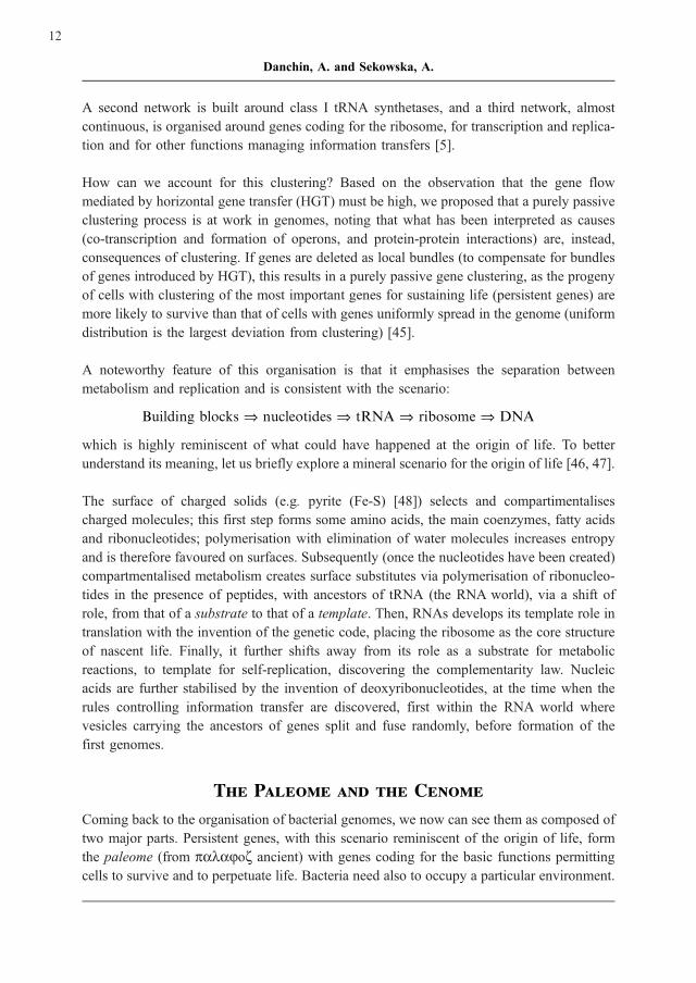
A second network is built around class I tRNA synthetases, and a third network, almost
continuous, is organised around genes coding for the ribosome, for transcription and replica-
tion and for other functions managing information transfers [5].
How can we account for this clustering? Based on the observation that the gene flow
mediated by horizontal gene transfer (HGT) must be high, we proposed that a purely passive
clustering process is at work in genomes, noting that what has been interpreted as causes
(co-transcription and formation of operons, and protein-protein interactions) are, instead,
consequences of clustering. If genes are deleted as local bundles (to compensate for bundles
of genes introduced by HGT), this results in a purely passive gene clustering, as the progeny
of cells with clustering of the most important genes for sustaining life (persistent genes) are
more likely to survive than that of cells with genes uniformly spread in the genome (uniform
distribution is the largest deviation from clustering) [45].
A noteworthy feature of this organisation is that it emphasises the separation between
metabolism and replication and is consistent with the scenario:
Building blocks � nucleotides � tRNA � ribosome � DNA
which is highly reminiscent of what could have happened at the origin of life. To better
understand its meaning, let us briefly explore a mineral scenario for the origin of life [46, 47].
The surface of charged solids (e.g. pyrite (Fe-S) [48]) selects and compartimentalises
charged molecules; this first step forms some amino acids, the main coenzymes, fatty acids
and ribonucleotides; polymerisation with elimination of water molecules increases entropy
and is therefore favoured on surfaces. Subsequently (once the nucleotides have been created)
compartmentalised metabolism creates surface substitutes via polymerisation of ribonucleo-
tides in the presence of peptides, with ancestors of tRNA (the RNA world), via a shift of
role, from that of a substrate to that of a template. Then, RNAs develops its template role in
translation with the invention of the genetic code, placing the ribosome as the core structure
of nascent life. Finally, it further shifts away from its role as a substrate for metabolic
reactions, to template for self-replication, discovering the complementarity law. Nucleic
acids are further stabilised by the invention of deoxyribonucleotides, at the time when the
rules controlling information transfer are discovered, first within the RNA world where
vesicles carrying the ancestors of genes split and fuse randomly, before formation of the
first genomes.
The Paleome and the Cenome
Coming back to the organisation of bacterial genomes, we now can see them as composed of
two major parts. Persistent genes, with this scenario reminiscent of the origin of life, form
the paleome (from palajoz ancient) with genes coding for the basic functions permitting
cells to survive and to perpetuate life. Bacteria need also to occupy a particular environment.
12
Danchin, A. and Sekowska, A.
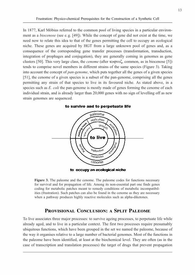
In 1877, Karl Mobius referred to the common pool of living species in a particular environ-
ment as a biocenose (see e. g. [49]). While the concept of gene did not exist at the time, we
need now to relate this idea to that of the genes permitting the cell to occupy an ecological
niche. These genes are acquired by HGT from a large unknown pool of genes and, as a
consequence of the corresponding gene transfer processes (transformation, transduction,
integration of prophages and conjugation), they are generally coming in genomes as gene
clusters [50]. This very large class, the cenome (after kojnoz, common, as in biocenose [5])
tends to comprise novel members in different strains of the same species (Figure 3). Taking
into account the concept of pan-genome, which puts together all the genes of a given species
[51], the cenome of a given species is a subset of the pan-genome, comprising all the genes
permitting any strain of that species to live in its favoured niche. As stated above, in a
species such as E. coli the pan-genome is mostly made of genes forming the cenome of each
individual strain, and is already larger than 20,000 genes with no sign of levelling off as new
strain genomes are sequenced.
Figure 3. The paleome and the cenome. The paleome codes for functions necessary
for survival and for propagation of life. Among its non-essential part one finds genes
coding for metabolic patches meant to remedy conditions of metabolic incompatibil-
ities (frustration). Such patches can also be found in the cenome as they are necessary
when a pathway produces highly reactive molecules such as alpha-diketones.
Provisional Conclusion: a Split Paleome
To live associates three major processes: to survive ageing processes, to perpetuate life while
already aged, and to live in a particular context. The first two processes require presumably
ubiquitous functions, which have been grouped in the set we named the paleome, because of
the way it organises relative to a large number of bacterial genomes. Most of the functions in
the paleome have been identified, at least at the biochemical level. They are often (as in the
case of transcription and translation processes) the target of drugs that prevent propagation
13
Frustration: Physico-chemical Prerequisites for the Construction of a Synthetic Cell
of the relevant organisms. While these functions are conserved, they are often not resulting
from similar structures, so that they can only be identified via analysis of gene persistence.
The functions of the paleome may be split following a variety of specific characters. For
example, half of its genes code for functions that are essential to permit formation of a
colony on plates supplemented by rich medium: they are the functions of essential genes
[52, 53]; the other half, while ubiquitous, does not have this property [42]. This latter half
comprises mostly genes that are essential to perpetuate life, but are not essential in the short
term [10]. Another split identifies functions which solve some of the metabolic incompat-
ibilities in the cell, resulting from chemical constraints such as spontaneous isomerisation of
aspartate and asparagine. This phenomenon of frustration is necessarily quite widespread, as
a large number of chemical intermediates, such as alpha-diketones, are extremely reactive
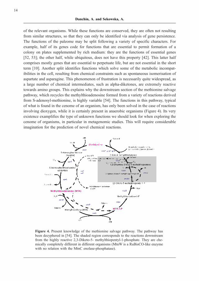
towards amino groups. This explains why the downstream section of the methionine salvage
pathway, which recycles the methylthioadenosine formed from a variety of reactions derived
from S-adenosyl-methionine, is highly variable [54]. The functions in this pathway, typical
of what is found in the cenome of an organism, has only been solved in the case of reactions
involving dioxygen, while it is certainly present in anaerobic organisms (Figure 4). Its very
existence examplifies the type of unknown functions we should look for when exploring the
cenome of organisms, in particular in metagenomic studies. This will require considerable
imagination for the prediction of novel chemical reactions.
Figure 4. Present knowledge of the methionine salvage pathway. The pathway has
been decyphered in [54]. The shaded region corresponds to the reactions downstream
from the highly reactive 2,3-Diketo-5- methylthiopentyl-1-phosphate. They are che-
mically completely different in different organisms (MtnW is a RuBisCO-like enzyme
with no relation with the MtnC enolase-phosphatase).
14
Danchin, A. and Sekowska, A.

Acknowledgements
This work benefited from many years of continuous discussions with the Stanislas Noria
group. Support for in silico analyses and experiments came from the PROBACTYS pro-
gramme, grant CT-2006 – 029104 in an effort to define genes essential for the construction
of a synthetic cell and the BioSapiens Network of Excellence, grant LSHG CT-2003 –
503265.
References
[1] Oliver, S.G., van der Aart, Q.J., Agostoni-Carbone, M.L., Aigle, M., Alberghina, L.,
Alexandraki, D., Antoine, G., Anwar, R., Ballesta, J.P., Benit, P., et al. (1992) The
complete DNA sequence of yeast chromosome III. Nature 357:38 – 46.
[2] Glaser, P., Kunst, F., Arnaud, M., Coudart, M.P., Gonzales, W., Hullo, M.F., Ionescu,
M., Lubochinsky, B., Marcelino, L., Moszer, I., Presecan, E., Santana, M., Schneider,
E., Schweizer, J., Vertes, A., Rapoport, G., Danchin, A. (1993) Bacillus subtilis
genome project: cloning and sequencing of the 97 kb region from 325 degrees to
333 degrees. Mol. Microbiol. 10:371 – 384.
[3] Danchin, A. (2003). The Delphic boat. What genomes tell us. Trans. A. Quayle.
Harvard University Press, Cambridge (Mass, USA).
[4] de Lorenzo, V., Danchin, A. (2008) Synthetic biology: discovering new worlds and
new words. The new and not so new aspects of this emerging research fields. EMBO
Reports 9:822 – 827.
[5] Danchin, A., Fang, G., Noria, S. (2007) The extant core bacterial proteome is an
archive of the origin of life. Proteomics 7:875 – 889.
[6] Hofstadter, D. (1979). ‘‘Godel, Escher, Bach: an Eternal Golden Braid’’. Basic
Books, New York.
[7] Muller, H. (1932) Some genetic aspects of sex. The American Naturalist
66:118 – 128.
[8] Orgel, L. (1963) The maintenance of the accuracy of protein synthesis and its
relevance to aging. Proc. Natl. Acad. Sci. U.S.A. 49:517 – 521.
[9] Dyson, F. J. (1985). Origins of life. Cambridge University Press, Cambridge, UK.
[10] Danchin, A. (2008) Natural Selection and Immortality. Biogerontology (submitted).
[11] Cover, T., Thomas, J. (1991). Elements of information theory. Wiley, New York.
15
Frustration: Physico-chemical Prerequisites for the Construction of a Synthetic Cell

[12] Bennett, C. (1988) Logical Depth and Physical Complexity. In The Universal Turing
Machine: a Half-Century Survey (R. Herken, ed.), pp. 227 – 257. Oxford University
Press, Oxford.
[13] Danchin, A. (1996) On genomes and cosmologies. In Integrative Approaches to
Molecular Biology (J. Collado-Vides, B. Magasanik, T. Smith, eds.), pp.
91 – 111. The MIT Press, Cambridge (USA).
[14] Turing, A. (1936 – 1937) On computable numbers, with an application to the En-
tscheidungsproblem. Proceedings of the London Mathematical Society 42:230 – 265.
[15] Turing, A. (1946 (1986)) A. M. Turing’s ACE Report of 1946 and Other Papers. In
Charles Babbage Institute reprint series for the History of Computing (B. Carpenter,
R. Doran, eds.), Vol. 10. MIT Press, Cambridge (Mass).
[16] von Neumann, J. (1958 (reprinted 1979)). The Computer and the Brain. Yale Uni-
versity Press, New Haven.
[17] Medigue, C., Rouxel, T., Vigier, P., Henaut, A., Danchin, A. (1991) Evidence for
horizontal gene transfer in Escherichia coli speciation. J. Mol. Biol. 222:851 – 856.
[18] Lartigue, C., Glass, J.I., Alperovich, N., Pieper, R., Parmar, P.P., Hutchison, C.A.,
3rd, Smith, H.O., Venter, J.C. (2007) Genome transplantation in bacteria: changing
one species to another. Science 317:632 – 638.
[19] Bussiere, D.E., Bastia, D. (1999) Termination of DNA replication of bacterial and
plasmid chromosomes. Mol. Microbiol. 31:1611 – 1618.
[20] Thanaraj, T.A., Argos, P. (1996) Ribosome-mediated translational pause and protein
domain organization. Protein Sci. 5:1594 – 1612.
[21] Bailly-Bechet, M., Danchin, A., Iqbal, M., Marsili, M., Vergassola, M. (2006) Codon
usage domains over bacterial chromosomes. PLoS Comput Biol 2:e37.
[22] Averof, M., Akam, M. (1995) Hox genes and the diversification of insect and
crustacean body plans. Nature 376:420 – 423.
[23] Averof, M. (1997) Arthropod evolution: same Hox genes, different body plans. Curr.
Biol. 7:R634 – 636.
[24] Tamames, J., Gonzalez-Moreno, M., Mingorance, J., Valencia, A., Vicente, M.
(2001) Bringing gene order into bacterial shape. Trends Genet. 17:124 – 126.
[25] Kitao, A., Yonekura, K., Maki-Yonekura, S., Samatey, F.A., Imada, K., Namba, K.,
Go, N. (2006) Switch interactions control energy frustration and multiple flagellar
filament structures. Proc. Natl. Acad. Sci. U.S.A. 103:4894 – 4899.
16
Danchin, A. and Sekowska, A.
[26] Gibson, D.G., Benders, G.A., Andrews-Pfannkoch, C., Denisova, E.A., Baden-Till-
son, H., Zaveri, J., Stockwell, T.B., Brownley, A., Thomas, D.W., Algire, M. A.,
Merryman, C., Young, L., Noskov, V.N., Glass, J.I., Venter, J.C., Hutchison, C.A.,
3 rd, Smith, H.O. (2008) Complete chemical synthesis, assembly, and cloning of a
Mycoplasma genitalium genome. Science 319:1215 – 1220.
[27] Rocha, E., Danchin, A., Viari, A. (1999) Universal replication biases in bacteria. Mol.
Microbiol. 32:11 – 16.
[28] Rocha, E., Danchin, A. (2003) Gene essentiality determines chromosome organisa-
tion in bacteria. Nucleic Acids Res. 31:6570 – 6577.
[29] Rocha, E.P., Danchin, A. (2004) An analysis of determinants of amino acids sub-
stitution rates in bacterial proteins. Mol. Biol. Evol. 21:108 – 116.
[30] Pascal, G., Medigue, C., Danchin, A. (2005) Universal biases in protein composition
of model prokaryotes. Proteins 60:27 – 35.
[31] Pascal, G., Medigue, C., Danchin, A. (2006) Persistent biases in the amino acid
composition of prokaryotic proteins. Bioessays 28:726 – 738.
[32] Riley, M., Staley, J.T., Danchin, A., Wang, T.Z., Brettin, T.S., Hauser, L.J., Land,
M.L., Thompson, L.S. (2008) Genomics of an extreme psychrophile, Psychromonas
ingrahamii. BMC Genomics 9:210.
[33] Medigue, C., Krin, E., Pascal, G., Barbe, V., Bernsel, A., Bertin, P.N., Cheung, F.,
Cruveiller, S., D’Amico, S., Duilio, A., Fang, G., Feller, G., Ho, C., Mangenot, S.,
Marino, G., Nilsson, J., Parrilli, E., Rocha, E.P., Rouy, Z., Sekowska, A.,
Tutino, M.L., Vallenet, D., von Heijne, G., Danchin, A. (2005) Coping with cold:
the genome of the versatile marine Antarctica bacterium Pseudoalteromonas halo-
planktis TAC125. Genome Res. 15: 1325 – 1335.
[34] Shimizu, T., Matsuoka, Y., Shirasawa, T. (2005) Biological significance of isoaspar-
tate and its repair system. Biol. Pharm. Bull. 28:1590 – 1596.
[35] Clarke, S. (2003) Aging as war between chemical and biochemical processes: protein
methylation and the recognition of age-damaged proteins for repair. Ageing Res. Rev.
2:263 – 285.
[36] David, C.L., Keener, J., Aswad, D.W. (1999) Isoaspartate in ribosomal protein S11
of Escherichia coli. J. Bacteriol. 181:2872 – 2877.
[37] Landauer, R. (1961) Irreversibility and heat generation in the computing process.
IBM Journal of research and development 3:184 – 191.
[38] Bennett, C. (1988) Notes on the history of reversible computation. IBM Journal of
research and development 44:270 – 277.
17
Frustration: Physico-chemical Prerequisites for the Construction of a Synthetic Cell

[39] Thompson, L.W., Krawiec, S. (1983) Acquisitive evolution of ribitol dehydrogenase
in Klebsiella pneumoniae. J. Bacteriol. 154:1027 – 1031.
[40] Ashida, H., Danchin, A., Yokota, A. (2005) Was photosynthetic RuBisCO recruited
by acquisitive evolution from RuBisCO-like proteins involved in sulphur metabo-
lism? Res. Microbiol. 156:611 – 618.
[41] Henaut, A., Danchin, A. (1996) Analysis and predictions from Escherichia coli
sequences or E. coli in silico. In Escherichia coli and Salmonella, Cellular and
Molecular Biology (F. Neidhardt, ed.), Vol. 1, pp. 2047 – 2065. ASM Press, Washing-
ton.
[42] Fang, G., Rocha, E., Danchin, A. (2005) How essential are nonessential genes? Mol.
Biol. Evol. 22:2147 – 2156.
[43] Danchin, A. (1988) Complete genome sequencing: future and prospects. In BAP
1988 – 1989 (A. Goffeau, ed.), pp. 1 – 24. Commission of the European Commu-
nities, Brussels.
[44] Mushegian, A.R., Koonin, E.V. (1996) A minimal gene set for cellular life derived by
comparison of complete bacterial genomes. Proc. Natl. Acad. Sci. U.S.A.
93:10268 – 10273.
[45] Fang, G., Rocha, E.P., Danchin, A. (2008) Persistence drives gene clustering in
bacterial genomes. BMC Genomics 9:4.
[46] Granick, S. (1957) Speculations on the origins and evolution of photosynthesis. Ann.
N. Y. Acad. Sci. 69:292 – 308.
[47] Danchin, A. (1989) Homeotopic transformation and the origin of translation. Prog.
Biophys. Mol. Biol. 54:81 – 86.
[48] Wachtershauser, G. (1988) Before enzymes and templates: theory of surface metabo-
lism. Microbiol. Rev. 52:452 – 484.
[49] Movila, A., Uspenskaia, I., Toderas, I., Melnic, V., Conovalov, J. (2006) Prevalence
of Borrelia burgdorferi sensu lato and Coxiella burnetti in ticks collected in different
biocenoses in the Republic of Moldova. International Journal of Medical Microbiol-
ogy 296:172 – 176.
[50] Lawrence, J.G., Roth, J.R. (1996) Selfish operons: horizontal transfer may drive the
evolution of gene clusters. Genetics 143:1843 – 1860.
[51] Tettelin, H., Masignani, V., Cieslewicz, M.J., Donati, C., Medini, D., Ward, N.L.,
Angiuoli, S.V., Crabtree, J., Jones, A.L., Durkin, A.S., Deboy, R.T., Davidsen, T.M.,
Mora, M., Scarselli, M., Margarit y Ros, I., Peterson, J.D., Hauser, C.R., Sundaram,
J.P., Nelson, W.C., Madupu, R., Brinkac, L.M., Dodson, R.J., Rosovitz, M.J., Sulli-
18
Danchin, A. and Sekowska, A.

van, S.A., Daugherty, S.C., Haft, D.H., Selengut, J., Gwinn, M.L., Zhou, L., Zafar,
N., Khouri, H., Radune, D., Dimitrov, G., Watkins, K., O’Connor, K.J., Smith, S.,
Utterback, T.R., White, O., Rubens, C.E., Grandi, G., Madoff, L.C., Kasper, D.L.,
Telford, J.L., Wessels, M.R., Rappuoli, R., Fraser, C.M. (2005) Genome analysis of
multiple pathogenic isolates of Streptococcus agalactiae: implications for the micro-
bial ‘‘pangenome’’. Proc. Natl. Acad. Sci. U.S.A. 102:13950 – 13955.
[52] Kobayashi, K., Ehrlich, S.D., Albertini, A., Amati, G., Andersen, K.K., Arnaud, M.,
Asai, K., Ashikaga, S., Aymerich, S., Bessieres, P., Boland, F., Brignell, S.C., Bron,
S., Bunai, K., Chapuis, J., Christiansen, L.C., Danchin, A., Debarbouille, M., Der-
vyn, E., Deuerling, E., Devine, K., Devine, S.K., Dreesen, O., Errington, J., Fillinger,
S., Foster, S.J., Fujita, Y., Galizzi, A., Gardan, R., Eschevins, C., Fukushima, T.,
Haga, K., Harwood, C.R., Hecker, M., Hosoya, D., Hullo, M.F., Kakeshita, H.,
Karamata, D., Kasahara, Y., Kawamura, F., Koga, K., Koski, P., Kuwana, R., Im-
amura, D., Ishimaru, M., Ishikawa, S., Ishio, I., Le Coq, D., Masson, A., Mauel, C.,
Meima, R., Mellado, R.P., Moir, A., Moriya, S., Nagakawa, E., Nanamiya, H., Nakai,
S., Nygaard, P., Ogura, M., Ohanan, T., O’Reilly, M., O’Rourke, M., Pragai, Z.,
Pooley, H.M., Rapoport, G., Rawlins, J.P., Rivas, L.A., Rivolta, C., Sadaie, A.,
Sadaie, Y., Sarvas, M., Sato, T., Saxild, H.H., Scanlan, E., Schumann, W.,
Seegers, J.F., Sekiguchi, J., Sekowska, A., Seror, S.J., Simon, M., Stragier, P.,
Studer, R., Takamatsu, H., Tanaka, T., Takeuchi, M., Thomaides, H.B., Vagner, V.,
van Dijl, J.M., Watabe, K., Wipat, A., Yamamoto, H., Yamamoto, M., Yamamoto, Y.,
Yamane, K., Yata, K., Yoshida, K., Yoshikawa, H., Zuber, U., Ogasawara, N. (2003)
Essential Bacillus subtilis genes. Proc. Natl. Acad. Sci. U.S.A. 100:4678 – 4683.
[53] Joyce, A.R., Reed, J.L., White, A., Edwards, R., Osterman, A., Baba, T., Mori, H.,
Lesely, S.A., Palsson, B.O., Agarwalla, S. (2006) Experimental and computational
assessment of conditionally essential genes in Escherichia coli. J. Bacteriol.
188:8259 – 8271.
[54] Sekowska, A., Denervaud, V., Ashida, H., Michoud, K., Haas, D., Yokota, A.,
Danchin, A. (2004) Bacterial variations on the methionine salvage pathway. BMC
Microbiol 4:9.
19
Frustration: Physico-chemical Prerequisites for the Construction of a Synthetic Cell


����������������
Catalysis at the Origin of Life Viewed in
the Light of the (M,R)-Systems of
Robert Rosen
Athel Cornish-Bowden*
and Mar�a Luz C�rdenas
Unite de Bioenergetique et Ingenierie des Proteines, Centre National de la Recherche
Scientifique, 31 chemin Joseph-Aiguier, B.P. 71, 13402 Marseilles, France
E-Mail: *[email protected]
Received: 1st September 2008 / Published: 16th March 2009
Abstract
Living systems as we know them today are both complex, displaying
emergent properties, and extremely complicated, with huge numbers of
different components. At the origin of life they must also have had
emergent properties, and hence must have been complex, but they
cannot have been as complicated as modern organisms, because we
cannot imagine that the first organisms started with anything as elabo-
rate as a ribosome and all of the protein-synthesis machinery. Under-
standing how complexity could arise in even the simplest early organ-
ism requires, however, a theory of life, something that is largely lack-
ing from modern biology. Various authors have contributed elements of
such a theory, and the (M,R)-systems of Robert Rosen provide a con-
venient starting point.
Introduction
In an earlier contribution to this series [1] we commented that many phenomena are de-
scribed as complex when in reality they are no more than complicated, because they can be
fully accounted for in terms of the properties of their components: there is no ‘‘emergence’’.
It must be recognized, however, that it is not always easy to decide whether a property is
truly emergent or not, in part because of disagreements about how emergence should be
defined [2]. Living organisms in their totality, however, are complex, because it appears to
21
http://www.beilstein-institut.de/Bozen2008/Proceedings/CornishBowden/CornishBowden.pdf
Systems Chemistry, May 26th – 30th, 2008, Bozen, Italy

be impossible to deduce their properties solely by applying the reductionist programme of
studying all of the properties of all of the components in sufficient detail. Some authors [3,
4] go further, and say that it not only appears to be impossible but it really is impossible
even in principle, but this aspect remains controversial [5] and we shall not discuss it here.
We shall, however, try to resume the current state of understanding of the nature of life and
the definition of a living organism. Although this might seem an essential component of
biology, it is in practice ignored by nearly all biologists [6] and regarded as irrelevant to the
practice of modern biology by many [7], as we discussed previously [8]. The famous
question raised by Erwin Schrodinger [9] of what life is remains unanswered more than
60 years later. Parts of it, of course, have been answered: we can identify Schrodinger’s
‘‘codescript’’ with the DNA in which protein sequences are recorded; we now rarely need to
speak of organisms feeding on negative entropy because we understand that living organ-
isms are thermodynamically open systems that can maintain themselves far from equilibrium
without violating any thermodynamic principles. Nonetheless, the crucial question of what
biological organization actually means and how it is maintained almost indefinitely remains
inadequately studied. In 2005 the editors of Science [10] celebrated 125 years of existence of
the magazine with a list of 125 questions, ‘‘the most compelling puzzles and questions
facing scientists today’’. A high proportion of these were questions about biology, and
included such vogue items as ‘‘is an effective HIV vaccine feasible?’’, but Schrodinger’s
question was not among them.
The first modern attempts to understand biological organization were made by Stephane
Leduc [11]. His osmotic experiments produced impressively complicated and biological-
looking structures (which can be reproduced in full colour illustrations today: see Querbes
[12]), but few biologists today would accept that osmosis tells us much about the forms of
real organisms. However, his more general belief that natural selection is not the only
explanation of biological forms, and that chemical reactivity as well as physicochemical
and mechanical forces also play major roles remains important, and was taken up by D’Arcy
Thompson [13] in a closely argued book that has been very influential in modern thinking.
There are four principal current theories that try to explain biological organization, the
(M,R)-systems of Rosen [3], the chemoton of Ganti [14], the autopoiesis of Maturana and
Varela [15], and the autocatalytic sets of Kauffman [16]. Despite the fact that all of these
theories contain some of the same ideas, they are by no means the same as one another, and
none of the authors mentioned makes any reference to any of the others in their principal
publications. Although all attach importance to ‘‘closure’’ and their definitions of this over-
lap, they underline different aspects. Rosen [3], for example, refers to closure to efficient
causation, which means that all the catalysts required by an organism need to be products of
its own metabolism; Maturana and Varela [15] stress structural closure, or the need for an
organism to be enclosed within a membrane, cell wall, or skin; Kauffman [16] considers that
catalytic closure is the consequence of very large sets of different polypeptides or polynu-
22
Cornish-Bowden, A. and Cardenas, M.L.

cleotides; Ganti [14] agrees on the necessity for structural closure, and also emphasizes the
need for any theory of life to be rooted in an adequate knowledge of chemistry. Clearly,
therefore, an important task for the future will be to integrate all of these threads into a single
theory of life. Here we shall be less ambitious, concentrating on the ideas of Robert Rosen,
which are the most abstract and difficult to understand of those we have mentioned, and will
use them to analyse aspects of catalysis at the origin of life.
Limits of Reductionism
The reductionist approach has taken biochemistry a very long way since Buchner [17] first
demonstrated that alcoholic fermentation could occur in a cell-free extract of yeast, and it is
very unlikely that we should know much about biochemistry today, and still less about
molecular biology, if the approach in the 20th century had not been overwhelmingly reduc-
tionist. However, it is one thing to recognize the progress that reductionism has brought, but
it is another to suppose that this can continue indefinitely. One can certainly understand the
behaviour of components of the cell, such as metabolic pathways, in terms of their compo-
nents, enzymes in this case, and enzymes can be understood in terms of the properties of
their side-chains. However, especially at the level of the cell or the whole organism, the
reductionist approach cannot provide the whole truth, because these entities have complex
and emergent properties. However, today, and in contrast to the early 20th century, we do try
to understand the chemistry of whole organisms (systems chemistry) and, in the words of
Henrik Kacser [18], ‘‘one thing is certain: to understand the whole you must study the
whole’’, an idea more picturesquely expressed by a Russian proverb, ‘‘a hundred rabbits
do not make a horse’’.
It may be illuminating to compare modern biology with modern physics. In the 19th century
theory followed from experimental observations: thermodynamics, for example, developed
from Sadi Carnot’s efforts to determine whether steam engines could be improved without
limit. In this and other 19th century cases the experiments preceded the development of the
theory, but in 20th century physics theory usually preceded experiment: Albert Einstein, for
example, did not develop the theory of relativity after wondering how the satellite navigation
system in his car worked; on the contrary, this and other applications of relativity came many
years after the theory had been worked out. The comparison is not just with physics, and as
Gunter von Kiedrowski remarked earlier in this symposium, ‘‘a century ago chemistry was
in the same situation as biology today’’. We believe, in summary, that future advances in
biology will require a more complete theoretical basis than is provided by the theory of
natural selection, the only general biological theory that exists, which is valuable for inter-
preting observations, but is not the whole truth.
23
Catalysis at the Origin of Life Viewed in the Light of the (M,R)-Systems of Robert Rosen

Closure to Efficient Causation
Metabolism is often represented as a large and complicated set of processes catalysed by
enzymes or transporters, these processes including both chemical reactions and transport
across membranes. This description, however, while true as far as it goes, is seriously
incomplete. As biologists know, and as Rosen [3] emphasized, the enzymes and transporters
(which for brevity we shall consider together just as enzymes) are subject to turnover,
dilution by growth of the organism, and losses due to their finite stability, processes that
we shall abbreviate to decay. So, even if all the necessary enzymes are present in one
moment, they cannot continue to be present indefinitely unless mechanisms exist to replace
them. From what, however, can they be made? Clearly the only possible answer is that they
must be made from the products of metabolism, so they are themselves products of meta-
bolism, and hence metabolites.
Not only must all ‘‘enzymes’’ be considered metabolites in this sense, but many ‘‘metabo-
lites’’ are also enzymes, because they are biological molecules that act as catalysts: meta-
bolic cycles require not only the protein catalysts usually regarded as enzymes, but also the
molecules that are consumed and regenerated in the process: the urea cycle, for example,
requires not only three proteins, citrullinase, arginine deaminase and arginase, but also
ornithine, and so ornithine has just as much right to be called an enzyme as the three
proteins. It has more right, even, as the cycle would still occur (slowly) if some or all of
the proteins were missing, but it would not occur without ornithine or a molecule that could
replace it, such as citrulline or arginine. It follows that the usual distinction between en-
zymes and metabolites is formally meaningless [19]. Similar considerations apply to other
metabolic cycles, such as the tricarboxylate cycle.
To refer to this organized replacement Rosen [3] used the unfortunate term repair, inviting
confusion with more standard notions of repair in modern biochemistry, such as DNA repair
and action of chaperones, so we prefer to refer to replacement [20, 21]. Fortunately this
begins with the same letter of the alphabet as Rosen’s word, so we can continue to use the
term (M,R)-system as a short form of metabolism-replacement system, which summarizes
Rosen’s view of an organism. The essential point is that catalysts need to be replaced
internally, by the organism itself; they cannot be replaced by an external agency. For this
reason an organism is fundamentally different from a machine, because regardless of how
one defines a machine, whether a simple tool such as an axe or something as complicated as
an airliner, or even as a complete factory, at no level of definition does the machine make
itself or maintain itself. The machine analogy may be helpful for understanding certain
properties of organism, for example how the heart work, but in general it fails, because an
organism is not a machine.
24
Cornish-Bowden, A. and Cardenas, M.L.

Unfortunately, however, Rosen did not make it easy for his readers to study his work. He
presented it in resolutely mathematical terms, making no concessions to readers without
mathematical expertise, he provided no examples to illustrate his central points – not even
mathematical examples, and certainly no biological examples, and he did not define the
range of validity of his ideas. We have therefore tried to fill these voids [20, 21].
Catalysis at the Origin of Life
At the origin of life there was no natural selection as we understand it today, but there was
certainly chemical selection resulting from differences in rates of reactions derived from
kinetic or thermodynamic properties [22]. Thus (M,R)-systems (metabolism-replacement-
systems) probably emerged in prebiotic conditions thanks to the presence of inorganic
catalysts or simple organic molecules that could act as catalysts. Modern organisms are
not only complex; they are also extremely complicated, with a wide array of regulatory
mechanisms, both metabolic and genetic, that were surely absent at the origin of life. These
mechanisms are not explicitly visible in (M,R)-systems (though they are not excluded, and
can be considered to be implicit), and so the representation of an organism as an (M,R)-
system may be closer to the reality at the origin of life than to the reality of today.
Catalysis is fundamental for the organization of living systems, and must have been neces-
sary at the origin of life, to permit organized systems to appear, to maintain themselves, and
to grow. Some degree of specificity was also necessary, to allow one system to be different
from another. Thus although the first catalysts must have been much simpler molecules than
the protein or RNA catalysts that we know today, they must have had properties closer to
these than to highly unspecific catalysts like platinum black. Specificity could then have
developed progressively, first through chemical evolution and then through natural selection,
to arrive finally at present-day bio-catalysts. However, specificity cannot be complete,
because it is not possible for a system to fabricate its own catalysts if each one needs its
own unique catalyst.
Types of Closure
The idea of closure may be understood in three fundamentally different ways, as illustrated
in Figure 1. The simplest is the structural closure (Fig. 1a) produced by the physical bound-
ary (membranes, cell walls, skin) that encloses an individual: in a present-day organism this
is always fabricated by the organism itself, but this may not have been true for the first
organisms, which could perhaps have made use of already existing inorganic compartments.
Organisms must be closed in this sense, because every individual must be distinguishable
from every other, and it is in general clear where one individual ends and another begins (if
we exclude consideration of Dictyostelium discoideum and other organisms that challenge
any attempt at a simple definition of an individual). Note that there can be no competition
between individuals, and no way of assigning an identity to an individual, without structural
25
Catalysis at the Origin of Life Viewed in the Light of the (M,R)-Systems of Robert Rosen
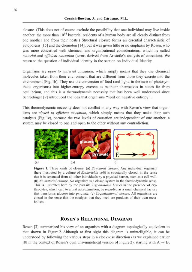
closure. (This does not of course exclude the possibility that one individual may live inside
another: the more than 1014 bacterial residents of a human body are all clearly distinct from
one another and from their hosts.) Structural closure forms an essential characteristic of
autopoiesis [15] and the chemoton [14], but it was given little or no emphasis by Rosen, who
was more concerned with chemical and organizational considerations, which he called
material and efficient causation (terms derived from Aristotle’s analysis of causation). We
return to the question of individual identity in the section on Individual Identity.
Organisms are open to material causation, which simply means that they use chemical
molecules taken from their environment that are different from those they excrete into the
environment (Fig. 1b). They use the conversion of food (and light, in the case of photosyn-
thetic organisms) into higher-entropy excreta to maintain themselves in states far from
equilibrium, and this is a thermodynamic necessity that has been well understood since
Schrodinger [9] introduced the idea that organisms ‘‘feed on negative entropy’’.
This thermodynamic necessity does not conflict in any way with Rosen’s view that organ-
isms are closed to efficient causation, which simply means that they make their own
catalysts (Fig. 1c), because the two levels of causation are independent of one another: a
system may be closed to one and open to the other without any contradiction.
Figure 1. Three kinds of closure. (a) Structural closure. Any individual organism
(here illustrated by a culture of Escherichia coli) is structurally closed, in the sense
that it is separated from all other individuals by a physical barrier, such as a cell wall.
(b) No material closure. No organism is a closed system in the thermodynamic sense.
This is illustrated here by the parasite Trypanosoma brucei in the presence of ery-
throcytes, which can, to a first approximation, be regarded as a small chemical factory
that transforms glucose into pyruvate. (c) Organizational closure. All organisms are
closed in the sense that the catalysts that they need are products of their own meta-
bolism.
Rosen's Relational Diagram
Rosen [3] summarized his view of an organism with a diagram topologically equivalent to
that shown in Figure 2. Although at first sight this diagram is unintelligible, it can be
understood by following the various steps in a clockwise direction (as we explained earlier
[8] in the context of Rosen’s own unsymmetrical version of Figure 2), starting with A ? B,
26
Cornish-Bowden, A. and Cardenas, M.L.

which represents the whole of metabolism, as full arrows represent material causation: the
substrates A of the whole set of metabolic reactions are the material cause of the products B
of the same set of reactions. The broken arrows represent efficient causation, and so f - -> A
? B means that metabolism A ? B is catalysed by the whole set of catalysts f. These
catalysts are replaced from the only pool available, the set of metabolic products B under the
influence of a replacement system F, so F - -> B ? f .
This system must itself be replaced, because it is subject to the same problems of decay as
the metabolic catalysts. At this point Rosen avoided the incipient infinite regress by suppos-
ing that the efficient cause of this replacement of F from f could be caused by an entity bthat was considered as a property of B, so B --> f ? F allows the whole diagram to be
closed to efficient causation, though, as noted above, it is open to material causation, with a
net non-cyclic (or only partially cyclic) process A ? B. Rosen always made it clear in his
writing, and especially in his papers of 1966 and 1971 [24, 25], that although b is related to
B it is not the same as B, and in mathematical terms it is best understood as the inverse of B
[20], but he was less careful to be clear about this in the diagram on which Figure 2 is based,
an oversight that has caused a great deal of confusion and misunderstanding in the literature.
The whole diagram is thus closed to efficient causation, or in other words it shows metabolic
circularity or organizational invariance. It contains no final causes – no explanations of
purpose, or of why anything happens as it does. The fourth of Aristotle’s categories of
causation, the formal cause (what makes a metabolite a metabolite? What makes an enzyme
an enzyme?), is also absent from the diagram, as it played little role in Rosen’s thinking.
We have already noted that Rosen’s term ‘‘repair’’ has nothing to do with conventional uses
of this term in modern biochemistry. Perhaps even more misleading, his term ‘‘replication’’
for what we call organizational invariance has nothing to do with DNA replication, etc. We
include both of these terms in Figure 2 to facilitate comparison with Rosen’s publications,
but they are otherwise best avoided.
Recent years have seen an enormous growth in ‘‘systems biology’’, with greatly increased
interested in ‘‘small-world’’ models of metabolism [26, 27] and the ‘‘bow-tie model’’ of
metabolic regulation [28]. However, the idea of metabolic closure is completely absent from
these. The small-models are completely concerned with material causation and do not
address the question of where the catalysts come from, and although the bow-tie model
does imply the existence of catalysts it also does not address the question of where they
come from. So although Figure 1 of Csete & Doyle [28], for example, contains numerous
feedback loops it has a clear left-to-right reading direction, with no suggestion of closure.
27
Catalysis at the Origin of Life Viewed in the Light of the (M,R)-Systems of Robert Rosen

Figure 2. Closure to efficient cause (metabolic circularity). The diagram is based on
Figure 10C.6 of Rosen [3], redrawn in the more symmetrical way suggested by
Cottam et al. [23]. Broken arrows show efficient causes, or catalysis, whereas full
arrows represent material causes, or chemical transformations. The diagram is highly
abstract: the transformation f - -> A ? B represents the whole of metabolism; the
transformation F - -> B ? f represents replacement of enzymes from available pro-
ducts of metabolism, and b � B - -> f ? F represents the processes need to maintain
organizational invariance. Rosen’s terms ‘‘repair’’ and ‘‘replication’’ shown in par-
entheses for these last two processes are misleading, as they have nothing to do with
the ordinary uses of these words in modern biochemistry, for example for DNA repair
and DNA replication. Note that although b is related to B it is not the same as B.
Representing Rosen's Model in a More Biological Way
As noted, Rosen’s representation as illustrated in Figure 2 is highly abstract, and not easy to
relate to ordinary ideas of biological models. In an attempt to make it more concrete,
therefore, we extended a model outlined by Moran et al. [29] to arrive at the minimal
biological model of an (M,R)-system illustrated in Figure 3 [8, 19 – 21]. In this representa-
tion the three catalytic processes are shown as three cycles of chemical reactions:
1. S + STU ? STUS;STUS + T ? STUST ? STU + ST (metabolism)
2. ST + SU ? SUST;SUST + U ? SUSTU ? SU + STU (replacement)
3. S + STU ? STUS;STUS + U ? STUSU ? STU + SU (organizational invariance)
These can alternatively be written as three catalysed reactions:
S þ T �!STU ST ; ST þ U �!SU STU ; S þ U �!STU SU
but the greater simplicity is only apparent, because writing the cycles as catalysed reactions
just hides the chemical reality of what is happening. An important point to note here is that
closure was achieved by requiring one molecule STU to catalyse two different processes.
28
Cornish-Bowden, A. and Cardenas, M.L.

We believe that this will be generally true, that for models of arbitrary complexity it will
always be necessary to include multifunctional catalysts if closure is to be possible. This in
turn implies a general principle: that ‘‘moonlighting’’, or multifunctionality of proteins, as
well exemplified by the many different functions of glyceraldehyde 3-phosphate dehydro-
genase, is not simply an interesting property of biochemical systems that is being increas-
ingly observed [30, 31], but is an absolute necessity for life. In present-day organisms the
ribosome, which participates in the synthesis of a wide variety of different types of protein,
certainly contributes to the closure though it is far from being the only example. For
consideration of the organisms shortly after the origin of life we need to envisage vastly
simpler solutions than that represented by the ribosome and modern catalytic proteins.
Figure 3. A model of an (M,R)-system. This is the biological model suggested pre-
viously [8, 19 – 21]. The two decay reactions shown with double arrowheads are
considered to be irreversible. Unfortunately, Figure 3b of [21], which should have
been very similar to this one, was printed incorrectly, as three intermediates were
named incorrectly, STUST as SUST, STUSU as SUSU, and SUSTU also as SUSU
(with the result that the names SUST and SUSU occurred twice each). It was printed
correctly in the other papers that have similar diagrams [8, 19].
Another important point to note is that the ‘‘solution’’ to the problem of closure shown in
Figure 3 is not unique, because the assignment of STU, SU and again STU as catalysts of the
three processes is just one of the 33 = 27 ways in which the three possible catalysts STU, ST
and SU could be distributed among the three reactions. Thus the structure of the network
does not by itself define Rosen’s function b needed for organizational invariance.
29
Catalysis at the Origin of Life Viewed in the Light of the (M,R)-Systems of Robert Rosen

A possible way out of the difficulty comes from reexamining the definition of an enzyme or
of a catalyst. Any catalysed reaction can be written as a cycle of uncatalysed reactions, as
was done in the preceding section, and, as noted in the section Closure to Efficient Causa-
tion, it is only convention, not logical necessity, that leads us to called arginase but not
ornithine a catalyst of the urea cycle, for example. Catalysis is nothing more fundamental
than a human interpretation of chemical cycles, and if this is recognized then Figure 3
represents not a set of three catalysed reactions, but a set of eight uncatalysed chemical
reactions, and on this interpretation the question of which catalyst catalyses which reaction
does not arise [19]. This means that the catalytic properties follow simply from chemical
reactivities, and makes it easier to understand how an organism can ‘‘know’’ which mole-
cule, whether catalyst or other metabolite, is needed for maintaining its metabolism.
Individual Identity
Natural selection implies competition between individuals, and needs identifiable and dis-
tinguishable individuals to do the competing. However, Figure 3 does not provide any
indication of how the system is contained, and thus ‘‘individual’’, and does not suggest
how one individual might be distinguished from another. An obvious step to overcome the
first objection will be to incorporate the obligatory formation of a membrane, already
explicitly included in autopoiesis [15] and the chemoton [14], into the representation of
(M,R)-systems. We have discussed elsewhere [32] how the second objection may be over-
come. Although this was in terms of a model somewhat more complicated that the one in
Figure 3, the main idea can still be presented in terms of Figure 3. First we need to suppose
that the whole system is contained within a barrier (structural closure), as without this there
can be no competition. Suppose now that a chance accident causes one of the catalysts, for
example SU, to appear in a variant form SU’ that has similar properties to the normal form
but catalyses the production of a slightly different form of another catalyst (which must be
STU in this case as that is the only possibility). Although initially there may be only one
molecule of SU’ the number will increase if it happens that the variant system reacts faster
than the normal one and will eventually replace it.
Conclusion
Future development of systems chemistry and systems biology will need what Woese [33]
has called a guiding vision: the main theories about the nature of life [3, 14 – 16], in
particular the (M,R)-systems of Robert Rosen could constitute this guiding vision. For
example, current research efforts to define the minimal genome that allows a system to be
autonomous should benefit greatly from the incorporation of such concepts as metabolic
closure and organizational invariance.
30
Cornish-Bowden, A. and Cardenas, M.L.

Efforts must be made to integrate the theories about the nature of life in a single coherent
one, which could serve as a basis for understanding how life originated and how it is
maintained. Catalysis at the origin of life probably made some use of external agents, so
prebiotic systems probably did not fully satisfy the criterion of metabolic closure, but only
when they acquired it did they become truly alive, even if the structural closure still
depended on external inorganic support. It is tempting therefore to speculate that metabolic
closure was acquired before structural closure.
References
[1] Cornish-Bowden, A., Cardenas, M.L. (2003) Metabolic analysis in drug design:
complex, or just complicated? In: Molecular Informatics: Confronting Complexity
(ed. M. Hicks & C. Kettner), pp. 95 – 107, Beilstein-Institut, Frankfurt.
[2] Bedau, M.A. (2003) Downward causation and autonomy in weak emergence. Prin-
cipia 6:5 – 50.
[3] Rosen, R. (1991) Life itself: A comprehensive inquiry into the nature, origin and
fabrication of life. New York, Columbia University Press.
[4] Louie, A.H. (2007) A living system must have noncomputable models. Artif. Life
13:293 – 297.
[5] Stewart, J., Mossio, M. (2007) Is ‘‘life’’ computable? Proc. ENACTIVE/07 (Proceed-
ings of the 4th International Conference on Enactive Interfaces 2007, Grenoble,
France), http://www.enactive2007.org.
[6] Harold, F. M. (2001) The Way of the Cell, Oxford University Press.
[7] Atlan, H., Bousquet, C. (1994) Questions de Vie, Le Seuil, Paris.
[8] Cornish-Bowden, A., Cardenas, M.L. (2007) Bring chemistry to life: what does it
mean to be alive? In: Molecular Interactions: Bringing Chemistry to Life
(ed. M. Hicks & C. Kettner), pp. 27 – 39, Beilstein-Institut, Frankfurt.
[9] Schrodinger, E. (1944) What is Life? Cambridge University Press, Cambridge.
[10] Science 309: 1 – 204 (2005).
[11] Leduc, S. (1912) La Biologie Synthetique, Poinat, Paris.
[12] Querbes, S. (2007) http://www.stephanequerbes.com/books/recherche/recherche.html.
[13] Thompson, D’A.W. (1961) On Growth and Form (ed. J.T. Bonner, abridged edition
of a book originally published in 1917, with second edition in 1942), Cambridge
University Press, Cambridge.
31
Catalysis at the Origin of Life Viewed in the Light of the (M,R)-Systems of Robert Rosen

[14] Ganti, T. (2003) The Principles of Life, Oxford University Press, Oxford.
[15] Maturana, H.R. and Varela, F.J. (1980) Autopoiesis and Cognition: the Realisation of
the Living, D. Reidel Publishing Company, Dordrecht, The Netherlands.
[16] Kauffman, S.A. (1986) Autocatalytic sets of proteins. J. Theor. Biol. 119:1 – 24.
[17] Buchner, E. (1897) Alkoholische Gahrung ohne Hefezellen. Ber. Dt. Chem. Ges.
30:117 – 124.
[18] Kacser, H. (1987) On parts and wholes in metabolism, in The organization of cell
metabolism (ed. G.R. Welch & J.S. Clegg), pp. 327 – 337, Plenum, New York.
[19] Cornish-Bowden, A., Cardenas, M.L. (2007) Organizational invariance in (M,R)-
systems. Chem. Biodiversity 4:2396 – 2406.
[20] Letelier, J.-C., Soto-Andrade, J., F. Guınez Abarzua, F., Cornish-Bowden, A., Carde-
nas, M.L. (2006) Organizational invariance and metabolic closure: analysis in terms
of (M,R)-systems. J. Theor. Biol. 238:949 – 961.
[21] Cornish-Bowden, A., Cardenas, M.L., Letelier, J.-C., Soto-Andrade, J. (2007) Be-
yond reductionism: metabolic circularity as a guiding vision for a real biology of
systems. Proteomics 7:839 – 845.
[22] Melendez-Hevia, E., Montero-Gomez, N, Montero, F. (2008) From prebiotic chem-
istry to cellular metabolism – the chemical evolution of metabolism before Darwinian
natural selection. J. Theor. Biol. 252:505 – 519.
[23] Cottam, R., Ranson, W., Vounckx, R. (2007) Re-mapping Robert Rosen’s (M,R)-
systems. Chem. Biodivers. 4:2352 – 2368.
[24] Rosen, R. (1966) A note on replication in (M,R)-systems. Bull. Math. Biophys.
28:149 – 151.
[25] Rosen, R. (1971) Some realizations of (M,R)-systems and their interpretation. Bull.
Math. Biophys. 33:303 – 319.
[26] Fell, D.A., Wagner, A. (2000) The small word of metabolism. Nat. Biotechnol.
18:1121 – 1122.
[27] Jeong, H., Tombor, B., Albert, R., Oltvai, Z.N., Barabasi, A.L. (2000) The large-scale
organization of metabolic networks. Nature 407:651 – 654.
[28] Csete, M. & Doyle, J. (2004) Bow ties, metabolism and disease. Trends Biotechnol.
22:446 – 450.
32
Cornish-Bowden, A. and Cardenas, M.L.

[29] Moran, F., Moreno, A., Minch, E., Montero, F. (1996) Further steps towards a
realistic description of the essence of life. In Langton, C. G., Shimohara, K., editors,
Artificial Life V, pp. 255 – 263. MIT Press, Cambridge, Massachusetts.
[30] Tipton, K.F., O’Sullivan, M.I., Davey, G.P., O’Sullivan, J. (2003) It can be a com-
plicated life being an enzyme. Biochem. Soc. Trans. 31:711 – 715.
[31] Sriram, G., Martinez, J.A., McCabe, E.R.B., Liao, J.C., Dipple, K.M. (2005) Single-
gene disorders: what role could moonlighting enzymes play? Am. J. Hum. Genet.
76:911 – 924.
[32] Cornish-Bowden, A., Cardenas, M.L. (2008) Self-organization at the origin of life.
J. Theor. Biol. 252:411 – 418.
[33] Woese, C.R. (2004) A new biology for a new century. Microb. Molec. Biol. Rev.
68:173 – 186.
33
Catalysis at the Origin of Life Viewed in the Light of the (M,R)-Systems of Robert Rosen


����������������
New Concepts for Catalysis
Corinna M. Reisinger, Subhas Chandra Pan
and Benjamin List*
Max-Planck-Institut fur Kohlenforschung, 45470Mulheim an der Ruhr, Germany
E-Mail: *[email protected]
Received: 16th January 2009 / Published: 16th March 2009
Abstract
Organocatalysis, the catalysis with low-molecular weight catalysts
where a metal is not part of the catalytic principle, can be as efficient
and selective as metal- or biocatalysis. Important discoveries in this
area include novel Lewis base-catalyzed enantioselective processes
and, more recently, simple Brønsted acid organocatalysts that rival
the efficiency of traditional metal-based asymmetric Lewis acid-cata-
lysts. Contributions to organocatalysis from our laboratories include
several new and broadly useful concepts such as enamine catalysis
and asymmetric counteranion directed catalysis. Our lab has discov-
ered the proline-catalyzed direct asymmetric intermolecular aldol reac-
tion and introduced several other organocatalytic reactions.
Introduction: Organocatalysis
When chemists make chiral compounds – molecules that behave like object and mirror
image, such as amino acids, sugars, drugs, or nucleic acids – they like to use asymmetric
catalysis, in which a chiral catalyst selectively accelerates the reaction that leads to one
mirror-image isomer, also called enantiomer. For decades, the generally accepted view has
been that there are two classes of efficient asymmetric catalysts: enzymes and synthetic
metal complexes [1]. However, this view is currently being challenged, with purely organic
catalysts emerging as a third class of powerful asymmetric catalysts (Figure 1).
35
http://www.beilstein-institut.de/Bozen2008/Proceedings/List/List.pdf
Systems Chemistry, May 26th – 30th, 2008, Bozen, Italy
Most biological molecules are chiral and are synthesized in living cells by enzymes using
asymmetric catalysis. Chemists also use enzymes or even whole cells to synthesize chiral
compounds and for a long time, the perfect enantioselectivities observed in enzymatic
reactions were considered beyond reach for non-biological catalysts. Such biological cata-
lysis is increasingly used on an industrial scale and is particularly favoured for hydrolytic
reactions. However, it became evident that high levels of enantioselectivity can also be
achieved using synthetic metal complexes as catalysts. Transition metal catalysts are parti-
cularly useful for asymmetric hydrogenations, but may leave possibly toxic traces of heavy
metals in the product.
In contrast, in organocatalysis, a purely organic and metal-free small molecule is used to
catalyze a chemical reaction. In addition to enriching chemistry with another useful strategy
for catalysis, this approach has some important advantages. Small organic molecule catalysts
are generally stable and fairly easy to design and synthesize. They are often based on
nontoxic compounds, such as sugars, peptides, or even amino acids, and can easily be linked
to a solid support, making them useful for industrial applications. However, the property of
organocatalysts most attractive to organic chemists may be the simple fact that they are
organic molecules. The interest in this field has increased spectacularly in the last few years
[2].
Figure 1. The three pillars of asymmetric catalysis: Biocatalyis, Metal Catalysis and
Organocatalysis
Organocatalysts can be broadly classified as Lewis bases, Lewis acids, Brønsted bases, and
Brønsted acids [3]. The corresponding (simplified) catalytic cycles are shown in Scheme
1. Accordingly, Lewis base catalysts (B:) initiate the catalytic cycle via nucleophilic addition
to the substrate (S). The resulting complex undergoes a reaction and then releases the
36
Reisinger, C.M. et al.

product (P) and the catalyst for further turnover. Lewis acid catalysts (A) activate nucleo-
philic substrates (S:) in a similar manner. Brønsted base and acid catalytic cycles are initiated
via a (partial) deprotonation or protonation, respectively.
Scheme 1. Organocatalytic cycles.
Enamine Catalysis
Enamine catalysis involves a catalytically generated enamine intermediate that is formed via
deprotonation of an iminium ion and that reacts with various electrophiles or undergoes
pericyclic reactions. The first example of asymmetric enamine catalysis is the Hajos-Parrish-
Eder-Sauer-Wiechert reaction [4] (Scheme 2), an intramolecular aldol reaction catalyzed by
proline. Despite its use in natural product and steroid synthesis, the scope of the the Hajos-
Parrish-Eder-Sauer-Wiechert reaction had not been explored, its mechanism was poorly
understood, and its use was limited to a narrow context. Inspired by the development of
elegant biocatalytic and transition metal complex-catalyzed direct asymmetric aldolizations
[5], a revival of this chemistry was initiated with the discovery of the proline-catalyzed
direct asymmetric intermolecular aldol reaction about thirty years later [6]. Since then pro-
line-catalyzed enantioselective intermolecular aldol reactions [7], Mannich reactions [8] and
Michael additions [9] have been developed [10].
37
New Concepts for Catalysis

Scheme 2. The Hajos-Parrish-Eder-Sauer-Wiechert reaction.
This concept has also been extended to highly enantioselective a-functionalizations of
aldehydes and ketones such as aminations [11], hydroxylations [12], alkylations [13], chlor-
ination [14], fluorination [15], bromination [16], sulfenylation [17] and an intramolecular
Michael reaction [18] using proline, as well as other chiral secondary amines and chiral
imidazolidinones as the catalysts.
The proline-catalyzed asymmetric aldol reaction: scope, mechanism and consequences
In addition to catalyzing the well-known Hajos-Parrish-Eder-Sauer-Wiechert reaction
(Scheme 3, eq. 1), we found in early 2000 that proline also catalyzes intermolecular aldo-
lizations (e. g. eq. 2). Thereafter, our reaction has been extended to other substrate combina-
tions (aldehyde to aldehyde, aldehyde to ketone, and ketone to ketone, eq. 3 – 5) and to
enolexo-aldolizations (eq. 6) [7a, 7b, 19]. Proline seems to be a fairly general, efficient, and
enantioselective catalyst of the aldol reaction and the substrate scope is still increasing
continuously.
Both experimental and theoretical studies have contributed significantly to the elucidation of
the reaction mechanism. We found that in contrast to earlier proposals [20], proline catalyzed
aldol reactions do not show any non-linear effects in the asymmetric catalysis [21]. These
lessons as well as isotope incorporation studies provided experimental support for our
previously proposed single proline enamine mechanism and for Houk’s similar DFT-model
of the transition state of the intramolecular aldol reaction [22]. On the basis of these results
we proposed the mechanism shown in Scheme 4. Key intermediates are the iminium ion and
the enamine. Iminium ion formation effectively lowers the LUMO energy of the system. As
a result, both nucleophilic additions and a-deprotonation become more facile. Deprotonation
leads to the generation of the enamine, which is the actual nucleophilic carbanion equivalent.
Its reaction with the aldehyde then provides, via transition state TS and hydrolysis, the
enantiomerically enriched aldol product.
38
Reisinger, C.M. et al.

Scheme 3. Proline-catalyzed asymmetric aldol reactions.
39
New Concepts for Catalysis

Scheme 4. Proposed mechanism and transition state of proline-catalyzed aldolizations.
For us, the intriguing prospect arose, that the catalytic principle of the proline-catalyzed
aldol reaction may be far more general than originally thought. We reasoned that simple
chiral amines including proline should be able to catalytically generate chiral enamines as
carbanion equivalents, which then may undergo reactions with various electrophiles. We
termed this catalytic principle enamine catalysis (Scheme 5) [23]. Accordingly, the enamine,
which is generated from the carbonyl compound via iminium ion formation can react with
an electrophile X =Y (or X-Y) via nucleophilic addition (or substitution) to give an a-modified iminium ion and upon hydrolysis the a-modified carbonyl product (and HY).
Scheme 5. Enamine catalysis of nucleophilic addition (A)- and substitution (B) reac-
tions (arrows may be considered equilibria).
40
Reisinger, C.M. et al.

Enamine catalysis has developed dramatically in the last few years and it turns out that its
scope not only exceeds our most optimistic expectations but also that of the traditional
stoichiometric enamine chemistry of Stork and others.
Enamine catalysis of nucleophilic addition reactions
Enamine catalysis using proline or related catalysts has now been applied to both intermo-
lecular and intramolecular nucleophilic addition reactions with a variety of electrophiles. In
addition to carbonyl compounds (C = O), these include imines (C = N) in Mannich reactions
[8], azodicarboxylates (N =N) [11], nitrosobenzene (O =N) [12], and Michael acceptors
(C = C) [18, 24] (see Scheme 6, eq. 7 – 10 for selected examples).
Scheme 6. Enamine catalysis of nucleophilic addition reactions.
Enamine catalysis often delivers valuable chiral compounds such as alcohols, amines,
aldehydes, and ketones. Many of these are normally not accessible using established reac-
tions based on transition metal catalysts or on preformed enolates or enamines, illustrating
the complimentary nature of organocatalysis and metallocatalysis.
Enamine catalysis of nucleophilic substitution reactions
The first example of an asymmetric enamine catalytic nucleophilic substitution was a reac-
tion that may have been considered impossible only a few years ago. We found that proline
and certain derivatives such as a-methyl proline efficiently catalyze the asymmetric a-alkylation of aldehydes [13]. Catalytic a-alkylation reactions of substrates other than glycine
41
New Concepts for Catalysis

derivatives have been rare and that of aldehydes has been completely unknown before. In
our process we could cyclize 6-halo aldehydes to give cyclopentane carbaldehydes in
excellent yields and ees (Scheme 7, eq. 11). Other important and remarkably useful enamine
catalytic nucleophilic substitution reactions have been developed subsequently and include
enantioselective a-chlorinations [14], a-fluorinations [15], a-brominations [16], a-iodina-tions, and a-sulfenylations [17] (eq. 12 – 16).
Scheme 7. Enamine catalysis of nucleophilic substitution reactions.
Once again, most of these reactions have never been realized before using preformed
enamines or any other methodology but lead to highly valuable products of potential
industrial relevance.
42
Reisinger, C.M. et al.
The proline-catalyzed asymmetric Mannich reactions
The catalytic asymmetric Mannich reaction is arguably the most useful approach to chiral a-amino carbonyl compounds. In the year 2000, we discovered a proline-catalyzed version of
this powerful reaction [8a]. Originally, ketones, aldehydes, and an aniline as the amine
component were used in a catalytic asymmetric three-component reaction (Scheme 8, eq
17). After our report, proline catalyzed Mannich reactions with aldehydes as the donor have
also been developed [8d, 8e] (eq 18 – 19). Despite its frequent use, both in an academic as
well as an industrial context, the main limitation of the proline-catalyzed Mannich reaction
has been the requirement to use anilines as the amine component. Although optically
enriched p-anisidylamines are of potential utility in asymmetric synthesis, facile and efficient
removal of the N-protecting group to yield the unfunctionalized amine is required. Generally,
the removal of the most commonly used p-methoxyphenyl (PMP) group from nitrogen
requires rather drastic oxidative conditions involving harmful reagents such as ceric ammo-
nium nitrate that are not compatible with all substrates. We have now identified reaction
conditions that allow for the use of simple preformed aromatic N-Boc-imines in proline-
catalyzed Mannich reactions (eq. 20). Remarkably, the reaction provides chiral b-amino
aldehydes and ketones as stable, crystalline compounds in generally high diastereo- and
enantioselectivities without the requirement for chromatographic purification [25].
Scheme 8. Proline-catalyzed asymmetric Mannich reactions.
43
New Concepts for Catalysis

A typical experimental procedure is illustrated in Figure 2. Mixing the 2-naphthaldehyde-
derived N-Boc imine with isovaleraldehyde in the presence of (S)-proline (20mol%) in
acetonitrile at 0 �C resulted in an initially homogenous reaction mixture (Figure 2a). After
complete consumption of the starting material (10 h), a large amount of the desired product
had precipitated and could easily be collected by filtration (Figure 2b).
Figure 2. The reaction of isovaleraldehyde with 2-naphthyl N-Boc-imine in the pre-
sence of (S)-proline (20 mol%) in CH3CN. (a) Homogenous reaction mixture after
mixing all components. (b) Reaction mixture after completion of the reaction (10 h).
The N-Boc-imine-derived Mannich products can readily be converted into the corresponding
a,b-branched-b-amino acids (b2,3-amino acids). For example, oxidation of the product 1 to
the carboxylic acid followed by acid-mediated deprotection provided the amino acid salt 2
without loss of stereochemical integrity (Scheme 9, TFA= trifluoroacetic acid). Measuring
NMR spectra and optical rotation of the corresponding HCl salt allowed us to confirm the
expected absolute and relative configuration of the product.
Scheme 9. Conversion of the Mannich product 1 to the b-amino acid 2.
44
Reisinger, C.M. et al.

Brønsted Acid Catalysis
In the proline-based enamine catalysis, proline actually plays a dual role. The amino-group
of proline acts as a Lewis base, whereas the carboxylic group acts as a Brønsted acid
(Scheme 10).
Scheme 10. Proline: a bifunctional catalyst.
The potential of using relatively strong chiral organic Brønsted acids as catalysts (Specific
Brønsted acid catalysis) has been essentially ignored over the last decades. Achiral acids
such as p-TsOH have been used as catalysts for a variety of reactions since a long time, but
applications in asymmetric catalysis have been extremely rare. Only very recently, Akiyama
et al. [26] and Terada et al. [27] in pioneering studies demonstrated that relatively strong
chiral binaphthol-derived phosphoric acids are efficient and highly enantioselective catalysts
for addition reactions to aldimines (Scheme 11).
Scheme 11. Phosphoric acid catalysis pioneered by Akiyama and Terada.
45
New Concepts for Catalysis

Catalytic asymmetric Pictet-Spengler reaction
The Pictet-Spengler reaction [28] is an important acid-catalyzed transformation frequently
used in the laboratory as well as by various organisms for the synthesis of tetrahydro-b-carbolines and tetrahydroisoquinolines from carbonyl compounds and 2-phenylethylamines
or tryptamines, respectively.
Very recently, Jacobsen et al. [29] reported the first truly catalytic version by using an
elegant organocatalytic acyl-Pictet-Spengler approach. The direct Pictet-Spengler reaction
of aldehydes with 2-arylethylamines however, has been an illusive target for small molecule
catalysis. Since the addition reactions to aldimines developed by Akiyama and Terada are
assumed to involve chiral iminium phosphate ion pairs, we reasoned that a chiral phosphoric
acid-catalyzed approach might be as well applicable to the Pictet-Spengler reaction, which
also proceeds via iminium ion intermediates.
Scheme 12. Brønsted acid-catalyzed Pictet-Spengler reaction.
46
Reisinger, C.M. et al.

In line with observations by Jacobsen et al. attempts toward Brønsted acid catalysis of the
Pictet-Spengler reaction of simple substrates such as unsubstituted tryptamines 5 and 2-
phenylethylamines failed due to competing homoaldol condensation followed by imine
formation (Scheme 12, eq. 25). A solution to this problem was the use of more reactive
substrates such as geminally disubstituted tryptamines 7a [30] predisposed for cyclization by
virtue of a Thorpe-Ingold effect. Treatment of 7a with TFA cleanly provided the desired
Pictet-Spengler product 8a in > 90% yield (eq. 26). Encouraged by this result we went on to
develop an asymmetric version with the use of a chiral Brønsted acid catalyst. In the
presence of BINOL phosphate TRIP (9) bearing bulky 2,4,6-triisopropylphenyl substituents
at the 3,3’-positions of the binaphthyl scaffold and Na2SO4, tetrahydro-b-carbolines 8 were
obtained in high yields along with excellent enantioselectivities (eq. 27) [31]. Remarkably,
the reaction tolerates a variety of both aliphatic and aromatic aldehydes with excellent
results.
Organocatalytic asymmetric reductive amination
Catalytic asymmetric hydrogenations are among the most important transformations in
organic chemistry. Although numerous methods employing olefins or ketones as substrates
have been described [32], the corresponding hydrogenations or transfer hydrogenations of
imines are less advanced [33]. Living organisms apply cofactors such as nicotinamide
adenine dinucleotide (NADH) for enzyme-catalyzed reductions of imines [34].
Inspired by the recent observation that imines are reduced with Hantzsch dihydropyridines
as a NADH analogue in the presence of achiral Lewis or Brønsted acid catalysts [35], we
envisioned a catalytic cycle for the reductive amination of ketones which is initiated by
protonation of the in situ generated ketimine 10 by a chiral Brønsted acid catalyst (Scheme
13). The resulting iminium ion pair is chiral and its reaction with the Hantzsch ester 11 could
give rise to enantiomerically enriched a-branched amine 12 and pyridine 13.
Scheme 13. Chiral Brønsted acid-catalyzed reductive amination.
47
New Concepts for Catalysis

Among all the phosphoric acids tested as chiral Brønsted acid catalyst in this reaction, TRIP
was found to be the best. Only 1 mol% of TRIP was sufficient to give the desired product in
an excellent yield of 96% and with 93% ee. (Scheme 14, eq. 29) [36]. A similar study by the
Rueping group using Akiyama’s phosphoric acid catalyst 14 appeared during the preparation
of our manuscript (eq. 30) [37]. MacMillan and co-workers also developed a reductive
amination of various ketones catalyzed by BINOL phosphate 15 (eq. 31) [38].
Scheme 14. Chiral phosphoric acid-catalyzed asymmetric reductive amination of
ketones.
The previous examples are selected asymmetric reductive aminations of ketones to give
chiral, a-branched amines (eq. 32); however, the corresponding reactions of aldehydes are
unknown. We reasoned that such a process might be realized if enolizable, a-branchedaldehydes are employed. Their asymmetric reductive amination should give b-branchedamines via an enantiomer-differentiating kinetic resolution (eq. 33).
48
Reisinger, C.M. et al.
At the onset of this study, we hypothesized that under our reductive amination conditions an
a-branched aldehyde substrate would undergo a fast racemization in the presence of the
amine and acid catalyst via an imine/enamine tautomerization. The reductive amination of
one of the two imine enantiomers would then have to be faster than that of the other,
resulting in an enantiomerically enriched product via a dynamic kinetic resolution (Scheme
15) [39].
Scheme 15. Catalytic asymmetric reductive amination of aldehydes.
Indeed, when we studied various phosphoric acid catalysts for the reductive amination of
hydratopicaldehyde (16) with p-anisidine (PMPNH2) in the presence of Hantzsch ester 11 to
give amine 17, the observed enantioselectivities and conversions are consistent with a facile
in situ racemization of the substrate and a resulting dynamic kinetic resolution (Scheme 16).
49
New Concepts for Catalysis

TRIP (9) once again turned out to be the most effective and enantioselective catalyst for this
transformation and provided the chiral amine products with different a-branched aldehydes
and amines in high enantioselectivities [40].
Scheme 16. Catalytic asymmetric reductive amination of aldehydes using TRIP.
We later developed an analogous enantioselective hydrogenation of aldehydes to the corre-
sponding b-branched alcohols using [RuCl2(xylyl-BINAP)(DPEN or DACH)] as the catalyst
[41].
Iminium Catalysis
The in situ generation of an iminium ion from a carbonyl compound lowers the LUMO
energy of the system. Iminium catalysis is comparable to Brønsted- or Lewis acid activation
of carbonyl compounds. The LUMO energy is lowered, the a-CH-acidity increases, and
nucleophilic additions including conjugate additions as well as pericyclic reactions are
facilitated (eq. 34).
The first highly enantioselective examples of this catalysis strategy were reported by Mac-
Millan et al. in 2000 [42], shortly after our first report on the proline-catalyzed intermole-
cular aldol reaction had appeared. The MacMillan group has quickly established that Diels-
Alder reactions, 1,3-dipolar cycloadditions [43], and conjugate additions of electron rich
aromatic and heteroaromatic compounds can be catalyzed using chiral amino acid derived
imidazolidinones as catalysts (Scheme 17, eq. 35 – 38) [44]. In addition, highly enantiose-
lective epoxidations [45] and cyclopropanations [46] have recently been developed.
50
Reisinger, C.M. et al.

Scheme 17. Iminium catalytic asymmetric transformations.
Organocatalytic conjugate reduction of a,b-unsaturated aldehydes
In 2001, we reasoned that this catalysis strategy might be applicable to the conjugate
reduction of a,b-unsaturated carbonyl compounds if a suitable hydride-donor could be
identified. Hantzsch ester 11 was chosen as the hydride source for this reaction (Scheme 18).
Scheme 18. Iminium catalytic transfer hydrogenation of a,b-unsaturated aldehydes.
51
New Concepts for Catalysis

Scheme 19. Organocatalytic transfer hydrogenation of enals.
This process was published in 2004 and constitutes the first metal-free organocatalytic
transfer hydrogenation of a,b-unsaturated aldehydes [47a]. Dibenzylammonium trifluoroa-
cetate 18, was found to be an efficient catalyst for this reaction. The reduction worked
extremely well with a diverse set of unsaturated aldehydes, including substituted aromatic
and aliphatic ones and the yields exceed 90% in almost all cases (Scheme 19). A variety of
functional groups that are sensitive to standard hydrogenation condition (nitro, nitrile, ben-
zyloxy, and alkene functional groups) were tolerated in the process.
The first example of an asymmetric catalytic version was also presented in our first pub-
lication [47a]. This protocol was subsequently optimized and we developed a highly
enantioselective variant using the trichloroacetate salt of MacMillan’s second generation
imidazolidinone (19) as the catalyst [47b]. We found that upon treating aromatic, trisubsti-
tuted a,b-unsaturated aldehydes 20 with a slight excess of dihydropyridine 21 and a cata-
lytic amount of 19 at 13 �C in dioxane, the corresponding saturated aldehydes 22 were
obtained in high yields and enantioselectivities (Scheme 20).
52
Reisinger, C.M. et al.

Scheme 20. Organocatalytic asymmetric transfer hydrogenation of enals.
Asymmetric Counteranion-directed Catalysis (ACDC)
Most chemical reactions proceed via charged intermediates or transition states. In asym-
metric Brønsted acid catalysis the substrate is protonated by the catalyst and a chiral H-
bond-assisted ion pair is generated. We reasoned that in principle any reaction that proceed
via cationic intermediates can be conducted highly enantioselectively if a chiral counteranion
is introduced into the catalyst, as a result of the generation of a chiral ion pair. We termed
this new strategy as Asymmetric Counteranion-Directed Catalysis (ACDC) (Scheme 21).
Scheme 21. Asymmetric counteranion-directed catalysis (ACDC).
53
New Concepts for Catalysis

Although efficient asymmetric catalytic transformations involving anionic intermediates
with chiral, cationic catalysts have been realized [48], analogous versions of inverted polar-
ity with reasonable enantioselectivity, despite attempts, have been illusive [49].
Asymmetric counteranion-directed catalysis: application to iminium catalysis
In iminium catalysis, both we and the group of MacMillan had observed a strong counter-
anion effect on the yield and enantioselectivity of the reactions. Inspired by recent use of
chiral phosphoric acid derivatives as asymmetric catalysts, we hypothesized that catalytic
salts of achiral amines and chiral phosphoric acids could induce asymmetry in these pro-
cesses (Scheme 22).
Scheme 22. Asymmetric counteranion-directed catalysis: application to iminium cat-
alysis.
We thought to start with the metal-free biomimetic transfer hydrogenation of a,b-unsatu-rated aldehydes as a model reaction which has been earlier discovered in our laboratory and
independently in that of MacMillan et al. (Scheme 23). We have prepared a large number of
ammonium salts as crystalline solids by mixing different primary and secondary amines with
a chiral phosphoric acid. In particular, the ammonium salts of sterically hindered chiral
phosphoric acids could catalyze the reaction with significant enantiomeric excess (ee) values
(Scheme 23). After a thorough screening of various amines we identified morpholine salt 28
as a highly enantioselective catalyst [50].
54
Reisinger, C.M. et al.

Scheme 23. ACDC: Screening studies.
Treating aromatic, trisubstituted a,b-unsaturated aldehydes 20 with a slight excess of dihy-
dropyridine 21 and a catalytic amount of salt 28 at 50 �C in dioxane for 24 h, the corre-
sponding saturated aldehydes 22 were obtained in high yields and in enantioselectivities of
96 – 99% ee (Scheme 24).
Scheme 24. ACDC: Transfer hydrogenation of enals.
Significantly, the previously developed chiral amine based catalysts that we and MacMillan
and co-workers have studied have not been of use for sterically nonhindered aliphatic
substrates. For example, citral (29), of which the hydrogenation product citronellal (30) is
an intermediate in the industrial synthesis of menthol and used as a perfume ingredient,
could not readily be used (Scheme 25, eq. 41). We could not achieve high enantioselectivity
for this particular substrate with either our previous system [47b] or with that of MacMillan
and coworkers [47c]. However, with our novel chiral counteranion catalyst 28, citral is
55
New Concepts for Catalysis

converted into (R)-citronellal (30) with an e.r. value of 95:5. This has been the highest
enantioselectivity reported for a catalytic asymmetric (transfer) hydrogenation of citral
[51]. Similarly, farnesal (31) gave (R)-dihydrofarnesal (32) in 77% yield and 96:4 er
(Scheme 25, eq. 42).
Scheme 25. ACDC: Transfer hydrogenation of citral and farnesal.
Next, we sought to extend this methodology to the conjugate reduction of a,b-unsaturatedketones. However, neither these ACDC-catalysts, nor the commonly used chiral imid-
azolidinone-catalysts gave satisfying yields or enantioselectivities in the conjugate reduction
of 3-methyl cyclohexenone 33 (Scheme 26).
Scheme 26. ACDC: Transfer hydrogenation of 3-methyl cyclohexenone: first
attempts.
56
Reisinger, C.M. et al.

Hypothesizing that primary amine salt catalysts might be suitable for the activation of
ketones due to their reduced steric requirements we studied various salts of a-amino acid
esters [52]. We have developed a new class of catalytic salts, in which both the cation and
the anion are chiral. In particular, valine ester phosphate salt 35 proved to be an active
catalyst for the transfer hydrogenation of a variety of a,b-unsaturated ketones 36 with
commercially available Hantzsch ester 11 to give saturated ketones 37 in excellent enantios-
electivities (Scheme 27) [53].
Scheme 27. ACDC: Transfer hydrogenation of enones.
Independently, MacMillan et al. developed an efficient catalyst system based on a chiral
secondary amine for the transfer hydrogenation of cyclic enones [54].
Furthermore, the ACDC concept was successfully applied to the asymmetric epoxidation of
enals (Scheme 28) [55]. Among all the combinations tested the catalyst salt 38 comprising
an achiral dibenzylamine derivative together with a chiral binol phosphate counteranion
proved to be the catalyst of choice furnishing the desired epoxides in good yields along
with high enantioselectivities. Remarkably, also b,b-disubstituted a,b-unsaturated aldehydes
gave the corresponding epoxides with excellent enantioselectivities in presence of the
ACDC catalyst 38. This is in sharp contrast to the results obtained when using the system
described by Jørgensen and co-workers, where this substrate class could be converted into
the corresponding epoxides only with moderate enantioselectivities [45].
57
New Concepts for Catalysis

Scheme 28. ACDC: Epoxidation of enals.
The high enantioselectivity observed with these trisubstituted substrates raises interesting
mechanistic questions. Since the initial addition product is achiral, the stereogenic center is
created in the subsequent cyclization. Consequently, the chiral phosphate must be involved
in this C-O bond-forming event and we propose the enantioselectivity to result from a
TRIP-assisted cyclization of the achiral enamine intermediate (Scheme 29).
Scheme 29. ACDC: Proposed mechanism for the enal epoxidation.
58
Reisinger, C.M. et al.

Conclusions
Selected recent developments in the area of asymmetric organocatalysis in our laboratory
have been briefly summarized. Enamine catalysis, Brønsted acid catalysis, and iminium
catalysis turn out to be powerful new strategies for organic synthesis. Using Hantzsch ester
as the hydride source, highly enantioselective transfer hydrogenantion reactions have been
developed. We have also developed an additional new concept in asymmetric catalysis
namely Asymmetric Counteranion-Directed Catalysis (ACDC) and successfully applied it
to asymmetric iminium catalysis. Asymmetric induction presumably occurs in the cationic
iminium ion transition state of the reaction by virtue of a sterochemical communication with
the chiral phosphate counteranion, possibly via hydrogen bonding interaction. Our discovery
may be of general applicability to other reactions that proceed via cationic intermediates.
Despite its long roots, asymmetric organocatalysis is a relatively new and explosively
growing field that, without doubt, will continue to yield amazing results for some time to
come.
Acknowledgement (from BL)
The present and past co-workers in my laboratory, whose names are given in the list of
references, are highly acknowledged for their hard work, skill and enthusiasm. I thank the
National Institute of Health for funding my work at Scripps. Generous support by the Max-
Planck-Society, by Novartis (Young Investigator Award to BL), AstraZeneca (Award in
Organic Chemistry to BL), and the Fonds der Chemischen Industrie (Kekule fellowship to
C.M.R. and Award to B.L.) is gratefully acknowledged. I also thank the DFG (Priority
Program Organocatalysis SPP1179), Degussa, Wacker, Merck, Saltigo, Sanofi-Aventis and
BASF for general support and donating chemicals.
References
[1] Nicolaou, K.C., Sorensen, E.J. (1996) Classics in Total Synthesis. Wiley-VCH,
Weinheim, p. 344.
[2] Berkessel, A., Groger, H. (2005) Asymmetric Organocatalysis. Wiley-VCH, Wein-
heim; List, B., Yang, J.W. (2006) Science 313(5793):1584.
[3] For a review, see: Seayad, J., List, B. (2005) Org. Biomol. Chem. 3:719.
[4] a) Eder, U., Sauer, G., Wiechert, R. (1971) Angew. Chem. Int. Ed. 10:496; b) Hajos,
Z.G., Parrish, D.R. (1974) J. Org. Chem. 39:1615. For a review, see: c) List, B.
(2002) Tetrahedron 58:5572.
59
New Concepts for Catalysis

[5] a) Barbas, C.F. III, Heine, A., Zhong, G., Hoffmann, T., Gramatikova, S., Bjoern-
stedt, R., List, B., Anderson, J., Stura, E.A., Wilson, I.. Lerner, R.A. (1997) Science
278:2085; b) Yamada, Y.M.A., Yoshikawa, N., Sasai, H., Shibasaki, M. (1997)
Angew. Chem. Int. Ed. 36:1871.
[6] a) List, B., Lerner, R.A., Barbas, C.F. III (2000) J. Am. Chem. Soc. 122:2395. Also
see: b) Notz, W., List, B. (2000) J. Am. Chem. Soc. 122:7386.
[7] a) Northrup, A.B., MacMillan, D.W.C. (2002) J. Am. Chem. Soc. 124:6798;
b) Bøgevig, A., Kumaragurubaran, N., Jørgensen, K.A. (2002) Chem. Commun.
620; c) Chowdari, N.S., Ramachary, D.B., Cordova, A., Barbas, C.F. III (2002)
Tetrahedron Lett. 43:9591; d) Cordova, A., Notz, W., Barbas, C.F. III (2002)
J. Org. Chem. 67:301; e) Cordova, A., Notz, W., Barbas, C.F. III (2002) Chem.
Commun. 3024; f) Sekiguchi, Y., Sasaoka, A., Shimomoto, A., Fujioka, S., Kotsuki,
H. (2003) Synlett. 1655; g) Bøgevig, A., Poulsen, T.B., Zhuang, W., Jørgensen, K.A.
(2003) Synlett. 1915.
[8] a) List, B. (2000) J. Am. Chem. Soc. 122:9336; b) List, B., Pojarliev, P., Biller, W.T.,
Martin, H.J. (2002) J. Am. Chem. Soc. 124:827; c) Hayashi, Y., Tsuboi, W.,
Shoji, M., Suzuki, N. (2003) J. Am. Chem. Soc. 125:11208; d) Cordova, A., Wata-
nabe, S., Tanaka, F., Notz, W., Barbas, C.F. III (2002) J. Am. Chem. Soc. 124:1866;
e) Hayashi, Y., Tsuboi, W., Ashimine, I., Urushima, T., Shoji, M., Sakai, K. (2003)
Angew. Chem. Int. Ed. 42:3677; f) Cordova, A. (2003) Synlett. 1651; g) Notz, W.,
Tanaka, F., Watanabe, S., Chaudari, N.S., Turner, J.M., Thayumanavan, R.,
Barbas, C.F. III (2003) J. Org. Chem. 68:9624; h) Enders, D., Grondal, C.,
Vrettou, M., Raabe, G. (2005) Angew. Chem. Int. Ed. 44:4079; i) Ibrahem, I.,
Casas, J., Cordova, A. (2004) Angew. Chem. Int. Ed. 43:6528;
[9] a) List, B., Pojarliev, P., Martin, H.J. (2001) Org. Lett. 3:2423; (b) Enders, D., Seki, A.
(2002) Synlett. 26.
[10] a) List, B. (2004) Acc. Chem. Res. 37:548; b) Allemann, C., Gordillo, R.,
Clemente, F.R., Cheong, P.H.-Y., Houk, K.N. (2004) Acc. Chem. Res. 37:558.
[11] a) List, B. (2002) J. Am. Chem. Soc. 124:5656; b) Kumaragurubaran, N., Juhl, K.,
Zhuang, W., Bøgevig, A., Jørgensen, K.A. (2002) J. Am. Chem. Soc. 124:6254;
c) Bøgevig, A., Juhl, K., Kumaragurubaran, N., Zhuang, W., Jørgensen, K.A.
(2002) Angew. Chem. Int. Ed. 41:1790.
[12] a) Brown, S.P., Brochu, M.P., Sinz, C.J., MacMillan, D.W.C. (2003) J. Am. Chem.
Soc. 125:10808; b) Zhongm, G. (2003) Angew. Chem. Int. Ed. 42:4247;
c) Hayashi, Y., Yamaguchi, J., Hibino, K., Shoji, M. (2003) Tetrahedron Lett.
44:8293; d) Hayashi, Y., Yamaguchi, J., Sumiya, T., Shoji, M. (2004) Angew. Chem.
Int. Ed. 43:1112; e) Bøgevig, A., Sundeen, H., Cordova, A. (2004) Angew. Chem. Int.
Ed. 43:1109; f) Yamamoto, H., Momiyama, N. (2005) Chem. Commun. 3514.
60
Reisinger, C.M. et al.
[13] Vignola, N., List, B. (2004) J. Am. Chem. Soc. 126:450.
[14] a) Brochu, M.P., Brown, S.P., Macmillan, D.W.C. (2004) J. Am. Chem. Soc.
126:4108; b) Halland, N., Braunton, A., Bachmann, S., Marigo, M.,
Jørgensen, K.A. (2004) J. Am. Chem. Soc. 126:4790.
[15] Enders, D., Huttl, M.R.M. (2005) Synlett. 99; b) Marigo, M., Fielenbach, D., Braun-
ton, A., Kjærsgaard, A., Jørgensen, K.A. (2005) Angew. Chem. Int. Ed. 44:3703;
c) Steiner, D.D., Mase, N., Barbas, C.F. III (2005) Angew. Chem. Int. Ed. 44:3706;
d) Beeson, T.D., MacMillan, D.W.C. (2005) J. Am. Chem. Soc. 127:8826.
[16] Bertelsen, S., Halland, N, Bachmann, S., Marigo, M., Braunton, A., Jørgensen, K.A.
(2005) Chem. Commun. 4821.
[17] Marigo, M., Wabnitz, T.C., Fielenbach, D., Jørgensen, K.A. (2005) Angew. Chem.
Int. Ed. 44:794.
[18] Hechavarria Fonseca, M.T., List, B. (2004) Angew. Chem. Int. Ed. 43:3958.
[19] a) Pidathala, C., Hoang, L., Vignola, N., List, B. (2003) Angew. Chem. Int. Ed.
42:2785; b) Tokuda, O., Kano, T., Gao, W.-G., Ikemoto, T., Maruoka, K. (2005)
Org. Lett. 7:5103.
[20] a) Agami, C., Meynier, F., Puchot, C., Guilhem, J., Pascard, C. (1984) Tetrahedron
40:1031; b) Agami, C., Puchot, C., Sevestre, H. (1986) Tetrahedron Lett. 27:1501;
c) Puchot, C., Samuel, O., Dunach, E., Zhao, S., Agami, C., Kagan, H.B. (1986)
J. Am. Chem. Soc. 108:2353; d) Agami, C., Puchot, C. (1986) J. Mol. Cat. 38:341;
e) Agami, C. (1987) Bull. Soc. Chim. Fr. 3:499.
[21] a) Hoang, L., Bahmanyar, S., Houk, K.N., List, B. (2003) J. Am. Chem. Soc. 125:16;
b) Klussmann, M., Iwamura, H., Mathew, S.P., Wells, D.H. Jr., Pandya, U., Arm-
strong, A., Blackmond, D.G. (2006) Nature 441(7093):621; For an more recent
mechanistic study, see: c) Seebach, D., Beck, A.K., Badine, D.M., Limbach, M.,
Eschenmoser, A., Treasurywala, A.M., Hobi, R., Prikoszovich, W., Linder, B. (2007)
Helv. Chim. Acta 90:425.
[22] a) List, B., Hoang, L., Martin, J.J. (2004) Proc. Natl. Acad. Sci. U.S.A. 101:5839;
b) Bahmanyar, S., Houk, K.N. (2001) J. Am. Chem. Soc. 123:12911; c) Bahman-
yar, S., Houk, K.N. (2001) J. Am. Chem. Soc. 123:11273; d) Clemente, F.R.,
Houk, K.N. (2004) Angew. Chem. Int. Ed. 43:5766; e) Cheong, P. H-Y., Houk, K.N.
(2004) J. Am. Chem. Soc. 126:13912; f) Allemann, C., Gordillom, R., Clemente, F.R.,
Cheong, P. H.-Y., Houk, K.N. (2004) Acc. Chem. Res. 37:558; g) Bahmanyar, S.,
Houk, K.N., Martin, H.J., List, B. (2003) J. Am. Chem. Soc. 125:2475.
[23] a) List, B. (2001) Synlett. 1675; For a comprehensive review, see: b) Mukherjee, S.,
Yang, J.W., Hoffmann, S., List, B. (2007) Chem. Rev. 107:5471 – 5569.
61
New Concepts for Catalysis

[24] a) List, B., Pojarliev, P., Martin, H.J. (2001) Org. Lett. 3:2423; b) Halland, N.,
Hazell, R.G., Jørgensen, K.A. (2002) J. Org. Chem. 67:8331; c) Peelen, T.J.,
Chi, Y., Gellman, S.H. (2005) J. Am. Chem. Soc. 127:11598; d) Chi, Y.,
Gellman, S.H. (2005) Org. Lett. 7:4253; d) Wang, W., Wang, J., Li, H. (2005) Angew.
Chem. Int. Ed. 44:1369; e) Betancort, J.M., Barbas, C.F. III (2001) Org. Lett. 3:3737;
e) Alexakis, A., Andrey, O. (2002) Org. Lett. 4:3611.
[25] a) Yang, J.W., Stadler, M., List, B. (2007) Angew. Chem. Int. Ed. 46:609; b)
Yang, J.W., Stadler, M., List, B. (2007) Nat. Protocols 2:1937. During our studies
Enders et al. also reported two examples of proline-catalyzed Mannich reaction
between ketones and BOC-imines: c) Enders, D., Vrettou, M. (2006) Synthesis
2155; d) Enders; D., Grondal, C., Vrettou, M. (2006) Synthesis 3597.
[26] a) Akiyama, T., Itoh, J., Yokota, K., Fuchibe, K. (2004) Angew. Chem. Int. Ed.
43:1566; b) Akiyama, T., Morita, H., Itoh, J., Fuchibe, K. (2005) Org. Lett.
7:2583; also see: c) Akiyama, T. (2004) PCT Int. Appl. WO 200409675;
d) Akiyama, T., Saitoh, Y., Morita, H., Fuchibe, K. (2005) Adv. Synth. Catal.
347:1523 – 1526.
[27] a) Uraguchi, D., Terada, M. (2004) J. Am. Chem. Soc. 126:5356; b) Uraguchi, D.,
Sorimachi, K., Terada, M. (2004) J. Am. Chem. Soc. 126:11804; c) Uraguchi, D.,
Sorimachi, K., Terada, M. (2005) J. Am. Chem. Soc. 127:9360; also see: d) Terada, M.,
Uraguchi, D., Sorimachi, K., Shimizu, H. (2005) PCT Int. Appl. WO 2005070875.
[28] a) Pictet, A., Spengler, T. (1911) Ber. 44:2030; b) Tatsui, G (1928) J. Pharm. Soc.
Jpn. 48:92.
[29] Taylor, M.S., Jacobsen, E.N. (2004) J. Am. Chem. Soc. 126:10558.
[30] Hagen, T.G., Narayanan, K., Names, J., Cook, J.M. (1989) J. Org. Chem. 54:2170.
[31] Seayad, J., Seayad, A.M., List, B. (2006) J. Am. Chem. Soc. 128:1086.
[32] a) Noyori, R. (2002), Angew. Chem. Int. Ed. 41:2008; b) Knowles, W.S. (2002)
Angew. Chem. Int. Ed. 41:1998.
[33] For reviews, see: a) Taratov, V.I., Borner, A. (2005) Synlett 203; b) Ohkuma, T.,
Kitamura, M., Noyori, R. (2000) In Catalytic Asymmetric Synthesis, 2nd ed.;
Ojima, I., Ed.; Wiley-VCH: New York; Chapter 1; c) Ohkuma, T., Noyori, R.
(2004) In Comprehensive Asymmetric Catalysis, Suppl. 1; Jacobsen, E.N.,
Pfaltz, A., Yamamoto, H., Eds.; Springer: New York, p 43. d) Nishiyama, H.,
Itoh, K. (2000) In Catalytic Asymmetric Synthesis, 2nd ed.; Ojima, I., Ed.; Wiley-
VCH: New York, Chapter 2.
[34] a) Sanwal, B.D., Zink, M.W. (1961) Arch. Biochem. Biophys. 94:430; b) Kula, M.-R.,
Wandrey, C. (1988) Meth. Enzymol. 136:34.
62
Reisinger, C.M. et al.

[35] Itoh, T., Nagata, K., Miyazaki, M., Ishikawa, H., Kurihara, A., Ohsawa, A. (2004)
Tetrahedron 60:6649.
[36] Hoffmann, S., Seayad, A.M., List, B. (2005) Angew. Chem. Int. Ed. 44:7424.
[37] Rueping, M., Sugiono, E., Azap, C., Theissmann, T., Bolte, M. (2005) Org. Lett.
7:3781.
[38] Storer, R.I., Carrera, D.E., Ni, Y., MacMillan, D.W.C. (2006) J. Am. Chem. Soc.
128:84.
[39] For reviews, see: a) Noyori, R., Tokunaga, M., Kitamura, M. (1995) Bull. Chem. Soc.
Jpn. 68:36; b) Ward, R.S. (1995) Tetrahedron: Asymmetry 6:1475; c) Caddick, S.,
Jenkins, K. (1996) Chem. Soc. Rev. 25:447; d) Stecher, H., Faber, K. (1997)
Synthesis 1; e) Huerta, F.F., Minidis, A.B.E., Backvall, J.E. (2001) Chem. Soc. Rev.
30:321; f) Perllissier, H. (2003) Tetrahedron 59:8291.
[40] Hoffmann, S., Nicoletti, M., List, B. (2006) J. Am. Chem. Soc. 128:13074.
[41] a) Li, X., List, B. (2007) Chem. Commun. 1739. For an independent study, see:
b) Xie, J.-H., Zhou, Z.-T., Kong, W.-L., Zhou, Q.-L. (2007) J. Am. Chem. Soc.
129:1868.
[42] a) Ahrendt, K.A., Borths, C.J., MacMillan, D.W.C. (2000) J. Am. Chem. Soc.
122:4243. Also see: b) Wilson, R.M., Jen, W.S., MacMillan, D.W.C. (2005) J. Am.
Chem. Soc. 127:11616; c) Northrup, A.B., MacMillan, D.W.C. (2002) J. Am. Chem.
Soc. 124:2458.
[43] Jen, W.S., Wiener, J.J. M., MacMillan, D.W.C. (2000) J. Am. Chem. Soc. 122:9874.
[44] a) Paras, N.A., MacMillan, D.W.C. (2001) J. Am. Chem. Soc. 123:4370; b) Austin, J.F.,
MacMillan, D.W.C. (2002) J. Am. Chem. Soc. 124:1172; c) Paras, N.A.,
MacMillan, D.W.C. (2002) J. Am. Chem. Soc. 124:7894; d) Brown, S.P.,
Goodwin, N.C., MacMillan, D.W.C. (2003) J. Am. Chem. Soc. 125:1192.
[45] Marigo, M., Franzen, J., Poulsen, T.B., Zhuang, W., Jørgensen, K.A. (2005) J. Am.
Chem. Soc. 127:6964.
[46] Kunz, R.K., MacMillan, D.W.C. (2005) J. Am. Chem. Soc. 127:3240.
[47] a) Yang, J.W., Hechavarria Fonseca, M.T., List, B. (2004) Angew. Chem. Int. Ed.
43:6660; b) Yang, J.W., Hechavarria Fonseca, M.T., Vignola, N., List, B. (2005)
Angew. Chem. Int. Ed. 44:108. For an independent study on the same reaction:
c) Ouellet, S.G., Tuttle, J.B., MacMillan, D.W.C. (2005) J. Am. Chem. Soc. 127:32.
[48] For several reviews, see: Special edition on Asymmetric Organocatalysis
(Eds. Houk, K.N. & List, B.) Acc. Chem. Res. 2004, 37:487.
63
New Concepts for Catalysis
[49] For a review, see: a) Lacour, J., Hebbe-Viton, V. (2003) Chem. Soc. Rev. 32:373; see
also: b) Llewellyn, D.B., Arndtsen, B.A. (2005) Tetrahedron: Asymmetry 16:1789;
c) Dorta, R., Shimon, L., Milstein, D. (2004) J. Organomet. Chem. 689:751;
d) Carter, C., Fletcher, S., Nelson, A. (2003) Tetrahedron: Asymmetry 14:1995.
[50] Mayer, S., List, B. (2006) Angew. Chem. Int. Ed. 45:4193.
[51] a) ‘‘Optical active citronellal’’ (Rhone-Poulenc Industries S.A., Fr.), JP 78 – 80630,
1979; b) Dang, T.-P., Aviron-Violet, P., Colleuille, Y., Varagnat, J. (1982) J. Mol.
Catal. 16:51; c) Kortvelyessym, G. (1985) Acta Chim. Hung. 119:347.
[52] For pioneering use of primary amine salts in asymmetric iminium catalysis involving
aldehyde substrates, see: a) Ishihara, K., Nakano, K. (2005) J. Am. Chem. Soc.
127:10504; b) Sakakura, A., Suzuki, K., Nakano, K., Ishihara, K. (2006) Org. Lett.
8:2229. For the use of preformed imines of a,b-unsaturated aldehydes and amino
acid esters in diastereoselective Michael additions, see: Hashimot, S., Komeshima, N.,
Yamada, S., Koga, K. (1977) Tetrahedron Lett. 33:2907.
[53] Martin, N.J.A., List, B. (2006) J. Am. Chem. Soc. 128:13368.
[54] Tuttle, J.B., Ouellet, S.G., MacMillan, D.W.C. (2006) J. Am. Chem. Soc. 128:12662.
[55] Wang, X., List, B. (2008) Angew. Chem. Int. Ed. 47:1119.
64
Reisinger, C.M. et al.

����������������
New Tools for Molecule Makers:
Emerging Technologies
Steven V. Ley*
and Ian R. Baxendale
Department of Chemistry, University of Cambridge,Lensfield Road, Cambridge CB2 1EW, UK
E-Mail: *[email protected]
Received: 1st October 2008 / Published: 16th March 2009
Introduction
If one reflects for a moment about the current practices used by a skilled synthesis chemist,
we must be impressed by the sheer complexity of what can be achieved. Moreover, the
impact on society is staggering, given the array of healing drugs, compounds that protect and
guarantee our food supply to the colours and materials of our modern society. All the
sciences benefit to some degree from our ability to assemble novel molecular architectures
that display function and beneficial properties. The synthesis chemist’s ability to understand
and create these selective features at a molecular level from simple building blocks is truly
awe-inspiring; especially given that a combination of only a small selection of nine different
elements of the periodic table and a molecular weight limit of 500 Daltons can, in principle,
generate a difficult to comprehend number of 1063 different molecules! Despite the obvious
achievements of chemical synthesis, it is not without its problems. These relate to its current
sustainability as a discipline, where we see issues of poor atom and step economy.
The rising costs, waste production and excessive use of solvents are equally unacceptable.
However, over recent years many new tools have become available to molecule makers to
aid them in their task [1 – 4]. These maybe biochemical in nature, such as gene shuffling
techniques whereby proteins are expressed to assemble complex natural products or we
could use a multitude of enzymes via directed evolution methods. The whole area of
synthetic biology is also poised to have a significant impact. There are also synthesis
techniques becoming available that allow us to work at lower scales such as the use of
microarrays, mini-reactor wells and microfluidics, which we will discuss later. In addition to
these, there is a range of computational methods and databases along with ReactArray
reaction optimising software for design of experiments (DOE) and principal component
analysis (PCA) [5 – 7]. Chemical tools for parallel synthesis or fast serial processing such
65
http://www.beilstein-institut.de/Bozen2008/Proceedings/Ley/Ley.pdf
Systems Chemistry, May 26th – 30th, 2008, Bozen, Italy

as microwave methods have also become popular [8 – 12]. In our work we have concen-
trated on the use of immobilised reagents and scavenging methods for multi-step molecular
synthesis and shown how powerful these concepts can be in the construction of pharmaceu-
tical agents [13, 14] and natural products [15 – 20]. Indeed, it is this holistic systems
chemistry approach that differentiates this from more conventional synthesis planning. In
this lecture we discuss how these immobilisation methods for reagents, scavengers and
catch-and-release techniques [21, 22] can be combined with phase switching and controlled
release techniques to achieve chemical synthesis by continuous processing in the flow mode
[23 – 25]. This will necessitate the development of suitable microreactors, packed flow
tubes, flow coils, microfluidic reactor chips and appropriate reaction engineering. In order
to maintain flexibility and reconfiguration of the equipment, modular units will be preferred.
User-friendly interfaces and ease of operation are also important components, and although
we recognise that this will represent a change in technology, it will constitute a massive
change in synthesis philosophy.
General Aspects of Flow Based Chemistry
In our work, we will focus on the quality of the synthesis product with the aim of progres-
sing to a multi-step operation without intermediate product isolation. While the pumping
devices and reactors are clearly important, they purely assist the synthesis, and it is the
processes and the quality of the product outcome that will justify their use. There is no point
in simply replacing the versatile batch mode round-bottomed flasks with new tools unless
clear productivity gains can be seen. Indeed, with this in mind, we can quickly list some of
the anticipated synthesis opportunities and advantages of moving to flow mode. We can
expect enhanced reactivity in the microfluidic channels, but also because we are working in
a contained environment, high pressures and temperatures can be readily achieved. Like-
wise, the use of toxic, odorous or hazardous reagents would be less problematic, as would
the incorporation of gases, enzymes and novel catalysts as these would be readily accom-
modated by the new concepts. Exothermic processes and reactive intermediates are also both
readily assimilated by the devices. We can anticipate lower solvent usage and less waste-
product generation leading to overall safer working practices. The machinery and processes
are clearly automatable and can be easily adapted for rapid optimisation. The idea of on-
demand synthesis being able to make material in the quantity and quality required at the time
it is needed is also an attractive concept. Moreover, the equipment is capable of working
over a 24/7 time framework, thereby extending the use and efficiency of expensive real
estate. Into the future, one can imagine many further advances by using segmented flow
processing for reaction and reagent profiling. Also, the idea that the machine itself, through
some smart information software via in silico avatars, may be able to evaluate opportunities
through interrogation of available literature and databases, a concept which we believe is
entirely realistic. Eventually, we can anticipate linkage of several of the flow reactors,
controlled by computers, to generate long synthesis sequences whereby all the modern drugs
and even complex natural products will come within reach of the technology.
66
Ley, S.V. and Baxendale, I.R.

Microfluidic Reactors Used in Flow Chemistry
To begin the process of achieving flow chemistry we first describe the consequences of
moving down in scale to microfluidic reactor chips which consist of serpent like channels
fabricated in glass to provide the robustness required for repeated use and reuse, and to be
inert to the chemistry and solvents planned in any synthesis scheme. These chips can be
heated, cooled or irradiated depending on the reaction needs. Various chemical inputs can be
introduced via connection ports and the reaction products can be monitored by infared or
LCMS techniques in the usual way (Scheme 1).
Scheme 1. Typical Flow Reactor Configuration
Recycle pathways are also possible to vary or optimise the reaction and product collection
via preparative mass directed LC can be easily incorporated into the system. Also, impor-
tantly, small scavenger or clean-up cartridges can be linked in-line to the output stream to aid
the product purity profile. In this configuration, especially with very fine channels, reactions
can be very fast (seconds) when compared to similar batch mode processes (hours). When
scale-up is required, it is useful to scale-out using multiple chips to achieve the output
volumes required. A particularly important aspect of these microfluidic arrangements is that
the exiting flow stream can be rapidly evaluated and the data collected can be used by
intelligent feedback algorithms to iterate and improve the compound design (the closed loop
concept). Especially attractive here is the ability to collect biological screening data using
similar microfluidic devices immediately the compounds have been synthesised (make and
screen concept). This ability to collect and assimilate data rapidly also creates wonderful
synthetic opportunities for reaction, reagent or catalyst prospecting. Given that very clean
products can be prepared very rapidly on this type of equipment, excellent applications to
translational medicine through, for example, positron emission tomography (PET) present
themselves.
As organic chemists, we expect these flow reactors to be capable of synthesis not only on a
research scale, but hopefully onto full scale as well, thereby providing better continuity in
the synthesis process. The equipment needs to be able to deliver pure material and avoid,
where possible, high skill techniques such as chromatography, crystallisation or distillation,
67
New Tools for Molecule Makers: Emerging Technologies

which are common to batch processing. In order to design systems that would fit these
criteria, we devised the use of pre-packed reagent (or scavenging) tubes. These are commer-
cially available, through Omnifit, and come in a variety of sizes, including adjustable end
units, which can add further flexibility to the systems. The internal packing consists of
commercially available reagents and scavengers generally supplied in bead formats. We
have, however, become attracted to the use of monolithic materials prepared in situ as these
give noticeably improved properties and loadings, and are discussed later in this article. The
flow tube arrangement (Scheme 2) is versatile and can be connected and pumped in series or
in parallel. Furthermore, it is possible to physically interact with the reagent through heating,
cooling, oscillation, microwave, sonication and irradiation.
Scheme 2. Typical Flow Reactor Configuration
Recycling, reagent regeneration and reverse pumping techniques are readily accommodated.
Real-time in-line analysis allows for appropriate checkpoints and valve switching so as to
address complex reaction schemes, catalyst or reaction screening protocols. By way of
example Scheme 3 illustrates a typical combination of chemical inputs coming together
via an assembly of packed tubes, flow coils, chips and immobilised reagents, scavengers
and catch-and-release techniques to affect the desired coupling reactions, and deal with any
impurities and by-products on route to the fully constructed target molecule.
68
Ley, S.V. and Baxendale, I.R.

Scheme 3. Typical Flow Reactor Configuration – More Advanced Systems
Flow Synthesis Equipment
At this point it is pertinent to comment in more detail on the peripheral equipment needed to
conduct these flow chemistry sequences. In its simplest form, syringe or cheap HPLC pumps
can be used to drive the systems. We chose, however, to use these in combination with
commercially available units from a variety of sources (see http://leyitc.ch.cam.ac.uk/) since
this gives us flexibility, ease of modular assembly and effective clean-up and analysis. Also,
the equipment can be tailored for specific applications such as gas reactions, microwave,
chemical library synthesis, or reaction screening and profiling. While the equipment is
generally used in the continuous flow mode, it should be recognised that segmental (or
plug) flow operation creates additional versatility and opportunity. Although space here does
not allow us to describe all of our systems in detail we would like to comment on specific
items that are generally applicable. More details can be found in our publications [26 – 31]
not all of which are discussed below.
The Vapourtec R4+R2 combination (Figure 1) consists of a two-pump unit, which can be
controlled either manually or by computer, delivering solvent and reagents from a storage
tray through various control valves to the R4 block. This is an independently temperature
controlled four channel convection heater block that can be rapidly modified to use either
packed reagent or scavenger flow tubes or connected to a variable length polymer or steel
flow coil adaptor. These set ups can conduct syntheses ranging from milligrams potentially
69
New Tools for Molecule Makers: Emerging Technologies

through to kilograms of material. Additional scavenger tubes can be added externally in-line,
should the products require further purification. Units can be linked together and computer
controlled to achieve multi-step mode of operation.
Figure 1. The Flow Coil Reactor System
70
Ley, S.V. and Baxendale, I.R.

Figure 2. The Uniqsis Reactor Unit
Figure 3. Flow Synthesis Equipment Configurations
71
New Tools for Molecule Makers: Emerging Technologies

The Uniqsis FlowSyn System is an attractive alternative commercially available flow chem-
istry reactor (Figure 2). This employs a similar two-pump arrangement, solvent store and
delivery valves with touch pad control. The primary reactor consists of a heatable steel or
Peek variable length flow coil. Temperatures can be varied up to 250 �C in a covered thermal
transfer heater arrangement. The exiting samples cool rapidly and can be channelled addi-
tionally through a further tube device, reaction, or scavenging as required. Alternatively,
custom-built devices (Figure 3) can be assembled from a mixture of commercially available
HPLC pumps, Syrris reaction chips, liquid handlers, Rheodyne valves, flow columns, de-
tectors and product collection equipment. These flow reactors are particularly versatile and
also permit intermediate sample purification through mass directed preparative chromato-
graphy, should this be deemed necessary.
Applications of Flow Chemistry
By way of application in this developing area of science, the Curtius reaction represents a
particularly instructive example (Scheme 4). This strategically important, but hazardous to
implement process converts acids through intermediate acyl azides, which rearrange at high
temperature (120 �C), to useful isocyanates and their derivatives. The first sequence com-
bines diphenylphosphoryl azide (DPPA) with a mixed stream of acid, triethylamine and a
nucleophile for subsequent in situ trapping of the final isocyanate [30].
Scheme 4. Curtius Rearrangements in Flow
Passage in acetonitrile of the flow solvent through a convection-heated flow coil at 120 �C,mounted on the R4 reaction block gave a product stream that was quenched through a mixed
bed of Amberlyst 21 and 15 scavengers. Alternatively, for more basic polar products, the
final acidic Amberlyst 15 could be used as a ‘‘catching’’ device, where clean products are
subsequently released by NH3 in methanol pumped through the column. In a further modifi-
cation of the Curtius process, we have shown that the intermediates acyl azides could be
formed by reaction of acid chlorides flowed through an azide loaded monolith reactor [33]
(Scheme 5).
72
Ley, S.V. and Baxendale, I.R.

Scheme 5. Curtius Rearrangements in Flow using Azide Monoliths
In other work, owing to the thermal hazards of using azides and, to some extent, acetylenes,
we have shown that the cycloaddition can be achieved smoothly by passage through a glass
serpent reactor chip and onto a CuI.dimethylamino resin. Following reaction stream clean up
through commercially available Quadrapure thiourea QP-TU and dibutylphosphine scaven-
gers excellent yields and purity of the corresponding triazoles are obtained [34] (Scheme 6).
73
New Tools for Molecule Makers: Emerging Technologies

Scheme 6. Azide Couplings in Flow
By way of extending this theme of using flow reactions to control other potentially hazar-
dous processes, we have also investigated the use of diethylaminosulfurtrifluoride (DAST)
in the R4 reactor coil to achieve fluorination (Scheme 7). Several useful transformations
were affected in good yield and purity, providing appropriate scavenging and product clean-
up, using a plug of CaCO3 and silica gel was incorporated into the flow sequence [35].
74
Ley, S.V. and Baxendale, I.R.

Scheme 7. Fluorination Reactions in Flow
We had mentioned earlier the improvements that accrue using monolithic polymer material
to support reagents for flow chemistry. Given the importance of palladium in mediating
chemical reactions, we have also shown that we can produce highly defined nanoparticles in
Omnifit flow tubes [36]. These reactors are then highly efficient at achieving Heck reactions
with outstanding conversion, yields and quality of product following Quadrapure scavenging
(QP-TU) (Scheme 8). Clearly these metal particle reactors could be applied to a vast range of
related reactions, and studies to are underway to exploit these aspects and opportunities.
75
New Tools for Molecule Makers: Emerging Technologies

Scheme 8. Nanoparticular Monolith Pd in Heck Reactions
Natural Product Synthesis in Flow Reactors
In other work we have reported on multi-channelled micro capillary flow tubes and shown
these to be useful for several key reaction types [37]. These included Diels-Alder reactions,
where we demonstrated that by scaling out to eight flow discs equivalent to 11.2 kilometres
of channel, quantities of product in the order of 4 kg per day were realised. Other applica-
76
Ley, S.V. and Baxendale, I.R.

tions to heterocyclic synthesis [38] using flow reactor microwave methods are also possible.
These methods have been effective in the generation of peptides [39] and various natural
products. Two of these are reported here, since they demonstrate further interesting aspects
of flow chemistry.
The first of these is the short synthesis of the neolignan natural product grossamide [40]
(Scheme 9). This involved the initial coupling of an appropriately functionalised carboxylic
acid, ferulic acid, by activation and trapping onto a hydroxybenzotriazole supported polymer
within an Omnifit flow tube and subsequent reactions with tryptamine to give the corre-
sponding amide. Next, after considerable experimentation with a wide range of oxidants, we
found that the desired oxidative dimerisation to give grossamide in 91% yield was best
accomplished using an immobilised enzyme, horseradish peroxidase, in flow mode. This
last, difficult, reaction is particularly interesting in that it does not work well in batch mode
due to competitive further oxidative transformations. However, by adjusting flow rates,
clean chemoselective transformation occurs as the first formed grossamide product is swept
through the flow tube into a benign environment where no further oxidative processes can
occur. The use of immobilised enzymes in this way when combined with other chemical
coupling reactions, is an attractive process for achieving molecular complexity. Considerable
other opportunities arise when directed evolution techniques are used to give a novel range
of enzymes for specific organic transformations. The flow equipment is ideally suited for
screening and evaluation of these biotransformations.
Scheme 9. Flow Synthesis of Grossamide using Immobilised Enzymes
The last synthesis describes the coupling of no less than seven chemical reactions to prepare
the alkaloid natural product oxomaritidine in flow (Scheme 10) [41]. In one flow stream, a
phenolic ethyl bromide was displaced by an azide delivered from an alkylammonium resin.
The azide was not isolated, but simply flows to the next immobilised phosphonium reagent,
where it becomes trapped as an iminophosphorane. In a second flow process, the requisite
aldehyde was prepared by passage of an alcohol through an immobilised perruthenate resin
[42].
77
New Tools for Molecule Makers: Emerging Technologies

The aldehyde then joins the prepared iminophosphorane, where it undergoes coupling at
55 �C to give the corresponding imine. This flow stream is then introduced to a flow
hydrogenation reactor, the Thales Nano H-Cube, where reduction takes place to give an
amine [43]. Solvent was then removed using a Vapourtec V10 evaporator and replaced with
methylene chloride to continue the flow synthesis. Next, trifluoroacetylation of the second-
ary amine was achieved by mixing with trifluoroacetic anhydride and passage through a
microfluidic reactor chip. Flow through a packed column containing an immobilised hyper-
valent iodine reagent [44] affects oxidative spirocyclization to a spirodienone. Finally, the
synthesis was completed by deprotection of the trifluoroacetate, which causes spontaneous
intramolecular conjugate addition of the amine to the enone to deliver the natural product.
What is remarkable about this synthesis is that the whole process takes only a few hours to
complete, which should be contrasted with the conventional batch mode preparation, which
takes some 4 days of synthesis time! Clearly, the important message from this work is that
the whole process can be automated to produce quality material competitively with more
conventional methods, and as a result releases skilled operator time for more complex tasks
such as compound and route design.
Scheme 10. Convergent Flow Synthesis of Oxomaritidine
78
Ley, S.V. and Baxendale, I.R.

Conclusion
We conclude this article by suggesting that the use of flow chemistry equipment, when
combined with supported reagents, scavengers and phase switch techniques, provides a power-
ful platform and enabling technology for future automation of synthesis. We are not alone in
this area and there is a growing user community as judged from the number of outstanding
papers appearing in the literature [45 – 67]. This is, however, a disruptive technology and, as
such, we cannot only expect further advances, but we can also expect resistance to adopt these
new ideas. It takes time to learn the new procedures, and an open mind is essential in order to
reap the eventual rewards. We have worked for over ten years with multi-step immobilised
systems, including flow chemistry, and there is still much to learn. Nevertheless, it is the
people that drive the changes, and the machines are only as good as the operators.
References
[1] Ley, S.V. and Baxendale, I.R. (2008) The Changing Face of Organic Synthesis.
Chimia 62:162 – 168.
[2] Baxendale, I.R., Hayward, J.J. and Ley, S.V. (2007) Microwave Reactions Under
Continuous Flow Conditions. Comb. Chem. And High Throughput Screening
35:802 – 836.
[3] Baxendale, I.R., Hayward, J.J., Ley, S.V. and Tranmer, G.K. (2007) Pharmaceutical
Strategy and Innovation: An Academic’s Perspective. Chem. Med. Chem.
2(6):768 – 788.
[4] Baxendale, I.R. and Ley, S.V. (2002) New Tools and Concepts for Modern Organic
Synthesis. Nat. Rev. Drug Disc. 1:573 – 586.
[5] Jamieson, C., Congreave, M.S., Emiabata-Smith, D.F. and Ley, S.V. (2000) A Rapid
Approach for the Optimisation of Polymer-Supported Reagents in Synthesis. Synlett.
1603 – 1607.
[6] Jamieson, C., Congreave, M.S., Emiabata-Smith, D.F., Ley, S.V. and Scicinski, J.J.
(2002) Application of ReactArray Robotics and Design of Experiments Techniques
in Optimisation of Supported Reagent Chemistry. Organic Process. Res. and Dev.
6:823 – 825.
[7] Scicinski, J.J., Congreave, M.C., Jamieson, C., Ley, S.V., Newman, E.S.,
Vinader, V.M. and Carr, R.A.E. (2001) Solid-Phase Development of a 1-Hydroxy-
benzotriazole Linker for Heterocycle Synthesis using Analytical Constructs. J. Com-
binatorial Chem. 3:387 – 396.
79
New Tools for Molecule Makers: Emerging Technologies

[8] Baxendale, I.R., Lee, A.-L. and Ley, S.V. (2005) Integrating Microwave-Assisted
Synthesis and Solid-Supported Reagents. In: Microwave-Assisted Organic Synthesis,
(Tierney, J.P., Lidstrom, P., Eds.), pp.133 – 176. Blackwells.
[9] Ley, S.V., Baxendale, I.R., Brusotti, G., Massi, A. and Nesi, M. (2002) Solid-Sup-
ported Reagents for Multi-Step Organic Synthesis: Preparation and Application.
Il Farmaco 57:321 – 330.
[10] Baxendale, I.R. and Ley, S.V. (2000) Polymer-Supported Reagents for Multistep
Organic Synthesis: Application to the Synthesis of Sildenafil. Bioorg. Med. Chem.
Lett. 10:1983 – 1986.
[11] Baxendale, I.R., Hayward, J.J., Ley, S.V. (2007) Microwave reactions under contin-
uous flow conditions. Comb. Chem. High Throughput Screening 35:802 – 836.
[12] Baxendale, I.R., Pitts, M.R. (2006) Microwave flow chemistry: the next evolutionary
step in synthetic chemistry? Chem. Today 24:41 – 45.
[13] Ley, S.V., Baxendale, I.R., Longbottom, D.A. and Myers, R.M. Ed. Chorghade, M.S.
(2007) Natural Products as an Inspiration for the Discovery of New High Throughput
Chemical Synthesis Tools. In: Drug Discovery & Development, Vol. 2 Drug Devel-
opment. (Chorghade, M.S., Ed.), pp.51 – 89. John Wiley & Sons.
[14] Siu, J., Baxendale, I.R. and Ley, S.V. (2004) Org. Biomol. Chem. 2:160 – 167.
[15] Ley, S.V., Baxendale, I.R. and Myers, R.M. (2006) Polymer-Supported Reagents and
Scavengers in Synthesis. In: Comprehensive Medicinal Chemistry II, Vol.3, Drug
Discovery Technologies. (Triggle, D.J. and Taylor, J.B., Eds.), pp.791 – 836. Elsevier,
Oxford.
[16] Baxendale, I.R. and Ley, S.V. (2005) Synthesis of Alkaloid Natural Products Using
Solid-Supported Reagents and Scavengers. Curr. Org. Chem. 9:1521 – 1534.
[17] Storer, R.I., Takemoto, T., Jackson, P.S., Brown, D.S., Baxendale, I.R. and Ley, S.V.
(2004) Multi-Step Application of Immobilised Reagents and Scavengers: A Total
Synthesis of Epothiolone C. Chem. Eur. J. 10:2529 – 2547.
[18] Baxendale, I.R., Davidson, T.D., Ley, S.V. and Perni, R.H. (2003) Enantioselective
Synthesis of the Tetrahydrobenzylisoquinoline Alkaloid (-)-Norarmepavine Using
Polymer Supported Reagents. Heterocycles 60:2707 – 2715.
[19] Baxendale, I.R., Ley, S.V. and Piutti, C. (2002) Total Synthesis of the Amaryllidaceae
Alkaloid (+)-Plicamine and Its Unnatural Enantiomer by Using Solid-Supported
Reagents and Scavengers in a Multistep Sequence of Reactions. Angew. Chem. Int.
Ed. 41:2194 – 2197.
80
Ley, S.V. and Baxendale, I.R.

[20] Baxendale, I.R., Lee, A.-L. and Ley, S.V. (2002) A Concise Synthesis of Carpanone
using Solid-Supported Reagents and Scavengers. J. Chem. Soc. Perkin Trans 1.
16:1850 – 1857.
[21] Ley, S.V., Baxendale, I.R., Bream, R.N., Jackson, P.S., Leach, A.G., Longbottom,
D.A., Nesi, M., Scott, J.S., Storer, R.I. and Taylor, S.J. (2000) Multistep Organic
Synthesis using Solid Supported Reagents and Scavengers: A New Paradigm in
Chemical Library Generation. J. Chem. Soc. Perkin. Trans. 1:3815 – 4195.
[22] Baxendale, I.R., Storer, R.I. and Ley, S.V. (2003) Supported Reagents and Scaven-
gers in Multi-Step Organic Synthesis. In: Polymeric Materials in Organic Synthesis
and Catalysis. (Buchmeiser, M.R. Ed.), pp.53 – 136. VCH Berlin.
[23] Baxendale, I.R. and Ley, S.V. (2007) Solid Supported Reagents in Multi-Step Flow
Synthesis. In: New Avenues to Efficient Chemical Synthesis: Emerging Technologies,
Symposium Proceeding 06.3. (Seeberger, P.H. and Blume, T., Eds),
pp. 151 – 185. Springer, Berlin Heidelberg.
[24] Baxendale, I.R., Hayward, J.J., Lanners, S., Ley, S.V. and Smith, C.D. (2008) Or-
ganic Chemistry in Microreactors; Heterogenous Reactions. In: Microreactors in
Organic Chemistry and Catalysis. (Wirth, T. and Wiley, J., Eds), pp. 84 – 122.
Wiley-VCH.
[25] Ley, S.V and Baxendale, I.R. (2008) The Changing Face of Organic Synthesis.
Chimia 62:162 – 168.
[26] Smith, C.J., Iglesias-Siguenza, F.J., Baxendale, I.R. and Ley, S.V. (2007) Flow and
batch mode focused microwave synthesis of 5-amino-4-cyanopyrazoles and their
further conversion to 4-aminopyrazolopyrimidines. Org. Biomol. Chem.
5:2758 – 2761.
[27] Smith, C.D., Baxendale, I.R., Tranmer, G.K., Baumann, M., Smith, S.C., Lewthwaite,
R.A. and Ley, S.V. (2007) Tagged phosphine reagents to assist reaction work-up by phase-
switched scavenging using a modular flow reactor. Org. Biomol. Chem. 5:1562 – 1568.
[28] Baxendale, I.R., Ley, S.V., Smith, C.D. and Tranmer, G.K. (2006) A flow reactor
process for the synthesis of peptides utilizing immobilized reagents, scavengers and
catch and release protocols. Chem. Commun. 46:4835 – 4837.
[29] Baxendale, I.R., Griffiths-Jones, C.M., Ley, S.V. and Tranmer, G.K. (2006) Micro-
wave-Assisted Suzuki Coupling Reactions with an Encapsulated Palladium Catalyst
for Batch and Continuous-Flow Transformations. Chem. Eur. J. 12(16):4407
[30] Saaby, S., Baxendale, I.R. and Ley, S.V. (2005) Non-metal-catalysed intramolecular
alkyne cyclotrimerization reactions promoted by focussed microwave heating in
batch and flow modes. Org. Biomol. Chem. 3:3365 – 3368.
81
New Tools for Molecule Makers: Emerging Technologies

[31] Ley, S.V., Ramarao, C., Gordon, R.S., Holmes, A.B., Morrison, A.J., McConvey, I.F.,
Shirley, I.M., Smith, S.C. and Smith, M.D. (2002) Polyurea-encapsulated palla-
dium(II) acetate: a robust and recyclable catalyst for use in conventional and super-
critical media. Chem. Commun. 10:1134 – 1135.
[32] Baumann, M., Baxendale, I.R., Ley, S.V., Nikbin, N., Smith, C.D. and Tierney, J.P.
(2008) A Modular Flow Reactor for Performing Curtius Rearrangements as a Con-
tinuous Flow Process. Org. Biomol. Chem. 6:1577 – 1586.
[33] Baumann, M., Baxendale, I.R., Ley, S.V., Nikbin, N., Smith, C.D. (2008) Azide
Monoliths as Convenient Flow Reactors for Efficient Curtius Rearrangement Reac-
tions. Org. Biomol. Chem. 6:1587 – 1593.
[34] Smith, C.D., Baxendale, I.R., Lanners, S., Hayward, J.J., Smith, S.C. and Ley, S.V.
(2007) [3 + 2] Cycloaddition of Acetylenes with Azides to give 1,4-Disubstituted
1,2,3- Triazoles in a Modular Flow Reactor. Org. Biomol. Chem. 5:1559 – 1561.
[35] Baumann, M., Baxendale, I.R. and Ley, S.V. (2008) The Use of Diethylaminosulfur
Trifluoride (DAST) for Fluorination in a Continuous-Flow Microreactor. Synlett.
14:2111 – 2114.
[36] Nikbin, N., Ladlow, M. and Ley, S.V. (2007) Continuous Flow Ligand-Free Heck
Reactions Using Monolithic Pd[0] Nanoclusters. Org. Proc. Res. Dev. 11:458 – 462.
[37] Hornung, C. H., Mackley, M. R., Baxendale I. R., and Ley, S.V. (2007) A Micro-
capillary Flow Disc (MFD) Reactor for Organic Synthesis. Org. Proc. Res. Dev.
11:399 – 405.
[38] Baumann, M., Baxendale, I.R., Ley, S.V., Smith, C.D. and Tranmer, G.K. (2006) A
Fully Automated Continuous Flow Synthesis of 4,5-Disbustituted Oxaxoles. Org.
Lett. 8:5231 – 5234.
[39] Baxendale, I.R., Ley, S.V., Smith C.D. and Tranmer, G.K. (2006) A Flow Reactor
Process for the Synthesis of Peptides Utilizing Immobilised Reagents, Scavengers
and Catch and Release Protocols. J. Chem. Soc. Chem. Commun. 4835 – 4837.
[40] Baxendale, I.R., Griffiths-Jones, C.M., Ley, S.V. and Tranmer, G.K. (2006) Prepara-
tion of the Neolignan Natural Product Grossamide by a Continuous Flow Process.
Synlett. 427 – 430.
[41] Baxendale, I.R., Deeley, J., Griffiths-Jones, C.M., Ley, S.V., Saaby,S. and Tranmer, G.
(2006) A Flow Process for the Multi-Step Synthesis of the Alkaloid Natural Product
Oxomaritidine: A New Paradigm for Molecular Assembly. J. Chem. Soc. Chem.
Commun. 2566 – 2568.
[42] Hinzen B., and Ley, S.V., (1997) Polymer-Supported Perruthenate (PSP): A New
Oxidant for Clean Organic Synthesis. J. Chem. Soc. Perkin Trans. 1:1907 – 1908.
82
Ley, S.V. and Baxendale, I.R.

[43] Saaby, S., Knudsen, K.R., Ladlow, M. and Ley, S.V. (2005) The Use of a Continuous
Flow-Reactor Employing a Mixed Hydrogen-Liquid Flow Stream for the Efficient
Reduction of Imines to Amines. J. Chem. Soc. Chem. Commun. 2901 – 2911
[44] Ley, S.V., Thomas, A.W., and Finch, H. (1999) Polymer-Supported Hypervalent
Iodine Reagents in ‘Clean’ Organic Synthesis with Potential Applications in Combi-
natorial Chemistry. J. Chem. Soc. Perkin Trans. 1:669 – 671.
[45] Jun-ichi Yoshida, Nagaki, A. and Yamada, T. (2008) Flash Chemistry: Fast Chemical
Synthesis by Using Microreactors. Chemistry – A European Journal 14:7450 – 7459.
[46] McConnell, J.R., Hitt, J.E., Daugs, E.D. and Rey, T.A. (2008) The Swern Oxidation:
Development of a High-Temperature Semicontinuous Process. Org. Process Res.
Dev. 12:940 – 945.
[47] Acke, D.R.J., Stevens, C.V. and Roman, B.I. (2008) Microreactor Technology: Con-
tinuous Synthesis of 1 H-Isochromeno[3,4-d]imidazol-5-ones. Org. Process Res. Dev.
12:921 – 928.
[48] Goto, S., Velder, J., Sheikh, S. E., Sakamoto, Y., Mitani, M., Elmas, S., Adler, A.,
Becker, A., Neudorfl, J.-M., Lex, J. and Schmalz, H.-G. (2008) Butyllithium-
Mediated Coupling of Aryl Bromides with Ketones under In-Situ-Quench (ISQ)
Conditions: An Efficient One-Step Protocol Applicable to Microreactor Technology.
Synlett. 9:1361 – 1365.
[49] Gustafsson, T., Gilmour, R. and Seeberger, P.H. (2008) Fluorination reactions in
microreactors. Chem. Commun. 26:3022 – 3024.
[50] Kraai, G.N., Zwol, F.V., Schuur, B., Heeres, H.J. and Vries, J.G.D. (2008) Two-Phase
(Bio)Catalytic Reactions in a Table-Top Centrifugal Contact Separator. Angew.
Chem. Int. Ed. 47:3905 – 3908.
[51] Tanaka, K., Motomatsu, S., Koyama, K. and Fukase, K. (2008) Efficient aldol con-
densation in aqueous biphasic system under microfluidic conditions. Tetrahedron
Lett. 49:2010 – 2012.
[52] Burguete, M.I., Erythropel, H., Garcia-Verdugo, E., Luis, S.V. and Sans, V. (2008)
Base supported ionic liquid-like phases as catalysts for the batch and continuous-flow
Henry reaction. Green Chem. 10:401 – 407.
[53] Csajagi, C., Szatzker, G., Tke, E.R., Urge, L., Darvas, F.and. Poppe, L. (2008)
Enantiomer selective acylation of racemic alcohols by lipases in continuous-flow
bioreactors. Tetrahedron: Asymmetry 19:237 – 246.
[54] Gustafsson, T., Ponten, F. and Seeberger, P.H. (2008) Trimethylammonium mediated
amide bond formation in a continuous flow microreactor as key to the synthesis of
rimonabant and efaproxiral. Chem. Commun. 1100 – 1102.
83
New Tools for Molecule Makers: Emerging Technologies

[55] Fukuyama, T., Kobayashi, M., Rahman, M.T., Kamata, N. and Ryu, I. (2008) Spur-
ring Radical Reactions of Organic Halides with Tin Hydride and TTMSS Using
Microreactors. Org. Lett. 10:533 – 536.
[56] Chambers, R.D., Sandford, G., Trmcic, J. and Okazoe, T. (2008) Elemental Fluorine.
Part 21. Direct Fluorination of Benzaldehyde Derivatives. Org. Process Res. Dev.
12:339 – 344.
[57] Ushiogi, Y., Hase, T., Iinuma, Y., Takata, A. and Yoshida, J. (2007) Synthesis of
photochromic diarylethenes using a microflow system. Chem. Commun.
28:2947 – 2949.
[58] Lozano, P., Garcıa-Verdugo, E., Piamtongkam, R., Karbass, N., Diego, T.D., Bur-
guete, M.I., Luis, S.V. and Iborra, J.L. (2007) Bioreactors Based on Monolith-Sup-
ported Ionic Liquid Phase for Enzyme Catalysis in Supercritical Carbon Dioxide.
Adv. Synth. Catal. 349:1077 – 1084.
[59] Drager, G., Kiss, C., Kunz, U. and Kirschning, A. (2007) Enzyme-purification and
catalytic transformations in a microstructured PASSflow reactor using a new tyro-
sine-based Ni-NTA linker system attached to a polyvinylpyrrolidinone-based matrix.
Org. Biomol. Chem. 5:3657 – 3664.
[60] Hartung, A., Keane, M.A. and Kraft, A. (2007) Advantages of Synthesizing trans-
1,2-Cyclohexanediol in a Continuous Flow Microreactor over a Standard Glass
Apparatus. J. Org. Chem. 72:10235 – 10238.
[61] Kralj, J.G., Sahoo, H.R. and Jensen, K.F. (2007) Integrated continuous microfluidic
liquid–liquid extraction. Lab Chip 7:256 – 263.
[62] Kawanami, H., Matsushima, K., Sato, M. and Ikushima, Y. (2007) Rapid and Highly
Selective Copper-Free Sonogashira Coupling in High-Pressure, High-Temperature
Water in a Microfluidic System. Angew. Chem. Int. Ed. 46:5129 – 5132.
[63] Hamper, B.C. and Tesfu, E. (2007) Direct Uncatalyzed Amination of 2-Chloropyr-
idine Using a Flow Reactor. Synlett 14:2257 – 2261.
[64] Murphy, E.R., Martinelli, J.R., Zaborenko, N., Buchwald, S.L. and Jensen, K.F.
(2007) Accelerating Reactions with Microreactors at Elevated Temperatures and
Pressures: Profiling Aminocarbonylation Reactions. Angew. Chem. Int. Ed.
46:1734 – 1737.
[65] Burguete, M.I., Cornejo, A., Garcıa-Verdugo, E., Gil, M.J., Luis, S.V., Mayoral, J.A.,
Martınez-Merino, V. and Sokolova, M. (2007) Pybox Monolithic Miniflow Reactors
for Continuous Asymmetric Cyclopropanation Reaction under Conventional and
Supercritical Conditions. J. Org. Chem. 72:4344 – 4350.
84
Ley, S.V. and Baxendale, I.R.

[66] Solodenko, W., Jas, G., Kunz, U. and Kirschning, A. (2007) Continuous Enantiose-
lective Kinetic Resolution of Terminal Epoxides Using Immobilized Chiral Cobalt-
Salen Complexes. Synthesis 4:583 – 589.
[67] Sahoo, H.R., Kralj, J.G. and Jensen, K.F. (2007) Multistep Continuous-Flow Micro-
chemical Synthesis Involving Multiple Reactions and Separations. Angew. Chem. Int.
Ed. 46:5704 – 5708.
85
New Tools for Molecule Makers: Emerging Technologies


����������������
Microreactors as the Key to the
Chemistry Laboratory of the Future
Karolin Geyer and Peter H. Seeberger*,1
Laboratory of Organic Chemistry,Swiss Federal Institute of Technology (ETH) ZurichWolfgang-Pauli-Strasse 10, 8093 Zurich, Switzerland
E-Mail: *[email protected]
Received: 4th September 2008 / Published: 16th March 2009
Abstract
The aim of synthetic chemists and the chemical industry is to perform
chemical transformations in a highly efficient and environmentally
benign manner. This involves atom economy and atom efficiency of
the particular reactions on one hand; on the other hand it includes
aspects of the reaction performance such as process safety, solvent
and reagent consumption, and purification procedures. A well-engi-
neered approach to elegantly overcome some of the aspects related to
process performance is the use of continuous-flow microfluidic devices
in chemical laboratories. This chapter highlights some application of
these new tools in synthetic laboratories and how continuous-flow
reactors may change the way chemists will perform their research in
the future.
Introduction
Research chemists typically perform chemical transformations in traditional glass round
bottomed flasks. Since the optimization of chemical transformations in batch reactors often
consumes substantial amounts of starting materials, a lot of precious building blocks as well
as effort and time are required in order to identify the ideal reaction conditions for a
87
http://www.beilstein-institut.de/Bozen2008/Proceedings/Seeberger/Seeberger.pdf
Systems Chemistry, May 26th – 30th, 2008, Bozen, Italy
1 New adress: Max-Planck-Institute of Colloids and Interfaces, Department of Biomolecular Systems,Am Muhlenberg 1, 14476 Potsdam-Golm, Germany; E-Mail: [email protected]

particular reaction. Having found the optimal conditions to achieve a certain reaction on a
small scale, process scale-up often poses additional challenges and requires further
adjustment of the reaction parameters. To overcome these hurdles in synthetic chemistry,
microstructured continuous-flow reactors and chip-based microreactors are becoming in-
creasingly popular [1].
Microstructured continuous-flow devices consist of a miniaturized channel system etched
into or established on materials such as metals, silicon, glass, ceramics or polymers, which
allows the chemical reaction to take place in a relatively narrow pore. These reaction
systems have a high interfacial area per volume (only depending on the radius of the
channel) and chemical reactions therefore profit from rapid heat and mass transfer. Due to
the high heat and mass transfer rates, the reaction time, yield and selectivity are strongly
influenced, often rendering processes to be more efficient and selective and thereby avoiding
the generally required purification processes. Besides the facile control of the physical
parameters, microreactors allow for low operational volumes to minimize reagent consump-
tion, the integration of on-line detection modules and an excellent process safety profile in
case highly exothermic reactions are undertaken, enhanced by the fact that only small
amounts of hazardous/explosive intermediates may be formed at any given time. To obtain
synthetically useful amounts of product, the reactors are simply run longer (‘‘scale-out’’
principle [1e]) or several reactors are placed in parallel (‘‘numbering up’’), assuring identical
conditions for the analytical and preparative modes. Many different applications of micro-
structured devices in synthetic chemistry have been reported and reviewed; [2, 3] here some
concepts and recent developments from our laboratory will be presented.
Microfluidic Reactor Types
Microreactors consist of a network of miniaturized channels often embedded in a flat sur-
face, referred to as the ‘‘chip’’ [1a]. Since different chemical applications call for different
types of reactors and materials, a variety of different possibilities have been explored.
Additionally, the size of microfluidic devices strongly influences their application and the
way in which reagents are introduced to the system [4]. The features of several selected
microreactors are summarized in Figure 1 [5].
Most commonly, glass, stainless steel, silicon or polymers (e. g. poly(dimethylsiloxane),
PDMS or poly(tetrafluoroethylene), PTFE) are used. Devices fabricated of PDMS are tra-
ditionally employed in biological and biochemical applications, [11] whereas stainless steel
based microreactors are mostly applied in meso-scale production efforts [2b, 12]. Glass
devices allow for the visual inspection of the reaction progress [13] or for application to
photochemical reactions, [14] whereas silicon reactors offer excellent heat transfer rates due
to the high thermal conductivity of silicon and therefore often are the reactors of choice due
to their well established fabrication procedures [2 d, 3b–e, 15].
88
Geyer, K. and Seeberger, P.H.

Figure 1. Selected microreactors; Top row from left to right: Silicon-based micro-
reactor designed by Jensen [3b, 10]; Glass microreactor by Syrris� [6]; Stainless steel
microreactor system by Ehrfeld Mikrotechnik� [7]. Bottom row from left to right:
Glass microreactor by Haswell [1b, 1 l]; Stainless steel microreactor of the CYTOS�
Lab system[8]; Glass microreactor by Micronit Microfluidics�[9]; Large picture right:
Vapourtec� tube reactor [10].
The initial investigations of our group employed a stainless steel microreactor system
designed by Ehrfeld Mikrotechnik [3a, 7] (see Figure 1). We further focused on the applica-
tion of silicon-glass microreactors [3b-e, 15] as the glass layer would offer the possibility to
visually inspect the reaction and the silicon portion would provide a rapid heat transfer.
Further investigations were carried out in commercially available glass and PTFE tube
microreactors [3f-h, 6], and other system are currently under evaluation or being developed.
Synthesis in Microchemical Systems
Due to the small channel dimensions and the increased surface to volume ratio of micro-
reactors, mass and heat transport are significantly more efficient than in the classic round-
bottomed flask. The mixing of reagents by diffusion occurs very quickly, and heat exchange
between the reaction medium and reaction vessel is highly efficient. As a result, the reaction
conditions in a continuous-flow microchannel are homogenous, and can be controlled pre-
cisely. Therefore, highly exothermic and even explosive reactions can be readily harnessed
in a microreactor. The careful control of reaction temperature and residence time has a
beneficial effect on the outcome of a reaction with respect to yield, purity and selectivity.
Below, selected examples from our laboratory are described to illustrate the potential appli-
cation of microreactors to organic synthesis. The application of microreactors to small-scale
medicinal and academic total synthesis is just beginning.
89
Microreactors as the Key to the Chemistry Laboratory of the Future

Chemical Reactions: Initial Investigations and Synthesis
of Biopolymers
Liquid-phase reactions performed in micro structured devices benefit particularly from the
efficient mass and heat transport characteristics of microreactors and the small amounts of
reactants (e. g. hazardous reagent or intermediates, precious starting materials) being present
in the system at any given time.
Our laboratory initially explored the relevance of microreactor technology in synthetic
chemistry employing a stainless steel microreactor system (see Figure 1, Scheme 1) [3a].
The system was used to undertake various important synthetic transformations such as
Williamson ether synthesis (eq. 1), Diels-Alder reactions (eq. 2), Horner-Wadsworth-Em-
mons reactions (eq. 3) and Henry reactions (eq. 4). Since a significant number of chemical
transformations require solid catalysts to proceed in a reasonable reaction time and selectiv-
ity, investigations were undertaken to perform reactions of a heterogeneous nature in con-
tinuous-flow. A pre-packed reaction cartridge, loaded with an active catalyst on polymer
beads, was placed in the continuous-flow system to allow palladium mediated cross-cou-
pling reactions (Heck reaction, eq. 5) to take place [3a].
Scheme 1. Selected transformations carried out in a stainless steel microreactor system
(y = yield) [3a].
90
Geyer, K. and Seeberger, P.H.

The chemical synthesis of carbohydrate building blocks or small oligosaccharides remains a
challenging task for synthetic chemists since a huge variety of components influence the
outcome of a given glycosylation [3b, d]. The chemical outcome of such transformations
depends strongly on the steric and electronic properties of the coupling partners, reagent
concentrations, reaction temperature and reaction time. Furthermore, the building blocks
used for oligosaccharide assembly often require multistep syntheses and are precious syn-
thetic intermediates themselves. A silicon-glass microreactor, allowing for careful control of
the reaction parameters, was employed to investigate glycosylations in continuous-flow
(Scheme 2) [3b]. The small internal volume of 78mL minimized reagent and building block
consumption. The reaction progress of the coupling between protected mannoside 14 and
galactoside 15 was monitored as a function of time and temperature (Fig. 2).
Scheme 2. Initial application of the silicon-glass microreactor to carbohydrate chem-
istry [3b].
It was revealed that at low temperatures (�80 �C to �70 �C) and short reaction times
(< 1 min) the formation of orthoester 17 was favored, whereas higher temperatures
(�40 �C) and longer reaction times (~ 4 min) led to formation of the desired a-linked product
16. Using as little as 100 mg of starting materials, 40 different reaction conditions were
scanned within one day [3b].
91
Microreactors as the Key to the Chemistry Laboratory of the Future

Figure 2. Starting material consumption and product formation of a glycosylation
reaction [3b].
Having proven the applicability of the silicon-glass microreactor in carbohydrate chemistry,
we were interested in investigating the influence of reaction temperature, reagent concentra-
tion, solvent and anomeric leaving group on the selective outcome of a given glycosylation.
Therefore comparative studies employing mannosyl building blocks 14, 19 and 20 respec-
tively were undertaken to screen for the optimal reaction conditions varying reaction time,
temperature, solvent and reagent concentration (Scheme 3) [3 d].
92
Geyer, K. and Seeberger, P.H.

Scheme 3. Comparative studies of glycosylation reactions using varying glycosylating
agents and solvent system [3 d].
Figure 3. Product distribution at the optimal reaction conditions found by screening;
Left side: Glycosylations employing reactant 14; Right side: Glycosylations employ-
ing reactant 20; Table: Scale-out of the optimized procedures [3 d].
93
Microreactors as the Key to the Chemistry Laboratory of the Future

It was discovered that glycosylations in toluene as the solvent proceeded more selectively
than in dichloromethane (Fig. 3). While in dichloromethane even at optimized reaction
conditions undesired b-disaccharide 22 and orthoester 23 are formed, a-disaccharide 21 is
exclusively formed in toluene. Scale-up of the established synthetic procedures yielded the
desired saccharide 21 in good to excellent yields (81% and 93% respectively) in short
reaction times (2 min, 8 min) [3 d].
After having established the reaction conditions for single glycosylation reactions in silicon-
glass microreactors, the synthesis of an oligosaccharide in continuous-flow was explored
[3e]. Glycosylphosphate 24 was iteratively coupled to a growing carbohydrate chain on a
perfluorinated support linker for facile purification by fluorous solid phase extraction (FSPE
[17]) to deliver the desired oligosaccharides (Scheme 4). In-situ deprotection of the primary
alcohol of each carbohydrate allowed for a reaction sequence of coupling and deprotection
within reaction times of up to one minute per sequence to obtain the desired oligosaccharide
in excellent yield (Scheme 5). The perfluorinated support linker of homotetrasaccharide 29
could, after successful assembly of the structure, be further functionalized to yield terminal
olefins, aldehydes and trichloroacetimidates allowing for further synthetic transformations
and attachment onto slides for biological investigations respectively (Scheme 6) [3e].
Scheme 4. Synthesis of the glucose based homotetramer 29 via iterative
glycosylation [3 e].
94
Geyer, K. and Seeberger, P.H.

Scheme 5. Scale-out of the established coupling procedures [3e].
Scheme 6. Functionalization of the perfluorinated linker after successful assembly of
the homotetramer 29 [3e].
b-Amino acid oligomers (b-peptides) present a unique class of peptides. In contrast to their
natural a-amino acid derived counterparts, b-peptides show remarkable metabolic stability
and are therefore of increasing interest for the pharmaceutical industry and the treatment of
various diseases. Nevertheless, b-peptides require relatively few residues to form secondary
structures like turns, helices or sheets [18], which usually leads to a lack of solubility in
commonly employed solvents. Therefore, the synthesis of b-peptide structures is severely
hampered [19]. Usually, high coupling temperatures are applied to circumvent precipitation
of the peptides during the reaction, but the problem of low solubility remains unsolved for
the required purification steps.
95
Microreactors as the Key to the Chemistry Laboratory of the Future

We investigated the application of a silicon-glass microfluidic reactor to the assembly of
oligo-b-peptides (Scheme 7) [3c]. The microfluidic device thereby not only allowed for
quick screening of reaction conditions and the controlled heating of the reaction mixture
to unconventionally high temperatures, but also the procurement of synthetically useful
amounts of peptides. The attachment of a perfluorinated linker again allowed for facile
purification of the reaction mixtures by FSPE [17] to yield the desired oligopeptides 35,
37 and 39 in excellent yields and short reaction times. Notably, the high reaction tempera-
tures of up to 120 �C did not affect deprotection of the tert-butyloxycarbonyl (Boc) protected
peptides, and highly reactive b2-and b3-homoaminoacid fluorides were employed for the b-peptide couplings. Furthermore, all possible b-peptide linkages were successfully installed,
even the sterically most demanding b3-b2 linkage. Including global deprotection and clea-
vage from the perfluorinated linker the microreactor approach significantly increased the
isolated overall yield of desired tetrapeptide 39 compared to solid phase or solution phase
approaches (Scheme 7) [3c].
Scheme 7. Synthesis of a b-tetrapeptide in continuous-flow [3c].
96
Geyer, K. and Seeberger, P.H.

Chemical Reactions: Evaluation of Microreactors for
Hazardous Chemical Transformations and Scale-Up
More recently, we explored the applicability of microreactor technology for hazardous
synthetic transformations that are unattractive, difficult or even impossible to be performed
using batch procedures, especially on large scale. With the aim to speed up the transfer from
development stage to production scales in chemical companies for synthetically useful
transformations, we investigated reactions such as AlMe3 mediated amide bond formations
[3f], deoxyfluorinations using diethylaminosulfur trifluoride (DAST) [3 g], radical defunc-
tionalisations and hydrosilylations in commercially available glass and tube microreactors
(Fig. 4) [3 h, 6].
Figure 4. Microreactor based approaches toward hazardous reactions and scale-up
chemistry [3f, g, h].
Amide bond formations are frequently used transformations in chemistry laboratories and
generally require, starting from esters, a three step procedure including hydrolysis, activation
and treatment with an amine to be synthesized (Scheme 8). Aluminium-mediated amide
bond formations resembling direct Weinreb-amidation methods are, even though the reaction
conditions render the transformation highly tolerant towards further functional groups in the
molecule, less commonly applied due to the difficulties in safe handling of trimethylalumi-
nium (AlMe3). In addition, the aluminium-amide intermediates are unstable at elevated
temperatures and are known to result in severe exotherms at room temperature. We inves-
tigated the applicability of microreactor technology for aluminium-mediated amide bond
formation to develop a general protocol for the safe and rapid production of useful amounts
of material (1 – 2 mol/day). A huge variety of primary and secondary amines as well as
carboxylic acids derivatives were transformed into their corresponding amides in just two
minutes reaction time (Scheme 9). Notably, installing a simple backpressure regulator
97
Microreactors as the Key to the Chemistry Laboratory of the Future

allowed for superheating of the solvents while still allowing for the reaction to proceed
chemoselectively (amide 60) and to tolerate functional groups such as carbon-carbon double
bonds (54, 56), hydroxyl groups (55, 57) or carbocycles (58, 59).
Scheme 8. General pathway for the formation of amides [3f].
Scheme 9. Selected examples for AlMe3 mediated direct amide bond formation [3f].
The developed general synthetic protocol was further applied to the continuous synthesis of
Rimonabant� (Scheme 10) [3f]. Rimonabant� is an anti-obesity drug by Sanofi-Aventis� that
acts as a central cannabinoid receptor antagonist and is approved in Europe [20]. The entire
synthesis was performed in continuous-flow, starting from aromatic ketone 62 and
98
Geyer, K. and Seeberger, P.H.

diethyloxalate 63 to form diketone 64 in 70% over 10 min. Formation of pyrazole 66 was
established in AcOH at 125 �C and a reaction time of 15 min. As the last synthetic trans-
formation, the AlMe3 mediated amide bond formation was performed to yield Rimonabant�
68 in an overall yield of 49% (Scheme 10) [3f].
Scheme 10. Synthesis of Rimonabant� 68 in continuous-flow with AlMe3 mediated
amide bond formation as the key step [3f].
Fluorinated biologically active organic compounds are of great interest in pharmaceutical
industry due to their unique biochemical and physical properties. Besides the generation of
fluorinated molecules by nucleophilic substitutions starting from halides and HF or KF [21],
or electrophilic fluorinations of b-diketones via an enole pathway [22], deoxyfluorinations
using deoxofluor or DAST are convenient reaction pathways due to the ready availability of
unprotected hydroxyl functionalities in organic molecules [3 g, 23]. Nevertheless, the synth-
esis of fluorinated drug candidates, their precursors or organic molecules in general via
deoxyfluorinations is severely hampered due to safety concerns. The most commonly used
reagent DAST detonates at 90 �C, forms HF after contact with moisture or water which
makes the use of special equipment necessary and is ideally avoided on large scale. We were
interested in investigating DAST mediated deoxyfluorinations of alcohols, aldehydes, lactols
and carboxylic acids in a continuous-flow PTFE tube reactor. Installation of a backpressure
regulator allowed for superheating of THF close to the detonation point of DAST, and an in-
situ quench with aqueous NaHCO3 immediately destroyed remaining DAST or formed HF
(Fig. 5) [3 g].
99
Microreactors as the Key to the Chemistry Laboratory of the Future

Figure 5. Overview of the DAST mediated deoxyfluorinations in continuous-
flow [3 g].
Various primary and secondary alcohols underwent transformation into the corresponding
fluorinated building block in high yields (Scheme 11). Consistent with the overall effort of
our group to facilitate oligosaccharide assembly, electron rich and electron deficient glucosyl
fluorides 74 and 75 were rapidly obtained in high yields from the corresponding lactols and
did not require further purification before being used as glycosylating agents.
Scheme 11. Deoxyfluorinations of various alcohols and lactols [3 g].
100
Geyer, K. and Seeberger, P.H.

In a further attempt to rapidly generate reactive intermediates, aromatic and aliphatic car-
boxylic acids were transformed into their corresponding acid fluorides (Scheme 12). Parti-
cularly impressive is the selective formation of the acid fluorides in case enolizable a-protons are present (78, 79, 80), no a-fluorination was observed. Furthermore, acid chloride
81 was selectively converted into the corresponding acid fluoride.
Scheme 12. Deoxyfluorinations of carboxylic acids and acid fluorides [3 g].
To generate difluoromethylene moieties, electron rich, electron deficient and sterically de-
manding aromatic aldehydes as well as aliphatic aldehydes underwent deoxyfluorination to
yield the analogous fluorinated derivative in high yields (Scheme 13). Remarkably, ketone
87 as a sterically and electronically more challenging substrate, was transformed into the
corresponding difluoromethylene derivative in 40% yield [3 g, 24].
101
Microreactors as the Key to the Chemistry Laboratory of the Future

Scheme 13. Synthesis of difluoromethylene moieties via DAST mediated deoxyfluor-
inations [3 g].
The latest investigations in our laboratories explored free radical mediated reactions such as
dehalogenations, Barton-McCombie type deoxygenations and hydrosilylations in continu-
ous-flow (Fig. 6) [3 h].
Figure 6. Free radical based transformations performed in glass microreactors [3 h].
Deoxygenations and dehalogenations are important synthetic transformations since they are
highly versatile reactions and tolerate a huge variety of further functional groups in the
molecules. Unfortunately, the concentration of free radicals in the reaction mixture often
influences the selective outcome of the reaction strongly. Careful control of the reaction
temperature is required to circumvent thermal runaways. Investigating reactions in a con-
tinuous-flow glass microreactor using tris(trimethylsilyl)silane (TTMSS) as a non-toxic
variant of the generally applied tin reagent Bu3SnH resulted in high yielding dehalogena-
tions and deoxygenations at a reaction temperature of 130 �C (superheated toluene,
102
Geyer, K. and Seeberger, P.H.
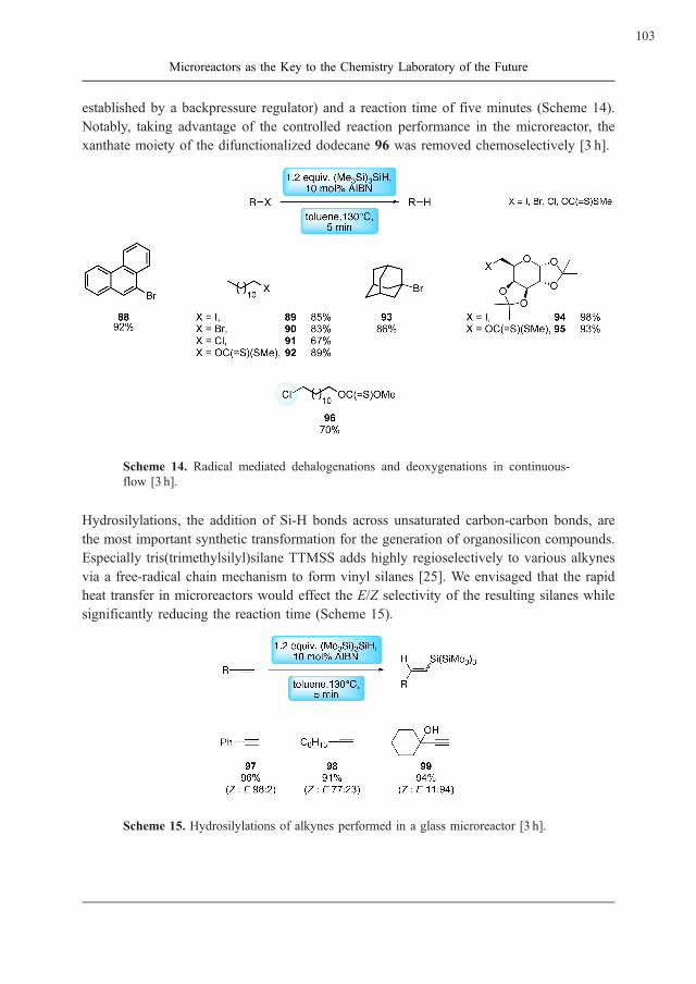
established by a backpressure regulator) and a reaction time of five minutes (Scheme 14).
Notably, taking advantage of the controlled reaction performance in the microreactor, the
xanthate moiety of the difunctionalized dodecane 96 was removed chemoselectively [3 h].
Scheme 14. Radical mediated dehalogenations and deoxygenations in continuous-
flow [3 h].
Hydrosilylations, the addition of Si-H bonds across unsaturated carbon-carbon bonds, are
the most important synthetic transformation for the generation of organosilicon compounds.
Especially tris(trimethylsilyl)silane TTMSS adds highly regioselectively to various alkynes
via a free-radical chain mechanism to form vinyl silanes [25]. We envisaged that the rapid
heat transfer in microreactors would effect the E/Z selectivity of the resulting silanes while
significantly reducing the reaction time (Scheme 15).
Scheme 15. Hydrosilylations of alkynes performed in a glass microreactor [3 h].
103
Microreactors as the Key to the Chemistry Laboratory of the Future

Again applying reaction conditions of 130 �C in toluene and a reaction time of five minutes
yielded the desired anti-Markovnikov products in excellent yields and good stereo-selectiv-
ities. The microreactor approach further furnished the hydrosilylation product of phenylace-
tylene 97 in greater yield (96% vs. 88%) and superior Z/E ratio (98:2 vs. 84:16) compared to
traditional batch strategies [3 h, 25].
Conclusion
Many chemical transformations can be performed in a faster, safer and cleaner manner when
performed in microfluidic devices. The down-scaling of reaction volumes in microreactors
offers the more precise control of the reaction conditions (temperature, time, mixing) and the
use of minimal amounts of precious compounds to rapidly screen a variety of conditions,
generating a wealth of information on reaction kinetics and pathways. Additionally, micro-
reactors present the opportunity to apply reaction conditions that are inaccessible using
conventional laboratory equipment, such as super heated solvents, and safely performing
reactions in explosive regimes. Even though numerous impressive examples on the applica-
tion of microreactor technology in synthetic chemistry have been reported, some major
drawbacks associated with this technique remain: the incompatibility of reactors with solid
reagents that cannot be used for wall-coatings or be supported on linkers, the sensitivity to
precipitations and the useful applicability mainly to fast reactions. Additionally, the efficient
on-line analysis of reaction mixtures in a high throughput format represents an outstanding
issue. The overall concept of highly efficient continuous-flow microreactor techniques re-
quires further improvements before it will be applied as a standard tool in chemical labora-
tories.
References
[1] For recent reviews and books on microchemical technology, see: (a) Ehrfeld, W.,
Hessel, V., Lowe, H. (2000) Microreactors: New Technology for Modern Chemistry,
Wiley-VCH, Weinheim. (b) Fletcher, P.D.I., Haswell, S.J., Pombo-Villar, E., War-
rington, B.H., Watts, P., Wong, S.Y.F., Zhang, X.L. (2002) Tetrahedron, 58:4735. (c)
Hessel, V., Hardt, S., Lowe, H. (2004) Chemical Micro Process Engineering, Wiley-
VCH, Weinheim. (d) Jahnisch, K., Hessel, V., Lowe, H., Baerns, M. (2004) Angew.
Chem. Int. Ed. 43:406. (e) Thayer, A.M. (2005) Chem. Eng. News, 83:43. (f) Watts,
P., Haswell, S.J. (2005) Chem. Soc. Rev. 34:235. (g) Geyer, K., Codee, J.D.C.
Seeberger, P.H. (2006) Chem. Eur. J. 12:8434. (h) Brivio, M., Verboom, W., Rein-
houdt, D.N. (2006) Lab Chip, 6:329. (i) Mason, B.P., Price, K.E., Steinbacher, J.L.,
Bogdan, A.R., McQuade, D.T. (2007) Chem. Rev., 107:2300. (j) Ahmed-Omer, B.,
Brandt, J.C., Wirth, T. (2007) Org. Biomol. Chem. 5:733. (k) Fukuyama, T., Rahman,
T., Sato, M., Ryu, I. (2008) Synlett. 2:151. (l) Wiles, C., Watts, P. (2008). Eur. J. Org.
Chem. 10:1655.
104
Geyer, K. and Seeberger, P.H.

[2] For recent publications of microreactors in organic synthesis see: (a) Usutani, H.,
Tomida, Y., Nagaki, A., Okamoto, H., Nokami, Y., Yoshida, J. (2007) J. Am. Chem.
Soc. 129:3046. (b) Hartung, A., Keane, M.A., Kraft, A. (2007) J. Org. Chem.
72:10235. (c) Bula, W.P., Verboom, W., Reinhoudt, D.N., Gardeniers, H.J. (2007)
Lab Chip 7:1717. (d) Sahoo, H.R., Kralj, J.G., Jensen, K.F. (2007) Angew. Chem. Int.
Ed. 46:5704. (e) Tanaka, K., Motomatsu, S., Koyama, K., Tanaka, S.I., Fukase, K.
(2007) Org. Lett. 9:299. (f) Trapp, O., Weber, S.K., Bauch, S., Hofstadt, W. (2007)
Angew. Chem. Int. Ed. 46:7307. (g) Fukuyama, T., Kobayashi, M., Rahman, T.,
Kamata, N., Ryu, I. (2008) Org. Lett. 10:433. (h) Baumna, M., Baxendale, I.R.,
Ley, S.V., Nikbin, N., Smith, C.D., Tierney, J.P. (2008) Org. Biomol. Chem.
6:1577. (i) Nagaki, A., Takabayashi, N., Tomida, Y., Yoshida, J. (2008) Org. Lett.
DOI:10.1021/ol8015572.
[3] For recent examples of microreactor-based transformations from this laboratory see
(a) Snyder, D.A., Noti, C., Seeberger, P.H., Schael, F., Bieber, T., Rimmel, G.,
Ehrfeld, W. (2005) Helv. Chim. Acta 88:1. (b) Ratner, D.M., Murphy, E.R., Jhunj-
hunwala, M., Snyder, D.A., Jensen, K.F., Seeberger, P.H. (2005) Chem. Comm.
5:578. (c) Flogel, O., Codee, J.D.C., Seebach, D., Seeberger, P.H. (2006) Angew.
Chem. Int. Ed. 45:7000. (d) Geyer, K., Seeberger, P.H. (2007) Helv. Chim. Acta
90:395. (e) Carrel, F.R., Geyer, K., Codee, J.D.C., Seeberger, P.H. (2007) Org. Lett.
9:2285. (f) Gustafsson, T., Ponten, F., Seeberger, P.H. (2008) Chem.Commun.
10:1100. (g) Gustafsson, T., Gilmour, R., Seeberger, P.H. (2008) Chem. Commun.
3022. (h) Odedram, A., Geyer, K., Gustafsson, T., Gilmour, R., Seeberger, P.H.
(2008) Chem. Commun. 3025.
[4] Reagent introduction and feed can occur via electroosmotic flow, hydrodynamic
pumping (HPLC pumps, syringe pumps) or capillary flow.
[5] A wide variety of other microreactors is available from other manufacturers and
suppliers, e. g.: Institute for Microtechnology Mainz (IMM), Fraunhofer Alliance
for Modular Microreaction Systems (FAMOS), the Little Things Factory (LTF) in
Ilmenau, the New Jersey Centre for MicroChemical Systems (NJCMCS), the Micro-
Chemical Process Technology Research Association (MCPT), or Sigma-Aldrich. The
selection presented here is by no means exhaustive and only serves to indicate the
diversity in systems developed to date.
[6] For further information visit the webpage: http://www.syrris.com/.
[7] For further information visit the webpage: http://www.ehrfeld.com.
[8] For further information visit the webpage: http://www.cpc-net.com/cytosls.shtml.
[9] For further information visit the webpage: http://www.micronit.com/
[10] For further information visit the webpage: http://www.vapourtec.co.uk/
105
Microreactors as the Key to the Chemistry Laboratory of the Future

[11] For recent reports on biological and biochemical applications see: (a) Hansen, C.L.,
Classen, S., Berger, J.M., Quake, S.R. (2006) J. Am. Chem. Soc. 3142. (b) Wang, B.,
Zhao, Q., Wang, F., Gao, C. (2006) Angew. Chem. Int. Ed. 45:1560. (c) Duan, J.,
Sun, L., Liang, Z., Zhang, J., Wang, H., Zhang, L., Zhang, W., Zhang, Y. (2006)
J. Chromatogr. A 1 – 2:165. (d) Utsumi, Y, Hitaka, Y., Matsui, K., Takeo, M.,
Negoro, S., Ukita, S. (2007) Microsystem Technologies 13:425. (e) Chen, L.,
West, J., Auroux, P.A., Manz, A., Day, P.J. (2007) Anal. Chem. 79:9185. (f) Liu, Y.,
Qu, H., Xue, Y., Wu, Z., Yang, P., Liu, B. (2007) Proteomics, 7:1373. (g) Le Nel, A.,
Minc, N., Smadja, C., Slovakova, M., Bilkovam, Z., Peyrin, J.M., Viovy, J.L.,
Taverna, M (2008) Lab Chip 8:294. (h) Yamamoto, T., Hino, M., Kakuhata, R.,
Nojima, T., Shinohara, Y., Baba, Y., Fujii, T. (2008) Anal. Sci. 24:234.
[12] For reports on larger scale productions using stainless steel microreactors see:
(a) Taghavi-Moghadam, S., Kleemann, A., Golbig, K.G. (2001) Org. Process Res.
Dev., 5:652. (b) Panke, G., Schwalbe, T., Stirner, W., Taghavi-Moghadam, S., Wille,
G. (2003) Synthesis 18:2827. (c) Zhang, X.N., Stefanick, S., Villani, F.J. (2004) Org.
Process Res. Dev. 8:455. (d) Liu, S.F., Fukuyama, T., Sato, M., Ryu, I. (2004) Org.
Process Res. Dev. 8:477. (e) Acke, D.R.J, Stevens, C.V. (2006) Org. Process Res.
Dev. 10:417. (f) Wheeler, R.C., Benali, O., Deal, M., Farrant, E., MacDonald, S.J.F.,
Warrington, B.H. (2007) Org. Process Res. Dev. 11:704.
[13] For reports on the application of glass microreactors see: (a) Nikbin, N., Watts, P.
(2004) Org. Process Res. Dev 8:942. (b) Wiles, C., Watts, P., Haswell, S.J. (2007)
Chem. Commun. 9:966. (c) Wiles, C., Watts, P., Haswell, S.J. (2007) Lab Chip 7:322.
(d) Hornung C.H., Mackley, M.R., Baxendale, I.R., Ley, S.V. (2007) Org. Process
Res. Dev. 11:399. (e) Smith, C.D., Baxendale, I.R., Tranmer, G.K., Baumann, M.,
Smith, S.C., Lewthwaite, R.A., Ley, S.V. (2007) Org. Biomol. Chem. 10:1562.
(f) Smith, C.D., Baxendale, I.R., Lanners, S., Hayward, J.J., Smith, S.C., Ley, S.V.
(2007) Org. Biomol. Chem. 10:1559.
[14] For recent reviews and reports on photochemical reactions using microflow see
(a) Fukuyama, T., Hino, Y., Kamata, N., Ryu, I. (2004) Chem. Lett. 33:1430.
(b) Maeda, H., Mukae, H., Mizuno, K. (2005) Chem. Lett. 34:66. (c) Maeda, H.,
Matsukawa, N., Shirai, K., Mizuno, K. (2005) Tetrahedron Lett. 46:3057. (d) Hook,
B.D.A., Dohle, W., Hirst, P.R., Pickworth, M., Berry, M.B., Booker-Milburn, L.I.
(2005) J. Org. Chem. 70:7558. (e) Matsushita, Y., Kumada, S., Wakabayashi, K.,
Sakeda, K., Ichimura, T. (2006) Chem. Lett. 35:410. (f) Lainchbury, M.D., Medley,
M.I., Taylor, P.M., Hirst, P., Dohle, W., Booker-Milburn, L.I. (2008) J. Org. Chem.
DOI: 10.1021/jo801108 h.
[15] For selected reviews and reports on silicon based microreactors see (a) Jensen, K.F.
(2001) Chem. Eng. Sci. 56:293. (b) Yen, B.K.H., Gunther, A., Schmidt, M.A.,
Jensen, K.F., Bawendi, M.G. (2005) Angew. Chem. Int. Ed. 44:5447. (c) Murphy, E.R.,
106
Geyer, K. and Seeberger, P.H.
Martinelli, J.R., Zaborenko, N., Buchwald, S.L., Jensen, K.F. (2007) Angew. Chem.
Int. Ed. 46:1734. (d) Kralj, J.G., Sahoo, H.R., Jensen, K.F. (2007) Lab Chip 7:256.
(e) Inoue, T., Schmidt, M.A., Jensen, K.F. (2007) Ind. Eng. Chem. Res. 46:1153.
[16] (a) Ravida, A., Liu, X.Y., Kovacs, L., Seeberger, P.H. (2006) Org. Lett. 8:1815.
(b) Liu, X.Y., Stocker, B.L., Seeberger, P.H. (2006) J. Am. Chem. Soc. 128:3638.
[17] (a) Curran, D.P. (2001) Synlett. 9:1488. (b) Curran, D.P., Luo, Z.Y. (1999) J. Am.
Chem. Soc. 121:9069. (c) Zhang, W. (2004) Chem. Rev. 104:2531.
[18] (a) Murray, J.K., Gellman, S.H. (2005) Org. Lett. 7:1517. (b) Lelais, G., Seebach, D.,
Jaun, B., Mathad, R.I., Flogel, O., Rossi, F., Campo, M., Wortmann, A. (2006) Helv.
Chim. Acta 89:361.
[19] Erdelyi, M., Gogoll, A. (2002) Synthesis 1592 – 1596.
[20] Monnier, O., Coquerel, G., Fours, B., Duplaa, H., Ochsenbein, P., PTC Patent, PCT/
FR2007/000201, 05.02.2007.
[21] For selected examples on nucleophilic substitutions using HF or fluorine salts, see
(a) Kim, D.W., Song, C.E., Chi, D.Y. (2003) J. Org. Chem. 68:4281. (b) Inagaki, T.,
Fukuhara, T., Hara, S. (2003) Synthesis 1157.
[22] For electrophilic fluorinations see for example Xiao, J., Shreeve, J.M. (2005)
J. Fluorine Chem. 126:473.
[23] For selected examples on fluorinations using DAST or deoxofluor see (a) Lal, G.S.,
Pez, G.P., Pesaresi, R.J., Prozonic, F.M., Cheng, H. (1999) J. Org. Chem. 64:7048.
(b) White, J.M., Tunoori, A.R., Turunen, B.J., Georg, G.I. (2004) J. Org. Chem.
69:2573.
[24] These finding are consistent with the literature, see also Negi, D.S., Koppling, L.,
Lovis, K., Abdallah, R., Geisler, J., Budde, U. (2008) Org. Process Res. Dev. 12:345.
[25] Kopping, B., Chatgilialoglu, C., Zehnder, M., Giese, B. (1992) J. Org. Chem.
57:3994.
107
Microreactors as the Key to the Chemistry Laboratory of the Future


����������������
Chemical Biology with Organometallics
Eric Meggers
Fachbereich Chemie, Philipps-Universitat Marburg,Hans-Meerwein-Straße, 35043 Marburg, Germany
E-Mail: [email protected]
Received: 7th August 2008 / Published: 16th March 2009
Abstract
Inorganic and organometallic moieties are explored as structural scaf-
folds for the design of biologically active compounds. In this strategy,
a metal center plays a structural role by organizing the organic ligands
in the three-dimensional receptor space. It is hypothesized that this
approach allows to access unexplored chemical space, thus giving
new opportunities for the design of small molecules with unprece-
dented biological properties. Along these lines, our group pioneered
the design of organometallic scaffolds for the inhibition of protein
kinases. These compounds are formally derived from the class of
ATP-competitive indolocarbazole alkaloids and allow to access novel
structures with very defined and rigid shapes in an economical fashion,
having resulted in the discovery of picomolar inhibitors for protein
kinases with impressive selectivity profiles. This article summarizes
the most important results over the last few years.
Introduction
The identification of compounds with novel and defined biological functions is of high
importance for the development of small-molecule pharmaceuticals and as tools (‘‘molecular
probes’’) for the investigation of biological processes. Nowadays, established technologies
such as combinatorial chemistry and high-throughput screening allow to synthesize and
identify initial lead structures in a rapid fashion and, in this respect, the chemical diversity
of scaffolds is a key factor for success. The stark majority of synthetic efforts is focused on
purely organic compounds. Surprisingly, a recent publication revealed that the diversity of
109
http://www.beilstein-institut.de/Bozen2008/Proceedings/Meggers/Meggers.pdf
Systems Chemistry, May 26th – 30th, 2008, Bozen, Italy

organic molecules is however quite limited: half of the 24 million organic compounds
registered in the Chemical Abstract Service (CAS) Registry can be classified with only
143 scaffolds [1]. Similarly, the diversity of topological shapes of known organic drugs is
also extremely low: a report on the analysis of the Comprehensive Medicinal Chemistry
(CMC) database demonstrated that out of more than 5000 compounds analyzed, half of the
known drugs fall into only 32 shape categories [2]. Most of the 32 frameworks contain at
least two six-membered rings linked or fused together. Thus, it can be concluded that current
efforts to obtain biologically active compounds draw from a limited set of structural scaf-
folds. Strategies to increase the molecular structural diversity are therefore demanded and
promise to provide opportunities for the discovery of compounds with novel and unprece-
dented properties.
Metal Complexes as Structural Scaffolds
A few years ago, our group hypothesized that structural diversity might be increased by
complementing organic elements with single metal atoms and to explore the opportunities of
metals to help building small compounds with defined three dimensional structures [3].
Transition metals appear especially appealing for this purpose since they can support a
multitude of coordination numbers and geometries which reach beyond the linear (sp-hy-
bridization), trigonal planar (sp2-hybridization), and tetrahedral (sp3-hybridization) binding
geometries of carbon (Fig. 1). For example, it is intriguing that an octahedral center with six
different substituents is capable of forming 30 stereoisomers compared to just two for an
asymmetric tetrahedral carbon. Thus, by increasing the number of substituents from four
(tetrahedral center) to six (octahedral center), the ability of the center to organize substituents
in the three dimensional space increases substantially. In addition, using a hexavalent center
may provide new synthetic opportunities for accessing globular shapes by building struc-
tures from a single center in six different directions.
Figure 1. Structural opportunities with an octahedral metal center.
Ruthenium as a Priviledged Metal
Current efforts in our laboratory are concentrating predominately on ruthenium. In our
opinion, ruthenium possesses a combination of properties that render it ideal for the design
of chemically inert bioactive organometallic species. Main reasons are a high kinetical
110
Meggers, E.

inertness of coordinative bonds to ruthenium if certain basic design principles are considered
(all here discussed compounds are air-stable, stable in water, and can withstand millimolar
concentrations of thiols and thus should stay intact within the biological environment), the
availability of inexpensive metal precursors (currently US$ 20/g for RuCl3), and a predict-
able and established synthetic chemistry. A significant concern in using metal-containing
compounds is the potential toxicity of the metal. In this respect, it is our opinion that a metal
complex that has a substitutionally inert coordination sphere should not display any ‘‘metal-
specific’’ toxicity itself and the biological properties of the metal complex should be instead
determined by the entire entity. However, toxic effects due to metabolic conversion of
coordinated ligands followed by the exposure of the metal to biological ligands cannot be
excluded in individual cases. It is therefore desirable that the metal itself or derived metal
salts have a low toxicity. In this respect, data from phase I clinical trials of two (substitu-
tionally labile) ruthenium anticancer drugs, KP1019 and NAMI-A, are encouraging since
these compounds have proven to be much less toxic compared to the anticancer drug
cisplatin [4, 5].
Staurosporine as a Lead Structure
We selected the natural product staurosporine as a structural inspiration (lead structure) for
the design of ATP-competitive organometallic kinase inhibitors 1 (Figs. 2 and 3) [6].
Staurosporine is a member of the class of indolocarbazole alkaloids, many of which are
potent ATP-competitive protein kinase inhibitors. This family of inhibitors shares the in-
dolo[2,3-a]carbazole moieties 2 (lactam form) or 3 (imide form, arcyriaflavin A) which bind
to the adenine binding site by establishing two hydrogen bonds to the backbone of the hinge
between the N-terminal and C-terminal kinase domain (Figs. 2b and 3). Staurosporine
adopts a defined globular structure with the carbohydrate moiety being oriented perpendi-
cular to the plane of the indolocarbazole heterocycle (Fig. 3). The indolocarbazole moiety
occupies the hydrophobic adenine binding cleft with the lactam group mimicking the hydro-
gen bonding pattern of the adenine base, and the carbohydrate moiety forming hydrophobic
contacts and hydrogen bonds within the globular ribose binding site (Fig. 2). Thus, staur-
osporine closely matches the shape of the ATP-binding site which makes it a highly potent
albeit unselective inhibitor for many protein kinases.
In order to match the overall shape of the ATP-binding site of protein kinases in a fashion
similar to staurosporine, but with less synthetic effort and more extended structural options,
we replaced the indolocarbazole alkaloid structure with simple metal complexes in which the
main features of the indolocarbazole heterocycle 3 are retained in a metal-chelating pyrido-
carbazole 4 (Fig. 3). This ligand 4 can serve as a bidentate ligand for ruthenium complexes
of type 1. Additional ligands in the coordination sphere of the metal can now substitute for
the carbohydrate moiety of staurosporine, with the metal center serving as a ‘‘glue’’ to unite
all of the parts. This approach has resulted in the successful design of nanomolar and even
picomolar protein kinase inhibitors.
111
Chemical Biology with Organometallics

Figure 2. Schematic representation of the ATP-binding pocket of cyclin dependent
protein kinase 2 (CDK2) in complex with a) ATP, b) staurosporine, and c) indolocar-
bazole-derived ruthenium complexes. Both ATP and staurosporine form hydrogen
bonds with the backbone amide groups of glutamate 81 and leucine 83. The green
area indicates a patch of high hydrophobicity. Adapted from ref. [7].
Figure 3. Designing ruthenium complexes of scaffold 1 as protein kinase inhibitors by
using the ATP-competitive natural product staurosporine as a lead for the overall
three-dimensional shape. The chelating ligand 4 contains the main pharmacophore
of the indolocarbazole heterocycles 2 and 3.
Proof-of-principle: Organoruthenium Inhibitors for the
Protein Kinases GSK-3 and Pim-1
We recently identified the cyclopentadienyl-CO sandwich scaffold 5 to be a nanomolar
inhibitor for the protein kinases GSK-3 and Pim-1 (Fig. 4). IC50 curves of racemic 5 and
the corresponding pyridocarbazole ligand 4 against the a-isoform of GSK-3 (GSK-3a) areshown in Figure 4c. Accordingly, the pyridocarbazole ligand 4 itself is 15000-times less
potent against GSK-3a compared to the entire complex 5. Consequently, the activity of 5
requires the complete ruthenium ligand sphere. Methylation of 5 at the imide nitrogen (Me5)
abolishes this activity completely. Figure 4c furthermore demonstrates that staurosporine is
by an order of magnitude less potent for this kinase than 5 [8].
Complex 5 displays metal-centered chirality and potency as well as selectivity for GSK-3 or
Pim-1 depend on the absolute configuration and additional substitutions at the periphery of
this half-sandwich scaffold. For example, whereas the racemic mixtures of 5 and 6 are dual
inhibitors for GSK-3 and Pim-1, (S)-6, having a additional OH-group at the indole, is very
112
Meggers, E.

selective for Pim-1 with an IC50 of 0.22 nM at 100 mM ATP compared to 9 nM for GSK-3a(a-isoform) [9]. On the other hand, (R)-7, having an OH and Br-group at the indole and a
methylester at the cyclopentadienyl moiety, displays an IC50 of 0.35 nM for GSK-3acompared to 35 nM for Pim-1. In fact, in a panel of 57 kinases, (R)-7 is highly selective
for just GSK-3 [10].
Figure 4. (a) The half-sandwich complexes 5-7 are potent inhibitors for the protein
kinases GSK-3 and/or Pim-1. The half-sandwich compounds 5-7 are pseudo-tetrahe-
dral and exist in two mirror-imaged configurations. The absolute configuration at the
ruthenium center has been assigned according to the priority order of the ligands being
h5-C5H5 > pyridine [N(C, C, C)] > indole [N(C, C, lone pair)] > CO. (b) Crystal
structure of N-benzylated 5 (ORTEP drawing with 35% probability thermal elipsoids).
(c) IC50 curves with GSK-3a measured with 10mM ATP. 5Me is the N-methylated
derivative of 5.
Cocrystal Structures of Protein Kinases with
Organometallics
In collaboration with Stefan Knapp (Oxford University) and later independently in our own
laboratory, we obtained cocrystal structures of ruthenium complexes bound to Pim-1 [9]. For
example, Figure 5 shows a cocrystal structure of Pim-1 with (S)-6. The structure demon-
strates that the ruthenium complex (S)-6 binds to the ATP binding site and that the ruthenium
centre does not undergo any direct interactions with the active site but has a purely structural
role. Furthermore, superimposing cocrystallized (S)-6 and Pim-1 (PDB code 2BZI) with
cocrystallized staurosporine and Pim-1 (PDB code 1YHS) reveals how closely the half-
sandwich complex mimics the binding mode of staurosporine (Figs. 5b and 5c). The pyr-
idocarbazole moiety of (S)-6 is nicely placed inside of hydrophobic pocket formed between
residues from the N-terminal and C-terminal domain, mimicking the binding position of the
indolocarbazole moiety of staurosporine, while the rest of the ruthenium complex occupies
the binding site of the carbohydrate moiety of staurosporine. Curiously, the oxygen of the
Ru-CO ligand is at a very close distance to Gly45 in the glycine-rich loop.
113
Chemical Biology with Organometallics

Currently, five structures of ruthenium half-sandwich complexes with protein kinases are
available in the protein databank, four with Pim-1 (2BZH, 2BZI, 2OI4, 2BZJ), and one with
Pim-2 (2IWI). In addition, a cocrystal structure of an osmium complex with Pim-1 was
published recently by us (3BWF) [11]. A structure of (R)-6 with GSK-3b has been solved in
our laboratory at 2.4 A and will be published soon (G. E. Atilla-Gockumen, E. Meggers,
unpublished results). The Marmorstein group (The Wistar Institute, Philadelphia, USA)
obtained several structures by soaking ruthenium complexes into crystal structures of the
lipid kinase PI3Kg [12].
Figure 5. Cocrystal structure of (S)-6 with Pim-1 at 1.9 A (PDB code 2BZI). (a) The
ruthenium complex occupies the ATP binding site of Pim-1. (b) and (c) Superimposed
relative binding positions of (S)-6 and staurosporine to Pim-1 (PDB code 1YHS). (d)
and (e) Some important interactions.
Cellular Data: Inhibition of GSK-3b by a Ruthenium
Complex within Mammalian Cells
Over the last two year we have repeatedly demonstrated that organometallic GSK-3 inhibi-
tors function inside of living mammalian cells [10, 13, 14]. GSK-3b is a negative regulator
of the wnt-signaling pathway by phosphorylating b-catenin. Phosphorylated b-catenin itself
is unstable due to rapid degradation by the proteasome. In the presence of a wnt signal,
GSK-3b is inactivated, resulting in an accumulation of b-catenin in the cytoplasm, followed
by a translocation into the nucleus where b-catenin serves as a transcriptional cofactor. Thus,
inhibition of GSK-3b by pharmacological inhibitors or by wnt signaling leads to increased
b-catenin levels and activation of wnt dependent transcription.
114
Meggers, E.

In order to probe the inhibition of GSK-3b within mammalian cells we routinely use Wes-
tern blot analysis (qualitative detection of b-catenin upregulation), cellular b-catenin staining
experiments (confirmation of b-catenin translocation into the nucleus), and a b-cateninreporter system for a quantitative analysis. For the latter we use human embryonic kidney
cells (HEK-293OT) that have stably incorporated a Tcf-luciferase transcription reporter (OT-
Luc cells). This transcription reporter generates luciferase in response to increased concen-
trations of b-catenin. An inhibition of GSK-3b can thus be determined by the luminescence
signal upon addition of luciferin to the cell lysate. For example, exposure of OT-Luc cells to
varying concentrations of the selective ruthenium GSK-3 inhibitor (R)-7 over a period of 24
hours yielded a strong upregulation of luciferase in the concentration window of 3mM down
to 10 nM (Figure 6a, compare also with (R,S)-5 in Figure 6b). Intriguingly, in contrast, the
popular organic GSK-3 inhibitors 6-bromo-indirubin-3’-oxime (BIO) (8) and kenpaullone
(9) (Figs. 6c, d) require micromolar concentrations for significant activities.
Figure 6. Cellular GSK-3b inhibition of (R)-7 and comparison with organic GSK-3
inhibitors. HEK-293OT cells transfected with a b-catenin-responsive luciferase repor-
ter were treated with different concentrations of inhibitors for 24 hours. After cell
lysis, the luminescence signal was measured after addition of luciferin.
It is noteworthy that organometallic compounds of this type can inhibit GSK-3b in frog
embryos (demonstrated for (R,S)-6) and zebrafish embryos (demonstrated for (R)-7) and
result in phenotypes that are related to the activation of the wnt pathway. For example, the
exposure of zebrafish embryos to LiCl, a known GSK-3 inhibitor, causes a perturbed
115
Chemical Biology with Organometallics

development of the head structure with a no-eye-phenotype, among others. Treatment of
zebrafish embryos with (R)-7 under analogous conditions, results in a similar phenotyp with
a decrease of the head structure without eyes and a stunted and crooked tail. In addition, the
yolk is enlarged and misshaped (Fig. 7c). However, zebrafish embryos which were instead
treated under identical conditions with the mirror-imaged compound (S)-7, develop normal
(Figs. 7a and 7b). These observations are consistent with an inhibition of GSK-3b by (R)-7,
but not (S)-7 and further demonstrate that the biological activity of (R)-7 is determined by
the overall three-dimensional structure of the ruthenium complex and not the ruthenium
itself.
Figure 7. Exposure of zebrafish embryos to (a) DMSO (2%), (b) 1mM (S)-7, and (c)
1mM (R)-7 (see Figure 4 for structures). The embryos were collected and maintained
in E3 media at 28.5 �C, compounds added at 4 hours post fertilization (hpf), and the
phenotypes were compared at 25 hpf.
In conclusion, organometallic (R)-7 is superior as a molecular probe for the function of
GSK-3b compared to established organic GSK-3b inhibitors with respect to binding affinity,
cellular potency, and kinase selectivity.
Combinatorial Chemistry with Ruthenium Complexes
We were interested in developing a method to rapidly assemble coordination spheres around
the ruthenium center in a combinatorial fashion and for this we deviced a synthesis of
complex 8 [15]. Although this compound bears four leaving groups, three acetonitriles
and one chloride, it is remarkably stable at room temperature. For example, 18 can be
purified on a regular silica gel column (CH2Cl2/MeOH) and is stable as a solid on the bench
for a few days without decomposition. This behaviour is a demonstration of the kinetic
inertness of the ruthenium pyridocarbazole scaffold. However, the acetonitrile ligands and
the chloride can be replaced upon heating of 8 in the presence of other ligands at elevated
temperatures of 70 – 110 �C (8?9) (Fig. 8a). Thus, 8 allows a rapid access to a diversity of
novel structures 9 by ligand replacement chemistry in the final synthetic step.
116
Meggers, E.

Figure 8b shows a selection of octahedral ruthenium complexes 10-13 that we synthesized
through this route. Compounds 10, 11, and 12 are interesting protein kinase inhibitors. For
example, the racemic compound 11, bearing a CO and 1,4,7-trithiacyclononane ligand, is a
subnanomolar inhibitor for the protein kinase Pim-1 with an IC50 of 0.45 nM at 100mMATP. Intriguingly, the nature of this monodentate ligand has a dramatic effect on kinase
inhibition. For example, substituting the CO for P(OMe)3 (13) renders the compound
completely inactive (Fig. 8b,c). Other ligands such as DMSO, cyanide, and ammonia also
result in a significant decrease in potency for Pim-1 (data not shown). Interestingly, sub-
stituting the CO for an azide (12) yields a fairly potent MSK-1 inhibitor (IC50 = 70 nM).
The bar diagram in Figure 8c further demonstrates that the CO complex 11 is most potent
against Pim-1 but also inhibits GSK-3a to a significant extent at 100 nM. Apparently, the
CO ligand, which is in the plane perpendicular to the pyridocarbazole chelate, is an im-
portant pharmacophore both for Pim-1 and GSK-3, but not for most other kinases. The rest
of the ligand sphere can then be used to render the complex either selective for Pim-1 or
GSK-3. This is well illustrated by the compounds 10 and 11. Whereas complex 11 signifi-
cantly prefers Pim-1, the complex 10 is instead selective for GSK-3a with an IC50 of 8 nM
at 100mM ATP.
Figure 8. (a) Synthesis of ruthenium pyridocarbazole complexes 9 by ligand substitu-
tion chemistry from the common precursor 8. (b) Selected complexes derived from 8.
IC50 values were measured at 100mM ATP. (c) Inhibitory activities of the organor-
uthenium compounds 10-13 at a concentration of 100 nM against the protein kinases
GSK-3a, Pim-1, MSK1, and CDK2/CyclinA. ATP concentration was 100mM. (d)
Crystal structure of the N-benzylated derivative of 10. ORTEP drawing with 40%
probability thermal elipsoids.
117
Chemical Biology with Organometallics

Conclusion
With GSK-3 and Pim-1 as model kinases we have established that organometallic scaffolds
are highly promising scaffolds for the design of kinase inhibitors. Accordingly, we have
identified low nanomolar and even picomolar inhibitors for these kinases, demonstrated their
exceptional kinase selectivities, have verified their usefulness in mammalian cell culture and
model organisms such as zebrafish and frog embryos, and we confirmed their binding modes
with cocrystal structures of ruthenium inhibitors bound to the ATP-binding site of Pim-1.
Furthermore, we have developed tools that allow to rapidly synthesize a diverse selection of
organometallic compounds and to screen them for their kinase activities. The discovered
organometallic GSK-3 and Pim-1 inhibitors are some of the most potent and selective
inhibitors for kinases know to date and therefore these results validate the strategy of
exploring organometallic scaffolds for the design of enzyme inhibitors. Future work has to
focus on octahedral metal complexes and expand the investigation to other biological
targets.
References
[1] Alan, H., Lipkus, A.H., Yuan, Q., Lucas, K.A., Funk, S.A., Bartelt III, W.F.,
Schenck, R.J., Trippe, A.J. (2008) Structural Diversity of Organic Chemistry.
A Scaffold Analysis of the CAS Registry. J. Org. Chem. 73:4443 – 4451.
[2] Bemis, G.W., Murcko, M.A. (1996) The properties of known drugs. 1. Molecular
frameworks. J. Med. Chem. 39:2887 – 2893.
[3] Meggers, E. (2007) Exploring biologically relevant chemical space with metal com-
plexes. Curr. Opin. Chem. Biol. 11:287 – 292
[4] Dyson, P.J., Sava, G. (2006) Metal-based antitumour drugs in the post genomic era.
Dalton Trans. 16:1929 – 1933.
[5] Hartinger, C.G., Zorbas-Seifried, S., Jakupec, M.A., Kynast, B., Zorbas, H., Keppler,
B.K. (2006) From bench to bedside – preclinical and early clinical development of
the anticancer agent indazolium trans-[tetrachlorobis(1 H-indazole)ruthenate(III)]
(KP1019 or FFC14A). J. Inorg. Biochem. 100:891 – 904.
[6] Meggers, E., Atilla-Gokcumen, G.E., Bregman, H., Maksimoska, J., Mulcahy, S.P.,
Pagano, N., Williams, D.S. (2007) Exploring Chemical Space with Organometallics:
Ruthenium Complexes as Protein Kinase Inhibitors. Synlett. 8:1177 – 1189.
[7] Huwe, A., Mazitschek, R., Giannis, A. (2003) Small molecules as inhibitors of
cyclin-dependent kinases. Angew. Chem. Int. Ed. 42:2122 – 2138.
118
Meggers, E.

[8] Bregman, H., Williams, D.S., Atilla, G.E., Carroll, P.J., Meggers, E. (2004) An
Organometallic Inhibitor for Glycogen Synthase Kinase 3. J. Am. Chem. Soc.
126:13594 – 13595.
[9] Debreczeni, J.E., Bullock, A.N., Atilla, G.E., Williams, D.S., Bregman, H., Knapp, S.,
Meggers, E. (2006) Ruthenium Half-Sandwich Complexes Bound to Protein Kinase
Pim-1. Angewandte Chem. Int. Ed. 45:1580 – 1585.
[10] Atilla-Gokcumen, G.E., Williams, D.S., Bregman, H., Pagano, N., Meggers, E.
(2006) Organometallic Compounds with Biological Activity: A Very Selective and
Highly Potent Cellular Inhibitor for Glycogen Synthase Kinase 3. ChemBioChem
7:1443 – 1450.
[11] Maksimoska, J., Williams, D.S., Atilla-Gokcumen, G.E., Smalley, K.S.M.,
Carroll, P.J., Webster, R.D., Filippakopoulos, P., Knapp, S., Herlyn, M., Meggers,
E. (2008) Isostructural Ruthenium and Osmium Complexes Display Highly Similar
Bioactivities. Chem. Eur. J. 14:4816 – 4822.
[12] Xie, P., Williams, D.S., Atilla-Gokcumen, G.E., Milk, L., Xiao, M., Smalley, K.S.M.,
Herlyn, M. Meggers, E. Marmorstein, R. (2008) Structure-Based Design of an Orga-
noruthenium Phosphatidyl-inositol-3-kinase Inhibitor Reveals a Switch Governing
Lipid Kinase Potency and Selectivity. ACS Chemical Biology 3:305 – 316.
[13] Williams, D.S., Atilla, G.E. Bregman, H., Arzoumanian, A., Klein, P.S., Meggers, E.
(2005) Switching on a signaling pathway with an organoruthenium complex. Angew.
Chem. Int. Ed. 44:1984 – 1987.
[14] Smalley, K.S.M., Contractor, R., Haass, N.K., Kulp, A.N., Atilla-Gokcumen, G.E.,
Williams, D.S., Bregman, H., Flaherty, K.T., Soengas, M.S., Meggers, E., Herlyn, M.
(2007) An Organometallic Protein Kinase Inhibitor Pharmacologically Activates p53
and Induces Apoptosis in Human Melanoma Cells. Cancer Res. 67:209 – 217.
[15] Bregman, H., Carroll, P.J., Meggers, E. (2006) Rapid Access to Unexplored Chemi-
cal Space by Ligand Scanning around a Ruthenium Center: Discovery of Potent and
Selective Protein Kinase Inhibitors. J. Am. Chem. Soc. 128:877 – 884.
119
Chemical Biology with Organometallics


����������������
Modelling for Regenerative Medicine:
Systems Biology Meets Systems
Chemistry
David A. Winkler*, Julianne D. Halley,
Frank R. Burden
CSIRO Molecular and Health Technologies,Private Bag 10, Clayton 3168, Australia
E-Mail: *[email protected]
Received: 5th September 2008 / Published: 16th March 2009
Introduction
Complex systems science is making substantial contributions to the study of biological
systems, and has made a substantial contribution to the new field of systems biology.
Systems biology focuses on the systematic study of complex interactions in biological
systems using an integrative rather than reductionist perspective. One of the goals of systems
biology is to study, model, and understand new emergent properties of biological systems
from a complex systems perspective [1, 2]. This integrative approach to biology is generat-
ing substantial benefits in facilitating study of larger more complicated systems, providing
improved understanding of nonlinear system properties, and provides an ability to model
systems at appropriate levels of detail where the model is matched to data density and
research questions. Various aspects of systems biology have been reviewed recently [3 –
11]. Chemistry has lagged behind most other disciplines in adopting complex systems
approaches, possibly because it has largely been a reductionist science, and reductionist
approaches have been very successful. Adopting a complementary complex systems ap-
proach to chemistry will build on this success to study more complex matter.
Paradigm Shifts
Chemistry focuses on the synthesis and properties of relatively small sets of molecular
species but is now increasingly embracing the generation and study of larger, more diverse
systems of molecules [12]. Chemistry is becoming increasingly multidisciplinary, embracing
important new research fields where chemistry, physics, biology and materials science
121
http://www.beilstein-institut.de/Bozen2008/Proceedings/Winkler/Winkler.pdf
Systems Chemistry, May 26th – 30th, 2008, Bozen, Italy
overlap. Emerging research fields such as nanomaterials and self-assembly will benefit from
an integrative, complex systems approach that is complementary to the reductionist methods
that characterize the discipline of chemistry [13]. Reductionist and deterministic concepts
like proteins and ligands as rigid ‘lock and key’ systems, cells as molecular machines, and
proteins as relatively rigid structures are increasingly at odds with new spectroscopic data,
computational experiments, and high throughput experimental results. An adaptive, dynamic
description of small molecules (chemistry), and larger molecules (biology but now increas-
ingly chemistry) is required. As Kurakin elegantly states [14], there is a global crisis of the
mechanistic, deterministic paradigm in life sciences that a complex systems description of
molecular processes can resolve. In his classic work on the nature of scientific revolutions,
Kuhn states that the success of a dominant paradigm brings about its own crisis and
necessitates a paradigm shift, as the advances in technology and methods lead to a wide-
spread accumulation of experimental data that cannot be readily explained and accommo-
dated within the existing conceptual framework [15]. Kurakin’s papers give some diverse
and important examples of these [14, 16 – 20].
This paper is not intended to be an exhaustive review of complex systems and their
application to chemistry, rather a summary of the main concepts, important areas of applica-
tion, and examples in an important area of overlap between chemistry and biology. The first
section reviews the major elements of complex systems science from a chemistry perspec-
tive, summarizes the relatively small research effort in chemical complex systems, and
indicates where a complex systems approach is likely to provide substantial benefit to
new and existing chemical and biological challenges. By analogy with systems biology,
we and others define the integrative study of very complicated chemical systems using the
tools and concepts of complex systems science as systems chemistry, a term first employed
by von Kiedrowski to define the chemical origins of biological organization [21]. Ludlow
and Otto published the first review of systems chemistry as a subject in 2008 [22], although
Whitesides noted the need to apply complexity concepts to chemistry in 1999 [12]. The
second section of the paper focuses on the importance of scale in efficient modelling of
complex systems, and discusses how Bayesian methods can be used to choose an appro-
priate level of scale for models. One of the most exciting new areas of chemical and
biological research is the understanding and molecular control of stem cell fate for use in
regenerative medicine. Complex systems tools and concepts are providing a novel, comple-
mentary approach to understanding and controlling stem cell fate. The third part of the paper
illustrates how this complexity can be applied to modelling processes driving stem cell fate
decisions.
Complex Systems
Complex systems science is a rapidly growing new paradigm for understanding and model-
ling extremely complicated systems (physical, social, economic, biological etc), and for
discovering common mechanisms in apparently diverse phenomena. They generally exhibit
122
Winkler, D.A. et al.

nonlinear dynamic behaviour caused by many interactions among the system components,
and have so-called emergent properties that are difficult or impractical to understand or
predict from knowledge of the components from which the system is constructed. Some
complex systems can evolve in ways that are very sensitive to initial conditions. This
behaviour can be described by nonlinear differential equations. Examples of complex sys-
tems are found in virtually all disciplines of science – from molecular self-organization and
self-assembly to weather patterns, social systems, ecosystems, electricity grids, economies,
and more esoteric properties like self-awareness, emotion, and life itself. Complex systems
have been reviewed extensively in the recent literature [23 – 39]. Although there is still some
debate about properties exhibited by complex systems, most exhibit a number of important
properties that distinguish them from simple systems.
Criticality and nonlinearity
Complex systems often undergo an abrupt change in properties when a threshold value of
some system parameter is crossed. This event is analogous to a phase transition that results
from component interactions within the system switching from local connectivity to global
connectivity. Many complex systems exhibit this type of behaviour, which is characterized
by order and stability below the critical point and instability and chaos above the critical
point. Phase transitions, crystallization, and alignment of magnetic domains in materials are
common examples of systems exhibiting critical behaviour. Cramer and Booksh published
one of the few reviews on chaos theory applied to chemistry, and list several interesting
classes of chemical systems in which chaotic behaviour occurs [40].
Autocatalytic systems, which abound in chemistry and biology, are examples of nonlinear
systems. The most familiar example of this type of behaviour is the complex spiral pattern
observed in the BZ (Belousov-Zhabotinsky) reaction (Fig. 1). Although often observed as
temporally varying series of patterns, reactions of this type also generate spatial patterns.
Reaction-diffusion processes have been proposed as a mechanism for pattern formation in
animals (e. g. tiger stripes), as a mechanism that generates body plans during embryogenesis.
Figure 1. Spiral waves in
BZ reaction
123
Modelling for Regenerative Medicine: Systems Biology Meets Systems Chemistry

Self-organization and self-assembly
Interactions between large numbers of components may generate self-organized and self-
assembled structures. Unfortunately, these terms are used in different ways in different
scientific disciplines, and are even used interchangeably. A simple way of distinguishing
them is to define self-assembly as a process that generates a structure that is in static
equilibrium and is thermodynamically more stable than its components, and the assembly
is driven by this energy difference [41]. Self-organizing systems increase their internal order
over time implying a decrease in system entropy, so that self-organizing systems cannot be
closed. The second law of thermodynamics requires energy transfers across the boundary
and there is a corresponding increase in the entropy of the environment in which the system
is embedded [33]. The self-organized structure is maintained in dynamic equilibrium and
decays if the energy source is removed. A recent paper discusses the relationship between
self-organization and self-assembly and gives examples of how these definitions apply
across multiple disciplines [41]. Examples of systems that are difficult to categorize and
that cross boundaries are also given. Understanding the mechanisms that underlie self-
assembly and self-organization will enhance our ability to design materials and new tech-
nology for important fields such as nanotechnology, tissue engineering, and biomaterials.
Complex systems concepts and modelling tools provide additional methods for understand-
ing the behaviour of these systems.
Emergence and scale
Reductionist methods typically attempt to explain how a system behaves by considering the
properties of the system components and their interactions. Complex systems approaches
highlight emergent properties of a system and allow very complicated systems to be viewed
at multiple levels of scale. Appropriate scales capture the emergent macroscopic properties
of interest, without requiring a full understanding of mechanisms at finer levels of detail. In
most cases, an ultimate fine-grained model of a complicated system is not possible because
of the difficulty of modelling at this level of scale and the large amount of information
required to build and validate such models. Generally, time, cost, or other resource limita-
tions prevent this information becoming fully available. Complex systems often exhibit
properties that are self-similar or scale-invariant (fractal) [42 – 45]. Power law behaviour
in which a log-log plot of frequency versus magnitude of the property is linear, is observed
in a diverse range of physical and biological complex systems [36, 46, 47]. The slopes of the
power law graphs (the exponent of the power law) for different systems tend to cluster,
generating Universality Classes that may have common underlying mechanisms [48 – 50].
Clearly, emergence, scale and complexity are interrelated and, as they are difficult concepts
to define precisely, there is considerable latitude in how the concepts are employed across
multiple disciplines. Recent work has attempted to generate more precise, widely applicable
definitions for terms such as emergence, scale, self-assembly and self-organization [13, 41,
51, 52].
124
Winkler, D.A. et al.

Emergent, holistic properties of complex systems are usually the ones that we observe and
attempt to engineer. As they are generated by the relatively simple nonlinear interactions of
many system components, these emergent properties are to some extent unexpected or
surprising. Examples of emergent properties include the self-organized convection cells in
heated liquids (Fig. 2), the oscillation of species in predator-prey systems, and weather
patterns. Emergence and self-organization are intimately related, and recent work has de-
scribed how emergence may be characterized in terms of self-organization [52].
Figure 2. Hexagonal, self-organized convection cells (Benard cells) in a heated fluid.
Emergent properties can vary in their ‘depth’ [41, 52]. Simple emergent properties include
gas pressure resulting from the interaction between atoms or molecules and their environ-
ment. More deeply emergent chemical properties include the formation of entirely new
molecular species with novel properties when molecules participate in chemical reactions.
Physical properties of molecules (e.g. aromaticity) can also be considered emergent, as it is
very difficult or impossible to predict them from first principles given the properties of their
components (atoms) and the interactions in which these components participate. However,
emergent properties can often be predicted at a different level of scale (a coarser level of
description). For example, the pressure of a gas can be calculated using a dynamical
simulation that accounts for the speed and direction of many atoms or molecules, but is
more readily calculated at a coarser level of scale using the gas law [53].
125
Modelling for Regenerative Medicine: Systems Biology Meets Systems Chemistry

Networks and interactions
Networks are very important paradigms for understanding interactions in complex systems
as they are a compact way of describing interactions between components. They have been
extensively studied in many fields other than chemistry. If a complex chemical system
consists of components that interact in a directed or non-directed way, this can be easily
presented as a network or graph. Although graph theory has been long been applied to
chemical systems such as molecules, relatively little work has been done on how network
representations of molecules and chemical reactions can yield new insight into the properties
of molecules. In systems biology, network paradigms are widely used to understand genetic
regulation, signalling, and protein-protein interactions. There has been recognition recently
that an understanding of network properties is necessary to properly exploit biological
vulnerabilities and target drugs more effectively. Network concepts are therefore becoming
increasingly important in medicinal chemistry and their applications to drug design have
been reviewed recently [54 – 58].
Systems Chemistry
Compared to systems biology, the application of complex systems science to chemistry is
almost non-existent. This is surprising given that Ilya Prigogine was awarded the Nobel
Prize in Chemistry in 1977 for work on nonlinear dynamical systems in chemistry that
helped to establish the foundations of complex systems science. Several other Nobel Prizes
have been awarded for research relating to self-assembling systems (Crick, Watson and
Wilkins, Medicine 1962; Krug, Chemistry 1982, Lehn, Chemistry, 1987) that build on
complex systems frameworks. Clearly, Watson, Crick and Wilkins’ work on understanding
the structure and self-assembly of DNA led to a paradigm shift in our understanding and
revolutionized the fields of biology and medicine. However, their work had a relatively
smaller impact on chemistry, a discipline that is well equipped to describe the interactions
that control DNA’s structure. A few other chemists, notably Testa, Kier and Belousov and
Zaboutinsky, explored complex behaviour in chemistry but the area did not develop in a
sustainable way and declined in prominence. In a sense, the field was ahead of its time, and
did not offer solutions to problems that reductionism could not tackle adequately. Within the
past decade, interest in applying complexity to chemistry has increased, and several workers,
notably Whitesides, have written seminal papers. Whitesides proposed that almost every-
thing of interest in chemistry is complex, by the above definitions of complexity [12]. For
example a single step in a multistep synthesis involves ~1022 molecules of several types, a
myriad of interacting nuclei and electrons and ~1024 molecules of solvent. As such real
systems cannot be dealt with at the molecule level, synthetic chemistry has employed
approximations, rules, and heuristics. Complex systems paradigms can provide additional,
complementary methods for understanding and dealing with such immense complexity.
126
Winkler, D.A. et al.

Contemporary Chemical Research Where a Complex
Systems Approach May Be Relevant
Most of the interesting new areas of chemistry are discipline spanning, and will benefit from
a complementary complex systems approach.
Nonlinear dynamical systems and pattern formation
Nonlinear dynamical systems can exhibit chaotic behaviour, often modelled using differen-
tial equations akin to those describing chemical kinetics. Spatial and cellular automata
methods are being applied increasingly to dynamical systems. As discussed, some chemical
reactions can behave in this manner. Although the most recognised example is the BZ
reaction [59] (see Figure 2), many other dynamic, pattern-forming reactions such as the
Briggs-Raucher reaction are known. Recently, reaction-diffusion or oscillating reactions
have been used to generate novel materials such as a self-walking gel [60, 61].
Fractal materials
Systems that exhibit fractal or self-similar behaviour are found widely in nature – snow-
flakes, lightning, crystals, river networks, ferns, blood vessels and coastlines. Real world
objects show fractal behaviour over an extended, but finite, scale range [108]. Fractal
behaviour is common in nano-structured materials, where self-similarity provides unique
properties such as giant enhancement of nonlinear optical properties, and related effects that
underlie surface-enhanced Raman scattering [62]. Fractal effects are also important in elec-
trochemistry [63].
Supramolecular polymers
Conventional polymers are linked by permanent covalent bonds. Supramolecular polymers
are a new class of polymers in which the linkages are reversible. The dynamic properties of
these materials open up the prospect of many new applications. The most important step in
developing practical supramolecular polymers was the discovery of the 2-ureido-4[1 H]-
pyrimidinone unit that could form four hydrogen bonds. This monomer has self-comple-
mentary allowing it to self-assemble into polymers with novel properties, such as the ability
to self-heal when cut [64].
Self-replicating systems
Self-replicating systems clearly exist in biology and there has been much speculation on the
types of autocatalytic systems that may have arisen on the primordial earth that may have led
to self-replicating molecules and life. Kauffman argued that a sufficiently large pool of
chemicals may be intrinsically capable of generating autocatalytic cycles and self-replicating
127
Modelling for Regenerative Medicine: Systems Biology Meets Systems Chemistry

molecules [65]. Seminal work by Wintner et al. showed how simple organic structures can
catalyse their own formation. Self-complementarity is the key to this autocatalytic behaviour
[66]. Design of self-replicating molecules is very difficult and progress in the field has been
slow. The recent review by Paul and Joyce summarizes progress in the area [67].
Artificial life
Artificial life and the closely related field of synthetic biology covers a range of research
areas, many of them intimately involved with chemistry. It spans synthesis of unnatural
organic molecules that function in living systems, through biomimetic chemistry that pro-
duces synthetic molecules that recapitulate the behaviour of natural analogues, to the greatest
challenge of recreating synthetic chemical systems that exhibit inheritance, genetics and
evolution. Recent reviews and papers by Benner, Rosen, and Cornish-Bowden provide a
summary of progress in artificial life [68 – 70].
Dynamic combinatorial libraries
Dynamic combinatorial chemistry is defined as combinatorial chemistry under thermody-
namic control, in which all constituents are in equilibrium. The library members are there-
fore constantly interconverting via reversible chemical reactions. The composition of the
library can therefore be altered by changing factors in the environment [71]. For example,
dynamic libraries have been exploited in drug discovery. When members of the library
interact favourably with a biological target, the composition of the library shifts and results
in enrichment of compounds that bind to the target protein. The environment can therefore
‘select’ or enrich the library with compounds that are more ‘fit’. Given the enormous size of
drug-like chemistry space, dynamic combinatorial libraries offer a more efficient method of
exploring this space to identify novel drug leads than covalently bonded combinatorial
libraries. The technique is currently limited by the small number of reversible reactions that
are biocompatible.
Evolutionary methods
Genetic algorithms and genetic programming are also evolutionary, adaptive tools that are
finding increased applications in chemistry and biology. Genetic algorithms are very effi-
cient methods for exploring vast search spaces such as those involved in protein folding, or
drug-like chemical space. When applied to chemistry, they involve a string representation of
a molecule or molecular property (e. g. a SMILES string, torsion angle list etc), a mutation
operator (e. g. swap one atom type or functional group for another, add or delete an atom or
functional group, change a torsion angle etc), and a fitness function (e. g. biological activity,
desired protein fold etc). The algorithm generates families of mutants that are assessed by
the fitness function. The fittest are retained and less fit discarded. The cycle repeats starting
with the fittest individuals from the previous cycle, and stops when some criterion is
128
Winkler, D.A. et al.

matched. They have been used to find optimum sets of molecular descriptors for modelling
of drug activity, for searching chemical databases for leads [73 – 75], and for optimising
combinatorial libraries [76], discovery and optimisation of new catalytic materials [77 – 79],
design of peptide mimetopes [78, 79], multiobjective optimisation [80, 81], synthesis plan-
ning [82 – 85], and in many other chemical applications.
Drug targeting
Interactions between molecules (e. g., drugs and proteins, proteins and DNA, etc.) in living
systems can be described by networks. Very recently, network approaches have been applied
to understand why the efficacy of new drug discovery is declining [54, 55, 86]. It has long
been recognized that almost all drugs hit multiple targets, which is problematic as it leads to
off-target effects and toxicity. Drugs are often designed to hit a specific target that is
presumed to induce the desired phenotypic change, but it has only recently been appreciated
that the robustness of biological networks resists such perturbations and may allow systems
to adapt to minimize the impact. If we understand the robustness of biological networks, we
could better engineer drugs to hit multiple intentional targets to affect the best outcome.
Supramolecular, self-organizing and self-assembling systems
Self-assembling, supramolecular, and self-organizing materials are currently of considerable
interest to chemists [87 – 90]. New research fields of nanoscience and smart materials aim to
design materials and molecular devices that can be assembled from the ‘bottom up’ rather
than ‘top down’. Chemical or biological materials have been shown to undergo self-assem-
bly, although a complete understanding of assembly mechanisms is still lacking, making
design challenging. Self-assembly has been applied to electronic device manufacture [91],
viral capsid modelling [92], nanoparticle assembly [93 – 95], peptides self-assembly, nano-
tubes, glycolipids, carbohydrates, polymers, crystals [94, 96 – 101], and many other materi-
als[30]. Aberrant self-assembly provides the mechanism for many protein folding diseases
such as prion and Alzheimer’s diseases, amyloidoses, cataracts, and diabetes [102, 103].
New complexity-based tools, such as agent-based models, have an increasing important role
to play in design of self-assembling materials [13, 104].
Self-organization also applies to chemistry but is less prominent in the chemical literature
largely due to the tendency of many authors to describe self-assembly as self-organiza-
tion[39]. Most examples of true self-organization involve biological systems because they
are invariably open [33, 105, 106]. A chemical example (although biology-based) is the
dynamic instability of microtubules [107]. Formation of microtubules involves a sponta-
neous self-assembly of tubulin, but the dynamics of the process also involves disassembly
using energy librated when tubulin hydrolyses GTP (tubulin is an enzyme as well as
structural protein) [108]. An example of a self-organized tubulin structure is given in
Figure 3.
129
Modelling for Regenerative Medicine: Systems Biology Meets Systems Chemistry

Cram and Lehn conducted seminal work on self-assembly, establishing the field of supra-
molecular chemistry involving the generation of complex structures from reversible non-
covalent bonds These structures exploit hydrogen bonding, metal coordination, hydrophobic
forces, van der Waals forces, p-p interactions and electrostatic effects [109].
Figure 3. A self-organized self-assembly of microtubules.
Mescoscale Modelling – Choosing the Correct Scale
Some complex systems appear to be ‘computationally irreducible’, raising the question of
how we model emergent properties of such systems. Recent work on diverse complex
systems suggests sparse, mesoscale, or ‘coarse grained’ models, that omit most of the detail
and concentrate only on the essential pattern forming process, can provide a path forward.
Choosing too low a level of detail (too coarse) results in models that have a limited ability to
generalize, and can predict only broad features of the system. Choosing a too detailed level
of scale produces more complicated models but requires much more data. As many model-
ling exercises are data limited, overly complex models pose problems for parameterisation,
validation, and they are prone to overfitting.
130
Winkler, D.A. et al.

Biologically, coarse graining of sensory information is essential so that organisms can
function without being overloaded. For example, in the visual system the eye does substan-
tial feature selection and classification before information is sent to the brain. If mesoscale
descriptions can adequately model the essential (emergent) properties of complex systems,
how do we find the correct level of scale that matches the usefulness of the predictions to the
availability of data? We adopt two approaches, one specific for biology and one more
generally applicable.
Sparse feature selection and modelling
Generically, mesoscale models can be generated using sparse feature selection methods that
select in a context-dependent manner, the most relevant aspects of the system under study.
Prominent of these are Bayesian methods like expectation maximization algorithms, de-
scribed elegantly by Figeuiredo [110].
Neural networks are another adaptive, learning method that is finding increased application
in chemistry and biology [58, 111 – 116]. Neural networks are algorithms that learn patterns
or associations between mathematical representations of objects and emergent properties of
systems that are comprised of, or interact with these objects. An important area where neural
networks have contributed to complexity-based modelling of chemical systems, is in QSAR
and chemometrics. Neural networks have been used widely in chemistry and pharmacology
for finding relationships between molecules and their biological or physicochemical proper-
ties. They have an inherent ability to model a complex system at a sufficiently coarse
grained scale that matches the model complexity to the availability of data. This is particu-
larly important in medicinal chemistry, which is often data-poor [12]. Neural networks are
platform technologies that have been used to model a myriad of emergent properties of
chemical and biological complex systems, such as drug target activity [117 – 121], drug-like
(ADME) properties [122 – 126], immune modulation [127, 128], physicochemical properties
[129 – 136], toxicity [112, 137 – 142].
Mesoscale models of regulatory interactions
Although there is vigorous debate [143] about the architecture of biological networks, many
are scale-free due to the intrinsic robustness of such architectures. Mesoscale models of cell
regulatory networks can be derived by focusing on key network hubs and genes controlled
by these hubs that are relevant to the biological process under study. Mesoscale regulator
models can represent many types of interacting components in biology. In gene regulation,
the hubs represent the set of genes most relevant to a particular biological process such as
differentiation, apoptosis or self-renewal. Mesoscale regulatory networks can be deduced by
a ‘bottom up’ reductionist analysis of gene microarray data, or data from other experiments
such as chromatin immunoprecipitation (ChIP). An alternative ‘top down’ approach is to
generate ‘blank’ mesoscale networks that are subsequently trained to recapitulate
131
Modelling for Regenerative Medicine: Systems Biology Meets Systems Chemistry

experimental data or predict the outcomes of new experiments. The latter approach was first
described by Geard and Wiles, and is a substantially novel analogue applied to modelling of
quantitative gene regulation in C. elegans embryogenesis [144, 145]. The approach is
illustrated in Figure 4.
This coarse-grained approach to cell differentiation allows us to rise above the details of
biochemical experiments to produce a map or ‘fate matrix’ of cell behaviour that reflects the
integration of multiple layers of complexity in cellular processes. Given a set of target genes
that characterize each cell embryogenesis pathway, a mesoscale regulatory network model
can be trained using gene microarray data, so that its output mimics changes in gene activity
as cells divide and/or differentiation. Once trained, the network architecture and weight
matrix represent a coarse-grained model of the regulatory network controlling cell fate. Each
gene in the model interacts with the weight matrix, reflecting the fact that the transcription of
a gene is the result of integrating the cell’s biochemical state rather than the action of any
single gene product. One set of master genes (i. e. one expression state) regulates the
transcription of the cell’s genes, leading to a new state. We aim to annotate these mesoscale
models with experiments identifying regulatory interactions, to produce a model of sufficient
detail to understand and control cell fate [146].
Figure 4. Recursive neural network structure, showing how the regulatory genes and
relative position index control the expression levels of genes in the various cells in the
lineage.
132
Winkler, D.A. et al.

Modelling Stem Cell Fate Decisions
Complex systems science coupled with interdisciplinary approaches provide scope to inte-
grate the many different layers of complexity that characterise complex biological systems.
Hence, important areas of research such as stem cell biology a growing literature views stem
cell function within the framework of a complex adaptive network, rather than the traditional
deterministic and reductionist approach that focuses on single stem cells [147]. We summar-
ize several areas where sparse methods have been applied to build mesoscale models of
properties of stem cell or related multipotent cells.
Stemness genes in human embryonic stem cells (hESCs)
Laslett et al. isolated sub-populations of embryonic stem cells that differed slightly in their
degree of ‘stemness’ (pluripotency) [148]. The experiments aimed to identify genes that
maintained pluripotency in hESC and those that drive early commitment to other lineages.
Flow cytometry was used to select a small set of pluripotent hESCs using fluorescent
antibody markers whose expression correlated with the pluripotency marker Oct4. This very
stemlike subpopulation was further sorted into hESC populations at four, graded, very early
stages of differentiation (P4-P7) (Fig. 5). Gene expression microarrays were generated from
each of the populations. Sparse Bayesian feature selection algorithms identified a small
number of markers that correlate with the pluripotency of the hESCs, and were classifier
genes with a putative key role in the maintenance of pluripotence. They could achieve this
classification with high statistical significance, suggesting the genes played a prominent role
in stemness.
Figure 5. Flow cytometric experiments identifying the four classes of hESCs.
133
Modelling for Regenerative Medicine: Systems Biology Meets Systems Chemistry

Erythroid differentiation
Welch and co-workers recently reported experiments on estradiol-induced differentiation
along the erythroid pathway over 30 hours [149]. This trajectory starts from the late burst-
forming unit-erythroid stage and progresses to the orthochromatic erythroblast stage. Estra-
diol was used to trigger the expression of the gene GATA-1, initiating synchronous differ-
entiation in the population. Microarrays were generated for populations of cells at each time
point during the experiment. Five classifier genes were found that correlate with the stage of
differentiation down the lineage. These genes were highly reliable markers for the differ-
entiation process, and are likely to play an important role in regulation of the erythroid
pathway.
Mesoscale network modelling of mouse embryogenesis
We modelled murine embryogenesis from the zygote to the blastocyst stage (Fig. 6) using a
‘top down’ mesoscale regulatory modelling framework.
Figure 6. The lineage diagram for mouse embryogenesis. Cell labels are shown.
Hamatami et al. measured the expression level of genes in carefully prepared samples of
cells from the unfertilized egg to the blastocyst using gene microarrays [150] (see Figure 6).
We modelled 135 genes from the mid-preimplantation gene activation (MGA) cluster for
each cell type in the embryogenesis pathway. We first trained the model using the gene
expression targets for each of the 135 genes in the cell types at the seven time points. We
then assessed the ability of the model to predict gene expression in cells in the pathway. The
mesoscale regulatory network model recapitulated quantitative gene expression profiles
within the cell lineage surprisingly high fidelity. We also generated models that predicted
expression in cells at an intermediate point in the lineage (interpolation) and at the end of the
lineage (extrapolation) when these were not used in training.
134
Winkler, D.A. et al.

Figure 7 summarizes these predictions. Figure 7A shows how well the model is able to
recapitulate the quantitative expression levels of all genes in all cells in the training set.
Figure 7B shows the most stringent test where the expression levels of the last three cell
divisions in the trajectory are predicted from a model trained on the first four cells in the
lineage. While the error is substantially higher than in the single cell expression prediction,
the predictions nevertheless have high statistical significance.
Figure 7. Predicted versus experimental log2 fold expression levels for genes in the
seven cell types. (A) Training set. (B) prediction of 8-cell, morula and blastocyst
expression for model trained on first 4 cells in embryogenic pathway.
Why sparse models work
Our sparse feature selection methods and mesoscale regulatory models support the common
observation that a relatively small number of factors control the fate of stem cells – as few as
three to six. Hart et al. also illustrated a similar sparse transcription factor requirement when
modelling the yeast cell cycle transcription network [151]. They initially used 204 transcrip-
tion factors to model the temporal variation of genes across the cell cycle. They pruned their
network models progressively, removing the least relevant transcription factors in turn, and
monitored the performance of the model. They found a gradual and limited degradation in
model performance until five transcription factors remained, after which the performance of
the model collapsed (Fig. 8).
135
Modelling for Regenerative Medicine: Systems Biology Meets Systems Chemistry

Figure 8. Performance of neural network model of temporal variation in gene expres-
sion during cell cycle. Note the dramatic drop in performance when the number of
transcription factors is 5 or less.
Conclusions and the Future
The evolution of chemistry away from manipulation of single molecules towards description
and manipulation of complex systems of molecules is one of the key driving forces for
embracing systems chemistry. In this way, the move to the study of more complex matter
may counter intuitively generate new types of problems that are sufficiently simple when
viewed in a complexity framework that they can be tackled in an analytical sense [12]. We
have arrived at Kuhn’s crisis of the dominant paradigm in chemistry, a period that is always
followed by an exciting period of extraordinary research and new discoveries [14]. However,
we can only achieve this by recognizing the opportunity and acting on it. Ludlow and Otto’s
call for chemists to embrace complexity and recognize systems chemistry is a very timely
challenge to expand this important new paradigm for understanding chemical problems [22].
Chemists may finally capitalize on the powerful but in some respects premature possibilities
that the Nobel Laureates presented to us, and move more rapidly into the exciting worlds of
new chemistries, materials, and biology.
136
Winkler, D.A. et al.

References
[1] Kitano, H. (2002) Computational systems biology. Nature 420:206 – 10.
[2] Kitano, H. (2002) Systems biology: a brief overview. Science 295:1662 – 4.
[3] Way, J.C., Silver, P.A. (2007) Systems engineering without an engineer: Why we
need systems biology. Complexity 13:22 – 29.
[4] Fisher, J., Henzinger, T.A. (2007) Executable cell biology. Nat. Biotechnol.
25:1239 – 49.
[5] Aigner, T., Haag, J., Zimmer, R. (2007) Functional genomics, evo-devo and systems
biology: a chance to overcome complexity? Curr. Opin. Rheumatol. 19:463 – 70.
[6] Marguet, P., Balagadde, F., Tan, C.M., You, L.C. (2007) Biology by design: reduc-
tion and synthesis of cellular components and behaviour. J. Roy. Soc. Interface.
4:607 – 23.
[7] Quackenbush, J. (2007) Extracting biology from high-dimensional biological data.
J. Exp. Biol. 210:1507 – 17.
[8] Materi, W., Wishart, D.S. (2007) Computational systems biology in drug discovery
and development: methods and applications. Drug Disc. Today. 12:295 – 303.
[9] van Regenmortel, M.H.V. (2007) The rational design of biological complexity:
A deceptive metaphor. Proteomics 7:965 – 75.
[10] Lin, J., Qian, J. (2007) Systems biology approach to integrative comparative geno-
mics. Expert. Rev. Proteomics 4:107 – 19.
[11] Palumbo, M.C., Farina, L., Colosimo, A., Tun, K., Dhar, P.K., Giuliani, A. (2006)
Networks everywhere? Some general implications of an emergent metaphor. Curr.
Bioinfo. 1:219 – 34.
[12] Whitesides, G.M., Ismagilov, R.F. (1999) Complexity in chemistry. Science
284:89 – 92.
[13] Lehn, J.-M. (2002) Toward self-organization and complex matter. Science
295:2400 – 3.
[14] Kurakin, A. (2007) Self-organization versus Watchmaker: ambiguity of molecular
recognition and design charts of cellular circuitry. J Mol Recog. 20:205 – 14.
[15] Kuhn, T. (1996) The Structure of Scientific Revolutions. Chicago: University of
Chicago Press.
[16] Kurakin, A. (2004) Self-organization versus watchmaker: stochasticity and determin-
ism in molecular and cell biology. Novato lectures.
137
Modelling for Regenerative Medicine: Systems Biology Meets Systems Chemistry

[17] Kurakin, A. (2005) Self-organization vs. Watchmaker: stochastic gene expression and
cell differentiation. Dev. Genes Evol. 215:46 – 52.
[18] Kurakin, A. (2005) Stochastic cell. IUBMB Life 57:59 – 63.
[19] Kurakin, A. (2005) Self-organization versus watchmaker: stochastic dynamics of
cellular organization. Biol. Chem. 386:247 – 54.
[20] Kurakin, A. (2006) Self-organization versus Watchmaker: Molecular motors and
protein translocation. Biosys. 84(1):15 – 23.
[21] Stankiewicz, J., Eckardt, L.H. (2006) Chembiogenesis 2005 and Systems Chemistry
Workshop. Angew. Chem. Int. Ed. 45:342 – 4.
[22] Ludlow, F.R., Otto, S. (2008) Systems Chemistry. Chem. Soc. Rev. 37:101 – 8.
[23] Batty, M. (2008) The size, scale, and shape of cities. Science 319:769 – 71.
[24] Luzzi, R, Vasconcellos, A.R., Ramos, J.G. (2007) Non-equilibrium statistical me-
chanics of complex systems: An overview. Rivista Del Nuovo Cimento 30:95 – 157.
[25] Mara, A., Holley, S.A. (2007) Oscillators and the emergence of tissue organization
during zebrafish somitogenesis. Trends Cell Biol. 17:593 – 9.
[26] Weber, B.H. (2007) Emergence of life. Zygon. 42:837 – 56.
[27] Foote, R. (2007) Mathematics and complex systems. Science 318:410 – 2.
[28] Eckmann, J.P., Feinerman, O., Gruendlinger, L., Moses, E., Soriano, J., Tiusty, T.
(2007) The physics of living neural networks. Physics Rep-Rev Section Phys Lett.
449:54 – 76.
[29] Rammel, C., Stagl, S., Wilfing, H. (2007) Managing complex adaptive systems –
A co-evolutionary perspective on natural resource management. Ecolog. Econom.
63(1):9 – 21.
[30] Balazs, A.C. (2007) Modelling self-assembly and phase behaviour in complex mix-
tures. Ann. Rev. Phys. Chem. 58:211 – 33.
[31] Almaas, E. (2007) Biological impacts and context of network theory. J. Exp. Biol.
210:1548 – 58.
[32] Mitchell, M. (2006) Complex systems: Network thinking. Artific. Intell.
170:1194 – 212.
[33] Newth, D., Finnigan, J. (2006) Emergence and self-organization in chemistry and
biology. Aust. J. Chem. 59:841 – 8.
138
Winkler, D.A. et al.

[34] Markose, S.M. (2005) Computability and evolutionary complexity: Markets as com-
plex adaptive systems (CAS). Econom. J. 115:F159-F92.
[35] Amaral, L.A.N., Ottino, J.M. (2004) Complex networks – Augmenting the frame-
work for the study of complex systems. Eur. Phys. J. B. 38:147 – 62.
[36] Gisiger, T. (2001) Scale invariance in biology: coincidence or footprint of a universal
mechanism? Biol. Rev. 76:161 – 209.
[37] Hess, B. (2000) Periodic patterns in biology. Naturwiss. 87:199 – 211.
[38] Wales, D.J., Scheraga, H.A. (1999) Review: Chemistry – Global optimization of
clusters, crystals, and biomolecules. Science 285:1368 – 72.
[39] Halley, J.D., Winkler, D.A. (2006) Classification of self-organization and emergence
in chemical and biological systems. Aust. J. Chem. 59:849 – 53.
[40] Cramer, J.A., Booksh, K.S. (2006) Chaos theory in chemistry and chemometrics:
a review. J. Chemom. 20:447 – 54.
[41] Halley, J.D., Winkler, D.A. (2008) Consistent concepts of self-organization and self-
assembly. Complexity 14(2):10 – 17.
[42] Kenkel, N.C., Walker, D.J. (1996) Fractals in the Biological Sciences. COENOSES
11:77 – 100.
[43] Mandelbrot, B.B. (1977) Fractals: Form, Chance and Dimension. San Francisco:
W. H. Freeman.
[44] Mandelbrot, B.B. (1982) The fractal geometry of nature. San Francisco:
W. H. Freeman.
[45] Sornette, D. (2000) Critical Phenomena in Natural Sciences. Chaos, Fractals, Self-
Organization and Disorder: Concepts and Tools. Berlin: Springer.
[46] Avnir, D., Bihan, O., Malcai, O. (1998) Is the geometry of nature fractal? Nature
279:39 – 40.
[47] Schroeder, M. (1991) Fractals, Chaos, Power Laws: Minutes from an Infinite Para-
dise. New York: W. H. Freeman and Company.
[48] Hughes, D., Paczuski, M. (2002) Large Scale Structures, Symmetry, and Universality.
Phys Rev Lett. 88:054302.
[49] Ward, M. (2001) Universality: the underlying theory behind life, the universe and
everything. London: Pan Books.
139
Modelling for Regenerative Medicine: Systems Biology Meets Systems Chemistry
[50] Stanley, H.E., Amaral, L.A.N., Gopikrishnan, P., Ivanov, P.C., Keitt, T.H., Plerou, V.
(2000) Scale invariance and universality: organizing principles in complex systems.
Physica A. 281:60 – 8.
[51] Ryan, A.J. (2007) Emergence is coupled to scope, not level. Complexity 13:67 – 77.
[52] Halley, J.D., Winkler, D.A. (2008) A simple description of emergence and its relation
to self-organization. Complexity 13:10 – 15.
[53] Coleman, P. (2007) Frontier at your fingertips. Nature 446:379.
[54] Hopkins, A.L. (2007) Network pharmacology. Nat. Biotechnol. 25:1110 – 1.
[55] Hopkins, A.L., Mason, J.S., Overington, J.P. (2006) Can we rationally design pro-
miscuous drugs? Curr. Opin. Struct. Biol. 16(1):127 – 136.
[56] Kitano, H. (2007) A robustness-based approach to systems-oriented drug design. Nat.
Rev. Drug Disc. 6:202 – 10.
[57] Sharom, J.R., Bellows, D.S., Tyers, M. (2004) From large networks to small mole-
cules. Curr. Opin. Chem. Biol. 8:81 – 90.
[58] Winkler, D.A. (2008) Network models in drug discovery and regenerative medicine.
Biotech. Ann. Rev. 14:143 – 170.
[59] Zaikin, A.N., Zhabotin, A.M. (1970) Concentration Wave Propagation in 2-Dimen-
sional Liquid-Phase Self-Oscillating System. Nature 225:535.
[60] Maeda, S., Hara, Y., Sakai, T., Yoshida, R., Hashimoto, S. (2007) Self-walking gel.
Adv. Mat. 19:3480.
[61] Yashin, V.V., Balazs, A.C. (2006) Pattern Formation and Shape Changes in Self-
Oscillating Polymer Gels. Science 314:798 – 801.
[62] Shalaev, V.M., Markel, V.A., Poliakov, E.Y., Armstrong, R.L., Safonov, V.P., Sary-
chev, A.K. (1998) Nonlinear Optical Phenomena in Nanostructured Fractal Materials.
J. Nonlin. Opt. Phys. Mat. 7:131 – 52.
[63] Hibbert, D.B., Melrose, J.R. (1988) Copper electrodeposits in paper support. Phys.
Rev. A. 38:1036 – 48.
[64] de Greef, T.F.A., Meijer, E.W. (2008) Materials science: Supramolecular polymers.
Nature 453:171 – 3.
[65] Kauffman, S.A. (1995) At Home in the Universe. The Search for Laws of Self-
Organization and Complexity. New York: Oxford University Press.
[66] Wintner, E.A., Conn, M.M., Rebek, J. (1994) Self-Replicating Molecules: A Second
Generation. J. Am. Chem. Soc. 116:8877 – 84.
140
Winkler, D.A. et al.

[67] Paul, N., Joyce, G.F. (2004) Minimal self-replicating systems. Curr. Opin. Chem.-
Biol. 8:634 – 9.
[68] Benner, S.A., Sismour, A.M. (2005) Synthetic biology. Nat. Rev. Genet. 6:533 – 43.
[69] Cornish-Bowden, A. (2006) Putting the Systems back into Systems Biology. Persp.
Biol. Med. 49:475 – 89.
[70] Rosen, R. (1991) Life itself:A comprehensive inquiry into the nature, origin, and
fabrication of life. New York: Columbia Univ.Press.
[71] Corbett, P.T., Leclaire, J., Vial, L., West, K.R., Wietor, J.-L., Sanders, J.K.M., et al.
(2006) Dynamic Combinatorial Chemistry. Chem. Rev. 106:3652 – 711.
[72] Tan, D.S. (2005) Diversity-oriented synthesis: exploring the intersections between
chemistry and biology. Nat. Chem. Biol. 1:74 – 84.
[73] Neri, F., Toivanen, J., Makinen, R.A.E. (2007) An adaptive evolutionary algorithm
with intelligent mutation local searchers for designing multidrug therapies for HIV.
Applied Intelligence 27(3):219 – 35.
[74] Grosdidier, A., Zoete, V., Michielin, O. (2007) EADock: Docking of small molecules
into protein active sites with a multiobjective evolutionary optimization. Proteins
67(4):1010 – 25.
[75] Jain, A.N. (2006) Scoring functions for protein-ligand docking. Curr. Prot. Pept. Sci.
7:407 – 20.
[76] Jung, Y.S., Kulshreshtha, C., Kim, J.S., Shin, N., Sohn, K.S. (2007) Genetic algo-
rithm-assisted combinatorial search for new blue phosphors in a (Ca,Sr,Ba,
Mg,Eu)(x)ByPzO delta system. Chem. Mat. 19:5309 – 18.
[77] Serra JM, Corma A, Valero S, Argente E, Botti V.(2007) Soft computing techniques
applied to combinatorial catalysis: A new approach for the discovery and optimiza-
tion of catalytic materials. QSAR Combin. Sci. 26:11 – 26.
[78] Belda, I., Madurga, S., Tarrago, T., Llora, X., Giralt, E. (2007) Evolutionary compu-
tation and multimodal search: A good combination to tackle molecular diversity in
the field of peptide design. Mol. Divers. 11:7 – 21.
[79] Hohm, T., Limbourg, P., Hoffmann, D. (2006) A multiobjective evolutionary method
for the design of peptidic mimotopes. J. Comp. Biol. 13:113 – 25.
[80] Nicolaou, C.A., Brown, N., Pattichis, C.S. (2007) Molecular optimization using
computational multi-objective methods. Curr. Opin. Drug Disc. Dev. 10:316 – 24.
[81] Rusu, T., Bulacovsch, V. (2006) Multiobjective tabu search method used in chem-
istry. Int. J. Quant. Chem. 106:1406 – 12.
141
Modelling for Regenerative Medicine: Systems Biology Meets Systems Chemistry

[82] Lameijer, E.W., Kok, J.N., Back, T., Ijzerman, A.P. (2006) The molecule evoluator.
An interactive evolutionary algorithm for the design of drug-like molecules. J. Chem.
Info. Mod. 46:545 – 52.
[83] Liao, C.Z., Liu, B., Shi, L.M., Zhou, J.J., Lu, X.P. (2005) Construction of a virtual
combinatorial library using SMILES strings to discover potential structure-diverse
PPAR modulators. Eur. J. Med. Chem. 40:632 – 40.
[84] Gillet, V.J., Khatib, W., Willett, P., Fleming, P.J., Green, D.V.S. (2002) Combinatorial
library design using a multiobjective genetic algorithm. J. Chem. Inf. Comp. Sci.
42:375 – 85.
[85] Weber, L. (2002) Multi-component reactions and evolutionary chemistry. Drug Dis.
Today 7:143 – 7.
[86] Yildirim, M.A., Goh, K., Cusick, M.E., Barabasi, L., Vidal, M. (2007) Drug-target
network. Nat. Biotechnol. 25:1119 – 26.
[87] Whitesides, G.M., Boncheva, M. (2002) Beyond molecules: self-assembly of meso-
scopic and macroscopic components. Proc. Nat. Acad. Sci. U.S.A 99:4769 – 74.
[88] Lehn, J.-M. (1995) Supramolecular Chemistry. New York Weinheim.
[89] Lindoy, L.F., Atkinson, I.M. (2000) Self-assembly in Supramolecular Systems. Cam-
bridge, UK: Royal Society of Chemistry.
[90] Whitesides, G.M., Ferguson, G.S., Allara, D., Scherson, D., Speaker, L., Ulman, A.
(1993) Organized molecular assemblies. Crit. Rev. Surf. Chem. 3:49 – 65.
[91] Black, C.T., Ruiz, R., Breyta, G., Cheng, J.Y., Colburn, M.E., Guarini, K.W., et al.
(2007) Polymer self assembly in semiconductor microelectronics. IBM J. Res. Dev.
51:605 – 33.
[92] Sitharam, M., Agbandje-Mckenna, M. (2006) Modelling virus self-assembly path-
ways: Avoiding dynamics using geometric constraint decomposition. J. Comp. Biol.
13:1232 – 65.
[93] Fukushima, T., Jin, W., Aida, T. (2007) Graphitic nanotubes formed by programmed
self-assembly. J. Synth. Org. Chem. Jap. 65:852 – 61.
[94] Arumugam, P., Xu, H., Srivastava, S., Rotello, V.M. (2007) ‘Bricks and mortar’
nanoparticle self-assembly using polymers. Polymer Int. 56:461 – 6.
[95] Shimizu, T. (2006) Self-assembly in discrete organic nanotubes. Sen-I Gakkaishi
62:P114-P8.
[96] Corti, M., Cantu, L., Brocca, P., Del Favero, E. (2007) Self-assembly in glycolipids.
Curr. Opin. Coll. Inter. Sci. 12:148 – 54.
142
Winkler, D.A. et al.

[97] Colombo, G., Soto, P., Gazit, E. (2007) Peptide self-assembly at the nanoscale:
a challenging target for computational and experimental biotechnology. Trends Bio-
tech. 25:211 – 8.
[98] Kokkoli, E., Mardilovich, A., Wedekind, A., Rexeisen, E.L., Garg, A., Craig, J.A.
(2006) Self-assembly and applications of biomimetic and bioactive peptide-amphi-
philes. Soft Matter 2:1015 – 24.
[99] Gray, D.G., Roman, M. (2006) Self-assembly of cellulose nanocrystals: Parabolic
focal conic films. In: Cellulose Nanocomposites: Processing, Characterization, and
Properties, (K. Oksman and M. Sain, Eds.) ACS Symposium Series 938, American
Chemical Society, Washington DC, U.S.A., p. 26 – 32.
[100] Wang, X.S., Winnik, M.A., Manners, I. (2006) Synthesis, self-assembly, and applica-
tions of polyferrocenylsilane block copolymers. In: Metal-Containing and Metallo-
supramolecular Polymers and Materials, (U.S. Schubert, G.R. Newkome, I. Man-
ners, Eds.) ACS Symposium Series 928. American Chemical Society, Washington
DC, p. 274 – 91.
[101] Johnston, M.R., Latter, M.J. (2005) Capsules, cages and three-dimensional hosts:
Self-assembly of complementary monomers. Supramol. Chem. 17:595 – 607.
[102] Dumoulin, M., Kumita, J.R., Dobson, C.M. (2006) Normal and aberrant biological
self-assembly: Insights from studies of human lysozyme and its amyloidogenic var-
iants. Acc. Chem. Res. 39:603 – 10.
[103] Gazit, E. (2005) Mechanisms of amyloid fibril self-assembly and inhibition. FEBS J.
272:5971 – 8.
[104] Whitesides, G.M., Grzybowski, B. (2002) Self-assembly at all scales. Science
295:2418 – 21.
[105] Pfeifer, R., Lungarella, M., Iida, F. (2007) Self-organization, embodiment, and bio-
logically inspired robotics. Science 318:1088 – 93.
[106] Miller, A.D. (2002) Order for free: Molecular diversity and complexity promote self-
organisation. Chembiochem 3:45 – 6.
[107] Gerhart, J., Kirschner, M. (1997) Cells, Embryos, and Evolution. MA: Blackwell
Science.
[108] Halley, J.D., Winkler, D.A. (2008) Critical-like self-organization and natural selec-
tion: two facets of a single evolutionary process? Biosystems 92:148 – 158.
[109] Lehn, J.-M. (2007) From supramolecular chemistry towards constitutional dynamic
chemistry and adaptive chemistry. Chem. Soc. Rev. 36:151 – 60.
143
Modelling for Regenerative Medicine: Systems Biology Meets Systems Chemistry

[110] Figueiredo, M.A.T. (2003) Adaptive sparseness for supervised learning. IEEE Trans.
Patt. Anal. Mach. Intell. 25:1150 – 9.
[111] Winkler, D. (2001) The broader applications of neural and genetic modelling meth-
ods. Drug Disc. Today 6:1198 – 9.
[112] Winkler, D.A. (2004) Neural networks in ADME and toxicity prediction. Drugs
Future 29:1043 – 57.
[113] Winkler, D.A. (2004) Neural networks as robust tools in drug lead discovery and
development. Mol. Biotech. 27:139 – 67.
[114] Winkler, D.A., Burden, F.R. (2000) Robust QSAR models from novel descriptors and
Bayesian Regularised Neural Networks. Mol. Sim. 24:243.
[115] Winkler, D.A., Burden, F.R. (2002) Application of neural networks to large dataset
QSAR, virtual screening and library design. In: Combinatorial Chemistry Methods
and Protocols (Ed. Bellavance-English, L.). Humana Press.
[116] Winkler, D.A., Burden, F.R. (2004) Bayesian Neural Networks for Modelling in
Drug Discovery. Biosilico 2:104 – 11.
[117] Polley, M.J., Winkler, D.A., Burden, F.R. (2004) Broad-based quantitative structure-
activity relationship modeling of potency and selectivity of farnesyltransferase inhi-
bitors using a Bayesian regularized neural network. J. Med. Chem. 47:6230 – 8.
[118] Wang, H., Chen, B., Yao, S.Z. (2006) Quantitative structure-activity relationship
modelling of angiotensin converting enzyme inhibitors by back propagation artificial
neural network. Chin. J. Anal. Chem. 34:1674 – 8.
[119] Fernandez, M., Carreiras, M.C., Marco, J.L., Caballero, J. (2006) Modelling of
acetylcholinesterase inhibition by tacrine analogues using Bayesian-regularized
Genetic Neural Networks and ensemble averaging. J. Enz. Inhib. Med. Chem.
21:647 – 61.
[120] Fernandez, M., Caballero, J. (2006) Bayesian-regularized genetic neural networks
applied to the modelling of non-peptide antagonists for the human luteinizing hor-
mone-releasing hormone receptor. J. Mol. Graph. Mod. 25:410 – 22.
[121] Arakawa, M., Hasegawa, K., Funatsu, K. (2006) QSAR study of anti-HIV HEPT
analogues based on multi-objective genetic programming and counter-propagation
neural network. Chemom. Intell. Lab. Sys. 83:91 – 8.
[122] Polley, M.J., Burden, F.R., Winkler, D.A. (2005) Predictive human intestinal absorp-
tion QSAR models using Bayesian regularized neural networks. Aust. J. Chem.
58:859 – 63.
144
Winkler, D.A. et al.
[123] Winkler, D.A., Burden, F.R. (2004) Modelling blood-brain barrier partitioning using
Bayesian neural nets. J. Mol. Graph. Mod. 22:499 – 505.
[124] Jung, E., Kim, J., Kim, M., Jung, D.H., Rhee, H., Shin, J.M. Choi, K., Kang, S.K.,
Kim, M.K., Yun, C.H., Choi, Y.J., Choi, S.H. (2007) Artificial neural network models
for prediction of intestinal permeability of oligopeptides. BMC Bioinformatics 8:245.
[125] Di Fenza, A., Alagona, G., Ghio, C., Leonardi, R., Giolitti, A., Madami, A. (2007)
Caco-2 cell permeability modelling: a neural network coupled genetic algorithm
approach. J. Comp.-Aided Mol. Des. 21:207 – 21.
[126] Chen, L.J., Lian, G.P., Han, L.J. (2007) Prediction of human skin permeability using
artificial neural network (ANN) modelling. Acta Pharm. Sinica. 28:591 – 600.
[127] Doytchinova, I.A., Flower, D.R. (2003) Towards the in silico identification of class II
restricted T-cell epitopes: a partial least squares iterative self-consistent algorithm for
affinity prediction. Bioinfo. 19:2263 – 70.
[128] Winkler, D.A., Burden, F.R. (2005) Predictive Bayesian Neural Network Models of
MHC Class II Peptide Binding. J. Mol. Graph. Mod. 23:481 – 9.
[129] Batouche, S., Rebbani, N., Gheid, A. (2006) Artificial neural network and topologi-
cal indices to predict retention indices in gas chromatography. Asian J. Chem.
18:2623 – 36.
[130] Liu, G.S., Yu, J.G. (2005) QSAR analysis of soil sorption coefficients for polar
organic chemicals: Substituted anilines and phenols. Water Res. 39:2048 – 55.
[131] Tetko, I.V., Bruneau, P. (2004) Application of ALOGPS to predict 1-octanol/water
distribution coefficients, logP, and logD, of AstraZeneca in-house database. J. Pharm.
Sci. 93:3103 – 10.
[132] Eros, D., Kovesdi, I., Orfi, L., Takacs-Novak, K., Acsady, G., Keri, G. (2002)
Reliability of logP predictions based on calculated molecular descriptors: A critical
review. Curr. Med. Chem. 9:1819 – 29.
[133] Arupjyoti, S., Iragavarapu, S. (1998) New electrotopological descriptor for prediction
of boiling points of alkanes and aliphatic alcohols through artificial neural network
and multiple linear regression analysis. Comp. Chem. 22:515 – 22.
[134] Zhang, R.S., Liu, S.H., Liu, M.C., Hu, Z. (1997) Neural network molecular descrip-
tors approach to the prediction of properties of alkenes. Comp. Chem. 21:335 – 41.
[135] Yan, A.X., Gasteiger, J., Krug, M., Anzali, S. (2004) Linear and nonlinear functions
on modeling of aqueous solubility of organic compounds by two structure represen-
tation methods. J. Comp.-Aided Mol. Des. 18:75 – 87.
145
Modelling for Regenerative Medicine: Systems Biology Meets Systems Chemistry

[136] Huuskonen, J., Rantanen, J., Livingstone, D. (2000) Prediction of aqueous solubility
for a diverse set of organic compounds based on atom-type electrotopological state
indices. Eur. J. Med. Chem. 35:1081 – 8.
[137] Burden, F.R., Winkler, D.A. (2000) A quantitative structure-activity relationships
model for the acute toxicity of substituted benzenes to Tetrahymena pyriformis using
Bayesian-regularized neural networks. Chem. Res. Toxicol. 13:436 – 40.
[138] Melagraki, G., Afantitis, A., Sarimveis, H., Igglessi-Markopoulou, O., Alexandri-
dis, A. (2006) A novel RBF neural network training methodology to predict toxicity
to Vibrio fischeri. Mol. Divers. 10:213 – 21.
[139] Caballero, J., Garriga, M., Fernandez, M. (2005) Genetic neural network modelling
of the selective inhibition of the intermediate-conductance Ca2+-activated K+ channel
by some triarylmethanes using topological charge indexes descriptors. J. Comp.-
Aided Mol. Des. 19:771 – 89.
[140] Vracko, M., Mills, D., Basak, S.C. (2004) Structure-mutagenicity modelling using
counter propagation neural networks. Environ. Toxicol. Pharmacol. 16:25 – 36.
[141] Mazzatorta, P., Vracko, M., Jezierska, A., Benfenati, E. (2003) Modeling toxicity by
using supervised Kohonen Neural Networks. J. Chem. Info. Comp. Sci. 43:485 – 92.
[142] Admans, G., Takahashi, Y., Ban, S., Kato, H., Abe, M., Hanai, S. (2001) Artificial
neural network for predicting the toxicity of organic molecules. Bull. Chem. Soc. Jap.
74:2451 – 61.
[143] Keller, E.F. (2005) Revisiting ‘‘scale free’’ networks. Bioessays 27:1060 – 1068.
[144] Geard, N., Wiles, J. (2005) A gene network model for developing cell lineages. Artif.
Life 11:249 – 67.
[145] Winkler, D.A., Burden, F.R., Halley, J.D. (2008) Using recursive networks to de-
scribe and predict gene expression and fate decisions during C. elegans embryogen-
esis. Artif. Life in press.
[146] Weaver, D.C. (1999) Modelling regulatory networks with weight matrices. Pac.
Symp. Biocomp. 4:112 – 23.
[147] Hussain, M.A., Theise, N.D. (2004) Post-natal stem cells as participants in complex
systems and the emergence of tissue integrity and function. Pediatric Diabetes
5:75 – 8.
[148] Laslett, A.L., Grimmond, S., Gardiner, B., Stamp, L., Lin, A., Hawes, S.M., et al.
(2007) Transcriptional Analysis of Early Lineage Commitment In Human Embryonic
Stem Cells. BMC Dev. Biol. 7:12 – 30.
146
Winkler, D.A. et al.

[149] Welch, J.J., Watts, J.A., Vakoc, C.R., Yao, Y. (2004) Global regulation of erythroid
gene expression by transcription factor GATA-1. Blood 104:3136 – 47.
[150] Hamatani, T., Carter, M.G., Sharov, A.A., Ko, M.S.H. (2004) Dynamics of Global
Gene Expression Changes during Mouse Preimplantation Development. Dev. Cell
6:117 – 31.
[151] Hart, C.E., Mjolsness, E., Wold, B.J. (2006) Transcription network: Inferences from
neural networks. PloS Comp. Biol. 2:1592 – 607.
147
Modelling for Regenerative Medicine: Systems Biology Meets Systems Chemistry


����������������
The Cellular Uptake of Pharmaceutical
Drugs is Mainly Carrier-mediated and is
thus an Issue not so Much of Biophysics
but of Systems Biology
Douglas B. Kell*
and Paul D. Dobson
School of Chemistry and Manchester Interdisciplinary Biocentre,The University of Manchester,
131 Princess St, Manchester M1 7DN, UK
E-Mail: *[email protected]
Received: 8th August 2008 / Published: 16th March 2009
Abstract
It is widely believed that most drug molecules are transported across
the phospholipid bilayer portion of biological membranes via passive
diffusion at a rate related to their lipophilicity (expressed as log P, a
calculated c log P or as log D, the octanol:water partition coefficient).
However, studies of this using purely phospholipid bilayer membranes
have been very misleading since transfer across these typically occurs
via the solvent reservoirs or via aqueous pore defects, neither of which
are prevalent in biological cells. Since the types of biophysical forces
involved in the interaction of drugs with lipid membranes are no
different from those involved in their interaction with proteins, argu-
ments based on lipophilicity also apply to drug uptake by membrane
transporters or carriers. A similar story attaches to the history of me-
chanistic explanations of the mode of action of general anaesthetics
(narcotics). Carrier-mediated and active uptake of drugs is far more
common than is usually assumed. This has considerable implications
for the design of libraries for drug discovery and development, as well
as for chemical genetics/genomics and systems chemistry.
149
http://www.beilstein-institut.de/Bozen2008/Proceedings/Kell/Kell.pdf
Systems Chemistry, May 26th – 30th, 2008, Bozen, Italy

Introduction
As is well known (e. g. [1 – 4]), attrition rates of drugs in pharmaceutical companies remain
extremely high, and nowadays this is mainly due either to lack of efficacy or for reasons of
toxicity. Arguably these issues are mainly due to the fact that drug candidates are typically
isolated on the basis of their potency in a screen against a molecular target, and only
subsequently are they tested in organisms in vivo. Since most modern targets have enjoyed
some degree of validation using e.g. genetic knockouts, it is likely that the problem of
ostensible potency in vitro but lack of efficacy in vivo is not so much with the target but with
the ability of the drug to find the target. In a similar vein, if drugs are accumulated to high
levels in particular tissues via the action of active solute transporters [5 – 7], it is the cellular
and tissue distributions of the relevant carriers, rather than any general biophysical properties
of the drugs of interest, that largely determine differential tissue distributions. An overview
of this article is given in Figure 1. We begin by rehearsing some of the relevant arguments.
Figure 1. An overview of this article in the form of a ‘mind map’ [8].
How Drugs Cross Cell Membranes
The prevailing view of cell membranes, popularised in Singer and Nicolson’s celebrated
paper of 1972 [9], is that of polytopic proteins floating (and diffusing) in a ‘sea’ of
phospholipid bilayer, as illustrated in the cartoon of Figure 2.
150
Kell, D.B. and Dobson, P.D.

Figure 2. A simple cartoon of two means by which a molecule such as a drug (D) may
cross a cellular membrane, either by diffusing through the phospholipid bilayer por-
tion (a, b) or being taken up via a carrier (c) (or both).
While the main elements of this are broadly accepted, two features are of note. First, the
protein:lipid ratio in membranes (by mass) is typically 1:1 and may be 3:1 [10], and
secondly that most lipids are partially or significantly influenced by the presence of the
protein component (and vice versa [11, 12]). However, the cartoon serves to cover the nexus
of this article, viz. the question of whether drugs mainly cross cellular membranes via
passage through the phospholipid bilayer portion or using carrier-mediated transport. Be-
cause it may be active, i. e. coupled to sources of free energy, the latter in particular, modulo
the existence of any membrane potential differences between compartments, is capable of
effecting considerable concentrative uptake. The question then arises as to whether there are
molecular or biophysical properties of drugs that can serve to explain their rate of transfer
across biological membranes.
‘Lipophilicity’ as a Candidate Descriptor for Rates of
Drug Transport across Biomembranes
From the time of Overton [13] it has been recognised that the transmembrane permeability
of non-electrolytes correlates well with their olive oil (nowadays octanol): water partition
coefficients, typically referred to as log D or log P (A more recent example with data can be
found in [14]). Thus there has been a tendency to assume that this gives a mechanistic
explanation by which such solutes must ‘dissolve’ or partition into the bilayer portion of
such biological membranes in order to cross them. Actually it means no such thing, as the
biophysical forces and mechanistic acts (e. g. making and breaking of H-bonds) required for
‘partitioning’ into appropriately hydrophobic protein pockets are the same, and so such
correlations may also mean that solute transfer is protein-mediated (and see below).
Lipinski's ‘‘Rule of Five’’ for Describing Drug
Bioavailability
As indicated above, drugs will only work when they can reach and thereby interact with
their ‘targets’, and a first step in understanding this relates to their so-called ‘bioavailability’
[15 – 17], a term that covers (among others things) solubility, absorption and permeability.
Indeed, it was the need to understand bioavailability that led Lipinski to devise his famous
‘rule of five’ (Ro5) [18]. The Ro5 predicts that poor absorption or permeation is more likely
151
The Cellular Uptake of Pharmaceutical Drugs is Mainly Carrier-mediated

when there are more than 5 H-bond donors, 10 H-bond acceptors, the molecular weight
(MW) is greater than 500 and the calculated Log P (CLogP) is greater than 5. While
empirical, the Ro5 has been massively important in influencing thinking about the kinds
of molecules companies might which to consider in designing drug screening libraries and
the subsequent drugs [4, 19 – 21]. Clearly it recognises the need to balance the forces that
enable a molecule to be at once both sufficiently hydrophilic to dissolve adequately in
aqueous media with a requirement to be sufficiently lipophilic to penetrate to or via more
hydrophobic environments. It was also explicitly recognised [18] that the Ro5 did not apply
to carrier-mediated uptake, and that many/most natural products ‘disobey’ the Ro5 (In the
more recently developed fragment-based screening – see e. g. [22, 23] – there is an even
more stringent ‘rule of three’ [24]). Log P in its various incarnations is thus seen as a very
important property of a candidate drug molecule, although as a macroscopic property it is
not entirely obvious how this would be terribly predictive of drug distributions.
Bilayer or Black Lipid Membranes
Notwithstanding this, the long history of the relation between permeability and log P,
coupled to the implication that it simply involves dissolving in a hydrophobic environment
while crossing from one aqueous phase to another in vivo, has meant that many have sought
to simplify the understanding of transmembrane molecule transport by studying it in bilayer
or ‘black’ lipid membranes (BLMs) [25 – 28] lacking protein (Fig. 3).
Figure 3. The principle of formation of conventional BLMs
152
Kell, D.B. and Dobson, P.D.

The problems with this kind of system are (i) that most BLMs are formed using organic
solvents, and the residual solvent reservoirs (forming an annulus as the edge of the BLM, see
Fig. 3) provide a vehicle for transport that does not involve the phospholipid bilayer, and (ii)
that many BLMs exhibit aqueous pore defects that biomembranes do not (see also [29, 30]),
some potentially induced by solutes themselves, and that these permit transport that does not
therefore involve dissolution in any phospholipid (e. g. [31 – 38]). Indeed, the enormous
Born charging energy for transferring electrostatic charge across any low dielectric medium
is prohibitive to the trans-lipid transport of ionic charges [39, 40]. Consequently, it is rather
doubtful whether such model systems possess the properties necessary for them to act as a
useful guide for the mechanisms of transport via natural membranes. The rate of transport of
drugs across more recently devised lipid-only membrane systems is also only weakly
correlated with the transport of the same molecules across biological membranes (see e. g.
[7, 41 – 44]).
The Mechanism of Action of General Anaesthetics
(Narcotics)
Correlation is of course a poor guide to mechanism or causality, and another example where
there are excellent correlations between bioactivity and lipophilicity, but where these have
proved mechanistically highly misleading, is represented by the mode of action of narcotic
agents (’general anaesthetics’). Starting with the studies of Meyer [45] and of Overton [46]
(see also [47]), a close relationship between lipophilicity (lop D) and narcotic potency was
established. The almost complete lack of a structure-activity relationship over 5 orders of
magnitude (but cf. [48, 49]) led many to assume that a simple biophysically based partition-
ing of anaesthetic molecules into cell membranes (followed, presumably, by some kind of
inhibitory pressure-induced effect on membrane ion channels) could account for narcosis
[50] (and also tended to imply a unitary mechanism). However, a number of molecules
deviate considerably from this picture, and some isomers with similar biophysical properties
have very different anaesthetic potencies [51 – 53]. Now, the biophysical forces underpin-
ning the interaction of such molecules with lipids are no different from those describing
interactions with proteins [54], and indeed equivalent interactions of these molecules with
fully soluble (non-membranous) proteins (e. g. [55 – 58], including direct structural evidence
for binding [57, 59], and the correlation between specific receptor binding (e. g. [60]) and
potency in specific mutant mice [61] (and see [62]), mean that this view is no longer
considered tenable (e. g. [54, 63 – 70]), and it is now recognised that general anaesthetics
of different functional classes have a variety of proteinaceous targets [54, 71], in particular
GABAA receptor subtypes [72, 73]. Indeed, even such a small molecule as ethanol is now
recognised as having relatively specific receptors [74]! Lipophilicity, and a gross analysis of
chemical structure per se, then, are poor guides to mechanism.
153
The Cellular Uptake of Pharmaceutical Drugs is Mainly Carrier-mediated

Evidence that Drugs do Hitchhike on ‘Natural’ Carriers
Space does not permit an exhaustive review, and printed papers as such are a poor means to
summarise knowledge of this type [75]. However, following early indications that even
lipophilic cations require carriers for transmembrane transport [76], a huge number of
‘exceptions’ (or at least instances) have been found. Some are listed in the supplementary
information to our recent review [7] while others can be found in other summaries [5, 6]. To
this end, we shall shortly be making available a database of human drug transporters (see
also [77]), based in part on our data model for metabolite databases [78, 79].
Systems Biology, Databases and Web 2.0 for
Understanding Drug Uptake
There is now a convergence [80 – 82] between (i) our understanding of those human meta-
bolites that can be determined from genomic reconstructions and the literature [83 – 85] and
(ii) the metabolic network models that alone will allow to effect true systems biology
modelling [86 – 89]. Our main strategy for assisting this involves the use of workflows of
loosely coupled elements [80 – 82, 90], with the models encoded in SBML [91]
(www.sbml.org) according to principled markup standards [92, 93]. Bottom-up approaches
(Fig. 4) have the merit of starting with molecular mechanism, but do rely on knowledge of
the relevant participants.
Figure 4. A ‘bottom-up’ systems biology approach (including top-down strategies,
and thereby ‘middle-out’ [94]) with which to develop metabolic network models that
include drug transporters
Where Next?
The human genome encodes some 900+ drug transporters [95, 96], and while the main ones
involved in cellular drug uptake are a comparatively small subset of these [7], it is clear that
we need to understand the specificity and distribution of these, just as is the case with the
cytochromes P450 that are so important in drug metabolism. Studies of specificity and
154
Kell, D.B. and Dobson, P.D.

QSAR measurements will require comparison of drug transport into cells containing or
lacking cloned carriers. The tissue and even subcellular distributions of carriers (e. g. in
mitochondria [97]) are emerging from studies such as the Human Proteome Atlas
(http://proteinatlas.org/) (e. g. [98, 99]. An example, showing the extreme differences in
carrier expression between tissues that can be observed, is given in figure 5. Such quantita-
tive proteomic data will be extremely valuable in assisting us in the development of systems
biology models, since although it is possible to make substantial progress by ‘guessing’
kinetic parameters from the topology and stoicheiometry of metabolic networks alone [100],
or better inferring them from measured fluxes and concentrations (e. g. [101 – 107]), experi-
mental measurements of Km, kcat and protein concentrations is altogether more satisfactory
for building and constraining kinetic models.
Figure 5. An example from the Human Protein Atlas, taken with permission on its
website, of representative tissue distributions of the protein SLC22A17, a so-called
brain-specific organic cation transporter. Links are via http://proteinatlas.org/tissue_
profile.php? antibody_id = 2728.
155
The Cellular Uptake of Pharmaceutical Drugs is Mainly Carrier-mediated

We need to understand much better than we do now the biophysical, chemical and molecular
descriptors that are important in determining drug dispositions, and this requires the produc-
tion of suitable models [96]. Evolutionary computing methods (e. g. [108 – 110]) are extre-
mely powerful but surprisingly underutilised for these purposes. Log D measurements are
still of value as a ‘baseline’, but tend to be poor predictors even of gross biological effects
when the chemical involved are not in homologous series [111].
Fragment-based Drug Discovery and Drug Transporters
One interesting approach to drug discovery, rather akin to an evolutionary computing type of
approach, involves the evolution of drug structures from smaller fragments (e. g. [23, 24,
112 – 130]), and the obvious question arises as to which kinds of fragments might best be
included in the libraries used. Clearly it will be of interest to compare the similarity of such
fragments to natural metabolites [131], since those that are most similar to ‘natural’ meta-
bolites that are known to enter cells are most likely to serve as transporter substrates (the
principle of molecular similarity [132 – 135]).
Concluding Remarks
‘‘When one admits that nothing is certain one must, I think, also admit that some things are
much more nearly certain than others.’’ [136]
One cannot fail to remark on the huge volume and continuing growth of the scientific
literature. Two and a half million peer-reviewed papers are published per year [137], with
over 1 million per year in Medline alone (http://www.nlm.nih.gov/bsd/medline_cit_counts_
yr_pub.html). The former equates to nearly 5 refereed scientific papers being published per
minute – and in a somewhat similar vein presently 10 hours of (albeit largely non-science-
related) video material are added at www.youtube.com in the same time! A consequence of
this is a kind of ‘balkanisation’ [138] of the literature in which scientists focus solely on
more detailed analyses of ever smaller parts of biology. This is clearly going to change [89],
and will have to do so, as a result of computerization, the internet and the emergence of
systems biology, since only a global overview can lead to general truths (inductive reasoning
[139]). Only by looking at many hundreds of papers did we recognize that carrier-mediated
uptake is the rule and not the exception [7]. Automation is therefore required.
Given suitably digitised literature and attendant metadata [75], we need to exploit methods
such as text mining [140 – 143], conceptual associations [144 – 146] and literature-based
discovery (e.g. [146 – 152] to create new knowledge.
Consequently, we hope we can look forward to the development of many computational
tools that will assist chemical biologists in putting together systems biology models that
describe accurately the internal biochemical mechanisms of the ‘digital human’ [88].
156
Kell, D.B. and Dobson, P.D.

Acknowledgments
Our interest in pursuing these issues has been helped considerably by grant BB/D007747/1
from the BBSRC (June 2006 – May 2008), together with attendant funding from GSK. We
thank Scott Summerfield and Phil Jeffrey of GSK for their support and interest, and Karin
Lanthaler and Steve Oliver for useful discussions. DBK also thanks the EPSRC and RSC for
financial support, and the Royal Society/Wolfson Foundation for a Research Merit Award.
This is a contribution from the BBSRC- and EPSRC-funded Manchester Centre for Inte-
grative Systems Biology (www.mcisb.org/).
References
[1] Ajay (2002). Predicting drug-likeness: why and how? Curr. Top. Med. Chem.
2:1273 – 86.
[2] Hann, M.M., Oprea, T.I. (2004). Pursuing the leadlikeness concept in pharmaceutical
research. Curr. Op. Chem. Biol. 8:255 – 263.
[3] Kola, I., Landis, J. (2004). Can the pharmaceutical industry reduce attrition rates?
Nat. Rev. Drug Discov. 3:711 – 5.
[4] Leeson, P.D. & Springthorpe, B. (2007). The influence of drug-like concepts on
decision-making in medicinal chemistry. Nat. Rev. Drug Discov. 6:881 – 90.
[5] Sai, Y., Tsuji, A. (2004). Transporter-mediated drug delivery: recent progress and
experimental approaches. Drug. Discov. Today. 9:712 – 20.
[6] Sai, Y. (2005). Biochemical and molecular pharmacological aspects of transporters as
determinants of drug disposition. Drug. Metab. Pharmacokinet. 20:91 – 9.
[7] Dobson, P.D., Kell, D.B. (2008). Carrier-mediated cellular uptake of pharmaceutical
drugs: an exception or the rule? Nat. Rev. Drug Discov. 7:205 – 220.
[8] Buzan, T. (2002). How to mind map. Thorsons, London.
[9] Singer, S.J., Nicolson, G.L. (1972). The fluid mosaic model of the structure of cell
membranes. Science 175:720 – 31.
[10] Westerhoff, H.V., Kell, D.B., Kamp, F., van Dam, K. (1988). The membranes in-
volved in proton-mediated free-energy transduction: thermodynamic implications of
their physical structure. In Microcompartmentation (ed. D. P. Jones), pp.
115 – 154. CRC Press, Boca Raton, Fl.
[11] Lee, A.G. (2004). How lipids affect the activities of integral membrane proteins.
Biochim. Biophys. Acta 1666:62 – 87.
157
The Cellular Uptake of Pharmaceutical Drugs is Mainly Carrier-mediated

[12] Reynwar, B.J., Illya, G., Harmandaris, V.A., Muller, M.M., Kremer, K., Deserno, M.
(2007). Aggregation and vesiculation of membrane proteins by curvature-mediated
interactions. Nature 447:461 – 464.
[13] Overton, E. (1899). Uber die allgemeinen osmotischen Eigenschaften der Zelle, ihre
vermutliche Ursachen und ihre Bedeutung fur die Physiologie Vierteljahrsschr.
Naturforsch. Ges. Zurich 44:88 – 114.
[14] Lieb, W.R., Stein, W.D. (1969). Biological membranes behave as non-porous poly-
meric sheets with respect to the diffusion of non-electrolytes. Nature 224:240 – 3.
[15] Amidon, G.L., Lennernas, H., Shah, V.P., Crison, J.R. (1995). A theoretical basis for
a biopharmaceutic drug classification: the correlation of in vitro drug product dis-
solution and in vivo bioavailability. Pharm. Res. 12:413 – 20.
[16] Lennernas, H., Abrahamsson, B. (2005). The use of biopharmaceutic classification of
drugs in drug discovery and development: current status and future extension.
J. Pharm. Pharmacol. 57:273 – 85.
[17] Wu, C.Y., Benet, L.Z. (2005). Predicting drug disposition via application of BCS:
transport/absorption/elimination interplay and development of a biopharmaceutics
drug disposition classification system. Pharm. Res. 22:11 – 23.
[18] Lipinski, C.A., Lombardo, F., Dominy, B.W., Feeney, P.J. (1997). Experimental and
computational approaches to estimate solubility and permeability in drug discovery
and development settings. Adv. Drug Deliv. Rev. 23:3 – 25.
[19] Lipinski, C.A. (2000). Drug-like properties and the causes of poor solubility and poor
permeability. J. Pharmacol. Toxicol. Methods 44:235 – 49.
[20] van De Waterbeemd, H., Smith, D.A., Beaumont, K., Walker, D.K. (2001). Property-
based design: optimization of drug absorption and pharmacokinetics. J. Med. Chem.
44:1313 – 33.
[21] Owens, J., Lipinski, C. (2003). Chris Lipinski discusses life and chemistry after the
Rule of Five. Drug Discov. Today 8:12 – 16.
[22] Carr, R., Jhoti, H. (2002). Structure-based screening of low-affinity compounds.
Drug Discov Today 7:522 – 7.
[23] Hartshorn, M.J., Murray, C.W., Cleasby, A., Frederickson, M., Tickle, I.J., Jhoti, H.
(2005). Fragment-based lead discovery using X-ray crystallography. J. Med. Chem.
48:403 – 13.
[24] Congreve, M., Carr, R., Murray, C., Jhoti, H. (2003). A rule of three for fragment-
based lead discovery? Drug Discov. Today 8:876 – 877.
158
Kell, D.B. and Dobson, P.D.

[25] Mueller, P., Rudin, D.O., Tien, H.T., Wescott, W.C. (1962). Reconstitution of cell
membrane structure in vitro and its transformation into an excitable system. Nature
194:979 – 980.
[26] Jain, M.K. (1972). The bimolecular lipid membrane. Van Nostrand Reinhold, New
York.
[27] Tien, H.T. (1974). Bilayer lipid membranes (BLM): theory and practice. Marcel
Dekker, New York.
[28] Tien, H.T., Ottova-Leitmannova, A. (2003). Planar lipid bilayers (BLMs) and their
applications. Elsevier, New York.
[29] Deamer, D.W. (2008). Origins of life: How leaky were primitive cells? Nature
454:37 – 8.
[30] Mansy, S.S., Schrum, J.P., Krishnamurthy, M., Tobe, S., Treco, D.A., Szostak, J.W.
(2008). Template-directed synthesis of a genetic polymer in a model protocell. Nature
454:122 – 5.
[31] Jansen, M., Blume, A. (1995). A comparative study of diffusive and osmotic water
permeation across bilayers composed of phospholipids with different head groups
and fatty acyl chains. Biophys. J. 68:997 – 1008.
[32] Bordi, F., Cametti, C., Naglieri, A. (1999). Ion transport in lipid bilayer membranes
through aqueous pores. Coll. Surf. A 159:231 – 237.
[33] Leontiadou, H., Mark, A.E., Marrink, S.J. (2004). Molecular dynamics simulations of
hydrophilic pores in lipid bilayers. Biophys. J. 86:2156 – 64.
[34] Tieleman, D.P., Marrink, S.J. (2006). Lipids out of equilibrium: energetics of desorp-
tion and pore mediated flip-flop. J. Am. Chem. Soc. 128:12462 – 7.
[35] Xiang, T.X., Anderson, B.D. (2006). Liposomal drug transport: a molecular perspec-
tive from molecular dynamics simulations in lipid bilayers. Adv. Drug. Deliv. Rev.
58:1357 – 78.
[36] Gurtovenko, A.A., Vattulainen, I. (2007). Molecular mechanism for lipid flip-flops.
J. Phys. Chem. B 111:13554 – 9.
[37] Gurtovenko, A.A., Anwar, J. (2007). Ion transport through chemically induced pores
in protein-free phospholipid membranes. J. Phys. Chem .B 111:13379 – 82.
[38] Leontiadou, H., Mark, A.E., Marrink, S.J. (2007). Ion transport across transmem-
brane pores. Biophys. J. 92:4209 – 15.
[39] Parsegian, A. (1969). Energy of an ion crossing a low dielectric membrane: solutions
to four relevant electrostatic problems. Nature 221:844 – 6.
159
The Cellular Uptake of Pharmaceutical Drugs is Mainly Carrier-mediated

[40] Weaver, J.C., Chizmadzhev, Y.A. (1996). Theory of electroporation: a review. Bio-
electrochem. Bioenerg. 41:135 – 160.
[41] Balimane, P.V., Han, Y.H., Chong, S.H. (2006). Current industrial practices of assess-
ing permeability and P-glycoprotein interaction. AAPS Journal 8:E1-E13.
[42] Corti, G., Maestrelli, F., Cirri, M., Zerrouk, N., Mura, P. (2006). Development and
evaluation of an in vitro method for prediction of human drug absorption –
II. Demonstration of the method suitability. Eur. J. Pharm. Sci. 27:354 – 362.
[43] Galinis-Luciani, D., Nguyen, L., Yazdanian, M. (2007). Is PAMPA a useful tool for
discovery? J. Pharm. Sci. 96:2886 – 92.
[44] Avdeef, A., Bendels, S., Di, L., Faller, B., Kansy, M., Sugano, K., Yamauchi, Y.
(2007). PAMPA – critical factors for better predictions of absorption. J. Pharm. Sci.
96:2893 – 909.
[45] Meyer, H.H. (1899). Welche Eigenschaft der Anasthetica bedingt ihre narkotische
Wirkung? Arch. Exp. Pathol. Pharmakol. 42:109 – 118.
[46] Overton, C.E. (1901). Studien uber die Narkose zugleich ein Beitrag zur allgemeinen
Pharmakologie. Gustav Fischer, Jena.
[47] De Weer, P. (2000). A century of thinking about cell membranes. Annu. Rev. Physiol.
62:919 – 26.
[48] Sewell, J.C., Sear, J.W. (2006). Determinants of volatile general anesthetic potency:
a preliminary three-dimensional pharmacophore for halogenated anesthetics. Anesth.
Analg. 102:764 – 71.
[49] Eckenhoff, R., Zheng, W., Kelz, M. (2008). From anesthetic mechanisms research to
drug discovery. Clin. Pharmacol. Ther. 84:144 – 8.
[50] Seeman, P. (1972). The membrane actions of anesthetics and tranquilizers. Pharma-
col. Rev. 24:583 – 655.
[51] Dickinson, R., Franks, N.P., Lieb, W.R. (1994). Can the stereoselective effects of the
anesthetic isoflurane be accounted for by lipid solubility? Biophys. J. 66:2019 – 23.
[52] Dickinson, R., White, I., Lieb, W.R., Franks, N.P. (2000). Stereoselective loss of
righting reflex in rats by isoflurane. Anesthesiology 93:837 – 43.
[53] Bertaccini, E.J., Trudell, J.R., Franks, N.P. (2007). The common chemical motifs
within anesthetic binding sites. Anesth. Analg. 104:318 – 24.
[54] Franks, N.P. (2008). General anaesthesia: from molecular targets to neuronal path-
ways of sleep and arousal. Nat. Rev. Neurosci. 9:370 – 86.
160
Kell, D.B. and Dobson, P.D.

[55] Ueda, I. (1965). Effects of diethyl ether and halothane on firefly luciferin biolumi-
nescence. Anesthesiology 26:603 – 6.
[56] Franks, N.P., Lieb, W.R. (1984). Do general anaesthetics act by competitive binding
to specific receptors? Nature 310:599 – 601.
[57] Franks, N.P., Jenkins, A., Conti, E., Lieb, W.R., Brick, P. (1998). Structural basis for
the inhibition of firefly luciferase by a general anesthetic. Biophys. J. 75:2205 – 11.
[58] Miller, K.W. (2002). The nature of sites of general anaesthetic action. Br. J. Anaesth.
89:17 – 31.
[59] Szarecka, A., Xu, Y., Tang, P. (2007). Dynamics of firefly luciferase inhibition by
general anesthetics: Gaussian and anisotropic network analyses. Biophys. J.
93:1895 – 905.
[60] Mihic, S.J., Ye, Q., Wick, M.J., Koltchine, V.V., Krasowski, M.D., Finn, S.E., Mas-
cia, M.P., Valenzuela, C.F., Hanson, K.K., Greenblatt, E.P., Harris, R.A., Harrison,
N.L. (1997). Sites of alcohol and volatile anaesthetic action on GABAA and glycine
receptors. Nature 389:385 – 9.
[61] Jurd, R., Arras, M., Lambert, S., Drexler, B., Siegwart, R., Crestani, F., Zaugg, M.,
Vogt, K.E., Ledermann, B., Antkowiak, B., Rudolph, U. (2003). General anesthetic
actions in vivo strongly attenuated by a point mutation in the GABAA receptor b3subunit. FASEB J. 17:250 – 2.
[62] Heurteaux, C., Guy, N., Laigle, C., Blondeau, N., Duprat, F., Mazzuca, M., Lang-
Lazdunski, L., Widmann, C., Zanzouri, M., Romey, G., Lazdunski, M. (2004).
TREK-1, a K+ channel involved in neuroprotection and general anesthesia.
EMBO J. 23:2684 – 95.
[63] Thompson, S.A., Wafford, K. (2001). Mechanism of action of general anaesthetics:
new information from molecular pharmacology. Curr. Opin. Pharmacol. 1:78 – 83.
[64] Urban, B.W. (2002). Current assessment of targets and theories of anaesthesia.
Br. J. Anaesth. 89:167 – 83.
[65] Campagna, J.A., Miller, K.W., Forman, S.A. (2003). Mechanisms of actions of
inhaled anesthetics. New England Journal of Medicine 348:2110 – 2124.
[66] Franks, N.P., Honore, E. (2004). The TREK K2P channels and their role in general
anaesthesia and neuroprotection. Trends Pharmacol. Sci. 25:601 – 8.
[67] Rudolph, U., Antkowiak, B. (2004). Molecular and neuronal substrates for general
anaesthetics. Nat. Rev. Neurosci. 5:709 – 20.
161
The Cellular Uptake of Pharmaceutical Drugs is Mainly Carrier-mediated

[68] Hemmings, H.C., Jr., Akabas, M.H., Goldstein, P.A., Trudell, J.R., Orser, B.A.,
Harrison, N.L. (2005). Emerging molecular mechanisms of general anesthetic action.
Trends Pharmacol. Sci. 26:503 – 10.
[69] Grasshoff, C., Drexler, B., Rudolph, U., Antkowiak, B. (2006). Anaesthetic drugs:
linking molecular actions to clinical effects. Curr. Pharm. Des. 12:3665 – 79.
[70] Franks, N.P. (2006). Molecular targets underlying general anaesthesia. Br. J. Phar-
macol. 147 Suppl 1, S72 – 81.
[71] Urban, B.W., Bleckwenn, M., Barann, M. (2006). Interactions of anesthetics with
their targets: non-specific, specific or both? Pharmacol. Ther. 111:729 – 70.
[72] Solt, K., Forman, S.A. (2007). Correlating the clinical actions and molecular mechan-
isms of general anesthetics. Curr. Opin. Anaesthesiol. 20:300 – 6.
[73] Bonin, R.P., Orser, B.A. (2008). GABAA receptor subtypes underlying general an-
esthesia. Pharmacol. Biochem. Behavior 90:105 – 112.
[74] Wallner, M., Hanchar, H.J., Olsen, R.W. (2006). Low-dose alcohol actions on alpha 4
beta 3 delta GABAA receptors are reversed by the behavioral alcohol antagonist
Ro15 – 4513. Proc. Natl. Acad. Sci. U.S.A. 103:8540 – 8545.
[75] Hull, D., Pettifer, S.R., Kell, D.B. (2008). Defrosting the digital library: bibliographic
tools for the next generation web. PLoS Comp. Biol. 4(10):e1000204.
[76] Barts, P.W.J.A., Hoeberichts, J.A., Klaassen, A., Borst-Pauwels, G.W.F.H. (1980).
Uptake of the lipophilic cation dibenzyldimethylammonium into Saccharomyces
cerevisiae. Interaction with the thiamine transport system. Biochim. Biophys. Acta
597:125 – 36.
[77] Yan, Q., Sadee, W. (2000). Human membrane transporter database: a Web-accessible
relational database for drug transport studies and pharmacogenomics. AAPS Pharm-
Sci. 2:E20.
[78] Jenkins, H., Hardy, N., Beckmann, M., Draper, J., Smith, A.R., Taylor, J., Fiehn, O.,
Goodacre, R., Bino, R., Hall, R., Kopka, J., Lane, G.A., Lange, B.M., Liu, J.R.,
Mendes, P., Nikolau, B.J., Oliver, S.G., Paton, N.W., Roessner-Tunali, U., Saito, K.,
Smedsgaard, J., Sumner, L.W., Wang, T., Walsh, S., Wurtele, E.S., Kell, D.B. (2004).
A proposed framework for the description of plant metabolomics experiments and
their results. Nat. Biotechnol. 22:1601 – 1606.
[79] Spasic, I., Dunn, W.B., Velarde, G., Tseng, A., Jenkins, H., Hardy, N.W., Oliver, S.G.,
Kell, D.B. (2006). MeMo: a hybrid SQL/XML approach to metabolomic data man-
agement for functional genomics. BMC Bioinformatics 7:281.
162
Kell, D.B. and Dobson, P.D.

[80] Kell, D.B. (2006). Systems biology, metabolic modelling and metabolomics in drug
discovery and development. Drug Disc. Today 11:1085 – 1092.
[81] Kell, D.B. (2006). Metabolomics, modelling and machine learning in systems biol-
ogy: towards an understanding of the languages of cells. The 2005 Theodor Bucher
lecture. FEBS J. 273:873 – 894.
[82] Kell, D.B. (2007). Metabolomic biomarkers: search, discovery and validation. Exp.
Rev. Mol. Diagnost. 7:329 – 333.
[83] Duarte, N.C., Becker, S.A., Jamshidi, N., Thiele, I., Mo, M.L., Vo, T.D., Srvivas, R.,
Palsson, B.Ø. (2007). Global reconstruction of the human metabolic network based
on genomic and bibliomic data. Proc. Natl. Acad. Sci. U.S.A. 104:1777 – 1782.
[84] Ma, H., Sorokin, A., Mazein, A., Selkov, A., Selkov, E., Demin, O., Goryanin, I.
(2007). The Edinburgh human metabolic network reconstruction and its functional
analysis. Mol. Syst. Biol. 3:135.
[85] Ma, H., Goryanin, I. (2008). Human metabolic network reconstruction and its impact
on drug discovery and development. Drug Discov. Today 13:2 – 8.
[86] Alon, U. (2006). An introduction to systems biology: design principles of biological
circuits. Chapman and Hall/CRC, London.
[87] Palsson, B.Ø. (2006). Systems biology: properties of reconstructed networks. Cam-
bridge University Press, Cambridge.
[88] Kell, D.B. (2007). The virtual human: towards a global systems biology of multi-
scale, distributed biochemical network models. IUBMB Life 59:689 – 95.
[89] Kell, D.B., Mendes, P. (2008). The markup is the model: reasoning about systems
biology models in the Semantic Web era. J. Theoret. Biol. 252:538 – 543.
[90] Li, P., Oinn, T., Stoiland, S., Kell, D.B. (2008). Automated manipulation of systems
biology models using libSBML within Taverna workflows. Bioinformatics
24:287 – 289.
[91] Hucka, M., Finney, A., Sauro, H.M., Bolouri, H., Doyle, J.C., Kitano, H., Arkin, A.P.,
Bornstein, B.J., Bray, D., Cornish-Bowden, A., Cuellar, A.A., Dronov, S.,
Gilles, E.D., Ginkel, M., Gor, V., Goryanin, II, Hedley, W.J., Hodgman, T.C., Hof-
meyr, J.H., Hunter, P.J., Juty, N.S., Kasberger, J.L., Kremling, A., Kummer, U.,
Le Novere, N., Loew, L. M., Lucio, D., Mendes, P., Minch, E., Mjolsness, E.D.,
Nakayama, Y., Nelson, M.R., Nielsen, P.F., Sakurada, T., Schaff, J.C., Shapiro, B.E.,
Shimizu, T.S., Spence, H.D., Stelling, J., Takahashi, K., Tomita, M., Wagner, J.,
Wang, J. (2003). The systems biology markup language (SBML): a medium for
representation and exchange of biochemical network models. Bioinformatics
19:524 – 31.
163
The Cellular Uptake of Pharmaceutical Drugs is Mainly Carrier-mediated

[92] Le Novere, N., Finney, A., Hucka, M., Bhalla, U.S., Campagne, F., Collado-Vides, J.,
Crampin, E.J., Halstead, M., Klipp, E., Mendes, P., Nielsen, P., Sauro, H., Shapiro,
B., Snoep, J.L., Spence, H.D., Wanner, B.L. (2005). Minimum information requested
in the annotation of biochemical models (MIRIAM). Nat. Biotechnol. 23:1509 – 15.
[93] Herrgard, M.J., Swainston, N., Dobson, P., Dunn, W.B., Arga, K.Y., Arvas, M.,
Bluthgen, N., Borger, S., Costenoble, R., Heinemann, M., Hucka, M.,
Le Novere, N., Li, P., Liebermeister, W., Mo, M.L., Oliveira, A.P., Petranovic, D.,
Pettifer, S., Simeonidis, E., Smallbone, K., Spasiæ, I., Weichart, D., Brent, R.,
Broomhead, D.S., Westerhoff, H.V., Kyrdar, B., Penttila, M., Klipp, E.,
Palsson, B.Ø., Sauer, U., Oliver, S.G., Mendes, P., Nielsen, J., Kell, D.B. (2008).
A consensus yeast metabolic network obtained from a community approach to sys-
tems biology. Nat. Biotechnol. 26:1155 – 1160.
[94] Noble, D. (2006). The music of life: biology beyond genes. Oxford University Press,
Oxford.
[95] Anderle, P., Huang, Y., Sadee, W. (2004). Intestinal membrane transport of drugs and
nutrients: genomics of membrane transporters using expression microarrays. Eur. J.
Pharm. Sci. 21:17 – 24.
[96] Ekins, S., Ecker, G.F., Chiba, P., Swaan, P.W. (2007). Future directions for drug
transporter modelling. Xenobiotica 37:1152 – 70.
[97] Palmieri, F. (2008). Diseases caused by defects of mitochondrial carriers: A review.
Biochim. Biophys. Acta 1777:564 – 78.
[98] Persson, A., Hober, S., Uhlen, M. (2006). A human protein atlas based on antibody
proteomics. Curr. Opin. Mol. Ther. 8:185 – 90.
[99] Barbe, L., Lundberg, E., Oksvold, P., Stenius, A., Lewin, E., Bjorling, E.,
Asplund, A., Ponten, F., Brismar, H., Uhlen, M., Andersson-Svahn, H. (2008).
Toward a confocal subcellular atlas of the human proteome. Mol. Cell Proteomics
7:499 – 508.
[100] Smallbone, K., Simeonidis, E., Broomhead, D.S., Kell, D.B. (2007). Something from
nothing: bridging the gap between constraint-based and kinetic modelling. FEBS J.
274:5576 – 5585.
[101] Mendes, P., Kell, D.B. (1998). Non-linear optimization of biochemical pathways:
applications to metabolic engineering and parameter estimation. Bioinformatics
14:869 – 883.
[102] Moles, C.G., Mendes, P., Banga, J.R. (2003). Parameter estimation in biochemical
pathways: a comparison of global optimization methods. Genome Res. 13:2467 – 74.
164
Kell, D.B. and Dobson, P.D.

[103] Hoops, S., Sahle, S., Gauges, R., Lee, C., Pahle, J., Simus, N., Singhal, M., Xu, L.,
Mendes, P., Kummer, U. (2006). COPASI: a COmplex PAthway SImulator. Bioinfor-
matics 22:3067 – 74.
[104] Rodriguez-Fernandez, M., Mendes, P., Banga, J.R. (2006). A hybrid approach for
efficient and robust parameter estimation in biochemical pathways. Biosystems
83:248 – 65.
[105] Bongard, J., Lipson, H. (2007). Automated reverse engineering of nonlinear dyna-
mical systems. Proc. Natl. Acad. Sci. U.S.A. 104:9943 – 8.
[106] Jayawardhana, B., Kell, D.B., Rattray, M. (2008). Bayesian inference of the sites of
perturbations in metabolic pathways via Markov Chain Monte Carlo. Bioinformatics
24:1191 – 1197.
[107] Wilkinson, S.J., Benson, N., Kell, D.B. (2008). Proximate parameter tuning for
biochemical networks with uncertain kinetic parameters. Mol. Biosyst. 4:74 – 97.
[108] Back, T., Fogel, D.B., Michalewicz, Z. (1997). Handbook of evolutionary computa-
tion. IOPPublishing/Oxford University Press, Oxford.
[109] Corne, D., Dorigo, M., & Glover, F. (1999). New ideas in optimization. McGraw
Hill, London.
[110] Kell, D.B., Darby, R.M., Draper, J. (2001). Genomic computing: explanatory analysis
of plant expression profiling data using machine learning. Plant Physiol.
126:943 – 951.
[111] Salter, G.J., Kell, D.B. (1995). Solvent selection for whole cell biotransformations in
organic media. CRC Crit. Rev. Biotechnol. 15:139 – 177.
[112] Shuker, S.B., Hajduk, P.J., Meadows, R.P., Fesik, S.W. (1996). Discovering high-
affinity ligands for proteins: SAR by NMR. Science 274:1531 – 4.
[113] Schneider, G., Lee, M.L., Stahl, M., Schneider, P. (2000). De novo design of mole-
cular architectures by evolutionary assembly of drug-derived building blocks.
J. Comput. Aided Mol. Des. 14:487 – 94.
[114] Rees, D.C., Congreve, M., Murray, C.W., Carr, R. (2004). Fragment-based lead
discovery. Nat. Rev. Drug Discov. 3:660 – 72.
[115] Erlanson, D.A., Hansen, S.K. (2004). Making drugs on proteins: site-directed ligand
discovery for fragment-based lead assembly. Curr. Opin. Chem. Biol. 8:399 – 406.
[116] Erlanson, D.A., McDowell, R.S., O’Brien, T. (2004). Fragment-based drug discovery.
J. Med. Chem. 47:3463 – 82.
165
The Cellular Uptake of Pharmaceutical Drugs is Mainly Carrier-mediated

[117] Hajduk, P.J. (2006). Fragment-based drug design: how big is too big? J. Med. Chem.
49:6972 – 6.
[118] Leach, A.R., Hann, M.M., Burrows, J.N., Griffen, E.J. (2006). Fragment screening:
an introduction. Mol. Biosyst. 2:430 – 46.
[119] Albert, J.S., Blomberg, N., Breeze, A.L., Brown, A.J., Burrows, J.N., Edwards, P.D.,
Folmer, R.H., Geschwindner, S., Griffen, E.J., Kenny, P.W., Nowak, T., Olsson, L.L.,
Sanganee, H., Shapiro, A.B. (2007). An integrated approach to fragment-based lead
generation: philosophy, strategy and case studies from AstraZeneca’s drug discovery
programmes. Curr. Top. Med. Chem. 7:1600 – 29.
[120] Alex, A.A., Flocco, M.M. (2007). Fragment-based drug discovery: what has it
achieved so far? Curr. Top. Med. Chem. 7:1544 – 67.
[121] Ciulli, A., Abell, C. (2007). Fragment-based approaches to enzyme inhibition. Curr.
Opin. Biotechnol. 18:489 – 96.
[122] Hajduk, P.J., Greer, J. (2007). A decade of fragment-based drug design: strategic
advances and lessons learned. Nat. Rev. Drug Discov. 6:211 – 9.
[123] Hubbard, R.E., Davis, B., Chen, I., Drysdale, M.J. (2007). The SeeDs approach:
integrating fragments into drug discovery. Curr. Top. Med. Chem. 7:1568 – 81.
[124] Hubbard, R.E., Chen, I., Davis, B. (2007). Informatics and modeling challenges in
fragment-based drug discovery. Curr. Opin. Drug Discov. Devel. 10:289 – 97.
[125] Jhoti, H., Cleasby, A., Verdonk, M., Williams, G. (2007). Fragment-based screening
using X-ray crystallography and NMR spectroscopy. Curr. Opin. Chem. Biol.
11:485 – 93.
[126] Jhoti, H. (2007). Fragment-based drug discovery using rational design. Ernst Sche-
ring Found Symp. Proc. 169 – 85.
[127] Morphy, R., Rankovic, Z. (2007). Fragments, network biology and designing multi-
ple ligands. Drug Discov. Today 12:156 – 60.
[128] Siegal, G., Ab, E., Schultz, J. (2007). Integration of fragment screening and library
design. Drug Discov. Today 12:1032 – 9.
[129] Hesterkamp, T., Whittaker, M. (2008). Fragment-based activity space: smaller is
better. Curr. Opin. Chem. Biol. 12:260 – 8.
[130] Hubbard, R.E. (2008). Fragment approaches in structure-based drug discovery.
J. Synchrotron Res. 15:227 – 230.
166
Kell, D.B. and Dobson, P.D.

[131] Dobson, P.D., Patel, Y., Kell, D.B. (2008). ‘‘Metabolite-likeness’’ as a criterion in the
design and selection of pharmaceutical drug libraries. Drug Disc. Today,
14(1–2):31 – 40.
[132] Willett, P., Barnard, J.M., Downs, G.M. (1998). Chemical similarity searching.
J. Chem. Inf. Comp. Sci. 38:983 – 996.
[133] Bajorath, J. (2004). Chemoinformatics: concepts, methods and tools for drug discov-
ery. Humana Press, Totowa, NJ.
[134] Maldonado, A.G., Doucet, J.P., Petitjean, M., Fan, B.T. (2006). Molecular similarity
and diversity in chemoinformatics: from theory to applications. Mol. Divers.
10:39 – 79.
[135] Eckert, H., Bajorath, J. (2007). Molecular similarity analysis in virtual screening:
foundations, limitations and novel approaches. Drug Discov. Today 12:225 – 33.
[136] Russell, B. (1947). Am I an atheist or an agnostic?
[137] Harnad, S., Brody, T., Vallieres, F., Carr, L., Hitchcock, S., Gingras, Y., Oppenheim,
C., Hajjem, C., Hilf, E.R. (2008). The access/impact problem and the green and gold
roads to open access: An update. Serials Review 34:36 – 40.
[138] Kostoff, R.N. (2002). Overcoming specialization. Bioscience 52:937 – 941.
[139] Kell, D.B., Oliver, S.G. (2004). Here is the evidence, now what is the hypothesis?
The complementary roles of inductive and hypothesis-driven science in the post-
genomic era. Bioessays 26:99 – 105.
[140] Hoffmann, R., Krallinger, M., Andres, E., Tamames, J., Blaschke, C., Valencia, A.
(2005). Text mining for metabolic pathways, signaling cascades, and protein net-
works. Sci. STKE 2005, pe21.
[141] Ananiadou, S., McNaught, J. (2006). Text mining in biology and biomedicine. Artech
House, London.
[142] Ananiadou, S., Kell, D.B., Tsujii, J.-I. (2006). Text Mining and its potential applica-
tions in Systems Biology. Trends Biotechnol. 24:571 – 579.
[143] Jensen, L.J., Saric, J., Bork, P. (2006). Literature mining for the biologist: from
information retrieval to biological discovery. Nat. Rev. Genet. 7:119 – 29.
[144] Torvik, V.I., Smalheiser, N.R. (2007). A quantitative model for linking two disparate
sets of articles in MEDLINE. Bioinformatics 23:1658 – 65.
[145] Smalheiser, N.R., Swanson, D.R. (1998). Using ARROWSMITH: a computer-as-
sisted approach to formulating and assessing scientific hypotheses. Comput. Methods
Programs Biomed. 57:149 – 53.
167
The Cellular Uptake of Pharmaceutical Drugs is Mainly Carrier-mediated

[146] Swanson, D.R., Smalheiser, N.R., Torvik, V.I. (2006). Ranking indirect connections
in literature-based discovery: The role of medical subject headings. J. Amer. Soc. Inf.
Sci. Technol. 57:1427 – 1439.
[147] Weeber, M., Kors, J.A., Mons, B. (2005). Online tools to support literature-based
discovery in the life sciences. Brief Bioinform. 6:277 – 86.
[148] van der Eijk, C.C., van Mulligen, E.M., Kors, J.A., Mons, B., an den Berg, J. (2004).
Constructing an associative concept space for literature-based discovery. J. Amer.
Soc. Information Sci. Technol. 55:436 – 444.
[149] Lindsay, R.K., Gordon, M.D. (1999). Literature-based discovery by lexical statistics.
J. Amer. Soc. Inf. Sci. 50:574 – 587.
[150] Yetisgen-Yildiz, M., Pratt, W. (2006). Using statistical and knowledge-based ap-
proaches for literature-based discovery. J. Biomed. Informatics 39:600 – 611.
[151] Weeber, M., Kors, J.A., Mons, B. (2005). Online tools to support literature-based
discovery in the life sciences. Briefings in Bioinformatics 6:277 – 286.
[152] Kostoff, R.N., Briggs, M.B., Solka, J.L., Rushenberg, R.L. (2008). Literature-related
discovery (LRD): Methodology. Technol. Forecast. Soc. Change, doi:10.1016/j.tech-
fore.2007.11.010.
168
Kell, D.B. and Dobson, P.D.

����������������
The Chemistry of Signal Transduction in
the TetR System
Harald Lanig and Timothy Clark*
Computer-Chemie-Centrum, Friedrich-Alexander Universitat Erlangen-Nurnberg,Nagelsbachstraße 25, 90152 Erlangen, Germany
E-Mail: *[email protected]
Received: 28th October 2008 / Published: 16th March 2009
Abstract
Signal transduction proteins in biological systems must be very flexible
to undergo the allosteric changes necessary for their function. It is
current practice to investigate the modes of action of these systems
by X-ray spectroscopy of the different states trapped as crystals. Un-
fortunately, the forces acting on the proteins by packing effects may
lead to distortions comparable to the changes that occur during the
allosteric movements. This makes it questionable as to whether X-ray
structures can be used to deduce induction mechanisms. In this work,
we show for DNA-binding tetracycline repressor proteins that molecu-
lar dynamics simulations offer an interesting alternative for determin-
ing the induction state and possible mechanisms switching between
them. Based on data sampled for different repressor classes with sev-
eral force field parameter sets, we show that MD simulations have
convincing advantages over the analysis of static structures influenced
by crystal packing.
Introduction
Signal transduction is the mechanism by which biological processes are turned on and off.
Organisms use signal-transduction pathways to control their development, react to external
stimuli such as heat, cold, excess or lack of nutrients, toxic chemicals etc., but also to
regulate cell growth and death. Malfunction of signal-transduction systems can lead, among
others, to cancer or autoimmune diseases. The effects of many hereditary diseases can also
169
http://www.beilstein-institut.de/Bozen2008/Proceedings/Clark/Clark.pdf
Systems Chemistry, May 26th – 30th, 2008, Bozen, Italy

be attributed to malfunction in signal-transduction processes. Thus, signal-transduction path-
ways and processes play a major role in the systems chemistry of living systems because
they combine to form complex signaling networks that control the development, metabolism
and defense mechanisms of living organs.
Perhaps the structurally and mechanistically best characterized signal-transduction protein is
the tetracycline repressor (TetR)[1]. TetR switches an efflux-pump defense mechanism [2] in
resistant Gram-negative bacteria when they are subjected to tetracycline antibiotics.
The parent compound tetracycline (Tc, shown above) is a natural product produced by
several Streptomyces strains. The metabolite 5a,6-anhydrotetracycline (ATc, show above)
is not an effective antibiotic but induces TetR approximately 500 times better than Tc [3] and
is therefore often used in biological experiments rather than a tetracycline antibiotic.
Figure 1. Schematic view of the tetracycline repressor as its homodimer. The
a-helices of the front monomer are numbered. The DNA-binding heads contain a
helix-turn-helix motif and consist of the first three helices.
170
Lanig, H. and Clark, T.

Apart from the therapeutic use of tetracycline derivatives (doxycycline is the most com-
monly used tetracycline and a new derivative, tigecycline, was introduced recently), the
importance of the tetracycline/TetR system lies in its use as a ‘‘gene switch’’ [4]. TetR can be
introduced into both prokaryotes and eukaryotes in order to be able to turn specific genes on
and off at will by administering tetracyclines. TetR has thus become an important tool in
molecular biology.
The mode of action and mechanism of induction of TetR are therefore important, both for
developing more specific TetR switches that can be used in parallel in one organism and also
because of the general significance of signal transduction in biological processes. Quite
generally, repressor proteins that switch the expression of other proteins on and off bind
to the promoter region of a gene and in doing so prevent expression of the encoded protein
by blocking access of RNA-polymerase, the enzyme that synthesizes transfer RNA based on
the sequence of the gene. Switching occurs when the repressor protein undergoes an allos-
teric rearrangement that weakens its binding to the promoter, so that the RNA-polymerase
can displace it and proceed to transcribe the gene. In bacteria, TetR binds to the promoters
belonging to the genes that encode the tetracycline antiporter (TetA), a membrane-bound
protein that pumps tetracycline as the complex of its anion with Mg2+ out of the cell. TetR,
however, also regulates its own expression, so that when TetA has pumped all the tetracy-
cline out of the cell, more TetR is available to turn off the expression of the two proteins
once more. Thus, any disadvantages that the bacterium may suffer from the presence of TetA
in its cell wall are avoided by the switching mechanism.
The structure of TetR is shown schematically in Figure 1 and its complex with DNA [5] in
Figure 2.
Figure 2. The homodimeric tetracycline repressor bound to DNA (structure taken
from PDB entry 1QPI [5])
171
The Chemistry of Signal Transduction in the TetR System

The contact between the protein and DNA occurs via the helices a3, so that the distance
between the centers of gravity of the Ca atoms of these two helices (denoted R(a3-a3’) inthe following) determines whether the repressor fits into the major groove of the double
helix, and therefore how well it binds.
Molecular Dynamics Simulations
The mechanism of induction of TetR was investigated using molecular dynamics (MD)
simulations. In this technique, a classical force field (in this case AMBER Parm94 [6],
Gromos87 [7] or OPLS_AA [8]) has been used to simulate the movements of the protein
by solving Newton’s second law of motion starting from random velocities for the atoms [9].
Because the TetR protein is very flexible, implicit water solvent must be used and long-
range electrostatic interactions included using the particle-mesh Ewald (PME) technique
[10]. The details of the simulations are given in the original article [11].
The main problem in determining the mechanism of induction, which was thought to occur
on a timescale of microseconds to milliseconds, was that the length of time that could be
simulated when the original studies were carried out was limited to tens of nanoseconds.
Thus, it was impossible to observe the allosteric change on induction in the simulations.
However, facile rearrangements of biological macromolecules reveal themselves in the
normal vibrations. The normal modes with the lowest frequency and the largest amplitude
are usually those that lead to the rearrangement. It is not necessary to calculate the force-
constant matrix for the system as the normal modes can be calculated by diagonalizing the
mass-weighted covariance matrix obtained from an MD simulation [12].
Figure 3. Ca trace of the backbone of TetR showing the flexible loop, which is not
resolved in most X-ray structures, the tetracycline binding site and the position of
Asp156.
172
Lanig, H. and Clark, T.

Figure 3 shows the main features of the induction mechanism revealed by the lowest-energy
normal mode. The so-called flexible loop, indicated by the blue oval in Figure 3, has only
recently been resolved in some X-ray structures. The lowest-energy normal mode revealed a
very large movement of aspartate 156 away from the tetracycline binding site towards the
DNA-binding heads. This movement is the key to the induction mechanism, which is shown
schematically in Figure 4.
Figure 4. Schematic view of the mechanism of induction of TetR by tetracyclines
complexed to magnesium. [11] In the non-induced structure (left), Asp156 forms a
loose salt bridge to the magnesium ion in the tetracycline binding site and Asp22
forms a tight salt bridge with Lys47 to bind the two DNA-binding heads together. In
the induced form (right), ATc displaces Asp156 from the magnesium. The aspartate
then migrates towards the DNA-binding heads and forms a new salt bridge with
Lys47, thus breaking the one with Asp22. This removes the salt bridge between the
two binding heads and allows them to move apart. The flexible loop is highlighted in
yellow.
The effect of the inducer (in this case ATc) is to displace Asp156 from the magnesium ion.
The aspartate then swings down towards the DNA-binding heads as part of the large move-
ment of the flexible loop observed in the lowest-frequency normal mode. It displaces Lys47
from its salt bridge with Asp22 and thus removes the salt bridge that binds the two DNA-
binding heads together.
The Reverse Phenotype
One of the most fascinating aspects of TetR is that a single mutation is enough to reverse the
behavior of the repressor [13]. The reverse phenotype is produced, for instance, by mutating
Gly95 in the wild type TetR class BD to glutamate. We will designate this mutant revTetR.
RevTetR is induced in the absence of tetracyclines, but not in their presence. Its induction
behavior is thus exactly the opposite of the wild type. We have also performed long MD
173
The Chemistry of Signal Transduction in the TetR System

simulations on this mutant [14] and conclude that the mechanism of induction is exactly the
same as that observed for wild type TetR. However, the structure of the revTetR dimer
without tetracycline differs significantly from that of the wild type.
Figure 5. Schematic diagram of the mechanism of induction of the TetR wild type
(green above) and the reverse phenotype (blue, below). The horizontal axis represents
the distance between the DNA-binding heads, which is optimal for binding to DNA at
the distance marked ‘‘Non-induced’’. Docked tetracycline molecules are indicated as
red ovals.
Figure 5 shows the situation schematically. The R(a3-a3’) distance is too short to bind
effectively to DNA in the wild type without tetracycline. The induction movement is the
same in the two TetR variants, but in the wild type it extends R(a3-a3’) to a value too large
to to bind ideally to DNA, whereas in revTetR it increases R(a3-a3’) to exactly the value
needed for optimal binding. Note, however, that revTetR shows signs of denaturing far more
easily than the wild type and is stabilized by tetracyclines [15], so that denaturation (and
hence induction) may occur in the absence of tetracyclines. Such an effect is too slow to be
revealed by the MD simulations.
Detecting Induction
Experimental determinations of the induction state of TetR are time-consuming and difficult.
It would therefore be useful to be able to determine whether a given TetR variant or mutation
is induced in the presence of a given inducer. The R(a3-a3’) criterion suggested above may,
however, indicate induction in X-ray structures or those taken from MD simulations. In the
following, we examine these two possibilities.
174
Lanig, H. and Clark, T.

Determining Induction from X-ray Structures
A total of 14 X-ray structures of TetR have been published [16 – 24] with and without a
variety of inducers and in one case complexed to DNA. One additional TetR class D
structure (2VKE) complexed with Tc and Co2+ was published 2007 and shows almost no
differences in backbone geometry and ligand position when comparing to 2TRT. Figure 6
shows a plot (in chronological order) of the R(a3-a3’) distances obtained from these
structures. There are actually 16 distances because the structures 2NS7 and 2NS8 contain
two different TetR dimers in the unit cell. The points in the graph are color coded according
to whether TetR is induced or not in the combination found in the X-Ray structure.
Figure 6. Plot of the R(a3-a3’) distances obtained from TetR X-ray structures. The
labels for the points indicate the PDB-codes. Two values are given for 2NS7 and
2NS8 because there are two non-equivalent TetR dimers in their unit cells. The points
are plotted in the order of their publication.
Figure 6 does not reveal a convincing relationship between R(a3-a3’) and the state of
induction of TetR. The definitive R(a3-a3’) value for the non-induced state of TetR is that
for 1QPI, which is the X-ray structure in which the TetR dimer is bound to DNA. However,
this distance is almost identical to that found in structure 2O7O, a complex with the strong
inducer doxycycline. Quite generally, the R(a3-a3’) values from the X-ray structures appear
not to be related to the induction state of TetR.
This is not completely surprising. Signal-transduction proteins that switch by undergoing an
allosteric change are by their nature very flexible. It is therefore reasonable to expect that the
forces needed to switch the conformation are of the same order of magnitude as
175
The Chemistry of Signal Transduction in the TetR System

crystal-packing forces. The structures of signal-transduction proteins in crystals will there-
fore be perturbed quite strongly from their solution conformations and cannot be expected to
reproduce the induction state of the protein correctly. Note, for instance, that if the first five
X-ray structures of TetR had been used to propose a mechanism, we would have concluded
that the R(a3-a3’) distance in the non-induced form is larger than in the induced one – the
reverse of what we now believe.
Determining Induction from MD Simulations
Molecular dynamics simulations offer a possible alternative for determining the induction
state of TetR. However, for this to be the case, the allosteric rearrangement must take place
on a time scale that makes it probable within the length of a simulation, which in our case is
50 – 100 ns. We will show below that, contrary to our expectations, this is the case.
Figure 7. Plot of the R(a3-a3’) distances obtained from TetR MD simulations. Red
points indicate those that are experimentally induced, blue those that are not. The
shapes of the symbols indicate the force field used. Circles are AMBER Parm94, [6]
triangles Gromos87 [7] and squares OPLS_AA. [8] The dashed box indicates the
limits of R(a3-a3’) for non-induced TetR. The error bars represent ± one standard
deviation over the sampling period.
Figure 7 shows the results of 19 simulations, all of which were 50 ns or longer. In contrast to
the X-ray results, the R(a3-a3’) distances from the simulations show a clear relationship to
the induction state of TetR. All simulations for which R(a3-a3’) falls within the ‘‘non-
induction window’’ from approximately 36 – 40 A represent TetR systems that are found
experimentally not to be induced. Those above and below this window are induced.
176
Lanig, H. and Clark, T.

The differences between some simulations close to the borders are not statistically signifi-
cant, but nevertheless the simulations provide a remarkably reliable prediction of the induc-
tion state.
This result is quite remarkable. MD simulations with three different force fields on a variety
of combinations of TetR variants and inducers can reproduce the induction behavior of TetR
with far higher reliability than X-ray structures, which are perturbed too much to reflect the
induction state correctly. This result even appears to be fairly independent of the force field
used. The simulations used three different common force fields, AMBER Parm94, [6]
Gromos87 [7] and OPLS_AA [8]. However, many of the simulations did not start in the
region of the mean R(a3-a3’) values over the sampling periods. The R(a3-a3’) values
shown in Figure 7 are taken from the later stages (typically after 20 ns or longer) of the
simulations after TetR has relaxed to its preferred geometry under the conditions of the
simulation. This process in itself is of interest.
Allosteric Rearrangements in MD Simulations
Figure 8 shows a trace of the R(a3-a3’) distance in an AMBER Parm94 simulation of TetR
with doxycycline starting from the geometry of the 2O7O X-ray structure. This is the
structure that exhibits the same R(a3-a3’) distance as 1QPI, the complex with DNA.
Figure 8. Trace of the R(a3-a3’) distance against time for a simulation of TetR with
two doxycycline molecules starting from the 2O7O X-ray structure.
177
The Chemistry of Signal Transduction in the TetR System

The red region of the plot from 0 – 20 ns has a mean R(a3-a3’) distance of 36.9 A, but
shows oscillations that appear to increase in amplitude with time. Between 20 and 62 ns,
R(a3-a3’) increases steadily to a final value (green region, 62 – 108 ns, mean R(a3-a3’) = 45.3 A) in which R(a3-a3’) no longer exhibits the strong oscillations seen at the
beginning of the simulation. Thus, the simulation remains stable at a non-induced value of
R(a3-a3’) for the first 20 ns before undergoing a transition over 40 ns to achieve a final,
apparently stable, induced conformation. We emphasize that this process does not necessa-
rily correspond to a real induction event in the biological system, but strictly speaking only
to the relaxation of the strained 2O7O X-ray structure. Nevertheless, the fact that the initial
conformation remains stable for 20 ns (the complete length of many simulations reported in
the current literature) is very significant. This is as far as we are aware the first direct
observation in an MD simulation of the pre-equilibrium mechanism of induction [25]. In
this mechanism, which has found wide acceptance, the inducer first docks into the non-
induced conformation of the repressor to form a metastable complex, which eventually
relaxes to the induced conformation of the repressor-inducer complex. In the reverse pro-
cess, the inducer dissociates from the induced complex to form a metastable, uncomplexed
induced conformation of the repressor, which then can relax to the stable non-induced
conformation.
Conclusions
We have shown that MD simulations of 50 – 100 ns are an effective tool for identifying the
mechanism of induction of TetR and for detecting its state of induction. These conclusions
are presumably also valid for other signal-transduction proteins that vary their binding
affinity by an allosteric rearrangement. Perhaps predictably, X-ray structures are not well
suited for determining the induction state of signal-transduction proteins because we believe
the crystal-packing forces to be of the same order of magnitude as those that cause the
allosteric rearrangement.
It is perhaps misleading to discuss ‘‘the mechanism of induction’’. Figure 9 illustrates this
point.
Thus, not induction is unique, but rather the fact that signal-transduction proteins can bind so
strongly to the promoter and that this bound conformation is so robust to environmental
factors such as temperature, pH, ion concentrations etc. that should not be able to cause
induction. Induction can be caused by just about any structural change, including denatura-
tion, that lowers the binding affinity. Thus, one and the same signal-transduction protein
may be induced in many ways. This is in fact true of TetR, which is induced by tetracyclines
in the presence of magnesium, by 5a,6-anhydrotetracycline [26] and the TetR-inducing
protein TIP [22] in its absence. In the latter case, a different mechanism of induction is
observed to that found with tetracycline-magnesium complexes [27].
178
Lanig, H. and Clark, T.

Figure 9. Schematic illustration of the induction of a signal-transduction protein. The
small, specific red area represents the non-induced protein, which can bind strongly to
the promoter. The surrounding yellow area represents induced geometries that do not
bind as strongly. Clearly, there are very many ways to distort the protein so that it no
longer binds strongly.
Acknowledgments
This work has been supported for the last eight years by the Deutsche Forschungsge-
meinschaft as part of SFB 473, Mechanisms of Transcriptional Regulation. We thank
Wolfgang Hillen, Chris Berens and Peter Gmeiner for many enlightening discussions. None
of the results described here would have been obtained without the support of Frank
Beierlein, Olaf Othersen, Ute Seidel and Florian Haberl. We gratefully acknowledge large
allocations of computer time on HLRB II.
179
The Chemistry of Signal Transduction in the TetR System

References
[1] Saenger, W., Orth, P., Kisker, C., Hillen, W., Hinrichs, W. (2000) The Tetracycline
Repressor-A Paradigm for a Biological Switch. Angew. Chem. Int. Ed.
39:2042 – 2052.
[2] Walsh, C. (2003) Antibiotics: Actions, Origins, Resistance. ASM Press, Washington,
D.C.
[3] Scholz, O., Schubert, P., Kintrup, M., Hillen, W. (2000) Tet Repressor Induction
without Mg2+. Biochemistry 39:10914 – 10920.
[4] Berens, C., Hillen, W. (2003) Gene regulation by tetracyclines. Constraints of resis-
tance regulation in bacteria shape TetR for application in eukaryotes. FEBS Journal
270:3109 – 3121.
[5] Orth, P., Schnappinger, D., Hillen, W., Saenger, W., Hinrichs, W. (2000) Structural
basis of gene regulation by the tetracycline inducible Tet repressor-operator system.
Nature Struct. Biol. 7:215 – 219.
[6] Cornell, W.D., Cieplak, P. Bayly, C. I., Gould, I.R., Merz, K.M. Jr., Ferguson, D.M.,
Spellmeyer, D.C., Fox, T., Caldwell, J.W., Kollman, P.A. (1995) A Second Genera-
tion Force Field for the Simulation of Proteins, Nucleic Acids, and Organic Mole-
cules. J. Am. Chem. Soc. 117:5179 – 5197.
[7] Van Gunsteren, W.F., Berendsen, H.J.C. (1987) Groningen Molecular Simulation
Library, Groningen.
[8] Jorgensen, W.L., Maxwell, D.S., Tirado-Rives, J. (1996) Development and Testing of
the OPLS All-Atom Force Field on Conformational Energetics and Properties of
Organic Liquids. J. Am. Chem. Soc. 118:11225 – 11236.
[9] Allen, M.P., Tildesley, D.J. (1987) Computer Simulation of Liquids, Oxford Univer-
sity Press, Oxford.
[10] Darden, T., York, D., Pedersen, L. (1993) Particle mesh Ewald: An N·log(N) method
for Ewald sums in large systems. J. Chem. Phys. 98:10089 – 10092.
[11] Lanig, H., Othersen, O.G., Beierlein, F. R., Seidel, U., Clark, T. (2006) Molecular
Dynamics Simulations of the Tetracycline-Repressor Protein: The Mechanism of
Induction. J. Mol. Biol. 359:1125 – 1136.
[12] Amadei, A., Linssen, A.B.M., Berendsen, H.J.C. (1993) Essential Dynamics of
Proteins. Proteins: Struct. Funct. Genet. 17:412 – 425.
180
Lanig, H. and Clark, T.
[13] Scholz, O., Henßler, E.-M., Bail, J., Schubert, P., Bogdanska-Urbaniak, J., Sopp, S.,
Reich, M., Wisshak, S., Kostner, M., Bertram, R., Hillen, W. (2004) Activity reversal
of Tet repressor caused by single amino acid exchanges. Mol. Microbiol.
53:777 – 789.
[14] Seidel, U., Othersen, O.G., Haberl, F., Lanig, H., Beierlein, F.R., Clark, T. (2007)
Molecular Dynamics Characterization of the Structures and Induction Mechanisms of
a Reverse Phenotype of the Tetracycline Receptor. J. Phys. Chem. B 111:6006 – 6014.
[15] Resch, M., Striegl, H., Hennsler, E.M., Sevvana, M., Egerer-Sieber, C., Schiltz, E.,
Hillen, W., Muller, Y.A. (2008) A protein functional leap: how a single mutation
reverses the function of the transcription regulator TetR. Nucleic Acids Res.
36:4390 – 4401.
[16] Hinrichs, W., Kisker, C., Duvel, M., Muller, A., Tovar, K., Hillen, W., Saenger, W.
(1994) Structure of the Tet Repressor-Tetracycline Complex and Regulation of Anti-
biotic Resistance. Science 264:418 – 420.
[17] Kisker, C., Hinrichs, W., Tovar, K., Hillen, W., Saenger, W. (1995) The Complex
Formed Between Tet Repressor and Tetracycline-Mg2+ Reveals Mechanism of Anti-
biotic Resistance. J. Mol. Biol. 247:260 – 280.
[18] Orth, P., Cordes, F., Schnappinger, D., Hillen, W., Saenger, W., Hinrichs, W. (1998)
Conformational Changes of the Tet Repressor Induced by Tetracycline Trapping.
J. Mol. Biol. 279:439 – 447.
[19] Orth, P., Saenger, W., Hinrichs, W. (1999) Tetracycline-Chelated Mg2+ Ion Initiates
Helix Unwinding in Tet Repressor Induction. Biochemistry 38:191 – 198.
[20] Orth, P., Schnappinger, D., Sum, P.-E., Ellestad, G. A., Hillen, W., Saenger, W.,
Hinrichs, W. (1999) Crystal Structure of the Tet Repressor in Complex with a Novel
Tetracycline, 9-(N, N-dimethylglycylamido)-6-demethyl-6-deoxy-tetracycline. J. Mol.
Biol. 285:455 – 461.
[21] Orth, P., Schnappinger, D., Hillen, W., Saenger, W., Hinrichs, W. (2000) Structural
basis of gene regulation by the tetracycline inducible Tet repressor-operator system.
Nature Struct. Biol. 7:215 – 219.
[22] Luckner, S.R., Klotzsche, M., Berens, C., Hillen, W., Muller, Y.A. (2007) How an
Agonist Peptide Mimics the Antibiotic Tetracycline to Induce Tet-Repressor. J. Mol.
Biol. 368:780 – 790.
[23] Aleksandrov, A., Proft, J., Hinrichs, W., Simonson, T. (2007) Protonation Patterns in
Tetracycline:Tet Repressor Recognition: Simulations and Experiments. ChemBio-
Chem 8:675 – 685.
181
The Chemistry of Signal Transduction in the TetR System

[24] Aleksandrov, A., Schuldt, L., Hinrichs, W., Simonson, T. (2008) Tet Repressor
Induction by Tetracycline: A Molecular Dynamics, Continuum Electrostatics, and
Crystallographic Study. J. Mol. Biol. 378:896 – 910.
[25] Kern, D., Zuiderweg, E.R.P. (2003) The role of dynamics in allosteric regulation.
Curr. Opin. Struct. Biol. 13:748 – 757.
[26] Scholz, O., Schubert, P., Kintrup, M., Hillen, W. (2000) Tet Repressor Induction
without Mg2+. Biochemistry 39:10914 – 10920.
[27] Haberl, F. (2008) PhD thesis, Universitat Erlangen-Nurnberg.
182
Lanig, H. and Clark, T.

����������������
Protein Interaction, Association and
Fibrillation
Sara Linse
Department of Biophysical Chemistry,Chemical Center, Lund University,
P O Box 124, S22100 Lund, Sweden
E-Mail: [email protected]
Received: 6th August 2008 / Published: 16th March 2009
Abstract
A protein can fold efficiently with high fidelity if on average native
contacts survive longer than non-native ones. If native contacts survive
long enough to obtain a certain level of probability that other native
contacts form before the first interacting unit dissociates this provides
the folding process with directionality towards the native state and no
particular pathway is needed. Interactions among hydrophobic residues
are by far more important than electrostatic interactions in protein
assembly, folding and stability. Proteins may under certain conditions
and as a function of time give up their native folded state and form
amyloid fibrils – a process that is involved in a number of human
diseases. The fibrillation process can be perturbed by the presence of
foreign surfaces, for example nanoparticles of different surface char-
acter.
Introduction
Protein folding and protein folds reflect the large number of non-covalent interactions that
form under the very substantial constraints imposed by the covalent chain. Due to steric
overlap, roughly ninety percent of the combinations of backbone torsion angles phi and psi
are inaccessible. Nevertheless, the accessible ~10% of the Ramachandran map allows for a
remarkable variation in protein folds through combinations of extended or helical segments
183
http://www.beilstein-institut.de/Bozen2008/Proceedings/Linse/Linse.pdf
Systems Chemistry, May 26th – 30th, 2008, Bozen, Italy

and loops. In this review we will provide examples of studies aiming at understanding the
role of fundamental intermolecular interactions in protein folding, binding and the role of
foreign surfaces in protein fibrillation.
Protein Folding through Kinetic Discrimination
Many proteins fold rapidly and spontaneously to the native state. This has puzzled investi-
gators for decades as the process would not be completed within the lifetime of the universe
if the protein was deemed to random search through all possible conformations, the
Levinthal paradox [1]. Yet, proteins fold on a ms – ms time scale [2 – 5] implying a high
degree of directionality of the process. Levinthal interpreted this as an evidence for pathways
that direct the search [1] and spontaneous folding is often taken as an evidence that there are
one or a few obligatory intermediate structures that the chain must adopt on its way from
unfolded to the native state. However, for many proteins no intermediates have been de-
tected and they are classified as two-state folders. The ‘‘new view’’ and funnel model
invokes parallel routes for ensembles of proteins [6, 7]. By Monte Carlo simulations we
resolved the Levinthal paradox and showed that a protein can fold efficiently with high
fidelity if on average native contacts survive longer than non-native ones (Fig. 1) [8]. An
important consequence of this finding is that no pathway needs to be specified. Instead,
kinetic discrimination among formed contacts is a sufficient criterion for rapid folding to the
native state. Successful folding requires that native contacts survive long enough to obtain a
certain level of probability that other native contacts form before the first interacting unit
dissociates. A modest degree of cooperativity among the native contacts shifts the required
ratio of dissociation rates into a realistic regime and makes folding a stochastic process with
a nucleation step [8].
Figure 1. Protein folding through kinetic discrimination. Monte Carlo simulations
were performed with the condition that native contacts (green) on average survive
longer than non-native ones (red) [8]. (A) Successful folding occurs if native contacts
survive long enough to obtain a certain level of probability that other native contacts
form before the first interacting unit dissociates. (B) Results of simulation with dif-
ferent ratios of the average life time for dissociation of native vs non-native contacts.
The actual life-rime of each contact was picked randomly from an exponentially
decaying function with native or non-native lifetime. A modest degree of cooperativity
was used among the native contacts so that contacts formed next to preexisting contact
get prolonged life time [8].
184
Linse, S.

Protein Reconstitution Studies of the Role of
Fundamental Interactions in Protein Assembly
The stability of a protein towards denaturation is often high enough that the protein can
tolerate one or more interruptions in the polypeptide chain. After separation, the protein
fragments may reassemble spontaneously to regain a complex with a structure and function
highly similar to that of the intact native protein [9]. Successful reconstitution has been
observed for a large number of proteins including thioredoxin, cytochrome c, ribonuclease,
dihydrofolate reductase, calbindins D9k and D28k, troponin C, calmodulin and Trp repressor
[10 – 20]. The protein reconstitution process leads to assembly of the fold from more than
one chain segment using native contacts in a reaction akin to intramolecular folding.
By the same reasoning, the folding of intact proteins can also be thought of as an association
reaction, but one in which the binding partners are imprisoned in the same chain. The
analogy between folding and reconstitution has prompted investigators to study the recon-
stitution reaction as an alternative way to gain insight into protein folding and factors
favoring the native states of protein [21 – 27]. In a recent review we made an attempt to
summarize the insights gained from reconstitution and folding studies into the molecular
factors governing these two processes [28].
Figure 2. Protein reconstitution and intramolecular folding. The two equilibria for
protein reconstitution from two fragments (top) and protein folding of a single cova-
lent chain (bottom) are illustrated for the protein monellin. The equilibrium constants
for the two folding processes reflect the difference in their molecularity, with conse-
quent need to define the standard state for the intermolecular reconstitution reaction.
185
Protein Interaction, Association and Fibrillation

While folding is unimolecular process, protein reconstitution is a bimolecular or higher order
process (Figure 2). The fraction of fragments associating as a reconstituted complex will like
for all binding reactions be governed by the total concentration of fragments and the
equilibrium constant for complex formation, while for the intact protein the fraction of
folded protein will be independent of protein concentration (within reasonable limits avoid-
ing aberrant aggregation).
Thereby the reconstitution reactions gain a very significant practical advantage by bringing
protein folding under the control of mass action. We have utilized this feature to develop
direct experimental approaches to studies of contributions of individual residues to protein
assembly (Figure 3; [18, 19, 29, 30]). Our approaches can be used under physiologically
relevant solution conditions, and at the conditions under which the protein is maximally
stable. This is not possible in traditional protein stability studies which require perturbation
of the equilibrium away from the folded state using harsh conditions.
Figure 3. Role of hydrophobic residues in protein assembly and stability. CPK model
of intact calbindin D9k (4icb.pdb) and two fragments representing its two EF-hands,
EF1 and EF2 (left). The fragments are cut out of the intact structure file, slided apart
and rotated 90 degrees towards the viewer to reveal the interaction surface. Hydro-
philic backbone and side-chains are colored blue and red, while hydrophobic groups
are colored yellow or gold. In this study, the hydrophobic residues with side-chains
colored cyan, magenta, green and black were mutated to alter the hydrophobicity and
the effects on free energy of fragment association was measured and correlated to the
free energy of unfolding of the corresponding intact proteins carrying the same sub-
stitutions (right).
The main conclusion from our protein reconstitution studies [18, 19, 29, 30] is that interac-
tions involving the hydrophobic core residues are by far more important for folding and
assembly of the protein structure compared to interactions involving charged residues. This
settles a long-standing debate as to which physical interactions are most relevant in deter-
mining protein tertiary structure.
186
Linse, S.

Three-dimensional domain swapping is a process in which part of the tertiary fold of one
chain is replaced by the corresponding part of another chain [17, 31, 32]. Three-dimensional
domain swapping may lead to formation of a dimer [33 – 37], or higher multimeric assembly
[38 – 40] in which the native fold of the protein is repeated two or more times through
association of subdomains from separate chains. Most of the original native contacts are
reformed in a reconstituted protein or a domain-swapped oligomer. Swapping thus has in
common with fragment reconstitution that the tertiary fold begins in one chain but is
completed using segments that originate from another chain. Domain-swapping has been
reviewed recently by Liu and Eisenberg [32], and also by Hakansson and Linse [17] who
pointed out its relationship to reconstitution. Numerous proteins are found to undergo both
processes using similar chain segments.
Protein Fibrillation – the Influence of Nanoparticles
The native fold of a protein represents a free energy minimum that is strongly sequence
dependent. In contrast, amyloid fibrils seem to represent an alternative free energy minimum
that has very wide tolerance for protein sequence. These amyloid fibrils have a characteristic
cross-beta structure [41] regardless of the native fold or sequence of the parent protein. Fibril
formation is documented for so many proteins that it may well be universal, and it has been
proposed to reflect universal properties of proteins [42].
Currently, about 30 different proteins and peptides are known to cause human amyloid
disease [for reviews see refs. 43 – 47]. These diseases involve self assembly of soluble
proteins into large insoluble fibrils through nucleation-dependent assembly, often via the
formation of oligomeric structures that possess toxic properties [48, 49]. It has been shown
that surfaces presented by lipid bilayers, collagen fibres, polysaccharides, and other liquid-
air, liquid-solid or liquid-liquid interfaces can have specific and significant effects in pro-
moting amyloid formation [50 – 55]. These observations suggest that interactions with dif-
ferent surfaces could promote protein self-assembly into amyloid fibrils and enhance protein
conformational changes associated with other protein misfolding diseases.
While the molecular events behind the processes leading from native to fibrillar states
remain elusive, accumulated data from many studies suggest that fibrillation involves a
number of intermediate oligomeric states of different association numbers and structures
[46]. The use of agents that interfere with these processes and/or allow for the isolation of
intermediate species may help elucidate the molecular mechanism of fibril formation. Such
strategies have also therapeutic potential for the treatment of neurodegenerative diseases.
We have recently identified co-polymeric nanoparticles as agents that accelerate the fibrilla-
tion of b2-microglobulin, b2m. Specifically, we found that the presence of nanoparticles
leads to a shortening of the lag phase for nucleation of the fibrillation process. The likely
187
Protein Interaction, Association and Fibrillation

role of the nanoparticles in this process is binding of b2m to the particles, thereby increasing
the local concentration and the likelihood of formation of a critical nucleus for fibrillation
[56].
These studies are part of an extensive search for potential hazards with nanoparticles. Our
premise has been that it is not the nanoparticles per se that constitutes the biological risk
factor but the nanoparticle with its corona of associated proteins [57]. Nanoparticles in a
biological fluid are invariantly coated with proteins and we have developed methodology for
mapping which these proteins are and also to study equilibrium affinities and exchange rates
for protein-nanoparticle interactions [58 – 60].
References
[1] Levinthal, C. (1968) Are there pathways for protein folding? J. Chim. Phys. 65:44.
[2] Anfinsen, C.B., Haber, E., Sela, M., White, F.H. Jr. (1961) The kinetics of formation
of native ribonuclease during oxidation of the reduced polypeptide chain. Proc. Natl.
Acad. Sci. U.S.A. 47:1309 – 1314.
[3] Hagen, S.J., Hofrichter, J., Szabo, A., Eaton, W. (1996) Diffusion-limited contact
formation in unfolded cytochrome c: estimating the maximum rate of protein folding.
Proc. Natl. Acad. Sci. U.S.A 93:11615 – 11617.
[4] Maxwell, K.L., Wildes, D., et al. (2006) Protein folding: defining a ‘‘standard’’ set of
experimental conditions and a preliminary kinetic data set of two-state proteins.
Protein Sci. 14:602 – 616.
[5] Fulton, K.F., Devlin, G.L., Jodun, R.A., Silvestri, L., Bottomley, S.P., Fersht, A.R.,
Buckle, A.M. (2005) PFD: a database for the investigation of protein folding kinetics
and stability. Nucleic Acids Res. 33:D279 – 83.
[6] Baldwin, R.L. (1995) The nature of protein folding pathways: the classical versus the
new view. J. Biomolec NMR 5:103 – 109.
[7] Dill, K.A., Chan, H.S. (1997) From Levinthal to pathways to funnels. Nature Struct.
Biol. 4:4 – 9.
[8] Linse, S., Linse, B. (2007) Protein folding through kinetic discrimination. J Am Chem
Soc 129:8481 – 8486.
[9] Richards, F.M. (1958) On the enzymatic activity of subtilisin-modified ribonuclease.
Proc. Natl. Acad. Sci. U.S.A. 44:162 – 166.
[10] Taniuchi, H., Parr, G.R., Juillerat, M.A. (1986) Complementation in folding and
fragment exchange. Methods Enzymol. 131:185 – 217.
188
Linse, S.

[11] Tasayco, M.L. Carey, J. (1992) Ordered self-assembly of polypeptide fragments to
form native-like dimeric trp repressor. Science 255:594 – 597.
[12] Hantgan, R.R., Taniuchi, H. (1977) Formation of a biologically-active, ordered com-
plex from 2 overlapping fragments of cytochrome-C. J. Biol. Chem.
252:1367 – 1374.
[13] Holmgren, A. (1972) Thioredoxin-C’ – Reconstitution of an active form of Escher-
ichia coli thioredoxin from 2 noncovalently linked cyanogen bromide peptide frag-
ments. FEBS Lett. 24:351 – 354.
[14] Kobayashi, N., Honda, S., Yoshii, H., Uedaira, H., Munekata, E. (1995) Complement
assembly of 2 fragments of the Streptococcal protein-G B1 domain in aqueous
solution. FEBS Lett. 366:99 – 103.
[15] Shaw, G.S., Hodges, R.S., Kay, C.M., Sykes, B.D. (1994) Relative stabilities of
synthetic peptide homodimeric and heterodimeric troponin-C domains. Protein Sci.
3:1010 – 1019.
[16] Shuman, C.F., Jiji, R., Akerfeldt, K.S., Linse, S. (2006) Reconstitution of calmodulin
from domains and subdomains: Influence of target peptide. J. Mol. Biol.
358:870 – 881.
[17] Hakansson, M., Linse, S. (2002) Protein reconstitution and 3D domain swapping.
Curr. Protein Pept. Sci. 3:629 – 642.
[18] Berggard, T., Julenius, K., Ogard, A., Drakenberg, T., Linse, S. (2001) Fragment
complementation studies of protein stabilization by hydrophobic core residues. Bio-
chemistry 40:1257 – 1264.
[19] Xue, W.-F., Carey, J., Linse, S. (2004) Multi-method global analysis of thermody-
namics and kinetics in reconstitution of monellin. Proteins 57:586 – 595.
[20] Linse, S., Thulin, E., Gifford, L.K., Radzewsky, D., Hagan, J., Wilk, R.R.,
Akerfeldt, K.S. (1997) Domain organization of calbindin D28k as determined from
the association of six synthetic EF-hand fragments. Protein Sci. 6:2385 – 2396.
[21] de Prat-Gay, G., Ruiz-Sanz, J., Davis, B., Fersht, A.R. (1994) The structure of the
transition state for the association of two fragments of the barley chymotrypsin
inhibitor 2 to generate native-like protein: Implications for mechanisms of protein
folding. Proc. Natl. Acad. Sci. U.S.A. 91:10943 – 10946.
[22] Ruiz-Sanz, J., de Prat-Gay, G., Otzen, D.E., Fersht, A.R. (1995) Protein fragments as
models for events in protein folding pathways: Protein engineering analysis of the
association of 2 complementary fragments of the barley chymotrypsin inhibitor-2.
Biochemistry 34:1695 – 1701.
189
Protein Interaction, Association and Fibrillation

[23] de Prat-Gay, G. (1996) Association of complementary fragments and the elucidation
of protein folding pathways. Protein Eng. 9:843 – 847.
[24] Kang, X.S., Carey, J. (1999) Structural organization in peptide fragments of cyto-
chrome c by heme binding. J. Mol. Biol. 285:463 – 468.
[25] Dutta, S., Batori, V., Koide, A., Koide, S. (2005) High-affinity fragment complemen-
tation of a fibronectin type III domain and its application to stability enhancement.
Protein Sci. 14:838 – 2848.
[26] Mitchinson, C., Baldwin, R.L. (1986) The design and production of semisynthetic
ribonucleases with increased thermostability by incorporation of S-peptide analogues
with enhanced helical stability. Proteins. 1:23 – 33.
[27] Schultz, D.R., Ladbury, J.E., Smith, G.P., Fox, R.O. (1998) Interactions of ribonu-
clease S with ligands from random peptide libraries. In Applications of Calorimetry
in the Biological Sciences. (ed. J.E. and C. Ladbury), pp. 123 – 138. John Wiley and
Sons, Ltd, New York.
[28] Carey, J., Lindman, S., Bauer, M.C., Linse, S. (2008) Protein reconstitution and
three-dimensional domain swapping. Limits and benefits of covalency. Protein
Science 16:2317 – 2333.
[29] Xue, W.F., Szczepankiewicz, O., Bauer, M.C., Thulin, E., Linse, S. (2006) Intra-
versus intermolecular interactions in monellin: Contribution of surface charges to
protein assembly. J. Mol. Biol. 358:1244 – 1255.
[30] Dell’Orco, D., Xue, W.F., Thulin, E., Linse, S. (2005) Electrostatic contributions to
the kinetics and thermodynamics of protein assembly. Biophys. J. 88:1991 – 2002.
[31] Bennett, M.J., Choe, S. Eisenberg, D. (1994) Domain swapping – Entangling alli-
ances between proteins. Proc. Natl. Acad. Sci. U.S.A. 91:3127 – 3131.
[32] Liu, Y., Eisenberg, D. (2002) 3D domain swapping: As domains continue to swap.
Protein Sci. 11:1285 – 1299.
[33] Hakansson, M., Svensson, A., Fast, J., Linse, S. (2001) An extended hydrophobic
core induces EF-hand swapping. Protein Sci. 10:927 – 933.
[34] Ekiel, I., Abrahamson, M. (1996) Folding-related dimerization of human cystatin C.
J. Biol. Chem. 271:1314 – 1321.
[35] Liu, Y.S., Hart, P.J., Schlunegger, M.P., Eisenberg, D. (1998) The crystal structure of
a 3D domain-swapped dimer of RNase A at a 2.1-angstrom resolution. Proc. Natl.
Acad. Sci. U.S.A. 95:3437 – 3442.
190
Linse, S.

[36] Byeon, I.J.L., Louis, J.M., Gronenborn, A.M. (2003) A protein contortionist: Core
mutations of GBI that induce dimerization and domain swapping (vol 333, pg 141,
2003). J. Mol. Biol. 334:605 – 605.
[37] Janowski, R., Kozak, M., Jankowska, E., Grzonka, Z., Grubb, A., Abrahamson, M.,
Jaskolski, M. (2001) Human cystatin C, an amyloidogenic protein, dimerizes through
three-dimensional domain swapping. Nat. Struct. Biol. 8:316 – 320.
[38] Lawson, C.L., Benoff, B., Berger, T., Berman, H.M., Carey, J. (2004) E. coli trp
repressor forms a domain-swapped array in aqueous alcohol. Structure
12:1099 – 1108.
[39] Sanders, A., Craven, C.J., Higgins, L.D., Giannini, S., Conroy, M.J., Hounslow,
A.M., Waltho, J.P., Staniforth, R.A. (2004) Cystatin forms a tetramer through struc-
tural rearrangement of domain-swapped dimers prior to amyloidogenesis. J. Mol.
Biol. 336:165 – 178.
[40] Guo, Z.F., Eisenberg, D. (2006) Runaway domain swapping in amyloid-like fibrils of
T7 endonuclease I. Proc. Natl. Acad. Sci. U.S.A. 103:8042 – 8047.
[41] Nelson, R., Sawaya, M.R., Balbirnie, M., Madsen, A.O., Riekel, C., Grothe, R.,
Eisenberg, D. (2005) Structure of the cross-beta spine of amyloid-like fibrils. Nature
435:773 – 778.
[42] Dobson, C.M. (1999) Protein misfolding, evolution and disease. Trends Biochem.
Sci. 24:329 – 332.
[43] Koo, E.H., Lansbury, P.T. Jr., Kelly, J.W. (1999). Amyloid diseases: abnormal protein
aggregation in neurodegeneration. Proc. Natl. Acad. Sci. U.S.A. 96:9989 – 9990.
[44] Chien, P., Weissman, J.S., DePace, A.H. (2004) Emerging principles of conforma-
tion-based prion inheritance. Ann. Rev. Biochem. 73:617 – 656.
[45] Westermark, P., Benson, M.D., Buxbaum, J.N., Cohen, A.S., Frangione, B., Ikeda, S.,
Masters, C.L., Merlini, G., Saraiva, M.J., Sipe, J.D. (2005) Amyloid: toward termi-
nology clarification. Report from the Nomenclature Committee of the International
Society of Amyloidosis. Amyloid 12:1 – 4.
[46] Chiti, F., Dobson, C.M. (2006) Protein misfolding, functional amyloid, and human
disease. Ann. Rev. Biochem. 75:333 – 366.
[47] Huff, M.E., Balch, W.E., Kelly, J.W. (2003) Pathological and functional amyloid
formation orchestrated by the secretory pathway. Curr. Opin. Struct. Biol.
13:674 – 682.
191
Protein Interaction, Association and Fibrillation

[48] Cleary, J.P., Walsh, D.M., Hofmeister, J.J., Shankar, G.M., Kuskowski, M.A.,
Selkoe, D.J., Ashe, K.H. (2005) Natural oligomers of the amyloid-beta protein spe-
cifically disrupt cognitive function. Nat. Neurosc. 8:79 – 84.
[49] Baglioni, S., Casamenti, F., Bucciantini, M., Luheshi, L.M., Taddei, N., Chiti, F.,
Dobson, C.M., Stefani, M. (2006) Prefibrillar amyloid aggregates could be generic
toxins in higher organisms. J. Neurosci. 26:8160 – 8167.
[50] Knight, J.D., Miranker, A.D. (2004) Phospholipid catalysis of diabetic amyloid
assembly. J. Mol. Biol. 341:1175 – 1187.
[51] Relini, A., Canale, C., De Stefano, S., Rolandi, R., Giorgetti, S., Stoppini, M.,
Rossi, A., Fogolari, F., Corazza, A., Esposito, G. et al. (2006). Collagen plays an
active role in the aggregation of beta2-microglobulin under physiopathological con-
ditions of dialysis-related amyloidosis. J. Biol. Chem. 281:16521 – 16529.
[52] Yamaguchi, I., Suda, H., Tsuzuike, N., Seto, K., Seki, M., Yamaguchi, Y.,
Hasegawa, K., Takahashi, N., Yamamoto, S., Gejyo, F. et al. (2003). Glycosamino-
glycan and proteoglycan inhibit the depolymerization of beta2-microglobulin amy-
loid fibrils in vitro. Kidney Int. 64:1080 – 1088.
[53] Myers, S.L., Jones, S., Jahn, T.R., Morten, I.J., Tennent, G.A., Hewitt, E.W., Rad-
ford, S.E. (2006) A systematic study of the effect of physiological factors on beta2-
microglobulin amyloid formation at neutral pH. Biochemistry 45:2311 – 2321.
[54] Powers, E.T., Kelly, J.W. (2001) Medium-dependent self-assembly of an amphiphilic
peptide: direct observation of peptide phase domains at the air-water interface. J. Am.
Chem. Soc. 123:775 – 776.
[55] Lu, J.R., Perumal, S., Powers, E.T., Kelly, J.W., Webster, J.R., Penfold, J. (2003)
Adsorption of beta-hairpin peptides on the surface of water: a neutron reflection
study. J. Am. Chem. Soc. 125:3751 – 3757.
[56] Linse, S., Cabaliero-Lago, C., Xue, W.-F., Lynch, I., Lindman, S., Thulin, E., Rad-
ford, S.R., Dawson, K.A. (2007) Nucleation of protein fibrillation by nanoparticles.
Proc. Natl. Acad. Sci. U.S.A. 104:8691 – 8696.
[57] Lynch, I., Dawson, K.A., Linse, S. (2006) Detecting cryptic epitopes created by
nanoparticles. Science STKE 327, pp. pe14.
[58] Cedervall, T., Lynch, I., Lindman, S., Berggard, T., Thulin, E., Nilsson, H.,
Dawson, K.A., Linse, S. (2007) Understanding the nanoparticle-protein corona using
methods to quantify exchange rates and affinities of proteins for nanoparticles. Proc.
Natl. Acad. Sci. U.S.A. 104:2050 – 2055.
192
Linse, S.

[59] Cedervall, T., Lynch, I., Foy, M., Berggard, T., James, P., Donnelly, S.C., Cagney, G.,
Linse, S., Dawson, K.A. (2007) Detailed Identification of Plasma Proteins Absorbed
to Copolymer Nanoparticles. Angewandte Chemie 46:5754 – 5756.
[60] Lindman, S., Lynch, I., Thulin, E., Nilsson, H., Dawson, K.A., Linse, S. (2007)
Systematic invesitigation of the thermodynamics of HSA adsorption to N-iso-propyl-
acrylamide N-tert-butylacrylamide copolymer nanoparticles. Effects of particle size
and hydrophobicity. Nanoletters 7:914 – 920.
193
Protein Interaction, Association and Fibrillation

����������������
Shedding Light on Nucleic Acids and DNA
under Construction
Alexander Heckel
University of Frankfurt,Cluster of Excellence Macromolecular Complexes,
Max-von-Laue-Str. 9, 60438 Frankfurt am Main, Germany
E-Mail: [email protected]
Received: 4th June 2008 / Published: 16th March 2009
Abstract
The first part of our research deals with the spatiotemporal regulation
of biological processes. We use light as addressing mechanism and
modify nucleic acids in a way to make them light-responsive. Light
has the advantage that it can be easily generated and manipulated with
well-established (laser and microscope) technologies and many of the
currently investigated model organisms are light-accessible. On the
other hand nucleic acids are the base of powerful techniques such as
for example RNA interference for the regulation of genes and aptamers
for the regulation of the function of proteins.
In the second part of our research we are exploring new ways to
assemble nanometer-scaled objects from DNA but instead of only
relying on the Watson-Crick interaction we are using alternative –
orthogonal – interaction strategies like for example ‘‘Dervan-type poly-
amides’’ which can sequence-selectively bind to double-stranded
DNA. Two of these polyamides – combined via a linker – form a
‘‘DNA strut’’ which can sequence-selectively ‘‘glue’’ together two
DNA double helices.
195
http://www.beilstein-institut.de/Bozen2008/Proceedings/Heckel/Heckel.pdf
Systems Chemistry, May 26th – 30th, 2008, Bozen, Italy

Light-activatable (‘‘Caged’’) Nucleic Acids –
Photochemistry in Living Cells
Most of the processes in living organisms are exquisitely spatiotemporally regulated – and
this is true at every level of organization. A cell is more than just the sum of its individual
(non-interacting) constituents, a tissue is more than just an assembly of individual cells and
an organism is more than just a statistic assembly of tissues. This aspect of complicated but
well-choreographed systems is for example especially important in developmental biology
or neurology. If one wants to understand the systems involved or diseases resulting from
perturbations thereof, a prerequisite is that we can ask nature our questions in equally
spatiotemporally well-defined manners – hence the need for tools that allow us to set
biological stimuli with exact control of the location, the point in time and the magnitude.
This requirement calls for a broadly applicable addressing mechanism and light is ideally
suited in this respect. Light is an orthogonal trigger signal because only a minority of
biological systems is already light-responsive by themselves. Light is also a ‘‘harmless’’
trigger signal if one chooses the right wavelength. Additionally many model organism or
even tissues are light-accessible and the technologies to generate and manipulate light are
very well established. For example a confocal microscope (with appropriate lasers) contains
already everything one needs: The laser light sources, the scanning system with mirrors to
position the beam in arbitrarily regions of the sample and the confocal setup to detect what is
happening upon stimulation. Using two-photon excitation it is even possible to irradiate
three-dimensionally well-defined volume elements – as if a ‘‘light cursor’’ were moving
through the object of interest [1 – 2]. These are the reasons why we got interested in doing
‘‘photochemistry in living cells’’.
The question now arises how to couple a trigger signal such as light to a biological process
so that for example genes or proteins can be switched on or off. There are several ways to
realize this. All of them involve either using photolabile ‘‘protecting’’ groups or bi- or
multistable photoswitchable systems [3]. The latter approach of attaching ‘‘photoswitches’’
to biologically active molecules has the appealing benefit of reversible switchability but it is
certainly very difficult to realize. In this case both the ON- and the OFF-state are modified
compounds and this makes it difficult to obtain a clear ‘‘binary’’ switching behavior which is
needed for most of the subsequent experiments. The approach of attaching a photolabile
group to the active site of a biologically active substance is much easier to realize. The price
to pay is that these compounds can only be irreversibly activated but in many cases this is
already enough. The general concept of temporarily blocking the biological activity of a
compound has already been realized thirty years ago when Hoffman at Yale prepared a
derivative of ATP which he called ‘‘caged ATP’’ [4]. Caged ATP has up to now been used in
a vast number of studies and is also commercially available nowadays. On the other hand
only very few studies existed with caged nucleic acids until some years ago. This is how we
got interested in this field.
196
Heckel, A.

Our approach of making nucleic acids light-responsive is to attach photolabile cageing
groups to the nucleobases (Fig. 1). These cages act both as steric block and also perturb
the hydrogen bonding capabilities of the nucleobases. Hence they can be seen as temporary
mismatches. This can for example be demonstrated by melting temperature studies. Before
photolysis the melting temperature of a duplex is reduced significantly if caged residues are
introduced whereas after photolysis the melting temperature of an unmodified duplex is
recovered [5].
Figure 1. Examples of our nucleobase-caged residues for light-activatable DNAs and
RNAs. The photolabile NPP group had been introduced by Pfleiderer et al. [6] and is
an interesting alternative to the NPE group. Instead of generating a nitrosocompound
upon photolysis this group yields a-methylnitrostyrene.
Figure 2. An example for light-induced transcription. Caged residues act as local
perturbations in a T7 promoter and prohibit transcription until the photolysis event
in which they are fully removed [5].
197
Shedding Light on Nucleic Acids and DNA under Construction
In a first attempt to demonstrate the usefulness of this type of caged nucleic acids we began
to study light-induced transcription (Fig. 2). Therefore we used a luciferase gene which was
under the control of a T7 promoter. Normally the T7 RNA polymerase would recognize this
promoter and start transcription. However, caged residues in the double-stranded promoter
region should result in a local perturbation which prevents the T7 RNA polymerase from
recognizing its target binding site and hence transcription should not occur until – after
irradiation – the perturbation is removed and the unmodified double-stranded promoter is
regenerated. Figure 2 shows the results we obtained: It can be seen that no matter where the
cage was introduced in the sense strand no transcription could be observed before irradiation
within error limits. However, after irradiation in every case as much transcript was produced
as if there had never been any modification. In particular one cage was enough to completely
prohibit transcription but even five cages could still be completely photolyzed.
We then proceeded to check if it was also possible to put RNA interference under the control
of a light trigger signal. The central players in RNAi are the siRNAs. However, upon
introduction of mismatches siRNAs become miRNAs which act in a different mechanism
but are certainly still active. However, it is known that there are regions in which mis-
matches in siRNAs or miRNAs are not tolerated [7]. Understandably, so this is true for the
central region in the so-called guide strand of the double-stranded siRNA which is the one
that is incorporated into the RISC complex and guides it as to which mRNA to cut. This
point of scission of the mRNA is exactly opposite of the center of the guide strand. Inter-
estingly the same study showed that at this position deoxyresidues are well tolerated. Even
though we had already prepared caged RNA it turned out not to be necessary in this case.
Thus, we prepared hybrids of siRNAs with one caged deoxyresidue in the middle and thus
circumvented several synthetic steps addressing the regiochemical aspects of RNA phos-
phoramidite synthesis [8]. As can be seen in Figure 3 our caged siRNAs were completely
inactive within error limits before irradiation but the activity could be restored very well
upon photolysis. In this assay HeLa cells were used which were transiently transfected with
plasmids coding for EGFP and RFP. By its sequence siRNA only targeted EGFP expression.
It is important to note that the reduction of fluorescence upon photolysis is not due to
photobleaching as can be seen from the bars corresponding to the experiments in which
(caged) nonsense RNA (nsRNA) targeting neither EGFP nor RFP was used with or without
irradiation.
198
Heckel, A.

Figure 3. Overview of RNA interference experiments using caged siRNAs [8].
Another interesting question is whether it is not only possible to regulate gene expression by
light but also to regulate protein activity – using the same approach based upon caged
nucleic acids! At first this might seem like a contradiction but the link is the aptamers
technology. Aptamers are single-stranded DNAs or RNAs which can be obtained in a
selection process (that does not involve rational design). Using this ‘‘SELEX’’ process it
is possible to generate aptamers against very many molecular targets or supramolecular
target structures up to even cells [9]. One such aptamers is for example a 15mer DNA
sequence which can fold into a G-quadruplex structure and bind and inhibit thrombin which
acts as factor IIa of the blood clotting cascade (Fig. 4) [10]. Thus, this aptamers can act as an
anticoagulant. From a crystal structure it was known how this aptamers interacts with
thrombin (shown as cartoon representation of the PDB structure 1HAO in the upper left
corner of Figure 4). This crystal structure identifies four T residues as mediating the inter-
action. Thus our rationale was that we block the access to one of them and thus make the
aptamers temporarily inactive [11]. Figure 4 also shows the result of blood clotting assays
using the caged aptamers variants and the wild type version. As can be seen the caged
aptamers variant A2 was indeed completely inactive and its activity could be regenerated by
irradiation – albeit not to 100% in this case. We could show that this is due to a pH-
dependent side reaction which is described in a mechanistic paper on the NPP group [6].
This reaction generates a photostable byproduct which is still modified on the nucleobase.
Even though this is undesired using a higher amount of caged aptamers this problem can
easily be circumvented.
199
Shedding Light on Nucleic Acids and DNA under Construction

Figure 4. Overview of a DNA aptamers which binds to thrombin (cartoon representa-
tion of the PDB crystal structure 1HAO shown in the upper left corner) and a strategy
to sterically block its activity with a cageing group [11].
While the previous example shows how aptamers can be switched on with light, the strategy
is not yet generally applicable because detailed information about the aptamers-target inter-
action must be present. However, while it is not easy to obtain for example crystal structures
of protein-nucleic acid complexes it is much easier to predict secondary structure formation
of DNA or RNA. Since aptamers are only active if they are present in the required con-
formation a more general approach is to use the cageing technology to trigger the formation
of this active conformation with light. The predominant structural feature of the above-
mentioned aptamers is the G-quadruplex. Hence, we got interested in the light-triggered
formation of G-quadruplex structures. Therefore we chose again the previous aptamers but
also a sequence that folds into a three-layer G-quadruplex structure (Fig. 5) [12]. This
sequence is derived from human telomers. The presence or absence of the G-quadruplex
structure was assayed by CD spectroscopy. Figure 5 shows the CD spectra of the wild type
sequence T1 both in buffer and in ion-free water. The first shows the presence of the G-
quadruplex. In the second case no central ions which are required for the formation of the G-
quadruplex are present so that the corresponding spectrum can serve as a positive control for
the absence of the G-quadruplex. As can be seen the caged sequence T2 yields the same
spectrum even in the presence of buffer salts. Upon irradiation the secondary structure
formation is triggered and the same spectrum as the one of the wild type is obtained.
200
Heckel, A.
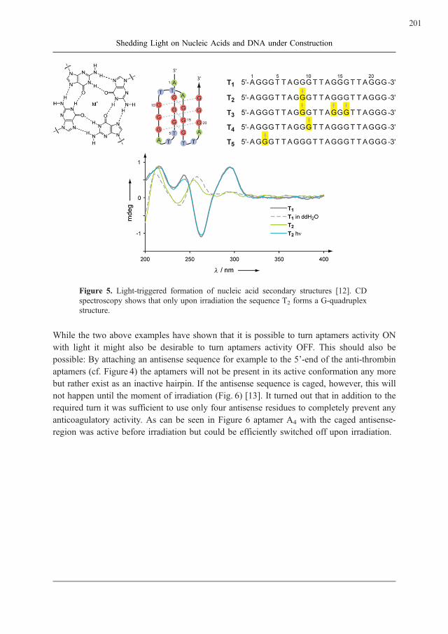
Figure 5. Light-triggered formation of nucleic acid secondary structures [12]. CD
spectroscopy shows that only upon irradiation the sequence T2 forms a G-quadruplex
structure.
While the two above examples have shown that it is possible to turn aptamers activity ON
with light it might also be desirable to turn aptamers activity OFF. This should also be
possible: By attaching an antisense sequence for example to the 5’-end of the anti-thrombin
aptamers (cf. Figure 4) the aptamers will not be present in its active conformation any more
but rather exist as an inactive hairpin. If the antisense sequence is caged, however, this will
not happen until the moment of irradiation (Fig. 6) [13]. It turned out that in addition to the
required turn it was sufficient to use only four antisense residues to completely prevent any
anticoagulatory activity. As can be seen in Figure 6 aptamer A4 with the caged antisense-
region was active before irradiation but could be efficiently switched off upon irradiation.
201
Shedding Light on Nucleic Acids and DNA under Construction

Figure 6. A strategy to turn aptamers OFF with light using a caged antisense-strand.
The graph shows dose-dependent anticoagulatory behavior [13].
The above examples demonstrate the broad applicability of the concept of using nucleobase-
caged nucleic acids. Ongoing research on all levels of the approach will even improve its
versatility and make it suitable for the next step which will be addressing spatiotemporal
questions in entire biological systems with all their inherent complexity.
Using DNA as Building Material
The second part of my group addresses an entirely different question. Throughout the last
decades computers have become tremendously more powerful. This development has pro-
found implications on society. Examples for these implications can be found everywhere and
include among others also the broad availability of the internet which has indeed changed
our everyday lives. Still ten years ago mobile phones were far from being common. The
steep increase in computing power has also left its footprints in the world of science and
made things possible that were still unthinkable several years ago. However, this miniatur-
ization trend cannot go on indefinitely. The reason is that photolithography – the process
used for generating the computers’ hearts – is using light to transfer the information of a
mask onto the silicon surface and even with ideal optics the size of the smallest feature that
can be generated is about the wavelength of the light that is used.
202
Heckel, A.

This has already been realized over a century ago by Ernst Abbe. Reducing this wavelength
indefinitely is also no viable solution. But instead of constantly trying to make things smaller
in a ‘‘top-down’’ approach an alternative is to try and use the power of molecular self-
assembly (‘‘bottom-up’’ approach) to generate structures beyond the diffraction limit. This
idea is not new and Richard Feynman was one of the most prominent advocates for this idea
for example when he gave his famous lecture entitles ‘‘There’s plenty of room at the
bottom’’ about 50 years ago [14].
Proofs that nano-machineries are possible exist amply inside of living organisms and cells.
The ATP synthase or multidrug efflux pumps are beautiful examples of efficient nano-
machineries. However, while we have come very far in understanding how these nanoscopic
miracles work we are still far from being able to construct similar objects from scratch. This
is for example due to the fact that the prediction of protein folding is still very difficult. On
the other hand proteins are not the only material from which functional or structural nano-
scopic objects can be constructed. Nucleic acids offer many advantages here: They form
well-known structures, have predictable, ‘‘programmable’’ interactions (via the Watson-
Crick base pairings), can be synthesized in automated fashion and manipulated with estab-
lished protocols and have exactly the right dimensions in the nanometer range.
While the usefulness of DNA and RNA as nano-material has already been nicely demon-
strated [15] there remains a problem; nucleic acids are linear, ‘‘soft’’ and do not form
significant tertiary interactions. The latter is what makes them different from proteins and
peptides which offer a richness of possible interactions. To compensate for this we have
decided to add other interaction principles to the world of nucleic acid nano-architectures.
What this means is that we want to add orthogonal structural elements – orthogonal to the
Watson-Crick base pairing.
An example of such orthogonal structural elements – but by far not the only solution – are
Dervan-polyamides [16]. This is a set of compounds which binds and sequence-selectively
recognizes the DNA minor groove. Dervan-polyamides can be generated to bind to almost
any possible DNA sequence. In the simplest realization of our approach to shape the tertiary
structure of DNAwe have therefore combined two of these polyamides with a flexible linker
and constructed what we called a ‘‘DNA strut’’ (Fig. 7) [17]. Such a strut can bind to double-
stranded DNA with both its ends and hence hold them together. For this reason one could
think of this system as ‘‘sequence-selective glue’’ for DNA-architectures. This new structural
element will allow whole new ways of assembling DNA objects.
203
Shedding Light on Nucleic Acids and DNA under Construction

Figure 7. Joining two Dervan-type polyamides (PA-A and PA-B, also shown as
cartoon representation) via a linker a DNA strut is obtained which is capable of
holding (‘‘gluing’’) two DNA double strands sequence-selectively together [17].
To prove and quantify that the expected ternary complex between two (different) DNA
double helices and the DNA strut is formed we had to explore new techniques because
none of the established ones for the characterization of Dervan-polyamides was suitable.
Using fluorescence cross-correlation spectroscopy (FCCS) [18] and small DNA hairpins we
were able to prove that the expected ternary complex is indeed formed and under the given
conditions a dissociation constant of 20 nM was measured (for more details see the original
paper [17]). What is equally important is that upon a slight change in DNA sequence no
binding could be observed any more.
The next question was to see if DNA objects bigger than hairpins could also be held together
using the DNA strut. Therefore, we started constructing small double-stranded DNA rings
containing 168 base pairs. These rings are relatively small and shape-persistent compared to
common DNA plasmids. The rings could be obtained using repetitive A-tract sequences
which are known to be inherently bent. Six constituting oligodeoxynucleotides were ligated
and objects with incomplete ring closure were digested using an exonuclease. Again by
FCCS we studied whether the ternary complex between two DNA rings and the strut was
formed and obtained 30 nM as dissociation constant. This is already a complex of about 208
kDa to which the strut only contributes with about 1%. Interestingly, after replacing three
base pairs in the binding site of one polyamide no interaction could be detected any more by
FCCS. Encouraged by these results we will now proceed and use this strategy to assemble
more complicated objects from nucleic acids as well as new alternative orthogonal
approaches for the interaction with double-stranded DNA.
204
Heckel, A.

References
[1] Zipfel, W.R., Williams, R.M., Webb, W.W. (2003) Nonlinear magic: multiphoton
microscopy in the biosciences. Nat. Biotechnol. 21:1369 – 1378.
[2] LaFratta, C.N., Fourkas, J.T., Baldacchini, T., Farrer, R.A. (2007) Multiphoton Fab-
rication. Angew. Chem. Int. Ed. 46:6238 – 6258.
[3] Mayer, G., Heckel, A. (2006) Biologically active molecules with a ‘‘light switch’’.
Angew. Chem. Int. Ed. 45:4900 – 4921.
[4] Kaplan, J.H., Forbush III, B., Hoffman, J.F. (1978) Rapid Photolytic release of
adenosine 5’-triphosphate from a protected analogue: utilization by the Na:K pump
of human red blood cell chosts. Biochemistry 17:1929 – 1935.
[5] Krock, L., Heckel, A. (2005) Photoinduced transcription by using temporarily mis-
matched caged oligonucleotides. Angew. Chem. Int. Ed. 44:471 – 473.
[6] Walbert, S., Pfleiderer, W., Steiner, U.E. (2001) Photolabile protecting groups for
nucleosides: mechanistic studies of the 2-(2-nitrophenyl)ethyl group. Helv. Chim.
Acta 84:1601 – 1611.
[7] Chiu, Y.L., Rana, T.M. (2003) siRNA function in RNAi: a chemical modification
analysis. RNA 9:1034 – 1048.
[8] Mikat, V., Heckel, A. (2007) Light-dependent RNA interference with nucleobase-
caged siRNAs. RNA 13:2341 – 2347.
[9] Famulok, M., Hartin, J.S., Mayer, G. (2007) Functional aptamers and aptazymes in
biotechnology, diagnosis, and therapy. Chem. Rev. 107:3715 – 3743.
[10] Bock, L.C., Griffin, L.C. Latham, J.A. Vermaas, E.H., Toole, J.J. (1992) Selection of
single-stranded DNA molecules that bind and inhibit thrombin. Nature
355:564 – 566.
[11] Heckel, A., Mayer, G. (2005) Light regulation of aptamer activity: an anti-thrombin
aptamer with caged thymidine nucleobases. J. Am. Chem. Soc. 127:822 – 823.
[12] Mayer, G., Krock, L., Mikat, V., Engeser, M., Heckel, A. (2005) Light-induced
formation of G-quadruplex DNA secondary structures. ChemBioChem
6:1966 – 1970.
[13] Heckel, A., Buff, M.C.R., Raddatz, M.S.L., Muller, J., Potzsch, B., Mayer, G. (2006)
An anticoagulant with light-triggered antidote activity. Angew. Chem. Int. Ed.
45:6748 – 6750.
[14] http://www.its.caltech.edu/~feynman/plenty.html
205
Shedding Light on Nucleic Acids and DNA under Construction

[15] Heckel, A., Famulok, M. (2008) Building objects from nucleic acids for a nanometer
world. Biochimie 90(7):1096 – 1107.
[16] Dervan, P.B. (2001) Molecular recognition of DNA by small molecules. Bioorg.
Med. Chem. 9:2215 – 2235.
[17] Schmidt, T.L., Nandi, C.K., Rasched, G., Parui, P.P., Brutschy, B., Famulok, M.,
Heckel, A. (2007) Polyamide struts for DNA architectures. Angew. Chem. Int. Ed.
46:4382 – 4384.
[18] Haustein, E., Schwille, P. (2007). Fluorescence correlation spectroscopy: novel var-
iations of an established technique. Annu. Rev. Biophys. Biomol. Struct. 36:151 – 169.
206
Heckel, A.

����������������
High-throughput Analysis of Nucleoside-
and Nucleotide-binding by Proteins
Justin K.M. Roberts1*
, Cecelia Webster1,
Thomas C. Terwilliger2
and Chang-Yub Kim2
1Department of Biochemistry, University of California,Riverside, CA 92521, USA.
2Bioscience Division, MS M888, Los Alamos National Laboratory,Los Alamos, NM 87545, USA.
E-Mail: *[email protected]
Received: 25th June 2008 / Published: 16th March 2009
Abstract
Many proteins function via selective binding of small molecules, and
an important class of ligands is nucleosides, including derivative
mono- and dinucleotides, which participate in processes such as cata-
lysis and signal transduction. Determining the specificity of nucleo-
side/nucleotide binding is therefore central to understanding the func-
tion of many proteins. We describe use of dye-ligand affinity chroma-
tography methods to identify putative nucleotide-binding proteins, and
to determine the specificity of binding to structurally related ligands. In
one approach, putative nucleoside-binding proteins are captured from
crude protein extracts of cells, for identification via standard proteomic
methods. In a second approach, interactions of different nucleosides
with purified recombinant protein targets, immobilized on dye, are
determined to assess the specificity of ligand recognition by a given
nucleotide-binding protein in the context of structural and functional
genomics, and drug development.
207
http://www.beilstein-institut.de/Bozen2008/Proceedings/Roberts/Roberts.pdf
Systems Chemistry, May 26th – 30th, 2008, Bozen, Italy

Introduction
Nucleosides and their derivatives are molecular carriers central to transmission of energy,
genetic information and intracellular signals, serving in these roles usually via specific
interactions with proteins [1]. Furthermore, many drugs contain nucleoside-related moieties;
for example nucleotide-protein interactions are considered critical in the action of isoniazid,
one of the most efficient drugs for the treatment of Mycobacterium tuberculosis (Mtb)
infections [2] and purine nucleoside analogs have been recently developed as anti-Mtb drug
candidates [3]. Identification of protein-nucleoside ligand couples, and understanding the
basis for functional molecular interactions, are complicated by obscurities in the relation-
ships between gene sequences and ligand binding specificity. For example, enzymes sharing
sequence similarity can exhibit differences in nucleotide specificity [4 – 6], and the specific
ligand-binding properties of individual proteins can be sensitive to small changes in gene
sequence [7 – 9]. Aiming to complement bioinformatics approaches to protein function [10,
11], we describe a biochemical screen based on dye-ligand chromatography [12] that can be
used to obtain evidence for specific protein-ligand interactions and to assess selectivity of
protein-ligand interactions in the human pathogen Mycobacterium tuberculosis.
Affinity binding technologies provide a means to identify physical interactions between
macromolecules and their small-molecule partners [13 – 15]. The technique of dye-ligand
chromatography has been used to purify a wide variety of proteins [12]. Cibacron Blue
F3GA binds many nucleoside-dependent enzymes and, by selective elution with salt or
ligands such as NADH or AMP, these proteins can often be purified [12]. Here we use
the selective elution of proteins from F3GA chromatography resin as a high-throughput
assay for protein-ligand interactions. The premise in our approach is that if a ligand can
trigger release of a protein adsorbed on the dye-column, the ligand probably interacts in a
specific fashion with that protein. This approach was first applied to crude cell extracts; the
specificity of ligand-protein interactions were then further examined using purified recom-
binant proteins.
Identification of Putative Nucleoside-binding Proteins:
Ligand-specific Elution of Native Proteins Bound to
Dye-resin
When we applied a crude extract of soluble proteins from Mycobacterium tuberculosis (Mtb)
to a column of F3GA resin, approximately 40% of the total protein remained adsorbed after
extensive washing. This Mtb protein-loaded F3GA resin was used in our first ligand-specific
elution screen for nucleoside/nucleotide-protein interactions, outlined in Figure 1, in which 6
to 20 potential ligands were applied successively to a given protein-loaded column, and the
proteins eluted by each ligand were identified by standard proteomic tools [16]. Relatively
low nucleoside concentrations (1 mM) and small volumes of eluting buffer (0.5 column bed
volume) were employed, with the aim of identifying specific ligand-protein interactions.
208
Roberts, J.K.M. et al.
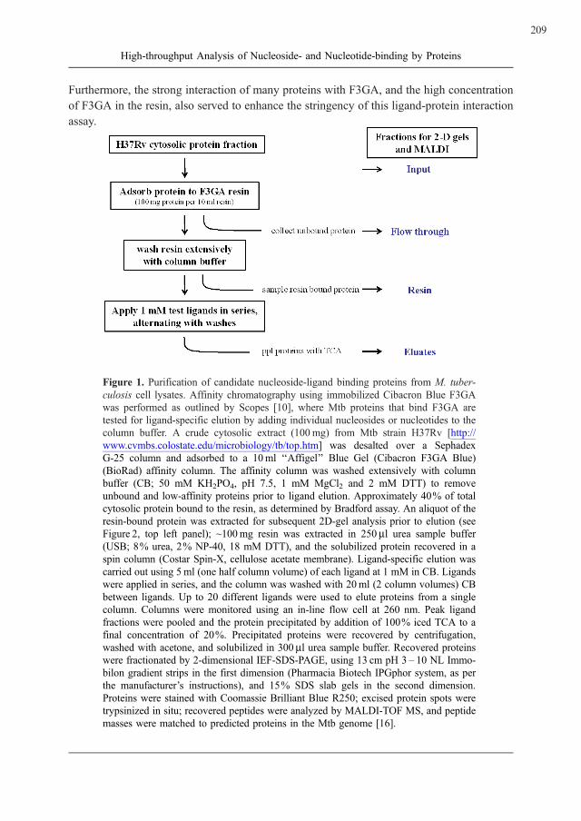
Furthermore, the strong interaction of many proteins with F3GA, and the high concentration
of F3GA in the resin, also served to enhance the stringency of this ligand-protein interaction
assay.
Figure 1. Purification of candidate nucleoside-ligand binding proteins from M. tuber-
culosis cell lysates. Affinity chromatography using immobilized Cibacron Blue F3GA
was performed as outlined by Scopes [10], where Mtb proteins that bind F3GA are
tested for ligand-specific elution by adding individual nucleosides or nucleotides to the
column buffer. A crude cytosolic extract (100 mg) from Mtb strain H37Rv [http://
www.cvmbs.colostate.edu/microbiology/tb/top.htm] was desalted over a Sephadex
G-25 column and adsorbed to a 10ml ‘‘Affigel’’ Blue Gel (Cibacron F3GA Blue)
(BioRad) affinity column. The affinity column was washed extensively with column
buffer (CB; 50 mM KH2PO4, pH 7.5, 1 mM MgCl2 and 2 mM DTT) to remove
unbound and low-affinity proteins prior to ligand elution. Approximately 40% of total
cytosolic protein bound to the resin, as determined by Bradford assay. An aliquot of the
resin-bound protein was extracted for subsequent 2D-gel analysis prior to elution (see
Figure 2, top left panel); ~100mg resin was extracted in 250 ml urea sample buffer
(USB; 8% urea, 2% NP-40, 18 mM DTT), and the solubilized protein recovered in a
spin column (Costar Spin-X, cellulose acetate membrane). Ligand-specific elution was
carried out using 5 ml (one half column volume) of each ligand at 1 mM in CB. Ligands
were applied in series, and the column was washed with 20ml (2 column volumes) CB
between ligands. Up to 20 different ligands were used to elute proteins from a single
column. Columns were monitored using an in-line flow cell at 260 nm. Peak ligand
fractions were pooled and the protein precipitated by addition of 100% iced TCA to a
final concentration of 20%. Precipitated proteins were recovered by centrifugation,
washed with acetone, and solubilized in 300 ml urea sample buffer. Recovered proteins
were fractionated by 2-dimensional IEF-SDS-PAGE, using 13 cm pH 3 – 10 NL Immo-
bilon gradient strips in the first dimension (Pharmacia Biotech IPGphor system, as per
the manufacturer’s instructions), and 15% SDS slab gels in the second dimension.
Proteins were stained with Coomassie Brilliant Blue R250; excised protein spots were
trypsinized in situ; recovered peptides were analyzed by MALDI-TOF MS, and peptide
masses were matched to predicted proteins in the Mtb genome [16].
209
High-throughput Analysis of Nucleoside- and Nucleotide-binding by Proteins

We observed ligand-specific release of distinct assortments of individual proteins from the
resin by many different ligands such as NAD and ATP (Fig. 2). Furthermore, the release of
individual proteins from the Mtb protein-loaded resin was sensitive to small differences in
the structure of individual ligands, such as the presence of methyl and phosphate groups on
adenosine (Fig. 3).
Figure 2. Ligand-specific elution of native Mtb proteins from Cibacron Blue F3GA
resin. Two dimensional IEF-SDS-PAGE separation of Mtb cytosolic proteins that bind
to the F3GA resin (top left panel), or are subsequently eluted by the indicated nucleo-
tide at a concentration of 1 mM. Data are from three different experiments, as per
figure 1. Only that portion of each gel corresponding to molecular mass ~100 to 35
kDa (top to bottom) and pI ~6 to 4.5 (left to right) is shown. Protein identities are
indicated by locus tag number (see Table 1).
210
Roberts, J.K.M. et al.

Figure 3. Selective elution of native Mtb proteins from F3GA resin by adenosine and
adenosine analogs. Two dimensional IEF-SDS-PAGE separation of Mtb cytosolic
proteins bound to F3GA and eluted successively by adenosine and related compounds.
Ligands were applied to the column in the same order as the panels (from top left to
bottom right, in order of increasing ligand mass), from a single experiment as per
Figure 1. Rv0577 eluted with adenosine and both of the methyl-adenosine xenobio-
tics, but not with nucleotides. Conditions were as in Figure 2, except that proteins
were stained with silver.
A summary of the predicted identities and functions (in Genbank) of the native Mtb proteins
identified in different fractions described in Figure 1 are given in Tables 1 and 2. Table 1
lists 26 proteins that exhibited ligand-specific elution and in Table 3 these elution results are
compared with the ligands associated with predicted protein functions available in public
gene data bases. These tabulated results provide annotations of gene and predicted protein
function at several levels. First, these data provide the first direct evidence that 28 Mtb
proteins predicted from analysis of the Mtb genome exist in vivo (eight other proteins in
Table 1 and 2, see footnotes). Second, the ligand-elution data provide specific annotations as
to ligands that bind to these proteins. In the cases of Rv2671 and Rv3336c, for example,
Table 3 shows that they bind NADPH and ATP, respectively. Table 3 contains many
examples of consistency between nucleotide-elution data and the predicted ligand specificity
based on gene sequence comparisons; Nineteen of the proteins in Table 3 were eluted by the
predicted ligand or a very closely related one (e.g. NADP vs. NADH). Half (18/36) of the
proteins identified in Tables 1 and 2 contain one or two Rossmann signatures that are
associated with many nucleotide-binding proteins, including some with extensions beyond
the GX1 – 2GXXG core [17] sequence.
211
High-throughput Analysis of Nucleoside- and Nucleotide-binding by Proteins

Table 1. Functional predictions for nucleotide-binding proteins from M. tuberculosis
cytosolic extracts, identified by dye-ligand chromatography.
Locus tag Protein name in GenBank Rossmann motif
Amino acid transport and metabolism
Rv1133c methyltransferase metE no
aRv2220 glutamine synthetase glnA1 no
aRv2780 secreted L-alanine dehydrogenase ald VIGAGTAGYNAA
Rv3754 prephenate dehydrogenase tyrA VLGLGLIGGSIM
Carbohydrate transport and metabolism
Rv1023 phosphopyruvate hydratase eno STGLGDEGGFAP
Rv1122 6-phosphogluconate dehydrogenase gnd2 MIGLGRMGANIV
Lipid transport and metabolism
Rv0468 3-hydroxybutyryl-CoA dehydrogenase fadB2 VVGAGQMGSGIA
Rv0751c 3-hydroxyisobutyrate dehydrogenase mmsB FLGLGNMGAPMS
Rv0824c acyl-[acyl-carrier-protein] desaturase desA1 NMGMDGAWGQWVN
Rv0859 acetyl-CoA acetyltransferase fadA no
Rv0860 fatty acid oxidation protein fadB VLGAGMMGAGIA
IVGYSGPAGTGKA
Nucleotide transport and metabolism
Rv1843c IMP dehydrogenase guaB1 AVGINGDVGAKAR
aRv2445c nucleotide diphosphate kinase ndkA no
Other metabolism
Rv0927c short-chain dehydrogenase no
Rv2671 hypothetical protein, possible ribD no
Energy production and conversion
Rv0155 NAD(P) transhydrogenase pntAa VLGVGVAGLQAL
Rv0223c aldehyde dehydrogenase no
Rv0458 aldehyde dehydrogenase QSGIGREGHQMM
Rv2858c aldehyde dehydrogenase aldC no
Rv3293 piperideine-6-carboxilic acid dehydrogenase pcd no
aRv3913 thioredoxin reductase trxB2 VIGSGPAGYTAA
Other cellular processes
Rv1511 GDP-D-mannose 4,6 dehydratase gmdA ITGITGQDGSYLA
Rv3336c tryptophanyl-tRNA synthetase trpS no
Poorly characterized
Rv0484c short-chain oxidoreductase no
Rv0577 hypothetical protein no
Rv1544 possible ketoacyl reductase SAGFGTSGRFWE
Locus tag (Rv numbers) are gene identifiers for predicted open reading frames (ORFs) in the M.tuberculosis H37Rv genome (accessed through NCBI Entrez Gene at http://www.ncbi.nlm.nih.gov/sites/entrez? db = gene). For each protein recovered by dye-ligand chromatography, the masses oftryptic peptides were determined by MALDI and matched to ORFs by ProteinProspector (http://prospector.ucsf.edu/).Rossmann motif shows which proteins contain the GX1 – 2GXXG core sequence characteristic ofFAD, NAD(P)-binding proteins that form a Rossmann fold (BOLD), and those that also have theextended sequence shown to stabilize the fold structure (ITALIC; I or V in the 1st position shown, andA or G in the last) (Ref. 17). Note that Rv0860 contains 2 motifs.a Indicates 4 proteins that have been previously studied (see also Table 2). The remainder are shownhere, for the first time, to be expressed in vivo.
212
Roberts, J.K.M. et al.
Table 2. M. tuberculosis proteins identified in this study for which no ligand-bindinginformation was determined.
Locus tag Protein name in GenBank bRossmann motif
Lipid transport and metabolism
Rv0154c probable acyl-CoA dehydrogenase fadE2 no
Rv0242c 3-ketoacyl-(acyl-carrier-protein) reductase fabG4 LVGNGSIGEGGR
IAGIAGNRGQTNY
Rv2831 enoyl-CoA hydratase echA16 no
aRv3280 propionyl-CoA carboxylase (beta chain 5) accD5 VMGASGAVGFVYR
Energy production and conversion
Rv3520 possible coenzyme F420-dependent oxidoreductase ILGLGVSGPQVV
LTGEGTTGLGKA
Molecular chaperones
aRv0350 molecular chaperone DnaK no
aRv0440 chaperonin groEL VPGGGDMGGMDF
aRv2031c heat shock protein hspX no
Rv2462c trigger factor tig no
Poorly characterized
Rv2731 conserved hypothetical alanine/arginine rich protein no
Proteins listed are those which bound to the affinity resin but did not elute with any of the test ligands(e. g., see Figure 1). In addition, all of the Molecular Chaperones were ubiquitous; present at lowlevels in all samples, including ligand-free washes.a, b See Table 1 footnotes. Note that Rv0242c and Rv3520 each contain 2 Rossmann motifs.
Some of the ligand-protein combinations identified by ligand elution are unexpected or not
predicted from sequence analyses and GenBank annotations (http://www.ncbi.nlm.nih.gov/
Genbank/index.html). For example, Rv0577 has neither a Rossmann signature, nor any
GenBank annotation suggesting nucleoside-dependent function. We therefore targeted such
proteins for recombinant expression to examine ligand-protein interactions under conditions
where the quantities and concentration of protein are well-defined, and the effects of differ-
ent ligands on protein-dye interactions can be more readily compared. The method with
crude extracts, just described, is a positive screen only; proteins that elute with ligands may
not be picked up due to protein abundance or detection issues, and the order of eluting
ligands can influence gel spot intensity and resolution for proteins which are substantially
depleted by several related ligands. Moreover, some proteins retained on the column may be
bound via other proteins in native complexes, rather than directly to the dye, while other
proteins may have increased or unchanged affinity for the dye when bound to ligands.
213
High-throughput Analysis of Nucleoside- and Nucleotide-binding by Proteins
Table 3. Predicted ligand-protein interactions for native M. tuberculosis proteinsversus interactions observed by dye-ligand chromatography.
Locus tag a Predictedinteractions
b Ligand[s] causing elutionfrom F3GA resin
Rv0155 NAD[P][H] NADH
Rv0223c NAD[P][H] ATP
Rv0458 NAD[H] N(6)mAdo, NAD
Rv0468 NAD[P][H] NADH
Rv0484c NAD[P][H] NADPH
Rv0577 na N(6)mAdo, 2mAdo >> Ado, FAD
Rv0751c NAD[H] NAD
Rv0824c NADPH NADPH
Rv0859 na NAD[H]
Rv0860 NAD[H] NAD[H]
Rv0927c NAD[P][H] NADP[H]
Rv1023 na ATP
Rv1122 NADP[H] NADH
Rv1133c na N(6)mAdo > ATP, GTP
Rv1511 NADP[H] GTP
Rv1544 NAD[P][H] NADPH
Rv1843c NAD[H] GMP
Rv2220 ATP/ADP/AMP 2mAdo >> N(6)mAdo > ATP, FAD
Rv2445c NTP/NDP ATP>GTP>>ubiquitous c
Rv2671 NADP[H] NADPH
Rv2780 NAD[H] NADH, ATP, cAMP >> ADP, Ado, N(6)mAdo
Rv2858c NAD[H] NADP > NADPH, NAD
Rv3293 NAD[P][H] NADH
Rv3336c ATP/AMP ATP
Rv3754 NAD[H] NADP
Rv3913 NADP[H], FAD NADP
a Predicted interactions were compiled from biochemical studies of homologous proteins in otherspecies, available in the NCBI, Prosite and BRENDA databases, and is not available (na) for allproteins.b Ligands causing elution were observed experimentally for each protein. Relative spot intensitiesobserved on 2-D gels are indicated by rank order, and do not reflect a systematic analysis of relativeligand affinity.c See Table 2 footnote re ubiquitous proteins.
Analysis of the Specificity of Ligand-protein Interactions
with Recombinant Proteins and Dye-ligand
Chromatography
In the second ligand-specific elution screen for nucleoside/nucleotide-protein interactions,
single recombinant Mtb proteins were adsorbed on multiple small (mg) aliquots of F3GA
resin in spin columns, and assayed for elution by various nucleotides and nucleosides using
one ligand per column, as outlined in Figure 4. The use of purified recombinant proteins
permitted us to identify effects of a single variable (ligand structure) on the stability of the
214
Roberts, J.K.M. et al.

dye-protein complexes. We found clear differences in the release of a particular protein from
the affinity resin by various ligands, shown for Rv0577 in Figure 5. We posit that greater
amounts of eluted protein reflect stronger interaction between the protein and eluting ligand.
Here, a stronger interaction between a protein and the eluting ligand could be due either to a
direct interaction of the ligand at the dye-binding site of the protein, or binding at a site
remote from the dye-binding site that causes conformational changes in the protein and
weakens protein-dye interactions [12]. Further, only interactions that lead to a decrease in
dye-binding affinity are expected to result in elution of a protein.
Figure 4. Analysis of nucleoside-ligand binding by recombinant proteins. Recombi-
nant proteins were evaluated for their ligand-binding properties using a modified
affinity elution chromatography protocol (22): individual proteins were diluted to
2 mg/ml in CB and adsorbed to multiple small aliquots of F3GA resin (100 mg protein
per 10 mg resin] in 2 ml spin-columns (Costar Spin-X, cellulose acetate membrane).
Binding was for 1 h at 4 �C with very gentle vortexing, followed by recovery of
unbound protein (flow-through fraction) and washing of the resin (8 x 0.4 ml washes
with CB); spin-columns were micro-centrifuged for 30 s at 13000 g, to recover frac-
tions and change solutions. Individual spin-columns containing resin-bound proteins
were then incubated (as for protein binding, above) with 50 ml 1 mM test ligand in CB,
and the elution fractions recovered by centrifugation. Protein, which remained bound
to the resin was recovered by heating at 95 �C for 5min in100 ml SDS sample buffer,
and centrifugation (resin fraction). Aliquots of initial protein, spin-column flow-
through and eluate fractions were diluted 1:1 with 2X SDS sample buffer. Equal
proportions of all fractions (equivalent to 1 mg input protein) were loaded on 15%
polyacrylamide one-dimensional mini-gels and stained with silver or Coomassie blue
R250.
215
High-throughput Analysis of Nucleoside- and Nucleotide-binding by Proteins

Figure 5. Ligand-specific elution of recombinant Mtb protein Rv0577 from Cibacron
Blue F3GA resin. The protocol is described in Figure 4. Shown is the protein input
(Rv0577), the protein that did not bind (FT), and the protein that was either released
from F3GA resin by buffer or 9 different ligands (eluates) or remained bound to F3GA
in the presence of the ligands (resins). SRI0977 = N(6)methyladenosine; SRI2085 = 2-
methyladenosine. Silver staining shows release of Rv0577 from F3GA by nucleosides,
but not by nucleotides.
The results with purified recombinant Mtb proteins such as Rv0577 (Fig. 5) are consistent
with the observations from experiments with crude cytosolic extracts (Fig. 3; Table 3), which
indicated that these proteins are nucleoside/nucleotide-binding proteins. Protein Rv0577 was
eluted by the nucleoside adenosine as well as by methylated adenosine xenobiotics that are
anti-Mtb drug candidates [3], whereas the common purine-containing mono- and di-nucleo-
tides (AMP/ADP/ATP/NAD/NADH/GTP) did not promote release from the dye-resin
(Fig. 5; Table 3).
Conclusions
We conclude that the two ligand-specific elution screens described here are useful for
obtaining clues about the functions of members in the important class of nucleoside-binding
proteins and their parent genes. Affinity methods are often effective in picking out functional
classes of proteins from complex mixtures [e.g. 18], and our first dye-based screen followed
this paradigm, sorting soluble proteins from a crude cell extract according to their ability to
be released from the F3GA dye by millimolar concentrations of nucleosides (Figs. 2 and 3;
Table 3). In addition to uncovering the natural ligands that nucleoside/nucleotide-binding
proteins can associate with in cells, the dye-based screens can be extended to identify protein
targets of nucleoside-analogs of interest as potential drugs [3]. Used in tandem, the two
screens allow proteins to be selected from crude cell extracts as candidate nucleoside/
nucleotide-binding proteins and further examined, after cloning and expression, with respect
to ligand preferences. Our results with proteins in crude extracts provided evidence that
many proteins bind to multiple ligands (Table 3), findings that were confirmed in screens
with pure, recombinant versions under conditions where differential release of protein from
the dye resin can be assigned to differences in ligand structure (example shown in Figure 5).
216
Roberts, J.K.M. et al.

The data from these combined screens are useful for confirming, refining or challenging
gene annotations based on sequence analysis [e. g. 10, 11], and may also serve to comple-
ment the wealth of virtual ligand screening methodologies [19]. Furthermore, the ligand-
binding data can guide protein crystallization efforts aimed at protein function via structure
determination. In this context, information on ligands likely has considerable practical
benefits, given that protein crystallization is often greatly improved by inclusion of ligands
during crystal formation [20], and production of crystals suitable for analysis by X-ray
diffraction is the principal bottleneck in structural genomics efforts today [21] (PSI target
status website; http://sg.pdb.org/target_centers.html).
Acknowledgements
We thank Dr. W.W.P. Chang for help developing affinity sorting methods for cell extracts;
Dr. J.T. Belisle for Mtb cytosol extracts (as part of NIH, NIAID Contract No.
HHSN266200400091C to Colorado State University, Fort Collins, CO); Dr. R.C. Reynolds,
Southern Research Institute, Birmingham, AL, for supplying the two methylated adenosine
derivatives (SRI0977 and SRI2805); N. Maes and E. Z. Alipio Lyon for technical assistance.
This work was in part supported by the LANL-UCR CARE program (STB-UC:06 – 29) and
the NIGMS Protein Structure Initiative program (NIH U54 GM074946 – 01).
References
[1] Voet, D., and Voet, J.G. (2004) Biochemistry, 3rd Ed., Volume 1. J. Wiley & Sons, NJ.
[2] Vilcheze C, Jacobs W.R. Jr. (2007) The mechanism of isoniazid killing: clarity
through the scope of genetics. Annu. Rev. Microbiol. 61:35 – 50.
[3] Parker, W.B., and Long, M.C. (2007) Purine metabolism in Mycobacterium tubercu-
losis as a target for drug development. Curr. Pharm. Des. 13:599 – 608.
[4] Soderling, S.H., Bayuga, S.J., and Beavo, J.A. (1999) Isolation and characterization
of a dual substrate phosphodiesterase gene family: PDE10A. Proc. Natl. Acad. Sci.
U.S.A. 96:7071 – 7076.
[5] Joyce, M.A., Fraser, M.E., Brownie, E.R., James, M.N., Bridger, W.A., and Wolod-
ko, W.T. (1999) Probing the nucleotide binding site of Escherichia coli succinyl CoA
synthetase. Biochemistry 38:7273 – 7283.
[6] Tsybovsky, Y., Donato, H., Krupenko, N.I., Davies, C., and Krupenko, S.A. (2007)
Crystal structures of the carboxyl terminal domain of rat 10-formyltetrahydrofolate
dehydrogenase: implications for the catalytic mechanism of aldehyde dehydro-
genases. Biochemistry 46:2917 – 2929.
217
High-throughput Analysis of Nucleoside- and Nucleotide-binding by Proteins

[7] Goward, C.R., and Nicholls, D.J. (1994) Malate dehydrogenase: a model for struc-
ture, evolution, and catalysis. Protein Sci. 3:1883 – 1888.
[8] Eppink, M.H., Overkamp, K.M., Schreuder, H.A., and Van Berkel, W.J. (1999)
Switch of coenzyme specificity of p-hydroxybenzoate hydroxylase. J. Mol. Biol.
292:87 – 96.
[9] Wang, X., and Kemp, RG. (1999) Identification of residues of Escherichia coli
phosphofructo-kinase that contribute to nucleotide binding and specificity. Biochem-
istry 38:4313 – 4318.
[10] George, R.A., Spriggs, R.V., Bartlett, G.J., Gutteridge, A., MacArthur, M.W., Porter,
C.T., Al-Lazikani, B., Thornton, J.M., and Swindells, M.B. (2005) Effective function
annotation through catalytic residue conservation. Proc. Natl. Acad. Sci. U.S.A.
102:12299 – 12304.
[11] Edgar, R.C., and Batzoglou, S. (2006) Multiple sequence alignment. Curr. Opin.
Struct. Biol. 16:368 – 373.
[12] Scopes, R.K. (1994) Protein Purification: Principles and Practice, 3rd Ed. Springer,
NY.
[13] Makara, G.M., and Athanasopoulos, J. (2005) Improving success rates for lead
generation using affinity binding technologies. Curr. Opin. Biotechnol. 16:666 – 673.
[14] Erster, O., Eisenstein, M., and Liscovitch, M. (2007) Ligand interaction scan: a
general method for engineering ligand-sensitive protein alleles. Nat. Methods
4:393 – 395.
[15] Turnbull, J.E., and Field, R.A. (2007) Emerging glycomics technologies. Nat. Chem.
Biol. 3:74 – 77.
[16] Chang, W.W.P., Huang, L., Shen, M., Webster, C., Burlingame, A., Roberts, J.K.M.
(2000) Patterns of Protein Synthesis and Tolerance of Anoxia in Root Tips of Maize
Seedlings Acclimated to a Low Oxygen Environment, and Identification of Proteins
by Mass Spectrometry. Plant Physiol. 122:295 – 317.
[17] Kleiger, G., and Eisenberg, D. (2002) GXXXG and GXXXA motifs stabilize FAD
and NAD(P)-binding Rossmann folds through Ca-H...O hydrogen bonds and van der
waals interactions. J. Mol. Biol. 323:69 – 76.
[18] Bieber, A.L., Tubbs, K.A., and Nelson, R.W. (2004) Metal ligand affinity pipettes
and bioreactive alkaline phosphatase probes: tools for characterization of phosphory-
lated proteins and peptides. Mol. Cell. Proteomics 3:266 – 72.
218
Roberts, J.K.M. et al.

[19] Villoutreix, B.O., Renault, N., Lagorce, D., Sperandio, O., Montes, M., and Miteva,
M.A. (2007) Free resources to assist structure-based virtual ligand screening experi-
ments. Curr. Protein Pept. Sci. 8:381 – 411.
[20] Vedadi, M., Niesen, F.H., Allali-Hassani, A., Fedorov, O.Y., Finerty, J.P.J., Wasney,
G.A., Yeung, R., Arrowsmith, C., Ball, L.J., Berglund, H., Hui, R., Marsden, B.D.,
Nordlund, P., Sundstrom, M., Weigelt, J., and Edwards, A.M. (2006) Chemical
screening methods to identify ligands that promote protein stability, protein crystal-
lization, and structure determination. Proc. Natl. Acad. Sci. U.S.A.
103:15835 – 15840.
[21] Goh, C.-S., Lan, N., Douglas, S.M., Wu, B., Echols, N., Smith, A., Milburn, D.,
Montelione, G.T., Zhao, H., and Gerstein, M. (2004) Mining the structural genomics
pipeline: Identification of protein properties that affect high-throughput experimental
analysis. J. Mol. Biol. 336:115 – 130.
[22] Kim, C.-Y., Takahashi, K., Nguyen, T.B., Roberts, J.K.M., and Webster, C. (1999)
Identification of a nucleic acid binding domain in eukaryotic initiation factor
eIFiso4G from wheat. J. Biol. Chem. 274:10603 – 10608.
219
High-throughput Analysis of Nucleoside- and Nucleotide-binding by Proteins


����������������
Systems Biology from Chemical
Combinations
Joseph Leh�r*,1,2
, Andrew Krueger2,
Grant Zimmermann1, Alexis Borisy
1
1CombinatoRx Incorporated,245 First St, Cambridge, MA 02142, U.S.A.
2Boston University Bioinformatics/Bioengineering,20 Cummington St, Boston, MA 02215, U.S.A.
E-Mail: *[email protected]
Received: 28th October 2008 / Published: 16th March 2009
Abstract
Systematic testing of chemical combinations in cell-based disease
models can yield novel information on how proteins interact in a
biological system, and thus can make important contributions to bio-
logical models of those diseases. Such combination screens can also
preferentially discover synergies with beneficial therapeutic selectivity,
especially when used in high-order mixtures of more than two agents.
These studies demonstrate the value obtainable from combination che-
mical genetics, and reinforce the growing realization that the most
useful paradigm for a drug target is no longer a single molecule in a
relevant pathway, but instead the set of targets that can cooperate to
produce a therapeutic response with reduced side effects.
Introduction
Living organisms can be thought of as systems of interacting molecules, whose function and
dysfunction would be better understood using a functional wiring diagram [1]. Such network
models have already been used to predict the development of complex biological phenotypes
[2], and will eventually help researchers gain a better understanding of multi-factorial
diseases, identify novel therapeutic targets, and develop personalized treatments [3].
221
http://www.beilstein-institut.de/Bozen2008/Proceedings/Lehar/Lehar.pdf
Systems Chemistry, May 26th – 30th, 2008, Bozen, Italy

The staggering complexity of biological systems requires large and diverse sets of data on
component connectivity and responses to system perturbations. The core topology of the
network is determined using direct interactions between genes, proteins, and metabolites,
and the models can then be refined using a biological system’s responses to perturbations,
such as drugs, mutations, or environmental changes. All of these approaches can associate
network components with functional roles, even without direct interactions connecting them.
Combined perturbations shift the focus of genetic and chemical genetic studies from the
functions of individual genes or proteins to interactions of those components within a
biological system [4]. Large efforts to test the viability of double mutants are underway in
bacteria [5], yeast [6], and nematodes [7]. Chemicals provide information that is distinct
from and complementary to genetic perturbations, given the differences between how they
modulate protein functions [8]. The advantages of chemical perturbations are that they can
target a single domain of a multi-domain protein, allow precise temporal control that is
critical for rapid-acting processes, can target orthologous or paralogous proteins enabling
comparisons between species or redundant functions, and do not directly alter the concen-
trations of a targeted protein, thus avoiding indirect effects upon multi-protein complexes.
Small molecules also lend themselves more readily to combination interventions, making
them especially useful for integrating systems and chemical biology [9, 10]. Most combined
perturbation studies with combinations have focused on exploring drug sensitivities across
large sets of knockouts are also being undertaken in various model organisms [11, 12] and
human cells [13]. However, these approaches are increasingly being extended to systematic
testing of purely chemical combinations [10, 14]. Here we discuss our work that explores
how combination studies can discover novel therapeutic treatments for complex disease
systems, as well as to obtain biological information on the organization of functional
components in those systems.
Discovering Combination Drugs
One of the most powerful resources for discovering novel therapies is the set of existing
drugs chemical probes with known biological activity. Over the past century, the pharma-
ceutical industry has had great success developing drugs with the following essential qua-
lities: selective therapeutic activity with tolerable side effects; desirable absorption, distribu-
tion, metabolism, and excretion properties; and stable chemistry to facilitate storage, man-
ufacturing, and distribution. Molecules with these properties are rare, and many disease
relevant targets cannot be addressed by chemical inhibitors, so there are currently only
~3,000 approved drug ingredients and research probes which modulate 200 – 500 molecular
targets within human biology [15]. Moreover, even the most specific small-molecule drugs
can affect many different proteins, leading to off-target effects either through direct binding
to secondary targets or through the downstream responses to these binding events.
222
Lehar, J. et al.

Such secondary activities can occasionally be beneficial towards other diseases for which the
drug was not originally developed, and indeed many successful drugs result from such
repurposing efforts.
Phenotypic screening is a very effective method for discovering new opportunities provided
by existing drugs [16]. Since the 1970s, the prevailing drug discovery paradigms have been
target-based drug design and phenotypic screening. The target-based approach involves
identifying a molecular target that is responsible for a disease phenotype and then system-
atically testing a variety of chemical agents to see if they bind selectively to that target. By
contrast, phenotypic screening tests chemical agents against a measurable quality of a whole
organism’s function, to identify those with a desirable response profile without necessarily
having a detailed understanding of the drug’s mechanism. While the target-based approach
can discover primary target activities, secondary targets are almost exclusively found by
phenotypic testing. Systematically testing many agents against phenotypes allows the biol-
ogy to reveal which agents are likely to yield beneficial activities.
The opportunity for discovering useful off-target effects increases dramatically when drugs
are used in combination [17]. Combinations are traditionally used to reduce toxicity, by
sparing doses between two different drugs affecting the same disease function, to take
advantage of complementary activities that independently improve the disease condition,
and as the most effective response to drug-resistant pathogens, notably for viral and bacterial
infections. Biological systems are robust, exhibiting a high degree of resilience that can
compensate for a variety of attacks [18]. For example, mutation studies in many organisms
[19, 20] find that only ~10 – 20% of genes affect viability when deleted from the genome.
Combinations can overcome this robustness by targeting compensatory pathways [21], and
many such cooperative relationships could work through known or yet undiscovered sec-
ondary drug targets.
Chemical combinations need to be tested at varying drug doses, in order to find synergistic
effects at unknown concentrations. This can be done with a ‘‘dose matrix’’, where a combi-
nation is tested in all possible permutations of serially-diluted single agent doses (Fig. 1), or
a fixed dose-ratio series, where component drugs are mixed at a high concentration and the
mixture is tested in serial dilutions. Synergy is calculated by comparing a combination’s
response to those of its single agents [21], usually against the drug-with-itself dose-additive
reference model [22]. Deviations from dose additivity can be assessed visually on an Iso-
bologram (Fig. 1) or numerically with a Combination Index [23]. To capture synergies that
can occur anywhere on full dose-matrix experiments, it is also useful to calculate a volume
VHSA= SX,Y lnfX lnfY (Idata – IHSA) between the data and the highest-single-agent surface,
normalized for single agent dilution factors fX,fY, to quantify the strength of combination
effects [21]. Both synergy scoring methods generalize readily to mixtures of three or more
chemicals.
223
Systems Biology from Chemical Combinations

Figure 1. Dose matrix and synergy. Phenotypes (e. g., inhibition of S. aureus growth
by Bactrim�) are measured for each drug, dosed in serial dilutions (bottom, left edges)
and in all paired mixtures. Synergy is scored on differences between combined and
single agent effects, while the isobologram shows the dose-sparing achieved over
drug-with-itself dose-additivity. Using combination screening, we have discovered
thousands of synergies in many disease areas. A cube showing 22 assays, each with
colored symbols scaled to synergy, shows that despite some prolific drugs, strong
synergies are rare.
CombinatoRx has been using a high throughput screening platform to explore combination
therapies [24], by systematically testing combinations of ~3,000 agents in cell-based assays
that preserve disease-relevant pathway complexity yet which are efficient enough to explore
the vast space of combinations [21]. Using this platform, we have conducted dose-matrix
screens in over ten disease areas to discover thousands of synergies (Fig. 1), many of which
we have advanced into clinical trials for inflammation, diabetes and cancer indications. In
addition, we have funded research programs utilizing our cHTS platform for drug discovery
in neurodegenerative disease, infectious diseases, cystic fibrosis, muscular dystrophy, drug-
device combinations and ophthalmology indications.
224
Lehar, J. et al.

Combinations for Systems Biology
Dose matrix responses to combinations of chemical probes also can be used to determine
mechanistic relationships between their targets [10]. Our therapeutic screens produce a wide
variety of response surfaces, with distinct shapes for combinations having different known
combination mechanisms. Many of these responses can be described by simple models that
use the single agent curves to predict the combination effect (Fig. 2), and whose parameters
provide quantitative measures of combination effects.
Figure 2. Synergy and connectivity. Simulated combination effects in a pathway of
enzymatic reactions (grey enzymes, black substrates) with paired inhibitors (joined
markers) depend on how the targets are connected. The effects can be described in
terms of simple response shape models based on the single agent curves. A yeast
experiment using sterol pathway inhibitors confirmed the expected response shapes in
the pathway (symbol size scales with synergy, color shows the response model), and
effects across pathways were weaker. Figure adapted from Lehar et al. 2007.
225
Systems Biology from Chemical Combinations

We established the relationship between synergy and connectivity by simulating a metabolic
pathway as a series of linked Michaelis-Menten reactions with pairs of competitive inhibi-
tors aimed at different targets with varying doses. We found that the shape and amount of
synergy of each combination response depended on how the inhibitor pair’s targets were
connected in the pathway (Fig. 2). The predicted response shapes were robust to kinetic
assumptions, parameter values, and nonlinear response functions, but were very sensitive to
topological alterations in the target connectivity, such as branching, feedback regulation, or
changing the type of junction at a branch point. The predicted shapes from these simulations
were confirmed in a yeast proliferation experiment using drug combinations targeting sterol
biosynthesis (Fig. 2), with further support from experiments on human cancer cells.
To explore the relationship between synergy and connectivity on a larger scale than can be
addressed with dynamic pathway modeling, we are using flux-balance analysis (FBA)
simulations of E. coli metabolism[25], with ‘‘minimization of metabolic adjustment’’
(MOMA) [26] to determine the impact of the perturbation upon bacterial growth. A variety
of combination effects are observed, including many synergies in excess of the single agents,
and a considerable variety of shapes that would have been indistinguishable with only
knockouts. We estimated the total level of synergy by integrating the volume between the
simulated data and the HSA model, VHSA= SX,Y lnfX lnfY (Idata – IHSA), summed over all
positive concentrations X,Y and corrected for the dilution factors fX and fY.
When the single agents are organized by their target location in the network, clear patterns
emerge (Fig. 3). Most combinations produce no synergy, but there are strong synergies
between adjacent enzymes within a pathway, and clear patterns of synergy between path-
ways, showing distinctions between the upper and lower ends of the pathway in some cases.
These simulations demonstrate that genome-scale simulations of partially inhibited networks
are tractable, and confirm that combination effects are strongly dependent on the connectiv-
ity of the perturbers’ targets. Some of the response shapes clearly do not fit the shape models
shown in Figure 2, so it is clear that we will need to broaden the reference set in order to
capture the increased complexity of the system. Measuring connectivity between targets is
considerably more challenging for a complex network like the E. coli FBA network,
226
Lehar, J. et al.

Figure 3. Simulated synergy. FBA/MOMA simulations of inhibited E. coli metabo-
lism (central metabolism detail). The matrix shows synergy (VHSA) for all pairs of
targets sorted by pathway location. Most inhibitor pairs don’t interact, but synergies
and antagonisms show clear mechanistic patterns. The response surface shapes are
more diverse than those shown in Figure 3.
We are also using flux balance analysis to model metabolism-dependent growth in Saccar-
omyces cerevisiae (yeast). This model organism has a fully sequenced genome, richly
annotated functional pathways, and some of the most advanced network models both for
metabolism and protein interactions [27, 28]. To provide an experimental basis for valida-
tion, we have screened all pairwise combinations of 60 chemical probes known to target
metabolic enzymes in the FBA model, using an Alamar Blue metabolic readout as a proxy
for cell growth (Fig. 4). Preliminary results from the combination screen are very encoura-
ging. The observed responses are diverse, roughly ~4% of the tested combinations showed
synergy (VHSA) that were significantly different from the agent-with-self score distribution.
The detected synergies are scattered throughout the combination space, but there are regions
of consistent synergy between some pathways. In our preliminary studies, both individual
combinations and synergy profiles confirm the results seen in our initial exploration of
Candida glabrata [10]. We have performed FBA simulations of S. cerevisiae metabolism,
which we will use for comparisons with our bacterial FBA models as well as with the yeast
experimental results.
227
Systems Biology from Chemical Combinations

Figure 4. Yeast experiment. (a) Yeast cells were grown in pairwise combinations of
60 probes targeting metabolism. (b) Resulting responses confirm sterol synergies and
produce diverse responses. (c) Analysis shows a 4% rate of interaction with synergies
and antagonisms. Synergy profiles show both isolated synergies and coherent pathway
interactions.
Higher order combinations (of three or more agents) can provide useful information about
the overall complexity of a biological system. Randomly connected networks become
increasingly sensitive to perturbations as the combination order is increased [29], and this
trend is confirmed in more realistic network simulations using FBA approaches [30]. In
principle, one would expect a trend towards more greater fragility to perturbation as the
system is subjected to perturbations of increasing combination orders, due to their over-
whelming any functional redundancy in the system (Fig. 5). From this it follows that by
determining the ‘‘combination order of fragility’’ (COF), high-order perturbation experi-
ments can be used to probe the robustness of a biological system [31]. We are experimenting
with systematic high-order perturbation screens on bacterial proliferation assays, and pre-
liminary results show that Escherichia coli survival networks have finite complexity that can
be overcome with combinations at ~4th order (Fig. 5). This study also has identified a
number of surprising synergies and antagonisms that arise at 3rd or 4th order without any
suggestion at lower order. Such studies can be used to identify useful high-order synergies,
to dissect a network in terms of its functional complexity, and to compare two systems (e. g.,
different bacterial species) in terms of their functional robustness.
228
Lehar, J. et al.

Figure 5. High order combinations. Single-pathway systems can be blocked by only
one inhibitor, but redundant systems can function until all alternative routes have been
blocked (upper). If perturbations are tested with increasing combination order, there
will be incremental synergies until the functional redundancy has been reached, lead-
ing to a critical order above which the system is fragile to further attacks. A pre-
liminary experiment testing all 1 – 6th order combinations of ten diverse antibiotics
(each with a 12-dose response curve) using an E. coli proliferation assay (lower)
shows that the median differential synergy [31] across all combinations at each order
peaks at ~4th order, suggesting a possible limit of functional redundancy.
229
Systems Biology from Chemical Combinations

Concluding Remarks
Our approach towards chemical combination studies is an efficient way to extract important
information on the topology of biological networks. Individual combination responses show
how targets are connected in ways that can’t be resolved by genetic interactions, and
analyzing synergy profiles across large combination experiments can be used to determine
drug targets and pathway interactions. It even seems that systematic exploration of high-
order combinations can provide information on the overall complexity of a system, in terms
of its functional redundancy. Because our approach relies on phenotypic screening, the
biases towards known pathways and mechanisms are minimized, increasing the opportunity
for discovering totally novel biology in the process. In many ways, this provides an excellent
empirical complement to systems biology efforts to generate network models to predict the
behavior of complex diseases.
For drug discovery, combination approaches will help reinforce the growing realization that
a useful paradigm for a therapeutic or bioengineering target is the set of nodes (e.g.,
metabolites, genes, proteins, or pathways) in a network that can selectively control the state
of a biological system [32, 33]. In principle, the behaviour of biological systems should be
controllable by individually adjusting the state of many components, and the precision with
which the system can be manipulated should depend on the number of such state settings.
Because of this increased precision, high-order perturbations are more likely than single
agents to produce a therapeutic outcome without triggering toxic side-effects. In practice,
therapeutic selectivity can result both from having more points of control and from the
ability to reduce the doses of individual perturbers if they cooperate towards a beneficial
endpoint. High order experiments can identify such selective synergies and determine the
optimal number of ingredients. Although there will always be some conditions that are best
treated by a single drug, high-order multi-target combinations represent a strategy that
addresses the very complexity of biological systems.
References
[1] Aderem, A. (2005) Systems biology: its practice and challenges. Cell 121:511 – 3.
[2] Davidson, E.H. et al. (2002) A genomic regulatory network for development. Science
295:1669 – 78.
[3] Weston, A.D. & Hood, L. (2004) Systems biology, proteomics, and the future of
health care: toward predictive, preventative, and personalized medicine. J. Proteome
Res. 3:179 – 96.
[4] Lehar, J., Stockwell, B.R., Giaever, G. & Nislow, C. (2008) Combination chemical
genetics. Nat. Chem. Biol. 4:674 – 81.
230
Lehar, J. et al.

[5] Butland, G., Babu, M., Dıaz-Mejıa, J.J., Bohdana, F., Phanse, S., Gold, B., Yang, W.,
Li, J., Gagarinova, A.G., Pogoutse, O., Mori, H., Wanner, B.L., Lo, H., Wasniewski,
J., Christopolous, C., Ali, M., Venn, P., Safavi-Naini, A., Sourour, N., Caron, S.,
Choi, J.Y., Laigle, L., Nazarians-Armavil, A., Deshpande, A., Joe, S., Datsenko,
K.A., Yamamoto, N., Andrews, B.J., Boone, C., Ding, H., Sheikh, B., Moreno-
Hagelseib, G., Greenblatt, J.F., Emili, A. (2008) eSGA: E. coli synthetic genetic
array analysis. Nat. Methods 5:798 – 795.
[6] Boone, C., Bussey, H. & Andrews, B.J. (2007) Exploring genetic interactions and
networks with yeast. Nat. Rev. Genet. 8:437 – 49.
[7] Kamath, R.S. et al. (2003) Systematic functional analysis of the Caenorhabditis
elegans genome using RNAi. Nature 421:231 – 7.
[8] Stockwell, B.R. (2004) Exploring biology with small organic molecules. Nature
432:846 – 54.
[9] Sharom, J.R., Bellows, D.S. & Tyers, M. (2004) From large networks to small
molecules. Curr. Opin. Chem. Biol. 8:81 – 90.
[10] Lehar, J. et al. (2007) Chemical combination effects predict connectivity in biological
systems. Mol. Syst. Biol. 3:80.
[11] Giaever, G. et al. (2004) Chemogenomic profiling: identifying the functional inter-
actions of small molecules in yeast. Proc. Natl. Acad. Sci. U.S A. 101:793 – 8.
[12] Lum, P.Y. et al. (2004) Discovering modes of action for therapeutic compounds using
a genome-wide screen of yeast heterozygotes. Cell 116:121 – 37.
[13] MacKeigan, J.P., Murphy, L.O. & Blenis, J. (2005) Sensitized RNAi screen of human
kinases and phosphatases identifies new regulators of apoptosis and chemoresistance.
Nat. Cell Biol. 7:591 – 600.
[14] Yeh, P., Tschumi, A.I. & Kishony, R. (2006) Functional classification of drugs by
properties of their pairwise interactions. Nat. Genet. 38:489 – 94.
[15] Hopkins, A.L. & Groom, C.R. (2002) The druggable genome. Nat. Rev. Drug Discov.
1:727 – 30.
[16] Butcher, E.C. (2005) Can cell systems biology rescue drug discovery? Nat. Rev. Drug
Discov. 4:461 – 7.
[17] Keith, C.T., Borisy, A.A. & Stockwell, B.R. (2005) Multicomponent therapeutics for
networked systems. Nat. Rev. Drug Discov. 4:71 – 8.
[18] Hartman, J.L.t., Garvik, B. & Hartwell, L. (2001) Principles for the buffering of
genetic variation. Science 291:1001 – 4.
231
Systems Biology from Chemical Combinations

[19] Winzeler, E.A. et al. (1999) Functional characterization of the S. cerevisiae genome
by gene deletion and parallel analysis. Science 285:901 – 6.
[20] Fraser, A.G. et al. (2000) Functional genomic analysis of C. elegans chromosome I
by systematic RNA interference. Nature 408:325 – 30.
[21] Zimmermann, G.R., Lehar, J. & Keith, C.T. (2007) Multi-target therapeutics: when
the whole is greater than the sum of the parts. Drug Discov. Today 12:34 – 42.
[22] Greco, W.R., Bravo, G. & Parsons, J.C. (1995) The search for synergy: a critical
review from a response surface perspective. Pharmacol. Rev. 47:331 – 85.
[23] Chou, T.C. & Talalay, P. (1984) Quantitative analysis of dose-effect relationships: the
combined effects of multiple drugs or enzyme inhibitors. Adv. Enzyme Regul.
22:27 – 55.
[24] Borisy, A.A. et al. (2003) Systematic discovery of multicomponent therapeutics.
Proc. Natl. Acad. Sci. U.S.A. 100:7977 – 82.
[25] Reed, J.L., Vo, T.D., Schilling, C.H. & Palsson, B.O. (2003) An expanded genome-
scale model of Escherichia coli K-12 (iJR904 GSM/GPR). Genome Biol. 4:R54.
[26] Segre, D., Vitkup, D. & Church, G.M. (2002) Analysis of optimality in natural and
perturbed metabolic networks. Proc. Natl. Acad. Sci. U.S.A. 99:15112 – 7.
[27] Ideker, T., Galitski, T. & Hood, L. (2001) A new approach to decoding life: systems
biology. Annu. Rev. Genomics Hum. Genet. 2:343 – 72.
[28] Zhang, L.V. et al. (2005) Motifs, themes and thematic maps of an integrated Sac-
charomyces cerevisiae interaction network. J. Biol. 4:6.
[29] Agoston, V., Csermely, P. & Pongor, S. (2005) Multiple weak hits confuse complex
systems: a transcriptional regulatory network as an example. Phys. Rev. E. Stat.
Nonlin. Soft Matter Phys. 71:051909.
[30] Deutscher, D., Meilijson, I., Kupiec, M. & Ruppin, E. (2006) Multiple knockout
analysis of genetic robustness in the yeast metabolic network. Nat. Genet. 38:993 – 8.
[31] Lehar, J., Krueger, A., Zimmermann, G. & Borisy, A. (2008) High-order combination
effects and biological robustness. Mol. Syst. Biol. 4:215.
[32] Kubinyi, H. (2003) Drug research: myths, hype and reality. Nat. Rev. Drug Discov.
2:665 – 8.
[33] Kitano, H. (2007) A robustness-based approach to systems-oriented drug design. Nat.
Rev. Drug Discov. 6:202 – 10.
232
Lehar, J. et al.

����������������
A Dynamical Supramolecular System for
Medicinal Chemistry – A Step Towards
Contiguous Chemical Spaces
Holger Wallmeier1,*
, Norbert Windhab2,
Gerhard Quinkert3
1Sossenheimer Weg 13, 65843 Sulzbach/Ts., Germany
2Evonik Rohm GmbH, Kirschenallee 41, 64293 Darmstadt, Germany
3Institut fur Organische Chemie und Chemische Biologie,Johann Wolfgang Goethe-Universitat,
Max-von-Laue-Str. 7, 60438 Frankfurt, Germany
E-Mail: *[email protected]
Received: 22nd February 2009 / Published: 16th March 2009
Abstract
A system based on pyranosyl-RNA (pRNA), a molecular scaffold
which is able to self-assemble by Watson-Crick-like base pairing is
presented. Molecular entities of very different types can be linked
covalently to the components of the self-assembly scaffold to form
conjugates which can be used in medicinal chemistry. Sets of conju-
gates with each of the different scaffold components define sublibraries
for supramolecular assembly. Combining conjugates from different
sublibraries, new (supra-)molecular entities with new properties can
be formed by self-assembly in a systematic way. The supramolecular
nature and the equilibrium of reversible self-assembly of the system
ensure its dynamical behavior. In the presence of a molecular receptor,
a complex system of equilibria exists, which allows controlling of the
entire system. The dynamical properties of the system enable contig-
uous adaptation to changes of the conditions and offer new perspec-
tives in obtaining structure/activity relationships.
233
http://www.beilstein-institut.de/Bozen2008/Proceedings/Wallmeier/Wallmeier.pdf
Systems Chemistry, May 26th – 30th, 2008, Bozen, Italy

Introduction
Oligonucleotides can self-associate specifically. Their interaction is based on hydrogen-
bonding, base-stacking, and to some extent on entropy-related phenomena. The most famous
example is the Watson-Crick-like pairing of DNA and RNA [1]. It results in supramolecular
entities with characteristic properties which are of paramount importance for biological
systems. The specificity of pairing, e. g., is crucial for transfer and handling of genetic
information in living organisms. These very characteristics can be used to assemble supra-
molecular constructs made from suitable conjugates.
Structural Considerations
DNA and RNA double strands have a characteristic helicity as a consequence of the back-
bone’s topology (Figure 1). Due to the ribose unit in the backbone of RNA and DNA, the
backbone has a certain twist which, together with the intra- and inter-strand interactions is
responsible for the helicity.
Figure 1. Schematic view of RNA and pRNA backbone (red) structure. Due to the 6-
ring in the backbone, pRNA forms a more straight, alternating chain with valence
angles close to the tetrahedron angle (109.47�) and shows less pronounced helicity,
compared to RNA.
If one substitutes the ribose of the RNA backbone by pyranose, one obtains pyranosyl-RNA
(pRNA) [2 – 4]. Whereas in RNA the backbone changes direction at the ribose units in a
very characteristic way, all the atoms of the pRNA backbone are aligned in an alternating
chain with valence angles close to the tetrahedron angle of 109.47�. As a consequence, the
helicity of pRNA double helices is very shallow (see Figures 2 and 3); a pRNA double
strand looks more like a warped ladder, rather than a helix.
234
Wallmeier, H. et al.

Figure 2. X-ray structure of the purine-pyrimidine alternating RNA double strand,
r(GUAUAUA)d(C), with a 3’-terminal deoxy residue (PDB: 246D) [5].
Figure 3. Structure of a pRNA double-strand. (A) Model structure of the pRNA
double-strand p(AAAATTT)p(TTTTAAAA) based on MM/MD simulations with the
AMBER 3.0 [4] force field. The simulation ensemble contained water and counter
ions employing periodic boundary conditions at 298 K. (B) NMR structure of a pRNA
double strand p(AAAATTT)p(TTTTAAAA) [7].
235
A Dynamical Supramolecular System for Medicinal Chemistry

A set of three complementary strands, say a, b, and c, with a complementary to the first part
of c and b to the second part, self-associate as depicted in Figure 4. If the sequences have
been chosen accordingly, c can pair with a or b to form binary complexes ac and cb, which
can then form the final ternary complex acb by pairing with the missing b or a. c is involved
in all steps of pairing. The sequences have been chosen such that a and b cannot pair with
each other, nor that any one of a, b, or c can pair with its own kind. In addition to Watson-
Crick-like base-pairing by hydrogen-bonds, inter-strand p-p-interactions contribute to the
stability of the supermolecules [8].
Figure 4. Self-association of complementary pRNA Strands.
236
Wallmeier, H. et al.

Self-association dynamics
Dynamics of the self-association is controlled by the equilibria (1) through (5). A set of 5
independent equilibrium constants K controls the system. The subscripts i, j, and k are used
to distinguish different sequences having the complementarity necessary.
ci þ aj $ aj : ci Kacji ¼
aj : ci� �
aj� �� ci½ �
(1)
ci þ bk $ ci : bk Kcbik ¼
ci : bk½ �ci½ � � bk½ �
(2)
aj : ci þ bk $ aj : ci : bk Kacbjik ¼
aj : ci : bk� �
aj : ci� �
� bk½ �(3)
ci : bk þ aj $ aj : ci : bk Kcbaikj ¼
aj : ci : bk� �
ci : bk½ � � aj� � (4)
aj þ bk þ ci $ aj : ci : bk Kabcjki ¼
aj : ci : bk� �
aj� �� bk½ � � ci½ �
(5)
It should be noted that (5) represents a synchronous association of a, b, and c. As a triple
collision, however, it is much less likely than the other four associations.
The set of equilibria (1) through (5) can be represented graphically. Figure 5 shows the
network of equilibria on the left hand side. The nodes represent the molecular species
involved. Each connecting line represents the conversion of one molecular species into
another one by association, or dissociation, respectively. The number of species involved
is two along the colored lines, and four along the black lines. Given a certain initial amount
of a, b, and c, the concentrations of free and paired molecular species vary accordingly.
237
A Dynamical Supramolecular System for Medicinal Chemistry

Figure 5. Network structure of the equilibria of self-association shown in Figure 4. A
trigonal bipyramide is a simplex-like 3-dimensional representation of the equilibria.
The corners represent single molecular species, the edges represent individual equili-
bria. The horizontal edge ac-cb represents an exchange-like process. It can be seen as
a conflation of two distinct equilibria, a + c /? ac, and b + c /? bc, which are
independent of each other.
In a three-dimensional representation, the nodes can be defined as the corners of a trigonal
bipyramide with the connections forming the edges. It should be noted that the lengths of the
edges depend on the relative amounts of a, b, and c. In other words, the symmetry of the
bipyramide depends on the stoichiometric composition of the molecular system. As a con-
sequence, whatever the positions of the equilibria are, the whole system is always found
somewhere inside this type of simplex.
Stability of pRNA double strands
Figure 6 shows the melting curve of a 14 base pair pRNA double strand (see Figure 7) of a
long strand and two short strands complementary to the first, respectively last 7 bases of the
long strand. One can easily realize a well defined melting transition resulting from perfect
pairing [8].
238
Wallmeier, H. et al.

Figure 6. Melting curve of a three-component pRNA double strand [8]. The composi-
tion is given in Figure 7.
Figure 7. pRNA double strand composed of a 14-base strand and two complementary
7-base strands.
239
A Dynamical Supramolecular System for Medicinal Chemistry

Conjugation
By the use of special linker bases, pRNA can be attached to other chemical entities forming
conjugates [9, 10], denoted by A, B, and C. For all examples considered, conjugation does
not interfere with base pairing (Figure 8), due to the uncritical topology of the pRNA double
strand. Conjugates with peptides, carbohydrates, steroids, lipids, and even Fab fragments of
antibodies have been prepared. Like the pure pRNA strands, also the conjugates self-associ-
ate according to the equilibria (1) through (5). Hence, on the basis of the three scaffolds, a,
b, and c, three types of structural (sub)libraries can be created. By simply mixing compo-
nents from each of the sublibraries, maximum possible diversity is obtained instantaneously.
Figure 8. Pairing of pRNA strands is not affected by conjugation. Conjugation has
been done with peptides, carbohydrates, steroids, lipids, and Fab fragments of anti-
bodies.
240
Wallmeier, H. et al.

Interaction with a receptor
The situation becomes more complex, if a receptor is added to the set of molecular species
(Figure 9). Assuming that the receptor can bind to any of the molecular species, a number of
additional equilibria appear (Figure 10). Bound to the receptor, paired pRNA conjugates can
dissociate and associate, resulting in substitution processes that only exist in contact with the
receptor. It is no longer guaranteed, however, that A and B can pair totally independent of
each other with C, if they are in contact with a receptor.
Figure 9. Adding a receptor to the self-associating system of pRNA conjugates.
In three dimensions the network of 8 molecular species and 28 equilibria can be represented
by a cube. The connections of the 2-dimensional representation are now edges, face- and
space-diagonals. Much in the sense of a simplex, the entire system always is found inside the
cube, at a position, determined by the equilibrium constants. In the course of time, environ-
mental influences on the system creates trajectories inside the cube, which can be taken as an
indication of the system’s intrinsic response flexibility.
241
A Dynamical Supramolecular System for Medicinal Chemistry

Figure 10. The equilibria of self-association in the presence of a receptor R. In
contrast to the pure three-component system of pRNA conjugates, exchange reactions
can occur in the presence of a receptor, since any of the molecular species can bind to
the receptor. The 3-dimensional representation is given by a cube. The equilibria are
now edges, face- and space-diagonals. The latter two kinds have been omitted for the
sake of clarity. Again, the exact symmetry of this simplex depends on the stoichio-
metric ratios of the molecular species involved.
The pRNA Pairing System as a Carrier of Activity
Doubtlessly, conjugates can have biological activity. To study the behavior of the pRNA
pairing system with respect to its effect on biological systems, three sublibraries with A-, B-,
and C-type hexa-peptide conjugates have been synthesized. The pRNA sequences used were
a: {CGGGGGN}, b: {NGAAGGG}, and c: {CCCTTCTNCCCCCC}. N is a Tryptamine
nucleoside [9, 10] used as the site of conjugation. The peptides were chosen as discrete
random sequences of the amino acids Cys, Glu, Phe, His, Lys, Leu, Asn, Arg, Ser, Thr, and
Trp.
Receptor interaction analysis
The results of an interaction analysis with an enzyme and a particular triple Ar, Bs, and Ct,based on surface plasmon resonance (Biacore instrument) have been combined with the
results of a fluorescence-based inhibition experiment with the same species. r, s, and tdenote different peptide sequences. For the interaction analysis a biotinylated conjugate of
type C had been used to immobilize the molecular species C, AC, BC, and ACB on a chip,
coated with streptavidin. For the measurement of activity a substrate of the enzyme labeled
with a fluorescence dye was used that upon cleavage by the enzyme was activated and could
242
Wallmeier, H. et al.

be detected quantitatively by photometry. Figure 11 shows the correlation of the results.
Starting from pure C, one sees that B lowers the affinity for the receptor R, but is important
for activity. On the other hand, A has a significant effect on the affinity, but only a modest
contribution to activity. Obviously there is a synergistic effect in the activity of the ternary
complex ACB.
Figure 11. Correlation of affinity and activity data for a triple of conjugates A, B, and
C from binding and inhibition experiments with an enzyme. Comparing C with AC
and CB it can be seen directly that affinity and activity are conveyed by different
components. In addition, a synergistic effect of the ternary complex ACB is evident.
Data have been normalized relative to ACB.
The measurements have clearly shown that affinity and activity can be widely independent
dimensions in pharmacological structure/activity relationships. Single-molecule entities are
hardly able to resolve this correlation.
Enzyme inhibition assay
Figure 12 shows the result of an enzyme inhibition screening in the format of 16/96-well
microtiter plates grouped in 4 columns and 4 rows. In 3 mM aqueous solution each well
contained one of 8 different A components as indicated on the left margin, and one of the
11 different B components as indicated on the top margin. In addition, each well contained
one of 1308 different C components. In the remaining wells, the individual A and B
components, and the solvent with and without buffer were given as controls. In addition,
the enzyme together with a fluorescence-labeled substrate was given into each well. In most
of the wells the enzyme could cleave the substrate and fluorescence of the activated
243
A Dynamical Supramolecular System for Medicinal Chemistry

fluorophore was found. In the case of inhibition only little or no fluorescence was detected.
In Figure 12 the color-coded hypersurface is shown that represents the degree of inhibition
obtained as an average of two independent experiments. Surprisingly, not only single hits
were found, but also extended areas of active combinations Ar, Bs, and Ct. For instance,most combinations containing A4 showed some activity. Obviously, however, this activity is
lost in some of the plates due to the respective C components. It should be noted that neither
any of the individual components, nor buffer and solvent used did show any notable
inhibitory activity.
Figure 12. Enzyme inhibition assay with three sublibraries of pRNA peptide conju-
gates. Components A and B are indicated on the top and left margin. Component C is
different in each well. The highest inhibitory activity (IC50 = 23 nM) was found in the
combination at position A8/B11 in the third row of the fourth column.
The strongest activity was found on the plate in column 4, row 3 at the position A8/B11. In a
separate measurement an IC50 value of 23 nM was found.
In principle, the result in each well can result from four different scenarios. Given the
substrate S, competitive inhibition (ACB:R), uncompetitive inhibition (ACB:R:S), mixed
inhibition (ACB:R + ACB:R:S), and substrate capture by the conjugates can occur. The
intra-conjugate interactions can be cooperative, as well as anti-cooperative. Furthermore, the
guiding influence of component C for the self-assembling system may be weakened in
contact with the receptor.
244
Wallmeier, H. et al.

The second strongest inhibitor was found in the plate of the second column and the third row
at position A3/B1. With this combination an enzyme inhibition experiment with variation of
the concentrations of A and B was made. Figure 13 shows the hypersurface of residual
enzyme activity as a function of the concentrations [A] and [B]. [C] was kept constant
throughout the experiment. It is surprising that the stoichiometric 1:1:1 composition of the
self-associating system is found just on top of a ridge, separating a flat area of the hypersur-
face from a descending slope. Hence, further increasing both, [A] and [B] leads to an
increase in activity. One conclusion to be drawn is that probably not a single molecular
species is responsible for the activity, which surely has some interesting consequences for
pharmacokinetics, as well as pharmacodynamics.
Figure 13. Variation of the stoichiometry of the self-associating system of pRNA
peptide conjugates in an enzyme inhibition experiment. The [A] = [B] = [C] = 550
nM composition is on top of the yellow ridge in the center of the diagram. Increasing
the concentrations of A and B leads to stronger inhibition of the enzyme.
Contiguous chemical structure spaces
Given the dynamical properties of the self-associating system of pRNA conjugates, one can
imagine the unique possibility of gradual transitions from one structure class of conjugates
to another one. By enriching the system A, B, and C with, e. g. a component B’, belonging
to another structure class one can observe the change in the response of, a biological target
system. The related equilibria guarantee that at all times the corresponding mixture of B and
245
A Dynamical Supramolecular System for Medicinal Chemistry

B’, BC and B’C, as well as ACB and ACB’ is present. Some questions still remain to be
answered. For instance, the existence of possible second-order phase transitions in the
transition between two structure classes has to be elucidated. The possible impact of such
phase transitions on the response of biological systems is yet another topic to be studied.
Conclusion
A self-associating system like the pRNA conjugates described above offers a very elegant
way of forming supermolecules in a well-defined manner. It can be seen as a mounting base
for a large variety of structural classes. Advantages for medicinal chemistry are emerging.
However, there is no obvious reason that such dynamical supramolecular systems could not
be used successfully in other areas like, e. g. material sciences, molecular electronics, or
catalysis.
With kind permission of Wiley-VCH, Figures 4, 5, and 8 – 13 have been taken from [11].
References
[1] Watson, J.D., Crick, F.H.C (1953) Nature 171:737.
[2] Pitsch, S., Wendeborn, S., Jaun, B., Eschenmoser, A. (1993) Pyranosyl-RNA
(p-RNA). Helv. Chim. Acta 76:2161.
[3] Schlonvogt, I., Pitsch, S., Lesneur, C., Eschenmoser, A., Jaun, B., Wolf, R.M. (1996)
Pyranosyl-RNA (p-RNA): Duplex Formation by Self-pairing. Helv. Chim. Acta
79:2316.
[4] Bolli, M., Micura, R., Pitsch, S., Eschenmoser, A. (1997) Pyranosyl-RNA: Further
Observations on Replication. Helv. Chim. Acta 80:1901.
[5] Wahl, M.C., Ban, C., Sekharudu, C., Ramakrishnan, B., Sundaralingam, M. (1996)
Structure of the purine-pyrimidine alternating RNA double helix,
r(GUAUAUA)d(C), with a 3’-terminal deoxy residue. Acta Crystallogr. D 52:655.
[6] Weiner, S.J., Kollman, P.A., Case, D.A., Singh, U.C., Ghio, C., Alagona, G., Pro-
feta, S., Weiner, P. (1984) J. Amer. Chem. Soc.106:765.
[7] Ilin, S., Schlonvogt, I., Ebert, M.-O., Jaun, B., Schwalbe, H. (2002) Comparison of
the NMR Spectroscopy Solution Structures of Pyranosyl-RNA and its Nucleo-d-peptide Analogue. ChemBioChem 3:93.
[8] Hamon, C., Brandstetter, T., Windhab, N. (1999) Pyranosyl-RNA Supramolecules
Containing Non-Hydrogen Bonding Base-Pairs. Synlett. S1:940.
246
Wallmeier, H. et al.

[9] Windhab, N. (2001) Final Report of the BMBF Project No. 0311030, Projekttrager
Julich.
[10] Miculka, C., Windhab, N., Quinkert, G., Eschenmoser, A. Novel Substance Library
and Supramolecular Complexes Produced Therewith. PCT Int. Appl. WO 97/43232.
Chem. Abstr. 1998, 128:34984.
[11] Quinkert, G., Wallmeier, H., Windhab, N., Reichert, D.(2007) Chemistry and
Biology – Historical and Philosophical Aspects. In: Chemical Biology. From Small
Molecules to Systems Biology and Drug Design (Eds. Schreiber, S.L., Kapoor, T.M.,
Wess, G.), Wiley-VCH, Weinheim.
247
A Dynamical Supramolecular System for Medicinal Chemistry


����������������
‘‘Promiscuous’’ Ligands and Targets
Provide Opportunities for Drug Design
Gisbert Schneider1,*
and Petra Schneider2
1 Johann Wolfgang Goethe-University,Siesmayerstr. 70, 60323 Frankfurt am Main, Germany,
2 Schneider Consulting GbR,George-C.-Marshall Ring 33, 61440 Oberursel, Germany
E-Mail: *[email protected]
Received: 9th February 2009 / Published: 16th March 2009
Abstract
Flexibility, structural adaptability and non-selective pharmacophoric
features of ligands and macromolecular receptors are major challenges
in molecular design. These properties can lead to promiscuous binding
behavior and often prevent modeling of reasonable structure-activity
relationships. We have analyzed neighborhood behavior of bioactive
ligands and protein binding pockets in terms of molecular shape and
pharmacophoric features. It turned out that there exist certain relation-
ships between the shape and the buriedness of a protein pocket and its
ability to accommodate a small molecule ligand. The self-organizing
map concept and clustering techniques is presented as a means for
predicting potential bioactivities of ligands, and ‘‘de-orphanizing’’ of
drugs and receptor proteins. Opportunities for ‘‘scaffold-hopping’’ and
‘‘re-purposing’’ are discussed in the context of systems chemistry.
Introduction
The notion of ‘‘frequent hitters’’ [1, 2], in particular bioactive compounds with a broad
(‘‘promiscuous’’) binding profile to multiple targets [3, 4], has led to systematic investiga-
tion of the requirements for ligand binding selectivity. Such approaches often include a
distinction between primary drug targets and ‘‘anti-targets’’ [5, 6], preferred and undesirable
substructural elements in lead structure optimization [7, 8] and pharmacophoric ligand
249
http://www.beilstein-institut.de/Bozen2008/Proceedings/Schneider/Schneider.pdf
Systems Chemistry, May 26th – 30th, 2008, Bozen, Italy

features [9, 10]. More recently these methods have been complemented by network analyses
of one-to-many and many-to-many relationships between ligands and targets on a systemic
level that is accessible by chemical biology and chemogenomics [11 – 14].
Computational methods support lead structure design by predicting target/anti-target binding
and pharmacological effects of screening compounds and lead candidates [15]. These
methods are either receptor- (relying on a three-dimensional receptor model) or ligand-based
(starting from known ligands), and ideally both concepts are applied in parallel [16]. Here,
we address the prediction and utilization of ligand ‘‘promiscuity’’ from these perspectives.
First, we briefly introduce the ‘‘self-organizing map’’ (SOM) as a computational tool for data
visualization and compound encoding [17]. Then, we demonstrate its applicability to ‘‘scaf-
fold-hopping’’ [18] and ‘‘re-purposing’’ [19, 20], as well as the ‘‘de-orphanizing’’ of drugs
and target proteins [21, 22]. We conclude that understanding the reasons for binding pro-
miscuity will offer rich opportunities for future drug design.
The Self-organizing Map
Among the different visualization techniques that are available to the drug designer, the
SOM or ‘‘Kohonen network’’ [23, 24] has found particularly successful applications in hit
and lead discovery. Clustering and visualization of high-dimensional data distributions are
the two main applications of the SOM. This is most relevant for drug design and target
prediction, as compounds are usually represented in a high-dimensional pharmacophoric
feature space. Such a data projection can be schematically visualized as a two-dimensional
map as illustrated in Figure 1. In this representation, high-dimensional compound distribu-
tions can be analyzed by visual inspection of the two-dimensional SOM projection [25].
Figure 1. A SOM projection (right) can be used to visualize the data distribution in a
high-dimensional data space (left) (figure adapted from ref. [22]). Here, a SOM
containing 6 x 6 = 36 clusters (shown as a squared grid) is depicted. Local neighbor-
hoods are conserved, that is, molecules that are close to each other on the SOM are
also close in the original high-dimensional space (e.g., a space spanned by many
pharmacophoric features). The donut depicts a SOM forming a torus, i. e. an ‘‘endless
two-dimensional space’’ [25].
250
Schneider, G. and Schneider, P.

The SOM belongs to a class of machine-learning systems which are optimized by ‘‘unsu-
pervised learning’’ techniques [24]. According to this principle, so-called ‘‘neuron vectors’’
(cluster centroids) are positioned in the data space such that their distribution approximates
the distribution of the data points. The term ‘‘unsupervised’’ means that no target informa-
tion or activity values are required for the training process. By help of the SOM algorithm, a
projection from a high-dimensional to a low-dimensional space can be obtained, so that
clusters of isofunctional molecules can be identified by visual inspection (Figure 2). Briefly,
unsupervised learning can be formulated as an iteration of a competitive (Step 2) and a
cooperative (Step 3) step:
Step 0: Define the SOM topology (e. g., rectangular toroidal) and number of clusters
to be formed (i. e., centroid vectors = neurons).
Step 1: Choose a molecule from the high-dimensional descriptor space.
Step 2: Determine the centroid vector (‘‘winner neuron’’) that is closest to the mole-
cule in high-dimensional space according to a distance metric.
Step 3: Move the winner neuron and its neighboring neurons toward the data point.
Step 4: Go to Step 1 or terminate.
A software tool demonstrating this algorithmic concept is freely available from our public
web server (http://gecco.org.chemie.uni-frankfurt.de/sommer/index.html).
SOM-based Prediction of Ligand Binding Profiles
In the attempt to predict the binding behavior of a small molecule, a first question to answer
is how to represent the compound for mathematical analysis. Numerous descriptors have
been suggested for this purpose [26]. Here, we used the topological pharmacophore descrip-
tor CATS2D, which encodes a molecule as a graph-based distribution of potential pharma-
cophoric points (hydrogen-bond donor or acceptor, lipophilic, positively or negatively
charged atoms) and was shown to be suited for virtual compound screening and scaffold-
hopping, i. e. the identification of isofunctional compounds with a different core structure
[27, 28]. A literature-derived compilation of recently published drugs and lead compounds
(COBRA version 8.6, 10840 compounds [29]) was encoded by the 150-dimensional
CATS2D descriptor and subjected to SOM clustering and projection (Figure 2). Here, the
SOM was arranged as a 15 x 10 rectangular array with toroidal topology. Local clusters of
compounds that bind to the same target are referred to as activity islands [29, 30]. Figure 2
shows examples of such activity islands. If compounds with a certain desired pharmacolo-
gical activity, such as enzyme inhibition, or specific target binding, are found to be clustered
on the SOM in an activity island, these clusters represent promising target areas for mole-
cular design. Compounds molecules that fall in the desired target area on the SOM are
candidates for synthesis and in vitro screening.
251
‘‘Promiscuous’’ Ligands and Targets Provide Opportunities for Drug Design

Figure 2. SOM projections of different ligand classes (annotated by target family).
Color shading indicates ligand density (white: none, yellow: few, magenta: many).
MMP: matrix metalloproteinases. Adapted from ref. [22].
Several hit and lead discovery projects have already successfully employed this method
[30 – 35]. Notably, the location of ligands with unknown target protein (‘‘orphan’’ ligands)
on the SOM projection can even be used to identify target candidates by neighborhood
analysis [20, 31]: From the known target-binding profiles of co-located drugs one can infer a
similar activity for the orphan ligand. Such an example is given by compound 1 (Figure 3),
which was predicted to bind to metabotropic glutamate receptor 1 (mGluR1) by selection of
candidate compounds that co-located with known mGluR1 ligands on a SOM. This strategy
led to the potent and subtype selective coumarine-derived mGluR1 antagonist 1 (Ki = 24
nM) [31], which was later developed into a potent lead series. It is important to keep note
that SOMs were only used for first-pass compound filtering with the aim to design an
activity-enriched focused library, and that subsequent chemical hit-to-lead optimization
was still required. Pursuing a similar SOM-based concept, mGluR1 antagonists 2 and 3
were identified by picking structurally diverse compounds from a large screening compound
database for bioactivity testing [34]. SOM-guided combinatorial library design yielded
compound 4, a selective antagonist (Ki = 2.4 nM) of purinergic receptor A2A [35].
Figure 3. Examples of potent ligands that were identified using the SOM technique
for virtual screening and target prediction.
252
Schneider, G. and Schneider, P.

These examples demonstrate the usefulness of ligand clustering by SOMs, with the aim to
identify activity islands and exploit promiscuous binding behavior for the design of new
bioactive compounds.
Ligand distributions are based on some sort of neighborhood relationship in chemical space,
depending on the choice of the molecular descriptor (e. g., pharmacophoric features, physi-
cochemical properties, or substructure composition) [13, 36, 37]. As we have seen, it is
possible to infer biological activity (e.g. receptor binding) from local ligand clusters. This
concept is based on the analysis of a single or few compounds. An extension is to consider
the distribution of all isofunctional ligands (defined, for example, as binding to the same
drug target) in some chemical space, and compare sets of ligands with each other [38]. This
enables the assessment of target similarity as measured by the similarity of the distributions
of their respective ligands.
To illustrate the idea, we trained a SOM with the complete COBRA data (10,840 compounds
represented by their CATS2D descriptors). The SOM contained 150 neurons (clusters)
arranged as a 15 x 10 rectangular array with toroidal topology. Then, this SOM was used
to prepare individual projections for each target-specific ligand set, resulting in a total of 174
projections (cf. Figure 2 for example projections). The resulting patterns of the 174 indivi-
dual ligand sets on the SOM were used as ‘‘target fingerprints’’ for the analysis of drug
target similarity. In this study, the fingerprints consisted of 15 x 10 = 150 real-valued num-
bers (ligand density in each of the 150 SOM clusters). Similarity between the target finger-
prints was expressed as Pearson correlation coefficient r, and graphically visualized as a
target interaction network.
The network resulting from target relations with r > 0.3 is presented in Figure 4. We chose
this threshold value because it represents a compromise between highly connected networks
(obtained for lower values of r) and meagerly connected networks (larger values of r). 165
of the 174 targets analyzed show such a correlation to at least one other target via 334 edges.
This suggests that most of the known drugs actually have multiple targets, which is in
perfect accordance with earlier findings [39, 40]. It is noteworthy that several node clusters
emerge, which can be attributed to similar ligand classes (not highlighted in Figure 4 for
reasons of clarity). In addition, strongly connected nodes (‘‘hubs’’) indicate targets by their
respective ligands that have promiscuous binding features. Such an analysis complements
receptor-based approaches for target comparisons and estimations of target ‘‘druggability’’
[41 – 45].
253
‘‘Promiscuous’’ Ligands and Targets Provide Opportunities for Drug Design

Figure 4. From ligand similarity to target similarity. Network representation of the
relationship between ligand sets for 165 drug targets. The network structure results
from correlation analysis of ligand properties (SOM-derived pharmacophoric fea-
tures). Nodes represent drug targets, and the lines are scaled according to their pair-
wise ligand similarity. Graphical representations of the target correlation network were
prepared with the software Cytoscape 2.5.1 (http://www.cytoscape.org/). The network
layout was optimized using the spring-embedded layout option provided within Cy-
toscape. Only targets with a pair-wise ligand correlation of r > 0.3 are shown. Selected
targets, which potentially share similar ligand features:
a) within the highlighted circle: DAT (dopamine transporter DAT), NET (norepinephr-
ine transporter), HTT (serotonine transporter), 5-HT3 (serotonine ion channel), dopa-
mine receptor (GPCR), serotonine receptor (GPCR), histamine receptor (GPCR),
potassium ion channel, nicotinic acetylcholine receptor (ion channel), monoamine
oxidase (enzyme);
b) dihydrofolate reductase (enzyme), nitric oxide synthase (enzyme), RNA-polymer-
ase (enzyme), P2Y (nuclotide receptor, GPCR), mGluR (metabotropic glutamate re-
ceptor, GPCR), AMPA (ionotropic glutamate receptor, ion channel), kainate receptor
(ionotropic glutamate receptor, ion channel), P2X (nuclotide receptor, ion channel).
The targets contained in the small clusters c), d), and e) have unique ligand features
that separate them from the majority of the known drug targets. These might therefore
be particularly attractive for the development of novel selective lead compounds.
254
Schneider, G. and Schneider, P.

Prediction of ‘‘Druggable’’ Ligand Binding Pockets
The assessment of ligand promiscuity immediately poses the question whether there are
preferable ligand-binding pockets that are ‘‘druggable’’ with the prospect of allowing for
selective ligands to be found [41]. Several studies have been performed addressing this
issue, and it turns out that comparably simplistic rules seem to allow for a distinction
between druggable and non-druggable surface cavities [44, 45]. We have performed a
comprehensive analysis of pocket shapes using known ligand-receptor complexes [45].
Figure 5 presents a SOM projection of liganded and unliganded pockets, which were
described by their shape and buriedness [46]. Clearly, there are preferred shape-types that
seem to govern the likeliness of ligand binding. This observation suggests that a limited set
of preferred pocket shapes have evolved to accommodate substrates and effector molecules.
Gaining a deeper understanding of these patterns will certainly help design not only ligands
with a high ‘‘general likelihood’’ of binding by incorporation of certain substructure ele-
ments, but provide the necessary structural basis for the identification of target-selective
ligands.
Figure 5. SOM giving the distribution of protein surface cavities that were encoded by
a shape descriptor (adapted from ref. [45]). In total, 98 pockets with a ligand bound
from structurally diverse proteins (distribution indicated by gray shading) were com-
pared to 2,257 ligand-free cavities. A clear separation between liganded and unli-
ganded pockets is visible.
An example of potential future applications of the pocket shape concept is presented in
Figure 6. Angiotensin-converting enzyme (ACE) was analyzed by the software PocketPicker
[46], resulting in many surface cavities (Figure 6, left panel). The largest pocket turned out
to be the actual active site. Using de novo ligand design software, several inhibitor candi-
dates were generated in machina, and docked into the active site pocket (Figure 6, right
panel). The predicted binding modes are similar to Lisinopril, a known ACE inhibitor [47].
255
‘‘Promiscuous’’ Ligands and Targets Provide Opportunities for Drug Design

Figure 6. Left: Example of pocket analysis for an X-ray structure of ACE (Protein
Data Bank identifier: 1o86 [48]). Right: Several de novo designed ligand candidates
were docked into the active site. They exhibit essentially the same binding mode as
the known ACE blocker Lisinopril (gray-colored co-crystal structure marked by the
arrow).
This preliminary study demonstrates how we envisage systems chemistry might contribute
to computer-assisted drug design in the future. Certainly, the SOM and other particular
techniques presented here are not the only methods that can be applied. Much more ad-
vanced algorithms have been conceived. Computer science and in particular the various
fields of technical engineering provide a rich source of pattern recognition and optimization
strategies that may – when properly adapted to chemical applications – help understand and
explore the potential of ligand ‘‘promiscuity’’ for rational drug design.
Acknowledgements
We are grateful to Martin Weisel, Ewgenij Proschak, Yusuf Tanrikulu, Bjorn Kruger, and Jan
Kriegl for helpful discussion and technical assistance. This research was supported by the
Beilstein-Institut zur Forderung der Chemischen Wissenschaften, Frankfurt.
256
Schneider, G. and Schneider, P.
References
[1] Roche, O., Schneider, P., Zuegge, J., Guba, W., Kansy, M., Alanine, A., Bleicher, K.,
Danel, F., Gutknecht, E.-M., Rogers-Evans, M., Neidhart, W., Stalder, H., Dillon, M.,
Sjogren, E., Fotouhi, N., Gillespie, P., Goodnow, R., Harris, W., Jones, P., Tani-
guchi, M., Tsujii, S., von der Saal, W., Zimmermann, G., Schneider, G. (2002)
Development of a virtual screening method for identification of ‘‘frequent hitters’’
in compound libraries. J. Med. Chem. 45:137 – 142.
[2] Crisman, T.J., Parker, C.N., Jenkins, J.L., Scheiber, J., Thoma, M., Kang, Z. B.,
Kim, R., Bender, A., Nettles, J.H., Davies, J.W., Glick, M. (2007) Understanding
false positives in reporter gene assays: in silico chemogenomics approaches to prior-
itize cell-based HTS data. J. Chem. Inf. Model 47:1319 – 1327.
[3] McGovern, S.L., Caselli, E., Grigorieff, N., Shoichet, B K. (2002) A common
mechanism underlying promiscuous inhibitors from virtual and high-throughput
screening. J. Med. Chem. 45:1712 – 1722.
[4] Morphy, R., Rankovic, Z. (2007) Fragments, network biology and designing multiple
ligands. Drug Discov. Today 12:156 – 160.
[5] Klabunde, T., Evers, A (2005) GPCR antitarget modeling: pharmacophore models for
biogenic amine binding GPCRs to avoid GPCR-mediated side effects. ChemBio-
Chem 6:876 – 889.
[6] Raschi, E., Vasina, V., Poluzzi, E., De Ponti, F. (2008) The hERG K+ channel: target
and antitarget strategies in drug development. Pharmacol. Res. 57:181 – 195.
[7] Hann, M.M., Oprea, T.I. (2004) Pursuing the leadlikeness concept in pharmaceutical
research. Curr. Opin. Chem. Biol. 8:255 – 263.
[8] Hann, M.M, Leach, A.R, Harper, G. (2001) Molecular complexity and its impact on
the probability of finding leads for drug discovery. J. Chem. Inf. Comput. Sci.
41:856 – 864.
[9] Sun, H. (2008) Pharmacophore-based virtual screening. Curr. Med. Chem.
15:1018 – 1024.
[10] Wolber, G., Seidel, T., Bendix, F., Langer, T. (2008) Molecule-pharmacophore super-
positioning and pattern matching in computational drug design. Drug Discov. Today
13:23 – 29.
[11] Gregori-Puigjane, E., Mestres., J. (2008) A ligand-based approach to mining the
chemogenomic space of drugs. Comb. Chem. High Throughput Screen. 11:669 – 676.
[12] Salemme, F.R. (2003) Chemical genomics as an emerging paradigm for postgenomic
drug discovery. Pharmacogenomics 4:257 – 267.
257
‘‘Promiscuous’’ Ligands and Targets Provide Opportunities for Drug Design

[13] Horvath, D., Jeandenans, C. (2003) Neighborhood behavior of in silico structural
spaces with respect to in vitro activity spaces – a novel understanding of the mole-
cular similarity principle in the context of multiple receptor binding profiles. J. Chem.
Inf. Comput. Sci. 43:680 – 690.
[14] Mestres, J., Martın-Couce, L., Gregori-Puigjane, E., Cases, M., Boyer, S. (2006)
Ligand-based approach to in silico pharmacology: nuclear receptor profiling.
J. Chem. Inf. Model 46:2725 – 2736.
[15] Schneider, G., Baringhaus, K.-H. (2008) Molecular Design – Concepts and Applica-
tions. Wiley-VCH, Weinheim.
[16] Bohm, H.-J., Schneider, G. (Eds) (2000) Virtual Screening for Bioactive Molecules.
Wiley-VCH, Weinheim.
[17] Bauknecht, H., Zell, A., Bayer, H., Levi, P., Wagener, M., Sadowski, J., Gasteiger, J.
(1996) Locating biologically active compounds in medium-sized heterogeneous data-
sets by topological autocorrelation vectors: dopamine and benzodiazepine agonists.
J. Chem. Inf. Comput. Sci. 36:1205 – 1213.
[18] Schneider, G., Schneider, P., Renner, S. (2006) Scaffold-hopping: how far can you
jump? QSAR Comb. Sci. 25: 1162 – 1171.
[19] Chong, C.R., Sullivan Jr., D.J. (2007) New uses for old drugs. Nature 448:645 – 646.
[20] Carley, D.W. (2005) Drug repurposing: identify, develop and commercialize new
uses for existing or abandoned drugs. Part I. Drugs 8:306 – 309.
[21] Cavasotto, C.N., Orry, A.J., Abagyan, R.A. (2003) Structure-based identification of
binding sites, native ligands and potential inhibitors for G-protein coupled receptors.
Proteins 51:423 – 433.
[22] Schneider, P., Tanrikulu, Y., Schneider, G. (2009) Self-organizing maps in drug
discovery: Library design, scaffold-hopping, repurposing. Curr. Med. Chem.
16:258 – 266.
[23] Kohonen, T. (1982) Self-organized formation of topologically correct feature maps.
Biol. Cybern. 43:59 – 69.
[24] Kohonen, T (2001) Self-Organizing Maps. Springer-Verlag: Berlin.
[25] Zupan, J., Gasteiger, J. (1999) Neural Networks in Chemistry and Drug Design,
Wiley-VCH: Weinheim.
[26] Todeschini, R., Consonni, V. (2000) Handbook of Molecular Descriptors, Wiley-
VCH: Weinheim.
258
Schneider, G. and Schneider, P.

[27] Schneider, G., Neidhart, W., Giller, T., Schmid, G. (1999) ‘‘Scaffold-Hopping’’ by
topological pharmacophore search: A contribution to virtual screening. Angew. Chem.
Int. Ed. 38:2894 – 2896.
[28] Fechner, U., Schneider, G. (2004) Optimization of a pharmacophore-based correla-
tion vector descriptor. QSAR Comb. Sci. 23:19 – 22.
[29] Schneider, P., Schneider, G. (2003) Collection of bioactive reference compounds for
focused library design. QSAR Comb. Sci. 22:713 – 718.
[30] Schneider, G., Schneider, P. (2004) Navigation in chemical space: Ligand-based
design of focused compound libraries. In: Chemogenomics in Drug Discovery (Ku-
binyi, H., Muller, H., Eds), Wiley-VCH: Weinheim, pp. 341 – 376.
[31] Noeske, T., Sasse, B.C., Stark, H., Parsons, C.G., Weil, T., Schneider, G. (2006)
Predicting compound selectivity by self-organizing maps: cross-activities of metabo-
tropic glutamate receptor antagonists. ChemMedChem 1:1066 – 1068.
[32] Yan, A. (2007) Application of self-organizing maps in compounds pattern recogni-
tion and combinatorial library design. Comb. Chem. High Throughput. Screen
9:473 – 480.
[33] Hristozov, D., Oprea, T.I., Gasteiger, J. (2007) Ligand-based virtual screening by
novelty detection with self-organizing maps. J. Chem. Inf. Model 47:2044 – 2062.
[34] Renner, S., Hechenberger, M., Noeske, T., Bocker, A., Jatzke, C., Schmuker, M.,
Parsons, C. G., Weil, T., Schneider, G. (2007) Searching for drug scaffolds with 3D
pharmacophores and neural network ensembles. Angew. Chem. Int. Ed.
46:5336 – 5339.
[35] Schneider, G., Nettekoven, M. (2003) Ligand-based combinatorial design of selective
purinergic receptor (A2A) antagonists using self-organizing maps. J. Comb. Chem.
5:233 – 237.
[36] Bajorath, J. (2008) Computational analysis of ligand relationships within target fa-
milies. Curr. Opin. Chem. Biol. 12:352 – 358.
[37] Li, A., Horvath, S. (2007) Network neighborhood analysis with the multi-node
topological overlap measure. Bioinformatics 23:222 – 231.
[38] Schneider, G., Tanrikulu, Y., Schneider, P. (2009) Self-organizing molecular finger-
prints: a ligand-based view on druglike chemical space and off-target prediction.
Future Med. Chem., in press.
[39] Keiser, M.J., Roth, B.L., Armbruster, B.N., Ernsberger, P., Irwin, J.J., Shoichet, B.K.
(2007) Relating protein pharmacology by ligand chemistry. Nat. Biotechnol.
25:197 – 206.
259
‘‘Promiscuous’’ Ligands and Targets Provide Opportunities for Drug Design
[40] Park, K., Kim, D. (2008) Binding similarity network of ligand. Proteins 71:960 – 971
(2008).
[41] An, J., Totrov, M., Abagyan, R. (2004) Comprehensive identification of ‘‘druggable’’
protein ligand binding sites. Genome Inform. 15:31 – 41.
[42] Zhang, Z., Grigorov, M.G. (2006) Similarity networks of protein binding sites.
Proteins 62:470 – 478 (2006).
[43] Kupas, K., Ultsch, A., Klebe, G. (2008) Large scale analysis of protein-binding
cavities using self-organizing maps and wavelet-based surface patches to describe
functional properties, selectivity discrimination, and putative cross-reactivity.
Proteins 71:1288 – 1306.
[44] Schalon, C., Surgand, J.S., Kellenberger, E., Rognan, D. (2008) A simple and fuzzy
method to align and compare druggable ligand-binding sites. Proteins
7:17775 – 1778.
[45] Weisel, M., Proschak, E., Kriegl, J.M., Schneider, G. (2009) Form follows function:
Shape analysis of protein cavities for receptor-based drug design. Proteomics
9:451 – 459.
[46] Weisel, M., Proschak, E., Schneider, G. (2007) PocketPicker: analysis of ligand
binding-sites with shape descriptors. Chem. Cent. J. 1:7.
[47] Krueger, B.A., Dietrich, A. Baringhaus, K.-H., Schneider, G. (2009) Scaffold-hop-
ping potential of fragment-based de novo design: The chances and limits of variation.
QSAR Comb. Sci., in press.
[48] Natesh, R., Schwager, S.L., Sturrock, E.D., Acharya, K.R. (2003) Crystal structure of
the human angiotensin-converting enzyme-lisinopril complex. Nature 421:551 – 554.
260
Schneider, G. and Schneider, P.

����������������
Biographies
Timothy Clark
was born in southern England and studied chemistry at the University of Kent at Canterbury,
where he was awarded a first class honors Bachelor of Science degree in 1969. He obtained
his Ph.D. from the Queen’s University Belfast in 1973 after working on the thermochemistry
and solid phase properties of adamantane and diamantane derivatives. After two years as an
Imperial Chemical Industries Postdoctoral Fellow in Belfast, he moved to Princeton Uni-
versity as a NATO Postdoctoral Fellow working for Paul Schleyer in 1975. He then followed
Schleyer to the Institut fur Organische Chemie of the Universitat Erlangen-Nurnberg in
1976. He is currently technical Director of the Computer-Chemie-Centrum in Erlangen.
His research areas include the development and application of quantum mechanical methods
in inorganic, organic and biological chemistry, electron-transfer theory and the simulation of
organic and inorganic reaction mechanisms. He is the author of 250 articles in scientific
journals and two books and is the editor of the Journal of Molecular Modeling and founding
director of Cepos InSilico Ltd., a company jointly owned by the Universities of Erlangen,
Southampton, Portsmouth and Aberdeen.
Athel Cornish-Bowden
carried out his undergraduate studies at Oxford, obtaining his doctorate with Jeremy R.
Knowles in 1967. After three post-doctoral years in the laboratory of Daniel E. Koshland,
Jr., at the University of California, Berkeley, he spent 16 years as Lecturer, and later Senior
Lecturer, in the Department of Biochemistry at the University of Birmingham. Since 1987 he
has been Directeur de Recherche in three different laboratories of the CNRS at Marseilles.
Although he started his career in a department of organic chemistry virtually all of his
research has been in biochemistry, with particular reference to enzymes, including pepsin,
mammalian hexokinases and enzymes involved in electron transfer in bacteria. He has
written several books relating to enzyme kinetics, including Analysis of Enzyme Kinetic
Data (Oxford University Press, 1995) and Fundamentals of Enzyme Kinetics (3 rd edition,
Portland Press, 2004). Since moving to Marseilles he has been particularly interested in
multi-enzyme systems, including the regulation of metabolic pathways. At present his main
interest is in the definition of life and the capacity of living organisms for self-organization.
In addition his principal areas of research, he has long had an interest in biochemical aspects
of evolution, and his semi-popular book in this field, The Pursuit of Perfection, was pub-
lished by Oxford University Press in 2004.
261
http://www.beilstein-institut.de/Bozen2008/Proceedings/Biographies/Biographies.pdf
Systems Chemistry, May 26th – 30th, 2008, Bozen, Italy
Antoine Danchin
trained as a mathematician and a physicist Antoine became an experimental microbiologist
in the early seventies. The main goal of his research has always been to try and understand
how bacterial genes can function collectively in the cell, as a pre-requisite to understand the
path from commensalism to pathogenicity. To this aim, he started in 1985 a collaboration
with computer scientists for evaluation of artificial intelligence techniques to the study of
integrated problems in molecular genetics. This convinced him that it was time to investigate
genomes as wholes, provided that an important effort in computer sciences was initiated in
parallel. Early in 1987 he proposed that a sequencing program should be undertaken for
Bacillus subtilis, the model of Gram positive bacteria. This proposal was actualized by an
European joint effort on this genome, starting in 1988. The complete sequence has been
published in 1997.
The first significant and unexpected discovery of this work was, in 1991, that many genes
(at that time half of the genes) were of completely unknown function. As a further outcome
of this work, it has been discovered that genomes are structures that are much more ordered
than previously suspected, and that there probably exists a strong interaction between the
organisation of the genes in the genome and the cell’s architecture.
Antoine has published more than 500 articles and four books (300 in international scientific
journals), including a book on the origin of life, and a book on genomes (The Delphic Boat,
Harvard University Press, 2003). He has a continuous interest in philosophy, and in ex-
changes with other civilisations (formation of a Chinese-European University Without Walls
in 1990), and has published many articles on subjects in epistemology and ethics. This was
at the root of his interest to promote genome research in Hong Kong, where he has created
the HKU-Pasteur Research Centre in 2000 at the Faculty of Medicine. He stayed in Hong
Kong for three years and set up there a working seminar with the Department of Mathe-
matics of Hong Kong University to discuss epistemological and ethical problems raised by
the recent status of Biology in human knowledge.
Alexander Heckel
Born: 26th February 1972, Lindau (Lake Constance), Bavaria, Germany
1992 – 1997 Chemistry studies at the University of Constance, Diploma thesis with
Prof. Dr. R. R. Schmidt (Solid-Phase Synthesis of Oligosaccharides)
1997 – 2001 Ph.D. thesis at the ETH Zurich with Prof. Dr. D. Seebach
(Enantioselective Heterogeneous Catalysis with TADDOL and Salen on
Silica Gel)
2001 – 2003 Postdoctoral Fellow at the California Institute of Technology with
Prof. Dr. P. B. Dervan (Recognition of DNAwith Minor Groove Binders)
262
Biographies

2003 – 2007 Independent Studies at the University of Bonn
Mentor: Prof. Dr. M. Famulok
since 2007 W2-Professor at the University of Frankfurt, Cluster of Excellence
Macromolecular Complexes
Research Interests:
Photochemistry in living organisms, Nucleic acid nanoarchitectures
Scholarships and Awards:
1992 – 1996 Hundhammer-Fellowship (Bavarian Government)
1997 Prize for best diploma in chemistry at the University of Constance
1998 – 1999 Kekule-Fellowship (Fonds der Chemischen Industrie)
2001 – 2003 Feodor Lynen-Fellowship (Alexander von Humboldt-Foundation)
2003 – 2006 Liebig-Fellowship (Fonds der Chemischen Industrie)
2006 Thieme Journal Award
since 2006 Emmy Noether Fellowship (DFG)
Martin G. Hicks
is a member of the board of management of the Beilstein-Institut. He received an honours
degree in chemistry from Keele University in 1979. There, he also obtained his PhD in 1983
studying synthetic approaches to pyridotropones under the supervision of Gurnos Jones. He
then went to the University of Wuppertal as a postdoctoral fellow, where he carried out
research with Walter Thiel on semi-empirical quantum chemical methods. In 1985, Martin
joined the computer department of the Beilstein-Institut where he worked on the Beilstein
Database project. His subsequent activities involved the development of cheminformatics
tools and products in the areas of substructure searching and reaction databases. Thereafter,
he took on various roles for the Beilstein-Institut, including managing directorships of
subsidiary companies and was head of the funding department 2000 – 7. He joined the board
of management in 2002 and his current interests and responsibilities range from organizing
the Beilstein Bozen Symposia with the aim of furthering interdisciplinary communication
between chemistry and neighbouring scientific areas to Beilstein Open Access involving the
publication of Open Access journals such as the Beilstein Journal of Organic Chemistry.
263
Biographies

Douglas B. Kell
1996 – 1970 Top Scholar, Bradfield College, Berks
1975 B.A. (Hons) Biochemistry at St John’s College, Oxford. (Class 2-1 with
Distinction in Chemical Pharmacology).
1978 Senior Scholar of St John’s College, Oxford, M.A. (Oxon), D.Phil.
(Oxon)
1978 – 1980 SRC Postdoctoral Research Fellow
1980 – 81 Postdoctoral Research Assistant
1981 – 1983 SERC Advanced Fellow
1983 – 1988 ’New Blood’ lecturer in Microbial Physiology, all at the Department of
Botany & Microbiology, University College of Wales, Aberystwyth
1988 – 1992 Reader in Microbiology, Dept of Biological Sciences, UCW, Aberystwyth
Founding Director, Aber Instruments Ltd, Science Park, Aberystwyth
1992 Personal Chair, The University of Wales
1997 – 2002 Director of Research, Institute of Biological Sciences, UWA
2001- Founding Director, Aber Genomic Computing
2002- EPSRC/RSC Research Professor of Bioanalytical Science, UMIST
2004- The University of Manchester
2005- Director, BBSRC Manchester Centre for Integrative Systems Biology
(http://www.mcisb.org/ )
Awards and Memberships
1986 Recipient of the Fleming Award of the Society for General Microbiology
1998 Aber Instruments received a Queen’s Award for Export Achievement
2004 Royal Society of Chemistry Interdisciplinary Science Award
2005 FEBS-IUBMB Theodor Bucher prize. 2005 Royal Society/Wolfson Merit
Award
2005 Royal Society of Chemistry Award in Chemical Biology
2006 Royal Society of Chemistry/Societry of Analytical Chemistry Gold Medal
2000 – 2006 Member, BBSRC Council, BBSRC Strategy Board, NERC Environ-
mental Genomics Committee
2007- Member, BBSRC Bioscience for Industry Panel; Member, UKPMC
Advisory Board
2007 – 2008 Member STFC Science Board
264
Biographies

He has published over 350 scientific papers, 24 of which have been cited over 100 times
(plus 3 over 90). His H-index is 52.
Carsten Kettner
studied biology at the University of Bonn and obtained his diploma at the University of
Gottingen. in the group of Prof. Gradmann which had the pioneering and futuristic name –
‘‘Molecular Electrobiology‘‘. This group consisted of people carrying out research in elec-
trophysiology and molecular biology in fruitful cooperation. In this mixed environment, he
studied transport characteristics of the yeast plasma membrane using patch clamp techni-
ques. In 1996 he joined the group of Dr. Adam Bertl at the University of Karlsruhe and
successfully narrowed the gap between the biochemical and genetic properties, and the
biophysical comprehension of the vacuolar proton-translocating ATP-hydrolase. He was
awarded his Ph.D for this work in 1999. As a post-doctoral student he continued both the
studies on the biophysical properties of the pump and investigated the kinetics and regula-
tion of the dominant plasma membrane potassium channel (TOK1). In 2000 he moved to the
Beilstein-Institut to represent the biological section of the funding department. Here, he is
responsible for the organization of the Beilstein symposia, research (proposals) and publica-
tion of the proceedings of the symposia. Since 2004 he coordinates the work of the
STRENDA commission and promotes along with the commissioners the proposed standards
of reporting enzyme data. In 2007 he became involved in the development of a program for
the establishment of Beilstein Endowed Chairs for Chemical Sciences and related sciences.
Joseph Leh�r
Positions and Employment
1985 – 1987 Teaching Assistant, Massachusetts Institute of Technology, Cambridge, MA
1987 – 1991 Research Assistant, MIT, Cambridge, MA
1991 – 1994 Postdoc. Res. Asst., Institute of Astronomy, Cambridge University, Cam-
bridge, UK
1994 – 2000 Research Associate, Harvard-Smithsonian Center for Astrophysics, Cam-
bridge, MA
2000 – 2002 Research Scientist, Whitehead Institute, Center for Genome Research, Cam-
bridge, MA
2002 – 2004 Team Leader, Computational Biology, CombinatoRx Inc, Cambridge, MA
2004-Present Director, Computational Biology, CombinatoRx Inc, Cambridge, MA
265
Biographies

Other Experience and Professional Memberships
1997 – 2000 Teaching Fellow, Harvard University, Cambridge, MA
1999 Visiting Scholar, Inst. of Astronomy, Cambridge University, Cambridge, UK
2000 – 2001 Consulting Scientist, NetGenics Inc., Cleveland, OH
2001 – 2002 Consulting Scientist, BioSift, Inc., Cambridge, MA
2002 Consulting Scientist, US Genomics, Inc., Woburn, MA
2002–Present Adjunct Assistant Professor, Boston University, Boston, MA
Honors
1980 National Merit Scholarship Program, Finalist
1985 Brandeis University, Magna cum Laude with Highest Honors
1985 Brandeis University, Physics Faculty Prize
1990 Sigma Xi, MIT Chapter, Elected to membership
2000 Harvard University, CUE Certificate of Distinction in Teaching
Steven V. Ley
Steve Ley is currently the BP (1702) Professor of Chemistry at the University of Cambridge,
and Fellow of Trinity College, Cambridge, UK. He studied for his Ph.D. at Loughborough
University working with Harry Heaney and then carried out postdoctoral work in the USA
with Leo Paquette at Ohio State University. In 1974 he returned to the UK to continue
postdoctoral studies with Sir Derek Barton at Imperial College. He was appointed to the staff
at Imperial College in 1975 and was promoted to Professor in 1983, and Head of Depart-
ment in 1989. In 1990 he was elected to the Royal Society (London). In1992 he moved to
his current post at Cambridge and from 2000 – 2002 he was also President of the Royal
Society of Chemistry.
Professor Ley’s work involves the discovery and development of new synthetic methods and
their application to biologically active systems. The TPAP catalytic oxidant that is now used
worldwide and cited extensively was one of his inventions. His group has published exten-
sively on the use of iron carbonyl complexes, organoselenium chemistry, the use of micro-
waves in organic chemistry, biotransformations for the synthesis of natural products, and
strategies for oligosaccharide assembly. To date more than 115 major natural products have
been synthesised by the group. The group is currently developing new methods and techni-
ques in particular the use solid-supported reagents in a designed sequential and multi-step
fashion, and in combination with advances in the use of scavenging agents and catch and
release techniques and for flow microreactor systems. Interesting advances have been made
in the design of new catalysts especially for asymmetric synthesis.
266
Biographies

Professor Ley has published over 630 papers and his achievements have been recognised by
32 major awards which include the Hickinbottom Research Fellowship, the Corday Morgan
Medal and Prize, the Pfizer Academic Award, the Royal Society of Chemistry Synthesis
Award for 1989, the Tilden Lectureship and Medal, the Pedler Medal and Prize, the Simon-
sen Lectureship and Medal and the Aldolf Windaus Medal of the German Chemical Society
and Gottingen University, the Royal Society of Chemistry Natural Products Award, the
Flintoff Medal, the Paul Janssen Prize for Creativity in Organic Synthesis, the Rhone-
Poulenc Lectureship and Medal of the Royal Society of Chemistry and the Glaxo-Wellcome
Award for Outstanding Achievement in Organic Chemistry.
Other notable accolades have been the Royal Society of Chemistry Haworth Memorial
Lectureship, Medal and Prize and The Royal Society Davy Medal and the German Chemical
Society August-Wilhelm-von Hofmann Medal together with the Pfizer Award for Innovative
Science. In 2003 he was awarded the American Chemical Society Ernest Guenther Award in
the Chemistry of Natual Products together with the Royal Society of Chemistry Industrially-
sponsored award in Carbohydrate Chemistry, the Chemical Industries Association Innova-
tion of the Year Award and the iAc Award; both jointly with AstraZeneca, Avecia and
Syngenta. In 2004 he received the Messel Medal Lecture of the Society of Chemical
Industry and the Alexander-von-Humboldt Award.
Recently his work has been acknowledged by the presigous Yamada-Koga Prize in 2005 and
the Nagoya Gold Medal (Banyu Life Science Foundation International, Japan) and the
Robert Robinson Award and Medal (Royal Society of Chemistry) in 2006 and in 2007 the
Award for Creative Work in Synthetic Organic Chemistry (American Chemical Society) and
the Karrer Gold Medal, University of Zurich.
Professor Ley has been the recipient of 74 named lectureships and given 303 special invited
lectures. He has served on 42 national and international committees, 43 editorships and
many industrial science advisory boards. He has been awarded honorary degrees and fellow-
ships from the universities of Loughborough, Salamanca, Imperial College, London, Hud-
dersfield and Cardiff.
Eric Meggers
Professional Preparation
1999 – 2002 The Scripps Research Institute
Postdoctoral fellow with Prof. Peter G. Schultz
a) Development of the concept of metallo-base pairing in DNA.
b) Phage display methodology for the ribosomal incorporation of unnatural
amino acids into proteins.
1999 Univ. of Basel, Switzerland, Ph.D. in Organic Chemistry
267
Biographies

1995 University of Bonn, Germany, Diploma in Chemistry
Appointments
2002 – June 2007 Assistant Professor, Department of Chemistry, University of
Pennsylvania
July 2007 – present Professor, Philipps-University of Marburg
July 2007 – present Adjunct Assistant Professor, Wistar Institute, Philadelphia, USA
Awards and Honors
1994 – 1995 Fellowship from the Theodor-Laymann Foundation
1996 Award from the Heinrich-Horlein-Memory Foundation
1995 – 1999 Fellowship from the Swiss National Science Foundation
1999 – 2000 Feodor Lynen Fellowship of the Alexander von Humboldt Foundation
2000 – 2001 Emmy Noether Fellowship from the DFG
2002 Camille and Henry Dreyfus New Faculty Award
2003 Synthesis-Synlett Journal Award
2006 – 2008 Alfred P. Sloan Research Fellow
2006 Camille Dreyfus Teacher-Scholar Award
2007 NanXiang Fellowship from Xiamen University, China
Justin K. M. Roberts
studied Agricultural and Forest Sciences at Oxford University (B.A., 1978) while perform-
ing research in plant biochemistry at ARC Letcombe Laboratory with D.T. Clarkson. On
completion of his undergraduate training, he joined P.M. Ray’s laboratory in Biological
Sciences at Stanford University, primarily conducting research in O. Jardetzky’s Stanford
Magnetic Resonance Laboratory to measure intracellular pH gradients in plant cells (Ph.D.
1982). His post-doctoral work applying NMR to plant metabolism continued at Stanford
until 1985, when he joined the Department of Biochemistry, University of California, River-
side as an assistant professor. He has been a Professor of Biochemistry at UCR since 1995,
serving as Chairman 2001 – 4.
A predominant focus of his research has been nucleotide metabolism, from the pool sizes
and physical states of nucleotides in plant cells, to rates of nucleotide turnover to, most
recently, aspects of protein-nucleotide recognition in the context of functional and structural
genomics in Mycobacterium tuberculosis.
268
Biographies

Gisbert Schneider
studied biochemistry at the Free University (FU) of Berlin, Germany. From 1991 to 1994 he
prepared his doctoral thesis on machine learning systems for peptide de novo design as a
fellow of the Fonds der Chemischen Industrie. From 1994 to 1997 he performed post-
doctoral research at the FU Berlin, the University of Stockholm, Sweden, the Massachusetts
Institute of Technology, Cambridge, MA, USA, and the Max-Planck-Institute of Biophysics
in Frankfurt, Germany. In 1997 he joined the pharmaceuticals division of F. Hoffmann-
La Roche AG in Basel, Switzerland, where he became Head of Cheminformatics. Since
2002 he is a full professor of Chem- and Bioinformatics (Beilstein Endowed Chair for
Cheminformatics) at Johann Wolfgang Goethe-University in Frankfurt, Germany, where
he concentrates on the development and application of software methods for virtual screen-
ing and molecular design. He has published more than 150 scientific papers, books and
patents and is author of two textbooks on molecular design. He is editor of the journal
‘‘QSAR and Combinatorial Science’’, and member of the editorial advisory boards of
several journals in the field of chemical biology and drug discovery.
Peter H. Seeberger
received his Vordiplom in 1989 from the Universitat Erlangen-Nurnberg, where he studied
chemistry as a Bavarian government fellow. In 1990 he moved as a Fulbright scholar to the
University of Colorado where he earned his Ph.D. in biochemistry under the guidance of
Marvin H. Caruthers in 1995. After a postdoctoral fellowship with Samuel J. Danishefsky at
the Sloan-Kettering Institute for Cancer Research he became Assistant Professor at the
Massachusetts Institute of Technology in 1998 and was promoted to Firmenich Associate
Professor of Chemistry with tenure in 2002. In June 2003 he assumed a position as Professor
for Organic Chemistry at the Swiss Federal Institute of Technology (ETH) in Zurich,
Switzerland and a position as Affiliate Professor at the Burnham Institute in La Jolla, CA.
Professor Seebergers research has been documented in over 170 articles in peer-reviewed
journals, fourteen issued patents and patent applications, more than 90 published abstracts
and more than 360 invited lectures. Among other awards he received the Technology Re-
view Top 100 Young Innovator Award (1999), MITs Edgerton Award (2002), an Arthur C.
Cope Young Scholar Award and the Horace B. Isbell Award from the American Chemical
Society (2003), Otto-Klung Weberbank Prize.
In 2007 he was selected among ‘‘The 100 Most Important Swiss 2007’’ by the magazine
‘‘Schweizer Illustrierte’’, received the Havinga Medal, the Yoshimasa Hirata Gold Medal
and the Korber Prize for European Sciences. Peter H. Seeberger is the Editor of the Journal
of Carbohydrate Chemistry and serves on the editorial advisory boards of eleven other
journals. He is a founding member of the board of the Tesfa-Ilg Hope for Africa Foundation
269
Biographies
that aims at improving health care in Ethiopia in particular by providing access to malaria
vaccines and HIV treatments. He is a consultant and serves on the scientific advisory board
of several companies.
The research in professor Seebergers laboratory has resulted in two spin-off companies:
Ancora Pharmaceuticals (founded in 2002, Medford, USA) is currently developing a pro-
mising malaria vaccine candidate in late preclinical trials as well as several other therapeu-
tics based on carbohydrates. i2chem (founded in 2005, Cambridge, USA) develops inte-
grated microchemical systems based on silicon microreactors.
Holger Wallmeier
obtained his PhD 1982 in Theoretical Chemistry from the University of Bochum in the
Group of Prof. Kutzelnigg with a thesis on relativistic quantum chemistry. In 1984 he took
over a position at Hoechst AG in Frankfurt in the Scientific Computing Department of
Corporate Research where he worked in computer-assisted drug design, simulation of bio-
molecular systems, and software development. 1997 he founded the bioinformatics group at
Hoechst Research & Technologies GmbH and was appointed head of the Core Technology
Area Biomathematics in 1998.
Since than he has initialized and supervised numerous projects in bioinformatics, proteo-
mics, expression analysis, development of bio-analytical software, and text mining. He has
published a number of papers in quantum chemistry, structure/activity relationships, and
protein structure prediction. 2003 he changed to Sanofi-Aventis doing consulting in the area
of biocomputing for Sanofi-Aventis biotech investments and spin-outs of former Aventis
Research & Technologies. Since 2008 he works as an independent consultant in the areas of
scientific computing and bioinformatics.
Dave A. Winkler
studied chemistry, chemical engineering, and physics at the Monash and RMIT Universities
in Melbourne. He completed a PhD in microwave spectroscopy and radioastronomy at
Monash University in 1980 as a General Motors postgraduate fellow. He then worked as a
tutor and senior research fellow, helping establish the computer-aided drug design group at
the Victorian College of Pharmacy under Prof. Peter Andrews, in the early 80s. He subse-
quently worked as a senior research scientist with the Defence Science and Technology
organization in Adelaide for two years, modelling energetic polymer properties.
He joined CSIRO Applied Organic Chemistry in Melbourne as a senior research scientist in
1985 where he worked on the design of biologically active small molecules as drugs and
agrochemicals. He was awarded Australian Academy of Science traveling fellowships to
Toshio Fujita’s lab in Kyoto in 1988, and to Graham Richards’ research group in Oxford in
1997. He has remained with CSIRO and his worked a common theme of the application of
270
Biographies

computational methods to understanding and designing novel small molecules with biolo-
gical activity. Major projects during this time involved industry collaborations: development
of HBV drugs with AMRAD, crop protection agents with Du Pont, and veterinary drugs
with Schering Plough.
His research contributed to the formation of the start up companies such as Betabiotics. He
was a member of the Biomolecular Research Institute, headed by Prof. Peter Colman who
led the structural biology behind the flu drug Relenza. During has time, at CSIRO, a
common theme of his work has been the development and application of Bayesian methods,
particularly the Bayesian regularized artificial neural network, to the modelling of biological
activity. During the last decade he has worked with a diverse complex systems science group
in Australia and internationally. This aimed to understand how complexity concepts such as
emergence, criticality, self-organization and self-assembly can contribute to the modelling,
understanding and design of complex chemical and biological systems. In the past five
years, his research has evolved to encompass biomaterials and regenerative medicine.
He currently leads two research projects; one involved in understanding regulatory processes
in stem cells, and how they trigger fate decisions from a complexity perspective; the other is
using more traditional molecular modelling methods to design small molecule mimics of
cytokines that influence stem cell fate. These projects involve collaboration with the Aus-
tralian Stem Cell Centre and other international stem cell groups.
Dave is a Fellow and past Chairman of the Board of the Royal Australian Chemical Institute,
an Adjunct Professor at Monash University, and is the current President of Asian Federation
for Medicinal Chemistry. He has published almost 150 scientific papers, confidential reports,
and patents, co-edited three multi-author books, and transferred the Bayesian neural network
technology to Bio-RAD as a software product, MolSAR. He a member of the editorial
advisory boards of the journals ChemMedChem and Journal of Molecular Graphics and
Modelling.
271
Biographies
272
Biographies

BBaxendale, Ian R. 65Borisy, Alexis 221Burden, Frank R. 121
CC�rdenas, Mar�a Luz 21Clark, Timothy 169Cornish-Bowden, Athel 21
DDanchin, Antoine 1Dobson, Paul D. 149
GGeyer, Karolin 87
HHalley, Julianne D. 121Heckel, Alexander 195
KKell, Douglas B. 149Kim, Chang-Yub 207Krueger, Andrew 221
LLanig, Harald 169Leh�r, Joseph 221Ley, Steven V. 65Linse, Sara 183List, Benjamin 35
MMeggers, Eric 109
PPan, Subhas Chandra 35
QQuinkert, Gerhard 233
RReisinger, Corinna M. 35Roberts, Justin K.M. 207
SSchneider, Gisbert 249Schneider, Petra 249Seeberger, Peter H. 87Sekowska, Agnieszka 1
TTerwilliger, Thomas C. 207
WWallmeier, Holger 233Webster, Cecelia 207Windhab, Norbert 233Winkler, David A. 121
ZZimmermann, Grant 221
273
Author's Index Systems Chemistry, May 26th – 30th, 2008, Bozen, Italy
Author's Index
AACDC 53activity
anticoagulatory 201biological 128drug 129protein 199
agentnarcotic 153pharmaceutical 66
a-disaccharide 94Alzheimer's disease 129AMBER Parm94 172amyloid
disease 187fibril 187formation 187
anaesthetics, general 153analysis, flux-balance 2265a,6-anhydrotetracycline 170antagonist, cannabinoid receptor 98antibiotic 170aptamer 199arcyriaflavin A 111assay, high-throughput 208ATP 3
caged 196synthase 203
autocatalytic sets 22autopoiesis 22
BBayesian methods 122b-catenin 114b-disaccharide 94b-peptide 95bilayer 152
phospholipid 153binding
geometry 110profile 249
biologicalfunction 109process 109
biologychemical 250stem cell 133symplectic 2synthetic 2, 65, 128systems 27, 30, 121, 154, 156
biomaterials 124biosynthesis, sterol 226
Ccancer 224Candida glabrata 227
carrier, molecular 208catalysis 25
asymmetric 35aymmetric counteranion-directed 53biological 36enamine 37iminium 50
catalyst 90active 90solid 90
cellkidney 115mammalian 114
cenome 13chaos theory 123chemical
reaction 125space 128, 253
chemistrycarbohydrate 92combinatorial 109, 128flow 67, 77medicinal 131supramolecular 130synthetic 88systems 23, 30, 66, 122, 170
chemogenomics 250chemoton 22chromatography, dye-ligand 208Cibacron Blue F3GA 208cisplatin 111coarse grained 130COBRA 251cofactor, transcriptional 114compartmentalisation 3complex, dye-protein 215compound
bioactive 249, 253chiral 35database 252encoding 250organosilicon 103screening 251
connectivity 226constant, equilibrium 186coupling
azide 74chemical 77
274
Systems Chemistry, May 26th – 30th, 2008, Bozen, Italy Index
Index

cyclechemical 30metabolic 24tricarboxylate 24
cytochrome P450 154
Ddata
proteomic 155spectroscopic 122visualization 250
database, compound 252defunctionalisation 97deoxygenation 102Dervan-polyamide 203design, compound 67device, microfluidic 88diabetes 224Dictyostelium discoideum 25
discrimination, kinetic 184disease
amyloid 187multi-factorial 221phenotype 223protein misfolding 187systems 222
DNA, replication 27drug 110, 150, 208, 222
activity 129anticancer 111candidate 99, 150, 208combination 226component 223design 250, 256discovery 156, 224disposition 156metabolism 154structure 156targeting 129transport 155transporter 154
druggability 253
Eelectrochemistry 127enamine 37enantiomer 35enantioselectivity 36energy 3enzyme 24
immobilised 77inhibition 251
Escherichia coli 5, 9, 13, 226
evolutionacquisitive 10chemical 25
experiment, computational 122
Ffermentation, alcoholic 23fibril, amyloid 187flow chemistry reactor 72fluorescence cross-correlation spectroscopy 204fluorophore 244force field 172, 177force-constant matrix 172formation
amide bond 97complex 186
function, nucleoside-dependent 213
GGenBank 213gene
luciferase 198sequencing 2switch 171
genetic engineering 5genome 6gluon 9glycosylation 91Gromos87 172grossamide 77group, photolabile 196
HHeLa cell 198homeogene 7Homo sapiens 9
homotetrasaccharide 94homunculus 7Human Proteome Atlas 155hydrosilylation 97, 102
Iiminium 50inflammation 224information 4
biological 6genetic 208transfer 6
inhibition, kinase 117inhibitor
chemical 222competitive 226kinase 111pharmacological 114
275
Index Systems Chemistry, May 26th – 30th, 2008, Bozen, Italy

interactionelectrostatic 172intermolecular 184ligand-protein 213non-covalent 183protein 227protein-ligand 208protein-protein 126
Isobologram 223isoniazid 208
Kkinase
protein 111selectivity 118
Kohonen network 250
LLevinthal paradox 184library
combinatorial 129dynamic 128
life, artificial 128ligand 122, 208
elution 213pharmacophoric 249structure 214
light-responsive 196luciferase 115, 198
Mmachine 3, 5, 24
molecular 122macromolecule, biological 172MD simulation 172mesoscale 130metabolism 3, 24
cell 9drug 154intermediary 11replacement-systems 25
metabolite 24metal
complex 111salt 111transition 110
metallocatalysis 41microarray 65microchannel, continuous-flow 89microfluidic
channel 66reactor chip 67
microfluidics 65
microreactor 88chip-based 88silicon-glass 94technology 97
microscope, confocal 196microtubule 129mimetope, peptide 129mini-reactor well 65miRNA 198molecular probe 109molecule
organic 110target 68
monolithic 68Monte Carlo simulation 184(M,R)-systems 22Mtb protein 214multifunctionality 29mutation 222Mycobacterium tuberculosis 208
Mycoplasma 8
Nnanomaterials 122nanoparticle 75
assembly 129nanoscience 129nanotechnology 124nanotubes 129narcotic
agent 153potency 153
natural product 66network 126
biological 129, 230metabolic 154model 221neural 131paradigm 126representation 126signaling 170target interaction 253transcription 135
nucleobase 197nucleoside 208
analog 208
Ooligonucleotide 234oligosaccharide, assembly 91OPLS_AA 172organism 24
model 196psychrophilic 9
276
Systems Chemistry, May 26th – 30th, 2008, Bozen, Italy Index

organization, biological 22organocatalysis 36organocatalyst 36organometallic, scaffold 118osmium, complex 114oxomaritidine 77
Ppaleome 12parameter, kinetic 155pathway 184
compensatory 223interaction 230metabolic 23, 226recycle 67signal-transduction 169wnt-signaling 114
phenotype, reverse 173photolithography 202photoswitch 196polymer, supramolecular 127prediction, target 250principle, thermodynamic 22pRNA 234probe, chemical 225program 3, 5
genetic 5protein 122
concentration 186fibrillation 184folding 128, 183, 203function 208, 222interaction 227kinase 114Mtb 214multi-domain 222nucleotide-binding 211reconstitution 185repressor 171sequence 22, 187structure 186TetR 172
pump, multidrug efflux 203pyranosyl-RNA 234
QQSAR, measurement 155
RRamachandran map 183Raman scattering 127reaction
catalysed 28chemical 24, 36, 88, 125, 128
Curtius 72Diels-Alder 76, 90enzymatic 36Hajos-Parrish-Eder-Sauer-Wiechert 37Heck 75, 90Henry 90Horner-Wadsworth-Emmons 90kinetics 104photochemical 88Pictet-Spengler 46reconstitution 186
reactivity, chemical 30reactor
batch 87microfluidic 96microstructured continuous-flow 88
reductionism 23redundancy, functional 228regulation
genetic 126metabolic 27
replication 4reporter, transcription 115reproduction 3RNA, interference 198RNAi 198Rossmann signature 211rule of five 151ruthenium 110
complex 111, 113
SSaccaromyces cerevisiae 227
scaffold 109organometallic 118
screeninghigh-throughput 109phenotypic 230
self-assembly 122, 124, 126, 129self-association 237self-organization 124self-organizing map 250sequence 10
antisense 201DNA 5, 204gene 208similarity 208
signalintracellular 208transduction 169
silane 103siRNA 198stability, metabolic 95staurosporine 111
277
Index Systems Chemistry, May 26th – 30th, 2008, Bozen, Italy

stereoisomer 110sterol, biosynthesis 226streptavidin 242Streptomyces 170
structurechromosome 9cocrystal 113lead 109, 111, 249X-ray 173
supermolecule 236synergy 223, 226synthesis
chemical 66heterocyclic 77multi-step molecular 66multistep 91, 126on-demand 66planning 129
systembiological 196, 234multistable photoswitchable 196supramolecular 246
systemsappro 122autocatalytic 123, 127biological 121, 222complex 122disease 222nonlinear 123perturbation 222science 121self-organizing 124self-replicating 127
Ttarget
molecular 150, 222proteinaceous 153therapeutic 221
technology, cageing 200TetR 170tetracycline
antiporter 171derivative 171repressor 170
tissue engineering 124transcription, light-induced 198transduction, signal 169transfer
electron 3gene 5horizontal gene 12information 3, 6, 11
transformationbio- 77chemical 87chemoselective 77organic 77synthetic 94, 102
transmembranepermeability 151transport 152
transport, drug 155transporter 24
solute 150tree, phylogenetic 7
UUniqsis FlowSyn System 72
VVapourtec 69virus 5
WWeinreb-amidation 97
XX-ray diffraction 217xenobiotic 216
Yyeast 23, 227
278
Systems Chemistry, May 26th – 30th, 2008, Bozen, Italy Index