Priming with Secreted Glycoprotein G of Respiratory Syncytial Virus (RSV) Augments Interleukin5...
10
JOURNAL OF VIROLOGY, 0022-538X/98/$04.0010 Apr. 1998, p. 2871–2880 Vol. 72, No. 4 Copyright © 1998, American Society for Microbiology Priming with Secreted Glycoprotein G of Respiratory Syncytial Virus (RSV) Augments Interleukin-5 Production and Tissue Eosinophilia after RSV Challenge TERESA R. JOHNSON, 1 JOYCE E. JOHNSON, 2 SHARON R. ROBERTS, 3 GAIL W. WERTZ, 4 ROBERT A. PARKER, 5 AND BARNEY S. GRAHAM 1,6 * Departments of Microbiology and Immunology, 1 Pathology, 2 and Medicine, 6 Vanderbilt University School of Medicine, Nashville, Tennessee; Department of Botany and Microbiology, Auburn University, Auburn, 3 and Department of Microbiology, University of Alabama at Birmingham, Birmingham, 4 Alabama; and Department of Medicine (Biostatistics), Harvard University School of Medicine, Boston, Massachusetts 5 Received 12 September 1997/Accepted 22 December 1997 The respiratory syncytial virus (RSV) G glycoprotein promotes differentiation of type 2 CD4 1 T lymphocytes and induces an eosinophilic response in lungs of RSV-infected mice. A unique feature of G is that a second initiation codon in the transmembrane region of the glycoprotein results in secretion of soluble protein from infected cells. Recombinant vaccinia viruses that express wild-type G (vvWT G), only secreted G (vvM48), or only membrane-anchored G (vvM48I) were used to define the influence of G priming on immunopathogenesis. Mice immunized with vvM48 had more severe illness following RSV challenge than did mice primed with vvWT G or vvM48I. Coadministration of purified G during priming with the construct expressing membrane- anchored G shifted immune responses following RSV challenge to a more Th2-like response. This was characterized by increased interleukin-5 in lung supernatants and an increase in G-specific immunoglobulin G1 antibodies. Eosinophils were present in the infiltrate of all mice primed with G-containing vectors but were greatest in mice primed with regimens including secreted G. These data suggest the form of G protein available for initial antigen processing and presentation is an important factor in promoting Th2-like immune re- sponses, including the induction of lung eosinophilia. The ability of RSV to secrete G protein may therefore represent a viral strategy for immunomodulation and be a key determinant of disease pathogenesis. Respiratory syncytial virus (RSV) is the leading cause of serious respiratory virus infections in infants and is a high priority for vaccine development. One obstacle to vaccine de- velopment is the legacy of vaccine-enhanced disease in chil- dren following natural infection (32, 34). Studies of vaccine- enhanced disease in animal models have suggested that the process is related to an altered pattern of CD4 1 T-lymphocyte activation and cytokine production (5, 10, 18, 19, 35, 49, 62, 69). The G glycoprotein in particular has been implicated as an RSV antigen that promotes activation of Th2 CD4 1 T lym- phocytes and induces eosinophilic infiltrates in the lung fol- lowing RSV challenge (2, 4, 58). The large glycoprotein G serves as the attachment protein of RSV (40) and is one of the major glycoproteins expressed in the membrane of the virus (67, 68). The protein is expressed on the surface of infected cells and secreted into the extracellular environment (26, 27). Detailed structural studies have demon- strated that G possesses characteristics that are unusual among viral membrane proteins (31, 39, 70). These traits include se- cretion of the protein, extensive O-glycosylation, and a unique peptide sequence with no significant homology to other viral proteins. RSV G protein plays a role in both induction of protective immunity and disease pathogenesis. RSV-infected children produce a well-characterized antibody response to G (23, 43, 64, 65). Passive transfer of antibodies to G (63, 66) or active vaccination with recombinant vaccinia viruses expressing RSV G (6, 45) is partially protective against live virus challenge in rodent models of RSV (60, 61, 66). Screening of a panel of monoclonal antibodies raised against RSV G demonstrated that the majority of antibodies were reactive only with the glycosylated form of the protein and did not recognize the unglycosylated protein backbone (47). When protein glycosyl- ation was blocked or altered, production of G protein was inhibited (38). Nevertheless, a peptide fragment, hypothesized to constitute a major part of the protection domain, elicited an antibody response and reduced viral titers in immunized mice (57). While capable of inducing an antibody response, G is not a recognized target of cytotoxic T-lymphocyte-mediated im- mune responses in humans or mice (2, 4, 7, 8, 48, 59). Use of recombinant vaccinia virus expressing RSV G (vacG) to prime mice generated a Th2 CD4 1 T lymphocyte response, while vaccination with fusion (F) protein-expressing virus (vacF) induced a Th1 CD4 1 T-cell response (2, 48). The dis- ease profiles and lung pathology in naive mice injected with T cells from vacG- or vacF-primed mice also differed dramati- cally (1, 3, 46). Transfer of G-specific CD4 1 T-cell clones to naive mice predisposed for more severe lung pathology and disease upon RSV challenge than did 22K- or F-specific clones (1). In vacG-primed RSV-challenged mice, bronchoalveolar lavage (BAL) revealed 14 to 25% of cells in the pulmonary infiltrate to be eosinophils, whereas ,3% of cells were eosin- ophils in mice primed with vacF or vacN (nucleoprotein [N]- expressing vaccinia virus) (46). Attempts to promote Th1-like responses rather than Th2 responses with the adjuvant QS21 minimally decreased eosinophilia and interleukin-5 (IL-5) pro- duction induced by G priming, whereas it had a much greater effect on responses to F (25). These data suggest that the primary or secondary antigenic structure of G may influence * Corresponding author. Mailing address: A-4103 MCN, Vanderbilt University School of Medicine, 1161 21st Ave., Nashville, TN 37232- 2582. Phone: (615) 343-3717. Fax: (615) 322-8222. E-mail: Barney [email protected]. 2871 on March 3, 2015 by guest http://jvi.asm.org/ Downloaded from
-
Upload
independent -
Category
Documents
-
view
0 -
download
0
Transcript of Priming with Secreted Glycoprotein G of Respiratory Syncytial Virus (RSV) Augments Interleukin5...

JOURNAL OF VIROLOGY,0022-538X/98/$04.0010
Apr. 1998, p. 2871–2880 Vol. 72, No. 4
Copyright © 1998, American Society for Microbiology
Priming with Secreted Glycoprotein G of Respiratory SyncytialVirus (RSV) Augments Interleukin-5 Production and
Tissue Eosinophilia after RSV ChallengeTERESA R. JOHNSON,1 JOYCE E. JOHNSON,2 SHARON R. ROBERTS,3 GAIL W. WERTZ,4
ROBERT A. PARKER,5 AND BARNEY S. GRAHAM1,6*
Departments of Microbiology and Immunology,1 Pathology,2 and Medicine,6 Vanderbilt University School of Medicine,Nashville, Tennessee; Department of Botany and Microbiology, Auburn University, Auburn,3 and Department of
Microbiology, University of Alabama at Birmingham, Birmingham,4 Alabama; and Department ofMedicine (Biostatistics), Harvard University School of Medicine, Boston, Massachusetts5
Received 12 September 1997/Accepted 22 December 1997
The respiratory syncytial virus (RSV) G glycoprotein promotes differentiation of type 2 CD41 T lymphocytesand induces an eosinophilic response in lungs of RSV-infected mice. A unique feature of G is that a secondinitiation codon in the transmembrane region of the glycoprotein results in secretion of soluble protein frominfected cells. Recombinant vaccinia viruses that express wild-type G (vvWT G), only secreted G (vvM48), oronly membrane-anchored G (vvM48I) were used to define the influence of G priming on immunopathogenesis.Mice immunized with vvM48 had more severe illness following RSV challenge than did mice primed with vvWTG or vvM48I. Coadministration of purified G during priming with the construct expressing membrane-anchored G shifted immune responses following RSV challenge to a more Th2-like response. This wascharacterized by increased interleukin-5 in lung supernatants and an increase in G-specific immunoglobulinG1 antibodies. Eosinophils were present in the infiltrate of all mice primed with G-containing vectors but weregreatest in mice primed with regimens including secreted G. These data suggest the form of G protein availablefor initial antigen processing and presentation is an important factor in promoting Th2-like immune re-sponses, including the induction of lung eosinophilia. The ability of RSV to secrete G protein may thereforerepresent a viral strategy for immunomodulation and be a key determinant of disease pathogenesis.
Respiratory syncytial virus (RSV) is the leading cause ofserious respiratory virus infections in infants and is a highpriority for vaccine development. One obstacle to vaccine de-velopment is the legacy of vaccine-enhanced disease in chil-dren following natural infection (32, 34). Studies of vaccine-enhanced disease in animal models have suggested that theprocess is related to an altered pattern of CD41 T-lymphocyteactivation and cytokine production (5, 10, 18, 19, 35, 49, 62,69). The G glycoprotein in particular has been implicated as anRSV antigen that promotes activation of Th2 CD41 T lym-phocytes and induces eosinophilic infiltrates in the lung fol-lowing RSV challenge (2, 4, 58).
The large glycoprotein G serves as the attachment protein ofRSV (40) and is one of the major glycoproteins expressed inthe membrane of the virus (67, 68). The protein is expressed onthe surface of infected cells and secreted into the extracellularenvironment (26, 27). Detailed structural studies have demon-strated that G possesses characteristics that are unusual amongviral membrane proteins (31, 39, 70). These traits include se-cretion of the protein, extensive O-glycosylation, and a uniquepeptide sequence with no significant homology to other viralproteins.
RSV G protein plays a role in both induction of protectiveimmunity and disease pathogenesis. RSV-infected childrenproduce a well-characterized antibody response to G (23, 43,64, 65). Passive transfer of antibodies to G (63, 66) or activevaccination with recombinant vaccinia viruses expressing RSV
G (6, 45) is partially protective against live virus challenge inrodent models of RSV (60, 61, 66). Screening of a panel ofmonoclonal antibodies raised against RSV G demonstratedthat the majority of antibodies were reactive only with theglycosylated form of the protein and did not recognize theunglycosylated protein backbone (47). When protein glycosyl-ation was blocked or altered, production of G protein wasinhibited (38). Nevertheless, a peptide fragment, hypothesizedto constitute a major part of the protection domain, elicited anantibody response and reduced viral titers in immunized mice(57). While capable of inducing an antibody response, G is nota recognized target of cytotoxic T-lymphocyte-mediated im-mune responses in humans or mice (2, 4, 7, 8, 48, 59).
Use of recombinant vaccinia virus expressing RSV G (vacG)to prime mice generated a Th2 CD41 T lymphocyte response,while vaccination with fusion (F) protein-expressing virus(vacF) induced a Th1 CD41 T-cell response (2, 48). The dis-ease profiles and lung pathology in naive mice injected with Tcells from vacG- or vacF-primed mice also differed dramati-cally (1, 3, 46). Transfer of G-specific CD41 T-cell clones tonaive mice predisposed for more severe lung pathology anddisease upon RSV challenge than did 22K- or F-specific clones(1). In vacG-primed RSV-challenged mice, bronchoalveolarlavage (BAL) revealed 14 to 25% of cells in the pulmonaryinfiltrate to be eosinophils, whereas ,3% of cells were eosin-ophils in mice primed with vacF or vacN (nucleoprotein [N]-expressing vaccinia virus) (46). Attempts to promote Th1-likeresponses rather than Th2 responses with the adjuvant QS21minimally decreased eosinophilia and interleukin-5 (IL-5) pro-duction induced by G priming, whereas it had a much greatereffect on responses to F (25). These data suggest that theprimary or secondary antigenic structure of G may influence
* Corresponding author. Mailing address: A-4103 MCN, VanderbiltUniversity School of Medicine, 1161 21st Ave., Nashville, TN 37232-2582. Phone: (615) 343-3717. Fax: (615) 322-8222. E-mail: [email protected].
2871
on March 3, 2015 by guest
http://jvi.asm.org/
Dow
nloaded from
the composition of subsequent RSV-specific immune re-sponses, but that RSV immunization with secreted glycopro-teins can itself predispose to Th2-like immune responses.
Membrane-anchored and secreted forms of G protein areproduced from alternative initiation codons (52). Character-ization of these two forms of RSV G demonstrate that the twoproteins are structurally and antigenically similar, except forlack of the cytoplasmic tail in the secreted form of the protein.The production of two forms of G protein is a trait conservedamong all known strains of RSV, suggesting that it provides anadvantage for RSV survival. We have addressed the contribu-tion of secreted G glycoprotein to the composition of subse-quent RSV-specific immune responses relative to influencesfrom the primary or secondary antigen structure of G alone.Recombinant vaccinia viruses which express the three forms ofG, secreted, membrane anchored, and wild type (both secretedand membrane anchored), have been constructed (52). Usingthese constructs, we examined whether the route of antigenprocessing of the G antigen, rather than its primary or second-ary antigenic structure, was the major determinant in regulat-ing subsequent immune responses.
MATERIALS AND METHODS
Virus stocks and cell lines. Recombinant vaccinia viruses were obtained thatexpress various RSV proteins (Table 1). Viral stocks were grown and purified bydensity gradient centrifugation on potassium tartrate gradients, using a modifi-cation of the protocol of Hall and Martin (24). Briefly, BSC40 monolayers wereinfected with vaccinia virus and grown for 2 days. The infected cells and mediawere then harvested and sonicated, and the material was layered onto a cushionof 1.46 M sucrose–1 mM NaH2PO4 (pH 7.2). After centrifugation for 90 min at62,000 3 g, the supernatant was removed and the pelleted virions were resus-pended in phosphate-buffered saline (PBS). The virus was sonicated and layeredonto a continuous gradient from 0.85 to 2.13 M potassium tartrate in 0.01 MTris-HCl (pH 8.5). The gradients were centrifuged 60 min at 62,000 3 g; theopaque band of virus was removed from the gradient and sonicated. The isolatedvirus was washed in cold PBS and pelleted by centrifugation for 90 min at85,000 3 g. The virus pellet was resuspended in Eagle minimal essential medium(EMEM)–10% fetal calf serum (FCS), aliquoted, and stored at 270°C. All stepsof vaccinia virus purification process were performed at 4°C. A stock of RSV (A2strain) was generated in HEp-2 cells and stored at 270°C as previously described(20). All cell lines and viral stocks were determined to be free of mycoplasmacontamination by PCR analysis (American Type Culture Collection, Rockville,Md.).
Purification of secreted RSV G protein. Monolayers of BSC40 cells wereinfected with vvM48 at a multiplicity of infection of 0.5. After 4 days growth, theculture supernatant was transferred to 50-ml centrifuge tubes and stored at270°C. To separate cells and intact virions from secreted proteins, the culturesupernatant was overlaid on a cushion of 1.46 M sucrose–1 mM NaH2PO4 (pH7.2) in an SW28 ultracentrifuge tube and centrifuged 90 min at 62,000 3 g in aBeckman (Palo Alto, Calif.) L8-80 ultracentrifuge. The supernatant was removedand dialyzed overnight at 4°C against 20 mM Tris-HCl–0.5 M NaCl (pH 7.4)(ConA [concanavalin A] binding buffer) and was then applied to a ConA-Sepharose lectin column (Pharmacia Biotech, Piscataway, N.J.). Unbound pro-teins were removed from the column by washing with binding buffer until OD280(optical density at 280 nm) readings were less than 0.02. Bound proteins wereeluted by application of a continuous gradient from 0 to 0.5 M methyl-a-man-nopyranoside in 20 mM Tris-HCl–0.5 M NaCl (pH 7.4). Three-milliliter fractionswere collected throughout the chromatographic run. Fractions were screened bycapture enzyme-linked immunosorbent assay (ELISA), and G-containing frac-tions were pooled and concentrated in Centriprep concentrators (Amicon, Inc.,Beverly, Mass.). The purified protein was stored at 270°C.
Mouse priming and challenge. Pathogen-free 8-week-old BALB/c mice wereobtained from Charles Rivers Laboratories (Raleigh, N.C.) and were housed ina barrier facility. Mice were primed with 5 3 105 PFU (in 0.05 ml) of recombi-nant vaccinia virus by intradermal inoculation at the base of the tail and weremonitored daily for lesion formation. In G reconstitution experiments, 850 ng ofpurified G (in 25 ml) was injected intradermally at the base of the tail on each ofthe days 0, 1, 2, and 3 of priming. Formalin-inactivated, alum-precipitated RSV(FI-RSV) was prepared as previously described (19) and administered intramus-cularly. Six weeks after immunization (4 weeks after clearance of cutaneouslesions), the mice were anesthetized and intranasally infected with 0.1 ml con-taining 107 PFU of live RSV. RSV-infected mice were weighed for 12 daysfollowing challenge. Illness was graded daily by a blinded observer where clinicalfeatures of illness were scored as follows: 0, no apparent illness; 1, slightly ruffledfur; 2, ruffled fur but active; 3, ruffled fur and inactive; 4, ruffled, inactive,hunched posture, and gaunt; 5, dead.
RSV plaque assays. Four or eight days following RSV challenge, mice weresacrificed by CO2 narcosis and cervical dislocation. The lungs were removed,placed in EMEM–10% FCS, and quick-frozen in an alcohol-dry ice bath. RSVtiters in the lungs were measured by standard plaque assays using 80% confluentHEp-2 monolayers. Briefly, lungs were quick-thawed and ground with a mortarand pestle. Serial 10-fold dilutions of lung supernatants were used to infect themonolayers in triplicate, and cultures were grown under 0.75% methylcellulosein EMEM–10% FCS. Cells were formalin fixed 5 days after infection and stainedwith hematoxylin and eosin, and plaques were counted under a dissecting mi-croscope. Data are represented as the geometric mean log10 PFU per gram oflung tissue 6 standard error of the mean (SEM) at the dilution producing morethan five plaques per well.
Analysis of vaccinia virus replication in vivo. Pathogen-free 8-week-oldBALB/c mice were infected with 5 3 105 PFU (in 0.05 ml) of recombinantvaccinia virus by intradermal inoculation at the base of the tail and were mon-itored daily for lesion formation. On days 4, 7, 12, and 17 postinfection, micewere sacrificed. Spleens and inoculation site (10 to 12 mm of tail) were removed,placed in EMEM–10% FCS, and quick-frozen. Vaccinia virus titers in each tissuesample were measured by plaque assay as described above for RSV plaque assaywith modification. The modifications included use of 80% confluent BSC40 cells,a 2-day incubation, and staining with crystal violet rather than with hematoxylinand eosin.
Quantitation of IFN-g and IL-5 in lung tissues. Levels of gamma interferon(IFN-g) and IL-5 in lung tissues of primed and challenged mice were measuredusing commercially available ELISA kits (Endogen, Woburn, Mass.) accordingto the kit protocols. Briefly, supernatants from ground lungs of primed mice 4and 8 days following RSV challenge (used for RSV titrations above) werethawed, added to precoated wells, and incubated for 2 h. Dilutions of recombi-nant cytokine were included for generation of a standard curve. Peroxidase-labeled anticytokine antibody was added to detect bound cytokine, and the plateswere developed by the addition of tetramethylbenzidene substrate. Concentra-tions of cytokine in the lung supernatants were calculated from the standardcurve produced.
Histopathology of lungs from RSV-challenged mice. Mice were sacrificed 8days after RSV challenge, and their uncut lungs were fixed in phosphate-buffered10% formalin for histopathological examination. Thin sections of formalin-fixed,paraffin-embedded lung tissue were stained with hematoxylin and eosin. Analysisincluded grading the degree of cellular infiltration and edema at the level of thealveolus and determination of the types of infiltrating cells. Inflammation wasassessed according to the following scale: 0, no infiltrate; 1, mixed generalizedincrease in interstitial mononuclear cells without widening of alveolar septa; 2,dense septal mononuclear infiltrates with septal thickening and occasional foci ofintra-alveolar cells; 3, significant alveolar space consolidation (intra-alveolaredema, inflammation, or hemorrhage) in addition to interstitial inflammation.Eosinophil-specific Luna staining was used to demonstrate tissue eosinophilia.
To quantitatively assess and compare the tissue eosinophilia induced by thevarious priming regimens, the percentage of eosinophils was determined bycounting cells in lung infiltrates. Bronchovascular bundles in which the arterieswere cut in cross section (where the length and width of the diameter were,1.53 different in relative lengths) were identified in Luna-stained lung sections.Total cells and the number of eosinophils in the infiltrate around each qualifyingvessel were counted. The entire infiltrate around each bronchovascular bundlewas counted until the total cell count exceeded 300 cells. An average of three tofive bronchovascular bundle infiltrates were counted per lung. The percentage ofeosinophils were then calculated as (number of eosinophils/number of total cells)3 100.
BAL of RSV-challenged mice. Mice were sacrificed 6 days after RSV challenge.The trachea was surgically exposed, and a 19-gauge blunt-end needle was in-serted into a small cut made in the trachea. Through this endotracheal tube, 0.5ml PBS containing 5% FCS was injected into the lungs. After washing forapproximately 30 s, the fluid was withdrawn and transferred to a microcentrifugetube. Smears were made of all BAL samples and were air dried overnight. Thedried smears were then stained with Diff-Quick (Fisher Scientific, Pittsburgh,Pa.), and differential counts of standard cell types were performed.
Titers and isotype profiles of G-specific antibodies. Prechallenge sera werecollected from vaccinia virus-primed mice by retro-orbital bleeding the daybefore RSV challenge. Sera were stored at 270°C until postchallenge samples
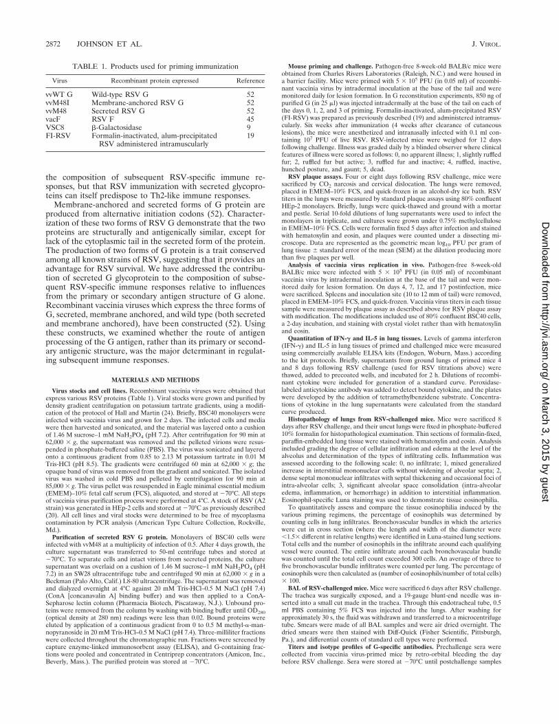
TABLE 1. Products used for priming immunization
Virus Recombinant protein expressed Reference
vvWT G Wild-type RSV G 52vvM48I Membrane-anchored RSV G 52vvM48 Secreted RSV G 52vacF RSV F 45VSC8 b-Galactosidase 9FI-RSV Formalin-inactivated, alum-precipitated
RSV administered intramuscularly19
2872 JOHNSON ET AL. J. VIROL.
on March 3, 2015 by guest
http://jvi.asm.org/
Dow
nloaded from

were collected. Six weeks following RSV challenge, the mice were again bled,and serum was separated and frozen. To measure G-specific antibody, NuncImmulon II microtiter plates were coated for 1 h with purified G protein (Wyeth-Lederle, Pearl River, N.Y.), the G protein isolated from RSV subtype A virus.Serial two-fold dilutions of pre- and postchallenge sera were added to the coatedwells in duplicate and incubated for 1 h. Bound immunoglobulin was detected bythe addition of horseradish peroxidase-conjugated rabbit anti-mouse immuno-globulin G1 (IgG1), IgG2a, or IgG/A/M (Zymed Laboratories, San Francisco,Calif.). The plates were developed with tetramethylbenzidene. Data are repre-sented as log2 values of the serum dilution producing 0.100 OD450 unit andgreater than twice the antigen-negative well.
Production of neutralizing antibodies to RSV and vaccinia virus. Titers ofneutralizing antibodies to both RSV and vaccinia virus in pre- and postchallengesera were measured by plaque reduction neutralization assays as previouslydescribed (20).
Statistical analysis. Data from individual experiments were maintained in aParadox database. Statistical analysis was performed by transferring data fromthe database into the SAS (Chapel Hill, N.C.) statistical software to performanalysis of variance using Kruskal-Wallis and Wilcoxon rank sum tests. Compar-isons were made between individual experiments by using statistical modelingand trend analysis calculated by the general linear model method in the SASpackage. P values less than 0.05 were considered statistically significant.
RESULTS
Weight loss and illness following challenge of primed mice.Mice were immunized with vaccinia virus constructs expressingF, wild-type G (vvWT G), secreted G only (vvM48), or mem-brane-anchored G only (vvM48I) (Table 1). Consistent withprevious observations (1, 61), illness in mice vaccinated withvvWT G was more severe and weight loss was significantlygreater than in vacF-immunized mice (P , 0.05 at days 4 to 12,Kruskal-Wallis test [data not shown]). Mice primed withvvM48I exhibited patterns of illness and weight loss similar tothose of vvWT G-primed mice (Fig. 1; P . 0.05 at all days).However, mice immunized with vvM48 demonstrated illnessand weight loss profiles significantly more severe than observedin vvM48I-primed mice (Fig. 1; P , 0.05 at all days except day7, where P 5 0.07). Thus, the presence of secreted G aloneduring priming, rather than membrane-anchored G, was asso-ciated with more severe disease upon subsequent RSV infec-tion. However, the presence of membrane-anchored G canhave a modulating effect on the illness profile induced by
secreted G, decreasing the severity, as seen in mice immunizedwith vvWT G.
RSV replication following viral challenge. When RSV titersin the lungs were measured 4 days postchallenge, the degree ofviral clearance differed among the priming groups (Fig. 2).VacF priming resulted in greater reduction of RSV titers inlung than vvWT G, confirming earlier observations (45). Re-duction in RSV titers in lung was similar between vvWT G-and vvM48I-primed mice. In contrast, priming with vvM48resulted in significantly less reduction in RSV titers than didvvWT G priming (P , 0.05, Tukey’s Studentized range test).Therefore, G is less immunogenic than F and secreted G ap-pears to be less immunogenic than membrane-anchored G.
In vivo production of G. Antigen dose has been shown toinfluence T-cell differentiation (12, 29, 51). Thus, productionof different concentrations of antigen by different virus vectorsmay potentially result in different profiles of immune responsesfollowing challenge. vvWT G, vvM48I, and vvM48 have beenshown to produce similar levels of RSV G in vitro in tissueculture systems (52). However, due to immunoregulatorymechanisms, this may not be the case in vivo. Thus, we soughtto confirm equal antigen production in vivo. Due to dissemi-nation of vaccinia virus, a direct measure of RSV G concen-trations in a given tissue was not considered to be an accuraterepresentation of antigen production. Therefore, we measuredthe dissemination and titers of vvM48I and vvM48. Similarpatterns of dissemination to spleen and equivalent viral titersin tail sections were observed at all time points examined (datanot shown). To further evaluate replication of these two vac-cinia virus constructs in vivo, neutralizing antibody titers weremeasured. vvM48I and vvM48 immunizations resulted in levelsof neutralizing antibodies similar to those for vaccinia virus(data not shown). Production of equal amounts of G fromvvM48I and vvM48 in vitro (52) suggests that in vivo levels ofproduction of G antigen between vvM48I and vvM48 are sim-ilar.
Reconstitution with purified G during priming. To deter-mine whether enhanced disease and reduced viral clearanceproduced by vvWT G and vvM48 sensitization were indeedrelated to the presence of secreted G during priming, immu-nization protocols in which purified G was coadministeredearly in the vaccination schedule were designed. Secreted G
FIG. 1. Illness in vaccine-primed mice after RSV challenge. Mice wereprimed with either vvWT G (F), vvM48I (}), vvM48 (Œ), or VSC8 (■) intra-dermally at the base of the tail and 6 weeks later challenged intranasally with liveRSV. Illness was monitored for 12 days following challenge. The data representthe mean and SEM for each group from four experiments (n 5 20 for all groups).P . 0.05 at days 5 to 11 when VSC8-primed mice are compared to vvM48-primedmice and when vvWT G- and vvM48I-primed mice are compared. When vvM48-primed mice are compared to vvM48I-primed mice, P , 0.05 at each day exceptday 7 (where P 5 0.07). Illness mirrored weight loss measurements (data notshown).
FIG. 2. RSV titers in lungs of vaccine-primed mice. Mice were primed andchallenged as for Fig. 1. On day 4 postchallenge, viral titers were measured bystandard plaque assay on HEp-2 monolayers and are represented as the log10(PFU/gram of lung) 6 SEM. Data from a representative experiment (of four)are shown (n 5 6 for FI-RSV and mock-primed groups; n 5 5 for all othergroups; P , 0.005).
VOL. 72, 1998 SECRETED RSV G PRIMING INDUCES IL-5 AND EOSINOPHILIA 2873
on March 3, 2015 by guest
http://jvi.asm.org/
Dow
nloaded from

was purified to homogeneity from vvM48-infected cell culturesupernatant by chromatography on a ConA-Sepharose affinitycolumn (data not shown). To simulate the conditions of prim-ing with wild-type G, purified G was administered on days 0 to3 to vvM48I-primed mice. The immunogenicity of G alone wasexamined by administration of purified protein during VSC8priming. All mice were challenged with RSV 6 weeks afterimmunization, and illness and weight loss were monitored. Theaddition of purified G during VSC8 priming established im-munologic conditions in which enhanced disease occurred fol-lowing RSV challenge, as evidenced by increased illness andweight loss relative to VSC8-primed mice (Fig. 3 and data notshown). Coadministration of purified G during vvM48I immu-nization did not result in a significant change in either illness orweight loss compared to mice primed with vvWT G or vvM48Ialone.
Clearance of RSV was also examined in mice primed withthe recombinant vaccinia constructs and purified G. Whilepurified G in combination with VSC8 priming resulted in en-hanced disease, it did not reduce RSV titers after challengecompared to mice primed with VSC8 alone (Fig. 4). Miceimmunized with vvM48I had significantly lower RSV titers inlung following challenge than mice primed with vvWT G or
vvM48 (Fig. 4; P , 0.05, Tukey’s Studentized range test).However, in mice coimmunized with purified G and vvM48I,the RSV titers in lungs 4 days after RSV challenge were notsignificantly different from those in vvM48- or vvWT G-primedmice (P . 0.05). Thus, addition of purified G during vaccina-tion alters the immunogenicity of vvM48I priming, decreasingviral clearance, suggesting secreted G actively influences themagnitude, specificity, or composition of the immune re-sponse.
G-specific antibody isotype response. Antibody titers in pre-challenge sera of immunized mice were evaluated in a G cap-ture ELISA. Detectable levels of immunoglobulin were mea-sured in each priming group except mice primed with VSC8(Fig. 5). Vaccination with regimens containing membrane-an-chored G produced higher titers of antibody than did regimenscontaining only secreted G protein. Subsequent to RSV chal-lenge, antibody titers were increased to similar levels in allgroups (data not shown).
In mice primed with vvM48I, IgG2a antibodies were thedominant isotype generated (n 5 5, P 5 0.01, Student’s t testcomparing IgG1 and IgG2a titers in vvM48I-primed mice). InvvWT G- and vvM48-primed mice, IgG2a were higher thanIgG1 titers, but the ratio of IgG2a to IgG1 was lower. With thecoadministration of secreted G during priming, a shift to IgG1-producing conditions was observed. In VSC8-primed micewhich also received purified G, the major antibody responsewas of an IgG1 isotype (n 5 5, P 5 0.025 relative to IgG2atiters from the same priming group). Coadministration of se-creted G during vvM48I immunization resulted in a shift froman IgG2a-dominated antibody response to a response in whichIgG1 and IgG2a antibody titers were about equal. The IgG1/IgG2a ratios in vvM48I-primed mice were significantly differ-ent from the IgG1/IgG2a ratios in mice primed with vvM48Iplus G (P 5 0.04, Student’s t test comparing IgG1/IgG2a ratios[data not shown]). Thus, addition of secreted G to the primingenvironment can induce a significant shift away from an IgG2a-dominated antibody response. These data suggest that thepresence of secreted G during priming reinforces the tendencyfor G-specific responses to produce IgG1 antibodies.
FIG. 3. RSV-induced illness in mice primed with RSV G-vaccinia virus con-structs and RSV G reconstitution. Mice were primed intradermally at the base ofthe tail with the indicated constructs and challenged intranasally with live RSV6 weeks after priming; illness was measured daily. Data from a representativeexperiment (of three) are shown and represented as mean 6 SEM (n 5 5; P ,0.05 at days 4 to 12).
FIG. 4. RSV titers in lungs of mice primed with RSV G-vaccinia virus con-structs and RSV G reconstitution. Mice were primed and challenged as for Fig.3. On day 4 postchallenge, viral titers were measured by standard plaque assay onHEp-2 monolayers and are represented as the log10 (PFU/gram of lung) 6 SEM.Data from a representative experiment (of three) are shown (n 5 5 for allgroups; P , 0.0001).
FIG. 5. G-specific antibody titers and isotype profiles. Mice were primed andchallenged as for Fig. 3. Serum samples were collected the day prior to challenge.Titers and isotypes of G-specific antibodies were measured in a capture ELISAby coating wells with purified G protein, adding dilutions of sera, and detectingbound IgG with horseradish peroxidase-conjugated anti-isotype antibodies. Pre-challenge IgG1 and IgG2a were measured. Data from a representative experi-ment (of three) are represented as the mean 6 SEM of the log2 (serum dilution)producing 0.1 OD450 unit for each group. When IgG1 and IgG2a titers werecompared in each priming group, the following P values were obtained: .0.5 forVSC8, vvWT G, vvM48, and vvM48I plus G; 0.025 for VSC8 plus G; and 0.01 forvvM48I (Student’s t test; n 5 5 for all groups but vvWT G and vvM48, where n 56 and 7, respectively).
2874 JOHNSON ET AL. J. VIROL.
on March 3, 2015 by guest
http://jvi.asm.org/
Dow
nloaded from
Cytokine production in the lungs. The concentrations ofIL-5 and IFN-g in the lung supernatants from mice 4 and 8days after RSV challenge were measured by ELISA. Both IL-5and IFN-g could be measured in day 4 samples and are shownin Table 2. Little IL-5 was detectable in day 8 lung superna-tants, and IFN-g levels were greatly reduced (data not shown).Little IL-5 was measured in the lung supernatants of miceimmunized with VSC8 (Table 2). Highest concentrations ofIL-5 were found in mice primed with vvM48I plus G andvvM48. Statistically significant differences were found whenIL-5 levels in vvWT G-primed mice were compared to those ineither vvM48-primed mice or vvM48I-plus-G-primed mice(P . 0.0001, Student’s t test). Similarly, compared to vvM48I-plus-G-primed mice, the IL-5 levels in vvM48I-primed mice(P 5 0.01) and in vvM48-primed mice (P 5 0.004) were sig-nificantly different. Mice primed with vvWT G and VSC8 plusG had intermediate IL-5 levels, and vvM48I-primed mice hadthe lowest levels, although the differences between the threegroups were not statistically significant (P . 0.05, Student’s ttest). Thus, increased IL-5 levels were correlated with primingregimens that included secreted G and paralleled the titers ofG-specific IgG1.
Lung pathology following RSV challenge. The alveolar in-flammation induced by priming with the different forms of Gprotein was evaluated (Fig. 6 and 7). In mice immunized withVSC8, vvWT G, or vvM48, moderate alveolar mononuclearinfiltration occurred upon RSV challenge (Fig. 6A, C, and D,respectively). In mice primed with vvWT G and vvM48, asignificant eosinophil component was present in the infiltratesaround arteries (Fig. 7C, D, and G). Although vvM48I primingreduced the density of infiltration (Fig. 6E), small numbers ofeosinophils were still detected focally (Fig. 7E and G). When Gwas coadministered during VSC8 priming, the pathology re-sulting from challenge was more severe (Fig. 6B and G) andinduced prominent eosinophilia (Fig. 7F and G), absent inmice immunized with VSC8 alone (Fig. 7A). The addition ofsecreted G during vvM48I priming did not significantly in-crease the severity of lymphocytic infiltration (Fig. 6F and G);however, this priming regimen did result in the recruitment ofabundant eosinophils into the periarterial infiltrates (Fig. 7F
and G). When the degree of eosinophilia was quantitativelyexamined (Fig. 7G), similar percentages of eosinophils werefound in the lungs of mice primed with VSC8 plus G, vvWT G,and vvM48I plus G (P . 0.1, Student’s t test). The addition ofsecreted G to vvM48I priming resulted in significantly greaterpercentage of eosinophils infiltrating the lung (P 5 0.002,vvM48I compared to vvM48I plus G). Similar increases werefound with the addition of secreted G to VSC8 priming (P ,0.0001). Thus, the presence of G-specific IgG1 titers and IL-5content in lung correlated with the amount of tissue eosino-philia.
Cellular composition of BAL infiltrate after RSV challenge.To determine whether infiltration of cells into the air spacediffered from that seen in the periarterial regions and to quan-titate the cellular components of the infiltrate, BAL was per-formed on day 6 following challenge (Table 2). The greatestnumber of eosinophils occurred in mice primed with VSC8plus purified G and with vvM48, and slightly fewer eosinophilswere obtained in BAL of vvWT G-primed mice, although nostatistically significant difference was detected between thesegroups. Significantly fewer eosinophils were recruited into thebronchial airways of mice immunized with vvM48I with orwithout purified G (P , 0.05 relative to vvWT G or vvM48),contrasting with the increased interstitial eosinophilia ob-served with coadministration of secreted G during vvM48Ipriming (Table 2 and Fig. 7F). Therefore, groups with a highdegree of BAL eosinophilia also had increased illness andweight loss. Although the degree of BAL eosinophilia gener-ally corresponded to tissue eosinophilia, mice primed withvvM48I plus G had a higher frequency of tissue eosinophiliayet few eosinophils in BAL.
DISCUSSION
These data suggest that the presence of the secreted form ofRSV G during initial antigen presentation (and not solely theprimary or secondary antigenic structure) affects the composi-tion of immune responses to RSV infection. Previous work hasshown that immunization with RSV G results in more severedisease and lung pathology following RSV challenge (25, 61).The molecular basis for this alteration in the immune responsehas not been defined. The unique nature of G provides severalattributes which may generate the altered immune response toRSV challenge. First, G is extensively glycosylated: .50% ofthe weight of the mature protein is contributed by carbohy-drate residues, attached predominantly via O-glycosidic bonds(70). Also, G contains a high proportion (30.6%) of serine andthreonine residues which serve as acceptors for the O-linkedoligosaccharides. Third, G protein lacks homology to any otherknown paramyxovirus protein. The high proline content of10.1% has more in common with mucinous glycoproteins thanviral glycoproteins (31, 39, 70). G exhibits neither hemagglu-tinating nor neuraminidase activities (22) and is also unusual inthat it lacks both a hydrophobic NH2-terminal signal sequenceand a hydrophobic COOH-terminal domain, features presentin RSV F and other paramyxovirus proteins (70).
In contrast to previously published studies using recombi-nant vaccinia virus expressing wild-type RSV G (25, 60, 61),mice primed with recombinant vaccinia virus expressing onlymembrane-anchored G are partially protected during RSVchallenge, with reduced viral titers observed, although illnesspatterns are similar. However, in the absence of membrane-anchored G, secreted G increases illness and decreases viralclearance. We have shown that priming with secreted G (forboth vvWT G and vvM48) is associated with increased IL-5production and more severe immunopathology, including in-
TABLE 2. Quantitative measures of lung immunopathology inprimed mice after RSV challengea
Primingregimen
% Eosinophil inBAL fluidb
Concn (mean 6 SEM; n 5 5)
IL-5(pg/ml)c
IFN-g(ng/ml)
VSC8 0.0 6 0.0 10.4 6 2.1 17.8 6 3.8VSC8 1 G 8.3 6 3.7 21.9 6 3.5 5.1 6 0.3vvWT G 5.9 6 2.6 22.3 6 5.1 10.5 6 2.9vvM48 7.6 6 3.4 104.4 6 5.4 27.5 6 4.4vvM48I 1.9 6 0.9 15.1 6 6.5 31.4 6 2.5vvM48I 1 G 1.3 6 0.6 64.0 6 3.4 15.7 6 2.7
a The mice were primed and challenged as for Fig. 3. Percentage of eosinophilsin BAL fluid was determined by counting Diff-Quik-stained smears. Concentra-tions of IL-5 and IFN-g were measured in day 4 (shown) and day 8 (not shown)lung supernatants by ELISA. Limits of detection are 10 pg/ml for IL-5 and 50pg/ml for IFN-g.
b Differences between VSC8, vvWT G, and vvM48 were not statistically sig-nificant by Student’s t test, nor were the differences between vvM48I and vvM48plus G. However, the differences between the mice primed with vvM48I (orvvM48I plus G) and the mice primed with VSC8 plus G, vvWT G, or vvM48 weresignificant (P , 0.05 for all comparisons).
c Statistically significant differences were found as follows: vvM48I comparedto vvM48I plus G (P 5 0.01), and vvM48 compared to all other groups (P ,0.025). Differences between VSC8 and VSC8 plus G, between vvWT G andvvM48I, and between vvWT G and VSC8 plus G were not significant.
VOL. 72, 1998 SECRETED RSV G PRIMING INDUCES IL-5 AND EOSINOPHILIA 2875
on March 3, 2015 by guest
http://jvi.asm.org/
Dow
nloaded from

creased tissue eosinophilia. However, illness correlates withalveolar infiltration of eosinophils, as determined by their pres-ence in BAL, and suggests that secreted G induces factorsother than IL-5 which may be responsible for a second step inthe activation and migration of eosinophils. These data suggestthe secreted form of RSV G modulates the composition of theimmune response by inducing IL-5 production and alteringpatterns of leukocyte trafficking, while the membrane-an-
FIG. 6. Lung histopathology. Mice were primed with VSC8 (A), VSC8 plusG (B), vvWT G (C), vvM48 (D), vvM48I (E), or vvM48I plus G (F) andchallenged as for Fig. 3. Eight days following challenge, lung histopathology wasevaluated by staining formalin-fixed lung sections with hematoxylin and eosin(original magnification, 325). Day 8 lungs were examined and graded for severityand composition of alveolar inflammation on a semiquantitative scale (G). Nostatistical difference was found among any group except the VSC8-plus-G group,in which inflammation was significantly higher than in any other group (P , 0.03,Student’s t test, n 5 5).
2876 JOHNSON ET AL. J. VIROL.
on March 3, 2015 by guest
http://jvi.asm.org/
Dow
nloaded from

chored form of G may have a separate and dominant immu-noregulatory effect on factors producing illness.
Several immunoregulatory mechanisms have been describedfor soluble antigen. Soluble antigen has been shown to regulatethe development of germinal center B cells by selectively in-ducing apoptosis of high-affinity antigen-specific B lympho-cytes (50, 56). This ability to direct B-cell differentiation ishypothesized to indicate clonal deletion of self-reactive B cells
FIG. 7. Tissue eosinophilia. Mice were primed with VSC8 (A), VSC8 plus G(B), vvWT G (C), vvM48 (D), vvM48I (E), or vvM48I plus G (F) and challengedas for Fig. 3. Eight days following challenge, lung eosinophilia was evaluated bystaining tissue sections with Luna’s stain (original magnification, 3250). Tissueeosinophilia was quantitatively measured by counting the percentages of eosin-ophils in Luna-stained formalin-fixed day 8 lungs (G; n 5 5). Differences be-tween VSC8 plus G, vvWT G, and vvM48I plus G were not statistically signifi-cant. vvM48-primed mice were significantly different from all other groups (P ,0.05). P 5 0.002 for vvM48I compared to vvM48I plus G; P , 0.0001 for VSC8compared to VSC8 plus G and for vvWT G compared to vvM48I.
VOL. 72, 1998 SECRETED RSV G PRIMING INDUCES IL-5 AND EOSINOPHILIA 2877
on March 3, 2015 by guest
http://jvi.asm.org/
Dow
nloaded from

and, thus, be a mechanism of avoiding autoimmune disease.Soluble antigen will be processed and presented in the contextof major histocompatibility (MHC) class II molecules. MHCcontext of antigen presentation (41), the type of antigen-pre-senting cell used (11, 54), and the presence of different co-stimulatory molecules (13, 55) have been shown to regulate thedevelopment and maturation of T-cell subsets. Antigen dosemay also be a determinant in T-cell differentiation. In mostsystems, immunization with large doses of antigen induceseffector cells exhibiting a type 1 cytokine profile, whereas lowerconcentrations of priming antigen tend to generate type 2effector cells (12, 29). However, some antigens have the re-versed profile, with high doses favoring development of Th2cells and low doses inducing Th1 cells (51). Thus, the inductionof severe disease by the presence of soluble G during primingsuggests that either the concentration or processing of G or itssubsequent recognition by selected T-cell subsets may be a keydeterminant regulating immune responses following subse-quent RSV challenge.
Infectious organisms ensure their survival by developingmechanisms to avoid detection or subvert immune responses.Some strategies include production of cytokine receptors bythe organism (30) or blocking induction of the immune re-sponse at the level of antigen presentation (14, 28). A body ofevidence which suggests that induction of Th2-mediated im-mune responses provides a survival advantage for many intra-cellular pathogens, including viruses, is also accumulating (13,21). For example, measles virus suppresses cell-mediated im-munity by down-regulation of IL-12 expression through signalsmediated by measles virus binding CD46 (33). Also, diseaseprogression and infection with human immunodeficiency virushave been associated with Th2-dominated responses (16, 37).Human immunodeficiency virus infection has been shown tosuppress IL-2, IL-12, and IFN-g, but not tumor necrosis factoralpha or IL-6, induction by Toxoplasma gondii (15). Thus, theinduction of a type 2 T-cell response by secreted RSV G mayserve as a defense mechanism the virus has evolved to favor-ably modulate immune responses.
Previous work has established priming or infection with G-expressing vectors results in increased illness and pathology(61). The ability of RSV G to produce cytokines associatedwith a Th2 response has been clearly demonstrated (2, 4, 25).In this and other studies, FI-RSV priming has also been shownto induce Th2-type cytokine profiles in mice (19, 62, 69),thereby associating this type of response with the RSV vaccine-enhanced illness seen in children in FI-RSV vaccine studies inthe 1960s (32, 34). In this study, we have addressed whether itis the primary or secondary antigen structure of G, or thepresence of secreted G or antigens with obligate processingthrough the endocytic pathway and MHC class II expression,which increases disease severity. The IgG2a isotype antibodiesand disease protection observed in mice primed with mem-brane-anchored G and the IgG1 isotype antibodies and moresevere illness induced by immunization with secreted G implythat different Th1- and Th2-associated cytokine profiles maybe induced by priming without and with secreted G, respec-tively. We therefore propose that the link between RSV Gimmunization, FI-RSV-induced vaccine-enhanced illness, andinduction of type 2 cytokine profiles is not induced by G itselfbut rather results from the initial presentation of G to theimmune system in secreted form. This might then promote theinduction of Th2 CD41 T-cell differentiation in part by pre-cluding intracellular antigen processing, presentation of anti-gen by MHC class I molecules, and loss of the early contribu-tion of CD81 T cells to the cytokine milieu (17, 59).
Evidence suggests that full activation of eosinophils requires
a two-step process involving IL-5, eotaxin, or other cofactors(42, 53). Extracellular matrix proteins (36) and VLA-4 oneosinophils (44) may also be involved in eosinophil recruit-ment and infiltration. These observations may be invoked toexplain the differential patterns of eosinophilia and diseaseseverity induced by priming with membrane-anchored and se-creted G. Our data suggest the hypothesis that immunizationwith any form of G (membrane anchored or secreted) mayrecruit eosinophils upon challenge via the induction of IL-5,but the presence of secreted G is required for inducing theactivation associated with movement of eosinophils into alve-olar spaces to produce more severe disease.
The data presented in this paper suggest that priming withsecreted RSV G promotes eosinophil recruitment associatedwith IL-5 production and that illness is associated with themovement of eosinophils from the interstitial compartment inthe lung to the alveoli. We propose the secretion of G fromRSV-infected cells represents a strategy to modulate andevade effective immune responses. The presence of a smallnumber of alveolar eosinophils in mice primed with mem-brane-anchored G suggests a direct effect of the primary orsecondary antigenic structure of G on the induction of type 2immune responses that is compounded by its secretion as asoluble protein, resulting in increased eosinophilia. Under-standing how secreted G interacts with different elements ofthe innate and adaptive immune system might provide insightinto basic mechanisms of clearance for RSV and other viruses.
ACKNOWLEDGMENTS
This work was supported by National Institutes of Health grantsRO1-AI-37216 (B.S.G.) and RO1-AI-20181 (G.W.W.).
We thank Rauf Kuli-Zade and Peter Veldkamp for technical assis-tance and James E. Crowe, Jr., for editorial comments.
REFERENCES
1. Alwan, W., W. Kozlowska, and P. Openshaw. 1994. Distinct types of lungdisease caused by functional subsets of antiviral T cells. J. Exp. Med. 179:81–89.
2. Alwan, W., and P. Openshaw. 1993. Distinct patterns of T- and B-cell im-munity to respiratory syncytial virus induced by individual viral proteins.Vaccine 11:431–437.
3. Alwan, W., F. Record, and P. Openshaw. 1992. CD41 T cells clear virus butaugment disease in mice infected with respiratory syncytial virus: comparisonwith the effects of CD81 T cells. Clin. Exp. Immunol. 88:527–536.
4. Alwan, W., F. Record, and P. Openshaw. 1993. Phenotypic and functionalcharacterization of T cell lines specific for individual respiratory syncytialvirus proteins. J. Immunol. 150:5211–5218.
5. Anderson, L., and C. Heilman. 1995. Protective and disease-enhancing im-mune response to respiratory syncytial virus. J. Infect. Dis. 171:1–7.
6. Ball, L., K. Young, K. Anderson, P. Collins, and G. Wertz. 1986. Expressionof the major glycoprotein G of human respiratory syncytial virus from re-combinant vaccinia virus vectors. Proc. Natl. Acad. Sci. USA 83:246–250.
7. Bangham, C., M. Cannon, D. Karzon, and B. Askonas. 1985. Cytotoxic T-cellresponse to respiratory syncytial virus in mice. J. Virol. 56:55–59.
8. Bangham, C., P. Openshaw, A. Ball, A. King, G. Wertz, and B. Askonas.1986. Human and murine cytotoxic T cells specific to respiratory syncytialvirus recognize the viral nucleoprotein (N), but not the major glycoprotein(G), expressed by vaccinia virus recombinants. J. Immunol. 137:3973–3977.
9. Chakrabarti, S., K. Brechling, and B. Moss. 1985. Vaccinia virus expressionvector: coexpression of b-galactosidase provides visual screening of recom-binant virus plaques. Mol. Cell. Biol. 5:3403–3409.
10. Connors, M., P. Collins, C. Firestone, A. Sotnikov, A. Waitze, A. Davis, P.Hung, R. Chanock, and B. Murphy. 1992. Cotton rats previously immunizedwith a chimeric RSV FG glycoprotein develop enhanced pulmonary pathol-ogy when infected with RSV, a phenomenon not encountered followingimmunization with vaccinia-RSV recombinants or RSV. Vaccine 10:475–484.
11. Conradt, P., and S. Kaufmann. 1995. Impact of antigen-presenting cells oncytokine profiles of human Th clones established after stimulation withMycobacterium tuberculosis antigens. Infect. Immun. 63:2079–2081.
12. Constant, S., C. Pfeiffer, A. Woodard, T. Pasqualini, and K. Bottomly. 1995.Extent of T cell receptor ligation can determine the functional differentiationof naive CD41 T cells. J. Exp. Med. 182:1591–1596.
2878 JOHNSON ET AL. J. VIROL.
on March 3, 2015 by guest
http://jvi.asm.org/
Dow
nloaded from

13. Corry, D., S. Reiner, P. Linsley, and R. Locksley. 1994. Differential effects ofblockade of CD28-B7 on the development of Th1 or Th2 effector cells inexperimental leishmaniasis. J. Immunol. 153:4142–4148.
14. Fruh, K., K. Ahn, H. Djaballah, P. Sempe, P. M. van Endert, R. Tampe, P. A.Peterson, and Y. Yang. 1995. A viral inhibitor of peptide transporters forantigen presentation. Nature 375:415–418.
15. Gazzinelli, R. T., S. Baia, R. Stevens, M. Baseler, L. Wahl, J. Kovacs, and A.Sher. 1995. HIV infection suppresses type 1 lymphokine and IL-12 responsesto Toxoplasma gondii but fails to inhibit the synthesis of other parasite-induced monokines. J. Immunol. 155:1565–1574.
16. Gazzinelli, R. T., M. Makino, S. K. Chattopadhyay, C. M. Snapper, A. Sher,A. W. Hugin, and H. C. Morse. 1992. CD41 subset regulation in viralinfection. Preferential activation of Th2 cells during progression of retrovi-rus-induced immunodeficiency in mice. J. Immunol. 148:182–188.
17. Graham, B. S. 1996. Immunological determinants of disease caused by re-spiratory syncytial virus. Trends Microbiol. 4:290–294.
18. Graham, B. 1995. Pathogenesis of respiratory syncytial virus vaccine-aug-mented pathology. Am. J. Respir. Crit. Care Med. 152:563–566.
19. Graham, B., G. Henderson, Y. Tang, X. Lu, K. Neuzil, and D. Colley. 1993.Priming immunization determines T helper cytokine mRNA expression pat-terns in lungs of mice challenged with respiratory syncytial virus. J. Immunol.151:2032–2040.
20. Graham, B., M. Perkins, P. Wright, and D. Karzon. 1988. Primary respira-tory syncytial virus infection in mice. J. Med. Virol. 26:153–162.
21. Griffin, D. E., and B. J. Ward. 1993. Differential CD4 T cell activation inmeasles. J. Infect. Dis. 168:275–281.
22. Gruber, C., and S. Levine. 1983. Respiratory syncytial virus polypeptides. III.The envelope-associated proteins. J. Gen. Virol. 64:825–832.
23. Hall, C., E. Walsh, C. Long, and K. Schnabel. 1991. Immunity to andfrequency of reinfection with respiratory syncytial virus. J. Infect. Dis. 163:693–698.
24. Hall, W., and S. Martin. 1973. Purification and characterization of measlesvirus. J. Gen. Virol. 19:175–188.
25. Hancock, G., D. Speelman, K. Heers, E. Bortell, J. Smith, and C. Cosco.1996. Generation of atypical pulmonary inflammatory responses in BALB/cmice after immunization with the native attachment (G) glycoprotein ofrespiratory syncytial virus. J. Virol. 70:7783–7791.
26. Hendricks, D., K. Baradaran, K. McIntosh, and J. Patterson. 1987. Appear-ance of a soluble form of the G protein of respiratory syncytial virus in fluidsof infected cells. J. Gen. Virol. 68:1705–1714.
27. Hendricks, D., K. McIntosh, and J. Patterson. 1988. Further characteriza-tion of the soluble form of the G glycoprotein of respiratory syncytial virus.J. Virol. 62:2228–2233.
28. Hill, A., P. Jugovic, I. York, G. Russ, J. Bennicnk, J. Yewdell, H. Ploegh, andD. Johnson. 1995. Herpes simplex virus turns off the TAP to evade hostimmunity. Nature 375:411–415.
29. Hosken, N. A., K. Shibuya, A. W. Heath, K. M. Murphy, and A. O’Garra.1995. The effect of antigen dose on CD41 T helper cell phenotype devel-opment in a T cell receptor-alpha beta-transgenic model. J. Exp. Med.182:1579–1584.
30. Hu, F. Q., C. A. Smith, and D. J. Pickup. 1994. Cowpox virus contains twocopies of an early gene encoding a soluble secreted form of the type II TNFreceptor. Virology 204:343–356.
31. Johnson, P., M. Spriggs, R. Olmstead, and P. Collins. 1987. The G glyco-protein of human respiratory syncytial viruses of subgroups A and B: exten-sive sequence divergence between antigenically related proteins. Proc. Natl.Acad. Sci. USA 84:5625–5629.
32. Kapikian, A., R. Mitchell, R. Chanock, R. Shvedoff, and C. Stewart. 1969. Anepidemiologic study of altered clinical reactivity to respiratory syncytial (RS)virus infection in children previously vaccinated with an inactivated RS virusvaccine. Am. J. Epidemiol. 89:405–421.
33. Karp, C. L., M. Wysocka, L. M. Wahl, J. M. Ahearn, P. J. Cuomo, B. Sherry,G. Trinchieri, and D. E. Griffin. 1996. Mechanism of suppression of cell-mediated immunity by measles virus. Science 273:228–231.
34. Kim, H. W., J. G. Canchola, C. D. Brandt, G. Pyles, R. M. Chanock,K. Jensen, and R. H. Parrott. 1969. Respiratory syncytial virus disease ininfants despite prior administration of antigenic inactivated vaccine. Am. J.Epidemiol. 89:422–434.
35. Kim, H., S. Leikin, J. Arrobio, C. Brandt, R. Chanock, and R. Parrott. 1976.Cell-mediated immunity to respiratory syncytial virus induced by inactivatedvaccine or by infection. Pediatr. Res. 10:75–78.
36. Kita, H., S. Horie, and G. J. Gleich. 1996. Extracellular matrix proteinsattenuate activation and degranulation of stimulated eosinophils. J. Immu-nol. 156:1174–1181.
37. Kurup, V. P., B. W. Seymour, H. Choi, and R. L. Coffman. 1994. ParticulateAspergillus fumigatus antigens elicit a TH2 response in BALB/c mice. J.Allergy Clin. Immunol. 93:1013–1020.
38. Lambert, D. 1988. Role of oligosaccharides in the structure and function ofrespiratory syncytial virus glycoproteins. Virology 16:458–466.
39. Langedijk, J., W. Schaaper, R. Meloen, and J. van Oirschot. 1996. Proposedthree-dimensional model for the attachment protein G of respiratory syncy-tial virus. J. Gen. Virol. 77:1249–1257.
40. Levine, S., R. Franco-Klaiber, and P. Paradiso. 1987. Demonstration thatglycoprotein G is the attachment protein of respiratory syncytial virus.J. Gen. Virol. 68:2521–2524.
41. Milich, D., D. Peterson, F. Schodel, J. Jones, and J. Hughes. 1995. Prefer-ential recognition of hepatitis B nucleocapsid antigens by Th1 or Th2 cells isepitope and major histocompatibility complex dependent. J. Virol. 69:2776–2785.
42. Mould, A. W., K. I. Matthaei, I. G. Young, and P. S. Foster. 1997. Relation-ship between interleukin-5 and eotaxin in regulating blood and tissue eosin-ophilia in mice. J. Clin. Invest. 99:1064–1071.
43. Murphy, B., B. Graham, G. Prince, E. Walsh, R. Chanock, D. Karzon, andP. Wright. 1986. Serum and nasal-wash immunoglobulin G and A antibodyresponse of infants and children to respiratory syncytial virus F and Gglycoproteins following primary infection. J. Clin. Microbiol. 23:1009–1014.
44. Nakajima, H., H. Sano, T. Nishimura, S. Yoshida, and I. Iwamoto. 1994.Role of vascular cell adhesion molecule 1/very late activation antigen 4 andintercellular adhesion molecule 1/lymphocyte function-associated antigen 1interactions in antigen-induced eosinophil and T cell recruitment into thetissue. J. Exp. Med. 179:1145–1154.
45. Olmsted, R., N. Elango, G. Prince, B. Murphy, P. Johnson, B. Moss, R.Chanock, and P. Collins. 1986. Expression of the F glycoprotein of respira-tory syncytial virus by a recombinant vaccinia virus: comparison of the indi-vidual contributions of the F and G glycoproteins to host immunity. Proc.Natl. Acad. Sci. USA 83:7462–7466.
46. Openshaw, P., S. Clarke, and F. Record. 1992. Pulmonary eosinophilic re-sponse to respiratory syncytial virus infection in mice sensitized to the majorsurface glycoprotein G. Int. Immunol. 4:493–500.
47. Palomo, C., B. Garcia-Barreno, C. Penas, and J. Melero. 1991. The Gprotein of human respiratory syncytial virus: significance of carbohydrateside-chains and the C-terminal end to its antigenicity. J. Gen. Virol. 72:669–675.
48. Pemberton, R., M. Cannon, P. Openshaw, L. Ball, G. Wertz, and B. Askonas.1987. Cytotoxic T cell specificity for respiratory syncytial virus proteins:fusion protein is an important target antigen. J. Gen. Virol. 68:2177–2182.
49. Prince, G., A. Jenson, V. Hemming, B. Murphy, E. Walsh, R. Horswood, andR. Chanock. 1986. Enhancement of respiratory syncytial virus pulmonarypathology in cotton rats by prior intramuscular inoculation of formalin-inactivated virus. J. Virol. 57:721–728.
50. Pulendran, B., G. Kannourakis, S. Nouri, K. Smith, and G. Nossal. 1995.Soluble antigen can cause enhanced apoptosis of germinal-centre B cells.Nature 375:331–334.
51. Rizzo, L. V., R. H. DeKruyff, D. T. Umetsu, and R. R. Caspi. 1995. Regula-tion of the interaction between Th1 and Th2 T cell clones to provide help forantibody production in vivo. Eur. J. Immunol. 25:708–716.
52. Roberts, S., D. Lichtenstein, L. Ball, and G. Wertz. 1994. The membrane-associated and secreted forms of the respiratory syncytial virus attachmentglycoprotein G are synthesized from alternative initiation codons. J. Virol.68:4538–4546.
53. Rothenberg, M. E., R. Ownbey, P. D. Mehlhop, P. M. Loiselle, M. van deRijn, J. V. Bonventre, H. C. Oettgen, P. Leder, and A. D. Luster. 1996.Eotaxin triggers eosinophil-selective chemotaxis and calcium flux via a dis-tinct receptor and induces pulmonary eosinophilia in the presence of inter-leukin 5 in mice. Mol. Med. 2:334–348.
54. Schmitz, J., M. Assenmacher, and A. Radbruch. 1993. Regulation of Thelper cell cytokine expression: functional dichotomy of antigen-presentingcells. Eur. J. Immunol. 23:191–199.
55. Shanafelt, M., C. Soderberg, A. Allsup, D. Adelman, G. Peltz, and R. La-hesmaa. 1995. Costimulatory signals can selectively modulate cytokine pro-duction by subsets of CD41 T cells. J. Immunol. 154:1684–1690.
56. Shokat, K., and C. Goodnow. 1995. Antigen-induced B-cell death and elim-ination during germinal-centre immune responses. Nature 375:334–338.
57. Simard, C., F. Nadon, C. Seguin, and M. Trudel. 1995. Evidence that theamino acid region 124-203 of glycoprotein G from the respiratory syncytialvirus (RSV) constitutes a major part of the polypeptide domain that isinvolved in the protection against RSV infection. Antiviral Res. 28:303–315.
58. Srikiatkhachorn, A., and T. Braciale. 1997. Virus-specific memory and ef-fector T lymphocytes exhibit different cytokine responses to antigens duringexperimental murine respiratory syncytial virus infection. J. Virol. 71:678–685.
59. Stott, E., L. Ball, K. Young, J. Furze, and G. Wertz. 1986. Human respiratorysyncytial virus glycoprotein G expressed from a recombinant vaccinia virusvector protects mice against live-virus challenge. J. Virol. 60:607–613.
60. Stott, E., G. Taylor, L. Ball, K. Anderson, K. Young, A. King, and G. Wertz.1987. Immune and histopathological responses in animals vaccinated withrecombinant vaccinia viruses that express individual genes of human respi-ratory syncytial virus. J. Virol. 61:3855–3861.
61. Tang, Y., and B. Graham. 1994. Anti-IL-4 treatment at immunization mod-ulates cytokine expression, reduces illness, and increases cytotoxic T lym-phocyte activity in mice challenged with respiratory syncytial virus. J. Clin.Invest. 94:1953–1958.
62. Taylor, G., E. Stott, M. Bew, B. Fernie, P. Cote, A. Collins, M. Hughes, andJ. Jebbett. 1984. Monoclonal antibodies protect against respiratory syncytial
VOL. 72, 1998 SECRETED RSV G PRIMING INDUCES IL-5 AND EOSINOPHILIA 2879
on March 3, 2015 by guest
http://jvi.asm.org/
Dow
nloaded from
virus infection in mice. Immunology 52:137–142.63. Wagner, D., P. Muelenaer, F. Henderson, M. Snyder, C. Reimer, E. Walsh,
L. Anderson, D. Nelson, and B. Murphy. 1989. Serum immunoglobulin Gantibody subclass response to respiratory syncytial virus F and G glycopro-teins after first, second, and third infections. J. Clin. Microbiol. 27:589–592.
64. Wagner, D., B. Graham, P. Wright, E. Walsh, H. Kim, C. Reimer, D. Nelson,R. Chanock, and B. Murphy. 1986. Serum immunoglobulin G antibodysubclass responses to respiratory syncytial virus F and G glycoproteins afterprimary infection. J. Clin. Microbiol. 24:304–306.
65. Walsh, E., C. Hall, M. Briselli, M. Brandiss, and J. Schlesinger. 1987.Immunization with glycoprotein subunits of respiratory syncytial virus toprotect cotton rats against viral infection. J. Infect. Dis. 155:1198–1204.
66. Walsh, E., C. Hall, M. Briselli, M. Brandiss, and J. Schlesinger. 1987.Immunization with glycoprotein subunits of respiratory syncytial virus pro-tect cotton rats against viral infection. J. Infect. Dis. 155:1198–1204.
67. Walsh, E., J. Schlesinger, and M. Brandiss. 1984. Purification and charac-terization of GP90, one of the envelope glycoproteins of respiratory syncytialvirus. J. Gen. Virol. 65:761–767.
68. Ward, K., P. Lambden, M. Oglivie, and P. J. Watt. 1983. Antibodies torespiratory syncytial virus polypeptides and their significance in human in-fection. J. Gen. Virol. 64:1867–1876.
69. Waris, M., C. Tsou, D. Erdman, S. Zaki, and L. Anderson. 1996. Respiratorysyncytial virus infection in BALB/c mice previously immunized with forma-lin-inactivated virus induces enhanced pulmonary inflammatory responsewith a predominant Th2-like cytokine pattern. J. Virol. 70:2852–2860.
70. Wertz, G., P. Collins, Y. Huang, C. Gruber, and S. Levine. 1985. Nucleotidesequence of the G protein gene of human respiratory syncytial virus revealsan unusual type of viral membrane protein. Proc. Natl. Acad. Sci. USA82:4075–4079.
2880 JOHNSON ET AL. J. VIROL.
on March 3, 2015 by guest
http://jvi.asm.org/
Dow
nloaded from