Flow of winter-transformed Pacific water into the Western Arctic
Upload
independentCategory
view
0download
0
VIROLOGY 216, 347–356 (1996)ARTICLE NO. 0070
Two Novel Variants of the v-src Oncogene Isolated from Lowand High Metastatic RSV-Transformed Hamster Cells
ALEXANDER TATOSYAN,*,† BOGDAN YATSULA,* MICHAIL SHTUTMAN,† ELENA MOINOVA,† IRINA KAVERINA,†ELENA MUSATKINA,† KONSTANTIN LESKOV,† OLGA MIZENINA,† ELINA ZUEVA,†
GEORGES CALOTHY,1,* and PHILIPPE DEZELEE*
*Unite Mixte de Recherche 146 du CNRS, Institut Curie, Centre Universitaire, 91405, Orsay, France;and †Cancer Research Center, 115478, Moscow, Russia
Received August 23, 1995; accepted December 19, 1995
Four different transformed cell lines were isolated as a result of independent infection of primary hamster fibroblasts byRous sarcoma virus (RSV SR-D stocks). These lines differ by the level of their spontaneous metastatic activity: HET-SR-1,HET-SR-8, and HET-SR-10 cell lines induced 70–200 metastatic nodules in the lung and/or lymph nodes of inoculatedanimals (high metastatic lines, HM). Metastatic activity was not identified after injection of HET-SR cells (low metastaticline, LM). All cell lines contained one copy of integrated and expressed intact RSV provirus. The difference in the amountof v-src protein in cell lines was not correlated with their metastatic potential in vivo. Complete v-srcHM and v-srcLM geneswere cloned from corresponding gene libraries and sequenced. In the unique region of both v-src isoforms a GC-rich insertof 60 nucleotides (20 a.a.) was found. The presence of this insert explains the unusual apparent molecular weight of proteinencoded by v-srcHM and v-srcLM: 62 kDa. Both genes had 10 identical amino acid changes when compared to the knownRSV SR-D v-src sequence. v-srcHM and v-srcLM differ by several amino acid changes. Most of them are localized in theunique domain and the extreme carboxy-terminal region of the oncoprotein. Both v-src variants and chimeric v-src withmutually substituted parts were subcloned in a retroviral vector and introduced into avian neuroretina cells. Significantdifferences in the morphology of transformed neuroretina cells were associated with the mutations in the carboxy-terminalregion of the v-src oncogene. Low metastatic HET-SR cells transfected with v-srcHM and the chimeric gene v-src-LHremarkably increased their metastatic potential. In contrast, this effect was not observed when the same cells weretransfected with v-srcLM and the chimeric v-srcHL gene. Specific changes in the distribution of fibronectin matrix typicalfor high metastatic cells were found in the lines transfected with v-srcHM. q 1996 Academic Press, Inc.
INTRODUCTION They were designated domains SH3 (residues 86–140)and SH2 (residues 141–260). The most conserved region
The transforming gene (v-src) of Rous sarcoma virus of proteins of the src family is the catalytic domain (resi-(RSV) encodes a 60-kDa phosphoprotein (pp60src) that dues 265–516), which shares a high degree of homologyexhibits tyrosine-specific protein kinase activity essential with other tyrosine kinases and to a lesser extent withfor transformation (reviewed in Jove and Hanafusa, 1987). serine/threonine kinases. Mutations in the catalytic do-The v-src gene is derived from the cellular protoonco- main usually alter the kinase activity and the transforminggene c-src (Stehelin et al., 1976). The src protein is struc- potential of the v-src protein (Jove and Hanafusa, 1987;tured in several functional domains (reviewed in Parsons Parsons and Weber, 1989). A major difference is ob-and Weber, 1989; Koegel and Courtneige, 1991). At its N served between the carboxy ends of c-src and v-src pro-terminus p60src contains sequences specific for myristoy- teins. The last 19 amino acids of the c-src protein includelation, allowing anchoring to the cell membrane. The a tyrosine residue at position 527, the phosphorylationamino acid sequence downstream from the membrane of which downregulates its kinase activity. In v-src thesebinding region, named unique domain (residues 18–84),
19 amino acids are replaced by a tail of 12 amino acidsis specific in each tyrosine kinases of the src family.
which is present in all known RSV strains (Dutta et al.,This region is thought to interact with specific cellular
1985). v-Src induces major phenotypic changes whichsubstrates (Parsons and Weber, 1989). After residue 84,
are also generally observed in cells transformed by vari-two homology domains are found that present sequence
ous agents (Jove and Hanafusa, 1987). Transformed cellssimilarity with a number of proteins involved in signalhave an altered morphology, proliferate under conditionstransduction pathways (Pawson, 1988; Koch et al., 1991).in which normal cells do not, and acquire anchorage-independent growth capacity.
During the recent years remarkable progress has been1 To whom correspondence and reprint requests should be ad-dressed. made toward understanding of the mechanisms of src
3470042-6822/96 $18.00Copyright q 1996 by Academic Press, Inc.All rights of reproduction in any form reserved.
/ 6a11$$7732 01-27-96 00:03:38 vira AP-Virology

348 TATOSYAN ET AL.
gene function and its role in cell transformation (reviewed biological activities were associated with modificationsin the C-terminal regions of the proteins.in Jove and Hanafusa, 1987; Parsons and Weber, 1989;
Taylor and Shalloway, 1993). Much less is known aboutthe role of the src gene in the process of metastasis RESULTSformation. It has been shown that transformation by v-
Nucleotide sequences of v-srcHM and v-srcLMsrc induces the metastatic phenotype (Egan et al., 1987;Stoker and Sieweke, 1989). Interestingly, in vitro RSV High-molecular-weight DNA from hamster HET-SR and
HM HET-SR-8 cells, transformed by different stocks oftransformed cells, even in the absence of selection invivo, possess all properties necessary for maximal ex- SR RSV-D (SR-D), were used to prepare genomic libraries
in lgt10 after EcoRI digestion. v-src clones containingpression of metastatic activity (Deichman et al., 1989).We previously reported the isolation of four different the 3.1-kb DNA fragment of RSV provirus (3* end of env–
v-src–U3 region of LTR) were isolated from both librar-transformed cell lines named HET-SR, HET-SR-1, HET-SR-8, and HET-SR-10, as a result of independent infection ies. These inserts were subcloned in pBSKS/ (Bluescript)
plasmid and sequenced on both strands. The completeof primary Syrian hamster fibroblasts with differentstocks of Schmidt–Ruppin RSV-D (SR RSV-D) from the nucleotide sequences of v-srcHM and v-srcLM are
shown in Fig. 1 and compared with the sequence of v-srcRussian Cancer Research Center virus collection. Alllines had a typically transformed phenotype and were from SR RSV-D (Reddy et al., 1990). The main difference
between v-srcHM, v-srcLM, and all other known srchighly tumorigenic in syngenic hamsters. However, re-markable differences were found in the spontaneous genes is the existence, in the unique region of both v-
src variants, of a GC-rich (86%) insert of 60 nucleotidesmetastatic activity of transformed cells: within 2 monthsafter subcutaneous injection of HET-SR-1, HET-SR-8, and (Figs. 1 and 2). No stop codons were found in the three
reading frames of the insert. Its sequence included shortHET-SR-10 cells, between 70 and 200 metastatic nodulesappear in the lung and/or other organs of inoculated direct and inverted repeats on both ends and a potential
site for phosphorylation by protein kinase C in the middlehamsters. These cell lines were designated as highlymetastatic (HM). On the other hand, metastatic nodules (Fig. 2). It shared 74% homology with part of the 5* un-
translated region of the bcr gene (Hariharan and Adams,were not usually identified after inoculation with HET-SRcells (low metastatic line, LM) (Deichman et al., 1992). 1987). We do not exclude that this apparent homology is
due to the high GC content in both sequences. When weAll cell lines contained one copy of integrated RSV provi-rus and expressed comparable levels of src-specific tyro- used these sequences as a probe, they appeared to be
homologous to chicken genomic DNA but not to severalsine kinase activity (Brashishkite et al., 1989; Topol et al.,1993; and Shtutman, unpublished results). The apparent mammalian DNAs (data not shown). Therefore, these in-
serted sequences are likely to be of chicken origin andmolecular weight of v-src protein in both HM and LMtypes of cells was slightly higher than usual: 62 kDa might be due to recombination with cellular DNA during
long-term passaging of SR-D virus stocks by tumor trans-(Topol et al., 1993).The differences in metastatic potential of HM and LM plantation in chicken. This insertion explains the unusual
apparent molecular weight (62 kDa) of the protein en-cells could be due either to modifications of some cellu-lar factors or to changes in the structure and properties coded by v-srcHM and v-srcLM in transformed cells (To-
pol et al., 1993).of the v-src oncogene. The latter supposition was basedon the presence of subtle differences between HM and In comparison with v-src SR-D, 24- and 27-nucleotide
changes, respectively, were identified in v-srcHM and v-LM cell lines in restriction analysis of the v-src genes andV8 protease digestion patterns of v-src proteins (Topol et srcLM. Mutations were equally allocated in the different
regions of the genes except the myristoylation domainal., 1993). Therefore, we molecularly cloned and se-quenced the v-src genes from both types of cells. We (positions 1–15), which had no nucleotide alteration in
both v-src variants (Fig. 1). Sixteen nucleotide differencesfound, that the v-src genes in both HM and LM cells hadsignificant structural changes that were not observed in that were identical in both v-srcHM and v-srcLM were
observed. Among these, 6 mutations were silent but thethe alleles of the v-src gene so far described. The pro-teins encoded by the new variants differed from SR-D 10 others caused a change in the amino acid sequence.
These substitutions are E62 (src SR-D) to G82, G77 rp60src essentially by the presence of a 20-amino-acidinsert in the unique domain and by several amino acid R97, E159 r G179, Q318 r R338, D348 r N368, D368 r
A388, V399 r E419, I426 r A446, A434 r G454, andmutations mostly located in the unique domain and inthe extreme carboxy-terminal coding region. In addition, M461 r V481. In 4 of 10 cases (positions 82, 338, 388,
and 481 of v-srcHM and v-srcLM), these substitutionssome point mutations were observed between v-srcHMand v-srcLM. Retroviral vectors carrying v-srcHM and v- restored the amino acid sequence of the chicken c-src
protein (Figs. 3 and 4).srcLM caused distinguishable morphological cell trans-formation and changes of the metastatic activity in vivo. The differences between the primary structure of v-
srcHM and v-srcLM are of particular interest becauseWe also obtained data suggesting that these distinct
/ 6a11$$7732 01-27-96 00:03:38 vira AP-Virology

349TWO NOVEL VARIANTS OF THE v-src ONCOGENE
FIG. 1. Nucleotide sequence of v-srcHM and v-srcLM in comparison with v-src SR-D (Reddy et al., 1990). Single nucleotide mutations in the v-src of SRHM and SRLM are indicated by boldface characters. The sequences of v-srcHM and v-srcLM will appear in the EMBL data base underAccession Nos. X84073 and X84074.
they might be responsible for the distinct metastatic ca- One mutation was found in the C-terminal region of thekinase domain: position 522, D (v-srcHM) r E (v-srcLM).pacities of transformed cells. There were 18 nucleotideThe other amino acid differences between HM and LMdifferences between v-srcHM and LM, resulting in eightisoforms of v-src were identified in the carboxy-terminalamino acid differences (Figs. 1 and 3). Four amino acidtail that is characteristic of all v-src oncogenes (Fig. 3).changes were localized in the unique domain of the srcThese amino acid replacements were in position 541, Mgene: position 73, N (v-srcLM); position 81 L (v-srcHM)(v-srcHM) r V (v-srcLM); in position 543, E (v-srcHM) rr F (v-srcLM); and position 98 A (v-srcHM) r A (v-srcHM)A (v-srcLM); and in position 544 of v-srcLM the absencer T (v-srcLM); position 76, T (v-srcHM) r T (v-srcLM).of valine was caused by a deletion of 3 nucleotides (Figs.1 and 3). The mutations inside the C-terminal tail wereunexpected, because this region was shown to be wellconserved in all v-src genes from the main strains ofRSV. Among the eight residue changes between v-srcHMand v-srcLM, four mutations represent substitutions of
FIG. 2. Structure of the 60-nucleotide insert in the unique region of amino acids pertaining to the same group in the mutationv-srcHM and v-srcLM. Direct repeats on both ends are indicated by
matrix of Dayhoff and are not expected to affect the func-rectangles. Inverted repeats are indicated by arrows. A potential sitetion of the Src protein (positions 76, 81, 522, 541). There-for phosphorylation by PK-C in the deduced amino acid sequence is
underlined. fore, the amino acid differences that are likely to be re-
/ 6a11$$7732 01-27-96 00:03:38 vira AP-Virology

350 TATOSYAN ET AL.
FIG. 3. Comparison of amino acid sequences of SR-D v-src (Reddy et al., 1990) with v-srcHM and v-srcLM and schematic representation of aminoacid mutations of v-src proteins in SRHM and SRLM. Top: the site of the 20-amino-acid insertion within v-srcHM and v-srcLM is indicated by anarrow. Bottom: mutations which differentiate SRHM from SRLM are indicated in white letters with a black background.
sponsible for the metastatic status of the tumor cells Dezelee et al., 1992): since an MluI site is located inthe middle of the v-src coding sequence and another iswere two alanine–threonine substitutions at residues 73
and 98, a glutamic acid–alanine replacement at residue present 112 base pairs downstream of the v-src stopcodon, digestion of pICHM or pICLM plasmids by this543, and the deletion of valine at position 544.enzyme yields two fragments: the smallest (920 bp) con-tains the 3* moiety of the gene, whereas the largest (7.6Plasmid constructs expressing v-srcHM, v-srcLM, andkb) contains the 5* moiety. By ligation of the appropriatechimeric v-src genesfragments the following chimeric genes were con-
To analyze the biological properties of these new v- structed: 5*srcHM–3*srcLM (v-srcHL), and 5*srcLM–srcHM and v-srcLM variants, retrovirus-based plasmids 3*srcHM (v-srcLH). The corresponding plasmids werecarrying these oncogenes were constructed. For this pur- named pICHL and pICLH, respectively.pose we used a retroviral vector, pIC10Neo, derived from All four plasmids (pICHM, pICLM, pICHL, and pICLH)the IC10 virus (Felder et al., 1994). were transfected into a packaging cell line (Kundry), pro-
The cloned EcoRI v-src-containing fragments from ducing helper-free nonreplicative retrovirus with a C-sub-both types of RSV proviruses were digested by MaeI and group envelope (Cosset et al., 1990, 1992). We thus ob-2.3-kb fragments containing the coding sequence of v- tained stocks of retrovirus infectious for both avian andsrc and its splice-acceptor site were isolated. After addi- mammalian cells, carrying v-srcHM, v-srcLM, and thetion of an XbaI linker, v-srcHM and v-srcLM were cloned chimeric genes v-srcLH and v-srcHL.in the XbaI site of pIC10Neo. The resulting plasmids were
Properties of v-srcHM and v-srcLM genesnamed pICHM and pICLM, respectively.To identify the specific region of v-srcHM and v-srcLM Plasmids and derived virus stocks expressing v-
srcHM, v-srcLM, and the chimeric v-src genes were usedthat is associated with the gain or loss of metastaticability, chimeric genes containing reciprocally substi- to study the morphological transformation of neuroretina
cells, the metastatic activity of transformed cells, and thetuted 5* and 3* moieties of src were constructed. Thestrategy was similar to that used earlier (Jove et al., 1986; structure of the fibronectin matrix.
/ 6a11$$7732 01-27-96 00:03:38 vira AP-Virology

351TWO NOVEL VARIANTS OF THE v-src ONCOGENE
FIG. 4. Morphology of avian neuroretina cells infected with viruses carrying v-srcHM, v-srcLM, and chimeric variants of these genes. (A) v-srcHM-infected NR cells; (B) v-srcLM-infected NR cells; (C) v-srcLH-infected NR cells; (D) v-srcHL-infected NR cells.
Morphological transformation of neuroretina cells. We cal properties of v-srcHM and v-srcLM isoforms wereassociated with structural changes in the C-terminal re-previously showed that infection of avian embryonic NR
cells with RSV results in transformation and in acquisition gion of the protein.Metastatic activity. To compare the metastatic activityof sustained proliferation capacity (Pessac and Calothy,
1974). The mitogenic activity of RSV requires continuous of the various v-src genes, pICHM, pICLM, pICHL, andpICLH vectors were transfected in the HET-SR LM trans-expression of a functional p60v-src (Calothy et al., 1980).
These cells proved useful in analyzing v-src variants that formed cells. Several G418-resistant colonies from eachtransfected cell culture were selected. We identified theretain mitogenic capacity but display reduced trans-
forming properties and are therefore suitable to identify presence of 2 to 10 copies of introduced plasmids inthe different cell clones by restriction mapping of thedistinct morphological phenotypes (Calothy et al., 1980).
To analyze the effects of the amino acid substitutions integrated vector (data not shown). Only cells carryingexogenous intact v-src genes were used in subsequentlocalized in the unique and the C-terminal regions of v-
srcHM and v-srcLM, retroviruses carrying v-srcHM, v- metastatic assays in vivo.The cells were injected in syngenic Syrian hamsterssrcLM, and chimeric v-srcLH and v-srcHL were used for
infection of chicken neuroretina cells. All four viral vari- (15–20 animals for each cell line). The results of analysisof pICHM-transfected cells (HETHM-11, HETHM-13),ants expressed mitogenic activity and transformed in-
fected cells. However, NR cells infected by v-srcHM and pICLM-transfected cells (HETLM-7, HETLM-14), pICHL(HETHL-22), and pICLH (HETLH-25) are presented in Fig.v-srcLH viruses had similar morphology, with the pres-
ence of abundant rounded morphologically transformed 6, in comparison with the parental low and high meta-static cells. The tumorigenic activity of all transfectedcells (Figs. 4A and 4C). In contrast, cells infected by v-
srcLM and v-srcHL viruses displayed a less transformed cells was high and corresponded to that of parental cells(Deichman et al., 1992). Cells transfected with v-srcHMphenotype. The majority of cells had an elongated shape
and almost no rounded cells were visible (Figs. 4B and remarkably increased their metastatic potential since al-most half of inoculated animals presented more than 504D). This result suggested that the differences in biologi-
/ 6a11$$7732 01-27-96 00:03:38 vira AP-Virology

352 TATOSYAN ET AL.
extracellular fibronectin, a major extracellular adhesionprotein, is believed to play an important role in cell migra-tion through the extracellular matrix during tumor inva-sion (Liotta, 1986; Stetler-Stevenson et al., 1993). Usually,fibronectin is lost from the cell surface after transforma-tion with RSV. Disappearance of fibronectin from the ex-tracellular matrix is probably due to proteases, activatedas a result of p60src kinase effects on the plasma mem-brane at the cell/substartum contact sites (Chen et al.,1984, 1985). The structure of the fibronectin matrix wasexamined in HET-SR cells containing transfected v-srcHM and v-srcLM genes. Indirect immunofluorescentstaining using rabbit polyclonal antibodies showed thatall clones hardly formed some fibronectin fibers in sparseculture. However, significant differences in the fibronec-tin matrix formed by cells in dense cultures were ob-served. In monolayer cultures of nontransfected low met-astatic HET-SR cells, we observed the presence of asubstantial amount of thin fibers organized in a nonregu-lar loose network under most of the cells (Fig. 6A). Simi-larly, in HETLM-7 and HETLM-14 clones short fibers andpatches formed separated small networks under approx-imately half of the cells (Figs. 6C and 6D). In comparison,in cultures of HETHM-11 and HETHM-13, fibronectinstructure appeared as short fibers and patches under afew cells (Figs. 6E and 6F). A similar picture was ob-served in the parental high metastatic HET-SR-8 cells(Fig. 6B).
Thus, the differences in fibronectin distribution andin vivo metastatic activity appear to be related to theintroduced genes: v-srcHM-transfected cells acquired
FIG. 5. Metastatic activity in vivo of HET-SR cells transfected with v-the metastatic potential and lost fibronectin matrix,srcHM, v-srcLM genes, and chimeric genes v-srcLH and v-srcHL (seewhereas v-srcLM-transfected cells did not undergo sig-text).nificant changes in fibronectin organization.
metastatic nodules in the lungs. Moreover, hamsters with DISCUSSION100–200 metastases were found in 30–35% of the cases.Only 10–20% of the animals injected with HETHM-11 We have cloned two novel variants of the v-src onco-
gene and studied their possible involvement in metasta-and HETHM-13 had less than 10 metastatic nodules inthe lungs (Fig. 5). In contrast, in 60% of HETLM-7-injected sis. Several amino acid mutations found only in v-srcHM
and v-srcLM were characterized in the different domainshamsters, the number of metastatic nodules was lessthan 10, and 50 or more nodules were found in only 20% of these v-src proteins. The most unusual structural alter-
ation is the existence of an insert of 60 nucleotides withinof the cases. In the second v-srcLM-transfected cell line(HETLM-14) metastatic activity was very low (Fig. 5). Sev- the unique region of these genes. This insert had no
significant effect on the tumorigenic activity of the trans-eral other cell lines carrying v-srcHM and v-srcLM genesdemonstrated a similar pattern of potential metastatic formed cells and does not seem to be associated with
the difference in the metastatic potential since it wasactivity in vivo (data not shown). A cell line transfectedwith the chimeric v-srcHL gene (HETHL-22) had a rather present in both v-srcHM and v-srcLM. This result is in
agreement with a previous report showing that linkerlow metastatic potential in contrast to a cell line (HETLH-25) transfected with the chimeric gene v-srcLH. This re- insertions within the unique domain of src gene do not
alter the phenotypic transformation of the cells (DeCluesult suggests that the changes in the C-terminal moietyof v-srcHM protein are likely responsible for the high and Martin, 1989).
Although the central core of the catalytic domain ismetastatic phenotype. The level of metastatic activity oftransfected cells was not correlated to the number of highly conserved in c-src and different v-src genes, sev-
eral coding and silent mutations were found in v-srcHMintegrated v-srcHM or v-srcLM copies (data not shown).Structure of the fibronectin matrix. The degradation of and v-srcLM, within regions 382 to 419 and 427 to 460
/ 6a11$$7732 01-27-96 00:03:38 vira AP-Virology

FIG. 6. Structure of fibronectin matrix of HET-SR cells transfected with v-srcHM and v-srcLM genes. (A) HET-SR cells; (B) HET-SR-8 cells; (C)HETLM-7 cells; (D) HETLM-14 cells; (E) HETHM-11 cells; (F) HETHM-13 cells.
/ 6a11$$7732 01-27-96 00:03:38 vira AP-Virology

354 TATOSYAN ET AL.
(Fig. 3). These amino acid replacements (residues 419 not shown). Thus, no specific differences were found atthe level of production or catalytic properties of v-srcHMand 454 in v-srcHM and v-srcLM corresponding to resi-
dues 399 and 434 in all other src genes) showed in and v-srcLM proteins, associated with the metastatic po-tential of transformed cells. However, this does not ex-contrast to previous reports (Parsons and Weber, 1989;
Reddy et al., 1990) that the intactness of this area is clude that the structural changes between v-srcHM andv-srcLM, notably in the C-terminal region, may influencenot absolutely required for v-src gene activity. In three
instances, the mutations in srcHM or v-srcLM were iden- the metastatic properties by subtle modifications of theinteractions of both genes with v-src-specific substratestical to those found in v-src from the Bryan strain of RSV:
A r G at position 217 (v-srcHM and LM), A r G at or with components of the v-src signaling complex. Theseeffects could be connected either with amino acid substi-position 536, and a paired transition AT r GC at positions
1337–1338 (Fig. 1). tutions in the protein tail by themselves or in combinationwith mutations in other parts of the gene. Since wild-typeOur main result is the identification of naturally oc-
curring highly tumorigenic variants of the v-src oncogene RSV v-src was shown to be HM (Egan et al., 1987; Stokerand Sieweke, 1989), it is possible that the 20-amino-acidwhich apparently induced different levels of metastatic
activity of the corresponding transformed cells. The dem- insertion in the unique domain contributes to the LMphenotype, whereas additional differences between LMonstration that expression of v-src from HM cell lines
could change the phenotype of LM cells to that of HM and HM would restore an HM capacity.It is clear that an oncogene product per se does notshowed that specific structures of the described genes
may be responsible for the acquisition or loss of meta- directly mediate all the phenotypic changes required formetastatic behavior of tumor cells. A cascade of addi-static potential. V-srcHM and v-srcLM differ in several
amino acid replacements, mostly localized within the tional cellular factors are involved in this process. Identifi-cation of the peculiarities of protein interactions with theunique domain and in the carboxy-terminal region of the
oncoprotein. The distinctions in the metastatic potential v-srcHM and v-srcLM products is necessary to under-stand the various biological effects of these genes inand the type of morphological transformation of NR cells,
induced by chimeric variants of v-srcHM and v-srcLM, transformed cells.indicate that mutations in the C-terminal part of the geneare likely to be responsible for the differences in their MATERIALS AND METHODStransforming properties. Three amino acid changes and
Cellsone deletion in v-srcLM are located in this area. Two ofthem are in the very end of the carboxy-terminal region, HET-SR, HET-SR-1, and HET-SR-8 are hamster cellwhich is not present in the cellular src protein, but is lines transformed in vitro with different stocks of thefound in all v-src proteins, including v-srcHM. Although Schmidt–Ruppin D strain of RSV from Russian Cancerit was earlier shown that changes in carboxy-terminal Research Center viral collection (Deichman et al., 1989).amino acids of v-src do not affect the kinase activity HET-SR was low metastatic, whereas HET-SR-1 andor cell transformation (Yaciuk and Shalloway, 1986), our HET-SR-8 are highly metastatic (Deichman et al., 1989,results suggest that it is likely that the differences be- 1992). All cells were maintained in Dulbecco’s modifiedtween v-srcHM and v-srcLM in this site are responsible Eagle’s medium supplemented with 5% fetal calf serum,for the differences in the metastatic activity. Among 100 mg/ml gentamycin at 377 in 5% CO2 atmosphere.these, the exchanges of E522 (v-srcLM) for D and V541 Preparation and infection of neuroretinal (NR) cell cul-(v-srcLM) for M are unlikely to modify the structure of the tures from 7-day-old chicken was previously describedv-src protein because they belong to the same groups (Pessac and Calothy, 1974). They were maintained inof conformational homology. For this reason we under- Eagle’s basal medium supplemented with 5% fetal calftook the mutagenesis of v-srcLM by the change of A543 serum.in E, the reinsertion of V544, or both changes. We arecurrently investigating the transforming properties of Molecular cloning and DNA sequencingthese mutants.
We previously reported that v-src proteins from high High-molecular-weight cellular DNA was preparedfrom HET-SR and HET-SR-8 cell lines, digested withand low metastatic RSV transformed hamster cells dis-
played similar levels of autophosphorylating and trans- EcoRI, and subjected to 0.8% agarose gel electrophore-sis. DNA fragments of 2.8–3.4 kb in size were recoveredphosphorylating tyrosine kinase activity in vitro (Topol et
al., 1993). Also, no direct correlation between the amount using the ‘‘Geneclean’’ technique (Bio101, La Jolla), li-gated to purified EcoRI arms of lgt10 phage, and pack-of p62src protein and metastatic capacity was found in
HET-SR-transfected cells (data not shown). According to aged in vitro by standard procedures (Maniatis et al.,1982). Recombinant clones containing v-src sequencespreliminary analysis, the overall phosphotyrosine level in
cellular proteins was approximately similar in all cell were selected by plaque hybridization with a 32P-labeledsrc-specific PvuII fragment (DeLorbe et al., 1980). Severallines and independent of their metastatic potential (data
/ 6a11$$7732 01-27-96 00:03:38 vira AP-Virology

355TWO NOVEL VARIANTS OF THE v-src ONCOGENE
clones from each gene library were further purified by 1992). Groups of 5 to 20 normal adult animals were usedfor each cell line.three rounds of plaque purification. The 3.1 kb of RSV
provirus containing the 3 * end of the env–v-src –U3 re-Immunofluorescence studygion of LTR from both libraries was subcloned into the
EcoRI site of Bluescript (KS/) plasmid (pBSKS/). TheseFor fibronectin detection indirect immunofluorescenseclones were sequenced by the dideoxy-chain termination
assay with polyclonal antibodies to fibronectin was usedmethod using internal oligonucleotide primers (Sanger,(Ljubimov et al., 1985). Extraction and fixation were per-1981). All regions were sequenced at least twice on bothformed as described (Bershadsky et al., 1987). Cells wereDNA strands.examined by photomicroscope 3 (Opton) 401 oil-immer-sion objective.Retroviral vector construction
To prepare expression vectors carrying the cloned ACKNOWLEDGMENTSgenes, we used a nonreplicative retroviral vector, pIC10-
We are indebted to Dr. F.-L. Cosset for the gift of the avian packagingNeo, based on virus pIC10 (Felder et al., 1994). Briefly,cell line Kundry. We thank Dr. F. Kisseljiov and Dr. G. Deichman frompIC10Neo contains the 5* part of avian retroviral gagthe Cancer Research Center (Moscow, Russia) and Dr. E. Feinshtein
sequences with the gag splice donor, an XbaI site for from the Weizman Institute (Rehovoth, Israel) for fruitful discussion. Wecloning the gene of interest, introduced by addition of are grateful for excellent technical assistance from A. Komelkov and
O. Lecoq. A.T. was supported by a fellowship from the French Ministerethe 1062 New England Biolabs XbaI amber linker to thede la Recherche et de la Technologie and by a Henri de Rothschildblunted XhoI site in gag, and the neomycin resistanceAward from the Institut Curie. B.Y. was supported by a postdoctoralgene (Neo) preceded by an AEV-derived splice acceptor.fellowship from the French Ministere de la Recherche et de la Techno-
These regions are flanked by LTR from the avian IC10 logie and the Association pour la Recherche contre le Cancer. Thisvirus, which derived from the RAV-1 LTR (Eychene et al., work was funded by the Centre National de la Recherche Scientifique,
by the Institut Curie, by a special INSERM grant for French-Russian1989), and cloned in HindIII and SacI sites of pBSKS/.collaboration, by ISF Grant MOWOO, by the Russian National Fund forThe strategy of src variant cloning is described underBasic Research, and by Russian National Programs in Genetics andResults.Oncology.
DNA transfection and recovery of infectious virusesREFERENCES
HET-SR cells were transfected by the calcium phos-Bershadsky, A. D., Tint, I. S., and Svitkina, T. M. (1987). Association ofphate precipitation method (Wigler et al., 1978). Briefly,
intermediate filaments with vinculin-containing adhesion plaques ofthe cells were plated 24 hr before transfection. Plasmid fibroblasts. Cell Motil. Cytoskeleton 8, 274–283.DNA was used for transfection of cells according to stan- Brashishkite, D., Tatosyan, A., Kisseljov, F., and Deichman, G. (1989).
Differences in provirus distribution in high and low metastatic ham-dard protocols. Precipitates were removed after 4 hr andster cells. Mol. Biol. 23, 758–764.the cells were dispersed and replated at 1:2 dilution.
Calothy, G., Poirier, F., Dambrine, G., Mignatti, P., Combes, P., andCells expressing the neo gene were selected in mediumPessac, B. (1980). Expression of viral oncogenes in differentiating
containing 400 mg/ml G418. chick embryo neuroretinal cells infected with avian tumor viruses.To obtain retrovirus of the C-subgroup envelope car- Quant. Biol. 44, 983–990.
Chen, W.-T., Olden, K., Bernard, B. A., and Chu, F.-F. (1984). Expressionrying the different isoforms and chimeric variants of srcof transformation-associated protease(s) that degrade fibronectin atgene, a recently described ALV-based packaging cellcell contact sites. J. Cell Biol. 98, 1546–1555.line (Kundry) producing helper-free avian viruses was
Chen, W.-T., Chen, J.-M., Parsons, S. J., and Parsons, J. T. (1985). Localused (Cosset et al., 1990, 1992). DNAs from expression degradation of fibronectin at sites of expression of the transformingvectors were transfected into Kundry cells. Transfected gene product pp60src. Nature 316, 156–158.
Cosset, F.-L., Legras, C., Chebloune, Y., Savatier, P., Thoraval, P.,cells were selected in medium containing 200 mg G418Thomas, J. L., Samarut, J., Nigon, V. M., and Verdier, G. (1990). A newper milliliter. Resistant colonies were isolated after 12 –avian leukosis virus-based packaging cell line that uses two separate15 days. Fresh medium was added on confluent pro-transcomplementing helper genomes. J. Virol. 64, 1070–1078.
ducer cells and 6–8 hr later virus samples were har- Cosset, F.-L., Ronfort, C., Molina, R.-M., Flamant, F., Drynda, A., Ben-vested and kept at 0807. Recovered viruses were then chaibi, M., Valsesia, S., Nigon, V.-M., and Verdier, G. (1992). Packag-
ing cell for avian leukosis virus-based vectors with various hostused to infect NR cells according to standard protocolranges. J. Virol. 66, 5671–5676.(Pessac and Calothy, 1974).
DeClue, J. E., and Martin, G. S. (1989). Linker insertion/deletion muta-genesis of the v-src gene: isolation of host- and temperature-depen-Metastatic activity assaydent mutants. J. Virol. 63, 542–554.
Deichman, G., Kashleva, L., Kluchareva, T., and Matveeva, V. (1989).Spontaneous metastatic activity of different cells wasClustering of discrete cell properties essential for tumorigenicity anddetermined by examining autopsied Syrian hamsters atmetastasis. II. Studies of Syrian hamster embryo fibroblasts trans-
least 2 months after subcutaneous inoculation of about formed by Rous sarcoma virus. Int. J. Cancer 44, 908–910.104 cultured cells. The number of lung metastases was Deichman, G., Topol, L., Kluchareva, T., Zakamaldina, T., Uvarova, E.,
and Tatosyan, A. (1992). Clustering of discrete cell properties essen-counted using a dissection microscope (Deichman et al.,
/ 6a11$$7732 01-27-96 00:03:38 vira AP-Virology
356 TATOSYAN ET AL.
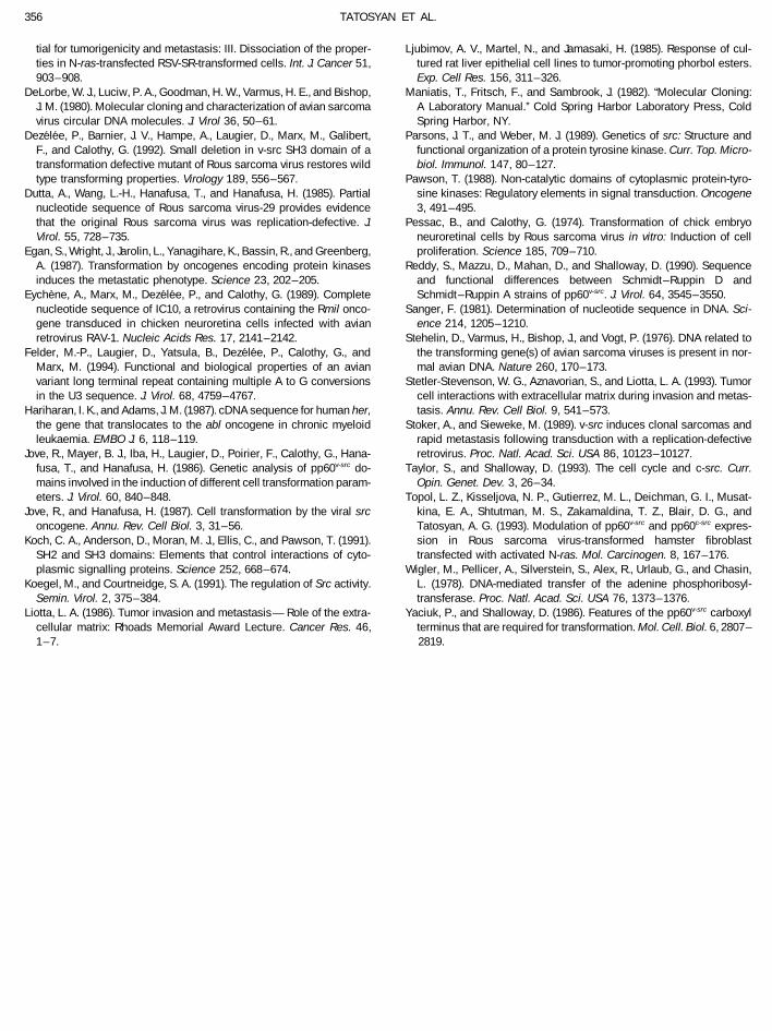
tial for tumorigenicity and metastasis: III. Dissociation of the proper- Ljubimov, A. V., Martel, N., and Jamasaki, H. (1985). Response of cul-tured rat liver epithelial cell lines to tumor-promoting phorbol esters.ties in N-ras-transfected RSV-SR-transformed cells. Int. J. Cancer 51,
903–908. Exp. Cell Res. 156, 311–326.Maniatis, T., Fritsch, F., and Sambrook, J. (1982). ‘‘Molecular Cloning:DeLorbe, W. J., Luciw, P. A., Goodman, H. W., Varmus, H. E., and Bishop,
J. M. (1980). Molecular cloning and characterization of avian sarcoma A Laboratory Manual.’’ Cold Spring Harbor Laboratory Press, ColdSpring Harbor, NY.virus circular DNA molecules. J. Virol 36, 50–61.
Dezelee, P., Barnier, J. V., Hampe, A., Laugier, D., Marx, M., Galibert, Parsons, J. T., and Weber, M. J. (1989). Genetics of src: Structure andfunctional organization of a protein tyrosine kinase. Curr. Top. Micro-F., and Calothy, G. (1992). Small deletion in v-src SH3 domain of a
transformation defective mutant of Rous sarcoma virus restores wild biol. Immunol. 147, 80–127.Pawson, T. (1988). Non-catalytic domains of cytoplasmic protein-tyro-type transforming properties. Virology 189, 556–567.
Dutta, A., Wang, L.-H., Hanafusa, T., and Hanafusa, H. (1985). Partial sine kinases: Regulatory elements in signal transduction. Oncogene3, 491–495.nucleotide sequence of Rous sarcoma virus-29 provides evidence
that the original Rous sarcoma virus was replication-defective. J. Pessac, B., and Calothy, G. (1974). Transformation of chick embryoneuroretinal cells by Rous sarcoma virus in vitro: Induction of cellVirol. 55, 728–735.
Egan, S., Wright, J., Jarolin, L., Yanagihare, K., Bassin, R., and Greenberg, proliferation. Science 185, 709–710.Reddy, S., Mazzu, D., Mahan, D., and Shalloway, D. (1990). SequenceA. (1987). Transformation by oncogenes encoding protein kinases
induces the metastatic phenotype. Science 23, 202–205. and functional differences between Schmidt–Ruppin D andSchmidt–Ruppin A strains of pp60v-src. J. Virol. 64, 3545–3550.Eychene, A., Marx, M., Dezelee, P., and Calothy, G. (1989). Complete
nucleotide sequence of IC10, a retrovirus containing the Rmil onco- Sanger, F. (1981). Determination of nucleotide sequence in DNA. Sci-ence 214, 1205–1210.gene transduced in chicken neuroretina cells infected with avian
retrovirus RAV-1. Nucleic Acids Res. 17, 2141–2142. Stehelin, D., Varmus, H., Bishop, J., and Vogt, P. (1976). DNA related tothe transforming gene(s) of avian sarcoma viruses is present in nor-Felder, M.-P., Laugier, D., Yatsula, B., Dezelee, P., Calothy, G., and
Marx, M. (1994). Functional and biological properties of an avian mal avian DNA. Nature 260, 170–173.Stetler-Stevenson, W. G., Aznavorian, S., and Liotta, L. A. (1993). Tumorvariant long terminal repeat containing multiple A to G conversions
in the U3 sequence. J. Virol. 68, 4759–4767. cell interactions with extracellular matrix during invasion and metas-tasis. Annu. Rev. Cell Biol. 9, 541–573.Hariharan, I. K., and Adams, J. M. (1987). cDNA sequence for human her,
the gene that translocates to the abl oncogene in chronic myeloid Stoker, A., and Sieweke, M. (1989). v-src induces clonal sarcomas andrapid metastasis following transduction with a replication-defectiveleukaemia. EMBO J. 6, 118– 119.
Jove, R., Mayer, B. J., Iba, H., Laugier, D., Poirier, F., Calothy, G., Hana- retrovirus. Proc. Natl. Acad. Sci. USA 86, 10123–10127.Taylor, S., and Shalloway, D. (1993). The cell cycle and c-src. Curr.fusa, T., and Hanafusa, H. (1986). Genetic analysis of pp60v-src do-
mains involved in the induction of different cell transformation param- Opin. Genet. Dev. 3, 26–34.Topol, L. Z., Kisseljova, N. P., Gutierrez, M. L., Deichman, G. I., Musat-eters. J. Virol. 60, 840–848.
Jove, R., and Hanafusa, H. (1987). Cell transformation by the viral src kina, E. A., Shtutman, M. S., Zakamaldina, T. Z., Blair, D. G., andTatosyan, A. G. (1993). Modulation of pp60v-src and pp60c-src expres-oncogene. Annu. Rev. Cell Biol. 3, 31–56.
Koch, C. A., Anderson, D., Moran, M. J., Ellis, C., and Pawson, T. (1991). sion in Rous sarcoma virus-transformed hamster fibroblasttransfected with activated N-ras. Mol. Carcinogen. 8, 167–176.SH2 and SH3 domains: Elements that control interactions of cyto-
plasmic signalling proteins. Science 252, 668–674. Wigler, M., Pellicer, A., Silverstein, S., Alex, R., Urlaub, G., and Chasin,L. (1978). DNA-mediated transfer of the adenine phosphoribosyl-Koegel, M., and Courtneidge, S. A. (1991). The regulation of Src activity.
Semin. Virol. 2, 375–384. transferase. Proc. Natl. Acad. Sci. USA 76, 1373–1376.Yaciuk, P., and Shalloway, D. (1986). Features of the pp60v-src carboxylLiotta, L. A. (1986). Tumor invasion and metastasis—Role of the extra-
cellular matrix: Rhoads Memorial Award Lecture. Cancer Res. 46, terminus that are required for transformation. Mol. Cell. Biol. 6, 2807–2819.1 –7.
/ 6a11$$7732 01-27-96 00:03:38 vira AP-Virology
Copyright © 2022 FDOKUMEN