Modification of aortic contractility in the cardiomyopathic hamster

Free Radical Biology & Medicine 42 (2007) 270–279www.elsevier.com/locate/freeradbiomed
Original Contribution
Antioxidant and antiplatelet effects of rosuvastatin in ahamster model of prediabetes
Shane Miersch, Inga Sliskovic, Arun Raturi, Bulent Mutus ⁎
Department of Chemistry and Biochemistry, University of Windsor, 401 Sunset Avenue, Windsor, Ontario, Canada N9B 3P4
Received 5 June 2006; revised 2 October 2006; accepted 17 October 2006Available online 20 October 2006
Abstract
The objectives of this study were to determine the relationships among Type II diabetes (T2DM)-dependent elevations in platelet-derived reactiveoxygen species (ROS), platelet-surface protein disulfide isomerase (psPDI) NO-releasing activity, and platelet aggregation and to evaluate theefficacy of rosuvastatin in normalizing these parameters in primary cells derived from a hamster model of prediabetic insulin resistance induced byfructose feeding. Platelets from rosuvastatin-treated non-fructose-fed (NFF) and fructose-fed (FF) hamsters were analyzed for aggregability andpsPDI-denitrosation activity. Platelets from NFF animals treated with xanthine/xanthine oxidase (X/XO) were assessed for the same parameters andprimary aortic endothelial cells (AEC) cultivated with a range of [rosuvastatin] ± mevalonate were analyzed for ROS production. Platelets from FFhamsters displayed statistically significant enhanced ROS production, diminished psPDI-mediated NO-releasing activity, and hyperaggregability.Suggestively, platelets from NFF animals treated with X/XO displayed characteristics similar to platelets from FF animals. Rosuvastatin elicited anormalizing effect on all parameters measured in platelets from FF animals. Further, ROS production in primary AEC from FF animals could beblunted to that of NFF animals by concentrations of rosuvastatin in the range of those achieved in the bloodstream. Diminished psPDI-dependent NO-releasing activity and increased initial aggregation rates of FF platelets may result from elevated vascular ROS production under conditions of insulinresistance. Normalization of ROS production and platelet aggregation by rosuvastatin indicates its potential use as a vasculoprotective agent.© 2006 Elsevier Inc. All rights reserved.
Keywords: Cardiovascular disease; Platelets; Endothelium; Free radicals; Diabetes mellitus
Introduction
Elevated reactive oxygen species (ROS) production indiabetes-related cardiovascular disease has been recognizedin both diabetic humans [1] and animal models of diabetes[2]. ROS have been shown to alter platelet physiology in avariety of ways [3–7], thus promoting hyperaggregability.Thrombotic complications in diabetes remain a leading cause
Abbreviations: AEC, aortic endothelial cells; DPI, diphenyleneiodoniumchloride; FF, fructose-fed; HMG-CoA reductase, 3-hydroxy-3-methylglutarylcoenzyme A reductase; HRP, horseradish peroxidase; Mn(III)TMPyP, Mn(III)tetrakis(1-methyl-4-pyridyl)porphyrin pentachloride; NFF, non-fructose-fed;NOX, NADPH oxidase; PBS, phosphate-buffered saline; PM, plasmamembrane; PRP, platelet-rich plasma; psPDI, platelet-surface protein disulfideisomerase; ROS, reactive oxygen species; SOD, superoxide dismutase; T2DM,Type II diabetes; X/XO, xanthine/xanthine oxidase.⁎ Corresponding author. Fax: +1 519 973 7098.E-mail address: [email protected] (B. Mutus).
0891-5849/$ - see front matter © 2006 Elsevier Inc. All rights reserved.doi:10.1016/j.freeradbiomed.2006.10.045
of mortality [8], thus warranting the investigation and adop-tion of therapeutic strategies that mitigate this enhancedpotential for thrombogenesis observed in Type II diabetes(T2DM) [9].
We have recently made the novel and interesting observationthat platelets derived from human Type II diabetics display bothelevated NADPH oxidase (NOX)–dependent ROS productionand a concomitant decrease in platelet-mediated liberation ofnitric oxide from low molecular weight S-nitrosothiols (termeddenitrosation) [7]. Indeed, we have shown that healthy humanplatelets treated with exogenous ROS exhibit a similar loss inability to metabolize S-nitrosothiols, as well as an enhancedpotential for aggregation. Central to both of these events is theenzyme protein disulfide isomerase. This enzyme is known tolocalize to the platelet surface (psPDI) where it is catalyticallyactive and responsible for both proaggregatory disulfideexchange reactions [10–12] and antiaggregatory liberation ofnitric oxide from low molecular weight S-nitrosothiols [13].

271S. Miersch et al. / Free Radical Biology & Medicine 42 (2007) 270–279
We have thus attributed the observed loss in platelet meta-bolism of S-nitrosothiols to the oxidation of psPDI active-sitethiols by increased oxidant load and have sought within thispaper to investigate therapeutic avenues for mitigating platelethyperaggregability.
Inhibitors of 3-hydroxy-3-methylglutaryl coenzymeA reduc-tase (HMG-CoA reductase) have shown efficacy in loweringcirculating lipid levels and in reducing cardiovascular mortality[14]. However, it has recently come to light that this class ofdrugs, referred to as statins, may have additional vasculopro-tective benefits that are not derived directly from cholesterollowering. This assertion stems from the observations that lowerserum cholesterol levels positively correlate with improved riskof coronary vascular events, but not with risk of stroke [15].Alternatively, statin treatment has been shown to reduce not onlycoronary events but also ischemic stroke [16].
Investigation of the “pleiotropic effects” of statins is based,in part, on the hypothesis that they bear antioxidant propertiesand are capable of inhibiting the production of reactive oxygenspecies from enzyme complexes such as NAD(P)H oxidase.
Other “vascular NOX isozymes” found in endothelial andsmooth muscle cells have been implicated in the promotion ofatherogenesis [17] and in animal models of diabetes [2]. In lightof the potential role of NOX in diabetes and cardiovasculardisease, we hypothesized that rosuvastatin would attenuateNOX activity and perhaps normalize platelet activity by pro-tecting psPDI from excess ROS, thus providing the impetus forour study of the potential antithrombotic and antiatherogenicbenefits in the vasculature.
We employed the Syrian Golden hamster, which becomesmetabolically disturbed upon fructose feeding for 7 days suchthat it develops hyperinsulinemia and hyperlipidemia in theabsence of overt hyperglycemia, thus representing a viablemodel of the prediabetic, insulin-resistant state [18]. Weobserved that platelets and aortic endothelial cells (AEC)from fructose-fed (FF) animals exhibit enhanced production ofNOX-dependent ROS. Further, platelets showed diminishedpsPDI-denitrosation activity and hyperaggregability in compar-ison to non-fructose-fed (NFF) control animals. These observa-tions could be mimicked by treatment of platelets from NFFanimals with exogenous ROS.
As expected, rosuvastatin exerts antioxidant properties inboth endothelial cells and platelets derived from fructose-fedanimals. Most importantly, we conclude that rosuvastatin showsefficacy in reversing ROS-mediated inhibition of plateletdenitrosation activity and hyperaggregability.
Experimental procedures
Materials
Ellman's reagent (DTNB), glutathione (GSH), sodium ni-trite, trisodium citrate dihydrate, citric acid monohydrate,dextrose, apyrase, NaCl, Na2HPO4, KH2PO4, KCl, NaHCO3,MgCl2·6H20, CaCl2·6H20, fructose, BSA, F12K media, endo-thelial cell growth supplement (ECGS), porcine heparin,xanthine, xanthine oxidase, thrombin, diphenyleneiodonium
chloride (DPI), and mevalonate were purchased from Sigma-Aldrich Canada (Oakville, ON). Isofluorane was purchasedfrom the Leamington Animal Clinic (Leamington, ON).Medical grade oxygen was purchased from BOC Gases(Windsor, ON). 10-Acetyl-3,7-dihydroxyphenoxazine (Amplexred) and horseradish peroxidase (HRP) were obtained fromMolecular Probes (Eugene, OR). Mn(III)tetrakis(1-methyl-4-pyridyl)porphyrin pentachloride (Mn(III)TMPyP) was obtainedfromCedarlane Labs (Hornby, Ontario).Matrigel was purchasedfrom BD Biosciences (Mississauga, ON). Rosuvastatin wasprovided by AstraZeneca (UK).
Preparation of S-nitrosoglutathione
S-nitrosoglutathione was prepared as previously described[13]. Briefly, GSH [free thiol] content was determined withDTNB [19]. Acidified NaNO2 was added to GSH solution in astoichiometric amount and reacted for 30 min at 4°C. The finalpH of the solution was adjusted to 7.4. GSNO was furtherrecrystallized by the slow addition of ice-cold acetone andresuspended in the appropriate buffer.
Animal feeding and monitoring protocols
All experimental procedures performed on the Syrianhamsters were approved by the University of Windsor ResearchEthics and Animal Care Committees and in accordance with theCCAC Guide to the Care and Use of Experimental Animals.Syrian Golden hamsters were obtained from Charles RiverCanada (Montreal, PQ). Animals were housed one per cage andgiven free access to food and water. Animals were divided intotwo groups: water feeding (control) and 10% fructose feeding.After 2 weeks of fructose feeding, animals were subdivided intofour groups as follows: control with no rosuvastatin, controlwith 10 mg/kg rosuvastatin, fructose-fed with no rosuvastatin,and fructose-fed with 10 mg/kg rosuvastatin. Rosuvastatin wasprepared in water and administered daily to animals by oralgavage for 7 days.
Pharmacokinetics of rosuvastatin in the Syrian Golden hamster
Plasma concentration (Cp) of rosuvastatin was determined bycollecting 200-μL samples of blood at 1-h intervals post-oralgavage for 24 h. These samples were analyzed for their rosuva-statin content by reverse-phase HPLCwith electrospray ionizationtandemmass spectrometric detection atAstraZeneca (Wilmington,DE). Primary cultures of endothelial cell explants were cultivatedin the presence of a range of rosuvastatin concentrationscomparable to the Cp max values in order to simulate in vivoexposure to the drug.
Collection of blood and tissue samples from Syrian hamsters
Hamsters were anesthetized using isoflurane delivered with1% oxygen through an isoflurane vaporizer in an inductionchamber and then transferred to a heating pad in the supineposition and fitted with a rodent circuit. The thorax and abdomen
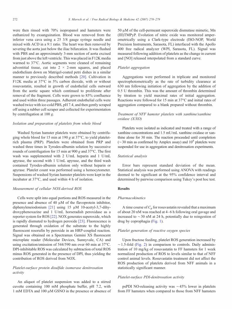
272 S. Miersch et al. / Free Radical Biology & Medicine 42 (2007) 270–279
were then rinsed with 70% isopropanol and hamsters wereeuthanized by exsanguination. Blood was removed from theinferior vena cava using a 25 3/8 gauge syringe needle andmixed with ACD in a 9:1 ratio. The heart was then removed bysevering the aorta just below the iliac bifurcation. It was flushedwith PBS and an approximately 3-mm section of aorta excisedfrom just above the left ventricle. This was placed in F12Kmediawarmed to 37°C. Aortic segments were cleaned of remainingadventitial tissue, cut into 2 × 2-mm squares, and placedendothelium down on Matrigel-coated petri dishes in a similarmanner to previously described methods [20]. Cultivation inF12K media at 37°C in 5% carbon dioxide, with or withoutrosuvastatin, resulted in growth of endothelial cells outwardfrom the aortic square which continued to proliferate afterremoval of the fragment. Cells were grown to 85% confluenceand used within three passages. Adherent endothelial cells werewashed twice with ice-cold PBS, pH 7.4, and then gently scrapedoff using a rubber cell scraper and collected for experimentationby centrifugation at 100 g.
Isolation and preparation of platelets from whole blood
Washed Syrian hamster platelets were obtained by centrifu-ging whole blood for 15 min at 190 g at 37°C, to yield platelet-rich plasma (PRP). Platelets were obtained from PRP andwashed three times in Tyrodes-albumin solution by successiverounds of centrifugation for 15 min at 900 g and 37°C. The firstwash was supplemented with 2 U/mL heparin and 1 U/mLapyrase, the second with 1 U/mL apyrase, and the third washcontained Tyrodes-albumin solution only without heparin orapyrase. Platelet count was performed using a hemocytometer.Suspensions of washed Syrian hamster platelets were kept in theincubator at 37°C, and used within 4 h of isolation.
Measurement of cellular NOX-derived ROS
Cells were split into equal portions and ROS measured in thepresence and absence of 40 μM of the flavoprotein inhibitor,diphenyleneiodonium [21] using 15 μM 10-acetyl-3,7-dihy-droxyphenoxazine and 1 U/mL horseradish peroxidase as areporter system for ROS [22]. NOX generates superoxide, whichis rapidly dismuted to hydrogen peroxide [23]. Fluorescence isgenerated through oxidation of the substrate to the highlyfluorescent resorufin by peroxide in an HRP-coupled reaction.Signal was obtained on a Spectramax Gemini XS fluorescentmicroplate reader (Molecular Devices, Sunnyvale, CA) andusing excitation/emission of 544/590 nm over 60 min at 37°C.DPI-inhibitable ROS was calculated by subtraction of total ROSminus ROS generated in the presence of DPI, thus yielding thecontribution of ROS derived from NOX.
Platelet-surface protein disulfide isomerase denitrosationactivity
An aliquot of platelet suspension was added to a stirredcuvette containing 100 mM phosphate buffer, pH 7.2, with1 mM EDTA and 100 μM GSNO in the presence or absence of
50 μM of the cell-permeant superoxide dismutase mimetic, Mn(III)TMPyP. Evolution of nitric oxide was monitored amper-ometrically using a Clark-type electrode (ISO-NOP, WorldPrecision Instruments, Sarasota, FL) interfaced with the Apollo400 free radical analyzer (WPI, Sarasota, FL). Signal wasmeasured following addition of platelets as the change in currentand [NO] released interpolated from a standard curve.
Platelet aggregation
Aggregations were performed in triplicate and monitoredspectrophotometrically as the rate of turbidity clearance at630 nm following initiation of aggregation by the addition of0.5 U thrombin. This was the amount of thrombin determinedby titration to yield maximal rates of initial aggregation.Reactions were followed for 15 min at 37°C and initial rates ofaggregation compared to a blank prepared without thrombin.
Treatment of NFF hamster platelets with xanthine/xanthineoxidase (X/XO)
Platelets were isolated as indicated and treated with a range ofxanthine concentrations and 1.5 mU/mL xanthine oxidase or xan-thine alone for 30 min. The reaction proceeded until completion(∼30 min as confirmed by Amplex assay) and 108 platelets weresuspended for use in aggregation and denitrosation experiments.
Statistical analysis
Error bars represent standard deviation of the mean.Statistical analysis was performed using ANOVAwith readingsdeemed to be significant at the 95% confidence interval anddetermined by pairwise comparison using Tukey's post hoc test.
Results
Pharmacokinetics
A time course ofCp for rosuvastatin revealed that a maximumof about 20 nM was reached at 4–6 h following oral gavage andincreased to ∼30 nM at 24 h, potentially due to reingestion ofdrug by coprophagia (Fig. 1).
Platelet generation of reactive oxygen species
Upon fructose feeding, platelet ROS generation increased by∼1.5-fold (Fig. 2) in comparison to controls. Daily adminis-tration of 10 mg/kg of rosuvastatin to FF hamsters for 1 weeknormalized production of ROS to levels similar to that of NFFcontrol animal levels. Rosuvastatin treatment did not affect theROS production of platelets derived from NFF animals in astatistically significant manner.
Platelet-surface PDI-denitrosation activity
psPDI NO-releasing activity was ∼45% lower in plateletsfrom FF hamsters when compared to those from NFF hamsters
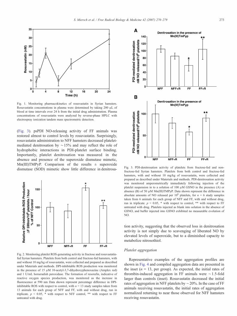
Fig. 3. PDI-denitrosation activity of platelets from fructose-fed and non-
Fig. 1. Monitoring pharmacokinetics of rosuvastatin in Syrian hamsters.Rosuvastatin concentrations in plasma were determined by taking 200 μL ofblood at time intervals over 24 h from the initial drug administration. Plasmaconcentrations of rosuvastatin were analyzed by reverse-phase HPLC withelectrospray ionization tandem mass spectrometric detection.
273S. Miersch et al. / Free Radical Biology & Medicine 42 (2007) 270–279
(Fig. 3). psPDI NO-releasing activity of FF animals wasrestored almost to control levels by rosuvastatin. Surprisingly,rosuvastatin administration to NFF hamsters decreased platelet-mediated denitrosation by ∼15% and may reflect the role ofhydrophobic interactions in PDI-platelet surface binding.Importantly, platelet denitrosation was measured in theabsence and presence of the superoxide dismutase mimetic,Mn(III)TMPyP. Comparison of the results ± superoxidedismutase (SOD) mimetic show little difference in denitrosa-
fructose-fed Syrian hamsters. Platelets from both control and fructose-fedhamsters, with and without 10 mg/kg of rosuvastatin, were collected andprepared as described under Materials and methods. PDI-denitrosation activitywas monitored amperometrically immediately following injection of theplatelet suspension in to a solution of 100 μM GSNO in the presence (A) orabsence (B) of 50 μM Mn(III)TMPyP. Data shown represent the difference inabsolute amounts of NO released per 108 platelets, for n = 6 study samplestaken from 6 animals for each group of NFF and FF, with and without drug,run in triplicate. p < 0.05, * with respect to control, ** with respect to FFuntreated with drug. Platelets injected as blank into solution in the absence ofGSNO, and buffer injected into GSNO exhibited no measurable evolution ofNO.
Fig. 2. Monitoring platelet ROS-generating activity in fructose and rosuvastatin-fed Syrian hamsters. Platelets from both control and fructose-fed hamsters, withand without 10 mg/kg of rosuvastatin, were collected and prepared as describedunder Materials and methods. DPI-inhibitable ROS production was monitoredin the presence of 15 μM 10-acetyl-3,7-dihydroxyphenoxazine (Amplex red)and 1 U/mL horseradish peroxidase. The formation of resorufin, indicative ofreactive oxygen species production, was monitored as the increase influorescence at 590 nm Data shown represent percentage difference in DPI-inhibitable ROS with respect to control, with n = 13 study samples taken from13 animals for each group of NFF and FF, with and without drug, run intriplicate. p < 0.05, * with respect to NFF control, ** with respect to FFuntreated with drug.
tion activity, suggesting that the observed loss in denitrosationactivity is not simply due to scavenging of liberated NO byelevated levels of superoxide, but to a diminished capacity tometabolize nitrosothiol.
Platelet aggregation
Representative examples of the aggregation profiles areshown in Fig. 4 and compiled aggregation data are presented inthe inset (n = 13, per group). As expected, the initial rates ofthrombin-induced aggregation in FF animals were ∼1.5-foldlarger than controls (inset). Rosuvastatin decreased the initialrates of aggregation in NFF platelets by∼20%. In the case of FFanimals receiving rosuvastatin, the initial rates of aggregationnormalized returning to near those observed for NFF hamstersreceiving rosuvastatin.
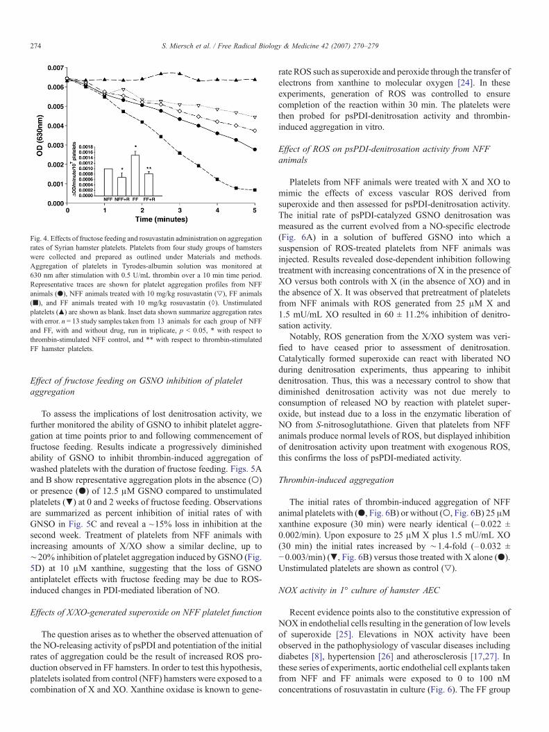
Fig. 4. Effects of fructose feeding and rosuvastatin administration on aggregationrates of Syrian hamster platelets. Platelets from four study groups of hamsterswere collected and prepared as outlined under Materials and methods.Aggregation of platelets in Tyrodes-albumin solution was monitored at630 nm after stimulation with 0.5 U/mL thrombin over a 10 min time period.Representative traces are shown for platelet aggregation profiles from NFFanimals (●), NFF animals treated with 10 mg/kg rosuvastatin (▿), FF animals(■), and FF animals treated with 10 mg/kg rosuvastatin (◊). Unstimulatedplatelets (▴) are shown as blank. Inset data shown summarize aggregation rateswith error. n = 13 study samples taken from 13 animals for each group of NFFand FF, with and without drug, run in triplicate, p < 0.05, * with respect tothrombin-stimulated NFF control, and ** with respect to thrombin-stimulatedFF hamster platelets.
274 S. Miersch et al. / Free Radical Biology & Medicine 42 (2007) 270–279
Effect of fructose feeding on GSNO inhibition of plateletaggregation
To assess the implications of lost denitrosation activity, wefurther monitored the ability of GSNO to inhibit platelet aggre-gation at time points prior to and following commencement offructose feeding. Results indicate a progressively diminishedability of GSNO to inhibit thrombin-induced aggregation ofwashed platelets with the duration of fructose feeding. Figs. 5Aand B show representative aggregation plots in the absence (○)or presence (●) of 12.5 μM GSNO compared to unstimulatedplatelets (▼) at 0 and 2 weeks of fructose feeding. Observationsare summarized as percent inhibition of initial rates of withGNSO in Fig. 5C and reveal a ~15% loss in inhibition at thesecond week. Treatment of platelets from NFF animals withincreasing amounts of X/XO show a similar decline, up to∼20% inhibition of platelet aggregation induced by GSNO (Fig.5D) at 10 μM xanthine, suggesting that the loss of GSNOantiplatelet effects with fructose feeding may be due to ROS-induced changes in PDI-mediated liberation of NO.
Effects of X/XO-generated superoxide on NFF platelet function
The question arises as to whether the observed attenuation ofthe NO-releasing activity of psPDI and potentiation of the initialrates of aggregation could be the result of increased ROS pro-duction observed in FF hamsters. In order to test this hypothesis,platelets isolated from control (NFF) hamsters were exposed to acombination of X and XO. Xanthine oxidase is known to gene-
rate ROS such as superoxide and peroxide through the transfer ofelectrons from xanthine to molecular oxygen [24]. In theseexperiments, generation of ROS was controlled to ensurecompletion of the reaction within 30 min. The platelets werethen probed for psPDI-denitrosation activity and thrombin-induced aggregation in vitro.
Effect of ROS on psPDI-denitrosation activity from NFFanimals
Platelets from NFF animals were treated with X and XO tomimic the effects of excess vascular ROS derived fromsuperoxide and then assessed for psPDI-denitrosation activity.The initial rate of psPDI-catalyzed GSNO denitrosation wasmeasured as the current evolved from a NO-specific electrode(Fig. 6A) in a solution of buffered GSNO into which asuspension of ROS-treated platelets from NFF animals wasinjected. Results revealed dose-dependent inhibition followingtreatment with increasing concentrations of X in the presence ofXO versus both controls with X (in the absence of XO) and inthe absence of X. It was observed that pretreatment of plateletsfrom NFF animals with ROS generated from 25 μM X and1.5 mU/mL XO resulted in 60 ± 11.2% inhibition of denitro-sation activity.
Notably, ROS generation from the X/XO system was veri-fied to have ceased prior to assessment of denitrosation.Catalytically formed superoxide can react with liberated NOduring denitrosation experiments, thus appearing to inhibitdenitrosation. Thus, this was a necessary control to show thatdiminished denitrosation activity was not due merely toconsumption of released NO by reaction with platelet super-oxide, but instead due to a loss in the enzymatic liberation ofNO from S-nitrosoglutathione. Given that platelets from NFFanimals produce normal levels of ROS, but displayed inhibitionof denitrosation activity upon treatment with exogenous ROS,this confirms the loss of psPDI-mediated activity.
Thrombin-induced aggregation
The initial rates of thrombin-induced aggregation of NFFanimal platelets with (●, Fig. 6B) or without (○, Fig. 6B) 25 μMxanthine exposure (30 min) were nearly identical (–0.022 ±0.002/min). Upon exposure to 25 μM X plus 1.5 mU/mL XO(30 min) the initial rates increased by ∼1.4-fold (–0.032 ±−0.003/min) (▼, Fig. 6B) versus those treated with X alone (●).Unstimulated platelets are shown as control (▿).
NOX activity in 1° culture of hamster AEC
Recent evidence points also to the constitutive expression ofNOX in endothelial cells resulting in the generation of low levelsof superoxide [25]. Elevations in NOX activity have beenobserved in the pathophysiology of vascular diseases includingdiabetes [8], hypertension [26] and atherosclerosis [17,27]. Inthese series of experiments, aortic endothelial cell explants takenfrom NFF and FF animals were exposed to 0 to 100 nMconcentrations of rosuvastatin in culture (Fig. 6). The FF group

Fig. 5. Effect of fructose feeding on GSNO inhibition of platelet aggregation. Platelets from hamsters were collected and prepared as outlined under Materials andMethods at 0, 1, and 2 weeks of fructose feeding. Aggregation of platelets in Tyrodes-albumin solution was monitored at 630 nm after stimulation with 0.5 U/mLthrombin in the presence or absence of 12.5 μMGSNO over a 10-min time period. Representative traces are shown for platelet aggregation profiles from NFF animalsin the presence (●) and absence (○) of the indicated [GSNO]. Unstimulated platelets (▼) are shown as blank. Percentage inhibition of aggregation was calculated asthe ratio of the GSNO-inhibited to uninhibited initial rate of aggregation and summarized in panel C at Weeks 0, 1, and 2. n = 6 study samples taken at each time pointand run in triplicate, p < 0.05, * with respect to thrombin-stimulated NFF control. Similarly, platelets isolated from NFF-fed animals were suspended in Tyrodes-albumin solution and exposed to a transient (30 min) flux of superoxide generated from 0 to 10 μM xanthine and 1.5 mU/mL xanthine oxidase, verified to be completewithin the allotted time. Aggregation of platelets was then monitored at 630 nm after stimulation with 0.5 U/mL thrombin in the presence or absence of 12.5 μMGSNOover a 10-min time period. Percent inhibition of aggregation was calculated the same as above.
275S. Miersch et al. / Free Radical Biology & Medicine 42 (2007) 270–279
had ∼1.6-fold larger NOX activity than NFF. The exposure ofthe NFF group to increasing concentrations of rosuvastatin hadno statistically significant effect on the basal NOX activity.However, elevated NOX activity of endothelial cells derivedfrom FF animals decreased in a rosuvastatin dose-dependentmanner and was statistically identical to the basal NFF-NOXactivity in the presence of 50 nM rosuvastatin. This is verysignificant as 50 nM is in range of the in vivo Cp (∼30 nM) of thedrug determined in this study. Further, in the presence of 200 μMmevalonate, the antioxidant effects of 100 nM rosuvastatin werepartially lost, suggesting that restoration of isoprenoid biosynth-esis was associated with restoration of NOX function.
Discussion
The present study was initiated by our observations that bothin humans with T2DM and in an animal model of prediabeticinsulin resistance, elevated platelet production of ROS isobserved concomitantly with a decrease in psPDI-denitrosationactivity and enhanced aggregatory potential. Studies within ourlab suggest that elevated vascular ROS oxidize active-site thiolsof psPDI, thus contributing to both an inhibition of the anti-aggregatory nitric oxide liberating activity and an increasedpotential for aggregation. The hypothetical mechanism of
ROS-mediated inhibition of psPDI RSNO-denitrosation activityis thought to be as follows: ROS is produced by enzymes such asNOX or other ROS-producing enzymes and is rapidly convertedto H2O2 by extracellular SOD or by spontaneous dismutation.H2O2 readily oxidizes vicinal dithiols [7,41], such as those foundat the active site of PDI, thus inactivating the RSNO-denitrosation activity of PDI and potentially contributing toenhanced potential for aggregation through provision of oxida-tive equivalents. Bolstering this hypothesis is the recent reportindicating that psPDI and NOX are in close spatial contact in theplasma membrane (PM) [42].
Another crucial piece of evidence supporting our hypothesisis our recent demonstration [13] that the RSNO-denitrosationactivity of psPDI inhibits platelet aggregation by two routes:first, PDI denitrosates RSNOs, releasing UNO that attenuatesplatelet activation via the guanylate cyclase/G-kinase route;secondly, RSNOs are denitrosated at the same PDI active sitethat catalyses the disulfide bond formation between integrinsand their ligands [10–12], thereby attenuating irreversibleaggregation.
In the past 5 years several isozymes of NOX have been iden-tified in nonphagocytic cells like platelets [31], smooth musclecells [32], neurons [33], endothelial cells [34], and fibroblasts [35].In platelets, NOX is believed to be plasma-membrane associated

Fig. 6. (A) Effect of transient platelet exposure to superoxide on psPDI-denitrosation activity. Washed platelets were obtained from NFF hamsters bycentrifugation and then exposed to a transient (30 min) flux of superoxidegenerated from 25 μM xanthine and 1.5 mU/mL xanthine oxidase, verified to becomplete within the allotted time. Release of NO was measured amperome-trically following injection of treated platelets into a stirred cuvette containing1 mM EDTA and 100 μMGSNO. Data shown represent the absolute amount ofNO released per minute for each of n = 4 study samples with standard deviationand indicate an IC50 of ∼25 μM xanthine. (B) Effect of transient plateletexposure to superoxide on platelet aggregation. Washed platelets were obtainedfrom NFF hamsters by centrifugation and then exposed to a transient (30 min)flux of superoxide generated from 25 μM xanthine and 1.5 mU/mL xanthineoxidase. Platelets (108) were suspended in Tyrodes-albumin buffer in triplicateand aggregation was initiated by the addition of 0.5 U/ml thrombin. Traces arerepresentative of the initial aggregation run in triplicate from n = 4 experimentsand show that platelets treated with X/XO (▼) exhibit an approximately 1.4-foldincrease in the initial rates of aggregation versus either untreated platelets (○) orplatelets treated with xanthine alone (●) all compared to unstimulated platelets(▿). p < 0.05, * with respect to thrombin-stimulated control treated with 25 μMxanthine alone.
276 S. Miersch et al. / Free Radical Biology & Medicine 42 (2007) 270–279
[36]; however, in endothelial cells the NOX subunits are largelyassembled and found associated with the intracellular cytoskele-ton and distributed perinuclearly [37]. Vascular NOX isoformsdisplay low (nM) constitutive O2
U− generation and this basalactivity is reported to be potentiated by a variety of stimulicommon to Type II diabetes pathology, including increased insulin[38], glucose [39], oxidized LDL [40], and hyperlipidemia [41].
NADPH oxidases catalyze the one-electron reduction ofmolecular oxygen to yield superoxide anion (O2
U−). NOX,initially identified in neutrophils, is a multimeric proteincomposed of plasma membrane catalytic subunits p22phox andp91phox containing cytochrome b558, cytosolic regulatorysubunits p47phox, p40
phox, p67
phox, and the small G-protein
Rac1 or Rac2, which translocate to the PM to activate thecatalytic subunits. Assembly of a functional enzyme complexrequires association of Rac GTPase and this association appearsto be regulated by its carboxyterminal geranylgeranylation(isoprenylation) [28]. Biosynthesis of isoprenoids, as well ascholesterol, requires mevalonate; thus NOX activity can beattenuated with HMG-CoA inhibitors [29,30] like rosuvastatinwhich inhibit the rate-limiting conversion of hydroxymethyl-glutaryl CoA to mevalonate.
Therefore, the first aim of this study was to determine theeffects of rosuvastatin on platelet PDI and NOX activities andon in vitro platelet aggregation in the animal model ofprediabetes—the Syrian Golden hamster. Previous studieshave demonstrated that fructose feeding of hamsters for 2–3weeks induces insulin resistance, as confirmed by in vivo eugly-cemic hyperinsulinemic clamp studies [18]. Fructose feeding isassociated with hyperinsulinemia, hyperlipidemia, but a normo-glycemic state, suggesting the development of a prediabetic,insulin-resistant state.
Fructose feeding of hamsters resulted in a ∼60% increase inplatelet ROS production in comparison to NFF controls (Fig. 2),a concomitant ∼42% reduction in psPDI RSNO-denitrosationactivity (Fig. 3), and thrombin-induced aggregation rates (Fig.4) that were on average ∼1.5-fold larger than platelets fromNFF hamsters. Fructose feeding of hamsters was also found toresult in a progressive loss in inhibition of platelet aggregationmediated by GSNO that varied with the duration of feeding(Fig. 5) to a maximum of <15% loss at the second week. Similarresults were obtained in platelets from NFF animals treated withX/XO revealing a <20% loss in GSNO-mediated plateletinhibition at 10 μM xanthine, further supporting our hypothesisthat ROS readily oxidizes the active-site vicinal dithiols of PDI,blocking denitrosation activity, and thus limiting the antiplateleteffects of GSNO.
The second aim of the study was to determine whether theobserved attenuation of the NO-releasing activity of psPDI andpotentiation of the initial rates of aggregation could be the resultof increased NOX activity, i.e., superoxide overproduction,observed in platelets of FF hamsters. To test this, we exposedplatelets from NFF animals to the xanthine/xanthine oxidasesuperoxide-generating system and in support of our hypothesis,platelets exposed to a range of xanthine concentrations and1.5 mU/mL xanthine oxidase over 30 min (1) inhibited thepsPDI-dependent RSNO-denitrosation activity by ∼50% at25 μM xanthine (Fig. 6A) and (2) potentiated the initial rates ofthrombin-induced aggregation by ∼1.4-fold (Fig. 6B). Theseeffects are very similar to the FF platelet results lendingcredence to our hypothesis.
The third aim was to determine the effects of short-termrosuvastatin treatment on the platelet parameters tested here.Rosuvastatin had no statistically significant effect on the platelet

Fig. 7. Effect of varying rosuvastatin on primary hamster AEC. Hamster aortaswere collected as indicated from FF and NFF hamsters. Primary AEC werecultivated until 85% confluent in the presence of varying levels of drug for 1week at which time they were harvested for measurement of DPI-inhibitablereactive oxygen species. Data shown represent the percentage difference in DPI-inhibitable ROS produced in 1 h as measured by Amplex fluorescence emissionat 590 nm. n = 6 study samples derived from 6 animals for each group of NFFand FF, with and without drug and run in triplicate. p < 0.05 * with respect toNFF controls without drug and ** with respect to FF without drug and *** withrespect to FF treated with 100 nM rosuvastatin.
277S. Miersch et al. / Free Radical Biology & Medicine 42 (2007) 270–279
ROS production by NFF hamsters. However, drug administeredto FF animals attenuated the generation of ROS to controllevels (Fig. 2). In the case of psPDI, rosuvastatin unexpectedlydecreased the NFF-psPDI activity by ∼20% (Fig. 3). AlthoughPDI is generally considered to bind to the plasma membranethrough electrostatic interactions, we have recently investigatedand provided evidence that interaction with the platelet plasmamembrane is mediated, in part, by hydrophobic interactions(Raturi, manuscript submitted). It is then possible thatcholesterol lowering induced by rosuvastatin in NFF animalsalters the hydrophobic character of the platelet plasmamembrane potentially diminishing the bound enzyme andaccounting for this ∼20% loss in denitrosation activity.Alternately, exposure of FF animals to rosuvastatin resulted inthe restoration of the psPDI activity to near control levels. Theseresults suggest that rosuvastatin at nM Cp is effective in bluntingthe production of ROS by platelets, and thus permitting psPDI-catalyzed release of sufficient NO from RSNOs to inhibitplatelets. This was borne out in aggregation studies in whichrosuvastatin treatment slightly decreased (∼20%) the initialrates of aggregation in NFF animals and significantly decreased(∼50%) the initial rates in FF animals (Fig. 4). Results obtainedfrom the aggregation study suggest that rosuvastatin treatment ofFF animals not only contributes to the normalization of plateletROS production and psPDI activities but also restores normalplatelet aggregability.
Numerous studies have established links between an elevatedpathologic endothelial NOX activity and atherogenesis[1,12,20,23], thus the fourth aim of this study was to examinethe effect of rosuvastatin on NFF and FF hamster aortic endo-thelial cell NOX activity (Fig. 7). To this end, aortic explants fromNFF and FF animals were grown in primary culture and exposedto varying nanomolar concentrations of rosuvastatin. NFF NOXactivity was insensitive to in vitro rosuvastatin exposure. Incontrast, FF animals displayed aortic endothelial NOX activitythat was on average ∼1.8-fold larger than controls and could beattenuated to near control levels by 50 nM drug. At 100 nMrosuvastatin, FF-NOX-dependent ROS production was ∼40%less thanNFF controls, an effect which could be partially reversedby the addition of 200 μMmevalonate. That ROS production wasnot fully reversed by mevalonate suggests that other sources mayalso contribute to DPI-inhibitable ROS. Diphenyleneiodonium isa flavoprotein inhibitor and will inhibit other enzymes such asnitric oxide synthase which has been shown to produce super-oxide in platelets of patients with coexisting hypertension anddiabetes [43]. However, that mevalonate could in part reverse theantioxidant effects of rosuvastatin in AEC corroborates theassertion that drug effects are possibly elicited by the inhibition ofisoprenoid formation (Fig. 7).
The relative insensitivity of AECs derived from NFF animalscompared to the mitigation of ROS production in those derivedfrom FF animals at the same level of drug suggests that perhaps adifferent NOX isozyme is being induced in the animal model ofprediabetes. It can be postulated that the rosuvastatin-insensitiveNFF isoform of NOX possesses a higher affinity for geranylger-anylate or the geranylgeranyl transferase in comparison to the FFisoform of NOX. The net result would be that NFF-NOX would
remain active even if rosuvastatin attenuated geranylgeranyla-tion, whereas the activity of the FF-NOX would diminish withdecreasing levels of geranylgeranylate.
In summary, this study elaborates on the novel finding thatincreased ROS production in animal models of prediabetes isaccompanied by an attenuation of psPDI-denitrosation activitythat likely contributes to platelet hyperaggregability and thrombo-genesis. Treatment of hamsters with 10mg/kg rosuvastatin showsefficacy in normalizing both of these activities in this animalmodel of prediabetes. Further, investigation of ROS generatedfrom primary endothelial cells similarly shows a significantincrease in fructose-fed versus non-fructose-fed hamsters thatwould perturb normal intracellular signaling and contribute toendothelial dysfunction. Treatment of primary endothelial cellswith levels of rosuvastatin in the range of those found in vivoresults in a restoration of ROS production to control levels.
Based on the demonstrated efficacy of rosuvastatin to restoreplatelet and endothelial ROS levels, as well as normalizingplatelet aggregation and PDI-denitrosation activity in models ofprediabetes, broadening of the pathological states to which thisdrug is utilized may be beneficial in preventing secondaryatherothrombotic complications associated with non-insulin-dependent diabetes mellitus.
Acknowledgments
This research was sponsored by a Grant in Aid from theCanadian Diabetes Association and a grant from AstraZeneca(UK).
References
[1] Spitaler, M. M.; Graier, W. F. Vascular targets of redox signalling indiabetes mellitus. Diabetologia 45:476–494; 2002.

278 S. Miersch et al. / Free Radical Biology & Medicine 42 (2007) 270–279
[2] Sonta, T.; Inoguchi, T.; Tsubouchi, H.; Sekiguchi, N.; Kobayashi, K.;Matsumoto, S.; Utsumi, H.; Nawata, H. Evidence for contribution ofvascular NAD(P)H oxidase to increased oxidative stress in animal modelsof diabetes and obesity. Free Radic. Biol. Med. 37:115–123; 2004.
[3] Gregg, D.; de Carvalho, D. D.; Kovacic, H. Integrins and coagulation: arole for ROS/redox signaling? Antioxid. Redox Signal. 6:757–764; 2004.
[4] Redondo, P. C.; Jardin, I.; Hernandez-Cruz, J. M.; Pariente, J. A.; Salido,G. M.; Rosado, J. A. Hydrogen peroxide and peroxynitrite enhance Ca2+
mobilization and aggregation in platelets from type 2 diabetic patients.Biochem. Biophys. Res. Commun. 333:94–102; 2005.
[5] Walsh, G. M.; Sheehan, D.; Kinsella, A.; Moran, N.; O'Neill, S. Redoxmodulation of integrin [correction of integin] alpha IIb beta 3 involves anovel allosteric regulation of its thiol isomerase activity. Biochemistry43:473–480; 2004.
[6] Chlopicki, S.; Olszanecki, R.; Janiszewski, M.; Laurindo, F. R.; Panz, T.;Miedzobrodzki, J. Functional role of NADPH oxidase in activation ofplatelets. Antioxid. Redox Signal. 6:691–698; 2004.
[7] Miersch, S.; Raturi, A.; Root, P.; Vignini, A.; Mazzanti, L.; Adeli, K.;Mutus, B. Reactive oxygen promotes hyperaggregability via redoxswitching of platelet surface PDI. Manuscript submitted for publication.
[8] Beckman, J. A.; Creager, M. A.; Libby, P. Diabetes and atherosclerosis:epidemiology, pathophysiology, and management. JAMA 287:2570–2581;2002.
[9] Ferroni, P.; Basili, S.; Falco, A.; Davi, G. Platelet activation in type.J. Thromb. Haemost. 2:1282–1291; 2004.
[10] Lahav, J.; Gofer-Dadosh, N.; Luboshitz, J.; Hess, O.; Shaklai, M. Proteindisulfide isomerase mediates integrin-dependent adhesion. FEBS Lett.475:89–92; 2000.
[11] Lahav, J.; Jurk, K.; Hess, O.; Barnes, M. J.; Farndale, R. W.; Luboshitz, J.;Kehrel, B. E. Sustained integrin ligation involves extracellular freesulfhydryls and enzymatically catalyzed disulfide exchange. Blood100:2472–2478; 2002.
[12] Lahav, J.; Wijnen, E. M.; Hess, O.; Hamaia, S. W.; Griffiths, D.; Makris,M.; Knight, C. G.; Essex, D. W.; Farndale, R. W. Enzymatically catalyzeddisulfide exchange is required for platelet adhesion to collagen via integrinalpha2beta1. Blood 102:2085–2092; 2003.
[13] Root, P.; Sliskovic, I.; Mutus, B. Platelet cell-surface protein disulphide-isomerase mediated S-nitrosoglutathione consumption. Biochem. J.382:575–580.
[14] Maron, D. J.; Fazio, S.; Linton, M. F. Current perspectives on statins.Circulation 101:207–213; 2000.
[15] Collins, R.; Armitage, J.; Parish, S.; Sleight, P.; Peto, R. Effects ofcholesterol-lowering with simvastatin on stroke and other major vascularevents in 20536 people with cerebrovascular disease or other high-riskconditions. Lancet 363:757–767; 2004.
[16] Amarenco, P.; Labreuche, J.; Lavallee, P.; Touboul, P. J. Statins in strokeprevention and carotid atherosclerosis: systematic review and up-to-datemeta-analysis. Stroke 35:2902–2909; 2004.
[17] Laufs, U.; Wassmann, S.; Czech, T.; Munzel, T.; Eisenhauer, M.; Bohm,M.; Nickenig, G. Physical inactivity increases oxidative stress, endothelialdysfunction, and atherosclerosis. Arterioscler. Thromb. Vasc. Biol.25:809–814; 2005.
[18] Taghibiglou, C.; Rashid-Kolvear, F.; Van Iderstine, S. C.; Le-Tien, H.;Fantus, I. G.; Lewis, G. F.; Adeli, K. Hepatic very low density lipoprotein-ApoB overproduction is associated with attenuated hepatic insulinsignaling and overexpression of protein-tyrosine phosphatase 1B in afructose-fed hamster model of insulin resistance. J. Biol. Chem. 277:793–803; 2002.
[19] Riddles, P. W.; Blakeley, R. L.; Zerner, B. Reassessment of Ellman'sreagent. Methods Enzymol. 91:49–60; 1983.
[20] Suh, S. H.; Vennekens, R.; Manolopoulos, V. G.; Freichel, M.; Schweig,U.; Prenen, J.; Flockerzi, V.; Droogmans, G.; Nilius, B. Characterisation ofexplanted endothelial cells from mouse aorta: electrophysiology and Ca2+signalling. Pflugers Arch. 438:612–620; 1999.
[21] O'Donnell, B. V.; Tew, D. G.; Jones, O. T.; England, P. J. Studies on theinhibitory mechanism of iodonium compounds with special reference toneutrophil NADPH oxidase. Biochem. J. 290 (Pt 1):41–49; 1993.
[22] Zhou, M.; Diwu, Z.; Panchuk-Voloshina, N.; Haugland, R. P. A stable
nonfluorescent derivative of resorufin for the fluorometric determination oftrace hydrogen peroxide: applications in detecting the activity ofphagocyte NADPH oxidase and other oxidases. Anal. Biochem. 253:162–168; 1997.
[23] Cai, H. NAD(P)H oxidase-dependent self-propagation of hydrogenperoxide and vascular disease. Circ. Res. 96:818–822; 2005.
[24] Vorbach, C.; Harrison, R.; Capecchi, M. R. Xanthine oxidoreductase iscentral to the evolution and function of the innate immune system. TrendsImmunol. 24:512–517; 2003.
[25] Li, J. M.; Shah, A. M. Endothelial cell superoxide generation: regulationand relevance for cardiovascular pathophysiology. Am. J. Physiol., Regul.Integr. Comp. Physiol. 287:R1014–R1030; 2004.
[26] Paravicini, T. M.; Chrissobolis, S.; Drummond, G. R.; Sobey, C. G.Increased NADPH-oxidase activity and Nox4 expression during chronichypertension is associated with enhanced cerebral vasodilatation toNADPH in vivo. Stroke 35:584–589; 2004.
[27] Rueckschloss, U.; Duerrschmidt, N.; Morawietz, H. NADPH oxidase inendothelial cells: impact on atherosclerosis. Antioxid. Redox Signal.5:171–180; 2003.
[28] Ando, S.; Kaibuchi, K.; Sasaki, T.; Hiraoka, K.; Nishiyama, T.; Mizuno, T.;Asada, M.; Nunoi, H.; Matsuda, I.; Matsuura, Y.; et al. Post-translationalprocessing of rac p21s is important both for their interaction with theGDP/GTP exchange proteins and for their activation of NADPH oxidase.J. Biol. Chem. 267:25709–25713; 1992.
[29] Bandoh, T.; Sato, E. F.; Mitani, H.; Nakashima, A.; Hoshi, K.; Inoue, M.Antioxidative potential of fluvastatin via the inhibition of nicotinamideadenine dinucleotide phosphate (NADPH) oxidase activity. Biol. Pharm.Bull. 26:818–822; 2003.
[30] Takayama, T.; Wada, A.; Tsutamoto, T.; Ohnishi, M.; Fujii, M.; Isono, T.;Horie, M. Contribution of vascular NAD(P)H oxidase to endothelialdysfunction in heart failure and the therapeutic effects of HMG-CoAreductase inhibitor. Circ. J. 68:1067–1075; 2004.
[31] Krotz, F.; Sohn, H. Y.; Gloe, T.; Zahler, S.; Riexinger, T.; Schiele, T. M.;Becker, B. F.; Theisen, K.; Klauss, V.; Pohl, U. NAD(P)H oxidase-dependent platelet superoxide anion release increases platelet recruitment.Blood 100:917–924; 2002.
[32] Hilenski, L. L.; Clempus, R. E.; Quinn, M. T.; Lambeth, J. D.; Griendling,K. K. Distinct subcellular localizations of Nox1 and Nox4 in vascularsmooth muscle cells. Arterioscler. Thromb. Vasc. Biol. 24:677–683; 2004.
[33] Hilburger, E. W.; Conte, E. J.; McGee, D. W.; Tammariello, S. P.Localization of NADPH oxidase subunits in neonatal sympathetic neurons.Neurosci. Lett. 377:16–19; 2005.
[34] Griendling, K. K.; Sorescu, D.; Ushio-Fukai, M. NAD(P)H oxidase: role incardiovascular biology and disease. Circ. Res. 86:494–501; 2000.
[35] Krotz, F.; Sohn, H. Y.; Pohl, U. Reactive oxygen species: players in theplatelet game. Arterioscler. Thromb. Vasc. Biol. 24:1988–1996; 2004.
[36] Li, J. M.; Shah, A. M. Intracellular localization and preassembly of theNADPH oxidase complex in cultured endothelial cells. J. Biol. Chem.277:19952–19960; 2002.
[37] Kashiwagi, A.; Shinozaki, K.; Nishio, Y.; Maegawa, H.; Maeno, Y.;Kanazawa, A.; Kojima, H.; Haneda, M.; Hidaka, H.; Yasuda, H.; Kikkawa,R. Endothelium-specific activation of NAD(P)H oxidase in aortas ofexogenously hyperinsulinemic rats. Am. J. Physiol. 277:E976–E983;1999.
[38] Inoguchi, T.; Li, P.; Umeda, F.; Yu, H. Y.; Kakimoto, M.; Imamura, M.;Aoki, T.; Etoh, T.; Hashimoto, T.; Naruse, M.; Sano, H.; Utsumi, H.;Nawata, H. High glucose level and free fatty acid stimulate reactiveoxygen species production through protein kinase C-dependent activationof NAD(P)H oxidase in cultured vascular cells. Diabetes 49:1939–1945;2000.
[39] Rueckschloss, U.; Galle, J.; Holtz, J.; Zerkowski, H. R.; Morawietz, H.Induction of NAD(P)H oxidase by oxidized low-density lipoprotein inhuman endothelial cells: antioxidative potential of hydroxymethylglutarylcoenzyme A reductase inhibitor therapy. Circulation 104:1767–1772;2001.
[40] Li, J. M.; Shah, A. M. ROS generation by nonphagocytic NADPH oxidase:potential relevance in diabetic nephropathy. J. Am. Soc. Nephrol. 14:S221–S226; 2003.

279S. Miersch et al. / Free Radical Biology & Medicine 42 (2007) 270–279
[41] Conway, M. E.; Yennawa, N.; Wallin, R.; Poole, L. B.; Hutson, S. M.Identification of a peroxide-sensitive redox switch at the CXXC motif inthe human mitochondrial branched chain aminotransferase. Biochemistry41:9070–9078; 2002.
[42] Janiszewski, M.; Lopes, L. R.; Carmo, A. O.; Pedro, M. A.; Brandes, R. P.;Santos, C. X.; Laurindo, F. R. Regulation of NAD(P)H oxidase by
associated protein disulfide isomerase in vascular smooth muscle cells.J. Biol. Chem. 280:40813–40819; 2005.
[43] Dixon, L. J.; Hughes, S. M.; Rooney, K.; Madden, A.; Devine, A.; Leahey,W.; Henry, W.; Johnston, G. D.; McVeigh, G. E. Increased superoxideproduction in hypertensive patients with diabetes mellitus: role of nitricoxide synthase. Am. J. Hypertens. 18 (6):839–843.
Copyright © 2022 FDOKUMEN