The Increased Type-1 and Type-2 Chemokine Levels in Children with Acute RSV Infection Alter the...
11
Research Article The Increased Type-1 and Type-2 Chemokine Levels in Children with Acute RSV Infection Alter the Development of Adaptive Immune Responses Valerija Vojvoda, 1 Ana SaviT Mlakar, 1 Mladen JergoviT, 1 Mirela KukuruzoviT, 2 Leo MarkovinoviT, 3 Neda Aberle, 4 Sabina RabatiT, 5 and Krešo Bendelja 1,6 1 Center for Research and Knowledge Transfer in Biotechnology, University of Zagreb, Rockefellerova 10, 10000 Zagreb, Croatia 2 Faculty of Food Technology and Biotechnology, University of Zagreb, Pierotti Street 6, 10000 Zagreb, Croatia 3 University Hospital for Infectious Diseases “Dr. Fran Mihaljevi´ c”, Mirogojska 8, 10000 Zagreb, Croatia 4 Department of Pediatrics, General Hospital “Dr. Josip Benˇ cevi´ c”, Andrije ˇ Stampara 42, 35000 Slavonski Brod, Croatia 5 Institute of Immunology, Rockefellerova 2, 10000 Zagreb, Croatia 6 National Jewish Health, 1400 Jackson Street, Denver, CO 80206, USA Correspondence should be addressed to Kreˇ so Bendelja; [email protected] Received 27 February 2014; Revised 17 May 2014; Accepted 19 May 2014; Published 11 June 2014 Academic Editor: Marija Mostarica-Stojkovi´ c Copyright © 2014 Valerija Vojvoda et al. is is an open access article distributed under the Creative Commons Attribution License, which permits unrestricted use, distribution, and reproduction in any medium, provided the original work is properly cited. Severe RSV infections and frequent recurrence could be related to the altered polarization of type-2/type-1 T cells. is increases the importance of determining distinctive chemokines and chemokine receptor profiles on memory T cells. We analyzed systemic adaptive T cell response in the acute ( = 17) and convalescent phase (=7) of RSV-infected children, in the acute ( = 11) and convalescent phase (=6) of children with other viral respiratory infections (adenovirus and influenza virus), and in healthy children ( = 18). Expression of CCR4 and CXCR3 on effector-memory (T EM ) and central-memory (T CM ) T cells was compared between tested groups. Serum concentrations of specific chemokines were determined. High CXCL10 levels were detected in acutely infected children regardless of virus pathogen, whereas increased CCL17 production was RSV-specific. Higher percentages of CCR4 + CD4 T EM cells in acute RSV infection were accompanied with higher percentages of CXCR3 + CD8 T EM cells, whereas the development of long-lived memory CXCR3 + CD4 and CD8 T CM cells seems to be compromised, as only children with other viral infections had higher percentages in the convalescent phase. Presence of type-2 and type-1 adaptive antiviral immune response, together with insufficient development of long-lived type-1 T cell memory, could play an important role in RSV pathogenesis and reinfection. 1. Introduction RSV is the most common respiratory pathogen in infants which can cause severe bronchiolitis with pronounced air- way hyperreactivity [1]. Frequent rate of RSV reinfections throughout the childhood implicates inadequate develop- ment of 1 and 2 responses which is evident from simultaneous IFN- and IL-4/IL-5 production [2, 3]. e efficient antiviral response induces infiltration of 1 polarized virus-specific CD4 and CD8 T cells via multiple chemokines and their cognate chemokine receptors. Cor- respondingly, CXCR3 and CCR5 chemokine receptors are expressed on activated type-1 T cells and associated with their local recruitment toward specific chemokines CXCL9, CXCL10, and CCL5 released by RSV-infected epithelial cells [4, 5]. Activated IFN- producing type-1 T cells are required for the efficient defense against lower respiratory tract RSV infection [6], but overactivated type-1 effector-memory CD8 T cells (T EM ), observed as the occurrence of high CD8/CD4 T EM cell ratio in lungs [7], can also contribute to the development of more severe pathology and RSV-airway hyperreactivity, respectively. It has also been noticed that, at the peak of the disease, RSV-infected infants have increased CXCL10 levels [8] and enrichment of CXCR3 + T cells in Hindawi Publishing Corporation BioMed Research International Volume 2014, Article ID 750521, 11 pages http://dx.doi.org/10.1155/2014/750521
-
Upload
independent -
Category
Documents
-
view
0 -
download
0
Transcript of The Increased Type-1 and Type-2 Chemokine Levels in Children with Acute RSV Infection Alter the...
Research ArticleThe Increased Type-1 and Type-2 Chemokine Levels inChildren with Acute RSV Infection Alter the Development ofAdaptive Immune Responses
Valerija Vojvoda,1 Ana SaviT Mlakar,1 Mladen JergoviT,1 Mirela KukuruzoviT,2
Leo MarkovinoviT,3 Neda Aberle,4 Sabina RabatiT,5 and Krešo Bendelja1,6
1 Center for Research and Knowledge Transfer in Biotechnology, University of Zagreb, Rockefellerova 10, 10000 Zagreb, Croatia2 Faculty of Food Technology and Biotechnology, University of Zagreb, Pierotti Street 6, 10000 Zagreb, Croatia3 University Hospital for Infectious Diseases “Dr. Fran Mihaljevic”, Mirogojska 8, 10000 Zagreb, Croatia4Department of Pediatrics, General Hospital “Dr. Josip Bencevic”, Andrije Stampara 42, 35000 Slavonski Brod, Croatia5 Institute of Immunology, Rockefellerova 2, 10000 Zagreb, Croatia6National Jewish Health, 1400 Jackson Street, Denver, CO 80206, USA
Correspondence should be addressed to Kreso Bendelja; [email protected]
Received 27 February 2014; Revised 17 May 2014; Accepted 19 May 2014; Published 11 June 2014
Academic Editor: Marija Mostarica-Stojkovic
Copyright © 2014 Valerija Vojvoda et al. This is an open access article distributed under the Creative Commons AttributionLicense, which permits unrestricted use, distribution, and reproduction in any medium, provided the original work is properlycited.
Severe RSV infections and frequent recurrence could be related to the altered polarization of type-2/type-1 T cells. This increasesthe importance of determining distinctive chemokines and chemokine receptor profiles on memory T cells. We analyzed systemicadaptive T cell response in the acute (𝑛 = 17) and convalescent phase (𝑛 = 7) of RSV-infected children, in the acute (𝑛 = 11)and convalescent phase (𝑛 = 6) of children with other viral respiratory infections (adenovirus and influenza virus), and in healthychildren (𝑛 = 18). Expression of CCR4 and CXCR3 on effector-memory (TEM) and central-memory (TCM) T cells was comparedbetween tested groups. Serum concentrations of specific chemokines were determined.HighCXCL10 levels were detected in acutelyinfected children regardless of virus pathogen, whereas increased CCL17 production was RSV-specific. Higher percentages ofCCR4+ CD4 TEM cells in acute RSV infection were accompanied with higher percentages of CXCR3+ CD8 TEM cells, whereasthe development of long-lived memory CXCR3+ CD4 and CD8 TCM cells seems to be compromised, as only children with otherviral infections had higher percentages in the convalescent phase. Presence of type-2 and type-1 adaptive antiviral immune response,together with insufficient development of long-lived type-1 T cell memory, could play an important role in RSV pathogenesis andreinfection.
1. Introduction
RSV is the most common respiratory pathogen in infantswhich can cause severe bronchiolitis with pronounced air-way hyperreactivity [1]. Frequent rate of RSV reinfectionsthroughout the childhood implicates inadequate develop-ment of Th1 and Th2 responses which is evident fromsimultaneous IFN-𝛾 and IL-4/IL-5 production [2, 3].
The efficient antiviral response induces infiltration ofTh1polarized virus-specific CD4 and CD8 T cells via multiplechemokines and their cognate chemokine receptors. Cor-respondingly, CXCR3 and CCR5 chemokine receptors are
expressed on activated type-1 T cells and associated withtheir local recruitment toward specific chemokines CXCL9,CXCL10, and CCL5 released by RSV-infected epithelial cells[4, 5]. Activated IFN-𝛾 producing type-1 T cells are requiredfor the efficient defense against lower respiratory tract RSVinfection [6], but overactivated type-1 effector-memory CD8T cells (TEM), observed as the occurrence of high CD8/CD4TEM cell ratio in lungs [7], can also contribute to thedevelopment of more severe pathology and RSV-airwayhyperreactivity, respectively. It has also been noticed that, atthe peak of the disease, RSV-infected infants have increasedCXCL10 levels [8] and enrichment of CXCR3+ T cells in
Hindawi Publishing CorporationBioMed Research InternationalVolume 2014, Article ID 750521, 11 pageshttp://dx.doi.org/10.1155/2014/750521
2 BioMed Research International
lungs that might be responsible for the underlying pathology.CXCR3 expression occurs upon activation on naıve T cellswhich potently migrate toward cognate CXCL10 ligand [9].These proinflammatory T cells also coproduce high TNF-𝛼levels, which could also be related to the disease severity [10].
RSV has developed different mechanisms to modifya local microenvironment and influence effector-memorydifferentiation of CD8 T cells [11] irrespective of systemicanti-RSV response [12]. Heidema et al. have shown thatlung CD8 TEM cells, known for high IFN-𝛾 productionand granzyme/perforin-mediated cytotoxicity [7], undergofunctional switch to FasL and TRAIL-dependent cytotoxicitythat is associated with severe pathology [13].
Another possible underlying mechanism of RSV-mediated interference with antiviral immune response couldbe the induction of Th2 immune response mediated bythymic stromal lymphopoietin (TSLP), which is capable ofmodifyingTh1-polarization capacity of dendritic cells towardTh2, as observed in infected mice [14]. These dendritic cellsrelease CCL17 and CCL22 chemokines [15, 16] and ultimatelypotentiate migration of CCR4+Th2 cells [17, 18] involved intype-2 immune response [19]. Moreover, the immune systemof newborns favors the Th2 adaptive immune response [2]that might confront efficient Th1 antiviral immune responsein primary RSV infection as shown in higher CCR4+ toCXCR3+ CD4 T cell ratio [20].
In the present study we analyzed systemic adaptiveT cell response in the acute and convalescent phase ofprimary RSV infection in hospitalized infants with bron-chiolitis. Increased chemokines CCL17 and CXCL10 levelswere detected in serum of infected infants compared tohealthy control subjects but increased CCL17 productionwas strictly RSV-specific. In parallel, higher percentage ofCD4 TEM cells expressed CCR4 in the acute RSV phase,withCXCR3 expression comparable to healthy and non-RSV-infected infants.
RSV-infected infants had higher percentages of CD8 TEMcells expressing CXCR3 receptor in the acute infection, butdevelopment of long-lived central-memory CXCR3+ CD4and CD8 T cells (TCM) seems to be compromised as onlyinfants with other viral infections had higher percentages inthe convalescent phase.
2. Materials and Methods
2.1. Patients. During two respiratory infectious seasonsblood samples were collected from children under 2 yearsof age hospitalized due to severe acute respiratory infectionscaused by RSV or other viruses (adenovirus and influenza).Samples were acquired within the first 6 days of hospitaliza-tion. Patients receiving steroids, or with ongoing allergies,or with immunologic or hematologic disorders were notincluded. Second blood drawing was performed 4–6 weeksafter first sampling (convalescent phase). Blood samples fromhealthy children younger than 2 years of age, without clinicalevidence of allergic, immunologic, or hematologic disorders,infectious diseases, or ongoing corticosteroid therapy at leastduring the period of 4 weeks prior to blood drawing, werealso collected.
Clinical findings like wheezing, oxygen saturation, res-piratory frequency, and heart rate together with lengthof hospitalization, need for O
2supplementation, need for
mechanic ventilation, and X-ray findings were individuallyassessed to describe disease severity. Wheezing was deter-mined with inspection of prolonged expiration and with lungauscultation. It was described with 2 variables: the presenceof wheezing and number of days that the wheezing wasobserved. Chest X-ray findings provided us with the infor-mation regarding the presence of bronchiolitis or pneumonia.O2saturation was determined by percutaneous method.Virus detection in infected children was performed in
nasopharyngeal secretions using direct immunofluorescencetest (light diagnostics) for respiratory viruses (adenovirus,influenza virus, parainfluenza virus, and RSV). Blood andurine culture was performed only if indicated.
Laboratory parameters such as erythrocyte sedimenta-tion rate (ESR), white blood cell count, andC-reactive protein(CRP) [21], together with pharyngeal and nasopharyngealswabs, were used to exclude bacterial infections.
The study was approved by themedical ethics committeesof participating hospitals and parental/guardian consent wasgiven.
2.2. Serum and PBMCs. Peripheral blood was collected intoblood sample tubes containing either sodium heparin forisolation of peripheral blood mononuclear cells (PBMCs) orclot activator and silicon for serum isolation (BD VacutainerBlood Collection Tubes, BD). PBMCs were isolated usingdensity gradient on Ficoll-Paque Plus (GE Healthcare Bio-Sciences AB). Frozen PBMCs were stored in liquid nitrogenuntil further processing. For each subject, serum was sepa-rated and stored at −80∘C until further analysis.
2.3. Detection of Chemokines. Serum chemokine levels ofCXCL9 (MIG), CXCL10 (IP-10), CXCL11 (I-TAC), CCL17(TARC), and CCL22 (MDC) were determined using com-mercially available ELISA kits (Quantikine immunoassays,R&D Systems Inc.) according to standard protocol procedureprovided by manufacturer. Optical density at 450 nm withwavelength correction at 540 nmwasmeasured onMultiscanSpectrum spectrophotometer (Thermo Fisher Scientific).
2.4. Cell Surface Staining. Chemokine receptor expressionon T cells was analyzed upon immunostaining procedureas follows; after thawing, cells were simultaneously labeledwith antihumanmonoclonal antibodies for CD3 (Biolegend),CD4 (Biolegend), CD45Ro (eBioscience), CCR7 (R&D Sys-tems), CCR4, and CXCR3 (both BD Pharmingen). Cellsamples were acquired on BDTM LSRII flow cytometer andcompensation adjustments were done using single stainedPBMCs for each fluorescence channel. Multiparametric dataanalysis was performed using FlowJo v. 7.6.5. (Tree StarInc.). T cell population was determined as CD3-positiveand further distinguished as Th cells (CD3+CD4+, in textCD4 T cells) and Tc cells (CD3+CD4−, in text CD8 Tcells). Each of T cell subpopulations was gated based onCD45Ro expression. CD45Ro+ cells were analyzed for CCR7
BioMed Research International 3
Table 1: Disease severity score.
Score0 1 2 3
Radiological findings and clinical examination Normal URTI Bronchiolitis PneumoniaLength of hospitalization (days) 0 1–3 4–10 >10Duration of stay in intensive care unit No N/A yes N/AWheezing duration (days) 0 1–3 4–7 >7O2 saturation (%) >95 90–95 80–90 <80O2 supplementation (days) 0 1-2 3-4 >5Respiratory rate Normal N/A Bradypnea or tachypnea∗ N/AHeart rate Normal N/A Bradycardia or tachycardia∗ N/ALength of fever (days) 0 1-2 2-3 >4∗Based on Pediatric Advanced Life Support (PALS) guidelines for respiratory rate and heart rate.
and CCR4/CXCR3 chemokine receptor expression to detectpercentage of CCR4+/CXCR3+ effector-memory (CD45Ro+CCR7−) or central-memory (CD45Ro+CCR7+) T cells.
2.5. Statistics. Statistical data analysis was performed usingStatistica ver. 7 software package (StatSoft Inc.) with Kruskal-Wallis (nonparametric ANOVA) test to compare testedgroups (RSV, non-RSV, and healthy controls), while Mann-Whitney 𝑈 test was used to compare patients during acuteand convalescent phase of certain viral infection. All dataare described as median values (marked on graphs as aline for each group). Spearman rank test was performedfor correlation analysis. The 𝑃 value <0.05 was consideredstatistically significant.
3. Results
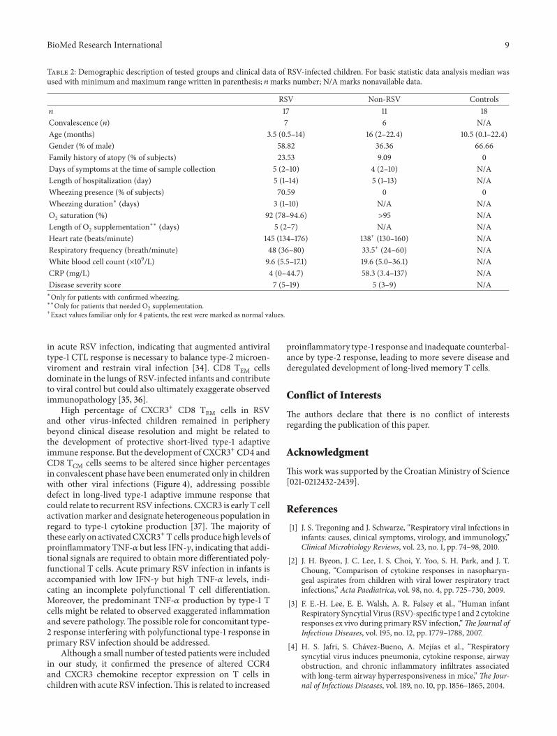
3.1. Collected Samples. Among 46 tested children, therewere 17 RSV positive (age: median 3.5 months, range 0.5–14 months; 7 followed up 4–6 weeks later), 11 with otherrespiratory viral infections (non-RSV infants; age: median16 months, range 2–22.4 months; 2 infected with influenzavirus and 9 infected with adenovirus; 6 followed up 4–6 weeks later), and 18 healthy controls (age: median 10.5months, range 0.1–22.4 months). All the tested children wereCaucasian.
Clinical parameters revealed that 12 patients in RSVgroup suffered from wheezing that lasted in average for 3days (range 1–10 days), and in 4 RSV-infected patients familyatopy was noticed. RSV-infected children had O
2saturation
less than 95% (range 78%–94.6%) but only 4 required oxygensupplementation. Respiratory frequency was in average 48breaths per minute (range 36–80) and heart rate 145 beatsper minute (range 134–176). Only one infant needed to beplaced in intensive care unit and stayed there for 3-dayobservation. None of tested children needed intubation orinvasive/noninvasive ventilatory support. To describe diseaseseverity of infected infants, severity score was generated asdescribed in Table 1. Complete demographic and clinical datafindings are provided in Table 2.
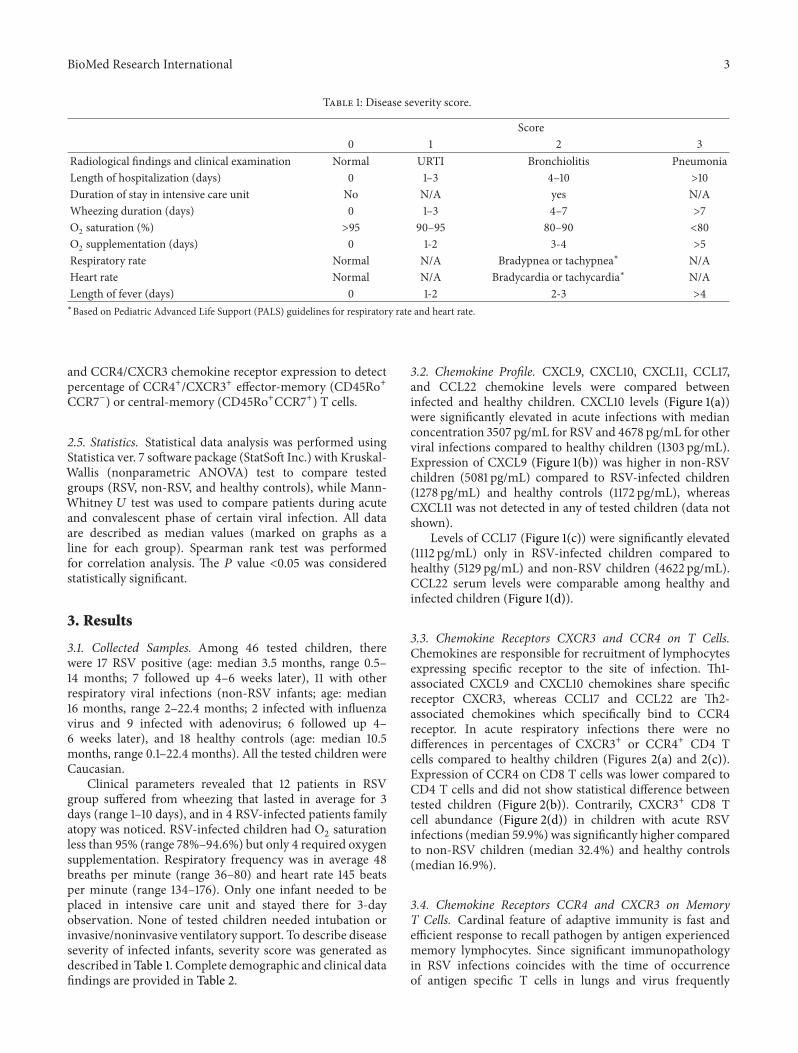
3.2. Chemokine Profile. CXCL9, CXCL10, CXCL11, CCL17,and CCL22 chemokine levels were compared betweeninfected and healthy children. CXCL10 levels (Figure 1(a))were significantly elevated in acute infections with medianconcentration 3507 pg/mL for RSV and 4678 pg/mL for otherviral infections compared to healthy children (1303 pg/mL).Expression of CXCL9 (Figure 1(b)) was higher in non-RSVchildren (5081 pg/mL) compared to RSV-infected children(1278 pg/mL) and healthy controls (1172 pg/mL), whereasCXCL11 was not detected in any of tested children (data notshown).
Levels of CCL17 (Figure 1(c)) were significantly elevated(1112 pg/mL) only in RSV-infected children compared tohealthy (5129 pg/mL) and non-RSV children (4622 pg/mL).CCL22 serum levels were comparable among healthy andinfected children (Figure 1(d)).
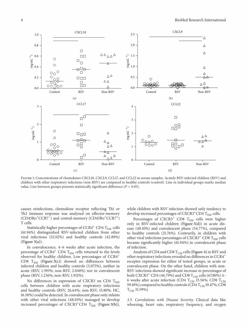
3.3. Chemokine Receptors CXCR3 and CCR4 on T Cells.Chemokines are responsible for recruitment of lymphocytesexpressing specific receptor to the site of infection. Th1-associated CXCL9 and CXCL10 chemokines share specificreceptor CXCR3, whereas CCL17 and CCL22 are Th2-associated chemokines which specifically bind to CCR4receptor. In acute respiratory infections there were nodifferences in percentages of CXCR3+ or CCR4+ CD4 Tcells compared to healthy children (Figures 2(a) and 2(c)).Expression of CCR4 on CD8 T cells was lower compared toCD4 T cells and did not show statistical difference betweentested children (Figure 2(b)). Contrarily, CXCR3+ CD8 Tcell abundance (Figure 2(d)) in children with acute RSVinfections (median 59.9%) was significantly higher comparedto non-RSV children (median 32.4%) and healthy controls(median 16.9%).
3.4. Chemokine Receptors CCR4 and CXCR3 on MemoryT Cells. Cardinal feature of adaptive immunity is fast andefficient response to recall pathogen by antigen experiencedmemory lymphocytes. Since significant immunopathologyin RSV infections coincides with the time of occurrenceof antigen specific T cells in lungs and virus frequently
4 BioMed Research International
RSVControl0.0
0.2
0.4
0.6
0.8
1.0(n
g mL−
1)
CXCL10
Non-RSV
(a)
RSVControl0.0
0.5
1.5
2.0
2.5
1.0(ng m
L−1)
CXCL9
Non-RSV
(b)
RSVControl Non-RSV
(ng m
L−1)
CCL17
0
1
2
3
(c)
RSVControl
(ng m
L−1)
CCL22
0
1
2
3
4
Non-RSV
(d)
Figure 1: Concentrations of chemokines CXCL10, CXCL9, CCL17, and CCL22 in serum samples. Acutely RSV-infected children (RSV) andchildren with other respiratory infections (non-RSV) are compared to healthy controls (control). Line in individual groups marks medianvalue. Line between groups presents statistically significant difference (𝑃 < 0.05).
causes reinfections, chemokine receptor reflecting Th1 orTh2 immune response was analyzed on effector-memory(CD45Ro+CCR7−) and central-memory (CD45Ro+CCR7+)T cells.
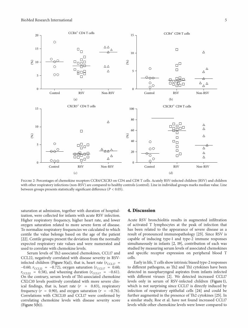
Statistically higher percentages of CCR4+ CD4 TEM cells(61.94%) distinguished RSV-infected children from otherviral infections (52.02%) and healthy controls (42.89%)(Figure 3(a)).
In convalescence, 4–6 weeks after acute infection, thepercentage of CCR4+ CD4 TEM cells returned to the levelsobserved for healthy children. Low percentages of CCR4+CD8 TEM (Figure 3(c)) showed no differences betweeninfected children and healthy controls (3.075%), neither inacute (RSV, 1.395%; non-RSV, 2.048%) nor in convalescentphase (RSV, 1.236%; non-RSV, 1.921%).
No differences in expression of CXCR3 on CD4 TEMcells between children with acute respiratory infectionsand healthy controls (RSV, 26.64%; non-RSV, 15.80%; HC,16.50%) could be detected. In convalescent phase only infantswith other viral infections (48.03%) managed to developincreased percentages of CXCR3+CD4 TEM (Figure 3(b)),
while children with RSV infection showed only tendency todevelop increased percentages of CXCR3+CD4 TEM cells.
Percentages of CXCR3+ CD8 TEM cells were higheronly in RSV-infected children (Figure 3(d)) in acute dis-ease (48.10%) and convalescent phase (54.77%), comparedto healthy controls (21.76%). Contrarily, in children withother viral infections percentages of CXCR3+ CD8 TEM cellsbecame significantly higher (61.94%) in convalescent phaseof infection.
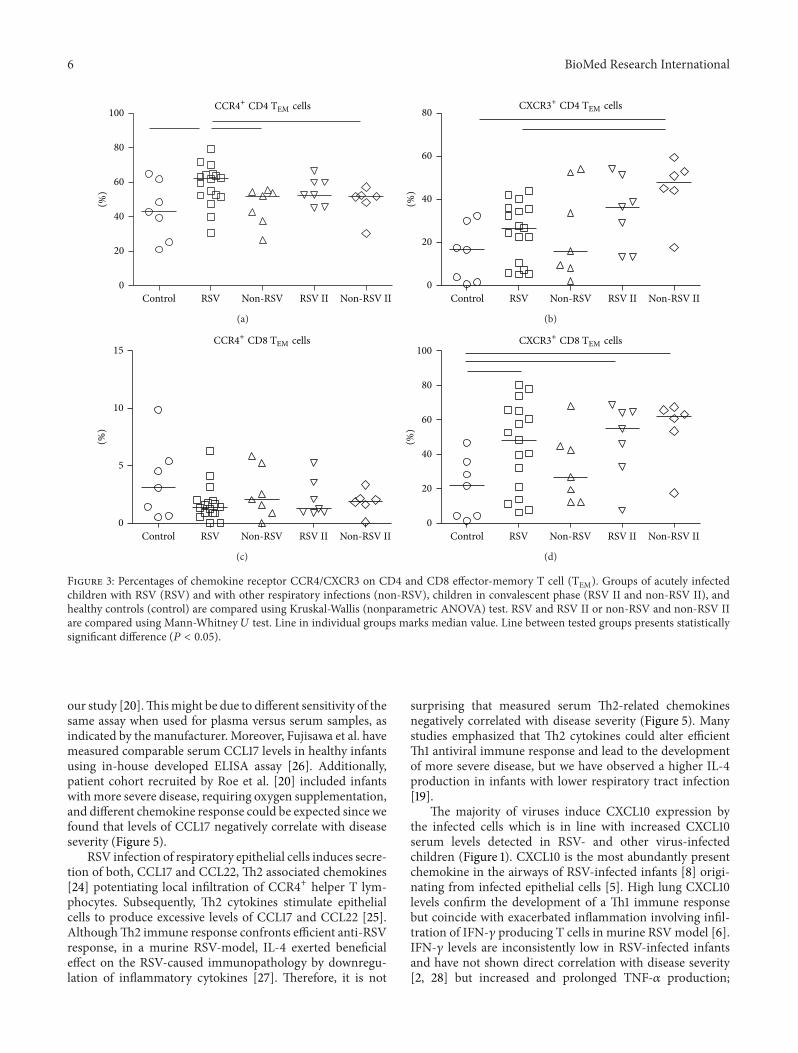
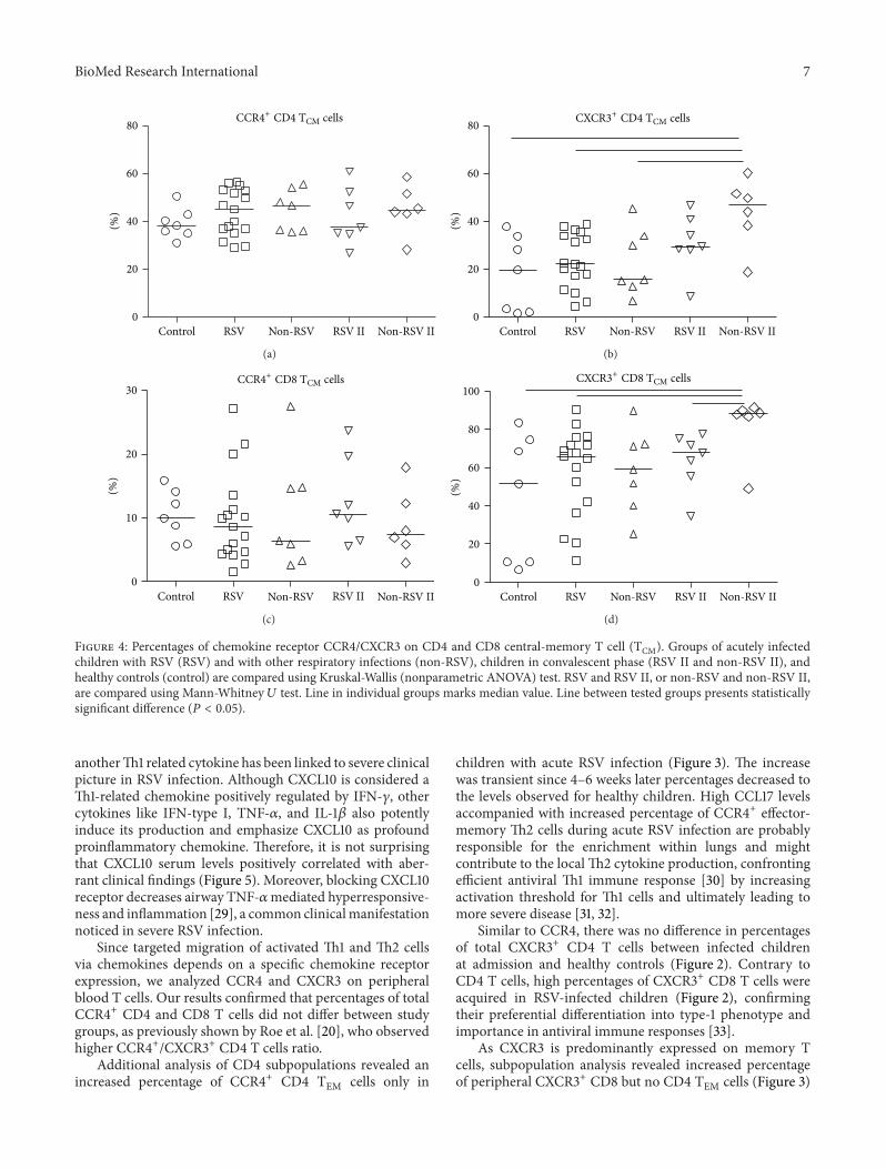
Analysis of CD4 andCD8TCM cells (Figure 4) inRSVandother respiratory infections revealed no differences in CCR4+receptor expression for either of tested groups, in acute orconvalescent phase. On the other hand, children with non-RSV infections showed significant increase in percentages ofboth CXCR3+ CD4 (46.79%) and CD8 TCM cells (67.80%) 4–6 weeks after acute infection (CD4 TCM 15.56%; CD8 TCM59.10%) compared to healthy controls (CD4TCM 19.47%;CD8TCM 51.59%).
3.5. Correlations with Disease Severity. Clinical data likewheezing, heart rate, respiratory frequency, and oxygen
BioMed Research International 5
Control RSV0
5
10
15
20(%)
CCR4+ CD4 T cells
Non-RSV
(a)
Control RSV0
5
10
15
(%)
CCR4+ CD8 T cells
Non-RSV
(b)
Control RSV0
5
10
15
(%)
CXCR3+ CD4 T cells
Non-RSV
(c)
Control RSV0
20
40
60
80
100
(%)
CXCR3+ CD8 T cells
Non-RSV
(d)
Figure 2: Percentages of chemokine receptors CCR4/CXCR3 on CD4 and CD8 T cells. Acutely RSV-infected children (RSV) and childrenwith other respiratory infections (non-RSV) are compared to healthy controls (control). Line in individual groups marks median value. Linebetween groups presents statistically significant difference (𝑃 < 0.05).
saturation at admission, together with duration of hospital-ization, were collected for infants with acute RSV infection.Higher respiratory frequency, higher heart rate, and loweroxygen saturation related to more severe form of disease.To normalize respiratory frequencies we calculated to whichcentile the value belongs based on the age of the patient[22]. Centile groups present the deviation from the normallyexpected respiratory rate values and were numerated andused to correlate with chemokine levels.
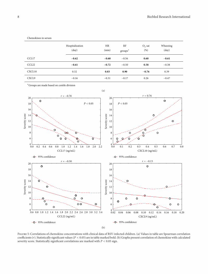
Serum levels of Th2-associated chemokines, CCL17 andCCL22, negatively correlated with disease severity in RSV-infected children (Figure 5(a)), that is, heart rate (𝑟CCL17 =−0.60; 𝑟CCL22 = −0.72), oxygen saturation (𝑟CCL17 = 0.60;𝑟CCL22 = 0.58), and wheezing duration (𝑟CCL17 = −0.61).On the contrary, serum levels of Th1-associated chemokineCXLC10 levels positively correlated with more severe clin-ical findings, that is, heart rate (𝑟 = 0.83), respiratoryfrequency (𝑟 = 0.90), and oxygen saturation (𝑟 = −0.76).Correlations with CXCL10 and CCL17 were confirmed bycorrelating chemokine levels with disease severity score(Figure 5(b)).
4. Discussion
Acute RSV bronchiolitis results in augmented infiltrationof activated T lymphocytes at the peak of infection thathas been related to the appearance of severe disease as aresult of pronounced immunopathology [23]. Since RSV iscapable of inducing type-1 and type-2 immune responsessimultaneously in infants [2, 19], contribution of each wasstudied by measuring serum levels of associated chemokinesand specific receptor expression on peripheral blood Tcells.
Early in life, T cells show intrinsic biased type-2 responsesto different pathogens, as Th2 and Th1 cytokines have beendetected in nasopharyngeal aspirates from infants infectedwith different viruses [2]. We detected increased CCL17levels only in serum of RSV-infected children (Figure 1),which is not surprising since CCL17 is directly induced byinfection of respiratory epithelial cells [24] and could befurther augmented in the presence of Th2 cytokines [25]. Ina similar study, Roe et al. have not found increased CCL17levels while other chemokine levels were lower compared to
6 BioMed Research International
CCR4+ CD4 TEM cells
0
20
40
60
80
100
RSV IIRSV
(%)
Control Non-RSV IINon-RSV
(a)
CXCR3+ CD4 TEM cells
0
20
40
60
80
(%)
RSV IIRSVControl Non-RSV IINon-RSV
(b)
CCR4+ CD8 TEM cells
0
5
10
15
(%)
RSV IIRSV Non-RSV IIControl Non-RSV
(c)
CXCR3+ CD8 TEM cells
0
20
40
60
80
100
(%)
RSV IIRSVControl Non-RSV IINon-RSV
(d)
Figure 3: Percentages of chemokine receptor CCR4/CXCR3 on CD4 and CD8 effector-memory T cell (TEM). Groups of acutely infectedchildren with RSV (RSV) and with other respiratory infections (non-RSV), children in convalescent phase (RSV II and non-RSV II), andhealthy controls (control) are compared using Kruskal-Wallis (nonparametric ANOVA) test. RSV and RSV II or non-RSV and non-RSV IIare compared using Mann-Whitney 𝑈 test. Line in individual groups marks median value. Line between tested groups presents statisticallysignificant difference (𝑃 < 0.05).
our study [20].Thismight be due to different sensitivity of thesame assay when used for plasma versus serum samples, asindicated by the manufacturer. Moreover, Fujisawa et al. havemeasured comparable serum CCL17 levels in healthy infantsusing in-house developed ELISA assay [26]. Additionally,patient cohort recruited by Roe et al. [20] included infantswithmore severe disease, requiring oxygen supplementation,and different chemokine response could be expected since wefound that levels of CCL17 negatively correlate with diseaseseverity (Figure 5).
RSV infection of respiratory epithelial cells induces secre-tion of both, CCL17 and CCL22, Th2 associated chemokines[24] potentiating local infiltration of CCR4+ helper T lym-phocytes. Subsequently, Th2 cytokines stimulate epithelialcells to produce excessive levels of CCL17 and CCL22 [25].AlthoughTh2 immune response confronts efficient anti-RSVresponse, in a murine RSV-model, IL-4 exerted beneficialeffect on the RSV-caused immunopathology by downregu-lation of inflammatory cytokines [27]. Therefore, it is not
surprising that measured serum Th2-related chemokinesnegatively correlated with disease severity (Figure 5). Manystudies emphasized that Th2 cytokines could alter efficientTh1 antiviral immune response and lead to the developmentof more severe disease, but we have observed a higher IL-4production in infants with lower respiratory tract infection[19].
The majority of viruses induce CXCL10 expression bythe infected cells which is in line with increased CXCL10serum levels detected in RSV- and other virus-infectedchildren (Figure 1). CXCL10 is the most abundantly presentchemokine in the airways of RSV-infected infants [8] origi-nating from infected epithelial cells [5]. High lung CXCL10levels confirm the development of a Th1 immune responsebut coincide with exacerbated inflammation involving infil-tration of IFN-𝛾 producing T cells in murine RSV model [6].IFN-𝛾 levels are inconsistently low in RSV-infected infantsand have not shown direct correlation with disease severity[2, 28] but increased and prolonged TNF-𝛼 production;
BioMed Research International 7
RSV IIRSVControl0
20
40
60
80(%)
CCR4+ CD4 TCM cells
Non-RSV IINon-RSV
(a)
RSV IIRSVControl0
20
40
60
80
(%)
CXCR3+ CD4 TCM cells
Non-RSV IINon-RSV
(b)
RSV IIRSVControl0
10
20
30
(%)
CCR4+ CD8 TCM cells
Non-RSV IINon-RSV
(c)
RSV IIRSVControl
CXCR3+ CD8 TCM cells
0
20
40
60
80
100
(%)
Non-RSV IINon-RSV
(d)
Figure 4: Percentages of chemokine receptor CCR4/CXCR3 on CD4 and CD8 central-memory T cell (TCM). Groups of acutely infectedchildren with RSV (RSV) and with other respiratory infections (non-RSV), children in convalescent phase (RSV II and non-RSV II), andhealthy controls (control) are compared using Kruskal-Wallis (nonparametric ANOVA) test. RSV and RSV II, or non-RSV and non-RSV II,are compared using Mann-Whitney 𝑈 test. Line in individual groups marks median value. Line between tested groups presents statisticallysignificant difference (𝑃 < 0.05).
anotherTh1 related cytokine has been linked to severe clinicalpicture in RSV infection. Although CXCL10 is considered aTh1-related chemokine positively regulated by IFN-𝛾, othercytokines like IFN-type I, TNF-𝛼, and IL-1𝛽 also potentlyinduce its production and emphasize CXCL10 as profoundproinflammatory chemokine. Therefore, it is not surprisingthat CXCL10 serum levels positively correlated with aber-rant clinical findings (Figure 5). Moreover, blocking CXCL10receptor decreases airway TNF-𝛼mediated hyperresponsive-ness and inflammation [29], a common clinicalmanifestationnoticed in severe RSV infection.
Since targeted migration of activated Th1 and Th2 cellsvia chemokines depends on a specific chemokine receptorexpression, we analyzed CCR4 and CXCR3 on peripheralblood T cells. Our results confirmed that percentages of totalCCR4+ CD4 and CD8 T cells did not differ between studygroups, as previously shown by Roe et al. [20], who observedhigher CCR4+/CXCR3+ CD4 T cells ratio.
Additional analysis of CD4 subpopulations revealed anincreased percentage of CCR4+ CD4 TEM cells only in
children with acute RSV infection (Figure 3). The increasewas transient since 4–6 weeks later percentages decreased tothe levels observed for healthy children. High CCL17 levelsaccompanied with increased percentage of CCR4+ effector-memory Th2 cells during acute RSV infection are probablyresponsible for the enrichment within lungs and mightcontribute to the local Th2 cytokine production, confrontingefficient antiviral Th1 immune response [30] by increasingactivation threshold for Th1 cells and ultimately leading tomore severe disease [31, 32].
Similar to CCR4, there was no difference in percentagesof total CXCR3+ CD4 T cells between infected childrenat admission and healthy controls (Figure 2). Contrary toCD4 T cells, high percentages of CXCR3+ CD8 T cells wereacquired in RSV-infected children (Figure 2), confirmingtheir preferential differentiation into type-1 phenotype andimportance in antiviral immune responses [33].
As CXCR3 is predominantly expressed on memory Tcells, subpopulation analysis revealed increased percentageof peripheral CXCR3+ CD8 but no CD4 TEM cells (Figure 3)
8 BioMed Research International
Chemokines in serum
Hospitalization(day)
HR(min)
RF
groups∗ (%)Wheezing
(day)
CCL17 −0.56
CCL22 −0.50 −0.38
CXCL10 0.32 0.83 0.90 0.39
CXCL9 −0.16 −0.31 −0.17 0.26 −0.47
∗Groups are made based on centile division
O2 sat
−0.62 −0.60 0.60 −0.61
−0.61 −0.72 0.58
−0.76
(a)
P < 0.05P < 0.05
r = −0.78
r = −0.50 r = −0.15
0.0 0.2 0.0 0.2 0.3 0.4 0.60.5 0.7 0.80.10.4 0.6 0.8 1.0 1.2 1.4 1.6 1.8 2.0
0.02 0.04 0.06 0.08 0.10 0.12 0.14 0.16 0.18 0.20
2.2
0.6 0.8 1.0 1.2 1.4 3.41.6 1.8 2.0 2.42.2 2.6 2.8 3.0 3.2
4
6
8
10
12
14
16
18
20
Seve
rity
scor
e
4
6
8
10
12
14
16
18
20
Seve
rity
scor
e
4
6
8
10
12
14
16
18
20
Seve
rity
scor
e
4
6
8
10
12
14
16
18
20
Seve
rity
scor
e
95% confidence95% confidence
95% confidence 95% confidence
CCL17 (ng/mL)
CCL22 (ng/mL)
r = 0.76
CXCL9 (ng/mL)
CXCL10 (ng/mL)
(b)
Figure 5: Correlations of chemokine concentrations with clinical data of RSV-infected children. (a) Values in table are Spearman correlationcoefficients (𝑟). Statistically significant values (𝑃 < 0.05) are in tablemarked bold. (b) Graphs present correlation of chemokinewith calculatedseverity score. Statistically significant correlations are marked with 𝑃 < 0.05 sign.
BioMed Research International 9
Table 2: Demographic description of tested groups and clinical data of RSV-infected children. For basic statistic data analysis median wasused with minimum and maximum range written in parenthesis; 𝑛marks number; N/A marks nonavailable data.
RSV Non-RSV Controls𝑛 17 11 18Convalescence (𝑛) 7 6 N/AAge (months) 3.5 (0.5–14) 16 (2–22.4) 10.5 (0.1–22.4)Gender (% of male) 58.82 36.36 66.66Family history of atopy (% of subjects) 23.53 9.09 0Days of symptoms at the time of sample collection 5 (2–10) 4 (2–10) N/ALength of hospitalization (day) 5 (1–14) 5 (1–13) N/AWheezing presence (% of subjects) 70.59 0 0Wheezing duration∗ (days) 3 (1–10) N/A N/AO2 saturation (%) 92 (78–94.6) >95 N/ALength of O2 supplementation∗∗ (days) 5 (2–7) N/A N/AHeart rate (beats/minute) 145 (134–176) 138+ (130–160) N/ARespiratory frequency (breath/minute) 48 (36–80) 33.5+ (24–60) N/AWhite blood cell count (×109/L) 9.6 (5.5–17.1) 19.6 (5.0–36.1) N/ACRP (mg/L) 4 (0–44.7) 58.3 (3.4–137) N/ADisease severity score 7 (5–19) 5 (3–9) N/A∗Only for patients with confirmed wheezing.∗∗Only for patients that needed O2 supplementation.+Exact values familiar only for 4 patients, the rest were marked as normal values.
in acute RSV infection, indicating that augmented antiviraltype-1 CTL response is necessary to balance type-2 microen-viroment and restrain viral infection [34]. CD8 TEM cellsdominate in the lungs of RSV-infected infants and contributeto viral control but could also ultimately exaggerate observedimmunopathology [35, 36].
High percentage of CXCR3+ CD8 TEM cells in RSVand other virus-infected children remained in peripherybeyond clinical disease resolution and might be related tothe development of protective short-lived type-1 adaptiveimmune response. But the development of CXCR3+ CD4 andCD8 TCM cells seems to be altered since higher percentagesin convalescent phase have been enumerated only in childrenwith other viral infections (Figure 4), addressing possibledefect in long-lived type-1 adaptive immune response thatcould relate to recurrent RSV infections. CXCR3 is early T cellactivationmarker and designate heterogeneous population inregard to type-1 cytokine production [37]. The majority ofthese early on activatedCXCR3+ T cells produce high levels ofproinflammatory TNF-𝛼 but less IFN-𝛾, indicating that addi-tional signals are required to obtain more differentiated poly-functional T cells. Acute primary RSV infection in infants isaccompanied with low IFN-𝛾 but high TNF-𝛼 levels, indi-cating an incomplete polyfunctional T cell differentiation.Moreover, the predominant TNF-𝛼 production by type-1 Tcells might be related to observed exaggerated inflammationand severe pathology.The possible role for concomitant type-2 response interfering with polyfunctional type-1 response inprimary RSV infection should be addressed.
Although a small number of tested patients were includedin our study, it confirmed the presence of altered CCR4and CXCR3 chemokine receptor expression on T cells inchildren with acute RSV infection.This is related to increased
proinflammatory type-1 response and inadequate counterbal-ance by type-2 response, leading to more severe disease andderegulated development of long-lived memory T cells.
Conflict of Interests
The authors declare that there is no conflict of interestsregarding the publication of this paper.
Acknowledgment
This work was supported by the CroatianMinistry of Science[021-0212432-2439].
References
[1] J. S. Tregoning and J. Schwarze, “Respiratory viral infections ininfants: causes, clinical symptoms, virology, and immunology,”Clinical Microbiology Reviews, vol. 23, no. 1, pp. 74–98, 2010.
[2] J. H. Byeon, J. C. Lee, I. S. Choi, Y. Yoo, S. H. Park, and J. T.Choung, “Comparison of cytokine responses in nasopharyn-geal aspirates from children with viral lower respiratory tractinfections,” Acta Paediatrica, vol. 98, no. 4, pp. 725–730, 2009.
[3] F. E.-H. Lee, E. E. Walsh, A. R. Falsey et al., “Human infantRespiratory Syncytial Virus (RSV)-specific type 1 and 2 cytokineresponses ex vivo during primary RSV infection,”The Journal ofInfectious Diseases, vol. 195, no. 12, pp. 1779–1788, 2007.
[4] H. S. Jafri, S. Chavez-Bueno, A. Mejıas et al., “Respiratorysyncytial virus induces pneumonia, cytokine response, airwayobstruction, and chronic inflammatory infiltrates associatedwith long-term airway hyperresponsiveness in mice,”The Jour-nal of Infectious Diseases, vol. 189, no. 10, pp. 1856–1865, 2004.
10 BioMed Research International
[5] R. Villenave, S. Thavagnanam, S. Sarlang et al., “In vitromodeling of respiratory syncytial virus infection of pediatricbronchial epithelium, the primary target of infection in vivo,”Proceedings of the National Academy of Sciences of the UnitedStates of America, vol. 109, no. 13, pp. 5040–5045, 2012.
[6] T. Ostler, W. Davidson, and S. Ehl, “Virus clearance andimmunopathology by CD8+ T cells during infection with respi-ratory syncytial virus are mediated by IFN-gamma,” EuropeanJournal of Immunology, vol. 32, pp. 2117–2123, 2002.
[7] J. Heidema, M. V. Lukens, W. W. C. van Maren et al., “CD8+T cell responses in bronchoalveolar lavage fluid and peripheralbloodmononuclear cells of infants with severe primary respira-tory syncytial virus infections,” Journal of Immunology, vol. 179,no. 12, pp. 8410–8417, 2007.
[8] P. S. McNamara, B. F. Flanagan, C. A. Hart, and R. L. Smyth,“Production of chemokines in the lungs of infants with severerespiratory syncytial virus bronchiolitis,” The Journal of Infec-tious Diseases, vol. 191, no. 8, pp. 1225–1232, 2005.
[9] R. L. Rabin, M. A. Alston, J. C. Sircus et al., “CXCR3 is inducedearly on the pathway of CD4+ T cell differentiation and bridgescentral and peripheral functions,” Journal of Immunology, vol.171, no. 6, pp. 2812–2824, 2003.
[10] C. M. Tabarani, C. A. Bonville, M. Suryadevara et al., “Novelinflammatory markers, clinical risk factors, and virus typeassociated with severe respiratory syncytial virus infection,”Pediatric Infectious Disease Journal, vol. 32, no. 12, 2013.
[11] J. Chang andT. J. Braciale, “Respiratory syncytial virus infectionsuppresses lung CD8+ T-cell effector activity and peripheralCD8+ T-cell memory in the respiratory tract,”Nature Medicine,vol. 8, no. 1, pp. 54–60, 2002.
[12] R. B. Fulton, M. R. Olson, and S. M. Varga, “Regulation ofcytokine production by virus-specific CD8 T cells in the lungs,”Journal of Virology, vol. 82, no. 16, pp. 7799–7811, 2008.
[13] S. Aung and B. S. Graham, “IL-4 diminishes perforin-mediatedand increases Fas ligand-mediated cytotoxicity in vivo,” Journalof Immunology, vol. 164, no. 7, pp. 3487–3493, 2000.
[14] J. Han, A. Dakhama, Y. Jia et al., “Responsiveness to respiratorysyncytial virus in neonates is mediated through thymic stromallymphopoietin andOX40 ligand,” Journal of Allergy andClinicalImmunology, vol. 130, pp. 1175–1186, 2012.
[15] T. Imai, M. Nagira, S. Takagi et al., “Selective recruit-ment of CCR4-bearing T(h)2 cells toward antigen-presentingcells by the CC chemokines thymus and activation-regulatedchemokine andmacrophage-derived chemokine,” InternationalImmunology, vol. 11, no. 1, pp. 81–88, 1999.
[16] J. Qiao, A. Li, and X. Jin, “TSLP fromRSV-stimulated rat airwayepithelial cells activates myeloid dendritic cells,” Immunologyand Cell Biology, vol. 89, no. 2, pp. 231–238, 2011.
[17] L. Rivino, M. Messi, D. Jarrossay, A. Lanzavecchia, F. Sallusto,and J. Geginat, “Chemokine receptor expression identifies pre-T helper (Th)1, pre-Th2, and nonpolarized cells among humanCD4+ central memory T cells,” The Journal of ExperimentalMedicine, vol. 200, no. 6, pp. 725–735, 2004.
[18] D. P. Andrew, N. Ruffing, C. H. Kim et al., “C-C chemokinereceptor 4 expression defines a major subset of circulatingnonintestinal memory T cells of both Th1 and Th2 potential,”Journal of Immunology, vol. 166, no. 1, pp. 103–111, 2001.
[19] K. Bendelja, A. Gagro, A. Bace et al., “Predominant type-2 response in infants with respiratory syncytial virus (RSV)infection demonstrated by cytokine flow cytometry,” ClinicalandExperimental Immunology, vol. 121, no. 2, pp. 332–338, 2000.
[20] M. F. E. Roe, D. M. Bloxham, A. S. Cowburn, and D. R.O'Donnell, “Changes in helper lymphocyte chemokine receptorexpression and elevation of IP-10 during acute respiratorysyncytial virus infection in infants,” Pediatric Allergy andImmunology, vol. 22, no. 2, pp. 229–234, 2011.
[21] A. Lukic-Grlic, A. Bace, R. Lokar-Kolbas et al., “Clinical andepidemiological aspects of respiratory syncytial virus lowerrespiratory tract infections,” European Journal of Epidemiology,vol. 15, no. 4, pp. 361–365, 1999.
[22] S. Fleming, M. Thompson, R. Stevens et al., “Normal ranges ofheart rate and respiratory rate in children from birth to 18 yearsof age: a systematic review of observational studies,”The Lancet,vol. 377, no. 9770, pp. 1011–1018, 2011.
[23] P. J. M. Openshaw and J. S. Tregoning, “Immune responsesand disease enhancement during respiratory syncytial virusinfection,” Clinical Microbiology Reviews, vol. 18, no. 3, pp. 541–555, 2005.
[24] Y. Zhang, B. A. Luxon, A. Casola, R. P. Garofalo,M. Jamaluddin,and A. R. Brasier, “Expression of respiratory syncytial virus-induced chemokine gene networks in lower airway epithelialcells revealed by cDNA microarrays,” Journal of Virology, vol.75, no. 19, pp. 9044–9058, 2001.
[25] M. M. Monick, L. S. Powers, I. Hassan et al., “Respiratorysyncytial virus synergizes withTh2 cytokines to induce optimallevels of TARC/CCL17,” Journal of Immunology, vol. 179, no. 3,pp. 1648–1658, 2007.
[26] T. Fujisawa,M.Nagao, Y. Hiraguchi et al., “Serummeasurementof thymus and activation-regulated chemokine/CCL17 in chil-dren with atopic dermatitis: elevated normal levels in infancyand age-specific analysis in atopic dermatitis,” Pediatric Allergyand Immunology, vol. 20, no. 7, pp. 633–641, 2009.
[27] K. A. Shirey, L. M. Pletneva, A. C. Puche et al., “Control of RSV-induced lung injury by alternatively activated macrophages isIL-4R𝛼-, TLR4-, and IFN-Β-dependent,”Mucosal Immunology,vol. 3, no. 3, pp. 291–300, 2010.
[28] T. P. Welliver, J. L. Reed, and R. C. Welliver Sr., “Respiratorysyncytial virus and influenza virus infections observations fromtissues of fatal infant cases,” Pediatric Infectious Disease Journal,vol. 27, no. 10, pp. S92–S96, 2008.
[29] Y. Lin, H. Yan, Y. Xiao et al., “Attenuation of antigen-inducedairway hyperresponsiveness and inflammation in CXCR3knockout mice,” Respiratory Research, vol. 12, article 123, 2011.
[30] K. M. Cautivo, S. M. Bueno, C. M. Cortes, A. Wozniak, C. A.Riedel, and A. M. Kalergis, “Efficient lung recruitment of respi-ratory syncytial virus-specificTh1 cells induced by recombinantbacillusCalmette-Guerin promotes virus clearance andprotectsfrom Infection,” Journal of Immunology, vol. 185, no. 12, pp.7633–7645, 2010.
[31] T. R. Johnson,M. E. Rothenberg, andB. S. Graham, “Pulmonaryeosinophilia requires interleukin-5, eotaxin-1, and CD4+ Tcells in mice immunized with respiratory syncytial virus Gglycoprotein,” Journal of Leukocyte Biology, vol. 84, no. 3, pp.748–759, 2008.
[32] S. M. Varga, X. Wang, R. M. Welsh, and T. J. Braciale,“Immunopathology in RSV infection is mediated by a discreteoligoclonal subset of antigen-specific CD4+ T cells,” Immunity,vol. 15, no. 4, pp. 637–646, 2001.
[33] G. J. De Bree, E. M. M. van Leeuwen, T. A. Out, H. M. Jansen,R. E. Jonkers, and R. A. W. van Lier, “Selective accumulation ofdifferentiated CD8+ T cells specific for respiratory viruses in thehuman lung,”The Journal of ExperimentalMedicine, vol. 202, no.10, pp. 1433–1442, 2005.
BioMed Research International 11
[34] A. Srikiatkhachorn and T. J. Braciale, “Virus-specific CD8+T lymphocytes downregulate T helper cell type 2 cytokinesecretion and pulmonary eosinophilia during experimentalmurine respiratory syncytial virus infection,” The Journal ofExperimental Medicine, vol. 186, no. 3, pp. 421–432, 1997.
[35] J. S. Tregoning, Y. Yamaguchi, J. Harker, B. Wang, and P.J. M. Openshaw, “The role of T cells in the enhancementof respiratory syncytial virus infection severity during adultreinfection of neonatally sensitized mice,” Journal of Virology,vol. 82, no. 8, pp. 4115–4124, 2008.
[36] R. B. Fulton, D. K. Meyerholz, and S. M. Varga, “Foxp3+CD4 regulatory T cells limit pulmonary immunopathologyby modulating the CD8 T cell response during respiratorysyncytial virus infection,” Journal of Immunology, vol. 185, no.4, pp. 2382–2392, 2010.
[37] H.H. Zhang,K. Song, R. L. Rabin et al., “CCR2 identifies a stablepopulation of human effector memory CD4+ T cells equippedfor rapid recall response,” Journal of Immunology, vol. 185, no.11, pp. 6646–6663, 2010.