Primary and secondary scintillation measurements in a Xenon Gas Proportional Scintillation Counter
16
1 Primary and secondary scintillation measurements in a xenon Gas Proportional Scintillation Counter L.M.P. Fernandes 1 , E.D.C. Freitas 1 , M. Ball 2 , J.J. Gómez-Cadenas 2 , C.M.B. Monteiro 1 , N. Yahlali 2 , D. Nygren 3 and J.M.F. dos Santos 1 1 Instrumentation Centre, Physics Department, University of Coimbra, P-3004-516 Coimbra, Portugal 2 Instituto de Física Corpuscular, E-46071 Valencia, Spain 3 Lawrence Berkeley National Laboratory, Berkeley, CA 94720, USA E-mail: [email protected] ABSTRACT NEXT is a new experiment to search for neutrinoless double beta decay using a 100 kg radio-pure high-pressure gaseous xenon TPC. The detector requires excellent energy resolution, which can be achieved in a Xe TPC with electroluminescence readout. Hamamatsu R8520-06SEL photomultipliers are good candidates for the scintillation readout. The performance of this photomultiplier, used as VUV photosensor in a gas proportional scintillation counter, was investigated. Initial results for the detection of primary and secondary scintillation produced as a result of the interaction of 5.9 keV X- rays in gaseous xenon, at room temperature and at pressures up to 3 bar, are presented. An energy resolution of 8.0% was obtained for secondary scintillation produced by 5.9 keV X-rays. No significant variation of the primary scintillation was observed for different pressures (1, 2 and 3 bar) and for electric fields up to 0.8 V cm -1 torr -1 in the drift region, demonstrating negligible recombination luminescence. A primary scintillation yield of 81 ± 7 photons was obtained for 5.9 keV X-rays, corresponding to a mean energy of 72 ± 6 eV to produce a primary scintillation photon in xenon. Keywords: Interaction of radiation with matter; Gaseous detectors; Photomultipliers. 1. INTRODUCTION NEXT stands for Neutrino Experiment with a Xenon TPC, a search for neutrinoless double-beta decay (0νββ) [1]. The experiment will use the 136 Xe isotope in a 100 kg high pressure (~10 bar) TPC (time projection chamber), to be operated at room temperature in the Canfranc Underground Laboratory. The unambiguous observation of 0νββ would demonstrate that the neutrino has a Majorana nature. This would represent a breakthrough of new physics, beyond the Standard Model. The TPC should combine an excellent energy resolution with a unique topological signature to achieve high sensitivity to a light Majorana neutrino. Xenon offers several advantages for 0νββ experiments. As a noble gas, it can be used for tracking particles. It does not have long-lived radioactive isotopes other than 136 Xe, which decays by double-beta. This isotope has a relatively high abundance in natural xenon (8.9%) and can be easily enriched by centrifugation at a reasonable cost.
-
Upload
independent -
Category
Documents
-
view
1 -
download
0
Transcript of Primary and secondary scintillation measurements in a Xenon Gas Proportional Scintillation Counter

1
Primary and secondary scintillation measurements in a xenon Gas Proportional Scintillation Counter
L.M.P. Fernandes1, E.D.C. Freitas1, M. Ball2, J.J. Gómez-Cadenas2, C.M.B. Monteiro1, N. Yahlali2, D. Nygren3 and J.M.F. dos Santos1
1 Instrumentation Centre, Physics Department, University of Coimbra, P-3004-516 Coimbra, Portugal
2 Instituto de Física Corpuscular, E-46071 Valencia, Spain
3 Lawrence Berkeley National Laboratory, Berkeley, CA 94720, USA
E-mail: [email protected]
ABSTRACT
NEXT is a new experiment to search for neutrinoless double beta decay using a 100 kg
radio-pure high-pressure gaseous xenon TPC. The detector requires excellent energy
resolution, which can be achieved in a Xe TPC with electroluminescence readout.
Hamamatsu R8520-06SEL photomultipliers are good candidates for the scintillation
readout. The performance of this photomultiplier, used as VUV photosensor in a gas
proportional scintillation counter, was investigated. Initial results for the detection of
primary and secondary scintillation produced as a result of the interaction of 5.9 keV X-
rays in gaseous xenon, at room temperature and at pressures up to 3 bar, are presented.
An energy resolution of 8.0% was obtained for secondary scintillation produced by 5.9
keV X-rays. No significant variation of the primary scintillation was observed for
different pressures (1, 2 and 3 bar) and for electric fields up to 0.8 V cm-1
torr-1
in the
drift region, demonstrating negligible recombination luminescence. A primary
scintillation yield of 81 ± 7 photons was obtained for 5.9 keV X-rays, corresponding to
a mean energy of 72 ± 6 eV to produce a primary scintillation photon in xenon.
Keywords: Interaction of radiation with matter; Gaseous detectors; Photomultipliers.
1. INTRODUCTION
NEXT stands for Neutrino Experiment with a Xenon TPC, a search for neutrinoless
double-beta decay (0νββ) [1]. The experiment will use the 136
Xe isotope in a 100 kg
high pressure (~10 bar) TPC (time projection chamber), to be operated at room
temperature in the Canfranc Underground Laboratory. The unambiguous observation of
0νββ would demonstrate that the neutrino has a Majorana nature. This would represent a
breakthrough of new physics, beyond the Standard Model. The TPC should combine an
excellent energy resolution with a unique topological signature to achieve high
sensitivity to a light Majorana neutrino.
Xenon offers several advantages for 0νββ experiments. As a noble gas, it can be
used for tracking particles. It does not have long-lived radioactive isotopes other than 136
Xe, which decays by double-beta. This isotope has a relatively high abundance in
natural xenon (8.9%) and can be easily enriched by centrifugation at a reasonable cost.

2
The 136
Xe 136
Ba transition has a high Q-value, 2457.83(37) keV [2], allowing a
reduction of the background resulting from lower energetic gamma-rays emitted by
other radioactive materials by means of pattern recognition of the events in the offline
analysis. Furthermore, xenon can be used at the same time as the detection medium for
the charged particles released in the decay. Another interesting property of pure Xe is
the large amount of both primary ionization and primary VUV scintillation (~175 nm)
induced by the radiation interaction.
The detector concept requires excellent energy resolution (< 1% at 2.46 MeV).
This is essential not only to reduce the tail of the double-beta decay with neutrino
emission (2νββ) spectrum from overlapping the region of interest of the 0νββ spectrum,
but also to prevent the contamination of the region of interest by the most severe
gamma-ray background (2614 keV from 208
Tl and 2447 keV from 214
Bi). Furthermore,
external backgrounds should also be reduced using topological properties of the 0νββ
events.
For half a century it has been known that secondary scintillation, also called
electroluminescence, provides a mechanism for high gain with very low fluctuations
[3]. This technique offers large signals with negligible electronic noise, and is the
optimum amplification technique for this kind of experiment. Therefore, it is of great
importance, especially in experiments with very low event rates and/or high background
levels such as 0νββ experiments, to use the secondary scintillation signal rather than the
signal from either unamplified primary ionization or avalanche ionization [4]. This is
the technique to be used in NEXT, with a nominal xenon pressure of 10 bar [1].
The proposed detector design for NEXT, called Separated Optimized Function
TPC (SOFT) approach, is based on a specific readout that separates the technologies for
pattern recognition and energy measurement [4]. Electroluminescence photons emitted
towards the hemisphere of the anode can be used to recognise the specific track pattern
of the two electrons emitted in the double beta decay. The technology does not require
excellent energy resolution capabilities but does require robust pattern recognition and
the capability to separate nearby hits. The photons emitted in the opposite direction can
be detected with a series of PMTs mounted behind the cathode. Here, energy resolution
and the identification of the start-of-event signal (t0) are the major needs, the latter
being determined by the primary scintillation. Due to a uniform light distribution at the
cathode, the whole area does not have to be covered. However the coverage has to
guarantee a good identification of the t0 signal to ensure a full three-dimensional event
reconstruction. Therefore the precise knowledge of the expected primary and secondary
light densities is crucial to optimise the technology for these two tasks.
The PMTs considered for energy readout are from Hamamatsu R8520-06SEL
series [5]. A similar type of PMT, R8520-06-AL, was developed for the double phase
detector of the XENON collaboration and optimized for cryogenic operation [6,7]. This
type of PMT, which is square shaped with a bialkali (Rb-Cs-Sb) photocathode and a
quartz window, presents a quantum efficiency of about 30% at 175 nm. The PMTs are
compact (1 in2 active area, 3.5 cm long), have 10 multiplication stages (dynodes) and
reach a maximum gain of a few 106. The PMT investigated has a gain of 1.7×10
6 for a

3
PMT bias of 800V, according to the manufacturer datasheet. The PMT, operating at
room temperature, is able to detect a small number of UV photons.
The study of the performance of such PMTs for the detection of primary and
secondary scintillation produced in xenon at room temperature is an important part of
the NEXT program. For this purpose, we built a xenon Gas Proportional Scintillation
Counter (GPSC) [8] equipped with a R8520-06SEL PMT as VUV photosensor. In such
a detector, primary electrons released by ionization of the gas medium drift under an
external electric field, below the Xe scintillation threshold, towards a region between
two parallel meshes separated by a few mm. In this region, the so-called secondary
scintillation region, the electric field is such that the electron energy is kept below the
Xe ionization threshold but high enough to excite Xe atoms. The de-excitation of Xe
results in isotropic emission of secondary scintillation photons of 175 ± 10 nm, which
are detected by a photosensor. This multiplication process presents a linear dependence
on the applied electric field [9,10] and smaller statistical fluctuations, resulting in
improved energy resolution when compared to charge avalanche processes [8].
In this work, we report the results obtained with such a GPSC for 5.9 keV X-rays
absorbed in the xenon. The results for the detection of both primary and secondary
scintillation are presented and compared with other high performance GPSCs equipped
with standard PMTs.
2. EXPERIMENTAL SETUP
The GPSC investigated is schematically depicted in Fig. 1. The R8520-06SEL PMT
was glued with low vapour pressure epoxy (TRA-CON 2116) to the pressure vessel on
the anode plane. The GPSC has an aluminized Kapton window, a 3 cm thick drift region
between the window and mesh G1, and a scintillation gap of 0.5 cm between mesh G1
and mesh G2, which covers the PMT window, used as the anode plane. A radioactive
source is positioned outside the chamber, on top of the detector window. The radiation
window and mesh G1 are biased to negative high voltage, -HV0 and -HV1, while mesh
G2 and the detector body are connected to ground. The PMT is operated with positive
high voltage. A Macor piece is used to hold and provide electric insulation to the
radiation window and mesh G1. Vacuum sealing is achieved by means of low vapour
pressure epoxy. The drift electric field is determined by HV1-HV0 and the scintillation
electric field by HV1.
The GPSC was pumped to vacuum pressures of about 10-6
mbar prior to xenon
(99.999% pure) filling. The GPSC was operated at room temperature, with the gas
circulating by convection through SAES Getters St707 operated at 180ºC.
The study includes gain and energy resolution measurements for secondary
scintillation as a function of the reduced electric fields (E/p) in the drift and scintillation
regions, and also primary scintillation amplitude measurements as a function of the drift
field. The measurements were made with both a digital oscilloscope and a multichannel
analyser (MCA) and were performed at gas pressures of 1, 2 and 3 bar. PMT signals
were fed through a low-noise charge sensitive preamplifier (Canberra Model 2005, with
a charge conversion gain of 4.5 mV/pC) to a spectroscopy amplifier (Tennelec TC243,

4
with coarse gain selectable between 5 and 2000 and shaping time constants between 0.5
and 12 μs). Then signals were sent to a digital oscilloscope (Tektronix TDS 2022B) or
were pulse-height analysed by a 1024-channel MCA (Nucleus PCA II). Measurements
were made with shaping time constants sufficiently large to integrate fully over
variations in collection time.
Fig. 1. Schematic of the xenon GPSC with a R8520-06SEL PMT as VUV photosensor.
3. ELECTROLUMINESCENCE MEASUREMENTS
The response of the R8520-06SEL PMT to the electroluminescence produced within the
xenon GPSC was investigated. The amplitude and energy resolution of the scintillation
pulses resulting from the interaction of 5.9 keV X-rays, emitted by a 55
Fe radioactive
source, were determined for different electric fields in the drift and scintillation regions
of the GPSC and for gas pressures up to 3 bar.
A thin chromium film was placed between the radioactive source and the window
to efficiently reduce the interaction of 6.4 keV X-rays (Mn Kα line) in the gas volume,
while the absorption of 5.9 keV X-rays (Mn Kβ line) in the chromium film is not so
significant. A typical pulse-height distribution obtained for 5.9 keV X-rays is depicted
in Fig. 2. The distribution was obtained at atmospheric pressure for optimal reduced
electric fields in the GPSC drift and scintillation regions of 0.6 and 5.0 V cm-1
torr-1
,
respectively, and for a PMT bias voltage of 660 V, which corresponds to a gain of about
3×10⁵ according to the manufacturer datasheet. An energy resolution of 8.0% (FWHM)
was obtained for the 5.9 keV X-ray peak, demonstrating high performance, similar to
that obtained with GPSCs instrumented with larger PMTs [11,12].

5
Fig. 2. Pulse-height distribution for 5.9 keV X-rays absorbed in the xenon GPSC. The
PMT was biased to 660V. Optimal electric field values of 0.6 and 5.0 V cm-1
torr-1
were
used in the drift and scintillation regions, respectively.
Since the statistical fluctuations associated to the production of VUV scintillation
can be neglected, the energy resolution of a conventional GPSC is determined by the
statistical fluctuations occurring in the primary ionization processes and in the
photosensor. For a PMT photosensor, the energy resolution R (FWHM) is
approximately [8]:
R 2.355F
N2
Ne (1)
where N is the average number of primary electrons produced per incident X-ray
photon, F (Fano factor) is the relative variance of N, and Ne is the average number of
photoelectrons produced in the photosensor per X-ray photon absorbed in the drift
region. Taking into account that N = Ex/w (Ex being the X-ray photon energy and w the
mean energy to produce a primary electron) and defining the number of photoelectrons
produced per primary electron, L = Ne/N, the energy resolution can be given by:
R 2.355w
ExF
2
L
(2)
L is a parameter that describes the photosensor performance. For the present PMT, L =
19 assuming F = 0.2 [8]. This value is in good agreement with calculations presented in
section 5.
0
100
200
300
0 200 400 600 800 1000
Counts
Channel number
5.9 keV 8.0 % FWHM

6
Fig. 3. Pulse amplitude (open symbols) and energy resolution (full symbols) for 5.9 keV
X-rays absorbed in the GPSC as a function of: (a) E/p-scint, the reduced electric field in
the scintillation region; (b) E/p-drift, the reduced electric field in the drift region. In (a),
a fixed drift electric field of 0.5 V cm-1
torr-1
was used and the PMT was biased to 690
V. In (b), a scintillation electric field of 4 V cm-1
torr-1
was used and the PMT was
biased to 710 V. Results for different gas pressures (1, 2 and 3 bar) are shown.
To obtain this excellent energy resolution, the reduced electric fields in the
scintillation region (E/p-scint) and in the drift region (E/p-drift) had to be optimized.
The variation of the amplitude and energy resolution with E/p-scint is shown in Fig. 3
(a), while the variation with E/p-drift is shown in Fig. 3 (b). Fig. 3 clearly shows that
E/p-scint has to be larger than 4 V cm-1
torr-1
in order to get the best energy resolution,
while E/p-drift has to be larger than 0.2 V cm-1
torr-1
. In both cases, the energy
resolution does not have significant variations with pressure. The signal amplitude

7
increases linearly with pressure, as expected. For the same E/p value, the ratio
amplitude/pressure is then not significantly dependent on pressure, in accordance with
former studies [10].
4. PRIMARY SCINTILLATION MEASUREMENTS
Primary scintillation is produced in xenon during the formation of the primary electron
cloud following the absorption of radiation and the subsequent thermalisation of the
photoelectron and other Auger electrons. The amplitude of primary scintillation pulses
is very low and difficult to distinguish from noise. However, by averaging out the noise
to a very low level, using a digital oscilloscope, the primary scintillation pulse
amplitude can be determined. The oscilloscope is triggered with the secondary
scintillation pulse, which takes place a few microseconds later due to the transit time of
the primary electrons through the drift region. The amplitude is determined from the
average of 128 pulses. Fig. 4 shows typical primary and secondary scintillation pulses,
obtained in a Tektronix TDS 2022B oscilloscope. Electric fields of 0.2 and 2.0 V cm-1
torr-1
were applied to the drift and scintillation regions, respectively. As seen, the
primary scintillation pulse is very well distinguished from the noise as a result of the
averaging process.
Fig. 4. Typical primary and secondary scintillation pulses observed in the oscilloscope,
after averaging 128 pulses, for 5.9 keV X-rays interacting in gaseous xenon. The
oscilloscope was triggered at the secondary scintillation pulse.
The primary scintillation pulse amplitude was measured as a function of the drift
electric field for pressures of 1, 2 and 3 bar, using an electric field of 2.0 V cm-1
torr-1
in
the scintillation region (Fig. 5). As seen, within the experimental errors, the amplitude
variation is not significant for drift electric fields between 0.2 and 0.8 V cm-1
torr-1
and
no significant variation with pressure is observed. Below 0.2 V cm-1
torr-1
both primary

8
and secondary scintillation pulse amplitudes drop significantly, an effect that has been
also observed in Ref. [13]. The drop in the primary scintillation amplitude is not real.
This effect can be due to time jitter affecting the averaging in the scope or to the
reduction of the electroluminescence pulse amplitude as a result of diffusion and loss of
primary electrons to electronegative impurities as the electric field becomes weaker; the
subsequent decrease of the signal-to-noise ratio for the trigger pulses adds fluctuations
to the signals recorded by the oscilloscope. As the primary signal is within the noise, its
averaging is affected. In fact, the amount of the primary scintillation, for low drift fields,
could even increase with decreasing electric field due to the presence of additional
scintillation resulting from electron-ion recombination. For alpha particles interacting in
xenon, the amount of recombination luminescence can reach a fraction above 50% of
the total primary scintillation at zero drift electric field [14].
Fig. 5. Primary scintillation pulse amplitude measured in the oscilloscope, for 5.9 keV
X-ray interactions in xenon, as a function of the drift electric field.
To overcome the oscilloscope limitation and to look for experimental evidence of
recombination luminescence, the electronic settings were optimized in order to detect
primary scintillation pulses in the MCA. From the ratio between secondary and primary
scintillation pulse amplitudes, we can estimate the region where the pulse-height
distribution of primary scintillation should be. To be more sensitive to the detection of
the primary scintillation, the amplifier gain and the PMT voltage were increased to
higher values.
In order to look for pulse-height distributions of primary scintillation, pulse
amplitude distributions were recorded with and without irradiation by 5.9 keV X-rays.
This way, the pulse-height distribution for background due to residual visible light
entering the chamber could be identified and subtracted (Fig. 6). No electric fields in the

9
GPSC drift and scintillation regions were used. The background rate decreases by
improving the light shielding of the detector and by taking data during the night.
Fig. 6. Pulse-height distribution obtained for 5.9 keV X-rays absorbed in the detector,
with no electric fields applied to the drift and scintillation regions and for a PMT bias
voltage of 730 V.
Fig. 7. Pulse-height distribution obtained for the 109
Cd radioactive source, with no
electric fields applied to the drift and scintillation regions of the detector and for a PMT
bias voltage of 730 V.
0
200
400
600
800
0 200 400 600 800 1000
Counts
Channel number
Raw spectrum
Corrected
Background
0
500
1000
1500
2000
0 200 400 600 800 1000
Counts
Channel number
Raw spectrum
Corrected
Background
Primary scintillation

10
The pulse-height distribution obtained after background subtraction has other
contributions than primary scintillation due to 5.9 keV X-rays absorbed in xenon. In
fact, a similar distribution is obtained in the low-energy region by irradiating the
detector with 22.1 and 25.0 keV X-rays from a 109
Cd radioactive source. The
distribution in the higher energy region corresponds to the primary scintillation resulting
from 22.1 and 25.0 keV X-rays absorbed in xenon (Fig.7). We believe that the peak
obtained in the low energy region results from interactions in the presence of X-rays,
such as luminescence and/or fluorescence of the detector materials as a result of X-ray
and/or VUV photon interactions.
In order to extract the pulse-height distribution for primary scintillation, the low-
energy peak was subtracted from the pulse-height distributions assuming a Gaussian
shape (Fig. 8). Note that the fitted curves are very similar for 6 keV and 22 keV. The
non-gaussian shape of the obtained primary scintillation distributions comes from solid
angle effects as the amount of scintillation photons reaching the PMT depends on the
position (depth) where the primary scintillation is produced.
Fig. 8. Estimated pulse height distributions of primary scintillation produced by 5.9 keV
and 22.1 keV X-rays absorbed in the xenon, obtained by subtraction of a Gaussian
curve fitted to the low-energy region of the X-ray distribution.
The variation of the primary scintillation distributions obtained for 5.9 keV X-rays
with the drift electric field and with the xenon pressure was investigated (Fig. 9). For
these measurements, no electric field was applied to the scintillation region. The pulse-
height distributions of Fig. 9 (a) were obtained at 1 bar. The distributions of Fig. 9 (b)
were obtained at a drift electric field of 0.2 V cm-1
torr-1
. As seen, no significant
dependence was found on both the drift electric field and the pressure.
0
500
1000
1500
0
200
400
600
0 200 400 600 800 1000
Counts
(22 k
eV
X-r
ays)
Counts
(6 k
eV
X-r
ays)
Channel number
X-ray distribution (6 keV)
Gaussian fit (6 keV)
Primary scintillation (6 keV)
X-ray distribution (22 keV)
Gaussian fit (22 keV)
Primary scintillation (22 keV)

11
Fig. 9. Estimated primary scintillation distributions for 5.9 keV X-rays absorbed in the
xenon: (a) for different drift electric fields; (b) for different gas pressures.
The results demonstrate promising operation characteristics of large volume xenon
TPCs. No evidence of significant recombination at low drift electric field and high
pressure was found.
5. PRIMARY SCINTILLATION YIELD
The ratio between primary and secondary scintillation pulse amplitudes can be used to
determine the absolute number of primary scintillation photons produced upon X-ray
absorption, enabling the calculation of the average energy required to produce a primary
0
100
200
300
400
0 200 400 600 800 1000
Counts
Channel number
0.0
0.1
0.2
0.3
(a)Xe (1 bar)
E/p-drift (V cm-1 torr-1)
0
100
200
300
400
0 200 400 600 800 1000
Counts
Channel number
1 bar
2 bar
3 bar
(b)E/p-drift = 0.2 V cm-1 torr-1
12
scintillation photon (Ws). The primary and secondary scintillation pulse amplitudes are
proportional to the average numbers of primary and secondary scintillation photons,
respectively, reaching the PMT.
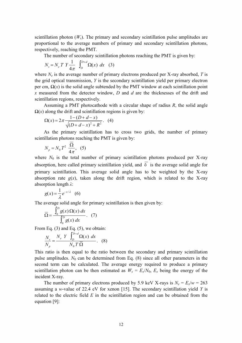
The number of secondary scintillation photons reaching the PMT is given by:
Ns Ne T Y1
4(x) dx
D
Dd
(3)
where Ne is the average number of primary electrons produced per X-ray absorbed, T is
the grid optical transmission, Y is the secondary scintillation yield per primary electron
per cm, Ω(x) is the solid angle subtended by the PMT window at each scintillation point
x measured from the detector window, D and d are the thicknesses of the drift and
scintillation regions, respectively.
Assuming a PMT photocathode with a circular shape of radius R, the solid angle
Ω(x) along the drift and scintillation regions is given by:
(x) 21 (D d x)
(D d x)2 R2. (4)
As the primary scintillation has to cross two grids, the number of primary
scintillation photons reaching the PMT is given by:
N p N0T2
4, (5)
where N0 is the total number of primary scintillation photons produced per X-ray
absorption, here called primary scintillation yield, and ō is the average solid angle for
primary scintillation. This average solid angle has to be weighted by the X-ray
absorption rate g(x), taken along the drift region, which is related to the X-ray
absorption length λ:
g(x) 1
ex / (6)
The average solid angle for primary scintillation is then given by:
g(x)(x) dx
0
D
g(x) dx
0
D
. (7)
From Eq. (3) and Eq. (5), we obtain:
Ns
N p
Ne Y (x) dx
D
Dd
N0 T
. (8)
This ratio is then equal to the ratio between the secondary and primary scintillation
pulse amplitudes. N0 can be determined from Eq. (8) since all other parameters in the
second term can be calculated. The average energy required to produce a primary
scintillation photon can be then estimated as Ws = Ex/N0, Ex being the energy of the
incident X-ray.
The number of primary electrons produced by 5.9 keV X-rays is Ne = Ex/w = 263
assuming a w-value of 22.4 eV for xenon [15]. The secondary scintillation yield Y is
related to the electric field E in the scintillation region and can be obtained from the
equation [9]:

13
116105 p
E
p
Y (9)
where Y/p is expressed in photons per electron per cm per bar and E/p in V cm-1
torr-1
.
For E/p = 2.0 V cm-1
torr-1
, Eq. (9) gives Y/p = 94 photons per electron per cm per bar.
The other parameters of Eq. (8) are T = 0.84, D = 3 cm, d = 0.5 cm. The solid angle
was calculated numerically assuming a PMT with a circular shaped photocathode of the
same area (R = 1.24 cm), and taking into account the absorption length of 5.9 keV X-
rays in xenon (0.26 cm at 1 bar [16]). The solid angle parameters obtained for 1 bar are
then:
(x) dxD
Dd
= 2.533 sr cm;
= 0.424 sr.
The ratio between the secondary and primary scintillation pulse amplitudes is
shown in Fig. 10 as a function of the drift electric field. These results were obtained
from oscilloscope measurements, like in Fig. 5. In order to observe secondary
scintillation, an electric field of 2.0 V cm-1
torr-1
was used in the scintillation region.
Within the experimental errors, the ratio is approximately constant for drift electric
fields between 0.2 and 0.8 V cm-1
torr-1
. An average value within this interval was used
for calculations.
Fig. 10. Ratio between the secondary and primary scintillation pulse amplitudes as a
function of the drift electric field, obtained from oscilloscope measurements, for 5.9
keV X-ray interactions.
Table I shows several parameters used in Eq. (8) to determine N0 and Ws for
different gas pressures. As expected, the results don’t have significant variations with
pressure. The three values obtained are very compatible taking into account the
respective errors. Averaging the results for the three different gas pressures (1, 2 and 3

14
bar), a final value N0 = 81 ± 7 photons is obtained. Accordingly, the mean energy
required to produce a primary scintillation photon is Ws = 72 ± 6 eV. This value is lower
than the previous measurement, 111 ± 16 eV [13], but is however similar to the value
measured for 60 keV -rays at 20 bar, 76 ± 12 eV [17].
Table I – Parameters of Eq. (8) used to determine the primary scintillation yield in
xenon for different gas pressures.
Pressure
(bar)
Ns/Np (×103)
[Fig. 10]
Y (photons/e-/cm)
Ref. [9] λ (cm) Ω
N0
(photons)
Ws (eV)
1 2.2 ± 0.2 94 (±10%) 0.261 0.424 80 ± 11 74 ± 10
2 4.5 ± 0.4 188 (±10%) 0.131 0.389 85 ± 11 69 ± 9
3 7.5 ± 0.9 282 (±10%) 0.087 0.379 79 ± 12 75 ± 12
In the present conditions, about 2 primary scintillation photons were detected in the
PMT per 5.9 keV X-ray absorbed in xenon. For NEXT, double beta decay events have
2458 keV energy. The total number of primary scintillation photons produced in xenon
will be about 2458×103/72 = 3.4×10
4. As the PMT dimensions are much smaller than
the distance to the interaction point, the solid angle subtended by the PMT is
approximately given by 2dA/ , where A is the PMT active area and d is the distance
to the interaction point. For d = 50 cm, the fractional solid angle is then Ω/4π =
(2.2/50)2/4π = 1.5×10
-4. This means that about 5 photons should reach the PMT.
Furthermore, a large number of PMTs will be used and the sum of all PMT signals will
make the signal large enough to be detected above the noise level. In addition, the
visible light and X-ray background observed in our detector will not be present in the
NEXT detector.
The number of photoelectrons produced in the PMT per primary electron crossing
the scintillation region (L) can be estimated as it is determined by the secondary
scintillation yield, Eq. (9), the average solid angle subtended by the PMT for secondary
scintillation, the grid transmission and the quantum efficiency of the PMT (30% [5]). A
value of L = 20 is obtained for E/p = 5 V cm-1
torr-1
in the scintillation region, which is
in good agreement with that obtained from the energy resolution values (section 3).
6. CONCLUSIONS
The performance of a Hamamatsu R8520-06SEL photomultiplier, used as VUV
photosensor in a xenon GPSC, has been investigated, demonstrating that this PMT is a
good candidate for the scintillation readout in the TPC to be used in NEXT.
The PMT high performance for secondary scintillation detection was demonstrated.
An energy resolution of 8.0% (FWHM) was obtained for 5.9 keV X-rays absorbed in

15
the xenon, similar to GPSCs instrumented with larger PMTs, demonstrating the very
low statistical variance of electroluminescent gain.
Primary scintillation measurements have been carried out. The pulse-height
distributions obtained in the MCA don’t have significant variations with the electric
field in the drift region and with the xenon pressure, demonstrating negligible
recombination luminescence. Amplitude measurements of the primary scintillation
produced by 5.9 keV X-rays were possible using an averaging process in the
oscilloscope and triggering at the corresponding secondary scintillation pulse. These
oscilloscope measurements allowed a determination of the primary scintillation yield in
xenon gas. An average of 81 ± 7 primary scintillation photons produced by 5.9 keV X-
rays absorbed in xenon was obtained. The average energy required to produce a primary
scintillation photon in xenon was deduced, resulting Ws = 72 ± 6 eV. This value is lower
than the previous measurement, 111 ± 16 eV [13], but is however similar to that
measured for 60 keV -rays at 20 bar, 76 ± 12 eV [17]. Since the production of primary
scintillation results from collisional processes of the photoelectrons and other Auger and
shake-of electrons with the gas atoms, it is expected that Ws does not vary significantly
with the gas pressure.
Measurements were carried out only up to 3 bar pressures due to a limitation of our
experimental setup. A new chamber was built at IFIC (Valencia) in order to study the
PMT response at higher pressures. The aim is to verify the PMT performance at the
TPC pressure of 10 bar.
ACKNOWLEDGMENTS
This work was supported by FCT (Portugal) and FEDER through project
PTDC/FIS/103860/2008. E.D.C. Freitas acknowledges grant SFRH/BD/46711/2008
from FCT. C.M.B. Monteiro acknowledges grant SFRH/BD/25569/2005 from FCT. M.
Ball, J.J. Gómez-Cadenas and N. Yahlali acknowledge the Spanish MICINN for the
Consolider-Ingenio grants CSD2008-00037 and CSD2007-00042 and the research
grants FPA2009-13697-C04-04 and FPA2009-13697-C04-B23/12. D.R. Nygren
acknowledges support by the Director, Office of Science, Office of High Energy
Physics, of the U.S. Department of Energy under contract DE-AC02-05CH11231.
REFERENCES
[1] F. Grañena et al. (NEXT Collaboration), NEXT Letter of Intent, Laboratorio Subterráneo
de Canfranc EXP-05 [hep-ex/0907.4054v1].
[2] M. Redshaw, E. Wingfield, J. McDaniel, E.G. Myers, Mass and Double-Beta-Decay Q
Value of 136
Xe, Phys. Rev. Lett. 98 (2007) 053003.
[3] C.A.N. Conde, A.J.P.L. Policarpo, A gas proportional scintillation counter, Nucl. Instrum.
Meth. 53 (1967) 7.

16
[4] D. Nygren, Optimal detectors for WIMP and 0–ν ββ searches: Identical high-pressure
xenon gas TPCs?, Nucl. Instrum. Meth. A 581 (2007) 632.
[5] http://www.hamamatsu.com
[6] J. Angle et al. (XENON Collaboration), First Results from the XENON10 Dark Matter
Experiment at the Gran Sasso National Laboratory, Phys. Rev. Lett. 100 (2008) 021303.
[7] J. Angle et al. (XENON Collaboration), Constraints on inelastic dark matter from
XENON10, Phys. Rev. D 80 (2009) 115005.
[8] J.M.F. dos Santos et al., Development of portable gas proportional scintillation counters
for x-ray spectrometry, X-Ray Spectrom. 30 (2001) 373.
[9] C.M.B. Monteiro et al., Secondary scintillation yield in pure xenon, 2007 JINST 2 P05001.
[10] E.D.C. Freitas et al., Secondary scintillation yield in high-pressure xenon gas for
neutrinoless double beta decay (0νββ) search, Phys. Lett. B 684 (2010) 205.
[11] J.M.F. dos Santos, A.C.S.S. Bento, C.A.N. Conde, A simple, inexpensive gas proportional
scintillation counter for X-ray fluorescence analysis, X-Ray Spectrom. 22 (1993) 328.
[12] A. Peacock et al., Performance characteristics of a gas scintillation spectrometer for X-ray
astronomy, Nucl. Instrum. Meth. 169 (1980) 613.
[13] S.J.C. do Carmo et al., Absolute primary scintillation yield of gaseous xenon under low
drift electric fields for 5.9 keV X-rays, 2008 JINST 3 P07004.
[14] M. Mimura et al., Intensity and time profile of recombination luminescence produced by
an α-particle in dense xenon gas, Nucl. Instrum. Meth. A 613 (2010) 106.
[15] T.H.V.T. Dias et al., Full-energy absorption of x-ray energies near the Xe L- and K-
photoionization thresholds in xenon gas detectors: Simulation and experimental results, J.
Appl. Phys. 82 (1997) 2742.
[16] http://physics.nist.gov/PhysRefData/XrayMassCoef/ElemTab/z54.html
[17] A. Parsons et al., High pressure gas scintillation drift chambers with wave shifter fiber
readout, IEEE Trans. Nucl. Sci. 37 (1990) 541.