Shielding Effectiveness of a Thick Multilayered Panel in a Reverberating Environment

Preparation of magnetically dopedmultilayered functional silica particles viasurface modification with organic polymerM. A. Rahmana, M. A. J. Miaha, H. Minamib and H. Ahmada*
Magnetically modified functional particles are emerging as one of the most promising candidate in numerousmultidisciplinary applications. In this research, a simple process has been developed to prepare magnetically modifiedaminated silica (SiO2) particles. Herein, submicron-sized SiO2 particles were modified with poly(methylmethacrylate-methacrylic acid) by seeded polymerization without any stabilizer. The carboxyl groups localized near the particlessurface were then covalently linkedwith ethylene diamine to prepare aminated composite particles. Iron ionswere thenprecipitated on the surface of aminated composite particles to obtainmagnetically doped functional SiO2 particles. Thepreparation of such particles was confirmed by scanning electronmicroscopy, Fourier transform infrared, 1H NMR, X-rayphotoelectron spectroscopy and thermogravemetric analyses. Relative measurement of adsorption study of differentbiomolecules suggested that magnetically doped functional silica particles are comparatively hydrophobic. Copyright© 2012 John Wiley & Sons, Ltd.
Keywords: SiO2 particles; functionalization; seeded polymerization; precipitation of Fe3O4
INTRODUCTION
Inorganic–organicmicrospheres are attractingmuch interest consid-ering their unusual and possibly unique optical, mechanical,rheological, electrical, catalytic, and flame-retardant properties.[1–9]
A huge variety of inorganic materials has been used for preparingsuch hybrid microspheres with silica (SiO2) being the most widelyused because colloidal silica spheres uniform in size, shape, andcomposition are widespread in nature and in electronics andmedicine.[10–12]
The fabrication of SiO2/organic/Fe3O4 tri-layered microsphereswould be interesting because of the wide application potentialof magnetic materials in magnetic bioseparation, drug delivery,magnetic resonance imaging contrast enhancement, and targeteddrug.[13–16] Lu et al. prepared magnetizable SiO2 compositeparticles from heteroaggregates of carboxylic polystyrene latexand Fe3O4 nanoparticles by regulating the pH value.[17] Wanget al. reported the preparation raspberry-like monodispersemagnetic hollow hybrid microspheres by coating polystyrenetemplate with Fe3O4/SiO2 particles.[18] They mainly used theprinciple of electrostatic interaction between positively chargedPS microspheres and negatively charged Fe3O4/SiO2 particlesfor preparing such multilayered particles. However, in bothaforementioned cases, there is a strong possibility of leachingout magnetic particles from the particles surface under changedenvironment such as pH variation and/or repeated washing. Inanother work, Wang and his group reported the preparationof poly(N-isopropylacrylamide)-coated luminescent/magneticSiO2 microspheres via a tedious four-step process.[19] Numerousworks have also be carried out on the preparation of mesoporousmagnetic SiO2 spheres particularly for application in drug targetingand as metal adsorbent.[20–25] Most of these syntheses requiredmultisteps and often resulted in polydisperesed particles notsuitable in many biological applications.
The carboxyl group is one of the most widely studied functionalgroups for polymer particles intended for biomedical or diagnosticapplications. This functional group can also undergo easy derivatiza-tion using water/oil-soluble coupling agents. In a previous work,Ahmad et al. reported the amination of carboxyl functionalizedsubmicron-sized copolymer latex particles using amine-nucleophilesin ethanol medium.[26] The present work is an extension of this earlywork, emphasizing the deposition of magnetic nanoparticles on thesurface of aminated SiO2 particles to diversify their applications inmagnetic support-based separation in various fields of biotechnol-ogy and biomedicine, such as cell separation, enzyme immobiliza-tion, drug delivery, and protein separation.[27–32] It is to be noted thatsome literature are already available related to the introduction ofamine group on SiO2 particles via silane coupling agent[33–36];however, these coupling agents had mostly short aliphatic chainnot enough to cover the SiO2 surface, and this silanization treatmentoften requires a long complex procedure. Contrary to this, directpolymerization of acrylate monomers in the presence of SiO2
particles under optimum conditions may result in the completecoverage of SiO2 surface with long polymer chains, and additionally,the surface can also be modified with various functional groupsusing functional monomer. A core/acrylate monomer ratio (w/w)of 1/0.5 during seeded polymerization is reported to be enoughfor complete coverage of core particles.[37,38] The complete coverage
* Correspondence to: H. Ahmad, Department of Chemistry, Rajshahi University,Rajshahi 6205, Bangladesh.E-mail: [email protected]
a M. A. Rahman, M. A. J. Miah, H. AhmadDepartment of Chemistry, Rajshahi University, Rajshahi 6205, Bangladesh
b H. MinamiGraduate School of Engineering, Kobe University, Kobe 657-8501, Japan
Research article
Received: 24 April 2012, Revised: 15 July 2012, Accepted: 16 July 2012, Published online in Wiley Online Library: 2 August 2012
(wileyonlinelibrary.com) DOI: 10.1002/pat.3067
Polym. Adv. Technol. 2013, 24 174–180 Copyright © 2012 John Wiley & Sons, Ltd.
174
of SiO2 particles by acrylate polymers would improve the dispersibil-ity in the solvents for acrylate polymer and in the polymer matrix[39]
and also would open up the application potential of modifiedparticles because of the presence of biocompatible poly-acrylatelayer.In this investigation, submicron-sized monodisperse Fe3O4-
doped functional SiO2 microspheres were prepared by a simplethree-step process as diagrammatically shown in Scheme 1.Seeded copolymerization of methylmethacrylate (MMA) andmethacrylic acid (MAA) was carried out in the presence ofanionically charged commercially available submicron-sizedmonodisperse SiO2 particles to induce carboxyl groups on theparticles surface. The modified SiO2 particles were abbreviatedas SiO2/P(MMA-MAA) composite particles. Ethylene diamine(EDA) was then covalently bonded to the carboxyl groups onthe composite particle surface via –CONH– linkage. We calledthese aminated SiO2 particles as SiO2/P(MMA-MAA)-EDA.Nanosized Fe3O4 particles were then deposited on the surfaceof the particles to prepare magnetically doped functional SiO2
particles named as SiO2/P(MMA-MAA)-EDA/Fe3O4. The aminefunctional groups localized at the chain ends on the SiO2
particles can be utilized for coupling of affinity ligands or bindingof biomolecules, and additionally, the magnetic nature of theparticles would favor the separation from the existing media.
EXPERIMENTAL
Materials
MMAandMAApurchased fromFluka Chemika (Buchs, Switzerland)were distilled under reduced pressure to remove inhibitors andpreserved in the refrigerator until use. SiO2 dispersion donated byNippon Shokubai Co. Ltd (Osaka, Japan) was used after centrifugal
washing. 2,20-Azobis(2-amidinopropane) hydrochloride (V-50) ofreagent grade was recrystallized fromwater before use. Three differ-ent biomolecules were used: trypsin (TR) from E. Merck (Darmstadt,Germany), albumin (AL) from LOBA Chem. (Mumbai, India), and lyso-zyme (LZ) from Fluka, Biochemika (Buchs, Switzerland). Dicyclohexylcarbodiimide (DCC) and EDA both from LOBA Chem. were used assupplied. Ferric chloride hexahydrate (FeCl3�6H2O), ferrous sulfate(FeSO4), sodium hydroxide (NaOH), oleic acid, and other chemicalsused were of analytical grade. Distilled deionized water ofconductivity lower than 5mS/cm was used for all the experiments.Deionized water was distilled using a glass (Pyrex) distillationapparatus.
Instruments
Scanning electron microscope (SEM; LEO Electron MicroscopyLtd, Cambridge, UK), Bruker Proton NMR (250MHz, Billerica,MA), Fourier transform infrared (FTIR; 8044 Shimadzu, Kyoto,Japan), centrifuge machine from Kokuson Corporation (Tokyo,Japan), Helios gamma single beam UV–Visible spectrophotometerfrom Unicam (Leeds, UK), and HI 9321 Microprocessor pH meterand conductivity meter both from HANNA instruments (Póvoa deVarzim, Portugal) were used in this study. The X-ray diffraction(XRD) patterns of the powder samples were taken on a PANalytical(Almelo, the Netherlands) X’Pert PRO diffractometer using Cu Karadiation. Sherwood Scientific Magnetic Susceptibility Balancewas used for susceptibility measurement. Thermal analysis wascarried out using a thermogravimetry (TG) analyser from SeikoInstruments Inc (Chiba, Japan) EXSTAR 6000 TG/DTA 6300.
Preparation of SiO2/P(MMA-MAA) composite polymer particles
Five grams of SiO2 particles, 2.0 g of MMA, and 0.50 g ofMAA were taken in a three-necked round-bottomed flaskdipped in a thermostat water bath maintained at 70�C. Seededpolymerization was carried out in aqueous medium (50 g) byusing cationic V-50 initiator (0.05 g) under a nitrogen atmospherefor 24 h.
Preparation of SiO2/P(MMA-MAA)-EDA composite particlesin ethanol medium
SiO2/P(MMA-MAA) latex particles were transferred from water toanhydrous ethanol medium by centrifugal washing at 8000 rpm,and the solid content of the latex was adjusted to about 2%. EDAwas covalently bonded to the carboxyl groups of compositeparticles by the following method as previously carried out.[26]
Pre-activation method: 2 g of latex particles dispersed in 98 ganhydrous ethanol was mixed with 1.24 g of coupling agent(DCC) in a reagent bottle, magnetically stirred at 25�C for 1 h.The activated carboxyl group was then reacted with 0.4660 g ofEDA at 25�C for 3 h and then again at 0�C for 24 h. SiO2/P(MMA-MAA)-EDA composite particles dispersed in ethanol werethen transferred to the aqueous media by serum replacementby using a centrifuge machine.
Preparation of SiO2/P(MMA-MAA)-EDA/Fe3O4 particles
SiO2/P(MMA-MAA)-EDA particles (3.0 g) were taken in a round-bottomed flask containing 70ml water. The dispersion was cooled(0–5�C) in an ice water bath under a nitrogen gas bubbling; 0.60g(3.6mmol) of FeCl3�6H2O and 0.2558g (3.2mmol) of FeSO4 weredissolved in 25ml water. Then the iron solution was added to the
SiO2SiO2
MMA/MAA/AIBAPolymerization
EDA, DCC
Fe2+/ Fe3+
NaOH
NH2
NH2
NH2
NH2
H2N
H2N
H2N
H2N
H2NNH2
NH2
HO2C C O2H
C O2H
H O2C
H O2C
CO2H
CO2H
C O2H
H O2C
HO2C
H O2C
HO2C
SiO2/P(MMA-MAA)
NH2
NH2
NH2
NH2
H2N
H2N
H2N
H2N
H2NNH2
NH2
SiO2/P(MMA-MAA)-EDA/Fe3O4 SiO2/P(MMA-MAA)-EDA
Scheme 1. Preparation of multilayered SiO2/P(MMA-MAA)-EDA/Fe3O4
composite polymer particles via a three-step method. This schemeappears in color.
MAGNETICALLY DOPED FUNCTIONAL SILICA PARTICLES
Polym. Adv. Technol. 2013, 24 174–180 Copyright © 2012 John Wiley & Sons, Ltd. wileyonlinelibrary.com/journal/pat
175
flask containing the aminated composite dispersion. A light browncolor mixture was formed. The ice bath was removed, and the flaskwas immersed in a preheated water bath at 85�C. Immediately,4.0ml of 5M aqueous NaOH solution was added. The reactionmixture gradually turned to black. The mixture was kept stirringat 85�C for 2 h. Oleic acid (0.0345 g, 5.36mmol) was slowly addedtowards the end of the process to stabilize the aminated-Fe3O4
dispersion. Then the dispersion was cooled to room temperature.The resultant magnetic particles were centrifuged and thoroughlywashed with deionized distilled water, followed by 0.1M HCl andagain by deionized distilled water.
Measurement of magnetic susceptibility
SiO2/P(MMA-MAA)-EDA/Fe3O4 particles were washed, separatedfrom their dispersion by centrifugation, and dried in oven at80�C for 2 h. The dried powders were placed in a pre-weighedsample tube, and the magnetic susceptibility (wg) was measuredusing the following equation:
wg ¼ C � L� R� R0ð ÞM� 109
where C is the calibration constant of the balance, L is the lengthof the sample, R0 and R are the readings of the empty andsample tubes, and M is the weight of the sample (dry basis) inCGS unit.
Thermogravemetric analysis
Thermal properties of the dry powdered samples of SiO2, SiO2/P(MMA-MAA), and SiO2/P(MMA-MAA)-EDA/Fe3O4 microsphereswere measured by heating samples under flowing nitrogenatmosphere from 40�C to 600�C at a heating rate of 20�C/min,and the weight loss was recorded.
X-ray photoelectron spectroscopy
The surface compositions of SiO2, SiO2/P(MMA-MAA), SiO2/P(MMA-MAA)-EDA, and SiO2/P(MMA-MAA)-EDA/Fe3O4 latex particleswere evaluated using a Kratos Axis Ultra DLD X-ray photoelectronspectrometer (XPS) equipped with a monochromatic Al X-raysource operating at 6.0mA and 15 kV at a typical base pressure of10�8 Torr. The step size was 1.0 eV for the survey spectra (passenergy 160 eV) and 0.1 eV for the high-resolution spectra (passenergy 80 eV). Spectra were typically acquired from two separatesample areas of dried latex. The latex dispersion (purified by serumreplacement after repeated centrifugation) was dried onto anindium foil before recording the spectrum.
Adsorption of biomolecules on SiO2 and SiO2/P(MMA-MAA)-EDA/Fe3O4 composite polymer particles
A mixture of 20ml was prepared from purified latex (polymersolid, 0.15 g) and biomolecule (200mg/g of particles) aqueoussolution. The pH value of the mixture was immediately adjustedat the respective isoelectric point (TR: pH 10.0; LZ: 10.5; and AL:6.0) using a buffer solution. In each measurement, the disper-sion–biomolecule mixture was allowed to stand for 45minmaintained at 25�C. The latex particles were then separated bycentrifugation at 10 000 rpm for 15min. In order to removewafting particles completely, the supernatant was centrifugedonce more. The concentration of the biomolecule in thesupernatant was determined by a UV–Visible spectrophotometer
at a wavelength of 280 nm. The amount of biomoleculeadsorbed was calculated by subtracting the concentration inthe medium from that of the initial concentration. A calibrationcurve was used for this purpose.
DISCUSSION
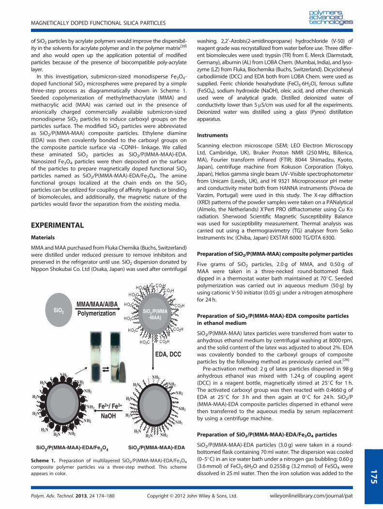
Figure 1 shows the SEM photographs of SiO2, SiO2/P(MMA-MAA),SiO2/P(MMA-MAA)-EDA, and SiO2/P(MMA-MAA)-EDA/Fe3O4 latexparticles. All the particles were washed by serum replacementprior to the observation by SEM. The average diameters andcoefficient of variations are 526.0 nm and 3.25% for SiO2 seed,547.0 nm and 2.43% for SiO2/P(MMA-MAA), and 566.0 nm and2.33% for SiO2/P(MMA-MAA)-EDA composite particles. Theaverage size of SiO2/P(MMA-MAA) composite particles increasedby 21 nm after seeded copolymerization, indicating a shellthickness of around 10 nm in the dried state. Subsequent surfacemodification of composite particles with EDA also increased thesize. All the SiO2 seed and the composite polymer particles arealmost spherical. No secondary copolymer particle is producedduring seeded copolymerization as nanosize particle is notobserved in Fig. 1(b). This observation suggests that the seededcopolymerization of MMA and MAA proceeded mainly on SiO2
seed particles, without any secondary nucleation. The increasein average particle size after amination with EDA is attributedto the incorporation of hydrocarbon layer. The surface of theaminated SiO2/P(MMA-MAA)-EDA composite polymer particlesis smooth but comparatively the surface of the SiO2/P(MMA-MAA)-EDA/Fe3O4 particles is not smooth with many smallFe3O4 particles attached to the surface. Such SiO2/P(MMA-MAA)-EDA/Fe3O4 composite particles with uneven surfaceresulted from the co-precipitation and deposition of iron ionsas Fe3O4 on the surface of SiO2/P(MMA-MAA)-EDA compositeparticles. It is reasonable to mention that after three repeatedwashings by decantation and redispersion, the supernatantobtained from the SiO2/P(MMA-MAA)-EDA/Fe3O4 compositeemulsion was clear, indicating that impregnated Fe3O4 nanopar-ticles are strongly bonded to the composite particle surface toprevent leaching. Additionally, the precipitation of Fe3O4
particles on the functional SiO2 particles was confirmed by themeasurement of magnetic susceptibility, and the value(8.63� 10�4) was recorded as positive and very high. This resultsuggests that the prepared magnetically doped functional SiO2
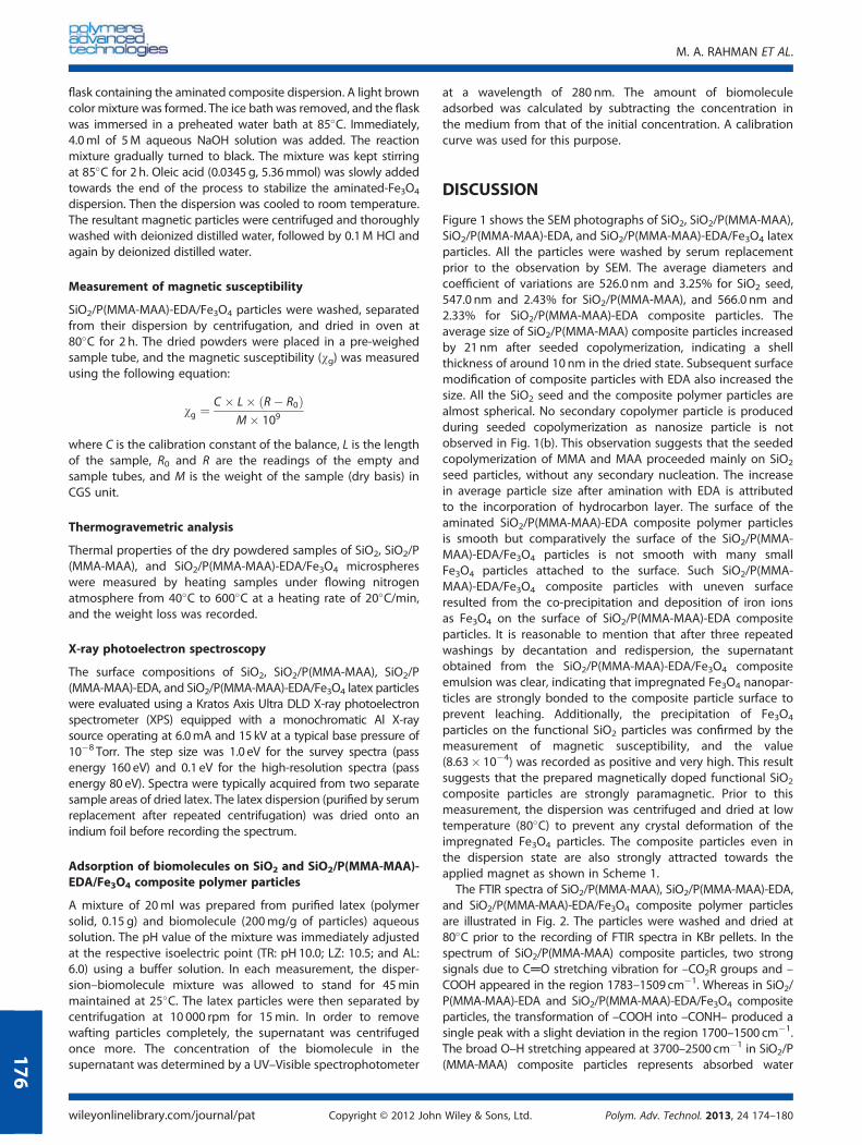
composite particles are strongly paramagnetic. Prior to thismeasurement, the dispersion was centrifuged and dried at lowtemperature (80�C) to prevent any crystal deformation of theimpregnated Fe3O4 particles. The composite particles even inthe dispersion state are also strongly attracted towards theapplied magnet as shown in Scheme 1.The FTIR spectra of SiO2/P(MMA-MAA), SiO2/P(MMA-MAA)-EDA,
and SiO2/P(MMA-MAA)-EDA/Fe3O4 composite polymer particlesare illustrated in Fig. 2. The particles were washed and dried at80�C prior to the recording of FTIR spectra in KBr pellets. In thespectrum of SiO2/P(MMA-MAA) composite particles, two strongsignals due to C═O stretching vibration for –CO2R groups and –COOH appeared in the region 1783–1509 cm�1. Whereas in SiO2/P(MMA-MAA)-EDA and SiO2/P(MMA-MAA)-EDA/Fe3O4 compositeparticles, the transformation of –COOH into –CONH– produced asingle peak with a slight deviation in the region 1700–1500 cm�1.The broad O–H stretching appeared at 3700–2500 cm�1 in SiO2/P(MMA-MAA) composite particles represents absorbed water
M. A. RAHMAN ET AL.
wileyonlinelibrary.com/journal/pat Copyright © 2012 John Wiley & Sons, Ltd. Polym. Adv. Technol. 2013, 24 174–180
176
molecules and carboxyl groups. On the contrary, in both SiO2/P(MMA-MAA)-EDA and SiO2/P(MMA-MAA)-EDA/Fe3O4 compositeparticles, this same peak representing the combined stretchingvibration for N–H and –OH groups derived from substituted amide,primary amine, unconverted carboxyl (if any), and surface watermolecules appeared at almost in the same region, but the signalshape and intensity changed considerably. The stretching signaldue to Fe–O is also observed in magnetically doped SiO2/P(MMA-MAA)-EDA/Fe3O4 composite particles at around 465 cm�1.Compared with this, in SiO2/P(MMA-MAA) composite particles, abroad vibration signal at 442 cm�1 having different intensity and
shape is observed. The aforementioned results confirmed thatthe nucleophile EDA is covalently bonded to –COOH groupsderived from the MAA comonomer unit, at least partially if notcompletely, via a –CONH– linkage, and Fe3O4 particles havesuccessfully been incorporated.
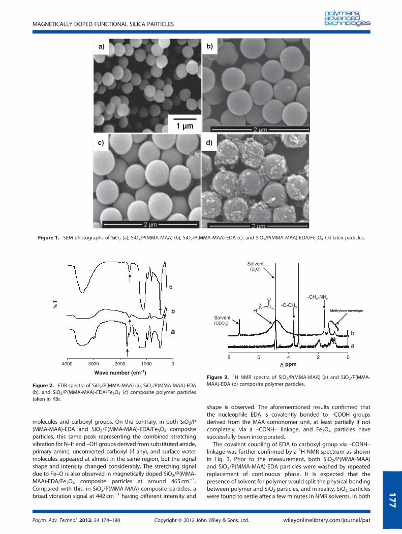
The covalent coupling of EDA to carboxyl group via –CONH–linkage was further confirmed by a 1H NMR spectrum as shownin Fig. 3. Prior to the measurement, both SiO2/P(MMA-MAA)and SiO2/P(MMA-MAA)-EDA particles were washed by repeatedreplacement of continuous phase. It is expected that thepresence of solvent for polymer would split the physical bondingbetween polymer and SiO2 particles, and in reality, SiO2 particleswere found to settle after a few minutes in NMR solvents. In both
1 µm
a) b)
c) d)
Figure 1. SEM photographs of SiO2 (a), SiO2/P(MMA-MAA) (b), SiO2/P(MMA-MAA)-EDA (c), and SiO2/P(MMA-MAA)-EDA/Fe3O4 (d) latex particles.
01000200030004000
Wave number (cm-1)
% T
a
b
c
Figure 2. FTIR spectra of SiO2/P(MMA-MAA) (a), SiO2/P(MMA-MAA)-EDA(b), and SiO2/P(MMA-MAA)-EDA/Fe3O4 (c) composite polymer particlestaken in KBr.
}
02468ppm
a
b
-CH2-NH2
-O-CH3NH
O
C
Solvent(CDCl3)
Solvent(D2O)
}Methylene envelope
Figure 3. 1H NMR spectra of SiO2/P(MMA-MAA) (a) and SiO2/P(MMA-MAA)-EDA (b) composite polymer particles.
MAGNETICALLY DOPED FUNCTIONAL SILICA PARTICLES
Polym. Adv. Technol. 2013, 24 174–180 Copyright © 2012 John Wiley & Sons, Ltd. wileyonlinelibrary.com/journal/pat
177
the spectrum of SiO2/P(MMA-MAA) and SiO2/P(MMA-MAA)-EDAcomposite polymer particles, the chemical shifts of the aliphatic–CH2 and –CH groups appeared in the region from 0.85 to1.25 ppm. A signal due to –O–CH3 protons from methacrylate isobserved at 3.6 ppm. The characteristic NMR signals due toacidic (�COOH) protons are expected to be at 10.0–12.0 ppm.But such a peak was not observed in the spectrum of SiO2/P(MMA-MAA) composite particles (the region not shown). It isknown that the chemical shift of this group is variable, depend-ing not only on the chemical environment but also on concen-tration, temperature, and solvent.[40] For SiO2/P(MMA-MAA)composite polymer particles, D2O was used as the lone NMRsolvent. Therefore, the possibility of a deuterium exchangereaction between carboxyl groups and solvent molecules cannotbe ruled out under the conditions.[40,41] In this case the chemicalshift due to acidic protons is expected to disappear at theexpense of solvent DOH peak that appeared at 4.7 ppm. In thespectrum of SiO2/P(MMA-MAA)-EDA composite particles takenin CDCl3, a broad signal appeared at 4.6 ppm, which indicatedthe presence of a –CONH– group. A signal observed at1.56 ppm represents the primary amine (�NH2) groups.
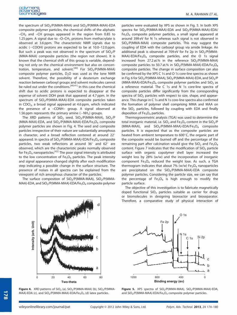
The XRD patterns of SiO2 seed, SiO2/P(MMA-MAA), SiO2/P(MMA-MAA)-EDA, and SiO2/P(MMA-MAA)-EDA/Fe3O4 compositepolymer particles are shown in Fig. 4. The seed and compositeparticles irrespective of their nature are substantially amorphousin character, and a broad reflection centered at around 22�
appeared. In spectra of SiO2/P(MMA-MAA)-EDA/Fe3O4 compositeparticles, two weak reflections at around 36� and 62� areobserved, which are the characteristic peaks normally observedfor Fe3O4 nanoparticles.
[42] The poor signal intensity is attributedto the low concentration of Fe3O4 particles. The peak intensityand signal appearance changed slightly after each modificationstep indicating a possible change in the surface structure. Thepresence of noises in all spectra can be explained from theviewpoint of rich amorphous character of the particles.
The surface composition of SiO2/P(MMA-MAA), SiO2/P(MMA-MAA)-EDA, and SiO2/P(MMA-MAA)-EDA/Fe3O4 composite polymer
particles were evaluated by XPS as shown in Fig. 5. In both XPSspectra for SiO2/P(MMA-MAA)-EDA and SiO2/P(MMA-MAA)-EDA/Fe3O4 composite polymer particles, a small signal appeared ataround 399 eV for N 1s whereas such signal is not observed inSiO2/P(MMA-MAA) composite particles. This may suggest thecoupling of EDA with the carboxyl group via amide linkage. Anadditional peak is observed at 709 eV for Fe 2p in SiO2/P(MMA-MAA)-EDA/Fe3O4 composite particles, and the O 1s signalincreased from 27.2 at.% in the reference SiO2/P(MMA-MAA)composite particles to 50.7 at.% in SiO2/P(MMA-MAA)-EDA/Fe3O4
composite particles. The change in surface composition can alsobe confirmed by the XPS C 1s and O 1s core-line spectra as shownin Fig. 6 for SiO2/P(MMA-MAA), SiO2/P(MMA-MAA)-EDA, and SiO2/P(MMA-MAA)-EDA/Fe3O4 composite polymer particles and SiO2 asa reference material. The C 1s and N 1s core-line spectra ofcomposite particles differ significantly from the correspondingspectra of SiO2 particles with respect to size, shape, and appear-ance. This change in C 1s and N 1s core-line spectra also confirmedthe formation of polymer shell comprising MMA and MAA onthe SiO2 particles, followed by coupling with EDA and finallydeposition of Fe3O4 particles.Thermogravemetric analysis (TGA) was used to determine the
total inorganic material, i.e. SiO2 and Fe3O4 content in the SiO2/P(MMA-MAA), and SiO2/P(MMA-MAA)-EDA/Fe3O4 compositeparticles. It is expected that as the composite particles areheated from ambient temperature to 600�C, the organic part ofthe composite would be burned off and the percentage of theremaining part after calcination would give the SiO2 and Fe3O4
content. Figure 7 indicates that the modification of SiO2 particlesurface with organic copolymer shell layer increased theweight loss by 28% (w/w) and the incorporation of inorganiccomponent Fe3O4 reduced the weight lose. As such, a TGAthermogram indicates that about 7% (w/w) Fe3O4 nanoparticlesare precipitated on the SiO2/P(MMA-MAA)-EDA compositepolymer particles. Considering the particle size, we can say thatthe percentage of Fe3O4 is high enough to modify theparticle surface.The objective of this investigation is to fabricate magnetically
doped functional SiO2 particles suitable as carrier for drugsor biomolecules in designing bioreactor and bioseparator.Therefore, a comparative study of physical interaction of
a
0 20 40 60 80
Two-theta
Inte
nsi
ty (
arb
. Un
it)
b
c
d
Figure 4. XRD patterns of SiO2 (a), SiO2/P(MMA-MAA) (b), SiO2/P(MMA-MAA)-EDA (c), and SiO2/P(MMA-MAA)-EDA/Fe3O4 (d) latex particles.
04008001200
Binding energy (ev)
Fe 2p
C 1sN 1s
O 1s
Si 2p
ab
c
Figure 5. XPS spectra of SiO2/P(MMA-MAA), SiO2/P(MMA-MAA)-EDA,and SiO2/P(MMA-MAA)-EDA/Fe3O4 composite polymer particles.
M. A. RAHMAN ET AL.
wileyonlinelibrary.com/journal/pat Copyright © 2012 John Wiley & Sons, Ltd. Polym. Adv. Technol. 2013, 24 174–180
178
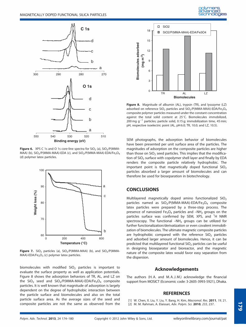
biomolecules with modified SiO2 particles is important toevaluate the surface property as well as application potentials.Figure 8 shows the adsorption behaviors of TR, AL, and LZ onthe SiO2 seed and SiO2/P(MMA-MAA)-EDA/Fe3O4 compositeparticles. It is well known that magnitude of adsorption is largelydependent on the degree of hydrophobic interaction betweenthe particle surface and biomolecules and also on the totalparticle surface area. As the average sizes of the seed andcomposite particles are not the same as observed from the
SEM photographs, the adsorption behavior of biomoleculeshave been presented per unit surface area of the particles. Themagnitudes of adsorption on the composite particles are higherthan those on SiO2 seed particles. This implies that the modifica-tion of SiO2 surface with copolymer shell layer and finally by EDArenders the composite particle relatively hydrophobic. Theimportant point is that magnetically doped functional SiO2
particles absorbed a larger amount of biomolecules and cantherefore be used for bioseparation in biotechnology.
CONCLUSIONS
Multilayered magnetically doped amino functionalized SiO2
particles named as SiO2/P(MMA-MAA)-EDA/Fe3O4 compositelatex particles were prepared by a three-step process. Thepresence of nanosized Fe3O4 particles and –NH2 groups on theparticles surface was confirmed by SEM, XPS, and 1H NMRspectroscopy. The functional –NH2 groups can be utilized forfurther functionalization/derivatization or even covalent immobili-zation of biomolecules. The ultimate magnetic composite particlesare hydrophobic compared with the reference SiO2 particlesand adsorbed larger amount of biomolecules. Hence, it can bepredicted that multilayered functional SiO2 particles can be usefulin designing bioseparator and bioreactor, and the magneticnature of the composite latex would favor easy separation fromthe dispersion.
Acknowledgements
The authors (H. A. and M. A. J. M.) acknowledge the financialsupport from MOSICT (Economic code: 3-2605-3993-5921), Dhaka.
REFERENCES[1] W. Chen, X. Liu, Y. Liu, Y. Bang, H. Kim, Macromol. Res. 2011, 19, 21.[2] M. M. Rahman, A. Elaissari, Adv. Polym. Sci. 2010, 233, 237.
270280290300
a
b
c
d
C 1s
510520530540550
Binding energy (eV)
a
b
c
d
O 1s
Figure 6. XPS C 1s and O 1s core-line spectra for SiO2 (a), SiO2/P(MMA-MAA) (b), SiO2/P(MMA-MAA)-EDA (c), and SiO2/P(MMA-MAA)-EDA/Fe3O4
(d) polymer latex particles.
60
70
80
90
100
0 200 400 600
Temperature (°C)
Wei
gh
t lo
ss (
%)
a
b
c
Figure 7. SiO2 particles (a), SiO2/P(MMA-MAA) (b), and SiO2/P(MMA-MAA)-EDA/Fe2O3 (c) polymer latex particles.
0
3
6
9
12
15
18
Biomolecules
Bio
mo
lecu
les
adso
rbed
(m
g m
-2)
SiO2
SiO2/P(MMA-MAA)-EDA/Fe3O4
TR AL LZ
Figure 8. Magnitude of albumin (AL), trypsin (TR), and lysozyme (LZ)adsorbed on reference SiO2 particles and SiO2/P(MMA-MAA)-EDA/Fe3O4
composite polymer particles measured under the constant concentrationagainst the total solid content at 25�C. Biomolecules immobilized,200mg g�1 particles; particle solid, 0.15 g; immobilization time, 45min;pH, respective isoelectric point (AL, pH 6.0; TR, 10.0; and LZ, 10.5).
MAGNETICALLY DOPED FUNCTIONAL SILICA PARTICLES
Polym. Adv. Technol. 2013, 24 174–180 Copyright © 2012 John Wiley & Sons, Ltd. wileyonlinelibrary.com/journal/pat
179

[3] C. Houphouet-Boigny, C. J. G. Plummer, M. D. Wakeman, J.-A. E. Månson,Polym. Eng. Sci. 2007, 47, 1122.
[4] R. Ruggerone, V. Geiser, S. D. Vacche, Y. Leterrier, J.-A. E. Månson,Macromolecules 2010, 43, 10490.
[5] P. B. Messersmith, E. P. Giannelis, J. Polym. Sci. Polym. Chem. Ed.1995, 33, 1047.
[6] H. Zhang, X. Zhang, X. Yang, J. Colloid Interface Sci. 2010, 348, 431.[7] R. Ianchis, D. Donescu, M. C. Corobea, C. Petcu, M. Ghiurea, S. Serban,
C. Radovici, Colloid Polym. Sci. 2010, 288, 1215.[8] S. Kawano, A. Sei, M. Kunitake, J. Colloid Interface Sci. 2010, 352, 348.[9] C. C. Corten, M. W. Urban, Adv. Mater. 2009, 21, 5011.[10] M. A. Rahman, M. R. Karim, M. A. J. Miah, H. Ahmad, Macromol. Res.
2010, 18, 247.[11] H. Zou, S. Wu, J. Sen, Chem. Rev. 2008, 108, 3893.[12] J. A. Balmer, A. Schmid, S. P. Armes, J. Mater. Chem. 2008, 18, 5722.[13] H. Ahmad, M. A. Rahman, M. A. J. Miah, Macromol. Res. 2008,
16, 637.[14] M. M. Rahman, M. M. Chehim, H. Fessi, A. Elaissari, J. Colloid Interface
Sci. 2011, 360, 556.[15] H. Dong, J. Huang, R. R. Koepsel, P. Ye, A. J. Russell, K. Matyjaszewski,
Biomacromolecules 2011 12, 1305.[16] M. Mahmoudi, H. Hosseinkhani, M. Hosseinkhani, S. Boutry, A. Simchi,
W. S. Journeay, K. Subramani, S. Laurent, Chem. Rev. 2011, 111, 253.[17] Z. Y. Lu, Y. Q. Qin, J. Y. Fang, J. Sun, J. Li, F. Q. Liu, W. S. Yang,
Nanotechnology 2008, 19, 055602.[18] C. Wang, J. Yan, X. Cui, H. Wang, J. Colloid Interface Sci. 2011, 354, 94.[19] J. Guo, W. Yang, C. Wang, J. He, J. Chen, Chem. Mater. 2006, 18, 5562.[20] T. Valdés-Solís, A. F. Rebolledo,M. Sevilla, P. Valle-Vigón, O. Bomatí-Miguel,
A. B. Fuertes, P. Tartaj, Chem. Mater. 2009, 21, 1806.[21] M. Sevilla, T. Valdés-Solís, P. Tartaj, A. B. Fuertes, J. Colloid Interface
Sci. 2009, 340, 230.[22] X. Li, V. T. John, J. Zhan, G. He, J. He, L. Spinu, Langmuir 2011, 27, 6252.
[23] E. Ruiz-Hernández, A. López-Noriega, D. Arcos, I. Izquierdo-Barba,O. Terasaki, M. Vallet-Reg, Chem. Mater. 2007, 19, 3455.
[24] M. H. Yun, J. W. Yeon, J. H. Kim, H. I. Lee, J. M. Kim, S. Kim, Y. Jung,Macromol. Res., 2011, 19, 421.
[25] T. Valdés-Solís, A. F. Rebolledo, M. Sevilla, P. Valle-Vigón,O. Bomatí-Miguel, A. B. Fuertes, P. Tartaj, Chem. Mater. 2009, 21, 1806.
[26] H. Ahmad, M. E. Hossain, M. A. Rahman, M. M. Rahman, M. A. J. Miah,K. Tauer, e-Polymers 2008, No. 096, 1.
[27] P. Kronick, R. W. Giplin, J. Biochem. Biophys. Methods 1986, 12, 73.[28] X. Li, Z. H. Sun, J. Appl. Polym. Sci. 1995, 58, 1991.[29] X. Liu, Y. Guan, R. Shen, H. Liu, J. Chromatogr. 2005, 822, 91.[30] T. K. Jain, M. A. Morales, S. K. Sahoo, D. L. Seslie-Pelecky, V. Labhasetwar,
Mol. Pharmaceutics 2005, 2, 194.[31] A. M. Schmidt, Colloid Polym. Sci. 2007, 285, 953.[32] A. Elaissari, V. Bourrel, J. Magn. Magn. Mater. 2001, 225, 151.[33] J.-S. Kang, C.-li. Yu, F.-A. Zhang, Iran. Polym. J. 2009, 18, 927.[34] N. Tsubokawa, T. Kimoto, K. Koyama, Colloid Polym. Sci. 1993, 271, 940.[35] G. S. Caravajal, D. E. Leyden, G. R. Quinting, G. E. Maciel, Anal. Chem.
1988, 60, 1776.[36] N. Enomoto, S. Furukawa, Y. Ogasawara, H. Akano, Y. Kawamura,
E. Yashima, Y. Okamoto, Anal. Chem. 1996, 68, 2798.[37] M. Okubo, A. Yamaguchi, T. Fujiwara, Colloid Polym. Sci. 1999,
277, 1005.[38] M. Okubo, J. Ijumi, T. Hosotani, T. Yamashita, Colloid Polym. Sci.
1997, 275, 797.[39] D.-M. Qi, Y.-Z. Bao, Z.-X. Weng, Z.-M. Huang, Polymer 2006, 47, 4622.[40] D. L. Pavia, G. M. Lampman, G. S. Kriz, J. A. Vyvyan, Introduction to
spectroscopy, Harcourt Brace College, New York, 2008.[41] T. Itou, Y. Yoshimi, K. Nishikawa, T. Morita, Y. Okada, N. Ichinose,
M. Hatanaka, Chem. Commun. 2010, 46, 6177.[42] C. Hui, C. Shen, T. Yang, L. Bao, J. Tian, H. Ding, C. Li, H.-J. Gao, J. Phys.
Chem. C 2008, 112, 11336.
M. A. RAHMAN ET AL.
wileyonlinelibrary.com/journal/pat Copyright © 2012 John Wiley & Sons, Ltd. Polym. Adv. Technol. 2013, 24 174–180
180
Copyright © 2022 FDOKUMEN