Reaction kinetics of cellulose hydrolysis in subcritical and ...
Upload
independentCategory
view
4download
0
Journal of Membrane Science 348 (2010) 224–230
Contents lists available at ScienceDirect
Journal of Membrane Science
journa l homepage: www.e lsev ier .com/ locate /memsci
Preparation and characterization of a cellulose affinity membrane for humanimmunoglobulin G (IgG) purification
Telma Barroso, Márcio Temtem, Abid Hussain, Ana AguiarRicardo ∗, Ana C.A. Roque ∗∗
REQUIMTE, Departamento de Química, Faculdade de Ciências e Tecnologia, Universidade Nova de Lisboa, 2829516 Caparica, Portugal
a r t i c l e i n f o
Article history:
Received 7 October 2009
Received in revised form 26 October 2009
Accepted 1 November 2009
Available online 10 November 2009
Keywords:
Immunoglobulins purification
Affinity membrane
Biomimetic ligand
Cellulose
Ionic liquid
a b s t r a c t
This paper reports the design, preparation and characterization of cellulose affinity membranes for
antibody purification using a new methodology. Cellulose membranes were prepared from polymer
ionic liquid solutions, namely 1butyl3methylimidazolium chloride ([BMIM][Cl]), by the water induced
phase inversion process. After functionalization with a synthetic ligand 2(3aminophenol)6(4amino
1naphthol)4chlorostriazine (ligand 22/8), these were evaluated as affinity supports for human
immunoglobulin G (IgG). Membranes were characterized in terms of morphology (SEM), porosity (mer
cury porosimetry), hydrophilicity (contact angle measurement), transport properties (permeability) and
mechanical performance (DMA). Membranes prepared with varying cellulose contents (5 and 10 wt.%
cellulose in ionic liquid solutions) lead to films with different properties. The 10 wt.% cellulose mem
brane presented enhanced morphological and mechanical properties, however, the morphology of this
membrane was significantly altered after ligand coupling. Adsorption isotherms for human IgG onto
10 wt.% matrix activated with ligand 22/8 were obtained. Preliminary results showed that the bovine
serum albumin (BSA), a model impurity, did not adsorb onto the membrane while up to 6 mg IgG/g was
bound and 2 mg IgG/g recovered.
© 2009 Elsevier B.V. All rights reserved.
1. Introduction
Antibodies or immunoglobulins are important biopharmaceuticals as they bind to target molecules with high affinity andspecificity [1]. The efficient recovery of native or engineered antibodies from cell culture media account for about 60–80% of theproduction costs. A typical antibody purification scheme involvesmoving from high dilution to high purity through a series of columnchromatography operations: capturing, intermediate purificationand polishing [2]. The capturing step essentially relies on theantibodybinding properties of bacterial protein A (SpA), whichis utilized as the affinity ligand. However, SpA alone accountsfor up to 10% of the manufacturing costs [3]. Several reportshave suggested that small synthetic affinity ligands could replacenatural ligands and overcome their low stability, high cost ofproduction and leaching [4]. In particular, the biomimetic ligands 22/8 [2(3aminophenol)6(4amino1naphthol)4chlorostriazine] and 8/7 [4(4(4carbamoylphenylamino)6chloro
∗ Corresponding author. Tel.: +351 21 2948385; fax: +351 21 2948385.∗∗ Corresponding author at: REQUIMTE, Departamento de Química, Universidade
Nova de Lisboa, 2829516 Caparica, Portugal, Tel.: +351 21 2948385;
fax: +351 21 2948550.
Email addresses: [email protected] (A. AguiarRicardo),
[email protected] (A.C.A. Roque).
1,3,5triazin2ylamino)butanoic acid] have proven to be analternative to protein A and protein L, respectively, in the purification of immunoglobulins by affinity chromatography [5,6]. Theoptimization of antibody purification processes also relies on thesearch for new separation technologies as alternatives for packedbed chromatography since this type of separation encompassesseveral limitations, such as the dependence on intraparticle diffusion for the transport of solute molecules to their binding siteswithin the pores [7]. In fact, the pressure drop across a packed bedis generally high and tends to increase during a process due to thecombined effects of bed consolidation, and bed column blindingcaused by accumulated material. Therefore, affinity membraneshave been designed to bypass the limitations of conventional affinity gel chromatography [8,9], and it has been argued that thepredominance of convective solute transport in membrane chromatography processes might be an advantage, allowing higher flowrates with reduced pressure drop, better scalability, higher surfacearea, and lower costs of production [7,10]. The materials evaluatedas membrane adsorbers for antibody purification include modifiednatural polymers (e.g. cellulose acetate, chitosan and chitinbasedpolymers), and synthetic polymers (e.g. polyethylene, polysulfone,nylon and polyvinyl alcohol), and composites [11]. Cellulose is avery consistent and compact macromolecule, insoluble in waterand in most organic liquids [12]. The traditional methods for cellulose dissolution are usually complex and sometimes expensive,making use of aggressive toxic solvents that are not treatable or
03767388/$ – see front matter © 2009 Elsevier B.V. All rights reserved.
doi:10.1016/j.memsci.2009.11.004

T. Barroso et al. / Journal of Membrane Science 348 (2010) 224–230 225
reusable [13]. To overcome these obstacles, a new strategy involving the use of ionic liquids was devised in order to achieve a moreefficient and greener dissolution process [13–15]. In this work, wereport for the first time the integration of two concepts in a membrane affinityassisted purification procedure: (i) the preparation ofcellulose membranes using the ionic liquid [BMIM][Cl] as a solvent;(ii) the modification of this cellulose matrix with the biomimeticligand 22/8 for the affinity separation of human IgG.
2. Experimental
2.1. Chemicals
Citric acid (purity ≥ 99%), disodium hydrogen phosphatemonodibasic (pro analysis), disodium hydrogen phosphatedibasic (pro analysis), disodium tetraborate, ethanol absolute,Folin–Ciocalteau reagent and sodium citrate dihydrate werepurchased from Merck. Isopropanol and sodium bicarbonate werepurchased from RiedeldeHaën. Acetone and ethyl acetate weresupplied by Roth. 6Aminocaproic acid, 3aminophenol, 4amino1naphthol hydrochloride, cyanuric chloride, N,Ndimethylformamide (DMF), dimethylsulfoxide (DMSO), epichlorohydrin(ECH) and trinitrobenzenesulfonic acid solution (TNBS) werepurchased from Sigma Aldrich. [BMIM][Cl] was purchased fromSolchemar. Cellulose (99% purity), bicinchoninic acid (BCA) kit,bovine serum albumin (BSA) (purity ≥ 98%) and human IgG(purity > 95%) were supplied by Sigma Aldrich.
2.2. Preparation of cellulose membranes
Cellulose membranes were prepared by the phase inversionmethod using [BMIM][Cl] as the solvent and water as the antisolvent. Solutions of cellulose in ionic liquid were prepared byadding 0.2 or 0.4 g of cellulose to 4 g of [BMIM][Cl] and heatedat approximately 100 ◦C while stirring. After complete dissolutionof the polymer, the membrane was prepared as described: (i) thesolution of cellulose and ionic liquid (casting solution) was homogeneously distributed into a Teflon cap (with a diameter of 68 mmand 1 mm height—the height of the Teflon cap determines the thickness of the membranes); (ii) the loaded Teflon cap was immersedin water to induce the precipitation of the cellulose membrane;(iii) the cellulose membrane was washed with distilled water threetimes to assure complete removal of ionic liquid. The supernatantcontaining water and ionic liquid was recovered to be distilled later[16].
2.3. Characterization of cellulose membranes
Membranes morphology was investigated using scanning electron microscopy (SEM) in a Hitachi S 2400 equipment, with anaccelerating voltage set to 15 kV. Membrane samples were frozenand fractured in liquid nitrogen for crosssectional analysis. Allsamples were gold coated before analysis.
Membrane porosity and pore size distribution were determinedby mercury porosimetry (Micromeritics, autopore IV) using sampleweights between 0.080 and 0.122 g in order to obtain a good distribution. These analyses were performed in two steps, first applyinglow pressure at 345 kPa, followed by the application of high pressure at 223 MPa. The results were treated using SigmaPlot program.
The permeability to pure water was determined by measuring the water flux through the membranes using a 10 mL filtrationunit (Amicon Corp., Model 8010) with an effective area of 4.1 cm2
[17,18]. All the experiments were carried out varying the appliedhydrostatic pressure from 0 to 0.4 MPa. At least three clean waterflux measurements were performed for each membrane. The per
meability of the membranes was obtained by the Darcy Law:
F = Lp 1P (1)
where F is the flux that pass through the membrane (L m−2 h−1), Lp
is the permeability (L m−2 h−1 bar−1) and 1P is the drop of pressure(bar).
Membrane hydrophobicity was evaluated through the measurement of the contact angles of Millipore water droplets in a KSVGoniometer model CAM 100 at 20 ◦C.
The tensile properties of the membranes were determined withtensile testing equipment (MINIMAT firmware v.3.1) at room temperature [19]. The samples were cut into two strips with 15 mm2
each. The length between clamps was set at 5 mm and the speed oftesting set to 0.1 mm min−1. A full scale load of 20 N and maximumextension of 90 mm were used. Measurements were performedwith dried membranes at 25 ◦C. The elastic modulus was calculatedfrom the slope of the linear portion of the stress–strain curve. Loadextension graphs were obtained and converted to stress–straincurves applying equations (2) and (3):
Stress = � =F
A(2)
Strain = ε =1l
L(3)
where F is the applied force, A the cross sectional area, 1l is thechange in length and L is the length between clamps.
The absence of the ionic liquid on the membranes was confirmedby elemental analysis using a 2 mg of sample. Elemental analysis was performed in a Thermo FinnigonCE Instruments modelElemental Analyser 1112 series.
2.4. Synthesis of
2(3aminophenol)6(4amino1naphthol)4chlorostriazine
(ligand 22/8)
A solution of 3aminophenol (10 mmol; 1.1 g) in acetone (15 mL)was added dropwise to a cyanuric chloride suspension made bypouring cyanuric chloride (10 mmol; 1.8 g) in acetone (15 mL)into cold deionised water (25 mL) while stirring at 0 ◦C. NaHCO3
(10 mmol; 0.8 g) in distilled water (10 mL) was added to maintainthe pH between 6 and 7 during the reaction. After 1.5 h a white crystalline solid precipitated out and was filtered with cold distilledwater. The resulting white powder is 2(3aminophenol)4,6dichlorostriazine [20] (compound A in Fig. 5a) was dried undervacuum (4.0 g, 85%). Mp 197–201 ◦C.; 1H NMR, ppm (400 MHz,DMSO) ı = 6.94–7.17 (4H, overlapping multiplet, ArH) (see Fig. 5a);�max (KBr)/cm−1 3372 (NH and OH); TOF MS (FI) m/z calcd. forC9H6N4Cl2O (M+) 257, found 256. Purity: 95% as adjudged by 1HNMR integration. NaHCO3 (1.0 mmol; 0.80 g) was used to bring thepH of a 4amino1naphthol hydrochloride (1.8 g) solution to neutral in a mixture of acetone (15 mL) and distilled water (15 mL).This mixture was added to a solution of compound A (8.0 mmol;2.2 g) in acetone (25 mL) and heated to 45 ◦C in a sand bath. NaHCO3
(1.0 mmol; 0.85 g) in distilled water (10 mL) was added to maintainthe pH between 6 and 7 during the reaction. After 5 h, the mixturewas concentrated in vacuum and the resulting residue was partitioned between ethylacetate (80 mL) and distilled water (20 mL).The organic phase was recovered and the solvent was removedin vacuum to yield crude 22/8 (Fig. 5b). The latter was recrystallized from hot ethanol using activated charcoal. Ligand 22/8was obtained as a brown powder (1.9 g, 79%). Mp 120–130 ◦C; 1HNMR, ppm (400 MHz, DMSO) ı = 6.4–8.1 (10H, overlapping multiplet, ArH), 9.69 (1H, s, NH), 9.64 (1H, s, NH) 10.04 (2H, s, OH) (seeFig. 5b); �max (KBr)/cm−1 3372 (OH) and 2958 (NH); TOF MS (FI)m/z calcd. for C19H14N5O2Cl (M+) 380, found 379. Purity: 95% asadjudged by 1H NMR.
226 T. Barroso et al. / Journal of Membrane Science 348 (2010) 224–230
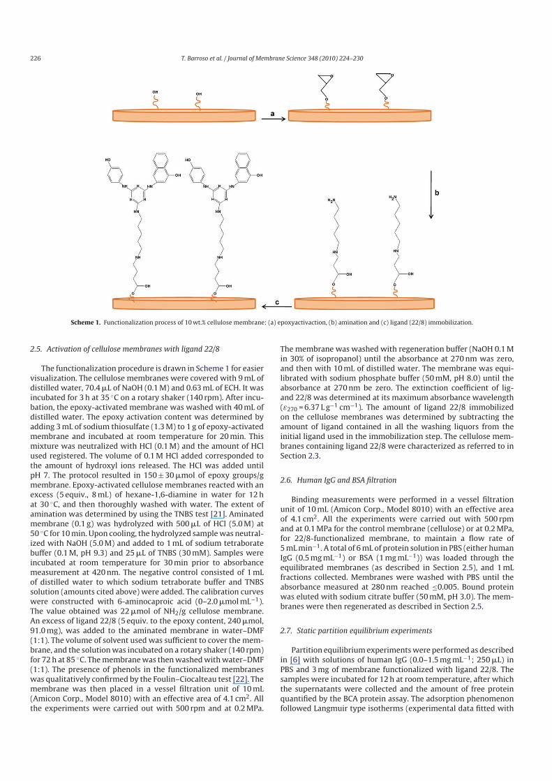
Scheme 1. Functionalization process of 10 wt.% cellulose membrane: (a) epoxyactivaction, (b) amination and (c) ligand (22/8) immobilization.
2.5. Activation of cellulose membranes with ligand 22/8
The functionalization procedure is drawn in Scheme 1 for easiervisualization. The cellulose membranes were covered with 9 mL ofdistilled water, 70.4 mL of NaOH (0.1 M) and 0.63 mL of ECH. It wasincubated for 3 h at 35 ◦C on a rotary shaker (140 rpm). After incubation, the epoxyactivated membrane was washed with 40 mL ofdistilled water. The epoxy activation content was determined byadding 3 mL of sodium thiosulfate (1.3 M) to 1 g of epoxyactivatedmembrane and incubated at room temperature for 20 min. Thismixture was neutralized with HCl (0.1 M) and the amount of HClused registered. The volume of 0.1 M HCl added corresponded tothe amount of hydroxyl ions released. The HCl was added untilpH 7. The protocol resulted in 150 ± 30 mmol of epoxy groups/gmembrane. Epoxyactivated cellulose membranes reacted with anexcess (5 equiv., 8 mL) of hexane1,6diamine in water for 12 hat 30 ◦C, and then thoroughly washed with water. The extent ofamination was determined by using the TNBS test [21]. Aminatedmembrane (0.1 g) was hydrolyzed with 500 mL of HCl (5.0 M) at50 ◦C for 10 min. Upon cooling, the hydrolyzed sample was neutralized with NaOH (5.0 M) and added to 1 mL of sodium tetraboratebuffer (0.1 M, pH 9.3) and 25 mL of TNBS (30 mM). Samples wereincubated at room temperature for 30 min prior to absorbancemeasurement at 420 nm. The negative control consisted of 1 mLof distilled water to which sodium tetraborate buffer and TNBSsolution (amounts cited above) were added. The calibration curveswere constructed with 6aminocaproic acid (0–2.0 mmol mL−1).The value obtained was 22 mmol of NH2/g cellulose membrane.An excess of ligand 22/8 (5 equiv. to the epoxy content, 240 mmol,91.0 mg), was added to the aminated membrane in water–DMF(1:1). The volume of solvent used was sufficient to cover the membrane, and the solution was incubated on a rotary shaker (140 rpm)for 72 h at 85 ◦C. The membrane was then washed with water–DMF(1:1). The presence of phenols in the functionalized membraneswas qualitatively confirmed by the Foulin–Ciocalteau test [22]. Themembrane was then placed in a vessel filtration unit of 10 mL(Amicon Corp., Model 8010) with an effective area of 4.1 cm2. Allthe experiments were carried out with 500 rpm and at 0.2 MPa.
The membrane was washed with regeneration buffer (NaOH 0.1 Min 30% of isopropanol) until the absorbance at 270 nm was zero,and then with 10 mL of distilled water. The membrane was equilibrated with sodium phosphate buffer (50 mM, pH 8.0) until theabsorbance at 270 nm be zero. The extinction coefficient of ligand 22/8 was determined at its maximum absorbance wavelength(ε270 = 6.37 L g−1 cm−1). The amount of ligand 22/8 immobilizedon the cellulose membranes was determined by subtracting theamount of ligand contained in all the washing liquors from theinitial ligand used in the immobilization step. The cellulose membranes containing ligand 22/8 were characterized as referred to inSection 2.3.
2.6. Human IgG and BSA filtration
Binding measurements were performed in a vessel filtrationunit of 10 mL (Amicon Corp., Model 8010) with an effective areaof 4.1 cm2. All the experiments were carried out with 500 rpmand at 0.1 MPa for the control membrane (cellulose) or at 0.2 MPa,for 22/8functionalized membrane, to maintain a flow rate of5 mL min−1. A total of 6 mL of protein solution in PBS (either humanIgG (0.5 mg mL−1) or BSA (1 mg mL−1)) was loaded through theequilibrated membranes (as described in Section 2.5), and 1 mLfractions collected. Membranes were washed with PBS until theabsorbance measured at 280 nm reached ≤0.005. Bound proteinwas eluted with sodium citrate buffer (50 mM, pH 3.0). The membranes were then regenerated as described in Section 2.5.
2.7. Static partition equilibrium experiments
Partition equilibrium experiments were performed as describedin [6] with solutions of human IgG (0.0–1.5 mg mL−1; 250 mL) inPBS and 3 mg of membrane functionalized with ligand 22/8. Thesamples were incubated for 12 h at room temperature, after whichthe supernatants were collected and the amount of free proteinquantified by the BCA protein assay. The adsorption phenomenonfollowed Langmuir type isotherms (experimental data fitted with
T. Barroso et al. / Journal of Membrane Science 348 (2010) 224–230 227
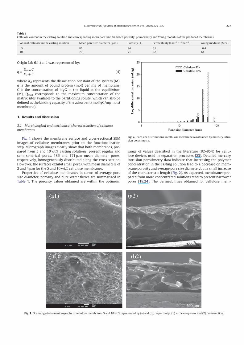
Table 1
Cellulose content in the casting solution and corresponding mean pore size diameter, porosity, permeability and Young modulus of the produced membranes.
Wt.% of cellulose in the casting solution Mean pore size diameter (mm) Porosity (%) Permeability (L m−2 h−1 bar−1) Young modulus (MPa)
5 85 84 0.2 0.4
10 70 71 0.5 12
Origin Lab 6.1.) and was represented by:
q =QmaxC
Kd + C(4)
where Kd represents the dissociation constant of the system (M),q is the amount of bound protein (mol) per mg of membrane,C is the concentration of hIgG in the liquid at the equilibrium(M), Qmax corresponds to the maximum concentration of thematrix sites available to the partitioning solute, which can also bedefined as the binding capacity of the adsorbent (mol IgG/mg moistmembrane).
3. Results and discussion
3.1. Morphological and mechanical characterization of cellulose
membranes
Fig. 1 shows the membrane surface and crosssectional SEMimages of cellulose membranes prior to the functionalizationstep. Micrograph images clearly show that both membranes, prepared from 5 and 10 wt.% casting solutions, present regular andsemispherical pores, 186 and 171 mm mean diameter pores,respectively, homogeneously distributed along the crosssection.However, the surfaces exhibit small pores, with mean diameters of2 and 4 mm for the 5 and 10 wt.% cellulose membranes.
Properties of cellulose membranes in terms of average poresize diameter, porosity and pure water fluxes are summarized inTable 1. The porosity values obtained are within the optimum
Fig. 2. Pore size distributions in cellulose membranes as obtained by mercury intru
sion porosimetry.
range of values described in the literature (82–85%) for cellulose devices used in separation processes [23]. Detailed mercuryintrusion porosimetry data indicate that increasing the polymerconcentration in the casting solution lead to a decrease on membrane porosity and average pore size diameter, but a small increaseof the characteristic length (Fig. 2). As expected, membranes prepared from more concentrated solutions tend to present narrowerpores [19,24]. The permeabilities obtained for cellulose mem
Fig. 1. Scanning electron micrographs of cellulose membranes 5 and 10 wt.% represented by (a) and (b), respectively: (1) surface top view and (2) crosssection.
228 T. Barroso et al. / Journal of Membrane Science 348 (2010) 224–230
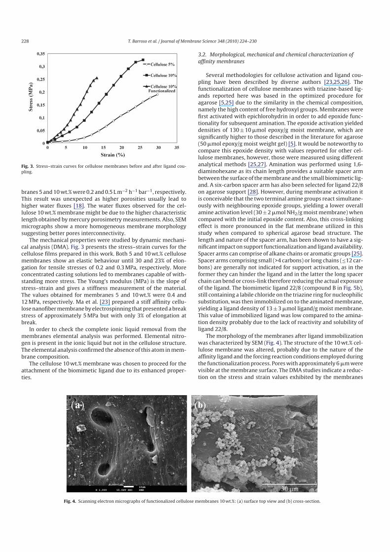
Fig. 3. Stress–strain curves for cellulose membranes before and after ligand cou
pling.
branes 5 and 10 wt.% were 0.2 and 0.5 L m−2 h−1 bar−1, respectively.This result was unexpected as higher porosities usually lead tohigher water fluxes [18]. The water fluxes observed for the cellulose 10 wt.% membrane might be due to the higher characteristiclength obtained by mercury porosimetry measurements. Also, SEMmicrographs show a more homogeneous membrane morphologysuggesting better pores interconnectivity.
The mechanical properties were studied by dynamic mechanical analysis (DMA). Fig. 3 presents the stress–strain curves for thecellulose films prepared in this work. Both 5 and 10 wt.% cellulosemembranes show an elastic behaviour until 30 and 23% of elongation for tensile stresses of 0.2 and 0.3 MPa, respectively. Moreconcentrated casting solutions led to membranes capable of withstanding more stress. The Young’s modulus (MPa) is the slope ofstress–strain and gives a stiffness measurement of the material.The values obtained for membranes 5 and 10 wt.% were 0.4 and12 MPa, respectively. Ma et al. [23] prepared a stiff affinity cellulose nanofiber membrane by electrospinning that presented a breakstress of approximately 5 MPa but with only 3% of elongation atbreak.
In order to check the complete ionic liquid removal from themembranes elemental analysis was performed. Elemental nitrogen is present in the ionic liquid but not in the cellulose structure.The elemental analysis confirmed the absence of this atom in membrane composition.
The cellulose 10 wt.% membrane was chosen to proceed for theattachment of the biomimetic ligand due to its enhanced properties.
3.2. Morphological, mechanical and chemical characterization of
affinity membranes
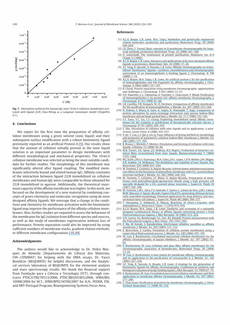
Several methodologies for cellulose activation and ligand coupling have been described by diverse authors [23,25,26]. Thefunctionalization of cellulose membranes with triazinebased ligands reported here was based in the optimized procedure foragarose [5,25] due to the similarity in the chemical composition,namely the high content of free hydroxyl groups. Membranes werefirst activated with epichlorohydrin in order to add epoxide functionality for subsequent amination. The epoxide activation yieldeddensities of 130 ± 10 mmol epoxy/g moist membrane, which aresignificantly higher to those described in the literature for agarose(50 mmol epoxy/g moist weight gel) [5]. It would be noteworthy tocompare this epoxide density with values reported for other cellulose membranes, however, those were measured using differentanalytical methods [25,27]. Amination was performed using 1,6diaminohexane as its chain length provides a suitable spacer armbetween the surface of the membrane and the small biomimetic ligand. A sixcarbon spacer arm has also been selected for ligand 22/8on agarose support [28]. However, during membrane activation itis conceivable that the two terminal amine groups react simultaneously with neighbouring epoxide groups, yielding a lower overallamine activation level (30 ± 2 mmol NH2/g moist membrane) whencompared with the initial epoxide content. Also, this crosslinkingeffect is more pronounced in the flat membrane utilized in thisstudy when compared to spherical agarose bead structure. Thelength and nature of the spacer arm, has been shown to have a significant impact on support functionalization and ligand availability.Spacer arms can comprise of alkane chains or aromatic groups [25].Spacer arms comprising small (>4 carbons) or long chains (≤12 carbons) are generally not indicated for support activation, as in theformer they can hinder the ligand and in the latter the long spacerchain can bend or crosslink therefore reducing the actual exposureof the ligand. The biomimetic ligand 22/8 (compound B in Fig. 5b),still containing a labile chloride on the triazine ring for nucleophilicsubstitution, was then immobilized on to the aminated membrane,yielding a ligand density of 13 ± 3 mmol ligand/g moist membrane.This value of immobilized ligand was low compared to the amination density probably due to the lack of reactivity and solubility ofligand 22/8.
The morphology of the membranes after ligand immobilizationwas characterized by SEM (Fig. 4). The structure of the 10 wt.% cellulose membrane was altered, probably due to the nature of theaffinity ligand and the forcing reaction conditions employed duringthe functionalization process. Pores with approximately 6 mm werevisible at the membrane surface. The DMA studies indicate a reduction on the stress and strain values exhibited by the membranes
Fig. 4. Scanning electron micrographs of functionalized cellulose membranes 10 wt.%: (a) surface top view and (b) crosssection.

T. Barroso et al. / Journal of Membrane Science 348 (2010) 224–230 229
Fig. 5. Chemical structures obtained during the synthesis of ligand (22/8): (a)
compound A (2(3aminophenol)4,6dichlorostriazine) and (b) ligand 22/8 (2
(3aminophenol)6(4amino1naphthol)4chlorostriazine).
upon ligand attachment (Fig. 3). The membranes still presentedan elastic behaviour but a lower elongation at brake (13%) whencompared with the unmodified membranes (25 and 30%).
Permeability measurements showed that for modified membrane assays the applied pressure was doubled in order to achievethe same flow rate (5 mL min−1) exhibited by bare cellulose matrices. This fact is related with the reduced porosity of the membraneupon ligand attachment as indicated by intrusion porosimetry measurements (4% porosity). The comparison with porosity valuesreported by other authors cannot be accomplished due to the lackof available data for cellulose membranes after ligand coupling pro
cedure. Nevertheless, the flow rate at which the cellulose affinitymembranes operated (5 mL min−1) is higher than those describedin the literature with the same flat sheet configuration [26,29].There are cellulose membranes commercially available that presenthigher permeability values. However, as these operate in distinctconfigurations than those utilized in this work no direct comparisoncan be made.
3.3. Human IgG and BSA filtration assays
In order to assess the binding and elution of human IgG usingthe prepared affinity membranes, standard assays were performedfollowing the optimized procedure reported [5,28] for IgG bindingand recovery using 22/8agarose supports. Nonspecific adsorptionwas evaluated by control experiments with unmodified cellulosemembranes (loaded with human IgG and BSA solutions), and with22/8modified cellulose membranes (loaded with BSA solution).The binding and elution profiles of BSA and human IgG obtainedwith 10 wt.% affinity cellulose membranes are shown in Fig. 6. Weobserved that the unmodified cellulose membrane did not bindthe tested proteins, and BSA was not adsorbed on to affinity membranes either. On the other hand, it was possible to bind and elute 6and 2 mg IgG/g membrane, respectively. These values correspondto 80% binding and 25% elution of human IgG. As we have performedpreliminary assays in standard conditions it is envisioned that thevalues for human IgG binding and recovery can be improved ifdifferent buffers, flow rates or membrane geometries are used.
Partition equilibrium studies were performed with human IgGand the affinity membranes. The experimental data was fitted in aLangmuir type isotherm (Fig. 7) yielding an affinity constant (Ka) of0.3 mM−1 and a theoretical maximum capacity (Qmax) of 630.3 mgIgG/g moist membrane (R2 = 0.95). The Ka value is in accordancewith that previously reported by Teng and coworkers when performing scatchard analysis of the binding isotherm for IgG onagaroseimmobilized 22/8 (0.14 mM−1), but the Qmax value for oursystem was considerably higher than the value of 151.9 mg IgG/gmoist weight gel obtained by the same authors [5]. Castilho et al.[9] evaluated ten different affinity membranes through coupling ofIgG specific affinity ligands. They observed that the Ka values werewithin the range 0.1–1 mM−1 depending on the nature of the ligand and membrane. Also, they reported Qmax values calculated ona different basis and therefore not directly comparable with ours. Itshould be noted that as distinct affinity ligands were tested, humanIgG adsorption behaviour would differ from the observed in thiswork.
Fig. 6. Binding and elution profiles for human IgG and BSA onto 10 wt.% cellulose membranes activated with ligand 22/8.
230 T. Barroso et al. / Journal of Membrane Science 348 (2010) 224–230
Fig. 7. Adsorption isotherm for human IgG onto 10 wt.% cellulose membranes acti
vated with ligand 22/8. Data fitting as a Langmuir monolayer model (OriginPro
6.1).
4. Conclusions
We report for the first time the preparation of affinity cellulose membranes using a green solvent (ionic liquid) and theirsubsequent surface modification with a robust biomimetic ligandpreviously reported as an artificial Protein A [5]. Our results showthat the amount of cellulose initially present in the ionic liquidsolution is an important parameter to design membranes withdifferent morphological and mechanical properties. The 10 wt.%cellulose membrane was selected as being the most suitable candidate for further studies. The morphology of this membrane wassignificantly altered after ligand coupling. The modified membranes selectively bound and eluted human IgG. Affinity constantsof the interaction between ligand 22/8 immobilized on cellulosemembranes and human IgG were comparable to those obtained for22/8 immobilized in agarose. Additionally, the theoretical maximum capacity of the affinity membrane was higher. In this work, wefocused on the development of a new material by combining concepts of green chemistry and molecular recognition using de novodesigned affinity ligands. We envisage that a change in the conditions and chemistry for membrane activation with the biomimeticligand may improve the performance of the affinity cellulose membranes. Also, further studies are required to assess the behaviour ofthe membranes for IgG isolation from different species and sources,as well as the study of membranes regeneration without loss ofperformance. Protein separation could also be improved by usingsufficient numbers of membrane stacks, gradient elution methods,or different membrane configurations [10,30].
Acknowledgments
The authors would like to acknowledge to Dr. Pedro Marques de Almeida (Departamento de Ciência dos Materiais,I3N—CENIMAT) for helping with the DMA assays, Dr. VascoBonifácio (REQUIMTE) for helpful discussions, and the Analytical services laboratory of REQUIMTE for the elemental analysisand mass spectroscopy results. We thank the financial supportfrom Fundacão para a Ciência e Tecnologia (FCT), through contracts PTDC/CTM/70513/2006, PTDC/BIO/65383/2006, SFRH/BD/16908/2004 for M.T., SFRH/BPD/34768/2007 for A.H., FEDER, FSE,and MITPortugal Program, Bioengineering Systems Focus Area.
References
[1] A.C.A. Roque, C.R. Lowe, M.A. Taipa, Antibodies and genetically engineeredrelated molecules: production and purification, Biotechnol. Progr. 20 (2004)639–654.
[2] J.X. Zhou, T. Tressel, Basic concepts in Q membrane chromatography for largescale antibody production, Biotechnol. Progr. 22 (2006) 341–349.
[3] U. Gottschalk, The renaissance of protein purification, Biopharm. Int. 8–9(Suppl. S) (2006).
[4] A.C.A. Roque, C.R. Lowe, Advances and applications of de novo designed affinityligands in proteomics, Biotechnol. Adv. 24 (2006) 17–26.
[5] S.F. Teng, K. Sproule, A. Hussain, C.R. Lowe, Affinity chromatography on immobilized “biomimetic” ligands—synthesis, immobilization and chromatographicassessment of an immunoglobulin Gbinding ligand, J. Chromatogr. B 740(2000) 1–15.
[6] A.C.A. Roque, M.A. Taipa, C.R. Lowe, An artificial protein L for the purificationof immunoglobulins and Fab fragments by affinity chromatography, J. Chromatogr. A 1064 (2005) 157–167.
[7] R. Ghosh, Protein separation using membrane chromatography: opportunitiesand challenges, J. Chromatogr. A 952 (2002) 13–27.
[8] O.P. Dancette, J.L. Taboureau, E. Tournier, C. Charcosset, P. Blond, Purificationof immunoglobulins G by protein A/G affinity membrane chromatography, J.Chromatogr. B 723 (1999) 61–68.
[9] L.R. Castilho, F.B. Anspach, W.D. Deckwer, Comparison of affinity membranesfor the purification of immunoglobulins, J. Membr. Sci. 207 (2002) 253–264.
[10] N. Kubota, S. Miura, K. Saito, K. Sugita, K. Watanabe, T. Sugo, Comparison ofprotein adsorption by anionexchange interaction onto porous hollowfibermembrane and gel beadpacked bed, J. Membr. Sci. 117 (1996) 135–142.
[11] S.Y. Suen, Y.C. Liu, C.S. Chang, Exploiting immobilised metal affinity membranes for the isolation or purification of therapeutically relevant species, J.Chromatogr. B 797 (2003) 305–319.
[12] S. Zhu, Dissolution of cellulose with ionic liquids and its application: a minireview, Green Chem. 8 (2006) 325–327.
[13] X. Jie, Y. Cao, J.J. Qin, J. Liu, Q. Yuan, Influence of drying method on morphologyand properties of asymmetric cellulose hollow fiber membrane, J. Membr. Sci.246 (2005) 157–165.
[14] B. Kosan, C. Michels, F. Meister, Dissolution and forming of cellulose with ionicliquids, Cellulose 15 (2008) 59–66.
[15] M.B. Turner, S.K. Spear, J.D. Holbrey, R.D. Rogers, Production of bioactive cellulose films reconstituted from ionic liquids, Biomacromolecules 5 (2004)1379–1384.
[16] M.J. Earle, J.M.S.S. Esperanca, M.A. Gilea, J.N.C. Lopes, L.P.N. Rebelo, J.W. Magee,K.R. Seddon, J.A. Widegren, The distillation and volatility of ionic liquids, Nat.Biotechnol. 439 (2006) 831–834.
[17] M. Temtem, T. Casimiro, A. AguiarRicardo, Solvent power and depressurizationrate effects in the formation of polysulfone membrane with CO2assisted phaseinversion method, J. Membr. Sci. 282 (2006) 244–252.
[18] M. Temtem, T. Casimiro, J.F. Mano, A. AguiarRicardo, Preparation of membranes with polysulfone/polycaprolactone blends using a high pressure cellspecially designed for a CO2assisted phase inversion, J. Supercrit. Fluids 43(2007) 542–548.
[19] M. Temtem, L.M.C. Silva, P.Z. Andrade, F. Santos, C. Lobato da Silva, J.M.S. Cabral,M.M. Abecasis, A. AguiarRicardo, Supercritical CO2 generating chitosan deviceswith controlled morphology. Potential application for drug delivery and mesenchymal stem cell culture, J. Supercrit. Fluids 48 (2009) 269–277.
[20] T. Harayama, S. Sekiguchi, K. Matsui, Reactions of chorostriazines withaminophenol, J. Heterocycl. Chem. 7 (1970) 975–979.
[21] A.C.A. Roque, M.A. Taipa, C.R. Lowe, Synthesis and screening of a rationallydesigned combinatorial library of affinity ligands mimicking protein L fromPeptrostreptococcus magnus, J. Mol. Recognit. 18 (2005) 213–224.
[22] O.H. Lowry, N.J. Rosebrough, A.L. Farr, R.J. Randall, Protein measurement withthe Folin phenol reagent, J. Biol. Chem. 193 (1951) 265.
[23] Z. Ma, M. Kotaki, S. Ramakrishna, Electrospun cellulose nanofiber as affinitymembrane, J. Membr. Sci. 265 (2005) 115–123.
[24] E. Reverchon, S. Cardea, Formation of cellulose acetate membranes using asupercritical fluid assisted process, J. Membr. Sci. 240 (2004) 187–195.
[25] W. Guo, E. Ruckenstein, Crosslinked mercerized cellulose membranes for theaffinity chromatography of papain inhibitors, J. Membr. Sci. 197 (2002) 53–62.
[26] E. Ruckenstein, W. Guo, Cellulose and glass fiber affinity membranes for thechromatographic separation of biomolecules, Biotechnol. Progr. 20 (2004)13–25.
[27] W. Guo, E. Ruckenstein, A new matrix for membrane affinity chromatographyand its application to the purification of concanavalin A, J. Membr. Sci. 182(2001) 227–234.
[28] S.F. Teng, K. Sproule, A. Husain, C.R. Lowe, A strategy for the generation ofbiomimetic ligands for affinity chromatography. Combinatorial synthesis andbiological evaluation of an IgG binding ligand, J. Mol. Recognit. 12 (1999) 67–75.
[29] E. Ruckenstein, W. Guo, Crosslinked mercerized cellulose membranes and theirapplication to membrane affinity chromatography, J. Membr. Sci. 187 (2001)277–286.
[30] C. Charcosset, Purification of proteins by membrane chromatography, J. Chem.Technol. Biotechnol. 71 (1998) 95–110.
Copyright © 2022 FDOKUMEN