Reticulum fibres in relation to retinal vessels
7
British Journal of Ophthalmology, 1977, 61, 339-344 Reticulin fibres in relation to retinal vessels AMJAD RAHI AND NORMAN ASHTON from the Department of Pathology, Institute of Ophthalmology, University of London SUMMARY Argyrophilic perivascular and intervascular fibres in the mammalian retina are shown by specific antireticulin immunofluorescence to consist of reticulin. The possible significance of these findings is briefly discussed. Some recent studies in the retina of man and animals have reported the presence of perivascular and intervascular fibres which, by reason of their argyrophilia, ultrastructure, and enzymatic digestion were believed to consist of reticulin (Daicker, 1971a, b; Ashton and Tripathi, 1975). The latter authors, however, pointed out the unreliability of these criteria and suggested that the problem might be resolved by the use of specific immunofluorescent staining of reticulin. An immunohistochemical method for demonstrat- ing reticulin fibres in tissues has recently been described (Seah et al., 1971). Human sera containing reticulin antibodies were found to stain reticulin fibres in the rat liver, stomach, and kidney, and the patterns resembled very closely the silver impreg- nation staining technique. The immunofluorescence was not impaired, and was even enhanced by pre- treatment with collagenase but was abolished by periodate, and the authors were convinced that the fibres were in fact reticulin. Moreover, a considerable advance was made by Pras and Glynn (1973), who recently extracted from reticulin by a water dispersion method a noncollagenous protein, the antibodies of which showed specific anti- reticulin activity by immunofluorescent techniques. We have employed both these methods of investi- gation in the retinas of monkeys and rats and now report our findings. Material and methods RETICULIN ANTISERA The specific antisera used in the present study were obtained from patients suffering from either uveitis or coeliac disease, conditions known to be associated with the presence of antireticulin anti- Address for reprints: Professor Norman Ashton, Institute of Ophthalmology, Judd Street, London WCIH 9QS bodies (Seah et al., 1971; Rahi et al., 1976) or else prepared in this laboratory in New Zealand albino rabbits according to the following schedule: 4 mg of a noncollagenous component of reticulin protein prepared according to the method of Pras and Glynn (1973)*, was ultrasonically dispersed in 2 ml of distilled water, and emulsified in equal volumes of Freund's complete adjuvant and injected intra- muscularly into the animals at 4 different sites. The injection was repeated twice at 2 weeks and 4 weeks after the initial dose, and the animal was bled 1 week after the last injection. The serum was stored in aliquots at -20°C. IMMUNOFLUORESCENCE TEST Frozen sections of a composite block consisting of the liver, kidney, diaphragm, and stomach of a rat were used to test the specificity of the antisera. Frozen sections of eyes from rats and monkeys and capillaries isolated from rat retinas by the acid water technique were stained to demonstrate the peri- vascular and intervascular reticular fibres. A standard indirect immunofluorescent staining tech- nique was used. Fluorescein labelled antihuman and antirabbit immunoglobulins were obtained from the Wellcome Laboratories. A Zeiss epifluorescence microscope equipped with a fluorescein isothio- cyanate (FITC) interference filter was used to examine the stained sections. Findings The reticulin antisera obtained from patients and from immunised rabbits gave typical staining patterns (Rizzetto and Doniach, 1973; Williamson et al., 1976) in the liver, kidney, and stomach (Figs. 1, 2, and 3). The septal fibres within the connective tissue of the optic nerve stained intensely (Fig. 4). *This extract was kindly provided by Dr L. E. Glynn, director of the Mathilda and Terence Kennedy Institute of Rheumatology, London 339 group.bmj.com on July 20, 2016 - Published by http://bjo.bmj.com/ Downloaded from
-
Upload
independent -
Category
Documents
-
view
0 -
download
0
Transcript of Reticulum fibres in relation to retinal vessels

British Journal of Ophthalmology, 1977, 61, 339-344
Reticulin fibres in relation to retinal vesselsAMJAD RAHI AND NORMAN ASHTON
from the Department of Pathology, Institute of Ophthalmology, University of London
SUMMARY Argyrophilic perivascular and intervascular fibres in the mammalian retina are shownby specific antireticulin immunofluorescence to consist of reticulin. The possible significance ofthese findings is briefly discussed.
Some recent studies in the retina of man andanimals have reported the presence of perivascularand intervascular fibres which, by reason of theirargyrophilia, ultrastructure, and enzymatic digestionwere believed to consist of reticulin (Daicker, 1971a,b; Ashton and Tripathi, 1975). The latter authors,however, pointed out the unreliability of thesecriteria and suggested that the problem might beresolved by the use of specific immunofluorescentstaining of reticulin.An immunohistochemical method for demonstrat-
ing reticulin fibres in tissues has recently beendescribed (Seah et al., 1971). Human sera containingreticulin antibodies were found to stain reticulinfibres in the rat liver, stomach, and kidney, and thepatterns resembled very closely the silver impreg-nation staining technique. The immunofluorescencewas not impaired, and was even enhanced by pre-treatment with collagenase but was abolished byperiodate, and the authors were convinced thatthe fibres were in fact reticulin. Moreover, aconsiderable advance was made by Pras and Glynn(1973), who recently extracted from reticulin by awater dispersion method a noncollagenous protein,the antibodies of which showed specific anti-reticulin activity by immunofluorescent techniques.We have employed both these methods of investi-gation in the retinas of monkeys and rats and nowreport our findings.
Material and methods
RETICULIN ANTISERAThe specific antisera used in the present study wereobtained from patients suffering from eitheruveitis or coeliac disease, conditions known to beassociated with the presence of antireticulin anti-
Address for reprints: Professor Norman Ashton, Institute ofOphthalmology, Judd Street, London WCIH 9QS
bodies (Seah et al., 1971; Rahi et al., 1976) or elseprepared in this laboratory in New Zealand albinorabbits according to the following schedule: 4 mgof a noncollagenous component of reticulin proteinprepared according to the method of Pras and Glynn(1973)*, was ultrasonically dispersed in 2 ml ofdistilled water, and emulsified in equal volumes ofFreund's complete adjuvant and injected intra-muscularly into the animals at 4 different sites. Theinjection was repeated twice at 2 weeks and 4 weeksafter the initial dose, and the animal was bled 1 weekafter the last injection. The serum was stored inaliquots at -20°C.
IMMUNOFLUORESCENCE TESTFrozen sections of a composite block consisting ofthe liver, kidney, diaphragm, and stomach of a ratwere used to test the specificity of the antisera.Frozen sections of eyes from rats and monkeys andcapillaries isolated from rat retinas by the acid watertechnique were stained to demonstrate the peri-vascular and intervascular reticular fibres. Astandard indirect immunofluorescent staining tech-nique was used. Fluorescein labelled antihuman andantirabbit immunoglobulins were obtained from theWellcome Laboratories. A Zeiss epifluorescencemicroscope equipped with a fluorescein isothio-cyanate (FITC) interference filter was used toexamine the stained sections.
Findings
The reticulin antisera obtained from patients andfrom immunised rabbits gave typical stainingpatterns (Rizzetto and Doniach, 1973; Williamsonet al., 1976) in the liver, kidney, and stomach (Figs.1, 2, and 3). The septal fibres within the connectivetissue of the optic nerve stained intensely (Fig. 4).*This extract was kindly provided by Dr L. E. Glynn, director of theMathilda and Terence Kennedy Institute of Rheumatology, London
339
group.bmj.com on July 20, 2016 - Published by http://bjo.bmj.com/Downloaded from

Amjad Rahi and Norman Ashton
Fig. 1 Indirectimmunofluorescence test
on rat liver. Theantireticulin serum reactswith the reticulin fibres inthe central portal vein (A)
and the branches ofhepatic artery (B) toproduce a bright
fluorescence. Unfixedcryostat section x 360
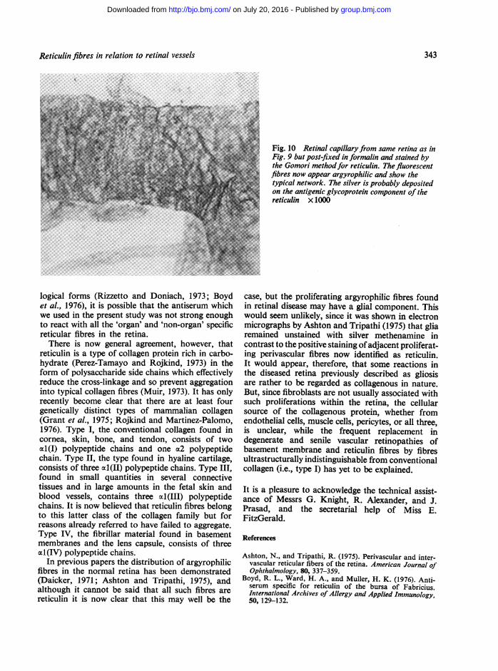
In the sections of the retina fluorescent stainingwas confined to the perivascular fibres and alsooutlined the vascular endothelium (Figs. 5, 6, 7, and8). Isolated rat retinal vessels showed coarse longi-tudinal and branching fluorescent networks ofperivascular fibres (Fig. 9). These appearancesexactly corresponded to those demonstrated in silver-stained preparations (Ashton and Tripathi, 1975)and established the reticulin nature of these fibres(Fig. 10).
Discussion
The fact that reticulin exists in the retina in theform of perivascular networks and intercapillarystrands and bridges is of academic interest and ofpathological significance. There has for many yearsbeen controversy as to the biochemical nature ofreticulin fibres and hesitancy in correlating theirmicroscopical and ultrastructural properties, to theextent that the term 'reticulin' is generally avoided
Fig. 2 (left) Peritubularreticulin fluorescence in arat kidney demonstratedby the same technique asin Fig. 1 x 360
Fig. 3 (right)Periglandular reticulinfluorescence in a rat
stomach demonstrated bythe same technique as
Fig. I x 270
340
group.bmj.com on July 20, 2016 - Published by http://bjo.bmj.com/Downloaded from

Reticulin fibres in relation to retinal vessels
Fig. 4 The septal reticulin fibresin the optic nerve ofa monkeyshow brightfluorescence whentreated with specific antiserum.Unfixed cryostat section x 175
by electron microscopists. Cervos-Navarro andMatakas (1975) claim that they were the first todescribe the ultrastructure of reticulin in 1972
Fig. 5 Cross-section of rat retina treated withantireticulin serum. Bright perivascular fluorescence wasseen in the retinal vessels (right) and in the choroidalvessels (left). Indirect immunofluorescence test. Unfixedcryostat section x 360
(Matakas and Cervos-Navarro, 1972). In view of theclose anatomical relationship between basementmembrane and reticulin, described in the retina byAshton and Tripathi (1975), it is noteworthy thatCervos-Navarro and Matakas (1975) consider that
Fig. 6 Flat section of rat retina treated by the sametechnique as in Fig. 5. Bright fluorescence is presentwithin the capillary walls x 360
341
group.bmj.com on July 20, 2016 - Published by http://bjo.bmj.com/Downloaded from

Amjad Rahi and Norman Ashton
Fig. 7 Positive fluorescence of reticulin fibres in the Fig. 8 Flat section of monkey retina showingcapillaries of the monkey retina (cross-section) seen fluorescence in a pericapillary reticulin network withpartially in focus in a thick unfixed cryostat section strands extending into retina. Unfixed cryostat sectionx 360 x 480
basement membrane is the substrate of reticulin, acorrespondence first mentioned by Robb-Smith(1970). They emphasise that the fibrillar form ofreticulin seems to be only a light-microscopicalfinding and possibly caused by formalin fixation insome instances. In the present study, however, wehave demonstrated them by immunofluorescence inunfixed frozen sections; nevertheless, perivascularreticulin networks are certainly more readily demon-strated by silver staining in formalin fixed tissue.
The reticular fibres in the retinal vessels stainedless brilliantly than those in the optic nerve, the liver,the kidneys, and the stomach. As the product ofimmunofluorescence, like any other chemical reac-tion, is generally directly proportional to theconcentration of the reactants, this weak stainingmay be due to a relatively smaller amount of thesefibres in the retina than in other tissues.
Since reticulin appears to exist in several differentmorphological (Vizioli et al., 1974) and immuno-
Fig. 9 (A and B) Rat retina. Capillaries isolated by acid-water techniques show fluorescent networks aroundcapillaries treated with specific antiserum. Unfixed x 1000
342
group.bmj.com on July 20, 2016 - Published by http://bjo.bmj.com/Downloaded from
Reticulin fibres in relation to retinal vessels
Fig. 10 Retinal capillaryfrom same retina as inFig. 9 but post-fixed informalin and stained bythe Gomori methodfor reticulin. The fluorescentfibres now appear argyrophilic and show thetypical network. The silver is probably depositedon the antigenic glycoprotein component of thereticulin x 1000
logical forms (Rizzetto and Doniach, 1973; Boydet al., 1976), it is possible that the antiserum whichwe used in the present study was not strong enoughto react with all the 'organ' and 'non-organ' specificreticular fibres in the retina.There is now general agreement, however, that
reticulin is a type of collagen protein rich in carbo-hydrate (Perez-Tamayo and Rojkind, 1973) in theform of polysaccharide side chains which effectivelyreduce the cross-linkage and so prevent aggregationinto typical collagen fibres (Muir, 1973). It has onlyrecently become clear that there are at least fourgenetically distinct types of mammalian collagen(Grant et al., 1975; Rojkind and Martinez-Palomo,1976). Type I, the conventional collagen found incornea, skin, bone, and tendon, consists of twoal(I) polypeptide chains and one oa2 polypeptidechain. Type II, the type found in hyaline cartilage,consists of three al(II) polypeptide chains. Type III,found in small quantities in several connectivetissues and in large amounts in the fetal skin andblood vessels, contains three al(III) polypeptidechains. It is now believed that reticulin fibres belongto this latter class of the collagen family but forreasons already referred to have failed to aggregate.Type IV, the fibrillar material found in basementmembranes and the lens capsule, consists of threea1(IV) polypeptide chains.
In previous papers the distribution of argyrophilicfibres in the normal retina has been demonstrated(Daicker, 1971; Ashton and Tripathi, 1975), andalthough it cannot be said that all such fibres arereticulin it is now clear that this may well be the
case, but the proliferating argyrophilic fibres foundin retinal disease may have a glial component. Thiswould seem unlikely, since it was shown in electronmicrographs by Ashton and Tripathi (1975) that gliaremained unstained with silver methenamine incontrast to the positive staining ofadjacent proliferat-ing perivascular fibres now identified as reticulin.It would appear, therefore, that some reactions inthe diseased retina previously described as gliosisare rather to be regarded as collagenous in nature.But, since fibroblasts are not usually associated withsuch proliferations within the retina, the cellularsource of the collagenous protein, whether fromendothelial cells, muscle cells, pericytes, or all three,is unclear, while the frequent replacement indegenerate and senile vascular retinopathies ofbasement membrane and reticulin fibres by fibresultrastructurally indistinguishable from conventionalcollagen (i.e., type I) has yet to be explained.
It is a pleasure to acknowledge the technical assist-ance of Messrs G. Knight, R. Alexander, and J.Prasad, and the secretarial help of Miss E.FitzGerald.
References
Ashton, N., and Tripathi, R. (1975). Perivascular and inter-vascular reticular fibers of the retina. American Journal ofOphthalmology, 80, 337-359.
Boyd, R. L., Ward, H. A., and Muller, H. K. (1976). Anti-serum specific for reticulin of the bursa of Fabricius.International Archives of Allergy and Applied Immunology,50, 129-132.
343
group.bmj.com on July 20, 2016 - Published by http://bjo.bmj.com/Downloaded from
Amjad Rahi and Norman Ashton
Cervos-Navarro, J., and Matakas, F. (1975). The ultra-structure of reticulin. Acta Neuropathologica, Suppl. 6,173-176.
Daicker, B. (1971a). Die adventitiellen und intervascularenReticulinfasern der menschlichen Netzhaut. Albrecht vonGraefes Archiv far klinische und experimentelle Ophthal-mologie, 181, 179-191.
Daicker, B. (1971b). Hyperplasie der adventitiellen undmuralen Reticulinfasern bei vasopathia diabetica retinae.Albrecht von Graefes Archivfur klinische und experimentelleOphthalmologie, 181, 192-206.
Grant, M. E., Harwood, R., and Schofield, J. D. (1975).Dynamics of Connective Tissue Macromolecules, Ch. 1.Edited by P. M. C. Burleigh and A. R. Poole. NorthHolland: Amsterdam.
Matakas, F., and Cervos-Navarro, J. (1972). Die Ultra-struktur des Retikulins. Virchows Archiv Abteilung B.Zellpathologie, 10, 67-82.
Muir, I. H. M. (1973). Biochemistry. Adult Articular Cartilage,p. 100. Edited by M. A. R. Freeman. Pitman Medical:London.
Perez-Tamayo, R., and Rojkind, M. (1973). MolecularPathology of Connective Tissue. Dekker: New York.
Pras, M., and Glynn, L. E. (1973). Isolation of a non-collagenous reticulin component and its primary character-isations. British Journal of Experimental Pathology, 54,449-456.
Rahi, A. H. S., Holborow, E. J., Perkins, E. S., Gungen, Y.,and Dinning, W. J. (1976). Immunological investigationsin uveitis. Transactions of the Ophthalmological Societies ofthe United Kingdom, 96, 113-122.
Rizzetto, M., and Doniach, D. (1973). Types of 'reticulin'antibodies detected in human sera by immunofluorescence.Journal of Clinical Pathology, 26, 841-851.
Robb-Smith, A. H. T. (1970). The Functional Significance ofConnective Tissue in General Pathology, p. 457. Edited byLord Florey. Lloyd-Luke: London.
Rojkind, M., and Martinez-Palomo, A. (1976). Increase intype I and type III collagens in human alcoholic liversclerosis. Proceedings of the National Academy of Sciencesof the United States of America, 73, 539-543.
Seah, P. P., Fry, L., Hoffbrand, A. V., and Holborow, E. J.(1971). Tissue antibodies in dermatitis herpetiformis andadult coeliac disease. Lancet, 1, 834-836.
Vizioli, M. R., Valdrighi, L., and Bozzo, L. (1974). Theappearing and evolution of reticulin: observations on therepair of post-extraction marmoset sockets. Annalesd'Histochimie, 19, 65-72.
Williamson, N., Asquith, P., Stokes, P. L., Jowett, A. W.,and Cooke, W. T. (1976). Anticonnective tissue and otherantitissue antibodies in the sera of patients with coeliacdisease compared with findings in a mixed hospitalpopulation. Journal of Clinical Pathology, 29, 484-494.
344
group.bmj.com on July 20, 2016 - Published by http://bjo.bmj.com/Downloaded from
retinal vessels.Reticulum fibres in relation to
A. Rahi and N. Ashton
doi: 10.1136/bjo.61.5.3391977 61: 339-344 Br J Ophthalmol
http://bjo.bmj.com/content/61/5/339Updated information and services can be found at:
These include:
serviceEmail alerting
online article. article. Sign up in the box at the top right corner of the Receive free email alerts when new articles cite this
Notes
http://group.bmj.com/group/rights-licensing/permissionsTo request permissions go to:
http://journals.bmj.com/cgi/reprintformTo order reprints go to:
http://group.bmj.com/subscribe/To subscribe to BMJ go to:
group.bmj.com on July 20, 2016 - Published by http://bjo.bmj.com/Downloaded from