Molecular diversity and biennial circulation of enterovirus D68
Upload
khangminh22Category
view
0download
0
MODULE 9: CARDIOVASCULAR SYSTEM
O U T L I N E
23.1 Anatomy of Blood Vessels 684
23.1a Blood Vessel Tunics 68423.1b Arteries 68523.1c Capillaries 68823.1d Veins 689
23.2 Blood Pressure 691
23.3 Systemic Circulation 692
23.3a General Arterial Flow Out of the Heart 69323.3b General Venous Return to the Heart 69323.3c Blood Flow Through the Head and Neck 69323.3d Blood Flow Through the Thoracic and Abdominal Walls 69723.3e Blood Flow Through the Thoracic Organs 70023.3f Blood Flow Through the Gastrointestinal Tract 70123.3g Blood Flow Through the Posterior Abdominal Organs,
Pelvis, and Perineum 70523.3h Blood Flow Through the Upper Limb 70523.3i Blood Flow Through the Lower Limb 709
23.4 Pulmonary Circulation 712
23.5 Review of Heart, Systemic, and Pulmonary Circulation 714
23.6 Aging and the Cardiovascular System 715
23.7 Blood Vessel Development 716
23.7a Artery Development 71623.7b Vein Development 71723.7c Comparison of Fetal and Postnatal Circulation 718
Vessels and Circulation
23C A R D I O V A S C U L A R S Y S T E M
mck78097_ch23_683-723.indd 683mck78097_ch23_683-723.indd 683 2/14/11 4:31 PM2/14/11 4:31 PM

684 Chapter Twenty-Three Vessels and Circulation
B lood vessels are analogous to highways—they are an efficient mode of transport for oxygen, carbon dioxide, nutrients, hor-
mones, and waste products to and from body tissues. The heart is the mechanical pump that propels the blood through the vessels. Together, the heart and blood vessels form a closed-loop system, whereby blood is continuously pumped to and from all areas of the body. Blood vessels are not rigid and immobile; rather, they can pulsate and change shape in accordance with the body’s needs. This chapter examines the basic histologic structure common to all blood vessels and traces the flow of blood through all of the different body regions.
larger as they merge and come closer to the heart. The site where two or more arteries (or two or more veins) converge to supply the same body region is called an anastomosis (a -nas′to -mo ′sis; pl., anastomoses). Arterial anastomoses provide alternate blood sup-ply routes to body tissues or organs. (For an example, see figure 23.12, which shows anastomoses among the superior and inferior epigastric arteries.) Some arteries do not form anastomoses; these so-called end arteries provide only one pathway through which blood can reach an organ. Examples of end arteries include the renal artery of the kidney and the splenic artery of the spleen. Other arteries (such as the coronary arteries in the heart wall) are called functional end arteries, meaning that their anastomoses are so tiny that the arteries may almost be considered end arteries. Veins tend to form many more anastomoses than do arteries. Often, an artery travels with a corresponding vein. These vessels are called companion vessels because they supply the same body region and tend to lie next to one another.
23.1a Blood Vessel Tunics
Both artery and vein walls have three layers, called tunics (too′nik; tunica = coat). The tunics surround the lumen (loo′men), or inside space, of the vessel through which blood flows. These tunics are the tunica intima, tunica media, and tunica externa (figure 23.1). The innermost layer of a blood vessel wall is the tunica intima (too′ni-ka in-ti′ma ; intimus = inmost), or tunica interna. It is composed of an endothelium (a simple squamous epithelium lining the blood vessel lumen) and a subendothelial layer made up of a thin layer of areolar connective tissue. The tunica media (me ′de -a ; medius = middle) is the middle layer of the vessel wall. It is composed of circularly arranged lay-ers of smooth muscle cells. Sympathetic innervation causes the smooth muscle to contract, resulting in vasoconstriction (va ′so -kon-strik′shu n), or narrowing of the blood vessel lumen. When the fibers relax, vasodilation (va ′so -dı-la ′shu n), or widening of the blood vessel lumen, results. The tunica externa (eks-ter′na ; externe = outside), or tunica adventitia, is the outermost layer of the blood vessel wall. It is composed of areolar connective tissue that contains elastic and collagen fibers. The tunica externa helps anchor the vessel to other structures. Very large blood vessels require their own blood sup-ply to the tunica externa in the form of a network of small arteries called the vasa vasorum (va -sa va -so r′u m; vessels of vessels). The vasa vasorum extend through the tunica externa. In arteries, the thickest layer is the tunica media, while veins have a thicker tunica externa. The lumen in an artery is nar-rower than in its companion vein of the same size (figure 23.2). Further, arteries tend to have more elastic and collagen fibers in all their tunics, which means that artery walls remain open (pat-ent), can spring back to shape, and can withstand changes in blood pressure. In contrast, vein walls tend to collapse if there is no blood in them. Table 23.1 compares the characteristics of arteries and veins. Finally, capillaries contain only the tunica intima, but this layer consists of a basement membrane and endothelium only. Intercellular clefts are the thin spaces between adjacent cells in
Study Tip!Here are some tips to help you remember the names and kinds of
blood vessels:
1. Blood vessels often share names with either the body region they traverse or the bone next to them. For example, the radial artery travels near the radius, and the axillary artery is in the axillary region.
2. Some blood vessels are named for the structure they supply. For example, the renal arteries supply the kidneys, the gonadal arteries supply the gonads, and the facial arteries supply the face.
3. Arteries and veins that travel together (called companion vessels) sometimes share the same name. For example, the femoral artery is accompanied by the femoral vein.
4. Writing out your own simplified flowchart of blood vessels for each body region will help you better understand and remember the pattern of blood flow in that region.
23.1 Anatomy of Blood VesselsLearning Objectives: 1. Compare and contrast the structure of arteries, capillaries,
and veins. 2. Describe how the different types of vessels interconnect to
transport blood.
The systemic circulation consists of the blood vessels that extend to all body regions. The pulmonary circulation consists of the vessels that take the blood to and from the lungs for the pur-pose of gas exchange. Both circulations work continuously and in tandem with each other. The three classes of blood vessels are arteries, capillaries, and veins. In the systemic circulation, as the ventricles of the heart contract, arteries convey blood away from the heart to the body. Arteries branch into smaller and smaller vessels until they feed into the capillaries, where gas and nutrient exchange occurs. From the capillaries, veins return blood to the heart. Arteries become progressively smaller as they branch and extend farther from the heart, while veins become progressively
mck78097_ch23_683-723.indd 684mck78097_ch23_683-723.indd 684 2/14/11 4:31 PM2/14/11 4:31 PM

Tunica intima
Tunica media
Tunica externa
Valves
Vein
Capillary bed
Artery
Basement membrane
Lumen Lumen
Lumen
Endothelium
Capillary
Endothelium
Subendothelial layer
Internal elastic lamina
External elastic lamina
Vasa vasorum
Chapter Twenty-Three Vessels and Circulation 685
the capillary wall. Having only the tunica intima, without con-nective tissue and muscle layers, allows for rapid gas and nutrient exchange between the blood and the tissues.
23.1b Arteries
Arteries (ar′ter-e ) transport blood away from the heart. The arter-ies in the systemic circulation carry oxygenated blood to the body tissues. In contrast, the pulmonary arteries (part of the pulmonary circulation) carry deoxygenated blood to the lungs.
The three basic types of arteries are elastic arteries, muscular arteries, and arterioles (figure 23.3, right). In general, as an artery’s diameter decreases, there is a corresponding decrease in the amount of elastic fibers and a relative increase in the amount of smooth muscle.
Elastic Arteries
Elastic arteries are the largest arteries, with diameters ranging from 2.5 to 1 centimeter. They are also called conducting arteriesbecause they conduct blood away from the heart to the smaller
Figure 23.1Walls of an Artery, a Capillary, and a Vein. Both arteries and veins have a tunica intima, tunica media, and tunica externa. However, an artery has a thicker tunica media and a relatively smaller lumen, whereas a vein’s thickest layer is the tunica externa, and it has a larger lumen. Some veins also have valves. Capillaries typically have only a tunica intima, but they do not have a subendothelial layer—just the endothelium and a basement membrane.
mck78097_ch23_683-723.indd 685mck78097_ch23_683-723.indd 685 2/14/11 4:31 PM2/14/11 4:31 PM

Lumen of artery
Nerve
Nerve
Lumen of artery
Lumen of vein (collapsed) Lumen of
vein
Artery and vein
Tunica externa Tunica externa
Tunica media
Tunica intima Tunica intima
Tunica media
LM 13x
LM 100x
686 Chapter Twenty-Three Vessels and Circulation
muscular arteries. As their name suggests, these arteries have a large proportion of elastic fibers throughout all three tunics, especially in the tunica media (figure 23.4a). The abundant elastic fibers allow the elastic artery to stretch when a heart ven-tricle ejects blood into it. In this manner, the elastic arteries are able to withstand the strong pulsations of the ejected blood as well as reduce the force of the pulsations somewhat, so that the pressure of the arterial blood equalizes slightly as it reaches the smaller arteries and eventually the capillaries. Examples of elas-tic arteries include the aorta and the pulmonary, brachiocephalic, common carotid, subclavian, and common iliac arteries. Elastic arteries branch into muscular arteries.
Muscular Arteries
Muscular arteries typically have diameters ranging from 1 centi-meter to 3 millimeters. These medium-sized arteries are also called distributing arteries because they distribute blood to body organs and tissues. Unlike elastic arteries, the elastic fibers in muscular arteries are confined to two circumscribed rings: The internal elastic lamina (lam′i-na ) separates the tunica intima from the tunica media, and the external elastic lamina separates the tunica media from the tunica externa (figure 23.4b). Muscular arteries have a proportionately thicker tunica media, with multiple layers of smooth muscle fibers. The greater amount of
Figure 23.2Microscopic Comparison of Arteries and Veins. Arteries generally maintain their shape in tissues as a result of the thicker tunica media layer and more elastic fibers in their walls. The walls of neighboring veins often collapse when they are not filled with blood.
Table 23.1 Comparison of Arteries and Veins
Characteristic Artery Vein
Lumen Diameter Narrower than vein lumen
Wider than artery lumen; often appears collapsed when cut in cross section
General Wall Thickness
Thicker than companion vein
Thinner than companion artery
Cross-Sectional Shape Retains its circular cross-sectional shape
Cross section tends to fl atten out (collapse) if no blood is in the vein
Thickest Tunic Tunica media Tunica externa
Elastic and Collagen Fibers in Tunics
More than in vein Less than in artery
Valves None Present in most veins
Blood Pressure Higher than in veins (larger arteries typically > 90 mm Hg)
Lower than in arteries (approximately 2 mm Hg)
Blood Flow Transports blood away from heart
Transports blood to heart
Blood Oxygen Levels Systemic arteries carry blood high in O2
Pulmonary arteries carry blood low in O2
Systemic veins carry blood low in O2
Pulmonary veins carry blood high in O2
mck78097_ch23_683-723.indd 686mck78097_ch23_683-723.indd 686 2/14/11 4:31 PM2/14/11 4:31 PM

Large vein
Tunica intima
Tunica media
Tunica externa
Medium-sized vein
Tunica intima
Valve
Tunica media
Tunica externa
Venule
Tunica intima
Tunica media
Tunica externa
Capillary
Aorta Inferior vena cava
Endothelium
Basement membrane
Elastic artery
Muscular artery
Arteriole
Tunica intima
Tunica intima
Tunica intima
Internal elastic lamina
External elastic lamina
Tunica media
Tunica media
Tunica media Tunica externa
Tunica externa
Tunica externa
Veins Arteries
Chapter Twenty-Three Vessels and Circulation 687
Figure 23.3Comparison of Companion Vessels. Arteries and veins supplying the same region are called companion vessels. The thickness of the tunics differs, depending on the size of the vessels.
mck78097_ch23_683-723.indd 687mck78097_ch23_683-723.indd 687 2/14/11 4:31 PM2/14/11 4:31 PM

(a) Elastic artery
(b) Muscular artery
Tunica media with fewlayers of smooth muscle
(c) Arteriole
Elastic fibers throughout tunica media
Tunica media
Tunica intima
Tunica externa
Tunica media
Tunica intima
Internal elastic lamina
External elastic lamina
Tunica externa
LM 100x
LM 220x
LM 100x
688 Chapter Twenty-Three Vessels and Circulation
muscle and lesser amount of elastic fibers result in less distensibil-ity but better ability to vasoconstrict and vasodilate. Most of the named blood vessels (such as the brachial, anterior tibial, coronary, and inferior mesenteric arteries) are examples of muscular arteries. Muscular arteries branch into arterioles.
Arterioles
Arterioles (ar-te r′e -o l ) are the smallest arteries, with diameters ranging from 3 millimeters to 10 micrometers. In general, arterioles have less than six layers of smooth muscle in their tunica media (figure 23.4c). Larger arterioles have all three tunics, whereas the smallest arterioles may have an endothelium surrounded by a single layer of smooth muscle fibers. Sympathetic innervation
causes contraction in the smooth muscle of the arteriole wall and results in vasoconstriction of the arteriole, which raises blood pressure. Arteriole vasoconstriction decreases blood flow into the capillaries, whereas arteriole vasodilation increases blood flow into the capillaries.
Figure 23.4Types of Arteries. (a) Elastic arteries have vast arrays of elastic fibers in their tunica media. (b) Muscular arteries have a tunica media composed of numerous layers of smooth muscle flanked by elastic laminae. (c) Arterioles typically have a tunica media composed of six or fewer layers of smooth muscle cells.
Study Tip!Is that a muscular artery or an arteriole you are examining under
the microscope? One easy way to figure this out is to count the smooth muscle layers in the tunica media: ■ Arterioles have six or fewer layers of smooth muscle in their
tunica media.
■ Muscular arteries usually have many more layers of smooth muscle in their tunica media.
23.1c Capillaries
Capillaries (kap′-i-lar-e ; capillaris = relating to hair), the smallest blood vessels, connect arterioles to venules (see figure 23.3). The average capillary diameter is 8 to 10 micrometers, just slightly larger than the diameter of a single erythrocyte. The narrow vessel diameter means erythrocytes must travel in single file (called a rouleau; see chapter 21) through each capillary. Most capillaries consist solely of a tunica intima composed of a very thin, single layer of endothelium and a basement membrane; there is no subendothelial layer. The thin wall and the narrow vessel diameter are optimal for diffusion of gases and nutrients between blood in the capillaries and body tissues. Oxygen and nutrients from the blood can pass through the endothelial lining to the tissues, while carbon dioxide and waste products may be removed from the tissues and enter the cardiovascular system for removal. Some capillaries have additional functions; for example, the capillaries in the small intestine mucosa are also responsible for receiving digested nutrients. Thus, capillaries are called the functional units of the cardiovascular system. The extensive capillaries in a capillary bed create an increase in the total surface area of these blood vessels (especially when compared to a single arteriole or venule). The increase in total surface area results in slower blood flow through the capillaries, thus allowing sufficient time for nutrient, gas, and waste exchange between the tissues and the blood. The capillary bed architecture, along with this slower blood flow, results in a change from a pul-satile flow to a steady flow of blood. Capillaries do not function independently; rather, a group of capillaries (10–100) functions together and forms a capillary bed(figure 23.5). A capillary bed is fed by a metarteriole (met′ar-te r′e -o l; meta = after), which is a vessel branch of an arteriole. The proximal part of the metarteriole is encircled by scattered smooth muscle fibers, whereas the distal part of the metarteriole (called the thoroughfare channel) has no smooth muscle fibers. The thoroughfare channel connects to a postcapillary venule (ven′ool, ve ′nool), which drains the capillary bed. Vessels called true capil-laries branch from the metarteriole and make up the bulk of the capillary bed. At the origin of each true capillary, a smooth muscle ring called the precapillary sphincter controls blood flow into the true capillaries. Sphincter relaxation permits blood to flow into the true capillaries, whereas sphincter contraction causes blood to flow directly from the metarteriole into the postcapillary venule
mck78097_ch23_683-723.indd 688mck78097_ch23_683-723.indd 688 2/14/11 4:31 PM2/14/11 4:31 PM

Arterial end
Arteriole
Metarteriole
Precapillary sphincters
Endothelium
Endothelium
Smooth muscle fibers
True capillaries
Thoroughfare channel
Postcapillary venule Venous end
Pinocytotic vesicles
Basement membrane
Nucleus of endothelial cell
Lumen
Intercellular cleft
(a) Continuous capillary
Erythrocyte
Nuclei of endothelial cells
Erythrocyte
Fenestrations Intercellular cleft
Lumen
Basement membrane
(b) Fenestrated capillary
Large fenestrations
Lumen
Nucleus of endothelial cell
Discontinuous basement membrane
Intercellular cleft
(c) Sinusoid
Chapter Twenty-Three Vessels and Circulation 689
via the thoroughfare channel. The precapillary sphincters open when the tissue needs nutrients, and they close when the tissue’s needs have been met. These precapillary sphincters go through cycles of contracting and relaxing at a rate of about 5–10 cycles per minute. This cyclical process is called vasomotion. The three basic kinds of capillaries are continuous capillaries, fenestrated capillaries, and sinusoids (figure 23.6). In continuous capillaries, the most common type, endothelial cells form a com-plete, continuous lining and are connected by tight junctions (see
chapter 4). Materials can pass through the endothelial cells or the intercellular clefts via either simple diffusion or pinocytosis (a type of endocytosis whereby droplets of fluid are packaged in pinocy-totic vesicles, see chapter 2). Continuous capillaries are found in muscle, skin, the thymus, the lungs, and the CNS.
Fenestrated (fen′es-tra ′ted; fenestra = window) capillaries have fenestrations (or pores) within each endothelial cell. These fenestra-tions measure 10 to 100 nanometers in diameter. The basement mem-brane remains continuous. Fenestrated capillaries are seen where a great deal of fluid transport between the blood and interstitial tissue occurs, such as in the small intestine (intestinal villi), the ciliary process of the eye, most of the endocrine glands, and the kidney.
Sinusoids (si′nu -soyd; sinus = cavity, eidos = appearance), or discontinuous capillaries, have larger gaps than fenestrated capil-laries, and their basement membrane is either discontinuous or absent. Sinusoids tend to be wider, larger vessels with openings that allow for transport of larger materials, such as proteins or cells (e.g., blood cells). Sinusoids are found in bone marrow, the anterior pituitary, the parathyroid glands, the adrenal glands, the spleen, and the liver.
23.1d Veins
Veins (va n) drain capillaries and return the blood to the heart. Compared with a corresponding artery, vein walls are relatively thin, and the vein lumen is larger (see figure 23.3, left). Systemic veins carry deoxygenated blood to the right atrium of the heart, while pulmonary veins carry oxygenated blood to the left atrium of the heart. Because blood pressure gradually decreases as blood travels through smaller arteries and into capillaries, the pressure has been substantially reduced by the time blood reaches the veins. At rest, the body’s veins hold about 60% of the body’s blood. Thus, veins function as blood reservoirs.
Venules
Venules are the smallest veins, measuring from 8 to 100 micro-meters in diameter. Venules are companion vessels with arterioles since both supply the same areas and generally are of similar size. The smallest ones, called postcapillary venules, drain capillar-ies (see figure 23.5). Postcapillary venules resemble continuous capillaries in structure, except that they have a slightly wider lumen. The mechanism of diapedesis (dı ′a -pe -de ′sis), by which
Figure 23.5Capillary Bed Structure. A capillary bed originates from a metarteriole. The distal part of the metarteriole is called the thoroughfare channel, and it merges with the postcapillary venule. True capillaries branch from the metarteriole, and blood flow into these true capillaries is regulated by the precapillary sphincters.
Figure 23.6Types of Capillaries. (a) Continuous capillaries have tight junctions between the endothelial cells that permit minimal fluid leakage. (b) Fenestrated capillaries have fenestrations (pores) in the endothelial cells to permit small molecules to move out of the vessel. (c) Sinusoids have big gaps between the endothelial cells and a discontinuous basement membrane that promotes transport of larger molecules.
mck78097_ch23_683-723.indd 689mck78097_ch23_683-723.indd 689 2/14/11 4:31 PM2/14/11 4:31 PM

Increased pressure opens valve
Contracted skeletal muscles
To heart
Valve closed (to prevent blood backflow)
Vein
Blood flow from tissues
Varicose veins
690 Chapter Twenty-Three Vessels and Circulation
leukocytes migrate from blood vessels into interstitial fluid, occurs primarily in the postcapillary venules. Smaller venules merge to form larger venules. The largest venules have all three tunics. Venules merge to form veins.
Veins
A venule becomes a vein when its diameter is greater than 100 micro-meters. Smaller and medium-sized veins typically travel with mus-cular arteries, while the largest veins travel with (correspond to) elastic arteries (see figure 23.3). Blood pressure in veins is too low to overcome the forces of gravity. To prevent blood from pooling in the limbs and assist blood moving back to the heart, most veins contain numerous valves formed primarily of tunica intima and strengthened by elastic and collagen fibers. Thus, as blood flows superiorly in the limbs, these one-way valves close to prevent back-flow (figure 23.7). In addition to valves, many deep veins pass between skel-etal muscle groups. As the skeletal muscles contract, veins are squeezed to help pump the blood toward the heart. This process is called the skeletal muscle pump. When skeletal muscles are more active—for example, when a person is walking—blood is pumped more quickly and efficiently toward the heart. Conversely, inactivity (as when a person has been sitting for a long time or is bedridden) leads to blood pooling in the leg veins.
Study Tip!Arteries always transport blood away from the heart, while veins
always transport blood back to the heart, regardless of whether the blood is oxygenated or deoxygenated.
Figure 23.7Valves in Veins. Valves are one-way flaps that prevent pooling and backflow of venous blood to ensure that blood flows toward the heart. Particularly in the lower limbs, the contraction of skeletal muscles squeezes the veins passing between muscles and forces blood toward the heart.
Varicose VeinsVaricose (var′i-kos; varix = dilated vein) veins are dilated, tortuous (having many curves or twists) veins. The valves in these veins have become non-functional, causing blood to pool in one area and the vein to swell and bulge. Varicose veins are most common in the superficial veins of the lower limbs. They may be a result of genetic predisposition, aging, or some form of stress on the venous system that inhibits venous return (such as standing for long periods of time, obesity, or pregnancy). Varicose veins may become inflamed and painful, especially if fluid leaks from them into the tissues.
Symptoms of varicose veins may be alleviated by elevating the affected body part or wearing compression stockings (to promote blood movement in the lower limbs). In a procedure called sclero-therapy, an irritant is injected into small varicose veins to make them scar and seal off. Typically, a patient needs multiple sclerotherapy sessions before optimal results are seen. For larger varicose veins, an outpatient surgical procedure called stripping or vein removal (phlebec-tomy) is necessary. These veins can be removed without affecting the circulation, since the blood may be shunted to other veins that are not varicose. Even after treatment, it is possible for varicose veins to recur.
Varicose veins in the anorectal region are called hemorrhoids (hem′o -royd). Hemorrhoids occur due to increased intra-abdominal pressure, as when a person strains to have a bowel movement or is in labor during childbirth. Hemorrhoids may need to be surgically excised if they become too painful or bleed excessively.
CLINICAL VIEW
mck78097_ch23_683-723.indd 690mck78097_ch23_683-723.indd 690 2/14/11 4:31 PM2/14/11 4:31 PM

Systolic pressure
120
Blo
od p
ress
ure
(mm
Hg)
Diastolic pressure
Aor
ta
Art
erie
s
Art
erio
les
Cap
illar
ies
Venu
les
Vein
s
Vena
e ca
vae
100
80
60
40
20
0 (a) (b)
Chapter Twenty-Three Vessels and Circulation 691
WHAT DID YOU LEARN?
●1 What is the structure and function of an anastomosis?
●2 Describe the capillary types. List a location for each in the body.
●3 Explain how valves and muscular pumps help veins propel blood back to the heart.
23.2 Blood PressureLearning Objectives: 1. Describe the factors that influence blood pressure and its
measurement. 2. Compare and contrast diastolic and systolic blood pressure.
WW 3. Explain how blood pressure changes as blood travels through arteries, capillaries, and veins.
The rhythmic pumping of blood through the heart produces a rhythmic pulsation of blood through the arteries. When you check your pulse (pu ls; pulsus = a stroke), you are feeling these pulsations of blood. Arterial walls contain elastic connective tis-sue, which allows them to expand and recoil in response to the pulsating blood. Due to this pulsating blood, larger arteries tend to exhibit high pressure, which gradually decreases as blood travels into smaller and smaller arteries.
Blood pressure is the force per unit area that blood places on the inside wall of a blood vessel and is measured in millime-ters of mercury (mm Hg). Arterial blood pressure is measured with a sphygmomanometer (sfig′mo -ma -nom′e -ter; sphygmos = pulse, manos = thin, metron = measure) (figure 23.8a). A cuff
Figure 23.8Blood Pressure. (a) A sphygmomanometer is used to measure blood pressure in the brachial artery. (b) A tracing compares the blood pressure changes as blood flows through the different vessels in the cardiovascular system.
Deep Vein ThrombosisDeep vein thrombosis (throm-bo ′sis; a clotting) (DVT) refers to a thrombus, which is a blood clot in a vein. The most common site for the thrombus is a vein in the calf (sural) region; the femoral region is another common site. The blood clot partially or completely blocks the flow of blood in the vein. DVT typically occurs in individuals with heart disease or those who are inactive or immobile for a long period of time, such as bedridden patients or those who have been immobilized in a cast. Even healthy individuals who have been on a long airline trip may develop DVT. In fact, DVT is sometimes called “economy class syndrome” in reference to the reduced amount of leg room in economy class seating on airlines. DVT may also be a complication in pregnancy, where fluid accumulation in the legs and impingement of the fetus on the inferior vena cava may prevent efficient blood flow back to the heart. For inactive individuals, the leg muscles (e.g., gastrocnemius and soleus) do not contract as often and can’t help propel blood through the deep veins, thus allowing the blood to pool and potentially to clot.
Initial signs of DVT include fever, tenderness and redness in the affected area, severe pain and swelling in the areas drained by the affected vein, and rapid heartbeat. A person experiencing these symptoms should seek immediate medical attention. The most serious complication of DVT is a pulmonary embolus (em′bo -lu s; a plug), in which a blood clot breaks free within the vein and travels through vessels to the lung, eventu-ally blocking a branch of the pulmonary artery and potentially causing respiratory failure and death. If a DVT is diagnosed, the patient is given anticoagulation medication (such as low-molecular-weight heparin) to help prevent further clotting and break up the existing clot.
To reduce the risk for DVT, a person should maintain a healthy weight, stay active, and treat medical conditions that may increase the risk for DVT. On a long airline flight or car trip, stretching the legs and moving the feet frequently assist venous circulation in the legs. Bedridden individuals may wear full-length compression stockings to assist circulation in the lower limbs.
CLINICAL VIEW
mck78097_ch23_683-723.indd 691mck78097_ch23_683-723.indd 691 2/14/11 4:31 PM2/14/11 4:31 PM

692 Chapter Twenty-Three Vessels and Circulation
is wrapped around the arm such that when inflated it completely compresses the brachial artery, temporarily stopping the flow of blood. Then air is gradually released from the cuff while a health-care practitioner places a stethoscope just distal to the compressed artery and listens for the sound of blood flow. Once the first pulsa-tion is heard through the stethoscope, the sphygmomanometer is read. The reading on the dial at this time is the systolic blood pres-sure, the pressure in the vessel during ventricular systole (ventric-ular contraction). The health-care practitioner continues to listen to artery pulsations, and when the pulsations stop (because blood is flowing evenly through the blood vessel), the sphygmomanometer is read again for a measurement of diastolic blood pressure, the pressure during ventricular diastole (ventricular relaxation). Systolic pressure is greater than diastolic pressure due to the greater force from ventricular contraction. Blood pressure is expressed as a ratio, in which the numerator (upper number) is the systolic pressure and the denominator (lower number) is the diastolic pressure. The average adult has a blood pressure of 120/80 mm Hg. Blood pressure is produced in the ventricles of the heart, so pressure is highest in the arteries closest to the heart, such as the aorta. As the arteries branch into smaller vessels and travel greater distances from the ventricles, blood pressure decreases (figure 23.8b). By the time the blood reaches the capillaries, fluctuations between systolic and diastolic blood pressure dis-appear. Blood pressure at this point is about 40 mm Hg, and once blood leaves the capillaries, pressure is below 20 mm Hg. Blood pressure drops from 20 mm Hg in the venules to almost 0 mm Hg by the time blood travels through the venae cavae to the right atrium of the heart.
Several factors can influence arterial blood pressure. Increased blood volume and increased cardiac output (the amount of blood pumped out of a ventricle in 1 minute) both can increase blood pressure. Vasoconstriction raises blood pressure, while vasodilation reduces blood pressure. Some medicines and drugs can either increase or decrease pressure as well. People who are overweight or less healthy tend to have increased blood pressure.
WHAT DO YOU THINK?
●1 Nicotine stimulates the heart and raises cardiac output. It also causes arteriole vasoconstriction. Given this information, would you expect the blood pressure of a smoker to be high or relatively low?
WHAT DID YOU LEARN?
●4 What is blood pressure? What are the similarities and differences between systolic pressure and diastolic pressure?
23.3 Systemic CirculationLearning Objective: 1. List and describe the major blood vessels involved in blood
flow to and from all the body tissues.
The systemic circulation consists of blood vessels that extend to all body regions. In this section, we discuss arterial and venous systemic blood flow according to body region. As you read these
WW
Hypertension: The “Silent Killer”Hypertension is chronically elevated blood pressure, defined as a systolic pressure greater than 140 mm Hg and/or a diastolic pressure greater than 90 mm Hg. About 90–95% of all hypertension cases are essential hypertension, in which the cause is idiopathic (unknown). Secondary hypertension accounts for the other 5–10% of cases, meaning that the high blood pressure is caused by another condition, usually renal disease or an adrenal gland tumor.
Hypertension has many serious effects on the body. It causes changes in the blood vessel walls, making them prone to further injury. Increased damage makes the blood vessels more likely to develop atherosclerosis (see Clinical View: In Depth on page 708). In addition, hypertension causes undue stress on arterioles, resulting in thickening of the arteriole walls and reduction in luminal diameter, a condition called arteriolosclerosis. Furthermore, hypertension causes thickening of the renal arteries, leading to renal failure, and it can greatly damage
the cerebral arteries, making them prone to rupture, which results in a fatal brain hemorrhage or stroke. Finally, hypertension is a major cause of heart failure owing to the extra workload placed on the heart.
Since hypertension is initially asymptomatic (meaning no notice-able symptoms are present), it has been dubbed the “silent killer.” Everyone is encouraged to have their blood pressure checked early in life, and regularly, to make sure they are not suffering from hyperten-sion. Mild hypertension may be controlled by losing weight, eating a healthy diet, exercising regularly, and not smoking. Stress is also associated with hypertension; thus, reducing stress, or learning to manage it, is important in treatment. In many instances, however, medication may be needed to control hypertension. Diuretics increase urine output, thereby reducing salt and water retention and conse-quently lowering blood volume. Beta-blockers slow the heart rate and lower heart output, while ACE (angiotensin-converting enzyme) inhibitors block angiotensin II (a protein that constricts arterioles), thereby increasing vasodilation.
CLINICAL VIEW
mck78097_ch23_683-723.indd 692mck78097_ch23_683-723.indd 692 2/14/11 4:31 PM2/14/11 4:31 PM

Chapter Twenty-Three Vessels and Circulation 693
descriptions, it may help you to refer to figure 23.9, which shows the locations of the major arteries and veins.
23.3a General Arterial Flow Out of the Heart
Oxygenated blood is pumped out of the left ventricle of the heart and enters the ascending aorta. The left and right coronary arteries emerge immediately from the wall of the ascending aorta and supply the heart. The ascending aorta curves toward the left side of the body and becomes the aortic arch (arch of the aorta). Three main arterial branches emerge from the aortic arch: (1) the brachiocephalic (bra ′ke -o -se-fal′ik) trunk, which bifurcates into the right common carotid (ka-rot′id) artery supplying arterial blood to the right side of the head and neck, and the right sub-clavian (su b-kla ′ve -an; sub = beneath; clavicle) artery supplying the right upper limb and some thoracic structures; (2) the left com-mon carotid artery supplying the left side of the head and neck; and (3) the left subclavian artery supplying the left upper limb and some thoracic structures. The aortic arch curves and projects inferiorly as the descend-ing thoracic aorta that extends several branches to supply the tho-racic wall. When this artery extends inferiorly through the aortic opening (hiatus) in the diaphragm, it is renamed the descending abdominal aorta. In the abdomen, arterial branches originate from the aorta wall to supply the abdominal wall and organs. At the level of the fourth lumbar vertebra, the descending abdominal aorta bifurcates into left and right common iliac (il′e -ak; ileum = groin) arteries. Each of these arteries further divides into an internal iliac artery (to supply pelvic and perineal struc-tures) and an external iliac artery (to supply the lower limb).
23.3b General Venous Return to the Heart
Once oxygenated blood is distributed throughout the body, it returns to the heart through veins that often share the same names as their corresponding arteries (figure 23.9). The veins that drain the head, neck, and upper limbs merge to form the left and right brachioce-phalic veins, which in turn merge to form the superior vena cava. The superior vena cava drains directly into the right atrium. The veins inferior to the diaphragm merge to collectively form the inferior vena cava. The inferior vena cava lies to the right side of the descending abdominal aorta, and is responsible for carrying venous blood toward the heart from the lower limbs, pelvis and perineum, and abdominal structures. The inferior vena cava extends through the caval opening in the diaphragm and also drains blood directly into the right atrium.
23.3c Blood Flow Through the Head and Neck
The left and right common carotid arteries supply most of the blood to the head and neck. They travel parallel immediately lat-eral to either side of the trachea (figure 23.10a). At the superior border of the thyroid cartilage, each artery divides into an external carotid artery that supplies structures external to the skull, and an internal carotid artery that supplies internal skull structures. Prior to this bifurcation, the common carotid artery contains a structure called the carotid sinus, which is a receptor that detects changes in blood pressure.
Blood Flow Through the Neck and Superficial
Head Structures
The external carotid artery supplies blood to several branches: (1) the superior thyroid artery supplies the thyroid gland, larynx, and some anterior neck muscles; (2) the ascending pharyngeal(fa -rin′je -a l; pharynx = throat) artery supplies the pharynx; (3) the lingual (ling′gwa l) artery supplies the tongue; (4) the facial arterysupplies most of the facial region; (5) the occipital artery supplies the posterior portion of the scalp; and (6) the posterior auricular artery supplies the ear and the scalp around the ear. Thereafter, the external carotid artery divides into the maxillary artery,which supplies the teeth, gums, nasal cavity, and meninges, and the superficial temporal artery, which supplies the side of the head and the parotid gland. Venous return is through smaller veins that merge to form the facial, superficial temporal, and maxillary (mak′si-la r-e ) veins (figure 23.10b). Some of these veins merge and drain into either the internal jugular vein or the external jugular vein that drains into the subclavian vein and then into the brachiocephalic vein. Figure 23.10c shows the major arteries and veins of the head and neck.
Blood Flow Through the Cranium
The internal carotid artery branches only after it enters the skull through the carotid canal. Once inside the skull, it forms multiple branches, including the anterior and middle cerebral arteries, which supply the brain, and the ophthalmic (op′thal′mik; ophthal-mos = eye) arteries, which supply the eyes (figure 23.11). The vertebral arteries emerge from the subclavian arteries and travel through the transverse foramina of the cervical ver-tebrae before entering the skull through the foramen magnum, where they merge to form the basilar (bas′i-la r; basis = base) artery. The basilar artery travels immediately anterior to the pons and extends many branches prior to subdividing into the posterior cerebral arteries, which supply the posterior portion of the cerebrum. The cerebral arterial circle (circle of Willis) is an important anastomosis of arteries around the sella turcica. The circle is formed from posterior cerebral arteries, posterior communicat-ing arteries (branches of the posterior cerebral arteries), internal carotid arteries, anterior cerebral arteries, and anterior commu-nicating arteries (which connect the two anterior cerebral arter-ies). This arterial circle equalizes blood pressure in the brain and can provide collateral channels should one vessel become blocked.
WHAT DO YOU THINK?
●2 If both common carotid arteries were blocked, would any blood be able to reach the brain? Why or why not?
Some cranial venous blood is drained by the vertebral veinsthat extend through the transverse foramina of the cervical verte-brae and drain into the brachiocephalic veins. However, most of the venous blood of the cranium drains through several large veins
mck78097_ch23_683-723.indd 693mck78097_ch23_683-723.indd 693 2/14/11 4:31 PM2/14/11 4:31 PM

Brachiocephalic trunk
Right subclavian artery
Right axillary artery
Right brachial artery Ascending aorta
Right ulnar artery
Right femoral artery
Right popliteal artery
Right anterior tibial artery
Right posterior tibial artery
Left subclavian artery
Right internal thoracic artery
Left testicular (gonadal) artery Right common iliac artery
Right internal iliac artery Left external iliac artery
Right deep femoral artery
Right common carotid artery
Descending abdominal aorta
Descending thoracic aorta
Right radial artery
Left external carotid artery
Left internal carotid artery
Left common carotid artery
Aortic arch
Celiac trunk
Left posterior intercostal artery
Superior mesenteric artery Left renal artery
Inferior mesenteric artery
Left dorsalis pedis artery
(a) Arteries, anterior view
694 Chapter Twenty-Three Vessels and Circulation
Figure 23.9General Vascular Distribution. Arteries in the systemic circulation carry blood from the heart to systemic capillary beds; systemic veins return this blood to the heart. (a) Anterior view of the systemic arteries. (b) Anterior view of the systemic veins.
mck78097_ch23_683-723.indd 694mck78097_ch23_683-723.indd 694 2/14/11 4:31 PM2/14/11 4:31 PM

Left brachiocephalic vein Right brachiocephalic vein Right internal thoracic vein
Right subclavian vein
Superior vena cava Right axillary vein
Right brachial veins
Inferior vena cava
Hepatic veins
Right renal vein
Right radial veins
Right ulnar veins
Right femoral vein
Right popliteal vein
Right posterior tibial veins
Right anterior tibial veins
Left external jugular vein
Left internal jugular vein
Left subclavian vein
Left basilic vein
Left cephalic vein Left posterior intercostal vein Hemiazygos vein
Left testicular (gonadal) vein Right testicular (gonadal) vein Left median cubital vein
Left common iliac vein
Left internal iliac vein
Left external iliac vein
Left deep femoral vein
Left great saphenous vein
Left small saphenous vein
Left dorsal venous arch Superficial veins Deep veins
(b) Veins, anterior view
Chapter Twenty-Three Vessels and Circulation 695
mck78097_ch23_683-723.indd 695mck78097_ch23_683-723.indd 695 2/14/11 4:31 PM2/14/11 4:31 PM

Superficial temporal vein
Posterior auricular vein
External jugular vein
Vertebral vein
Subclavian vein Right brachiocephalic vein
Facial vein
Maxillary vein
Pharyngeal vein
Lingual vein
Superior thyroid vein
Internal jugular vein
Internal thoracic vein
Superficial temporal artery
Posterior auricular artery
Occipital artery Maxillary artery
Internal carotid artery External carotid artery
Carotid sinus
Vertebral artery
Common carotid artery
Thyrocervical trunk Subclavian artery
Branches of external carotid artery
Facial artery
Lingual artery Ascending pharyngeal artery
Superior thyroid artery
Brachiocephalic trunk
Internal thoracic artery
(a) Arteries, right lateral view
(b) Veins, right lateral view
696 Chapter Twenty-Three Vessels and Circulation
Figure 23.10Blood Flow to the External Head and Neck. Right lateral views show (a) major arteries and (b) major veins of the head and neck. (c) A cadaver photo shows the major vessels of the head and neck.
mck78097_ch23_683-723.indd 696mck78097_ch23_683-723.indd 696 2/14/11 4:31 PM2/14/11 4:31 PM

(c) Head and neck vessels, right lateral view
Internal carotid artery
Superficial temporal artery
Facial vein (cut)
Facial artery
External carotid artery
Superior thyroid artery
Common carotid artery
Internal jugular vein
External jugular vein
Subclavian vein
Chapter Twenty-Three Vessels and Circulation 697
collectively known as the dural (doo-ra l) venous sinuses (figure 23.11b). Recall that these large veins are formed between the two layers of dura mater and also receive excess CSF. There are no valves in the dural venous sinus system, so potentially the blood can flow in more than just one direction. The dural venous sinus system has several components (see also figure 15.5). The superior sagittal sinus is located superior to the longitudinal fissure of the brain; it drains into one of the transverse sinuses (usually the right one). The inferior sagit-tal sinus occupies the inferior free edge of the falx cerebri. The straight sinus is formed by the merging of the inferior sagittal sinus and the great cerebral vein; this sinus drains into left and right transverse sinuses that run horizontally along the internal margin of the occipital bone. Finally, the S-shaped left and right sigmoid (sig′moyd; sigma = letter S) sinuses are a continuation of the transverse sinuses; they drain into the internal jugular veins. The internal jugular veins and subclavian veins merge to form the brachiocephalic veins that drain into the superior vena cava. Additional components of the dural venous sinus system include the occipital and marginal sinuses, the superior and inferior petrosal sinuses, and the cavernous sinuses.
23.3d Blood Flow Through the Thoracic
and Abdominal Walls
The arterial flow to the thoracic and abdominal walls is supplied by several pairs of arteries (figure 23.12). A left and right inter-nal thoracic artery emerges from each subclavian artery to supply the mammary gland and anterior thoracic wall. Each internal thoracic artery has the following branches: the first six anterior intercostal arteries that supply the anterior intercostal spaces, and a musculophrenic (mu s′ku -lo -fren′ik; phren = diaphragm) artery that divides into anterior intercostal arteries 7–9. The internal thoracic artery then becomes the superior epigastric (ep-i-gas′trik; epi = upon, gastric = stomach) artery, which carries blood to the superior abdominal wall. The inferior epigastric artery, a branch of the external iliac artery, supplies the inferior abdominal wall. Together, the superior and inferior epigastric arteries form exten-sive anastomoses. The left and right costocervical (kos′to -ser′vi-kal) trunksand thyrocervical trunks emerge from each subclavian artery. The costocervical trunk has a branch called the supreme inter-costal artery, which branches into the first and second posterior intercostal arteries. Posterior intercostal arteries 3–11 are branches
mck78097_ch23_683-723.indd 697mck78097_ch23_683-723.indd 697 2/14/11 4:31 PM2/14/11 4:31 PM

Optic chiasm Ophthalmic artery
Middle cerebral artery
Internal carotid artery
Pituitary gland
Posterior cerebral artery
Cerebral arterial circle (circle of Willis)
Anterior communicating artery Anterior cerebral artery
Internal carotid artery
Posterior communicating artery
Posterior cerebral artery
Basilar artery
Vertebral artery
Straight sinus
Transverse sinus
Occipital sinus Marginal sinuses
Sigmoid sinus
Superior petrosal sinus
Inferior petrosal sinus
Internal jugular vein
Superior sagittal sinus
Inferior sagittal sinus
Cavernous sinus
Ophthalmic veins
Facial vein
(a) Arteries of the brain, inferior view
(b) Cranial and facial veins, right superior anterolateral view
Anterior
Posterior
698 Chapter Twenty-Three Vessels and Circulation
Figure 23.11Blood Flow Through the Cranium. The internal carotid and vertebral arteries supply blood to the cranium, and blood is drained from the cranium by the internal jugular veins. (a) The arterial circulation is revealed in an inferior view of the brain with a portion of the right temporal lobe and right cerebellar hemisphere removed. (b) Venous drainage of the cranium is shown from a superior anterolateral view. The dural venous sinuses are labeled in bold.
mck78097_ch23_683-723.indd 698mck78097_ch23_683-723.indd 698 2/14/11 4:31 PM2/14/11 4:31 PM

Vertebral artery Costocervical trunk
Thyrocervical trunk
Left common carotid artery Right common carotid artery
Right subclavian artery Left subclavian artery
Supreme intercostal artery
Brachiocephalic trunk
Internal thoracic artery
Posterior intercostal arteries
Superior phrenic arteries
Anterior intercostal arteries
Musculophrenic artery
Descending thoracic aorta
Superior epigastric artery
Common iliac artery
Inferior epigastric artery
Aortic arch
Celiac trunk
Right middle suprarenal (adrenal) artery Right renal artery
Right gonadal artery
Right lumbar artery
Left gastric artery
Left inferior phrenic artery
Left middle suprarenal artery
Superior mesenteric artery
Left renal artery
Left gonadal artery
Inferior mesenteric artery
Descending abdominal aorta
Median sacral artery
Inguinal ligament
Left internal iliac artery
Left external iliac artery
Left femoral artery
Chapter Twenty-Three Vessels and Circulation 699
Figure 23.12Arterial Circulation to the Thoracic and Abdominal Body Walls. The torso is supplied by arteries that have extensive anastomoses. In this illustration, the more anteriorly placed arteries as well as the “vessel arcs” formed by the anterior and posterior intercostal arteries are shown on the right side of the body. The left side of the body illustrates the deeper, posteriorly placed arteries only.
of the descending thoracic aorta. The posterior and anterior inter-costal arteries anastomose, and each pair forms a horizontal vessel arc that spans a segment of the thoracic wall. Finally, five pairs of lumbar arteries branch from the descending abdominal aorta to supply the posterolateral abdominal wall. In addition to the paired vessels just described, an unpaired median sacral artery arises at the bifurcation of the aorta in the pelvic region to supply the sacrum and coccyx. Venous drainage of the thoracic and abdominal walls is a bit more complex than the arterial pathways (figure 23.13). Anterior intercostal veins, a superior epigastric vein, and a musculophrenic vein all merge into the internal thoracic vein. Each internal thoracic vein drains into its respective brachioce-
phalic vein. The inferior epigastric vein merges with the external iliac vein that eventually drains into the inferior vena cava. The first and second posterior intercostal veins then merge with the supreme intercostal vein that drains into the brachiocephalic vein. Remember, the brachiocephalic veins merge to form the supe-rior vena cava. The lumbar veins and posterior intercostal veins drain into the azygos system of veins along the posterior thoracic wall. The hemiazygos (hem′e -az′ı -gos) and accessory hemiazygos veins on the left side of the vertebrae drain the left-side veins. The azygos vein drains the right-side veins and also receives blood from the hemiazygos veins. The azygos vein also receives blood from the esophageal veins, bronchial veins, and pericardial veins. The azygos vein merges with the superior vena cava.
mck78097_ch23_683-723.indd 699mck78097_ch23_683-723.indd 699 2/14/11 4:31 PM2/14/11 4:31 PM

Right subclavian vein
Right brachiocephalic vein Left brachiocephalic vein
Anterior intercostal veins
Internal thoracic vein
Right superior epigastric vein
Right inferior epigastric vein
Inferior vena cava
Superior vena cava
Right posterior intercostal vein
Azygos vein
Diaphragm
Hepatic veins
Superior phrenic veins
Musculophrenic vein
Right suprarenal vein
Right renal vein
Right ascending lumbar vein
Right gonadal vein
Right lumbar veins
Left subclavian vein
Left supreme intercostal vein
Left posterior intercostal vein
Hemiazygos vein
Inguinal ligament
Accessory hemiazygos vein
Left inferior phrenic vein
Left suprarenal vein
Left renal vein
Left ascending lumbar vein
Left gonadal vein
Inferior vena cava
Left common iliac vein
Left internal iliac vein
Left external iliac vein
Left femoral vein
700 Chapter Twenty-Three Vessels and Circulation
Figure 23.13Venous Circulation to the Thoracic and Abdominal Body Walls. The right side of this body primarily illustrates the more anteriorly placed veins as well as the “vessel arcs” formed by the anterior and posterior intercostal veins. The left side of the body illustrates the deeper, posteriorly placed veins only.
Figure 23.14 shows the arteries and veins of the posterior thoracic and abdominal walls.
23.3e Blood Flow Through the Thoracic Organs
The main thoracic organs include the heart, the lungs, the esopha-gus, and the diaphragm. The vessels of the heart were described in chapter 22; the vessels of the other thoracic organs are discussed here (see figures 23.12 and 23.13).
Lungs
The bronchi, bronchioles, and connective tissue of the lungs are supplied by three or four small bronchial arteries that emerge as tiny branches from the anterior wall of the descending thoracic
aorta. Left and right bronchial veins drain into the azygos system of veins. The rest of the lung receives its oxygen via diffusion directly from the tiny air sacs (alveoli) of the lungs.
Esophagus
Several small esophageal (e -sof′a -je ′a l, e ′so-faj′e -a l) arteries emerge from the anterior wall of the descending thoracic aorta and supply the esophagus. Additionally, the left gastric artery forms several esophageal branches that supply the abdominal portion of the esophagus. Esophageal veins drain the esopha-geal wall, and may take either of two routes: into the azygos vein or into the left gastric vein. The latter merges with the hepatic portal vein.
mck78097_ch23_683-723.indd 700mck78097_ch23_683-723.indd 700 2/14/11 4:31 PM2/14/11 4:31 PM

Trachea (cut)
Right brachiocephalic vein
Superior vena cava
Posterior intercostal vessels
Sympathetic trunk
Azygos vein
Descending thoracic aorta
Diaphragm
Hepatic portal vein
Inferior vena cava
Descending abdominal aorta
Lumbar arteries
Common iliac veins
Gonadal vein
Right common iliac artery
Thoracic duct
Left brachiocephalic vein
Hemiazygos vein
Celiac trunk
Spleen
Splenic artery
Splenic vein
Superior mesenteric artery
Superior mesenteric vein
Inferior mesenteric artery
Left common iliac artery
Left external iliac artery
Left internal iliac artery
Chapter Twenty-Three Vessels and Circulation 701
Figure 23.14Vessels of the Posterior Body Wall. A cadaver photo illustrates the venous and arterial supply to the posterior thoracic and abdominal walls.
Diaphragm
Arterial blood is supplied to the diaphragm by paired vessels. Superior phrenic (fren′ik; phren = diaphragm) arteries emerge from the descending thoracic aorta; both musculophrenic arteriesand pericardiacophrenic arteries arise from the internal thoracic artery; and inferior phrenic arteries emerge from the descending abdominal aorta to supply the diaphragm. Superior phrenic and inferior phrenic veins merge with the inferior vena cava, while the musculophrenic and pericardiacophrenic veins drain into the internal thoracic veins that merge with the brachiocephalic veins.
WHAT DID YOU LEARN?
●5 What are the three main branches of the aortic arch, and which main body regions are supplied by each branch?
●6 Which arteries supply the brain?
●7 Describe venous drainage into the azygos system.
WW
23.3f Blood Flow Through the Gastrointestinal Tract
Arterial Supply to the Abdomen
Three unpaired arteries emerge from the anterior wall of the descending abdominal aorta to supply the gastrointestinal (GI) tract. From superior to inferior, these arteries are the celiac trunk, superior mesenteric artery, and inferior mesenteric artery. The celiac (se ′le -ak; koilia = belly) trunk is located imme-diately inferior to the aortic opening (hiatus) of the diaphragm (figure 23.15a). It supplies the stomach, part of the duodenum, the liver, part of the pancreas, and the spleen. Three branches emerge from this arterial trunk: the left gastric, splenic, and common hepatic arteries. (1) The left gastric artery supplies the lesser curvature of the stomach, and extends some esophageal branches. The left gastric artery anastomoses with the right gas-tric artery. (2) The splenic (splen′ik) artery supplies the spleen part of the stomach (via branches called the left gastroepiploic
mck78097_ch23_683-723.indd 701mck78097_ch23_683-723.indd 701 2/14/11 4:31 PM2/14/11 4:31 PM

Liver (cut)
Inferior vena cava
Celiac trunk
Hepatic artery proper
Left hepatic artery Right hepatic artery
Common hepatic artery
Right gastric artery Gallbladder Gastroduodenal artery
Right gastroepiploic artery Duodenum
Descending abdominal aorta
Esophageal branches of left gastric artery
Diaphragm
Esophagus Left gastric artery
Splenic artery
Left gastroepiploic artery
Spleen
Pancreatic arteries
Short gastric arteries
Pancreas
Superior mesenteric artery
Middle colic artery
Right colic artery
Ascending colon
Ileocolic artery
Ileum
Cecum
Appendix
Transverse colon
Celiac trunk
Superior mesenteric artery
Intestinal arteries (cut)
Left colic artery
Inferior mesenteric artery
Descending abdominal aorta
Descending colon
Sigmoid arteries
Sigmoid colon
Left common iliac artery
Superior rectal artery
Rectum
(a) Celiac trunk branches
(b) Superior and inferior mesenteric arteries
702 Chapter Twenty-Three Vessels and Circulation
Figure 23.15Arterial Supply to the Gastrointestinal Tract and Abdominal Organs. The celiac trunk, superior mesenteric artery, and inferior mesenteric artery supply most of the abdominal organs. (a) Branches of the celiac trunk supply part of the esophagus, stomach, spleen, pancreas, liver, and gallbladder. (b) Branches of the superior mesenteric and inferior mesenteric arteries primarily supply the intestines.
mck78097_ch23_683-723.indd 702mck78097_ch23_683-723.indd 702 2/14/11 4:31 PM2/14/11 4:31 PM

Hepatic veins
Liver
Cystic vein
Hepatic portal vein
Duodenum
Pancreas (cut)
Superior mesenteric vein
Ascending colon
Intestinal veins
Inferior vena cava
Stomach
Diaphragm
Gastric veins
Spleen
Splenic vein Right gastroepiploic vein
Inferior mesenteric vein
Descending colon
Small intestine
Chapter Twenty-Three Vessels and Circulation 703
[gas′tro -ep′i-plo ′ik] artery and short gastric arteries), and the pan-creas (via branches called pancreatic arteries). (3) The last branch of the celiac trunk is the common hepatic (he-pat′ik; hepat = liver) artery, which extends to the right side of the body where it divides into a hepatic artery proper and a gastroduodenal artery. The hepatic artery proper supplies the liver (via left and right hepatic arteries), the gallbladder (via the cystic artery), and part of the stomach (via the right gastric artery). The gastroduodenal (gas′tro -doo′o -de ′na l) artery supplies the greater curvature of the stomach (via the right gastroepiploic artery), the duodenum, and the pancreas (via the superior pancreaticoduodenal arteries). The superior mesenteric (mez-en-ter′ik; mesos = middle, enteron = intestine) artery is located immediately inferior to the celiac trunk (figure 23.15b). The superior mesenteric artery supplies blood to most of the small intestine, the pancreas, and the proximal part of the large intestine. Its branches include: (1) the inferior pancreaticoduode-nal arteries that anastomose with the superior pancreaticoduodenal arteries to supply the duodenum and pancreas; (2) 18–20 intestinal arteries that supply the jejunum and ileum; (3) the ileocolic (il′e -o -kol′ik) artery that supplies the ileum, cecum, and appendix; (4) the right colic artery that typically supplies the ascending colon; and (5) the middle colic artery that supplies most of the transverse colon. The inferior mesenteric artery is the most inferior of the three unpaired arteries that arise from the descending abdominal
aorta. It emerges approximately 5 centimeters superior to bifurca-tion of the aorta at about the level of vertebra L3. The branches of the inferior mesenteric artery include: (1) the left colic arterythat supplies the distal part of the transverse colon and part of the descending colon; (2) the sigmoid arteries that supply the inferior descending colon and the sigmoid colon; and (3) the superior rectal (rek′ta l; rectus = straight) artery that is a continuation of the inferior mesenteric artery and supplies the rectum.
Venous Return from the Abdomen
In contrast to most arteries and veins that run parallel to one another, the celiac artery and the common hepatic artery do not have corresponding veins of the same name. Rather, the veins of the gastrointestinal tract all merge into some part of the hepatic portal system.
Hepatic Portal System
The hepatic portal (po r′ta l; porta = gate) system is a venous net-work that drains the GI tract and shunts the blood to the liver for absorption and processing of transported materials (figure 23.16). Following nutrient absorption, the blood exits the liver through hepatic veins that merge with the inferior vena cava. The hepatic portal system is needed because the GI tract absorbs digested nutrients, and these nutrients must be absorbed
Figure 23.16Hepatic Portal System. The hepatic portal system is a network of veins that transports venous blood from the GI tract to the liver for nutrient processing.
mck78097_ch23_683-723.indd 703mck78097_ch23_683-723.indd 703 2/14/11 4:31 PM2/14/11 4:31 PM

Celiac trunk Left gastric artery
Splenic artery
Splenic vein
Spleen
Superior mesenteric artery
Inferior mesenteric vein (cut) Superior mesenteric vein (cut)
Left renal vein
Ureter
Left testicular vein
Inferior mesenteric artery
Descending abdominal aorta
Common iliac arteries
Sympathetic trunk
Common hepatic artery
Hepatic portal vein
Left hepatic artery
Right hepatic artery
Hepatic artery proper
Cystic artery
Gastroduodenal artery
Inferior vena cava
Right testicular vein
Ureter
Common iliac veins
Hepatic portal vein
Splenic vein
Superior mesenteric vein
Inferior mesenteric vein
704 Chapter Twenty-Three Vessels and Circulation
and processed in the liver. The liver also detoxifies harmful agents that have been absorbed by the gastrointestinal blood vessels. The most efficient route for handling and processing the absorbed nutrients and any absorbed harmful substances is a system of vessels that drains the GI tract directly into the liver, rather than distributing these materials throughout the entire cardiovascular system. The hepatic portal system also receives products of eryth-rocyte destruction from the spleen, so that the liver can “recycle” some of these components. The hepatic portal vein is the large vein that receives deoxy-genated (oxygen-poor) but nutrient-rich blood from the gastroin-testinal organs. Three main venous branches merge to form this vein: (1) The inferior mesenteric vein, a vertically positioned vein draining the distal part of the large intestine, receives blood from the superior rectal vein, sigmoid veins, and left colic vein. The inferior mesenteric vein typically (but not always) drains into the splenic vein. (2) The splenic vein, a horizontally positioned vein draining the spleen, receives blood from pancreatic veins, short gastric veins, and the right gastroepiploic vein. (3) The superior mesenteric vein,another vertically positioned vein on the right side of the body, drains the small intestine and part of the large intestine. It receives blood from the intestinal veins, pancreaticoduodenal veins, ileocolic vein, and right and middle colic veins. Some small veins, such as the left and right gastric veins, drain directly into the hepatic portal vein.
Figure 23.17 is a cadaver photo showing the arteries and veins of the posterior abdominal wall region. Note in this example that the inferior mesenteric vein drains into the superior mesenteric vein, not the splenic vein. This figure illustrates that the hepatic portal system can show great variation in some individuals.
Figure 23.17Major Vessels of the Posterior Abdominal Wall. In this cadaver photo, note that the inferior mesenteric vein varies from the “average” hepatic portal system pattern by draining into the superior mesenteric vein.
Study Tip! Although the pattern of the veins of the hepatic portal system can
vary, together they typically resemble the side view of a chair. The front leg of the “chair” represents the inferior mesenteric vein, while the back leg represents the superior mesenteric vein. The seat of the chair is the splenic vein, while the back represents the hepatic portal vein.
The configuration of the veins of the hepatic portal system resembles the
side view of a chair.
The venous blood in the hepatic portal vein flows through the sinusoids of the liver for absorption, processing, and storage of nutrients. In these sinusoids, the venous blood mixes with some arterial blood entering the liver in the hepatic arteries. Thus, liver
mck78097_ch23_683-723.indd 704mck78097_ch23_683-723.indd 704 2/14/11 4:31 PM2/14/11 4:31 PM

Common iliac artery
External iliac artery
Medial umbilical ligament (obliterated umbilical artery of fetus)
Obturator artery
Superior vesical artery
Internal iliac artery
Lateral sacral artery
Iliolumbar artery
Inferior gluteal artery
Superior glutealartery
Middle rectal artery
Vaginal artery
Uterine artery
Internal pudendal artery
Chapter Twenty-Three Vessels and Circulation 705
cells also receive oxygenated blood. Once nutrient absorption has occurred, blood leaves the liver through hepatic veins that merge with the inferior vena cava.
23.3g Blood Flow Through the Posterior
Abdominal Organs, Pelvis, and Perineum
In addition to the arteries already mentioned, three other paired arterial branches emerge from the sides of the descending abdomi-nal aorta: (1) The middle suprarenal artery supplies each adrenal gland; (2) the renal (re ′na l) artery supplies each kidney; and (3) the gonadal (go -nad′a l) artery supplies each gonad (testes in males, ovaries in females) (see figure 23.12). (Note: The right middle suprarenal artery may branch from the right renal artery in some individuals.) Subsequently, these organs are drained by veins having the same name as the arteries. Left suprarenal and gonadal veins typically merge with and drain into the left renal vein. The left renal vein, right renal vein, right suprarenal vein,and right gonadal vein merge directly into the inferior vena cava. The primary arterial supply to the pelvis and perineum is from the internal iliac artery, one of the two main branches of the common iliac artery (figure 23.18). Some branches of the internal iliac artery include: (1) the superior and inferior gluteal(gloo′te -a l) arteries that supply the gluteal region; (2) the obtura-tor (ob′too-ra -to r) artery that supplies medial muscles of the thigh; (3) the internal pudendal (pu -den′da l; pudeo = to feel ashamed) artery that supplies the anal canal and perineum; (4) the middle rectal artery that supplies the lower portion of the rectum; and (5) the uterine (u ′ter-in) artery and vaginal artery (in females) that supply the uterus and vagina. Some minor branches of the internal iliac artery include the iliolumbar arteries that supply the posterior abdominal wall muscles, the lateral sacral arteries that supply the region around the sacrum, and the vesical arteries that supply the bladder and the prostate (in males). The pelvis and perineum are drained by veins with the same name as the supplying arteries. The veins merge with the internal iliac vein that merges with the common iliac vein, which subse-quently drains into the inferior vena cava.
WHAT DID YOU LEARN?
●8 What three main branches arise from the celiac trunk?
●9 Describe the hepatic portal system. Which veins drain into it? What is the main function of the hepatic portal system?
●10 What branches of the internal iliac artery are seen in females only?
23.3h Blood Flow Through the Upper Limb
Blood flow through the upper limb closely mirrors its flow through the lower limb in many respects. Both the upper and lower limbs are supplied by a main arterial vessel: the subclavian artery for the upper limb and the femoral artery for the lower limb. This artery bifurcates at the elbow or knee. Arterial and venous arches are seen in both the hand and foot. Finally, both the upper limb and the lower limb have superficial and deep networks of veins.
Arterial Flow
A subclavian artery supplies blood to each upper limb (figure 23.19a). The left subclavian artery emerges directly from the aortic arch, while the right subclavian artery is a division of the brachiocephalic trunk. The subclavian artery extends multiple branches: (1) the vertebral artery, (2) the thyrocervical trunk (to supply the thyroid gland and some neck and shoulder muscles), (3) the costocervical trunk, and (4) the internal thoracic artery. After the subclavian artery passes over the lateral border of the first rib, it is renamed the axillary (ak′sil-a r-e ) artery. The axillary artery extends many branches to the shoulder and thoracic region, including the supreme thoracic artery (not to be confused with the supreme intercostal artery), the thoracoacromial artery, the lateral thoracic artery, the humeral circumflex arter-ies, and the subscapular artery. When the axillary artery passes the inferior border of the teres major muscle, it is renamed the brachial (bra ′ke -a l) artery. The brachial artery travels alongside the humerus. One of its branches is the deep brachial artery (also known as the profunda brachii artery or deep artery of the arm), which supplies blood to most brachial (arm) muscles. In the cubi-tal fossa, the brachial artery divides into an ulnar (u l′na r) artery and a radial (ra ′de -a l) artery. Both arteries supply the forearm and wrist before they anastomose and form two arterial arches in the palm: the superficial palmar (pawl′ma r) arch (formed primarily from the ulnar artery) and the deep palmar arch (formed primar-ily from the radial artery). Digital arteries emerge from the arches to supply the fingers.
WHAT DO YOU THINK?
●3 If the left ulnar artery were cut, would any blood be able to reach the left hand and fingers? Why or why not?
Venous drainage of the upper limb is through two groups of veins: superficial and deep.
Superficial Venous Drainage of the Upper Limb
On the dorsum of the hand, a dorsal venous network (or arch) of veins drains into both the basilic (ba-sil′ik) vein and the cephalic (se-fal′ik) vein (figure 23.19b). The basilic vein runs adjacent to the medial surface of the upper limb and eventually helps form the axillary vein. The cephalic vein runs alongside the lateral aspect of the upper limb, enters the deltopectoral triangle, and drains into the axillary vein. These superficial veins have perforating branches that allow them to connect to the deeper veins.
WW
Figure 23.18Arterial Supply to the Pelvis. Branches of the right internal iliac artery distribute blood to the pelvic organs. Shown is a female pelvis; a male pelvis would have no uterine artery, and instead of a vaginal artery, would have an inferior vesical artery.
mck78097_ch23_683-723.indd 705mck78097_ch23_683-723.indd 705 2/14/11 4:31 PM2/14/11 4:31 PM

Cephalic vein
Vertebral artery Supreme thoracic artery
Suprascapular artery Thyrocervical trunk Subclavian artery
Thoracoacromial artery
Axillary artery
Lateral thoracic artery
Anterior humeral circumflex artery
Posterior humeral circumflex artery
Subscapular artery
Deep brachial artery
Brachial artery
Radial artery
Anterior interosseous artery
Common interosseous artery
Posterior interosseous artery
Ulnar artery
Deep palmar arch
Superficial palmar arch
Digital arteries
Axillary vein
Basilic vein
Median cubital vein
Cephalic vein
Subclavian vein
Brachial veins
Radial veins Ulnar veins
Basilic vein
Dorsal venous network
Deep palmar venous arch Superficial palmar venous arch
(a) Arteries of right upper limb (b) Veins of right upper limb
Superficial veins Deep veins
706 Chapter Twenty-Three Vessels and Circulation
Figure 23.19Vascular Supply to the Upper Limb. The subclavian artery carries oxygenated blood to the upper limb; veins merge to return deoxygenated blood to the heart. (a) Arteries that supply the upper limb. (b) Veins that return blood from the upper limb. (c) Cadaver photo of the major vessels of the right arm. (d) Cadaver photo of the major vessels of the right forearm.
mck78097_ch23_683-723.indd 706mck78097_ch23_683-723.indd 706 2/14/11 4:31 PM2/14/11 4:31 PM

Axillary artery
Anterior humeral circumflex artery
Posterior humeral circumflex artery
Cephalic vein
Cephalic vein
Deep brachial artery
Brachial artery
Basilic vein
Median cubital vein
Median nerve
Subclavian vein
Subclavian artery
Subscapular artery
Brachial plexus
(c) Right axilla and arm, anterior view
Brachial artery
Common interosseous artery
Ulnar artery
Radial artery
Median nerve
Flexor digitorum profundus tendons
Flexor muscles (cut)
Superficial palmar arch
(d) Right forearm, anterior view
Chapter Twenty-Three Vessels and Circulation 707
mck78097_ch23_683-723.indd 707mck78097_ch23_683-723.indd 707 2/14/11 4:31 PM2/14/11 4:31 PM
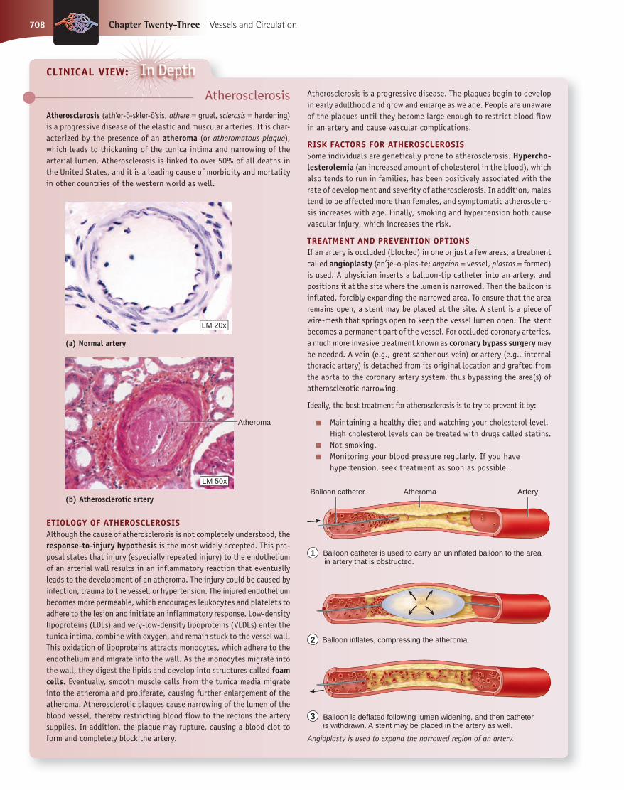
(a) Normal artery
(b) Atherosclerotic artery
Atheroma
LM 50x
LM 20x
Balloon catheter Artery Atheroma
Balloon catheter is used to carry an uninflated balloon to the area in artery that is obstructed.
Balloon is deflated following lumen widening, and then catheter is withdrawn. A stent may be placed in the artery as well.
Balloon inflates, compressing the atheroma.
1
2
3
708 Chapter Twenty-Three Vessels and Circulation
AtherosclerosisAtherosclerosis (ath′er-o -skler-o ′sis, athere = gruel, sclerosis = hardening) is a progressive disease of the elastic and muscular arteries. It is char-acterized by the presence of an atheroma (or atheromatous plaque), which leads to thickening of the tunica intima and narrowing of the arterial lumen. Atherosclerosis is linked to over 50% of all deaths in the United States, and it is a leading cause of morbidity and mortality in other countries of the western world as well.
CLINICAL VIEW: In Depth
Angioplasty is used to expand the narrowed region of an artery.
Atherosclerosis is a progressive disease. The plaques begin to develop in early adulthood and grow and enlarge as we age. People are unaware of the plaques until they become large enough to restrict blood flow in an artery and cause vascular complications.
RISK FACTORS FOR ATHEROSCLEROSISSome individuals are genetically prone to atherosclerosis. Hypercho-lesterolemia (an increased amount of cholesterol in the blood), which also tends to run in families, has been positively associated with the rate of development and severity of atherosclerosis. In addition, males tend to be affected more than females, and symptomatic atherosclero-sis increases with age. Finally, smoking and hypertension both cause vascular injury, which increases the risk.
TREATMENT AND PREVENTION OPTIONSIf an artery is occluded (blocked) in one or just a few areas, a treatment called angioplasty (an′je -o -plas-te ; angeion = vessel, plastos = formed) is used. A physician inserts a balloon-tip catheter into an artery, and positions it at the site where the lumen is narrowed. Then the balloon is inflated, forcibly expanding the narrowed area. To ensure that the area remains open, a stent may be placed at the site. A stent is a piece of wire-mesh that springs open to keep the vessel lumen open. The stent becomes a permanent part of the vessel. For occluded coronary arteries, a much more invasive treatment known as coronary bypass surgery may be needed. A vein (e.g., great saphenous vein) or artery (e.g., internal thoracic artery) is detached from its original location and grafted from the aorta to the coronary artery system, thus bypassing the area(s) of atherosclerotic narrowing.
Ideally, the best treatment for atherosclerosis is to try to prevent it by:
■ Maintaining a healthy diet and watching your cholesterol level. High cholesterol levels can be treated with drugs called statins.
■ Not smoking. ■ Monitoring your blood pressure regularly. If you have
hypertension, seek treatment as soon as possible.
ETIOLOGY OF ATHEROSCLEROSISAlthough the cause of atherosclerosis is not completely understood, the response-to-injury hypothesis is the most widely accepted. This pro-posal states that injury (especially repeated injury) to the endothelium of an arterial wall results in an inflammatory reaction that eventually leads to the development of an atheroma. The injury could be caused by infection, trauma to the vessel, or hypertension. The injured endothelium becomes more permeable, which encourages leukocytes and platelets to adhere to the lesion and initiate an inflammatory response. Low-density lipoproteins (LDLs) and very-low-density lipoproteins (VLDLs) enter the tunica intima, combine with oxygen, and remain stuck to the vessel wall. This oxidation of lipoproteins attracts monocytes, which adhere to the endothelium and migrate into the wall. As the monocytes migrate into the wall, they digest the lipids and develop into structures called foam cells. Eventually, smooth muscle cells from the tunica media migrate into the atheroma and proliferate, causing further enlargement of the atheroma. Atherosclerotic plaques cause narrowing of the lumen of the blood vessel, thereby restricting blood flow to the regions the artery supplies. In addition, the plaque may rupture, causing a blood clot to form and completely block the artery.
mck78097_ch23_683-723.indd 708mck78097_ch23_683-723.indd 708 2/14/11 4:31 PM2/14/11 4:31 PM

Chapter Twenty-Three Vessels and Circulation 709
In the cubital region, an obliquely positioned median cubital vein connects the cephalic and basilic veins. The median cubital vein is a common site for venipuncture, in which a vein is punc-tured to draw blood or inject a solute. All of these superficial veins are highly variable among individuals and have multiple superfi-cial tributaries draining into them.
Deep Venous Drainage of the Upper Limb
The digital veins and superficial and deep palmar venous archesdrain into pairs of radial veins and ulnar veins that run parallel to arteries of the same name. (Paired veins that run alongside an artery are collectively known as venae commitantes.) At the level of the cubital fossa, the radial and ulnar veins merge to form a pair of brachial veins that travel with the brachial artery. Brachial veins and the basilic vein merge to form the axillary vein. Superior to the lateral border of the first rib, the axillary vein is renamed the subclavian vein. When the subclavian vein and jugular veins of the neck merge, they form the brachiocephalic vein. As we have seen, the left and right brachiocephalic veins form the superior vena cava. The combined arteries and veins of the upper limbs are shown in cadaver photos in figure 23.19c, d.
23.3i Blood Flow Through the Lower Limb
The arterial and venous blood flow of the lower limb is very simi-lar to that of the upper limb. As we discuss lower limb blood flow, compare it with that of the upper limb.
Arterial Flow
The main arterial supply for the lower limb is the external iliac artery, which is a branch of the common iliac artery (figure 23.20a). The external iliac artery travels inferior to the inguinal ligament, where it is renamed the femoral (fem′o -ra l) artery. The deep femoral artery(profunda femoris artery or deep artery of the thigh) emerges from the femoral artery to supply the hip joint (via medial and lateral femoral circumflex arteries) and many of the thigh muscles, before travers-ing posteromedially along the thigh. The femoral artery passes through an opening in the adductor magnus muscle and enters the posteriorly placed popliteal fossa, where the vessel is renamed the popliteal (pop-lit′e -a l, pop-li-te ′a l) artery. The popliteal artery sup-plies the knee joint and muscles in this region. The popliteal artery divides into an anterior tibial (tib′e -a l) artery that supplies the anterior compartment of the leg, and a posterior tibial artery that supplies the posterior compartment of the leg. The posterior tibial artery extends a branch called the fibular (fib′u -la r) artery, which supplies the lateral compartment leg muscles. The posterior tibial artery continues to the plantar side of the foot, where it branches into medial and lateral plantar arteries.
The anterior tibial artery crosses over the anterior surface of the ankle, where it is renamed the dorsalis pedis artery. The dorsalis pedis artery and a branch of the lateral plantar artery unite to form the plantar (plan′ta r) arch of the foot. Digital arteries extend from the plantar arch and supply the toes.
WHAT DO YOU THINK?
●4 If the right femoral artery were blocked, would any blood be able to reach the right leg? Why or why not?
Venous drainage of the lower limb is through two groups of veins: superficial and deep.
Superficial Venous Drainage of the Lower Limb
On the dorsum of the foot, a dorsal venous arch drains into the great saphenous (sa -fe ′nu s) vein and the small saphenous vein(figure 23.20b). The great saphenous vein originates in the medial ankle and extends adjacent to the medial surface of the entire lower limb before it drains into the femoral vein. The small saphe-nous vein extends adjacent to the lateral ankle and then travels along the posterior calf, before draining into the popliteal vein. These superficial veins have perforating branches that connect to the deeper veins. If the valves in these veins (or the perforating branches) become incompetent, varicose veins develop (see the Clinical View on page 690).
Deep Venous Drainage of the Lower Limb
The digital veins and deep veins of the foot drain into pairs of medial and lateral plantar veins. These veins drain into a pair of posterior tibial veins. A pair of fibular veins travel alongside the fibular artery and drain into the posterior tibial veins. On the dor-sum of the foot and ankle, deep veins drain into a pair of anterior tibial veins, which traverse alongside the anterior tibial artery. The anterior and posterior tibial veins merge to form a popliteal veinthat accompanies the popliteal artery in the popliteal fossa. This vein curves to the anterior portion of the thigh and is renamed the femoral vein. Once this vein passes superior to the inguinal ligament, it is renamed once again as the external iliac vein. The external and internal iliac veins merge in the pelvis, forming the common iliac vein. Left and right common iliac veins then merge to form the inferior vena cava.
Figure 23.21 shows arteries and veins of the lower limb in the femoral region.
WHAT DID YOU LEARN?
●11 What are the superficial veins that help drain the upper limb?
●12 How does arterial blood travel through the lower limb? List the branching that occurs, beginning with the external iliac artery.
WW
mck78097_ch23_683-723.indd 709mck78097_ch23_683-723.indd 709 2/14/11 4:31 PM2/14/11 4:31 PM

External iliac artery
Femoral circumflex arteries
Fibular artery
Dorsalis pedis artery
Common iliac artery
Internal iliac artery
Inguinal ligament
Obturator artery
Femoral artery
Deep femoral artery
Posterior tibial artery
Anterior tibial artery
Lateral plantar artery
Medial plantar artery
Digital arteries
Plantar arch
Femoral circumflex arteries
Popliteal artery
Fibular artery
Posterior view Anterior view
(a) Arteries of right lower limb
710 Chapter Twenty-Three Vessels and Circulation
Figure 23.20Vascular Supply to the Lower Limb. The external iliac artery carries oxygenated blood to the lower limb; veins merge to return deoxygenated blood to the heart. (a) Anterior and posterior views of arteries distributed throughout the right lower limb. (b) Anterior and posterior views of veins that return blood from the right lower limb.
mck78097_ch23_683-723.indd 710mck78097_ch23_683-723.indd 710 2/14/11 4:31 PM2/14/11 4:31 PM

Deep femoral vein
Femoral circumflex veins
Great saphenous vein
Anterior tibial veins
Dorsal venous arch
Femoral vein
Great saphenous vein
Small saphenous vein
Popliteal vein
Posterior tibial veins
Fibular veins
Medial plantar veins
Digital veins
Femoral circumflex veins
Lateral plantar veins
Anterior view Posterior view
Common iliac vein
External iliac vein Internal iliac vein
(b) Veins of right lower limb
Fibular veins
Deep veins Superficial veins
Chapter Twenty-Three Vessels and Circulation 711
mck78097_ch23_683-723.indd 711mck78097_ch23_683-723.indd 711 2/14/11 4:31 PM2/14/11 4:31 PM

Femoral nerve
Femoral artery
Lateral femoral circumflex artery
Great saphenous vein
Medial femoral circumflex artery
Femoral vein Inguinal ligament
Spermatic cord
External iliac vein
External iliac artery
712 Chapter Twenty-Three Vessels and Circulation
Figure 23.21Vascular Supply to the Lower Limb. A cadaver photo shows major vessels of the anterior thigh.
23.4 Pulmonary CirculationLearning Objective: 1. List the pulmonary circulation vessels, and trace their
pathways.
The pulmonary circulation is responsible for carrying deoxy-genated blood from the right side of the heart to the lungs, and then returning the newly oxygenated blood to the left side of the heart (figure 23.22). Blood low in oxygen is pumped out of the right ventricle into the pulmonary trunk. This vessel travels superiorly and slightly to the left before it bifurcates into a left pulmonary artery and a right pulmonary artery that go to the lungs. The pulmonary arteries divide into smaller arteries that continue to subdivide to form arterioles. These arterioles finally branch into pulmonary capillaries, where gas exchange occurs. Carbon dioxide is removed from the blood and enters the tiny air sacs (alveoli) of the lungs, while oxygen moves in the opposite direction, from the
air sacs into the blood. The capillaries merge to form venules and then the pulmonary veins. Typically, two left and two right pulmo-nary veins carry the newly oxygenated blood to the left atrium of the heart. Compared to the systemic circulation, the vessels that make up the pulmonary circulation are relatively short. Blood doesn’t need to be pumped as far in the pulmonary circulation, since the lungs are close to the heart. In addition, the pulmonary arteries have less elastic connective tissue and wider lumens than systemic arteries. As a result, blood pressure is lower in the pulmonary arteries than in the systemic arteries, and pressure is correspondingly lower on the right side of the heart than on the left side.
WHAT DID YOU LEARN?
●13 Compare the bronchial arteries and veins (discussed earlier in this chapter) with the pulmonary arteries and veins.
WW
mck78097_ch23_683-723.indd 712mck78097_ch23_683-723.indd 712 2/14/11 4:31 PM2/14/11 4:31 PM

Right pulmonary artery
Right pulmonary veins
Right atrium
Right ventricle
(a)
(b)
Aortic arch
Left pulmonary artery
Pulmonary trunk
Left pulmonary veins
Left atrium
Left ventricle
Branch of pulmonary vein
Venule
Branch of pulmonary artery
Alveoli
Pulmonary capillaries
Arteriole
Chapter Twenty-Three Vessels and Circulation 713
Figure 23.22Pulmonary Circulation. The pulmonary circulation conducts blood from the heart to and from the gas exchange surfaces of the lungs. Blood circulation through the heart is indicated by colored arrows (blue = deoxygenated blood; red = oxygenated blood). (a) Deoxygenated blood is pumped from the right ventricle to the lungs through the pulmonary arteries. Oxygenated blood returns to the heart from the lungs within the pulmonary veins. (b) At the microscopic level, pulmonary capillaries are associated with the alveoli of the lungs.
mck78097_ch23_683-723.indd 713mck78097_ch23_683-723.indd 713 2/14/11 4:31 PM2/14/11 4:31 PM

Abdominal aneurysm
Abdominal aortic aneurysm
Plaque
Common iliac artery
Stent graft released from catheter
Catheter insterted into femoral artery
Blood flows through stent graft
Endovascular stent graft in place
Aorta
714 Chapter Twenty-Three Vessels and Circulation
23.5 Review of Heart, Systemic, and Pulmonary CirculationLearning Objectives: 1. Trace and describe the flow of blood and oxygenation of
blood in the systemic and pulmonary circulations. 2. Identify the heart chambers involved in the systemic and
pulmonary circulations.
A simplified flowchart of blood circulation is outlined in figure 23.23 and described here:
1. The systemic circulation begins when oxygenated blood flows from the left atrium to the left ventricle and then is pumped into the aorta.
2. Blood in the aorta enters elastic arteries and then flows into muscular arteries before entering arterioles.
3. Blood enters the systemic capillaries from arterioles; gases, nutrients, and wastes are exchanged in the capillaries.
4. Deoxygenated blood that is low in nutrients exits capillary beds. It drains into venules that merge to form veins.
5. Deoxygenated blood is conducted by the venous circulation to either the superior vena cava or the inferior vena cava for entry into the right atrium of the heart. Now the blood has entered the pulmonary circulation.
6. Blood flows from the right atrium into the right ventricle, and then it is pumped into the pulmonary trunk.
7. The pulmonary trunk bifurcates into left and right pulmonary arteries that carry deoxygenated blood to the lungs.
AneurysmsAn aneurysm is a localized, abnormal dilation of a blood vessel. Although an aneurysm can form in any type of vessel, aneurysms are particularly common in arteries, especially the aorta, because of the higher blood pressure on the arterial side of the circulation. After being initiated by a weakness in the wall of the vessel, an aneurysm tends to increase in size over a period of weeks or months until it ruptures.
Abdominal aortic aneurysm is a relatively common medical problem, and is most often a consequence of atherosclerosis. Most abdominal aortic aneurysms develop between the level of the renal arteries and the point near where the aorta bifurcates into the common iliac arteries. Since no pain fibers are associated with the aorta, an aneurysm can increase in size and reach the point of rupture without the patient ever being aware of it. A ruptured aorta is a surgical emergency that few people survive. An abdominal aortic aneurysm may be detected during a physical exam as a pulsating abdominal mass. X-ray and ultrasound studies can confirm the diagnosis and determine the size and extent of the aneurysm. For a number of years, aortic aneurysm repair has involved removing the dilated segment of aorta and replacing it with an artificial vascular prosthesis. This risky surgical proce-dure requires making a large abdominal incision to gain access to the dilated segment of aorta. More recently, stent grafts have been developed that can be inserted through an incision in the femoral artery, positioned in the area of the aneurysm using x-ray guidance, and then expanded to reinforce the weakened and dilated area of the aortic wall. This procedure is less invasive and traumatic than major abdominal surgery. Unfortunately, the stent graft does not always lead to a complete cure, and complications are still possible.
A berry (cerebral or saccular) aneu-rysm is a weak area in a cerebral blood vessel that balloons out and fills with blood. It is so named because it is a saclike outpocketing of a cerebral blood
CLINICAL VIEW
vessel that appears berry-shaped. Usually, berry aneurysms develop at the point where a blood vessel bifurcates because the blood vessel walls near this location are structurally weaker. Most cerebral aneurysms are located along the arteries that form the cerebral arterial circle. All cere-bral aneurysms have the potential to rupture and cause bleeding within the brain, which can cause serious complications such as hemorrhagic stroke, subarachnoid hemorrhage, nerve damage, or death. The disorder may result from congenital defects or from other conditions such as high blood pressure, atherosclerosis, head trauma, or infection. A small aneurysm generally will not produce symptoms, but a larger one that is steadily growing may press on tissues and nerves. Symptoms may include pain above and behind the eye, numbness, weakness, or paralysis on one side of the face, dilated pupils, and vision changes. When a berry aneurysm ruptures, an individual may experience a sudden and extremely severe headache (described by patients as the "worst headache of my life"), double vision, nausea, vomiting, stiff neck, and/or loss of conscious-ness. A ruptured berry aneurysm is an emergency condition; about 25% of people die within 1 day, and another 25% die within 3 to 4 months. Treatment of the aneurysm typically involves using sutures, clamps, or other materials to repair the ruptured site.
mck78097_ch23_683-723.indd 714mck78097_ch23_683-723.indd 714 2/14/11 4:31 PM2/14/11 4:31 PM

Aorta
Pulmonary capillaries of right lung
Pulmonary trunk
Superior vena cava
Right atrium
Inferior vena cava
Right ventricle
Venules
Right pulmonary artery
Systemic capillaries of head, neck, and upper limbs
Left pulmonary artery
Pulmonary capillaries of left lung
Left pulmonary veins Right pulmonary veins
Left atrium
Left ventricle Aorta
Arterioles
Systemic capillaries of trunk
and lower limbs
1
1
2
3
4
5
6
7
8
9
10
11
2
2
3
3
4
4
5 6
7
8
9 10
11
Blood passes from the aorta into elastic arteries and then into muscular arteries before entering arterioles.
The pulmonary trunk conducts deoxygenated blood into pulmonary arteries to the lungs.
The blood passes through smaller and smaller arteries before entering pulmonary capillaries for gas exchange. Oxygenated blood exits the lung via a series of progressively larger veins that merge to form the pulmonary veins.
Pulmonary veins drain into the left atrium.
The cycle repeats.
Oxygenated blood flows from the left atrium to the left ventricle and then is pumped into the aorta.
Blood in arterioles enters systemic capillaries for exchange of gases and nutrients.
Deoxygenated blood exits capillary beds into venules and then into veins.
Deoxygenated blood is conducted to either the superior or inferior vena cava and then enters the right atrium of the heart.
Pulmonary circulation (yellow arrows)
Blood flows from the right atrium to the right ventricle and is then pumped into the pulmonary trunk.
Systemic circulation (black arrows)
Systemic circulation Pulmonary circulation
Chapter Twenty-Three Vessels and Circulation 715
Figure 23.23Cardiovascular System Circulatory Routes. Blood travels through two routes: the systemic circulation and the pulmonary circulation. In the systemic circulation (black arrows), blood is pumped into the arteries, through the systemic capillary beds, and then back to the heart in systemic veins. In the pulmonary circulation (yellow arrows), blood is pumped through the pulmonary arteries to pulmonary capillary beds in the lungs and then back to the heart in pulmonary veins.
8. This blood passes through a series of smaller and smaller arteries before entering pulmonary capillaries, where gas exchange occurs.
9. Oxygenated blood exits the lung through a series of progressively larger veins that merge to form the pulmonary veins.
10. Pulmonary veins empty into the left atrium. 11. The cycle repeats.
Keep in mind that this outline is an oversimplification of complex events. For example, remember that both ventricles con-tract together. Thus, while some blood is traveling through the systemic vessels, blood is also traveling through the pulmonary vessels.
WHAT DID YOU LEARN?
●14 Oxygenated blood leaves what chamber of the heart and travels through which major vessel?
WW
23.6 Aging and the Cardiovascular SystemLearning Objective: 1. Describe the structure, function, and durability of blood
vessels during aging.
As adults get older, the heart and blood vessels become less resilient. Many of the elastic arteries are less able to withstand the forces from the pulsating blood. Systolic blood pressure may increase with age, exacerbating this problem. As a result, older individuals are more apt to develop an aneurysm (an′u-rizm; aneurysma = a dilation), whereby part of the arterial wall thins and balloons out. This wall is more prone to rupture, which can cause massive bleeding and may lead to death. In addition, as we grow older, the incidence and severity of atherosclerosis increases, at least for people living in the developed world.
WHAT DID YOU LEARN?
●15 How are aging and blood pressure related?
WW
mck78097_ch23_683-723.indd 715mck78097_ch23_683-723.indd 715 2/14/11 4:31 PM2/14/11 4:31 PM

Aortic arch vessels
Right dorsal aorta
Common dorsal aorta
Regressing arches
Aortic sac
Truncus arteriosus
Left dorsal aorta
(a) Late week 4 to early week 5: Paired aortic arch vessels connect to left and right dorsal aortae
(c) Week 8: Aortic arch and branches formed(b) Week 7: Right dorsal aorta degenerates; left dorsal aorta becomes descending thoracic aorta
Right subclavianartery
Right commoncarotid artery
Brachiocephalic trunk
Pulmonary artery
Pulmonary trunk
Left subclavianartery
Pulmonary arteries
Common dorsal aorta
Subclavian artery
Degenerating rightdorsal aorta
Ductus arteriosus
Pulmonaryartery
Descending thoracic aorta
Ductus arteriosus
Left dorsal aorta (becomes descending thoracic aorta)
Left commoncarotid artery
Aortic arch
Small part of maxillary arteries
Small part of stapedial arteries
Left and right common carotid arteries
Right vessel: proximal part of right subclavian arteryLeft vessel: aortic arch (connects to the left dorsal aorta)
Right vessel: right pulmonary arteryLeft vessel: left pulmonary artery and ductus arteriosus
Postnatal Structure Formed by VesselsAortic
Arch Vessel
1
2
3
4
6
1
2
3
4
6
1
2
3
4
6
3
4
6
3
4
6
3
4
6
3
4
6
716 Chapter Twenty-Three Vessels and Circulation
Figure 23.24Thoracic Artery Development. Aortic arch vessels form most of the major thoracic and head and neck arteries. (a) By late week 4/early week 5, paired aortic arch vessels arise from the truncus arteriosus and attach to paired left and right dorsal aortae. (b) During week 7, the right dorsal artery starts to degenerate, and the left dorsal aorta becomes the descending thoracic aorta. (c) By week 8, the aortic arch vessels have undergone remodeling to form the aortic arch, major branches of the arch, and the pulmonary arteries.
23.7 Blood Vessel DevelopmentLearning Objectives: 1. Explain the developmental fates of the embryonic vessels. 2. Compare and contrast the fetal and postnatal circulatory
patterns.
The heart and its blood vessels begin to develop in the embryo during the third week. The blood vessels form by a process called vasculogenesis (vas′ku -lo -jen′e -sis; vasculum = small vessel, genesis = production), whereby some of the mesoderm forms cells called angioblasts, and these angioblasts connect to form the first primitive blood vessels. These vessels then grow and invade devel-oping tissues throughout the embryo.
23.7a Artery Development
The embryo initially has both a left and right dorsal aorta(figure 23.24a). These two vessels remain separate until the level
of the fourth thoracic vertebra, where they fuse to form a common dorsal aorta that supplies blood to the inferior part of the body. Eventually, the superior part of the right dorsal aorta degener-ates and disappears, leaving the left dorsal aorta and common dor-sal aorta (figure 23.24b, c). The left dorsal aorta and common dorsal aorta form the descending thoracic aorta. Beginning the fourth week, the truncus arteriosus of the heart connects to the left and right dorsal aortae by a series of paired aortic arch vessels (figure 23.24a). These vessels are numbered 1–6. Aortic arch vessels 1 and 2 primarily regress, and only a small portion of them remains to form small segments of arteries in the head. Aortic arch vessel 5 never forms in humans. The remaining aortic arch vessels—3, 4, and 6—develop into adult arteries (figure 23.24b, c). In addition, the most superior part of the truncus arteriosus (called the aortic sac) forms the brachiocephalic trunk. The dorsal aorta (now part of the descending aorta) develops vascular “sprouts” that form many of the blood vessels in the body. These blood vessel sprouts grow and migrate to the areas that need vascularization.
mck78097_ch23_683-723.indd 716mck78097_ch23_683-723.indd 716 2/14/11 4:31 PM2/14/11 4:31 PM
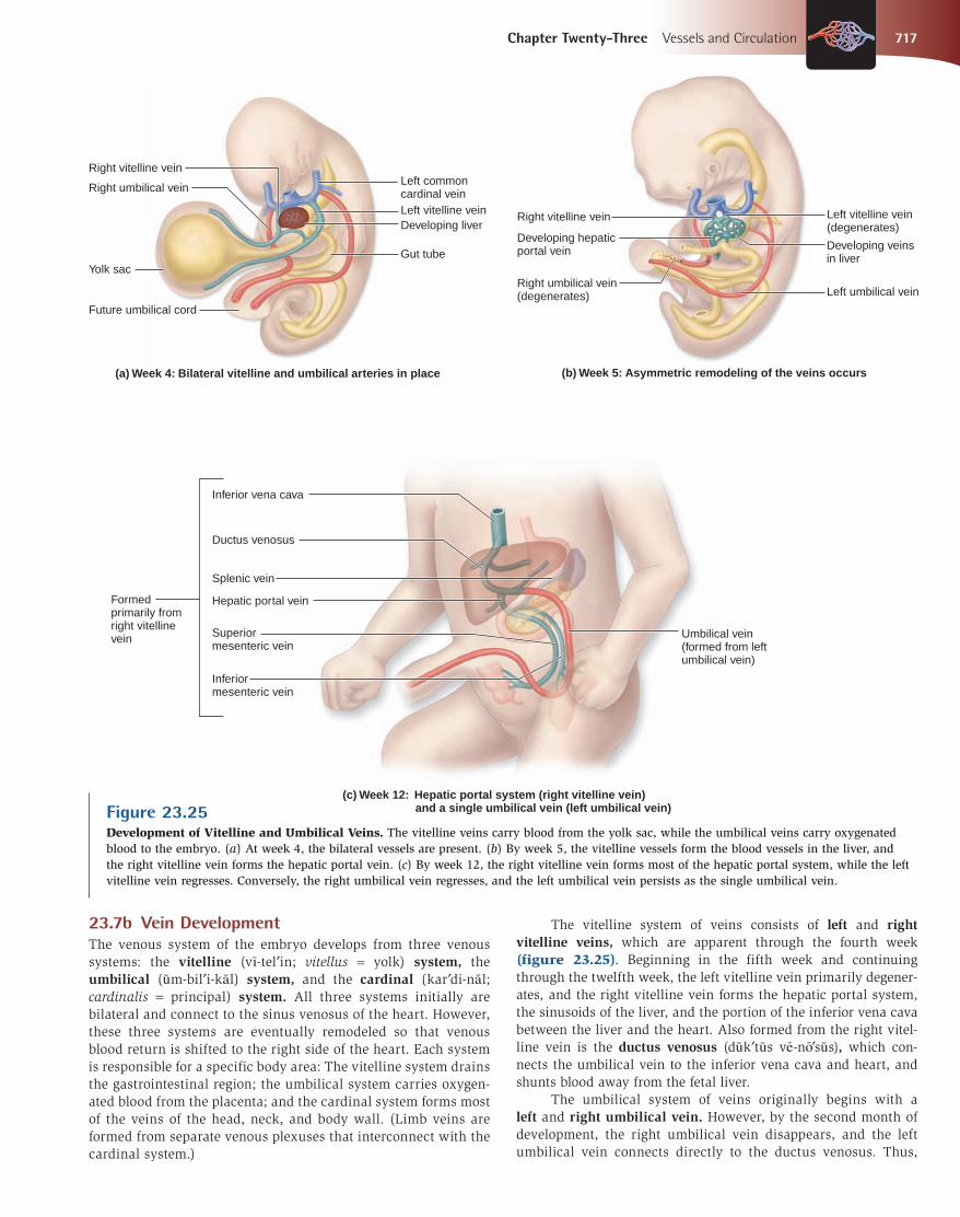
Right vitelline vein
Developing liver
Yolk sac
Right umbilical vein
Future umbilical cord
Left common cardinal vein Left vitelline vein
Gut tube
(a) Week 4: Bilateral vitelline and umbilical arteries in place (b) Week 5: Asymmetric remodeling of the veins occurs
(c) Week 12: Hepatic portal system (right vitelline vein) and a single umbilical vein (left umbilical vein)
Right vitelline vein
Developing veins in liver
Right umbilical vein (degenerates)
Developing hepatic portal vein
Left vitelline vein (degenerates)
Left umbilical vein
Inferior vena cava
Ductus venosus
Hepatic portal vein
Splenic vein
Superior mesenteric vein
Formed primarily from right vitelline vein Umbilical vein
(formed from left umbilical vein)
Inferior mesenteric vein
Chapter Twenty-Three Vessels and Circulation 717
23.7b Vein Development
The venous system of the embryo develops from three venous systems: the vitelline (vı -tel′in; vitellus = yolk) system, the umbilical (u m-bil′i-ka l) system, and the cardinal (kar′di-na l; cardinalis = principal) system. All three systems initially are bilateral and connect to the sinus venosus of the heart. However, these three systems are eventually remodeled so that venous blood return is shifted to the right side of the heart. Each system is responsible for a specific body area: The vitelline system drains the gastrointestinal region; the umbilical system carries oxygen-ated blood from the placenta; and the cardinal system forms most of the veins of the head, neck, and body wall. (Limb veins are formed from separate venous plexuses that interconnect with the cardinal system.)
The vitelline system of veins consists of left and right vitelline veins, which are apparent through the fourth week (figure 23.25). Beginning in the fifth week and continuing through the twelfth week, the left vitelline vein primarily degener-ates, and the right vitelline vein forms the hepatic portal system, the sinusoids of the liver, and the portion of the inferior vena cava between the liver and the heart. Also formed from the right vitel-line vein is the ductus venosus (du k′tu s ve -no ′su s), which con-nects the umbilical vein to the inferior vena cava and heart, and shunts blood away from the fetal liver. The umbilical system of veins originally begins with a left and right umbilical vein. However, by the second month of development, the right umbilical vein disappears, and the left umbilical vein connects directly to the ductus venosus. Thus,
Figure 23.25Development of Vitelline and Umbilical Veins. The vitelline veins carry blood from the yolk sac, while the umbilical veins carry oxygenated blood to the embryo. (a) At week 4, the bilateral vessels are present. (b) By week 5, the vitelline vessels form the blood vessels in the liver, and the right vitelline vein forms the hepatic portal vein. (c) By week 12, the right vitelline vein forms most of the hepatic portal system, while the left vitelline vein regresses. Conversely, the right umbilical vein regresses, and the left umbilical vein persists as the single umbilical vein.
mck78097_ch23_683-723.indd 717mck78097_ch23_683-723.indd 717 2/14/11 4:31 PM2/14/11 4:31 PM

Left anterior cardinal vein
Superior vena cava
Right vitelline portion of inferior vena cava Subcardinal portion of inferior vena cava
Supracardinal portion of inferior vena cava
Posterior cardinal portion of inferior vena cava
(a) Week 8: Supra- and subcardinal veins undergo asymmetric remodeling
(b) Birth: Mature vessel pattern formed
Right brachiocephalic vein
Superior vena cava Right subclavian vein
Inferior vena cava
Internal jugular vein External jugular vein
Left brachiocephalic vein
Left subclavian vein
Intercostal vein
Azygos vein
Hemiazygos vein
Renal vein
Gonadal vein Common iliac vein
Supracardinal vein
Degenerating posterior cardinal vein
Subclavian vein
718 Chapter Twenty-Three Vessels and Circulation
within the umbilical cord are one umbilical vein and a pair of umbilical arteries. The cardinal system of veins consists of a series of paired veins: the anterior cardinal veins, posterior cardinal veins, supracardinal veins, and subcardinal veins (figure 23.26). The anterior cardinal veins develop into the veins of the head and neck and the veins superior to the heart. By the eighth week of development, the posterior cardinal veins degenerate and are largely replaced by the supracardinal and subcardinal veins. The supracardinal and subcardinal veins undergo asymmetrical remodeling, whereby venous blood flow is shifted to the right
side of the body. The subcardinal veins form veins that drain the posterior abdominal wall, while the supracardinal veins form the hemiazygos and azygos system of veins. The inferior vena cava is formed from parts of the right vitelline, right subcardi-nal, right supracardinal, and right posterior cardinal veins. The mature vessel pattern is formed well before birth and shown in figure 23.26b.
23.7c Comparison of Fetal and Postnatal Circulation
The cardiovascular system of the fetus is structurally and func-tionally different from that of the newborn. Whereas the fetus
Figure 23.26Cardinal Vein Development. The primary venous drainage for the embryo is initially formed by the cardinal veins. (a) By week 8, some of these cardinal veins undergo asymmetric remodeling and begin to form some of the named veins in the body. (b) At birth, the cardinal veins have been remodeled to form the inferior vena cava and the posterior thoracic and abdominal wall vessels.
mck78097_ch23_683-723.indd 718mck78097_ch23_683-723.indd 718 2/14/11 4:31 PM2/14/11 4:31 PM

Right atrium Right ventricle
Aortic arch
Superior vena cava
Lung
Ductus arteriosus Pulmonary artery
Pulmonary veins Pulmonary trunk
Heart
Foramen ovale
Liver
Ductus venosus
Umbilical vein Umbilicus (not visible)
Inferior vena cava
Descending abdominal aorta
Common iliac artery
Internal iliac artery
Umbilical arteries
Urinary bladder
Umbilical cord
Placenta
Ductus arteriosus
Ductus venosus
Foramen ovale
Umbilical arteries
Umbilical vein
Ligamentum arteriosum
Ligamentum venosum
Fossa ovalis
Medial umbilical ligaments
Round ligament of liver (ligamentum teres)
Postnatal Structure Fetal Cardiovascular Structure
1
2
3
4 5
6
8
7
Chapter Twenty-Three Vessels and Circulation 719
Figure 23.27Fetal Circulation. Structural changes in both the heart and blood vessels accommodate the different needs of the fetus and the newborn. The pathway of blood flow is indicated by black arrows. The numbers correspond to the steps listed in the text. The chart at the bottom of the drawing summarizes the fate of each of the fetal cardiovascular structures.
receives oxygen and nutrients directly from the mother through the placenta, its postnatal cardiovascular system is indepen-dent. In addition, since the fetal lungs are not functional, the blood pressure in the pulmonary arteries and right side of the heart is greater than the pressure in the left side of the heart. Finally, several fetal vessels help shunt blood directly to the organs in need and away from the organs that are not yet func-tional. As a result, the fetal cardiovascular system has some structures that are modified or that cease to function once the human is born. Figure 23.27 compares the fetal and postnatal circulation patterns. Fetal circulation occurs as follows:
1. Oxygenated blood from the placenta enters the body of the fetus through the umbilical vein.
2. The blood from the umbilical vein is shunted away from the liver and directly toward the inferior vena cava through the ductus venosus.
3. Oxygenated blood in the ductus venosus mixes with deoxygenated blood in the inferior vena cava.
4. Blood from the superior and inferior venae cavae empties into the right atrium.
5. Since pressure is greater on the right side of the heart (compared to the left side), most of the blood is shunted from the right atrium to the left atrium via the foramen ovale. This blood flows into the left ventricle and then is pumped out through the aorta.
6. A small amount of blood enters the right ventricle and pulmonary trunk, but much of this blood is shunted from the pulmonary trunk to the aorta through a vessel detour called the ductus arteriosus (ar-te r′e -o ′su s).
7. Blood travels to the rest of the body, and the deoxygenated blood returns to the placenta through a pair of umbilical arteries.
8. Nutrient and gas exchange occurs at the placenta, and the cycle repeats.
At birth, the fetal circulation begins to change into the post-natal pattern. When the baby takes its first breath, pulmonary resistance drops, and the pulmonary arteries dilate. As a result,
Patent Ductus ArteriosusIn some infants (especially premature infants), the ductus arte-riosus fails to constrict and close after birth. This open (patent) ductus arteriosus occurs in approximately 8 per 10,000 births. Since the systemic circulation is under higher pressure than the pulmonary circulation, a patent ductus arteriosus serves as a conduit through which blood from the aorta can enter the pul-monary system. If left untreated, this shunting will, over a period of several years, result in high blood pressure in the pulmonary circuit. This pulmonary hypertension then leads to failure of the right ventricle. Because circulating chemicals called prostaglan-dins help keep the ductus arteriosus open during fetal life, the first form of treatment for a patent ductus arteriosus is prostaglandin-inhibiting medication. In the uncommon instance that medication does not work, the ductus arteriosus is surgically repaired.
CLINICAL VIEW
mck78097_ch23_683-723.indd 719mck78097_ch23_683-723.indd 719 2/14/11 4:31 PM2/14/11 4:31 PM

720 Chapter Twenty-Three Vessels and Circulation
pressure on the right side of the heart decreases so that the pres-sure is greater on the left side of the heart, which handles the systemic circulation. The postnatal changes occur as follows:
■ The umbilical vein and umbilical arteries constrict and become nonfunctional. They turn into the round ligament of the liver (or ligamentum [lig′a -men′tu m; band] teres) and the medial umbilical ligaments, respectively.
■ The ductus venosus ceases to be functional and constricts, becoming the ligamentum venosum (ve -no ′su m).
■ Since pressure is now greater on the left side of the heart, the two flaps of the interatrial septum close off the
foramen ovale. The only remnant of the foramen ovale is a thin, oval depression in the wall of the septum called the fossa ovalis.
■ Within 10 to 15 hours after birth, the ductus arteriosus closes and becomes a fibrous structure called the ligamentum arteriosum.
WHAT DID YOU LEARN?
●16 The hepatic portal system is formed primarily from what embryonic vein system?
●17 Each medial umbilical ligament is a remnant of what embryonic vessel?
WW
Clinical Terms
edema Noticeable swelling from fluid accumulation in body tissues. Edema most commonly occurs in the feet and legs, where it is referred to as peripheral edema.
hypotension Low blood pressure.Korotkoff (kô-rot'kôf) sounds Distinctive sounds heard through a
stethoscope when taking a blood pressure reading, resulting from blood turbulence in the artery. The sounds are first heard when the cuff pressure equals the systolic pressure and cease to be heard once the cuff has deflated past the diastolic pressure.
orthostatic hypotension Also known as a dizzy spell; a form of hypotension in which a person's blood pressure suddenly
and temporarily falls when the person stands up quickly or stretches after standing. The pressure decrease is often most pronounced after lying down.
vasculitis (vas-ku -lı ′tis) Inflammation of any type of blood vessel. If only arteries are inflamed, the condition is called arteritis; if only veins are inflamed, it is called phlebitis.
vasodilators Drugs that relax the smooth muscle in and/or around blood vessels, causing the vessels to dilate. Dilation of arteries leads to a reduction in systemic vascular resistance, which produces a fall in arterial blood pressure. Dilation of venous vessels decreases venous blood pressure.
■ Blood vessels form a closed supply system to transport oxygen and nutrients to body tissues, and remove waste products from these tissues.
23.1 Anatomy of
Blood Vessels 684
■ Arteries conduct blood away from the heart; capillaries exchange gases, nutrients, and wastes with body tissues; and veins conduct blood to the heart.
23.1a Blood Vessel Tunics 684
■ The tunica intima (innermost layer) is composed of an endothelium, a basement membrane, and a layer of areolar connective tissue.
■ The tunica media (middle layer) is composed of smooth muscle. This is the largest tunic in an artery.
■ The tunica externa (outermost layer) is composed of areolar connective tissue and adipose connective tissue. This is the largest tunic in a vein.
■ Capillaries have a tunica intima, composed of an endothelial layer and a basement membrane only.
23.1b Arteries 685
■ Elastic arteries have the largest diameter and the greatest proportion of elastic fibers in their walls.
■ Muscular arteries are medium-sized arteries with more smooth muscle and fewer elastic fibers to ensure vasodilation and vasoconstriction.
■ Arterioles are the smallest arteries.
23.1c Capillaries 688
■ Capillaries, the smallest blood vessels, connect arterioles with venules. Gas and nutrient exchange occurs in the capillaries.
■ The three types of capillaries are continuous capillaries, fenestrated capillaries, and sinusoids.
23.1d Veins 689
■ Venules are small veins that merge into larger veins. Blood pressure is low in the veins, which act as reservoirs and hold about 60% of the body’s blood at rest.
■ One-way valves prevent blood backflow in veins.
23.2 Blood
Pressure 691
■ Blood pressure is the force exerted by the blood on the vessel wall. Systolic blood pressure is a measure of pressure during ventricular contraction, and diastolic pressure is a measure of pressure during ventricular relaxation.
Chapter Summary
mck78097_ch23_683-723.indd 720mck78097_ch23_683-723.indd 720 2/14/11 4:32 PM2/14/11 4:32 PM

Chapter Twenty-Three Vessels and Circulation 721
23.3 Systemic
Circulation 692
■ The systemic circulation conducts oxygenated blood to and deoxygenated blood from peripheral capillary beds.
23.3a General Arterial Flow Out of the Heart 693
■ The ascending aorta gives off the left and right coronary arteries to supply the heart.
■ The aortic arch has three branches: the brachiocephalic trunk, the left common carotid artery, and the left subclavian artery.
■ The descending thoracic aorta extends several branches to supply the thoracic wall.
■ The descending abdominal aorta bifurcates into common iliac arteries; these vessels divide into internal and external iliac arteries.
23.3b General Venous Return to the Heart 693
■ Deoxygenated blood returns to the heart via the superior and inferior venae cavae.
23.3c Blood Flow Through the Head and Neck 693
■ Common carotid arteries branch into the internal and external carotid arteries, which supply most of the blood to the head and neck.
■ The cerebral arterial circle is an arterial anastomosis that supplies the brain.
■ Vertebral veins and the dural venous sinuses drain the cranium.
23.3d Blood Flow Through the Thoracic and Abdominal Walls 697
■ The thoracic and abdominal walls are supplied by paired arteries.
■ Hemiazygos and accessory hemiazygos veins drain the left side of the thorax, and the azygos vein drains the right side of the thorax.
23.3e Blood Flow Through the Thoracic Organs 700
■ Bronchial arteries and bronchial veins supply the connective tissue, bronchi, and bronchioles of the lung.
■ Esophageal arteries and veins supply the esophagus.
■ Superior phrenic arteries, the musculophrenic arteries, and the inferior phrenic arteries and veins supply the diaphragm.
23.3f Blood Flow Through the Gastrointestinal Tract 701
■ Three unpaired arteries supply the gastrointestinal tract organs: the celiac trunk, the superior mesenteric artery, and the inferior mesenteric artery.
■ The hepatic portal vein is a large vein that receives oxygen-poor but nutrient-rich blood from the gastrointestinal organs and takes it to the liver. Blood exits the liver via hepatic veins.
23.3g Blood Flow Through the Posterior Abdominal Organs, Pelvis, and Perineum 705
■ Paired branches of the descending abdominal aorta supply the posterior abdominal organs and the pelvis and perineum. Venous drainage is by veins of the same name as the arteries.
23.3h Blood Flow Through the Upper Limb 705
■ The subclavian artery continues as the axillary artery and then becomes the brachial artery. The brachial artery divides into an ulnar artery and a radial artery.
■ Anastomoses of ulnar and radial arteries form the superficial palmar arch and the deep palmar arch; digital arteries emerge from the arches to supply the fingers.
■ The superficial group of veins contains the basilic, median cubital, and cephalic veins that drain into the axillary vein. The deep group of veins contains veins that bear the same names as the arteries.
23.3i Blood Flow Through the Lower Limb 709
■ The external iliac artery extends inferior to the inguinal ligament and is renamed the femoral artery. It enters the popliteal fossa, and then becomes the popliteal artery before dividing into anterior and posterior tibial arteries. The posterior tibial artery gives off a fibular artery. The posterior tibial artery branches into medial and lateral plantar arteries.
■ The superficial group of veins includes the great saphenous vein and the small saphenous vein. The deep group of veins consists of veins that bear the same names as the corresponding arteries.
23.4 Pulmonary
Circulation 712
■ The pulmonary circulation carries deoxygenated blood to the lungs and returns oxygenated blood to the heart.
23.5 Review of
Heart, Systemic, and
Pulmonary
Circulation 714
■ Oxygenated blood is pumped from the left ventricle through the systemic circulation and back to the right side of the heart. This deoxygenated blood is pumped from the right ventricle through the pulmonary circulation and returns as oxygenated blood to the left side of the heart.
23.6 Aging and the
Cardiovascular
System 715
■ As adults get older, the heart and blood vessels become less resilient, systolic blood pressure may rise, and the incidence and severity of atherosclerosis increase.
(continued on next page)
mck78097_ch23_683-723.indd 721mck78097_ch23_683-723.indd 721 2/14/11 4:32 PM2/14/11 4:32 PM

722 Chapter Twenty-Three Vessels and Circulation
MatchingMatch each numbered item with the most closely related lettered item.
______ 1. hepatic portal vein
______ 2. capillary
______ 3. median cubital vein
______ 4. common iliac artery
______ 5. dural venous sinus
______ 6. azygos vein
______ 7. hemiazygos vein
______ 8. popliteal artery
______ 9. brachiocephalic trunk
______ 10. pulmonary vein
a. common site for venipuncture
b. drains venous blood from the brain
c. drains right posterior intercostal veins
d. sends oxygenated blood to right upper limb
e. continuation of femoral artery
f. drains directly into left atrium
g. composed of endothelium and basement membrane only
h. bifurcation of descending abdominal aorta
i. superior mesenteric vein drains into it
j. left posterior intercostal veins drain into it
Multiple ChoiceSelect the best answer from the four choices provided.
______ 1. Which of the following is not a type of capillary? a. continuous b. sinusoid c. elastic d. fenestrated
______ 2. Some venous blood from the upper limb drains through the
a. cephalic vein. b. great saphenous vein. c. external jugular vein. d. inferior vena cava.
Challenge Yourself
______ 3. All of the following are direct branches of the celiac trunk except the
a. splenic artery. b. right gastric artery. c. left gastric artery. d. common hepatic artery.
______ 4. Which type of vessel has a large number of smooth muscle cell layers in its tunica media as well as elastic tissue confined to an internal elastic lamina and external elastic lamina?
a. elastic artery b. muscular artery c. arteriole d. venule
______ 5. Which statement is true about veins? a. Veins always carry deoxygenated blood. b. Veins drain into smaller vessels called venules. c. The largest tunic in a vein is the tunica externa. d. The lumen of a vein tends to be smaller than that
of a comparably sized artery.
______ 6. Circle the correct pathway that blood follows through the upper limb arteries:
a. subclavian → axillary → ulnar → radial → brachial
b. subclavian → axillary → brachial → cephalic → basilic
c. subclavian → ulnar → brachial → radial d. subclavian → axillary → brachial → radial and
ulnar
______ 7. Which of the following veins typically does not drain directly into the inferior vena cava?
a. renal b. hepatic portal c. common iliac d. right gonadal
______ 8. After birth, the umbilical vein becomes the a. medial umbilical ligament. b. ligamentum venosum. c. ligamentum arteriosum. d. round ligament of the liver.
Chapter Summary (continued)
23.7 Blood Vessel
Development 716
■ The blood vessels form by a process called vasculogenesis beginning in the third week of development.
23.7a Artery Development 716
■ The right dorsal aorta regresses, and the left dorsal aorta (plus the common aorta) form the descending aorta.
■ Aortic arch vessels 1, 2, 3, 4, and 6 develop into parts of adult arteries in the head, neck, and thorax.
23.7b Vein Development 717
■ Three bilateral venous systems connect the sinus venosus of the heart: the vitelline system, the umbilical system, and the cardinal system. These are the origin of the venous system.
23.7c Comparison of Fetal and Postnatal Circulation 718
■ The fetal cardiovascular system contains some structures that are modified or cease to function once the baby is born.
mck78097_ch23_683-723.indd 722mck78097_ch23_683-723.indd 722 2/14/11 4:32 PM2/14/11 4:32 PM

Chapter Twenty-Three Vessels and Circulation 723
______ 9. The left fourth aortic arch vessel in an embryo becomes the
a. left common carotid artery. b. left subclavian artery. c. aortic arch. d. left pulmonary artery.
______ 10. Vasa vasorum are found in the tunica ______ of a large blood vessel.
a. intima b. media c. externa d. All of these are correct.
Content Review 1. List and describe the three tunics in most blood vessels.
2. Compare and contrast arteries and veins with respect to function, tunic size, and lumen size.
3. Describe the three types of arteries, and give an example of each.
4. What is the main function of capillaries? What are the three kinds of capillaries?
5. Is blood pressure higher in arteries or veins? What are the consequences of hypertension?
6. Identify the three main branches of the aortic arch that receive oxygenated blood, and identify the areas of the body they supply.
7. How is blood flow through the upper and lower limbs similar?
8. Compare the systemic and pulmonary circulations. Discuss the function of arteries and veins in each system.
9. How does aging affect blood vessel anatomy and function?
10. What postnatal changes occur in the heart and blood vessels? Why do these occur?
Developing Critical Reasoning 1. Two 50-year-old men are trying to determine their risk for
developing atherosclerosis. John jogs three times a week, maintains a healthy weight, and eats a diet low in saturated fats. Thomas rarely exercises, is overweight, and only occasionally eats healthy meals. Based on your knowledge of the cardiovascular system and atherosclerosis, which man do you think is more at risk for developing the disease? What other factors could put a person at risk for atherosclerosis?
2. Arteries tend to have a lot of vascular anastomoses around body joints (such as the elbow and knee). Propose a reason why this would be beneficial.
3. The internal thoracic artery is frequently used as a coronary bypass vessel (a replacement artery for a blocked coronary artery). What makes this vessel a good choice for this surgery? Will blood flow to the thoracic wall be compromised as a result? Why or why not?
Answers to “What Do You Think?”
1. A smoker would have elevated blood pressure, since nicotine increases cardiac output and causes vasoconstriction.
2. Blood could still reach the brain through the vertebral arteries. However, it is unlikely that these arteries could provide sufficient blood to the entire brain and head.
3. If the left ulnar artery were cut, the left hand and fingers could still receive blood via the left radial artery.
4. If the right femoral artery were blocked, blood flow to the right leg would be cut off; in other words, the popliteal artery and the branches to the leg would not receive any blood.
www.mhhe.com/mckinley3 Enhance your study with practice tests and activities to assess your understanding. Your instructor may also recommend the interactive eBook, individualized learning tools, and more.
mck78097_ch23_683-723.indd 723mck78097_ch23_683-723.indd 723 2/14/11 4:32 PM2/14/11 4:32 PM
Copyright © 2022 FDOKUMEN