Predicting Genetic Interactions in Caenorhabditis elegans ...

Predicting the Proteins of Angomonas deanei,Strigomonas culicis and Their Respective EndosymbiontsReveals New Aspects of the Trypanosomatidae FamilyMaria Cristina Machado Motta1, Allan Cezar de Azevedo Martins1, Silvana Sant’Anna de Souza1,2, Carolina
Moura Costa Catta-Preta1, Rosane Silva2, Cecilia Coimbra Klein3,4,5, Luiz Gonzaga Paula de Almeida3,
Oberdan de Lima Cunha3, Luciane Prioli Ciapina3, Marcelo Brocchi6, Ana Cristina Colabardini7, Bruna de
Araujo Lima6, Carlos Renato Machado9, Celia Maria de Almeida Soares10, Christian Macagnan Probst11,12,
Claudia Beatriz Afonso de Menezes13, Claudia Elizabeth Thompson3, Daniella Castanheira Bartholomeu14,
Daniela Fiori Gradia11, Daniela Parada Pavoni12, Edmundo C. Grisard15, Fabiana Fantinatti-Garboggini13,
Fabricio Klerynton Marchini12, Gabriela Flavia Rodrigues-Luiz14, Glauber Wagner15, Gustavo
Henrique Goldman7, Juliana Lopes Rangel Fietto16, Maria Carolina Elias17, Maria Helena S. Goldman18,
Marie-France Sagot4,5, Maristela Pereira10, Patrıcia H. Stoco15, Rondon Pessoa de Mendonca-Neto9,
Santuza Maria Ribeiro Teixeira9, Talles Eduardo Ferreira Maciel16, Tiago Antonio de Oliveira Mendes14,
Turan P. Urmenyi2, Wanderley de Souza1, Sergio Schenkman19*, Ana Tereza Ribeiro de Vasconcelos3*
1 Laboratorio de Ultraestrutura Celular Hertha Meyer, Instituto de Biofısica Carlos Chagas Filho, Universidade Federal do Rio de Janeiro, Rio de Janeiro, Rio de Janeiro, Brazil,
2 Laboratorio de Metabolismo Macromolecular Firmino Torres de Castro, Instituto de Biofısica Carlos Chagas Filho, Universidade Federal do Rio de Janeiro, Rio de Janeiro, Rio de
Janeiro, Brazil, 3 Laboratorio Nacional de Computacao Cientıfica, Laboratorio de Bioinformatica, Petropolis, Rio de Janeiro, Brazil, 4 BAMBOO Team, INRIA Grenoble-Rhone-Alpes,
Villeurbanne, France, 5 Laboratoire de Biometrie et Biologie Evolutive, Universite de Lyon, Universite Lyon 1, CNRS, UMR5558, Villeurbanne, France, 6 Departamento de Genetica,
Evolucao e Bioagentes, Instituto de Biologia, Universidade Estadual de Campinas, Campinas, Sao Paulo, Brazil, 7 Departamento de Ciencias Farmaceuticas, Faculdade de Ciencias
Farmaceuticas de Ribeirao Preto, Universidade de Sao Paulo, Ribeirao Preto, Sao Paulo, Brazil, 8 Laboratorio Nacional de Ciencia e Tecnologia do Bioetanol, Campinas, Sao Paulo,
Brazil, 9 Departamento de Bioquımica e Imunologia, Instituto de Ciencias Biologicas, Universidade Federal de Minas Gerais, Belo Horizonte, Minas Gerais, Brazil, 10 Laboratorio de
Biologia Molecular, Instituto de Ciencias Biologicas, Universidade Federal de Goias, Goiania, Goias, Brazil, 11 Laboratorio de Biologia Molecular de Tripanossomatıdeos, Instituto
Carlos Chagas/Fundacao Oswaldo Cruz, Curitiba, Parana, Brazil, 12 Laboratorio de Genomica Funcional, Instituto Carlos Chagas/Fundacao Oswaldo Cruz, Curitiba, Parana, Brazil,
13 Centro Pluridisciplinar de Pesquisas Quımicas, Biologicas e Agrıcolas, Universidade Estadual de Campinas, Campinas, Sao Paulo, Brazil, 14 Departamento de Parasitologia,
Instituto de Ciencias Biologicas, Universidade Federal de Minas Gerais, Belo Horizonte, Minas Gerais, Brazil, 15 Laboratorios de Protozoologia e de Bioinformatica, Departamento de
Microbiologia, Imunologia e Parasitologia, Centro de Ciencias Biologicas, Universidade Federal de Santa Catarina, Florianopolis, Santa Catarina, Brazil, 16 Departamento de
Bioquımica e Biologia Molecular, Centro de Ciencias Biologicas e da Saude, Universidade Federal de Vicosa, Vicosa, Minas Gerais, Brazil, 17 Laboratorio Especial de Ciclo Celular,
Instituto Butantan, Sao Paulo, Sao Paulo, Brazil, 18 Departamento de Biologia, Faculdade de Filosofia, Ciencias e Letras de Ribeirao Preto, Universidade de Sao Paulo, Ribeirao Preto,
Sao Paulo, Brazil, 19 Departamento de Microbiologia, Imunologia e Parasitologia, Escola Paulista de Medicina, Universidade Federal de Sao Paulo, Sao Paulo, Sao Paulo, Brazil
Abstract
Endosymbiont-bearing trypanosomatids have been considered excellent models for the study of cell evolution because thehost protozoan co-evolves with an intracellular bacterium in a mutualistic relationship. Such protozoa inhabit a singleinvertebrate host during their entire life cycle and exhibit special characteristics that group them in a particularphylogenetic cluster of the Trypanosomatidae family, thus classified as monoxenics. In an effort to better understand suchsymbiotic association, we used DNA pyrosequencing and a reference-guided assembly to generate reads that predicted16,960 and 12,162 open reading frames (ORFs) in two symbiont-bearing trypanosomatids, Angomonas deanei (previouslynamed as Crithidia deanei) and Strigomonas culicis (first known as Blastocrithidia culicis), respectively. Identification of eachORF was based primarily on TriTrypDB using tblastn, and each ORF was confirmed by employing getorf from EMBOSS andNewbler 2.6 when necessary. The monoxenic organisms revealed conserved housekeeping functions when compared toother trypanosomatids, especially compared with Leishmania major. However, major differences were found in ORFscorresponding to the cytoskeleton, the kinetoplast, and the paraflagellar structure. The monoxenic organisms also contain alarge number of genes for cytosolic calpain-like and surface gp63 metalloproteases and a reduced number ofcompartmentalized cysteine proteases in comparison to other TriTryp organisms, reflecting adaptations to the presence ofthe symbiont. The assembled bacterial endosymbiont sequences exhibit a high A+T content with a total of 787 and 769ORFs for the Angomonas deanei and Strigomonas culicis endosymbionts, respectively, and indicate that these organismshold a common ancestor related to the Alcaligenaceae family. Importantly, both symbionts contain enzymes thatcomplement essential host cell biosynthetic pathways, such as those for amino acid, lipid and purine/pyrimidinemetabolism. These findings increase our understanding of the intricate symbiotic relationship between the bacterium andthe trypanosomatid host and provide clues to better understand eukaryotic cell evolution.
PLOS ONE | www.plosone.org 1 April 2013 | Volume 8 | Issue 4 | e60209

Citation: Motta MCM, Martins ACdA, de Souza SS, Catta-Preta CMC, Silva R, et al. (2013) Predicting the Proteins of Angomonas deanei, Strigomonas culicis andTheir Respective Endosymbionts Reveals New Aspects of the Trypanosomatidae Family. PLoS ONE 8(4): e60209. doi:10.1371/journal.pone.0060209
Editor: John Parkinson, Hospital for Sick Children, Canada
Received October 16, 2012; Accepted February 22, 2013; Published April 3, 2013
Copyright: � 2013 Motta et al. This is an open-access article distributed under the terms of the Creative Commons Attribution License, which permitsunrestricted use, distribution, and reproduction in any medium, provided the original author and source are credited.
Funding: This work was supported by Fundacao Carlos Chagas Filho de Amparo a Pesquisa do Estado do Rio de Janeiro (FAPERJ), Fundacao de Amparo aPesquisa do Estado de Sao Paulo (FAPESP) and Conselho Nacional de Desenvolvimento Cientıfico e Tecnologico (CNPq). The work of CCK as part of her PhD isfunded by the ERC AdG SISYPHE coordinated by MFS. The funders had no role in study design, data collection and analysis, decision to publish, or preparation ofthe manuscript.
Competing Interests: The co-author Maria Carolina Elias is a PLOS ONE Editorial Board member. This does not alter the authors’ adherence to all the PLOS ONEpolicies on sharing data and materials.
* E-mail: [email protected] (ATRdV); [email protected] (SS)
Introduction
Protists of the Trypanosomatidae family have been intensively
studied because some of them are agents of human illnesses such as
Chagas’ disease, African sleeping sickness, and leishmaniasis,
which have a high incidence in Latin America, Sub-Saharan
Africa, and parts of Asia and Europe, together affecting
approximately 33 million people. Some species are also important
in veterinary medicine, seriously affecting animals of economic
interest such as horses and cattle. In addition, some members of
the Phytomonas genus infect and kill plants of considerable
economical interest such as coconut, oil palm, and cassava. These
organisms circulate between invertebrate and vertebrate or plant
hosts. In contrast, monoxenic species, which predominate in this
family, inhabit a single invertebrate host during their entire life
cycle [1].
Among the trypanosomatids, six species found in insects bear a
single obligate intracellular bacterium in their cytoplasm [2], with
Angomonas deanei and Strigomonas culicis (previously named as Crithidia
deanei and Blastocrithidia culicis, respectively) representing the species
better characterized by ultrastructural and biochemical approach-
es [3]. In this obligatory association, the endosymbiont is unable to
survive and replicate once isolated from the host, whereas
aposymbiotic protozoa are unable to colonize insects [4,5]. The
symbiont is surrounded by two membrane units and presents a
reduced peptidoglycan layer, which is essential for cell division and
morphological maintenance [6]. The lack of a typical gram-
negative cell wall could facilitate the intense metabolic exchange
between the host cell and the symbiotic bacterium.
Biochemical studies revealed that the endosymbiont contains
enzymes that complete essential metabolic pathways of the host
protozoan for amino acid production and heme biosynthesis, such
as the enzymes of the urea cycle that are absent in the protozoan
[7,8,9,10,11]. Furthermore, the bacterium enhances the formation
of polyamines, which results in high rates of cell proliferation in
endosymbiont-bearing trypanosomatids compared to other species
of the family [12]. Conversely, the host cell supplies phosphati-
dylcholine, which composes the endosymbiont envelope [5], and
ATP produced through the activity of protozoan glycosomes [13].
The synchrony in cellular division is another striking feature of
this symbiotic relationship. The bacterium divides in coordination
with the host cell structures, especially the nucleus, with each
daughter cell carrying only one symbiont [14]. The presence of the
prokaryote causes ultrastructural alterations in the host trypano-
somatid, which exhibits a reduced paraflagellar structure and a
typical kinetoplast DNA network [15,16,17]. The endosymbiont-
harboring strains exhibit a differential surface charge and
carbohydrate composition than the aposymbiotic cells obtained
after antibiotic treatment [18,19]. Furthermore, the presence of
the symbiotic bacterium influences the protozoan interaction with
the insect host, which seems to be mediated by gp63 proteases,
sialomolecules, and mannose-rich glycoconjugates [20,21].
Molecular data support the grouping of all endosymbiont-
containing trypanosomatids together in a single phylogenetic
branch. Moreover, studies based on rRNA sequencing suggest that
symbionts from different protozoan species share high identities
and are most likely derived from an ancestor of a b-proteobacter-
ium of the genus Bordetella, which belongs to the Alcaligenaceae
family [2,22,23]. Taken together, these results suggest that a single
evolutionary event gave rise to all endosymbiont-bearing trypa-
nosomatids, recapitulating the process that led to the formation of
the mitochondrion in eukaryotic cells [24].
In this work, we analyzed the predicted protein sequences of A.
deanei and S. culicis and their respective symbionts. This is the first
time that genome databases have been generated from endosym-
biont-containing trypanosomatids, which represent an excellent
biological model to study eukaryotic cell evolution and the
bacterial origin of organelles. The analysis presented here also
clarifies aspects of the evolutionary history of the Trypanosomat-
idae family and helps us to understand how these protozoa
maintain a close symbiotic relationship.
Materials and Methods
Materials and methods are described in the Text S1.
Nucleotide Sequence Accession NumbersThe sequences of Angomonas deanei, Strigomonas culicis, Candidatus
Kinetoplastibacterium crithidii and Candidatus Kinetoplastibacter-
ium blastocrithidii were assigned as PRJNA169008,
PRJNA170971, CP003978 and CP003733, respectively, in the
DDBJ/EMBL/GenBank.
Results and Discussion
General CharacteristicsA 454-based pyrosequencing generated a total of 3,624,411
reads with an average length of 365 bp for A. deanei and a total of
2,666,239 reads with an average length of 379 bp for S. culicis
(Table 1). A total of 16,957 and 12,157 ORFs were obtained for A.
deanei and S. culicis genomes using this strategy, while their
respective endosymbionts held a total of 787 and 769 ORFs,
respectively. The total number of ORFs includes non-coding
protein tRNA and rRNA genes. Tables 1 and 2 present the
number of known proteins, hypothetical and partial ORFs for the
two trypanosomatids and their endosymbionts, respectively.
The tRNA genes representing all 20 amino acids were identified
in both trypanosomatids and their respective symbionts. At least
one copy of the rRNA genes (18S, 5.8S and 28S) was identified in
the genomes of A. deanei and S. culicis. We found that bacterial
Predicting Proteins of A. deanei and S. culicis
PLOS ONE | www.plosone.org 2 April 2013 | Volume 8 | Issue 4 | e60209

endosymbiont genomes also contain at least three copies of the
rRNA operon.
General Protein Cluster AnalysisA total of 16,648 clusters were identified. Of those, 2,616
(16.4%) contained proteins from all species analyzed. To provide a
more comprehensive coverage of the phylogenetic distribution, we
have separated the species into three groups: endosymbiont-
bearing trypanosomatids (A, s = 2 species), Leishmania sp. (B, s = 5)
and Trypanosoma sp. (C, s = 4), and we considered a protein cluster
to be present in the group even if zero, two or one species were
missing, respectively. The protein cluster distribution is shown in
Figure 1.
In this way, 2,979 protein clusters (17.9%) were identified in all
groups, with 130 (0.8%) identified only in groups A and B (AB
group), 31 (0.2%) only in groups A and C (AC group), and 501
(3.2%) only in groups B and C (BC group). The AB group
represents the proteins that are absent in the Trypanosoma sp.
branch. These proteins are mainly related to general metabolic
function (p = 46 proteins), hypothetical conserved (p = 37) or
transmembrane/surface proteins (p = 33). The AC group is four-
fold smaller than the AB group, in accordance with the closer
relationship between endosymbiont-bearing trypanosomatids and
Leishmania sp [25]. The proteins in the AC group are mainly
related to general metabolic function (p = 11), transmembrane/
surface proteins (p = 8) and hypothetical conserved proteins (p = 7),
and the relative distribution between these categories is very
similar to the distribution in the AB group. The BC group is
almost four-fold larger than the AB group, and mainly consists of
conserved hypothetical proteins. One hypothesis to explain these
different levels of conservation could be that organisms from the
genera Trypanosoma and Leishmania inhabit insect and mammalian
hosts, while the symbiont-bearing protozoa are mainly insect
parasites. Thus, different surface proteins would be involved in
host/protozoa interactions and distinct metabolic proteins are
required for survival in these diverse environments.
Only a small fraction of protein clusters (n = 54, 0.3%) was
identified in group A. This finding is in striking contrast to protein
clusters identified only in group B (n = 889, 5.3%) or only in group
C (n = 679, 4.5%), which represent specializations of the Leishmania
or Trypanosoma branches. This small set is mainly composed of
hypothetical proteins without similar proteins in the GenBank
database. Only three of the group A clusters are similar to
bacterial proteins, with two of these similar to Bordetella (clusters
04518 and 05756). The third one is similar to the bacterial-type
glycerol dehydrogenase of Crithidia sp. (cluster 07344).
Of all the clusters that are present in all species except for one
(n = 1,274, 7.6%), 694 (54.5%) are missing in S. culicis, followed by
T. congolense (n = 211, 16.6%), A. deanei (n = 201, 15.8%) and T.
vivax (n = 104, 8.0%). The fact that endosymbiont-bearing species
are better represented in these sets could be due to unidentified
proteins in the assembly and/or cluster analysis. This is reinforced
by the fact that among clusters containing proteins from just one
species (n = 9,477; 56.9%), most (73.9%) are from species with
genomes that are not completely assembled (T. vivax, n = 1,881,
19.8%; T. congolense, n = 1,845, 19.5%; A. deanei, n = 1,745, 18.4%;
Table 2. General characteristics of the A. deanei and S. culicissymbionts.
Parameter A. deanei symbiont S. culicis symbiont
Length (BP) 821,813 820,037
G+C (%) 30.96% 32.55%
Number of known protein CDSs 640 637
Number of hypothetical CDSs 94 78
Coding region (% of genomesize)
88 87
Average CDSs length (bp) 987 bp 1,004 bp
rRNA 9 9
rRNA 16 s 3 3
rRNA 23 s 3 3
rRNA 5 s 3 3
tRNA 44 45
Total number of genes 787 769
doi:10.1371/journal.pone.0060209.t002
Figure 1. Venn diagram illustrating the distribution of MCLprotein clusters. The diagram shows the cluster distributioncomparing endosymbiont-bearing trypanosomatids (group A), Leish-mania sp. (group B) and Trypanosoma sp. (group C). Protein clusterswith less clear phylogenetic distributions are identified as others.doi:10.1371/journal.pone.0060209.g001
Table 1. Protein Reference Sequence-Guided Assembly dataof A. deanei and S. culicis genomes.
Parameter A. deanei S. culicis
Reads 3,624,411 2,666,239
Average reads length (bp) 365 379
Steps 3 5
Genes in contigs (protein referencesequence)
12,469 9,902
Genes in exclusive contigs 4,435 2,202
Number of known protein ORFs 7,912 6,192
Number of hypothetical ORFs 8,791 5,700
Number of partial ORFs 206 217
Total number of genes(including tRNAs and rRNAs)
16,957 12,157
doi:10.1371/journal.pone.0060209.t001
Predicting Proteins of A. deanei and S. culicis
PLOS ONE | www.plosone.org 3 April 2013 | Volume 8 | Issue 4 | e60209

S. culicis, n = 1,530, 16.1%). T. brucei and T. cruzi also account for
significant numbers of clusters with only a single species (n = 1,094,
11.5% and n = 1,071, 11.3%, respectively), and these clusters
mainly consist of multigenic surface proteins.
Our data support the idea that endosymbiont-bearing trypano-
somatids share a larger proportion of their genes with the
Leishmania sp. in accordance with previous phylogenetic studies
[2,25]. Only one fifth of all trypanosomatid protein clusters are
shared among most of the species analyzed here. This proportion
increases to one fourth if we only analyze the Leishmania and
Trypanosoma genera; however, the number of clusters specific for
endosymbiont-bearing kinetoplastids is a relatively small propor-
tion (0.6%) of all clusters, indicating that the specialization of genes
in the species following this evolutionary process was relatively
small.
Genomic Characteristics of the A. deanei and S. culicisEndosymbionts
The endosymbiont genomes. Table 2 summarizes the
genome analyses of both symbionts. The genome of the A. deanei
endosymbiont contains 821,813 bp, with almost 31% G+C
content and 787 CDSs. Of these, 640 (81.3%) were characterized
as known CDSs, 94 (11.9%) as hypothetical, and 53 (6.7%) as
rRNA or tRNA. The average CDS length is 987 bp, and coding
regions account for 88% of the genome, indicating that the
genome is highly compact. There are three copies of each rRNA
and 44 tRNAs, suggesting a functional translation metabolism.
The endosymbiont of S. culicis has a genome composed of
820,037 bps and 769 CDSs, 637 (83.5%) coding for known
proteins, 78 (9.5%) annotated as hypothetical proteins, and 54
(6.0%) as rRNA or tRNA. The G+C content (32.6%) is similar to
but slightly higher than that of the A. deanei endosymbiont
(30.96%). A. deanei and S. culicis endosymbiont genomes are
composed of 88 and 87% of CDSs with few regions formed by
non-coding sequences.
A direct comparison between the two endosymbionts indicated
that they share 507 genes that meet the criteria for inclusion in a
cluster as described in the Materials and Methods. This represents
approximately 70% of the annotated genes in both genomes,
indicating a certain degree of genetic similarity. Figure 2A shows
the full alignment of the A. deanei and S. culicis symbionts. This
alignment indicates the occurrence of an inversion involving
approximately one half of the genomes. However, this inversion
would be validated by experimental work. The observed
differences agree with phylogenetic analyses suggesting the
classification of these symbionts as different species, Candidatus
Kinetoplastibacterium crithidii and Candidatus Kinetoplastibacter-
ium blastocrithidii [2,23].
The origins of symbionts in trypanosomatids. Previous
phylogenetic studies based on sequencing of the small-subunit
ribosomal DNA suggested that symbionts of trypanosomatids
descended from a common ancestor, a b-proteobacteria of the
Bordetella genus [2,22,23]. Comparisons of the endosymbiont
genomes with the KEGG database revealed eight organisms that
share high numbers of similar CDSs: Bordetella petrii, A. xylosoxidans,
Bordetella avium, Bordetella parapertussis, Pusillimonas, Bordetella bronch-
iseptica and Taylorella equigenitalis. All these species are phylogenet-
Figure 2. Genome alignments. The figure shows the alignment ofthe A. deanei endosymbiont (Endo-A. deanei) and the S. culicisendosymbiont (Endo-S. culicis) (A); between Endo-A. deanei and T.asinigenitalis (B), T. equigenitalis (C), or Wolbachia (D); and between
Wolbachia and T. asinigenitalis (E). Alignments were performed with theACT program based on tblastx analyses. Red (direct similarity) and bluelines (indirect similarity) connect similar regions with at least 700 bpand a score cutoff of 700. The numbers on the right indicate the size ofthe entire sequence for each organism.doi:10.1371/journal.pone.0060209.g002
Predicting Proteins of A. deanei and S. culicis
PLOS ONE | www.plosone.org 4 April 2013 | Volume 8 | Issue 4 | e60209

ically related to b-proteobacteria belonging to the Alcaligenaceae
family. The genus Taylorella consists of two species, T. equigenitalis
and T. asinigenitalis, which are microaerophilic, slow-growing
gram-negative bacteria belonging to the family Alcaligenaceae
[26,27]. T. equigenitalis is an intracellular facultative pathogen in
horses that causes contagious equine metritis (CEM), a sexually
transmitted infection [28].
Based on these facts, clustering analysis was performed to
compare these genomes and establish the genetic similarity among
them. The clustering analysis compared the genomes of A. deanei
and S. culicis endosymbionts, T. equigenitalis MCE9, T. asinigenitalis
MCE3, B. petrii DSM 12804, A. xylosoxidans A8 and Wolbachia
pipiens (WMel). For the A. deanei endosymbiont, the highest
numbers of shared clusters are observed for A. xylosoxidans (490
clusters) and B. petrii (483 clusters), followed by T. asinigenitalis (376
clusters) and T. equigenitalis (375 clusters). However, considering the
genome length, T. equigenitalis and T. asinigenitalis had the greater
proportion of genes in clusters (24.1 and 24.67% of the annotated
genes, respectively). The values for A. xylosoxidans and B. petrii are
7.59 and 9.61%, respectively. Note that the A. xylosoxidans plasmids
pA81 and pA82 are not included in these comparisons. The S.
culicis endosymbiont shares a high number of clusters (74%) with
other genomes; considering 714 annotated genes (rRNA and
tRNA genes were not taken into account), 544 (76.19%) were
similar to genes of the other microorganisms. The highest number
of clusters is shared between A. xylosoxidans (501 clusters) and B.
petrii (495 clusters), followed by T. asinigenitalis (390) and T.
equigenitalis (388 clusters). Using W. pipiens (wMel), an endosymbi-
ont of Drosophila melanogaster, as an out-group, we found 70 clusters
for A. deanei and 73 clusters for S. culicis. Wolbachia also shares a
lower number of clusters with T. asinigenitalis (79) and T. equigenitalis
(81).
T. equigenitalis MCE9 and T. asinigenitalis MCE3 contain
1,695,860 and 1,638,559 bps, respectively. Therefore, the A.
deanei and S. culicis symbiont genomes are reduced when compared
to Taylorella, which also have reduced genomes when compared to
Bordetella or Achromobacter [26,27]. Alignments indicate the
existence of similar sequences between the Taylorella and the
kinetoplastid symbionts (Figure 2B and C), corroborating the
results obtained in the clustering analyses. Much less similarity is
observed between A. deanei and W. pipientis wMel, as well as
between W. pipientis and T. asinigenitalis using the same alignment
parameters (Figure 2D and E). Both Taylorella genomes are AT-
rich (37.4 and 38.3% for T. equigenitalis and T. asinigenitalis,
respectively), a characteristic also shared with both symbionts.
Therefore, it is possible that the process of adaptation to
intracellular life involved substantial base-composition modifica-
tion, as most symbiotic bacteria are AT-rich [29,30].
The degree of similarity and even identity of the endosymbionts
with Taylorella genomes and even with genomes of other species
such as Bordetella and Achromobacter reinforce the origin of both
endosymbionts from an ancestor of the Alcaligenaceae group.
Both endosymbionts are similar to T. equigenitalis, T. asinigenitalis, B.
petrii, and A. xylosoxidans and to other species of this family to
different degrees. In absolute numbers, B. petrii and A. xylosoxidans
have the highest numbers of clusters in common with the
symbionts. However, considering the genome length, Taylorella
species have the highest proportions of clusters in common with
the A. deanei and S. culicis endosymbionts. A phylogenomic analysis
using 235 orthologs was performed in order to establish the
evolutionary history among A. xylosoxidans A8, B. petrii DSM
12804, T. asinigenitalis MCE3, T. equigenitalis MCE9, Ca. K.
blastocrithidii and Ca. K. crithidii. The results indicated that
symbionts present in both trypanosomatid species are closely
related to the Alcaligenaceae family (Figure S1). Pseudomonas
aeruginosa PA7 was the Gammaproteobacteria used as outgroup.
These data corroborate the results from Alves et al. 2011 [11].
Although the genome lengths of both trypanosomatid bacteria
are slightly larger than those of Buchnera sp. [31], they are several
fold larger than those of symbiotic bacteria, which have extremely
reduced genomes [32]. Analysis of the B. pertussis and B.
parapertussis genomes revealed a process of gene loss during host
adaptation [33,34]. This process was proposed to be associated
with mobile DNA elements such as Insertion Sequences (IS) and
the presence of pseudo genes [33,34]. However, the mechanism(s)
involved in the length reduction observed for the genomes of the
two symbionts studied here needs further investigation. Our data
enable future studies examining the relationship between endo-
symbiosis in trypanosomatids and the origin of organelles in
eukaryotic cells.
Host Trypanosomatid CharacteristicsThe microtubule cytoskeleton and flagellum of the host
trypanosomatids. The cytoskeleton is composed of structures
such as the microtubular subpelicular corset, the axoneme, the
basal body, and the paraflagellar rod [35]. Thus, the cytoskeleton
controls several characteristics of trypanosomatids such as their
shape, the positions of structures, the flagellar beating and the host
colonization. The presence of the symbiont has been related to
unique characteristics of the host trypanosomatid.
Six members of the tubulin superfamily (a, b, d, c, e and f) are
present in A. deanei and S. culicis. Accordingly, d and e-tubulins are
present in organisms that possess basal bodies and flagella [36]. c-
tubulin is localized in the basal body of A. deanei [14] as in other
trypanosomatids [35]. Additionally, in common with other
trypanosomatids, five centrins were identified in A. deanei and S.
culicis. Furthermore, symbiont-containing trypanosomatids contain
e-tubulin, as in algae genomes, which can be related to the
replication and inheritance of the centriole and basal bodies
[37,38]. Interestingly, the absence of microtubules that form the
subpelicular corset in areas where the mitochondrion touches the
plasma membrane is unique to symbiont-containing trypanoso-
matids [15]. However, we cannot explain this atypical microtubule
distribution based on database searches. Moreover, no classical
eukaryotic microtubule associated proteins (MAPs) or intermediate
filament homologues were identified in symbiont-bearing or other
trypanosomatids, except for TOG/MOR1 and Asp.
Actin and other protein homologues that play roles in the
binding and nucleation of actin filaments are present in A. deanei
and S. culicis. However, the ARP 2/3 complex, which is involved in
the nucleation of actin, is absent in symbiont-bearing species. As
actin seems to be necessary for endocytosis in trypanosomatids
[39], the absence of some proteins involved in actin nucleation
may be related to the low rates of endocytosis of these protozoa
(unpublished data). Indeed, both symbiont-bearing trypanosoma-
tids have low nutritional requirements, as the symbiotic bacterium
completes essential metabolic routes of the host cell [3].
Trypanosomatids are the only organisms from the orders
Euglenida and Kinetoplastida that have a paraflagellar rod. This
structure is continuously associated with axoneme and it contains
two major proteins designated PFR1 and PFR2 [35]. Importantly,
only PFR1 was identified in A. deanei and S. culicis. Perhaps we
missed PFR2 since these PFR proteins are highly repetitive and
their assemblies are difficult. Nevertheless, these species have a
reduced paraflagellar rod located at the proximal area of the
flagellum [15,16], although the same pattern of flagellar beating
described for other trypanosomatids is observed for A. deanei [40].
The paraflagellar rod components (PFC) 4, PFC 10, PFC 16, and
Predicting Proteins of A. deanei and S. culicis
PLOS ONE | www.plosone.org 5 April 2013 | Volume 8 | Issue 4 | e60209

PFC 18 were detected in the A. deanei database, whereas in S. culicis
PFC 11 was also identified. Other minor components of the
paraflagellar rod could not be detected. Accordingly, RNA
interference (RNAi) knockdown of PFCs such as PFC3 does not
impair the flagellar movement of T. brucei [41], differently from
PFC4 and PFC6 depletion [42].
Several other minor flagellar proteins detected in these and
other trypanosomatids are absent in A. deanei and S. culicis,
especially the flagellar membrane proteins and those involved in
intraflagellar transport (kinesins). Symbiont-containing species had
adenylate kinase B (ADKB) but not ADKA, in contrast to other
trypanosomatids, which express both. These proteins are involved
in the maintenance of ATP supply to the distal portion of the
flagellum [43,44].
Taken together, the differences in the composition and function
of the cytoskeleton in symbiont-containing trypanosomatids seem
to represent adaptations to incorporate the endosymbiont. Further
exploration of these differences could enable a better understand-
ing of how endosymbiosis was established.
The kinetoplast. The kinetoplast is an enlarged portion of
the single mitochondrion that contains the mitochondrial DNA,
which exhibits an unusual arrangement of catenated circles that
form a network. The kinetoplast shape and the kDNA topology
vary according to species and developmental stage. Endosymbi-
ont-containing trypanosomatids show differences in the morphol-
ogy and topology of the kDNA network when compared to other
species of the same family. Both species present a loose kDNA
arrangement, but in A. deanei, the kinetoplast has a trapezoid-like
shape with a characteristic transversal electron-dense band,
whereas in S. culicis the disk shape structure is wider at the center
in relation to the extremities [2,17].
Differences in kDNA arrangement are related to low molecular
weight basic proteins such as kinetoplast-associated protein (KAP),
taking part in the organization and segregation of the kDNA
network [45,46]. Our data indicate that KAP4 and KAP3
homologues are present in A. deanei, while KAP4, KAP2
homologues, and ScKAP-like protein are found in S. culicis (Table
S1). In addition, a conserved nine amino acid domain in the N-
terminal region, most likely a mitochondrial import signal [47,48],
is found in AdKAP4 and ScKAP4 (amino acid positions 10 to 16)
(Figure S2). Furthermore, ScKAP2 has a conserved domain called
the High Mobility Group (HMG), indicating that this protein may
be involved in protein-protein interactions. These KAPs might be
related to the typical kDNA condensation of symbiont-bearing
trypanosomatids.
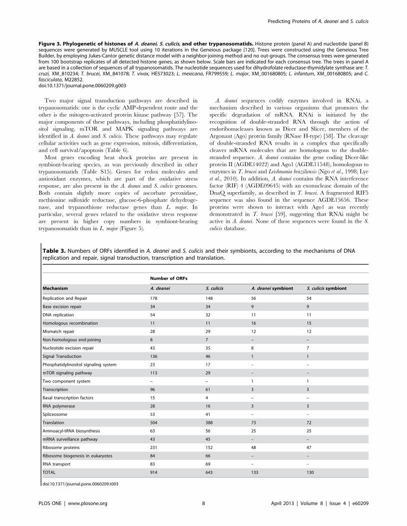
Housekeeping genes. Histones, which are responsible for
structuring the chromatin, are highly conserved proteins that
appeared in the eukaryotic branch of evolution. Although well
conserved, Trypanosomatidae histones display differences in the N
and C-terminal sequences, sites of post-translational modifications,
when compared to other eukaryotes. Phylogenetic analysis
revealed that histones and their variants in both A. deanei and S.
culicis are clustered in a separate branch, between the Trypanosoma
and Leishmania species (Figure 3A). Similar phylogenetic distribu-
tion is seen for the dihydrofolate reductase-thymidylate synthase
when we performed the analysis using nucleotide sequences
(Figure 3B). Nevertheless, the symbiont-bearing species show
conservation in the sites of post-translation when compared to
other trypanosomes as shown in supplementary Figure S3. In A.
deanei and S. culicis the proteins related to the chromatin assembly
are also maintained, including histones and histone-modifying
enzymes as shown in Tables S2–S7 and Figure S4 of the
supporting information. For a more detailed analysis about
housekeeping genes of A. deanei and S. culicis see Text S1.
DNA replication, repair, transcription, translation and signal
transduction in A. deanei and S. culicis functions can be respectively
attributed at least to 914 ORFs and 643 ORFs (Table 3). Most of
the genes are exclusive to the protozoan and are absent in the
endosymbiont (Table 4), thus indicating that these processes are
exclusive to the host organism as shown in the supplementary
Tables S8–S13, typically containing a conserved spliced-leader
RNA as found in other trypanosomes (see Figure S5 for more
information). A total of 133 and 130 proteins with similar
functions are detectable in the endosymbionts of both species, with
up to 95% amino acid identity to proteins of Bordetella sp. and A.
xylosoxidans.
Similar DNA repair proteins are present in both eukaryote and
prokaryote predicted sequences. These findings demonstrate that
the endosymbionts conserved essential housekeeping proteins
despite their genome reduction. Some differences were found in
mismatch repair (MMR) between symbiont-bearing trypanoso-
matid genomes. As microsatellite instability is considered the
molecular fingerprint of the MMR system, we compared the
abundance of tandem repeats in the genomes of A. deanei and S.
culicis and their respective endosymbionts. We noticed that the
genomes of S. culicis and its endosymbiont are more repetitive than
the genomes of A. deanei and its endosymbiont (Figure 4A).
However, the higher repetitive content of the genomes of S. culicis
and its endosymbiont is not only due to the higher number of
microsatellite loci (Figure 4B) but also to the expansion of the size
of the microsatellite sequences. These data suggest that microsat-
ellites of S. culicis and its endosymbiont evolved faster than those of
A. deanei and its endosymbiont. Interestingly, we identified some
missing components of the MMR machinery in S. culicis that are
present in A. deanei, such as exonuclease I (Exo I), a 59-39
exonuclease that is implicated in the excision step of the DNA
mismatch repair pathway (Table S9). Several studies have
correlated the silencing of the ExoI protein and/or mutations of
the ExoI gene and microsatellite instability with development of
lymphomas and colorectal cancer [49,50,51]. Therefore, we
speculate that deficiencies in the MMR machinery in S. culicis
may be related to the high proportion of microsatellites in its
genome. The association between microsatellite instability and
MMR deficiency has already been described for T. cruzi strains
[52,53]. The same variability pattern is observed for each
symbiont, despite the fact that the MMR machinery seems to be
complete in both symbiotic bacteria (Table S10). It is tempting to
speculate that this finding may indicate that the parasite and its
endosymbiont are exposed to the same environment and therefore
may be subjected to similar selective pressures imposed by an
external oxidative condition.
A. deanei and S. culicis have 607 and 421 putative kinase-
encoding genes, respectively (Table 5). Thirty one of the A. deanei
kinases were classified in the AGC family, 31 as atypical, 49 as
CAMK, 15 as CK1, 108 as CMGC, 64 as STE, 1 as TKL, 81 as
others, and 227 that could not be classified in any of these families.
No typical tyrosine kinases (TK) are present in A. deanei or S. culicis,
as in other trypanosomes, although tyrosine residues are subjected
to phosphorylation [54,55]. Several phosphatases have also been
described in trypanosomes, pointing toward their regulatory role
in the development of these organisms. The T. brucei PTP
(TbPTP1) is associated with the cytoskeleton and has been
reported to be intrinsically involved in this parasite’s cycle [56].
Similar sequences are found in the A. deanei genome, including
PTP1, which is not found in the S. culicis database. Additionally, a
large number of other PTPs appear in both genomes, including
ectophosphatases (Table S14).
Predicting Proteins of A. deanei and S. culicis
PLOS ONE | www.plosone.org 6 April 2013 | Volume 8 | Issue 4 | e60209

Predicting Proteins of A. deanei and S. culicis
PLOS ONE | www.plosone.org 7 April 2013 | Volume 8 | Issue 4 | e60209
Two major signal transduction pathways are described in
trypanosomatids: one is the cyclic AMP-dependent route and the
other is the mitogen-activated protein kinase pathway [57]. The
major components of these pathways, including phosphatidylino-
sitol signaling, mTOR and MAPK signaling pathways are
identified in A. deanei and S. culicis. These pathways may regulate
cellular activities such as gene expression, mitosis, differentiation,
and cell survival/apoptosis (Table 6).
Most genes encoding heat shock proteins are present in
symbiont-bearing species, as was previously described in other
trypanosomatids (Table S15). Genes for redox molecules and
antioxidant enzymes, which are part of the oxidative stress
response, are also present in the A. deanei and S. culicis genomes.
Both contain slightly more copies of ascorbate peroxidase,
methionine sulfoxide reductase, glucose-6-phosphate dehydroge-
nase, and trypanothione reductase genes than L. major. In
particular, several genes related to the oxidative stress response
are present in higher copy numbers in symbiont-bearing
trypanosomatids than in L. major (Figure 5).
A. deanei sequences codify enzymes involved in RNAi, a
mechanism described in various organisms that promotes the
specific degradation of mRNA. RNAi is initiated by the
recognition of double-stranded RNA through the action of
endoribonucleases known as Dicer and Slicer, members of the
Argonaut (Ago) protein family (RNase H-type) [58]. The cleavage
of double-stranded RNA results in a complex that specifically
cleaves mRNA molecules that are homologous to the double-
stranded sequence. A. deanei contains the gene coding Dicer-like
protein II (AGDE14022) and Ago1 (AGDE11548), homologous to
enzymes in T. brucei and Leishmania braziliensis (Ngo et al., 1998; Lye
et al., 2010). In addition, A. deanei contains the RNA interference
factor (RIF) 4 (AGDE09645) with an exonuclease domain of the
DnaQ superfamily, as described in T. brucei. A fragmented RIF5
sequence was also found in the sequence AGDE15656. These
proteins were shown to interact with Ago1 as was recently
demonstrated in T. brucei [59], suggesting that RNAi might be
active in A. deanei. None of these sequences were found in the S.
culicis database.
Figure 3. Phylogenetic of histones of A. deanei, S. culicis, and other trypanosomatids. Histone protein (panel A) and nucleotide (panel B)sequences were generated by MUSCLE tool using 10 iterations in the Geneious package [120]. Trees were constructed using the Geneious TreeBuilder, by employing Jukes-Cantor genetic distance model with a neighbor-joining method and no out-groups. The consensus trees were generatedfrom 100 bootstrap replicates of all detected histone genes, as shown below. Scale bars are indicated for each consensus tree. The trees in panel Aare based in a collection of sequences of all trypanosomatids. The nucleotide sequences used for dihydrofolate reductase-thymidylate synthase are: T.cruzi, XM_810234; T. brucei, XM_841078; T. vivax, HE573023; L. mexicana, FR799559; L. major, XM_001680805; L. infantum, XM_001680805; and C.fasciculata, M22852.doi:10.1371/journal.pone.0060209.g003
Table 3. Numbers of ORFs identified in A. deanei and S. culicis and their symbionts, according to the mechanisms of DNAreplication and repair, signal transduction, transcription and translation.
Number of ORFs
Mechanism A. deanei S. culicis A. deanei symbiont S. culicis symbiont
Replication and Repair 178 148 56 54
Base excision repair 34 34 9 9
DNA replication 54 32 11 11
Homologous recombination 11 11 16 15
Mismatch repair 28 29 12 12
Non-homologous end-joining 8 7 – –
Nucleotide excision repair 43 35 8 7
Signal Transduction 136 46 1 1
Phosphatidylinositol signaling system 23 17 – –
mTOR signaling pathway 113 29 – –
Two component system – – 1 1
Transcription 96 61 3 3
Basal transcription factors 15 4 – –
RNA polymerase 28 16 3 3
Spliceosome 53 41 – –
Translation 504 388 73 72
Aminoacyl-tRNA biosynthesis 63 56 25 25
mRNA surveillance pathway 43 45 – –
Ribosome proteins 231 152 48 47
Ribosome biogenesis in eukaryotes 84 66 – –
RNA transport 83 69 – –
TOTAL 914 643 133 130
doi:10.1371/journal.pone.0060209.t003
Predicting Proteins of A. deanei and S. culicis
PLOS ONE | www.plosone.org 8 April 2013 | Volume 8 | Issue 4 | e60209

Table 4. Summary of the origin of ORFs found in A. deanei and S. culicis.
A. deanei Symbiont
Functional Classification Prokaryotes* Eukaryotes** P/E***
Replication and Repair
Base excision repair 5 11 4/0
Nucleotide excision repair 2 16 9/0
Non-homologous end-joining 1 5 N
Mismatch repair 2 13 8/0
Homologous recombination 2 9 10/0
DNA replication 3 22 10/0
Signal Transduction
Two-component system N N 1
Phosphatidylinositol signaling system 0 16 N
mTOR signaling pathway 0 8 N
MAPK signaling pahway - yeast 0 1 N
Transcription
Spliceosome 0 20 N
RNA polymerase 0 16 3/0
Basal transcription factors 0 5 N
Translation
RNA transport 0 31 N
Ribosome biogenesis in eukaryotes 0 27 N
Ribosome 0 75 48/0
mRNA surveillance pathway 0 17 N
Aminoacyl-tRNA biosynthesis 0 22 23
S. culicis Symbiont
Functional Classification Prokaryotes Eukaryotes P/E
Replication and Repair
Base excision repair 2 6 5/0
Nucleotide excision repair 2 10 7/0
Non-homologous end-joining 1 1 N
Mismatch repair 1 5 8/0
Homologous recombination 1 4 11/0
DNA replication 2 15 9/0
Signal Transduction
Two-component system N N 1
Phosphatidylinositol signaling system 0 11 N
mTOR signaling pathway 0 8 N
MAPK signaling pathway - yeast 0 0 N
Transcription
Spliceosome 0 13
RNA polymerase 0 11 3/0
Basal transcription factors 0 2
Translation
RNA transport 0 19 N
Ribosome biogenesis in eukaryotes 0 20 N
Ribosome 0 53 46/0
mRNA surveillance pathway 0 16 N
Aminoacyl-tRNA biosynthesis 0 18 23
*Number of genes with identity to Prokaryotes.**Number of genes with identity to Eukaryotes.***Ratio of the number of genes with identity to Prokaryotes/Eukaryotes.doi:10.1371/journal.pone.0060209.t004
Predicting Proteins of A. deanei and S. culicis
PLOS ONE | www.plosone.org 9 April 2013 | Volume 8 | Issue 4 | e60209

The Coordinated Division of the Bacterium during theHost Protozoan Cell Cycle
Cell cycle control in host trypanosomes. In eukaryotes,
DNA replication is coordinated with cell division by a cyclin-CDK
complex that triggers DNA duplication during the S phase of the
cell cycle. Multiple copies of the CRK gene (cdc2-related protein
kinase) are found in A. deanei and four genes coding for two
different CRKs are present in S. culicis. Both proteins exhibit
structural features of the kinase subunits that make up the CDK
complex, as they contain the cyclin-binding PSTAIRE motif, an
ATP-binding domain and a catalytic domain. These motifs and
domains are not the same in different CRKs (Figure S6), strongly
suggesting that these CRKs might control different stages of the
cell cycle. A. deanei contains four genes coding for cyclins. Three of
these genes are homologues to mitotic cyclin from S. cerevisiae and
T. brucei. However, none of them contain the typical destruction
domain present in T. brucei mitotic cyclin [60]. The fourth codes
for a S. cerevisiae Clb5 homolog, an S-phase cyclin. These data
indicate that more than one CRK and more than one cyclin would
be involved in the cell cycle control of symbiont-containing
trypanosomatids, suggesting that tight regulation must occur to
guarantee the precise maintenance of only one symbiont per cell
[14].
Cell cycle control in the endosymbionts. Bacterial cell
division is a highly regulated event that mainly depends on two
structures, the peptidoglycan layer and the Z ring. The first step in
the segregation of the bacterium is the formation of a polymerized
Z ring at the middle of the cell. This structure acts as a platform
for the recruitment of other essential proteins named Filament
Temperature Sensitive (Fts), which are mainly involved in the
formation and stabilization of the Z ring [61,62] and in
establishing the peptidoglycan septum formation site in most
bacteria [63] (Figure 6A).
Two fts sequences were identified in A. deanei and S. culicis
symbionts based on Bordetella genes (Table 7). One of them is FtsZ,
which requires integral membrane proteins such as Zip A and
FtsA for anchoring. However, these sequences are absent in the
symbionts. FtsZ should also interact with FtsE, which is absent in
both symbionts. This protein is homologous to the ATP-binding
cassette of ABC transporters and co-localizes with the division
septum [64]. The lack of these proteins could be related to the
absence of a classical Z ring in these symbionts. The other
sequence is FtsK that docks FtsQ, FtsB and FtsL, which are related
to the formation of the peptidoglycan layer in E. coli and B. subtilis
[65,66,67], but these proteins are absent in symbionts, as in most
bacteria that exhibit reduced peptidoglycan production [64].
RodA, a homologous integral membrane protein involved in
bacterial cell growth, is detected in the endosymbionts. RodA
could replace FtsW, which is absent in both symbionts. FtsW is
Figure 4. Microsatellite content in the genomes of A. deanei, S.culicis, and their endosymbionts. Panel (A) shows the percentage ofrepetitive nucleotides for each repeat length. The total numbers ofnucleotides are derived from microsatellite sequences divided by thetotal number of assembled nucleotides. Panel (B) shows the microsat-ellite density. The values indicate the number of microsatellite locidivided by the genome length6100.doi:10.1371/journal.pone.0060209.g004
Table 5. Kinase families identified in trypanosomatids.
Kinase family A. deanei S. culicis
AGC 31 23
Atypical 31 21
CAMK 49 39
CK1 15 8
CMGC 108 77
STE 64 31
TKL 1 0
Other 81 58
No hits found 227 164
TOTAL 607 421
doi:10.1371/journal.pone.0060209.t005
Table 6. Representative ORFs involved in the signaltransduction pathways in A. deanei and S. culicis.
Product A. deanei S. culicis
Calmodulin AGDE02036 STCU01612
Diacylglycerol kinase AGDE02361 STCU00226
CDP-diacylglycerol-inositol-3-phosphatidyltransferase
AGDE04835 STCU01286
Myo-inositol-1(or 4) monophosphatase AGDE08470 STCU02993
Phospholipase C AGDE12052 STCU02439
Phosphatidylinositol 4-phosphate5-kinase alpha
AGDE09669 STCU03909
Inositol-1,4,5-trisphosphate (IP3) 5-phosphatase AGDE06690 nd
phosphatidate cytidylyltransferase AGDE09922 nd
Mitogen-activated protein kinase 5 AGDE00259 STCU00603
Protein kinase A AGDE06073 STCU01525
TP53 regulating kinase AGDE08400 nd
Serine/threonine-protein kinase CTR1 AGDE00613 nd
Casein kinase AGDE11868 STCU01611
Phosphoinositide-specific phospholipase C nd STCU09903
nd: not determined.doi:10.1371/journal.pone.0060209.t006
Predicting Proteins of A. deanei and S. culicis
PLOS ONE | www.plosone.org 10 April 2013 | Volume 8 | Issue 4 | e60209

Figure 5. Oxidative stress-related genes in the genomes of A. deanei, S. culicis and L. major. The figure shows the number of ORFs for theindicated enzymes for each species.doi:10.1371/journal.pone.0060209.g005
Figure 6. Schematic representation of the cell division machinery found in the endosymbionts. Panel (A) indicates the basic modelderived from a gram-negative bacterium with the localization of each component (shown on the right). Panel (B) represents the components foundin the endosymbiont of A. deanei, and Panel (C) shows the steps in the assembly of the Z-ring. The missing components of the A. deaneiendosymbiont are drawn in red.doi:10.1371/journal.pone.0060209.g006
Predicting Proteins of A. deanei and S. culicis
PLOS ONE | www.plosone.org 11 April 2013 | Volume 8 | Issue 4 | e60209

essential for the localization of FtsI (PBP3) in the Z ring [68],
which is absent in the symbiotic bacteria.
Endosymbionts have only one bifunctional synthase (PBP1A),
while E. coli has PBP1A, PBP1B, and PBP1C. Cells require at least
one of these synthases for viability. The peptidoglycan layer is
functional in trypanosomatid symbionts, as shown by treatment
with b-lactam antibiotics affecting the division of the bacterium,
generating filamentous structures and culminating in cell lysis.
PBP1 and PBP2 have also been detected at the symbiont envelope
[6]. PBP1B interacts with the two essential division proteins, FtsN
and PBP3/FtsI, which are absent in the symbiont. PBP1B can also
interact with PBP2 that is identified in both symbiont databases
(see Table 7).
A sequence encoding a minor PBP described in E. coli was also
identified in the symbionts. This protein is known as a putative
PBP precursor (PBP5/dacC). This PBP is involved in the
regulation of the peptidoglycan structure, along with 3 other
minor PBPs described in E. coli, but these are absent from the
symbiont (Table 7). On the other hand, all the enzymes involved
in the synthesis of activated nucleotide precursors for the assembly
of the peptidoglycan layer are present in the symbiont genome,
except for Braun’s lipoprotein (Lpp), which forms the lipid-
anchored disaccharide-pentapeptide monomer subunit [69]. In E.
coli strains, mutations in Lpp genes result in a significant reduction
of the permeability barrier, although small effects on the
maintenance of the cell growth and metabolism were observed
in these cells [70,71].
Taken together, we consider that gene loss in the dcw cluster
[72] (represented in Figure 6) explains the lack of the FtsZ ring in
the endosymbiont during its division process [73]. Moreover, the
symbiont envelope contains a reduced peptidoglycan layer and
lacks a septum during its division process, which can be related to
the facilitation of metabolic exchanges, as well as to the control of
division by the host protozoan [6]. These losses could be
understood since the host trypanosomatid is controlling the
number of symbiotic bacteria per cell. This phenomenon has
been described for obligatory intracellular bacteria that co-evolve
in eukaryotic cells, as well as for the organelles of prokaryotic
origin, the chloroplast and the mitochondrion [74,75].
Metabolic Co-evolution of the Bacterium and the HostTrypanosomatid
Symbiosis in trypanosomatids is characterized as a mutual
association where both partners benefit. These symbiont-bearing
protozoa have low nutritional requirements, as intense metabolic
exchanges occur. Our data corroborate previous biochemical and
ultrastructural analyses showing that the bacterium has enzymes
and metabolic precursors that complete important biosynthetic
pathways of the host [76].
Oxidative phosphorylation. FoF1-ATP synthase and the
entire mitochondrial electron transport chain are present in A.
deanei and S. culicis, although some subunits are missing (Table 8).
These species have a rotenone-insensitive NADH:ubiquinone
oxidoredutase in complex I, as do other trypanosomatids [77].
Ten complex II (succinate:ubiquinone reductase) subunits of the
twelve identified in T. cruzi [78] are also present in both
trypanosomatids. Many subunits from complex III, composed of
cytochrome c reductase, are found in A. deanei and S. culicis. In
addition, these protozoa contain genes for cytochrome c, as
previously suggested by biochemical studies in other symbiont-
containing trypanosomatids [3,79].
Both symbionts contain sequences with hits for all subunits of
complex I, NADH:ubiquinone oxidoredutase, similar to E. coli
(Table 8). Complexes II and III, including cytochrome c, and
complex IV (cytochrome c oxidase, succinate:ubiquinone reduc-
tase and cytochrome c reductase, respectively) are not found in
Table 7. Members of the Fts family and PBPs that are present in endosymbionts of A. deanei and S. culicis.
Function Protein A. deanei S. culicis
Stabilization and attachment of FtsZ polymers to the inner membrane FtsA nd nd
FtsE nd nd
ZipA nd nd
FtsK CKCE00084 CKBE00632
Interaction with peptidogycan synthases PBPs FtsQ nd nd
FtsB Nd nd
FtsL nd nd
FtsN nd nd
Lipid II flippase FtsW(RodA) CKCE 00486 CKBE00079
Forms a dynamic cytoplasmic ring structure at midcell FtsZ CKCE00034 CKBE00683
Penicillin binding proteins (PBPs) PBP1A CKCE00524 CKBE00119
PBP2 CKCE00487 CKBE00080
FtsI/PBP3 CKCE00487 CKBE00080
PBP4 nd nd
PBP5/dacC CKCE00510 CKBE00105
PBP6 nd nd
PBP6B nd nd
PBP7 nd nd
nd: not determined.doi:10.1371/journal.pone.0060209.t007
Predicting Proteins of A. deanei and S. culicis
PLOS ONE | www.plosone.org 12 April 2013 | Volume 8 | Issue 4 | e60209

either symbiont. However, we detected the presence of cyto-
chrome d as found in Allochromatium vinosum, and also a cytochrome
d oxidase with a sequence close to that of B. parapertussis. All
portions of the FoF1-ATP synthase were identified in symbionts,
although not every subunit of each portion was found.
Lipid metabolism. The sphingophospholipid (SPL) content
in A. deanei and its symbiont has been previously described, with
phosphatidylcholine (PC) representing the major SPL in the host,
whereas cardiolipin predominates in the symbiotic bacterium
[5,80]. The synthetic pathway of phosphatidylglycerol from
glycerol phosphate is present in both host trypanosomatids (Table
S16). The biosynthetic pathways of PC and PE from CDP-choline
and CDP-ethanolamine (Kennedy pathways), that synthesize PC
and PE respectively, are incomplete in A. deanei and S. culicis.
Nevertheless, the methylation pathway (Greenberg pathway),
which converts PE in PC, seems to be absent in both
trypanosomatids, even though one enzyme sequence was identi-
fied in A. deanei.
The symbiont of A. deanei exhibits two routes for phosphatidyl-
ethanolamine (PE) synthesis, starting from CDP-diacylglycerol and
producing phosphatidylserine as an intermediate (Table S17).
Interestingly, this last step of the pathway is not found in the S.
culicis endosymbiont. Importantly, both symbionts lack genes that
encode proteins of PC biosynthetic pathways, reinforcing the idea
that this phospholipid is mainly obtained from the host protozoa
[5]. Remarkably, phoshpatidylglycerophosphatase A, which pro-
duces the intermediate phosphatidylglycerol necessary for cardio-
lipin biosynthesis, was not found in either protozoa but is present
in both symbionts. As cardiolipin is present in the inner
membranes of host mitochondria, the symbionts may complete
cardiolipin biosynthesis.
Pathways for sphingolipid production, including the synthesis of
ceramide from sphingosine-1P, are present in A. deanei, while S.
culicis lacks enzymes of this pathway (Table S16). Both host
trypanosomatids have glycerol kinase and 3-glycerophosphate
acyltransferase, enzymes for the synthesis of 1,2-diacyl-sn-glycerol
and triacylglycerol from D-glycerate. In endosymbionts, glycer-
olipid metabolism seems to be reduced to two enzymes: 3-
glycerophosphate acyltransferase and 1-acylglycerol-3-phosphate
O-acyltransferase (Table S17), suggesting metabolic complemen-
tation between partners.
Furthermore, both hosts contain enzymes of the biosynthesis
pathway for ergosterol production from zymosterol, as well as the
pathway of sterol biosynthesis that produces lanosterol from
farnesyl-PP. These pathways are only complete in A. deanei. The
symbionts do not have enzymes for sterol biosynthesis, in
accordance with our previous biochemical analysis [80].
Metabolism of amino acids, vitamins, cofactors and
hemin. Symbiosis in trypanosomatids is characterized by
intensive metabolic exchanges, reducing the nutritional require-
ments of these trypanosomatids when compared to species without
the symbiotic bacterium, or to aposymbiotic strains. Several
biochemical studies have been carried out analyzing the biosyn-
thetic pathways involved in this intricate relationship as recently
reviewed [76], and our genomic data corroborate these findings. A
schematic description of the potential metabolic interactions
concerning the metabolism of amino acids, vitamins, cofactors,
and hemin is provided in Figure 7.
Both symbiotic bacteria have genes potentially encoding for all
necessary enzymes for lysine, phenylalanine, tryptophan and
tyrosine synthesis, in agreement with previous experimental data
[40]. Tyrosine is required in the growth medium of A. deanei [81],
but it is not essential for S. oncolpelti or S. culicis [41,82,83]. Here, in
the symbiotic bacteria, we found enzymes involved in tyrosine
synthesis, as well as indications that phenylalanine and tyrosine
can be interconverted. In fact, protozoan growth is very slow in
absence of phenylalanine and tryptophan [81], which may
Table 8. Respiratory chain complexes identified in the predicted proteome of A. deanei, S. culicis and their respectiveendosymbionts.
A. deanei A. deanei endosymbiont S. culicis S. culicis endosymbiont
Complex I 33 0 33 0
Complex II 10 0 10 0
Complex III 5 0 4 0
Complex IV 10 2* 2 2*
Complex V 10 8 3 8
*The complex IV of the endosymbionts might be a cytochrome d ubiquinol oxidase identified in both organisms, instead a classical cytochrome c oxidase.doi:10.1371/journal.pone.0060209.t008
Figure 7. Main metabolic exchanges between host andendosymbionts. Schematic representation of the amino acids,vitamins, and cofactors exchanged between A. deanei and S. culicisand their respective symbionts. Dotted lines indicate pathways thathave or might have contributions from both partners, whereasmetabolites inside one of the circles, representing the symbiont orhost, indicate that one partner holds candidate genes coding forenzymes of the whole biosynthetic pathway. *Candidate genes wereonly found for the symbiont of S. culicis and not for the symbiont of A.deanei. BCAA (branched-chain amino acids) are leucine, isoleucine andvaline.doi:10.1371/journal.pone.0060209.g007
Predicting Proteins of A. deanei and S. culicis
PLOS ONE | www.plosone.org 13 April 2013 | Volume 8 | Issue 4 | e60209

indicate that larger amounts of these amino acids are required for
rapid cell proliferation.
Our data indicate that branched-chain amino acid (BCAA)
synthesis mainly occurs in the symbionts except for the last step,
with the branched-chain amino acid aminotransferase found in the
host protozoan.
Among the pathways that (might) involve contributions from
both partners, two have previously been characterized in detail,
the urea cycle and heme synthesis. The urea cycle is complete in
both symbiont-harboring trypanosomatids. Symbiotic bacteria
contribute with ornithine carbamoyltransferase, which converts
ornithine to citrulline, and with ornithine acetyltransferase, which
transforms acetylornithine in ornithine. Conversely, aposymbiotic
strains and symbiont-free Crithidia species need exogenous arginine
or citrulline for cell proliferation [8] [68]. Our genomic data
corroborate these studies.
Contrary to symbiont-free trypanosomatids, A. deanei and S.
culicis do not require any source of heme for growth because the
bacterium contains the required enzymes to produce heme
precursors that complete the heme synthesis pathway in the host
cell [7,9,10,11,84]. Our results support the idea that heme
biosynthesis is mainly accomplished by the endosymbiont, with
the last three steps of this pathway performed by the host
trypanosomatid, and in most cases also by the bacterium as
described in [11]. Furthermore, this metabolic route may
represent the result of extensive gene loss and multiple lateral
gene transfer events in trypanosomatids [11].
According to our genomic analyses, the symbiotic bacteria also
perform the synthesis of histidine, folate, riboflavin, and coenzyme
A, but one step is missing in the middle of each pathway, making
them candidates for metabolic interchange with the host. In the
case of folate and coenzyme A biosynthesis, one candidate gene
was found in the host trypanosomatid. Moreover, none of these
four metabolites are required in the growth medium of A. deanei
and S. culicis [85], suggesting that these pathways are fully
functional.
Candidate genes for the ubiquinone biosynthetic pathway were
found in S. culicis but none for A. deanei endosymbionts. For the
route with chorismate as precursor, only the first out of nine steps
is missing in the S. culicis endosymbiont; moreover a candidate
gene for that step is found in S. culicis genome. Only a few steps of
these pathways are absent in A. deanei and S. culicis host organisms.
In L. major, the ubiquinone ring synthesis has been described as
having either acetate (via chorismate as in prokaryotes) or
aromatic amino acids (as in mammalian cells) as precursors [45].
Methionine is considered essential for the growth of A. deanei, S.
culicis and S. oncopelti [41,81,82]. We were not able to identify one
enzyme among the four involved in the synthesis of methionine
from either pyruvate or serine via cysteine in the genomes of A.
deanei and S. culicis. No candidate to complement this pathway was
found in the symbiotic bacteria.
Purine and pyrimidine metabolism for nucleotide
production. Trypanosomatids are not able to synthesize the
purine ring de novo [86,87,88]. We observed that endosymbiont-
bearing trypanosomatids contain sequences encoding ectonucleo-
tidases from the E-NTPDase family and the adenosine deaminase
family (Table S18), which are required for the hydrolysis and
deamination of extracellular nucleotides [89,90]. Interestingly,
sequences encoding 59-nucleotidases are not found in either
symbiont-bearing trypanosomatid. The absence of this enzyme
can be related to the presence of the endosymbiont, which can
supply adenosine to the host cell, as we found all genes involved in
the de novo pathway in the symbionts, indicating that they are able
to complement the purine requirements of the host (Figure 8).
However, we cannot discard the possibility that adenosine is
transported to the intracellular medium by carriers of monophos-
phate nucleoside or by the presence of other enzymes that have the
same function as 59-nucleotidase. On the other hand, the lack of
59-nucleotidase in A. deanei and S. culicis can be related to the fact
that such protozoa are only insect parasites. According to this idea,
several studies have shown the importance of ectonucleotidases in
the establishment of infection by some trypanosomatid species
[91]. The high activity of ectonucleotidases with concomitant
production of adenosine, a known immune system inhibitor, lead
to high susceptibility to Leishmania infection because adenosine can
induce anti-inflammatory effects on the host [92,93].
Nucleoside transporters can take up nucleosides and nucleo-
bases generated by ectonucleotidase activity. Genes encoding
nucleoside transporters are present in both trypanosomatid
genomes (Table S19), enabling cells to obtain exogenous purines
from the medium. Furthermore, A. deanei and S. culicis contain
intracellular enzymes that can convert purines to nucleotides, such
as adenine phosphoribosyltransferase, hypoxanthine-guanine
phosphoribosyltransferase, adenylate kinase, AMP deaminase,
inosine monophosphate dehydrogenase and GMP synthetase.
These data indicate that these organisms can interconvert
intracellular purines into nucleotides. In contrast, both endosym-
bionts lack all the genes encoding enzymes related to purine
salvage. Nevertheless, the symbiotic bacteria have genes encoding
all the enzymes expected to participate in the de novo synthesis of
purine nucleotides as previously proposed [94,95]. One interesting
possibility is that the symbiotic bacterium is able to supply the host
trypanosomatid with purines. According to this idea, the
endosymbiont participates in the de novo purine nucleotide pathway
of A. deanei, as the aposymbiotic strain is unable to utilize glycine
for the synthesis of purine nucleotides, only for pyrimidine
nucleotide production [87].
Protozoa are generally, but not universally considered to be
capable of synthesizing pyrimidines from glutamine and aspartic
acid, which are used as precursors. Our results indicate that both
symbiont-bearing trypanosomatids carry out de novo pyrimidine
synthesis (Table S19). Interestingly, in silico analyses also revealed
the presence of all the genes for de novo pyrimidine synthesis in both
symbiont genomes, but not for the pyrimidine salvage pathway. A
previous report indicated that A. deanei was able to synthesize
purine and pyrimidine nucleotides from glycine (‘‘de novo’’
pathway) and purine nucleotides from adenine and guanine
(‘‘salvage’’ pathway). Adenine would be incorporated into both
adenine and guanine nucleotides, whereas guanine was only
incorporated into guanine nucleotides, suggesting a metabolic
block at the level of GMP reductase [87].
Deoxyribonucleotides are derived from the corresponding
ribonucleotides by reactions in which the 29-carbon atom of the
D-ribose portion of the ribonucleotide is directly reduced to form
the 29-deoxy derivative. This reaction requires a pair of hydrogen
atoms that are donated by NADPH via the intermediate-carrying
protein thioredoxin. The disulfide thioredoxin is reduced by
NADPH in a reaction catalyzed by thioredoxin reductase,
providing the reducing equivalents for the ribonucleotide reduc-
tase, as observed for the endosymbionts that could provide 29-
deoxy derivatives. In folate metabolism, the formation of thymine
nucleotides requires methylation of dUMP to produce dTMP, a
reaction catalyzed by thymidilate kinase, which is present in A.
deanei, S. culicis, and their respective endosymbionts. Figure 8
summarizes the purine and pyrimidine metabolisms in A. deanei
and S. culicis considering the metabolic complementarity between
the protozoan and the endosymbiont.
Predicting Proteins of A. deanei and S. culicis
PLOS ONE | www.plosone.org 14 April 2013 | Volume 8 | Issue 4 | e60209

In this way, both symbiont-containing protozoa express a
unique complement of nutritionally indispensable salvage and
interconversion enzymes that enable the acquisition of purines
from the medium. The intracellular purines can be acquired
through the medium by the action of ectonucleotidases and
nucleoside transporters.
Factors Involved in Protozoa-host InteractionsMonoxenic trypanosomatids only parasitize invertebrates,
especially insects belonging to the orders Diptera and Hemiptera
[1]. These organisms have been found in Malphigian tubules, in
the hemolymph and hemocoel, and in the midgut, which is
considered the preferential site for protozoal multiplication and
colonization [1,96,97]. S. culicis, for example, is able to colonize the
insect midgut, to invade the hemocoel and to reach the salivary
glands [97,98]. The presence of the symbiotic bacterium has been
shown to influence the interactions between trypanosomatid cells
and insect cell lines, explanted guts and host insects [4,20]. This
seems to occur because the endosymbiont influences the glyco-
protein and polysaccharide composition of the host, the exposure
of carbohydrates on the protozoan plasma membrane, and the
surface charge [18,19,20,21].
Several glycosyltransferases from the two major families (GT-A
and GT-B [99]) and members of the family 25 (glycosyltransfer-
ases involved in lipo-oligosaccharide protein biosynthesis) are
present in both A. deanei and S. culicis genomes (Table S20). Other
glycosyltransferases with no characteristic domains that are thus
not classified as belonging to the GT-A or GT-B families are also
found in the A. deanei and S. culicis genomes. Importantly, 1,2-
fucosyltransferase transferase is present in A. deanei but not in the S.
culicis dataset, and fucose residues were found in high amounts on
glycoinositolphospholipid (GIPL) molecules of A. deanei, different
from the observations for other trypanosomatids (data not
published). Although the role of fucose is unknown, fucose and
arabinose transfer to lipophosphoglycan (LPG) of Leishmania is
noticed when the culture medium is supplemented with this
carbohydrate [100], suggesting that fucose might have a specific
role in A. deanei-insect interactions.
Another glycosyltransferase found in both A. deanei and S. culicis
genomes and involved in the N-glycosylation of asparagine
residues is the dolichyl-diphosphooligosaccharide-protein glycosyl-
transferase (DDOST), an oligosaccharyltransferase (OST) that is
not classified in any of the above-mentioned families. The A. deanei
and S. culicis DDOSTs contain the STT3 domain, a subunit
required to establish the activity of the oligosaccharyl transferase
(OTase) complex of proteins, and they are orthologous to the
human DDOST. These OTase complexes are responsible for
transferring lipid-linked oligosaccharides to the asparagine side
chain of the acceptor polypeptides in the endoplasmic reticulum
[101], suggesting a conserved N-glycosylation among the trypa-
nosomatids.
Five different GalfT sequences are also present in the
endosymbiont-bearing trypanosomatids, and all of them contain
the proposed catalytic site, indicating genetic redundancy.
Redundancy of GalfTs is commonly observed in many different
trypanosomatid species, as different transferases are used for each
linkage type [102]. As b-galactofuranose (b-Galf) has been shown
to participate in trypanosome-host interactions [103], their
presence in A. deanei and S. culicis might also indicate a role in
the interaction with the insect host. However, no enzymes
involved in synthesis of b-Galf-containing glycoconjugates are
detected in our A. deanei dataset, despite reports of enzymes
involved in b-Galf synthesis in Crithidia spp. [104,105,106].
Surface proteins and protease gene families. One
remarkable characteristic of trypanosomatid genomes is the large
expansion of gene families encoding surface proteins [107].
Experimental data indicated that these genes encode surface
proteins involved in interactions with the hosts. We selected eight
gene families encoding surface proteins present in T. cruzi, T. brucei
and Leishmania spp. to search for homologous sequences in the
genomes of the two symbiont-bearing trypanosomatids. Because
the draft assemblies of these genomes are still fragmented, we also
used a read-based analysis to search for sequences with homology
to these multigene families. It is well known that misassemblies
frequently occur for tandemly repeated genes, as most repetitive
copies collapse into only one or two copies. A total of 3,624,411
reads (corresponding to 1,595 Mb of sequences) from the A. deanei
genome and 2,666,239 reads (corresponding to 924 Mb) from the
S. culicis genome were used in this comparison. In A. deanei and S.
culicis, we identified gene families encoding amastins, gp63, and
Figure 8. Purine production, acquisition, and utilization in A. deanei and S. culicis. The figure illustrates the production, acquisition andutilization of purines in the host trypanosomes considering the presence of endosymbiont enzymes. This model suggests that the trypanosomatidacquires purines from the symbiont, which synthesizes them de novo. Some ecto-localized proteins, such as apyrase (APY) and adenosine deaminase(ADA), could be responsible for the generation of extracellular nucleosides, nucleobases, and purines. Nucleobases and purines could be acquired bythe parasite through membrane transporters (T) or diffusion and could be incorporated into DNA, RNA, and kDNA molecules after ‘‘purine salvagepathway’’ processing. Abbreviations: NTP (nucleoside tri-phosphate), NDP (nucleoside di-phosphate), NMP (nucleoside mono- phosphate), N(nucleobase), ADO (adenosine), INO (inosine).doi:10.1371/journal.pone.0060209.g008
Predicting Proteins of A. deanei and S. culicis
PLOS ONE | www.plosone.org 15 April 2013 | Volume 8 | Issue 4 | e60209

cysteine peptidases (Table S21). As expected, we could not identify
sequences homologous to mucin-like glycoproteins typical of T.
cruzi [108], variant surface glycoprotein (VSG) characteristic of
African trypanosomes, or trans-sialidases present in the genomes
of all Trypanosoma species.
Calpain-like cysteine peptidases constitute the largest gene
family identified in the A. deanei (85 members) and S. culicis (62
members) genomes, and they are also abundant in trypanosoma-
tids [46]. The presence of the N-terminal fatty acid acylation motif
was found in some members of calpain-like cysteine peptidases,
indicating that some of these peptidases are associated with
membranes, as has also been shown for other members of the
family [109,110]. The relatively large amount of calpain-like
peptidases may be related to the presence of the endosymbiont,
which would require a more complex regulation of the cell cycle
and intracellular organelle distribution [14], as cytosolic calpains
were found to regulate cytoskeletal remodeling, signal transduc-
tion, and cell differentiation [46].
A second large gene family in the A. deanei and S. culicis genomes
encoding surface proteins with proteolytic activity is gp63. In our
genomic analyses, we identified 37 and 9 genes containing
sequences homologous to the gp63 of Leishmania and Trypanosoma
spp. in the genomes of A. deanei and S. culicis, respectively. Proteins
belonging to this group of zinc metalloproteases, also known as
major surface protease (MSP) or leishmanolysin, have been
characterized in various species of Leishmania and Trypanosoma
[111]. Extensive studies on the role of this family in Leishmania
indicate that they are involved in several aspects of host-parasite
interaction including resistance to complement-mediated lysis, cell
attachment, entry, and survival in macrophages [112]. Gene
deletion studies in T. brucei indicated that the TbMSP of
bloodstream trypanosomes acts in concert with phospholipase C
to remove the variant surface protein from the membrane,
required for parasite differentiation into the procyclic insect form
[113]. Gp63-like molecules have been observed on the cell surface
of symbiont-harboring trypanosomatids [114]. Importantly, the
symbiont containing A. deanei displays a higher amount (2-fold) of
leishmanolysin-like molecules at the surface compared to the
aposymbiotic strain, which are unable to colonize insects [4]. As
anti-gp63 antibodies decrease protozoan-insect interactions [21],
our results reinforce the idea that the presence of such interactions
caused the expansion of this gene family in endosymbiont-bearing
organisms.
In contrast, only two copies of lysosomal cathepsin-like cysteine
peptidases were identified in the A. deanei (AGDE05983 and
AGDE10254) and S. culicis genomes (STCU01417 and
STCU06430). The two A. deanei sequences encode identical
cathepsin-B-like proteins, whereas the two S. culicis genes encode
proteases of the cathepsin-L-like group. This class of cysteine
peptidase is represented by cruzain or cruzipain, major lysosomal
proteinases of T. cruzi expressed by parasites found in insect and
vertebrate hosts, and encoded by a large gene family [115,116]. In
T. cruzi, these enzymes have important roles in various aspects of
the host/parasite relationship and in intracellular digestion as a
nutrient source [115]. Conversely, the low copy number of this
class of lysosomal peptidase in symbiont-containing trypanosoma-
tids seems to be related to their low nutritional requirements.
Amastins constitute a third large gene family in the A. deanei and
S. culicis genomes that encodes surface proteins. Initially described
in T. cruzi [117], amastin genes have also been identified in various
Leishmania species [118], in A. deanei and in another related insect
parasite, Leptomonas seymouri [119]. In Leishmania, amastins consti-
tute the largest gene family with gene expression that is regulated
during the parasite life cycle. As amastin has no sequence
similarity to any other known protein, its function remains
unknown. In this work, we identified 31 genes with sequences
belonging to all four sub-families of amastins in the genome of A.
deanei and 14 copies of amastin genes in S. culicis. Similar to
Leishmania, members of all four amastin subfamilies were identified
in symbiont-containing species (see Figure S7).
ConclusionThe putative proteome of symbiont-bearing trypanosomatids
revealed that these microorganisms exhibit unique features when
compared to other protozoa of the same family and that they are
most closely related to Leishmania species. Most relevant are the
differences in the genes related to cytoskeleton, paraflagellar and
kinetoplast structures, along with a unique pattern of peptidase
gene organization that may be related to the presence of the
symbiont and of the monoxenic life style. The symbiotic bacteria
of A. deanei and S. culicis are phylogenetically related with a
common ancestor, most likely a b-proteobacteria of the Alcali-
genaceae family. The genomic content of these symbionts is highly
reduced, indicating gene loss and/or transfer to the host cell
nucleus. In addition, we confirmed that both bacteria contain
genes that encode enzymes that complement several metabolic
routes of the host trypanosomatids, supporting the fitness of the
symbiotic relationship.
Supporting Information
Figure S1 Evolutionary history of endosymbionts ob-tained through a phylogenomic approach. The figure
indicates analysis using the Neighbor joining (NJ) (A) and
Maximum parsimony (MP) (B) methods. For NJ and MP, the
percentage of replicate trees in which the associated taxa clustered
together in the bootstrap test (1,500 replicates) is shown next to the
branches. The scale bar represents amino acids substitutions per
site.
(TIF)
Figure S2 Amino acid alignment of Kinetoplast Associ-ated Proteins. Panel (A) shows the KAP4 ClustalW alignment of
A. deanei (AdKAP-4), S. culicis (ScKAP-4) and C. fasciculata (CfKAP-
4). Panel (B) shows the ClustalW alignment of KAP2 of S. culicis
and C. fasciculata (CfKAP2-2, GenBank Q9TY84 and CfKAP2-1
GeneBank Q9TY83). Black color highlight is 100% similar gray is
80 to 99% similar light gray is 60 to 79% similar white is less than
59% similar.
(TIF)
Figure S3 Comparison of the histone sequences of A.deanei and S. culicis with other trypanosomes. Residues
indicated in red correspond to lysines that are acetylated and
green, methylated in T. cruzi and T. brucei [121]. Residues
indicated in blue are predicted site for phosphorylation upon DNA
damage as shown in T. brucei [122].
(TIF)
Figure S4 Phylogenetic tree of sirtuins from Trypano-somatids. The numbers represent bootstrap values. The proteins
from each species are grouped in nuclear and mitochondrial Sir2
based on the sequences of S. cerevisiae (nuclear), and the similarity
with S. coelicolor and S. enterica.
(TIF)
Figure S5 Phylogenetic tree of spliced leader (SL)sequences of A. deanei and S. culicis. A neighbor-joining
tree (1000 bootstraps) obtained by MEGA 5.0 using the SL gene
from the A. deanei and S. culicis genome sequences and sequences
Predicting Proteins of A. deanei and S. culicis
PLOS ONE | www.plosone.org 16 April 2013 | Volume 8 | Issue 4 | e60209

retrieved from GenBank (S. culicis DQ860203.1, L. pyrrhocoris
JF950600.1, H. samuelpessoai X62331.1, H. mariadeanei
AY547468.1, A. deanei EU099545.1, T. rangeli AF083351 and T.
cruzi AY367127).
(TIF)
Figure S6 Comparison between the amino acid se-quences of S. culicis CRK sequences. The figure shows a
ClustalW alignment with the ATP binding domains boxed in
yellow, PSTAIRE motifs boxed in blue, and the catalytic domain
boxed in pink. Red residues indicate the observed variations in the
amino acids involved in the activity.
(TIF)
Figure S7 Tree showing the distribution of amastin sub-families in A. deanei. The amastins are grouped as delta-
amastin (red), gamma-amastins (yellow), alpha-amastins (dark
blue) and beta-amastins (light blue).
(TIF)
Table S1 ORFs identified as Kinetoplast-associatedprotein (KAPs) in A. deanei and S. culicis.
(DOC)
Table S2 Histone acetyltransferases of the MYSTfamily present in A. deanei and S. culicis compared toother trypanosomes.
(DOC)
Table S3 Distribution of Sirtuins in the protozoan andendosymbiont species.
(DOC)
Table S4 Histone deacetylase identified in A. deaneiand S. culicis.
(DOC)
Table S5 Histone methyltransferase in A. deanei and S.culicis.
(DOC)
Table S6 Histone chaperones identified in A. deanei and S. culicis.
(DOC)
Table S7 Bromodomain proteins found in A. deanei andS. culicis.
(DOC)
Table S8 Components of replication mechanism of the kDNA
identified in A. deanei and S. culicis and similar endosymbionts
ORFs.
(DOC)
Table S9 Identified ORFs related to DNA replication and DNA
repair in A. deanei and S. culicis.
(DOC)
Table S10 DNA replication and repair ORFs found in the A.
deanei and S. culicis endosymbionts.
(DOC)
Table S11 Identified ORFs involved in DNA transcription and
RNA splicing in the genome of A. deanei and S. culicis.
(DOC)
Table S12 Transcription related proteins in the endosymbionts
of A. deanei and S. culicis.
(DOC)
Table S13 Main ORFs detected participating in ribosomal
biogenesis and translation in A. deanei and S. culicis.
(DOC)
Table S14 Identified phosphatases in A. deanei and S. culicis.
(DOC)
Table S15 Number of heat shock and stress response proteins in
A. deanei and S.culicis.
(DOC)
Table S16 Glycerophospholipids (GPL) enzymes of A. deanei and
S. culicis1.
(DOC)
Table S17 Glycerophospholipids (GPL) enzymes of A.deanei and S. culicis endosymbionts.(DOC)
Table S18 Ectonucleotidases families and identificationof ORFs found in A. deanei and S. culicis.(DOC)
Table S19 ORFs encoding enzymes involved in purineand pyrimidine metabolism of A. deanei, S. culicis andtheir symbionts.(DOC)
Table S20 Glysosyltransferases found in A. deanei andS. culicis.(DOC)
Table S21 Surface proteins of A. deanei e S. culicis.
(DOC)
Text S1
(DOC)
Acknowledgments
We would like to dedicate this paper to professors Erney Camargo and
Marta Teixeira who have made important contributions related to the
study of basic aspects of the biology of trypanosomatids, especially those
harboring an endosymbiont, and identified several new species of this
relevant and interesting group of eukaryotic microorganism.
Author Contributions
Conceived and designed the experiments: MCMM WS SS ATRV.
Analyzed the data: MCMM ACAM SSAS CMCCP RS CCK LGPA OLC
LPC MB ACC BAL CRM CMAS CMP CBAM CET DCB DFG DPP
ECG FFG FKM GFRL GW GHG JLRF MCE MHSG MFS MP PHS
RPMN SMRT TEFM TAOM TPU WS SS ATRV. Contributed
reagents/materials/analysis tools: ATRV LGPA OLC WS. Wrote the
paper: MCMM SS ATRV.
References
1. Wallace FG (1966) The trypanosomatid parasites of insects and arachnids.
Experimental Parasitology 18: 124–193.
2. Teixeira MM, Borghesan TC, Ferreira RC, Santos MA, Takata CS, et al.
(2011) Phylogenetic validation of the genera Angomonas and Strigomonas of
trypanosomatids harboring bacterial endosymbionts with the description of
new species of trypanosomatids and of proteobacterial symbionts. Protist 162:
503–524.
3. Edwards C, Chance B (1982) Evidence for the presence of two terminal
oxidases in the trypanosomatid Crithidia oncopelti. Journal of General
Microbiology 128: 1409–1414.
4. Fampa P, Correa-da-Silva MS, Lima DC, Oliveira SM, Motta MC, et al.
(2003) Interaction of insect trypanosomatids with mosquitoes, sand fly and the
respective insect cell lines. International Journal for Parasitology 33: 1019–
1026.
Predicting Proteins of A. deanei and S. culicis
PLOS ONE | www.plosone.org 17 April 2013 | Volume 8 | Issue 4 | e60209

5. de Azevedo-Martins AC, Frossard ML, de Souza W, Einicker-Lamas M, Motta
MC (2007) Phosphatidylcholine synthesis in Crithidia deanei: the influence of the
endosymbiont. FEMS Microbiology Letters 275: 229–236.
6. Motta MCM, Leal LHM, Souza WD, De Almeida DF, Ferreira LCS (1997)
Detection of Penicillin-binding Proteins in the Endosymbiont of the
Trypanosomatid Crithidia deanei. The Journal of Eukaryotic Microbiology 44:
492–496.
7. Chang KP, Chang CS, Sassa S (1975) Heme biosynthesis in bacterium-
protozoon symbioses: enzymic defects in host hemoflagellates and comple-
mental role of their intracellular symbiotes. Proceedings of the National
Academy of Sciences of the United States of America 72: 2979–2983.
8. Camargo EP, Freymuller E (1977) Endosymbiont as supplier of ornithine
carbamoyltransferase in a trypanosomatid. Nature 270: 52–53.
9. Galinari S, Camargo EP (1978) Trypanosomatid protozoa: survey of
acetylornithinase and ornithine acetyltransferase. Experimental Parasitology
46: 277–282.
10. Salzman TA, Batlle AM, Angluster J, de Souza W (1985) Heme synthesis in
Crithidia deanei: influence of the endosymbiote. The International Journal of
Biochemistry 17: 1343–1347.
11. Alves JM, Voegtly L, Matveyev AV, Lara AM, da Silva FM, et al. (2011)
Identification and phylogenetic analysis of heme synthesis genes in trypano-
somatids and their bacterial endosymbionts. PLoS One 6: e23518.
12. Frossard ML, Seabra SH, DaMatta RA, de Souza W, de Mello FG, et al.
(2006) An endosymbiont positively modulates ornithine decarboxylase in host
trypanosomatids. Biochemical and Biophysical Research Communications 343:
443–449.
13. Motta MC, Soares MJ, Attias M, Morgado J, Lemos AP, et al. (1997)
Ultrastructural and biochemical analysis of the relationship of Crithidia deanei
with its endosymbiont. European Journal of Cell Biology 72: 370–377.
14. Motta MC, Catta-Preta CM, Schenkman S, Azevedo Martins AC, Miranda K,
et al. (2010) The bacterium endosymbiont of Crithidia deanei undergoes
coordinated division with the host cell nucleus. PLoS One 5: e12415.
15. Freymuller E, Camargo EP (1981) Ultrastructural differences between species
of trypanosomatids with and without endosymbionts. The Journal of
Protozoology 28: 175–182.
16. Gadelha C, Wickstead B, de Souza W, Gull K, Cunha-e-Silva N (2005) Cryptic
paraflagellar rod in endosymbiont-containing kinetoplastid protozoa. Eukary-
otic Cell 4: 516–525.
17. Cavalcanti DP, Thiry M, de Souza W, Motta MC (2008) The kinetoplast
ultrastructural organization of endosymbiont-bearing trypanosomatids as
revealed by deep-etching, cytochemical and immunocytochemical analysis.
Histochemistry and Cell Biology 130: 1177–1185.
18. Dwyer DM, Chang KP (1976) Surface membrane carbohydrate alterations of a
flagellated protozoan mediated by bacterial endosymbiotes. Proceedings of the
National Academy of Sciences of the United States of America 73: 852–856.
19. Oda LM, Alviano CS, Filho FCS, Angluster J, Roitman I, et al. (1984) Surface
Anionic Groups in Symbiote-Bearing and Symbiote-Free Strains of Crithidia
deanei. The Journal of Eukaryotic Microbiology 31: 131–134.
20. d9Avila-Levy CM, Silva BA, Hayashi EA, Vermelho AB, Alviano CS, et al.
(2005) Influence of the endosymbiont of Blastocrithidia culicis and Crithidia deanei
on the glycoconjugate expression and on Aedes aegypti interaction. FEMS
Microbiology Letters 252: 279–286.
21. d9Avila-Levy CM, Santos LO, Marinho FA, Matteoli FP, Lopes AH, et al.
(2008) Crithidia deanei: influence of parasite gp63 homologue on the interaction
of endosymbiont-harboring and aposymbiotic strains with Aedes aegypti midgut.
Experimental Parasitology 118: 345–353.
22. Du Y, Maslov DA, Chang KP (1994) Monophyletic origin of beta-division
proteobacterial endosymbionts and their coevolution with insect trypanosoma-
tid protozoa Blastocrithidia culicis and Crithidia spp. Proceedings of the National
Academy of Sciences of the United States of America 91: 8437–8441.
23. Du Y, McLaughlin G, Chang KP (1994) 16S ribosomal DNA sequence
identities of beta-proteobacterial endosymbionts in three Crithidia species.
Journal of Bacteriology 176: 3081–3084.
24. Martin W, Hoffmeister M, Rotte C, Henze K (2001) An overview of
endosymbiotic models for the origins of eukaryotes, their ATP-producing
organelles (mitochondria and hydrogenosomes), and their heterotrophic
lifestyle. Biological chemistry 382: 1521–1539.
25. Hollar L, Lukes J, Maslov DA (1998) Monophyly of endosymbiont containing
trypanosomatids: phylogeny versus taxonomy. The Journal of Eukaryotic
Microbiology 45: 293–297.
26. Hebert L, Moumen B, Duquesne F, Breuil MF, Laugier C, et al. (2011)
Genome sequence of Taylorella equigenitalis MCE9, the causative agent of
contagious equine metritis. Journal of Bacteriology 193: 1785.
27. Hebert L, Moumen B, Pons N, Duquesne F, Breuil MF, et al. (2012) Genomic
characterization of the Taylorella genus. PLoS One 7: e29953.
28. Sugimoto C, Isayama Y, Sakazaki R, Kuramochi S (1983) Transfer of
Haemophilus equigenitalis Taylor et al. 1978 to the genusTaylorella gen. nov. as
Taylorella equigenitalis comb. nov. Current Microbiology 9: 155–162.
29. Moran NA, McCutcheon JP, Nakabachi A (2008) Genomics and evolution of
heritable bacterial symbionts. Annual Review of Genetics 42: 165–190.
30. Toft C, Andersson SG (2010) Evolutionary microbial genomics: insights into
bacterial host adaptation. Nature Reviews Genetics 11: 465–475.
31. Shigenobu S, Watanabe H, Hattori M, Sakaki Y, Ishikawa H (2000) Genome
sequence of the endocellular bacterial symbiont of aphids Buchnera sp. APS.
Nature 407: 81–86.
32. McCutcheon JP, Moran NA (2012) Extreme genome reduction in symbiotic
bacteria. Nature Reviews Microbiology 10: 13–26.
33. Parkhill J, Sebaihia M, Preston A, Murphy LD, Thomson N, et al. (2003)
Comparative analysis of the genome sequences of Bordetella pertussis, Bordetella
parapertussis and Bordetella bronchiseptica. Nature Genetics 35: 32–40.
34. Cummings CA, Brinig MM, Lepp PW, van de Pas S, Relman DA (2004)
Bordetella species are distinguished by patterns of substantial gene loss and host
adaptation. Journal of Bacteriology 186: 1484–1492.
35. Gull K (1999) The cytoskeleton of trypanosomatid parasites. Annual Review of
Microbiology 53: 629–655.
36. Berriman M, Ghedin E, Hertz-Fowler C, Blandin G, Renauld H, et al. (2005)
The Genome of the African Trypanosome Trypanosoma brucei. Science 309:
416–422.
37. Beech PL, Heimann K, Melkonian M (1991) Development of the Flagellar
Apparatus during the Cell-Cycle in Unicellular Algae. Protoplasma 164: 23–
37.
38. Lange BM, Gull K (1996) Structure and function of the centriole in animal
cells: progress and questions. Trends in Cell Biology 6: 348–352.
39. Garcia-Salcedo JA, Perez-Morga D, Gijon P, Dilbeck V, Pays E, et al. (2004) A
differential role for actin during the life cycle of Trypanosoma brucei. The EMBO
Journal 23: 780–789.
40. Gadelha C, Wickstead B, Gull K (2007) Flagellar and ciliary beating in
trypanosome motility. Cell Motility and the Cytoskeleton 64: 629–643.
41. Portman N, Gull K (2010) The paraflagellar rod of kinetoplastid parasites: from
structure to components and function. International Journal for Parasitology
40: 135–148.
42. Lacomble S, Vaughan S, Gadelha C, Morphew MK, Shaw MK, et al. (2009)
Three-dimensional cellular architecture of the flagellar pocket and associated
cytoskeleton in trypanosomes revealed by electron microscope tomography.
Journal of Cell Science 122: 1081–1090.
43. Oberholzer M, Marti G, Baresic M, Kunz S, Hemphill A, et al. (2007) The s
cAMP phosphodiesterases TbrPDEB1 and TbrPDEB2: flagellar enzymes that
are essential for parasite virulence. The FASEB Journal 21: 720–731.
44. Ginger ML, Portman N, McKean PG (2008) Swimming with protists:
perception, motility and flagellum assembly. Nature Reviews Microbiology 6:
838–850.
45. Xu C, Ray DS (1993) Isolation of proteins associated with kinetoplast DNA
networks in vivo. Proceedings of the National Academy of Sciences of the
United States of America 90: 1786–1789.
46. Ersfeld K, Barraclough H, Gull K (2005) Evolutionary relationships and
protein domain architecture in an expanded calpain superfamily in
kinetoplastid parasites. Journal of Molecular Evolution 61: 742–757.
47. Avliyakulov NK, Lukes J, Ray DS (2004) Mitochondrial histone-like DNA-
binding proteins are essential for normal cell growth and mitochondrial
function in Crithidia fasciculata. Eukaryotic Cell 3: 518–526.
48. Cavalcanti DP, Shimada MK, Probst CM, Souto-Padron TC, de Souza W, et
al. (2009) Expression and subcellular localization of kinetoplast-associated
proteins in the different developmental stages of Trypanosoma cruzi. BMC
Microbiology 9: 120.
49. Wei K, Clark AB, Wong E, Kane MF, Mazur DJ, et al. (2003) Inactivation of
Exonuclease 1 in mice results in DNA mismatch repair defects, increased
cancer susceptibility, and male and female sterility. Genes & Development 17:
603–614.
50. Wu Y, Berends MJ, Post JG, Mensink RG, Verlind E, et al. (2001) Germline
mutations of EXO1 gene in patients with hereditary nonpolyposis colorectal
cancer (HNPCC) and atypical HNPCC forms. Gastroenterology 120: 1580–
1587.
51. Kim YR, Yoo NJ, Lee SH (2010) Somatic mutation of EXO1 gene in gastric
and colorectal cancers with microsatellite instability. Acta oncologica 49: 859–
860.
52. Augusto-Pinto L, Teixeira SM, Pena SD, Machado CR (2003) Single-
nucleotide polymorphisms of the Trypanosoma cruzi MSH2 gene support the
existence of three phylogenetic lineages presenting differences in mismatch-
repair efficiency. Genetics 164: 117–126.
53. Machado CR, Augusto-Pinto L, McCulloch R, Teixeira SM (2006) DNA
metabolism and genetic diversity in Trypanosomes. Mutation Research 612:
40–57.
54. Andreeva AV, Kutuzov MA (2008) Protozoan protein tyrosine phosphatases.
International Journal for Parasitology 38: 1279–1295.
55. Brenchley R, Tariq H, McElhinney H, Szoor B, Huxley-Jones J, et al. (2007)
The TriTryp phosphatome: analysis of the protein phosphatase catalytic
domains. BMC Genomics 8: 434.
56. Szoor B, Wilson J, McElhinney H, Tabernero L, Matthews KR (2006) Protein
tyrosine phosphatase TbPTP1: a molecular switch controlling life cycle
differentiation in trypanosomes. The Journal of Cell Biology 175: 293–303.
57. Huang H (2011) Signal transduction in Trypanosoma cruzi. Advances in
Parasitology 75: 325–344.
58. Atayde VD, Tschudi C, Ullu E (2011) The emerging world of small silencing
RNAs in protozoan parasites. Trends in Parasitology 27: 321–327.
Predicting Proteins of A. deanei and S. culicis
PLOS ONE | www.plosone.org 18 April 2013 | Volume 8 | Issue 4 | e60209

59. Barnes RL, Shi H, Kolev NG, Tschudi C, Ullu E (2012) Comparative
genomics reveals two novel RNAi factors in Trypanosoma brucei and provides
insight into the core machinery. PLoS Pathogens 8: e1002678.
60. Van Hellemond JJ, Neuville P, Schwarz RT, Matthews KR, Mottram JC
(2000) Isolation of Trypanosoma brucei CYC2 and CYC3 cyclin genes by rescue of
a yeast G(1) cyclin mutant. Functional characterization of CYC2. The Journal
of Biological Chemistry 275: 8315–8323.
61. Carballido-Lopez R, Errington J (2003) A dynamic bacterial cytoskeleton.
Trends in Cell Biology 13: 577–583.
62. Pichoff S, Lutkenhaus J (2002) Unique and overlapping roles for ZipA and FtsA
in septal ring assembly in Escherichia coli. The EMBO Journal 21: 685–693.
63. Harry E, Monahan L, Thompson L (2006) Bacterial cell division: the
mechanism and its precison. International Review of Cytology 253: 27–94.
64. Margolin W (2005) FtsZ and the division of prokaryotic cells and organelles.
Nature Reviews Molecular Cell Biology 6: 862–871.
65. Buddelmeijer N, Beckwith J (2004) A complex of the Escherichia coli cell division
proteins FtsL, FtsB and FtsQ forms independently of its localization to the
septal region. Molecular Microbiology 52: 1315–1327.
66. Chen JC, Beckwith J (2001) FtsQ, FtsL and FtsI require FtsK, but not FtsN, for
co-localization with FtsZ during Escherichia coli cell division. Molecular
Microbiology 42: 395–413.
67. Chen JC, Weiss DS, Ghigo JM, Beckwith J (1999) Septal localization of FtsQ,
an essential cell division protein in Escherichia coli. Journal of Bacteriology 181:
521–530.
68. Mercer KL, Weiss DS (2002) The Escherichia coli cell division protein FtsW is
required to recruit its cognate transpeptidase, FtsI (PBP3), to the division site.
Journal of Bacteriology 184: 904–912.
69. Bouhss A, Trunkfield AE, Bugg TD, Mengin-Lecreulx D (2008) The
biosynthesis of peptidoglycan lipid-linked intermediates. FEMS microbiology
reviews 32: 208–233.
70. Ni Y, Chen R (2009) Extracellular recombinant protein production from
Escherichia coli. Biotechnology Letters 31: 1661–1670.
71. Ni Y, Reye J, Chen RR (2007) lpp deletion as a permeabilization method.
Biotechnology and Bioengineering 97: 1347–1356.
72. Mingorance J, Tamames J, Vicente M (2004) Genomic channeling in bacterial
cell division. Journal of molecular recognition 17: 481–487.
73. Motta MC, Picchi GF, Palmie-Peixoto IV, Rocha MR, de Carvalho TM, et al.
(2004) The microtubule analog protein, FtsZ, in the endosymbiont of
trypanosomatid protozoa. The Journal of Eukaryotic Microbiology 51: 394–
401.
74. Timmis JN, Ayliffe MA, Huang CY, Martin W (2004) Endosymbiotic gene
transfer: organelle genomes forge eukaryotic chromosomes. Nature Reviews
Genetics 5: 123–135.
75. Pyke KA (2010) Plastid division. AoB plants 2010: plq016.
76. Motta MC (2010) Endosymbiosis in trypanosomatids as a model to study cell
evolution. The Open Parasitology Journal 4: 139–147.
77. Opperdoes FR, Michels PA (2008) Complex I of Trypanosomatidae: does it
exist? Trends in Parasitology 24: 310–317.
78. Morales J, Mogi T, Mineki S, Takashima E, Mineki R, et al. (2009) Novel
mitochondrial complex II isolated from Trypanosoma cruzi is composed of 12
peptides including a heterodimeric Ip subunit. The Journal of Biological
Chemistry 284: 7255–7263.
79. Edwards C (1984) Terminal oxidases of Crithidia oncopelti. FEMS Microbiology
Letters 21: 319–322.
80. Palmie-Peixoto IV, Rocha MR, Urbina JA, de Souza W, Einicker-Lamas M, et
al. (2006) Effects of sterol biosynthesis inhibitors on endosymbiont-bearing
trypanosomatids. FEMS Microbiology Letters 255: 33–42.
81. Mundim MH, Roitman I, Hermans MA, Kitajima EW (1974) Simple nutrition
of Crithidia deanei, a reduviid trypanosomatid with an endosymbiont. The
Journal of Protozoology 21: 518–521.
82. Newton BA (1956) A synthetic growth medium for the trypanosomid flagellate
Strigomonas (Herpetomonas) oncopelti. Nature 177: 279–280.
83. Newton BS (1957) Nutritional requirements and biosynthetic capabilities of the
parasitic flagellate Strigomonas oncopelti. Journal of General Microbiology 17:
708–717.
84. Camargo EP, Coelho JA, Moraes G, Figueiredo EN (1978) Trypanosoma spp.,
Leishmania spp. and Leptomonas spp.: enzymes of ornithine-arginine metabolism.
Experimental Parasitology 46: 141–144.
85. Gill JW, Vogel HJ (1963) A Bacterial Endosymbiote in Crithidia (Strigomonas)
oncopelti: Biochemical and Morphological Aspects. The Journal of Eukaryotic
Microbiology 10: 148–152.
86. Marr JJ, Berens RL, Nelson DJ (1978) Purine metabolism in Leishmania donovani
and Leishmania braziliensis. Biochimica et Biophysica Acta 544: 360–371.
87. Ceron CR, Caldas RD, Felix CR, Mundim MH, Roitman I (1979) Purine
metabolism in trypanosomatids. The Journal of Protozoology 26: 479–483.
88. Berens RL, Krugg EC, Marr JJ (1995) Purine and Pyrimidine Metabolism. In:
Marr JJ, Muller M, editors. Biochemistry and Molecular Biology of Parasites.
London: Academic Press. 89–117.
89. Zimmermann H (2000) Extracellular metabolism of ATP and other
nucleotides. Naunyn-Schmiedeberg9s Archives of Pharmacology 362: 299–309.
90. Plesner L (1995) Ecto-ATPases: identities and functions. International Review
of Cytology 158: 141–214.
91. Sansom FM, Robson SC, Hartland EL (2008) Possible effects of microbial ecto-nucleoside triphosphate diphosphohydrolases on host-pathogen interactions.
Microbiology and Molecular Biology Reviews 72: 765–781.
92. Maioli TU, Takane E, Arantes RM, Fietto JL, Afonso LC (2004) Immuneresponse induced by New World Leishmania species in C57BL/6 mice.
Parasitology Research 94: 207–212.
93. Marques da Silva C, Miranda Rodrigues L, Passos da Silva Gomes A,Mantuano Barradas M, Sarmento Vieira F, et al. (2008) Modulation of P2X7
receptor expression in macrophages from mineral oil-injected mice. Immuno-biology 213: 481–492.
94. Rebora K, Desmoucelles C, Borne F, Pinson B, Daignan-Fornier B (2001)
Yeast AMP pathway genes respond to adenine through regulated synthesis of ametabolic intermediate. Molecular and Cellular Biology 21: 7901–7912.
95. Zalkin H, Nygaard P (1996) Biosynthesis of purine nucleotides. In: Frederick
Carl N, editor. Escherichia coli and Salmonella : cellular and molecular biology. 2ed. Washington, D.C.: ASM Press. 561–579.
96. Podlipaev SA (2000) Insect trypanosomatids: the need to know more.
Memorias do Instituto Oswaldo Cruz 95: 517–522.
97. Correa-da-Silva MS, Fampa P, Lessa LP, Silva Edos R, dos Santos Mallet JR,et al. (2006) Colonization of Aedes aegypti midgut by the endosymbiont-bearing
trypanosomatid Blastocrithidia culicis. Parasitology Research 99: 384–391.
98. Nascimento MT, Garcia MC, da Silva KP, Pinto-da-Silva LH, Atella GC, et
al. (2010) Interaction of the monoxenic trypanosomatid Blastocrithidia culicis with
the Aedes aegypti salivary gland. Acta Tropica 113: 269–278.
99. Lairson LL, Henrissat B, Davies GJ, Withers SG (2008) Glycosyltransferases:
structures, functions, and mechanisms. Annual Review of Biochemistry 77:
521–555.
100. Mengeling BJ, Turco SJ (1998) Microbial glycoconjugates. Current Opinion in
Structural Biology 8: 572–577.
101. Schwarz F, Aebi M (2011) Mechanisms and principles of N-linked proteinglycosylation. Current Opinion in Structural Biology 21: 576–582.
102. Oppenheimer M, Valenciano AL, Sobrado P (2011) Biosynthesis of
galactofuranose in kinetoplastids: novel therapeutic targets for treatingleishmaniasis and chagas9 disease. Enzyme research 2011: 415976.
103. de Lederkremer RM, Colli W (1995) Galactofuranose-containing glycoconju-
gates in trypanosomatids. Glycobiology 5: 547–552.
104. Moraes CT, Bosch M, Parodi AJ (1988) Structural characterization of several
galactofuranose-containing, high-mannose-type oligosaccharides present in
glycoproteins of the trypanosomatid Leptomonas samueli. Biochemistry 27:1543–1549.
105. Mendelzon DH, Previato JO, Parodi AJ (1986) Characterization of protein-linked oligosaccharides in trypanosomatid flagellates. Molecular and Biochem-
ical Parasitology 18: 355–367.
106. Mendelzon DH, Parodi AJ (1986) N-linked high mannose-type oligosaccha-rides in the protozoa Crithidia fasciculata and Crithidia harmosa contain
galactofuranose residues. The Journal of Biological Chemistry 261: 2129–2133.
107. El-Sayed NM, Myler PJ, Blandin G, Berriman M, Crabtree J, et al. (2005)Comparative genomics of trypanosomatid parasitic protozoa. Science 309:
404–409.
108. El-Sayed NM, Myler PJ, Bartholomeu DC, Nilsson D, Aggarwal G, et al.(2005) The genome sequence of Trypanosoma cruzi, etiologic agent of Chagas
disease. Science 309: 409–415.
109. Tull D, Vince JE, Callaghan JM, Naderer T, Spurck T, et al. (2004) SMP-1, amember of a new family of small myristoylated proteins in kinetoplastid
parasites, is targeted to the flagellum membrane in Leishmania. MolecularBiology of the Cell 15: 4775–4786.
110. Galetovic A, Souza RT, Santos MR, Cordero EM, Bastos IM, et al. (2011) The
repetitive cytoskeletal protein H49 of Trypanosoma cruzi is a calpain-like proteinlocated at the flagellum attachment zone. PLoS One 6: e27634.
111. Yao C, Li Y, Donelson JE, Wilson ME (2010) Proteomic examination of
Leishmania chagasi plasma membrane proteins: Contrast between avirulent andvirulent (metacyclic) parasite forms. Proteomics Clinical applications 4: 4–16.
112. Yao C, Donelson JE, Wilson ME (2003) The major surface protease (MSP or
GP63) of Leishmania sp. Biosynthesis, regulation of expression, and function.Molecular and Biochemical Parasitology 132: 1–16.
113. Grandgenett PM, Otsu K, Wilson HR, Wilson ME, Donelson JE (2007) A
function for a specific zinc metalloprotease of African trypanosomes. PLoSPathogens 3: 1432–1445.
114. Nogueira de Melo AC, d9Avila-Levy CM, Dias FA, Armada JL, Silva HD, et
al. (2006) Peptidases and gp63-like proteins in Herpetomonas megaseliae: possibleinvolvement in the adhesion to the invertebrate host. International Journal for
Parasitology 36: 415–422.
115. Cazzulo JJ (2002) Proteinases of Trypanosoma cruzi: patential targets for thechemotherapy of Chagas desease. Current Topics in Medicinal Chemistry 2:
1261–1271.
116. Caffrey CR, Lima AP, Steverding D (2011) Cysteine peptidases of kinetoplastid
parasites. Advances in experimental medicine and biology 712: 84–99.
117. Teixeira SM, Russell DG, Kirchhoff LV, Donelson JE (1994) A differentiallyexpressed gene family encoding ‘‘amastin,’’ a surface protein of Trypanosoma
cruzi amastigotes. The Journal of Biological Chemistry 269: 20509–20516.
118. Wu Y, El Fakhry Y, Sereno D, Tamar S, Papadopoulou B (2000) A newdevelopmentally regulated gene family in Leishmania amastigotes encoding a
homolog of amastin surface proteins. Molecular and Biochemical Parasitology
110: 345–357.
Predicting Proteins of A. deanei and S. culicis
PLOS ONE | www.plosone.org 19 April 2013 | Volume 8 | Issue 4 | e60209

119. Jackson AP (2010) The evolution of amastin surface glycoproteins in
trypanosomatid parasites. Molecular Biology and Evolution 27: 33–45.120. Drummond AJ, Ashton B, Buxton S, Cheung M, A C, et al. (2011) Geneious
v5.5.
121. Schenkman S, Pascoalino Bdos S, Nardelli SC (2011) Nuclear Structure of
Trypanosoma cruzi. Advances in Parasitology 75: 251–283.122. Glover L, Horn D (2012) Trypanosomal histone gammaH2A and the DNA
damage response. Molecular and Biochemical Parasitology 183: 78–83.
Predicting Proteins of A. deanei and S. culicis
PLOS ONE | www.plosone.org 20 April 2013 | Volume 8 | Issue 4 | e60209
Copyright © 2022 FDOKUMEN