Pores occlusion in MCM-41 spheres immersed in SBF and the effect on ibuprofen delivery kinetics: A...
9
Chemical Engineering Journal 156 (2010) 184–192 Contents lists available at ScienceDirect Chemical Engineering Journal journal homepage: www.elsevier.com/locate/cej Pores occlusion in MCM-41 spheres immersed in SBF and the effect on ibuprofen delivery kinetics: A quantitative model Renato Mortera b , Sonia Fiorilli b , Edoardo Garrone b , Enrica Verné a , Barbara Onida b,∗ a Dipartimento di Scienza dei Materiali ed Ingegneria Chimica, Politecnico di Torino, Corso Duca degli Abruzzi 24, 10129 Torino, Italy b CR-INSTM for Materials with Controlled Porosity, Italy article info Article history: Received 21 May 2009 Received in revised form 24 September 2009 Accepted 6 October 2009 Keywords: Ordered mesoporous silica Drug delivery MCM-41 spheres Release kinetics abstract MCM-41 silica particles have been synthesized with size in the low submicron range, loaded with ibupro- fen and characterized by means of XRD, N 2 adsorption and scanning electron microscopy, coupled with EDS analysis both before and after contact with different volumes of simulated body fluid (SBF) at 37 ◦ C up to 10 h. The particles do not show any change in morphology, composition and mesostructure as a consequence of soaking. MCM-41 spheres, though, are not inert towards SBF. Two processes take place, showing features independent from the soaking volume: (i) one within 1–2 h, bringing about dissolution of silica into the liquid phase up to a concentration of 2.2 mM and no change in the mesopore volume; (ii) the second, after an induction period of 1–2 h, bringing about a limited increase in the concentration of dissolved silica, but affecting severely the mesoporous volume, which decreases exponentially with time. Delivery curves differ significantly when varying the volume of SBF used. To account for release kinetics under the circumstances observed, a mathematical model is proposed, based on the standard Noyes–Whitney equation, taking into account both the SBF volume used and the mesopores occlusion, this latter through a time-dependent diffusion coefficient. A satisfactory agreement is observed, without the intervention of any adjustable parameter. © 2009 Elsevier B.V. All rights reserved. 1. Introduction Ordered mesoporous silicas have been proposed for the first time as carriers for drug delivery in 2001 by Vallet-Regí et al. [1] Their narrow pore size distribution and their high specific surface area [2], in combination with the possibility to modulate the pore diameter in a wide range by varying synthesis conditions, made these materials widely investigated for the incorporation and the release of pharmaceutical agents. Several studies have been carried out on the system pro- posed in 2001, and several other mesoporous silicas have been suggested as matrices for drug delivery systems, especially for non-steroidal anti-inflammatory drugs [3–15]. In 2003 Tourne- Peteilh et al. [16] proposed the functionalization of the hexagonal mesostructure MCM-41 with 3-glycidoxypropylsilane in order to obtain a covalent bond between the drug and the silica walls, and Lin and co-workers [17] synthesized MCM-41 nanoparticle- based stimuli-responsive systems using a concept of gatekeeping. Recently new designs have been proposed for MCM-41 silica, i.e. ∗ Corresponding author. Tel.: +39 0115644631; fax: +39 0115644699. E-mail address: [email protected] (B. Onida). the luminescent functionalization of the pore walls [18,19], the magnetic particles inclusion into the mesopore channels [20,21] and the inner pore surface modification to affect the drug release [22–24]. Since 2004, the in vitro studies of the mesoporous silica endocytosis into various cell types have pointed out that the bio- compatibility of these systems is fairly high [9,25–27]. In addition, Slowing et al. [28] reported no haemolysis of mammalian red blood cells in contact with MCM-41 nanoparticles, indicating their possi- ble intravenous administration and transport. The use of ordered mesoporous silicas has been studied in the context of bone tissue engineering [29–31], also in combination with bioactive glass–ceramic scaffolds [32–34]. In this field the possibility of delivering in a predetermined way drugs after sur- gical treatments or fractures is crucial to avoid the side effects of the current therapies based on large amounts of antibiotics and anti-inflammatory drugs. Among the various molecules tested, ibuprofen (IBU) was exten- sively adopted as model drug to characterize up-take capacity and release properties of different systems, especially in contact with the solution commonly used for the in vitro simulation of body fluid (SBF) [35]. Moreover, IBU dosed in appropriate concentration decreases the side effects on bone formation of the current heal- 1385-8947/$ – see front matter © 2009 Elsevier B.V. All rights reserved. doi:10.1016/j.cej.2009.10.018
-
Upload
independent -
Category
Documents
-
view
3 -
download
0
Transcript of Pores occlusion in MCM-41 spheres immersed in SBF and the effect on ibuprofen delivery kinetics: A...

Po
Ra
b
a
ARR2A
KODMR
1
tTadtr
psnPmoabR
1d
Chemical Engineering Journal 156 (2010) 184–192
Contents lists available at ScienceDirect
Chemical Engineering Journal
journa l homepage: www.e lsev ier .com/ locate /ce j
ores occlusion in MCM-41 spheres immersed in SBF and the effectn ibuprofen delivery kinetics: A quantitative model
enato Morterab, Sonia Fiorilli b, Edoardo Garroneb, Enrica Vernéa, Barbara Onidab,∗
Dipartimento di Scienza dei Materiali ed Ingegneria Chimica, Politecnico di Torino, Corso Duca degli Abruzzi 24, 10129 Torino, ItalyCR-INSTM for Materials with Controlled Porosity, Italy
r t i c l e i n f o
rticle history:eceived 21 May 2009eceived in revised form4 September 2009ccepted 6 October 2009
eywords:rdered mesoporous silicarug delivery
a b s t r a c t
MCM-41 silica particles have been synthesized with size in the low submicron range, loaded with ibupro-fen and characterized by means of XRD, N2 adsorption and scanning electron microscopy, coupled withEDS analysis both before and after contact with different volumes of simulated body fluid (SBF) at 37 ◦Cup to 10 h.
The particles do not show any change in morphology, composition and mesostructure as a consequenceof soaking. MCM-41 spheres, though, are not inert towards SBF. Two processes take place, showingfeatures independent from the soaking volume: (i) one within 1–2 h, bringing about dissolution of silicainto the liquid phase up to a concentration of 2.2 mM and no change in the mesopore volume; (ii) the
CM-41 sphereselease kinetics
second, after an induction period of 1–2 h, bringing about a limited increase in the concentration ofdissolved silica, but affecting severely the mesoporous volume, which decreases exponentially with time.
Delivery curves differ significantly when varying the volume of SBF used. To account for releasekinetics under the circumstances observed, a mathematical model is proposed, based on the standardNoyes–Whitney equation, taking into account both the SBF volume used and the mesopores occlusion,this latter through a time-dependent diffusion coefficient. A satisfactory agreement is observed, withoutthe intervention of any adjustable parameter.
. Introduction
Ordered mesoporous silicas have been proposed for the firstime as carriers for drug delivery in 2001 by Vallet-Regí et al. [1]heir narrow pore size distribution and their high specific surfacerea [2], in combination with the possibility to modulate the poreiameter in a wide range by varying synthesis conditions, madehese materials widely investigated for the incorporation and theelease of pharmaceutical agents.
Several studies have been carried out on the system pro-osed in 2001, and several other mesoporous silicas have beenuggested as matrices for drug delivery systems, especially foron-steroidal anti-inflammatory drugs [3–15]. In 2003 Tourne-eteilh et al. [16] proposed the functionalization of the hexagonalesostructure MCM-41 with 3-glycidoxypropylsilane in order to
btain a covalent bond between the drug and the silica walls,nd Lin and co-workers [17] synthesized MCM-41 nanoparticle-ased stimuli-responsive systems using a concept of gatekeeping.ecently new designs have been proposed for MCM-41 silica, i.e.
∗ Corresponding author. Tel.: +39 0115644631; fax: +39 0115644699.E-mail address: [email protected] (B. Onida).
385-8947/$ – see front matter © 2009 Elsevier B.V. All rights reserved.oi:10.1016/j.cej.2009.10.018
© 2009 Elsevier B.V. All rights reserved.
the luminescent functionalization of the pore walls [18,19], themagnetic particles inclusion into the mesopore channels [20,21]and the inner pore surface modification to affect the drug release[22–24].
Since 2004, the in vitro studies of the mesoporous silicaendocytosis into various cell types have pointed out that the bio-compatibility of these systems is fairly high [9,25–27]. In addition,Slowing et al. [28] reported no haemolysis of mammalian red bloodcells in contact with MCM-41 nanoparticles, indicating their possi-ble intravenous administration and transport.
The use of ordered mesoporous silicas has been studied in thecontext of bone tissue engineering [29–31], also in combinationwith bioactive glass–ceramic scaffolds [32–34]. In this field thepossibility of delivering in a predetermined way drugs after sur-gical treatments or fractures is crucial to avoid the side effects ofthe current therapies based on large amounts of antibiotics andanti-inflammatory drugs.
Among the various molecules tested, ibuprofen (IBU) was exten-
sively adopted as model drug to characterize up-take capacity andrelease properties of different systems, especially in contact withthe solution commonly used for the in vitro simulation of bodyfluid (SBF) [35]. Moreover, IBU dosed in appropriate concentrationdecreases the side effects on bone formation of the current heal-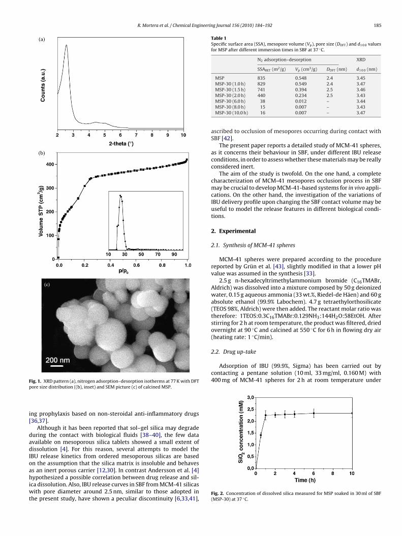
R. Mortera et al. / Chemical Engineering Journal 156 (2010) 184–192 185
Fp
i[
dadIoahiwt
Table 1Specific surface area (SSA), mesopore volume (Vp), pore size (DDFT) and d1 0 0 valuesfor MSP after different immersion times in SBF at 37 ◦C.
N2 adsorption–desorption XRD
SSABET (m2/g) Vp (cm3/g) DDFT (nm) d1 0 0 (nm)
MSP 835 0.548 2.4 3.45MSP-30 (1.0 h) 829 0.549 2.4 3.47MSP-30 (1.5 h) 741 0.394 2.5 3.46MSP-30 (2.0 h) 440 0.234 2.5 3.43
2.2. Drug up-take
Adsorption of IBU (99.9%, Sigma) has been carried out bycontacting a pentane solution (10 ml, 33 mg/ml, 0.160 M) with400 mg of MCM-41 spheres for 2 h at room temperature under
ig. 1. XRD pattern (a), nitrogen adsorption–desorption isotherms at 77 K with DFTore size distribution ((b), inset) and SEM picture (c) of calcined MSP.
ng prophylaxis based on non-steroidal anti-inflammatory drugs36,37].
Although it has been reported that sol–gel silica may degradeuring the contact with biological fluids [38–40], the few datavailable on mesoporous silica tablets showed a small extent ofissolution [4]. For this reason, several attempts to model the
BU release kinetics from ordered mesoporous silicas are basedn the assumption that the silica matrix is insoluble and behavess an inert porous carrier [12,30]. In contrast Andersson et al. [4]
ypothesized a possible correlation between drug release and sil-ca dissolution. Also, IBU release curves in SBF from MCM-41 silicasith pore diameter around 2.5 nm, similar to those adopted in
he present study, have shown a peculiar discontinuity [6,33,41],
MSP-30 (6.0 h) 38 0.012 – 3.44MSP-30 (8.0 h) 15 0.007 – 3.43MSP-30 (10.0 h) 16 0.007 – 3.47
ascribed to occlusion of mesopores occurring during contact withSBF [42].
The present paper reports a detailed study of MCM-41 spheres,as it concerns their behaviour in SBF, under different IBU releaseconditions, in order to assess whether these materials may be reallyconsidered inert.
The aim of the study is twofold. On the one hand, a completecharacterization of MCM-41 mesopores occlusion process in SBFmay be crucial to develop MCM-41-based systems for in vivo appli-cations. On the other hand, the investigation of the variations ofIBU delivery profile upon changing the SBF contact volume may beuseful to model the release features in different biological condi-tions.
2. Experimental
2.1. Synthesis of MCM-41 spheres
MCM-41 spheres were prepared according to the procedurereported by Grün et al. [43], slightly modified in that a lower pHvalue was assumed in the synthesis [33].
2.5 g n-hexadecyltrimethylammonium bromide (C16TMABr,Aldrich) was dissolved into a mixture composed by 50 g deionizedwater, 0.15 g aqueous ammonia (33 wt.%, Riedel-de Häen) and 60 gabsolute ethanol (99.9% Labochem). 4.7 g tetraethylorthosilicate(TEOS 98%, Aldrich) were then added. The reactant molar ratio wastherefore: 1TEOS:0.3C16TMABr:0.129NH3:144H2O:58EtOH. Afterstirring for 2 h at room temperature, the product was filtered, driedovernight at 90 ◦C and calcined at 550 ◦C for 6 h in flowing dry air(heating rate: 1 ◦C/min).
Fig. 2. Concentration of dissolved silica measured for MSP soaked in 30 ml of SBF(MSP-30) at 37 ◦C.

186 R. Mortera et al. / Chemical Engineering Journal 156 (2010) 184–192
t 77 K
cttSfidr
st
2
sfseS
Fig. 3. XRD patterns (a) and nitrogen adsorption–desorption isotherms a
ontinuous stirring. The amount of drug loaded was evaluatedhrough UV–vis spectrophotometry from the change in concen-ration in the pentane solution. The instrument was a Cary 500can, operating at � = 263 nm, where the molar extinction coef-cient (ε�) is 320 mol−1 cm−1. The calibration curve has beenrawn using pentane solutions of IBU in the same concentrationange.
The as-synthesized and the IBU-loaded MCM-41 mesoporousilica particles are hereafter referred to MSP and MSP-IBU, respec-ively.
.3. Drug release and MSP behaviour in SBF
The drug release kinetics were evaluated in vitro by soaking the
ame amount of MSP-IBU (100 mg) in different volumes (rangingrom 10 ml to 210 ml) of stirred SBF kept at 37 ◦C. The number repre-enting the volume used in milliliter features last in the acronym:.g. MSP-IBU-30 indicates 100 mg of MSP-IBU soaked in 30 ml ofBF.(b) of MSP-30 obtained after different immersion times in SBF at 37 ◦C.
At different time intervals, a small amount of SBF (0.1 ml)was collected and analysed through UV–vis spectrophotometry toassess the amount of released IBU. In SBF the maximum wavelengthis still at � = 263 nm, but ε� is equal to 440 mol−1 cm−1.
In a parallel set of experiments, 100 mg of IBU-free MSP sam-ples have been soaked directly in different volumes of SBF at 37 ◦C(the related acronym being MSP-X, X referring to the SBF volumein milliliter). The amount of silica dissolved in SBF was measuredusing molybdenum blue as a tracer and the concentration wasmonitored by means of UV–vis spectrophotometry (� = 410 nm)[4,44].
2.4. Characterization
The samples have been characterized by means of powder
X-ray diffraction (X’Pert Philips, CuK� radiation), nitrogen adsorp-tion/desorption measurements at 77 K (Quantachrome Autosorb1)and field emission scanning electron microscopy (Assing FESEMSupra 25) associated with energy dispersive spectroscopy (EDS,Oxford Instrument INCA X-Sight). Characterization has been car-
R. Mortera et al. / Chemical Engineering Journal 156 (2010) 184–192 187
Fd
rdm6
N
3
3
roToriooa
TI
ig. 4. Trend of mesoporous volume (a) and pore size distributions (b) of MSP-30uring the contact with SBF at 37 ◦C.
ied out both on MCM-41 as such and after immersion in SBF forifferent times. Before nitrogen adsorption/desorption measure-ents, each sample has been outgassed at room temperature forh.
Pore size has been evaluated through the DFT method, using theLDFT equilibrium model for cylindrical pore [45].
. Results and discussion
.1. MCM-41 spheres
XRD pattern of MSP shows the typical peak due to the (1 0 0)eflection of the ordered 2D hexagonal mesostructure at 2� valuef 2.56◦ (Fig. 1a). The corresponding d1 0 0 results 3.45 nm (Table 1).he (1 1 0) and (2 0 0) reflections appear ill-defined at 2� valuesf 4.44◦ and 5.02◦, respectively. Lowering the synthesis pH with
espect the value reported by Grün et al. [43] causes a broaden-ng of d1 1 0 and d2 0 0 peaks. This is ascribed to a lower mesoscopicrder, because a lower alkalinity usually favours the formationf disordered phases [46]. The cell parameter (a), calculated as= (2/√3)d1 0 0, results to be 3.98 nm.
able 2buprofen released from MSP-IBU-X after different immersion times in X ml of SBF at 37 ◦
Ibuprofen released (%)
2 h 10 h 17
MSP-IBU-10 53 57 6MSP-IBU-30 69 82 9MSP-IBU-210 78 99 10
Fig. 5. SEM pictures and EDS analysis (inset) of MSP-30 after 1 h (a) and 6 h (b) ofimmersion in SBF at 37 ◦C.
Fig. 1b reports nitrogen adsorption/desorption isotherms of cal-cined MSP. These are of type IV, and exhibit filling of the mesoporesat relative pressure p/p0 below 0.3. The values of BET specific sur-face area and mesopores volume are 835 m2/g and 0.55 cm3/g,respectively (Table 1). Pore diameter, as evaluated by DFT model(N2 on SiO2 kernel, fitting error = 0.65%), results 2.4 nm (Fig. 1b,inset). From the value of cell parameter a, wall thickness turns outto be 1.58 nm. The DFT fitting is reported in Figure S1 of the sup-plementary information.
MCM-41 material is in the form of spheres with size rangingfrom 200 nm to 800 nm, similarly to what observed by Grün et al.[43] (Fig. 1c).
3.2. MSP modification in SBF
Fig. 2 reports the profile describing the silica dissolution to SBFwith time for MSP-30. A relatively fast process is observed for con-tact times below 1 h, when the concentration of silica in the liquidphase is about 2.2 mM (corresponding to about 4% weight of initial
silica). Dissolution then practically stops and the concentration inthe liquid phase reaches only 2.3 mM after 10 h. The same exper-iments in two different volumes of SBF (10 ml and 210 ml) gavesimilar results (not reported).C and corresponding ibuprofen concentration in solution.
Ibuprofen concentration (mg/ml)
0 h 2 h 10 h 170 h
0 1.84 1.98 2.072 0.79 0.94 1.050 0.13 0.16 0.16

1 ineering Journal 156 (2010) 184–192
eidlsk
mro
ttotac
1astimc
tapfisctd
1er
eT(
sw(ac
mmi
3
([
taioc
Fig. 6. Kinetics of ibuprofen release from 100 mg of MSP-IBU to 10 ml (×), 30 ml (©)
88 R. Mortera et al. / Chemical Eng
Literature data on amorphous sol–gel silica show a sizablextent of dissolution also in the first 10 h of contact with biolog-cal fluids [38–40]. No results seem to be available concerning theissolution in SBF of dispersed mesoporous silica spheres. The few
iterature data on mesoporous silica tablets, containing IBU, show amaller extent of dissolution than the sol–gel silica, but a differentinetics with respect to that here reported for MSP-30 [4].
A reason for the lower extent of dissolution observed for orderedesoporous silicas with respect to amorphous sol–gel silica may
eside in the ageing and the subsequent calcination process typicalf the former systems [47].
Fig. 3a shows XRD patterns of MSP-30 after different soakingimes in SBF. These do not change significantly, so showing thathe mesostructure is stable during contact with SBF. A broadeningf (1 1 0) and (2 0 0) reflections is observed with respect to the pat-ern reported in Fig. 1a, but the mesoscopic order is evident evenfter 6 h of soaking (Fig. 3a) [47] and the d1 0 0 distance does remainonstant (Table 1).
Fig. 3b reports nitrogen adsorption–desorption isotherms afterh, 1.5 h, 2 h and 6 h of soaking. The initial isotherms show the char-cteristic mesopore filling at p/p0 below 0.3 (type IV). Increasing theoaking time, the N2 adsorbed volume decreases and the charac-eristic mesopore filling becomes less evident. After 6 h of soaking,sotherms do not show any longer the pore filling associated with
esopores. A hysteresis loop at p/p0 above 0.5 is observed in allases, probably due to the interparticle porosity [48].
The mesoporous volume remains constant at 0.55 cm3/g duringhe first hour of contact with SBF, then decays, reaching 0.01 cm3/gfter 8 h, as observed in Fig. 4a and Table 1. The corresponding DFTore size distributions (N2 on SiO2 kernel, fitting error = 0.65%) con-rm this trend with a marked decrease of the peak intensity withoaking time. The presence of mesopores at about 2.4 nm is dis-ernible up to 6 h of SBF contact (Fig. 4b). It is worth noting thathe decrease in mesoporous volume is not accompanied by anyecrease in pore diameter, which remains constant.
These data indicate a progressive occlusion of mesopores afterh of soaking rather than a progressive reduction of pores diam-ter. We ascribe this modification to dissolution of silica ande-precipitation as silica gel at the pores mouth [49,50].
The decrease in mesopore volume may be represented as anxponential with a negative time constant (−˛) equal to −0.82 h−1.he asymptotic value, reached after 8 h of SBF contact, is negligible0.01 cm3/g) and it will not be considered further.
SEM pictures of MSP-30 after 1 h (Fig. 5a) and 6 h (Fig. 5b) ofoaking in SBF reveal that particles maintain their spherical shapeithout any remarkable variations with respect to calcined MSP
Fig. 1c). EDS analysis show the peak ascribable to silica at 1 h andt 6 h (Fig. 5, insets). The presence in both cases of traces of sodiumhloride is due to residues coming from SBF solution.
MSP-10 and MSP-210 exhibit a very similar occlusion ofesopores during their contact with SBF (Figure S2 of the supple-entary information), so showing a behaviour that seems to be
ndependent from the soaking volume.
.3. Drug up-take and release
The amount of IBU adsorbed by MSP-IBU is about 34.7% (w/w)hereafter referred to as IL), in good agreement with the literature6,51–53].
Fig. 6 shows the kinetics of IBU release from 100 mg of MSP-IBU
o 10 ml, 30 ml and 210 ml, respectively, of stirred SBF maintainedt 37 ◦C. Data in Fig. 6a are expressed as the concentration ofbuprofen in SBF as a function of time and refer to the first 10 hf release, whereas in Fig. 6b, the percentage of IBU released isonsidered.and 210 ml (�) of stirred SBF maintained at 37 ◦C: concentration of ibuprofen in SBF(a) and amount released (b). The first 10 h of release are depicted in detail while theprofiles up to 170 h are reported as inset in (b).
The amount of ibuprofen in the solid phase is 34.7 mg, equiva-lent to 1.6 × 10⁻⁴ moles. Assuming the total release from the solidphase, it can be anticipated that the limit value for the concentra-tion in 210 ml, 30 ml and 10 ml of solution should be 0.16 mg/ml,1.16 mg/ml and 3.47 mg/ml respectively. This latter value can-not be reached because the solubility of ibuprofen in SBF (CS)is 2.8 mg/ml [54]. Fig. 6a shows, however, that only in the caseof MSP-IBU-210 the concentration of released ibuprofen reachesthe expected values. The three release profiles differ significantly:after 10 h MSP-IBU-10 released about 57% of incorporated drug,whereas about 82% and about 100% of ibuprofen was releasedfrom MSP-IBU-30 and from MSP-IBU-210, respectively (Table 2 andFig. 6b).
It is worth noting that the release curve of MSP-IBU-30 corre-sponds to the conditions commonly used in literature and presentsthe trend reported for MCM-41 with pore size around 2.5 nm[6,33,41,42,53].
In order to evidence possible pores occlusion during ibuprofenrelease, N2 adsorption–desorption isotherms and X-ray diffractionpatterns of MSP-IBU-210, MSP-IBU-30 and MSP-IBU-10 after 2 h
and 6 h of SBF contact have been collected. Results are reported inFig. 7.Whereas XRD patterns (inset) confirm the stability of the meso-porous structure, N2 physisorption data show for all samples an
R. Mortera et al. / Chemical Engineering Journal 156 (2010) 184–192 189
F ternsa
oItps6aodsM
lrcttlct
tro[l
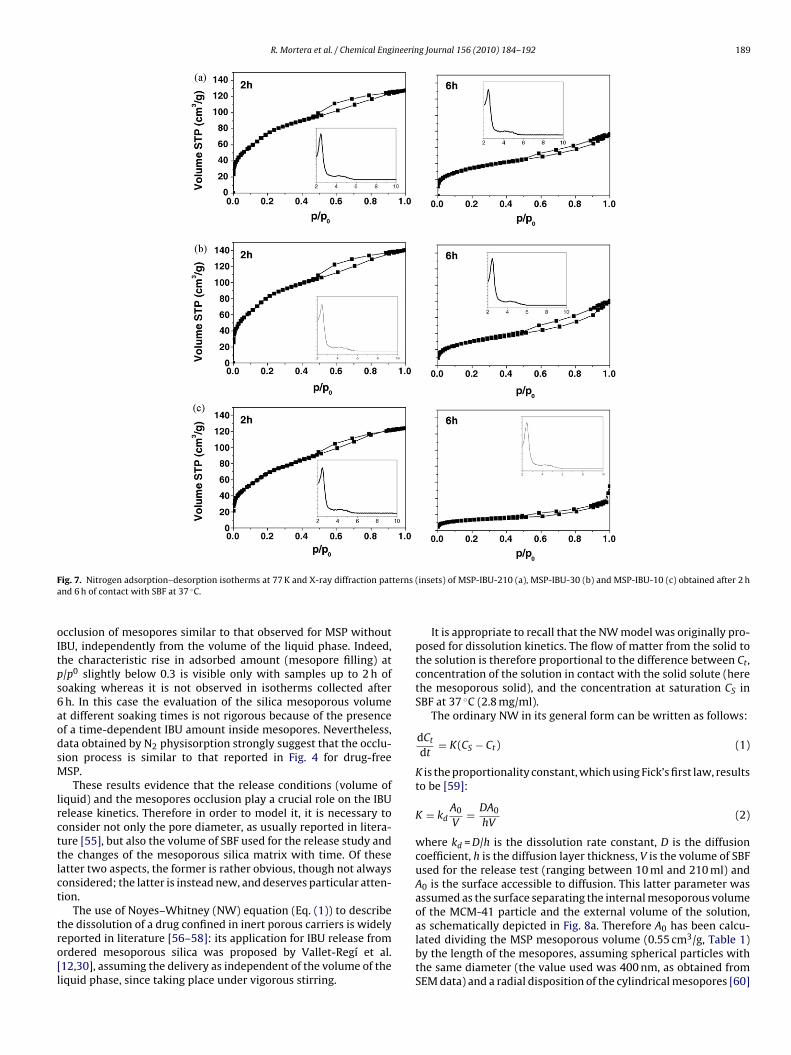
ig. 7. Nitrogen adsorption–desorption isotherms at 77 K and X-ray diffraction patnd 6 h of contact with SBF at 37 ◦C.
cclusion of mesopores similar to that observed for MSP withoutBU, independently from the volume of the liquid phase. Indeed,he characteristic rise in adsorbed amount (mesopore filling) at/p0 slightly below 0.3 is visible only with samples up to 2 h ofoaking whereas it is not observed in isotherms collected afterh. In this case the evaluation of the silica mesoporous volumet different soaking times is not rigorous because of the presencef a time-dependent IBU amount inside mesopores. Nevertheless,ata obtained by N2 physisorption strongly suggest that the occlu-ion process is similar to that reported in Fig. 4 for drug-freeSP.These results evidence that the release conditions (volume of
iquid) and the mesopores occlusion play a crucial role on the IBUelease kinetics. Therefore in order to model it, it is necessary toonsider not only the pore diameter, as usually reported in litera-ure [55], but also the volume of SBF used for the release study andhe changes of the mesoporous silica matrix with time. Of theseatter two aspects, the former is rather obvious, though not alwaysonsidered; the latter is instead new, and deserves particular atten-ion.
The use of Noyes–Whitney (NW) equation (Eq. (1)) to describe
he dissolution of a drug confined in inert porous carriers is widelyeported in literature [56–58]: its application for IBU release fromrdered mesoporous silica was proposed by Vallet-Regí et al.12,30], assuming the delivery as independent of the volume of theiquid phase, since taking place under vigorous stirring.(insets) of MSP-IBU-210 (a), MSP-IBU-30 (b) and MSP-IBU-10 (c) obtained after 2 h
It is appropriate to recall that the NW model was originally pro-posed for dissolution kinetics. The flow of matter from the solid tothe solution is therefore proportional to the difference between Ct,concentration of the solution in contact with the solid solute (herethe mesoporous solid), and the concentration at saturation CS inSBF at 37 ◦C (2.8 mg/ml).
The ordinary NW in its general form can be written as follows:
dCt
dt= K(CS − Ct) (1)
K is the proportionality constant, which using Fick’s first law, resultsto be [59]:
K = kdA0
V= DA0
hV(2)
where kd = D/h is the dissolution rate constant, D is the diffusioncoefficient, h is the diffusion layer thickness, V is the volume of SBFused for the release test (ranging between 10 ml and 210 ml) andA0 is the surface accessible to diffusion. This latter parameter wasassumed as the surface separating the internal mesoporous volumeof the MCM-41 particle and the external volume of the solution,
as schematically depicted in Fig. 8a. Therefore A0 has been calcu-lated dividing the MSP mesoporous volume (0.55 cm3/g, Table 1)by the length of the mesopores, assuming spherical particles withthe same diameter (the value used was 400 nm, as obtained fromSEM data) and a radial disposition of the cylindrical mesopores [60]
190 R. Mortera et al. / Chemical Engineering Journal 156 (2010) 184–192
Fafi
(tec
D
ır
Table 3List of constants and parameters used in the standard and modified Noyes–Whitneysimulation of ibuprofen release from MSP-IBU to different volumes of SBF.
Value
Saturation solubility CS 2.8 mg/mlIbuprofen radius a 0.5 nmMesopores radius r 1.2 nmDiffusion coefficient D 8.8 × 10⁻⁸ cm2/hDissolution rate constant kd 0.0022 cm/hAccessible surface at t = 0 h A0 27.00 cm2/mgWeight of MSP-IBU sample P 100 mgIbuprofen adsorbed by MSP-IBU IL 34.7%Time constant ˛ 0.82 h−1
ig. 8. Radial disposition of the cylindrical mesopores inside the silica spheres withmean diameter of 4070 nm (a) Trend of the accessible surface (A(t)) during therst 10 h of contact with SBF at 37 ◦C (b).
Table 3). D has been determined using the Stokes–Einstein equa-ion to derive the IBU diffusivity for the plasma (Dpl) and the Renkinquation to correct the value with the sterical hindrance (ı) and theonstrictivity (ωr) [55]:
= Dplı�r
�
= Dpl(1 − a/r)2(1 − 2.1(a/r) + 2.09(a/r)3 − 0.95(a/r)5)
�(3)
and ωr are both functions of the ratio between the IBU and the poreadii (a/r) and � is the tortuosity, assumed equal to one, since the
Fig. 9. Ibuprofen release kinetics of MSP-IBU-210 (a), MSP-IBU-30 (b) and MSP-IBU-10 (c) during the first 10 h of contact with SBF at 37 ◦C (©), compared with thesimulation using the Noyes–Whitney standard (×) and modified (�) equations.

ineerin
mDwi[
C
W
wa
pc
ftr
m
K
˛a⎧⎨⎩
{
nm
imopy
tdh
sv
ftswvr
[[
[
[
[[[
[
[
[
[
R. Mortera et al. / Chemical Eng
esopores are supposed cylindrical [55]. kd was calculated dividingby the diffusion layer thickness (h), here assumed to coincideith the mesopores length [12]. The obtained value (0.0022 cm/h)
s in agreement with what reported in literature for similar systems12,54].
Forcing Ct(0) = 0, Eq. (1) yields:
t = CS(1 − e−Kt) (4)
In terms of release as fraction of adsorbed drug, it results:
t = CtV
PIL(5)
here P is the weight of the MSP-IBU sample used for the releasend IL is the percentage amount of ibuprofen adsorbed by MSP-IBU.
To model the progressive occlusion of the mesopores, the pro-ortionality constant K has to be replaced with a time-dependentoefficient K(t):
dCt
dt= K(t)(CS − Ct) (6)
The time-dependent part of K(t) is the accessible surface to dif-usion A(t), calculated on the basis of the exponential decay ofhe mesoporous volume starting after 1 h of contact with SBF, aseported in Fig. 4a and Table 1.
Taking into account the occurrence of a process affecting theesopore volume, only after 1 h, K(t) can be written as:
(t) = D
hVA(t) ⇒
⎧⎨⎩
K(t) = D
hVA0 t ≤ 1 h
K(t) = D
hVA0e−˛(t−1) t > 1 h
(7)
being the time constant describing the exponential decay of theccessible surface area (Fig. 8b). The NW equation results:
dCt
dt= K(CS − Ct) t ≤ 1 h
dCt
dt= (Ke−˛(t−1))(CS − Ct) t > 1 h
(8)
Forcing Ct(0) = 0 and Ct(1) = CS(1–e−K), the solution for Eq. (8) is:
Ct = CS(1 − e−Kt) t ≤ 1 h
Ct = CS(1 − e(K/˛)e−˛(t−1)−K((1+˛)/˛)) t > 1 h(9)
Eqs. (9) and (4) obviously coincide up to 1 h of release wheno mesopore occlusion occurred, and diverge when the effect ofesopores occlusion becomes prevalent.The comparison between the experimental IBU release kinet-
cs of MSP-IBU-210, and the trends predicted by the standard NWodel (1) and the modified one (8) is reported in Fig. 9a. After 1 h
f immersion in SBF, as expected, the standard NW equation yieldsoor results. It is instead noticeable that the modified NW (square)ields a satisfactory prediction of the experimental profile.
A similar behaviour is observed for MSP-IBU-30 (Fig. 9b). Onlyhe modified simulation profile fits satisfactorily the experimentalata, whereas the standard NW equation diverges after the firstour and indicates a total delivery of the adsorbed IBU in 3 h.
The different time needed for total delivery predicted by the NWtandard equation in the two cases comes from the different SBFolumes considered.
Fig. 9c shows the experimental and the simulated release curvesor MSP-IBU-10. As for the previous cases, the modified NW equa-
ion approximates the whole experimental profile, whereas thetandard equation gives a good fitting only up to 1 h. At varianceith what discussed previously, in this case, due to the lowerolume, the standard equation predicts that the concentration ofeleased ibuprofen reaches CS before 10 h of immersion.
[
[[[
g Journal 156 (2010) 184–192 191
4. Conclusions
Two processes seem to be involved as it concerns the soakingof MCM-41 silica powders in SBF, one consisting in the solubilisa-tion of up to 4% of silica, but not affecting the mesopore volume,the other not really releasing silica into SBF but causing the poreblocking.
The release profiles from IBU-loaded MCM-41 particles differsignificantly as a function of the volume of SBF used for the deliverytest, in agreement with the description of the release process interms of the classical NW equation.
This latter accounts for the release kinetics for all values of SBFvolume only for release times shorter than 1 h. For longer times,due to occlusion of mesopores, the proportionality constant of thestandard NW equation is replaced by a time-dependent coefficient.A fair agreement with experimental data up to 10 h of release hasbeen obtained, without the use of any adjustable parameters.
This result suggests the possibility of predicting the drug deliv-ery from MSP silica particles to different amounts of SBF, thusmimicking different biological situations and different pathologicalrequirements.
Acknowledgements
The authors would like to thank Regione Piemonte (projectNanosafe) for financial support.
Appendix A. Supplementary data
Supplementary data associated with this article can be found, inthe online version, at doi:10.1016/j.cej.2009.10.018.
References
[1] M. Vallet-Regí, A. Ramila, R.P. del Real, J. Perez-Pariente, Chem. Mater. 13 (2001)308.
[2] C.T. Kresge, M.E. Leonowicz, W.J. Roth, J.C. Vartuli, J.S. Beck, Nature 359 (1992)710.
[3] C. Tourne-Peteilh, D.A. Lerner, C. Charnay, L. Nicole, S. Begu, J.M. Devoisselle,Chemphyschem 4 (2003) 281.
[4] J. Andersson, J. Rosenholm, S. Areva, M. Linden, Chem. Mater. 16 (2004) 4160.[5] G. Cavallaro, P. Pierro, F.S. Palumbo, F. Testa, L. Pasqua, R. Aiello, Drug Deliv. 11
(2004) 41.[6] P. Horcajada, A. Ramila, J. Perez-Pariente, M. Vallet-Regí, Micropor. Mesopor.
Mater. 68 (2004) 105.[7] F.Y. Qu, G.S. Zhu, H.M. Lin, W.W. Zhang, J.Y. Sun, S.G. Li, S.L. Qiu, J. Solid State
Chem. 179 (2006) 2027.[8] T.H. Chung, S.H. Wu, M. Yao, C.W. Lu, Y.S. Lin, Y. Hung, C.Y. Mou, Y.C. Chen, D.M.
Huang, Biomaterials 28 (2007) 2959.[9] J. Lu, M. Liong, J.I. Zink, F. Tamanoi, Small 3 (2007) 1341.10] I.I. Slowing, B.G. Trewyn, V.S.Y. Lin, J. Am. Chem. Soc. 129 (2007) 8845.11] V. Cauda, B. Onida, B. Platschek, L. Muhlstein, T. Bein, J. Mater. Chem. 18 (2008)
5888.12] M. Manzano, V. Aina, C.O. Arean, F. Balas, V. Cauda, M. Colilla, M.R. Delgado, M.
Vallet-Regí, Chem. Eng. J. 137 (2008) 30.13] E. Ruiz-Hernandez, A. Lopez-Noriega, D. Arcos, M. Vallet-Regí, Solid State Sci.
10 (2008) 421.14] B.G. Trewyn, J.A. Nieweg, Y. Zhao, V.S.Y. Lin, Chem. Eng. J. 137 (2008) 23.15] S.B. Wang, Micropor. Mesopor. Mater. 117 (2009) 1.16] C. Tourne-Peteilh, D. Brunel, S. Begu, B. Chiche, F. Fajula, D.A. Lerner, J.M. Devois-
selle, New J. Chem. 27 (2003) 1415.17] C.Y. Lai, B.G. Trewyn, D.M. Jeftinija, K. Jeftinija, S. Xu, S. Jeftinija, V.S.Y. Lin, J. Am.
Chem. Soc. 125 (2003) 4451.18] P.P. Yang, Z.W. Quan, C.X. Li, X.J. Kang, H.Z. Lian, J. Lin, Biomaterials 29 (2008)
4341.19] J.L. Vivero-Escoto, I.I. Slowing, C.W. Wu, V.S.Y. Lin, J. Am. Chem. Soc. 131 (2009)
3462.20] E. Ruiz-Hernandez, A. Lopez-Noriega, D. Arcos, I. Izquierdo-Barba, O. Terasaki,
M. Vallet-Regí, Chem. Mater. 19 (2007) 3455.
21] M. Arruebo, W.Y. Ho, K.F. Lam, X.G. Chen, J. Arbiol, J. Santamaria, K.L. Yeung,Chem. Mater. 20 (2008) 486.22] J. Rosenholm, M. Linden, J. Control. Release 128 (2008) 157.23] L. Pasqua, S. Cundari, C. Ceresa, G. Cavaletti, Curr. Med. Chem. 16 (2009) 3054.24] G. Wang, A.N. Otuonye, E.A. Blair, K. Denton, Z.M. Tao, T. Asefa, J. Solid State
Chem. 182 (2009) 1649.

1 ineerin
[
[[
[[[
[
[
[
[
[[[
[
[
[
[[
[
[[[
[
[
[[
[[
[[
[
[ences, vol. 15, Marcel Dekker, Inc., New York, 1982.
[57] N.V. Mulye, S.J. Turco, Drug Dev. Ind. Pharm. 21 (1995) 943.[58] P. Costa, J.M.S. Lobo, Eur. J. Pharm. Sci. 13 (2001) 123.
92 R. Mortera et al. / Chemical Eng
25] D.R. Radu, C.Y. Lai, K. Jeftinija, E.W. Rowe, S. Jeftinija, V.S.Y. Lin, J. Am. Chem.Soc. 126 (2004) 13216.
26] I.I. Slowing, B.G. Trewyn, V.S.Y. Lin, J. Am. Chem. Soc. 128 (2006) 14792.27] Z.M. Tao, M.P. Morrow, T. Asefa, K.K. Sharma, C. Duncan, A. Anan, H.S. Penefsky,
J. Goodisman, A.K. Souid, Nano Letters 8 (2008) 1517.28] I.I. Slowing, C.W. Wu, J.L. Vivero-Escoto, V.S.Y. Lin, Small 5 (2009) 57.29] M. Vallet-Regí, Chem. Eng. J. 137 (2008) 1.30] M. Vallet-Regí, F. Balas, M. Colilla, M. Manzano, Prog. Solid State Chem. 36
(2008) 163.31] M. Vallet-Regí, M. Colilla, I. Izquierdo-Barba, J. Biomed. Nanotechnol. 4 (2008)
1.32] V. Cauda, S. Fiorilli, B. Onida, E. Verne, C.V. Brovarone, D. Viterbo, G. Croce, M.
Milanesio, E. Garrone, J. Mater. Sci.: Mater. Med. 19 (2008) 3303.33] R. Mortera, B. Onida, S. Fiorilli, V. Cauda, C.V. Brovarone, F. Baino, E. Verne, E.
Garrone, Chem. Eng. J. 137 (2008) 54.34] C. Vitale-Brovarone, F. Baino, M. Miola, R. Mortera, B. Onida, E. Verne, J. Mater.
Sci.: Mater. Med. 20 (2009) 809.35] T. Kokubo, H. Takadama, Biomaterials 27 (2006) 2907.36] L.E. Dahners, B.H. Mullis, J. Am. Acad. Orthop. Surg. 12 (2004) 139.37] M. Fransen, C. Anderson, J. Douglas, S. MacMahon, B. Neal, R. Norton, M. Wood-
ward, I.D. Cameron, R. Crawford, S.K. Lo, G. Tregonning, M. Windolf, Br. Med. J.333 (2006) 519.
38] M. Ahola, P. Kortesuo, I. Kangasniemi, J. Kiesvaara, A. Yli-Urpo, Int. J. Pharm. 195(2000) 219.
39] T. Czuryszkiewicz, J. Ahvenlammi, P. Kortesuo, M. Ahola, F. Kleitz, M. Jokinen,
M. Linden, J.B. Rosenholm, J. Non-Cryst. Solids 306 (2002) 1.40] J.D. Bass, D. Grosso, C. Boissiere, E. Belamie, T. Coradin, C. Sanchez, Chem. Mater.19 (2007) 4349.
41] M. Vallet-Regí, Chem. Eur. J. 12 (2006) 5934.42] R. Mortera, S. Fiorilli, E. Garrone, B. Onida, Stud. Surf. Sci. Catal. B 174 (2008)
1001.
[
[
g Journal 156 (2010) 184–192
43] M. Grün, K.K. Unger, A. Matsumoto, K. Tsutsumi, Micropor. Mesopor. Mater. 27(1999) 207.
44] J.B. Mullin, J.P. Riley, Anal. Chim. Acta 12 (1955) 162.45] M. Thommes, R. Kohn, M. Froba, Appl. Surf. Sci. 196 (2002) 239.46] F. Di Renzo, A. Galarneau, P. Trens, F. Fajula, in: F. Schüth, K. Sing, J. Weitkamp
(Eds.), Handbook of Porous Materials, Wiley–VCH, 2002, p. 1311.47] M.V. Landau, S.P. Varkey, M. Herskowitz, O. Regev, S. Pevzner, T. Sen, Z. Luz,
Micropor. Mesopor. Mater. 33 (1999) 149.48] E. Prouzet, F. Cot, G. Nabias, A. Larbot, P. Kooyman, T.J. Pinnavaia, Chem. Mater.
11 (1999) 1498.49] P.J. Li, K. Nakanishi, T. Kokubo, K. Degroot, Biomaterials 14 (1993) 963.50] P.J. Li, I. Kangasniemi, K. Degroot, T. Kokubo, A.U. Yliurpo, J. Non-Cryst. Solids
168 (1994) 281.51] M. Colilla, M. Manzano, M. Vallet-Regí, Int. J. Nanomed. 3 (2008) 403.52] S.S. Huang, P.P. Yang, Z.Y. Cheng, C.X. Li, Y. Fan, D.Y. Kong, J. Lin, J. Phys. Chem.
C 112 (2008) 7130.53] A. Hillerstrom, J. van Stam, M. Andersson, Green Chem. 11 (2009) 662.54] A. Ramila, R.P. Del Real, R. Marcos, P. Horcajada, M. Vallet-Regí, J. Sol-Gel Sci.
Technol. 26 (2003) 1195.55] E.M. Renkin, F.E. Curry, in: G. Giebisch, D.C. Tosteson, H.H. Ussing (Eds.), Mem-
brane Transport in Biology, vol. IV, Springer Verlag, Berlin, 1979.56] M. Gibaldi, D. Perrier, Pharmacokinetics, Drugs and the Pharmaceutical Sci-
59] J. Crank, The Mathematics of Diffusion, 2nd ed., Oxford University Press, Oxford,1975.
60] C. Amatore, Chem. Eur. J. 14 (2008) 5449.


![[Model #SBF: Steel Bed Frame] - Assembly Instructions](https://static.fdokumen.com/doc/165x107/633d7723decf89045f05ef25/model-sbf-steel-bed-frame-assembly-instructions.jpg)