Femtosecond laser ionization mass spectrometric analysis of layers grown by pulsed laser deposition
Upload
independentCategory
view
0download
0
Anal. Chem. 1995,67, 843-848
Polyethylene Membrane as a Sample Support for Direct Matrix-Assisted Laser Desorption/lonization Mass Spectrometric Analysis of High Mass Proteins
James A. Blackledge and Anthony J. Alexander*
Bristol-Myers Squibb, Analytical Research & Development, Pharmaceutical Research Institute, P. 0. Box 4755, Syracuse, New York 1322 1-4 755
Matrix-assisted laser desorption/ionization mass spec- trometry (MALDI-MS) of high mass proteins ('100 000 Da), directly deposited on polyethylene membranes, is demonstrated. The spectral quality obtained, using stan- dard sample preparation conditions, is equal or superior to that obtained with metal sample stages. Compared to the use of poly(vinylidene difluoride) transfer membranes, this material allows the acquisition of excellent quality MALDI mass spectra from high-mass proteins with a standard W laser. This gain in capability is not at the expense of either mass accuracy or signal reproducibility; both approach that obtained with standard sample prepa- rations on stainless steel. In addition, for the applications shown, the use of PE as a sample support reduces the severe ion suppression effects typically observed in the MAL,DI analysis of high-mass mixtures. This permits more precise mass measurements to be made via the use of internal calibration and is illustrated by the mass measurement of a chimeric mouse/human antibody (MW -150 000 Da) by coaddition of bovine albumin dimer (MW -130 000 Da).
Matrix-assisted laser desorption/ionization timeof-flight mass spectrometry (MALDI-TOF MS) routinely gives molecular weight values with an accuracy of f 0.1% or better and is increasingly becoming the method of choice for the mass measurement of biopolymers.' The technique is simple, is rugged, has a mass range in excess of 200000 Da, and is extremely sensitive, requiring low picomole to subpicomole amounts of material. Additionally, the technique is relatively insensitive to the presence of various salts and buffers that are often associated with the isolation of biomolecules.2
Sodium dodecyl sulfate polyacrylamide gel electrophoresis (SDS-PAGE) is a classic technique used for the separation and molecular weight (MW) determination of biomolecules. Unfor- tunately, it gives only a rough estimation of molecular weight, with values of 15-1096 being typical. It may also be subject to gross systematic errors if the species under investigation has electrophoretic migration behavior different than the molecular weight markers3 As the separated protein bands in an SDS- PAGE gel are typically transferred (electroblotted) onto a sup
(1) Hillenkamp, F.; Karas, M.; Beavis, R C.; Chait, B. T. Anal. Chem. 1991,
(2) Beavis, R C.; Chait, B. T. Proc. Nafl. Acad. Sci. U.S.A. 1990, 87, 6873- 63, 1193A-1203A.
6877.
0003-2700/95/0367-0843$9.00/0 0 1995 American Chemical Society
porting membrane prior to further analysis, most of the effort to combine the resolving power of SDS-PAGE with the mass accuracy of MALDI-MS has focused on desorbing proteins directly from the transfer membrane. Membranes that have been cur- rently studied for this purpose are nitro~ellulose,4~~ nylon? poly- (vinylidne difluoride) WF) ,7-9 and Zitex.Io
While these materials function well for binding electrotrans- ferred protein bands, their use as sample supports for MALDI- MS is still somewhat problematic. With a UV laser, useful MALDI mass spectra have so far only been obtained for low molecular weight peptides and proteins (5-26 500 Da).7-9 A preliminary report has very recently appeared on the use of Zitex, a poly- (tetrafhoroethylene) membrane manufactured with a range of pore sizes, as a support for UV MALDI-MS analysis.'O Results were shown for a range of directly deposited peptides and proteins up to cytochrome c in mass (12 360 Da) using Zitex G104 (5-6 pm functional pore size, 0.004 in. thickness). While these results are encouraging, this present limitation on mass range does not permit utilization of the full mass measuring potential of TOF- MS. Also, electrospray ionization @SI) operates very effectively over this mass range, and methods for extracting SDS-PAGE protein bands for subsequent ESI-MS analysis are presently in the literature." Detection of higher molecular weight proteins, up to bovine serum albumin @SA) at 66430 Da, has been achieved with electroblotted PVDF by employing an IR laser.* However, compared to UV lasers, IR lasers have high shot-to- shot variability in output power, resulting in greater signal irreproducibility.s Also, due to their greater abrading power, typically only two to three spectra can be acquired from the same sample spot with a IR laserG8
(3) Creighton, T. E. Proteins (Structures and Molecular Principles); W. H.
(4) Klarskov, K; Roepstorff, P. Eiol. Mass Spectrom. 1993, 22, 433-440. (5) Mock, K K; Sutton, C. W.; Cottrell, J. S. Rapid Commun. Mass Specfrom.
(6) Zaluzek, E. J.; Gage, D. A; Allison, J.; Watson J. T.]. Am. SOC. Mass Spectrom.
(7) Vestling, M. M.; Fenselau, C. Anal. Chem. 1994, 66, 471-477. (8) Strupat, IC; Karas, M.; Hillenkamp, F.; Eckerskom, C.; Lottspeich, F. Anal.
(9) Vestling, M. M.; Fenselau, C. Eiol. SOC. Trans. 1994, 22, 547-551.
Freeman: New York, 1984; pp 33-34.
1992, 6, 233-238.
1994, 5, 230-237.
Chem. 1994, 66, 464-470.
(10) Bodnar, W. R; Anderegg, R J.; Moyer, M. B. MALDI-Mass Spectrometric Analysis of Proteins Immobilized on Nylon Based Membranes. Proceedings of the 42 nd ASMS Conference on Mass Spectrometry and Allied Topics, Chicago, IL, May 24-June 3, 1994.
(11) Le Maire, M.; Deschamps, S.; Moller, J. V.; Le Caer, J. P.; Rossier, J . Anal. Biochem. 1993, 214, 50-57.
Analytical Chemistry, Vol. 67, No. 5, March 1, 1995 843

In this paper we report investigations into the use of a novel membrane material, a porous polyethylene @E), as a surface to potentially link SDS-PAGE with MALDI-TOF MS. With this material, good-quality spectra, in many cases rivaling those of standard sample preparations, can be acquired over a wide mass range using a standard nitrogen UV laser. Additionally, spectra acquired from samples immobilized on PE membrane exhibit interesting qualitative differences compared to those acquired from stainless steel sample stages. A preliminary report of part of this work has been presented previously.12
MATERIALS AND METHODS MALDI mass spectra were acquired on a Bruker Reflex time-
of-flight mass spectrometer equipped with a 337 nm nitrogen laser and a multiple sample stage source. Data were acquired using an ion extraction voltage of 30 kV and a transient recorder time resolution of 10 ns and at a laser power just above that required for threshold generation of analyte ions. The detector response from low-mass matrix ions was minimized by the application of a 5 ps deflection voltage pulse. The spectra shown represent the sum of 20 consecutive laser shots using the linear detector and unless otherwise stated are not smoothed.
Disks (5 mm diameter) of a porous polyethylene membrane, used as a window material in disposable FT-IR cards manuhctured by 3M Corp. (Fisher Catalog No. 14385861), were prepared by prewetting with 2 pL of methanol. A 1 p L sample of the aqueous protein solution was then added, and the membrane allowed to air-dry at room temperature before addition of 2 p L of matrix solution and final air-drying. A standard stainless steel sample stage was masked and sprayed with an aerosol adhesive. After evaporation of residual solvent, the prepared membrane disks were aflixed with gentle pressure. Sample preparation onto PVDF (Immobilon P, 0.45 pm mean pore size, Millipore Corp., Bedford MA) was carried out using the same procedure.
The matrix used throughout these studies was 3,5diiethoxy- 4hydroxycinnamic acid (sinapinic acid, Aldrich Chemicals) pre- pared as a saturated solution in 1:l water/acetonitrile. For those samples that were washed, after the protein solution had dried the membrane spot was vortexed in an aqueous 50% methanol solution for 30 s and air-dried before addition of the matrix solution. Chimeric BR96 antibody was provided at a concentration of 10 mg/mL in a 50 mM sodium phosphate/100 mM sodium chloride aqueous solution at pH 7.2 and used without exchange of buffer. All nonproprietary protein samples were acquired from Sigma Chemical Co. (St. Louis, MO) and were used without further purification.
External mass calibration was used by peak centroiding at the 80% level, except where otherwise indicated. Sample concentra- tions are expressed as picomole per square millimeter of crystal- lized material, in order to account for the larger spread of the sample spots on PE (-4 mm) compared to the standard steel stages (-1.5 mm).
RESULTS Although clearly inferior to results obtained on stainless steel
sample stages, our results using WDF (Immobilon P) membranes
(12) Blackledge, J. A; Alexander, A. J. Techniques in Protein Chemisty, Crabb, J., Ed.; Academic Press: New York, Vol. VI, in press.
. I. 1 3 3 7 3 9
5 7 \ 7 1 , A
B
w L & y J i, , , , , , , I l o l l 0 ,woo 6 P m O ,0000 ,01000 *10000 1.1WO 1IPOOO 1 I O O O P I P O O P O 2 1 0 0 P P d.
Figure 1. Comparison of spectra acquired from immobilon P (A) and polyethylene membranes (B). Each spectrum was obtained from a standard preparation of BAD in sinapinic acid (final concentration of protein is -1 pmolk” ) at laser attenuations of 15% and 45%, respectively.
were comparable to previously published UV-MALDI results7j8 (data not shown). However, for the analysis of antibodies and other high-mass proteins, Immobilon P gave extremely poor quality ion signals, as did nitrocellulose. This is illustrated in Figure LA, which shows the MALDI mass spectrum of bovine albumin dimer (BAD) deposited on Immobilon P. Thus, we chose to seek out a novel support medium (PE) that gave excellent MALDI-MS results and subsequently adapt this material to the requirements of electroblotting, rather than attempting to adapt current electroblotting media to the requirements of W MALDI- MS. The corresponding BAD spectrum obtained using this surface is shown in Figure 1B. Although the sample preparation and quantity of protein are identical in each case, 35% less laser power was required to obtain a significantly superior spectrum from the PE surface.
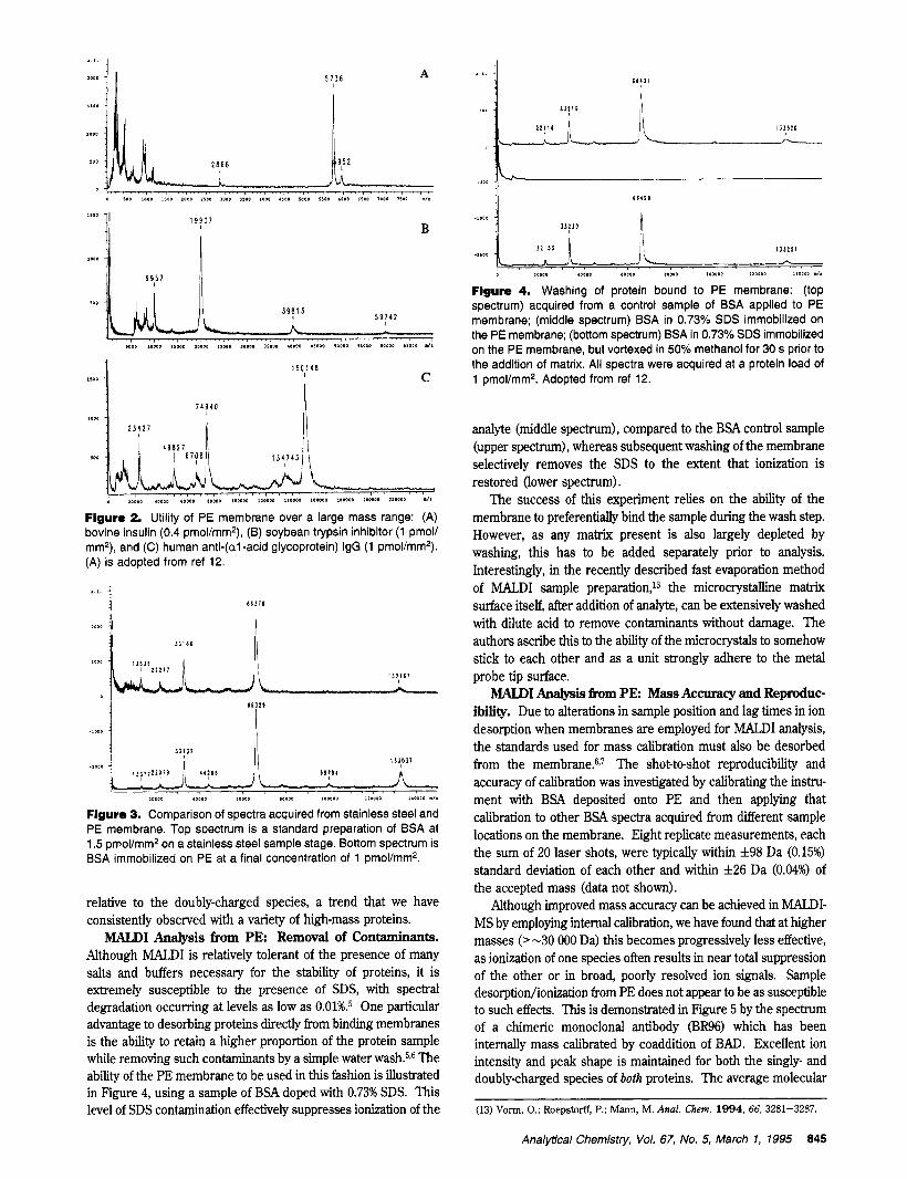
MALDI Analysis from P E Practical Mass Range. The MALDI mass spectra of three proteins encompassing a mass range of 5734 (bovine insulin) to over 150 000 Da (human anti- (al-acid glycoprotein) IgG) are shown .in Figure 2A-c. Each spectrum was obtained from the sample directly deposited onto a PE membrane. The quality of these spectra, particularly for human anti-(al-acid glycoprotein) IgG (Figure lC) , rivals those obtained from standard metal sample stages. The high signal- to-noise (S/N) ratios and symmetric peaks exhibited show the utility of PE as a support medium for UV-MALDI analysis of proteins over a wide range of molecular weights.
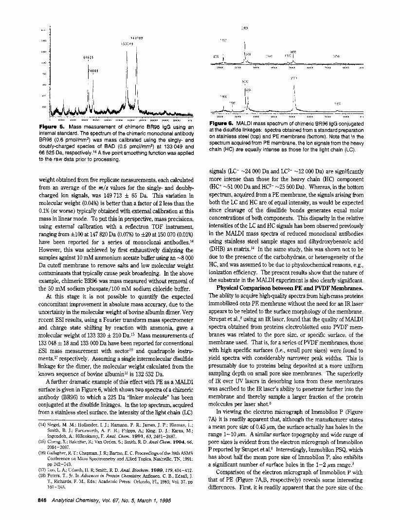
MALDI Analysis from PE: Spectral Quality. In Figure 3, the MALDI mass spectrum of BSA acquired from both a standard stainless steel surface (top) and a PE surface (bottom) are compared. The data acquired from the PE membrane does not show any discernable loss in spectral quality. The peak widths and S/N ratios are very similar in both spectra. The resolution of the singly-charged ion at m / z -66400 Da is -60 (m/Am, fwhm) in each case. Also, no discemable degradation in peak shape for the PE surface is evident, which would lead to subsequent errors in mass assignment. In addition, high-quality spectra could be acquired from anywhere within a large radius on the target, implying a consistently uniform sample/matrix ratio; with standard sample preparations one must often search to find the most judicious sampling spot. Interestingly, the spectrum acquired from PE also has a more intense singly-charged ion
844 Analytical Chemistv, Vol. 67, No. 5, March 1, 1995
1
L I D O i I 5 7 ? 6 A
I
B
Figure 2. Utility of PE membrane over a large mass range: (A) bovine insulin (0.4 pmol/mm2), (B) soybean trypsin inhibitor (1 pmol/ mmp), and (C) human anti-(a1 -acid glycoprotein) IgG (1 pmollmm'). (A) is adopted from ref 12.
I l O O D , o w 0 6 0 I O D , * P P I l l D P 0 l 110010 1.0010 .l.
Figure 3. Comparison of spectra acquired from stainless steel and PE membrane. Top spectrum is a standard preparation of BSA at 1.5 pmol/mm2 on a stainless steel sample stage. Bottom spectrum is BSA immobilized on PE at a final concentration of 1 pmol/mmz.
relative to the doubly-charged species, a trend that we have consistently observed with a variety of high-mass proteins.
MALDI Analysis from PE: Removal of Contaminants. Although MALDI is relatively tolerant of the presence of many salts and buffers necessary for the stability of proteins, it is extremely susceptible to the presence of SDS, with spectral degradation occurring at levels as low as 0.01%.5 One particular advantage to desorbing proteins directly from binding membranes is the ability to retain a higher proportion of the protein sample while removing such contaminants by a simple water washe5z6 The ability of the PE membrane to be used in this fashion is illustrated in Figure 4, using a sample of BSA doped with 0.73% SDS. This level of SDS contamination effectively suppresses ionization of the
. . i . 1 H'll
h
I J l ? d l
- 'Oo0 [ ,,~, J l , l l
.I300 L * 1 P P O O .om0 'LOP0 1PPOO ,000OP i l l D P l i 4 D D I O ./.
Figure 4. Washing of protein bound to PE membrane: (top spectrum) acquired from a control sample of BSA applied to PE membrane; (middle spectrum) BSA in 0.73% SDS immobilized on the PE membrane; (bottom spectrum) BSA in 0.73% SDS immobilized on the PE membrane, but vortexed in 50% methanol for 30 s prior to the addition of matrix. All spectra were acquired at a protein load of 1 pmol/mm2. Adopted from ref 12.
analyte (middle spectrum), compared to the BSA control sample (upper spectrum), whereas subsequent washing of the membrane selectively removes the SDS to the extent that ionization is restored (lower spectrum).
The success of this experiment relies on the ability of the membrane to preferentially bind the sample during the wash step. However, as any matrix present is also largely depleted by washing, this has to be added separately prior to analysis. Interestingly, in the recently described fast evaporation method of MALDI sample preparation,13 the microcrystalline matrix surface itself, after addition of analyte, can be extensively washed with dilute acid to remove contaminants without damage. The authors ascribe this to the ability of the microcrystals to somehow stick to each other and as a unit strongly adhere to the metal probe tip surface.
MALDI Analysis €ram PE Mass Accuracy and Reproduc- ibility. Due to alterations in sample position and lag times in ion desorption when membranes are employed for MALDI analysis, the standards used for mass calibration must also be desorbed from the memb1-ane.6,~ The shot-to-shot reproducibility and accuracy of calibration was investigated by calibrating the instru- ment with BSA deposited onto PE and then applying that calibration to other BSA spectra acquired from different sample locations on the membrane. Eight replicate measurements, each the sum of 20 laser shots, were typically within zt98 Da (0.15%) standard deviation of each other and within f 2 6 Da (0.04%) of the accepted mass (data not shown).
Although improved mass accuracy can be achieved in MALDI- MS by employing internal calibration, we have found that at higher masses (>-30 000 Da) this becomes progressively less effective, as ionization of one species often results in near total suppression of the other or in broad, poorly resolved ion signals. Sample desorption/ionization from PE does not appear to be as susceptible to such effects. This is demonstrated in Figure 5 by the spectrum of a chimeric monoclonal antibody "6) which has been internally mass calibrated by coaddition of BAD. Excellent ion intensity and peak shape is maintained for both the singly- and doubly-charged species of both proteins. The average molecular
(13) Vorm, 0.; Roepstorff, P.; Mann, M. Anal. Chem. 1994, 66, 3281-3287.
Analytical Chemistry, Vol. 67, No. 5, March 1, 1995 845
Flgure 5. Mass measurement of chimeric BR96 IgG using an internal standard. The spectrum of the chimeric monoclonal antibody BR96 (0.6 pmol/mmz) was mass calibrated using the singly- and doubly-charged species of BAD (0.5 pmol/mm2) at 133 049 and 66 525 Da, respectively.16 A five point smoothing function was applied to the raw data prior to processing.
weight obtained from five replicate measurements, each calculated from an average of the m/z values for the singly- and doubly- charged ion signals, was 149 713 f 65 Da. This variation in molecular weight (0.04%) is better than a factor of 2 less than the 0.1% (or worse) typically obtained with external calibration at this mass in linear mode. To put this in perspective, mass precisions, using external calibration with a reflectron TOF instrument, ranging from &lo0 at 147 820 Da (0.07%) to f20 at 150 070 (0.01%) have been reported for a series of monoclonal antibodie~.’~ However, this was achieved by first exhaustively dialyzing the samples against 10 mM ammonium acetate buffer using an -8 000 Da cutoff membrane to remove salts and low molecular weight contaminants that typically cause peak broadening. In the above example, chimeric BR96 was mass measured without removal of the 50 mM sodium phospate/100 mM sodium chloride buffer.
At this stage it is not possible to quantify the expected concomitant improvement in absolute mass accuracy, due to the uncertainty in the molecular weight of bovine albumin dimer. Very recent ESI results, using a Fourier transform mass spectrometer and charge state shifting by reaction with ammonia, gave a molecular weight of 133 320 f 210 Da.15 Mass measurements of 133 048 f 18 and 133 000 Da have been reported for conventional ESI mass measurement with sector16 and quadrupole instru- m e n t ~ , ~ ~ respectively. Assuming a single intermolecular disulfide linkage for the dimer, the molecular weight calculated from the known sequence of bovine albumin1s is 132 532 Da.
A further dramatic example of this effect with PE as a MALDI surface is given in Figure 6, which shows two spectra of a chimeric antibody (BR96) to which a 225 Da “linker molecule” has been conjugated at the disulfide linkages. In the top spectrum, acquired from a stainless steel surface, the intensity of the light chain (LC)
(14) Siegel, M. M.; Hollander, I. J.; Hamann, P. R; James, J. P.; Hinman, L.; Smith, B. J.; Famsworth, A P. H.; Phipps, A; King, D. J.; Karas, M.; Ingendoh, A; Hillenkamp, F. Anal. Chem. 1991, 63, 2481-2487.
(15) Cheng, X.; Bakhtiar, R; Van Orden, S.; Smith, R D. Anal Chem. 1994, 66, 2084-2087.
(16) Gallagher, R T.; Chapman, J. R; Barton, E. C. Proceedings of the 39thASMS Conference on Mass Spectrometry and Allied Topics, Nashville, TN, 1991;
(17) Loo, L. A; Udseth, H. R; Smith, R D. Anal. Biochem. 1989, 179,404-412. (18) Peters, T., Jr. In Advances in Protein Chemisty; Anfinsen, C. B., Edsall, J.
T., Richards, F. M., Eds.; Academic Press: Orlando, FL, 1985; Vol. 37, pp
pp 242-243.
161-245.
l l D l D l l m o ,1011 . I D D O , O O W 10001 10000 a0110 I!.
Figure 0. MALDI mass spectrum of chimeric BR96 IgG conjugated at the disulfide linkages: spectra obtained from a standard preparation on stainless steel (top) and PE membrane (bottom). Note that in the spectrum acquired from PE membrane, the ion signals from the heavy chain (HC) are equally intense as those for the light chain (LC).
signals (LC+ -24 000 Da and LC2+ -12 000 Da) are significantly more intense than those for the heavy chain (HC) component (HC’ -51 OOO Da and HC2+ -25 500 Da). Whereas, in the bottom spectrum, acquired from a PE membrane, the signals arising from both the LC and HC are of equal intensity, as would be expected since cleavage of the disulfide bonds generates equal molar concentrations of both components. This disparity in the relative intensities of the Lc and HC signals has been observed previously in the MALDI mass spectra of reduced monoclonal antibodies using stainless steel sample stages and dihydroxybenzoic acid @HB) as matrix.I4 In the same study, this was shown not to be due to the presence of the carbohydrate, or heterogeneity of the HC, and was assumed to be due to physicochemical reasons, e.g., ionization efficiency. The present results show that the nature of the substrate in the MALDI experiment is also clearly signiiicant.
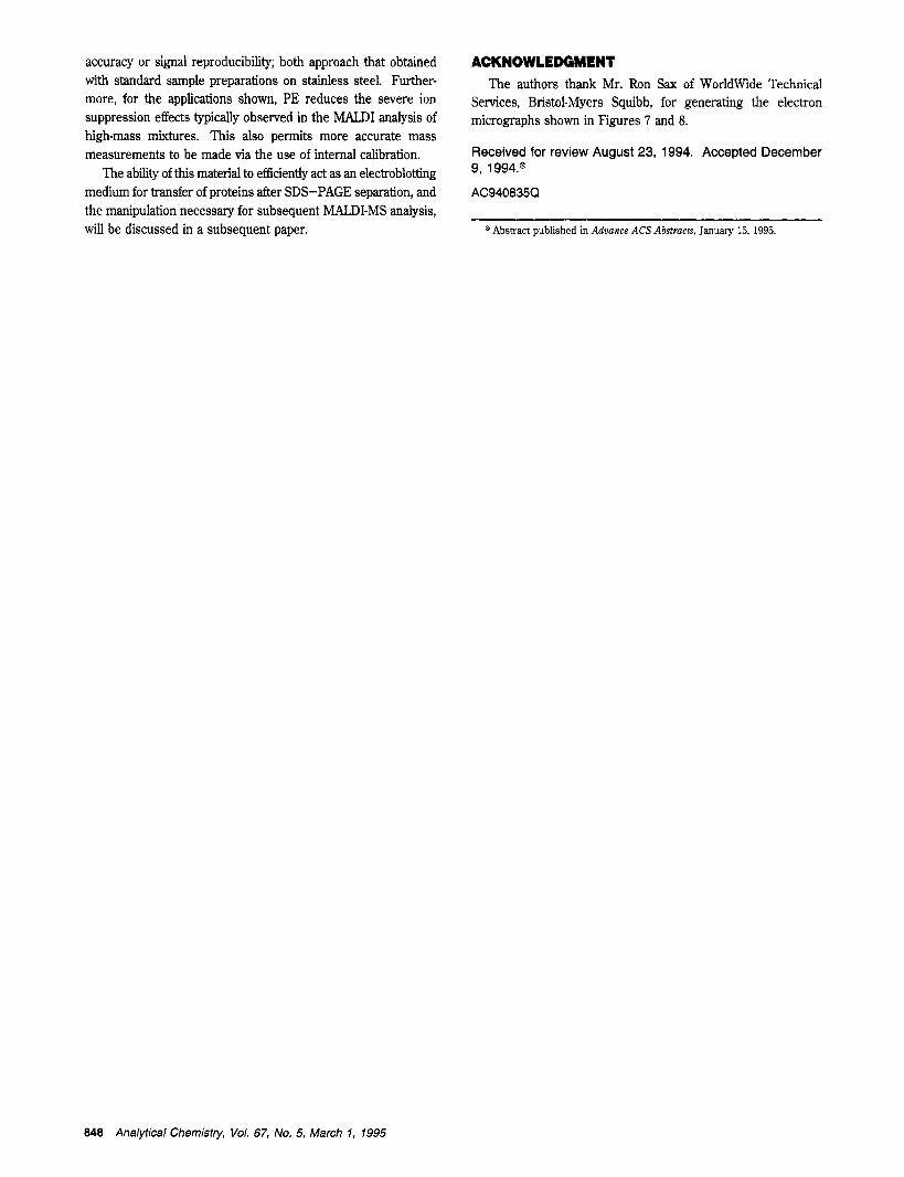
Physical Comparison between PE and PVDF Membranes. The ability to acquire highquality spectra from high-mass proteins immobilized onto PE membrane without the need for an IR laser appears to be related to the surface morphology of the membrane. Strupat et aLK using an IR laser, found that the quality of MALDI spectra obtained from proteins electroblotted onto PVDF mem- branes was related to the pore size, or specific surface, of the membrane used. That is, for a series of PVDF membranes, those with high specific surfaces (i.e., small pore sizes) were found to yield spectra with considerably narrower peak widths. This is presumably due to proteins being deposited at a more uniform sampling depth on small pore size membranes. The superiority of IR over W lasers in desorbing ions from these membranes was ascribed to the IR laser’s ability to penetrate further into the membrane and thereby sample a larger fraction of the protein molecules per laser shot.*
In viewing the electron micrograph of Immobilon P (Figure 7A) it is readily apparent that, although the manufacturer states a mean pore size of 0.45 pm, the surface actually has holes in the range 1-10 pm. A similar surface topography and wide range of pore sizes is evident from the electron micrograph of Immobilon P reported by Strupet et a1.8 Interestingly, Immobilon PSQ, which has about half the mean pore size of Immobilon P, also exhibits a signiicant number of surface holes in the 1-2 pm range.8
Comparison of the electron micrograph of Immobilon P with that of PE (Figure 7A,B, respectively) reveals some interesting differences. First, it is readily apparent that the pore size of the
846 Analytical Chemisfty, Vol. 67, No. 5, March 7, 7995
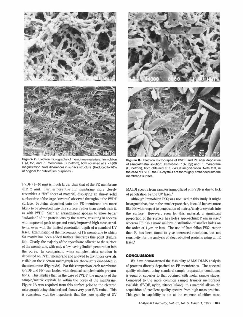
Figure 7. Electron micrographs of membrane materials: lmmobilon P (A, top) and PE membrane (B, bottom), both obtained at a x4800 magnification. Note differences in surface structure. (Reduced to 70% of original for publication purposes.)
PVDF (1-10 pm) is much larger than that of the PE membrane (0.2-2 pm). Furthermore the PE membrane more closely resembles a “flat” sheet of material, displaying an almost solid surface free of the large “caverns” observed throughout the PVDF surface. Proteins deposited onto the PE membrane are more likely to be absorbed onto this surface, rather than deeply into it, as with PVDF. Such an arrangement appears to allow better “solvation” of the protein ions by the matrix, resulting in spectra with improved peak shape and vastly improved high-mass sensi- tivity, even with the limited penetration depth of a standard UV laser. Examination of the micrograph of PE membrane to which SA matrix has been added further illustrates this point (Figure 8b). Clearly, the majority of the crystals are adhered to the surface of the membrane, with only a few having limited penetration into the pores. In comparison, when sample/matrix solution is deposited on PVDF membrane and allowed to dry, those crystals visible on the electron micrograph are thoroughly embedded in the membrane (Figure SA). For this comparison, each membrane (PVDF and PE) was loaded with identical sample/matrix prepara- tions. This implies that, in the case of PWF, the majority of the sample/matrix crystals lie within the pores of the membrane. Figure lA was acquired from this surface prior to the electron micrograph being obtained and shows very poor S/N ratios. This is consistent with the hypothesis that the poor quality of UV
Figure 8. Electron micrographs of PVDF and PE after deposition of sample/matrix solution: lmmobilon P (A, top) and PE membrane (B, bottom), both obtained at a x4800 magnification. Note that, in the case of PVDF, the SA crystals are thoroughly embedded into the membrane surface.
MALDI spectra from samples immobilized on PVDF is due to lack of penetration by the UV laser.8
Although Immobilon PSQ was not used in this study, it might be argued that, due to the smaller pore size, it would behave more like PE with respect to penetration of matrix/analyte crystals into the surface. However, even for this material, a significant proportion of the surface has holes approaching 2 pm in size: whereas PE has a more uniform distribution of smaller holes on the order of 1 pm or less.. The use of Immobilon PSQ, rather than P, has been found to give increased resolution, but not sensitivity, for the analysis of electroblotted proteins using an IR laser?
CONCLUSIONS We have demonstrated the feasibility of MALDI-MS analysis
of proteins directly deposited on PE membranes. The spectral quality obtained, using standard sample preparation conditions, is equal or superior to that obtained with metal sample stages. Compared to the more common sample transfer membranes available (PVDF, nylon, nitrocellulose), this material allows the acquisition of excellent quality spectra from high-mass proteins. This gain in capability is not at the expense of either mass
Analytical Chemistry, Vol. 67, No. 5, March 1, 1995 847
accuracy or signal reproducibility; both approach that obtained with standard sample preparations on stainless steel. Further- more, for the applications shown, PE reduces the severe ion suppression effects typically observed in the MALDI analysis of high-mass mixtures. This also permits more accurate mass measurements to be made via the use of internal calibration.
The ability of this material to efficiently act as an electroblotting medium for transfer of proteins after SDS-PAGE separation, and the manipulation necessary for subsequent MALDI-MS analysis, will be discussed in a subsequent paper.
ACKNOWLEDGMENT The authors thank Mr. Ron Sax of WorldWide Technical
Services, Bristol-Myers Squibb, for generating the electron micrographs shown in Figures 7 and 8.
Received for review August 23, 1994. Accepted December 9, 1994.B
AC940835Q
@ Abstract published in Advance ACS Abstracts, January 15, 1995.
848 Analytical Chemistry, Vol. 67, No. 5, March 1, 7995
Copyright © 2022 FDOKUMEN