AXL is a logical molecular target in head and neck squamous cell carcinoma
PIK3CA Mutations in Head and Neck Squamous Cell Carcinoma
Wanglong Qiu1, Frank Schönleben1, Xiaojun Li1, Daniel J. Ho1, Lanny G. Close1, SpirosManolidis1, Boyce P. Bennett2, and Gloria H. Su1,21 The Department of Otolaryngology/Head and Neck Surgery and the
2 Department of Pathology, Columbia University College of Physicians and Surgeons, New York,NY 10032.
AbstractPurpose—Recent studies have reported high frequencies of somatic mutations in thephosphoinositide-3-kinase, catalytic, alpha (PIK3CA) gene in several human solid tumors. Althoughgene amplifications of PIK3CA have been reported in head and neck squamous cell carcinoma(HNSCC), small mutation of the gene has not been evaluated in HNSCC previously. In this study,we examined the mutation frequency of PIK3CA in HNSCC.
Experimental Design—More than 75% of the somatic mutations of PIK3CA are clustered in thehelical (exon 9) and kinase domains (exon 20). To investigate the possible role of PIK3CA in HNSCCtumorigenesis, exons 1, 4, 5, 6, 7, 9, and 20 of the gene were analyzed by direct genomic DNAsequencing in 38 HNSCC specimens.
Results—We identified four missense mutations in the seven exons of PIK3CA from 38 HNSCCspecimens (11%). Three of the four mutations, named H1047R, E542K and E545K respectively,have been previously reported as hot-spot mutations. The remaining novel mutation, Y343C, isidentified at exon 4 nucleotide 1028 A → G. Three of the four mutations were shown to be somatic,while the forth mutation (H1047R) was identified in a cell line. Interestingly, three of the fourmutations identified were in pharyngeal cancer samples.
Conclusions—These data provide evidence that oncogenic properties of PIK3CA contributes tothe carcinogenesis of human head and neck cancers, especially in pharyngeal cancer. A specifickinase inhibitor to PIK3CA may potentially be an effective therapeutic reagent against HNSCC orpharyngeal cancer in particular.
The abbreviations used areHNSCC, Head and neck squamous cell carcinoma; PCR, polymerase chain reaction; PI3K,phosphatidylinositol 3-kinase; PIP3, phosphatidylinositol-3,4,5-triphosphate
INTRODUCTIONThe phosphatidylinositol 3-kinase (PI3K) signaling pathway regulates many normal cellularprocesses, such as cell proliferation, survival and apoptosis (1–3). Dysregulation or geneticaberration of components of this pathway, including AKT, PTEN, and PIK3CA, has beenassociated with cancer development (4–12).
To whom requests for reprints should be addressed: Dr. Su at the Department of Otolaryngology/Head and Neck Surgery, ColumbiaUniversity College of Physicians and Surgeons, 1130 St. Nicholas Ave, ICRC 10-11, New York, NY 10032. Phone: 212-851-4624; E-mail: [email protected] work was supported by the NCI Temin Award CA95434 and the NCI R01 CA109525.
NIH Public AccessAuthor ManuscriptClin Cancer Res. Author manuscript; available in PMC 2007 March 1.
Published in final edited form as:Clin Cancer Res. 2006 March 1; 12(5): 1441–1446.
NIH
-PA Author Manuscript
NIH
-PA Author Manuscript
NIH
-PA Author Manuscript

PIK3CA is located on chromosome 3q26.32 and encodes for the catalytic subunit p110α ofclass IA PI3-kinase. It has been implicated to function as an oncogene in human cancer becauseof its elevated kinase activity and genomic amplification in tumor samples (7–12). Recentlyhigh frequencies of somatic mutations in the PIK3CA gene have been reported in several humancancer types, including colon, brain, stomach, breast, and ovary (13–18). More than 75% ofthese mutations are clustered in the helical (exon 9) and kinase domains (exon 20) of the gene(13). The three most frequently reported mutation hot spots in PIK3CA, named E542K, E545Kand H1047R, have been shown to elevate its lipid kinase activity and lead to the activation ofthe downstream Akt signaling pathway (13,19). Interestingly, PIK3CA mutations and PTENloss are nearly mutually exclusive, suggesting that the homeostasis ofphosphatidylinositol-3,4,5-triphosphate regulated by both PIK3CA and PTEN is critical tocarcinogenesis (20). This further evinced the importance of the PI3K pathway in thetumorigenesis of many cancer types.
Although the PIK3/AKT/PTEN pathway has been implicated in HNSCC (12,21–23), nogenetic mutation of PIK3CA has been described to date. To investigate whether PIK3CAactivating mutation is a common mechanism involved in the tumorigenesis of HNSCC, weanalyzed for genetic alterations of the PIK3CA gene in 38 HNSCC specimens including eightcell lines by direct genomic DNA sequencing. Only exons 1, 4, 5, 6, 7, 9, and 20 of the genewere sequenced in these specimens because they covered the most common PIK3CA mutationspreviously observed in human cancer (13–17,24–26).
MATERIAL AND METHODSTissue samples and cell lines
Eight HNSCC cell lines, RPMI 2650, A-253, SW579, Detroit 562, FADU, CAL 27, SCC-15and SCC-25, were purchased from American Type Culture Collection (Rockville, MD). Thecell lines were maintained as recommended by ATCC.
Thirty frozen primary tumor samples and their corresponding match normal muscle specimenswere obtained from the Tumor Bank facility of the Herbert Irving Comprehensive CancerCenter and Department of Otolaryngology/Head and Neck Surgery of the Columbia UniversityMedical Center. Acquisition of the tissue specimens was approved by the Institutional ReviewBoard of Columbia University Medical Center and performed in accordance with HealthInsurance Portability and Accountability Act (HIPAA) regulations. Fresh-frozen tumorsamples were dissected to ensure that the specimen contained at least 75% cancer cells. Thecancer sites were nasal cavity (2), pharynx (6), larynx (10), oral cavity (8) and other sites (4).The patients’ ages ranged from 40 to 85 years, average 64.0 ± 14.5. The grades of the tumorswere moderately to poorly differentiated.
PCR amplification and PCR product direct sequencingGenomic DNAs were extracted from the cell lines and the frozen tissue samples using DNeasytissue kit (Qiagen, CA). The procedures were performed according to the manufacturer’sinstructions.
Exons 1, 4, 5, 6, 7, 9, and 20 of PIK3CA gene were analyzed by PCR amplification of genomicDNA and PCR product direct sequencing. Genomic DNAs (40 ng per sample) were amplifiedwith primers covering the entire coding region and the exon/intron boundaries of the desiredexons (PIK3CA-E9F:5’-ctgtgaatccagaggggaaa-3’; PIK3CA-E9R: 5’-gcatttaatgtgccaactacca-3’; PIK3CA-E9FS: 5’-tccagaggggaaaaatatgaca-3’, (13)). All genesequencings were performed with ABI's 3100 capillary automated sequencers at the DNAfacility of Columbia University Medical Center using previously published sequencing primers
Qiu et al. Page 2
Clin Cancer Res. Author manuscript; available in PMC 2007 March 1.
NIH
-PA Author Manuscript
NIH
-PA Author Manuscript
NIH
-PA Author Manuscript
(13). All samples found to have a genetic alteration in the target were subsequently sequencedin the reverse direction to confirm the mutation using the reverse PCR primers (13). Themutation was then further verified by sequencing of a second PCR product derivedindependently from the original template.
RESULTSA novel sequence identified similar to PIK3CA
While sequencing for mutations using primers we had designed for exon 9, we were surprisedto find an alteration at nucleotide 1634 A → C (E545A) in all the cases (Figures 1, 2). However,this nucleotide change of PIK3CA A1634C (E545A) always co-existed with another alterationof G → C at nucleotide 1658 and a base deletion at nucleotide 1659 (Figure 1). Subsequentsequencing analyses of the matching normal tissue specimens revealed that the same nucleotidechanges occurred in both tumor and normal tissues (data not shown). This unusual result ledus to blast search this PCR fragment (410-bp long) in the GenBank. We found two genomicDNA clones that contain fragments that are 97% (401/410) homologous to the exon 9 and itsflanking intronic sequences. These two clones are located at chromosome 22q11.2 cat eyesyndrome region (gi 5931525) and at chromosome 16 (gi 28913054) (Figure 2 and data notshown). Further comparisons of the sequences using the BLAST search revealed that bothgenomic clones on chromosome 22 and chromosome 16 contain sequences highly homologousto the exons 9, 11–13 and partial exon 10 of the PIK3CA gene (data not shown). An automaticcomputational analysis using the GNOMON gene prediction method predicted a protein thatcan be transcribed and translated from the chromosome 22 clone. The predicted protein (gi51475436) is similar to the helical domain of the PIK3CA protein. However, this sequencehomolog is likely to be a pseudogene since no RNA transcripts of the predicted protein can bedetected by RT-PCR (data not shown).
This sequence homolog was probably not reported by previous publications because itsdetectability depends highly on primer designs. When we moved the PCR primer sites, usedthe primers published in the study by Samuels et al. (13), or increased the stringency of ourPCR condition, all the nucleotide alterations including the so-called PIK3CA A1634C (E545A)“mutation” disappeared. We concluded that the A1634C (E545A) “mutation” observed in ourhands was an artifact created by interferences from the sequence homolog.
PIK3CA is activated by small mutation in HNSCCFour missense mutations of the PIK3CA gene were identified in the 38 HNSCC specimens(Figure 3 and Table 1)-Two of the mutations were in the exon 9 (E545K, E542K), one was inthe exon 20 (H1047Y) and one was in the exon 4 (Y343C). None of these mutations wasdetected in the corresponding normal tissues except for the H1047Y mutation, which wasidentified in HNSCC cell line Detroit 562. Three of the four PIK3CA missense mutations(E545K, E542K, and H1047R) are previously described hot-spot mutations (13). Functionalstudies showed that PI3-kinase carrying any one of the three hot-spot mutations is able to inducetransformation in cultures of chicken embryo fibroblasts, and that the transforming activity ofthe mutant is correlated with increased lipid kinase activity and activation of the Akt signalingpathway (13,19). The mutation in the exon 4 nucleotide 1028 A → G, which leads to alterationat codon 343 TAC (Y) → TGC(C), has not been described before (Fig. 3).
Two other nucleotide alterations were also detected in the exonic regions of the PIK3CA gene(Table 1). One alteration, located at the exon 6 nucleotide 1173 A→G ( codon 391 ATA (ILe)→ ATG (Met)), was found in six HNSCC tumor specimens. This nucleotide alteration wasalso detected in the six matching normal tissues. A search of the SNP database revealed thatA1173G is a known SNP (rs2230461) that has been validated by multiple PCR reactions and
Qiu et al. Page 3
Clin Cancer Res. Author manuscript; available in PMC 2007 March 1.
NIH
-PA Author Manuscript
NIH
-PA Author Manuscript
NIH
-PA Author Manuscript

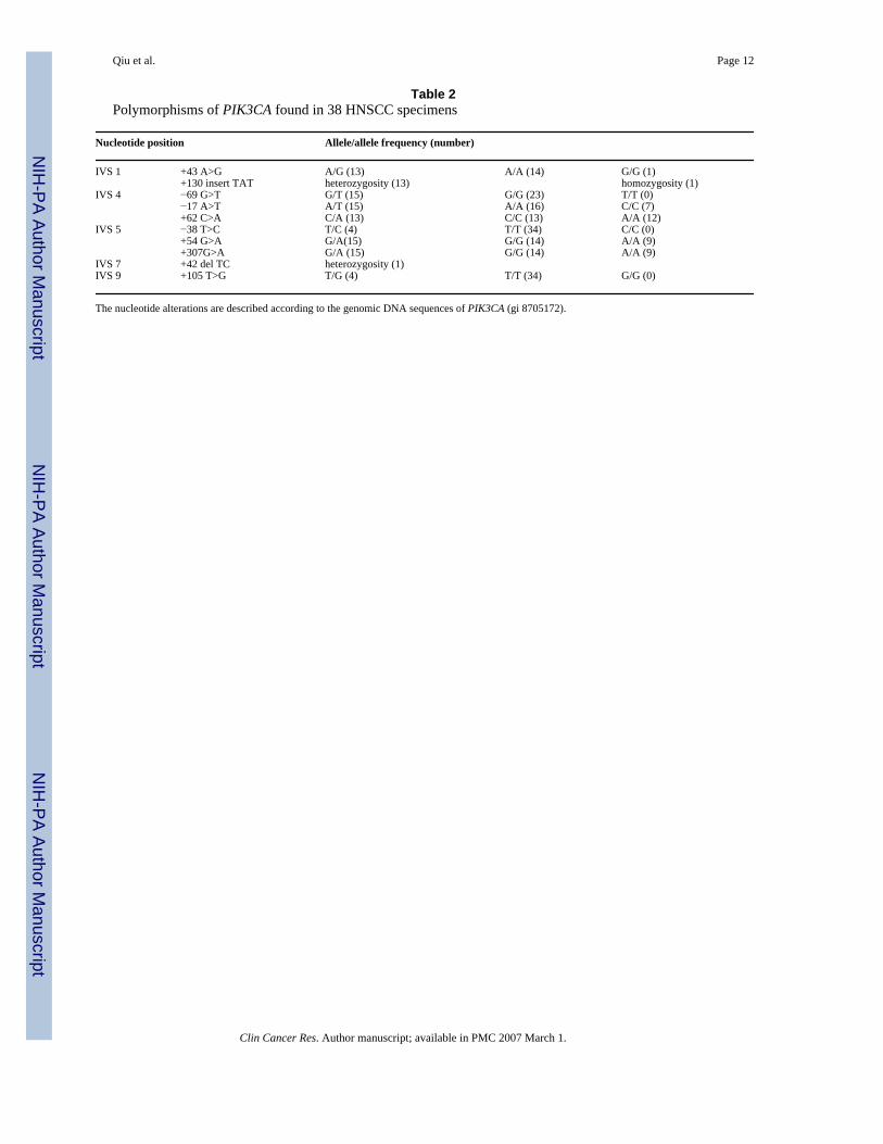
genotype data. Thus, we conclude that this germline alteration represents a non-disease-causingpolymorphism of PIK3CA. Another exonic alteration was also deemed a polymorphismbecause it occurred at exon 5 C1143G (P381P) without resulting in an amino acid change andwas observed in both the tumor and normal samples of one tongue cancer patient. All of thepolymorphisms observed in the intronic regions flanking the seven exons of PIK3CA examinedin this study are listed in Table 2. These polymorphisms are unlikely to cause significantchanges in the function of PIK3CA.
DISCUSSIONThe mutation frequency of PIK3CA has been reported at 32% in colon cancer, ~4–25% ingastric cancer, 8–40% in breast cancer, 5–27% in brain cancer, 4% in lung cancer, and 4–7%in ovarian cancer (13,16–18,25). In the present study we report 11% (4/38) of PIK3CAmutations in sporadic HNSCC. Interestingly, three out of the four cases with mutations arefrom the same organ site, pharynx (Table 1). Cancer of the pharynx is the 9th most commoncancer worldwide (27). It is characterized as the following subsites: posterior pharynx,hypopharynx and lateral pharyngeal walls. A total of six pharyngeal squamous cell carcinomacases were examined in this study, suggesting that as high as 50% (3/6) of pharyngeal tumorsamples may harbor PIK3CA mutations. This data is supported by a previous report that showedchromosome 3q26 is amplified in 100% of nasopharyngeal carcinoma (22). However, fromthe present study we are unable to conclude the exact mutational frequency of PIK3CA inpharyngeal carcinomas and comment on which subtype of pharyngeal carcinomas(nasopharynx, oropharynx and hypopharynx) is targeted for PIK3CA mutation. Among our sixpharyngeal samples, there was one oropharygneal cancer sample, one hypopharyngeal cancer,and four that were not subtyped. More studies with larger sample sizes and various pharyngealsubtypes are necessary to further investigate these potentials.
Gene amplification is a more commonly observed mechanism of oncogene activation inHNSCC than small genetic mutation. Cyclin D1 gene amplification has been observed in ~34–37% of HNSCC (28,29). EGF receptor gene amplification has been reported in 7–19% ofHNSCC (30–32). In contrast, RAS mutation is relatively rare in HNSCC in comparison to othersolid cancers- less than 6% in HNSCC vs. 99% in pancreatic cancer and 37–47% in colorectalcancer (33–39). Amplification of chromosome 3q26 is frequently observed in HNSCC and islinked to tumor progression and negatively correlated with clinical outcome (40–42). Geneamplification and overexpression of PIK3CA are observed in low to moderate dysplasic cases,but their increased frequencies are associated with transition to invasive cancer (21,23). Herewe showed that gene amplification is not the only mechanism to activate PIK3CA in HNSCC.Small mutation and gene amplification both contribute to the activation of PIK3CA in HNSCC.
In summary, we report missense mutations of the PIK3CA gene in HNSCC (4/38, 11%).Among the four cases identified here, the Y343C mutation, which is located at PIK3CA exon4 nucleotide 1028 A→ G, is novel and has not been described in previous studies. Althoughthe physiological significance of the novel mutation Y343C, which is located within thePIK3CA C2 domain, is not known, it has been shown that the C2 domain in the Class IB PI3Kinteracts primarily with the helical domain, and also interacts with the linker segment beforethe Ras-binding domain and with the C-terminal lobe of the catalytic domain (43). The C2domain is often involved in Ca2+-dependent or Ca2+-independent phospholipids membranebinding. By analogy with enzymes like protein kinase C and cytosolic phospholipase A2, theC2 domain of ClassIB PI3K might participate in Ca2+-independent phospholipids membranebinding (43). Since mutations found in the C2 domain account for 7% of total PIK3CAmutations found in a study of 396 cancer samples (13), it will be worthwhile to determine theexact function of the C2 domain of Class 1A PI3K in future studies. The other three are hot-spot mutations (E545K, E542K, and H1047R) and all were found in pharyngeal cancer patients.
Qiu et al. Page 4
Clin Cancer Res. Author manuscript; available in PMC 2007 March 1.
NIH
-PA Author Manuscript
NIH
-PA Author Manuscript
NIH
-PA Author Manuscript
The smoking histories of the mutated patients are unknown. We did not find any significantcorrelation of the PIK3CA gene mutation to the gender or age of the patients.
Here we also report the discovery of a PIK3CA homolog. This homolog is almost identical tothe exons 9, 11–13 and partial exon 10 of the PIK3CA gene and can be found on bothchromosomes 16 and 22. However, we think that this sequence homolog is likely to be apseudogene since no RNA transcripts of the predicted protein can be detected by RT-PCR. Inour study, interferences from this sequence homolog had caused confusions by creatingnucleotide alterations including the so-called PIK3CA A1634C (E545A) “mutation” (Figures1, 2), which subsequently vanished with better primer designs and more stringent PCRconditions. Intriguingly, this A1634C (E545A) mutation has been previously reported inhuman cancers by two publications. One study described 11 cases with the A1634C (E545A)mutation out of 73 hepatocellular carcinomas (15). More recently this exact mutation wasreported to contribute up to 88% (21/24) of the total PIK3CA mutations identified in ovariancancer (24). This mutation was not described in other reports on PIK3CA mutation (13, 14,16–18, 25, 26). In light of our discovery, it is important for future studies to be aware of thepossible interference from the homologous sequences on chromosomes 22 and 16. Althoughwe did not study exons 10–13 in our current study, potential artifacts there are also probable.
Our data confirm that PIK3CA is important to HNSCC tumorigenesis and provide evidencethat small mutation can also contribute to oncogene activation of PIK3CA in HNSCC.Furthermore, our data suggest that PIK3CA gene mutations may be more involved in thecarcinogenesis of a particular subset of human head and neck cancers (pharyngeal cancers)than others. The knowledge of the PIK3CA’s involvement in HNSCC is important because aspecific kinase inhibitor could be considered as a future therapeutic option for HNSCC patientswith PIK3CA mutations. Most HNSCC are diagnosed at advanced stage, and are usuallyunresectable despite significant surgical advances. Improvements in chemotherapy andradiotherapy in recent decades have not been translated into better prognosis of HNSCCpatients (44). Recently kinase inhibitors such as Gleevec (Imatinib), Herceptin (Trastzumab),and Iressa (Gefitinib) have been successfully developed for therapies in some cancer types(45). Since amplification and overexpression of the PIK3CA gene locus is an early oncogenicevent of HNSCC tumorigenesis and is also correlated to invasion (21,23), abrogation of itsoncogenic activities may conceivably slow or stop tumor progression. It is believed that sucha selective small-molecule inhibitor against PIK3CA would have tremendous potential as anovel cancer chemotherapeutic for HNSCC (46). Our findings further supports PIK3CA as animportant potential target in head and neck cancer for pathway-specific, kinase inhibitor basedtherapies.
References1. Klippel A, Escobedo MA, Wachowicz MS, et al. Activation of phosphatidylinositol 3-kinase is
sufficient for cell cycle entry and promotes cellular changes characteristic of oncogenic transformation.Mol Cell Biol 1998;18(10):5699–711. [PubMed: 9742087]
2. Chang HW, Aoki M, Fruman D, et al. Transformation of chicken cells by the gene encoding the catalyticsubunit of PI 3-kinase. Science 1997;276(5320):1848–50. [PubMed: 9188528]
3. Kennedy SG, Wagner AJ, Conzen SD, et al. The PI 3-kinase/Akt signaling pathway delivers an anti-apoptotic signal. Genes Dev 1997;11(6):701–13. [PubMed: 9087425]
4. Yuan ZQ, Sun M, Feldman RI, et al. Frequent activation of AKT2 and induction of apoptosis byinhibition of phosphoinositide-3-OH kinase/Akt pathway in human ovarian cancer. Oncogene 2000;19(19):2324–30. [PubMed: 10822383]
5. Bellacosa A, de Feo D, Godwin AK, et al. Molecular alterations of the AKT2 oncogene in ovarian andbreast carcinomas. Int J Cancer 1995;64(4):280–5. [PubMed: 7657393]
Qiu et al. Page 5
Clin Cancer Res. Author manuscript; available in PMC 2007 March 1.
NIH
-PA Author Manuscript
NIH
-PA Author Manuscript
NIH
-PA Author Manuscript
6. Cantley LC, Neel BG. New insights into tumor suppression: PTEN suppresses tumor formation byrestraining the phosphoinositide 3-kinase/AKT pathway. Proc Natl Acad Sci U S A 1999;96(8):4240–5. [PubMed: 10200246]
7. Phillips WA, St Clair F, Munday AD, Thomas RJ, Mitchell CA. Increased levels ofphosphatidylinositol 3-kinase activity in colorectal tumors. Cancer 1998;83(1):41–7. [PubMed:9655291]
8. Benistant C, Chapuis H, Roche S. A specific function for phosphatidylinositol 3-kinase alpha(p85alpha-p110alpha) in cell survival and for phosphatidylinositol 3-kinase beta (p85alpha-p110beta)in de novo DNA synthesis of human colon carcinoma cells. Oncogene 2000;19(44):5083–90.[PubMed: 11042696]
9. Andrew S. PIK3CA: determining its role in cellular proliferation and ovarian cancer. Clin Genet1999;56(3):190–1. [PubMed: 10563477]
10. Ma YY, Wei SJ, Lin YC, et al. PIK3CA as an oncogene in cervical cancer. Oncogene 2000;19(23):2739–44. [PubMed: 10851074]
11. Shayesteh L, Lu Y, Kuo WL, et al. PIK3CA is implicated as an oncogene in ovarian cancer. NatGenet 1999;21(1):99–102. [PubMed: 9916799]
12. Pedrero JM, Carracedo DG, Pinto CM, et al. Frequent genetic and biochemical alterations of the PI3-K/AKT/PTEN pathway in head and neck squamous cell carcinoma. Int J Cancer 2005;114(2):242–8. [PubMed: 15543611]
13. Samuels Y, Wang Z, Bardelli A, et al. High frequency of mutations of the PIK3CA gene in humancancers. Science 2004;304(5670):554. [PubMed: 15016963]
14. Bachman KE, Argani P, Samuels Y, et al. The PIK3CA Gene is Mutated with High Frequency inHuman Breast Cancers. Cancer Biol Ther 2004;3(8):772–5. [PubMed: 15254419]
15. Lee JW, Soung YH, Kim SY, et al. PIK3CA gene is frequently mutated in breast carcinomas andhepatocellular carcinomas. Oncogene. 2004
16. Campbell IG, Russell SE, Choong DY, et al. Mutation of the PIK3CA gene in ovarian and breastcancer. Cancer Res 2004;64(21):7678–81. [PubMed: 15520168]
17. Broderick DK, Di C, Parrett TJ, et al. Mutations of PIK3CA in anaplastic oligodendrogliomas, high-grade astrocytomas, and medulloblastomas. Cancer Res 2004;64(15):5048–50. [PubMed: 15289301]
18. Wang Y, Helland A, Holm R, Kristensen GB, Borresen-Dale AL. PIK3CA mutations in advancedovarian carcinomas. Hum Mutat 2005;25(3):322. [PubMed: 15712344]
19. Kang S, Bader AG, Vogt PK. Phosphatidylinositol 3-kinase mutations identified in human cancer areoncogenic. Proc Natl Acad Sci U S A 2005;102(3):802–7. [PubMed: 15647370]
20. Saal LH, Holm K, Maurer M, et al. PIK3CA mutations correlate with hormone receptors, nodemetastasis, and ERBB2, and are mutually exclusive with PTEN loss in human breast carcinoma.Cancer Res 2005;65(7):2554–9. [PubMed: 15805248]
21. Estilo CL, P OC, Ngai I, et al. The role of novel oncogenes squamous cell carcinoma-related oncogeneand phosphatidylinositol 3-kinase p110alpha in squamous cell carcinoma of the oral tongue. ClinCancer Res 2003;9(6):2300–6. [PubMed: 12796399]
22. Or YY, Hui AB, Tam KY, Huang DP, Lo KW. Characterization of chromosome 3q and 12q ampliconsin nasopharyngeal carcinoma cell lines. Int J Oncol 2005;26(1):49–56. [PubMed: 15586224]
23. Woenckhaus J, Steger K, Werner E, et al. Genomic gain of PIK3CA and increased expression ofp110alpha are associated with progression of dysplasia into invasive squamous cell carcinoma. JPathol 2002;198(3):335–42. [PubMed: 12375266]
24. Levine DA, Bogomolniy F, Yee CJ, et al. Frequent mutation of the PIK3CA gene in ovarian andbreast cancers. Clin Cancer Res 2005;11(8):2875–8. [PubMed: 15837735]
25. Li VS, Wong CW, Chan TL, et al. Mutations of PIK3CA in gastric adenocarcinoma. BMC Cancer2005;5(1):29. [PubMed: 15784156]
26. Samuels Y, Velculescu VE. Oncogenic mutations of PIK3CA in human cancers. Cell Cycle 2004;3(10):1221–4. [PubMed: 15467468]
27. Blanchaert RH Jr. Oral and oral pharyngeal cancer: an update on incidence and epidemiology,identification, advances in treatment, and outcomes. Compend Contin Educ Dent 2002;23(12 Suppl):25–9. [PubMed: 12789999]
Qiu et al. Page 6
Clin Cancer Res. Author manuscript; available in PMC 2007 March 1.
NIH
-PA Author Manuscript
NIH
-PA Author Manuscript
NIH
-PA Author Manuscript
28. Callender T, el-Naggar AK, Lee MS, Frankenthaler R, Luna MA, Batsakis JG. PRAD-1 (CCND1)/cyclin D1 oncogene amplification in primary head and neck squamous cell carcinoma. Cancer1994;74(1):152–8. [PubMed: 8004570]
29. Jares P, Fernandez PL, Campo E, et al. PRAD-1/cyclin D1 gene amplification correlates withmessenger RNA overexpression and tumor progression in human laryngeal carcinomas. Cancer Res1994;54(17):4813–7. [PubMed: 8062283]
30. Kearsley JH, Leonard JH, Walsh MD, Wright GR. A comparison of epidermal growth factor receptor(EGFR) and c-erbB-2 oncogene expression in head and neck squamous cell carcinomas. Pathology1991;23(3):189–94. [PubMed: 1685773]
31. Ishitoya J, Toriyama M, Oguchi N, et al. Gene amplification and overexpression of EGF receptor insquamous cell carcinomas of the head and neck. Br J Cancer 1989;59(4):559–62. [PubMed: 2713242]
32. Leonard JH, Kearsley JH, Chenevix-Trench G, Hayward NK. Analysis of gene amplification in head-and-neck squamous-cell carcinoma. Int J Cancer 1991;48(4):511–5. [PubMed: 2045198]
33. Anderson JA, Irish JC, Ngan BY. Prevalence of RAS oncogene mutation in head and neck carcinomas.J Otolaryngol 1992;21(5):321–6. [PubMed: 1361585]
34. Yarbrough WG, Shores C, Witsell DL, Weissler MC, Fidler ME, Gilmer TM. ras mutations andexpression in head and neck squamous cell carcinomas. Laryngoscope 1994;104(11 Pt 1):1337–47.[PubMed: 7968162]
35. Weber A, Langhanki L, Sommerer F, Markwarth A, Wittekind C, Tannapfel A. Mutations of theBRAF gene in squamous cell carcinoma of the head and neck. Oncogene 2003;22(30):4757–9.[PubMed: 12879021]
36. Almoguerra C, Shibata D, Forrester K, Martin J, Arnheim N, Perucho M. Most human carcinomasof the exocrine pancreas contain mutant c-K-ras genes. Cell 1988;53:549–54. [PubMed: 2453289]
37. Rozenblum E, Schutte M, Goggins M, et al. Tumor-suppressive pathways in pancreatic carcinoma.Cancer Res 1997;57:1731–4. [PubMed: 9135016]
38. Bos JL, Fearon ER, Hamilton SR, et al. Prevalence of ras gene mutations in human colorectal cancers.Nature 1987;327(6120):293–7. [PubMed: 3587348]
39. Vogelstein B, Fearon ER, Hamilton SR, et al. Genetic alterations during colorectal-tumordevelopment. N Engl J Med 1988;319:525–532. [PubMed: 2841597]
40. Hashimoto Y, Oga A, Kawauchi S, et al. Amplification of 3q26 approximately qter correlates withtumor progression in head and neck squamous cell carcinomas. Cancer Genet Cytogenet 2001;129(1):52–6. [PubMed: 11520567]
41. Liehr T, Ries J, Wolff E, et al. Gain of DNA copy number on chromosomes 3q26-qter and 5p14-pteris a frequent finding in head and neck squamous cell carcinomas. Int J Mol Med 1998;2(2):173–179.[PubMed: 9855685]
42. Singh B, Stoffel A, Gogineni S, et al. Amplification of the 3q26.3 locus is associated with progressionto invasive cancer and is a negative prognostic factor in head and neck squamous cell carcinomas.Am J Pathol 2002;161(2):365–71. [PubMed: 12163360]
43. Walker EH, Perisic O, Ried C, Stephens L, Williams RL. Structural insights into phosphoinositide3-kinase catalysis and signalling. Nature 1999;402(6759):313–20. [PubMed: 10580505]
44. Seiwert TY, Cohen EE. State-of-the-art management of locally advanced head and neck cancer. BrJ Cancer 2005;92(8):1341–8. [PubMed: 15846296]
45. Couzin J. Cancer drugs. Smart weapons prove tough to design. Science 2002;298(5593):522–5.[PubMed: 12386312]
46. Rogers SJ, Box C, Harrington KJ, Nutting C, Rhys-Evans P, Eccles SA. The phosphoinositide 3-kinase signalling pathway as a therapeutic target in squamous cell carcinoma of the head and neck.Expert Opin Ther Targets 2005;9(4):769–90. [PubMed: 16083342]
Qiu et al. Page 7
Clin Cancer Res. Author manuscript; available in PMC 2007 March 1.
NIH
-PA Author Manuscript
NIH
-PA Author Manuscript
NIH
-PA Author Manuscript

Figure 1. Identification of a sequence similar to PIK3CA on chromosomes 22 and 16Both A and B represent the so-called A1634C (E545A) “mutation” of the PIK3CA genedetected in all of our tumor samples. In our study, this “mutation” (black arrow) always co-existed with G1658C (red arrow) and a deletion of nucleotide 1659T. This sequencing profilewas also detected in the matching normal specimens. We subsequently concluded that thisabnormal profile is caused by the interference of a DNA sequence that is located at chromosome22q11.2 cat eye syndrome region and chromosome 16 that are 97% homologous to the exon9 of the PIK3CA gene.
Qiu et al. Page 8
Clin Cancer Res. Author manuscript; available in PMC 2007 March 1.
NIH
-PA Author Manuscript
NIH
-PA Author Manuscript
NIH
-PA Author Manuscript

Figure 2. The alignment of a PCR fragment of the PIK3CA gene containing exon 9 and its flankingintronic sequences with a human genomic DNA clone located at chromosome 22q11.2 Cat EyeSyndrome region (gi 5931525)The alignment shows that the homology between the two pieces of nucleotide sequences is97% (401/410). Arrows mark the three nucleotide differences located inside the exon 9 codingregion (in upper cases). The PCR primers designed by us (PIK3CA-E9F and PIK3CA-E9R)and Samuel et al. (hCT1640694-Ex9F and hCT1640694-Ex9R) are underlined.
Qiu et al. Page 9
Clin Cancer Res. Author manuscript; available in PMC 2007 March 1.
NIH
-PA Author Manuscript
NIH
-PA Author Manuscript
NIH
-PA Author Manuscript

Figure 3. PIK3CA mutations found in HNSCC.Three out of the four mutations (E545K, E542K, and Y343C) were confirmed to be somaticin sporadic HNSCC. The H1047R mutation was found in a HNSCC cell line.
Qiu et al. Page 10
Clin Cancer Res. Author manuscript; available in PMC 2007 March 1.
NIH
-PA Author Manuscript
NIH
-PA Author Manuscript
NIH
-PA Author Manuscript

NIH
-PA Author Manuscript
NIH
-PA Author Manuscript
NIH
-PA Author Manuscript
Qiu et al. Page 11
Table 1Nucleotide alterations within the coding exons of PIK3CA identified in 38 HNSCC specimens
Cases Exon Nucleotide Amino acid Present innormal tissue
Tumor site (number)
Detroit 562 20 A3140G H1047R N/A pharynx (1)102T 9 G1624A E542K no oropharynx (1)109T 9 G1633A E545K no hypopharynx (1)182T 4 A1028G Y343C no tongue (1)80T 5 C1143G P381P yes tongue (1)6 cases 6 A1173G I391M yes pharynx (2) oral (2)
larynx(1) neck (1)
The nucleotide alterations are described according to the cDNA sequence with GenBank accession number NM_006218.
Clin Cancer Res. Author manuscript; available in PMC 2007 March 1.
NIH
-PA Author Manuscript
NIH
-PA Author Manuscript
NIH
-PA Author Manuscript
Qiu et al. Page 12
Table 2Polymorphisms of PIK3CA found in 38 HNSCC specimens
Nucleotide position Allele/allele frequency (number)
IVS 1 +43 A>G A/G (13) A/A (14) G/G (1)+130 insert TAT heterozygosity (13) homozygosity (1)
IVS 4 −69 G>T G/T (15) G/G (23) T/T (0)−17 A>T A/T (15) A/A (16) C/C (7)+62 C>A C/A (13) C/C (13) A/A (12)
IVS 5 −38 T>C T/C (4) T/T (34) C/C (0)+54 G>A G/A(15) G/G (14) A/A (9)+307G>A G/A (15) G/G (14) A/A (9)
IVS 7 +42 del TC heterozygosity (1)IVS 9 +105 T>G T/G (4) T/T (34) G/G (0)
The nucleotide alterations are described according to the genomic DNA sequences of PIK3CA (gi 8705172).
Clin Cancer Res. Author manuscript; available in PMC 2007 March 1.
Copyright © 2022 FDOKUMEN