Relationship between single nucleotide polymorphism in genome of head and neck squamous cell...
324
-
Upload
independent -
Category
Documents
-
view
0 -
download
0
Transcript of Relationship between single nucleotide polymorphism in genome of head and neck squamous cell...


Treatment Optimization of Fluoropyrimidines as Single Agent
and in Combination Therapy

Treatment Optimization of Fluoropyrimidines as Single Agent
and in Combination TherapyMaarten Deenen

ISBN: 978-94-6108-118-6
© 2011 Maarten Deenen
Cover design and lay-out: West Studio – www.westudio.nl Print: Gildeprint drukkerijen – www.gildeprint.nl

Treatment Optimization of Fluoropyrimidines as Single Agent and in Combination Therapy
Optimalisering van de behandeling met fluoropyrimidines als monotherapie en
in combinatie therapie(met een samenvatting in het Nederlands)
PROEFSCHRIFT
ter verkrijging van de graad van doctor aan de Universiteit Utrecht op gezag van de rector magnificus,
prof.dr. J.C. Stoof, ingevolge het besluit van het college voor promoties in het openbaar te verdedigen
op woensdag 26 januari 2011 des middags te 2.30 uur
doorMaarten Jeroen Deenen
geboren op 17 april 1979 te Utrecht

6
Promotoren: Prof.dr. J.H.M. Schellens Prof.dr. J.H. Beijnen
Co-promotor: Dr. A. Cats

Treatment Optimization of Fluoropyrimidines as Single Agent and in Combination Therapy 7
The research described in this thesis was performed at the department of Medical Oncology and at the department of Experimental Therapy of the Netherlands Cancer Institute, Amster-dam, The Netherlands, and at the department of Pharmacy & Pharmacology of the Slotervaart Hospital, Amsterdam, The Netherlands.
Publication of this thesis was financially supported by:
- J.E. Jurriaanse Stichting, Rotterdam, The Netherlands- Roche Netherlands B.V., Woerden, The Netherlands- Merck Sharp & Dohme B.V., Haarlem, The Netherlands- The Netherlands Laboratory for Anticancer Drug Formulation (NLADF),
Amsterdam, The Netherlands- sanofi-aventis Netherlands B.V., Gouda, The Netherlands- Boehringer Ingelheim B.V., Alkmaar, The Netherlands- Novartis Oncology, Arnhem, The Netherlands

8

Treatment Optimization of Fluoropyrimidines as Single Agent and in Combination Therapy 9
The most exciting phrase to hear in science, the one that heralds new discoveries, is not “Eureka!” but rather, “hmm.... that's funny....” - Isaac Asimov

10
CONTENTS
Preface
Chapter 1 Pharmacogenetics: opportunities for patient-tailored anticancer therapy ............................................................ Series about pharmacogenetic variability in anticancer phase I and II drug metabolism, drug transport and pharmacodynamic drug effects.
1.1 Background, methodology and clinical adoption of pharmacogenetics .................
Submitted for publication
1.2 Pharmacogenetic variability in drug transport and phase I anticancer drug metabolism ......................................................................................................................................................................................
Submitted for publication
1.3 Pharmacogenetic variability in phase II anticancer drug metabolism .............................
Accepted for publication (The Oncologist)
1.4 Pharmacogenetic variability in anticancer pharmacodynamic drug effects .......
Submitted for publication
Chapter 2 Bioanalysis .....................................................................................................................................................................................................
2.1 Rapid and validated quantitative determination of capecitabine and six metabolites in human plasma using coupled liquid chromatography and tandem mass spectrometry ........................................................................................................................................................
Submitted for publication
Chapter 3 Clinical pharmacogenetics and pharmacokinetics of fluoropyrimidines ......................................................................................................................................................................
3.1 Upfront genotyping of DPYD to improve patient safety of fluoropyrimidine therapy .............................................................................................................................................................
Interim analysis
3.2 Relationship between single nucleotide polymorphisms and haplotypes in DPYD and toxicity and efficacy of capecitabine in advanced colorectal cancer ......
Submitted for publication
3.3 Effect of gastric surgery and radiotherapy on the systemic exposure to oral capecitabine in patients with gastric cancer ...............................................................................
Submitted for publication
15
17
43
71
97
127
129
151
153
175
201

Treatment Optimization of Fluoropyrimidines as Single Agent and in Combination Therapy 11
3.4 Standard-dose tegafur combined with uracil is not safe treatment after severe toxicity from 5-fluorouracil or capecitabine ............................................................................
Annals of Internal Medicine 2010 (in press)
3.5 Determination of the circadian rhythm of dihydropyrimidine dehydrogenase in peripheral blood mononuclear cells in healthy volunteers ...
Interim analysis
Chapter 4 Early clinical trials of capecitabine in combination therapy or with concomitant radiotherapy ..............................................................................................................................
4.1 Phase I/II study of docetaxel, oxaliplatin and capecitabine in patients with advanced cancer of the stomach or the gastro-esophageal junction .........................
Interim analysis
4.2 A phase I/II study of simultaneous integrated boost – intensity modulated radiation therapy with concomitant capecitabine and mitomycin-C for locally advanced anal carcinoma .................................................................................................................................
Submitted for publication
4.3 Phase I and pharmacokinetic study of capecitabine and the oral mTOR inhibitor everolimus in patients with advanced solid malignancies ..........................
Submitted for publication
Chapter 5 Conclusions and perspectives .....................................................................................................................................
Chemical structures of anticancer drugs used in this thesis ...........................................................................
Summary .............................................................................................................................................................................................................................................
Nederlandse samenvatting .....................................................................................................................................................................................
List of publications .............................................................................................................................................................................................................
Dankwoord .....................................................................................................................................................................................................................................
Curriculum vitae ....................................................................................................................................................................................................................
219
225
237
239
259
277
295
302
306
310
316
318
323

12

Treatment Optimization of Fluoropyrimidines as Single Agent and in Combination Therapy 13
PREFACE
The primary objective of the pharmacotherapeutic treatment of cancer is to provide effec-tive anticancer therapy with minimal toxicity to the host. Fluoropyrimidines are effective anticancer drugs, and demonstrate acceptable rates of adverse events, at least on a population level. For an individual patient though, encountered toxicity can be life-threatening, and is occasionally even lethal. The studies that are described in this thesis are focused on the treatment optimization of fluoropyrimidine therapy, in terms of both safety and efficacy. To this end, pharmacogenetic and pharmacokinetic approaches are described. In addition, new fluoropyrimidine-containing combination chemotherapeutic treatment regimens were devel-oped and tested for safety and efficacy.


Chapter 1
Pharmacogenetics:
opportunities for patient-
tailored anticancer therapy
15


Chapter 1.1
Pharmacogenetics: opportunities for
patient-tailored anticancer therapy
Series about pharmacogenetic variability in anticancer phase I and II drug metabolism, drug transport and pharmacodynamic drug effects
Series 1: Background, methodology and clinical adoption of pharmacogenetics
Submitted for publication
Maarten J. Deenen, Annemieke Cats,
Jos H. Beijnen, Jan H.M. Schellens
17

18 Chapter 1.1
1 ABSTRACTEquivalent drug doses may lead to a wide interpatient variability with regard to drug response, reflected by differences in drug activity and normal tissue toxicity. A major factor responsible for this variability is variation between patients in genetic constitution. Genetic polymorphism may affect the activity of proteins encoded, which in turn may lead to changes in pharma-cokinetic and pharmacodynamic behaviour of a drug, observed as differences in drug transport, drug metabolism and pharmacodynamic drug effects. Recent insights into the functional effect of polymorphism in genes that are involved in the pharmacokinetics and pharmaco dynamics of anticancer drugs have provided opportunities for patient-tailored therapy in oncology. Indi-vidualized pharmacotherapy based on genotype will help to increase treatment efficacy while reducing unnecessary toxicity, especially of drugs characterized by a narrow therapeutic win-dow, such as anticancer drugs. We provide a series of four reviews aimed at implementing pharmacogenetic-based drug and dose prescription in the daily clinical setting for the practicing oncologist. This first series describes the functional impact of genetic polymorphism, and provides a general back-ground to and insight into possible clinical consequences of pharmacogenetic variability. It also discusses different methodologies for clinical pharmacogenetic studies and provides a concise overview about the different laboratory technologies for genetic mutation analysis that are currently widely applied. Subsequently, pharmacogenetic association studies in anti-cancer drug transport, phase I and II drug metabolism, and pharmacodynamic drug effects will be discussed in the following series. Opportunities for patient-tailored pharmacotherapy will be highlighted.

Background, methodology and clinical adoption of pharmacogenetics 19
1INTRODUCTION INTO THE SERIESWe describe a series of four reviews about pharmacogenetic variability in anticancer phase I and II drug metabolism, drug transport and pharmacodynamic drug effects. In these series, opportunities for patient-tailored pharmacotherapy are provided, based on current knowledge in the field of pharmacogenetics in oncology. This is the first series of four, and deals with the background of pharmacogenetics, and describes frequently applied methodologies and tech-nologies in pharmacogenetic research.
INTRODUCTION ABOUT INTERINDIVIDUAL VARIABILITYWide interpatient variability exists in the dose-effect relationships of (chemotherapeutic) drugs. Several host-related factors have evolved over time as determinants affecting anticancer drug treat-ment outcome such as age, gender, renal and liver function, concomitant medication leading to drug-drug interactions, (co-)morbidity, compliance, environment and lifestyle (figure 1). To correct as much as possible for differences between subjects for example related to age, dosing of selected drugs in general clinical practice is roughly divided into three age groups, i.e. children (up to 16 or 18 yrs), adults and elderly (from 65 yrs of age onwards). In addition, for selected drugs the dose is adjusted based on body weight, renal or liver function, or is even adapted according to drug-plasma levels, such as is the case for aminoglucosides, tacrolimus or lithium.
Renal function
Lifestyle
Environment
Drug-drug interactionsLiver function
Genetic variability
Gender
ComplianceComorbidity
Age
Figure 1: Possible sources for interindividual variability in drug response.
Besides genetic polymorphism, various additional non-genetic factors may contribute to interindividual differences
in drug response.
Variabilityin
drug response

20 Chapter 1.1
1 Other sources for interpatient variability in drug response are differences in the absorption, dis-tribution, metabolism and elimination of a drug, and differences in the effect of drugs on drug targets between individuals, that is, differences in a drug’s pharmacokinetics (PK) and phar-macodynamics (PD), respectively. Variations in the genetic constitution of genes that encode for proteins involved in the PK and PD of a drug, thereby significantly contribute to individual differences in drug response. Amongst the various biological mechanisms for genetic variability are differences in transcription factor activity, gene expression, gene silencing (epigenetics), and genetic polymorphism. Genetic polymorphisms are DNA sequence alterations consisting of single nucleotide polymorphisms (SNPs), mutations, deletions, insertions and gene copy num-ber variants. All types of DNA sequence alterations may lead to changes in protein structure or stability, and hence protein activity (discussed further below). However, whether genetic vari-ability also affects anticancer drug therapy not only depends on the functional impact of the polymorphism on protein function, but also on the relevance of a gene in the drug’s pharmaco-logical pathway, and the possibility of escape pathways for drug elimination.
Types of genetic variabilityProteins are assembled of amino acids, of which sequence is encoded by DNA. DNA is subject to genetic polymorphism and occurs genome-wide on average every 1000 base pairs (bp). SNP is by far the most common genetic alteration, but also small insertions, deletions and even complete gene deletions and multiple gene copy number variants exist (1-3). A genetic poly-morphism is defined as the minor allelic variant present in more than 1% of the population, otherwise it is referred to as a mutation (4). Two similar copies of an allele is termed homozy-gotic, either mutant or wild type, whereas two different alleles at a certain locus in an individual is defined as heterozygously polymorphic.All types of genetic variability possibly affect protein function and activity, which is induced by various ways (table 1). Firstly, the functional effect of an allelic variant depends on the locus the genetic defect resides. SNPs, base pair deletions and insertions may occur in coding or non-coding regions, i.e. exons or introns, respectively. Exonic mutations may elicit altered protein structures due to either substitution of an amino acid, introduction of an early stop codon, creation of an alternative splice variant or alter the reading frame due to a frameshift (figure 2). Frameshifts can only be caused by base pair deletions or insertions; e.g. an insertion of one base pair shifts the transcription of the DNA sequence. Subsequent translation produces dif-ferent amino acids from that point onwards (figure 2). Although most intronic mutations have no functional effect, they can create alternative splice variants during the process of pre-mRNA splicing, which often drastically affects protein function. A subtype and often overlooked type of polymorphism is the synonymous SNP, also termed silent polymorphism (figure 2). Silent polymorphisms are exonic SNPs that encode for the same amino acid, and therefore do not influence the primary structure of the protein. Therefore, they are often overlooked as possibly relevant SNPs. However, at least three possible mechanisms

Background, methodology and clinical adoption of pharmacogenetics 21
1Type of polymorphism
Effect on protein expression and function
Example Affected anticancer drug(s)
Non-synony-mous SNP in coding region
Altered amino acid or early stop codon resulting in a variant protein
GSTP1*B(313A>G, Il-
e105Val)
Altered substrate affinity (46)
Platinum agents
Synonymous (silent) SNP in coding region
Similar protein, but mRNA translation capacity may be altered resulting in decreased or increased protein expression
ERCC 1(19007C>T, As-
n118Asn)
Reduced tran-scription and
decreased mRNA levels (6,7)
Platinum agents
Deletion or insertion in coding region
Frameshift or stop codon at another position, resulting in different protein
CYP2D6*6(1707delT, 118
Frameshift)
No enzyme activity left (47)
Tamoxifen
SNP in non-coding region
May induce alternative protein splice variants; may affect protein transcription or stability
DPYD*2A(IVS14+1G>A, exon 14 skip-
ping)
No enzyme activity left (48)
5-fluorouracil, capecitabine,
tegafur
Deletion or insertion in non-coding region
May induce alternative protein splice variants; may affect protein transcription or stability
Thymidylate synthase
3’ UTR 6 base pair deletion
Lower mRNA level (49,50)
5-Fluorouracil, capecitabine,
tegafur
SNP in promoter region
Similar protein, but expression may be altered
CYP2C19*17(-806C>T and
-3402C>T)
Increased enzyme activity (51)
Cyclophospha-mide, ifosfamide
Deletion or insertion in promoter region
Similar protein, but expression may be altered
UGT1A1*28(TA)6TAA ⃗ (TA)7TAA
Decreased enzyme expres-
sion (52,53)
Irinotecan
Gene copy number variants
Similar protein, but expression may be altered
CYP2D6*1XN Multiple copies lead to increased
activity (13)
Tamoxifen
Gene deletion No protein transcribed GSTM1*0 No enzyme activity left
(16,54)
Platinum agents, melphalan
Table 1: Possible effects of genetic polymorphism on protein structure and function
are reported by which silent polymorphism may lead to differences in protein activity: (i) by influencing mRNA stability and structure, (ii) by differences in the kinetics of translation as the codon has changed, and (iii) by alternate splicing (5). An example of a silent SNP is the 118C>T polymorphism in the excision repair cross-complementing group 1 (ERCC1, a protein involved in DNA repair). Although the wild type (AAC) and the variant allele codon (AAT) both encode the amino acid asparagine, the variant allele is associated with a 50% reduction in transcription and decreased mRNA levels (6,7). Furthermore, this polymorphism has been associated with altered clinical outcome in patients treated with platinum-based chemotherapy (8-10).

22 Chapter 1.1
1
In case of genetic polymorphism in the promoter region, or in the 3’ or 5’ untranslated region (3’UTR / 5’UTR) of a gene, also the primary amino acid sequence of the protein is not altered. However, protein activity may be significantly affected through altered ability or altered kinetics in protein transcription and translation.Another type of genetic variability that contributes to various phenotypes is gene copy number variants (CNV) (11,12). CNV leave the primary amino acid sequence unchanged, but when multiple gene copies are present, the protein activity is mostly significantly increased. An exam-ple of a CNV is the gene duplication of CYP2D6 (CYP2D6*1XN / CYP2D6*2XN). These genetic variants subsequently result in the CYP2D6 ultrarapid metabolizer phenotype (13,14). Finally, entire gene deletions may occur. As a consequence, gene deletions are not transcribed and thereby result in absent protein activity. Known frequently occurring gene deletions exist e.g. for the glutathione S-transferases GSTT1 and GSTM1 (15,16).
Differences between somatic and germline DNADNA analysis for pharmacogenetic purposes is usually performed in germline DNA. However in anticancer therapy, DNA is also analyzed in tumor tissue, so-called somatic mutation analy-sis. The major difference between germline and somatic mutations is that germline mutations are inherited and transmitted to offspring, whereas somatic mutations are not. The concordance rate between germline and somatic DNA differs per gene and per individual, and is therefore not always extrapolatable. Analysis of germline DNA in pharmacogenetics is very suitable for both PK and PD association analyses. However in oncology, analysis of tumor tissue (somatic DNA) is especially attractive when evaluating PD effects, such as tumor response. For example, somatic mutations in KRAS are significantly associated with response likelihood to tyrosine receptor kinase inhibitors and
Wild type sequence DNA: CTC CGA GAA AAC Protein: Leu - Arg - Glu - Asn
Non-synonymous SNP (missense) DNA: CTC CCA GAA AAC Protein: Leu - Pro - Glu - Asn
Non-synonymous SNP (nonsense) DNA: CTC CGA TAA AAC Protein: Leu - Arg stop
Synonymous SNP DNA: CTC CGA GAA AAT Protein: Leu - Arg - Glu - Asn
Insertion DNA: CTC CAG AGA AAA C Protein: Leu - Leu - Arg - Phe
Figure 2: Effects of genetic polymorphism on the encoded protein.
Dependent on its type and physical location, a genetic polymorphism may elicit changes to the primary amino acid
sequence of a protein by various ways.

Background, methodology and clinical adoption of pharmacogenetics 23
1monoclonal antibodies targeting the epidermal growth factor receptor (EGFR): almost exclu-sively patients that bear wild type KRAS tumors are likely to respond to EGFR-targeted therapy with cetuximab or panitumumab, whereas patients with mutated KRAS tumors do significantly less (17,18).
EpigeneticsAnother type of inherited gene transcription regulation that differs among individuals is epige-netics. Epigenetic variability does not depend on changes in the primary amino acid sequence, but depends on so-called gene silencing. This is amongst others induced by methylation of the promoter region (19,20). Methylation mostly occurs on so-called CpG islands, that are typically prevalent in promoter region of genes. A CpG site is a DNA region where a cytosine nucleotide lies adjacent to a guanine, separated by a phosphate linking the two nucleosides. If CpG islands are methylated, protein transcription is inhibited and thereby the protein amount and activity is decreased.
Adoption of pharmacogenetics in the clinic As described in the sections above, genetic variability can contribute to differences in drug response between subjects. In the treatment with chemotherapeutics, pharmacogenetic stud-ies are generally aimed at associating polymorphisms with drug-related toxicity, treatment response, and survival. It can be hypothesized that knowledge of the clinical impact of genetic variants could thereby enable patient-tailored pharmacotherapy. An example of how this knowledge could be applied in clinical practice is a guideline that has been developed with regard to CYP2D6 drug substrates (21). Herein, patients are categorized as either poor, intermediate or ultrarapid metabolizers based on their CYP2D6 genotype. Subsequently, therapeutic (dose) recommendations are provided for the individual categories for a variety of CYP2D6 substrate drugs. Especially in the treatment with compounds possessing a narrow therapeutic window such as chemotherapeutics, pharmacogenetics could be an important tool for patient-tailored pharmacotherapy (22). However, the use of pharmacogenetics in clinical practice to date, i.e. genotype-based individualized drug and dose prescription is still very limited, despite the fact thousands of pharmacogenetic association studies have been performed to date. However, these studied have revealed only a limited number of genetic variants that are predictive for clinical outcome. Moreover, many genetic polymorphisms have shown non-significant or even non-consistent associations among various clinical trials. For example, contradictory results have been published for the polymorphism CYP2D6*4 in patients with breast cancer given tamoxifen (23-25). To demonstrate how this non-con-sistency may arise, it is crucial to understand the methodology of pharmacogenetic research. This will be explained in the next section. Two main approaches are distinguished: the can-didate gene approach and the genome-wide approach.

24 Chapter 1.1
1 The candidate gene approachIn the candidate gene approach, only a limited number of polymorphisms which mostly reside in genes involved in the PK and PD of a drug, are associated with clinical outcome. Candidate genes are on forehand considered to be related with the pharmacology of the drug. Typical can-didate genes encode for example drug transporters, biotransformation enzymes or drug recep-tors. This is a very reasonable approach, however, thus far only a small percentage of all tested genetic variants have been identified as significant predictors for treatment outcome. A classical example of a clinically relevant candidate gene is TPMT, the gene that encodes thiopurine S-methyltransferase (TMPT). TPMT catalyzes the S-methylation of 6-mercaptopurine (6-MP) into inactive metabolites (26). A strong genotype-phenotype relationship exists between three polymorphisms in TPMT and TPMT enzyme activity. About 80 – 95% of the patients with decreased TPMT enzyme activity are explained by the presence of TPMT*2, TPMT*3A and TMPT*3C (27-32). Homo- and heterozygotic variant allele carriers for these SNPs present with the intermediate and poor metabolizer phenotype, respectively. Indeed, azathiopurine dose reductions of up to 50% in heterozygotic, and up to 90% in homozygotic variant allele carriers are required (33-35). Unfortunately however, in many other cases the predictive value of single genetic alterations remains low. Obviously, it appears that one single genetic trait is mostly not sufficient to explain the wide inter-individual differences in drug response. This is partially explained by the fact the pharmacological pathway of a drug is very complex, and involves many proteins, includ-ing PK/PD-related proteins. For example, cyclophosphamide is extensively metabolized by various CYP450 enzymes, including CYP2A6, CYP2B6, CYP2C8, CYP2C9, CYP2C19 and CYP3A4. Subsequently, a genetic deficiency of CYP2A6 will most likely not significantly influ-ence the pharmacokinetics or treatment outcome of cyclophosphamide. In addition, on the PD level the combined activity of multiple proteins together determine the response to a drug, such as receptors and signal transduction pathways. Moreover, in the case of chemotherapeutics also specific tumor-related proteins are involved. Due to this complexity, the effect of a single genetic alteration is mostly not sufficient predictive for treatment response to a drug. Genetic variability in additional genes involved in the pharmacology of a drug have an effect on treatment outcome as well (36-38). Therefore, it requires well-defined (prospective) clinical trials to determine whether genetic polymorphisms are possibly clinically relevant. Study populations need to be of sufficient size to demonstrate any possible relationships, if they do exist. The population size needed depends amongst others on the prevalence of the investigated polymorphism(s), and on the type of parameter (e.g. toxicity or survival) the genetic variants are associated to. Often, pharmacoge-netic association studies require up to hundreds or even thousands of patients. To conclude, the candidate gene approach enables identification of predictive and clinically relevant polymorphisms. However, by far not all polymorphisms have shown to be predic-tive. If inconsistent associations for a genetic variant are observed, possibly combinations

Background, methodology and clinical adoption of pharmacogenetics 25
1with additional polymorphisms in one or more genes might increase the predictive value for clinical outcome.
The genome-wide approachIn contrast to the candidate gene approach, in which only a limited number of polymorphisms are tested, the genome-wide approach analyses multiple polymorphisms (mostly SNPs) across the entire human genome. Therefore, this is independent of whether a gene is a priori expected to be involved in the pharmacological pathway of a drug. This approach requires high-throughput genotyping technologies that are able to analyze up to hundreds of thousands SNPs simultane-ously. These SNPs are mostly frequently occurring SNPs with a prevalence of more than 10%, and are present throughout the whole genome. In this way, every gene is covered by several SNPs. Since up to hundreds of thousands SNPs are analyzed, genome-wide association studies (GWAS) require the use of advanced bioinformatics to handle the extensive amount of data.The general methodology of GWAS is a case-control design. The case group consists of patients with a well-defined response after treatment with a specific drug. The control group is either a similar patient cohort given the same drug, but who did not develop that specific response, or otherwise, the control group is randomly selected from the population. The sizes of the case and control groups mostly consist of tens to up to a few hundreds of patients. Most discriminating SNPs between cases and controls may indicate a possible relevant role for these genes in the treatment with that drug. However, due to the high number of associa-tion tests typically performed in GWAS, positive findings always need to be confirmed in independent populations (39). The genome-wide approach differs from the candidate gene approach in that it is not hypoth-esis-driven, and it does not make use of the current knowledge about a drug’s mechanism of action. Thereby, it is capable of identifying genes that were previously unknown to be of rel-evance. On the other hand, the methodology of GWAS also has a couple of disadvantages, such as inclusion of selection bias in case and control selection. Furthermore, there is a relatively high risk for gaining false-positive and false-negative results due to the high number of SNPs ana-lyzed. In addition, GWAS lack sensitivity for rare genetic variants that are usually not covered using these types of assays. Finally, GWAS are costly (39). An example of a typical GWAS has been performed in patients taking daily simvastatin. The results of this trial showed that SNPs in the gene encoding the organic anion-transporting polypeptide OATP1B1 (SLCO1B1), were strongly associated with increased risk of statin-induced myopathy (40). Another area that uses the GWAS methodology is disease genetics. Disease genetics is focused on genome-wide differences in prevalence of SNPs in a patient cohort with a specific disease entity, in comparison with a healthy control group. The methodology for disease genetic stud-ies uses similar genome-wide screening technologies for polymorphism detection as in phar-macogenetic genome-wide studies. As polymorphisms can induce changes in protein activity and thereby affect human (patho)physiology, differences in the genetic constitution between

26 Chapter 1.1
1 a diseased and non-diseased population might identify genes that are possibly involved in the development of that disease (41-43). Thereby, this may lead to a better understanding of the mechanism of disease, and additionally, identify new possible targets for drug development (44). For example, a recent GWAS in disease genetics showed that four loci explained a substan-tial portion of disease risk to type 2 diabetes (45).
GENOTYPING TECHNOLOGIESA prerequisite for the routine application of pharmacogenetics in daily clinical practice is that reliable genotyping assays are available for the practicing clinician. Simplicity, sensitivity, costs, robustness, specificity, throughput (i.e. the number of reactions that can be simultaneously per-formed) and turnaround time of the assay are key elements for introducing pharma cogenetics successfully into the clinic. The molecular background and clinical applications of current com-monly applied DNA genotyping technologies is described in detail in the Appendix.
CONCLUSIONS AND FUTURE PERSPECTIVESGenetic polymorphism is a frequently occurring phenomenon that is prevalent throughout the whole genome. DNA alterations may affect protein transcription, translation and stability, which can have serious consequences for the activity of its encoded proteins. As a consequence, genetic variability in genes that interact with the PK and PD of a drug may contribute to inter-individual differences in drug response. The study of pharmacogenetics is aimed at elucidating the functional and clinical impact of genetic polymorphism. Implementation of this knowledge in clinical practice allows genotype-based drug and dose prescription for the individual patient. This enables safer and possibly more effective (chemotherapeutic) therapy. Using the candidate-gene approach, a series of clinically relevant allelic variants have been identified. However, results of pharmacogenetic trials have not always shown clear associations with clinical outcome. This is partially explained by differ-ences in study design, patient selection or treatment regimen. Most importantly however, these observations demonstrate that variation in response to a drug does not solely rely on a few poly-morphisms in genes that encode for PK/PD-related proteins, but in fact is much more complex. Future studies using pathway-guided approaches analyzing multiple genetic variants will prob-ably lead to clearer and additional associations with drug response. Furthermore, genome-wide association studies are becoming more common, which have the power to identify genes that were previously unknown to play a role in treatment outcome of a drug. Besides knowledge of the functional impact of genetic polymorphism, implementation of clini-cal pharmacogenetics will also be boosted by the availability of rapid, robust, high-throughput, sensitive and specific genotyping technologies. One of the various existing genotyping technol-ogies can be chosen dependent on the intended clinical application. For example retrospective genotyping studies may suffice with a sensitivity of little less than 100% (but preferably more than 90%), while assays used for prospective pharmacogenetic testing in personalized medicine

Background, methodology and clinical adoption of pharmacogenetics 27
1should be up to 100% specific and sensitive. This will enable the clinician to use pharmacoge-netics as a tool for patient-tailored pharmacotherapy. Furthermore, cost-effectiveness is an important factor that may determine whether genotype-based pharmacotherapy should become standard of care in the treatment with a drug. Especially in the treatment with highly expensive (chemotherapeutic) drugs such as monoclonal antibod-ies, genotype-based selection of patients for which the drug is most likely to be effective, could prevent unnecessary toxicity and high costs. Indeed, in the treatment with chemotherapeutics, it has been shown that genotype-based drug and patient selection is possible and individualized pharmacotherapy is possible. This leads to less severe side effects and improved treatment ben-efit in subgroups of patients that can be selected using pharmacogenetic approaches. The following series discuss pharmacogenetic trials performed in patients treated with chemo-therapeutics. Series 2 discusses pharmacogenetic variability in anticancer drug transport and phase I drug metabolism, series 3 describes pharmacogenetic variability in anticancer phase II drug metabolism, and series 4 deals with pharmacogenetic variability in pharmacodynamic drug effects. Opportunities for patient-tailored pharmacotherapy in anticancer therapy are highlighted.
References(1) Brookes AJ. The essence of SNPs. Gene 1999;234:177-186.(2) Sachidanandam R, Weissman D, Schmidt SC et al. A map of human genome sequence variation containing 1.42
million single nucleotide polymorphisms. Nature 2001;409:928-933.(3) Sebat J, Lakshmi B, Troge J et al. Large-scale copy number polymorphism in the human genome. Science
2004;305:525-528.(4) Nebert DW. Suggestions for the nomenclature of human alleles: relevance to ecogenetics, pharmacogenetics
and molecular epidemiology. Pharmacogenetics 2000;10:279-290.(5) Sauna ZE, Kimchi-Sarfaty C, Ambudkar SV et al. Silent polymorphisms speak: how they affect pharmacogenomics
and the treatment of cancer. Cancer Res 2007;67:9609-9612.(6) Yu JJ, Lee KB, Mu C et al. Comparison of two human ovarian carcinoma cell lines (A2780/CP70 and MCAS) that
are equally resistant to platinum, but differ at codon 118 of the ERCC1 gene. Int J Oncol 2000;16:555-560.(7) Yu JJ, Mu C, Lee KB et al. A nucleotide polymorphism in ERCC1 in human ovarian cancer cell lines and tumor
tissues. Mutat Res 1997;382:13-20.(8) Isla D, Sarries C, Rosell R et al. Single nucleotide polymorphisms and outcome in docetaxel-cisplatin-treated
advanced non-small-cell lung cancer. Ann Oncol 2004;15:1194-1203.(9) Ruzzo A, Graziano F, Loupakis F et al. Pharmacogenetic profiling in patients with advanced colorectal cancer
treated with first-line FOLFOX-4 chemotherapy. J Clin Oncol 2007;25:1247-1254.(10) Ryu JS, Hong YC, Han HS et al. Association between polymorphisms of ERCC1 and XPD and survival in non-
small-cell lung cancer patients treated with cisplatin combination chemotherapy. Lung Cancer 2004;44:311-316.(11) Redon R, Ishikawa S, Fitch KR et al. Global variation in copy number in the human genome. Nature 2006;444:
444-454.(12) Stranger BE, Forrest MS, Dunning M et al. Relative impact of nucleotide and copy number variation on gene
expression phenotypes. Science 2007;315:848-853.(13) Dahl ML, Johansson I, Bertilsson L et al. Ultrarapid hydroxylation of debrisoquine in a Swedish population.
Analysis of the molecular genetic basis. J Pharmacol Exp Ther 1995;274:516-520.

28 Chapter 1.1
1(14) Johansson I, Lundqvist E, Bertilsson L et al. Inherited amplification of an active gene in the cytochrome P450
CYP2D locus as a cause of ultrarapid metabolism of debrisoquine. Proc Natl Acad Sci U S A 1993;90:11825-11829.(15) Pemble S, Schroeder KR, Spencer SR et al. Human glutathione S-transferase theta (GSTT1): cDNA cloning and the
characterization of a genetic polymorphism. Biochem J 1994;300 ( Pt 1):271-276.(16) Seidegard J, Vorachek WR, Pero RW et al. Hereditary differences in the expression of the human glutathione
transferase active on trans-stilbene oxide are due to a gene deletion. Proc Natl Acad Sci U S A 1988;85:7293-7297.(17) Amado RG, Wolf M, Peeters M et al. Wild-Type KRAS Is Required for Panitumumab Efficacy in Patients With
Metastatic Colorectal Cancer. J Clin Oncol 2008;26:1626-1634.(18) Karapetis CS, Khambata-Ford S, Jonker DJ et al. K-ras mutations and benefit from cetuximab in advanced
colorectal cancer. N Engl J Med 2008;359:1757-1765.(19) Bird A. Perceptions of epigenetics. Nature 2007;447:396-398.(20) Jones PA, Takai D. The role of DNA methylation in mammalian epigenetics. Science 2001;293:1068-1070.(21) Swen JJ, Wilting I, de Goede AL et al. Pharmacogenetics: from bench to byte. Clin Pharmacol Ther 2008;83:781-787.(22) Evans WE, McLeod HL. Pharmacogenomics--drug disposition, drug targets, and side effects. N Engl J Med
2003;348:538-549.(23) Nowell SA, Ahn J, Rae JM et al. Association of genetic variation in tamoxifen-metabolizing enzymes with overall
survival and recurrence of disease in breast cancer patients. Breast Cancer Res Treat 2005;91:249-258.(24) Schroth W, Antoniadou L, Fritz P et al. Breast cancer treatment outcome with adjuvant tamoxifen relative to
patient CYP2D6 and CYP2C19 genotypes. J Clin Oncol 2007;25:5187-5193.(25) Wegman P, Vainikka L, Stal O et al. Genotype of metabolic enzymes and the benefit of tamoxifen in
postmenopausal breast cancer patients. Breast Cancer Res 2005;7:R284-R290.(26) Lennard L. The clinical pharmacology of 6-mercaptopurine. Eur J Clin Pharmacol 1992;43:329-339.(27) Gardiner SJ, Begg EJ, Barclay ML et al. Genetic polymorphism and outcomes with azathioprine and
6-mercaptopurine. Adverse Drug React Toxicol Rev 2000;19:293-312.(28) Otterness D, Szumlanski C, Lennard L et al. Human thiopurine methyltransferase pharmacogenetics: gene
sequence polymorphisms. Clin Pharmacol Ther 1997;62:60-73.(29) Spire-Vayron de la Moureyre, Debuysere H, Sabbagh N et al. Detection of known and new mutations in the
thiopurine S-methyltransferase gene by single-strand conformation polymorphism analysis. Hum Mutat 1998;12:177-185.
(30) Tai HL, Krynetski EY, Yates CR et al. Thiopurine S-methyltransferase deficiency: two nucleotide transitions define the most prevalent mutant allele associated with loss of catalytic activity in Caucasians. Am J Hum Genet 1996;58:694-702.
(31) Tai HL, Krynetski EY, Schuetz EG et al. Enhanced proteolysis of thiopurine S-methyltransferase (TPMT) encoded by mutant alleles in humans (TPMT*3A, TPMT*2): mechanisms for the genetic polymorphism of TPMT activity. Proc Natl Acad Sci U S A 1997;94:6444-6449.
(32) Yates CR, Krynetski EY, Loennechen T et al. Molecular diagnosis of thiopurine S-methyltransferase deficiency: genetic basis for azathioprine and mercaptopurine intolerance. Ann Intern Med 1997;126:608-614.
(33) McLeod HL, Coulthard S, Thomas AE et al. Analysis of thiopurine methyltransferase variant alleles in childhood acute lymphoblastic leukaemia. Br J Haematol 1999;105:696-700.
(34) Relling MV, Hancock ML, Rivera GK et al. Mercaptopurine therapy intolerance and heterozygosity at the thiopurine S-methyltransferase gene locus. J Natl Cancer Inst 1999;91:2001-2008.
(35) Stanulla M, Schaeffeler E, Flohr T et al. Thiopurine methyltransferase (TPMT) genotype and early treatment response to mercaptopurine in childhood acute lymphoblastic leukemia. JAMA 2005;293:1485-1489.
(36) Ulrich CM, Robien K, McLeod HL. Cancer pharmacogenetics: polymorphisms, pathways and beyond. Nat Rev Cancer 2003;3:912-920.
(37) Efferth T, Volm M. Pharmacogenetics for individualized cancer chemotherapy. Pharmacol Ther 2005;107:155-176.(38) Hoehe MR, Timmermann B, Lehrach H. Human inter-individual DNA sequence variation in candidate genes,
drug targets, the importance of haplotypes and pharmacogenomics. Curr Pharm Biotechnol 2003;4:351-378.(39) Pearson TA, Manolio TA. How to interpret a genome-wide association study. JAMA 2008;299:1335-1344.

Background, methodology and clinical adoption of pharmacogenetics 29
1(40) Link E, Parish S, Armitage J et al. SLCO1B1 variants and statin-induced myopathy--a genomewide study. N Engl J
Med 2008;359:789-799.(41) Nemer M. Genetic insights into normal and abnormal heart development. Cardiovasc Pathol 2008;17:48-54.(42) Rosner S, Giladi N, Orr-Urtreger A. Advances in the genetics of Parkinson's disease. Acta Pharmacol Sin 2008;29:21-34.(43) El Omar EM, Ng MT, Hold GL. Polymorphisms in Toll-like receptor genes and risk of cancer. Oncogene 2008;27:244-252.(44) Roses AD. Pharmacogenetics and the practice of medicine. Nature 2000;405:857-865.(45) Sladek R, Rocheleau G, Rung J et al. A genome-wide association study identifies novel risk loci for type 2
diabetes. Nature 2007;445:881-885.(46) Watson MA, Stewart RK, Smith GB et al. Human glutathione S-transferase P1 polymorphisms: relationship to
lung tissue enzyme activity and population frequency distribution. Carcinogenesis 1998;19:275-280.(47) Saxena R, Shaw GL, Relling MV et al. Identification of a new variant CYP2D6 allele with a single base deletion in
exon 3 and its association with the poor metabolizer phenotype. Hum Mol Genet 1994;3:923-926.(48) Meinsma R, Fernandez-Salguero P, Van Kuilenburg AB et al. Human polymorphism in drug metabolism: mutation
in the dihydropyrimidine dehydrogenase gene results in exon skipping and thymine uracilurea. DNA Cell Biol 1995;14:1-6.
(49) Mandola MV, Stoehlmacher J, Zhang W et al. A 6 bp polymorphism in the thymidylate synthase gene causes message instability and is associated with decreased intratumoral TS mRNA levels. Pharmacogenetics 2004;14:319-327.
(50) Ulrich CM, Bigler J, Velicer CM et al. Searching expressed sequence tag databases: discovery and confirmation of a common polymorphism in the thymidylate synthase gene. Cancer Epidemiol Biomarkers Prev 2000;9:1381-1385.
(51) Sim SC, Risinger C, Dahl ML et al. A common novel CYP2C19 gene variant causes ultrarapid drug metabolism relevant for the drug response to proton pump inhibitors and antidepressants. Clin Pharmacol Ther 2006;79: 103-113.
(52) Beutler E, Gelbart T, Demina A. Racial variability in the UDP-glucuronosyltransferase 1 (UGT1A1) promoter: a balanced polymorphism for regulation of bilirubin metabolism? Proc Natl Acad Sci U S A 1998;95:8170-8174.
(53) Bosma PJ, Chowdhury JR, Bakker C et al. The genetic basis of the reduced expression of bilirubin UDP-glucuronosyltransferase 1 in Gilbert's syndrome. N Engl J Med 1995;333:1171-1175.
(54) Abdel-Rahman SZ, el Zein RA, Anwar WA et al. A multiplex PCR procedure for polymorphic analysis of GSTM1 and GSTT1 genes in population studies. Cancer Lett 1996;107:229-233.


Appendix
31

32 Chapter 1.1
1 GENOTYPING TECHNOLOGIESA high number of technologies are available for DNA analysis. The method of first choice, how-ever, depends on the primary study objective, and on which genotyping parameter (e.g. costs, throughput, sensitivity, etc.) is considered to be of highest priority.Genotyping technologies make use of the special physicochemical properties of DNA, and of DNA replication related enzymes. This has led to five main principles on which current genotyping technologies are based. These principles are discussed in the following sections and include assays based on primer extension, hybridization, ligation, enzymatic cleavage and conformation. In addition, combinations of these techniques may be applied. By using (one of) these five principles the alleles at a certain locus can be discriminated. Following allelic discrimination, DNA detection strategies are required to visualize the investigated DNA region of interest. There are four main allelic detection strategies, including detection by mass spectro-metry, fluorescence, chemiluminescence, and gel-electrophoresis (Appendix figure 1).
Allelic discrimination strategy Allelic detection strategy
Primer extension
Mass spectrometry
Fluorescence
Electrophoresis
Chemiluminescence
Hybridization
Ligation
Enzymatic cleavage
Conformation
Appendix figure 1: Main principles of allelic discrimination and detection strategies.
Main principles of allelic discrimination and detection strategies; the lines indicate which detection methodology
can be applied following allelic discrimination.
One of the first steps in DNA analysis is amplification of the DNA region of interest using the polymerase chain reaction (PCR). This increases the amount of target DNA and opens the opportunity for sensitive and specific allelic detection strategies. Only a few genotyping methodologies are able to discriminate allelic variants directly from DNA without PCR, such as the Invader® (1), or the so-called rolling circle amplification assay (2). The following sections describe currently frequently applied genotyping methodologies. For extensive descriptions and additional techniques the reader is referred (3-6).

Background, methodology and clinical adoption of pharmacogenetics 33
1Biological basic principles in DNA analysisDNA analysis usually requires template amplification by the use of the polymerase chain reac-tion (PCR) (7,8). PCR is an in-vitro technique for the amplification of DNA using modified thermostable variants of the naturally occurring enzyme DNA polymerase (9). A PCR reaction is typically performed in a buffer that contains a forward and reverse primer (short oligonucleo-tides consisting of approximately 20 nucleotides) that flank the DNA region to be amplified, deoxyribonucleotide triphosphates (dNTPs), DNA polymerase and the DNA sample of interest. This mixture is held for 30-40 cycles at three different temperatures for short periods of time at which the DNA denaturates, the primers hybridize each to one of the denaturated DNA strands, and the polymerase incorporates the dNTPs. By doing so, the DNA strand is elongated from the primers on onwards, and creates a copy of the DNA strand between the primer pair. The primers are designed in such a way that the amplified region is highly specific with an opti-mal sequence-length of tenths to a few hundreds of base pairs (7). DNA amplification by PCR
Endonuclease
Exonuclease
Ligase
Endonuclease recognition site
Ligase
Exonuclease recognition site
Appendix figure 2: Typical enzymes used in genotyping technologies.
DNA ligases link double-strand DNA breaks by forming phosphodiester bonds, whereas endonucleases and
exonucleases cleave nucleotides of a polynucleotide sequence by hydrolyzing phosphodiester bonds.
5'
5'
5'
3'
3'
3'
3'
3'
3'
3'
5'
5'
5'
5'

34 Chapter 1.1
1 starts exponentially, but decreases with the higher number of cycles to finally reache a so-called plateau effect. Various enzymes are used to discriminate alleles, such as DNA polymerase, exonuclease, endo-nuclease and DNA ligase enzymes (Appendix figure 2). DNA ligases are able to link double-strand DNA breaks by forming phosphodiester bonds. If two oligonucleotides hybridize to single-stranded DNA immediately adjacent to each other, DNA ligase joins them together to form one hybridized oligonucleotide. Endonucleases and exonucleases cleave nucleotides of a polynucleotide sequence by hydrolyzing the phosphodiester bonds. The only difference between exonucleases and endonucleases is the position of cleavage in the polynucleotide chain. Exonu-cleases cleave either 3’ or 5’ from a polynucleotide chain end, whereas endonucleases hydrolyze the phosphodiester bond within a chain.
Primer extension based genotyping technologiesThree type of common primer extension assays exist, including sequencing by the chain termi-nation method, single base extension and allele-specific PCR. In general, all primer extension assays use an amplified PCR product as template for further DNA analysis. Most assays are common primer extension assays, which means that a common forward and reverse primer for both allelic variants anneal to the amplified PCR template whereupon polymerase incorporates the dNTPs. In contrast, allele-specific PCR uses allele-specific primers, and as a consequence only the perfectly hybridized primer is extended with dNTPs and accordingly amplified.
Chain termination sequencingThe first sequencing method was described in 1975 (10,11), but was improved shortly thereafter currently known as sequencing by the dideoxy method or the chain termination method (12). Dideoxy-sequencing has been extensively developed over time and it is considered the gold standard with up to 100% specificity and sensitivity. In sequencing the exact base pair order of the nucleotide sequence is determined. Thereby it is capable of identifying known as well as unknown allelic variants. The methodology relies on the incorporation of not only dNTPs but also 2’,3’-dideoxynucleotide triphosphates (ddNTPs). Because ddNTPs lack the 3’hydrox-ylgroup, polymerase is unable to incorporate additional nucleotides after incorporation of a ddNTP, and consequently chain extension is thereafter terminated (12). An amplified PCR product is used as template. After purification, a forward and reverse primer are added to the template each in separate reaction tubes. In presence of dNTPs, ddNTPs and polymerase, both DNA strands are elongated separately, when applying a second PCR. However, following incorporation of a ddNTP chain extension is terminated. As the number of PCR cycles increase, PCR products of variable sizes each ending with a ddNTP are formed. These products are separated by size using capillary electrophoresis. Detection is possible by fluorescence using fluorescently-labeled ddNTPs, but also mass spectrometric detection is pos-sible (13-16).

Background, methodology and clinical adoption of pharmacogenetics 35
1Single base extension Similar to sequencing by the dideoxy method, single base extension (SBE) genotyping is based on primer extension and chain termination by the addition of ddNTPs using a previously amplified PCR product as template. However, SBE includes in general no dNTPs, but only ddNTPs. A specific primer complementary to the region immediately 3’ upstream of the SNP of interest anneals to the amplified denaturated strand (Appendix figure 3a/b). Upon addition of ddNTPs to the reaction, the 3’ end of the primer is extended by only a single ddNTP that is complementary to the template strand, after which elongation is immediately terminated (17). Hence, SBE is also termed minisequencing. Allelic detection after SBE is typically per-formed by fluorescently-labeled ddNTPs, or by mass spectrometry. Matrix-assisted laser des-orption/ionization time-of-flight (MALDI-TOF) mass spectrometry discriminates the base pairs by mass. The difference in mass of the unextended versus the ddNTP extended primer then reveals the type of incorporated nucleotide, and hence the allelic variant (18,19). Several types of SBE assays have been described, such as the PinPoint assay (18,20,21), SBE with mass-tagged ddNTPs (22), the GOOD assay (23-25), the VSET assay (26), the NUDGE assay (27), SPC-SBE (15,28,29), the MassEXTEND (or PROBE) assay (30,31) and the MassARRAY (32). Some of these assays allow multiplexing, that is, simultaneous determination of several SNPs in one reaction tube. Thereby time, reaction materials, and subsequently costs can be saved.
Allele-specific PCRAllele-specific PCR uses a common reverse primer, but two differently fluorescently-labeled allele-specific forward primers (33,34). The allele-specific primers overlap the DNA region containing the polymorphism of interest. A PCR product is preferably obtained with the perfectly matched primer (Appendix figure 3c). Indeed, also the mismatched primer forms a PCR product, however, to a much lesser extent, and therefore allelic discrimination is sufficient sensitive (35).
Hybridization based genotyping technologies The TaqMan® assay is a very rapid, and highly specific and sensitive hybridization-based method for DNA analysis (36,37). As in typical PCR, the reaction requires two common primers. In addition, two allele-specific probes are added to the reaction tube, each of which is designed specifically for one of the two alleles at the locus of interest. These probes are also oligonu-cleotides of approximately 20 nucleotides in length. The probes are on one side labeled with a non-fluorescent dye (quencher), and on the other end with an allele-specific fluorescent dye (reporter) (Appendix figure 4). If the quencher and the reporter dye are in close proximity of each other (i.e. an intact probe), emission of fluorescence is suppressed. Following template amplification by PCR, only the perfectly matched allele-specific probe hybridizes to the target DNA, and dNTPs are incorporated by polymerase from the primers on onwards. However, when the hybridized probe is reached, the reporter dye is released from the probe. In absence of the quencher, the reporter dye now emits its fluorescence, and thereby the SNP is identified.

36 Chapter 1.1
1C
G
A
T
A
T
A
T A
T
CA
C
G
C
G C
G
The TaqMan® assay is especially suitable for analysis of a limited number of SNPs in a large population. This hybridization technology is also used in high-throughput platforms on microarrays (38,39). Up to millions of fluorescently-labeled allele-specific probes are attached on an array with multiple probes for one SNP to increase accuracy. A single array can be used for parallel genotyping of hundreds of thousands of SNPs and are typically used in genome-wide associa-tion studies (39).
Ligation based genotyping technologiesLigation based genotyping technologies also use the principle of hybridization of probes to single-stranded DNA. Following hybridization of two oligonucleotides that are immediately
Appendix figure 3: Principle of single base extension and allele-specific PCR.
3a/b) Single base extension: a common forward primer binds just upstream of the SNP. Upon addition of
dideoxynucleotide triphosphates (ddNTPs) and polymerase, the primer is extended with the complementary
ddNTP whereafter chain-extension stops. Detection is either by mass-spectrometry (a) or by fluorescence (b).
3c) Allele-specific PCR: allele-specific forward primers for the region of interest bind to the template DNA and are
subsequently extended by dNTPs. DNA amplification results in an increase in fluorescence signal.
SNP: T>G
Allele 1
a) SBE – mass spectrometry b) SBE – fluorescence c) Allele specific PCR
Allele 2
primer primer
primer C
primer AddNTPs labeled-ddNTPs
labeled-ddNTPs
Mass spectrometry
m/z
Fluorescence Fluorescence
primer

Background, methodology and clinical adoption of pharmacogenetics 37
1C
G
G
A
T
T
Appendix figure 4: Principle of the Taqman assay.
Taqman assay: this assay uses two allele-specific probes carrying a quencher dye and for each probe a different
reporter dye. Only the perfectly matched probe with 100% complementarity to the DNA template is hybridized
and cleaved during PCR amplification. Upon cleaving, the reporter is released from the quencher that subsequently
emits its fluorescence.
SNP: T>G
Allele 1
Allele 2
Fluorescence
Denaturation
Extension by polymerase
G
G T
T
C
C A
A
Hybridization of allele specific probes
Fc
Fc FA
FAQ
Q
Q
Q
Q
Extension and fluorescence emission
P
Fc FA
P P
P

38 Chapter 1.1
1 adjacent to each other on single-stranded DNA, ligase enzymes then joins them together to form one oligonucleotide (40). Specifically, three probes are employed in this assay, of which one common probe that hybridizes immediately upstream of the mutation of interest. Then, two allele-specific probes hybridize to the template, immediately adjacent to the common probe (Appendix figure 5). The ligase enzyme joins the probe together in case of perfect complemen-tarity of the allele-specific probe to the template DNA, whereas the mismatched probe is not ligated to the common probe. The ligated products can be detected using fluorescently-labelled probes (41-43).
Enzymatic cleavage based genotyping technologies One of the first described techniques for DNA analysis is restriction fragment length polymor-phism (RFLP) (44). RFLP uses PCR amplified DNA products as template that are incubated with specific restriction endonuclease enzymes. These restriction enzymes recognize specific short double-stranded nucleotide sequences, typically 4 to 6 nucleotides in length, which they are able to cleave. Using restriction enzymes specifically recognizing the nucleotide region that contains the polymorphic site, alleles are discriminated based on whether the enzyme restricts the PCR product or not. Subsequently, the (un-)restricted PCR fragments are visualized on an agarose gel. Although RFLP is still regarded as a valid genotyping method, it is nowadays mostly replaced by more rapid and more sensitive technologies for DNA analysis.
Conformation based genotyping technologiesSingle-strand conformation polymorphism (SSCP) is a widely used, and relatively inexpen-sive conformation based genotyping technology (45,46). In SSCP, an amplified PCR product is denaturated to produce single-stranded DNA (ssDNA). ssDNA sequences are folded into specific secondary structures that are stabilized through intrastrand base pairing. A genetic polymorphism alters the primary ssDNA sequence, that subsequently changes the secondary structure of ssDNA. This in turn changes its electrophoretic mobility on a gel, that can be visu-alized using capillary electrophoresis (47).SSCP is suitable for the identification of known polymorphisms, as well as previously unknown genetic variants. However, it requires the use of sequencing in order to determine the exact locus and type of an unknown mutation. A disadvantage of SSCP is a rather low sensitivity, and already slight changes in pH, gel type, temperature, and ionic composition of the buffer can affect the analysis. Another complication is that besides the polymorphism of interest, additional mutations influence the electrophoretic mobility of ssDNA. Thereby the assay looses specificity.

Background, methodology and clinical adoption of pharmacogenetics 39
1
Appendix figure 5: Principle of ligation-based genotyping.
Allele-specific labeled probes hybridize at the SNP-site and a common probe binds immediately downstream of it.
In case of perfect complementarity, DNA ligase joins the probes together to form one oligonucleotide, detectable
by fluorescence.
C
G
C
G
G
A
T
A
T
T
SNP: T>G
Allele 1
Labeled allele specific probe–C
Labeled allele specific probe–A Common probe
Allele 2
Denaturation
Hybridization
Ligation
Fluorescence

40 Chapter 1.1
1 References(1) Griffin TJ, Hall JG, Prudent JR et al. Direct genetic analysis by matrix-assisted laser desorption/ionization mass
spectrometry. Proc Natl Acad Sci U S A 1999;96:6301-6306.(2) Lizardi PM, Huang X, Zhu Z et al. Mutation detection and single-molecule counting using isothermal rolling-
circle amplification. Nat Genet 1998;19:225-232.(3) Jannetto PJ, Laleli-Sahin E, Wong SH. Pharmacogenomic genotyping methodologies. Clin Chem Lab Med
2004;42:1256-1264.(4) Kim S, Misra A. SNP genotyping: technologies and biomedical applications. Annu Rev Biomed Eng 2007;9:289-320.(5) Meijerman I, Sanderson LM, Smits PH et al. Pharmacogenetic screening of the gene deletion and duplications
of CYP2D6. Drug Metab Rev 2007;39:45-60.(6) Syvanen AC. Accessing genetic variation: genotyping single nucleotide polymorphisms. Nat Rev Genet
2001;2:930-942.(7) Mullis KB, Faloona FA. Specific synthesis of DNA in vitro via a polymerase-catalyzed chain reaction. Methods
Enzymol 1987;155:335-350.(8) Saiki RK, Scharf S, Faloona F et al. Enzymatic amplification of beta-globin genomic sequences and restriction site analysis for diagnosis of sickle cell anemia. Science 1985;230:1350-1354.(9) Saiki RK, Gelfand DH, Stoffel S et al. Primer-directed enzymatic amplification of DNA with a thermostable DNA
polymerase. Science 1988;239:487-491.(10) Sanger F, Coulson AR. A rapid method for determining sequences in DNA by primed synthesis with DNA
polymerase. J Mol Biol 1975;94:441-448.(11) Sanger F. The Croonian Lecture, 1975. Nucleotide sequences in DNA. Proc R Soc Lond B Biol Sci 1975;191:317-333.(12) Sanger F, Nicklen S, Coulson AR. DNA sequencing with chain-terminating inhibitors. Proc Natl Acad Sci U S A
1977;74:5463-5467.(13) Hutchison CA, III. DNA sequencing: bench to bedside and beyond. Nucleic Acids Res 2007;35:6227-6237.(14) Smith LM, Sanders JZ, Kaiser RJ et al. Fluorescence detection in automated DNA sequence analysis. Nature
1986;321:674-679.(15) Edwards JR, Itagaki Y, Ju J. DNA sequencing using biotinylated dideoxynucleotides and mass spectrometry.
Nucleic Acids Res 2001;29:E104.(16) Roskey MT, Juhasz P, Smirnov IP et al. DNA sequencing by delayed extraction-matrix-assisted laser desorption/
ionization time of flight mass spectrometry. Proc Natl Acad Sci U S A 1996;93:4724-4729.(17) Sokolov BP. Primer extension technique for the detection of single nucleotide in genomic DNA. Nucleic Acids
Res 1990;18:3671.(18) Haff LA, Smirnov IP. Single-nucleotide polymorphism identification assays using a thermostable DNA
polymerase and delayed extraction MALDI-TOF mass spectrometry. Genome Res 1997;7:378-388.(19) Sauer S. Typing of single nucleotide polymorphisms by MALDI mass spectrometry: principles and diagnostic
applications. Clin Chim Acta 2006;363:95-105.(20) Haff LA, Smirnov IP. Multiplex genotyping of PCR products with MassTag-labeled primers. Nucleic Acids Res
1997;25:3749-3750.(21) Ross P, Hall L, Smirnov I et al. High level multiplex genotyping by MALDI-TOF mass spectrometry. Nat Biotechnol
1998;16:1347-1351.(22) Fei Z, Ono T, Smith LM. MALDI-TOF mass spectrometric typing of single nucleotide polymorphisms with mass-
tagged ddNTPs. Nucleic Acids Res 1998;26:2827-2828.(23) Sauer S, Lechner D, Berlin K et al. A novel procedure for efficient genotyping of single nucleotide polymorphisms.
Nucleic Acids Res 2000;28:E13.(24) Sauer S, Lechner D, Berlin K et al. Full flexibility genotyping of single nucleotide polymorphisms by the GOOD
assay. Nucleic Acids Res 2000;28:E100.(25) Sauer S, Gut IG. Extension of the GOOD assay for genotyping single nucleotide polymorphisms by matrix-
assisted laser desorption/ionization mass spectrometry. Rapid Commun Mass Spectrom 2003;17:1265-1272.

Background, methodology and clinical adoption of pharmacogenetics 41
1(26) Sun X, Ding H, Hung K et al. A new MALDI-TOF based mini-sequencing assay for genotyping of SNPS. Nucleic Acids Res 2000;28:E68.
(27) Blondal T, Waage BG, Smarason SV et al. A novel MALDI-TOF based methodology for genotyping single nucleotide polymorphisms. Nucleic Acids Res 2003;31:e155.
(28) Li J, Butler JM, Tan Y et al. Single nucleotide polymorphism determination using primer extension and time-of-flight mass spectrometry. Electrophoresis 1999;20:1258-1265.
(29) Kim S, Edwards JR, Deng L et al. Solid phase capturable dideoxynucleotides for multiplex genotyping using mass spectrometry. Nucleic Acids Res 2002;30:e85.
(30) Braun A, Little DP, Koster H. Detecting CFTR gene mutations by using primer oligo base extension and mass spectrometry. Clin Chem 1997;43:1151-1158.
(31) Little DP, Braun A, O'Donnell MJ et al. Mass spectrometry from miniaturized arrays for full comparative DNA analysis. Nat Med 1997;3:1413-1416.
(32) Tang K, Fu DJ, Julien D et al. Chip-based genotyping by mass spectrometry. Proc Natl Acad Sci U S A 1999;96:10016-10020.
(33) Germer S, Holland MJ, Higuchi R. High-throughput SNP allele-frequency determination in pooled DNA samples by kinetic PCR. Genome Res 2000;10:258-266.
(34) Gibbs RA, Nguyen PN, Caskey CT. Detection of single DNA base differences by competitive oligonucleotide priming. Nucleic Acids Res 1989;17:2437-2448.
(35) Higuchi R, Fockler C, Dollinger G et al. Kinetic PCR analysis: real-time monitoring of DNA amplification reactions. Biotechnology (N Y) 1993;11:1026-1030.
(36) Holland PM, Abramson RD, Watson R et al. Detection of specific polymerase chain reaction product by utilizing the 5'----3' exonuclease activity of Thermus aquaticus DNA polymerase. Proc Natl Acad Sci U S A 1991;88:7276-7280.
(37) Livak KJ. Allelic discrimination using fluorogenic probes and the 5' nuclease assay. Genet Anal 1999;14:143-149.(38) Kennedy GC, Matsuzaki H, Dong S et al. Large-scale genotyping of complex DNA. Nat Biotechnol 2003;21:
1233-1237.(39) Matsuzaki H, Dong S, Loi H et al. Genotyping over 100,000 SNPs on a pair of oligonucleotide arrays. Nat Methods
2004;1:109-111.(40) Landegren U, Kaiser R, Sanders J et al. A ligase-mediated gene detection technique. Science 1988;241:1077-1080.(41) Grossman PD, Bloch W, Brinson E et al. High-density multiplex detection of nucleic acid sequences:
oligonucleotide ligation assay and sequence-coded separation. Nucleic Acids Res 1994;22:4527-4534.(42) Iannone MA, Taylor JD, Chen J et al. Multiplexed single nucleotide polymorphism genotyping by oligonucleotide
ligation and flow cytometry. Cytometry 2000;39:131-140.(43) Samiotaki M, Kwiatkowski M, Parik J et al. Dual-color detection of DNA sequence variants by ligase-mediated
analysis. Genomics 1994;20:238-242.(44) Botstein D, White RL, Skolnick M et al. Construction of a genetic linkage map in man using restriction fragment
length polymorphisms. Am J Hum Genet 1980;32:314-331.(45) Orita M, Suzuki Y, Sekiya T et al. Rapid and sensitive detection of point mutations and DNA polymorphisms using
the polymerase chain reaction. Genomics 1989;5:874-879.(46) Orita M, Iwahana H, Kanazawa H et al. Detection of polymorphisms of human DNA by gel electrophoresis as
single-strand conformation polymorphisms. Proc Natl Acad Sci U S A 1989;86:2766-2770.(47) Larsen LA, Christiansen M, Vuust J et al. High-throughput single-strand conformation polymorphism analysis
by automated capillary electrophoresis: robust multiplex analysis and pattern-based identification of allelic variants. Hum Mutat 1999;13:318-327.


Chapter 1.2
Pharmacogenetics: opportunities for
patient-tailored anticancer therapy
Series about pharmacogenetic variability in anticancer phase I and II drug metabolism, drug transport and pharmacodynamic drug effects
Series 2: Pharmacogenetic variability in drug transport and phase I anticancer drug metabolism
Submitted for publication
Maarten J. Deenen, Annemieke Cats,
Jos H. Beijnen, Jan H.M. Schellens
43

44 Chapter 1.1
1 ABSTRACTEquivalent drug doses in anticancer chemotherapy may lead to wide interpatient variability in drug response reflected by differences in treatment response or in severity of adverse drug reactions. Differences in the pharmacokinetic (PK) and pharmacodynamic (PD) behaviour of a drug contribute to variation in treatment outcome between patients. An important factor responsible for this variability is genetic polymorphism in genes encoding for proteins that are involved in PK/PD processes, including drug transporters, phase I and II metabolizing enzymes, drug targets and other genes that interfere with drug response. In order to achieve personalized pharmacotherapy, drug dosing and treatment selection based on genotype might help to increase treatment efficacy while reducing unnecessary toxicity. We describe a series of four reviews about pharmacogenetic variability in the treatment with chemotherapy. This is the second and is focused on genetic variability in drug transporters (ABCB1/MDR1 and ABCG2/BCRP) and phase I drug metabolizing enzymes (CYP2B6, CYP2C8, CYP2C9, CYP2C19, CYP2D6, CYP3A4, CYP3A5, DPYD, CDA and BLMH), and their associations with treatment outcome of anticancer drug pharmacotherapy. Based on the literature reviewed, opportunities for patient-tailored anticancer therapy will be discussed.

Pharmacogenetic variability in drug transport and phase I anticancer drug metabolism 45
1INTRODUCTION INTO THE SERIESWe describe a series of four reviews about pharmacogenetic variability in anticancer phase I and II drug metabolism, drug transport and pharmacodynamic drug effects. The first review focusses on the molecular biological background and methodologies and technologies of phar-macogenetic research. This second series deals with pharmacogenetic variability in drug trans-port and anticancer phase I drug metabolism, and emphasises opportunities for patient-tailored pharmacotherapy based on the current knowledge in the field of pharmacogenetics in oncology.
DRUG TRANSPORT BY ATP-BINDING CASSETTE TRANSPORTERSThe ATP-binding cassette (ABC) transporters are a family of transmembrane proteins that use ATP-derived energy to actively transport a variety of substrates across cell membranes. Thereby they are heavily involved in the absorption and disposition of many clinically used drugs, including anticancer drugs. Based on the sequence homology of ABC transporters, seven subfamilies (ABCA to ABCG) are distinguished, two of which (ABCB1 [P-gp] and ABCG2 [BCRP]) will be discussed.
P-glycoprotein (P-gp)P-glycoprotein (P-gp, ABCB1) or multi-drug resistance (MDR1) is expressed in the intestine, liver, kidney, brain and placenta, with highly varying expression levels between individuals (1-3). Substrate affinity of P-gp is broad, and many anticancer drugs are transported by P-gp including etoposide, teniposide, doxorubicin, vinblastine, vincristine, daunorubicin, irinotecan, paclitaxel and docetaxel (4). In MDR1, the gene encoding for P-gp, various functional polymorphisms have been described that range in allele frequency between various ethnicities (4-8). A commonly investigated SNP in MDR1 is 3435C>T (Ile1145Ile; MDR1*6 ), which is in strong linkage disequilibrium with another silent SNP 1236C>T (Gly412Gly; MDR1*8), and the triallelic variant 2677G>T/A (Ala893Ser/Thr) (7,9). The combination of these three SNPs (i.e. haplotype) is also designated as P-gp*2 (7). There is debate about the functional effect of 3435C>T. Some studies reported that this SNP affects mRNA stability, and results in lower mRNA expression and thereby lower protein levels (5,10-12), whereas others reported higher expression levels and enhanced activity of P-gp (7,13,14).With regard to MDR1 polymorphism and treatment outcome of irinotecan, the homozygous P-gp*2 variant haplotype showed to be associated with a reduced renal clearance of irinotecan and its active metabolite SN-38 (8), and showed a lower area under the plasma concentration-time curve (AUC) of SN-38 glucuronide in 2677TT / 3435TT individuals compared to wild type patients (15). Furthermore, 3435TT was significantly associated with grade 3 diarrhea in 107 patients with non-small cell lung cancer (NSCLC) given irinotecan and cisplatin (15).Besides irinotecan, taxanes are also substrate for P-gp. In 62 patients with NSCLC treated with docetaxel and cisplatin, 3435TT allele carriers also experienced more frequently (33%) grade ≥

46 Chapter 1.1
1 2 diarrhea compared to 4% and 11% in heterozygote and wild type patients, respectively. But, presence of 3435C>T did not translate in a better chemotherapy response (16). The pharma-cogenetic analysis from the SCOTROC1 trial, however, showed no significant relationships between polymorphisms in MDR1 and toxicity or treatment outcome in 914 patients with overian cancer that had received either docetaxel or paclitaxel combined with carboplatin (17). MDR1 polymorphisms have also been investigated in patients with acute myeloid leukemia (AML) and acute lymphoblastic leukemia (ALL). In the treatment of childhood ALL according to Berlin-Frankfurt-Münster protocols, a matched case-control study in Caucasians showed a lower rate of central nervous system relapse for 3435C>T variant allele carriers compared to wild type patients (18). Similarly, in 405 Caucasians with AML receiving etoposide, mitoxantrone or daunorubicin a significantly decreased overall survival and a higher probability of relapse was observed in 3435C>T wild type patients compared to hetero- or homozygous patients (14). In contrast, a smaller study in Asian patients with AML reported an increased response rate and 3-year event-free survival for the wild type genotype (19). In conclusion, polymorphisms in MDR1 have shown to possibly affect treatment outcome with chemotherapy, especially of irinotecan. However, some of the observed associations with clinical outcome for other anticancer drugs are not always consistent. This might result from differences in ethnicity, population size and type of treatment regimen in the various popula-tions that have been studied. Therefore, at this moment genetic polymorphism in MDR1 does not appear suitable yet for patient-tailored anticancer therapy; however, obtained study results should encourage the conduction of additional studies. Given the highly polymorphic charac-ter of MDR1 differing among ethnicities, haplotype analysis that includes additional genetic variants in MDR1 besides the above mentioned SNPs, may possibly better predict treatment outcome with P-gp (anticancer) drug substrates.
Breast Cancer Resistance Protein (BCRP) One of the most important ABC transporters of the ABCG family is ABCG2, better known as breast cancer resistance protein (BCRP). BCRP is highly expressed in the gastrointestinal tract, liver, kidney, brain, heart and placenta (20). Anticancer drugs that are known substrates for BCRP include amongst others mitoxantrone, methotrexate, SN-38, topotecan, imatinib and gefitinib, but as for P-gp, substrate affinity of BCRP is very broad and it transports many other drugs as well (21).Multiple polymorphisms in BCRP have been identified that potentially modulate the functional activity of BCRP (22-24). Particularly relevant SNPs in BCRP appear to be 421C>A (Gln-141Lys) and the nonsense SNP 376C>T (Gln126stop). Up till now, the nonsense SNP 376C>T has only been identified in Japanese individuals (25-27). The allele frequency of 421C>A is also higher in Japanese compared to Caucasian subjects (30% vs 10%). 421C>A has been reported to affect the translation efficiency of BCRP, and to result in a lower BCRP (placental) protein expression (25,26). Indeed, additional in vitro research showed increased drug accumulation

Pharmacogenetic variability in drug transport and phase I anticancer drug metabolism 47
1and decreased drug resistance for the 421C>A polymorphism (27-29). However, in Caucasian (30) and Asian (31) patients treated with irinotecan, 421C>A did not significantly affect the pharmacokinetics of irinotecan or its metabolites, although one of two homozygously mutated allele carriers showed extensive accumulation of SN-38 and SN-38 glucuronide (30). The clinical effect of 421C>A has also been investigated in patients treated with the tyrosine kinase inhibitors imatinib and gefitinib. One study in 82 patients with gastrointestinal tumors treated with imatinib showed no significant pharmacokinetic effect (32), while another study in 67 patients did observe a 22% reduced clearance of imatinib in 421C>A heterozygotes (33). Likewise, in gefitinib treated patients, 421C>A was associated with a higher accumulation of gefitinib (34), and with grade 1/2 diarrhea (35). However, in this study the majority of het-erozygous patients did not develop any diarrhea, and the single homozygous patient had no noticeable toxicity. Moreover, this association was not confirmed in a similar, but Asian study population (36). Overall, despite preclinical evidence that 421C>A functionally impairs ABCG2 activity, only in Caucasian patients treated with gefinitib a significant association with toxicity was observed. With other anticancer drugs, the clinical relevance of 421C>A in BCRP appears to be thus far of limited importance. However, additional trials among various geographic populations are awaited to evaluate the exact clinical relevance of polymorphisms and haplotypes of BCRP, especially in patients treated with gefitinib.
PHASE I ANTICANCER DRUG METABOLISMPhase I drug metabolizing enzymes mediate drug oxidation, reduction or hydrolysis reactions, by which drugs may be activated or inactivated (figure 1). In addition, phase I metabolism generally increases the polarity of a drug, and thereby facilitates excretion from the body. Phase I reac-tions may be followed by phase II reactions to further increase solubility, however, preceeding phase I reactions are not a prerequisite. Typical phase II reactions are glucuronidation, acetyla-tion, S-methylation, glutathione- or sulfo-conjugation of drugs. Genetic polymorphism in phase I metabolism potentially modulates the pharmacokinetics and disposition of drugs, and thereby affect toxicity and efficacy of treatment, which will be discussed in the following sections.
Oxidizing phase I metabolizing enzymesThe cytochrome P450 (CYP450) system is involved in oxidation reactions. The CYP450 genes particularly involved in anticancer therapy are CYP2B6, CYP2C8, CYP2C9, CYP2C19, CYP2D6, CYP3A4 and CYP3A5.
CYP2B6Cyclophosphamide and ifosfamide undergo extensive metabolism by CYP450. CYP2A6, CYP2B6, CYP2C8, CYP2C9, CYP2C19 and CYP3A4 are involved in the activation as well as inactivation of cyclophosphamide and ifosfamide (figure 2). CYP2B6 activates cyclophos-

48 Chapter 1.1
1 Phase I
metabolism:
- Hydrolysis
- Oxidation
- Reduction
phamide to 4-hydroxycyclophosphamide, whereas CYP2B6 inactivates the CYP3A4-derived hydroxylated active form of ifosfamide, 4-hydroxy-ifosfamide (37,38). In addition, thiotepa is a minor substrate for, but acts also as an inhibitor of CYP2B6 (39,40).Multiple functional polymorphisms in CYP2B6 exist (41-45). A commonly occurring genetic variant is CYP2B6*6, which is compromised of two SNPs, 516G>T (Q172H) and 785A>G (K262R). In vitro investigations on the functional effect of CYP2B6*6 showed inconsistent findings, as on one hand increased (46,47), but on the other hand also decreased enzyme activi-ties have been reported as well (41,48,49). This inconsistency in study results is possibly an effect of other (still unknown) mutations linked to these SNPs, creating various haplotypes with dif-ferent enzyme activities.The relationship between CYP2B6*6 and the pharmacokinetics of cyclophosphamide has been investigated in several studies. An increase in CYP2B6-mediated activation of cyclophospha-mide to 4-hydroxycyclophosphamide for 516G>T variant allele carriers has been observed (50,51), as well as a higher clearance and shorter half-life of cyclophosphamide for CYP2B6*6 homozygous mutant patients compared to wild type patients (52). These findings, however, could not be confirmed by others in a cohort of 124 patients with solid tumors (53). Despite the fact that a few studies reported a significant PK effect of cyclophosphamide by CYP2B6*6,
Figure 1: Phase I and phase II drug metabolism.
Phase I drug metabolizing enzymes mediate drug oxidation, reduction or hydrolysis reactions, by which drugs may
be activated or inactivated. This may be followed by phase II reactions to further increase solubility and thereby
facilitate excretion from the body, however, preceeding phase I reactions are not a prerequisite.
Drug A Drug B
(in-)activated
drug A
Conjugated inactive drug A
Excretion
Conjugated inactive drug B
Phase II
metabolism:
- Glucuronidation
- Acetylation
- S-methylation
- Glutathion conjugation
- Sulfo conjugation

Pharmacogenetic variability in drug transport and phase I anticancer drug metabolism 49
1
Figure 2: Biotransformation of cyclophosphamide.
The biotransformation of cyclophosphamide involves multiple drug metabolism enzymes, in which genetic
variability may affect the disposition of cyclophosphamide and its metabolites. However, due to the fact that
its metabolism is regulated by several phase I and phase II enzymes, a genetic defect in a single gene might go
unnoticed as other metabolic enzymes may serve as escape metabolic routes.
cyclophosphamide
4-glutathionylcyclophosphamide
diglutathionylphosphoramide mustard
4-hydroxycyclophosphamide
4-aldophosphamide
phosphoramide mustard + acrolein
4-ketocyclophosphamide
carboxyphosphamide
2-dechloroethylcyclophosphamide + chloracetaldehydeCYP3A4
GSTA1
GSTA1
ALDH1A1
CYP3A5
GSTP1
ALDH3A1
CYP2C9
CYP3A4
CYP2B6
CYP2C19
CYP3A5
no significant associations with adverse events (54,55), disease-free or overall survival (56) were observed in cancer patients treated with cyclophosphamide combination chemotherapy. There-fore, the clinical relevance of CYP2B6*6 appears to be limited in the treatment with cyclophos-phamdide. However, since cyclophosphamide is substrate for several subfamilies of the CYP450 system (figure 2), an effect of a genetic defect in a single gene might go unnoticed as other meta-bolic enzymes may serve as escape metabolic routes. A combined analysis that would include multiple genes involved in the pharmacological pathway could possibly help to clarify the broad range in drug response for compounds that are substrate for multiple metabolizing enzymes.
CYP2C8CYP2C8 is an important inactivating enzyme of the taxane paclitaxel (57). Several polymor-phisms have been identified such as CYP2C8*2 (805A>T, Ile269Phe), CYP2C8*3 (416G>A, Arg139Lys and 1196A>G, Lys399Arg) and CYP2C8*4 (792C>G, Ile264Met) (58-60).While in-vitro results showed a reduced metabolism of paclitaxel up to 15% for CYP2C8*3

50 Chapter 1.1
1 (59,60), no effect of genetic polymorphism in CYP2C8 on the clearance of unbound paclitaxel was observed in patients treated with paclitaxel (61,62). Moreover, pharmacogenetic analyses from the SCOTROC1 trial in 914 patients receiving either docetaxel or paclitaxel combined with carboplatin showed that CYP2C8 polymorphisms were not associated with toxicity or treatment outcome (17). In conclusion, polymorphisms in CYP2C8 have not demonstrated to date to affect treatment out-come of paclitaxel, and are therefore not suitable yet for patient-tailored therapy with paclitaxel.
CYP2C9CYP2C9 metabolizes amongst others the anticancer agents cyclophosphamide, etoposide, ifos-famide, tamoxifen and the experimental anticancer drug indisulam (E7070). CYP2C9 har-bors many allelic variants, of which at least two SNPs, CYP2C9*2 (430C>T, Arg144Cys) and CYP2C9*3 (1075A>C, Ile359Leu), are known to decrease CYP2C9 enzyme activity (63-65). Despite these significant in vitro observations, four recent studies in patients with cancer did not demonstrate a significant effect of CYP2C9 polymorphism on the pharmacokinetics of cyclophosphamide (51,53,66) or tamoxifen (67). In addition, no relationship between CYP2C9 genotype and survival was observed in patients with breast cancer treated with tamoxifen (68,69). However, a study in 67 patients treated with the experimental anticancer drug indi-sulam revealed a decreased elimination rate of 27% and a significantly increased risk of severe neutropenia in heterozygous carriers for CYP2C9*3 (70). To conclude, allelic variants of CYP2C9 do not appear to affect treatment outcome of cyclo-phosphamide or tamoxifen, however, does possibly for indisulam. This suggests that a possible substrate-specific pharmacogenetic effect might be present. Further studies are awaited to draw definite conclusions.
CYP2C19Besides cyclophosphamide, ifosfamide and tamoxifen, thalidomide is also substrate for CYP2C19, and is activated by CYP2C19-mediated hydroxylation (71). There are two SNPs in CYP2C19 that lead to the poor metabolizer phenotype. These are 681G>A (CYP2C19*2), which results in a splicing defect, and 636G>A (CYP2C19*3), which introduces a premature stopcodon. Both allelic variants have no residual activity left, and approximately 99% of the CYP2C19 poor metabolizer phenotype is explained by these two SNPs (72). Thus far, only one study investigated CYP2C19 polymorphism in relationship to response to treatment with thalidomide. In 92 patients with multiple myeloma treated with thalidomide, the extensive metabolizers experienced a significantly higher response rate (63%) than CYP2C19*2 induced poor metabolizers (33%) (73). Further studies are awaited.With regard to cyclophosphamide and CYP2C19 activity, poor metabolizers are theoretically expected to have a poor response and low toxicity probability upon therapy with cyclophospha-mide, since its CYP2C19-mediated activation is eliminated. Indeed, one study in 60 Caucasians

Pharmacogenetic variability in drug transport and phase I anticancer drug metabolism 51
1with cancer showed a CYP2C19*2-dependent reduction in the clearance of cyclophosphamide at doses below 1000 mg/m2 (66), however, no effect on the pharmacokinetics of cyclophospha-mide for CYP2C19*2 and *3 was observed in two larger trials conducted in Japanese (52) and European patients (53), and no effect with clinical outcome was reported (54).In summary, despite the fact that CYP2C19*2 and *3 lead to the poor metabolizer pheno-type, their clinical significance appears rather limited in the treatment with cyclophosphamide. However, this is not necesseraly true for other CYP2C19 substrates such as e.g. thalidomide, and requires additional investigation before definitive conclusions can be drawn.
CYP2D6The enzyme CYP2D6 is particularly important in the treatment of breast cancer with tamox-ifen. CYP2D6 oxidizes tamoxifen to 4-hydroxytamoxifen, which antioestrogen potency is 50 times higher than tamoxifen itself (74). Furthermore, the conversion of N-desmethyltamoxifen to endoxifen is primarily mediated by CYP2D6. The potency of endoxifen is also higher than that of tamoxifen, and comparable to the binding affinity and suppression of estradiol-stimu-lated cell proliferation of 4-hydroxy-tamoxifen (75). Thus, theoretically poor CYP2D6 metabo-lizers are expected to benefit less from therapy with tamoxifen, due to decreased formation of the active compounds. CYP2D6 is highly polymorphic. Multiple allelic variants have been described, of which some result in decreased, or even absent enzyme activity (76). Furthermore, copy number variants of CYP2D6 exist with either 2, 3, 4, 5 or 13 gene copies, which consequently lead to the ultrarapid metabolizer phenotype. The most abundant and functionally important SNPs are CYP2D6*4 (1846G>A), resulting in a splicing defect, CYP2D6*5, characterized by a com-plete CYP2D6 gene deletion, CYP2D6*6 (1707delT) resulting in a frameshift at amino acid 118, and CYP2D6*10 (100C>T), which markedly reduces enzyme activity (77-80). A few studies in patients with breast cancer treated with tamoxifen showed that plasma levels of endoxifen are reduced in CYP2D6 poor metabolizers compared to extensive metabolizers. Besides genetic variants, potent inhibitors of CYP2D6 such as paroxetine or fluoxetine also reduced levels of endoxifen (67,81,82). Moreover, several retrospective clini-cal trials demonstrated a shorter time to recurrence or reduced survival for women with the poor metabolizer phenotype (68,83-89), however, this could not be confirmed in other retrospective studies (90-93). Prospective evaluations are currently lacking. Plausible expla-nations for inconsistent findings that are observed between study reports are retrospective study design, incomplete CYP2D6 genotyping, lack of stratification for co-administration of no, weak or strong CYP2D6 inhibitors, inability to account for drug compliance, and differences in patient selection, duration of treatment and dose of tamoxifen. In addition, some studies analyzed tumor DNA whereas others used germline DNA. Notwithstanding, the concordance rate between tumor and germline DNA for CYP2D6 appeared to be 100% (88,89).

52 Chapter 1.1
1 In conclusion, poor metabolizers due to genetic defects appear to beneft less from treatment with tamoxifen, though inconsistent findings have been reported. Treatment with tamoxifen is also negatively affected by simultaneous use of potent CYP2D6 inhibitors. Well defined prospective trials are needed, with complete CYP2D6 genotyping that are supported by phar-macokinetic analyses. These trials should additionally differentiate between strength of co-administered inhibitors of CYP2D6 for tamoxifen (94), to establish the exact role of CYP2D6 pharmacogenetics in the treatment of tamoxifen.
CYP3A4 and CYP3A5The CYP3A subfamily is highly expressed in the liver and small intestine, and metabolizes more than 50% of clinically used drugs, including multiple anticancer drugs such as etopo-side, teniposide, docetaxel, paclitaxel, irinotecan, toremifene, vinblastine, vincristine, vinorel-bine, cyclophosphamide, ifosfamide, thiotepa, gefitinib and erlotinib (95-97). Enzyme activity of CYP3A ranges widely between subjects, and besides genetic polymorphism, its activity is largely affected by non-genetic factors such as age, endogenous hormone levels, transcription factor activity, health status and environmental stimuli (98,99). To date, approximately 40 allelic variants have been described for CYP3A4, of which some reduce its activity, such as CYP3A4*6, CYP3A4*8 and CYP3A4*17 (100). In addition, a com-mon SNP, -392A>G (CYP3A4*1B, CYP3A4-V), appears to influence CYP3A4 expression due to altered nuclear protein binding affinity to the polymorphic element (101). In CYP3A5, the main SNP of interest is 6986G>A (CYP3A5*3), which leads to a splicing defect that results in severely decreased enzyme activity. Most Caucasians are homozygous for this genetic defect and consequently live with a CYP3A5-deficiency (102,103). Docetaxel is metabolized by CYP3A4 and CYP3A5 for up to 93% (104). Therefore, variability in CYP3A enzyme activity is hypothesized to affect the metabolism of docetaxel and hence its toxicity and possibly efficacy. Although two studies showed a higher clearance of docetaxel for the CYP3A4*1B variant allele in patients treated with docetaxel (105,106), this was not observed by others (107). Further research is currently awaited, however for paclitaxel, a taxane as well, no associations were observed with CYP3A genotype and clinical outcome of treatment with paclitaxel (17,61,108). In the treatment with cyclophosphamide-based chemotherapy, controversial results have been reported with regard to CYP3A4*1B and treatment outcome. Two studies reported a shorter (disease-free) survival for variant allele carriers (56,109), whereas this could not be confirmed by others (53,54). With regard to other anticancer drugs, one study in 42 patients with advanced NSCLC treated with irinotecan and carboplatin, CYP3A4*1B was not associated with toxicity (110), however, a non-significant association with skin rash grade ≥ 2 for CYP3A4*1B and CYP3A5*3 was observed in a prospective study in 80 cancer patients receiving erlotinib monotherapy (111).Obviously, further research is warranted, however, it appears unlikely that solely genetic vari-

Pharmacogenetic variability in drug transport and phase I anticancer drug metabolism 53
1ability in CYP3A alone is able to explain its widely ranging enzyme activity (112). Future stud-ies will have to determine whether possibly CYP3A4 phenotypic approaches, although often more costly, could serve as better predictors for treatment outcome.
Additional oxidizing phase I metabolizing enzymesOther typical phase I oxidation enzymes are monoamine oxidase (MAO), cyclooxygenase (COX), alcohol dehydrogenase (ADH) and aldehyde dehydrogenase (ALDH). The enzymes MAO and COX are not involved in the biotransformation of anticancer drugs, but there is an increasing interest for COX-inhibitors in the prevention and treatment of cancer (113-115). In addition, polymorphisms in ADH have been associated with increased risk for development of cancer, especially in high alcohol consumers (116,117).ALDH oxidizes acetaldehyde (a metabolite of alcohol), and oxidizes also cyclophosphamide and ifosfamide. A study in 124 Caucasians treated with high-dose chemotherapy showed that two polymorphisms in ALDH (ALDH1A1*2 and ALDH3A1*2) did not affect the pharmacokinetics of cyclophosphamide, however, a significantly increased risk for liver toxicity and haemorrhagic cystitis was observed (53,54). Notwithstanding, this association was not observed in Asian patients (55). Besides differences in ethnicity, this discrepancy might also be due to differences in patient selection or treatment regimen, and therefore additional studies are warranted before definitive conclusions can be drawn.
Reducing, hydrolyzing and deaminating phase I metabolizing enzymesDihydropyrimidine dehydrogenase is a phase I reduction enzyme and a key detoxification enzyme of fluoropyrimidines. Other inactivating enzymes of anticancer drugs are cytidine deaminase for gemcitabine and cytarabine, and bleomycin hydrolase for bleomycin.
Dihydropyrimidine dehydrogenaseThe primary step in the 5-FU degradation pathway is mediated by dihydropyrimidine dehy-drogenase (DPD) (118,119). Furthermore, DPD also inactivates the oral prodrugs of 5-FU, i.e. capecitabine and tegafur. About 3-5% of the population has a (partial) DPD-deficiency, which predisposes these individuals to increased risk for severe toxicity of 5-FU-based chem-otherapy (120). Currently more than 50 polymorphisms in DPYD, the gene encoding for DPD, have been identified (121), however, the most predominant polymorphism associated with DPD-deficiency is IVS14+1G>A (DPYD*2A). This SNP results in complete skipping of exon 14 during pre-mRNA splicing, which consequently creates a truncated protein with no residual activity left (figure 3) (122-124). Another polymorphism in DPYD that nega-tively affects DPD enzyme activity, mainly by interfering with cofactor binding, is 2846A>T (Asp949Val) (125-127).

54 Chapter 1.1
1
Table 1 summarizes clinical pharmacogenetic studies of DPYD*2A and 2846A>T with clinical outcome of fluoropyrimidines. Multiple case reports have been reported, describing patients with severe, even lethal toxicity following 5-FU-based chemotherapy that appeared to be poly-morphic for DPYD*2A (123,128-136). In addition, retrospective investigations showed that DPYD*2A proved to be present in approximately 25% of patients presenting with severe toxicity following treatment with fluoropyrimidines (135,137). Moreover, several retro- and prospective population trials consisting of hundreds of patients per trial, showed that on average more than 70% of all patients polymorphic for DPYD*2A, developed severe, including lethal toxicity fol-lowing treatment with 5-FU or capecitabine (120,138-143). Similarly, the polymorphism 2846A>T in DPYD is also associated with severe toxicity to fluoropyrimidines, as demonstrated in multiple cohort studies (120,138,141,142). Despite the fact, however, that 2846A>T appears slightly less predictive than DPYD*2A, still the majority of patients with this polymorphism develop severe toxicity following 5-FU based treatment. Moreover, the simultaneous presence of both variant alleles (DPYD*2A and 2846A>T) in an
Figure 3: Functional effect of DPYD*2A (IVS14+1G>A)
The polymorphism DPYD IVS14+1G>A is a SNP that is located at the first position of intron 14. This SNP results in
complete skipping of exon 14 during the process of pre-mRNA splicing, and thereby, a truncated protein with
absent DPD activity is formed.
DPYD*2A heterozygote
Primary DPYD transcript
mRNA for DPYD*2A 13 15 16
Not translated
Exon 14 Intron 14
G G C T G A C T T T C C A G A C A A C R T A A G T G T G A T T T A A C A T C T
Exon 13 Exon 14
IVS14+1G>A
Exon 15 Exon 16

Pharmacogenetic variability in drug transport and phase I anticancer drug metabolism 55
1individual, a rarely (<1 in 1000 patients) occurring phenomenon however, showed to be lethal in multiple cases shortly after start of fluoropyrimidine treatment (120,129). In conclusion, these data demonstrate the clinical significance and high predictive value for toxicity of DPYD*2A and 2846A>T in the treatment with fluoropyrimidines, and advocate for prospective screening prior to start of therapy. Possibly, initial fluoropyrimidine dose reduc-tions of 50% in DPYD*2A, and 25% in 2846A>T heterozygotic patients followed by further dose titration upon clinical tolerability is a safe and effective strategy (142), which needs to be assessed in additional, prospective clinical trials.
Cytidine deaminaseThe enzyme cytidine deaminase (CDA) inactivates gemcitabine to dFdU, and deaminates cytarabine as well. Two non-synonymous SNPs in CDA, 79A>C (Lys27Gln) and 208G>A (Ala70Thr) showed to reduce CDA enzyme activity (144-147). 208G>A however, is only likely to occur in Japanese and Korean subjects, and has not yet been detected in African-, Cauca-sian- and Chinese-Americans (148). A few studies have evaluated the predictive value of these SNPs in the treatment with gemcitabine (table 2). A study in 256 Japanese patients treated with gemcitabine-based chemotherapy, showed an increased AUC and Cmax of gemcitabine in het-erozygous polymorphic patients for 208G>A. In addition 208G>A was associated with grade ≥ 3 neutropenia in patients who were coadministrated fluorouracil, and a platinum analogue (149). Furthermore, homozygosity for this SNP in Japanese patients has been associated with severe toxicity to gemcitabine (150,151). For 79A>C, one case report of patient with lethal toxicity following treatment with gemcitabine has been described that proved to be heterozygously polymorphic for 79A>C, but wild type for 208G>A. Additional phenotyping in this patient showed a 75% reduced CDA enzyme activity compared to non-toxic controls (152). However, it appears unlikely that solely 79A>C caused CDA-deficiency in this patient, as no effect on the pharmacokinetics of gemcitabine of 79A>C has been observed in Japanese (149) and Caucasian (153) patients. Moreover, a study in 65 chemotherapy-naïve NSCLC patients treated with gemcitabine and cisplatin, showed that wild type patients for 79A>C experienced more frequently grade ≥ 3 neutropenia and thrombocyto-penia, and longer time-to-progression and overall survival as well (154). In summary, inconsistent findings have been reported for 79A>C in CDA, describing positive and negative associations with clinical outcome of gemcitabine. This might be partly due to differences in patient selection, treatment regimen and ethnicity, but also possibly (yet undetected) polymorphisms might play a role. However, for CDA 208G>A clear associa-tions with severe toxicity of gemcitabine have been shown in Japanese patients. Caution and possibly initial dose reductions of gemcitabine for at least homozygotic carriers for 208G>A appear indicated. CDA 208G>A has the potential to become a predictive marker in the treatment with gemcitabine in Japanese patients, and requires additional studies for independent confirmation.

56 Chapter 1.1
1Gene Location SNP variant Amino acid
changeEffect on protein Caucasian allele
frequency (# of pts)Type of cancer / affected drug
Relation with toxicity Relation with efficacy
Patient cohort Level of evidence#
reference
DPYD Intron 14 IVS14+1G>A(DPYD*2A)
Deletion of 55 amino acids
Exon 14 skipping; truncated, non-functional protein (122,160)
1.1% (n=487) Advanced carcinomas / 5-FU
60% of HET grade 3/4 toxicity (2 HET without severe toxicity received initially reduced 5FU doses)
n.a. Prospective 2 (138)
1% (683) CRC, GI, UP, breast / 5-FU
46% of HET grade 3/4 toxicity
n.a. Prospective 2 (141)
0.6% (252) CRC / 5-FU 2 out of 3 HET grade 3/4 toxicity, 3rd pt safe on a 50% dose reduction in cycle 2
n.a. Prospective 2 (120)
0.5% (105) Breast / capecitabine Single HET deceased n.a. Prospective 2 (140)
0.6% (568) CRC / capecitabine 71% of HET grade 3/4 diarrhea, 100% grade 3/4 overall toxicity
No associa-tion
Retrospective 3 (142)
0.5% (851); 14% in toxic-ity grade 3/4 cohort (n=25)
Caucasian controls and cancer patient cohort with severe toxicity upon 5-FU treatment
24% of the 25 pts with severe toxicity attribut-able to DPYD*2A
n.a. Retrospective 3 (137)
- Various types of cancer / 5-FU or capecitabine
Severe toxicity associated with DPYD*2A
n.a. Case reports 4 (123, 128-136)
DPYD Exon 22 2846A>T Asp949Val Reduced enzyme activity(161)
1% (487) Advanced carcinomas / 5-FU
60% of HET grade 3/4 toxicity
n.a. Prospective 2 (138)
0.4% (656) CRC, GI, UP, breast / 5-FU
60% of HET grade 3/4 toxicity
n.a. Prospective 2 (162)
1.6% (252) CRC / 5-FU 75% of HET grade 3/4 toxicity
n.a. Prospective 2 (120)
Table 1: Clinical pharmacogenetics of dihydropyrimidine dehydrogenase (DPYD)
Bleomycin hydrolaseBleomycin hydrolase (BLMH) is the primary enzyme in the inactivation of bleomycin. The enzymatic activity of BLMH is amongst others regulated by its C-terminal region (155-157). A SNP that is located in this C-terminal region, 1450A>G, in vitro showed to affect bleomycin-induced chromatid breaks per cell (158). Moreover, a retrospective study in patients with tes-ticular germ cell cancer treated with bleomycin, reported a reduced progression-free and overall
Key legend: n.a. = not analyzed; CRC = colorectal cancer; GI = gastrointestinal; UP = unknown primary;
5-FU = 5-fluorouracil; HET = heterozygous mutant patients
# adapted from http://www.cancer.gov; see also table 3

Pharmacogenetic variability in drug transport and phase I anticancer drug metabolism 57
1Gene Location SNP variant Amino acid
changeEffect on protein Caucasian allele
frequency (# of pts)Type of cancer / affected drug
Relation with toxicity Relation with efficacy
Patient cohort Level of evidence#
reference
DPYD Intron 14 IVS14+1G>A(DPYD*2A)
Deletion of 55 amino acids
Exon 14 skipping; truncated, non-functional protein (122,160)
1.1% (n=487) Advanced carcinomas / 5-FU
60% of HET grade 3/4 toxicity (2 HET without severe toxicity received initially reduced 5FU doses)
n.a. Prospective 2 (138)
1% (683) CRC, GI, UP, breast / 5-FU
46% of HET grade 3/4 toxicity
n.a. Prospective 2 (141)
0.6% (252) CRC / 5-FU 2 out of 3 HET grade 3/4 toxicity, 3rd pt safe on a 50% dose reduction in cycle 2
n.a. Prospective 2 (120)
0.5% (105) Breast / capecitabine Single HET deceased n.a. Prospective 2 (140)
0.6% (568) CRC / capecitabine 71% of HET grade 3/4 diarrhea, 100% grade 3/4 overall toxicity
No associa-tion
Retrospective 3 (142)
0.5% (851); 14% in toxic-ity grade 3/4 cohort (n=25)
Caucasian controls and cancer patient cohort with severe toxicity upon 5-FU treatment
24% of the 25 pts with severe toxicity attribut-able to DPYD*2A
n.a. Retrospective 3 (137)
- Various types of cancer / 5-FU or capecitabine
Severe toxicity associated with DPYD*2A
n.a. Case reports 4 (123, 128-136)
DPYD Exon 22 2846A>T Asp949Val Reduced enzyme activity(161)
1% (487) Advanced carcinomas / 5-FU
60% of HET grade 3/4 toxicity
n.a. Prospective 2 (138)
0.4% (656) CRC, GI, UP, breast / 5-FU
60% of HET grade 3/4 toxicity
n.a. Prospective 2 (162)
1.6% (252) CRC / 5-FU 75% of HET grade 3/4 toxicity
n.a. Prospective 2 (120)
survival for 1450A>G homozygotes (159). As this is the first study report concerning this SNP, further (pre-)clinical studies on the functional effect of BLMH 1450A>G, and on its effect on the pharmacokinetics, toxicity and efficacy of bleomycin are required.
Conclusion: implications for clinical practice – opportunities for patient-tailored anticancer therapy
Based on the literature reviewed, genetic polymorphism in at least four candidate genes involved in phase I metabolism, could potentially serve as predictive markers to enable safer and possibly

58 Chapter 1.1
1Gene Location SNP variant Amino acid
changeEffect on protein Caucasian allele
frequency (# of pts)Type of cancer / affected drug
Relation with toxicity Relation with efficacy
Patient cohort Level of evidence#
reference
CDA Exon 1 79A>C Lys27Gln Reduced enzyme activity (144,145)
36% (65) NSCLC / gemcitabine WT associated with grade 3/4 neutropenia and thrombocytopenia
WT longer TTP and OS
Prospective 2 (163)
- Metastatic vesical cancer / gemcitabine
Heterozygous carrier severe toxicity leading to death
- Case report 4 (152)
CDA Exon 2 208G>A Ala70Thr Reduced enzyme activity (147,149)
3.7% (256) Carcinoma / gemcitabine
Variant allele increased risk for grade 3/4 neutropenia in combi-nation chemotherapy
n.a. Prospective 2 (149)
- Pancreas carcinoma / gemcitabine
Severe toxicity in HOM
- Case report 4 (150)
- Pancreas carcinoma / gemcitabine
Severe toxicity in HOM
Retrospective 4 (151)
BLMH Exon 11 1450A>G Ile443Val Might influence enzyme activity (155)
32% (304) Testicular germ cell cancer / Bleomycin
n.a. HOM decreased OS and PFS
Retrospective 3 (159)
Table 2: Clinical pharmacogenetics of cytidine deaminase (CDA) and bleomycin hydrolase (BLMH)
key legend: n.a. = not analyzed; NSCLC = non-small-cell lung cancer; TTP = time to progression; OS = overall survival;
PFS = progression free survival; WT = wild type patients; HOM = homozygous mutant patients
# adapted from http://www.cancer.gov; see also table 3
Level of evidence Type of evidence
1 Evidence obtained from meta-analyses or randomized controlled trials
2 Evidence obtained from non-randomized controlled trials
3 Evidence from cohort or case-control studies
4 Evidence from descriptive studies or case reports
5 Opinions of respected authorities based on clinical experience, descriptive studies, or reports of expert committees
Table 3: levels of evidence
(adapted from http://www.cancer.gov)

Pharmacogenetic variability in drug transport and phase I anticancer drug metabolism 59
1
more effective anticancer pharmacotherapy. These are DPYD*2A and 2846A>T in DPYD in the treatment with fluoropyrimidines, CYP2D6 polymorphism in breast cancer patients receiving tamoxifen, CDA 208G>A (which however appears only to occur in Asians) in the treatment with gemcitabine, and possibly 1450A>G in BLMH in patients treated with bleomycin (table 4). As determined in multiple studies, DPYD*2A and 2846A>T in DPYD consistently showed to be significantly associated with severe, possibly lethal toxicity following treatment with stand-ard dose fluoropyrimidines, also on evidence level 2. Initial dose reductions of at least 50% in DPYD*2A, and 25% in 2846A>T heterozygously polymorphic patients, both followed by further dose titration upon clinical tolerability are recommended. Despite some inconsistent findings, genetic polymorphism in CYP2D6 appears to negatively affect survival in the treatment of breast cancer with tamoxifen, due to decreased formation of active metabolites of tamoxifen in CYP2D6 poor metabolizers. Whether this genetic subgroup of patients should be given higher doses of tamoxifen or another type of treatment, such as e.g. with aromatase inhibitors, is currently unknown. Additional, prospective studies, preferentially supported by pharmacokinetic analyses will help to address these important questions. In the treatment with gemcitabine, especially homozygotic, but also heterozygotic patients for CDA 208G>A appear to be predisposed for severe toxicity to gemcitabine. In case this finding can be independently confirmed by additional, prospective studies, the question arises whether severe toxicity of gemcitabine is preventeble by initial dose reductions in at least homozygotic
Gene Location SNP variant Amino acid change
Effect on protein Caucasian allele frequency (# of pts)
Type of cancer / affected drug
Relation with toxicity Relation with efficacy
Patient cohort Level of evidence#
reference
CDA Exon 1 79A>C Lys27Gln Reduced enzyme activity (144,145)
36% (65) NSCLC / gemcitabine WT associated with grade 3/4 neutropenia and thrombocytopenia
WT longer TTP and OS
Prospective 2 (163)
- Metastatic vesical cancer / gemcitabine
Heterozygous carrier severe toxicity leading to death
- Case report 4 (152)
CDA Exon 2 208G>A Ala70Thr Reduced enzyme activity (147,149)
3.7% (256) Carcinoma / gemcitabine
Variant allele increased risk for grade 3/4 neutropenia in combi-nation chemotherapy
n.a. Prospective 2 (149)
- Pancreas carcinoma / gemcitabine
Severe toxicity in HOM
- Case report 4 (150)
- Pancreas carcinoma / gemcitabine
Severe toxicity in HOM
Retrospective 4 (151)
BLMH Exon 11 1450A>G Ile443Val Might influence enzyme activity (155)
32% (304) Testicular germ cell cancer / Bleomycin
n.a. HOM decreased OS and PFS
Retrospective 3 (159)

60 Chapter 1.1
1Drug Allelic
variantGeneral effect observed in standard treatment
Highest level of evidence
Pharmacogenetic based drug dose or treatment suggestions for future clinical trials
5-Fluorouracil / Capecitabine
DPYD*2A Increased risk for severe toxicity in variant allele carriers
2 50% dose reduction in heterozygous mutant patients, with further dose titration
5-Fluorouracil / Capecitabine
2846A>T in DPYD
Increased risk for severe toxicity in variant allele carriers
2 25% dose reduction in heterozy-gous mutant patients, with further dose titration
Tamoxifen CYP2D6 genetic variants
Decreased clinical benefit in poor CYP2D6 metabolizers
3 Prospective validation of pharmacogenetic effect. Possibly dose or treatment adjustments in poor metabolizers
Gemcitabine 208G>A in CDA
Increased risk for severe toxicity in homozygous mutant patients
2 In case data can be prospectively confirmed, dose reductions may need to be applied to prevent severe toxicity
Bleomycin 1450A>G in BLMH
Decreased clinical benefit in homozygous mutant patients
3 Prospective validation of pharmacogenetic effect. Possibly treatment adjustment in homozygous mutant patients
Table 4: Pharmacogenetics: opportunities for patient-tailored anticancer therapy
variant allele carriers of CDA 208G>A, without negatively affecting treatment response. For bleomycin, a single retrospective study in patients with testicular germ cell cancer treated with bleomycin-based chemotherapy, reported that homozygotic mutant carriers for 1450A>G in BLMH experienced a reduced overall and progression-free survival (level of evidence 3). Prospective studies should evaluate whether these findings can be confirmed. If so, the question evolves whether this genetically defined subgroup of patients should be treated with another type of chemotherapeutic regimen that does not include bleomycin. Overall, genetic polymorphism in candidate genes involved in phase I metabolism have shown to be able to affect the pharmacokinetics of anticancer drugs, toxicity of treatment, or treat-ment outcome. A selected group of candidate polymorphisms are, or at least have the potential to become, predictive markers for treatment outcome with anticancer drugs. It remains of main importance however, to continue pharmacogenetic research to enable individualized pharma-cogenetic-based drug and dose prescription in the clinic.

Pharmacogenetic variability in drug transport and phase I anticancer drug metabolism 61
1References(1) Thiebaut F, Tsuruo T, Hamada H et al. Cellular localization of the multidrug-resistance gene product
P-glycoprotein in normal human tissues. Proc Natl Acad Sci U S A 1987;84:7735-7738.(2) Cordon-Cardo C, O'Brien JP, Casals D et al. Multidrug-resistance gene (P-glycoprotein) is expressed by endothelial
cells at blood-brain barrier sites. Proc Natl Acad Sci U S A 1989;86:695-698.(3) Schuetz EG, Furuya KN, Schuetz JD. Interindividual variation in expression of P-glycoprotein in normal human
liver and secondary hepatic neoplasms. J Pharmacol Exp Ther 1995;275:1011-1018.(4) Schwab M, Eichelbaum M, Fromm MF. Genetic polymorphisms of the human MDR1 drug transporter. Annu Rev
Pharmacol Toxicol 2003;43:285-307.(5) Hoffmeyer S, Burk O, von Richter O et al. Functional polymorphisms of the human multidrug-resistance gene:
multiple sequence variations and correlation of one allele with P-glycoprotein expression and activity in vivo. Proc Natl Acad Sci U S A 2000;97:3473-3478.
(6) Cascorbi I, Gerloff T, Johne A et al. Frequency of single nucleotide polymorphisms in the P-glycoprotein drug transporter MDR1 gene in white subjects. Clin Pharmacol Ther 2001;69:169-174.
(7) Kim RB, Leake BF, Choo EF et al. Identification of functionally variant MDR1 alleles among European Americans and African Americans. Clin Pharmacol Ther 2001;70:189-199.
(8) Sai K, Kaniwa N, Itoda M et al. Haplotype analysis of ABCB1/MDR1 blocks in a Japanese population reveals genotype-dependent renal clearance of irinotecan. Pharmacogenetics 2003;13:741-757.
(9) Kroetz DL, Pauli-Magnus C, Hodges LM et al. Sequence diversity and haplotype structure in the human ABCB1 (MDR1, multidrug resistance transporter) gene. Pharmacogenetics 2003;13:481-494.
(10) Siegsmund M, Brinkmann U, Schaffeler E et al. Association of the P-glycoprotein transporter MDR1(C3435T) polymorphism with the susceptibility to renal epithelial tumors. J Am Soc Nephrol 2002;13:1847-1854.
(11) Hitzl M, Drescher S, van der Kuip H et al. The C3435T mutation in the human MDR1 gene is associated with altered efflux of the P-glycoprotein substrate rhodamine 123 from CD56+ natural killer cells. Pharmacogenetics 2001;11:293-298.
(12) Wang D, Johnson AD, Papp AC et al. Multidrug resistance polypeptide 1 (MDR1, ABCB1) variant 3435C>T affects mRNA stability. Pharmacogenet Genomics 2005;15:693-704.
(13) Nakamura T, Sakaeda T, Horinouchi M et al. Effect of the mutation (C3435T) at exon 26 of the MDR1 gene on expression level of MDR1 messenger ribonucleic acid in duodenal enterocytes of healthy Japanese subjects. Clin Pharmacol Ther 2002;71:297-303.
(14) Illmer T, Schuler US, Thiede C et al. MDR1 gene polymorphisms affect therapy outcome in acute myeloid leukemia patients. Cancer Res 2002;62:4955-4962.
(15) Han JY, Lim HS, Yoo YK et al. Associations of ABCB1, ABCC2, and ABCG2 polymorphisms with irinotecan-pharmacokinetics and clinical outcome in patients with advanced non-small cell lung cancer. Cancer 2007;110:138-147.
(16) Isla D, Sarries C, Rosell R et al. Single nucleotide polymorphisms and outcome in docetaxel-cisplatin-treated advanced non-small-cell lung cancer. Ann Oncol 2004;15:1194-1203.
(17) Marsh S, Paul J, King CR et al. Pharmacogenetic assessment of toxicity and outcome after platinum plus taxane chemotherapy in ovarian cancer: the Scottish Randomised Trial in Ovarian Cancer. J Clin Oncol 2007;25:4528-4535.
(18) Stanulla M, Schaffeler E, Arens S et al. GSTP1 and MDR1 genotypes and central nervous system relapse in childhood acute lymphoblastic leukemia. Int J Hematol 2005;81:39-44.
(19) Kim DH, Park JY, Sohn SK et al. Multidrug resistance-1 gene polymorphisms associated with treatment outcomes in de novo acute myeloid leukemia. Int J Cancer 2006;118:2195-2201.
(20) Maliepaard M, Scheffer GL, Faneyte IF et al. Subcellular localization and distribution of the breast cancer resistance protein transporter in normal human tissues. Cancer Res 2001;61:3458-3464.
(21) Cusatis G, Sparreboom A. Pharmacogenomic importance of ABCG2. Pharmacogenomics 2008;9:1005-1009.(22) Backstrom G, Taipalensuu J, Melhus H et al. Genetic variation in the ATP-binding cassette transporter gene
ABCG2 (BCRP) in a Swedish population. Eur J Pharm Sci 2003;18:359-364.

62 Chapter 1.1
1(23) Bosch TM, Kjellberg LM, Bouwers A et al. Detection of single nucleotide polymorphisms in the ABCG2 gene in a
Dutch population. Am J Pharmacogenomics 2005;5:123-131.(24) Zamber CP, Lamba JK, Yasuda K et al. Natural allelic variants of breast cancer resistance protein (BCRP) and their
relationship to BCRP expression in human intestine. Pharmacogenetics 2003;13:19-28.(25) Imai Y, Nakane M, Kage K et al. C421A polymorphism in the human breast cancer resistance protein gene is
associated with low expression of Q141K protein and low-level drug resistance. Mol Cancer Ther 2002;1:611-616.(26) Kobayashi D, Ieiri I, Hirota T et al. Functional assessment of ABCG2 (BCRP) gene polymorphisms to protein
expression in human placenta. Drug Metab Dispos 2005;33:94-101.(27) Mizuarai S, Aozasa N, Kotani H. Single nucleotide polymorphisms result in impaired membrane localization and
reduced atpase activity in multidrug transporter ABCG2. Int J Cancer 2004;109:238-246.(28) Tamura A, Wakabayashi K, Onishi Y et al. Re-evaluation and functional classification of non-synonymous single
nucleotide polymorphisms of the human ATP-binding cassette transporter ABCG2. Cancer Sci 2007;98:231-239.(29) Sparreboom A, Loos WJ, Burger H et al. Effect of ABCG2 genotype on the oral bioavailability of topotecan.
Cancer Biol Ther 2005;4:650-658.(30) de Jong FA, Marsh S, Mathijssen RH et al. ABCG2 pharmacogenetics: ethnic differences in allele frequency and
assessment of influence on irinotecan disposition. Clin Cancer Res 2004;10:5889-5894.(31) Jada SR, Lim R, Wong CI et al. Role of UGT1A1*6, UGT1A1*28 and ABCG2 c.421C>A polymorphisms in irinotecan-
induced neutropenia in Asian cancer patients. Cancer Sci 2007;98:1461-1467.(32) Gardner ER, Burger H, van Schaik RH et al. Association of enzyme and transporter genotypes with the
pharmacokinetics of imatinib. Clin Pharmacol Ther 2006;80:192-201.(33) Petain A, Kattygnarath D, Azard J et al. Population pharmacokinetics and pharmacogenetics of imatinib in
children and adults. Clin Cancer Res 2008;14:7102-7109.(34) Li J, Cusatis G, Brahmer J et al. Association of variant ABCG2 and the pharmacokinetics of epidermal growth
factor receptor tyrosine kinase inhibitors in cancer patients. Cancer Biol Ther 2007;6:432-438.(35) Cusatis G, Gregorc V, Li J et al. Pharmacogenetics of ABCG2 and adverse reactions to gefitinib. J Natl Cancer Inst
2006;98:1739-1742.(36) Akasaka K, Kaburagi T, Yasuda S et al. Impact of functional ABCG2 polymorphisms on the adverse effects of
gefitinib in Japanese patients with non-small-cell lung cancer. Cancer Chemother Pharmacol 2009.(37) Granvil CP, Madan A, Sharkawi M et al. Role of CYP2B6 and CYP3A4 in the in vitro N-dechloroethylation of (R)-
and (S)-ifosfamide in human liver microsomes. Drug Metab Dispos 1999;27:533-541.(38) Boddy AV, Yule SM. Metabolism and pharmacokinetics of oxazaphosphorines. Clin Pharmacokinet 2000;38:291-304.(39) Rae JM, Soukhova NV, Flockhart DA et al. Triethylenethiophosphoramide is a specific inhibitor of cytochrome
P450 2B6: implications for cyclophosphamide metabolism. Drug Metab Dispos 2002;30:525-530.(40) Jacobson PA, Green K, Birnbaum A et al. Cytochrome P450 isozymes 3A4 and 2B6 are involved in the in vitro
human metabolism of thiotepa to TEPA. Cancer Chemother Pharmacol 2002;49:461-467.(41) Lang T, Klein K, Fischer J et al. Extensive genetic polymorphism in the human CYP2B6 gene with impact on
expression and function in human liver. Pharmacogenetics 2001;11:399-415.(42) Hiratsuka M, Takekuma Y, Endo N et al. Allele and genotype frequencies of CYP2B6 and CYP3A5 in the Japanese
population. Eur J Clin Pharmacol 2002;58:417-421.(43) Lamba V, Lamba J, Yasuda K et al. Hepatic CYP2B6 expression: gender and ethnic differences and relationship to
CYP2B6 genotype and CAR (constitutive androstane receptor) expression. J Pharmacol Exp Ther 2003;307:906-922.(44) Rotger M, Tegude H, Colombo S et al. Predictive value of known and novel alleles of CYP2B6 for efavirenz plasma
concentrations in HIV-infected individuals. Clin Pharmacol Ther 2007;81:557-566.(45) Zukunft J, Lang T, Richter T et al. A natural CYP2B6 TATA box polymorphism (-82T--> C) leading to enhanced
transcription and relocation of the transcriptional start site. Mol Pharmacol 2005;67:1772-1782.(46) Ariyoshi N, Miyazaki M, Toide K et al. A single nucleotide polymorphism of CYP2b6 found in Japanese enhances
catalytic activity by autoactivation. Biochem Biophys Res Commun 2001;281:1256-1260.(47) Jinno H, Tanaka-Kagawa T, Ohno A et al. Functional characterization of cytochrome P450 2B6 allelic variants.
Drug Metab Dispos 2003;31:398-403.

Pharmacogenetic variability in drug transport and phase I anticancer drug metabolism 63
1(48) Hofmann MH, Blievernicht JK, Klein K et al. Aberrant splicing caused by single nucleotide polymorphism
c.516G>T (Q172H), a marker of CYP2B6*6, is responsible for decreased expression and activity of CYP2B6 in liver. J Pharmacol Exp Ther 2008;325:284-292.
(49) Hesse LM, He P, Krishnaswamy S et al. Pharmacogenetic determinants of interindividual variability in bupropion hydroxylation by cytochrome P450 2B6 in human liver microsomes. Pharmacogenetics 2004;14:225-238.
(50) Xie HJ, Yasar U, Lundgren S et al. Role of polymorphic human CYP2B6 in cyclophosphamide bioactivation. Pharmacogenomics J 2003;3:53-61.
(51) Xie H, Griskevicius L, Stahle L et al. Pharmacogenetics of cyclophosphamide in patients with hematological malignancies. Eur J Pharm Sci 2006;27:54-61.
(52) Nakajima M, Komagata S, Fujiki Y et al. Genetic polymorphisms of CYP2B6 affect the pharmacokinetics/pharmacodynamics of cyclophosphamide in Japanese cancer patients. Pharmacogenet Genomics 2007;17: 431-445.
(53) Ekhart C, Doodeman VD, Rodenhuis S et al. Influence of polymorphisms of drug metabolizing enzymes (CYP2B6, CYP2C9, CYP2C19, CYP3A4, CYP3A5, GSTA1, GSTP1, ALDH1A1 and ALDH3A1) on the pharmacokinetics of cyclophosphamide and 4-hydroxycyclophosphamide. Pharmacogenet Genomics 2008;18:515-523.
(54) Ekhart C, Rodenhuis S, Smits PH et al. Relations between polymorphisms in drug-metabolising enzymes and toxicity of chemotherapy with cyclophosphamide, thiotepa and carboplatin. Pharmacogenet Genomics 2008;18:1009-1015.
(55) Low SK, Kiyotani K, Mushiroda T et al. Association study of genetic polymorphism in ABCC4 with cyclophosphamide-induced adverse drug reactions in breast cancer patients. J Hum Genet 2009;54:564-571.
(56) Gor PP, Su HI, Gray RJ et al. Cyclophosphamide- metabolizing enzyme polymorphisms and survival outcomes after adjuvant chemotherapy for node-positive breast cancer: a retrospective cohort study. Breast Cancer Res 2010;12:R26.
(57) Kumar GN, Oatis JE, Jr., Thornburg KR et al. 6 alpha-Hydroxytaxol: isolation and identification of the major metabolite of taxol in human liver microsomes. Drug Metab Dispos 1994;22:177-179.
(58) Bahadur N, Leathart JB, Mutch E et al. CYP2C8 polymorphisms in Caucasians and their relationship with paclitaxel 6alpha-hydroxylase activity in human liver microsomes. Biochem Pharmacol 2002;64:1579-1589.
(59) Dai D, Zeldin DC, Blaisdell JA et al. Polymorphisms in human CYP2C8 decrease metabolism of the anticancer drug paclitaxel and arachidonic acid. Pharmacogenetics 2001;11:597-607.
(60) Soyama A, Saito Y, Hanioka N et al. Non-synonymous single nucleotide alterations found in the CYP2C8 gene result in reduced in vitro paclitaxel metabolism. Biol Pharm Bull 2001;24:1427-1430.
(61) Henningsson A, Marsh S, Loos WJ et al. Association of CYP2C8, CYP3A4, CYP3A5, and ABCB1 polymorphisms with the pharmacokinetics of paclitaxel. Clin Cancer Res 2005;11:8097-8104.
(62) Marsh S, Somlo G, Li X et al. Pharmacogenetic analysis of paclitaxel transport and metabolism genes in breast cancer. Pharmacogenomics J 2007;7:362-365.
(63) King BP, Khan TI, Aithal GP et al. Upstream and coding region CYP2C9 polymorphisms: correlation with warfarin dose and metabolism. Pharmacogenetics 2004;14:813-822.
(64) Shintani M, Ieiri I, Inoue K et al. Genetic polymorphisms and functional characterization of the 5'-flanking region of the human CYP2C9 gene: in vitro and in vivo studies. Clin Pharmacol Ther 2001;70:175-182.
(65) Takanashi K, Tainaka H, Kobayashi K et al. CYP2C9 Ile359 and Leu359 variants: enzyme kinetic study with seven substrates. Pharmacogenetics 2000;10:95-104.
(66) Timm R, Kaiser R, Lotsch J et al. Association of cyclophosphamide pharmacokinetics to polymorphic cytochrome P450 2C19. Pharmacogenomics J 2005;5:365-373.
(67) Jin Y, Desta Z, Stearns V et al. CYP2D6 genotype, antidepressant use, and tamoxifen metabolism during adjuvant breast cancer treatment. J Natl Cancer Inst 2005;97:30-39.
(68) Schroth W, Antoniadou L, Fritz P et al. Breast cancer treatment outcome with adjuvant tamoxifen relative to patient CYP2D6 and CYP2C19 genotypes. J Clin Oncol 2007;25:5187-5193.
(69) Jernstrom H, Bageman E, Rose C et al. CYP2C8 and CYP2C9 polymorphisms in relation to tumour characteristics and early breast cancer related events among 652 breast cancer patients. Br J Cancer 2009;101:1817-1823.

64 Chapter 1.1
1(70) Zandvliet AS, Huitema AD, Copalu W et al. CYP2C9 and CYP2C19 polymorphic forms are related to increased
indisulam exposure and higher risk of severe hematologic toxicity. Clin Cancer Res 2007;13:2970-2976.(71) Ando Y, Price DK, Dahut WL et al. Pharmacogenetic associations of CYP2C19 genotype with in vivo metabolisms
and pharmacological effects of thalidomide. Cancer Biol Ther 2002;1:669-673.(72) De Morais SM, Wilkinson GR, Blaisdell J et al. Identification of a new genetic defect responsible for the
polymorphism of (S)-mephenytoin metabolism in Japanese. Mol Pharmacol 1994;46:594-598.(73) Li Y, Hou J, Jiang H et al. Polymorphisms of CYP2C19 gene are associated with the efficacy of thalidomide based
regimens in multiple myeloma. Haematologica 2007;92:1246-1249.(74) Dehal SS, Kupfer D. CYP2D6 catalyzes tamoxifen 4-hydroxylation in human liver. Cancer Res 1997;57:3402-3406.(75) Johnson MD, Zuo H, Lee KH et al. Pharmacological characterization of 4-hydroxy-N-desmethyl tamoxifen, a
novel active metabolite of tamoxifen. Breast Cancer Res Treat 2004;85:151-159.(76) Human Cytochrome P450 (CYP) Allele Nomenclature Committee. Available at http://www.cypalleles.ki.se/
cyp2d6.htm. Accessed June 26, 2010(77) Aklillu E, Persson I, Bertilsson L et al. Frequent distribution of ultrarapid metabolizers of debrisoquine in an
ethiopian population carrying duplicated and multiduplicated functional CYP2D6 alleles. J Pharmacol Exp Ther 1996;278:441-446.
(78) Ingelman-Sundberg M. Genetic polymorphisms of cytochrome P450 2D6 (CYP2D6): clinical consequences, evolutionary aspects and functional diversity. Pharmacogenomics J 2005;5:6-13.
(79) Johansson I, Lundqvist E, Bertilsson L et al. Inherited amplification of an active gene in the cytochrome P450 CYP2D locus as a cause of ultrarapid metabolism of debrisoquine. Proc Natl Acad Sci U S A 1993;90:11825-11829.
(80) Hanioka N, Kimura S, Meyer UA et al. The human CYP2D locus associated with a common genetic defect in drug oxidation: a G1934----A base change in intron 3 of a mutant CYP2D6 allele results in an aberrant 3' splice recognition site. Am J Hum Genet 1990;47:994-1001.
(81) Gjerde J, Hauglid M, Breilid H et al. Effects of CYP2D6 and SULT1A1 genotypes including SULT1A1 gene copy number on tamoxifen metabolism. Ann Oncol 2008;19:56-61.
(82) Borges S, Desta Z, Li L et al. Quantitative effect of CYP2D6 genotype and inhibitors on tamoxifen metabolism: implication for optimization of breast cancer treatment. Clin Pharmacol Ther 2006;80:61-74.
(83) Bijl MJ, van Schaik RH, Lammers LA et al. The CYP2D6*4 polymorphism affects breast cancer survival in tamoxifen users. Breast Cancer Res Treat 2009;118:125-130.
(84) Goetz MP, Knox SK, Suman VJ et al. The impact of cytochrome P450 2D6 metabolism in women receiving adjuvant tamoxifen. Breast Cancer Res Treat 2007;101:113-121.
(85) Kiyotani K, Mushiroda T, Sasa M et al. Impact of CYP2D6*10 on recurrence-free survival in breast cancer patients receiving adjuvant tamoxifen therapy. Cancer Sci 2008;99:995-999.
(86) Newman WG, Hadfield KD, Latif A et al. Impaired tamoxifen metabolism reduces survival in familial breast cancer patients. Clin Cancer Res 2008;14:5913-5918.
(87) Schroth W, Goetz MP, Hamann U et al. Association between CYP2D6 polymorphisms and outcomes among women with early stage breast cancer treated with tamoxifen. JAMA 2009;302:1429-1436.
(88) Xu Y, Sun Y, Yao L et al. Association between CYP2D6 *10 genotype and survival of breast cancer patients receiving tamoxifen treatment. Ann Oncol 2008.
(89) Goetz MP, Rae JM, Suman VJ et al. Pharmacogenetics of tamoxifen biotransformation is associated with clinical outcomes of efficacy and hot flashes. J Clin Oncol 2005;23:9312-9318.
(90) Nowell SA, Ahn J, Rae JM et al. Association of genetic variation in tamoxifen-metabolizing enzymes with overall survival and recurrence of disease in breast cancer patients. Breast Cancer Res Treat 2005;91:249-258.
(91) Wegman P, Vainikka L, Stal O et al. Genotype of metabolic enzymes and the benefit of tamoxifen in postmenopausal breast cancer patients. Breast Cancer Res 2005;7:R284-R290.
(92) Wegman P, Elingarami S, Carstensen J et al. Genetic variants of CYP3A5, CYP2D6, SULT1A1, UGT2B15 and tamoxifen response in postmenopausal patients with breast cancer. Breast Cancer Res 2007;9:R7.

Pharmacogenetic variability in drug transport and phase I anticancer drug metabolism 65
1(93) Okishiro M, Taguchi T, Jin KS et al. Genetic polymorphisms of CYP2D6 10 and CYP2C19 2, 3 are not associated
with prognosis, endometrial thickness, or bone mineral density in Japanese breast cancer patients treated with adjuvant tamoxifen. Cancer 2009;115:952-961.
(94) Sideras K, Ingle JN, Ames MM et al. Coprescription of tamoxifen and medications that inhibit CYP2D6. J Clin Oncol 2010;28:2768-2776.
(95) Rodriguez-Antona C, Ingelman-Sundberg M. Cytochrome P450 pharmacogenetics and cancer. Oncogene 2006;25:1679-1691.
(96) Li J, Zhao M, He P et al. Differential metabolism of gefitinib and erlotinib by human cytochrome P450 enzymes. Clin Cancer Res 2007;13:3731-3737.
(97) Guengerich FP. Cytochrome P-450 3A4: regulation and role in drug metabolism. Annu Rev Pharmacol Toxicol 1999;39:1-17.
(98) Ozdemir V, Kalow W, Tang BK et al. Evaluation of the genetic component of variability in CYP3A4 activity: a repeated drug administration method. Pharmacogenetics 2000;10:373-388.
(99) Wojnowski L, Kamdem LK. Clinical implications of CYP3A polymorphisms. Expert Opin Drug Metab Toxicol 2006;2:171-182.
(100) Human Cytochrome P450 (CYP) Allele Nomenclature Committee. Available at http://www.cypalleles.ki.se/cyp3a4.htm. Accessed June 28, 2010
(101) Rodriguez-Antona C, Sayi JG, Gustafsson LL et al. Phenotype-genotype variability in the human CYP3A locus as assessed by the probe drug quinine and analyses of variant CYP3A4 alleles. Biochem Biophys Res Commun 2005;338:299-305.
(102) Hustert E, Haberl M, Burk O et al. The genetic determinants of the CYP3A5 polymorphism. Pharmacogenetics 2001;11:773-779.
(103) Kuehl P, Zhang J, Lin Y et al. Sequence diversity in CYP3A promoters and characterization of the genetic basis of polymorphic CYP3A5 expression. Nat Genet 2001;27:383-391.
(104) Shou M, Martinet M, Korzekwa KR et al. Role of human cytochrome P450 3A4 and 3A5 in the metabolism of taxotere and its derivatives: enzyme specificity, interindividual distribution and metabolic contribution in human liver. Pharmacogenetics 1998;8:391-401.
(105) Tran A, Jullien V, Alexandre J et al. Pharmacokinetics and toxicity of docetaxel: role of CYP3A, MDR1, and GST polymorphisms. Clin Pharmacol Ther 2006;79:570-580.
(106) Baker SD, Verweij J, Cusatis GA et al. Pharmacogenetic pathway analysis of docetaxel elimination. Clin Pharmacol Ther 2009;85:155-163.
(107) Lewis LD, Miller AA, Rosner GL et al. A comparison of the pharmacokinetics and pharmacodynamics of docetaxel between African-American and Caucasian cancer patients: CALGB 9871. Clin Cancer Res 2007;13:3302-3311.
(108) Green H, Soderkvist P, Rosenberg P et al. Pharmacogenetic studies of Paclitaxel in the treatment of ovarian cancer. Basic Clin Pharmacol Toxicol 2009;104:130-137.
(109) Petros WP, Hopkins PJ, Spruill S et al. Associations between drug metabolism genotype, chemotherapy pharmacokinetics, and overall survival in patients with breast cancer. J Clin Oncol 2005;23:6117-6125.
(110) Pillot GA, Read WL, Hennenfent KL et al. A phase II study of irinotecan and carboplatin in advanced non-small cell lung cancer with pharmacogenomic analysis: final report. J Thorac Oncol 2006;1:972-978.
(111) Rudin CM, Liu W, Desai A et al. Pharmacogenomic and pharmacokinetic determinants of erlotinib toxicity. J Clin Oncol 2008;26:1119-1127.
(112) Ingelman-Sundberg M, Sim SC, Gomez A et al. Influence of cytochrome P450 polymorphisms on drug therapies: pharmacogenetic, pharmacoepigenetic and clinical aspects. Pharmacol Ther 2007;116:496-526.
(113) Sahin M, Sahin E, Gumuslu S. Cyclooxygenase-2 in Cancer and Angiogenesis. Angiology 2008.(114) Clevers H. Colon cancer--understanding how NSAIDs work. N Engl J Med 2006;354:761-763.(115) Aparicio Gallego G, Diaz Prado S, Jimenez Fonseca P et al. Cyclooxygenase-2 (COX-2): a molecular target in
prostate cancer. Clin Transl Oncol 2007;9:694-702.(116) Seitz HK, Becker P. Alcohol metabolism and cancer risk. Alcohol Res Health 2007;30:38-7.

66 Chapter 1.1
1(117) Yokoyama A, Omori T. Genetic polymorphisms of alcohol and aldehyde dehydrogenases and risk for esophageal
and head and neck cancers. Alcohol 2005;35:175-185.(118) Diasio RB, Harris BE. Clinical pharmacology of 5-fluorouracil. Clin Pharmacokinet 1989;16:215-237.(119) Heggie GD, Sommadossi JP, Cross DS et al. Clinical pharmacokinetics of 5-fluorouracil and its metabolites in
plasma, urine, and bile. Cancer Res 1987;47:2203-2206.(120) Boisdron-Celle M, Remaud G, Traore S et al. 5-Fluorouracil-related severe toxicity: a comparison of different
methods for the pretherapeutic detection of dihydropyrimidine dehydrogenase deficiency. Cancer Lett 2007;249:271-282.
(121) Maekawa K, Saeki M, Saito Y et al. Genetic variations and haplotype structures of the DPYD gene encoding dihydropyrimidine dehydrogenase in Japanese and their ethnic differences. J Hum Genet 2007;52:804-819.
(122) Meinsma R, Fernandez-Salguero P, Van Kuilenburg AB et al. Human polymorphism in drug metabolism: mutation in the dihydropyrimidine dehydrogenase gene results in exon skipping and thymine uracilurea. DNA Cell Biol 1995;14:1-6.
(123) Wei X, McLeod HL, McMurrough J et al. Molecular basis of the human dihydropyrimidine dehydrogenase deficiency and 5-fluorouracil toxicity. J Clin Invest 1996;98:610-615.
(124) Vreken P, Van Kuilenburg AB, Meinsma R et al. A point mutation in an invariant splice donor site leads to exon skipping in two unrelated Dutch patients with dihydropyrimidine dehydrogenase deficiency. J Inherit Metab Dis 1996;19:645-654.
(125) Mattison LK, Johnson MR, Diasio RB. A comparative analysis of translated dihydropyrimidine dehydrogenase cDNA; conservation of functional domains and relevance to genetic polymorphisms. Pharmacogenetics 2002;12:133-144.
(126) Van Kuilenburg AB, Haasjes J, Richel DJ et al. Clinical implications of dihydropyrimidine dehydrogenase (DPD) deficiency in patients with severe 5-fluorouracil-associated toxicity: identification of new mutations in the DPD gene. Clin Cancer Res 2000;6:4705-4712.
(127) Dobritzsch D, Schneider G, Schnackerz KD et al. Crystal structure of dihydropyrimidine dehydrogenase, a major determinant of the pharmacokinetics of the anti-cancer drug 5-fluorouracil. EMBO J 2001;20:650-660.
(128) Blasco H, Boisdron-Celle M, Bougnoux P et al. A well-tolerated 5-FU-based treatment subsequent to severe capecitabine-induced toxicity in a DPD-deficient patient. Br J Clin Pharmacol 2008;65:966-970.
(129) Ezzeldin H, Johnson MR, Okamoto Y et al. Denaturing high performance liquid chromatography analysis of the DPYD gene in patients with lethal 5-fluorouracil toxicity. Clin Cancer Res 2003;9:3021-3028.
(130) Johnson MR, Hageboutros A, Wang K et al. Life-threatening toxicity in a dihydropyrimidine dehydrogenase-deficient patient after treatment with topical 5-fluorouracil. Clin Cancer Res 1999;5:2006-2011.
(131) Saif MW, Ezzeldin H, Vance K et al. DPYD*2A mutation: the most common mutation associated with DPD deficiency. Cancer Chemother Pharmacol 2007;60:503-507.
(132) Steiner M, Seule M, Steiner B et al. 5-Fluorouracil/irinotecan induced lethal toxicity as a result of a combined pharmacogenetic syndrome: report of a case. J Clin Pathol 2005;58:553-555.
(133) Van Kuilenburg AB, Vreken P, Beex LV et al. Heterozygosity for a point mutation in an invariant splice donor site of dihydropyrimidine dehydrogenase and severe 5-fluorouracil related toxicity. Eur J Cancer 1997;33:2258-2264.
(134) Van Kuilenburg AB, Muller EW, Haasjes J et al. Lethal outcome of a patient with a complete dihydropyrimidine dehydrogenase (DPD) deficiency after administration of 5-fluorouracil: frequency of the common IVS14+1G>A mutation causing DPD deficiency. Clin Cancer Res 2001;7:1149-1153.
(135) Van Kuilenburg AB, Meinsma R, Zoetekouw L et al. High prevalence of the IVS14 + 1G>A mutation in the dihydropyrimidine dehydrogenase gene of patients with severe 5-fluorouracil-associated toxicity. Pharmacogenetics 2002;12:555-558.
(136) Maring JG, Van Kuilenburg AB, Haasjes J et al. Reduced 5-FU clearance in a patient with low DPD activity due to heterozygosity for a mutant allele of the DPYD gene. Br J Cancer 2002;86:1028-1033.
(137) Raida M, Schwabe W, Hausler P et al. Prevalence of a common point mutation in the dihydropyrimidine dehydrogenase (DPD) gene within the 5'-splice donor site of intron 14 in patients with severe 5-fluorouracil (5-FU)- related toxicity compared with controls. Clin Cancer Res 2001;7:2832-2839.

Pharmacogenetic variability in drug transport and phase I anticancer drug metabolism 67
1(138) Morel A, Boisdron-Celle M, Fey L et al. Clinical relevance of different dihydropyrimidine dehydrogenase gene
single nucleotide polymorphisms on 5-fluorouracil tolerance. Mol Cancer Ther 2006;5:2895-2904.(139) Braun MS, Richman SD, Thompson L et al. Association of molecular markers with toxicity outcomes in a randomized
trial of chemotherapy for advanced colorectal cancer: the FOCUS trial. J Clin Oncol 2009;27:5519-5528.(140) Largillier R, Etienne-Grimaldi MC, Formento JL et al. Pharmacogenetics of capecitabine in advanced breast
cancer patients. Clin Cancer Res 2006;12:5496-5502.(141) Schwab M, Zanger UM, Marx C et al. Role of genetic and nongenetic factors for fluorouracil treatment-related
severe toxicity: a prospective clinical trial by the German 5-FU Toxicity Study Group. J Clin Oncol 2008;26:2131-2138.(142) Deenen MJ, Tol J, Burylo AM et al. DPYD single nucleotide polymorphisms and haplotypes in patients with
metastatic colorectal cancer and toxicity of capecitabine. J Clin Oncol 28(15_suppl). 2010. (143) Boige V, Mendiboure J, Pignon JP et al. Pharmacogenetic assessment of toxicity and outcome in patients
with metastatic colorectal cancer treated with LV5FU2, FOLFOX, and FOLFIRI: FFCD 2000-05. J Clin Oncol 2010;28:2556-2564.
(144) Gilbert JA, Salavaggione OE, Ji Y et al. Gemcitabine pharmacogenomics: cytidine deaminase and deoxycytidylate deaminase gene resequencing and functional genomics. Clin Cancer Res 2006;12:1794-1803.
(145) Kirch HC, Schroder J, Hoppe H et al. Recombinant gene products of two natural variants of the human cytidine deaminase gene confer different deamination rates of cytarabine in vitro. Exp Hematol 1998;26:421-425.
(146) Tibaldi C, Giovannetti E, Vasile E et al. Correlation of CDA, ERCC1, and XPD Polymorphisms with Response and Survival in Gemcitabine/Cisplatin-Treated Advanced Non-Small Cell Lung Cancer Patients. Clin Cancer Res 2008;14:1797-1803.
(147) Yue L, Saikawa Y, Ota K et al. A functional single-nucleotide polymorphism in the human cytidine deaminase gene contributing to ara-C sensitivity. Pharmacogenetics 2003;13:29-38.
(148) Sugiyama E, Lee SJ, Lee SS et al. Ethnic differences of two non-synonymous single nucleotide polymorphisms in CDA gene. Drug Metab Pharmacokinet 2009;24:553-556.
(149) Sugiyama E, Kaniwa N, Kim SR et al. Pharmacokinetics of gemcitabine in Japanese cancer patients: the impact of a cytidine deaminase polymorphism. J Clin Oncol 2007;25:32-42.
(150) Yonemori K, Ueno H, Okusaka T et al. Severe drug toxicity associated with a single-nucleotide polymorphism of the cytidine deaminase gene in a Japanese cancer patient treated with gemcitabine plus cisplatin. Clin Cancer Res 2005;11:2620-2624.
(151) Ueno H, Kaniwa N, Okusaka T et al. Homozygous CDA*3 is a major cause of life-threatening toxicities in gemcitabine-treated Japanese cancer patients. Br J Cancer 2009;100:870-873.
(152) Mercier C, Raynal C, Dahan L et al. Toxic death case in a patient undergoing gemcitabine-based chemotherapy in relation with cytidine deaminase downregulation. Pharmacogenet Genomics 2007;17:841-844.
(153) Maring JG, Wachters FM, Slijfer M et al. Pharmacokinetics of gemcitabine in non-small-cell lung cancer patients: impact of the 79A>C cytidine deaminase polymorphism. Eur J Clin Pharmacol 2010;66:611-617.
(154) Tibaldi C, Giovannetti E, Vasile E et al. Correlation of CDA, ERCC1, and XPD Polymorphisms with Response and Survival in Gemcitabine/Cisplatin-Treated Advanced Non-Small Cell Lung Cancer Patients. Clin Cancer Res 2008;14:1797-1803.
(155) Koldamova RP, Lefterov IM, Gadjeva VG et al. Essential binding and functional domains of human bleomycin hydrolase. Biochemistry 1998;37:2282-2290.
(156) Morris G, Mistry JS, Jani JP et al. Neutralization of bleomycin hydrolase by an epitope-specific antibody. Mol Pharmacol 1992;42:57-62.
(157) Bromme D, Rossi AB, Smeekens SP et al. Human bleomycin hydrolase: molecular cloning, sequencing, functional expression, and enzymatic characterization. Biochemistry 1996;35:6706-6714.
(158) Tuimala J, Szekely G, Gundy S et al. Genetic polymorphisms of DNA repair and xenobiotic-metabolizing enzymes: role in mutagen sensitivity. Carcinogenesis 2002;23:1003-1008.
(159) de Haas EC, Zwart N, Meijer C et al. Variation in bleomycin hydrolase gene is associated with reduced survival after chemotherapy for testicular germ cell cancer. J Clin Oncol 2008;26:1817-1823.

68 Chapter 1.1
1(160) Nauck M, Gierens H, Marz W et al. Rapid detection of a common dihydropyrimidine dehydrogenase mutation
associated with 5-fluorouracil toxicity and congenital thymine uraciluria using fluorogenic hybridization probes. Clin Biochem 2001;34:103-105.
(161) Seck K, Riemer S, Kates R et al. Analysis of the DPYD gene implicated in 5-fluorouracil catabolism in a cohort of Caucasian individuals. Clin Cancer Res 2005;11:5886-5892.
(162) Schwab M, Zanger UM, Marx C et al. Role of Genetic and Nongenetic Factors for Fluorouracil Treatment-Related Severe Toxicity: A Prospective Clinical Trial by the German 5-FU Toxicity Study Group. J Clin Oncol 2008.
(163) Tibaldi C, Giovannetti E, Vasile E et al. Correlation of CDA, ERCC1, and XPD Polymorphisms with Response and Survival in Gemcitabine/Cisplatin-Treated Advanced Non-Small Cell Lung Cancer Patients. Clin Cancer Res 2008;14:1797-1803.

Pharmacogenetic variability in drug transport and phase I anticancer drug metabolism 69
1


Chapter 1.3
Pharmacogenetics: opportunities for
patient-tailored anticancer therapy
Series about pharmacogenetic variability in anticancer phase I and II drug metabolism, drug transport and pharmacodynamic drug effects
Series 3: Pharmacogenetic variability in phase II anticancer drug metabolism
Accepted for publication (The Oncologist)
Maarten J. Deenen, Annemieke Cats,
Jos H. Beijnen, Jan H.M. Schellens
71

72 Chapter 1.1
1 ABSTRACT
Equivalent drug doses may lead to wide interpatient variability in drug response to antican-cer therapy. Known determinants that may affect the pharmacological response to a drug are amongst others non-genetic factors including age, gender, use of co-medication, liver and renal function. These covariates however, are unable to explain all the observed interpatient vari-ability. Differences in the genetic constitution between patients have been identified to be an important factor that contributes to differences in drug response. As genetic polymorphism may affect the expression and activity of proteins encoded, it is a key covariate that is responsible for variability in drug metabolism, drug transport and pharmacodynamic drug effects. We describe a series of four reviews about pharmacogenetic variability. This 3rd series of reviews is focused on genetic variability in phase II drug metabolizing enzymes (glutathione S-trans-ferases, uridine diphosphoglucuronosyl transferases, methyltransferases, sulfotransferases and N-acetyltransferases), and discusses the effects of genetic polymorphism within these genes on treatment outcome of anticancer drug therapy. Based on the literature reviewed, opportunities for patient-tailored anticancer therapy are proposed.

Pharmacogenetic variability in phase II anticancer drug metabolism 73
1INTRODUCTION INTO THE SERIESWe describe a series of four reviews about pharmacogenetic variability in anticancer phase I and II drug metabolism, drug transport and pharmacodynamic drug effects. The first series provides a molecular background in the field of pharmacogenetics, and gives an overview about current methodologies and technologies used in pharmacogenetic research. Series 2 has focused on clinical implications in drug transport and anticancer phase I drug metabolism, and this 3rd series continues the line and discusses clinical implications of pharmacogenetic variability in anticancer phase II drug metabolism. Subsequently, opportunities for patient-tailored pharmacotherapy are provided, based on the current knowledge in the field of phar-macogenetics in oncology.
PHASE II ANTICANCER DRUG METABOLISMPhase I enzymes mediate drug oxidation, reduction or hydrolysis, that leads to either acti-vation or inactivation of the drug. Phase II enzymatic conversion of a compound is often preceded by a phase I reaction, however, this is not a prerequisite. Phase II enzymes are highly capable of polarizing lipophilic drugs by conjugation with a polar substrate that facili-tates increased renal and biliary excretion. Typical phase II reactions are conjugation reac-tions catalyzed by transferase enzymes and consist of i) glutathione S-transferases (GST), ii) uridine diphosphoglucuronosyl transferases (UGT), iii) methyltransferases, iv) sulfotrans-ferases (SULT) and v) N-acetyltransferases (NAT). The following sections discuss the effect of pharmacogenetic variability within phase II drug metabolism on treatment outcome of anticancer therapy.
Glutathione S-transferaseGlutathione S-transferases (GSTs) catalyze the conjugation of glutathione to both endogenous and xenobiotic substrates, including various therapeutic drugs. Anticancer drugs that are substrate for GST are amongst others platinum agents, melphalan, busulfan, BCNU, chloram-bucil, cyclophosphamide and anthracyclines (1). Four subfamilies of GST exist, namely GSTA, GSTM, GSTP and GSTT. The relationship between genetic polymorphism in GSTs and clinical outcome of anticancer therapy is discussed in the following sections.
GSTA1 Two metabolites of cyclophosphamide, 4-hydroxycyclophosphamide and phosphoramide mus-tard, are conjugated with glutathione by GSTA1. The variant allele GSTA1*B, consisting of three single nucleotide polymorphisms (SNPs) in the promoter region of GSTA1 (-567T>G, -69C>T and -52G>A), has shown to reduce the hepatic expression of GSTA1 in vitro (2). With regard to its clinical effect, a study in 245 patients with breast cancer receiving cyclophos-phamide-based combination chemotherapy, showed that homozygous mutant individuals for GSTA1*B experienced a significantly reduced risk of death during the first five years after diag-

74 Chapter 1.1
1 nosis compared to heterozygous and wild type patients (3). However, another study reported no significant effect of GSTA1*B on the pharmacokinetics and clinical outcome of cyclophos-phamide in patients with solid tumors (4,5). Clearly, additional investigation is required to draw further conclusions. However, the polymorphism GSTA1*B appears to be relevant in the treatment with busulfan, as two independent pharmacokinetic studies in children showed that presence of GSTA1*B reduced the clearance of busulfan up to 30% (6,7). This could possibly necessitate significant dose adjustments of busulfan, at least in children, based on GSTA1*B genotype. These data plead for additional research on the effect of GSTA1*B in the treatment with busulfan.
GSTM1The most important allelic variant in GSTM1 is a deletion of the entire GSTM1 gene. As a consequence, the null genotype has no GSTM1 enzyme activity (8,9). The effect of GSTM1 genotype on clinical outcome of anticancer therapy has been investigated among various patient cohorts that were treated with different types of chemotherapy regi-mens. It appears that there exists a substrate-specific effect of GSTM1 genotype on treatment outcome. Namely, multiple retro- and prospective studies in patients with (advanced) colorectal cancer (10-14) and advanced gastric cancer (15-17) treated with adjuvant or palliative platinum-based chemotherapy, showed no significant associations between GSTM1 genotype and toxicity of treatment, progression-free or overall survival. However, significant associations of GSTM1 genotype with treatment outcome have been reported in non-platinum-based chemotherapy, for example in children with acute lymphoblastic leukemia (ALL). A case-control study in chil-dren with ALL treated with various anticancer drugs (vincristine, daunorubicin, cyclophospha-mide, cytarabine and more), reported that absence of GSTM1 conferred a 2-fold reduction in risk of relapse (18). These findings could be independently confirmed by others (19). In addition, in 85 chemonaïve patients with breast cancer receiving chemotherapy followed by high-dose cyclophosphamide and also carmustine and cisplatin, a higher median survival in patients with absent GSTM1 (3.5 yrs) versus patients with present GSTM1 (1.5 yrs) was observed (20). How-ever, no such association could be detected in a retrospective study in 1029 Chinese patients with breast cancer receiving various chemotherapeutics (21), which might be explained by amongst others differences in population size, ethnicity and type of treatment regimen.In summary, the effect of GSTM1 genotype in anticancer therapy appears to depend on type of treatment, and at this moment, the GSTM1 deletion is not likely clinically significant in the treatment of gastro-intestinal cancer with platinum-based chemotherapy. However in the treat-ment of ALL, two studies showed a significant effect of GSTM1 genotype on the risk of relapse. These data warrant further research to see whether this can be prospectively confirmed, and to determine whether possibly treatment adjustment is indicated in GSTM1 genotype-based high risk of relapse patients.

Pharmacogenetic variability in phase II anticancer drug metabolism 75
1GSTP1Two SNPs in GSTP1 have been identified that lead to decreased enzyme activity, these are 313A>G (Ile105Val) in exon 5 and 341C>T (Ala114Val) in exon 6 (22). Both SNPs are in the electrophile-binding active site of GSTP1 and lead to altered substrate specificity and thereby decreased enzyme activity (22-24). Multiple clinical studies have demonstrated increased effi-cacy of platinum drugs in patients polymorphic for GSTP1 313A>G (table 1). For instance, in patients with (advanced) gastrointestinal cancer treated with platinum-contain-ing chemotherapy regimens, the GSTP1 313A>G variant genotype was significantly associated with increased risk for severe, potentially treatment-limiting neurotoxicity (13,25,26), but also with superiour response rate, or progression-free or overall survival (11,12,15,25-30). Though, it has to be marked that the associations with response or survival could not be confirmed by others (10,13,14,31-33). Also a retrospective analysis among 914 patients with ovarian cancer receiving carboplatin-based chemotherapy showed no significant associations with clinical out-come (34). This study however, was criticized for its conservative validation strategy, which led to a too poor power to detect polymorphisms of possible clinical relevance (35). Positive associations of the 313A>G homozygous variant allele with survival were also reported in two studies in patients with breast cancer that were treated with cyclophosphamide or other chemotherapeutics (21,36). Similarly, in 128 patients with childhood ALL treated with various chemotherapeutics a 3-fold, though non-significant decrease in risk of relapse for homozygous variant allele carriers compared to other genotypes was reported (18). Furthermore, in non-Hodgkin lymphoma patients the 313A>G variant allele was associated with mucositis that was induced by melphalan (37). Thus far, most studies reported increased effectiveness in terms of both toxicity and efficacy of chemotherapy for the 313A>G variant genotype. On the contrary, two retrospective studies that investigated the relationship between GST genotype and cisplatin-induced long-term tox-icity (at median 12 years after treatment) in survivors of testicular cancer, tinnitus and paresthe-sias in finger and toes were more frequently observed in wild type patients compared to variant allele carriers (38,39). Obviously, short-term toxicity does not equal long-term toxicity, which pathophysiology is additionally affected by various other factors over time, with subsequently a possibly different role of 313A>G.Taken together, there is mounting proof (level of evidence 2; table 2) that the GSTP1 313A>G variant genotype beneficially affects the efficacy of platinum-based anticancer chemotherapy in terms of increased probability of response and survival, but is also associated with increased risk for severe, potentially treatment-limiting neurotoxicity. Therefore, platinum-based chemo-therapy in polymorphic individuals appears to be a rational choice, however, caution should be taken and the patient should be warned for increased likelihood of severe neurotoxic adverse events.

76 Chapter 1.1
1Gene Location SNP variant Effect on
proteinCaucasion allele frequency (# of pts)
Type of cancer / affected drug Relation with toxicity Relation with efficacy Patient cohort Level of evidence #
Reference
GSTP1 Exon 5 313A>G Amino acid change Ile105Val; leads to altered substrate affinity (23)
26% (166) mCRC / oxaliplatin HOM higher neurotoxicity vs HET and WT
No association Prospective 2 (13)
40% (126) mCRC / oxaliplatin Non-significant association towards higher cumulative neuropathy in WT vs HET and HOM
n.a. Prospective 2 (31)
32% (106) mCRC / oxaliplatin n.a. HOM increased survival and time to progression
Retrospective 3 (12)
30% (107) mCRC / oxaliplatin n.a. HOM increased survival Retrospective 3 (11)
34% (463) mCRC / oxaliplatin HOM associated with more neu-rotoxicity and treatment discon-tinuations due to neurotoxicity
No association Retrospective 3 (25)
33% (307) mCRC / oxaliplatin No association No association Retrospective 3 (32)
29% (91) mCRC / oxaliplatin No association No association Retrospective 3 (33)
38% (60) mCRC / oxaliplatin No association HET and HOM increased response rate and PFS
Prospective 2 (27)
13% (166, Asian)
mCRC / oxaliplatin Het and HOM increased neuro-toxicity vs WT
HET and HOM increased response rate, PFS and OS
Retrospective 3 (28)
25% (48) Advanced gastric cancer / cisplatin n.a. HOM associated with in-creased response rate and OS
Retrospective 3 (29)
28% (89, Chinese)
Advanced gastric cancer / oxaliplatin WT increased neurotoxicity vs HET and HOM
HET and HOM increased response rate, PFS and OS
Retrospective 3 (26)
23% (64) GI solid tumors / oxaliplatin WT higher cumulative neuro-pathy vs HET and HOM
n.a. Retrospective 3 (10)
30% (175) Advanced gastric cancer / cisplatin n.a. HET and HOM increased response rate, PFS and OS
Prospective 2 (15)
34% (881) Ovarian cancer / carboplatin No association No association Retrospective 2 (34)
30% (192) Breast cancer / cyclophosphamide n.a. HOM increased OS Retrospective 3 (36)
21% (n=1032, Chinese)
Breast cancer / diverse chemotherapeutics n.a. HOM 60% reduction in mortality risk versus WT
Retrospective 3 (21)
36% (33) NSCLC / cisplatin No association Trends toward increased response for variant allele
Retrospective 3 (64)
30% (128) Childhood ALL / diverse chemotherapeutics n.a. HOM 3-fold decrease in risk of relapse (p=0.099)
Case-control 3 (18)
31% (84) Multiple myeloma and NHL / melphalan Variant allele associated with mucositis
n.a. Retrospective 3 (37)
38% (173)(39) Testicular cancer survivors / cisplatin WT and HET higher long-term hearing impairment(39) and pares-thesias and tinnitus(38) vs HOM
n.a. Retrospective 3 (38,39)
37% (238)(38)
Table 1: Clinical pharmacogenetics of 313A>G (Ile105Val) in glutathione S-transferase P1 (GSTP1)

Pharmacogenetic variability in phase II anticancer drug metabolism 77
1Gene Location SNP variant Effect on
proteinCaucasion allele frequency (# of pts)
Type of cancer / affected drug Relation with toxicity Relation with efficacy Patient cohort Level of evidence #
Reference
GSTP1 Exon 5 313A>G Amino acid change Ile105Val; leads to altered substrate affinity (23)
26% (166) mCRC / oxaliplatin HOM higher neurotoxicity vs HET and WT
No association Prospective 2 (13)
40% (126) mCRC / oxaliplatin Non-significant association towards higher cumulative neuropathy in WT vs HET and HOM
n.a. Prospective 2 (31)
32% (106) mCRC / oxaliplatin n.a. HOM increased survival and time to progression
Retrospective 3 (12)
30% (107) mCRC / oxaliplatin n.a. HOM increased survival Retrospective 3 (11)
34% (463) mCRC / oxaliplatin HOM associated with more neu-rotoxicity and treatment discon-tinuations due to neurotoxicity
No association Retrospective 3 (25)
33% (307) mCRC / oxaliplatin No association No association Retrospective 3 (32)
29% (91) mCRC / oxaliplatin No association No association Retrospective 3 (33)
38% (60) mCRC / oxaliplatin No association HET and HOM increased response rate and PFS
Prospective 2 (27)
13% (166, Asian)
mCRC / oxaliplatin Het and HOM increased neuro-toxicity vs WT
HET and HOM increased response rate, PFS and OS
Retrospective 3 (28)
25% (48) Advanced gastric cancer / cisplatin n.a. HOM associated with in-creased response rate and OS
Retrospective 3 (29)
28% (89, Chinese)
Advanced gastric cancer / oxaliplatin WT increased neurotoxicity vs HET and HOM
HET and HOM increased response rate, PFS and OS
Retrospective 3 (26)
23% (64) GI solid tumors / oxaliplatin WT higher cumulative neuro-pathy vs HET and HOM
n.a. Retrospective 3 (10)
30% (175) Advanced gastric cancer / cisplatin n.a. HET and HOM increased response rate, PFS and OS
Prospective 2 (15)
34% (881) Ovarian cancer / carboplatin No association No association Retrospective 2 (34)
30% (192) Breast cancer / cyclophosphamide n.a. HOM increased OS Retrospective 3 (36)
21% (n=1032, Chinese)
Breast cancer / diverse chemotherapeutics n.a. HOM 60% reduction in mortality risk versus WT
Retrospective 3 (21)
36% (33) NSCLC / cisplatin No association Trends toward increased response for variant allele
Retrospective 3 (64)
30% (128) Childhood ALL / diverse chemotherapeutics n.a. HOM 3-fold decrease in risk of relapse (p=0.099)
Case-control 3 (18)
31% (84) Multiple myeloma and NHL / melphalan Variant allele associated with mucositis
n.a. Retrospective 3 (37)
38% (173)(39) Testicular cancer survivors / cisplatin WT and HET higher long-term hearing impairment(39) and pares-thesias and tinnitus(38) vs HOM
n.a. Retrospective 3 (38,39)
37% (238)(38)

78 Chapter 1.1
1 Key legend: n.a. = not analyzed; mCRC = metastatic colorectal cancer; GI = Gastro-intestinal; ALL = acute
lymphoblastic leukemia; AML = acute myeloid leukemia; WT = wild type patients; HET = heterozygous
mutant patients; HOM = homozygous mutant patients; OS = overall survival; PFS = progression-free survival.
# adapted from http://www.cancer.gov;
Compared to 313A>G, the clinical relevance of 341C>T appears to be less pronounced. In 867 patients with ovarian cancer participating in a randomized trial receiving carboplatin-based chemotherapy, no association with treatment outcome was observed (34). In addition, in 64 patients receiving oxaliplatin-based chemotherapy 341C>T was not associated with efficacy (10). Furthermore, 341C>T was not predictive for toxicity in the treatment of childhood ALL with various chemotherapeutics (18). Single assessment of 341C>T appears thus far not to be usefull, however, combined with for example 313A>G, diplotype analysis (the analysis of both alleles combined) of GSTP1 may be of use in predicting anticancer treatment outcome with GSTP1 drug substrates.
Level of evidence Type of evidence
1 Evidence obtained from meta-analyses or randomized controlled trials
2 Evidence obtained from non-randomized controlled trials
3 Evidence from cohort or case-control studies
4 Evidence from descriptive studies or case reports
5 Opinions of respected authorities based on clinical experience, descriptive studies, or reports of expert committees
Table 2: levels of evidence
(adapted from http://www.cancer.gov)
GSTT1Similarly to GSTM1, a complete gene deletion of GSTT1 exists that subsequently results in absent enzyme activity (8,40). Two retrospective studies in patients with metastatic or refrac-tory colorectal cancer treated with 5-FU and oxaliplatin did not reveal a significant association for the GSTT1 gene deletion with clinical response, time to progression or overall survival (11,12). This could be prospectively confirmed, and additionaly, no association with toxicity was observed in this study (13). Similarly, in 175 patients with advanced gastric cancer that received cisplatin and fluorouracil combination chemotherapy, the gene deletion of GSTT1 did not affect clinical response or survival (15). In addition, absence of GSTT1 did not appear to affect the mortality risk in more than 1000 Chinese patients with breast cancer having received chemotherapy (21).

Pharmacogenetic variability in phase II anticancer drug metabolism 79
1On the other hand, in patients with multiple myeloma and non-Hodgkin lymphoma treated with melphalan the GSTT1 deletion was predictive for diarrhea (37). Furthermore, two studies in patients with childhood and adult ALL treated with various anticancer agents, GSTT1 null-geno-type was associated with poor response and shorter overall survival (41,42). In contrast, Stanulla et al. showed a 2.8-fold reduction in risk of relapse for the GSTT1 null-genotype in childhood ALL treated with multiple anticancer agents (18). Differences in amongst other patient selection and variety in treatment regimens are possible causes for these contradictory findings. In summary, inconsistent associations with treatment outcome for the GSTT1 deletion have been reported. In the treatment of solid tumors with platinum agents, deletion of GSTT1 appears to be less relevant compared to hematologic diseases that are treated with a variety of (non-platinum) anticancer drugs. In this field, further studies on the 2nd or higher level of evidence are required to draw definite conclusions.
Uridine diphosphoglucuronosyl transferaseVarious endogenous substrates and xenobiotics are glucuronidated, catalyzed by uridine diphos-phoglucuronosyl transferases (UGTs). UGT1A1 is the primary enzyme responsible for the glu-curonidation of bilirubin. Unconjugated hyperbilirubinemias such as Gilbert’s syndrome and Crigler-Najjar syndrome have found to be associated with polymorphic variants of UGT1A1, especially with UGT1A1*28. To date, more than 60 polymorphisms in UGT1A1 have been identified, of which UGT1A1*28 is the clinically most relevant (43).
The role of UGT1A1*28 in the treatment with irinotecanThe UGT1A1*28 variant allele is a microsatellite polymorphism consisting of an extra TA tan-dem repeat insertion in the regulatory TATA box, which is located in the promoter region of UGT1A1 (44). The wild type sequence consists of six TA repeats (A(TA)6TAA), whereas the variant allele contains seven (A(TA)7TAA) (44,45). In addition, variants with five and eight TA repeats exist, but these occur less frequently. Glucuronidation activity of UGT1A1 inversely relates with the number of TA repeats in the promoter region (44,46-48).The camptothecin derivate irinotecan is activated through hydrolysis by carboxyesterases to SN-38. Subsequently, SN-38 is substrate for various UGTs, including UGT1A1, that inac-tivate SN-38 to SN-38-glucuronide (SN-38G) (49,50). Subsequently, when given irinotecan to patients with reduced glucuronidation activity, for example induced by UGT1A1*28, the SN-38/SN-38G ratio is increased. Because of this decreased inactivation capacity, polymor-phic individuals are predisposed for severe toxicity of irinotecan, though simultaneously, its efficacy might be beneficially affected. Multiple studies have addressed the clinical effect of UGT1A1*28 on treatment outcome of irinotecan (table 3). Indeed, presence of the (TA)7 allele showed to result in lower glucuronidation activity, expressed as lower conjugated bilirubin lev-els or increased SN-38/SN-38G ratio compared to (TA)6 wild type patients (51-54). Moreo-ver, homozygosity for the UGT1A1*28 variant genotype has been repeatedly associated with

80 Chapter 1.1
1Gene Location Allelic
variantEffect on protein
Caucasion allele frequency (# of pts)
Type of cancer / affected drug Relation with toxicity Relation with efficacy Patient cohort Level of evidence #
Reference
GSTP1 Exon 5 313A>G Reduced transcriptional efficiency (46) / Decreased expression (44)
34% (95) mCRC / irinotecan (350, 180 or 80mg/m2)
(TA)7 allele associated with grade 3/4 diarrhea and asthenia
No association with clinical response or OS
Prospective 2 (52)
32% (250) mCRC / irinotecan (180mg/m2) (TA)7/7 higher risk of grade 3/4 hematologic toxicity
(TA)7/7 higher response rate and non-significant survival advantage
Prospective 2 (54)
29% (222) mCRC / irinotecan (250 or 350 mg/m2)
(TA)7 allele associated with febrile neutropenia
No association Retrospective 3 (58)
28% (136) mCRC / irinotecan (180mg/m2) (TA)7/7 higher risk of grade 3/4 neutropenia
No association Retrospective 3 (61)
34% (75) mCRC (85 or 180 mg/m2) (TA)7/7 associated with grade 3/4 toxicity
n.a. Retrospective 3 (60)
33% (96) mCRC (180 mg/m2) (TA)7/7 associated with grade 3/4 diarrhea
n.a. Prospective 2 (59)
30% (89) CRC / irinotecan (180mg/m2) Tendency towards increased hematologic toxicity for (TA)7/7 (p=0.06).
Tendency for better DFS for (TA)7/7 (p= 0.06)
Retrospective 3 (62)
7% (81, Korean population)
NSCLC / irinotecan (80mg/m2) No association No association Prospective 2 (63)
38% (34) NSCLC / irinotecan (200mg/m2) Tendency towards increased neutro-penia grade 4 for (TA)7/7 (p=0.09)
n.a. Retrospective 3 (64)
29% (65), (TA)5 and (TA)8 3%
Advanced disease / irinotecan (350mg/m2)
(TA)7/7 associated with grade 4 neutropenia and ANC nadir
n.a. Prospective 2 (57)
36% (74) Solid tumors / irinotecan (15-75mg/m2)
No association n.a. Retrospective 3 (53)
38% (20) Solid tumors / irinotecan (350mg/m2)
Only grade 3/4 diarrhea and/or neutropenia in (TA)7 carriers
n.a. Prospective 2 (51)
14% (118, Japanese) Various tumors / irinotecan (TA)7 associated with grade 4 neutropenia or leucopenia
No association with response
Retrospective 3 (55)
28% (52) Solid tumors / irinotecan (350mg/m2)
(TA)7 allele associated with grade 2-3 diarrhea
n.a. Prospective 2 (56)
N = 821 Various tumors / irinotecan Higher risk of toxicity for (TA)7/7 at high and medium dose irinotecan, but not at low dose (~80 mg/m2)
n.a. Meta-analysis 1 (65)
Table 3: Clinical pharmacogenetics of uridine diphosphoglucuronosyl transferase 1A1*28 (UGT1A1*28)
Key legend: n.a. = not analyzed; mCRC = metastatic colorectal cancer; NSCLC = Non-small cell lung cancer; ANC =
absolute neutrophil count; OS = overall survival; DFS = disease-free survival.
# adapted from http://www.cancer.gov; see also table

Pharmacogenetic variability in phase II anticancer drug metabolism 81
1Gene Location Allelic
variantEffect on protein
Caucasion allele frequency (# of pts)
Type of cancer / affected drug Relation with toxicity Relation with efficacy Patient cohort Level of evidence #
Reference
GSTP1 Exon 5 313A>G Reduced transcriptional efficiency (46) / Decreased expression (44)
34% (95) mCRC / irinotecan (350, 180 or 80mg/m2)
(TA)7 allele associated with grade 3/4 diarrhea and asthenia
No association with clinical response or OS
Prospective 2 (52)
32% (250) mCRC / irinotecan (180mg/m2) (TA)7/7 higher risk of grade 3/4 hematologic toxicity
(TA)7/7 higher response rate and non-significant survival advantage
Prospective 2 (54)
29% (222) mCRC / irinotecan (250 or 350 mg/m2)
(TA)7 allele associated with febrile neutropenia
No association Retrospective 3 (58)
28% (136) mCRC / irinotecan (180mg/m2) (TA)7/7 higher risk of grade 3/4 neutropenia
No association Retrospective 3 (61)
34% (75) mCRC (85 or 180 mg/m2) (TA)7/7 associated with grade 3/4 toxicity
n.a. Retrospective 3 (60)
33% (96) mCRC (180 mg/m2) (TA)7/7 associated with grade 3/4 diarrhea
n.a. Prospective 2 (59)
30% (89) CRC / irinotecan (180mg/m2) Tendency towards increased hematologic toxicity for (TA)7/7 (p=0.06).
Tendency for better DFS for (TA)7/7 (p= 0.06)
Retrospective 3 (62)
7% (81, Korean population)
NSCLC / irinotecan (80mg/m2) No association No association Prospective 2 (63)
38% (34) NSCLC / irinotecan (200mg/m2) Tendency towards increased neutro-penia grade 4 for (TA)7/7 (p=0.09)
n.a. Retrospective 3 (64)
29% (65), (TA)5 and (TA)8 3%
Advanced disease / irinotecan (350mg/m2)
(TA)7/7 associated with grade 4 neutropenia and ANC nadir
n.a. Prospective 2 (57)
36% (74) Solid tumors / irinotecan (15-75mg/m2)
No association n.a. Retrospective 3 (53)
38% (20) Solid tumors / irinotecan (350mg/m2)
Only grade 3/4 diarrhea and/or neutropenia in (TA)7 carriers
n.a. Prospective 2 (51)
14% (118, Japanese) Various tumors / irinotecan (TA)7 associated with grade 4 neutropenia or leucopenia
No association with response
Retrospective 3 (55)
28% (52) Solid tumors / irinotecan (350mg/m2)
(TA)7 allele associated with grade 2-3 diarrhea
n.a. Prospective 2 (56)
N = 821 Various tumors / irinotecan Higher risk of toxicity for (TA)7/7 at high and medium dose irinotecan, but not at low dose (~80 mg/m2)
n.a. Meta-analysis 1 (65)

82 Chapter 1.1
1 severe hematological (especially neutropenia) and non-hematological toxicity (mainly diarrhea) following anticancer treatment with irinotecan (25,51,52,54-61). Though of note, few studies could not confirm this association (53,62-64), which is mainly due to small study population size, or low prevalence of the (TA)7 allele because of Asian ethnicity, but most importantly, the administered dose of irinotecan. The dose of irinotecan appears to be an important predictor for severe toxicity in homozygotes for UGT1A1*28. Namely, a meta-analysis including 821 patients treated with various irinotecan-containing treatment regimens concluded that (TA)7/7 appeared to be a risk factor for hematologic grade 3-4 toxicity, both at high-dose (>300 mg/m2) and medium-dose (~180 mg/m2) irinotecan, however, at low-dose (~100 mg/m2) irinotecan the risk for severe toxicity was similar as compared to wild type patients (figure 1) (65).In summary, a clinically significant relationship exists between homozygosity of the UGT1A1 (TA)7 allele and development of severe toxicity following treatment with medium or high-dose irinotecan. Thereby, UGT1A1*28 is a highly potential candidate for patient-tailored anticancer therapy. However, when considering testing for UGT1A1*28 prior to start of therapy, the dose of irinotecan has to be taken into account as it may range between 15 mg/m2 daily for five days for two consecutive weeks to 350 mg/m2 once every three weeks. Future studies in medium- and high-dose irinotecan regimens will have to determine whether a priori dose reductions in patients with the homozygous UGT1A1*28 variant genotype reduces the total incidence of severe toxicity of irinotecan, without reducing the probability of response and survival.
Other UGT polymorphisms and anticancer treatment outcomeA second polymorphism that is located within the promoter region of UGT1A1 is -3156G>A (UGT1A1*93). Prospectively, homozygosity of UGT1A1*93 has been associated with grade 4 neutropenia and ANC nadir in patients treated with high-dose irinotecan. Furthermore, this SNP seemed to distinguish different phenotypes of total bilirubin within patients with the UGT1A1*28 polymorphism (table 4) (57). Similarly, a retrospective study in patients with colo-rectal cancer revealed that homozygous mutant patients treated with medium-dose irinotecan combined with 5-FU plus leucovorin experienced more frequently severe toxicity than wild type patients (62). UGT1A1*6 (211G>A; Gly71Arg) is a SNP located in exon 1 and results in reduced enzymatic acitivity (66). In homozygous mutant patients with non-small cell lung cancer a lower response rate, shorter progression-free and overall survival and a higher incidence of grade 4 neutropenia has been reported compared to wild type allele carriers, and appeared to be a better predictor for toxicity of irinotecan than UGT1A1*28 in this Korean population (63). However, given the fact that irinotecan has a non-selective mechanism of action, increased toxicity rate and lower response rate appear to contrast, and more studies have to evaluate the clinical consequences of UGT1A1*6 in the treatment with irinotecan. In vitro, UGT1A7 has shown to glucuronidate SN-38 (66,67). UGT1A7*3 consists of three polymorphisms (387T>G; Asn129Lys, 391C>A and 392G>A; Arg131Lys and 622T>C;

Pharmacogenetic variability in phase II anticancer drug metabolism 83
1
Figure 1: Relationships between irinotecan dose and incidence of hematologic toxicity by UGT1A1*28 genotype.
Patients with the homozygous UGT1A1*28 variant genotype (A) experience more frequently severe hematologic
toxicity of irinotecan at doses of 180 mg/m2 or higher, however, the incidence is similar to wild type allele carriers
(B) at low dose irinotecan. (From Hoskins, JM et al. J Natl Cancer Inst 2007;99:1290-1295. Reprinted with permission.)
Trp208Arg) and has shown to decrease its enzyme activity (67,68). A higher incidence of grade 3 diarrhea and a lower response rate have been observed in homozygous polymorphic patients for UGT1A7*3 treated with irinotecan, but, UGT1A7*3 was strongly linked with UGT1A1*6 and UGT1A9*22 (63). On the contrary, another study reported an increased response rate for this polymorphism (69). Therefore, the clinical impact of UGT1A7*3 on treatment outcome of irinotecan requires further investigation. UGT2B15 metabolizes, amongst others, the active metabolite of tamoxifen. For UGT2B15*2, a non-synonymous SNP (253G>T, Asp85Tyr) that is located in exon 1, no association with survival was observed in breast cancer patients receiving tamoxifen (70,71). However, when combined with homozygosity for SULT1A1*2, a trend towards decreased progression-free and overall survival was observed (70). Though, as also discussed in the second of our series of reviews, genetic variability in CYP2D6 appears more predictive for treatment outcome of breast cancer with tamoxifen.
Thiopurine S-methyltransferaseThiopurine methyl transferase catalyzes the S-methylation of 6-mercaptopurine (6-MP), its oral prodrug azathioprine, and 6-thioguanine (6-TG) into inactive metabolites (72). 6-MP, 6-TG and azathioprine belong to the category of thiopurine drugs and are indicated for amongst oth-ers chronic inflammatory diseases, transplantation, and also haematological diseases including childhood and adult ALL, childhood acute myeloid leukaemia (AML) and childhood non-Hodgkin’s lymphoma (73).

84 Chapter 1.1
1Gene Location Allelic variant Amino acid
changeEffect on protein Caucasion allele
frequency (# of pts)Type of cancer / affected drug
Relation with toxicity Relation with efficacy
Patient cohort
Level of evidence #
Reference
UGT1A1 Promoter region
-3156G>A (UGT1A1*93)
- Unknown, probably reduced enzyme activity
26% (65) Advanced disease / irinotecan (350mg/m2)
HOM associated with grade 4 neutropenia and ANC nadir, discriminated within UGT1A1*28 genotypes
n.a. Prospective 2 (57)
32% (89) Colorectal cancer / irinotecan (180mg/m2)
Higher risk of severe hemato-logic toxicity in HOM
No association Retrospective 3 (62)
UGT1A1 Exon 1 211G>A(UGT1A1*6)
Gly71Arg Reduced enzyme activity (66)
24% (81, Asian population) NSCLC / irinotecan (80mg/m2)
HOM higher incidence of grade 4 neutropenia
HOM lower response rate, shorter PFS and OS
Prospective 2 (63)
UGT1A7 Exon 1 387T>G, 391C>A and 392G>A, 622T>C(UGT1A7*3)
Asn129Lys Arg131Lys Trp208Arg
Reduced enzyme activity (67,68)
31% (66) mCRC cancer / irinotecan (100-125mg/m2)
Absence of toxicity in HOM Higher re-sponse rate in HOM
Prospective 2 (69)
26% (81, Asian) NSCLC / irinotecan (80mg/m2)
Higher incidence of grade 3 diarrhea in HOM
Lower response rate in HOM
Prospective 2 (63)
UGT2B15 Exon 1 253G>T(UGT2B15*2)
Asp85Tyr Increased enzyme activity (114)
47% (162) Breast cancer / tamoxifen
n.a. No association Retrospective 3 (70)
48% (445) Breast cancer / tamoxifen
n.a. No association Retrospective 3 (71)
Table 4: Clinical pharmacogenetics of other uridine diphosphoglucuronosyl transferase (UGT ) enzymes
Key legend: n.a. = not analyzed; NSCLC = non-small cell lung cancer; mCRC = metastatic colorectal cancer; HOM
= homozygous mutant patients; ANC = absolute neutrophil count; PFS = progression-free survival; OS = overall
survival. # adapted from http://www.cancer.gov; see also table 2
Thiopurines are inactive prodrugs that require conversion to its active metabolite 6-thiogua-nine nucleotide (6-TGN), which is subsequently incorporated into DNA to cause cell cycle arrest and induce apoptosis (72,74). Like other anticancer drugs, thiopurines are character-ized by a small therapeutic index, and the main serious clinical complication of thiopurines is myelosuppression. The tolerability of thiopurine drugs ranges widely between individuals, and has been shown to depend majorly on the enzymatic activity of TPMT. Approximately 90% of the population are TPMT extensive metabolizers, 10% intermediate, and 0.3% poor metabolizers (75-77). Patients with intermediate to low TPMT acitivity are prone to develop severe toxicity at stand-ard doses of thiopurine drugs. Moreover, an inverse relationship exists between the cellular accumulation of 6-TGN and TPMT enzyme activity (75,78). Accumulation of 6-TGN has

Pharmacogenetic variability in phase II anticancer drug metabolism 85
1Gene Location Allelic variant Amino acid
changeEffect on protein Caucasion allele
frequency (# of pts)Type of cancer / affected drug
Relation with toxicity Relation with efficacy
Patient cohort
Level of evidence #
Reference
UGT1A1 Promoter region
-3156G>A (UGT1A1*93)
- Unknown, probably reduced enzyme activity
26% (65) Advanced disease / irinotecan (350mg/m2)
HOM associated with grade 4 neutropenia and ANC nadir, discriminated within UGT1A1*28 genotypes
n.a. Prospective 2 (57)
32% (89) Colorectal cancer / irinotecan (180mg/m2)
Higher risk of severe hemato-logic toxicity in HOM
No association Retrospective 3 (62)
UGT1A1 Exon 1 211G>A(UGT1A1*6)
Gly71Arg Reduced enzyme activity (66)
24% (81, Asian population) NSCLC / irinotecan (80mg/m2)
HOM higher incidence of grade 4 neutropenia
HOM lower response rate, shorter PFS and OS
Prospective 2 (63)
UGT1A7 Exon 1 387T>G, 391C>A and 392G>A, 622T>C(UGT1A7*3)
Asn129Lys Arg131Lys Trp208Arg
Reduced enzyme activity (67,68)
31% (66) mCRC cancer / irinotecan (100-125mg/m2)
Absence of toxicity in HOM Higher re-sponse rate in HOM
Prospective 2 (69)
26% (81, Asian) NSCLC / irinotecan (80mg/m2)
Higher incidence of grade 3 diarrhea in HOM
Lower response rate in HOM
Prospective 2 (63)
UGT2B15 Exon 1 253G>T(UGT2B15*2)
Asp85Tyr Increased enzyme activity (114)
47% (162) Breast cancer / tamoxifen
n.a. No association Retrospective 3 (70)
48% (445) Breast cancer / tamoxifen
n.a. No association Retrospective 3 (71)
been associated with haematopoietic, even lethal toxicity upon treatment with 6-MP, but also with antileukemic effects (72,75,79-81). Genetic polymorphism in TPMT is the major factor responsible for reduced TPMT enzyme activity (82,83). Three alleles, TPMT*2 (238G>C; Ala80Pro), TPMT*3A (460G>A; Ala154Thr and 719G>A; Tyr240Cys) and TMPT*3C (719G>A; Tyr240Cys) account for 80 – 95% of decreased TPMT enzyme activity (82-87). Heterozygosity or homozygosity for one of these three genetic variants results in the intermediate or poor TPMT metabolizer phenotype, respec-tively. TPMT*3A is the most common variant allele with an allele frequency of 3 – 6%, whereas TPMT*2 and TPMT*3C occur less with allele frequencies of approximately 0.5% (83,88,89). Besides these three alleles, at least seventeen additional (but rare) variant alleles of TPMT have been identified to date, of which some may also reduce TPMT activity (73). To investigate the genotype-phenotype relationship of TPMT polymorphisms, erythrocytes have been typically used as surrogate phenotypic marker. Genotyping for the correct prediction of the different TPMT phenotypes appears highly accurate with concordance rates ranging between study populations from 65% up to 99% (90-95). Observed differences in concordance

86 Chapter 1.1
1 rates are largely explained by incomplete TPMT genotyping, but possibly also epigenetic and non-genetic factors affecting TPMT enzyme activity may play a role. It has been convincingly demonstrated that hetero- and homozygous polymorphic patients for TPMT*2, TPMT*3A or TPMT*3C, experience more frequently severe toxicity of thiopurines compared to wild type patients. Furthermore, treatment discontinuations due to toxicity occurs more frequently in TPMT polymorphic patients; otherwise, treatment is resumed at strongly reduced thiopurines doses (figure 2) (96-100). Prospective dose-titration studies have to deter-mine the therapeutic window of thiopurines in intermediate and poor metabolizers, for which doses may need to be reduced to 60% and 10% of the conventional dose, respectively (101). Besides gains in safety, pharmaco-economic models suggest that TPMT genotyping prior to start of therapy with subsequent treatment adaptation is cost-effective (102-104). To this aim, several simple, rapid and reliable genotyping assays have been described that would allow such prospective screening for TPMT-deficiency on a routine basis in a clinical setting, thereby ena-bling patient-tailored pharmacotherapy (105-108). In conclusion, TPMT genotype is highly predictive for clinical outcome of thiopurines, and large dose reductions of thiopurines are required in patients with the polymorphism-induced TPMT intermediate and poor metabolizer phenotype. Thereby, TPMT genotype and treatment with thiopurines serves as a key example for genotype-driven, personalized medicine.
N-acetyltransferase and sulfotransferaseAlthough genetic polymorphism in N-acetyltransferase (NAT) may result in slow and rapid acetylator phenotypes, the number of pharmacogenetic studies evaluating the role of polymor-phisms in NAT with treatment outcome in anticancer therapy is rather low. This is also true for polymorphisms in sulfotransferase (SULT). A likely explanation for this is the fact that only few anticancer drugs are substrate for NAT (amonafide) or SULT (tamoxifen). The role of genetic variability in NAT and SULT however, appears to be more pronounced in the development of cancer and other diseases (109,110). The transformation of tamoxifen into its active metabolite endoxifen is primarily mediated by CYP2D6. Further conjugation by SULT1A1 leads to inactivation of endoxifen (111,112). A decreased enzyme activity of SULT1A1 induced by SULT1A1*2 (638G>A; Arg213His) would theoretically lead to longer exposure of the active metabolite and thereby a favorable clinical outcome. However, a retrospective study revealed a reduced recurrence-free survival with 2 years of tamoxifen for hetero- and homozygous mutant carriers compared to wild type patients, but, no effect was observed in patients treated with 5 years of tamoxifen (71). Similarly, a retro-spective study showed that homozygous mutant patients experienced a three times higher risk of death compared to wild type allele carriers (113). This unanticipated effect might be due to the retrospective character of the studies, but moreover, the biotransformation of tamoxifen is complex and involves various phase I and II drug metabolism enzymes including CYP2B6, CYP2C9, CYP2C19, CYP2D6, CYP3A, SULT and UGT. A thorough analysis taking into

Pharmacogenetic variability in phase II anticancer drug metabolism 87
1
account genetic variability in all pathway-associated genes of tamoxifen could help to resolve the genetic contribution in the variability of response to tamoxifen.
Conclusion: implications for clinical practice – opportunities for patient-tailored anticancer therapy
Phase II drug metabolism enzymes are essential for the detoxification of multiple anticancer drugs, and as a result, interindividual differences in the pharmacokinetic activity of phase II enzymes may have serious implications for the pharmacotherapeutic treatment. These differ-ences in activity are for a substantial part induced by genetic polymorphism within genes that encode for phase II drug metabolism enzymes. Thereby, genetic variability is thus a crucial ele-ment in the clinical pharmacology of anticancer drugs. Knowledge of the clinical consequences of genetic polymorphism may enable personalized medicine based on genotype. In this third series of reviews we have focused on the clinical effect of genetic variability in phase II drug metabolism in the pharmacotherapeutic treatment of cancer. This section highlights the key findings and provides opportunities for patient-tailored pharmacotherapy (table 5).
Figure 2: Cumulative incidence of requiring a 6-mercaptopurine dose reduction by TPMT genotype.
Genetically defined deficient and intermediate (heterozygous) TPMT metabolizers require more frequently dose
reductions of 6-mercaptopurine due to severe toxicity compared to TPMT extensive metabolizers (wild type). (From
Relling, MV et al. J Natl Cancer Inst 1999;91:2001-2008. Reprinted with permission.)

88 Chapter 1.1
1
Several subfamilies of the glutathione S-transferases have shown to clinically affect anticancer treatment outcome. Firstly, the GSTA1*B polymorphism has shown to reduce the clearance of busulfan, which suggests that possibly dose adjustments are indicated for these subjects. Sec-ondly, while the gene deletion of GSTM1 did prove to be not predictive for treatment outcome of platinum-based chemotherapy for gastrointestinal cancer, it showed to reduce the risk of relapse in patients with ALL treated with a variety of (non-platinum-containing) anticancer drugs. Additional prospective studies have to confirm this association in patients with ALL before possibly treatment adjustment is indicated in high-risk of relapse patients. Thirdly, the GSTP1 313A>G variant genotype showed to increase the effectiveness of platinum agents in the treatment of gastrointestinal cancer, reflected by increased probabilities of response and survival, though also by elevated risk of severe neurotoxicity. This pleads for platinum-based chemotherapy regimens for this genetically defined subgroup of patients, however, with the caution of increased risk of severe neurotoxic adverse events. Besides clinically relevant genetic variability in GSTs, genetic polymorphism in UGTs has also shown to significantly affect clinical outcome of the pharmacotherapeutic treatment of cancer, especially UGT1A1*28 in the treatment with irinotecan. It appears that following administra-tion of irinotecan at doses of 180 mg/m2 or higher, patients with the UGT1A1*28 homozygous variant genotype suffer more frequently from severe neutropenia than wild type allele carri-ers. However, whether initial dose reductions of irinotecan are indicated for these patients to enable safer chemotherapy without compromising efficacy remains unclear thus far. This awaits further prospective trials, for example with initial 25% dose reductions of irinotecan plus fur-
Drug Allelic variant
General effect observed in standard treatment
Highest level of evidence
Pharmacogenetic based drug dose or treatment suggestions for future prospective clinical trials
Busulfan GSTA1*B Reduced clearance of busulfan (in children)
3 Prospective validation
Oxaliplatin / cisplatin (other platinum agents unknown)
313A>G in GSTP1
Increased response rate or superiour survival for homozygous mutant individuals
2 Patient selection for platinum-based therapy; caution for elevated risk of severe neurotoxicity
Irinotecan UG-T1A1*28
Increased risk for severe neutropenia in homozygote mutant patients at doses above 180 mg/m2 q3w
1 In high dose treatment with irinotecan randomized trial with e.g. 25% dose reduction plus further dose titration versus standard dose irinotecan
Thiopurine drugs
TPMT*2, *3A *3C
High risk for severe toxicity in patients with the variant genotype
2 Initial dose reduction plus further dose titration in hetero- and homozygous polymorphic patients
Table 5: Pharmacogenetics: opportunities for patient-tailored anticancer therapy

Pharmacogenetic variability in phase II anticancer drug metabolism 89
1ther dose titration versus standard dose irinotecan in homozygous polymorphic patients for UGT1A1*28 (table 5).A key example for which there is substantial evidence that genotype-based dosing enables safer pharmacotherapy is genetic polymorphism in TPMT in the treatment with thiopurines. Patients hetero- or homozygous polymorphic for TMPT*2, TPMT*3A and TPMT*3C rep-resent patients with the intermediate and poor TPMT metabolizer phenotype, respectively. Subsequently, upon treatment at standard doses of thiopurine drugs these subjects are predis-posed for severe adverse events with subsequently serious treatment complications. An initial thiopurine dose reduction in patients with a genotype-based TPMT-deficiency improves safety of treatment, and moreover, appears cost-effective. In conclusion, pharmacogenetic research over the past years has provided several candidate polymorphisms within phase II drug metabolism enzymes that enable personalized medicine in oncology, thereby improving at least safety, but possibly also efficacy of treatment. This knowl-edge underscores the importance and clinical relevance of pharmacogenetics in oncology, and should encourage the continuation of pharmacogenetic research within this field.
References(1) Salinas AE, Wong MG. Glutathione S-transferases--a review. Curr Med Chem 1999;6:279-309.(2) Coles BF, Morel F, Rauch C et al. Effect of polymorphism in the human glutathione S-transferase A1 promoter on
hepatic GSTA1 and GSTA2 expression. Pharmacogenetics 2001;11:663-669.(3) Sweeney C, Ambrosone CB, Joseph L et al. Association between a glutathione S-transferase A1 promoter
polymorphism and survival after breast cancer treatment. Int J Cancer 2003;103:810-814.(4) Ekhart C, Doodeman VD, Rodenhuis S et al. Influence of polymorphisms of drug metabolizing enzymes
(CYP2B6, CYP2C9, CYP2C19, CYP3A4, CYP3A5, GSTA1, GSTP1, ALDH1A1 and ALDH3A1) on the pharmacokinetics of cyclophosphamide and 4-hydroxycyclophosphamide. Pharmacogenet Genomics 2008;18:515-523.
(5) Ekhart C, Rodenhuis S, Smits PH et al. Relations between polymorphisms in drug-metabolising enzymes and toxicity of chemotherapy with cyclophosphamide, thiotepa and carboplatin. Pharmacogenet Genomics 2008;18:1009-1015.
(6) Gaziev J, Nguyen L, Puozzo C et al. Novel pharmacokinetic behavior of intravenous busulfan in children with thalassemia undergoing hematopoietic stem cell transplantation: a prospective evaluation of pharmacokinetic and pharmacodynamic profile with therapeutic drug monitoring. Blood 2010;115:4597-4604.
(7) Johnson L, Orchard PJ, Baker KS et al. Glutathione S-transferase A1 genetic variants reduce busulfan clearance in children undergoing hematopoietic cell transplantation. J Clin Pharmacol 2008;48:1052-1062.
(8) Abdel-Rahman SZ, el Zein RA, Anwar WA et al. A multiplex PCR procedure for polymorphic analysis of GSTM1 and GSTT1 genes in population studies. Cancer Lett 1996;107:229-233.
(9) Seidegard J, Vorachek WR, Pero RW et al. Hereditary differences in the expression of the human glutathione transferase active on trans-stilbene oxide are due to a gene deletion. Proc Natl Acad Sci U S A 1988;85:7293-7297.
(10) Lecomte T, Landi B, Beaune P et al. Glutathione S-transferase P1 polymorphism (Ile105Val) predicts cumulative neuropathy in patients receiving oxaliplatin-based chemotherapy. Clin Cancer Res 2006;12:3050-3056.
(11) Stoehlmacher J, Park DJ, Zhang W et al. Association between glutathione S-transferase P1, T1, and M1 genetic polymorphism and survival of patients with metastatic colorectal cancer. J Natl Cancer Inst 2002;94:936-942.

90 Chapter 1.1
1(12) Stoehlmacher J, Park DJ, Zhang W et al. A multivariate analysis of genomic polymorphisms: prediction of
clinical outcome to 5-FU/oxaliplatin combination chemotherapy in refractory colorectal cancer. Br J Cancer 2004;91:344-354.
(13) Ruzzo A, Graziano F, Loupakis F et al. Pharmacogenetic profiling in patients with advanced colorectal cancer treated with first-line FOLFOX-4 chemotherapy. J Clin Oncol 2007;25:1247-1254.
(14) Funke S, Timofeeva M, Risch A et al. Genetic polymorphisms in GST genes and survival of colorectal cancer patients treated with chemotherapy. Pharmacogenomics 2010;11:33-41.
(15) Ruzzo A, Graziano F, Kawakami K et al. Pharmacogenetic profiling and clinical outcome of patients with advanced gastric cancer treated with palliative chemotherapy. J Clin Oncol 2006;24:1883-1891.
(16) Goekkurt E, Al Batran SE, Hartmann JT et al. Pharmacogenetic analyses of a phase III trial in metastatic gastroesophageal adenocarcinoma with fluorouracil and leucovorin plus either oxaliplatin or cisplatin: a study of the arbeitsgemeinschaft internistische onkologie. J Clin Oncol 2009;27:2863-2873.
(17) Shim HJ, Yun JY, Hwang JE et al. BRCA1 and XRCC1 polymorphisms associated with survival in advanced gastric cancer treated with taxane and cisplatin. Cancer Sci 2010;101:1247-1254.
(18) Stanulla M, Schrappe M, Brechlin AM et al. Polymorphisms within glutathione S-transferase genes (GSTM1, GSTT1, GSTP1) and risk of relapse in childhood B-cell precursor acute lymphoblastic leukemia: a case-control study. Blood 2000;95:1222-1228.
(19) Rocha JC, Cheng C, Liu W et al. Pharmacogenetics of outcome in children with acute lymphoblastic leukemia. Blood 2005;105:4752-4758.
(20) Petros WP, Hopkins PJ, Spruill S et al. Associations between drug metabolism genotype, chemotherapy pharmacokinetics, and overall survival in patients with breast cancer. J Clin Oncol 2005;23:6117-6125.
(21) Yang G, Shu XO, Ruan ZX et al. Genetic polymorphisms in glutathione-S-transferase genes (GSTM1, GSTT1, GSTP1) and survival after chemotherapy for invasive breast carcinoma. Cancer 2005;103:52-58.
(22) Ali-Osman F, Akande O, Antoun G et al. Molecular cloning, characterization, and expression in Escherichia coli of full-length cDNAs of three human glutathione S-transferase Pi gene variants. Evidence for differential catalytic activity of the encoded proteins. J Biol Chem 1997;272:10004-10012.
(23) Watson MA, Stewart RK, Smith GB et al. Human glutathione S-transferase P1 polymorphisms: relationship to lung tissue enzyme activity and population frequency distribution. Carcinogenesis 1998;19:275-280.
(24) Zimniak P, Nanduri B, Pikula S et al. Naturally occurring human glutathione S-transferase GSTP1-1 isoforms with isoleucine and valine in position 104 differ in enzymic properties. Eur J Biochem 1994;224:893-899.
(25) McLeod HL, Sargent DJ, Marsh S et al. Pharmacogenetic predictors of adverse events and response to chemotherapy in metastatic colorectal cancer: results from north american gastrointestinal intergroup trial n9741. J Clin Oncol 2010;28:3227-3233.
(26) Li QF, Yao RY, Liu KW et al. Genetic polymorphism of GSTP1: prediction of clinical outcome to oxaliplatin/5-FU-based chemotherapy in advanced gastric cancer. J Korean Med Sci 2010;25:846-852.
(27) Zarate R, Rodriguez J, Bandres E et al. Oxaliplatin, irinotecan and capecitabine as first-line therapy in metastatic colorectal cancer (mCRC): a dose-finding study and pharmacogenomic analysis. Br J Cancer 2010;102:987-994.
(28) Chen YC, Tzeng CH, Chen PM et al. Influence of GSTP1 I105V polymorphism on cumulative neuropathy and outcome of FOLFOX-4 treatment in Asian patients with colorectal carcinoma. Cancer Sci 2010;101:530-535.
(29) Goekkurt E, Hoehn S, Wolschke C et al. Polymorphisms of glutathione S-transferases (GST) and thymidylate synthase (TS)--novel predictors for response and survival in gastric cancer patients. Br J Cancer 2006;94:281-286.
(30) Huang ZH, Hua D, Du X. Polymorphisms in p53, GSTP1 and XRCC1 predict relapse and survival of gastric cancer patients treated with oxaliplatin-based adjuvant chemotherapy. Cancer Chemother Pharmacol 2009;64: 1001-1007.
(31) Pare L, Marcuello E, Altes A et al. Pharmacogenetic prediction of clinical outcome in advanced colorectal cancer patients receiving oxaliplatin/5-fluorouracil as first-line chemotherapy. Br J Cancer 2008;99:1050-1055.
(32) Boige V, Mendiboure J, Pignon JP et al. Pharmacogenetic assessment of toxicity and outcome in patients with metastatic colorectal cancer treated with LV5FU2, FOLFOX, and FOLFIRI: FFCD 2000-05. J Clin Oncol 2010;28: 2556-2564.

Pharmacogenetic variability in phase II anticancer drug metabolism 91
1(33) Kweekel DM, Gelderblom H, Antonini NF et al. Glutathione-S-transferase pi (GSTP1) codon 105 polymorphism
is not associated with oxaliplatin efficacy or toxicity in advanced colorectal cancer patients. Eur J Cancer 2009;45:572-578.
(34) Marsh S, Paul J, King CR et al. Pharmacogenetic assessment of toxicity and outcome after platinum plus taxane chemotherapy in ovarian cancer: the Scottish Randomised Trial in Ovarian Cancer. J Clin Oncol 2007;25: 4528-4535.
(35) Vach W, Bergmann TK, Brosen K. No evidence for taxane/platinum pharmacogenetic markers: just lack of power? J Clin Oncol 2008;26:1903-1904.
(36) Sweeney C, McClure GY, Fares MY et al. Association between survival after treatment for breast cancer and glutathione S-transferase P1 Ile105Val polymorphism. Cancer Res 2000;60:5621-5624.
(37) Kuhne A, Sezer O, Heider U et al. Population pharmacokinetics of melphalan and glutathione S-transferase polymorphisms in relation to side effects. Clin Pharmacol Ther 2008;83:749-757.
(38) Oldenburg J, Kraggerud SM, Brydoy M et al. Association between long-term neuro-toxicities in testicular cancer survivors and polymorphisms in glutathione-s-transferase-P1 and -M1, a retrospective cross sectional study. J Transl Med 2007;5:70.
(39) Oldenburg J, Kraggerud SM, Cvancarova M et al. Cisplatin-induced long-term hearing impairment is associated with specific glutathione s-transferase genotypes in testicular cancer survivors. J Clin Oncol 2007;25:708-714.
(40) Pemble S, Schroeder KR, Spencer SR et al. Human glutathione S-transferase theta (GSTT1): cDNA cloning and the characterization of a genetic polymorphism. Biochem J 1994;300 ( Pt 1):271-276.
(41) Davies SM, Robison LL, Buckley JD et al. Glutathione S-transferase polymorphisms and outcome of chemotherapy in childhood acute myeloid leukemia. J Clin Oncol 2001;19:1279-1287.
(42) Voso MT, D'Alo' F, Putzulu R et al. Negative prognostic value of glutathione S-transferase (GSTM1 and GSTT1) deletions in adult acute myeloid leukemia. Blood 2002;100:2703-2707.
(43) Nagar S, Remmel RP. Uridine diphosphoglucuronosyltransferase pharmacogenetics and cancer. Oncogene 2006;25:1659-1672.
(44) Bosma PJ, Chowdhury JR, Bakker C et al. The genetic basis of the reduced expression of bilirubin UDP-glucuronosyltransferase 1 in Gilbert's syndrome. N Engl J Med 1995;333:1171-1175.
(45) Monaghan G, Ryan M, Seddon R et al. Genetic variation in bilirubin UPD-glucuronosyltransferase gene promoter and Gilbert's syndrome. Lancet 1996;347:578-581.
(46) Beutler E, Gelbart T, Demina A. Racial variability in the UDP-glucuronosyltransferase 1 (UGT1A1) promoter: a balanced polymorphism for regulation of bilirubin metabolism? Proc Natl Acad Sci U S A 1998;95:8170-8174.
(47) Iyer L, Hall D, Das S et al. Phenotype-genotype correlation of in vitro SN-38 (active metabolite of irinotecan) and bilirubin glucuronidation in human liver tissue with UGT1A1 promoter polymorphism. Clin Pharmacol Ther 1999;65:576-582.
(48) Raijmakers MT, Jansen PL, Steegers EA et al. Association of human liver bilirubin UDP-glucuronyltransferase activity with a polymorphism in the promoter region of the UGT1A1 gene. J Hepatol 2000;33:348-351.
(49) Ciotti M, Basu N, Brangi M et al. Glucuronidation of 7-ethyl-10-hydroxycamptothecin (SN-38) by the human UDP-glucuronosyltransferases encoded at the UGT1 locus. Biochem Biophys Res Commun 1999;260:199-202.
(50) Garcia-Carbonero R, Supko JG. Current perspectives on the clinical experience, pharmacology, and continued development of the camptothecins. Clin Cancer Res 2002;8:641-661.
(51) Iyer L, Das S, Janisch L et al. UGT1A1*28 polymorphism as a determinant of irinotecan disposition and toxicity. Pharmacogenomics J 2002;2:43-47.
(52) Marcuello E, Altes A, Menoyo A et al. UGT1A1 gene variations and irinotecan treatment in patients with metastatic colorectal cancer. Br J Cancer 2004;91:678-682.
(53) Stewart CF, Panetta JC, O'Shaughnessy MA et al. UGT1A1 promoter genotype correlates with SN-38 pharmacokinetics, but not severe toxicity in patients receiving low-dose irinotecan. J Clin Oncol 2007;25: 2594-2600.
(54) Toffoli G, Cecchin E, Corona G et al. The role of UGT1A1*28 polymorphism in the pharmacodynamics and pharmacokinetics of irinotecan in patients with metastatic colorectal cancer. J Clin Oncol 2006;24:3061-3068.

92 Chapter 1.1
1(55) Ando Y, Saka H, Ando M et al. Polymorphisms of UDP-glucuronosyltransferase gene and irinotecan toxicity: a
pharmacogenetic analysis. Cancer Res 2000;60:6921-6926.(56) de Jong FA, Kehrer DF, Mathijssen RH et al. Prophylaxis of irinotecan-induced diarrhea with neomycin and
potential role for UGT1A1*28 genotype screening: a double-blind, randomized, placebo-controlled study. Oncologist 2006;11:944-954.
(57) Innocenti F, Undevia SD, Iyer L et al. Genetic variants in the UDP-glucuronosyltransferase 1A1 gene predict the risk of severe neutropenia of irinotecan. J Clin Oncol 2004;22:1382-1388.
(58) Kweekel DM, Gelderblom H, Van der Straaten T et al. UGT1A1*28 genotype and irinotecan dosage in patients with metastatic colorectal cancer: a Dutch Colorectal Cancer Group study. Br J Cancer 2008;99:275-282.
(59) Ferraldeschi R, Minchell LJ, Roberts SA et al. UGT1A1*28 genotype predicts gastrointestinal toxicity in patients treated with intermediate-dose irinotecan. Pharmacogenomics 2009;10:733-739.
(60) Rouits E, Boisdron-Celle M, Dumont A et al. Relevance of different UGT1A1 polymorphisms in irinotecan-induced toxicity: a molecular and clinical study of 75 patients. Clin Cancer Res 2004;10:5151-5159.
(61) Glimelius B, Garmo H, Berglund A et al. Prediction of irinotecan and 5-fluorouracil toxicity and response in patients with advanced colorectal cancer. Pharmacogenomics J 2010.
(62) Cote JF, Kirzin S, Kramar A et al. UGT1A1 polymorphism can predict hematologic toxicity in patients treated with irinotecan. Clin Cancer Res 2007;13:3269-3275.
(63) Han JY, Lim HS, Shin ES et al. Comprehensive analysis of UGT1A polymorphisms predictive for pharmacokinetics and treatment outcome in patients with non-small-cell lung cancer treated with irinotecan and cisplatin. J Clin Oncol 2006;24:2237-2244.
(64) Pillot GA, Read WL, Hennenfent KL et al. A phase II study of irinotecan and carboplatin in advanced non-small cell lung cancer with pharmacogenomic analysis: final report. J Thorac Oncol 2006;1:972-978.
(65) Hoskins JM, Goldberg RM, Qu P et al. UGT1A1*28 genotype and irinotecan-induced neutropenia: dose matters. J Natl Cancer Inst 2007;99:1290-1295.
(66) Sai K, Saeki M, Saito Y et al. UGT1A1 haplotypes associated with reduced glucuronidation and increased serum bilirubin in irinotecan-administered Japanese patients with cancer. Clin Pharmacol Ther 2004;75:501-515.
(67) Villeneuve L, Girard H, Fortier LC et al. Novel functional polymorphisms in the UGT1A7 and UGT1A9 glucuronidating enzymes in Caucasian and African-American subjects and their impact on the metabolism of 7-ethyl-10-hydroxycamptothecin and flavopiridol anticancer drugs. J Pharmacol Exp Ther 2003;307:117-128.
(68) Guillemette C, Ritter JK, Auyeung DJ et al. Structural heterogeneity at the UDP-glucuronosyltransferase 1 locus: functional consequences of three novel missense mutations in the human UGT1A7 gene. Pharmacogenetics 2000;10:629-644.
(69) Carlini LE, Meropol NJ, Bever J et al. UGT1A7 and UGT1A9 polymorphisms predict response and toxicity in colorectal cancer patients treated with capecitabine/irinotecan. Clin Cancer Res 2005;11:1226-1236.
(70) Nowell SA, Ahn J, Rae JM et al. Association of genetic variation in tamoxifen-metabolizing enzymes with overall survival and recurrence of disease in breast cancer patients. Breast Cancer Res Treat 2005;91:249-258.
(71) Wegman P, Elingarami S, Carstensen J et al. Genetic variants of CYP3A5, CYP2D6, SULT1A1, UGT2B15 and tamoxifen response in postmenopausal patients with breast cancer. Breast Cancer Res 2007;9:R7.
(72) Lennard L. The clinical pharmacology of 6-mercaptopurine. Eur J Clin Pharmacol 1992;43:329-339.(73) Sahasranaman S, Howard D, Roy S. Clinical pharmacology and pharmacogenetics of thiopurines. Eur J Clin
Pharmacol 2008;64:753-767.(74) Swann PF, Waters TR, Moulton DC et al. Role of postreplicative DNA mismatch repair in the cytotoxic action of
thioguanine. Science 1996;273:1109-1111.(75) Krynetski EY, Tai HL, Yates CR et al. Genetic polymorphism of thiopurine S-methyltransferase: clinical importance
and molecular mechanisms. Pharmacogenetics 1996;6:279-290.(76) McLeod HL, Lin JS, Scott EP et al. Thiopurine methyltransferase activity in American white subjects and black
subjects. Clin Pharmacol Ther 1994;55:15-20.(77) McLeod HL, Krynetski EY, Relling MV et al. Genetic polymorphism of thiopurine methyltransferase and its clinical
relevance for childhood acute lymphoblastic leukemia. Leukemia 2000;14:567-572.

Pharmacogenetic variability in phase II anticancer drug metabolism 93
1(78) Krynetski EY, Evans WE. Pharmacogenetics as a molecular basis for individualized drug therapy: the thiopurine
S-methyltransferase paradigm. Pharm Res 1999;16:342-349.(79) Evans WE, Horner M, Chu YQ et al. Altered mercaptopurine metabolism, toxic effects, and dosage requirement
in a thiopurine methyltransferase-deficient child with acute lymphocytic leukemia. J Pediatr 1991;119:985-989.(80) McLeod HL, Miller DR, Evans WE. Azathioprine-induced myelosuppression in thiopurine methyltransferase
deficient heart transplant recipient. Lancet 1993;341:1151.(81) Schutz E, Gummert J, Mohr F et al. Azathioprine-induced myelosuppression in thiopurine methyltransferase
deficient heart transplant recipient. Lancet 1993;341:436.(82) Otterness D, Szumlanski C, Lennard L et al. Human thiopurine methyltransferase pharmacogenetics: gene
sequence polymorphisms. Clin Pharmacol Ther 1997;62:60-73.(83) Yates CR, Krynetski EY, Loennechen T et al. Molecular diagnosis of thiopurine S-methyltransferase deficiency:
genetic basis for azathioprine and mercaptopurine intolerance. Ann Intern Med 1997;126:608-614.(84) Spire-Vayron de la Moureyre, Debuysere H, Sabbagh N et al. Detection of known and new mutations in the
thiopurine S-methyltransferase gene by single-strand conformation polymorphism analysis. Hum Mutat 1998;12:177-185.
(85) Tai HL, Krynetski EY, Schuetz EG et al. Enhanced proteolysis of thiopurine S-methyltransferase (TPMT) encoded by mutant alleles in humans (TPMT*3A, TPMT*2): mechanisms for the genetic polymorphism of TPMT activity. Proc Natl Acad Sci U S A 1997;94:6444-6449.
(86) Tai HL, Krynetski EY, Yates CR et al. Thiopurine S-methyltransferase deficiency: two nucleotide transitions define the most prevalent mutant allele associated with loss of catalytic activity in Caucasians. Am J Hum Genet 1996;58:694-702.
(87) Gardiner SJ, Begg EJ, Barclay ML et al. Genetic polymorphism and outcomes with azathioprine and 6-mercaptopurine. Adverse Drug React Toxicol Rev 2000;19:293-312.
(88) McLeod HL, Pritchard SC, Githang'a J et al. Ethnic differences in thiopurine methyltransferase pharmacogenetics: evidence for allele specificity in Caucasian and Kenyan individuals. Pharmacogenetics 1999;9:773-776.
(89) Spire-Vayron de la Moureyre, Debuysere H, Mastain B et al. Genotypic and phenotypic analysis of the polymorphic thiopurine S-methyltransferase gene (TPMT) in a European population. Br J Pharmacol 1998;125:879-887.
(90) Schaeffeler E, Fischer C, Brockmeier D et al. Comprehensive analysis of thiopurine S-methyltransferase phenotype-genotype correlation in a large population of German-Caucasians and identification of novel TPMT variants. Pharmacogenetics 2004;14:407-417.
(91) Szumlanski CL, Honchel R, Scott MC et al. Human liver thiopurine methyltransferase pharmacogenetics: biochemical properties, liver-erythrocyte correlation and presence of isozymes. Pharmacogenetics 1992;2:148-159.
(92) Rossi AM, Bianchi M, Guarnieri C et al. Genotype-phenotype correlation for thiopurine S-methyltransferase in healthy Italian subjects. Eur J Clin Pharmacol 2001;57:51-54.
(93) Indjova D, Atanasova S, Shipkova M et al. Phenotypic and genotypic analysis of thiopurine s-methyltransferase polymorphism in the bulgarian population. Ther Drug Monit 2003;25:631-636.
(94) Coulthard SA, Rabello C, Robson J et al. A comparison of molecular and enzyme-based assays for the detection of thiopurine methyltransferase mutations. Br J Haematol 2000;110:599-604.
(95) Ansari A, Hassan C, Duley J et al. Thiopurine methyltransferase activity and the use of azathioprine in inflammatory bowel disease. Aliment Pharmacol Ther 2002;16:1743-1750.
(96) Black AJ, McLeod HL, Capell HA et al. Thiopurine methyltransferase genotype predicts therapy-limiting severe toxicity from azathioprine. Ann Intern Med 1998;129:716-718.
(97) McLeod HL, Coulthard S, Thomas AE et al. Analysis of thiopurine methyltransferase variant alleles in childhood acute lymphoblastic leukaemia. Br J Haematol 1999;105:696-700.
(98) Relling MV, Hancock ML, Rivera GK et al. Mercaptopurine therapy intolerance and heterozygosity at the thiopurine S-methyltransferase gene locus. J Natl Cancer Inst 1999;91:2001-2008.
(99) Stanulla M, Schaeffeler E, Flohr T et al. Thiopurine methyltransferase (TPMT) genotype and early treatment response to mercaptopurine in childhood acute lymphoblastic leukemia. JAMA 2005;293:1485-1489.

94 Chapter 1.1
1(100) Ansari A, Arenas M, Greenfield S et al. Prospective evaluation of the pharmacogenetics of azathioprine in the
treatment of inflammatory bowel disease. Aliment Pharmacol Ther 2008.(101) Kirchheiner J, Fuhr U, Brockmoller J. Pharmacogenetics-based therapeutic recommendations--ready for clinical
practice? Nat Rev Drug Discov 2005;4:639-647.(102) van den Akker-van Marle ME, Gurwitz D, Detmar SB et al. Cost-effectiveness of pharmacogenomics in clinical
practice: a case study of thiopurine methyltransferase genotyping in acute lymphoblastic leukemia in Europe. Pharmacogenomics 2006;7:783-792.
(103) Priest VL, Begg EJ, Gardiner SJ et al. Pharmacoeconomic analyses of azathioprine, methotrexate and prospective pharmacogenetic testing for the management of inflammatory bowel disease. Pharmacoeconomics 2006;24:767-781.
(104) Tavadia SM, Mydlarski PR, Reis MD et al. Screening for azathioprine toxicity: a pharmacoeconomic analysis based on a target case. J Am Acad Dermatol 2000;42:628-632.
(105) Nasedkina TV, Fedorova OE, Glotov AS et al. Rapid genotyping of common deficient thiopurine S-methyltransferase alleles using the DNA-microchip technique. Eur J Hum Genet 2006;14:991-998.
(106) Schaeffeler E, Zanger UM, Eichelbaum M et al. Highly Multiplexed Genotyping of Thiopurine S-Methyltransferase Variants Using MALDI-TOF Mass Spectrometry: Reliable Genotyping in Different Ethnic Groups. Clin Chem 2008.
(107) Litos IK, Emmanouilidou E, Glynou KM et al. Rapid genotyping of CYP2D6, CYP2C19 and TPMT polymorphisms by primer extension reaction in a dipstick format. Anal Bioanal Chem 2007;389:1849-1857.
(108) Davison JE, McMullin MF, Catherwood MA. Genotyping of thiopurine methyltransferase in patients with acute leukemia using LightCycler PCR. Leuk Lymphoma 2006;47:1624-1628.
(109) Butcher NJ, Boukouvala S, Sim E et al. Pharmacogenetics of the arylamine N-acetyltransferases. Pharmacogenomics J 2002;2:30-42.
(110) Glatt H, Meinl W. Pharmacogenetics of soluble sulfotransferases (SULTs). Naunyn Schmiedebergs Arch Pharmacol 2004;369:55-68.
(111) Falany CN, Wheeler J, Oh TS et al. Steroid sulfation by expressed human cytosolic sulfotransferases. J Steroid Biochem Mol Biol 1994;48:369-375.
(112) Nishiyama T, Ogura K, Nakano H et al. Reverse geometrical selectivity in glucuronidation and sulfation of cis- and trans-4-hydroxytamoxifens by human liver UDP-glucuronosyltransferases and sulfotransferases. Biochem Pharmacol 2002;63:1817-1830.
(113) Nowell S, Sweeney C, Winters M et al. Association between sulfotransferase 1A1 genotype and survival of breast cancer patients receiving tamoxifen therapy. J Natl Cancer Inst 2002;94:1635-1640.
(114) Levesque E, Beaulieu M, Green MD et al. Isolation and characterization of UGT2B15(Y85): a UDP-glucuronosyltransferase encoded by a polymorphic gene. Pharmacogenetics 1997;7:317-325.

Pharmacogenetic variability in phase II anticancer drug metabolism 95
1


Chapter 1.4
Pharmacogenetics: opportunities for
patient-tailored anticancer therapy
Series about pharmacogenetic variability in anticancer phase I and II drug metabolism, drug transport and pharmacodynamic drug effects
Series 4: Pharmacogenetic variability in anticancer pharmacodynamic drug effects
Submitted for publication
Maarten J. Deenen, Annemieke Cats,
Jos H. Beijnen, Jan H.M. Schellens
97

98 Chapter 1.1
1 ABSTRACTResponse to treatment with anticancer drugs is subject to wide interindividual variability. This variability is expressed as differences in severity and type of toxicity, but also in differences in effectiveness. Variability in the genetic constitution of genes involved in the pharmacokinetic and pharmacodynamic pathways of anticancer drugs have shown to possibly translate into dif-ferences in treatment outcome. The overall knowledge in the field of pharmacogenetics has tremendously increased over the last couple of years, and has thereby provided opportunities for patient-tailored anticancer therapy. In previous series, we have described pharmacogenetic variability in anticancer phase I and II drug metabolism and drug transport. This 4th series of reviews is focused on pharmacodynamic variability and encompasses genetic variation in drug target genes such as thymidylate synthase, methylene tetrahydrofolate reductase, ribonucleo-tide reductase. Furthermore, genetic variability in other pharmacodynamic candidate genes involved in response to anticancer drugs are discussed including genes involved in DNA repair such as the excision repair cross-complementing group 1 and 2, X-ray cross-complementing group 1 and 3, and breast cancer genes 1 and 2. Finally, somatic mutations in KRAS and EGFR and implications for EGFR-targeted drugs are discussed. Potential implications and opportuni-ties for patient and drug selection for genotype-driven anticancer therapy are outlined.

Pharmacogenetic variability in anticancer pharmacodynamic drug effects 99
1INTRODUCTION INTO THE SERIESWe describe a series of four reviews about pharmacogenetic variability in anticancer phase I and II drug metabolism, drug transport and pharmacodynamic drug effects. The previous three series focused on the molecular background and clinical implications of pharmacogenetic variability in candidate genes encoding for proteins involved in the pharmacokinetics of anti-cancer drugs. This 4th series deals with pharmacogenetic variability in genes involved in phar-macodynamic drug effects and other drug response mechanisms. Subsequently, opportunities for patient-tailored anticancer pharmacotherapy are provided based on the current knowledge in the field of oncology.
INTRODUCTION INTO PHARMACODYNAMIC VARIABILITYComplementary to the field of pharmacokinetics, which studies the absorption, distribution, metabolism and elimination of drugs, the area of pharmacodynamics explores the effect of drugs on receptors and other response mechanisms. Interindividual differences in expression and activity of proteins involved in drug disposition and drug effects, for example induced by genetic polymorphism, may significantly affect response to (anticancer) drugs. Typical candi-date genes involved in anticancer drug pathways that may contribute on a pharmacodynamic level to interpatient variability include thymidylate synthase, methylene tetrahydrofolate reduc-tase, ribonucleotide reductase, excision repair cross-complementing group 1 and 2, X-ray repair cross-complementing group 1 and 3, breast cancer gene 1 and 2, KRAS and epidermal growth factor receptor. As polymorphism within these genes is capable to affect anticancer pharmaco-therapy, knowledge of the clinical impact of these polymorphisms may then enable personal-ized medicine by genotype-based drug and dose selection for the individual patient. Besides the above mentioned genes however, multiple additional pharmacodynamic candidate genes exist, such as BCR-ABL, KIT (CD117), PDGFR, BRAF, PIK3CA etcetera, for which the reader is referred (1-3).
Thymidylate synthaseThymidylate synthase (TYMS) catalyzes the conversion of deoxyuridine monophosphate to deoxythymidine monophosphate (dTMP), which is thereafter further phosphorylated to deoxythymidine triphosphate (dTTP). By providing the sole de novo intracellular source of dTMPs and thereby dTTPs, TYMS is essential in DNA synthesis and repair, and cell proliferation. Anticancer drugs targeting TYMS are amongst others fluoropyrimidines and methotrexate. The main active metabolite of fluoropyrimidines, 5-fluoro-deoxyuridine-monophosphate, forms together with TYMS and methylenetetrahydrofolate (5,10-CH2FH4) a ternary complex. This leads to inhibition of TYMS and subsequently arrest of DNA synthe-sis followed by cell death (4-6).

100 Chapter 1.1
1 Table 1: Clinical pharmacogenetics of thymidylate synthase (TYMS)
Gene Location SNP variant Effect on protein
Caucasian allele frequency (# of pts)
Type of cancer / treatment Relation with toxicity Relation with efficacy Patient cohort Level of evidence#
reference
TYMS 5’UTR 28-bp double (*2) or triple (*3) tandem repeat
G⃗C SNP in *3 (*3G or *3C)
Enhanced mRNA translational efficiency and stability (9,13,14), Higher expression (16,17)
3RC has similar tran-scriptional activity as 2R(14)
*3G: 32% (165) mCRC / 5FU-OX No association No association with PFS Prospective 2 (26)
*3: 58% (130) CRC / 5-FU No association No association Prospective 2 (27)
*3: 23% (86) CRC / adjuvant or palliative 5FU-based *3 allele less frequently grade 3/4 toxicity
No association with survival or response
Retrospective 3 (19)
*3: 64% (50) mCRC / 5FU *3/*3 less severe toxicity *3 allele lower RR Retrospective 3 (18)
*3: 58% (24) mCRC / capecitabine n.a. *3 carriers lower RR Retrospective 3 (20)
*3: 63% (105) mCRC / 5FU-OX n.a. No association Retrospective 3 (25)
*3: 60%; *3G: 26% (89)
mCRC / 5FU-based n.a. *3G carriers lower RR and shorter OS
Prospective 2 (15)
*3G: 29% (175) AGC / 5FU-CDDP n.a. *3G/*3G lower RR vs *2/*3G, *3C/*3G vs *2/*2, *2/*3C, *3C/*3C
Prospective 2 (22)
- AGC / 5FU-CDDP n.a. *3G/*3G trend towards decreased OS versus *2 and *3C alleles
Retrospective 3 (23)
*3G: 40% (123) AGC / 5FU plus CDDP or OX No association No association Retrospective 3 (28)
*3: 62% (65) Rectal cancer / 5-FU based chemoradiation n.a. *3 allele decreased RR Retrospective 3 (21)
*3G : 27% (103) Breast / capecitabine Higher tox rates in *3 carriers vs *2 (not significant)
*3G/*3G shorter TTP vs *2/*3G, *3C/*3G and *2/*2, *2/*3C, *3C/*3C
Prospective 2 (24)
*3 : 50% (205) Childhood ALL / methotrexate n.a. *3/*3 shorter event-free survival vs *2 allele
Retrospective 3 (152)
TYMS 3’UTR 6 bp deletion
Lower mRNA levels of TYMS (32,33)
34% (85) CRC / 5FU No asssociation No association Retrospective 3 (19)
33% (96) mCRC / 5FU-OX or capecitabine plus OX n.a. HOM longer PFS in 5FU, not in capecitabine arm
Prospective 2 (35)
38% (102) mCRC / 5FU-OX n.a. No association Retrospective 3 (25)
38% (54) mCRC / capecitabine No association No association Prospective 2 (34)
39% (175) AGC / 5FU-CDDP n.a. Trend towards higher RR with 6bp-deleted allele
Prospective 2 (22)
33% (132) AGC / 5-FU with CDDP or OX No association No association Retrospective 3 (28)
32% (103) Breast / capecitabine No association No association Prospective 2 (24)
Abbreviations: n.a. = not analyzed; mCRC = metastatic colorectal cancer; AGC = advanced gastric cancer; ALL =
acute lymphoblastic leukemia; 5FU = 5-fluorouracil; OX = oxaliplatin; CDDP = cisplatin; RR = response rate; OS =
overall survival; PFS = progression-free survival; TTP = time to progression; HOM = homozygous mutant patients
# adapted from http://www.cancer.gov; see also table 2

Pharmacogenetic variability in anticancer pharmacodynamic drug effects 101
1Gene Location SNP variant Effect on
proteinCaucasian allele frequency (# of pts)
Type of cancer / treatment Relation with toxicity Relation with efficacy Patient cohort Level of evidence#
reference
TYMS 5’UTR 28-bp double (*2) or triple (*3) tandem repeat
G⃗C SNP in *3 (*3G or *3C)
Enhanced mRNA translational efficiency and stability (9,13,14), Higher expression (16,17)
3RC has similar tran-scriptional activity as 2R(14)
*3G: 32% (165) mCRC / 5FU-OX No association No association with PFS Prospective 2 (26)
*3: 58% (130) CRC / 5-FU No association No association Prospective 2 (27)
*3: 23% (86) CRC / adjuvant or palliative 5FU-based *3 allele less frequently grade 3/4 toxicity
No association with survival or response
Retrospective 3 (19)
*3: 64% (50) mCRC / 5FU *3/*3 less severe toxicity *3 allele lower RR Retrospective 3 (18)
*3: 58% (24) mCRC / capecitabine n.a. *3 carriers lower RR Retrospective 3 (20)
*3: 63% (105) mCRC / 5FU-OX n.a. No association Retrospective 3 (25)
*3: 60%; *3G: 26% (89)
mCRC / 5FU-based n.a. *3G carriers lower RR and shorter OS
Prospective 2 (15)
*3G: 29% (175) AGC / 5FU-CDDP n.a. *3G/*3G lower RR vs *2/*3G, *3C/*3G vs *2/*2, *2/*3C, *3C/*3C
Prospective 2 (22)
- AGC / 5FU-CDDP n.a. *3G/*3G trend towards decreased OS versus *2 and *3C alleles
Retrospective 3 (23)
*3G: 40% (123) AGC / 5FU plus CDDP or OX No association No association Retrospective 3 (28)
*3: 62% (65) Rectal cancer / 5-FU based chemoradiation n.a. *3 allele decreased RR Retrospective 3 (21)
*3G : 27% (103) Breast / capecitabine Higher tox rates in *3 carriers vs *2 (not significant)
*3G/*3G shorter TTP vs *2/*3G, *3C/*3G and *2/*2, *2/*3C, *3C/*3C
Prospective 2 (24)
*3 : 50% (205) Childhood ALL / methotrexate n.a. *3/*3 shorter event-free survival vs *2 allele
Retrospective 3 (152)
TYMS 3’UTR 6 bp deletion
Lower mRNA levels of TYMS (32,33)
34% (85) CRC / 5FU No asssociation No association Retrospective 3 (19)
33% (96) mCRC / 5FU-OX or capecitabine plus OX n.a. HOM longer PFS in 5FU, not in capecitabine arm
Prospective 2 (35)
38% (102) mCRC / 5FU-OX n.a. No association Retrospective 3 (25)
38% (54) mCRC / capecitabine No association No association Prospective 2 (34)
39% (175) AGC / 5FU-CDDP n.a. Trend towards higher RR with 6bp-deleted allele
Prospective 2 (22)
33% (132) AGC / 5-FU with CDDP or OX No association No association Retrospective 3 (28)
32% (103) Breast / capecitabine No association No association Prospective 2 (24)

102 Chapter 1.1
1 5’UTR 28-base pair repeat in thymidylate synthaseThe antitumor activity of 5-FU is inversely related to the expression level of TYMS; cells with a low expression are more sensitive towards 5-FU compared to cells with a high expression (7,8). A tandem repeat polymorphism in the 5’ promoter enhancer region of TYMS (TSER) is known to affect its level of expression. This allelic variant consists of a 28-bp sequence that is present with different numbers of repeats: 2, 3, 4, 5 and 9 (TSER*2, TSER*3, TSER*4, TSER*5 and TSER*9, respectively) repeats have been described, however, TSER*2 and TSER*3 are predominant (9-12). The higher the number of repeats, the higher the expression of TYMS (10). Furthermore, within the second 28-bp repeat of TSER*3, a G>C SNP exists (TSER*3G / TSER*3C), which again reduces its expression. Namely, TSER*3C has a lower expression of TYMS than TSER*3G, comparable to the level of TSER*2 (13-17). Based on these three alleles (TSER*2, TSER*3G and TSER*3C), the expression and hence activity of TYMS can be divided into three phenotypes: low activity (*2/*2 or *2/*3C or *3C/*3C), intermediate activity (*2/*3G or *3C/*3G) and high activity (*3G/*3G) phenotype.
Level of evidence Type of evidence
1 Evidence obtained from meta-analyses or randomized controlled trials
2 Evidence obtained from non-randomized controlled trials
3 Evidence from cohort or case-control studies
4 Evidence from descriptive studies or case reports
5 Opinions of respected authorities based on clinical experience, descriptive studies, or reports of expert committees
Table 2: levels of evidence
(adapted from http://www.cancer.gov)
Multiple retro- and prospective pharmacogenetic studies analyzing germline or normal tissue DNA have evaluated the effect of genetic polymorphism in TYMS with clinical outcome of fluoropyrimidines (table 1). The majority of these showed reduced clinical activity in patients with the high TYMS expression genotypes compared to patients with low TYMS expression, in patients with (metastatic) gastrointestinal or breast cancer treated with fluoropyrimidine-based chemotherapy. This is reflected by decreased incidences of fluoropyrimidine-induced severe toxicity, but also a decreased response rate or shorter progression-free or overall survival (15,18-24). A few studies though, showed no significant (25-28), or even opposite associations (29). Possible explanations for this reported divergence in study outcomes besides differences in study design, treatment regimen, patient selection and population size, are additional genetic factors that might play a role. Especially the G>C SNP within the second repeat of TSER*3, of which is known to reduce TYMS expression, is not consistently assessed among the vari-

Pharmacogenetic variability in anticancer pharmacodynamic drug effects 103
1ous studies. Nonetheless, based on these findings it appears that the high TYMS expression genotype predicts for a reduced clinical benefit of fluoropyrimidines compared to low expres-sion genotype. A meta-analysis would give further insight into the predictive value and clinical significance of this polymorphism. The hypothesis however, is strengthened by the observations from two studies that analyzed tumor tissue DNA instead of germline DNA. Namely, in 221 patients with colorectal cancer of which 117 received 5-FU-based adjuvant chemotherapy and 104 patients did not, showed that *3/*3 patients had no long-term benefit from chemotherapy in terms of survival, whereas those with the *2/*2 or *2/*3 genotype had a significant gain in survival when given chemotherapy (30). In addition, another study demonstrated that due to loss of heterozygosity at the TYMS locus in tumor cells, heterozygous patients for *2/*3 may either become *2/loss or *3/loss. Interestingly, the tumor *2/loss genotype had a superior response to S-1 of 80% versus 14% for the *3/loss genotype (31). To conclude, the 28-bp repeat polymorphism in TYMS affects its level of expression, and sev-eral studies provided evidence for an inverse relationship between the expression level of TYMS and treatment outcome of fluoropyrimidines. Few studies though, reported no such association. A meta-analysis and additional prospective validation studies are warranted, before possibly patients with the TSER*2 or TSER*3C allele could preferably be selected for anticancer therapy with fluoropyrimidines.
3’UTR 6-base pair deletion in thymidylate synthaseA deletion of 6 base pairs in its 3’UTR region is another common polymorphism in TYMS (32). In vitro experiments in colorectal tumor tissue demonstrated a decreased mRNA stabil-ity and reduced protein expression of TYMS for this 6-bp deletion (33). Its clinical relevance, however, appears to be less pronounced compared to the 28-bp tandem repeat polymorphism, as the 3’UTR 6-bp deletion showed not to be associated with toxicity or efficacy in patients with (metastatic) colorectal (19,25,34), gastric (28), or breast cancer (24) treated with 5-FU-based chemotherapy (table 1). One study though in patients with metastatic colorectal cancer given oxaliplatin combined with either 5-FU or capecitabine, reported that the 6-bp del/del polymor-phism was associated with a longer progression-free survival, however, only in patients treated with 5-FU, and not in patients receiving capecitabine (35). Given the fact however, that by far most studies did not report a significant association with clinical outcome of fluoropyrimidine therapy, it can be concluded that the 6-bp deletion in TYMS alone is not a clinically useful parameter for patient-tailored anticancer therapy. Possibly in diplotype with the 28-bp repeat polymorphism its positive predictive value increases.
Methylene tetrahydrofolate reductaseThe enzyme methylene tetrahydrofolate reductase (MTHFR) irreversibly reduces 5,10-meth-ylenetetrahydrofolate (5,10-CH2FH4) to 5-methyltetrahydrofolate (5-CH3FH4). 5-CH3FH4
serves as a methyl-group provider for the remethylation of homocysteine to methionine and

104 Chapter 1.1
1Gene Location SNP variant Amino acid
changeEffect on protein
Caucasian allele frequency (# of pts)
Type of cancer / treatment Relation with toxicity Relation with efficacy
Patient cohort
Level of evidence#
reference
MTHFR Exon 4 677C>T Ala222Val Reduced enzyme activity (37,39)
54% (175) AGC / 5FU-CDDP n.a. No association Prospective 2 (22)
41% (105) Breast / capecitabine No association No association Prospective 2 (24)
36% (98) mCRC / 5-FU n.a. HOM higher RR vs WT Retrospective 3 (40)
31% (54) mCRC / capecitabine HOM lower incidence of grade 2/3 toxicity
No association Prospective 2 (34)
40% (94) mCRC / 5FU-CPT-11 or 5FU-OX
n.a. No association Retrospective 3 (41)
33% (318) mCRC / 5FU-based chemotherapy
n.a. No association Retrospective 3 (43)
29% (220) Chronic myelogenous leukemia / methotrexate
HOM increased risk for mucositis
n.a. Retrospective 3 (46)
42% (78) AL / methotrexate HOM increased toxicity n.a. Retrospective 3 (47)
MTHFR Exon 7 1298A>C Glu429Ala Reduced enzyme activity (39)
30% (94) mCRC / 5FU-CPT-11 or 5FU-OX
n.a. No association Retrospective 3 (41)
32% (54) mCRC / capecitabine Variant allele carriers increased grade 2/3 toxicity rate
No association Prospective 2 (34)
30% (98) mCRC / 5-FU n.a. HOM decreased OS Retrospective 3 (40)
28% (76) mCRC / 5FU HOM increased risk for toxicity No association Retrospective 3 (44)
27% (105) Breast / capecitabine No association No association Prospective 2 (24)
RRM1 5’ UTR -37C>A - Unknown 26% (206) NSCLC / surgical resection
n.a. Variant allele reduced OS and DFS Retrospective 3 (54)
24% (97) NSCLC / gemcitabine n.a. HET higher RR; no association with OS and PFS
Retrospective 3 (53)
26% (62) NSCLC / CDDP- docetaxel
No association No association Prospective 2 (55)
Table 3: Clinical pharmacogenetics of methylene tetrahydrofolate reductase (MTHFR) and ribonucleotide reductase (RRM1)
Abbreviations: n.a. = not analyzed; mCRC = metastatic colorectal cancer; AGC = advanced gastric cancer; AL = acute
leukemia; NSCLC = non-small cell lung cancer; 5FU = 5-fluorouracil; OX = oxaliplatin; CDDP = cisplatin; CPT-11 =
irinotecan; RR = response rate; OS = overall survival; PFS = progression-free survival; DFS = disease-free survival; HOM

Pharmacogenetic variability in anticancer pharmacodynamic drug effects 105
1Gene Location SNP variant Amino acid
changeEffect on protein
Caucasian allele frequency (# of pts)
Type of cancer / treatment Relation with toxicity Relation with efficacy
Patient cohort
Level of evidence#
reference
MTHFR Exon 4 677C>T Ala222Val Reduced enzyme activity (37,39)
54% (175) AGC / 5FU-CDDP n.a. No association Prospective 2 (22)
41% (105) Breast / capecitabine No association No association Prospective 2 (24)
36% (98) mCRC / 5-FU n.a. HOM higher RR vs WT Retrospective 3 (40)
31% (54) mCRC / capecitabine HOM lower incidence of grade 2/3 toxicity
No association Prospective 2 (34)
40% (94) mCRC / 5FU-CPT-11 or 5FU-OX
n.a. No association Retrospective 3 (41)
33% (318) mCRC / 5FU-based chemotherapy
n.a. No association Retrospective 3 (43)
29% (220) Chronic myelogenous leukemia / methotrexate
HOM increased risk for mucositis
n.a. Retrospective 3 (46)
42% (78) AL / methotrexate HOM increased toxicity n.a. Retrospective 3 (47)
MTHFR Exon 7 1298A>C Glu429Ala Reduced enzyme activity (39)
30% (94) mCRC / 5FU-CPT-11 or 5FU-OX
n.a. No association Retrospective 3 (41)
32% (54) mCRC / capecitabine Variant allele carriers increased grade 2/3 toxicity rate
No association Prospective 2 (34)
30% (98) mCRC / 5-FU n.a. HOM decreased OS Retrospective 3 (40)
28% (76) mCRC / 5FU HOM increased risk for toxicity No association Retrospective 3 (44)
27% (105) Breast / capecitabine No association No association Prospective 2 (24)
RRM1 5’ UTR -37C>A - Unknown 26% (206) NSCLC / surgical resection
n.a. Variant allele reduced OS and DFS Retrospective 3 (54)
24% (97) NSCLC / gemcitabine n.a. HET higher RR; no association with OS and PFS
Retrospective 3 (53)
26% (62) NSCLC / CDDP- docetaxel
No association No association Prospective 2 (55)
Table 3: Clinical pharmacogenetics of methylene tetrahydrofolate reductase (MTHFR) and ribonucleotide reductase (RRM1)
= homozygous mutant patients; HET = heterozygous mutant patients; WT = wild type patients
# adapted from http://www.cancer.gov; see also table 2

106 Chapter 1.1
1 subsequent DNA methylation reactions. MTHFR enzyme activity and its related 5,10-CH2FH4 levels play a role in the treatment with drugs that are involved in the folate pathway including fluoropyrimidines and methotrexate. Theoretically, high levels of 5,10-CH2FH4, e.g. due to low activity of MTHFR, result in increased inhibition of TYMS following treatment with fluoropy-rimidines, which translates into superior efficacy of treatment. Indeed, in head and neck cancer patients receiving induction treatment with 5-FU-based chemotherapy, intratumoral levels of 5,10-CH2FH4 appeared to be significantly higher in complete responders compared to partial or non-responders (36).Two common non-synonymous polymorphisms in MTHFR, 677C>T (Ala222Val) and 1298A>C (Glu429Ala), showed to decrease MTHFR enzyme activity in vitro (37-39). In line with the hypothesis, a retrospective study that analyzed tumor tissue DNA from 98 patients with colorectal cancer treated with fluorouracil demonstrated a higher response rate in patients with 677C>T homozygous mutated tumors compared to wild type (40). Surprisingly however, patients with 677C>T heterozygous tumors experienced the lowest response rate. This is rather strikingly since an intermediate response rate for this category would be expected, so the ques-tion evolves whether this observed effect really exists. Moreover, in studies analyzing germline DNA for 677C>T, the polymorphism was not associated with toxicity or survival of fluoro-uracil-based chemotherapy in patients with (metastatic) colorectal cancer (27,41-44), gastric cancer (22,28) or breast cancer (24) (table 3). In addition, a recent meta-analysis concluded that 677C>T was not predictive for response to chemotherapy in colorectal cancer (45). This might, however, not necessarily be true for patients with leukemia treated with methotrex-ate, as two studies reported that homozygous variant allele carriers for 677C>T experienced more frequently severe toxicity compared to wild type patients (46,47). Additional prospective studies to confirm these findings are awaited. In summary, the clinical significance of 677C>T appears to be low in the treatment with fluo-ropyrimidines, however, might be relevant for patients treated with methotrexate, for which further research is warranted. With regard to MTHFR 1298A>C, two studies in patients with metastatic colorectal cancer treated with 5-FU (34) and capecitabine (44) reported that the variant allele was associated with increased risk for toxicity, however, this was not observed in patients with breast cancer receiv-ing capecitabine (24). Furthermore, 1298A>C appears to have no effect on overall survival of cancer patients treated with fluoropyrimidines (24,27,34,41,44). Further prospective studies have to determine whether 1298A>C truly affects toxicity of fluoropyrimidines, without nega-tively influencing its effectiveness on survival endpoints.
Ribonucleotide reductase RRM1 encodes the regulatory subunit of ribonucleotide reductase. This enzyme is involved in the production of deoxyribonucleotides that are essential for DNA synthesis. Additionally, RRM1 is a tumor suppressor gene, and overexpression of RRM1 has shown to reduce cellular migration,

Pharmacogenetic variability in anticancer pharmacodynamic drug effects 107
1invasion and formation of metastases (48,49). Furthermore, RRM1 is a molecular target of the anticancer drug gemcitabine. One of the active metabolites of gemcitabine, gemcitabine diphos-phate, inhibits the enzyme ribonucleotide reductase by which the natural pool of deoxycytidine triphosphates for incorporation into DNA is depleted (50). Therefore, although acting as a tumor suppressor gene, RRM1 overexpression is associated with reduced efficacy in gemcitabine-based chemotherapy (51,52). Thus far, only a few studies have investigated the clinical effect of two polymorphisms (-37C>A and -524T>C) that are located in the promoter region of RRM1. Indeed, in patients with NSCLC treated with gemcitabine, an increased response rate was observed for the reduced activity genotype, however, no association with survival was observed (53). Furthermore, in patients with resected NSCLC not given gemcitabine, the highest predicted RRM1 activity by genotype was associated with an increased disease-free survival (54). However, in cisplatin-docetaxel treated patients with NSCLC, no effect on treatment outcome of these polymorphisms was reported (55). Additional trials in non-gemcitabine and gemcitabine-treated populations are needed to draw further conclusions on the clinical effect of polymorphism in RRM1.
DNA DAMAGE AND REPAIRDNA is continuously exposed to DNA damaging sources, such as reactive oxygen species, UV-light and other radiation sources, and chemicals including certain (anticancer) drugs. DNA damage is amongst others expressed as single and double strand breaks (SSBs and DSBs, respectively), for which various coordinated pathways exist to repair the DNA strand breaks (56,57). These include the nucleotide excision repair (NER) pathway and the base excision repair (BER) pathway for SSBs, and non-homologues end joining (NHEJ) and homologous recombination (HR) for DSBs (58-61). Besides many others, specific proteins that are involved in these DNA repair mechanisms are the excision repair cross-complementing group 1 and 2 (ERCC1 and ERCC2, respectively), X-ray cross-complementing group 1 and 3 (XRCC1 and XRCC3, respectively), poly(ADP)-ribose polymerases (PARP) and the tumor suppressor pro-teins BRCA1 and BRCA2.
ERCC1ERCC1 plays a role in the nucleotide excision repair pathway and is involved in the repair of interstrand cross-links in DNA and in recombination processes (62,63). Furthermore, it removes cisplatin-induced DNA adducts (64), and a high tumoral expression of ERCC1 has been asso-ciated with resistance to platinum-based chemotherapy (65-67). 19007C>T (Asn118Asn) is a synonymous SNP located in exon 4 of ERCC1, which reduces the transcription and mRNA levels of ERCC1, resulting in decreased ERCC1 expression (68,69). Therefore, it is hypoth-esized that patients possessing the variant allele could favorably response to platinum-based treatment regimens through reduced removal of tumoral platinum-DNA adducts. The effect of 19007C>T has been evaluated in several clinical trials (table 4). Indeed, in patients with ovarian cancer treated with platinum agents, two retrospective studies reported a higher over-

108 Chapter 1.1
1Gene Location SNP variant Amino acid
changeEffect on protein
Caucasian allele frequency (# of pts)
Type of cancer / treatment
Relation with toxicity Relation with efficacy Patient cohort
Level of evidence #
Reference
ERCC1 Exon 4 19007C>T Asn118Asn Reduced transcrip-tion(68)
Decreased mRNA levels(69)
55% (62) NSCLC / CDDP-docetaxel
No association WT higher OS and TTP Prospective 2 (55)
58% (65) NSCLC / CDDP-gemcitabine
No association No association Prospective 2 (76)
60% (128) NSCLC / CDDP or carboplatin
n.a. No association Retrospective 3 (77)
60% (214) NSCLC / platinum-based chemotherapy
No association n.a. Retrospective 3 (75)
29% (107) (Asian population)
NSCLC / CDDP combination chemotherapy
n.a. WT higher OS versus variant allele Prospective 2 (74)
57% (174) Ovarian cancer / carbo or CDDP ± paclitaxel
n.a. WT decreased OS and PFS in non-paxlitaxel treated group
Retrospective 3 (70)
37% (869) Ovarian cancer / carboplatinum-based
No association No association Retrospective 3 (73)
34% (60) (Asian population)
Ovarian cancer / carboplatin-paclitaxel
n.a. Not associated with OS; WT increased risk of platinum resistance
Retrospective 3 (71)
50% (106) mCRC / 5FU-OX n.a. HET and HOM reduced survival Retrospective 3 (25)
56% (61) mCRC / 5FU-OX n.a. HOM higher response rate Retrospective 3 (78)
56% (166) mCRC / 5FU-OX No association HOM reduced PFS Prospective 2 (26)
57% (134) AGC / 5FU-CDDP or 5FU-OX
No association No association Retrospective 3 (28)
56% (175) AGC / 5FU-CDDP n.a. No association Prospective 2 (22)
ERCC1 3’UTR 8092C>A - May affect mRNA stability(79)
27% (128) NSCLC / CDDP or carboplatin
n.a. WT higher overall survival Retrospective 3 (77)
26% (214) NSCLC / platinum-based chemotherapy
Variant allele increased risk for GI toxicity
n.a. Retrospective 3 (75)
26% (854) Ovarian cancer / carboplatinum-based
No association No association Retrospective 3 (73)
Table 4: Clinical pharmacogenetics of excision repair cross complementing group 1 (ERCC1)
Abbreviations: n.a. = not analyzed; mCRC = metastatic colorectal cancer; AGC = advanced gastric cancer; NSCLC
= non-small cell lung cancer; GI = gastrointestinal; 5FU = 5-fluorouracil; OX = oxaliplatin; CDDP = cisplatin; RR =
response rate; OS = overall survival; PFS = progression-free survival; HOM = homozygous mutant patients; HET =
heterozygous mutant patients; WT = wild type patients

Pharmacogenetic variability in anticancer pharmacodynamic drug effects 109
1Gene Location SNP variant Amino acid
changeEffect on protein
Caucasian allele frequency (# of pts)
Type of cancer / treatment
Relation with toxicity Relation with efficacy Patient cohort
Level of evidence #
Reference
ERCC1 Exon 4 19007C>T Asn118Asn Reduced transcrip-tion(68)
Decreased mRNA levels(69)
55% (62) NSCLC / CDDP-docetaxel
No association WT higher OS and TTP Prospective 2 (55)
58% (65) NSCLC / CDDP-gemcitabine
No association No association Prospective 2 (76)
60% (128) NSCLC / CDDP or carboplatin
n.a. No association Retrospective 3 (77)
60% (214) NSCLC / platinum-based chemotherapy
No association n.a. Retrospective 3 (75)
29% (107) (Asian population)
NSCLC / CDDP combination chemotherapy
n.a. WT higher OS versus variant allele Prospective 2 (74)
57% (174) Ovarian cancer / carbo or CDDP ± paclitaxel
n.a. WT decreased OS and PFS in non-paxlitaxel treated group
Retrospective 3 (70)
37% (869) Ovarian cancer / carboplatinum-based
No association No association Retrospective 3 (73)
34% (60) (Asian population)
Ovarian cancer / carboplatin-paclitaxel
n.a. Not associated with OS; WT increased risk of platinum resistance
Retrospective 3 (71)
50% (106) mCRC / 5FU-OX n.a. HET and HOM reduced survival Retrospective 3 (25)
56% (61) mCRC / 5FU-OX n.a. HOM higher response rate Retrospective 3 (78)
56% (166) mCRC / 5FU-OX No association HOM reduced PFS Prospective 2 (26)
57% (134) AGC / 5FU-CDDP or 5FU-OX
No association No association Retrospective 3 (28)
56% (175) AGC / 5FU-CDDP n.a. No association Prospective 2 (22)
ERCC1 3’UTR 8092C>A - May affect mRNA stability(79)
27% (128) NSCLC / CDDP or carboplatin
n.a. WT higher overall survival Retrospective 3 (77)
26% (214) NSCLC / platinum-based chemotherapy
Variant allele increased risk for GI toxicity
n.a. Retrospective 3 (75)
26% (854) Ovarian cancer / carboplatinum-based
No association No association Retrospective 3 (73)
Table 4: Clinical pharmacogenetics of excision repair cross complementing group 1 (ERCC1)
# adapted from http://www.cancer.gov; see also table 2

110 Chapter 1.1
1 all and progression-free survival for homozygous mutant patients (70), and a reduced risk for platinum resistance for variant allele carriers (71). Nonetheless, this could not be confirmed by others (72,73). Moreover, in patients with NSCLC that were treated with platinum-based chemotherapy, two studies reported that wild type patients for 19007C>T, i.e. the ERCC1 high expression genotypes, experienced a longer survival compared to variant allele carriers (55,74). However, these associations contrast the hypothesis, and were not observed in three other stud-ies in patients with NSCLC (75-77). Also in the treatment of metastatic colorectal cancer with platinum-based chemotherapy incon-sistent findings with clinical outcome for 19007C>T were reported. Two studies showed a reduced (progression-free) survival for the variant allele (25,26), whereas others observed an increased response rate in homozygous mutant carriers (78). Furthermore, in patients with advanced gastric cancer treated with platinum agents, 19007C>T was not associated with clinical outcome (22,28). In summary, multiple studies have addressed the question whether ERCC1 19007C>T affects platinum-based treatment outcome. These trials show inconsistent findings, describing positive, negative, and no significant associations. This leads us to conclude that to date, there is no par-ticular evidence that 19007C>T in ERCC1 plays a significant role in the anticancer treatment with platinum drugs. These inconsistent associations might be explained by differences in study design, population size, patient selection, stage of disease, treatment regimen and ethnicity, but also (linkage to) additional genetic variants might play a role. However, as some significant associations were observed, the next question to address is why these were inconsistent over so many trials. For this, a meta-analysis would be of value. 8092C>A is another common polymorphism in ERCC1. It is located in the 3’UTR region and has shown to affect ERCC1 mRNA stability (79). In patients with NSCLC treated with platinum-based chemotherapy a higher overall survival was reported for wild type patients compared to variant allele carriers (77). Though, in a larger study population of ovarian cancer patients receiving carboplatin plus either paclitaxel or docetaxel, no association with toxicity or survival was observed (73). Further studies investigating the relationship between 8092C>A in ERCC1 and platinum-based treatment outcome are awaited.
ERCC2ERCC2, also known as xeroderma pigmentosum group D (XPD), encodes for a DNA helicase and is an essential enzyme in the nucleotide excision repair pathway, and is amongst others involved in the repair of platinum-DNA adducts. Two frequently occurring SNPs in ERCC2 are 934G>A (Asp312Asn) and 2251A>C (Lys751Gln). Despite inconsistent reports in the litera-ture, it is assumed that these SNPs are associated with altered DNA repair capacity, and thereby potentially lead to differences in response to platinum agents between subjects (table 5) (80-82). Nonetheless, for 934G>A there were no associations with survival in patients with advanced gastric cancer (22), metastatic colorectal cancer (26) or NSCLC (55,74,76) treated with plati-num-based chemotherapy, except for a retrospective study that showed that homozygous poly-

Pharmacogenetic variability in anticancer pharmacodynamic drug effects 111
1morphic patients with NSCLC experienced a shorter survival, however, only in patients with stage IIIA and IIIB disease, but not with stage IV (83). Also for ERCC2 2251A>C no significant associations with survival in patients with NSCLC given platinum-based therapy were observed (55,74,76,84). Nonetheless, two studies in patients with metastatic colorectal cancer showed that 2251A>C variant allele carriers experienced a shorter progression-free or overall survival compared to wild type patients (25,26), but, this could not be confirmed by others (85). To conclude, the clinical relevance of 934G>A in platinum-based treatment in ERCC2 appears thus far rather low, but for 2251A>C, little evidence of a worse outcome in advanced metastatic colorectal cancer for variant allele carriers exist. Undoubtedly, additional trials are awaited.
XRCC1 and XRCC3Besides many other proteins involved in DNA repair, the X-ray cross-complementing group 1 and 3 (XRCC1 and XRCC3, respectively) proteins are involved in the base excision repair pathway. Two polymorphisms in XRCC1 and XRCC3 (1196G>A (Arg399Gln) and 18067C>T (Thr241Met), respectively) have shown to reduce DNA damage repair capacity (86,87). Thereby, these SNPs may contribute to interindividual variability in treatment response to DNA damag-ing chemotherapeutics, including platinum agents, but also to ionizing radiation. However, in chemotherapy-treated patients with metastatic colorectal cancer (25,88), ovarian cancer (73) and advanced gastric cancer (22), 1196G>A was not associated with survival (table 6). Furthermore, two retrospective investigations in patients with (metastatic) breast cancer showed inconsistent results: one showed an increased progression-free survival for wild type allele carriers, whereas the other study showed an increased event-free survival for homozy-gous mutant carriers (89,90). Similarly, in patients with NSCLC treated with platinum-based chemotherapy, one retrospective study showed a trend towards increased overall survival for patients with the wild type allele (83), whereas a prospective study reported an increased OS for homozygous mutant patients (91). In addition, a second prospective study in NSCLC did not find an association with survival for 1196G>A in XRCC1 (92). Overall, several studies reported inconsistent results on the clinical effect of XRCC1 1196G>A, which leads us to conclude that at this moment, this SNP on its own is not a suitable predictor for treatment outcome of DNA-damaging chemotherapeutic agents.
Poly(ADP)-ribose polymerase and BRCA1 and BRCA2-deficient tumorsPARP is a family of multifunctional proteins of which PARP-1 is the most important one. PARP has an important assisting function in the repair of SSBs via the BER pathway (93,94). By inhibiting PARP, these DNA SSBs may accumulate and subsequently progress to double strand breaks when encountering a replication fork. These are normally repaired by the homolo-gous recombination double strand repair pathway, in which BRCA1 and BRCA2 play an essen-tial role (95). BRCA1 and BRCA2 belong to the class of tumor suppressor genes and are key

112 Chapter 1.1
1 Table 5: Clinical pharmacogenetics of excision repair cross complementing group 2 (ERCC2)
Abbreviations: n.a. = not analyzed; mCRC = metastatic colorectal cancer; AGC = advanced gastric cancer; NSCLC =
non-small cell lung cancer; 5FU = 5-fluorouracil; OX = oxaliplatin; CDDP = cisplatin; PFS = progression-free survival;
WT = wild type patients
# adapted from http://www.cancer.gov; see also table 2
Gene Location SNP variant Amino acid change
Effect on protein
Caucasian allele frequency (# of pts)
Type of cancer / treatment
Relation with toxicity Relation with efficacy Patient cohort
Level of evidence #
Reference
ERCC2 (XPD)
Exon 10 934G>A Asp312Asn Altered DNA repair capacity (80-82)
39% (165) mCRC / 5FU-OX No association No association Prospective 2 (26)
48% (175) AGC / 5FU-CDDP n.a. No association Prospective 2 (22)
32% (62) NSCLC / CDDP-docetaxel
No association No association Prospective 2 (55)
43% (65) NSCLC / CDDP-gemcitabine
n.a. No association Retrospective 3 (76)
3.7% (108) (Asian) NSCLC / CDDP combination chemotherapy
n.a. No association Prospective 2 (74)
ERCC2 (XPD)
Exon 23 2251A>C Lys751Gln Altered DNA repair capacity (80-82)
30% (42) mCRC / 5FU-OX No association No association Prospective 2 (85)
44% (165) mCRC / 5FU-OX No association Variant allele associated with reduced PFS
Prospective 2 (26)
37% (106) mCRC / 5FU-OX n.a. Variant allele decreased survival Retrospective 3 (25)
34% (62) NSCLC / CDDP-docetaxel
WT associated with neutrope-nia ≥ grade 2
No association Prospective 2 (55)
5.6% (108) (Asian) NSCLC / CDDP combination chemotherapy
n.a. No association Prospective 2 (74)
35% (248) NSCLC / 81% platinum based
n.a. No association Prospective 2 (91)
37% (901) Ovarian cancer / carboplatinum-based
No association No association Retrospective 3 (73)
components in the HR repair pathway of double strand breaks (96). Germline loss-of-function mutations in BRCA1 and BRCA2 have been associated with a high risk of developing malig-nancies such as breast and ovarian cancer (97). Cells that carry heterozygous loss-of-function mutations may lose the wild type allele (loss of heterozygosity), that subsequently lead to cells with a deficient HR system, driving carcinogenesis. This leads to a tumor-specific defect, in which tumor cells lack the double strand break DNA repair (HR) capacity, whereas normal tis-

Pharmacogenetic variability in anticancer pharmacodynamic drug effects 113
1Gene Location SNP variant Amino acid
changeEffect on protein
Caucasian allele frequency (# of pts)
Type of cancer / treatment
Relation with toxicity Relation with efficacy Patient cohort
Level of evidence #
Reference
ERCC2 (XPD)
Exon 10 934G>A Asp312Asn Altered DNA repair capacity (80-82)
39% (165) mCRC / 5FU-OX No association No association Prospective 2 (26)
48% (175) AGC / 5FU-CDDP n.a. No association Prospective 2 (22)
32% (62) NSCLC / CDDP-docetaxel
No association No association Prospective 2 (55)
43% (65) NSCLC / CDDP-gemcitabine
n.a. No association Retrospective 3 (76)
3.7% (108) (Asian) NSCLC / CDDP combination chemotherapy
n.a. No association Prospective 2 (74)
ERCC2 (XPD)
Exon 23 2251A>C Lys751Gln Altered DNA repair capacity (80-82)
30% (42) mCRC / 5FU-OX No association No association Prospective 2 (85)
44% (165) mCRC / 5FU-OX No association Variant allele associated with reduced PFS
Prospective 2 (26)
37% (106) mCRC / 5FU-OX n.a. Variant allele decreased survival Retrospective 3 (25)
34% (62) NSCLC / CDDP-docetaxel
WT associated with neutrope-nia ≥ grade 2
No association Prospective 2 (55)
5.6% (108) (Asian) NSCLC / CDDP combination chemotherapy
n.a. No association Prospective 2 (74)
35% (248) NSCLC / 81% platinum based
n.a. No association Prospective 2 (91)
37% (901) Ovarian cancer / carboplatinum-based
No association No association Retrospective 3 (73)
sue cells have a proficient HR system. Subsequent PARP inhibition in HR deficient tumor cells leads to accumulation of unrepaired SSBs that may progress to DSBs. These cannot be repaired and result thereby in a cytotoxic effect (98,99). Conversely, the HR repair system is proficient in normal cells, providing a high tumor-selectivity that can be exploited using specific PARP inhibitors such as olaparib (100). Indeed, a recent phase I study showed substantial and durable antitumor activity in patients treated with single agent olaparib refractory to standard therapies (101). Other phase I and II trials in BRCA1/2 mutation carriers are currently ongoing.
KRASKRAS is an intracellular protein and belongs to the superfamily of RAS proteins. KRAS is a GTP-binding protein and is a key regulator involved in the transduction of growth factor recep-

114 Chapter 1.1
1 Table 6: Clinical pharmacogenetics of X-ray cross complementing group 1 and group 3 (XRCC1 and XRCC3)
Gene Location SNP variant Amino acid change
Effect on protein
Caucasian allele frequency (# of pts)
Type of cancer / treatment
Relation with toxicity Relation with efficacy Patient cohort
Level of evidence #
Reference
XRCC1 Exon 10 1196G>A Arg399Gln Reduced efficacy in DNA repair(86)
36% (175) AGC / 5FU-CDDP n.a. No association Prospective 2 (22)
32% (869) Ovarian cancer / carboplatin-based
No association No association Retrospective 3 (73)
32% (248) NSCLC / 81% platinum-based
n.a. HOM increased survival in cisplatin treated pts
Prospective 2 (91)
30% (103) NSCLC / platinum-based
n.a. Wild type allele trend towards increased OS
Retrospective 3 (83)
38% (135) NSCLC / CDDP-gemcitabine
n.a. No association Prospective 2 (92)
38% (94) Metastatic breast cancer / HDC
n.a. Wild type allele increased PFS Retrospective 3 (89)
40% (165) Breast cancer / anthra-cyclines or CMF
n.a. HOM increased event-free survival Retrospective 3 (90)
37% (107) mCRC / CPT-11 No association No association Retrospective 3 (88)
34% (105) mCRC / 5FU-OX n.a. No association Retrospective 3 (25)
XRCC3 Exon 7 18067C>T Thr241Met Reduced efficacy in DNA repair(87)
38% (95) Metastatic breast cancer / HDC
n.a. C allele increased PFS Retrospective 3 (89)
AGC / 5FU-CDDP n.a. No association Prospective 2 (22)
Abbreviations: n.a. = not analyzed; mCRC = metastatic colorectal cancer; AGC = advanced gastric cancer; NSCLC
= non-small cell lung cancer; 5FU = 5-fluorouracil; OX = oxaliplatin; CDDP = cisplatin; CPT-11 = irinotecan; CMF=
cyclophosphamide / methotrexate / 5FU; HDC = high-dose chemotherapy; PFS = progression-free survival; OS =
overall survival HOM = homozygous mutant patients; WT = wild type patients
# adapted from http://www.cancer.gov; see also table 2
tor-induced signals (102,103). Stimulation of the epidermal growth factor receptor (EGFR) by ligand binding (such as growth factors), leads to activation of KRAS that then binds to GTP. This subsequently results in activation of further downstream pathways including the MAP kinase and PI3K/Akt pathway, which are the main pathways by which cell cycle progression, desensitization of pro-apoptotic stimuli, angiogenesis, cellular proliferation and growth are regulated (103,104).Cetuximab and panitumumab are anti-EGFR monoclonal antibodies that inhibit the signal transduction to KRAS by extracellular binding to EGFR. However, activating mutations in

Pharmacogenetic variability in anticancer pharmacodynamic drug effects 115
1Gene Location SNP variant Amino acid
changeEffect on protein
Caucasian allele frequency (# of pts)
Type of cancer / treatment
Relation with toxicity Relation with efficacy Patient cohort
Level of evidence #
Reference
XRCC1 Exon 10 1196G>A Arg399Gln Reduced efficacy in DNA repair(86)
36% (175) AGC / 5FU-CDDP n.a. No association Prospective 2 (22)
32% (869) Ovarian cancer / carboplatin-based
No association No association Retrospective 3 (73)
32% (248) NSCLC / 81% platinum-based
n.a. HOM increased survival in cisplatin treated pts
Prospective 2 (91)
30% (103) NSCLC / platinum-based
n.a. Wild type allele trend towards increased OS
Retrospective 3 (83)
38% (135) NSCLC / CDDP-gemcitabine
n.a. No association Prospective 2 (92)
38% (94) Metastatic breast cancer / HDC
n.a. Wild type allele increased PFS Retrospective 3 (89)
40% (165) Breast cancer / anthra-cyclines or CMF
n.a. HOM increased event-free survival Retrospective 3 (90)
37% (107) mCRC / CPT-11 No association No association Retrospective 3 (88)
34% (105) mCRC / 5FU-OX n.a. No association Retrospective 3 (25)
XRCC3 Exon 7 18067C>T Thr241Met Reduced efficacy in DNA repair(87)
38% (95) Metastatic breast cancer / HDC
n.a. C allele increased PFS Retrospective 3 (89)
AGC / 5FU-CDDP n.a. No association Prospective 2 (22)
KRAS have shown to lead to constitutively activated KRAS, independent on stimuli at EGFR, rendering EGFR inhibition by cetuximab or panitumumab ineffective in tumors harboring these activating mutations. Seven oncogenic mutations within exon 2 at codons 12 and 13 of KRAS (Gly12Asp, Gly12Ala, Gly12Val, Gly12Ser, Gly12Arg, Gly12Cys and Gly13Asp) result in a constitutively activated KRAS protein with subsequently pro-malignant downstream sig-nal transduction, and are frequently present in various types of tumors (102,105).
KRAS and colorectal cancerIndeed, results from various trials in patients with metastatic colorectal cancer receiving cetuximab or panitumumab have convincingly demonstrated that only patients with wild type tumors for KRAS at codons 12 and 13 are likely to respond to the EGFR-inhibitors in terms of increased response rate, progression-free and overall survival, whereas patients with mutated KRAS tumors do not respond at all, and show a similar survival as patients not given anti-EGFR therapy (106-115). This conclusive evidence has prompted the European Medi-

116 Chapter 1.1
1 cines Agency (EMA) followed by the US Food and Drug Administration (FDA) to update the label of cetuximab to advice against its use in colorectal cancer with codon 12 and 13 KRAS mutations. Panitumumab always had the “wild type KRAS” label since registration for colorectal cancer.
KRAS and lung cancerSimilar correlations have been observed for the small-molecules erlotinib and gefitinib that target the tyrosine kinase (TK) domain of EGFR, but the evidence is thus far less convincing as compared to treatment with anti-EGFR antibodies in colorectal cancer. In patients with lung cancer receiving either one of the EGFR-TK inhibitors (EGFR-TKIs) erlotinib or gefi-tinib showed that activating mutations in KRAS predict for a decreased response rate (116-120); however, two studies reported that KRAS mutations are negative predictors for overall survival (116,121), whereas three other trials showed no association (117,118,120). Moreover, a phase III study in patients with NSCLC given the antibody cetuximab reported no correla-tion between efficacy parameters and KRAS mutation status. Although ample evidence has shown that mutated KRAS lung cancers do not likely respond to EGFR-TKIs, more evidence is required to draw further conclusions on the survival benefit in wild type and mutant KRAS patients given EGFR-TKIs.
EGFREGFR is a tyrosine kinase transmembrane growth factor receptor and is an important regulator in the maintenance of normal cell function and proliferation. However, overexpression of EGFR is associated with development and progression of several types of malignancies (104). EGFR overexpression may arise as a result of increase in gene copy number and through acquirement of activating mutations in EGFR. The two main types of somatic gain-of-function mutations in EGFR are in-frame deletions in exon 19 around codons 746 to 750, and a missense substitu-tion at codon 858 (Leu858Arg) in exon 21 (122-124). These are nowadays termed the classical EGFR mutations and make up for approximately 80% - 90% of all genetic variants within the tyrosine kinase domain of EGFR. Furthermore, presence of these mutations is highly associ-ated with Asian ethnicity, never-smoking history, female gender and adenocarcinoma histology, and is most frequently observed in patients with NSCLC. Moreover, response to EGFR-TKIs such as erlotinib and gefitinib is almost exclusively observed in patients harbouring classical EGFR mutations; by enhancing EGFR activity the sensitivity towards EGFR-TKIs is increased, which then often results in remarkable and durable responses (122-128). Outside NSCLC, these gain-of-function alterations are present but occur less frequently (129-131). In unselected white NSCLC patients the prevalence of somatic EGFR mutations is about 5 – 15%, but 25 – 35% in Asian patients (132). Furthermore, as EGFR mutations and response to EGFR-TKIs are more frequently present in Asian patients compared to Caucasians, most studies evaluating EGFR mutations are performed in Asians. Besides, gefitinib has been withheld from the US

Pharmacogenetic variability in anticancer pharmacodynamic drug effects 117
1market by de FDA in 2005 after failure to show an improved overall survival compared to best supportive care in non-Asian NSCLC patients as 2nd or 3rd line therapy (133). Recently, it has been approved in the European Union for locally advanced or metastatic NSCLC in tumors with activating mutations. Over the last couple of years numerous trials have demonstrated a significant positive correla-tion between response to EGFR-TKIs and presence of somatic EGFR mutations in patients with NSCLC (116-118,120-122,125,127,128,132-144). In addition, some studies have shown posi-tive correlations with progression-free and overall survival (116,118,120,134-136,140,142-144). Moreover, newly diagnosed Caucasian and Asian patients with NSCLC are currently screened for classical EGFR mutations to select patients for EGFR-TKI treatment. This strategy achieves remarkably high response rates of about 75% (range 55% - 91%) and prolonged survival using these targeted agents in a selected group of patients, compared to historical chemotherapy-treated controls (145-151). Like wild type KRAS in colorectal cancer treated with cetuximab, classical EGFR mutations in lung cancer targeted by EGFR-TKIs is a key example of genotype-driven targeted therapy for the individual patient.
Conclusion: implications for clinical practice – opportunities for patient-tailored anticancer therapy
Pharmacodynamic proteins directly or indirectly interfere with the effect of drugs on receptors and other response mechanisms. Genetic polymorphism in genes involved in pharmacody-namic drug effects, has shown to, at least partially, explain variability in treatment outcome of anticancer drugs. This literature review shows that several genetic polymorphisms within pharmacodynamic candidate genes have evolved as predictive markers for treatment outcome of anticancer drugs.For example, the TYMS high expression genotype TSER*3(G) appears to negatively affect treat-ment outcome of fluoropyrimidines. As currently this polymorphism has been evaluated in multiple studies, a meta-analysis would be of further help to determine the predictive value of this polymorphism. A key example of patient-tailored anticancer therapy however, is the use of PARP-inhibitors in BRCA1 or BRCA2 mutation carriers. Cells carrying heterozygous BRCA1/2 loss-of-function mutations may lose the wild type allele; this drives carcinogenesis and results in a tumor-spe-cific homologues recombination DNA repair defect. Subsequent inhibition of the base excision repair pathway by PARP inhibition has shown substantial antitumor activity in patients refrac-tory to standard chemotherapeutic therapies. A second classical example of genotype-driven anticancer therapy is the use of monoclonal antibodies targeting EGFR in colorectal cancer without codon 12 and 13 KRAS mutations. KRAS mutations lead to constitutively activated KRAS. Multiple studies have convincingly demonstrated (at least in colorectal cancer) that subsequent inhibition upstream of KRAS

118 Chapter 1.1
1 with monoclonal antibodies against EGFR is only effective in wild type, but not in mutated KRAS tumors. Another example of individualized pharmacotherapy is the use of the small-molecule tyrosine kinase inhibitors erlotinib and gefitinib in NSCLC patients harboring activating EGFR muta-tions. By selecting only activating EGFR mutation carriers for treatment with EGFR-TKIs, response rates up to 91% have been achieved and survival is significantly increased compared to standard chemotherapy regimens. Overall, recent trials have provided several opportunities for genotype-based patient and drug selection that enables patient-tailored anticancer therapy. This especially accounts for the newer targeted agents, which have shown extremely high clinical benefits in subgroups of patients that can be selected using genetic approaches. These results highlight the opportunities for pharma-cogenetics in oncology, and should encourage the continuation of pharmacogenetic research in the pharmacotherapeutic treatment of cancer.
References(1) Tefferi A. Molecular drug targets in myeloproliferative neoplasms: mutant ABL1, JAK2, MPL, KIT, PDGFRA, PDGFRB
and FGFR1. J Cell Mol Med 2009;13:215-237.(2) Jabbour E, Hochhaus A, Cortes J et al. Choosing the best treatment strategy for chronic myeloid leukemia
patients resistant to imatinib: weighing the efficacy and safety of individual drugs with BCR-ABL mutations and patient history. Leukemia 2010;24:6-12.
(3) Siena S, Sartore-Bianchi A, Di Nicolantonio F et al. Biomarkers predicting clinical outcome of epidermal growth factor receptor-targeted therapy in metastatic colorectal cancer. J Natl Cancer Inst 2009;101:1308-1324.
(4) Grem JL. 5-Fluorouracil: forty-plus and still ticking. A review of its preclinical and clinical development. Invest New Drugs 2000;18:299-313.
(5) Rustum YM, Harstrick A, Cao S et al. Thymidylate synthase inhibitors in cancer therapy: direct and indirect inhibitors. J Clin Oncol 1997;15:389-400.
(6) Welsh SJ, Titley J, Brunton L et al. Comparison of thymidylate synthase (TS) protein up-regulation after exposure to TS inhibitors in normal and tumor cell lines and tissues. Clin Cancer Res 2000;6:2538-2546.
(7) Leichman CG, Lenz HJ, Leichman L et al. Quantitation of intratumoral thymidylate synthase expression predicts for disseminated colorectal cancer response and resistance to protracted-infusion fluorouracil and weekly leucovorin. J Clin Oncol 1997;15:3223-3229.
(8) Nishimura R, Nagao K, Miyayama H et al. Thymidylate synthase levels as a therapeutic and prognostic predictor in breast cancer. Anticancer Res 1999;19:5621-5626.
(9) Horie N, Aiba H, Oguro K et al. Functional analysis and DNA polymorphism of the tandemly repeated sequences in the 5'-terminal regulatory region of the human gene for thymidylate synthase. Cell Struct Funct 1995;20:191-197.
(10) Kaneda S, Takeishi K, Ayusawa D et al. Role in translation of a triple tandemly repeated sequence in the 5'-untranslated region of human thymidylate synthase mRNA. Nucleic Acids Res 1987;15:1259-1270.
(11) Luo HR, Lu XM, Yao YG et al. Length polymorphism of thymidylate synthase regulatory region in Chinese populations and evolution of the novel alleles. Biochem Genet 2002;40:41-51.
(12) Marsh S, Ameyaw MM, Githang'a J et al. Novel thymidylate synthase enhancer region alleles in African populations. Hum Mutat 2000;16:528.
(13) Kawakami K, Watanabe G. Identification and functional analysis of single nucleotide polymorphism in the tandem repeat sequence of thymidylate synthase gene. Cancer Res 2003;63:6004-6007.

Pharmacogenetic variability in anticancer pharmacodynamic drug effects 119
1(14) Mandola MV, Stoehlmacher J, Muller-Weeks S et al. A novel single nucleotide polymorphism within the 5' tandem
repeat polymorphism of the thymidylate synthase gene abolishes USF-1 binding and alters transcriptional activity. Cancer Res 2003;63:2898-2904.
(15) Marcuello E, Altes A, del Rio E et al. Single nucleotide polymorphism in the 5' tandem repeat sequences of thymidylate synthase gene predicts for response to fluorouracil-based chemotherapy in advanced colorectal cancer patients. Int J Cancer 2004;112:733-737.
(16) Kawakami K, Omura K, Kanehira E et al. Polymorphic tandem repeats in the thymidylate synthase gene is associated with its protein expression in human gastrointestinal cancers. Anticancer Res 1999;19:3249-3252.
(17) Morganti M, Ciantelli M, Giglioni B et al. Relationships between promoter polymorphisms in the thymidylate synthase gene and mRNA levels in colorectal cancers. Eur J Cancer 2005;41:2176-2183.
(18) Pullarkat ST, Stoehlmacher J, Ghaderi V et al. Thymidylate synthase gene polymorphism determines response and toxicity of 5-FU chemotherapy. Pharmacogenomics J 2001;1:65-70.
(19) Lecomte T, Ferraz JM, Zinzindohoue F et al. Thymidylate synthase gene polymorphism predicts toxicity in colorectal cancer patients receiving 5-fluorouracil-based chemotherapy. Clin Cancer Res 2004;10:5880-5888.
(20) Park DJ, Stoehlmacher J, Zhang W et al. Thymidylate synthase gene polymorphism predicts response to capecitabine in advanced colorectal cancer. Int J Colorectal Dis 2002;17:46-49.
(21) Villafranca E, Okruzhnov Y, Dominguez MA et al. Polymorphisms of the repeated sequences in the enhancer region of the thymidylate synthase gene promoter may predict downstaging after preoperative chemoradiation in rectal cancer. J Clin Oncol 2001;19:1779-1786.
(22) Ruzzo A, Graziano F, Kawakami K et al. Pharmacogenetic profiling and clinical outcome of patients with advanced gastric cancer treated with palliative chemotherapy. J Clin Oncol 2006;24:1883-1891.
(23) Goekkurt E, Hoehn S, Wolschke C et al. Polymorphisms of glutathione S-transferases (GST) and thymidylate synthase (TS)--novel predictors for response and survival in gastric cancer patients. Br J Cancer 2006;94:281-286.
(24) Largillier R, Etienne-Grimaldi MC, Formento JL et al. Pharmacogenetics of capecitabine in advanced breast cancer patients. Clin Cancer Res 2006;12:5496-5502.
(25) Stoehlmacher J, Park DJ, Zhang W et al. A multivariate analysis of genomic polymorphisms: prediction of clinical outcome to 5-FU/oxaliplatin combination chemotherapy in refractory colorectal cancer. Br J Cancer 2004;91:344-354.
(26) Ruzzo A, Graziano F, Loupakis F et al. Pharmacogenetic profiling in patients with advanced colorectal cancer treated with first-line FOLFOX-4 chemotherapy. J Clin Oncol 2007;25:1247-1254.
(27) Gusella M, Frigo AC, Bolzonella C et al. Predictors of survival and toxicity in patients on adjuvant therapy with 5-fluorouracil for colorectal cancer. Br J Cancer 2009;100:1549-1557.
(28) Goekkurt E, Al Batran SE, Hartmann JT et al. Pharmacogenetic analyses of a phase III trial in metastatic gastroesophageal adenocarcinoma with fluorouracil and leucovorin plus either oxaliplatin or cisplatin: a study of the arbeitsgemeinschaft internistische onkologie. J Clin Oncol 2009;27:2863-2873.
(29) Jakobsen A, Nielsen JN, Gyldenkerne N et al. Thymidylate synthase and methylenetetrahydrofolate reductase gene polymorphism in normal tissue as predictors of fluorouracil sensitivity. J Clin Oncol 2005;23:1365-1369.
(30) Iacopetta B, Grieu F, Joseph D et al. A polymorphism in the enhancer region of the thymidylate synthase promoter influences the survival of colorectal cancer patients treated with 5-fluorouracil. Br J Cancer 2001;85:827-830.
(31) Uchida K, Hayashi K, Kawakami K et al. Loss of heterozygosity at the thymidylate synthase (TS) locus on chromosome 18 affects tumor response and survival in individuals heterozygous for a 28-bp polymorphism in the TS gene. Clin Cancer Res 2004;10:433-439.
(32) Ulrich CM, Bigler J, Velicer CM et al. Searching expressed sequence tag databases: discovery and confirmation of a common polymorphism in the thymidylate synthase gene. Cancer Epidemiol Biomarkers Prev 2000;9:1381-1385.
(33) Mandola MV, Stoehlmacher J, Zhang W et al. A 6 bp polymorphism in the thymidylate synthase gene causes message instability and is associated with decreased intratumoral TS mRNA levels. Pharmacogenetics 2004;14:319-327.

120 Chapter 1.1
1(34) Sharma R, Hoskins JM, Rivory LP et al. Thymidylate synthase and methylenetetrahydrofolate reductase gene
polymorphisms and toxicity to capecitabine in advanced colorectal cancer patients. Clin Cancer Res 2008;14:817-825.(35) Martinez-Balibrea E, Abad A, Aranda E et al. Pharmacogenetic approach for capecitabine or 5-fluorouracil
selection to be combined with oxaliplatin as first-line chemotherapy in advanced colorectal cancer. Eur J Cancer 2008.
(36) Cheradame S, Etienne MC, Formento P et al. Tumoral-reduced folates and clinical resistance to fluorouracil-based treatment in head and neck cancer patients. J Clin Oncol 1997;15:2604-2610.
(37) Frosst P, Blom HJ, Milos R et al. A candidate genetic risk factor for vascular disease: a common mutation in methylenetetrahydrofolate reductase. Nat Genet 1995;10:111-113.
(38) van der Put NM, Gabreels F, Stevens EM et al. A second common mutation in the methylenetetrahydrofolate reductase gene: an additional risk factor for neural-tube defects? Am J Hum Genet 1998;62:1044-1051.
(39) Weisberg I, Tran P, Christensen B et al. A second genetic polymorphism in methylenetetrahydrofolate reductase (MTHFR) associated with decreased enzyme activity. Mol Genet Metab 1998;64:169-172.
(40) Etienne MC, Formento JL, Chazal M et al. Methylenetetrahydrofolate reductase gene polymorphisms and response to fluorouracil-based treatment in advanced colorectal cancer patients. Pharmacogenetics 2004;14:785-792.
(41) Marcuello E, Altes A, Menoyo A et al. Methylenetetrahydrofolate reductase gene polymorphisms: genomic predictors of clinical response to fluoropyrimidine-based chemotherapy? Cancer Chemother Pharmacol 2006;57:835-840.
(42) Pare L, Salazar J, del Rio E et al. Methylenetetrahydrofolate reductase gene polymorphisms: genomic predictors of clinical response to fluoropyrimidine-based chemotherapy in females. J Clin Oncol 2008;26:3468-3469.
(43) Zhang W, Press OA, Haiman CA et al. Association of methylenetetrahydrofolate reductase gene polymorphisms and sex-specific survival in patients with metastatic colon cancer. J Clin Oncol 2007;25:3726-3731.
(44) Capitain O, Boisdron-Celle M, Poirier AL et al. The influence of fluorouracil outcome parameters on tolerance and efficacy in patients with advanced colorectal cancer. Pharmacogenomics J 2008;8:256-267.
(45) Zintzaras E, Ziogas DC, Kitsios GD et al. MTHFR gene polymorphisms and response to chemotherapy in colorectal cancer: a meta-analysis. Pharmacogenomics 2009;10:1285-1294.
(46) Ulrich CM, Yasui Y, Storb R et al. Pharmacogenetics of methotrexate: toxicity among marrow transplantation patients varies with the methylenetetrahydrofolate reductase C677T polymorphism. Blood 2001;98:231-234.
(47) Chiusolo P, Reddiconto G, Casorelli I et al. Preponderance of methylenetetrahydrofolate reductase C677T homozygosity among leukemia patients intolerant to methotrexate. Ann Oncol 2002;13:1915-1918.
(48) Fan H, Huang A, Villegas C et al. The R1 component of mammalian ribonucleotide reductase has malignancy-suppressing activity as demonstrated by gene transfer experiments. Proc Natl Acad Sci U S A 1997;94: 13181-13186.
(49) Bepler G, Gautam A, McIntyre LM et al. Prognostic significance of molecular genetic aberrations on chromosome segment 11p15.5 in non-small-cell lung cancer. J Clin Oncol 2002;20:1353-1360.
(50) Heinemann V, Xu YZ, Chubb S et al. Inhibition of ribonucleotide reduction in CCRF-CEM cells by 2',2'-difluorodeoxycytidine. Mol Pharmacol 1990;38:567-572.
(51) Bepler G, Kusmartseva I, Sharma S et al. RRM1 modulated in vitro and in vivo efficacy of gemcitabine and platinum in non-small-cell lung cancer. J Clin Oncol 2006;24:4731-4737.
(52) Rosell R, Danenberg KD, Alberola V et al. Ribonucleotide reductase messenger RNA expression and survival in gemcitabine/cisplatin-treated advanced non-small cell lung cancer patients. Clin Cancer Res 2004;10:1318-1325.
(53) Kim SO, Jeong JY, Kim MR et al. Efficacy of gemcitabine in patients with non-small cell lung cancer according to promoter polymorphisms of the ribonucleotide reductase M1 gene. Clin Cancer Res 2008;14:3083-3088.
(54) Bepler G, Zheng Z, Gautam A et al. Ribonucleotide reductase M1 gene promoter activity, polymorphisms, population frequencies, and clinical relevance. Lung Cancer 2005;47:183-192.
(55) Isla D, Sarries C, Rosell R et al. Single nucleotide polymorphisms and outcome in docetaxel-cisplatin-treated advanced non-small-cell lung cancer. Ann Oncol 2004;15:1194-1203.
(56) Jackson SP. Detecting, signalling and repairing DNA double-strand breaks. Biochem Soc Trans 2001;29:655-661.

Pharmacogenetic variability in anticancer pharmacodynamic drug effects 121
1(57) Lindahl T. Instability and decay of the primary structure of DNA. Nature 1993;362:709-715.(58) Friedberg EC. DNA damage and repair. Nature 2003;421:436-440.(59) Shuck SC, Short EA, Turchi JJ. Eukaryotic nucleotide excision repair: from understanding mechanisms to
influencing biology. Cell Res 2008;18:64-72.(60) Takata M, Sasaki MS, Sonoda E et al. Homologous recombination and non-homologous end-joining pathways
of DNA double-strand break repair have overlapping roles in the maintenance of chromosomal integrity in vertebrate cells. EMBO J 1998;17:5497-5508.
(61) van Gent DC, Hoeijmakers JH, Kanaar R. Chromosomal stability and the DNA double-stranded break connection. Nat Rev Genet 2001;2:196-206.
(62) De Silva IU, McHugh PJ, Clingen PH et al. Defining the roles of nucleotide excision repair and recombination in the repair of DNA interstrand cross-links in mammalian cells. Mol Cell Biol 2000;20:7980-7990.
(63) Sargent RG, Meservy JL, Perkins BD et al. Role of the nucleotide excision repair gene ERCC1 in formation of recombination-dependent rearrangements in mammalian cells. Nucleic Acids Res 2000;28:3771-3778.
(64) Sancar A. Mechanisms of DNA excision repair. Science 1994;266:1954-1956.(65) Dabholkar M, Vionnet J, Bostick-Bruton F et al. Messenger RNA levels of XPAC and ERCC1 in ovarian cancer tissue
correlate with response to platinum-based chemotherapy. J Clin Invest 1994;94:703-708.(66) Metzger R, Leichman CG, Danenberg KD et al. ERCC1 mRNA levels complement thymidylate synthase mRNA
levels in predicting response and survival for gastric cancer patients receiving combination cisplatin and fluorouracil chemotherapy. J Clin Oncol 1998;16:309-316.
(67) Olaussen KA, Dunant A, Fouret P et al. DNA repair by ERCC1 in non-small-cell lung cancer and cisplatin-based adjuvant chemotherapy. N Engl J Med 2006;355:983-991.
(68) Yu JJ, Mu C, Lee KB et al. A nucleotide polymorphism in ERCC1 in human ovarian cancer cell lines and tumor tissues. Mutat Res 1997;382:13-20.
(69) Yu JJ, Lee KB, Mu C et al. Comparison of two human ovarian carcinoma cell lines (A2780/CP70 and MCAS) that are equally resistant to platinum, but differ at codon 118 of the ERCC1 gene. Int J Oncol 2000;16:555-560.
(70) Smith S, Su D, Rigault de la Longrais IA et al. ERCC1 genotype and phenotype in epithelial ovarian cancer identify patients likely to benefit from paclitaxel treatment in addition to platinum-based therapy. J Clin Oncol 2007;25:5172-5179.
(71) Kang S, Ju W, Kim JW et al. Association between excision repair cross-complementation group 1 polymorphism and clinical outcome of platinum-based chemotherapy in patients with epithelial ovarian cancer. Exp Mol Med 2006;38:320-324.
(72) Krivak TC, Darcy KM, Tian C et al. Relationship between ERCC1 polymorphisms, disease progression, and survival in the Gynecologic Oncology Group Phase III Trial of intraperitoneal versus intravenous cisplatin and paclitaxel for stage III epithelial ovarian cancer. J Clin Oncol 2008;26:3598-3606.
(73) Marsh S, Paul J, King CR et al. Pharmacogenetic assessment of toxicity and outcome after platinum plus taxane chemotherapy in ovarian cancer: the Scottish Randomised Trial in Ovarian Cancer. J Clin Oncol 2007;25:4528-4535.
(74) Ryu JS, Hong YC, Han HS et al. Association between polymorphisms of ERCC1 and XPD and survival in non-small-cell lung cancer patients treated with cisplatin combination chemotherapy. Lung Cancer 2004;44:311-316.
(75) Suk R, Gurubhagavatula S, Park S et al. Polymorphisms in ERCC1 and grade 3 or 4 toxicity in non-small cell lung cancer patients. Clin Cancer Res 2005;11:1534-1538.
(76) Tibaldi C, Giovannetti E, Vasile E et al. Correlation of CDA, ERCC1, and XPD polymorphisms with response and survival in gemcitabine/cisplatin-treated advanced non-small cell lung cancer patients. Clin Cancer Res 2008;14:1797-1803.
(77) Zhou W, Gurubhagavatula S, Liu G et al. Excision repair cross-complementation group 1 polymorphism predicts overall survival in advanced non-small cell lung cancer patients treated with platinum-based chemotherapy. Clin Cancer Res 2004;10:4939-4943.
(78) Viguier J, Boige V, Miquel C et al. ERCC1 codon 118 polymorphism is a predictive factor for the tumor response to oxaliplatin/5-fluorouracil combination chemotherapy in patients with advanced colorectal cancer. Clin Cancer Res 2005;11:6212-6217.

122 Chapter 1.1
1(79) Chen P, Wiencke J, Aldape K et al. Association of an ERCC1 polymorphism with adult-onset glioma. Cancer
Epidemiol Biomarkers Prev 2000;9:843-847.(80) Au WW, Salama SA, Sierra-Torres CH. Functional characterization of polymorphisms in DNA repair genes using
cytogenetic challenge assays. Environ Health Perspect 2003;111:1843-1850.(81) Lunn RM, Helzlsouer KJ, Parshad R et al. XPD polymorphisms: effects on DNA repair proficiency. Carcinogenesis
2000;21:551-555.(82) Spitz MR, Wu X, Wang Y et al. Modulation of nucleotide excision repair capacity by XPD polymorphisms in lung
cancer patients. Cancer Res 2001;61:1354-1357.(83) Gurubhagavatula S, Liu G, Park S et al. XPD and XRCC1 genetic polymorphisms are prognostic factors in
advanced non-small-cell lung cancer patients treated with platinum chemotherapy. J Clin Oncol 2004;22:2594-2601.
(84) Giachino DF, Ghio P, Regazzoni S et al. Prospective assessment of XPD Lys751Gln and XRCC1 Arg399Gln single nucleotide polymorphisms in lung cancer. Clin Cancer Res 2007;13:2876-2881.
(85) Monzo M, Moreno I, Navarro A et al. Single nucleotide polymorphisms in nucleotide excision repair genes XPA, XPD, XPG and ERCC1 in advanced colorectal cancer patients treated with first-line oxaliplatin/fluoropyrimidine. Oncology 2007;72:364-370.
(86) Lunn RM, Langlois RG, Hsieh LL et al. XRCC1 polymorphisms: effects on aflatoxin B1-DNA adducts and glycophorin A variant frequency. Cancer Res 1999;59:2557-2561.
(87) Savas S, Ozcelik H. Phosphorylation states of cell cycle and DNA repair proteins can be altered by the nsSNPs. BMC Cancer 2005;5:107.
(88) Hoskins JM, Marcuello E, Altes A et al. Irinotecan pharmacogenetics: influence of pharmacodynamic genes. Clin Cancer Res 2008;14:1788-1796.
(89) Bewick MA, Conlon MS, Lafrenie RM. Polymorphisms in XRCC1, XRCC3, and CCND1 and survival after treatment for metastatic breast cancer. J Clin Oncol 2006;24:5645-5651.
(90) Jaremko M, Justenhoven C, Schroth W et al. Polymorphism of the DNA repair enzyme XRCC1 is associated with treatment prediction in anthracycline and cyclophosphamide/methotrexate/5-fluorouracil-based chemotherapy of patients with primary invasive breast cancer. Pharmacogenet Genomics 2007;17:529-538.
(91) Giachino DF, Ghio P, Regazzoni S et al. Prospective assessment of XPD Lys751Gln and XRCC1 Arg399Gln single nucleotide polymorphisms in lung cancer. Clin Cancer Res 2007;13:2876-2881.
(92) las Penas R, Sanchez-Ronco M, Alberola V et al. Polymorphisms in DNA repair genes modulate survival in cisplatin/gemcitabine-treated non-small-cell lung cancer patients. Ann Oncol 2006;17:668-675.
(93) Dantzer F, de La Rubia G, Menissier-De Murcia J et al. Base excision repair is impaired in mammalian cells lacking Poly(ADP-ribose) polymerase-1. Biochemistry 2000;39:7559-7569.
(94) Ame JC, Spenlehauer C, de Murcia G. The PARP superfamily. Bioessays 2004;26:882-893.(95) McCabe N, Turner NC, Lord CJ et al. Deficiency in the repair of DNA damage by homologous recombination and
sensitivity to poly(ADP-ribose) polymerase inhibition. Cancer Res 2006;66:8109-8115.(96) Gudmundsdottir K, Ashworth A. The roles of BRCA1 and BRCA2 and associated proteins in the maintenance of
genomic stability. Oncogene 2006;25:5864-5874.(97) Wooster R, Weber BL. Breast and ovarian cancer. N Engl J Med 2003;348:2339-2347.(98) Bryant HE, Schultz N, Thomas HD et al. Specific killing of BRCA2-deficient tumours with inhibitors of poly(ADP-
ribose) polymerase. Nature 2005;434:913-917.(99) Farmer H, McCabe N, Lord CJ et al. Targeting the DNA repair defect in BRCA mutant cells as a therapeutic
strategy. Nature 2005;434:917-921.(100) Tutt AN, Lord CJ, McCabe N et al. Exploiting the DNA repair defect in BRCA mutant cells in the design of new
therapeutic strategies for cancer. Cold Spring Harb Symp Quant Biol 2005;70:139-148.(101) Fong PC, Boss DS, Yap TA et al. Inhibition of poly(ADP-ribose) polymerase in tumors from BRCA mutation
carriers. N Engl J Med 2009;361:123-134.(102) Schubbert S, Shannon K, Bollag G. Hyperactive Ras in developmental disorders and cancer. Nat Rev Cancer
2007;7:295-308.

Pharmacogenetic variability in anticancer pharmacodynamic drug effects 123
1(103) Downward J. Targeting RAS signalling pathways in cancer therapy. Nat Rev Cancer 2003;3:11-22.(104) Hynes NE, Lane HA. ERBB receptors and cancer: the complexity of targeted inhibitors. Nat Rev Cancer
2005;5:341-354.(105) Malumbres M, Barbacid M. RAS oncogenes: the first 30 years. Nat Rev Cancer 2003;3:459-465.(106) Amado RG, Wolf M, Peeters M et al. Wild-Type KRAS Is Required for Panitumumab Efficacy in Patients With
Metastatic Colorectal Cancer. J Clin Oncol 2008;26:1626-1634.(107) Benvenuti S, Sartore-Bianchi A, Di Nicolantonio F et al. Oncogenic activation of the RAS/RAF signaling pathway
impairs the response of metastatic colorectal cancers to anti-epidermal growth factor receptor antibody therapies. Cancer Res 2007;67:2643-2648.
(108) De Roock W, Piessevaux H, De Schutter J et al. KRAS wild-type state predicts survival and is associated to early radiological response in metastatic colorectal cancer treated with cetuximab. Ann Oncol 2008;19:508-515.
(109) Di Fiore F, Blanchard F, Charbonnier F et al. Clinical relevance of KRAS mutation detection in metastatic colorectal cancer treated by Cetuximab plus chemotherapy. Br J Cancer 2007;96:1166-1169.
(110) Karapetis CS, Khambata-Ford S, Jonker DJ et al. K-ras mutations and benefit from cetuximab in advanced colorectal cancer. N Engl J Med 2008;359:1757-1765.
(111) Lievre A, Bachet JB, Le Corre D et al. KRAS mutation status is predictive of response to cetuximab therapy in colorectal cancer. Cancer Res 2006;66:3992-3995.
(112) Lievre A, Bachet JB, Boige V et al. KRAS mutations as an independent prognostic factor in patients with advanced colorectal cancer treated with cetuximab. J Clin Oncol 2008;26:374-379.
(113) Van Cutsem E, Kohne CH, Hitre E et al. Cetuximab and chemotherapy as initial treatment for metastatic colorectal cancer. N Engl J Med 2009;360:1408-1417.
(114) Yen LC, Yeh YS, Chen CW et al. Detection of KRAS oncogene in peripheral blood as a predictor of the response to cetuximab plus chemotherapy in patients with metastatic colorectal cancer. Clin Cancer Res 2009;15:4508-4513.
(115) Khambata-Ford S, Garrett CR, Meropol NJ et al. Expression of epiregulin and amphiregulin and K-ras mutation status predict disease control in metastatic colorectal cancer patients treated with cetuximab. J Clin Oncol 2007;25:3230-3237.
(116) Eberhard DA, Johnson BE, Amler LC et al. Mutations in the epidermal growth factor receptor and in KRAS are predictive and prognostic indicators in patients with non-small-cell lung cancer treated with chemotherapy alone and in combination with erlotinib. J Clin Oncol 2005;23:5900-5909.
(117) Massarelli E, Varella-Garcia M, Tang X et al. KRAS mutation is an important predictor of resistance to therapy with epidermal growth factor receptor tyrosine kinase inhibitors in non-small-cell lung cancer. Clin Cancer Res 2007;13:2890-2896.
(118) Miller VA, Riely GJ, Zakowski MF et al. Molecular characteristics of bronchioloalveolar carcinoma and adenocarcinoma, bronchioloalveolar carcinoma subtype, predict response to erlotinib. J Clin Oncol 2008;26:1472-1478.
(119) Pao W, Wang TY, Riely GJ et al. KRAS mutations and primary resistance of lung adenocarcinomas to gefitinib or erlotinib. PLoS Med 2005;2:e17.
(120) Douillard JY, Shepherd FA, Hirsh V et al. Molecular predictors of outcome with gefitinib and docetaxel in previously treated non-small-cell lung cancer: data from the randomized phase III INTEREST trial. J Clin Oncol 2010;28:744-752.
(121) Zhu CQ, Cunha Santos G, Ding K et al. Role of KRAS and EGFR as biomarkers of response to erlotinib in National Cancer Institute of Canada Clinical Trials Group Study BR.21. J Clin Oncol 2008;26:4268-4275.
(122) Kosaka T, Yatabe Y, Endoh H et al. Mutations of the epidermal growth factor receptor gene in lung cancer: biological and clinical implications. Cancer Res 2004;64:8919-8923.
(123) Marchetti A, Martella C, Felicioni L et al. EGFR mutations in non-small-cell lung cancer: analysis of a large series of cases and development of a rapid and sensitive method for diagnostic screening with potential implications on pharmacologic treatment. J Clin Oncol 2005;23:857-865.
(124) Shigematsu H, Lin L, Takahashi T et al. Clinical and biological features associated with epidermal growth factor receptor gene mutations in lung cancers. J Natl Cancer Inst 2005;97:339-346.

124 Chapter 1.1
1(125) Lynch TJ, Bell DW, Sordella R et al. Activating mutations in the epidermal growth factor receptor underlying
responsiveness of non-small-cell lung cancer to gefitinib. N Engl J Med 2004;350:2129-2139.(126) Sordella R, Bell DW, Haber DA et al. Gefitinib-sensitizing EGFR mutations in lung cancer activate anti-apoptotic
pathways. Science 2004;305:1163-1167.(127) Paez JG, Janne PA, Lee JC et al. EGFR mutations in lung cancer: correlation with clinical response to gefitinib
therapy. Science 2004;304:1497-1500.(128) Pao W, Miller V, Zakowski M et al. EGF receptor gene mutations are common in lung cancers from "never
smokers" and are associated with sensitivity of tumors to gefitinib and erlotinib. Proc Natl Acad Sci U S A 2004;101:13306-13311.
(129) Barber TD, Vogelstein B, Kinzler KW et al. Somatic mutations of EGFR in colorectal cancers and glioblastomas. N Engl J Med 2004;351:2883.
(130) Gwak GY, Yoon JH, Shin CM et al. Detection of response-predicting mutations in the kinase domain of the epidermal growth factor receptor gene in cholangiocarcinomas. J Cancer Res Clin Oncol 2005;131:649-652.
(131) Lee JW, Soung YH, Kim SY et al. Somatic mutations of EGFR gene in squamous cell carcinoma of the head and neck. Clin Cancer Res 2005;11:2879-2882.
(132) Ciardiello F, Tortora G. EGFR antagonists in cancer treatment. N Engl J Med 2008;358:1160-1174.(133) Thatcher N, Chang A, Parikh P et al. Gefitinib plus best supportive care in previously treated patients with
refractory advanced non-small-cell lung cancer: results from a randomised, placebo-controlled, multicentre study (Iressa Survival Evaluation in Lung Cancer). Lancet 2005;366:1527-1537.
(134) Cappuzzo F, Hirsch FR, Rossi E et al. Epidermal growth factor receptor gene and protein and gefitinib sensitivity in non-small-cell lung cancer. J Natl Cancer Inst 2005;97:643-655.
(135) Cortes-Funes H, Gomez C, Rosell R et al. Epidermal growth factor receptor activating mutations in Spanish gefitinib-treated non-small-cell lung cancer patients. Ann Oncol 2005;16:1081-1086.
(136) Han SW, Kim TY, Hwang PG et al. Predictive and prognostic impact of epidermal growth factor receptor mutation in non-small-cell lung cancer patients treated with gefitinib. J Clin Oncol 2005;23:2493-2501.
(137) Huang SF, Liu HP, Li LH et al. High frequency of epidermal growth factor receptor mutations with complex patterns in non-small cell lung cancers related to gefitinib responsiveness in Taiwan. Clin Cancer Res 2004;10:8195-8203.
(138) Kim KS, Jeong JY, Kim YC et al. Predictors of the response to gefitinib in refractory non-small cell lung cancer. Clin Cancer Res 2005;11:2244-2251.
(139) Rosell R, Ichinose Y, Taron M et al. Mutations in the tyrosine kinase domain of the EGFR gene associated with gefitinib response in non-small-cell lung cancer. Lung Cancer 2005;50:25-33.
(140) Taron M, Ichinose Y, Rosell R et al. Activating mutations in the tyrosine kinase domain of the epidermal growth factor receptor are associated with improved survival in gefitinib-treated chemorefractory lung adenocarcinomas. Clin Cancer Res 2005;11:5878-5885.
(141) Tokumo M, Toyooka S, Kiura K et al. The relationship between epidermal growth factor receptor mutations and clinicopathologic features in non-small cell lung cancers. Clin Cancer Res 2005;11:1167-1173.
(142) Yang CH, Yu CJ, Shih JY et al. Specific EGFR mutations predict treatment outcome of stage IIIB/IV patients with chemotherapy-naive non-small-cell lung cancer receiving first-line gefitinib monotherapy. J Clin Oncol 2008;26:2745-2753.
(143) Takano T, Ohe Y, Sakamoto H et al. Epidermal growth factor receptor gene mutations and increased copy numbers predict gefitinib sensitivity in patients with recurrent non-small-cell lung cancer. J Clin Oncol 2005;23:6829-6837.
(144) Zhang XT, Li LY, Mu XL et al. The EGFR mutation and its correlation with response of gefitinib in previously treated Chinese patients with advanced non-small-cell lung cancer. Ann Oncol 2005;16:1334-1342.
(145) Rosell R, Moran T, Queralt C et al. Screening for epidermal growth factor receptor mutations in lung cancer. N Engl J Med 2009;361:958-967.
(146) Asahina H, Yamazaki K, Kinoshita I et al. A phase II trial of gefitinib as first-line therapy for advanced non-small cell lung cancer with epidermal growth factor receptor mutations. Br J Cancer 2006;95:998-1004.

Pharmacogenetic variability in anticancer pharmacodynamic drug effects 125
1(147) Inoue A, Suzuki T, Fukuhara T et al. Prospective phase II study of gefitinib for chemotherapy-naive patients
with advanced non-small-cell lung cancer with epidermal growth factor receptor gene mutations. J Clin Oncol 2006;24:3340-3346.
(148) Tamura K, Okamoto I, Kashii T et al. Multicentre prospective phase II trial of gefitinib for advanced non-small cell lung cancer with epidermal growth factor receptor mutations: results of the West Japan Thoracic Oncology Group trial (WJTOG0403). Br J Cancer 2008;98:907-914.
(149) Sequist LV, Martins RG, Spigel D et al. First-line gefitinib in patients with advanced non-small-cell lung cancer harboring somatic EGFR mutations. J Clin Oncol 2008;26:2442-2449.
(150) Yoshida K, Yatabe Y, Park JY et al. Prospective validation for prediction of gefitinib sensitivity by epidermal growth factor receptor gene mutation in patients with non-small cell lung cancer. J Thorac Oncol 2007;2:22-28.
(151) Sunaga N, Tomizawa Y, Yanagitani N et al. Phase II prospective study of the efficacy of gefitinib for the treatment of stage III/IV non-small cell lung cancer with EGFR mutations, irrespective of previous chemotherapy. Lung Cancer 2007;56:383-389.
(152) Krajinovic M, Costea I, Chiasson S. Polymorphism of the thymidylate synthase gene and outcome of acute lymphoblastic leukaemia. Lancet 2002;359:1033-1034.


Chapter 2
Bioanalysis
127


Chapter 2.1
Rapid and validated quantitative
determination of capecitabine and
six metabolites in human plasma
using coupled liquid chromatography
and tandem mass spectrometry
Submitted for publication
Maarten J. Deenen, Hilde Rosing,
Michel J. Hillebrand, Jan H.M. Schellens,
Jos H. Beijnen
129

130 Chapter 2
2
ABSTRACTCapecitabine is the oral pre-prodrug of the anticancer drug 5-fluorouracil (5-FU). The purpose of this study was to quantify capecitabine and its metabolites including 5’-deoxy-5-fluorocytidine (5’-dFCR), 5’-deoxy-5-fluorouridine (5’-dFUR), 5-FU, dihydro-5-fluoroura-cil (FUH2), α-fluoro-ureidopropionic acid (FUPA) and fluoro-B-alanine (FBAL) in human plasma using liquid chromatography coupled with tandem mass spectrometry. To this end two individual assays were developed: one for the simultaneous quantification of capecitabine, 5’-dFCR and 5’-dFUR using reversed phase chromatography and gradient elution, and one assay for 5-FU, FUH2, FUPA and FBAL using normal phase chromatography and isocratic elution. Both assays were fully validated according to current FDA guidelines. Total run time for the capecitabine assay was 9.0 minutes, and of the 5-FU assay 5.0 minutes. Ana-lyte extraction was performed by protein precipitation. Stable labeled isotopes for each of the analytes were used as internal standards. The linear ranges of the analytes were 50-6,000 ng/mL for the capecitabine assay and 50-5,000 ng/mL for the 5-FU assay. Validation results demonstrate that capecitabine and its metabolites can be rapidly, accurately, precisely and robustly quantified in human plasma with the presented methods. Both assays are currently in extensive use in support of pharmacokinetic studies in patients treated with capecitabine or 5-FU.

Bioanalysis of capecitabine and metabolites 131
2
Figure 1: Biotransformation of capecitabine and 5-FU.
Capecitabine is metabolized by carboxylesterase (CES) to 5’-dFCR, which is subsequently metabolized by cytidine
deaminase (CDA) to 5’-dFUR, followed by conversion to 5-FU by thymidine phosphorylase (TP). 5-FU is inactivated
by dihydropyrimidine dehydrogenase (DPD) to dihydro-fluorouracil (FUH2), which is thereafter converted by
dihydropyrimidinase (DHP) to α-fluoro-β-ureidopropionic acid (FUPA). α-Fluoro-β-alanine (FBAL) is the final
catabolite in this cascade and is formed by β-ureidopropionase (β-UP).
O
N
N O
NH
F
OH OH
CH3
O
OCH3
O
NH
N O
F
OH OH
CH3
O
O
N
N O
NH2
F
OH OH
CH3
NH
NH
O
O
F
NH2
O
FOH NH
NH
O
O
F
O
FOH
NH
NH2
O
CES CDA
DPD
TP
DHPB-UP
CAPECITABINE 5'-dFCR 5'-dFUR
5-FUFUH 2FUPAFBAL
INTRODUCTION5-Fluorouracil (5-FU) and its oral prodrug capecitabine (Xeloda®) belong to the group of fluoropyrimidines, and are among the most commonly prescribed anticancer drugs for the adjuvant and palliative treatment of various types of solid tumors. While 5-FU is administered intravenously, capecitabine is applied orally, usually in bi-daily doses that range between 825 mg/m2 and 1250 mg/m2 of body surface area (1). Upon ingestion, capecitabine is rapidly and almost completely absorbed in the gastro-intestinal tract as unchanged drug. Subsequently, it is converted by carboxylesterase to 5’-deoxy-5-fluorocytidine (5’-dFCR), then via cytidine deaminase to 5’-deoxy-5-fluorouridine (5’-dFUR) and thereafter by thymidine phosphorylase to 5-FU. 5-FU is intracellularly phosphorylated into its active moieties that primarily inter-fere with DNA and RNA synthesis. However, the majority of 5-FU is inactivated by dihydro-pyrimidine dehydrogenase mainly in the liver to dihydro-5-fluorouracil (FUH2). Subsequent and last catabolites in the fluoropyrimidine degradation cascade are α-fluoro-ureidopropionic acid (FUPA) formed by dihydropyrimidinase, and α-fluoro-β-alanine (FBAL) that is formed by β-ureidopropionase with the co-release of CO2 and NH3 (figure 1)(2).
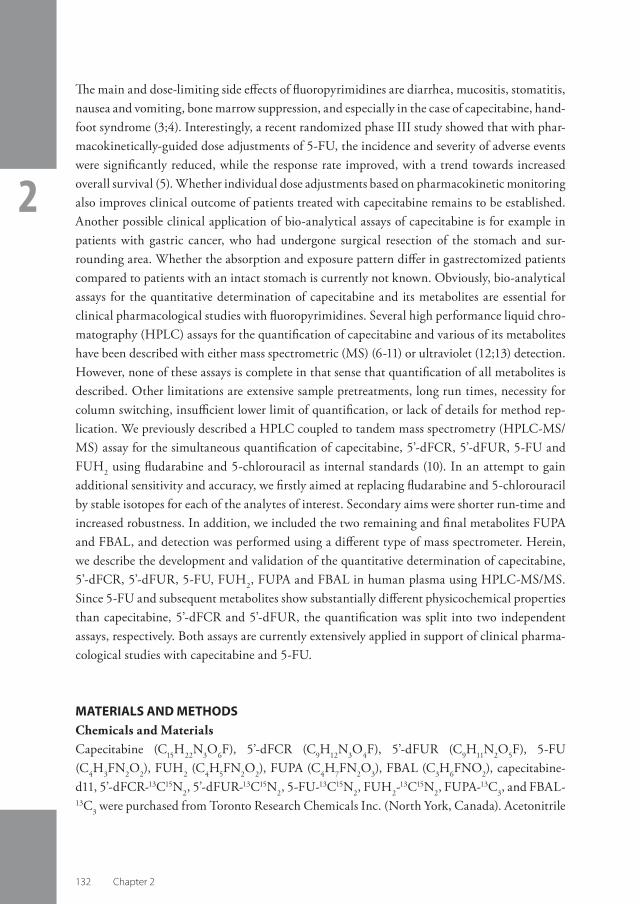
132 Chapter 2
2
The main and dose-limiting side effects of fluoropyrimidines are diarrhea, mucositis, stomatitis, nausea and vomiting, bone marrow suppression, and especially in the case of capecitabine, hand-foot syndrome (3;4). Interestingly, a recent randomized phase III study showed that with phar-macokinetically-guided dose adjustments of 5-FU, the incidence and severity of adverse events were significantly reduced, while the response rate improved, with a trend towards increased overall survival (5). Whether individual dose adjustments based on pharmacokinetic monitoring also improves clinical outcome of patients treated with capecitabine remains to be established. Another possible clinical application of bio-analytical assays of capecitabine is for example in patients with gastric cancer, who had undergone surgical resection of the stomach and sur-rounding area. Whether the absorption and exposure pattern differ in gastrectomized patients compared to patients with an intact stomach is currently not known. Obviously, bio-analytical assays for the quantitative determination of capecitabine and its metabolites are essential for clinical pharmacological studies with fluoropyrimidines. Several high performance liquid chro-matography (HPLC) assays for the quantification of capecitabine and various of its metabolites have been described with either mass spectrometric (MS) (6-11) or ultra violet (12;13) detection. However, none of these assays is complete in that sense that quantification of all metabolites is described. Other limitations are extensive sample pretreatments, long run times, necessity for column switching, insufficient lower limit of quantification, or lack of details for method rep-lication. We previously described a HPLC coupled to tandem mass spectrometry (HPLC-MS/MS) assay for the simultaneous quantification of capecitabine, 5’-dFCR, 5’-dFUR, 5-FU and FUH2 using fludarabine and 5-chlorouracil as internal standards (10). In an attempt to gain additional sensitivity and accuracy, we firstly aimed at replacing fludarabine and 5-chlorouracil by stable isotopes for each of the analytes of interest. Secondary aims were shorter run-time and increased robustness. In addition, we included the two remaining and final metabolites FUPA and FBAL, and detection was performed using a different type of mass spectrometer. Herein, we describe the development and validation of the quantitative determination of capecitabine, 5’-dFCR, 5’-dFUR, 5-FU, FUH2, FUPA and FBAL in human plasma using HPLC-MS/MS. Since 5-FU and subsequent metabolites show substantially different physicochemical properties than capecitabine, 5’-dFCR and 5’-dFUR, the quantification was split into two independent assays, respectively. Both assays are currently extensively applied in support of clinical pharma-cological studies with capecitabine and 5-FU.
MATERIALS AND METHODSChemicals and MaterialsCapecitabine (C15H22N3O6F), 5’-dFCR (C9H12N3O4F), 5’-dFUR (C9H11N2O5F), 5-FU (C4H3FN2O2), FUH2 (C4H5FN2O2), FUPA (C4H7FN2O3), FBAL (C3H6FNO2), capecitabine-d11, 5’-dFCR-13C15N2, 5’-dFUR-13C15N2, 5-FU-13C15N2, FUH2-
13C15N2, FUPA-13C3, and FBAL-13C3 were purchased from Toronto Research Chemicals Inc. (North York, Canada). Acetonitrile

Bioanalysis of capecitabine and metabolites 133
2
and methanol (supra-gradient grade) were from Biosolve Ltd (Valkenswaard, The Netherlands). Formic acid 98% and Water (LiChrosolve) originated from Merck (Darmstadt, Germany) and distilled water was obtained from Aqua B. Braun (Melsungen, Germany).
Mass spectrometryDetection of the analytes was performed on a triple quadrupole mass spectrometer equipped with turbo ionspray interface (API4000, Applied Biosystems, Ontario, Canada). An Agilent 1100 series liquid chromatograph system was used consisting of a binary pump, in-line degas-ser, column oven and autosampler (Agilent Technologies, Palo Alto, CA, USA). Capecitabine and 5’-dFCR were detected in the positive mode, whereas the other analytes in the negative ion mode. MS/MS experiments were performed to determine the most abundant product ions, for which multiple reaction monitoring (MRM) parameters were optimized. Optimized mass transitions and operating procedures are provided in table 1. The curtain gas (N2) was set at 50 psi. The collision gas was set at 8 psi for the capecitabine assay and 6 psi for the 5-FU assay. AnalystTM software v1.5 (AB Sciex) was used for data processing.
Chromatography for capecitabine, 5’-dFCR and 5’-dFURCapecitabine, 5’-dFCR and 5’-dFUR were separated using a XBridge C18 column (50 x 2.1 mm ID, particle size 5 µm; Waters Corp., Mildford, MA, USA) protected with a 0.5 µm filter (Upchurch Scientific, Oak Harbor, WA, USA), thermostatted at 30ºC. Chromatography was performed using a gradient system consisting of mobile phase solution A (0.05% formic acid in water) and solution B (0.05% formic acid in methanol). The gradient started for the first three minutes with solution A:B 95:5 (v/v) at a constant flow rate of 0.3 mL/min. From 3.01 to 6.00 minutes the flow rate was maintained at 0.4 ml/min and the gradient changed to A:B 5:95 (v/v). At t = 6.01 minutes, the gradient changed back again to A:B 95:5 (v/v) with an increased flow rate of 0.5 ml/min for the following two minutes. At t = 8.01 minutes, the flow rate returned to its starting value of 0.3 ml/min for equilibration. Total run time of the assay was 9.0 minutes. By using a divert valve, the eluent was directed to the mass spectrometer in the period between 1.8 and 7.0 minutes. Otherwise, the eluent was directed to the waste.
Chromatography for 5-FU, FUH2, FUPA and FBALThe chromatography for 5-FU, FUH2, FUPA and FBAL was performed on a Luna HILIC column (150 x 2.1 mm ID, particle size 3 µm; Phenomenex) protected with an inline filter of 0.5 µm (Upchurch Scientific, Oak Harbor, WA, USA) with the column oven maintained at ambient temperature. 5-FU, FUH2, FUPA and FBAL were chromatographically separated using an isocratic elution consisting of 20% solution A (10 mM formic acid in water at pH 4.0) mixed with 80% solution B (100% acetonitrile). The flow rate was maintained at 0.3 mL/min and total run time was 5.0 minutes. During the first 1.2 minutes and after 4.5 minutes the flow was directed to the waste.
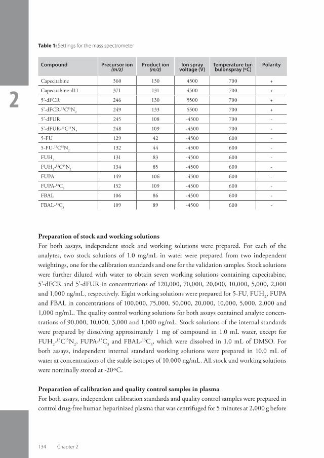
134 Chapter 2
2
Preparation of stock and working solutionsFor both assays, independent stock and working solutions were prepared. For each of the analytes, two stock solutions of 1.0 mg/mL in water were prepared from two independent weightings, one for the calibration standards and one for the validation samples. Stock solutions were further diluted with water to obtain seven working solutions containing capecitabine, 5’-dFCR and 5’-dFUR in concentrations of 120,000, 70,000, 20,000, 10,000, 5,000, 2,000 and 1,000 ng/mL, respectively. Eight working solutions were prepared for 5-FU, FUH2, FUPA and FBAL in concentrations of 100,000, 75,000, 50,000, 20,000, 10,000, 5,000, 2,000 and 1,000 ng/mL. The quality control working solutions for both assays contained analyte concen-trations of 90,000, 10,000, 3,000 and 1,000 ng/mL. Stock solutions of the internal standards were prepared by dissolving approximately 1 mg of compound in 1.0 mL water, except for FUH2-
13C15N2, FUPA-13C3 and FBAL-13C3, which were dissolved in 1.0 mL of DMSO. For both assays, independent internal standard working solutions were prepared in 10.0 mL of water at concentrations of the stable isotopes of 10,000 ng/mL. All stock and working solutions were nominally stored at -20ºC.
Preparation of calibration and quality control samples in plasmaFor both assays, independent calibration standards and quality control samples were prepared in control drug-free human heparinized plasma that was centrifuged for 5 minutes at 2,000 g before
Compound Precursor ion (m/z)
Product ion (m/z)
Ion spray voltage (V)
Temperature turbulonspray (ºC)
Polarity
Capecitabine 360 130 4500 700 +
Capecitabine-d11 371 131 4500 700 +
5’-dFCR 246 130 5500 700 +
5’-dFCR-13C15N2 249 133 5500 700 +
5’-dFUR 245 108 -4500 700 -
5’-dFUR-13C15N2 248 109 -4500 700 -
5-FU 129 42 -4500 600 -
5-FU-13C15N2 132 44 -4500 600 -
FUH2 131 83 -4500 600 -
FUH2-13C15N2 134 85 -4500 600 -
FUPA 149 106 -4500 600 -
FUPA-13C3 152 109 -4500 600 -
FBAL 106 86 -4500 600 -
FBAL-13C3 109 89 -4500 600 -
Table 1: Settings for the mass spectrometer

Bioanalysis of capecitabine and metabolites 135
2
use. Calibration standards were freshly prepared at concentrations of capecitabine, 5’-dFCR and 5’-dFUR of 50, 100, 250, 500, 1,000, 3,500 and 6,000 ng/mL, and at 50, 100, 250, 500, 1,000, 2,500, 3,750 and 5,000 ng/mL for 5-FU, FUH2, FUPA and FBAL, each by adding 20 µL of the appropriate working solution to 380 µL of plasma, followed by short vortex mixing. Quality control samples were prepared for both assays in analyte concentrations of 50 (lower limit of quantification [LLOQ]), 150 (low), 500 (mid), 4,500 (high) and 25,000 (>upper limit of quantification [ULOQ]) ng/mL. For the 5-FU assay an additional quality control concentra-tion of 100 ng/mL (an alternative, but higher LLOQ) was included. Quality control samples were stored in replicates of 200 µL at -20ºC.
Sample pre-treatment for the analysis of capecitabine, 5’-dFCR and 5’-dFURAn amount of 10 µL of the internal standard solution was added to 100 µL human lithium heparinized plasma. Proteins were precipitated by adding 200 µL of methanol:acetonitrile 1:1 (v/v), followed by short vortex mixing and automated shaking at 1250 rpm for 10 minutes (Labinco, Breda, the Netherlands). Then, samples were centrifuged for 10 minutes at approxi-mately 23,100 g, and 50 µL of the supernatant was transferred into a glass autosampler vial with insert containg 150 µL water. Injection volume of the processed sample solution was 5 µL.
Sample pre-treatment for the analysis of 5-FU, FUH2, FUPA and FBALTo 100 µL of human heparinized plasma, 10 µL of the internal standard working solution was added. Protein precipitation was performed by adding 400 µL of 0.5% NH4OH in acetonitrile, followed by short vortex mixing and automated shaking at 1250 rpm for 10 minutes. Samples were then centrifuged for 10 minutes at approximately 23,100 g. A volume of 100 µL of the supernatant was transferred into a glass autosampler vial with insert for the analysis of 5-FU and FBAL. The remainder of the supernatant was filtered through a HybridSPE-PPT cartridge (30 mg/mL; Sigma-Aldrich, Zwijndrecht, The Netherlands). The filtrate was collected in a 10 mL propylene tube, and transferred into a glass autosampler vial with insert for the analysis of FUH2 and FUPA. 5 µL of the processed sample solutions were used for injection.
Validation proceduresBoth assays were validated in accordance with the current FDA guideline on bioanalytical method validation (14).
Linearity Calibration standards, including standards containing no analyte or internal standard (double blank) and samples containing only internal standard (blank), were analyzed in duplicate in three separate analytical runs. The linear regression of the peak area versus the concentra-tion was weighted by 1/x2 for determination of the concentration of the analytes. To assess linearity, deviations of the back-calculated concentrations from the nominal concentrations

136 Chapter 2
2
should be within 85-115%. At the lower limit of quantification (LLOQ) level a deviation of ± 20% was allowed.
Accuracy and precisionFive replicates of each of the quality control samples at LLOQ, low, mid, and high concentra-tions were analyzed in three separate analytical runs. Five replicates above the upper limit of quantification (ULOQ) at 25,000 ng/mL were diluted ten times and analyzed in one analytical run. The inter- and intra-assay accuracies were calculated as the difference between the nomi-nal and measured concentrations. The coefficient of variation (CV) was calculated to assess the precision. Accuracy should be within ±20% for the LLOQ and within 15% at the other concentrations. The CVs should be less than 20% at the LLOQ, and less than 15% at the other concentration levels.
Specificity and selectivitySix individual batches of control drug-free lithium heparinized human plasma were used to investigate whether endogenous matrix constituents interfere with the assay. Blank, double blank, and samples spiked at the LLOQ were freshly prepared, and analyzed in one analytical run. Peak areas of compounds co-eluting with the analyte or internal standard should not exceed 20% of the analyte peak area at the LLOQ, or 5% of the internal standard area. Deviations at the LLOQ level from the nominal concentrations should be within ±20%. To determine the cross analyte and internal standard interferences, all analytes were sepa-rately spiked at their ULOQ concentration to control human plasma. The internal standards were separately spiked at their nominal concentration. The interference at the retention times of the analytes and internal standards should be less than 20% and 5% of the peak areas at the LLOQ, respectively.
Matrix factorThe matrix effect (ion suppression) was determined in triplicate by analyzing the analytes at mid concentrations in water, and measuring the analytes spiked in blank plasma extract in the same concentrations as in processed QC mid samples. The matrix factor was defined as the ratio of each analyte peak response in the presence and absence of matrix ions.
Carry-over Carry-over was determined in one analytical run by injecting a double blank matrix sample after the highest calibration standard. Responses in the double blank matrix at the retention times of the analytes and internal standards should be less than 20% and 5% of the mean response of the LLOQ, respectively.

Bioanalysis of capecitabine and metabolites 137
2
StabilityStability of the stock solutions in water was determined after six hours of storage at room tem-perature, and for 5-FU and FBAL also after 4 months of storage at -20ºC. The stability of the analytes in plasma was investigated during three freeze (-20ºC)/thaw cycles. Short-term stabil-ity of capecitabine, 5’-dFCR and 5’-dFUR in plasma was determined after storage for 6, 24 and 48 hours at ambient temperature, on ice-water, and at ambient temperature stabilized with 0.1mg/mL tetrahydrouridine (THU). Short-term stability of 5-FU, FUH2, FUPA and FBAL in plasma was determined after 6 hours at ambient temperature. Long-term stability in plasma stored at -20ºC was determined after 3 and 6 months for capecitabine, 5’-dFCR and 5’-dFUR. Long-term stability experiments for 5-FU, FUH2, FUPA and FBAL are currently ongoing. The stability of the processed samples was determined after storage for two weeks at 2-8ºC. The re-injection reproducibility in the autosampler was determined 24 hours after start of the original run, during which period samples were stored at 2-8ºC. All stability experiments were performed in triplicate at concentration levels of QC low, mid and high. The analytes were con-sidered stable if the determined concentrations did not deviate more than ± 15% from the initial (i.e. freshly prepared or processed sample) concentrations. The precisions should be within 20% at the level of the LLOQ and within 15% at the other concentration levels.
RESULTS AND DISCUSSIONChromatographyIn our previous report for the determination of capecitabine, 5’-dFCR, 5’-dFUR, 5-FU and FUH2 in plasma, a Hypercarb column (30 x 2.1 mm ID, particle size 5 µm) was used, by which all analytes could be successfully separated in one assay within 15 minutes (10). Upon frequent application of the assay however, we noticed that when analyzing a run containing approximately more than 40 samples, the signal intensity decreased by more than 50%, thereby significantly loosing sensitivity. In addition, the number of runs that could be maximally performed using this column was limited, despite extensive washing and reconditioning procedures. Therefore, we decided to develop a new assay using a different type of chromatography. Given the extensive differences in physicochemical properties of capecitabine, 5’-dFCR and 5’-dFUR compared to 5-FU and subsequent metabolites, it was decided to split the quantification into two independ-ent assays. Hereby, analytes in the capecitabine assay could be separated within 9.0 minutes and for the 5-FU assay within 5.0. Typical retention times of capecitabine, 5’-dFCR, 5’-dFUR, 5-FU, FUH2, FUPA and FBAL were 4.9, 2.4, 3.4, 1.8, 1.7, 2.1 and 3.8 minutes, respectively. Both assays proved to be very stable and robust, also when extensively used. In addition, the capecitabine assay could be successfully applied using 96-wells plates (additional validation data not shown). Figures 2 and 3 depict the representative chromatograms of the analytes on the level of the LLOQ and of the internal standards, respectively.

138 Chapter 2
2
Figure 2: Representative HPLC-MS/MS chromatograms from a spiked human heparinized plasma sample at the
LLOQ of capecitabine (A), 5’-dFCR (B), 5’-dFUR (C), 5-FU (D), FUH2 (E), FUPA (F) and FBAL (G).

Bioanalysis of capecitabine and metabolites 139
2
Figure 3: Representative HPLC-MS/MS chromatograms from the internal standards of capecitabine-d11 (A), 5’-dFCR-13C15N2 (B), 5’-dFUR-13C15N2 (C), 5-FU-13C15N2 (D), FUH2-
13C15N2 (E), FUPA-13C3 (F) and FBAL-13C3 (G) spiked in human
heparinized plasma.

140 Chapter 2
2
Sample pre-treatmentSample pre-treatment was initially started by using the same procedure as described in our previous assay, using 10% (v/v) trichloroacetic acid (TCA) for protein precipitation (10). However, in this assay fludarabine and 5-chlorouracil were replaced by stable isotopes for each of the analytes as internal standards. Results revealed that capecitabine-d11 rapidly converted into 5’-dFUR in acid environment. Storage of the TCA-processed samples at 2-8ºC for only a couple of hours already resulted in an unacceptable increase in the con-centration of 5’-dFUR of more than 25%, whereas the area of capecitabine-d11 decreased with a similar amount. Therefore, 10% TCA appeared not suitable for protein precipita-tion in this assay. Using a pH-neutral solution of methanol:acetonitrile (1:1, v/v), protein precipitation was successful, and guaranteed sufficient stability of capecitabine, 5’-dFCR, 5’-dFUR and their stable isotopes, respectively. For the 5-FU assay, 100% acetonitrile was used for protein precipitation, which was added in a 4:1 (v/v) ratio to plasma. Herewith, the same percentage of acetonitrile as is used in the mobile phase of the isocratic elution is reached. Thereby, when injecting a processed sample, potentially disturbing effects on the chromatography caused by differences in polarity are minimized. During pre-validation experiments, it was noted that the sensitivity of FUH2 and FUPA significantly increased using a HybridSPE-PPT cartridge filter, which removes amongst others phospholipids from the extract. Therefore, FUH2 and FUPA were quantified filtered, whereas 5-FU and FBAL were quantified unfiltered.
Mass spectrometryFigure 4 depict the MS/MS product ion scans and proposed fragmentation pattern of all ana-lytes. Clear responses were observed at m/z of 360, 246, 245, 129, 131, 149, and 106, which correspond to the protonated molecular ions of capecitabine and 5’-dFCR, and deprotonated molecular ions of 5’-dFUR, 5-FU, FUH2, FUPA and FBAL, respectively. The most abundant fragments of capecitabine and 5’-dFCR were product ions with m/z of 130, corresponding to loss of the sugar moiety and for capecitabine additionally the pentane-acid chain. For 5’-dFUR the m/z of the product ion was 108, corresponding to the loss of the sugar moiety and the fluor atom. The product ion of 5-FU had a m/z of 42 and represents the formamide moiety; the product ion of FUH2 with m/z 83 represents loss of the fluoro-ethane group; the product ion of FUPA had a m/z 106 corresponding to the loss of the formamide moiety; and the m/z for the product ion of FBAL was 86, representing the loss of the fluor atom.
ValidationLinearityThe assays were linear over the tested concentration range of 50-6,000 ng/mL for capecitabine, 5’-dFCR and 5’-dFUR, and 50-5,000 ng/mL for 5-FU, FUH2, FUPA and FBAL in human plasma. The mean accuracies did not deviate more than -5.8% and 3.2% from the nominal con-

Bioanalysis of capecitabine and metabolites 141
2
Figure 4: Product ion scans of capecitabine (A), 5’-dFCR (B) 5’-dFUR (C) 5-FU (D), FUH2 (E), FUPA (F) and FBAL (G).

142 Chapter 2
2 Compound Nominal concen tration
(ng/mL)
Mean measured
concentration
(ng/mL)
Interassay accuracy (%)
Interassay precision (%)
Number of replicates
Capecitabine 48.61464864370
46.6143489
4480
-4.1-2.30.52.5
9.64.32.22.0
15151515
5’-dFCR 50.41515044530
45.3143472
4400
-10.2-5.5-6.4-2.9
9.26.74.74.3
15151515
5’-dFUR 49.5149495
4460
47.5152527
4780
-4.02.36.57.2
9.76.43.93.3
15151515
5-FU 49.899.6149498
4480
50.2104148495
4490
0.84.3-0.6-0.50.3
7.07.85.13.03.7
1515151515
FUH2 49.098.01474904410
49.394.2148482
4480
0.6-3.80.6-1.61.6
9.911.38.15.76.5
1515151515
FUPA 51.5103155515
4610
51.0101151506
4460
-0.9-2.1-2.9-1.7-3.3
6.75.83.47.46.6
1515151515
FBAL 58.01161745805220
59.51171715635310
2.50.6-1.6-3.01.7
8.511.88.76.36.4
1515151515
Table 2: Assay performance data for capecitabine and metabolites in human plasma
centrations for all compounds at all concentration levels, with a maximum CV for the precision of 9.6% above LLOQ concentrations, and a maximum CV of 16.3% at the LLOQ. Correlation coefficients for all compounds were 0.995 or higher.

Bioanalysis of capecitabine and metabolites 143
2
Accuracy and precisionTable 2 lists the assay performance data (inter-assay accuracies and precisions) for capecitabine and its metabolites. The intra-assay accuracies at the LLOQ were within -15.9% and 7.6%, and at the other levels within -11.1% and 10.9%. The maximum intra-assay precisions at the LLOQ and at the other levels were 11.2% and 14.2%, respectively. In addition, 10-fold dilution of the samples above the ULOQ resulted in acceptable deviations from nominal concentrations with intra-assay accuracies within -7.9% and 11.0% and intra-assay precisions were maximally CV = 12.6% (data not shown). In summary, all accuracies and precisions for all compounds were within the predefined acceptance criteria.
Specificity and selectivityThe endogenous, cross analyte and internal standard interference tests showed no co-eluting peaks in the blanks with areas exceeding 20% of the area at the LLOQ level of the analytes in the blanks, and no co-eluting peaks exceeding 5% of the area of the internal standards. The deviations from the nominal concentrations were within ± 20%, therefore, the specificity and selectivity of the assay were considered acceptable.
Matrix factorThe ratio’s of the mean areas of capecitabine, 5’-dFCR, 5’-dFUR, 5-FU, FUH2, FUPA and FBAL in the presence of compared to absence of matrix ions were 1.10, 0.99, 1.03, 0.99, 0.98, 1.01 and 1.07, respectively. The results (matrix factor ratio around 1) indicate that the stable iso-topically labeled internal standards are most effective minimizing the influence of matrix ions.
Carry-overThe responses in the first double blank sample after injection of the highest calibration standard were 0.00% of the areas of a LLOQ sample for all analytes and internal standards, except for capecitabine, for which the carry-over was 11.6% of the area at LLOQ. Thus, the carry-over was satisfactory.
StabilityTable 3 shows the results of the stability experiments. All tested analytes were stable in stock solution (in water) at ambient temperatures for at least 6 hours. The deviation of the freeze/thaw stability experiments also remained within ± 15% of the nominal concentrations. In the final extract, all analytes were stable for at least 15 days when stored at 2-8ºC, and also, the re-injection reproducibility was satisfactory. All analytes were stable in plasma within the margins of acceptance at ambient temperature for 6 hours, except for FUH2, which concen-tration decreased by more than 40% during this storage period. Furthermore, there was a clear decrease visible in the concentration of 5’-dFCR, whereas the concentration of 5’-dFUR increased. Upon storage for 24 and 48 hours at ambient temperature, a definitively unaccep-

144 Chapter 2
2
>
Matrix Condition Compound Initial concentration
(ng/mL)
Measured concentration
(ng/mL)
Deviation (%)
CV
(%)
Stock solution
Ambient T, 6h
Capecitabine5’-dFCR5’-dFUR5-FUFBAL
1.00 x 10⁶
1.00 x 10⁶
1.00 x 10⁶
1.00 x 10⁶
1.00 x 10⁶
1.01 x 10⁶
1.02 x 10⁶
0.9 x 10⁶
0.97 x 10⁶
1.01 x 10⁶
1.41.5-1.7-3.00.8
1.85.81.01.91.7
Stock solution
-20ºC, 4 months
5-FUFBAL
1.00 x 10⁶
1.00 x 10⁶
0.99 x 10⁶
1.03 x 10⁶
-0.83.0
1.10.5
Plasma 3 freeze (-20º) / thaw cycles
Capecitabine
5’-dFCR
5’-dFUR
5-FU
FUH2
FUPA
FBAL
1464370151
4530149
4460149
4480147
4410155
4640156
4670
1444440134
4460165
4790142
4373154
43531544510154
4580
-1.11.7
-11.0-1.510.57.4-4.9-2.44.8-1.3-0.9-2.8-1.5-1.9
5.80.93.01.12.82.87.91.4
12.26.86.31.22.00.0
Plasma 3 months, -20ºC
Capecitabine
5’-dFCR
5’-dFUR
1464370151
4530149
4460
14544901394143167
5073
-0.72.7-7.9-8.512.113.8
0.72.24.52.83.34.0
Plasma 6 months, -20ºC
Capecitabine
5’-dFCR
5’-dFUR
1464370151
4530149
4460
14443571293753154
5013
-1.6-0.3-14.3-17.13.4
12.4
2.62.415.15.69.44.9
Table 3: Stability data for capecitabine and its metabolites
table conversion of 5’-dFCR into 5’-dFUR occurred. It is known that cytidine deaminase, which is the enzyme responsible for this conversion, has high activity in plasma, but can be competitively inhibited using the substrate THU (15;16). Therefore, additional stability experiments were performed with THU-stabilized plasma samples, but also with unstabi-

Bioanalysis of capecitabine and metabolites 145
2
>
Matrix Condition Compound Initial concentration
(ng/mL)
Measured concentration
(ng/mL)
Deviation (%)
CV
(%)
Plasma Ambient, 6h
Capecitabine
5’-dFCR
5’-dFUR
5-FU
FUH2
FUPA
FBAL
14648243701514944530149488
4460149
4480147
4410155
4640174
5220
145467
43301354443923171501
4903141
439385
2573151
4463159
5253
-0.9-3.1-0.9-10.6-10.2-13.414.52.69.9-5.6-1.9
-42.3-41.6-2.6-3.8-8.60.6
3.13.51.62.23.30.45.04.45.44.70.38.75.26.10.78.65.5
Plasma Ambient, 24h
Capecitabine
5’-dFCR
5’-dFUR
14648243701514944530149488
4460
1344414137107373
33031865825740
-8.2-8.4-5.3-29.2-24.6-27.124.819.328.7
0.85.31.29.26.52.33.83.22.7
Plasma Ambient, 48h
Capecitabine5’-dFCR5’-dFUR
482494488
407320642
-15.6-35.231.6
0.96.96.9
Plasma Ice-water, 6h
Capecitabine5’-dFCR5’-dFUR
482494488
452478469
-6.2-3.2-4.0
1.44.54.4
Plasma Ice-water, 24h
Capecitabine5’-dFCR5’-dFUR
482494488
443452504
-8.1-8.63.3
3.26.68.4
Plasma Ice-water, 48h
Capecitabine5’-dFCR5’-dFUR
482494488
450448496
-6.7-9.31.5
2.74.54.4
Table 3 Continued: Stability data for capecitabine and its metabolites

146 Chapter 2
2
Matrix Condition Compound Initial concentration
(ng/mL)
Measured concentration
(ng/mL)
Deviation (%)
CV (%)
Plasma Ambient, stabilized with THU, 6h
Capecitabine5’-dFCR5’-dFUR
482494488
482515473
-0.14.1-3.1
3.47.33.2
Plasma Ambient, stabilized with THU, 24h
Capecitabine5’-dFCR5’-dFUR
482494488
450517481
-6.64.6-1.5
3.35.85.6
Plasma Ambient, stabilized with THU, 48h
Capecitabine5’-dFCR5’-dFUR
482494488
418523472
-13.25.9-3.2
2.63.22.4
Final extract
2-8ºC, 15 days
Capecitabine
5’-dFCR
5’-dFUR
1464370151
4530149
4460
1494330163
4400165
4770
2.3-1.08.2-2.811.07.0
3.71.33.50.74.52.2
Final extract
2-8ºC, 17 days
5-FU
FUH2
FUPA
FBAL
1494480147
4410155
4640174
5220
149457014946101714710164
4893
0.22.01.44.510.11.5-5.6-6.3
5.52.211.15.1
12.05.95.24.4
Final extract
Re-injection reproducibil-ity, 2-8ºC, 24h
Capecitabine
5’-dFCR
5’-dFUR
5-FU
14648643701515044530149495
4460149498
4480
139483
4320141490
4220137511
44001434934313
-4.6-0.5-1.1-6.4-2.8-6.8-8.13.3-1.4-3.8-1.1-3.7
6.14.52.97.65.82.07.65.83.31.51.24.8
Table 3 Continued: Stability data for capecitabine and its metabolites
>

Bioanalysis of capecitabine and metabolites 147
2
Matrix Condition Compound Initial concentration
(ng/mL)
Measured concentration
(ng/mL)
Deviation (%)
CV (%)
Final extract
Re-injection reproducibil-ity, 2-8ºC, 24h
FUH2
FUPA
FBAL
1474904410155515
4640156519
4670
1354624590153514
4427146491
4410
-8.2-5.74.1-1.3-0.3-4.6-6.2-5.4-5.6
13.72.05.02.62.92.31.71.91.3
Table 3 Continued: Stability data for capecitabine and its metabolites
lized plasma stored on ice-water. Under both conditions, the CDA-mediated conversion of 5’-dFCR to 5’-dFUR was successfully inhibited for at least 48 hours. Long-term stability in plasma at -20ºC was acceptable when stored for 3 months, however, also under this condi-tion, 5’-dFCR slowly metabolized to 5’-dFUR. Importantly, at 6 months, the decrease in the concentration of 5’-dFCR exceeded the predefined level of acceptance. Therefore, long-term stability experiments at -70ºC have been started. To obtain the most reliable pharmacokinetic data of patients treated with capecitabine, it can be concluded that obtained blood samples should be immediately cooled on ice-water and centrifuged at 2-8ºC. Plasma is best stored at -70ºC, and in case of storage for more than 6 months, should be stabilized using THU. Ideally, plasma samples are analyzed as soon as possible after blood drawing.
Figure 5: Plasma concentration-time curves of capecitabine, 5’-dFCR, 5’-dFUR, 5-FU, FUH2, FUPA and FBAL
in a patient with gastric cancer following administration of 1650 mg capecitabine.

148 Chapter 2
2
APPLICATION OF THE ASSAYSThe described assays are currently successfully applied in support of pharmacokinetic studies in patients treated with capecitabine or 5-FU. Figure 5 shows the measured concentrations of capecitabine and all of its metabolites in a patient with gastric cancer after oral administration of 1650 mg capecitabine. Samples were taken and analyzed after written informed consent from the patient had been obtained. The selected linear ranges cover typically observed plasma concentrations for all analytes after administration of capecitabine.
CONCLUSIONWe report the development and validation of the quantitative determination of the frequently applied anticancer drug capecitabine and all of its metabolites 5’-dFCR, 5’-dFUR, 5-FU, FUH2, FUPA and FBAL in human plasma, using HPLC-MS/MS. Due to significant differ-ences in physicochemical properties of capecitabine, 5’-dFCR and 5’-dFUR compared to 5-FU, FUH2, FUPA and FBAL, the quantification was split into two independent assays, respectively. Thereby, a highly robust, accurate, sensitive and specific quantification could be achieved, maintaining short run-times. All analytes were extracted using protein precipitation methods, and stable isotopes for each of the analytes were used as internal standard. Reversed-phase chromatography was used for the capecitabine assay, whereas the separation in the 5-FU-assay was conducted using normal phase chromatography. The tested linear range of the analytes was 50-6,000 ng/mL for the capecitabine assay, and 50-5,000 ng/mL for the 5-FU assay. These concentrations cover the ranges of typically observed plasma concentrations after administra-tion of capecitabine or 5-FU. Both assays appeared highly robust and well-suitable for support of pharmacokinetic studies with capecitabine and 5-FU.

Bioanalysis of capecitabine and metabolites 149
2
References(1) Schellens JH. Capecitabine. Oncologist 2007 Feb;12(2):152-5.(2) Malet-Martino M, Martino R. Clinical studies of three oral prodrugs of 5-fluorouracil (capecitabine, UFT, S-1):
a review. Oncologist 2002;7(4):288-323.(3) Koopman M, Antonini NF, Douma J, Wals J, Honkoop AH, Erdkamp FL, et al. Sequential versus combination
chemotherapy with capecitabine, irinotecan, and oxaliplatin in advanced colorectal cancer (CAIRO): a phase III randomised controlled trial. Lancet 2007 Jul 14;370(9582):135-42.
(4) Hoff PM, Ansari R, Batist G, Cox J, Kocha W, Kuperminc M, et al. Comparison of oral capecitabine versus intravenous fluorouracil plus leucovorin as first-line treatment in 605 patients with metastatic colorectal cancer: results of a randomized phase III study. J Clin Oncol 2001 Apr 15;19(8):2282-92.
(5) Gamelin E, Delva R, Jacob J, Merrouche Y, Raoul JL, Pezet D, et al. Individual fluorouracil dose adjustment based on pharmacokinetic follow-up compared with conventional dosage: results of a multicenter randomized trial of patients with metastatic colorectal cancer. J Clin Oncol 2008 May 1;26(13):2099-105.
(6) Guichard SM, Mayer I, Jodrell DI. Simultaneous determination of capecitabine and its metabolites by HPLC and mass spectrometry for preclinical and clinical studies. J Chromatogr B Analyt Technol Biomed Life Sci 2005 Nov 5;826(1-2):232-7.
(7) Reigner B, Verweij J, Dirix L, Cassidy J, Twelves C, Allman D, et al. Effect of food on the pharmacokinetics of capecitabine and its metabolites following oral administration in cancer patients. Clin Cancer Res 1998 Apr;4(4):941-8.
(8) Reigner B, Clive S, Cassidy J, Jodrell D, Schulz R, Goggin T, et al. Influence of the antacid Maalox on the pharmacokinetics of capecitabine in cancer patients. Cancer Chemother Pharmacol 1999;43(4):309-15.
(9) Siethoff C, Orth M, Ortling A, Brendel E, Wagner-Redeker W. Simultaneous determination of capecitabine and its metabolite 5-fluorouracil by column switching and liquid chromatographic/tandem mass spectrometry. J Mass Spectrom 2004 Aug;39(8):884-9.
(10) Vainchtein LD, Rosing H, Schellens JH, Beijnen JH. A new, validated HPLC-MS/MS method for the simultaneous determination of the anti-cancer agent capecitabine and its metabolites: 5'-deoxy-5-fluorocytidine, 5'-deoxy-5-fluorouridine, 5-fluorouracil and 5-fluorodihydrouracil, in human plasma. Biomed Chromatogr 2010 Apr;24(4):374-86.
(11) Xu Y, Grem JL. Liquid chromatography-mass spectrometry method for the analysis of the anti-cancer agent capecitabine and its nucleoside metabolites in human plasma. J Chromatogr B Analyt Technol Biomed Life Sci 2003 Jan 5;783(1):273-85.
(12) Dhananjeyan MR, Liu J, Bykowski C, Trendel JA, Sarver JG, Ando H, et al. Rapid and simultaneous determination of capecitabine and its metabolites in mouse plasma, mouse serum, and in rabbit bile by high-performance liquid chromatography. J Chromatogr A 2007 Jan 5;1138(1-2):101-8.
(13) Zufia L, Aldaz A, Giraldez J. Simple determination of capecitabine and its metabolites by liquid chromatography with ultraviolet detection in a single injection. J Chromatogr B Analyt Technol Biomed Life Sci 2004 Sep 25;809(1):51-8.
(14) U.S.Department of Health and Human Services Food and Drug Administration: Center for Drug Evaluation and Research. Guidance for Industry: Bioanalytical Method Validation. FDA 2001 May 1
(15) Riccardi R, Chabner B, Glaubiger DL, Wood J, Poplack DG. Influence of tetrahydrouridine on the pharmacokinetics of intrathecally administered 1-beta-D-arabinofuranosylcytosine. Cancer Res 1982 May;42(5):1736-9.
(16) Besnard T, Renee N, Etienne-Grimaldi MC, Francois E, Milano G. Optimized blood sampling with cytidine deaminase inhibitor for improved analysis of capecitabine metabolites. J Chromatogr B Analyt Technol Biomed Life Sci 2008 Jul 1;870(1):117-20.


Chapter 3
Clinical pharmacogenetics
and pharmacokinetics
of fluoropyrimidines
151


Chapter 3.1
Upfront genotyping of DPYD
to improve patient safety of
fluoropyrimidine therapy, a safety
and cost-effectiveness analysis
Interim analysis
Maarten J. Deenen, Annemieke Cats,
Marjolein K. Sechterberger, Johan L. Severens,
Paul H.M. Smits, Remco Bakker, Otilia Dalesio,
Caroline M.P.W. Mandigers, Marcel Soesan,
Jos H. Beijnen, Jan H.M. Schellens
153

154 Chapter 3.1
3
ABSTRACTPurposeTo determine the safety and cost-effectiveness of DPYD*2A pharmacogenetic guided dosing in patients treated with fluoropyrimidines.
Patients and MethodsGermline DNA was prospectively obtained from patients treated with capecitabine or 5-FU and screened prior to start of therapy for the DPYD*2A polymorphism. Patients with the DPYD*2A mutation received an initial dose reduction plus further dose-titration in following courses based on clinical tolerance. Toxicity by genotype-guided dosing was compared to toxic-ity in historical DPYD*2A controls having received full dose treatment. Historical control data were obtained from cohort studies and were identified by literature search. DPD enzyme activ-ity and fluoropyrimidine pharmacokinetics were determined in DPYD*2A patients. A decision analytical model was used to evaluate the cost-effectiveness of genotype-guided dosing. ResultsIn total, 1600 patients with cancer were prospectively screened for DPYD*2A, of which 17 were heterozygous variant allele carriers (1.2%). The historical cohort consisted of 40 standard-dose treated patients with DPYD*2A, who were identified from 9 cohort studies that included a total of 3391 fluoropyrimidine-treated patients. The overall incidence of grade ≥ 3 toxicity was 14%, and 13% was hospitalized due to treatment-induced toxicity. In total, 13 patients polymorphic for DPYD*2A could be safely treated with a median dose intensity of 48% (range, 24- 91%), resulting in a 15% incidence of grade 3 toxicity, i.e. short-lasting diarrhea. The gen-otype-predicted DPD deficiency was confirmed by DPD enzyme activity analysis, and was by half reduced in patients with the variant genotype. The cost-effectiveness analysis showed that total health care costs for the screening strategy (€ 5839.-) were lower than of non-screening (€ 5854.-). The risk of grade ≥ 3 toxicity thereby reduced from 68% in historical controls to 15% by genotype-guided dosing. Drug-induced death reduced from 10% to 0%.
ConclusionUpfront genotyping of DPYD*2A improves patient safety of fluoropyrimidine therapy, and is cost-effective.

Upfront genotyping of DPYD in fluoropyrimidine therapy 155
3
INTRODUCTIONFluoropyrimidines, including 5-fluorouracil (5-FU) and capecitabine, are among the most commonly prescribed anticancer drugs for the adjuvant and palliative treatment of various types of solid malignancies, including gastrointestinal, breast, and head and neck cancers. Treatment with fluoropyrimidines is generally well tolerated, except for approximately 10% of the treated population that develop severe, potentially life-threatening toxicity (1-3). The major known cause of intolerance is deficiency of the primary fluoropyrimidine detoxifying enzyme dihydropyrimidine dehydrogenase (DPD), which has an incidence of approximately 3% in Caucasians (4).5-FU is administered intravenously, whereas its pre-prodrug capecitabine is given orally, which, after absorption, is sequentially converted to 5-FU (5). Approximately 10% of 5-FU is intra-cellularly phosphorylated to active metabolites that interfere with DNA synthesis and repair, however, more than 85% of administered 5-FU is inactivated by DPD to dihydrofluoroura-cil (FUH2) (6). The enzymatic activity of DPD has been shown to be predictive for toxicity to fluoropyrimidines: patients with low DPD activity have a decreased potential for inactiva-tion of fluoropyrimidines and are thereby more sensitive towards its toxic effects compared to patients with high DPD enzymatic activity; thereby its activity is an important predictor for 5-FU-induced toxicity (6-10). Several molecular causes may underlay such a DPD-deficiency, of which genetic polymorphism in its encoding gene DPYD is one. The clinically most relevant known polymorphism in DPYD is DPYD*2A (IVS14+1G>A), of which heterozygous popula-tion prevalence is approximately 1 – 2 % in the Western world; homozygous polymorphic individuals are rarely detected (11). The loss of functional DPD activity induced by DPYD*2A results from alternate splicing: the point mutation at the invariant splice donor site of intron 14 results in skipping of exon 14 during pre-mRNA splicing, and thereby creates a truncated protein (12). As a result, the likelihood for severe toxicity to standard-dose fluoropyrimidine therapy in genetically determined poor metabolizers is significantly increased. Treatment of toxicity is associated with interruption or even stop of potentially effective anticancer therapy, but also with significant health care costs. Though a few retrospective trials have reported only weak or non-significant associations of DPYD*2A with toxicity to fluoropyrimidines (13;14), the impression prevails that the standard fluoropyrimidine dose-intensity is principally too high in DPYD*2A polymorphic patients, resulting in unacceptable high rates of severe, potentially lethal toxicity. Evidence for this association has been reported over the last years by multiple case reports (11;15-20), other retrospective trials, (10;21-24), and also prospective population studies (25-28). Based on these observations, we hypothesized that upfront genotyping of DPYD*2A followed by individualized dose adjustment would improve patient safety of fluoropyrimidine therapy, and would be cost-effective.

156 Chapter 3.1
3
PATIENTS AND METHODSStudy designThis prospective, multi-center study (ClinicalTrials.gov Identifier: NCT00838370) was con-ducted at the Netherlands Cancer Institute (NKI; Amsterdam, the Netherlands), the Slotervaart Hospital (Amsterdam, the Netherlands), and at the Canisius Wilhelmina Hospital (Nijmegen, the Netherlands). The primary endpoint was safety, secondary endpoints were cost-effectiveness of DPYD*2A-guided dosing of fluoropyrimidines and additionally included pharmacokinetic and DPD enzyme activity measurements.Given the strong lines of evidence that the standard fluoropyrimidine dose-intensity results in unacceptable high rates of severe, potentially lethal toxicity, a randomized trial was considered unethical. Therefore, it was decided to compare toxicity in DPYD*2A variant allele carriers treated by genotype-guided dosing to toxicity observed in historical controls, i.e. patients with the DPYD*2A variant genotype that had been previously treated with full-dose fluoropyrimi-dine treatment. Historical controls included DPYD*2A polymorphic patients selected from previous publications that either prospectively or retrospectively tested an entire, unselected cohort of patients treated with fluoropyrimidines for DPYD*2A. Appropriate trials were identi-fied by literature research. To avoid selection bias, DPYD*2A polymorphic patients described in individual case reports or identified in case-control studies were not considered eligible, and therefore excluded for the historical control cohort. Toxicity was graded according to the National Cancer Institute Common Terminology Criteria for Adverse Events version 3.0. A decision analytical model was used to evaluate the cost-effectiveness of upfront DPYD genotyp-ing in fluoropyrimidine therapy.
Patients and treatmentGermline DNA was prospectively obtained from patients with cancer intended to undergo standard treatment with fluoropyrimidine-containing anticancer therapy, either as single agent, in combination chemotherapy, or in combination with radiotherapy. Prior (fluoropyrimidine) chemotherapy was allowed. Patients were genotyped prior to start of fluoropyrimidine therapy for DPYD*2A as a routine screening procedure. The assay was conducted five times a week to avoid potential delay in start of treatment. No further intervention was applied in patients that proved to be wild type for the DPYD*2A polymorphism, in whom treatment after screening then proceeded as initially foreseen. Patients with the DPYD*2A variant genotype were treated with an initially reduced dose of the fluoropyrimidine drug, followed by further dose titration based on clinical tolerability. The precise fluoropyrimidine dose was left to the discretion of treating oncologist, but had to be at least reduced by 50% in heterozygous, and at least by 85% in homozygous polymorphic patients during the first two cycles of treatment. Further dose escalation was allowed up to 100% of the conventional dose, provided that previous cycles were fully completed and no grade ≥ 3 toxicity had occurred. Dose escalation had to be determined in the best interest of the patient. Doses of non-fluoropyrimidine drugs and/or radiotherapy

Upfront genotyping of DPYD in fluoropyrimidine therapy 157
3
were standard and left unchanged at start of treatment. The study was approved by the Medical Ethics Committee at one central (NKI) and at all local study sites; patients provided written informed consent before study registration.
GenotypingA volume of 3 mL of whole EDTA blood was obtained prior to start of therapy for DNA analy-sis. Genomic DNA was isolated using the QIAamp DNA mini kit (Qiagen, Inc. Valencia, CA). DPYD*2A was determined on week days by a validated real-time PCR assay and included appro-priate wild type, heterozygous, homozygous and negative controls in every run (29). DNA anal-ysis was performed in certified molecular biology laboratories under good laboratory practice (GLP) conditions. All DNA samples were additionally measured for control in one central insti-tute (NKI). Sensitivity and specificity of the genotyping assay were assumed to be both 100%.
Fluoropyrimidine pharmacokinetics and DPD enzyme activityIn DPYD*2A variant allele carriers, additional blood was obtained for pharmacokinetic and DPD enzyme activity analyses. Whole blood for pharmacokinetic purposes was collected in heparinized tubes on day one of the first treatment cycle, and in the case of dose modification also on day one of that cycle. Blood sampling times were predose, and at 0.25, 0.5, 1, 2, 3, 4, 6 and 8 hours after the morning administration of capecitabine. Obtained blood was immediately centrifuged after drawing at 3000 rpm for 10 minutes at 4°C. Plasma was stored at -70°C until analysis. Values of the pharmacokinetic parameters, including area under the plasma concentra-tion-time curve (AUC), clearance (CL/F, where F is the bioavailability), maximum concentra-tion (Cmax), and time to reach Cmax (Tmax) were compared with pharmacokinetics data described in literature. Plasma concentrations of capecitabine and its metabolites were determined using a validated method with liquid chromatography coupled to tandem mass spectrometry (30). For determination of the DPD enzyme activity in peripheral blood mononuclear cells, 20 mL of EDTA blood were obtained prior to start on the first day of treatment. DPD enzyme activity was determined as described (31).
Cost-effectiveness analysisEconomic analysis was performed prospectively in parallel with the clinical trial from a health care payer perspective. A decision analytical model was build according to the principles of good practice for decision analytical modeling (32). The model compared the conventional, standard-dose treatment strategy (non-screening arm) with the pharmacogenetic-guided dosing strategy as described in the present study (screening arm). Differences in costs, and differences in safety between both strategies were calculated. Parameter estimations that were incorporated in the model were derived from data from the present trial, but also included relevant data from literature, when available. Data derived from the present trial included amongst others patient demographics and treatment characteristics (safety and costs) from all patients, includ-

158 Chapter 3.1
3
ing DPYD*2A wild type patients; data derived from literature included data about population prevalence frequencies of the DPYD*2A polymorphism, and data on the incidences and severity of toxicity to standard-dose fluoropyrimidine treatment in DPYD*2A variant allele carriers.
Costs Total health care costs of all patients (DPYD*2A wild type and polymorphic) were calculated in the screening and the non-screening arm, and was restricted to direct medical costs only. Specif-ically, cost parameters included costs for genotyping, fluoropyrimidine drug therapy including hospitalization for drug administration, doctor visits, and costs generated by (hospitalization of) adverse events. Costs for other than fluoropyrimidine anticancer drugs were not included in the analysis since these were considered equal in both intervention arms without providing information of additional value. Events, as considered in the model, were multiplied with unit costs per event. Cost estimates for the various parameters were derived from the Dutch guide of pharmacotherapy (33), and the Dutch guide for health-economic research (34). Costs are given in euros and are based on the year 2010.
Sensitivity analysisA probabilistic sensitivity analysis was performed by running 1000 Monte Carlo simulations, to examine the effect on the cost-effectiveness of variations within the ranges of the model’s parameters. All parameters of the wild type patients remained fixed in the probabilistic sensitiv-ity analysis, since the parameter estimates of the wild type patients for both the screening and the non-screening arm are identical. In addition, one-way variate analyses were performed to determine the effect on the cost-effectiveness of changes in values of single parameters.
Statistical analysisThe calculation of the sample size was based on the initial hypothesis that the incidence of grade ≥ 3 toxicity in DPYD*2A variant allele carriers would be reduced by the intervention from 85% to 20%. An exact binomial test with a nominal 0.050 one-sided significance level had 94% power to detect this difference with a total of 5 variant allele carriers. Provided that the DPYD*2A popula-tion frequency is approximately 1%, a total number of 500 cancer patients was needed to screen; obviously, power, but also clinical experience of adaptive dosing increased with increasing number of patients. Data on all patients were analyzed on an intention-to-treat basis.
RESULTSBetween May 2007 and October 2010 a total of 1600 patients with cancer intended to be treated with fluoropyrimidine-based chemotherapy were prospectively genotyped for DPYD*2A prior to start of therapy. In total, seventeen patients (1.1%) were heterozygous polymorphic for DPYD*2A; none were homozygous polymorphic. Of the seventeen patients with the variant

Upfront genotyping of DPYD in fluoropyrimidine therapy 159
3
genotype, thirteen were treated with initially reduced doses of capecitabine; in one women (79 years of age) it was decided to withheld fluoropyrimidine treatment (i.e. 100% dose reduction) as a combined result of presence of DPYD*2A and her old age; one patient visited the hospital for 2nd opinion, and was advised a wait and see policy, and was returned to referring center; two patients died prior to start of fluoropyrimidine therapy, one due to postoperative complications and one due to rapid disease progression.
Characteristic N = 590 / 1600 * %
Sex
Male 262 44
Female 328 56
Ethnic origin
Caucasian 561 95
African descent 1 0.2
East Asian 4 0.7
Hispanic 4 0.7
Other 20 3.4
Median age, years 62
Range 26 – 88
Median BSA, m2 1.9
Range 1.4 – 2.7
WHO performance status
0 500 85
1 70 12
2 20 3
Disease status
Colorectal cancer 175 30
Advanced colorectal cancer 124 21
Gastric cancer 102 17
Breast cancer 10 2.0
Advanced breast cancer 114 19
Pancreatic cancer 3 0.5
Head and neck cancer 6 1
Other 56 9.5
Table 1: Patient demographics and treatment characteristics at baseline

160 Chapter 3.1
3
Patient and treatment characteristicsAt the time of the present evaluation, patient and treatment characteristics from 590 out of the 1600 (37%) patients were recorded in case record forms. Patient demographics at baseline and treatment characteristics are presented in table 1.
ToxicityGrade ≥ 3 toxicity events as observed in the entire treated population are listed in table 2. In total, 14% of all treated patients developed any type of toxicity grade ≥ 3, and 13% of the treated patients were hospitalized due to treatment-induced toxicity for on average 10 days, of which two patients were attended to the intensive care unit. Details of the dose-individualized treatment in DPYD*2A variant allele carriers are shown in table 3. The median fluoropyrimidine dose intensity per treatment cycle by individualized dos-ing was 48% (range, 24% - 91%) of the standard indicated dose. In five of thirteen patients (38%) the fluoropyrimidine dose was escalated during treatment, which in one patient had to be reduced again from 53% to 47% due to intolerance (HFS grade 2) at the higher dosage of capecitabine. In two patients the initial reduced dose appeared still too high, and had to be
Characteristic N = 590 / 1600 * %
Previous chemotherapy
No 447 76
Yes 143 24
Capecitabine based 44 7.5
5-FU based 28 4.5
Other chemotherapy 71 12
Type of fluoropyrimidine
Capecitabine 392 66
5-FU 60 10
No treatment 138 23
Type of treatment regimen
Monotherapy 164 28
Combination chemotherapy 149 25
Chemoradiotherapy 139 24
Number of treatment cycles
5-FU (median, range) 7 (1 – 16)
Capecitabine (median, range) 6 (1 – 20)
Continued Table 1: Patient demographics and treatment characteristics at baseline
* the study included a total of 1600 patients, the table reports data of the first 590 patients

Upfront genotyping of DPYD in fluoropyrimidine therapy 161
3
Table 2: Adverse events in wild type patients for DPYD*2A
Adverse event grade ≥ 3 (n=452) *
N %
Diarrhea 44 9.7
Mucositis 7 1.5
Hand-foot syndrome 23 5.0
Leukocytopenia 13 2.8
Neutropenia 31 6.8
Thrombocytopenia 10 2.2
Chest pain 5 1.1
Any toxicity 63 13.9
reduced further from 50% to 40% in one patient, and from 38% to 24% in another. Overall, with the given dose intensity, all except for one patient with DPYD*2A had any type of grade 1 – 2 toxicity (table 3). Despite initial dose reductions, 2 out of the 13 (15%) treated polymorphic patients for DPYD*2A developed diarrhea grade 3. However, severe diarrhea was short-lasting, could be well-controlled using loperamide, and did not require hospitalization. In table 4 the treatment data of the DPYD*2A genotype-guided dosing patients with the historical cohort patients who received standard-dose fluoropyrimidine therapy are com-pared. Literature search identified in total nine eligible population trials in which the association of standard-dose fluoropyrimidine treatment with clinical outcome was tested by DPYD*2A genotype (13;14;23-28;35). These nine trials included a total number 3391 patients; 45 were polymorphic for DPYD*2A of which 40 patients were actually treated with standard-dose fluoropyrimidine therapy and are referred to as the historical controls (table 4). The average risk for grade ≥ 3 toxicity in the historical controls (i.e. standard-dosed patients) was at least 68%, this was significantly higher compared to 14% by our genotype-guided dosing strategy. Furthermore, the incidence of drug-induced death was 10% in the historical controls, whereas no patient died as a result of drug-induced toxicity by genotype-guided dosing (table 4).
Pharmacokinetic and pharmacodynamic assessmentsIn twelve of the thirteen (92%) treated variant allele carriers blood samples for pharmacokinetic purposes were obtained on the first day of treatment, of which five also provided blood samples on a second, and one patient also on a third occasion during subsequent treatment cycles at another dose of capecitabine. Bioanalysis of capecitabine and its metabolites in plasma is cur-rently in process.

162 Chapter 3.1
3
Patient no# 1 2 3 4 5 6 7 8 9 10 11 12 13
Sex M F M M F M F F M F M F F
Age (years) 54 55 64 49 63 66 71 70 57 67 59 61 76
BSA (m2) 2.1 1.7 2.0 1.7 1.8 2.0 1.8 1.9 2.1 1.7 1.8 1.7 2.0
Primary tumor Rectum Breast Colorectal Head and neck
Breast Rectum Breast Rectum Rectum Breast Colorectal Rectum Rectum
DPYD*2A Het Het Het Het Het Het Het Het Het Het Het Het Het
DPD enzyme activity (nmol/hr/mg) Unknown 8.6 4.2 Unknown 7.9 Unknown 3.8 6.8 7.8 2.9 3.7 4.4 5.8
Treatment CAP / RT CAP CAPOX + BEV
CAP CAP CAP / RT CAP CAP / RT CAP / RT CAP CAPOX CAP CAP / RT
Conventional dose(mg/m2 bid)
825 1250 1000 1250 1250 825 1250 825 825 1250 1000 1250 825
Given dose (%)
Cycle 1 30 47 50 29 45 48 36 48 48 38 43 47 48
Cycle 2 30 47 50 59 45 48 48 48 38 43 47 48
Cycle 3 30 70 50 68 48 48 48 24 43 47 48
Cycle 4 46 70 50 91 48 48 48 24 43 53 48
Cycle 5 46 70 40 48 48 48 24 43 53 48
Cycle 6 70 40 24 43 47
Cycle 7 70 40 47
Cycle 8 70 47
Cycle 9 70
Cycle 10 70
Cycle 11 70
Cycle 12 70
Cycle 13 70
Type of toxicity (maximum grade) FAT1NAU1Dry hands1
FAT2NEU1PAIN1
MUC2,NEU2, SKIN2,HFS1, DIA1, FAT1, NAU1
FAT2 LEU2HFS2,DIA1,CAR1, FAT1, NAU1, VOM1
FAT2 Angina pectoris
DIA3,HFS1
PAIN1 DIA3 - HFS2,LEU1
NEU1
Reason stop treatment End of treatment
Disease progression
Disease progression
Disease progression
Toxicity and no response
End of treatment
Cardiac toxicity
End of treatment
End of treatment
Disease progression
End of treatment
End of treatment
End of treatment
Table 3: Patient and treatment characteristics of DPYD*2A polymorphic patients treated with fluoropyrimidines
Note: each week in chemoradiotherapy schedules was considered one week;
Abbreviations: BSA = body surface area; Het = heterozygous; CAP = capecitabine; RT = radiotherapy; OX = oxaliplatin;

Upfront genotyping of DPYD in fluoropyrimidine therapy 163
3
Patient no# 1 2 3 4 5 6 7 8 9 10 11 12 13
Sex M F M M F M F F M F M F F
Age (years) 54 55 64 49 63 66 71 70 57 67 59 61 76
BSA (m2) 2.1 1.7 2.0 1.7 1.8 2.0 1.8 1.9 2.1 1.7 1.8 1.7 2.0
Primary tumor Rectum Breast Colorectal Head and neck
Breast Rectum Breast Rectum Rectum Breast Colorectal Rectum Rectum
DPYD*2A Het Het Het Het Het Het Het Het Het Het Het Het Het
DPD enzyme activity (nmol/hr/mg) Unknown 8.6 4.2 Unknown 7.9 Unknown 3.8 6.8 7.8 2.9 3.7 4.4 5.8
Treatment CAP / RT CAP CAPOX + BEV
CAP CAP CAP / RT CAP CAP / RT CAP / RT CAP CAPOX CAP CAP / RT
Conventional dose(mg/m2 bid)
825 1250 1000 1250 1250 825 1250 825 825 1250 1000 1250 825
Given dose (%)
Cycle 1 30 47 50 29 45 48 36 48 48 38 43 47 48
Cycle 2 30 47 50 59 45 48 48 48 38 43 47 48
Cycle 3 30 70 50 68 48 48 48 24 43 47 48
Cycle 4 46 70 50 91 48 48 48 24 43 53 48
Cycle 5 46 70 40 48 48 48 24 43 53 48
Cycle 6 70 40 24 43 47
Cycle 7 70 40 47
Cycle 8 70 47
Cycle 9 70
Cycle 10 70
Cycle 11 70
Cycle 12 70
Cycle 13 70
Type of toxicity (maximum grade) FAT1NAU1Dry hands1
FAT2NEU1PAIN1
MUC2,NEU2, SKIN2,HFS1, DIA1, FAT1, NAU1
FAT2 LEU2HFS2,DIA1,CAR1, FAT1, NAU1, VOM1
FAT2 Angina pectoris
DIA3,HFS1
PAIN1 DIA3 - HFS2,LEU1
NEU1
Reason stop treatment End of treatment
Disease progression
Disease progression
Disease progression
Toxicity and no response
End of treatment
Cardiac toxicity
End of treatment
End of treatment
Disease progression
End of treatment
End of treatment
End of treatment
BEV = bevacizumab; Fat = fatigue; Nau = nausea; Neu = Neuropathy; HFS = hand-foot syndrome; muc = mucositis;
Vom = vomiting; Car = cardiac toxicity; Leu = leukocytes;

164 Chapter 3.1
3
Genotypeguided dosing
Historical controls
Data source Present study References
(13;14;2328;35)
N % N %
Total number of patients 1600 3391
# of pts with DPYD*2A 17 § 45 §
Evaluable # of pts with DPYD*2A 13 100 40 100
Gender
Male 6 46 12 30
Female 7 54 12 30
Unknown 0 0 16 40
Type of tumor
Colorectal 8 61 21 52
Breast 4 31 2 5
Head and Neck 1 8 1 3
Unknown 0 0 16 40
Type of fluoropyrimidine
5-FU 0 0 31 77
Capecitabine 13 100 9 23
Median dose intensity / course (%; range)
First cycle 47 (29 – 50) Unknown; started with standard dose (100%)Entire treatment 48 (24 – 91)
Setting
Adjuvant 6 1 2
Palliative 7 20 50
Unknown 0 19 48
Any grade ≥ 3 toxicity
Yes 2 27 68
No 0 12 30
Unknown 0 1 2
Gastro-intestinal toxicity ≥ grade 3
Yes 2 16 40
No 0 12 30
Unknown 0 12 30
Table 4: Treatment outcome of DPYD*2A genotype-guided dosing versus standard-dosing (historical controls)
>

Upfront genotyping of DPYD in fluoropyrimidine therapy 165
3
In ten of the thirteen (77%) treated DPYD*2A polymorphic patients the enzyme activity of DPD in peripheral mononuclear blood cells was determined. The mean (± SD) DPD enzyme activity in patients heterozygous polymorphic for DPYD*2A was 5.6 ± 2.1 nmol hr-1 mg-1 pro-tein, and, as predicted by genotype analysis, was approximately by half reduced compared to the mean Caucasian DPD enzyme activity (9.9 ± 2.8 nmol hr-1 mg-1 protein).
Cost-effectiveness analysisA decision was tree was considered the most appropriate analytical model for the cost-effective-ness analysis. The model is depicted in figure 1. Probability and cost parameter estimates that were used in the model are provided in table 5 with their references. Results from base case analysis showed that the total medical treatment costs per treated patient in the non-screening strategy were € 5854.- compared to € 5839.- in the screening strategy, giving a difference of € 14.-. The overall risk in the population (including wild type and polymorphic patients) of devel-opment of severe toxicity thereby reduced by 1% from 15% in the non-screening strategy to 14% by screening. These results show that screening is the dominant strategy: safety increased, whereas total health care costs slightly decreased. Figure 2 presents the results from the proba-bilistic sensitivity analysis. These data underscore the finding that the screening strategy is more likely cost-effective than non-screening.
Genotypeguided dosing
Historical controls
Present study References
(13;14;2328;35)
N % N %
Hematological toxicity ≥ grade 3
Yes 0 8 20
No 0 20 50
Unknown 0 12 30
Drug-induced death 0 4 10
Table 4 continued: Treatment outcome of DPYD*2A genotype-guided dosing versus standard-dosing
(historical controls)
§ In the genotype-guided dosing cohort, 4 of 17 patients did not receive fluoropyrimidine therapy (see results
section); in the historical controls, 3 patients were treated with initially reduced doses, 2 patients were not treated
with fluoropyrimidines. These patient data are therefore not included in the table.

166 Chapter 3.1
3
Variable Baseline
value *
SE Sensitivity range tested
Reference
Probabilities
Genotype prevalence
DPYD*2A wild type 0.9878 0.0012 0.99 – 0.98 Present study + (11;13;14;21;23-28;35)DPYD*2A polymorphic 0.0122 0.0012 0.010 – 0.019
% of patients treated after screening 77 Present study
Grade ≥ 3 toxicity
DPYD*2A wild type 0.14 0.02 - Present study(13;14;23-28;35)
Present study + (25;27)DPYD*2A polymorphic, standard dose 0.70 0.2 0.5– 1.0
DPYD*2A polymorphic, reduced dose 0.12 0.076 0.05 – 0.25
DPYD*2A wild type and polymorphic patients:
Hospitalization (non-IC) in case of grade 0 – 2 tox
0.05 0.01 0.04 – 0.06 Present study
Mean duration (days) 10.2 8 2 – 40
DPYD*2A wild type patients:
Hospitalization (non-IC) in case of grade ≥ 3 tox
0.5 0.15 0.4 – 0.6 Present study
Mean duration (days) 12 8 2 – 28
DPYD*2A polymorphic patients standard dose:
Hospitalization (non-IC) in case of grade ≥ 3 tox
0.5 0.15 0.4 – 0.6 (37)
Mean duration (days) 18 15 2 – 35
DPYD*2A polymorphic patients standard-dose:
Hospitalization (IC) in case of grade ≥ 3 tox 0.3 0.15 0.15 – 0.45 Present study
Mean duration (days) 10 5 3 – 15
DPYD*2A wild type patients
Hospitalization (IC) in case of grade ≥ 3 tox 0.05 Present study
Mean duration (days) 7
Costs (€)
Hospitalization nursing reward / day (total) 371 (34)
Consulting physician (specialist) 32
Doctor-assistant 14
Nursing personnel 150
Medication 32
Material (food, wash, etc.) 42
Housing / Lodging 23
Overhead + apparatus 78
Table 5: cost and probability parameters used in the decision model
>

Upfront genotyping of DPYD in fluoropyrimidine therapy 167
3
Table 5 continued: cost and probability parameters used in the decision model
DISCUSSIONThe results of this study show that upfront genotyping of DPYD improves patient safety of fluo-ropyrimidine therapy, and is cost-effective. Prospective screening followed by dose adjustment in variant allele carriers reduced the incidence of grade 3 – 5 toxicity from on average 68% in historical controls to 15% in the genotype-guided treatment cohort. Furthermore, the incidence of drug-induced death reduced from 10% to 0%. The cost-effectiveness analysis showed that on a population level, the screening strategy is not more expensive, but slightly saved costs (€ 14.-). Thereby, prospective screening is the dominant strategy. Over the last years it has been of debate whether screening should become standard of care in the treatment with fluoropyrimidines (36). This is the first trial that has prospectively evaluated the clinical safety and also additionally the cost-effectiveness of DPYD*2A pharmacogenetic-
Variable Baseline
value *
SE Sensitivity range tested
Reference
Hospitalization intensive care per day (total) 1852 3.0 (34)
Consulting physician (specialist) 64
Doctor-assistant 32
Nursing personnel 913
Medication 132
Material (food, wash, etc.) 261
Housing / Lodging 74
Overhead + apparatus 378
Additional costs for medical interventions not covered with standard hospitalization costs
1185 1005 Present study
Treatment costs capecitabine per cycle
Capecitabine tablets 292 161 Present study + (33;34)Treating physician 105.6 10
Mean number of cycles 6.0 3.0
Treatment costs capecitabine per cycle
5-FU + administration at day care 402 31 Present study + (33;34)Treating physician 105.6 10
Mean number of cycles 7.0 3.0
Screening for DPYD*2A per patient 75 Present study
Additional screening costs for untreated patients
17 25 – 150
Note: costs are expressed in Euros
Abbreviations: SE, standard error; IC, intensive care; tox, toxicity

168 Chapter 3.1
3Figure 1: A decision tree was used as analytical model to evaluate the cost-effectiveness of upfront DPYD*2A
genotyping
Figure 2: probabilistic sensitivity analysis of the cost-effectiveness of DPYD*2A pharmacogenetic-guided dosing
versus standard dosing
Patientstreated withcapecitabineor 5-FU
DPYD*2Ascreening
no screening
variant genotype
variant genotype
grade 0-2 toxicity
grade 0-2 toxicity
grade 3-5 toxicity
grade 3-5 toxicity
wild type
wild type

Upfront genotyping of DPYD in fluoropyrimidine therapy 169
3
guided dosing in patients given fluoropyrimidine-based chemotherapy. It is also by far the larg-est trial to date with a total number of 1600 patients screened for DPYD*2A. The analysis from the historical control population clearly demonstrates that standard-dose treatment with fluo-ropyrimidines in DPYD*2A variant allele carriers results in unacceptable high toxicity rates. A strength of the pooled historical control population, which consisted of a total of 3391 patients with 40 patients polymorphic for DPYD*2A, is the fact that only data from cohort studies were included, whereas individual DPYD*2A cases and DPYD*2A patients identified in case-con-trol studies were excluded. Thereby, the historical control cohort served as an unbiased patient cohort, without individual patient selection.A question has been the extent to which doses need to be reduced so that safety is guaranteed without negatively affecting the probability of anti-tumor response. In this trial, we showed that a median fluoropyrimidine dosage of 48% (range 24% - 91%) resulted in comparable incidences and severities of drug-induced toxicity. In a recent analysis, we retrospectively genotyped a colo-rectal cancer patient cohort treated with capecitabine-based chemotherapy. In this study, the dose of capecitabine needed to be reduced to on average 50% of the planned dose intensity in patients with the DPYD*2A variant genotype, as a result of capecitabine-induced toxicity; the strength of this finding is the fact that at the time of dose-prescription the doctors were una-ware of the patient’s genotype (24). Thereby, the results independently confirm the observation in this study, that an approximate fluoropyrimidine dose reduction of 50% is a valid starting dose, that thereafter then can be further individualized based on clinical tolerability. It needs to be pointed out however, that within the group of variant allele carriers still a wide variation in fluoroypirimidine tolerability was observed: in one female patient the dose was titrated upwards to 68% in the third and 91% in the fourth cycle without having experienced any grade ≥ 3 tox-icity, whereas another patient required a dose reduction to 24% of the conventional dose due to diarrhea grade 3. Interestingly, DPD enzyme activities were 7.9 and 2.9 nmol/hr/mg protein in the two patients, respectively, suggesting that the DPD enzyme activity within DPYD*2A vari-ant allele carriers might additionally be of value in choosing a safe individualized starting dose.Although the primary objective of this study was to determine the safety of DPYD*2A pharma-cogenetic-guided dosing, the question evolves whether these a priori dose reductions in partial DPD-deficient patients may affect the effectiveness of fluoropyrimidine-based anticancer treat-ment. Clearly, given the heterogeneity of our patient population, the wide range in types of treatment and the limited number of patients polymorphic for DPYD*2A, this study is under-powered to draw definitive conclusions on effectiveness; efficacy was therefore also not included as one the endpoints. Nonetheless, response to treatment was measured in variant allele carriers (analysis in process). We speculate however that since DPD enzyme activity is intrinsically reduced, 5-FU is less rapidly and to a lesser extent inactivated to FUH2, and that despite dose reductions, an equal disposition of the active metabolites is achieved as compared to wild type patients receiving a full dose, and that the probability of antitumor response is not reduced. The pharmacokinetic analysis (in process) needs to confirm that the exposure of 5-FU in variant

170 Chapter 3.1
3
allele carriers is not suboptimal as a result of the adaptive dosing strategy. Notwithstanding, in the previously described retrospective analysis in 568 advanced colorectal cancer patients treated with capecitabine-based chemotherapy, the overall and progression-free survival were not different between DPYD*2A variant and wild type patients, despite the capecitabine dose reductions of 50% these patients received after the first cycle of treatment (24). It has been argued that the population frequency of DPYD*2A is too low, and that not all DPD-deficient patients are identified using this strategy. Indeed, many other factors may induce a DPD-deficiency, and population-wide, the absolute risk reduction in developing grade ≥ 3 toxicity is only 1 percentage point; however a significant part of the population still develops severe toxicity. We like to emphasize that currently no other strategies are currently available or applied on a wide scale in routine clinical practice to prospectively identify patients with a DPD-deficiency. This study showed the feasibility of upfront genotyping; screening did not result in delay in start of treatment. To date, this is the largest population of cancer patients screened for DPYD*2A, and we observed a SNP prevalence of 1.2 % (17/1600). As worldwide yearly hundred thousands of fluoropyrimidine-based treatments are given, this could possibly safe more than thousands of patients from developing fluoropyrimidine-induced severe toxicity. To conclude, we show that prospective screening for DPYD*2A is a cost-effective strategy and should become standard of care in the treatment with fluoropyrimidines.
References(1) Hoff PM, Ansari R, Batist G, Cox J, Kocha W, Kuperminc M, et al. Comparison of oral capecitabine versus
intravenous fluorouracil plus leucovorin as first-line treatment in 605 patients with metastatic colorectal cancer: results of a randomized phase III study. J Clin Oncol 2001 Apr 15;19(8):2282-92.
(2) Koopman M, Antonini NF, Douma J, Wals J, Honkoop AH, Erdkamp FL, et al. Sequential versus combination chemotherapy with capecitabine, irinotecan, and oxaliplatin in advanced colorectal cancer (CAIRO): a phase III randomised controlled trial. Lancet 2007 Jul 14;370(9582):135-42.
(3) Van Cutsem E, Twelves C, Cassidy J, Allman D, Bajetta E, Boyer M, et al. Oral capecitabine compared with intravenous fluorouracil plus leucovorin in patients with metastatic colorectal cancer: results of a large phase III study. J Clin Oncol 2001 Nov 1;19(21):4097-106.
(4) Mattison LK, Fourie J, Desmond RA, Modak A, Saif MW, Diasio RB. Increased prevalence of dihydropyrimidine dehydrogenase deficiency in African-Americans compared with Caucasians. Clin Cancer Res 2006 Sep 15;12(18):5491-5.
(5) Reigner B, Blesch K, Weidekamm E. Clinical pharmacokinetics of capecitabine. Clin Pharmacokinet 2001;40(2):85-104.(6) Heggie GD, Sommadossi JP, Cross DS, Huster WJ, Diasio RB. Clinical pharmacokinetics of 5-fluorouracil and its
metabolites in plasma, urine, and bile. Cancer Res 1987 Apr 15;47(8):2203-6.(7) Gardiner SJ, Begg EJ, Robinson BA. The effect of dihydropyrimidine dehydrogenase deficiency on outcomes
with fluorouracil. Adverse Drug React Toxicol Rev 2002;21(1-2):1-16.(8) Lee A, Ezzeldin H, Fourie J, Diasio R. Dihydropyrimidine dehydrogenase deficiency: impact of pharmacogenetics
on 5-fluorouracil therapy. Clin Adv Hematol Oncol 2004 Aug;2(8):527-32.(9) Tuchman M, Stoeckeler JS, Kiang DT, O'Dea RF, Ramnaraine ML, Mirkin BL. Familial pyrimidinemia and
pyrimidinuria associated with severe fluorouracil toxicity. N Engl J Med 1985 Jul 25;313(4):245-9.

Upfront genotyping of DPYD in fluoropyrimidine therapy 171
3
(10) Magne N, Etienne-Grimaldi MC, Cals L, Renee N, Formento JL, Francoual M, et al. Dihydropyrimidine dehydrogenase activity and the IVS14+1G>A mutation in patients developing 5FU-related toxicity. Br J Clin Pharmacol 2007 Aug;64(2):237-40.
(11) Van Kuilenburg AB, Muller EW, Haasjes J, Meinsma R, Zoetekouw L, Waterham HR, et al. Lethal outcome of a patient with a complete dihydropyrimidine dehydrogenase (DPD) deficiency after administration of 5-fluorouracil: frequency of the common IVS14+1G>A mutation causing DPD deficiency. Clin Cancer Res 2001 May;7(5):1149-53.
(12) Vreken P, Van Kuilenburg AB, Meinsma R, Smit GP, Bakker HD, De Abreu RA, et al. A point mutation in an invariant splice donor site leads to exon skipping in two unrelated Dutch patients with dihydropyrimidine dehydrogenase deficiency. J Inherit Metab Dis 1996;19(5):645-54.
(13) Braun MS, Richman SD, Thompson L, Daly CL, Meade AM, Adlard JW, et al. Association of molecular markers with toxicity outcomes in a randomized trial of chemotherapy for advanced colorectal cancer: the FOCUS trial. J Clin Oncol 2009 Nov 20;27(33):5519-28.
(14) McLeod HL, Sargent DJ, Marsh S, Green EM, King CR, Fuchs CS, et al. Pharmacogenetic predictors of adverse events and response to chemotherapy in metastatic colorectal cancer: results from North American Gastrointestinal Intergroup Trial N9741. J Clin Oncol 2010 Jul 10;28(20):3227-33.
(15) Blasco H, Boisdron-Celle M, Bougnoux P, Calais G, Tournamille JF, Ciccolini J, et al. A well-tolerated 5-FU-based treatment subsequent to severe capecitabine-induced toxicity in a DPD-deficient patient. Br J Clin Pharmacol 2008 Jun;65(6):966-70.
(16) Ezzeldin H, Johnson MR, Okamoto Y, Diasio R. Denaturing high performance liquid chromatography analysis of the DPYD gene in patients with lethal 5-fluorouracil toxicity. Clin Cancer Res 2003 Aug 1;9(8):3021-8.
(17) Johnson MR, Hageboutros A, Wang K, High L, Smith JB, Diasio RB. Life-threatening toxicity in a dihydropyrimidine dehydrogenase-deficient patient after treatment with topical 5-fluorouracil. Clin Cancer Res 1999 Aug;5(8):2006-11.
(18) Saif MW, Ezzeldin H, Vance K, Sellers S, Diasio RB. DPYD*2A mutation: the most common mutation associated with DPD deficiency. Cancer Chemother Pharmacol 2007 Sep;60(4):503-7.
(19) Steiner M, Seule M, Steiner B, Bauer I, Freund M, Kohne CH, et al. 5-Fluorouracil/irinotecan induced lethal toxicity as a result of a combined pharmacogenetic syndrome: report of a case. J Clin Pathol 2005 May;58(5):553-5.
(20) Van Kuilenburg AB, Vreken P, Beex LV, Meinsma R, Van Lenthe H, De Abreu RA, et al. Heterozygosity for a point mutation in an invariant splice donor site of dihydropyrimidine dehydrogenase and severe 5-fluorouracil related toxicity. Eur J Cancer 1997 Nov;33(13):2258-64.
(21) Raida M, Schwabe W, Hausler P, Van Kuilenburg AB, Van Gennip AH, Behnke D, et al. Prevalence of a common point mutation in the dihydropyrimidine dehydrogenase (DPD) gene within the 5'-splice donor site of intron 14 in patients with severe 5-fluorouracil (5-FU)- related toxicity compared with controls. Clin Cancer Res 2001 Sep;7(9):2832-9.
(22) Van Kuilenburg AB, Haasjes J, Richel DJ, Zoetekouw L, Van Lenthe H, De Abreu RA, et al. Clinical implications of dihydropyrimidine dehydrogenase (DPD) deficiency in patients with severe 5-fluorouracil-associated toxicity: identification of new mutations in the DPD gene. Clin Cancer Res 2000 Dec;6(12):4705-12.
(23) Capitain O, Boisdron-Celle M, Poirier AL, Abadie-Lacourtoisie S, Morel A, Gamelin E. The influence of fluorouracil outcome parameters on tolerance and efficacy in patients with advanced colorectal cancer. Pharmacogenomics J 2008 Aug;8(4):256-67.
(24) Deenen MJ, Tol J, Burylo A, de Boer A, Vincent A, Guchelaar HJ, et al. DPYD single nucleotide polymorphism and haplotypes in patients with metastatic colorectal cancer and toxicity of capecitabine. J Clin Oncol 2010 May 20;28: Abstract No. 3099.
(25) Boisdron-Celle M, Remaud G, Traore S, Poirier AL, Gamelin L, Morel A, et al. 5-Fluorouracil-related severe toxicity: a comparison of different methods for the pretherapeutic detection of dihydropyrimidine dehydrogenase deficiency. Cancer Lett 2007 May 8;249(2):271-82.

172 Chapter 3.1
3
(26) Largillier R, Etienne-Grimaldi MC, Formento JL, Ciccolini J, Nebbia JF, Ginot A, et al. Pharmacogenetics of capecitabine in advanced breast cancer patients. Clin Cancer Res 2006 Sep 15;12(18):5496-502.
(27) Morel A, Boisdron-Celle M, Fey L, Soulie P, Craipeau MC, Traore S, et al. Clinical relevance of different dihydropyrimidine dehydrogenase gene single nucleotide polymorphisms on 5-fluorouracil tolerance. Mol Cancer Ther 2006 Nov;5(11):2895-904.
(28) Schwab M, Zanger UM, Marx C, Schaeffeler E, Klein K, Dippon J, et al. Role of genetic and nongenetic factors for fluorouracil treatment-related severe toxicity: a prospective clinical trial by the German 5-FU Toxicity Study Group. J Clin Oncol 2008 May 1;26(13):2131-8.
(29) Bosch TM, Bakker R, Schellens JH, Cats A, Smits PH, Beijnen JH. Rapid detection of the DPYD IVS14+1G>A mutation for screening patients to prevent fluorouracil-related toxicity. Mol Diagn Ther 2007;11(2):105-8.
(30) Vainchtein LD, Rosing H, Schellens JH, Beijnen JH. A new, validated HPLC-MS/MS method for the simultaneous determination of the anti-cancer agent capecitabine and its metabolites: 5'-deoxy-5-fluorocytidine, 5'-deoxy-5-fluorouridine, 5-fluorouracil and 5-fluorodihydrouracil, in human plasma. Biomed Chromatogr 2009 Aug 2.
(31) Van Kuilenburg AB, Van Lenthe H, Tromp A, Veltman PC, Van Gennip AH. Pitfalls in the diagnosis of patients with a partial dihydropyrimidine dehydrogenase deficiency. Clin Chem 2000 Jan;46(1):9-17.
(32) Weinstein MC, O'Brien B, Hornberger J, Jackson J, Johannesson M, McCabe C, et al. Principles of good practice for decision analytic modeling in health-care evaluation: report of the ISPOR Task Force on Good Research Practices--Modeling Studies. Value Health 2003 Jan;6(1):9-17.
(33) College voor Zorgverzekeringen. Dutch guide for pharmacotherapy; www.fk.cvz.nl. Accessed October 15, 2010. (34) Oostenbrink JB, Bouwmans CAM, Koopmanschap MA, Rutten FFH. Dutch guide for health-economic research.
College voor Zorgverzekeringen; 2004. (35) Joerger M, Huitema ADR, Boot H, Cats A, Doodeman V, van Tellingen O, et al. Pharmacokinetic and
pharmacogenetic profiling in patients with advanced gastro-esophageal cancer receiving epirubicin, cisplatin and capecitabine combination chemotherapy. Thesis M. Joerger Dec 18, 2007.
(36) Coate L, Cuffe S, Horgan A, Hung RJ, Christiani D, Liu G. Germline genetic variation, cancer outcome, and pharmacogenetics. J Clin Oncol 2010 Sep 10;28(26):4029-37.
(37) Bosch TM, Schellens JH, van der Salm A, Sparidans RW, Smits PH, Beijnen JH, et al. Dihydropyrimidine dehydrogenase screening to predict toxicity of fluoropyrimidines. Thesis TM Bosch, Mar 3, 2006.

Upfront genotyping of DPYD in fluoropyrimidine therapy 173
3


Chapter 3.2
Relationship between single
nucleotide polymorphisms and
haplotypes in DPYD and toxicity
and efficacy of capecitabine in
advanced colorectal cancer
Submitted for publication
Maarten J. Deenen, Jolien Tol, Artur M. Burylo,
Valerie D. Doodeman, Anthonius de Boer,
Andrew Vincent, Henk-Jan Guchelaar,
Paul. H.M. Smits, Jos H. Beijnen, Cornelis J.A. Punt,
Jan H.M. Schellens, Annemieke Cats
175

176 Chapter 3.2
3
ABSTRACTPurposeTo explore the effect of dihydropyrimidine dehydrogenase single nucleotide polymorphisms (SNPs) and haplotypes on outcome of capecitabine.
Experimental DesignGermline DNA was available from 568 previously untreated patients with advanced colorectal cancer participating in the CAIRO2 trial, assigned to capecitabine, oxaliplatin, bevacizumab ± cetuximab. The DPYD coding region was sequenced in 45 cases with grade ≥3 capecitabine-related toxicity and in 100 randomly selected controls (cohort). Most discriminating (p<0.1) or frequently occurring (>1%) non-synonymous SNPs were analyzed in all 568 patients. SNPs and haplotypes were associated with toxicity, capecitabine dose modifications, and survival.
Results29 SNPs were detected in the case-cohort analysis, of which eight were analyzed in all 568 patients. Of the patients polymorphic for DPYD IVS14+1G>A, 2846A>T and 1236G>A, 71% (5/7), 63% (5/8) and 50% (14/28) developed grade 3-4 diarrhea, respectively, compared to 24% in the overall population. All patients polymorphic for IVS14+1G>A developed any grade 3-4 toxicity including one possibly capecitabine-related death. Due to toxicity, a mean capecitabine dose reduction of 50% was applied in IVS14+1G>A and 25% in 2846A>T variant allele carriers. Patients were categorized into six haplotype groups: one predicted for reduced (10%), and two for increased risks (41% and 33%) for severe diarrhea. SNPs were not, whereas one haplotype was associated with overall survival [hazard ratio (95% CI) = 0.57 (0.35 – 0.95)].
ConclusionsDPYD IVS14+1G>A and 2846A>T predict for severe toxicity to capecitabine, for which patients require dose reductions. Haplotypes assist in selecting patients at risk for toxicity to capecitabine.

DPYD SNPs and haplotypes and clinical outcome of capecitabine 177
3
INTRODUCTIONColorectal cancer is the second most frequent cause of cancer-related death in the Western world (1;2). At early stages resection with a curative intent is the first choice of therapy, but chemotherapy remains the backbone of treatment for irresectable, metastasized colorectal cancer (3). Commonly used chemotherapeutics are oxaliplatin, irinotecan and fluoropyrimi-dines such as capecitabine and 5-fluorouracil (5-FU) (4-9). Recently, the addition of targeted agents against the vascular endothelial growth factor (VEGF) or epidermal growth factor receptor (EGFR) has shown to improve survival and the current first-line standard treatment for metastatic colorectal cancer is fluoropyrimidine-based chemotherapy plus bevacizumab (10-12).Although fluoropyrimidine drugs are generally well-tolerated, approximately 10% of the patients suffer from severe fluoropyrimidine-induced toxicity. This may lead to prolonged hos-pitalization periods for recovery and undesired treatment delays (13). One plausible explanation for this intolerance in a subgroup of patients is inter-individual variability in activity of proteins related to the pharmacokinetics (PK) and pharmacodynamics (PD) of fluoropyrimidine drugs. Especially the polymorphically expressed enzyme dihydropyrimidine dehydrogenase (DPD), encoded by DPYD, plays a crucial role in the pharmacology of fluoropyrimidines, as it inacti-vates up to 85% of 5-FU to 5,6-dihydro-5-fluorouracil (14).Knowledge of the clinical impact of polymorphisms in DPYD and in other genes involved in the PK/PD of fluoropyrimidines may provide opportunities for patient-tailored chemotherapy, resulting in decreased incidence of severe side-effects, reduced numbers of treatment delays or cessations, and possibly increased survival probability.The clinically most relevant polymorphism in DPYD is DPYD*2A (IVS14+1G>A), a single nucleotide substitution at the invariant splice donor site of intron 14 that leads to skipping of exon 14 during pre-mRNA splicing. As a consequence, a truncated protein is formed with absent DPD activity (15;16). Indeed, DPD enzyme activity in heterozygous indivi- duals for IVS14+1G>A is on average reduced by approximately 50% compared to the popu-lation average (17-19). Moreover, multiple case reports (20-26), retrospective investigations (27-29) and prospective pharmacogenetic trials (30-33) have demonstrated a significantly increased inborn risk for severe, potentially lethal toxicity for IVS14+1G>A hetero- and homozygotic patients, when given standard doses of fluoropyrimidine drugs. Though, few studies could not confirm this association (34). Besides IVS14+1G>A, more than 50 other polymorphisms in DPYD have been identified to date (35;36). Although very few of these polymorphisms have been associated with increased risk for toxicity, the clinical relevance for the majority of these polymorphisms is low or unclear. Furthermore, data on the association of DPYD single nucleotide polymorphisms (SNPs) with survival and dose modifications of capecitabine are scarce.Therefore, the purpose of this study was to determine whether polymorphisms in DPYD are associated with toxicity of capecitabine in patients with metastatic colorectal cancer receiving

178 Chapter 3.2
3
capecitabine-based chemotherapy plus targeted agents. Secondary aims were to assess the effect of DPYD polymorphisms on dose modifications of capecitabine and on progression-free (PFS) and overall survival (OS). In addition, DPYD haplotypes were tested for these associations.
MATERIALS AND METHODSPatientsBlood samples were obtained from patients enrolled in a randomized, multi-center phase III trial, the CAIRO2 study of the Dutch Colorectal Cancer Group (DCCG) (37). We refer to this article for detailed study descriptions. Briefly, 736 eligible patients with metastatic colo-rectal cancer were randomly assigned to 3-weekly cycles of capecitabine (1000 mg/m2 bid for 14 days), oxaliplatin (130 mg/m2 on day one) and bevacizumab (7.5 mg/kg body weight on day one), without (CB group) or with (CBC group) cetuximab (400 mg/m2 on day one of the first treatment cycle, followed by 250 mg/m2 weekly thereafter). To reduce the incidence of periph-eral sensory neurotoxicity oxaliplatin was administered for a maximum of six cycles, and from course seven on the dose of capecitabine was increased to 1250 mg/m2. No previous chemo-therapy for metastatic disease was allowed, and no adjuvant chemotherapy within six months before randomization. Full recovery from previous adjuvant chemotherapy was required for study participation. Adverse events were graded according to the National Cancer Institute Common Toxicity Criteria, version 3.0. Dose reductions due to toxicity were performed for each agent as specified in the study protocol. Tumor evaluation was performed every nine weeks according to RECIST 1.0 (38). The study was approved by one central and all local institutional review boards. All patients provided written informed consent before study entry, including for translational research. Germline DNA was obtained from 568 eligible patients prior to start of therapy. All clinical results were blinded by genotype.
Study designTo retrospectively explore the effect of polymorphisms in DPYD on outcome of capecitabine-based chemotherapy, we began with a nested case-cohort study, followed by analysis of selected SNPs of interest in all individuals of the CAIRO2 study whose DNA was obtained (n=568).Cases were selected based on the presence of typically capecitabine-induced toxicity events that occurred during the first two treatment courses, i.e.: diarrhea, dehydration, nausea/vomiting, sto-matitis, hand-foot syndrome, leukopenia and febrile neutropenia. Specific selection criteria for cases were at least one of these events grade ≥ 4, or at least two events grade ≥ 3. This led to a total of 45 cases. While the cases were selected on specific toxicity criteria, the cohort consisted of 100 patients randomly selected from the CAIRO2 population. Due to random selection of the cohort, eight subjects selected for the case population belonged to the cohort population as well.The entire DPYD coding region (23 exons including their flanking intronic regions) and the 3’ UTR were sequenced in the 45 cases and 100 cohort controls. Genotype frequencies of observed

DPYD SNPs and haplotypes and clinical outcome of capecitabine 179
3
polymorphisms were calculated. Most discriminating SNPs (p<0.1), or non-synonymous SNPs with a genotype frequency of >1% were analyzed in all individuals of the CAIRO2 study using SNP-specific TaqMan® assays. SNPs assessed in the entire CAIRO2 population that had a geno-type population prevalence of >2% were included in the DPYD haplotype estimation, using Phase v2.1 software (39;40). This software provides the most likely haplotype allele pair for each individual. Haplotype pairs were then associated with outcome parameters. Rarely occurring haplotype pairs (frequency <5%) were grouped for the association tests into one group consist-ing of patients having one wild type allele and one of the rare haplotype alleles, and into a group of patients which had both haplotype alleles mutated. Haploview v4.1 (http://www.broad.mit.edu/mpg/haploview) was used for the analysis and visualization of linkage disequilibrium (LD). SNPs assessed in the entire CAIRO2 population and DPYD haplotype pairs were tested for association with toxicity, survival and dose modifications of capecitabine.
Genotyping of DPYDGenomic DNA was isolated from peripheral blood cells using the MagNA Pure Total Nucleic Acid Isolation Kit I on MagNA Pure LC, Roche Diagnostics, Mannheim, Germany. After DNA amplification by PCR, the PCR products were purified, and both DNA strands were sequenced on an Applied Biosystems 3730 DNA analyzer. Seqscape® v2.5 (Applied Biosystems, Foster city, CA, USA) was used for sequence alignment. PCR amplifications were performed in a reaction volume of 50 µL containing ~20 ng of genomic DNA, final concentrations of 0.2 µM forward and reverse primer, 0.2 mM dNTPs and 1.5 mM MgCl2, 1 U AmpliTaq Gold, 5 µL 10X PCR buffer II (Applied Biosystems, Foster City, CA, USA) and water on a PTC-200 thermocycler (MJ Research, Inc. Waltham, MA, USA).All PCR reactions started with denaturation and activation of the Taq enzyme for 9 minutes at 95ºC, followed by 39 cycles of 1 minute at 95ºC, 1 minute at the appropriate annealing tem-perature (Supplemental table A2) and 1 minute at 72ºC. Exon 23 and the adjacent 3’UTR were amplified in three reactions (23A, 23B and 23C) due to the large fragment size. PCR products were purified using 2 µL ExoSAP-IT® (Amersham Biosciences, Roosendaal, the Netherlands) and 10 µL PCR-product. The mixture was incubated for 15 minutes at 37ºC and inactivated by heating to 80ºC for 15 minutes. After purification, DNA cycle sequencing was carried out essentially as described by the manufacturer (Applied Biosystems, Foster city, CA, USA) in 20 µL reactions on a PTC-200 thermocycler (MJ Research, Inc. Waltham, MA, USA), using identical forward and reverse primers as in the PCR amplifications. Primer sequences are given in Supplemental table A2.Real-time PCR reactions (RT-PCR) were performed in an endvolume of 5 µL containing ~10 ng genomic DNA, water, 0.25 µL Assay Mix consisting of SNP-specific primers and FAMTM and VIC® dye-labeled TaqMan® MGB probes and 2.5 µL 2x Taqman Universal PCR Fast Mas-ter Mix No AmpErase® UNG on a 7500 Fast real-time PCR system (all Applied Biosystems, Foster city, CA, USA). Primer and probe sequences for 2846A>T were 5’-TGAATTGAG-

180 Chapter 3.2
3
CAACGTAGAGCA-3’; 5’-TGTAGCATTTACCACAGTTGATACACA-3’; VIC-TGGC-TATGATTGATGAAGAA-MGB; 6-FAM-TGGCTATGATTGTTGAAGAA-MGB, and for IVS14+1G>A 5’-CATATTGGTGTCAAAGTGTCACTGAA-3’; 5’-CAACTTATGC-CAATTCTCTTGTTTTAGA-3’; FAM-AGACAACGTAAGTGTGATTTA-MGB; VIC-AGACAACATAAGTGTGATTTA-MGB, respectively. Primers and probes for 1236G>A, 85T>C, 496A>G and 2194G>A were designed by Applied Biosystems (Foster City, CA, USA), and therefore unknown to the investigators. Cross-validation by sequencing confirmed that the right polymorphism of interest were amplified for all RT-PCRs. Appropriate non-template, wild-type, heterozygous and homozygous control samples were included in all reactions, except for 2846A>T for which no homozygous control was available. As a quality control, 10% of all real-time PCR reactions were performed in duplicate. The concordance rate between duplicated reactions and the primary reactions was 100%.
DPYD haplotypesSix out of the eight SNPs had a population frequency of >2% and were used for the haplo-type reconstruction. This led to the identification of 15 haplotype alleles (H1-H15) (table 4). The wild type haplotype allele (H1) was most frequently present with an allele frequency of 52.3%. Based on the most likely haplotype allele pair combination for the individual patient which was provided by the software, patients were categorized into 6 haplotype pairs (HP1 – HP6): HP1 for patients with two wild type haplotype alleles; HP2 – HP4 for patients with one wild type and one of the three most common variant haplotype alleles (H2 – H4, respec-tively); HP5 for patients with one wild type and one of the rare (< 3%) variant haplotype alleles (grouped as H5 – H15); HP6 for patients with two variant haplotype alleles (grouped as H2 – H15) (table 4).
Statistical analysisHardy-Weinberg equilibrium was evaluated using the chi-square test. SNPs in the case-cohort study part were analyzed using uni- and multivariate logistic regression, dichotomized as wild type (wt) versus homozygote/heterozygote (hom/het). Association tests of SNPs assessed in the entire CAIRO2 population and DPYD haplotypes with incidences of diarrhea grade ≥ 3, hand-foot syndrome grade ≥ 2, and any toxicity (i.e. any hematological or non-hematological toxicity) grade ≥ 3 were performed using Fisher’s exact tests. The SNPs were tested dichoto-mized twice to calculate the positive predictive values, both as wt versus hom/het and as wt/het versus hom, but only when there were ≥ 15 patients in each level of the dichotomized SNP variables. For survival endpoints hazard ratios were calculated and tested using log-rank tests. Dose modifications of capecitabine were tested using mixed effect modeling. The set of p-values were combined and false-discovery rates (FDR) calculated in accordance with Strimmer (41). Associations significant at the 5% level with FDR < 0.3 were considered strong markers, with FDR 0.3 ≤ x < 0.4 intermediate and with FDR ≥ 0.4 were considered weak markers.
DPYD SNPs and haplotypes and clinical outcome of capecitabine 181
3
RESULTSPatient characteristicsPatient and treatment characteristics are shown in table 1. Germline DNA and full clinical data were available from 568 patients (77% of the total CAIRO2 population). Patient demographics and clinical characteristics were consistent with those of the entire trial population (37).
Characteristic n %
Median age, years 63
Range 31-83
Gender
Male 345 61
Female 223 39
Median body surface area, m2 1.90
Range 1.37-2.51
WHO performance status
0 364 64
1 202 36
No data 2 0.3
Site of primary tumor
Colon 265 47
Rectum 152 27
Rectosigmoid 148 26
No data 3 0.5
Previous adjuvant therapy
Yes 77 14
No 491 86
Treatment arm
CB group (without cetuximab) 281 49
CBC group (with cetuximab) 287 51
Median number of treatment courses 10
Range 0-44*
Table 1: Patient demographics and clinical characteristics (n=568)
Abbreviations: WHO, World Health Organization
* only one patient received zero courses, and was accidently included in the analysis. All other patients received one
or more courses of treatment.

182 Chapter 3.2
3
Case-cohort analysisTo determine the association of polymorphisms in DPYD with toxicity of capecitabine, all 23 exons in DPYD including their flanking intronic regions and the 3’ UTR were sequenced in 45 cases and in the cohort of 100 randomly selected patients. By this method 29 SNPs were identi-fied: nine non-synonymous, two synonymous, eleven intronic and seven 3’UTR SNPs (table 2). No previously unidentified polymorphisms were detected. SNP detection by sequencing was successful in 1245/1305 (95%) and 2840/2900 (99%) of the investigated polymorphisms in the case and cohort population, respectively. All SNPs were in Hardy-Weinberg equilibrium (p>0.05) except for 4079T>C, which was therefore excluded for further analysis. LD-plot and LD-values are provided in Supplemental figure A1. The prevalence of IVS14+1G>A was higher in the case compared to the cohort population (11% vs 1%; p=0.004), and was significantly associated with capecitabine-related toxicity (table 2). The second and third strongest associa-tions were observed for IVS9-51T>G (14% vs 5%; p=0.07) and 1236G>A (13% vs 5%; p=0.08), which showed high LD. When the exonic SNP 1236G>A was included in the multivariate analysis including IVS14+1G>A, gender and treatment arm, both SNPs were significantly asso-ciated with toxicity (Supplemental table 1). Furthermore, two exonic (2567C>T and 2846A>T) and three intronic SNPs (IVS4+66G>C, IVS11-181C>A, IVS11-119A>G) were only detected heterozygously in the cases (n=1, 3, 1, 1 and 2 patients, respectively), but not in the cohort. On the other hand, 1679C>T and 2303C>A were only detected each in one individual from the cohort population. The single patient heterozygous for 1679T>G experienced no severe toxic-ity. Finally, eight out of the 29 observed SNPs were selected for further analysis in the entire CAIRO2 population (table 2). Since IVS9-51T>G was in strong LD with 1236G>A, only the exonic SNP was analyzed.
SNPs and toxicityOut of 29 detected SNPs in the case-cohort section, eight were assessed in the entire CAIRO2 population (n=568). Table 3 lists the associations of these SNPs with diarrhea, hand-foot syn-drome and any (hematological or non-hematological) toxicity. IVS14+1G>A and 1236G>A were strongly associated (p<0.05; FDR < 0.3) with diarrhea grade 3-4, whereas 2846A>T and 2194G>A showed an intermediate association (p<0.05; FDR 0.3 ≤ x < 0.4). Positive predictive values were 0.71, 0.50, 0.63 and 0.41, respectively. A weak association (p<0.05; FDR ≥ 0.4) was observed for 496A>G with grade 3-4 diarrhea, as well as with grade 2-3 hand-foot syndrome, however, with low positive predictive values. None of the other tested SNPs was associated with hand-foot syndrome grade 2-3.The probability of developing any hematological or non-hematological grade 3-4 toxicity for any genotype was 85% in this study population, which probably explains why no significant associa-tion with any grade 3-4 toxicity was observed for the tested SNPs. With regard to IVS14+1G>A, all patients carrying the variant allele developed grade 3-4 toxicity, of which one patient died during the third cycle of treatment that was possibly related to capecitabine, oxaliplatin and cetuximab.

DPYD SNPs and haplotypes and clinical outcome of capecitabine 183
3
Table 2: Univariate logistic regression analysis of SNPs in DPYD in cases with severe capecitabine-related toxicity
compared to population cohort frequencies
Genotype Location Effect Cases Cohort Univariate analysis
n Frequency (%)
n Frequency (%)
OR 95%CI p
85T>C ‡
T/T Exon 2 Cys29Arg 28 62 61 61 1
T/C+C/C 12 / 5 38 36 / 3 39 1.0 0.5 - 2.0 0.89
IVS4+66G>C
G/G Intron 4 unknown 44 98 100 100
G/C+C/C 1 / 0 2 0 / 0 0
IVS5+18G>A
G/G Intron 5 unknown 40 89 92 95 1
G/A+A/A 4 / 1 11 5 / 0 5 2.3 0.6 - 8.4 0.20
496A>G ‡
A/A Exon 6 Met166Val 34 76 84 84 1
A/G+G/G 9 / 2 24 14 / 2 16 1.7 0.7 - 4.0 0.23
IVS9-51T>G
T/T Intron 9 unknown 37 86 95 95 1
T/G+G/G 5 / 1 14 5 / 0 5 3.1 0.9 - 10.7 0.07
IVS10-15T>C
T/T Intron 10 unknown 31 70 78 78 1
T/C+C/C 10 / 2 30 20 / 2 22 1.4 0.6 - 3.1 0.45
1236G>A ‡
G/G Intron 11 unknown 39 87 95 95 1
G/A+A/A 5 / 1 13 5 / 0 5 2.9 0.8 - 10.1 0.08
IVS11-181C>A
C/C Intron 11 unknown 42 98 99 100
C/A+A/A 1 / 0 2 0 / 0 0
IVS11-119A>G
A/A Intron 11 unknown 41 95 99 100
A/G+G/G 2 / 0 5 0 / 0 0
IVS11-106T>A
T/T Intron 11 unknown 36 84 82 83 1
T/A+A/A 7 / 0 16 16 / 1 17 0.9 0.4 - 2.5 0.90
1601G>A ‡
G/G Exon 13 Ser534Asn 41 95 97 97 1
G/A+A/A 2 / 0 5 3 / 0 3 1.6 0.3 - 9.8 0.63

184 Chapter 3.2
3
Table 2 continued: Univariate logistic regression analysis of SNPs in DPYD in cases with severe capecitabine-related
toxicity compared to population cohort frequencies
Genotype Location Effect Cases Cohort Univariate analysis
n Frequency (%)
n Frequency (%)
OR 95%CI p
1627A>G ‡
A/A Exon 13 Ile543Val 30 70 62 62 1
A/G+G/G 12 / 1 30 34 / 4 38 0.7 0.3 - 1.5 0.38
1679T>G
T/T Exon 13 Ile560Ser 43 100 99 99
T/G+G/G 0 / 0 0 1 / 0 1
IVS13+39C>T
C/C Intron 13 unknown 30 70 63 63 1
C/T+T/T 12 / 1 30 33 / 4 37 0.7 0.3 - 1.6 0.44
IVS13+40A>G
A/A Intron 13 unknown 6 14 19 19 1
A/G+G/G 25 / 12 86 46 / 35 81 1.4 0.5 - 3.9 0.47
1896T>C
T/T Exon 14 Phe632Phe 40 91 90 90 1
T/C+C/C 4 / 0 9 10 / 0 10 0.9 0.3 - 3.0 0.87
IVS14+1G>A ‡
G/G Intron 14 Exon 14 skipping
40 89 99 99 1
G/A+A/A 5 / 0 11 1 / 0 1 12 1.4 – 109 0.004
2194G>A ‡
G/G Exon 18 Val732Ile 41 91 93 93 1
G/A+A/A 4 / 0 9 7 / 0 7 1.3 0.4 - 4.7 0.69
IVS18-39G>A
G/G Intron 18 Unknown 36 82 77 81 1
G/A+A/A 8 / 0 18 17 / 1 19 1.0 0.4 - 2.4 0.92
2303C>A
C/C Exon 19 Thr768Lys 44 100 94 99
C/A+A/A 0 / 0 0 1 / 0 1
2567C>T
C/C Exon 20 Thr856Ile 41 98 98 100
C/T+T/T 1 / 0 2 0 / 0 0
2846A>T ‡
A/A Exon 22 Asp949Val 42 93 100 100
A/T+T/T 3 / 0 7 0 / 0 0

DPYD SNPs and haplotypes and clinical outcome of capecitabine 185
3
Genotype Location Effect Cases Cohort Univariate analysis
n Frequency (%)
n Frequency (%)
OR 95%CI p
3338A>G
A/A 3’UTR unknown 41 95 97 97 1
A/G+G/G 2 / 0 5 3 / 0 3 1.6 0.3 - 9.8 0.63
3453T>C
T/T 3’UTR unknown 32 74 67 67 1
T/C+C/C 11 / 0 26 32 / 1 33 0.7 0.3 - 1.6 0.38
3752G>A
G/G 3’UTR unknown 36 86 85 85 1
G/A+A/A 6 / 0 14 15 / 0 15 0.9 0.3 - 2.6 0.91
3947G>C
G/G 3’UTR unknown 10 24 23 23 1
G/C+C/C 15 / 17 76 50 / 27 77 1.0 0.4 - 2.2 0.92
3959C>T
C/C 3’UTR unknown 20 47 54 54 1
C/T+T/T 18 / 5 53 40 / 6 46 1.4 0.7 - 2.8 0.42
4079T>C
T/T 3’UTR unknown 34 92 84 94 - - †
T/C+C/C 1 / 2 8 2 / 3 6
4241A>G
A/A 3’UTR unknown 36 97 85 96 1
A/G+G/G 1 / 0 3 3 / 1 4 0.6 0.06 - 5.5 0.64
Table 2 continued: Univariate logistic regression analysis of SNPs in DPYD in cases with severe capecitabine-related
toxicity compared to population cohort frequencies
Abbreviations: pts, patients; OR, odds ratio; 95%-CI, 95% confidence interval; 3’ UTR, 3’ untranslated region
‡ Selected SNPs that were hereafter analyzed in the entire CAIRO2 population (n=568)
† excluded for analysis due to deviation from Hardy-Weinberg equilibrium
SNPs and dose modifications of capecitabineThe cumulative administered dose of capecitabine for the first six courses was calculated expressed as a percentage of the planned dose according to the protocol. The cumulative dose of capecitabine per course was significantly reduced in patients heterozygous for IVS14+1G>A (p<0.0001) or 2846A>T (p=0.005) (Fig 1). An average dose reduction of up to 50% was applied in IVS14+1G>A and 25% in 2846A>T variant allele carriers compared to 10% in wild type

186 Chapter 3.2
3
Genotype Diarrhea HandFoot Syndrome Any (non)hematological toxicity
Grade 02 Grade 34 Grade 01 Grade 23 Grade 02 Grade 34
n (%) n (%) P n (%) n (%) P n (%) n (%) P
Any genotype 429 76 139 24 323 57 245 43 83 15 485 85
85T>C
wt 251 75 85 25 199 59 137 41 44 13 292 87
het/hom 178 77 54 23 0.62 124 53 108 47 0.20 39 17 193 83 0.23
wt/het 407 76 127 24 306 57 228 43 78 15 456 85
hom 22 65 12 35 0.15 17 50 17 50 0.48 5 15 29 85 1.00
496A>G
wt 362 77 106 23 276 59 192 41 66 14 402 86
het/hom 67 67 33 33 0.04† 47 47 53 53 0.03† 17 17 83 83 0.44
1236G>A
wt 415 77 125 23 310 57 230 43 81 15 458 85
het/hom 14 50 14 50 0.006* 13 46 15 54 0.34 2 7 26 93 0.29
1601G>A
wt 345 76 112 24 257 56 200 44 67 15 390 85
het/hom 14 58 10 42 0.09 16 67 8 33 0.40 4 17 20 83 0.77
1627A>G
wt 217 74 78 26 163 55 136 45 45 15 250 85
het/hom 141 76 44 24 0.59 110 60 75 40 0.40 26 14 159 86 0.79
wt/het 343 75 115 25 260 57 198 43 68 15 390 85
hom 15 68 7 32 0.46 13 59 9 41 1.00 3 14 19 86 1.00
IVS14+1G>A
wt 426 76 134 24 318 57 242 43 83 15 477 85
het 2 29 5 71 0.01* 4 57 3 43 1.00 0 0 7 100 0.60
2194G>A
wt 406 77 123 23 301 57 228 43 80 15 449 85
het/hom 23 59 16 41 0.02‡ 22 56 17 44 1.00 3 8 36 92 0.25
2846A>T
wt 426 76 134 24 319 57 241 43 82 15 478 85
het 3 38 5 62 0.02‡ 4 50 4 50 0.73 1 12 7 88 1.00
Table 3: Relationships between DPYD SNPs and haplotypes and toxicities

DPYD SNPs and haplotypes and clinical outcome of capecitabine 187
3
Genotype Diarrhea HandFoot Syndrome Any (non)hematological toxicity
Grade 02 Grade 34 Grade 01 Grade 23 Grade 02 Grade 34
n (%) n (%) P n (%) n (%) P n (%) n (%) P
Any genotype 429 76 139 24 323 57 245 43 83 15 485 85
85T>C
wt 251 75 85 25 199 59 137 41 44 13 292 87
het/hom 178 77 54 23 0.62 124 53 108 47 0.20 39 17 193 83 0.23
wt/het 407 76 127 24 306 57 228 43 78 15 456 85
hom 22 65 12 35 0.15 17 50 17 50 0.48 5 15 29 85 1.00
496A>G
wt 362 77 106 23 276 59 192 41 66 14 402 86
het/hom 67 67 33 33 0.04† 47 47 53 53 0.03† 17 17 83 83 0.44
1236G>A
wt 415 77 125 23 310 57 230 43 81 15 458 85
het/hom 14 50 14 50 0.006* 13 46 15 54 0.34 2 7 26 93 0.29
1601G>A
wt 345 76 112 24 257 56 200 44 67 15 390 85
het/hom 14 58 10 42 0.09 16 67 8 33 0.40 4 17 20 83 0.77
1627A>G
wt 217 74 78 26 163 55 136 45 45 15 250 85
het/hom 141 76 44 24 0.59 110 60 75 40 0.40 26 14 159 86 0.79
wt/het 343 75 115 25 260 57 198 43 68 15 390 85
hom 15 68 7 32 0.46 13 59 9 41 1.00 3 14 19 86 1.00
IVS14+1G>A
wt 426 76 134 24 318 57 242 43 83 15 477 85
het 2 29 5 71 0.01* 4 57 3 43 1.00 0 0 7 100 0.60
2194G>A
wt 406 77 123 23 301 57 228 43 80 15 449 85
het/hom 23 59 16 41 0.02‡ 22 56 17 44 1.00 3 8 36 92 0.25
2846A>T
wt 426 76 134 24 319 57 241 43 82 15 478 85
het 3 38 5 62 0.02‡ 4 50 4 50 0.73 1 12 7 88 1.00

188 Chapter 3.2
3
Genotype Diarrhea HandFoot Syndrome Any (non)hematological toxicity
Grade 02 Grade 34 Grade 01 Grade 23 Grade 02 Grade 34
n (%) n (%) P n (%) n (%) P n (%) n (%) P
HP1 132 78 37 22 96 57 73 43 21 12 148 88
Not HP1 297 74 102 26 0.39 226 57 173 43 1.00 62 16 337 84 0.37
HP2 80 82 18 18 57 58 41 42 15 15 83 85
Not HP2 349 74 121 26 0.16 265 56 205 44 0.82 68 14 402 86 0.88
HP3 63 90 7 10 45 64 25 36 14 20 56 80
Not HP3 366 73 132 27 0.002* 277 56 221 44 0.20 69 14 429 86 0.20
HP4 33 75 11 25 20 45 24 55 9 20 35 80
Not HP4 396 76 128 24 1.00 302 58 222 42 0.15 74 14 450 86 0.27
HP5 27 59 19 41 28 61 18 39 5 11 41 89
Not HP5 402 77 120 23 0.01* 294 56 228 44 0.64 78 15 444 85 0.66
HP6 94 67 47 33 76 54 65 46 19 13 122 87
Not HP6 335 78 92 22 0.01* 246 58 181 42 0.50 64 15 363 85 0.68
Table 3 continued: Relationships between DPYD SNPs and haplotypes and toxicities
Abbreviations: wt, wild type; het, heterozygous; hom, homozygous; DPYD, dihydropyrimidine dehydrogenase;
HP1-6, Haplotype pairs 1-6; FDR, False discovery rate
* p<0.05 and FDR < 0.3
‡ p<0.05 and FDR 0.3 ≤ x < 0.4
† p<0.05 and FDR ≥ 0.4
patients, by which treatments could be safely continued. Other SNPs in DPYD were not sig-nificantly associated with dose modifications of capecitabine.
DPYD haplotypes and toxicityTable 4 provides the results from the haplotype allele estimation and construction of haplotype allele pairs, and table 3 lists the associations of haplotype allele pairs with toxicity. The com-monly occurring HP3 (wild type at all SNP loci except heterozygous for 85T>C) was strongly associated with a decreased risk for grade 3-4 diarrhea (p<0.05; FDR <0.03). Conversely, patients with one rare variant haplotype allele (HP5), as well as patients with two variant hap-lotype alleles (HP6) showed a strongly increased risk for grade 3-4 diarrhea. None of the hap-lotype pairs were associated with hand-foot syndrome grade 2-3, any grade 3-4 toxicity, or with dose modifications of capecitabine.

DPYD SNPs and haplotypes and clinical outcome of capecitabine 189
3
Genotype Diarrhea HandFoot Syndrome Any (non)hematological toxicity
Grade 02 Grade 34 Grade 01 Grade 23 Grade 02 Grade 34
n (%) n (%) P n (%) n (%) P n (%) n (%) P
HP1 132 78 37 22 96 57 73 43 21 12 148 88
Not HP1 297 74 102 26 0.39 226 57 173 43 1.00 62 16 337 84 0.37
HP2 80 82 18 18 57 58 41 42 15 15 83 85
Not HP2 349 74 121 26 0.16 265 56 205 44 0.82 68 14 402 86 0.88
HP3 63 90 7 10 45 64 25 36 14 20 56 80
Not HP3 366 73 132 27 0.002* 277 56 221 44 0.20 69 14 429 86 0.20
HP4 33 75 11 25 20 45 24 55 9 20 35 80
Not HP4 396 76 128 24 1.00 302 58 222 42 0.15 74 14 450 86 0.27
HP5 27 59 19 41 28 61 18 39 5 11 41 89
Not HP5 402 77 120 23 0.01* 294 56 228 44 0.64 78 15 444 85 0.66
HP6 94 67 47 33 76 54 65 46 19 13 122 87
Not HP6 335 78 92 22 0.01* 246 58 181 42 0.50 64 15 363 85 0.68
DPYD SNPs and haplotypes and survivalSNPs in DPYD were not significantly associated with PFS or OS in this patient population. On the other hand, an intermediate association was observed for HP5 with increased OS [HR (95% CI; p-value) = 0.57 (0.35 – 0.95; p=0.03; FDR 0.3 ≤ x < 0.4)] and a trend with increased PFS [HR = 0.71 (0.50 – 1.0; p=0.06)] (figures 2A and 2B). A trend towards increased OS was observed for wild type (HP1) versus mutated haplotype patients (not HP1) (figure 2C). Other HPs were not associated with PFS or OS.
DISCUSSIONWe show that SNPs in DPYD are associated with toxicity of capecitabine in patients with metastatic colorectal cancer treated with capecitabine-based chemotherapy plus targeted agents. We demonstrate that variant allele carriers of IVS14+1G>A, 1236G>A, 2846A>T, 2194G>A or 496A>G are at significantly increased risk for severe diarrhea. In addition, all patients that were heterozygous for IVS14+1G>A developed any grade 3-4 toxicity, of which one died that was possibly related to capecitabine treatment. IVS14+1G>A has previously been associated with severe and potentially lethal fluoropyrimidine-induced toxicity, although its positive predictive value in comparable study populations has shown to range widely from 0.46 to 1.00 (29-33;42).

190 Chapter 3.2
3
Figure 1: Dose modifications of capecitabine by genotype.
Mean cumulative doses of capecitabine (plus standard deviations) expressed as a percentage of the planned dose
according to the protocol for wild type and mutant patients for IVS14+1G>A and 2846A>T.
Cycle number 1 2 3 4 5 6
n Mean dose
intensity (%)
n Mean dose
intensity (%)
n Mean dose
intensity (%)
n Mean dose intensity
(%)
n Mean dose
intensity (%)
n Mean dose
intensity (%)
Wild types 544 96 515 94 490 93 457 92 431 91 410 90
2846A>T 8 93 8 77 6 76 7 73 6 75 5 76
IVS14+1G>A 7 89 6 62 6 49 5 60 5 60 5 56
Explanations for this observed variability besides additional genetic variations are non-genetic factors including disease status, co-morbidity, and age, which were not tested for in this study, but perhaps more important fluoropyrimidine dose intensity and concomitant chemotherapy. Notwithstanding this, the results from this study support the importance of routine screening for IVS14+1G>A prior to start of therapy. Future studies will have to resolve whether this strat-egy is cost-effective.Schwab et al. noted a pronounced gene-sex interaction for IVS14+1G>A in their study in patients receiving various schedules 5-FU monotherapy (33). Herein, male gender increased the prediction rate for severe toxicity, whereas female gender did not. In our study, this unex-pected gene-sex interaction could not be confirmed, in contrast, all females (43% of all patients with the IVS14+G>A variant genotype) developed grade 3-4 toxicity, which required treatment delays followed by significant dose reductions in subsequent treatment cycles. The observed relationships of 2846A>T and 1236G>A with toxicity are in line with recent findings from others (30;42;43). It is known that 2846A>T affects the DPD activity through direct interference with cofactor binding and electron transport (28;44). However, the func-tional effect of the silent SNP 1236G>A that is in high LD with two intronic SNPs though

DPYD SNPs and haplotypes and clinical outcome of capecitabine 191
3
Table 4: estimated DPYD haplotype alleles and constructed haplotype allele pairs based on 6 SNPs determined in
568 patients
Haplotype allele
85T>C 496A>G 1236G>A 1601G>A 1627A>G 2194G>A Haplotype allele frequency (%)
H1 T A G G A G 52
H2 T A G G G G 17
H3 C A G G A G 13
H4 C G G G A G 7.7
H5 T A G G A A 2.7
H6 T A G A A G 2.2
H7 C A A G A G 1.8
H8 T G G G A G 1.0
H9 T A A G A G 0.8
H10 C A G G G G 0.7
H11 T A G G G A 0.3
H12 T G G G A A 0.3
H13 C G G G G G 0.2
H14 C G G G A A 0.1
H15 T A A G G G 0.1
Haplotype pair HP1 HP2 HP3 HP4 HP5 HP6
Haplotype alleles H1 + H1 H1 + H2 H1 + H3 H1 + H4 H1 + H(5-15) H(2-15) + H(2-15)
n 168 98 70 44 47 141
(%) 30 17 12 7.7 8.3 25
Abbreviations: HP, haplotype allele pair; H, haplotype allele
(Supplemental figure A1), is unknown. Although 2194G>A and 496A>G were significantly associated with grade 3-4 diarrhea, their positive predictive values, being 0.41 and 0.33, respectively, were rather low, and as such their clinical relevance and consequently usefulness is limited.We started off with a nested case-cohort design to identify polymorphisms of possible clini-cal relevance by sequencing the entire coding region of DPYD. Sequencing has the advantage of possibly identifying new polymorphisms. Instead of taking matched control patients, we compared SNP frequencies observed in the cases to those in a cohort consisting of randomly selected patients. Strengths of this strategy are that the cohort is a non-biased control popu-lation, and the number of individuals is easily increased to a higher number than there are cases. Thus, statistical power is increased, without facing the possibility of running out of well-matched control patients.

192 Chapter 3.2
3
Figure 2: Survival by haplotype.
(A and B) Overall and progression-free survival analyses of patients with one wild type allele and one of the rare
DPYD haplotype alleles grouped as HP5 versus not HP5, and (C) overall survival of patients with two wild type DPYD
haplotype alleles (HP1) versus non-wild type haplotype pairs (not HP1).
Figure 2A HP5 OS
HP1 OS
HP5 PFSFigure 2B
Figure 2C

DPYD SNPs and haplotypes and clinical outcome of capecitabine 193
3
Supplemental table A1: Multivariate logistic regression of cases with severe capecitabine-related toxicity
compared to population cohort frequencies
Covariate Case frequency (%)
Cohort Frequency (%)
OR 95%CI p
Gender
Male 51 61 1 0.21
Female 49 39 1.6 0.7 - 3.4
Treatment Arm
Arm CB 44 49 1 0.61
Arm CBC 56 51 1.2 0.6 - 2.6
1236G>A
wild type 87 95 1 0.04
mutant 13 5 3.7 1.0 - 13
IVS14+1G>A
wild type 89 99 1 0.02
mutant 11 1 13 1.5 - 121
Abbreviations: OR, odds ratio; 95%-CI, 95% confidence interval
A drawback of this study is that only a subselection of SNPs that were identified in the nested case-cohort analysis, were analyzed in the entire population set. Thereby, SNPs of possible clini-cal relevance might have been missed. For example, 2567C>T, IVS4+66G>C, IVS11-181C>A and IVS11-119A>G were only detected in the cases, but not in the controls, though not further analyzed because they were either intronic or the genotype prevalence was too low. Additional studies are required to determine whether these SNPs are possibly predictive for severe toxicity.To our knowledge, this is the first study that investigates the relationship between dose modi-fications of capecitabine and DPYD genotypes. Patients heterozygous for IVS14+1G>A and 2846A>T required large dose reductions in subsequent courses due to severe toxicity. Nonethe-less, these SNPs were not associated with reduced overall or progression-free survival. We there-fore speculate that initial 50% and 25% dose reductions of capecitabine for the IVS14+1G>A and 2846A>T genotypes, respectively, with further dose titration upon tolerability is a safe and effective strategy for patients receiving this type of treatment. This however, requires prospec-tive testing. Ideally, personalized dosing of capecitabine would be additionally guided using pharmacokinetic monitoring. For example, Gamelin et al showed that individual pharmacoki-netically-guided dosing in 5-FU treated patients with metastatic colorectal cancer resulted in a significantly improved response rate, fewer severe side effects, and a trend towards increased overall survival (45). However, this has not yet been demonstrated to be applicable for patients treated with capecitabine.

194 Chapter 3.2
3
Patients with the common HP3 haplotype showed a decreased risk for severe diarrhea. Patients carrying the HP3 haplotype are heterozygous for the clinically non-significant SNP 85T>C, and wild type at the other loci. Based on this genotype and the decreased risk for severe diar-rhea, a normal DPD enzyme activity can be assumed for HP3 patients. The observed inverse relationship with risk to toxicity is a new finding and in line with phenotypic findings where 5-FU-treated patients with normal to high DPD activities had reduced risk for developing severe toxicity (46). In contrast to HP3, patients heterozygous for rare haplotypes and patients with two variant haplotype alleles (HP5 and HP6, respectively) experienced increased risk for severe diarrhea. These data suggest that besides the use of SNPs, DPYD haplotypes might addi-tionally assist in patient-tailored chemotherapy with fluoropyrimidines; however, this warrants additional research. Furthermore, as regional differences are observed in the tolerability of fluo-ropyrimidines (47), it would be interesting to test for differences in haplotype frequencies in various geographic populations. This is the largest study to date that has associated DPYD polymorphisms with survival in fluoropyrimidine-based chemotherapy regimens. Our initial hypothesis that polymorphisms in DPYD might beneficially affect survival, as they may lead to reduced DPD enzyme activities, appeared not true. For haplotypes on the other hand, we observed a significant association with OS for HP5 (patients with one rare haplotype allele), and a trend towards significance for HP1. In contrast to the high numbers of SNPs and HPs that were significantly associated with toxic-ity, the number of associations with survival was rather low. The additional chemotherapy and targeted agents in this treatment regimen might have overshadowed or at least hampered the ability to demonstrate such relationships, if they do exist. This idea is supported by the observa-tion that the survival curves overlap during the first six months after treatment randomization, as well as for HP1 vs non-HP1, as well as for HP5 vs non-HP5, but split up thereafter. Interest-ingly, oxaliplatin was limited to a maximum of six cycles (i.e. ~ 4.5 months of therapy), after which capecitabine was increased to its usual dose as monotherapy of 1250 mg/m2 according to the study protocol. This might possibly explain why these survival curves started to split from six months onwards. Clearly however, whether these relationships of DPYD haplotypes with survival do exist has to be confirmed in additional patient populations.In summary, we conclude that SNPs and DPYD haplotypes are useful predictive markers for toxicity in capecitabine-based chemotherapy regimens. The data suggest that initial dose reduc-tions of 50% in IVS14+1G>A and 25% in 2846A>T variant allele carriers with further dose titration is a safe and effective strategy, that reduces the total number of severe toxicity events.

DPYD SNPs and haplotypes and clinical outcome of capecitabine 195
3Supplemental figure A1: Linkage disequilibrium (LD) of SNPs in DPYD.
The 28 SNPs (4079T>C excluded due to deviation from Hardy-Weinberg equilibrium) that were identified by PCR-
sequencing are shown in their physical position along chromosome 1. Each square in the triangle represents the LD
between 2 SNPs. Black boxes represent high logarithm of odds (LOD) and D’=1. Where there is no evidence of LD
(low LOD), squares are pink for D’=1 and shades of pink for D’<1.

196 Chapter 3.2
3
Supplemental table A2: PCR and sequencing primer sequencesEx
onFo
rwar
d pr
imer
seq
uenc
es (5
’ 3’
)Re
vers
e pr
imer
seq
uenc
es (5
’ 3’
)A
nnea
ling
tem
pera
ture
(ºC)
Prod
uct s
ize
(bp)
1T
TT
CG
ACT
CG
CG
CT
CC
CAC
CT
ACC
CG
CAG
AGC
A60
204
2G
TG
ACA
AAG
TG
AGAG
AGAC
CG
TG
TC
GC
CT
TAC
AAT
GT
GT
GG
AGT
GAG
G66
285
3T
GC
ATAG
ATAC
AAT
TT
TT
CT
CAG
GA
TG
AAT
GG
TG
GC
AAT
GA
ACT
C58
241
4G
GT
AGA
AA
ATAG
ATT
ATC
TC
GAT
TT
GC
TA
AGAC
AAG
CT
G57
245
5G
TT
TG
TC
GT
AAT
TT
GG
CT
GAT
TT
GT
GC
ATG
GT
GAT
GG
5728
4
6G
AGG
ATG
TA
AGC
TAG
TT
TC
CAT
TT
GT
GT
GC
GT
GA
AGT
TC
5535
7
7G
TC
CT
CAT
GC
ATAT
CT
TG
TG
TG
GC
TT
CT
GC
CT
GAT
GT
AGC
5836
0
8T
GG
AAT
CT
CAT
AGA
ATT
TT
TG
GC
ACT
TT
CAT
CAC
GA
AA
ATG
TC
TG
AA
6030
0
9C
CC
TC
CT
CC
TG
CT
AAT
GG
AAC
AAT
GT
GC
TG
CT
GAG
5524
2
10G
AGAG
TG
ACAC
TT
CAT
CC
TG
GC
TG
TT
GG
TG
TAC
AAC
TC
C58
342
11AC
TG
GT
AAC
TG
AA
ACT
CA
CA
ATT
CC
CT
GA
AAG
CT
AG57
442
12T
TC
CT
GT
ATG
TG
AGG
TG
TA
GA
AGC
ACT
TAT
CC
ATT
GG
5745
3
13C
CG
ATG
ACT
GT
GT
TG
AAG
TG
TG
TG
TA
ATG
ATAG
GT
CG
TG
TC
5744
0
14T
CC
TC
TG
CA
AA
AAT
GT
GAG
AAG
GG
ACC
TC
ACC
AAC
TT
ATG
CC
AAT
TC
TC
6037
0
15T
ATC
TT
TG
TG
TAC
AAC
TG
GA
TG
TG
AA
ATC
CA
AGG
GAC
C57
355
16A
ACG
GT
GA
AAG
CC
TAT
TG
GT
AGT
AAC
TAT
CC
ATAC
GG
GG
G58
223
17C
ACG
TC
TC
CAG
CT
TT
GC
TG
TT
GC
GG
GC
AAC
TG
ATT
CA
AGT
CA
AG63
238
18G
ATG
TG
AGG
GT
TT
GA
ATG
GG
GG
GAT
CAT
AA
AGG
GC
ACA
AA
5824
8
19T
GT
CC
AGT
GAC
GC
TG
TC
ATC
ACC
ATT
GC
ATT
TG
TG
AGAT
GG
AG58
300
20G
AGA
AGT
GA
ATT
TG
TT
TG
GAG
CAC
AGAC
CC
ATC
ATAT
GG
CT
G58
424
21T
CT
GAC
CT
AAC
ATG
CT
TC
CC
AGT
AA
AGT
AGG
CAT
AC57
228
22G
AGC
TT
GC
TA
AGT
AAT
TC
AGT
GG
CAG
AGC
AAT
ATG
TG
GC
ACC
5829
1
23 /
3’U
TR
(A)
TG
GG
GAC
ATT
GT
TG
ACC
TT
TAG
CAC
AGC
ATAG
GG
CA
ATT
T55
569
23 /
3’U
TR
(B)
TC
ACT
GC
CAG
TT
GT
CT
TAT
GT
GG
CAT
TG
CAT
ACT
TT
GAG
CT
TT
G58
600
23 /
3’U
TR
(C)
TC
CT
GG
TT
ACC
ACT
CT
TT
TG
CC
CA
AA
AAC
TG
CT
CT
ATC
TC
AA
AAT
C60
561

DPYD SNPs and haplotypes and clinical outcome of capecitabine 197
3
Sequencing exon 23 and the adjacent 3’ UTR was performed in 3 reactions (23 A, B and C) due to the large
fragment size.
Abbreviations: PCR, polymerase chain reaction; 3’ UTR, 3’ untranslated region; bp, base pairs
References(1) Boyle P, Ferlay J. Cancer incidence and mortality in Europe, 2004. Ann Oncol 2005;16:481-8.(2) Jemal A, Siegel R, Ward E, Hao Y, Xu J, Thun MJ. Cancer statistics, 2009. CA Cancer J Clin 2009;59:225-49.(3) Koopman M, Punt CJ. Chemotherapy, which drugs and when. Eur J Cancer 2009;45 Suppl 1:50-6.(4) Grothey A, Sargent D, Goldberg RM, Schmoll HJ. Survival of patients with advanced colorectal cancer improves
with the availability of fluorouracil-leucovorin, irinotecan, and oxaliplatin in the course of treatment. J Clin Oncol 2004;22:1209-14.
(5) Koopman M, Antonini NF, Douma J et al. Sequential versus combination chemotherapy with capecitabine, irinotecan, and oxaliplatin in advanced colorectal cancer (CAIRO): a phase III randomised controlled trial. Lancet 2007;370:135-42.
(6) de Gramont A, Figer A, Seymour M et al. Leucovorin and fluorouracil with or without oxaliplatin as first-line treatment in advanced colorectal cancer. J Clin Oncol 2000;18:2938-47.
(7) Douillard JY, Cunningham D, Roth AD et al. Irinotecan combined with fluorouracil compared with fluorouracil alone as first-line treatment for metastatic colorectal cancer: a multicentre randomised trial. Lancet 2000;355:1041-7.
(8) Diaz-Rubio E, Tabernero J, Gomez-Espana A et al. Phase III study of capecitabine plus oxaliplatin compared with continuous-infusion fluorouracil plus oxaliplatin as first-line therapy in metastatic colorectal cancer: final report of the Spanish Cooperative Group for the Treatment of Digestive Tumors Trial. J Clin Oncol 2007;25:4224-30.
(9) Hoff PM, Ansari R, Batist G et al. Comparison of oral capecitabine versus intravenous fluorouracil plus leucovorin as first-line treatment in 605 patients with metastatic colorectal cancer: results of a randomized phase III study. J Clin Oncol 2001;19:2282-92.
(10) Hurwitz H, Fehrenbacher L, Novotny W et al. Bevacizumab plus irinotecan, fluorouracil, and leucovorin for metastatic colorectal cancer. N Engl J Med 2004;350:2335-42.
(11) Kabbinavar FF, Hambleton J, Mass RD, Hurwitz HI, Bergsland E, Sarkar S. Combined analysis of efficacy: the addition of bevacizumab to fluorouracil/leucovorin improves survival for patients with metastatic colorectal cancer. J Clin Oncol 2005;23:3706-12.
(12) Saltz LB, Clarke S, Diaz-Rubio E et al. Bevacizumab in combination with oxaliplatin-based chemotherapy as first-line therapy in metastatic colorectal cancer: a randomized phase III study. J Clin Oncol 2008;26:2013-9.
(13) Van Cutsem E, Twelves C, Cassidy J et al. Oral capecitabine compared with intravenous fluorouracil plus leucovorin in patients with metastatic colorectal cancer: results of a large phase III study. J Clin Oncol 2001;19:4097-106.
(14) Heggie GD, Sommadossi JP, Cross DS, Huster WJ, Diasio RB. Clinical pharmacokinetics of 5-fluorouracil and its metabolites in plasma, urine, and bile. Cancer Res 1987;47:2203-6.
(15) Vreken P, Van Kuilenburg AB, Meinsma R et al. A point mutation in an invariant splice donor site leads to exon skipping in two unrelated Dutch patients with dihydropyrimidine dehydrogenase deficiency. J Inherit Metab Dis 1996;19:645-54.
(16) Wei X, McLeod HL, McMurrough J, Gonzalez FJ, Fernandez-Salguero P. Molecular basis of the human dihydropyrimidine dehydrogenase deficiency and 5-fluorouracil toxicity. J Clin Invest 1996;98:610-5.
(17) Etienne MC, Lagrange JL, Dassonville O et al. Population study of dihydropyrimidine dehydrogenase in cancer patients. J Clin Oncol 1994;12:2248-53.
(18) Lu Z, Zhang R, Diasio RB. Dihydropyrimidine dehydrogenase activity in human peripheral blood mononuclear cells and liver: population characteristics, newly identified deficient patients, and clinical implication in 5-fluorouracil chemotherapy. Cancer Res 1993;53:5433-8.

198 Chapter 3.2
3
(19) Van Kuilenburg AB, Meinsma R, Zoetekouw L, Van Gennip AH. Increased risk of grade IV neutropenia after administration of 5-fluorouracil due to a dihydropyrimidine dehydrogenase deficiency: high prevalence of the IVS14+1g>a mutation. Int J Cancer 2002;101:253-8.
(20) Ezzeldin H, Johnson MR, Okamoto Y, Diasio R. Denaturing high performance liquid chromatography analysis of the DPYD gene in patients with lethal 5-fluorouracil toxicity. Clin Cancer Res 2003;9:3021-8.
(21) Johnson MR, Hageboutros A, Wang K, High L, Smith JB, Diasio RB. Life-threatening toxicity in a dihydropyrimidine dehydrogenase-deficient patient after treatment with topical 5-fluorouracil. Clin Cancer Res 1999;5:2006-11.
(22) Steiner M, Seule M, Steiner B et al. 5-Fluorouracil/irinotecan induced lethal toxicity as a result of a combined pharmacogenetic syndrome: report of a case. J Clin Pathol 2005;58:553-5.
(23) Van Kuilenburg AB, Muller EW, Haasjes J et al. Lethal outcome of a patient with a complete dihydropyrimidine dehydrogenase (DPD) deficiency after administration of 5-fluorouracil: frequency of the common IVS14+1G>A mutation causing DPD deficiency. Clin Cancer Res 2001;7:1149-53.
(24) Van Kuilenburg AB, Vreken P, Beex LV et al. Heterozygosity for a point mutation in an invariant splice donor site of dihydropyrimidine dehydrogenase and severe 5-fluorouracil related toxicity. Eur J Cancer 1997;33:2258-64.
(25) Blasco H, Boisdron-Celle M, Bougnoux P et al. A well-tolerated 5-FU-based treatment subsequent to severe capecitabine-induced toxicity in a DPD-deficient patient. Br J Clin Pharmacol 2008;65:966-70.
(26) Saif MW, Ezzeldin H, Vance K, Sellers S, Diasio RB. DPYD*2A mutation: the most common mutation associated with DPD deficiency. Cancer Chemother Pharmacol 2007;60:503-7.
(27) Raida M, Schwabe W, Hausler P et al. Prevalence of a common point mutation in the dihydropyrimidine dehydrogenase (DPD) gene within the 5'-splice donor site of intron 14 in patients with severe 5-fluorouracil (5-FU)- related toxicity compared with controls. Clin Cancer Res 2001;7:2832-9.
(28) Van Kuilenburg AB, Haasjes J, Richel DJ et al. Clinical implications of dihydropyrimidine dehydrogenase (DPD) deficiency in patients with severe 5-fluorouracil-associated toxicity: identification of new mutations in the DPD gene. Clin Cancer Res 2000;6:4705-12.
(29) Braun MS, Richman SD, Thompson L et al. Association of molecular markers with toxicity outcomes in a randomized trial of chemotherapy for advanced colorectal cancer: the FOCUS trial. J Clin Oncol 2009;27:5519-28.
(30) Boisdron-Celle M, Remaud G, Traore S et al. 5-Fluorouracil-related severe toxicity: a comparison of different methods for the pretherapeutic detection of dihydropyrimidine dehydrogenase deficiency. Cancer Lett 2007;249:271-82.
(31) Largillier R, Etienne-Grimaldi MC, Formento JL et al. Pharmacogenetics of capecitabine in advanced breast cancer patients. Clin Cancer Res 2006;12:5496-502.
(32) Morel A, Boisdron-Celle M, Fey L et al. Clinical relevance of different dihydropyrimidine dehydrogenase gene single nucleotide polymorphisms on 5-fluorouracil tolerance. Mol Cancer Ther 2006;5:2895-904.
(33) Schwab M, Zanger UM, Marx C et al. Role of genetic and nongenetic factors for fluorouracil treatment-related severe toxicity: a prospective clinical trial by the German 5-FU Toxicity Study Group. J Clin Oncol 2008;26:2131-8.
(34) McLeod HL, Sargent DJ, Marsh S et al. Pharmacogenetic predictors of adverse events and response to chemotherapy in metastatic colorectal cancer: results from North American Gastrointestinal Intergroup Trial N9741. J Clin Oncol 2010;28:3227-33.
(35) Van Kuilenburg AB. Dihydropyrimidine dehydrogenase and the efficacy and toxicity of 5-fluorouracil. Eur J Cancer 2004;40:939-50.
(36) Maekawa K, Saeki M, Saito Y et al. Genetic variations and haplotype structures of the DPYD gene encoding dihydropyrimidine dehydrogenase in Japanese and their ethnic differences. J Hum Genet 2007;52:804-19.
(37) Tol J, Koopman M, Cats A et al. Chemotherapy, bevacizumab, and cetuximab in metastatic colorectal cancer. N Engl J Med 2009;360:563-72.
(38) Therasse P, Arbuck SG, Eisenhauer EA et al. New guidelines to evaluate the response to treatment in solid tumors. European Organization for Research and Treatment of Cancer, National Cancer Institute of the United States, National Cancer Institute of Canada. J Natl Cancer Inst 2000;92:205-16.
(39) Stephens M, Smith NJ, Donnelly P. A new statistical method for haplotype reconstruction from population data. Am J Hum Genet 2001;68:978-89.

DPYD SNPs and haplotypes and clinical outcome of capecitabine 199
3
(40) Stephens M, Scheet P. Accounting for decay of linkage disequilibrium in haplotype inference and missing-data imputation. Am J Hum Genet 2005;76:449-62.
(41) Strimmer K. A unified approach to false discovery rate estimation. BMC Bioinformatics 2008;9:303.(42) Capitain O, Boisdron-Celle M, Poirier AL, Abadie-Lacourtoisie S, Morel A, Gamelin E. The influence of fluorouracil
outcome parameters on tolerance and efficacy in patients with advanced colorectal cancer. Pharmacogenomics J 2008;8:256-67.
(43) Amstutz U, Farese S, Aebi S, Largiader CR. Dihydropyrimidine dehydrogenase gene variation and severe 5-fluorouracil toxicity: a haplotype assessment. Pharmacogenomics 2009;10:931-44.
(44) Dobritzsch D, Schneider G, Schnackerz KD, Lindqvist Y. Crystal structure of dihydropyrimidine dehydrogenase, a major determinant of the pharmacokinetics of the anti-cancer drug 5-fluorouracil. EMBO J 2001;20:650-60.
(45) Gamelin E, Delva R, Jacob J et al. Individual fluorouracil dose adjustment based on pharmacokinetic follow-up compared with conventional dosage: results of a multicenter randomized trial of patients with metastatic colorectal cancer. J Clin Oncol 2008;26:2099-105.
(46) Van Kuilenburg AB, Klumpen HJ, Westermann AM et al. Increased dihydropyrimidine dehydrogenase activity associated with mild toxicity in patients treated with 5-fluorouracil and leucovorin. Eur J Cancer 2007;43:459-65.
(47) Haller DG, Cassidy J, Clarke SJ et al. Potential regional differences for the tolerability profiles of fluoropyrimidines. J Clin Oncol 2008;26:2118-23.


Chapter 3.3
Effect of gastric surgery and
radiotherapy on the systemic
exposure to oral capecitabine
in patients with gastric cancer
Submitted for publication
Maarten J. Deenen, Henk Boot,
Edwin P.M. Jansen, Alwin D.R. Huitema,
Ria Dubbelman, Marcel Verheij, Jos H. Beijnen,
Jan H.M. Schellens, Annemieke Cats
201

202 Chapter 3.3
3
ABSTRACTBackgroundFluoropyrimidine-based postoperative chemoradiotherapy improves locoregional control and overall survival in patients with gastric cancer compared to surgery alone. As 5-fluorouracil is more and more replaced by the oral 5-FU pre-prodrug capecitabine, we evaluated the effect of gastric surgery and radiotherapy on the systemic exposure to oral capecitabine and its primary metabolites 5’-deoxyfluorocytidine (5’-dFCR) and 5’-deoxyfluorouridine (5’-dFUR).
Patients and MethodsBlood samples were obtained from 86 patients with gastric cancer who underwent a total, partial or esophagogastrectomy, and received postoperative chemoradiotherapy. In total, 18 non-gastrectomized patients with anal cancer treated with similar dose-intensities of capecit-abine-based chemoradiotherapy served as reference population. PK parameters were calculated and compared across type of resection.
ResultsPatients with a total or partial resection absorbed capecitabine significantly faster, and showed significantly higher peak plasma concentrations of capecitabine, 5’-dFCR and 5’-dFUR com-pared with non-gastrectomized patients (i.e. patients with anal cancer). The median (interquar-tiles) dose-normalized AUC of capecitabine in patients with a total gastrectomy was 3420 (2750 – 7030) hr*ng/mL/g, and 4200 (2990 – 7360) ng/mL/g in patients with a partial gastrectomy, compared to 2470 (1700 – 3340) hr*ng/mL/g in non-gastrectomized patients (p=0.015 and p=0.001, respectively). Esophagogastrectomy had no significant effect on the pharmacokinetics of capecitabine.
ConclusionPatients with a total or partial gastrectomy absorb capecitabine faster, and reach higher plasma concentrations and a higher systemic exposure to capecitabine compared to patients with an intact stomach. Whether this has clinical implications with regard to incidence and severity of adverse events of capecitabine remains to be established. There was no effect of daily radio-therapy on the systemic exposure to capecitabine.

Effect of gastrectomy on the systemic exposure to capecitabine 203
3
INTRODUCTIONGastric cancer is known for its very poor prognosis, and is worldwide the second leading cause of cancer death (1). In patients with localized gastric cancer, surgical resection remains the cor-nerstone of treatment with curative intent. However, with surgery alone, locoregional control is often of relatively short duration and the risk of relapse at five years is approximately 40% (2). Both fluoropyrimidine-based adjuvant chemotherapy (3;4) and adjuvant chemoradiotherapy (5;6) have shown to significantly improve locoregional control and overall survival compared to surgery alone. Therefore, multimodality treatment is currently the standard of care for localized gastric cancer. Depending on the type and localization of the primary tumor, a total or partial gastrectomy, or esophagogastrectomy is indicated. The anatomical changes and associated loss of secretory cells induced by gastrectomy may not only affect the transition time and absorption of food, but also the uptake and disposition of oral (anticancer) drugs (7;8). Capecitabine is an orally administered pre-prodrug of 5-fluorouracil (5-FU), and is more and more replacing intravenous 5-FU-based schedules, also in gastric cancer (9). Capecitabine is rapidly and almost completely absorbed from the gastro-intestinal tract as unchanged parent drug with conventional peak plasma concentrations 1 – 2 hours after ingestion (10;11). After absorption, it is converted, mainly in the liver, to 5’-deoxyfluorocytidine (5’-dFCR) by carboxylesterase, and subsequently deaminated to 5’-deoxyfluorouridine (5’-dFUR) by cytidine deaminase. Thymidine phosphor-ylase then converts 5’-dFUR into 5-FU (11). The absorption pattern of capecitabine is known to be affected by food. Intake of capecitabine with food decreased the maximum plasma con-centration (Cmax) and the area under the plasma concentration-time curve (AUC) of capecit-abine, and increased the time to reach Cmax compared to intake of capecitabine in the fasted state (12). We hypothesized that gastrectomy also affects the pharmacokinetics of capecitabine. Furthermore, it is unknown whether daily fractionated radiotherapy to the stomach and the lymph nodes in the gastric bed has an effect on the disposition to capecitabine. We therefore evaluated the effects of gastric surgery and daily radiotherapy on the systemic exposure to oral capecitabine and its primary metabolites 5’-dFCR and 5’-dFUR in patients with gastric cancer.
PATIENTS AND METHODSPatients and treatmentData were obtained from 86 gastrectomized patients from three phase I-II dose-finding studies in patients with gastric cancer treated with postoperative capecitabine-based chemoradiother-apy (13-15). For detailed descriptions of these studies the reader is referred to these publications. Briefly, patients with histologically proven adenocarcinoma of the stomach or gastro-esophageal junction with AJCC (16) cancer stages Ib-IV, underwent either a total or partial gastrectomy, or an esophagogastrectomy. Type of resection depended on the stage and localization of the pri-mary tumor, and was performed together with a D0, D1 or D2 lymph node dissection (17;18).

204 Chapter 3.3
3
After recovery of surgery, the patients were treated with twice daily capecitabine 1000 mg/m2 monotherapy for fourteen days (days 1 – 14), followed by a drug-free week (days 15 – 21). Chemoradiotherapy started on day 22. Radiotherapy consisted of 25 fractions of 1.8 Gy in five weeks (five fractions per week) to a total dose of 45 Gy at the remaining part of the stomach or gastric bed and regional lymph nodes. Chemotherapy during radiotherapy consisted of capecit-abine on radiation days administered either as single agent (14), or in combination with daily (13) or once weekly (15) cisplatin intravenously (figure 1).
Figure 1: Treatment schedules and days of pharmacokinetic blood sampling.
Following surgery, patients were treated with capecitabine (denoted as dots) monotherapy for fourteen days twice
daily at a dose of 1000 mg/m2. After one week of rest, chemoradiotherapy started consisting of 45 Gy in 25x1.8 Gy
fractions plus capecitabine twice daily either as single agent, or capecitabine in combination with daily or weekly
cisplatin (denoted as stars). During chemoradiation, various dose-levels of capecitabine and cisplatin were used.
Pharmacokinetic blood samples for capecitabine were obtained on days 22 and 43 (indicated by arrows).
cap 1000 mg/m2 bid••••••••••••••
cap 1000 mg/m2 bid••••••••••••••
cap 1000 mg/m2 bid••••••••••••••
1 14 22 29 36 43 50
Time (days)
Surg
ery
capecitabine + radiotherapy••••• ••••• ••••• ••••• •••••
capecitabine + daily cisplatin + radiotherapy••••• ••••• ••••• ••••• •••••***** ***** ***** ***** *****
capecitabine + weekly cisplatin + radiotherapy••••• ••••• ••••• ••••• •••••* * * * *
During chemoradiation, the drug doses of capecitabine and cisplatin were dose-escalated in an alternate fashion according to predefined dose-levels, until the maximum tolerated dose was reached. Thereafter, an additional number of patients was treated at the recommended dose. The drug doses during dose-escalation ranged between 250 – 1000 mg/m2 bid for capecitabine, between 3 – 6 mg/m2 for cisplatin daily, and between 20 – 25 mg/m2 for cisplatin once weekly. Intra-patient dose-escalation was not allowed. Patient selection criteria were identical among the three studies, and recruitment in either one of the studies was therefore also independent on type of resection. The exposure to capecitabine in the gastrectomized patients was compared to the exposure in patients with an intact stomach. The cohort of patients with an intact stomach consisted of patients with locally advanced anal cancer. These patients also received chemora-diotherapy within another phase I/II dose-finding study in our institute, of which the objective

Effect of gastrectomy on the systemic exposure to capecitabine 205
3
was to determine the maximum tolerated dose of capecitabine with concomitant mitomycin plus radiotherapy. Treatment for these patients consisted of capecitabine (500 – 825 mg/m2 bid on radiation days), mitomycin-C (10 mg/m2) on day 1, and intensity-modulated radiotherapy in 33 fractions of 1.8 Gy to a total of 59.4 Gy at the primary tumor in 6.5 weeks (Deenen et al., submitted). We chose this population as reference cohort since the dose of capecitabine was in the similar range compared to the gastrectomized patients. Non-gastrectomized patients with gastric cancer were not considered a suitable control population due to a possible effect on the absorption of capecitabine in the presence of a local tumor. All studies were conducted in the Netherlands Cancer Institute (Amsterdam, The Netherlands), and were approved by the local Medical Ethics Committee. Studies were conducted in accordance with the Declaration of Helsinki and Good Clinical Practice guidelines. All patients provided written informed consent before enrolment.
Pharmacokinetic data collectionTo determine the effect of gastric surgery on the disposition to capecitabine and its metabolites, blood samples in the gastrectomized patients were collected on day 22 of treatment, which was the first day of chemoradiation. Patients were categorized by type of resection (i.e. total or par-tial gastrectomy or esophagogastrectomy). The observed pharmacokinetic (PK) parameters of capecitabine and metabolites in gastrectomized patients were compared to those obtained from anal cancer patients with an intact stomach treated with capecitabine-based chemoradiotherapy as the reference group. The intra-patient variability was calculated in all gastrectomized patients as a single group to determine a possible effect of radiotherapy on the exposure to oral capecitabine. For this pur-pose, blood samples were additionally collected on day 43 of treatment. By this time, patients had received a total irradiation dose of 27 Gy to the gastric bed in three weeks (figure 1). The overall intra-patient variability by analyte was expressed as the mean of the absolute individual percentage differences of the PK-parameters at day 22 and day 43. The individual percentage difference was calculated using the formula 100% x (PK value day 43 – PK value day 22) / PK value day 22. In total, 4 mL of whole blood were collected at each time point into heparinized tubes. Blood samples were drawn at predose, and at 0.25, 0.5, 0.75, 1, 1.5, 2, 3, 4, 6 and 8 hours after the morning administration of capecitabine. Immediately after sampling, the tubes were centri-fuged at 3000 rpm for 10 minutes at 4ºC, and plasma was stored at -70ºC until analysis. Plasma concentrations of capecitabine, 5’-dFCR and 5’-dFUR were measured by a validated liquid chromatography method coupled to tandem mass spectrometry (19). The area under the plasma concentration-time curve (AUC) was calculated using the trapezoid rule with AUC extrapola-tion to infinity (AUC0-∞) (20). Since the observed maximum concentration (Cmax) and the time to reach the maximum concentration (Tmax) are highly dependent on the blood sampling schedule, the median concentrations at the sampled time points (i.e. 0.25, 0.5, 0.75 hrs, etc)

206 Chapter 3.3
3
Characteristic Trial 1 (ref #14)
Trial 2 (ref #13)
Trial 3 (ref #15)
Reference population;
unpublished
Total
Chemotherapy during radiotherapy
Capecitabine Capecitabine + daily cisplatin
Capecitabine + weekly cisplatin
Capecitabine + mitomycin-C
-
No. of patients with PK values
35 35 16 18 104 (100%)
Gender
Male 27 26 11 4 68 (65%)
Female 8 9 5 14 36 (35%)
Median age, years 55 52 54 57 55
Range 28 – 80 37 – 73 33 – 71 43 – 78 28 – 80
WHO performance status
0 15 14 8 5 42 (40%)
1 19 21 8 12 60 (58%)
2 1 0 0 1 2 (2%)
Primary tumor site
Stomach 31 30 13 0 74 (71%)
Gastro-esophageal junction
3 4 2 0 9 (9%)
Esophagus 1 1 1 0 3 (3%)
Anus 0 0 0 18 18 (17%)
Tumor stage §
1 1 1 0 0 2 (2%)
2 7 5 3 6 15 (18%)
3 24 25 12 8 61 (71%)
4 3 4 1 4 8 (9%)
Nodal stage §
0 1 3 1 4 9 (9%)
1 22 16 6 8 52 (50%)
2 9 11 6 5 31 (30%)
3 2 5 3 1 11 (10%)
4 1 0 0 0 1 (1%)
Type of surgery
Total gastrectomy 11 10 6 0 27 (26%)
Partial gastrectomy 20 19 7 0 46 (44%)
Esophagogastrectomy 4 6 3 0 13 (13%)
No resection; anal cancer
0 0 0 18 18 (17%)
Table 1: Patient demographics and clinical characteristics

Effect of gastrectomy on the systemic exposure to capecitabine 207
3
were used as marker for the rate of absorption. Since dose proportionality of capecitabine has been described (21;22), all AUC and plasma concentrations were dose-normalized by dividing by the absolute single dose of capecitabine (in gram) to correct for the various administered doses of capecitabine between patients.
Characteristic Trial 1 (reference 14)
Trial 2 (reference 13)
Trial 3 (reference 15)
Reference population;
unpublished
Total
Median dose of capecitabine (mg, bid)
1500 1150 1150 1475 1300
Interquartile range 1300 – 1800 950 – 1300 1000 – 1188 1150 – 1500 1000 - 1500
Table 1 continued: Patient demographics and clinical characteristics
Abbreviations: WHO, World Health Organization§ Gastric cancer patients staged by gastric cancer criteria, anal cancer patients staged by anal cancer criteria (16)
Figure 2: Dose-proportional increases in the AUC of capecitabine (A), 5’-dFCR (B) and 5’-dFUR (C) with increasing
absolute dose of capecitabine. The dotted lines represent the 90%-confidence intervals.
208 Chapter 3.3
3
Statistical analysisPatient and treatment characteristics were evaluated using descriptive statistics. The Kruskal-Wallis test was used to determine whether type of resection affected the PK of capecitabine and metabolites. The Mann-Whitney test was used to analyze the effect for each individual category of resection with the non-gastrectomized patients as the reference category. The intra-patient variability was analyzed by the Wilcoxon signed-rank test. All significance tests were two-sided and P-values less than 0.05 were considered statistically significant. All statistical analyses were performed using SPSS statistics version 17.0 (SPSS, Chicago, IL).
RESULTSBetween December 2002 and September 2009 86 patients with gastric cancer and 18 patients with anal cancer who were treated with chemoradiotherapy in either one of the studies provided blood samples for pharmacokinetic measurements. Of the 86 patients with gastric cancer, 27 (31%) patients had undergone a total gastrectomy, 46 (54%) patients had a partial gastrectomy, and 13 (15%) patients had an esophagogastrectomy. A D0, D1 or D2 lymph node dissection was performed in 26 (30%), 41 (48%) and in 19 (22%) of the gastric cancer patients, respec-tively. Most of the patients were referred after surgery, so both the extent of the lymph node dis-section and/or pathological examination were not standardized. A simultaneous splenectomy or distal pancreatectomy was conducted in twelve (14%) and three (3%) patients, respectively. The median time interval between surgery and start of chemotherapy was 49 days and ranged between 25 and 88 days. Patient demographics at baseline are presented in table 1. Overall, capecitabine was rapidly absorbed and rapidly converted into 5’-dFCR and 5’-dFUR. After five to six hours after ingestion of capecitabine, the plasma concentrations of the ana-lytes were below the lower limit of quantification (50 ng/mL) in most of the patients. There was a dose-proportional increase in the AUC of capecitabine (correlation coefficient r2=0.150), 5’-dFCR (r2=0.337) and 5’-dFUR (r2=0.404) with increasing doses of capecitabine (figure 2). The median (interquartile) administered dose of capecitabine in patients with a total gastrec-tomy was 1300 (1000-1500) mg; in patients with a partial gastrectomy 1300 (1000-1650) mg; in patients with a esophagogastrectomy 1000 (1000-1300) mg; and in patients without a resec-tion (i.e. reference patients with anal cancer) 1475 (1150-1500) mg, and did not significantly differ across the four categories of resection (p=0.328).
Effect of gastric surgeryBoth the rate of absorption and the exposure to capecitabine were significantly affected by type of gastrectomy (p=0.001 and p=0.002, respectively; tables 2 and 3). Patients with a total and partial gastrectomy absorbed capecitabine faster, and reached significantly higher plasma concentrations of capecitabine than patients without a resection (i.e. the reference patients with anal cancer); the highest concentrations of capecitabine were already observed at 15 minutes

Effect of gastrectomy on the systemic exposure to capecitabine 209
3
after intake (figure 3). The median (interquartiles) dose-normalized plasma concentration of capecitabine at half an hour after oral intake (Ct=0.50) was also significantly higher in patients with a total (4070 [2140 – 5280] ng/mL/g; p=0.001) and partial gastrectomy (3340 [1740 – 6220] ng/mL/g; p=0.001) compared to non-gastrectomized patients (Ct=0.50 = 378 [79 – 3750] ng/mL/g). The Ct=0.50 for patients with esophagogastrectomy was in between the total/partial and non-gastrectomized patients, and was not statistically different compared to the reference group (p=0.097). The effect of gastrectomy on the rate of absorption of capecitabine also sig-nificantly affected the plasma concentrations of 5’-dFCR and 5’-dFUR. The median Ct=0.50 of 5’-dFCR and 5’-dFUR were significantly higher in patients with total (p=0.006 and p=0.004, respectively) and partial (p=0.022 and p=0.030, respectively) gastrectomy compared to patients without a resection (table 2). One hour after absorption, the plasma concentrations of capecit-abine, 5’-dFCR and 5’-dFUR were within the similar range across the resection types.
Type of surgery No. of patients with values
1st quartile 2nd quartile (median)
3rd quartile P # P ‡
Ct=0.50 of capecitabine (ng/mL/g) 0.001
Total gastrectomy 24 2140 4070 5280 0.001
Partial gastrectomy 35 1740 3340 6220 0.001
Esophagogastrectomy 12 528 1460 3910 0.097
No resection; anal cancer 15 79 378 3750 Reference
Ct=0.50 of 5’-dFCR (ng/mL/g) 0.010
Total gastrectomy 24 3260 4160 4580 0.006
Partial gastrectomy 38 1110 2450 4180 0.022
Esophagogastrectomy 12 827 2540 4110 0.111
No resection; anal cancer 14 74 664 3660 Reference
Ct=0.50 of 5’-dFUR (ng/mL/g) <0.001
Total gastrectomy 23 3790 5000 6170 0.004
Partial gastrectomy 37 1100 2410 4010 0.030
Esophagogastrectomy 9 1350 2170 3590 0.108
No resection; anal cancer 14 62 517 4160 Reference
Table 2: Dose-normalized plasma concentrations of capecitabine, 5’-dFCR and 5’-dFUR by type of surgery, 0.50
hours after oral ingestion of capecitabine
Values represent the quartile plasma concentrations of the analytes at half an hour (t=0.50 hr) after absorption.
Abbreviations: ng/mL/g, nanogram per milliliter per dosage of capecitabine (in gram); 5’-dFCR, 5’-deoxy-5-
fluorocytidine; 5’-dFUR, 5’-deoxy-5-fluorouridine
# Kruskal-Wallis test
‡ Mann-Whitney test

210 Chapter 3.3
3
Type of surgery No. of patients with values
1st quartile 2nd quartile (median)
3rd quartile P # P ‡
AUC of capecitabine (hr*ng/mL/g) 0.002
Total gastrectomy 26 2750 3420 7030 0.015
Partial gastrectomy 40 2990 4200 7360 0.001
Esophagogastrectomy 11 1710 2690 3890 0.621
No resection; anal cancer 18 1700 2470 3340 Reference
AUC of 5’-dFCR (hr*ng/mL/g) 0.183
Total gastrectomy 27 4210 5050 7810 0.287
Partial gastrectomy 45 4940 5810 6840 0.041
Esophagogastrectomy 12 4640 5360 6580 0.150
No resection; anal cancer 18 4130 4970 5850 Reference
AUC of 5’-dFUR (hr*ng/mL/g) 0.045
Total gastrectomy 26 5260 6190 7480 0.105
Partial gastrectomy 43 4980 6110 7300 0.152
Esophagogastrectomy 10 4480 4730 5680 0.429
No resection; anal cancer 18 4280 5050 6840 Reference
Table 3: Dose-normalized AUC of capecitabine, 5’-dFCR and 5’-dFUR by type of surgery
Abbreviations: AUC, Area under the plasma concentration-time curve; hr, hour; ng/mL, nanogram per milliliter; 5’-
dFCR, 5’-deoxy-5-fluorocytidine; 5’-dFUR, 5’-deoxy-5-fluorouridine
# Kruskal-Wallis test
‡ Mann-Whitney test
The increased absorption rate of capecitabine as a result of a total or partial gastrectomy trans-lated into a significantly higher AUC of capecitabine in patients with these types of resections (p=0.015 and p=0.001, respectively). Again, no effect was demonstrated for patients with an esophagogastrectomy (p=0.621). For 5’-dFCR, the AUC was only significantly higher in par-tially gastrectomized patients (p=0.045). With non-gastrectomized patients as the reference, the AUC of 5’-dFUR did not significantly differ relative to type of resection. Nonetheless, the medians were higher for totally and partially gastrectomized patients in comparison with patients that underwent esophagogastrectomy (table 3).
Effect of radiotherapy Figure 4 plots the intra-individual values for the AUC and the Ct=0.50 plasma concentrations at day 22 and day 43. There was no significant effect of radiotherapy over time when comparing the plasma concentrations of capecitabine, 5’-dFCR and 5’-dFUR at half an hour after intake of capecitabine at the two occasions. The plasma concentrations at t=0.5 hours after intake ranged

Effect of gastrectomy on the systemic exposure to capecitabine 211
3
Figure 3: Dose-normalized plasma concentrations of capecitabine (A), 5’-dFCR (B) and 5’-dFUR (C) over time by
type of gastric surgery. The median values are connected by lines; vertical bars represent the interquartile ranges.

212 Chapter 3.3
3
widely in an individual, with a mean absolute intra-patient percentage difference of 129% for capecitabine, 140% for 5’-dFCR, and 161% for 5’-dFUR. The intra-patient variability of the AUC ranged much less than the Ct=0.50 values of the analytes. The mean of the absolute per-centage differences of the AUC for capecitabine was 42%, for 5’-dFCR 27%, and for 5’-dFUR 24%. While there was no statistically significant difference in the AUC on day 22 and day 43 for capecitabine or 5’-dFUR, the median AUC of 5’-dFCR was significantly lower on day 43 compared to the AUC on day 22 (p=0.030; table 4).
Figure 4: Individual patient values of the AUC and Ct=0.50 plasma concentration of capecitabine, 5’-dFCR and 5’-dFUR
at the first day of chemoradiotherapy (day 22), and at three weeks after start of chemoradiotherapy (day 43).
DISCUSSIONThis study reveals that gastrectomy significantly affected the pharmacokinetics of capecitabine in patients with gastric cancer. Total and partial gastrectomy resulted in a significantly faster absorption of capecitabine, in higher plasma levels, and in higher drug exposure compared to patients with an intact stomach. The pharmacokinetics of capecitabine were not significantly altered in patients with an esophagogastrectomy compared to non-gastrectomized patients. The earlier and higher peak plasma concentrations of capecitabine as a result of total or partial gastrectomy also lead to earlier and significantly higher peak plasma concentrations of the two metabolites, formed sequentially, that were analyzed in this study, i.e. 5’-dFCR and 5’-dFUR. The effects were highest for capecitabine, and decreased in order of metabolite formation. 5-FU was not measured in this study, however, it is likely that 5-FU levels were also higher in resected

Effect of gastrectomy on the systemic exposure to capecitabine 213
3
Type of surgery No. of pts with values on two occasions
1st quartile 2nd quartile (median)
3rd quartile Mean % difference
P
Plasma concentration Ct=0.50 (ng/mL/g)
Capecitabine day 22 52 1940 3350 6580 129% 0.548
Capecitabine day 43 1630 2980 6290
5’-dFCR day 22 53 1420 3690 4650 140% 0.110
5’-dFCR day 43 877 2550 4240
5’-dFUR day 22 49 1380 3640 5720 161% 0.762
5’-dFUR day 43 1440 3560 5350
AUC (hr*ng/mL/g)
Capecitabine day 22 67 2720 3970 7030 42% 0.094
Capecitabine day 43 2780 4280 7220
5’-dFCR day 22 75 4880 5810 7100 27% 0.030
5’-dFCR day 43 4480 5550 7570
5’-dFUR day 22 70 4810 6000 7270 24% 0.953
5’-dFUR day 43 4990 6220 7460
Table 4: Intra-patient variability of the pharmacokinetics of capecitabine, 5’-dFCR and 5’-dFUR
patients versus non-resected patients. Whether this has clinical relevance is currently unknown. To our best knowledge, this is the first report on the effect of gastric surgery on the pharmacoki-netics of capecitabine. Due to its non-inferior efficacy (9;23) and its more patient-convenient route of administration in comparison with 5-FU, it is likely that capecitabine will more and more replace infusional 5-FU, also in gastric cancer. Moreover, with improvement of diagnos-tic techniques e.g. by introduction of mass screening programs in a high incidence country as Japan (24), gastric cancer will be detected more frequently in early stage, thereby rendering more patients for curative resection. Whether mass screening will ever be standard of care in the Western world is unsure, however, given the wide application of capecitabine in gastric cancer, knowledge of altered absorption and systemic exposure to capecitabine after gastric resection is of pivotal importance. It is currently unknown whether gastrectomy affects the efficacy and toxicity of capecitabine. A recent observation in seven gastrectomized patients showed that four of seven patients treated with capecitabine and oxaliplatin in standard dosage experienced grade 3 – 4 gastro-intestinal toxicity during the first two cycles, requiring dose reductions or delay. In two patients capecitabine was thereafter substituted by 5-FU, which apparently improved toler-ability (25). Obviously, only limited conclusions can be drawn from this uncontrolled series, although a possible relationship of gastrectomy with toxicity to capecitabine is likely, especially when considering the higher plasma concentrations and higher overall exposure to capecitabine in gastrectomized versus non-gastrectomized patients, as demonstrated here.

214 Chapter 3.3
3
As a result of the study design, we were not able to investigate the relationship between drug exposure and safety and efficacy of treatment. Firstly, PK data were obtained from four phase I/II dose-finding studies, in which the administered dose of capecitabine ranged from 250 mg/m2 bid to 1000 mg/m2 bid, possibly affecting safety and antitumor activity. Secondly, these four phase I/II trials tested four different types of treatment regimens, three for gastric cancer and one for anal cancer. A major question is why gastric surgery affects the exposure to capecitabine. A likely explana-tion for the faster absorption rate might be a shortened transit-time, since transit-time is reduced by gastrectomy and by the absence of food. Capecitabine then reaches the small intestine more rapidly, which is the preferred place for absorption for almost all oral drugs. This does, how-ever, not explain the increase in systemic exposure; the original mass balance study, which was conducted in fed patients, demonstrated an almost complete absorption of capecitabine, with 96% of the dose recovered in the urine (10). It appears also not to result from a difference in pH: food in the stomach, as well as gastrectomy, both increase the local pH. Therefore, the increased systemic exposure in gastrectomized patients cannot be attributed to an increase in pH, otherwise, a lower exposure would be expected, as is observed with the presence of food. A chemical instability of capecitabine at low pH is also unlikely, since on an empty stomach, i.e. low pH, more capecitabine is absorbed. There may however be a parallel with the effect of food on the pharmacokinetics of capecitabine: intake of capecitabine in fasted state resulted in a higher AUC as well as a higher and earlier Cmax of capecitabine compared to intake in a fed state, whereas the apparent elimination half-life remains unaffected (12). Possibly, patients with gastrectomy have lower dietary intake than non-gastrectomized patients, and thereby might explain the observed similarities with food. As a reference population, we selected patients with anal cancer treated with chemoradiother-apy. Non-gastrectomized gastric cancer patients, e.g. patients with advanced gastric cancer, were not considered a valid reference population, since presence of a tumor in or around the stomach was considered to potentially affect absorption of capecitabine. Especially, esophageal tumors or tumors located at the gastro-esphageal junction may prolong the transit time, pos-sibly affecting the absorption pattern of capecitabine. Patients with anal cancer treated with chemoradiotherapy therefore appeared to be a better reference cohort; it is also a gastro-intesti-nal tumor, and the dose-intensity in the selected phase I/II trial was similar to the dose-intensity that was administered in the gastric cancer patients. The reference population was also compa-rable in other patient demographics, except for gender and concomitant chemotherapy. There is, however, no evidence to date that the absorption of capecitabine varies by gender, or by the addition of cisplatin or mitomycin-C. Therefore, the selected control population appeared to be a suitable reference cohort.Compared to the wide inter-subject variability, the intra-subject variability in the AUC of capecitabine and its metabolites was low. The AUC of 5’-dFCR was slightly, though signifi-cantly reduced by day 43 compared to day 22. The AUC of the other compounds were not

Effect of gastrectomy on the systemic exposure to capecitabine 215
3
different, therefore, this modest reduction for 5’-dFCR is not likely to be clinically relevant. No relevant effect of daily irradiation on the exposure to capecitabine was identified. The intra-subject variability in the plasma concentrations at t=0.5 hours did range widely, which is caused by the rapid absorption of capecitabine in the first hour after intake. A modest change in time has a large effect on the plasma concentration. Fluoropyrimidines are often combined with radiotherapy due to the radiosensitizing properties of 5-FU. A more rapid absorption of capecitabine and a more rapid formation of its metabolites, suggests that the optimum timing of radiotherapy in gastrectomized patients would be earlier after oral intake of capecitabine than in non-gastrectomized patients. However, measurement of 5-FU plasma levels, or ideally, intracellular or even intra-tumoral levels of the active metabolites of 5-FU would be necessary to support this hypothesis. In conclusion, results of this study reveal that patients with a total or partial gastrectomy absorb capecitabine faster, and reach higher plasma concentrations and higher exposures to capecit-abine and its metabolites 5’-dFCR and 5’-dFUR compared to patients with an intact stomach. Whether this higher exposure to capecitabine as a result of gastrectomy translates into better treatment efficacy, altered drug safety or other dose recommendations remains to be established.
References(1) Kamangar F, Dores GM, Anderson WF. Patterns of cancer incidence, mortality, and prevalence across five
continents: defining priorities to reduce cancer disparities in different geographic regions of the world. J Clin Oncol 2006 May 10;24(14):2137-50.
(2) Bonenkamp JJ, Hermans J, Sasako M, van de Velde CJ, Welvaart K, Songun I, et al. Extended lymph-node dissection for gastric cancer. N Engl J Med 1999 Mar 25;340(12):908-14.
(3) Sakuramoto S, Sasako M, Yamaguchi T, Kinoshita T, Fujii M, Nashimoto A, et al. Adjuvant chemotherapy for gastric cancer with S-1, an oral fluoropyrimidine. N Engl J Med 2007 Nov 1;357(18):1810-20.
(4) Sun P, Xiang JB, Chen ZY. Meta-analysis of adjuvant chemotherapy after radical surgery for advanced gastric cancer. Br J Surg 2009 Jan;96(1):26-33.
(5) Kim S, Lim DH, Lee J, Kang WK, Macdonald JS, Park CH, et al. An observational study suggesting clinical benefit for adjuvant postoperative chemoradiation in a population of over 500 cases after gastric resection with D2 nodal dissection for adenocarcinoma of the stomach. Int J Radiat Oncol Biol Phys 2005 Dec 1;63(5):1279-85.
(6) Macdonald JS, Smalley SR, Benedetti J, Hundahl SA, Estes NC, Stemmermann GN, et al. Chemoradiotherapy after surgery compared with surgery alone for adenocarcinoma of the stomach or gastroesophageal junction. N Engl J Med 2001 Sep 6;345(10):725-30.
(7) Wojcicki J, Wojciechowski G, Wojcicki M, Kostyrka R, Sterna R, Gawronska-Szklarz B, et al. Pharmacokinetics of propranolol and atenolol in patients after partial gastric resection: a comparative study. Eur J Clin Pharmacol 2000 Apr;56(1):75-9.
(8) Yoo C, Ryu MH, Kang BW, Yoon SK, Ryoo BY, Chang HM, et al. Cross-sectional study of imatinib plasma trough levels in patients with advanced gastrointestinal stromal tumors: impact of gastrointestinal resection on exposure to imatinib. J Clin Oncol 2010 Mar 20;28(9):1554-9.
(9) Cunningham D, Starling N, Rao S, Iveson T, Nicolson M, Coxon F, et al. Capecitabine and oxaliplatin for advanced esophagogastric cancer. N Engl J Med 2008 Jan 3;358(1):36-46.

216 Chapter 3.3
3
(10) Judson IR, Beale PJ, Trigo JM, Aherne W, Crompton T, Jones D, et al. A human capecitabine excretion balance and pharmacokinetic study after administration of a single oral dose of 14C-labelled drug. Invest New Drugs 1999;17(1):49-56.
(11) Reigner B, Blesch K, Weidekamm E. Clinical pharmacokinetics of capecitabine. Clin Pharmacokinet 2001;40(2): 85-104.
(12) Reigner B, Verweij J, Dirix L, Cassidy J, Twelves C, Allman D, et al. Effect of food on the pharmacokinetics of capecitabine and its metabolites following oral administration in cancer patients. Clin Cancer Res 1998 Apr;4(4):941-8.
(13) Jansen EP, Boot H, Dubbelman R, Bartelink H, Cats A, Verheij M. Postoperative chemoradiotherapy in gastric cancer -- a Phase I/II dose-finding study of radiotherapy with dose escalation of cisplatin and capecitabine chemotherapy. Br J Cancer 2007 Sep 17;97(6):712-6.
(14) Jansen EP, Boot H, Saunders MP, Crosby TD, Dubbelman R, Bartelink H, et al. A phase I-II study of postoperative capecitabine-based chemoradiotherapy in gastric cancer. Int J Radiat Oncol Biol Phys 2007 Dec 1;69(5):1424-8.
(15) Jansen EP, Boot H, Dubbelman R, Verheij M, Cats A. Postoperative chemoradiotherapy in gastric cancer--a phase I-II study of radiotherapy with dose escalation of weekly cisplatin and daily capecitabine chemotherapy. Ann Oncol 2010 Mar;21(3):530-4.
(16) Greene FL, Balch CM, Fleming ID, Fritz A, Haller DG, Morrow M, et al. Cancer Staging Handbook: TNM Classification of Malignant Tumors. 6 ed. 2002.
(17) Japanese Gastric Cancer Association. Japanese Classification of Gastric Carcinoma - 2nd English Edition -. Gastric Cancer 1998 Dec;1(1):10-24.
(18) Karpeh MS, Leon L, Klimstra D, Brennan MF. Lymph node staging in gastric cancer: is location more important than Number? An analysis of 1,038 patients. Ann Surg 2000 Sep;232(3):362-71.
(19) Vainchtein LD, Rosing H, Schellens JH, Beijnen JH. A new, validated HPLC-MS/MS method for the simultaneous determination of the anti-cancer agent capecitabine and its metabolites: 5'-deoxy-5-fluorocytidine, 5'-deoxy-5-fluorouridine, 5-fluorouracil and 5-fluorodihydrouracil, in human plasma. Biomed Chromatogr 2010 Apr;24(4):374-86.
(20) Gibaldi M, Perrier D. Pharmacokinetics. 2nd Edition ed. 1982.(21) Budman DR, Meropol NJ, Reigner B, Creaven PJ, Lichtman SM, Berghorn E, et al. Preliminary studies of a novel
oral fluoropyrimidine carbamate: capecitabine. J Clin Oncol 1998 May;16(5):1795-802.(22) Mackean M, Planting A, Twelves C, Schellens J, Allman D, Osterwalder B, et al. Phase I and pharmacologic study
of intermittent twice-daily oral therapy with capecitabine in patients with advanced and/or metastatic cancer. J Clin Oncol 1998 Sep;16(9):2977-85.
(23) Kang YK, Kang WK, Shin DB, Chen J, Xiong J, Wang J, et al. Capecitabine/cisplatin versus 5-fluorouracil/cisplatin as first-line therapy in patients with advanced gastric cancer: a randomised phase III noninferiority trial. Ann Oncol 2009 Apr;20(4):666-73.
(24) Hamashima C, Shibuya D, Yamazaki H, Inoue K, Fukao A, Saito H, et al. The Japanese guidelines for gastric cancer screening. Jpn J Clin Oncol 2008 Apr;38(4):259-67.
(25) Fornaro L, Vasile E, Masi G, Loupakis F, Caponi S, Ginocchi L, et al. Capecitabine after gastrectomy for advanced gastric cancer: have we got the patient right? Ann Oncol 2010 Jan;21(1):181.

Effect of gastrectomy on the systemic exposure to capecitabine 217
3


Chapter 3.4
Standard-Dose Tegafur Combined
With Uracil Is Not Safe Treatment
After Severe Toxicity From
5-Fluorouracil or Capecitabine
Annals of Internal Medicine (2010, in press)
Maarten J. Deenen, Wim E. Terpstra,
Annemieke Cats, Henk Boot, Jan H.M. Schellens
219

220 Chapter 3.4
3

UFT in DPD-deficiency 221
3
BACKGROUNDThe most frequently prescribed anticancer drugs are the fluoropyrimidines, which include 5-fluorouracil (5-FU). Most patients tolerate fluoropyrimidines well, but about 10% develop severe, potentially life-threatening complications. The most important cause of these compli-cations is a deficiency of dihydropyrimidine dehydrogenase (DPD), the primary enzyme that detoxifies fluoropyrimidines. Fluoropyrimidines are often combined with a compound that inhibits DPD and creates an artificial DPD deficiency, which reduces interpatient variability in tolerance of the fluoropyrimidines and should allow uniform dosing for all patients. Most clini-cians who prescribe chemotherapy believe that this practice is safe for DPD-deficient patients.
OBJECTIVETo describe 4 cases in which this practice was not safe for DPD-deficient patients.
CASE REPORTSA white woman with metastatic rectal cancer received capecitabine, an orally administered prodrug for 5-FU, along with oxaliplatin and bevacizumab. Capecitabine therapy, 1000 mg per m2 was started but discontinued 10 days later when the patient developed severe abdominal cramps and grade 4 diarrhea (1). She was admitted to the hospital with dehydration, extensive mucositis, and grade 4 neutropenia; her length of stay was 25 days (figure 1). Several months later, leucovorin plus tegafur–uracil (UFT) therapy was started at a standard daily dose of 300 mg/m2. Tegafur is an orally administered prodrug for 5-FU, and uracil is an inhibitor of DPD. Ten days later, the patient developed severe diarrhea, mucositis, fever, dehydration, and grade 4 neutropenia, and she was rehospitalized for another 25 days (Figure 1). Her genotype revealed that she was heterozygous for IVS14+1G>A, a polymorphism within the DPD gene that creates a truncated enzyme with no activity.We have identified 3 additional white patients with advanced gastrointestinal cancer who devel-oped severe toxicity requiring hospitalization after treatment with either 5-FU or capecitabine. Severe toxicity recurred in all 3 patients when they were treated later with a standard dose of UFT. Each patient was heterozygous for IVS14+1G>A, 2846A>T, or 1236G>A, which are vari-ations of the DPD gene that are known predictors of severe toxicity from fluoropyrimidines. In addition, the patient who was heterozygous for 2846A>T and the patient who was heterozygous for 1236G>A had 50% reductions in DPD enzyme activity by phenotype analysis.

222 Chapter 3.4
3
Figure 1: Leukocyte counts and thrombocyte counts in the peripheral blood of the patient in the first case report
after capecitabine plus oxaliplatin and bevacizumab therapy and after UFT plus leucovorin therapy. Arrows indicate
the start and end of diarrhea. Time is measured in days after the start of chemotherapy, which is day 0. UFT =
tegafur–uracil.

UFT in DPD-deficiency 223
3
DISCUSSIONThese 4 cases demonstrate that a standard dose of UFT is not safe in patients with DPD defi-ciency. The probable explanation is an exaggerated effect of uracil. Because uracil is the natural substrate for DPD, it competes with 5-FU for DPD-mediated metabolism, which reduces the rate of 5-FU metabolism and increases the half-life of 5-FU in cells. The half-life of 5-FU is increased more in DPD-deficient patients than in other patients (2), which increases the likeli-hood of 5-FU toxicity.This explanation is supported by other evidence. Eighteen treatment-related deaths occurred in patients with cancer and herpes zoster who were given UFT plus the antiviral drug sorivudine. A metabolite of sorivudine irreversibly inactivates DPD by covalent binding, which has been identified as the mechanism for these deaths (3). Additional support is provided by experience with S-1, a drug similar to UFT that combines tegafur with an inhibitor of DPD. The dose of tegafur in S-1 is 3 times lower than that in UFT, and the DPD inhibitor in S-1 is 200 times more potent than that in UFT (4). After administration of S-1, DPD still metabolizes 5-FU (5), which means that DPD remains essential for the detoxification of 5-FU even when the enzyme is strongly inhibited. Finally, the Summary of Product Characteristics for UFT identifies DPD deficiency as a contraindication (6).
CONCLUSIONThe standard dose of UFT is not safe after severe toxicity to 5-FU or capecitabine in DPD-deficient patients.
References(1) NCI. Common Terminology Criteria for Adverse Events Version 3.0. Accessed at http://ctep.cancer.gov/
protocoldevelopment/electronic_applications/docs/ctcaev3.pdf on July 26, 2010(2) Mattison LK, Fourie J, Hirao Y, Koga T, Desmond RA, King JR, et al. The uracil breath test in the assessment of
dihydropyrimidine dehydrogenase activity: pharmacokinetic relationship between expired 13CO2 and plasma [2-13C]dihydrouracil. Clin Cancer Res. 2006;12:549-55.
(3) Ogura K, Nishiyama T, Takubo H, Kato A, Okuda H, Arakawa K, et al. Suicidal inactivation of human dihydropyrimidine dehydrogenase by (E)-5-(2-bromovinyl)uracil derived from the antiviral, sorivudine. Cancer Lett. 1998;122:107-13.
(4) Shirasaka T, Shimamato Y, Ohshimo H, Yamaguchi M, Kato T, Yonekura K, et al. Development of a novel form of an oral 5-fluorouracil derivative (S-1) directed to the potentiation of the tumor selective cytotoxicity of 5-fluorouracil by two biochemical modulators. Anticancer Drugs. 1996;7:548-57.
(5) Kim WY, Nakata B, Hirakawa K. Alternative pharmacokinetics of S-1 components, 5-fluorouracil, dihydrofluorouracil and alpha-fluoro-beta-alanine after oral administration of S-1 following total gastrectomy. Cancer Sci. 2007;98:1604-8.
(6) UFT Summary of Product Characteristics. Accessed at http://www.medicines.org.uk/EMC/medicine/4399/SPC/Uftoral+Hard+Capsules on July 26, 2010


Chapter 3.5
Determination of the circadian
rhythm of dihydropyrimidine
dehydrogenase enzyme activity
in peripheral blood mononuclear
cells in healthy volunteers
Interim analysis
Maarten J. Deenen, Robin M.J.M. van Geel,
Dick Pluim, Annemieke Cats, Jos H. Beijnen,
Jan H.M. Schellens
225

226 Chapter 3.5
3
ABSTRACTBackgroundThe enzyme activity of dihydropyrimidine dehydrogenase (DPD) plays a crucial role in the safety, and potentially also efficacy of fluoropyrimidine therapy. The aim of this study was to develop a robust assay for the determination of DPD enzyme activity in peripheral blood mono-nuclear cells (PBMCs), and to determine the circadian rhythm of DPD.
MethodsPBMCs were isolated from whole blood. Cytosolic extract was incubated for one hour with radio-labeled 3H-thymine in appropriate buffer medium. The DPD enzyme activity was expressed as the amount of degraded thymine per hour per mg protein. To determine the cir-cadian variation, twelve healthy volunteers provided whole blood samples in 4-hour intervals during 24 hours.
ResultsIntra-assay and inter-assay variations were < 15%. The average (± SD) population DPD enzyme activity of blood sampled between 9:00 and 10:00 h was 7.9 ± 2.6 nmol h-1 mg-1. A circadian rhythm of DPD could be demonstrated. Peak activities were observed around 5:00 a.m. and low activities were observed between 17:00 and 21:00 h, with a 1.7-fold difference between the lowest and highest enzyme activities.
ConclusionA DPD enzyme activity assay was developed by which the circadian rhythm of DPD could be demonstrated.

Circadian rhythm of DPD 227
3
INTRODUCTION5-Fluorouracil (5-FU) and its oral pre-prodrug capecitabine belong to the group of fluoropyri-midines, and are commonly prescribed anticancer drugs for the treatment of mainly colorectal, gastric and breast cancer. The cytotoxic effect of 5-FU is preliminary mediated by intracellular phosphorylated metabolites of 5-FU, such as e.g. 5-fluoro-2’-deoxyuridine-5’-monophosphate (5-FdUMP), that interfere with DNA synthesis and repair (1). However, the majority of 5-fluo-rouracil is rapidly cleared from the body with elimination half-lives of only several minutes, and approximately 85% of administered 5-FU is inactivated by the primary 5-FU detoxifying enzyme dihydropyrimidine dehydrogenase (DPD) to dihydrofluorouracil (FUH2) (2).The enzymatic activity of DPD has been shown to inversely relate with toxicity of fluoropyrimi-dine therapy: the likelihood of severe fluoropyrimidine-induced toxicity is higher in patients with low DPD activity compared to patients with high DPD activity (3-5). Thereby, its activity is an important predictor for safety, but potentially also efficacy of treatment. However, patient-tailored 5-FU-based chemotherapy based on individual DPD enzyme activity measurements is currently not common practice. One of the limitations that hampers the implementation of this strategy is that DPD activity follows a circadian rhythm, in which the within-day activ-ity may vary up to 2-fold (4;6;7). Thereby, measurement of DPD activity sampled at one time point might give an over- or underestimation of the actual mesor (rhythm-adjusted mean) DPD activity, with subsequent consequences for therapy. On the other hand, the circadian rhythm of DPD could be also exploited in an attempt to improve treatment safety and/or efficacy of 5-FU based chemotherapy, by using chronomodulated delivery schedules. For example, sinusoidal infusion of 5-FU for five days during the night with peak rates at 4.00 a.m. plus sinusoidal infu-sions of oxaliplatin during the days, resulted in improved response rate, and lower incidences of severe toxicity compared with flat-rate 5-day infusions of the same dosages (8;9). To gain more insight into the within-day variation in DPD activity, we developed an enzymatic activity assay, and determined the circadian rhythm of DPD in healthy volunteers.
METHODSSubjects and study designTo determine the circadian rhythm of DPD, a total of twelve (six female and six male) healthy volunteers provided blood on seven occasions at 4-hour time intervals over a period of 24 hours. A heparin-lock was placed in a peripheral arm vein to minimize the discomfort of blood draw-ings. Blood sampling time points were at 09:00, 13:00, 17:00, 21:00, 01:00 (the following day), 05:00 (the following day), and 09:00 h (the following day). At each occasion, a total volume of 16 mL of whole blood was collected in heparinized tubes, and stored at 4ºC on a rotating wheel for maximally 24 hours until analysis. Subjects asked for study participation included healthy volunteers that were aged 18 years or older, not known with cancer, not treated with investi-gational drugs within 30 days before start of the study, and had not undergone surgery within

228 Chapter 3.5
3
the past six months. The week prior to inclusion of the study, all participants had maintained a normal day / night rhythm. Time of awakening on the first day was between 06:30 and 07:30 h for all subjects. During the day of blood sampling, standard drinks and meals were provided at regular time points. The time of retiring was between 22:30 and 23:30 hours, and time of awakening the following day was at 07:00 h. During the nightly blood drawing procedures, volunteers were as much as possible left asleep. In addition, to obtain a more precise estimate of the mean DPD activity produced with the described method, an additional eight healthy volun-teers provided blood on one occasion between 09:00 and 10:00 in the morning. All volunteers provided written informed consent before study registration. The study was approved by the Medical Ethics Committee of the Slotervaart Hospital, Amsterdam, the Netherlands, and was conducted in accordance with Good Clinical Practice guidelines.
Determination of dihydropyrimidine dehydrogenase enzyme activity in peripheral blood mononuclear cellsPBMCs were isolated from a total volume of 16 mL of whole, freshly obtained heparinized blood, using a Ficoll density gradient. The lymphocyte layer that was obtained after centrifuga-tion was transferred to a clean falcon tube and washed with ice-cold PBS. After washing, the suspension wash centrifuged, the supernatant discarded, and the cell pellet was resuspended in 10 mL ice-cold PBS. A volume of 40 mL of erythrocyte lysis buffer (0.83% NH4Cl, 0.1% KHCO3 was 1 mM EDTA) was added, and the tube was kept on ice for 20 minutes. After centrifugation at 1500 g for 10 minutes at 4ºC, the cell pellet was resuspended in 100 µL of an ice-cold solution of Complete EDTA-free Protease Inhibitor in PBS. Aliquots of 5 µL were used for cell counting in duplicate. In order to lyse the PBMCs, the cell suspension was sonicated under constant cooling on ice, and centrifuged at 11,000 g for 20 minutes at 4ºC. The super-natant was transferred to a clean eppendorf tube, and an aliquot of 5 µL was used to determine the protein concentration by the Bradford assay. The DPD enzyme activity determination was performed in a final volume of 100 µL containing 40 µL of the isolated PBMC cytosolic extract plus 60 µL of a reaction mixture. The compo-nents (in final reaction concentrations) in the reaction mixture consisted of 35 mM potassium phosphate (pH 7.4), 250 µM β-NADPH in water, 2.5 mM MgCl2 in water, 1 mM dithiotrei-tol (DTT) in water, and 12.5 µM thymine. The thymine solution consisted of radio-activity-labeled 3H-thymine plus thymine (in a ratio of 1:125). The cytosolic sample and the reaction mixture were individually equilibrated for 2 minutes at 37ºC in a stirring water bath, and then they were mixed to initiate the reaction. After one hour of incubation at 37ºC, the reaction was terminated by heating the samples for 2 minutes at 100ºC in a heating block. Samples were centrifuged and 70 µL of the supernatant was used for HPLC analysis. Thymine and the reaction product dihydrothymine were chromatographi-cally separated by isocratic elution with 50 mM KH2PO4 (pH 4.5) in 2% methanol through a Interchrom C18 column (150x 4.6 mm, 5 µm particle size, Interchim) for 15 minutes, followed

Circadian rhythm of DPD 229
3
by washing of the column after each run with 50 mM KH2PO4 (pH 4.5) in 40% methanol. The flow rate was maintained at 0.8 mL/min, reaching typical retention times of 13.0 min-utes for thymine, and 10.5 minutes for dihydrothymine. 3H-thymine and 3H-dihydrothymine were detected by on-line radioactivity detection using a flow scintillation analyzer (Canberra Packard, Meridan, USA) with a 500 µL 3H-LSC cell (Canberra Packard, Meridan, USA) and Ultima-Flow M scintillation fluid (Perkin Elmer Inc., Waltham, USA) at a 1:1 ratio of column effluent to scintillation fluid. The activity was expressed as the amount of thymine degraded (in nmol) per hour (h) per milligram (mg) protein. Protein concentrations in the final reaction mixture had to be at least 10 µg per sample, since pre-validation experiments showed that con-centrations below 10 µg deviated from linearity.
Validation of the enzymatic activity assayThe intra-assay variation was assessed by determining the enzyme activity in five replicates of one human PBMC cytosolic extract. The inter-assay variation was determined by analyzing the enzyme activity in two individually processed blood samples that were drawn from one individual at the same time. Variations from the mean for these validation parameters were expressed as coefficients of variation (%CV), and had to be < 15%. The intra-subject variability was determined by analyzing two blood samples from the same individual that were obtained with an interval of 24 hours. The intra-subject variability was expressed as the mean of the abso-lute values of the individual percentage differences; the percentage difference was calculated by 100% x (value day 2 – value day 1) / value day 1. As stability experiment, the effect of storage of whole, freshly obtained blood was tested. For this purpose, three times a volume of 16 mL of whole blood were obtained on one occasion from one individual. In one sample of 16 mL the DPD enzyme activity was determined immediately after blood drawing; one sample of 16 mL was measured after storage for 24 hours at 4°C and one sample was measured after storage for 48 hours at 4°C.
Michaelis-Menten kineticsTo determine at which concentration of thymine the assay would discriminate best between DPD poor and extensive metabolizers, the Michaelis-Menten constant (Km) and maximum DPD enzyme velocity (Vmax) were determined in blood obtained from a genetically-determined partial DPD-deficient (DPYD*2A) volunteer, and in whole blood samples from a subject who was wild type for DPYD*2A (extensive metabolizer). The volunteer with DPYD*2A was a patient in our hospital known with DPD-deficiency. This patient provided whole blood after written informed consent was obtained; he was not on treatment with fluoropyrimidine therapy at time of blood drawing. The Km and Vmax were determined with the described assay using four different final concentrations of thymine: 0.25 µM, 2.5 µM, 5.0 µM, and 25 µM.

230 Chapter 3.5
3
Determination of the uracil to dihydrouracil ratio in human plasmaThe ratio of the endogenous plasma concentrations of uracil and dihydrouracil was determined using high performance liquid chromatography coupled to tandem mass spectrometry (in pro-cess). For this purpose, a volume of 4 mL of whole blood was collected into heparinized tubes at the same time points of blood drawings for the enzymatic activity measurement in PBMCs, i.e. at 09:00, 13:00, 17:00, 21:00, 01:00 (the following day), 05:00 (the following day), and at 09:00 h (the following day). Whole blood was centrifuged at 3000 rpm for 10 minutes at 4°C immediately after drawing, and plasma was stored at -20°C until analysis.
PharmacogeneticsThree polymorphisms within DPYD that are known to reduce the enzyme activity of DPD were analyzed in germline DNA, including IVS14+1G>A (DPYD*2A), 2846A>T (Asp949Val) and 1236G>A (Glu412Glu). To this end, 3 mL of EDTA whole blood was obtained, and DNA was isolated using the QIAamp DNA Blood Mini Kit (QIAGEN, Venlo, The Netherlands). Polymorphisms in DPYD were analyzed by validated real-time PCR assays using allele-specific TaqMan probes (Applied Biosystems, Bleijswijk, The Netherlands).
Sample sizeNo formal sample size calculations were performed, however, given the known Gaussian dis-tribution of DPD enzyme activity in PBMCs (10), a total number of twelve volunteers was considered appropriate for the determination of the circadian rhythm.
Statistical analysisDescriptive statistics were used to describe the performance and validation parameters of the assay. The circadian rhythm in DPD enzyme activity was analyzed by Cosinor analysis, which fits the data to a cosine wave by regression analysis using the method of least squares (in process). All statistical analyses were performed using SPSS statistics version 17.0 (Chicago, IL, USA).
RESULTSSubjectsA total of 20 healthy volunteers (nine female, eleven male) of Caucasian race provided blood on one occasion between 09:00 and 10:00 h in the morning. Their mean (±SD) age was 28.7 ± 4.1 years. Twelve of these volunteers (six female, six male) participated in the study to determine the circadian rhythm.

Circadian rhythm of DPD 231
3
Assay performanceThe intra-assay coefficient of variation (%CV) determined from five replicates of cytosolic extract was 4%. The inter-assay %CV was 8%. The average intra-subject variation (n=8) in samples obtained with a 24-hour interval was 33%. There was no significant effect of storage of whole blood for up to 48 hours at 4ºC on the level of DPD-activity. The average DPD activity (n=2) measured immediately after blood drawing was 7.2 nmol h-1 mg-1, the activity after storage for 24 hours was 6.8 nmol h-1 mg-1, and the activity after storage for 48 hours was 6.9 nmol h-1 mg-1. The population average (±SD) DPD enzyme activity measured in whole blood from the 20 volunteers obtained between 09:00 and 10:00 hour in the morning was 7.9 ± 2.6 nmol h-1 mg-1.
Circadian rhythm of DPD in PBMCsA circadian rhythm in the enzymatic activity of DPD was observed. Figure 1 shows the pooled mean, plus the 95% confidence intervals of the within-day variations at the sampled time points. The lowest activity was observed between 17:00 and 21:00 hours, whereas the highest DPD activity was at 05:00 a.m. The difference between the lowest and highest activity was 1.7-fold. Figure 2 plots the curves of the individual subjects. It can be derived from these curves that low and high values for each individual vary. Data from three of the twelve volunteers were not included in the graph, since in half of those samples insufficient PBMCs were isolated to run the assay properly.
Figure 1: Circadian rhythm of DPD enzyme activity as determined in peripheral mononuclear cells in twelve healthy
volunteers. Values represent mean plus 95% confidence intervals.

232 Chapter 3.5
3Figure 2: DPD-activity in individual volunteers over a 24-hour period demonstrating the circadian rhythm. The
x-axis plots the relative time points: point zero equals 09:00 hour in the morning.
Figure 3: Michaelis-Menten plot of a DPD-deficient patient versus a DPD extensive metabolizer.
Michaelis-Menten kineticsIn figure 3 the Michaelis-Menten curve of the DPD-deficient subject versus an extensive metab-olizer is plotted. The estimated Vmax value in this plot is 8 nmol h-1 mg-1 in the extensive metabo-lizer, and 3 nmol h-1 mg-1 in the poor metabolizer. The estimated Km, derived from ½ Vmax, was for both individuals approximately 2 µM thymine.

Circadian rhythm of DPD 233
3
Analysis of endogenous plasma uracil to dihydrouracil ratioThe endogenous uracil / dihydrouracil analysis in plasma is currently in process.
PharmacogeneticsNone of the 20 healthy volunteers was polymorphic for DPYD*2A. One individual was het-erozygous polymorphic for DPYD 2846A>T, and showed an activity of 7.2 nmol h-1 mg-1, and one individual was heterozygous polymorphic for 1236G>A and had an activity of 6.2 nmol h-1
mg-1. Although these mutations are known to affect DPD enzyme activity (11;12), the measured DPD activities of these two subjects were within the range of normal values. There were also no apparent differences in the circadian rhythm of these individuals.
DISCUSSIONIn this study an assay was developed to determine the enzyme activity of DPD in PBMCs. The assay proved to be robust and reproducible, and the %CV of the intra-assay and inter-assay vari-abilities were within the limits of 15%. The average DPD activity observed in 20 healthy volun-teers proved to be comparable to activities observed with other assays, using either thymine (13) or 5-FU (4;10) as radio-labeled substrates. Using this assay, the presence of a circadian rhythm of DPD-activity over the 24-hour day could be confirmed. The circadian variation of DPD has been previously demonstrated by variation over the 24-hour day in mRNA expression (6;14), endogenous uracil / dihydrouracil levels (7) or enzymatic activity assays (4). Analysis of pooled data of twelve healthy individuals showed that the peak activity was around 05:00 a.m., and the lowest activity was around 21:00 p.m. The maintained time interval for whole blood sampling of 4 hours could have been too long to demonstrate a 12-hour period between the minimum and maximum value; smaller time intervals of e.g. three hours, may have resulted in a more precise estimate of the minimum and maximum activities. The pooled data analysis also insufficiently reflects the wide inter-subject range in minimum and maximum observed enzyme activities. Therefore, to properly predict the minimum and maximum enzyme activities for an individual requires modeling of data, e.g. by the use of cosinor analysis (in process). The assay was developed to gain more insight into the circadian rhythm of DPD-activity. Within-day variation in DPD-activity could be exploited to improve fluoropyrimidine therapy, which concept has been demonstrated by Levi et al (8;9). They administered 5-FU at night using sinusoidal infusions with the highest rate of infusion at 04:00 a.m, at which time the DPD activity is approximately at its maximum. Despite this positive result the optimal strategy for chronomodulation of 5-FU-based delivery schedules warrants further investigation. In addi-tion, whether on the basis of the circadian variation in DPD-activity chronomodulated with the oral pre-prodrug of 5-FU capecitabine could be exploited to improve treatment outcome has not fully been explored. In such analysis, also circadian variation of the primary target enzyme

234 Chapter 3.5
3
of 5-FU, thymidylate synthase (15), and potential differences in such variation between tumor and normal cells (16) may need to be taken into account.In conclusion, a DPD enzyme activity assay was developed by which the circadian rhythm of DPD could be demonstrated.
References(1) Malet-Martino M, Martino R. Clinical studies of three oral prodrugs of 5-fluorouracil (capecitabine, UFT, S-1): a
review. Oncologist 2002;7(4):288-323.(2) Heggie GD, Sommadossi JP, Cross DS, Huster WJ, Diasio RB. Clinical pharmacokinetics of 5-fluorouracil and its
metabolites in plasma, urine, and bile. Cancer Res 1987 Apr 15;47(8):2203-6.(3) Fleming RA, Milano G, Thyss A, Etienne MC, Renee N, Schneider M, et al. Correlation between dihydropyrimidine
dehydrogenase activity in peripheral mononuclear cells and systemic clearance of fluorouracil in cancer patients. Cancer Res 1992 May 15;52(10):2899-902.
(4) Harris BE, Song R, Soong SJ, Diasio RB. Relationship between dihydropyrimidine dehydrogenase activity and plasma 5-fluorouracil levels with evidence for circadian variation of enzyme activity and plasma drug levels in cancer patients receiving 5-fluorouracil by protracted continuous infusion. Cancer Res 1990 Jan 1;50(1):197-201.
(5) Milano G, Etienne MC, Pierrefite V, Barberi-Heyob M, Deporte-Fety R, Renee N. Dihydropyrimidine dehydrogenase deficiency and fluorouracil-related toxicity. Br J Cancer 1999 Feb;79(3-4):627-30.
(6) Abolmaali K, Balakrishnan A, Stearns AT, Rounds J, Rhoads DB, Ashley SW, et al. Circadian variation in intestinal dihydropyrimidine dehydrogenase (DPD) expression: a potential mechanism for benefits of 5FU chrono-chemotherapy. Surgery 2009 Aug;146(2):269-73.
(7) Jiang H, Lu J, Ji J. Circadian rhythm of dihydrouracil/uracil ratios in biological fluids: a potential biomarker for dihydropyrimidine dehydrogenase levels. Br J Pharmacol 2004 Feb;141(4):616-23.
(8) Levi F, Zidani R, Misset JL. Randomised multicentre trial of chronotherapy with oxaliplatin, fluorouracil, and folinic acid in metastatic colorectal cancer. International Organization for Cancer Chronotherapy. Lancet 1997 Sep 6;350(9079):681-6.
(9) Levi FA, Zidani R, Vannetzel JM, Perpoint B, Focan C, Faggiuolo R, et al. Chronomodulated versus fixed-infusion-rate delivery of ambulatory chemotherapy with oxaliplatin, fluorouracil, and folinic acid (leucovorin) in patients with colorectal cancer metastases: a randomized multi-institutional trial. J Natl Cancer Inst 1994 Nov 2;86(21):1608-17.
(10) Etienne MC, Lagrange JL, Dassonville O, Fleming R, Thyss A, Renee N, et al. Population study of dihydropyrimidine dehydrogenase in cancer patients. J Clin Oncol 1994 Nov;12(11):2248-53.
(11) Van Kuilenburg AB, Haasjes J, Richel DJ, Zoetekouw L, Van Lenthe H, De Abreu RA, et al. Clinical implications of dihydropyrimidine dehydrogenase (DPD) deficiency in patients with severe 5-fluorouracil-associated toxicity: identification of new mutations in the DPD gene. Clin Cancer Res 2000 Dec;6(12):4705-12.
(12) Van Kuilenburg AB, Meijer J, Mul AN, Meinsma R, Schmid V, Dobritzsch D, et al. Intragenic deletions and a deep intronic mutation affecting pre-mRNA splicing in the dihydropyrimidine dehydrogenase gene as novel mechanisms causing 5-fluorouracil toxicity. Hum Genet 2010 Nov;128(5):529-38.
(13) Van Kuilenburg AB, Van Lenthe H, Tromp A, Veltman PC, Van Gennip AH. Pitfalls in the diagnosis of patients with a partial dihydropyrimidine dehydrogenase deficiency. Clin Chem 2000 Jan;46(1):9-17.
(14) Raida M, Kliche KO, Schwabe W, Hausler P, Clement JH, Behnke D, et al. Circadian variation of dihydropyrimidine dehydrogenase mRNA expression in leukocytes and serum cortisol levels in patients with advanced gastrointestinal carcinomas compared to healthy controls. J Cancer Res Clin Oncol 2002 Feb;128(2):96-102.
(15) Lincoln DW, Hrushesky WJ, Wood PA. Circadian organization of thymidylate synthase activity in normal tissues: a possible basis for 5-fluorouracil chronotherapeutic advantage. Int J Cancer 2000 Nov 1;88(3):479-85.
(16) Levi F, Okyar A, Dulong S, Innominato PF, Clairambault J. Circadian timing in cancer treatments. Annu Rev Pharmacol Toxicol 2010;50:377-421.

Circadian rhythm of DPD 235
3


Chapter 4
Early clinical trials
of capecitabine in
combination therapy
or with concomitant
radiotherapy
237


Chapter 4.1
Phase I/II study of docetaxel,
oxaliplatin and capecitabine in
patients with advanced cancer
of the stomach or the gastro-
esophageal junction
Interim analysis
Maarten J. Deenen, Henk Boot,
Marie-Cecile J.C. Legdeur, Emmy Harms,
Artur M. Burylo, Hilde Rosing, Jos H. Beijnen,
Jan H.M. Schellens, Annemieke Cats
239

240 Chapter 4.1
4
ABSTRACTBackgroundNew treatment regimens are indicated for the treatment of gastro-esophageal cancer. In this phase I/II dose-finding study we explored the safety and preliminary activity of the com-bination of docetaxel, oxaliplatin and capecitabine for advanced cancer of the stomach or the gastro-esophageal junction (GEJ).
Patients and MethodsPatients with advanced cancer of the stomach or GEJ were treated in three-weekly cycles with the combination of docetaxel, oxaliplatin and capecitabine. Doses were escalated according to a standard 3 x 3 phase I design. An expansion cohort was treated at the maximum tolerated dose level. The study included pharmacokinetic and pharmacogenetic analyses.
ResultsOf the 36 patients included in the study, 33 were evaluable for the study. The maximum tolerated dose was docetaxel 50 mg/m2 (day 1), oxaliplatin 100 mg/m2 (day 1) and capecitabine 850 mg/m2 twice daily for fourteen days in 3-weekly cycles, at which level 27 patients were treated. This interim analysis describes the study results from the first 28 patients that completed treatment. The median number of administered treatment cycles was 6 (range, 2-8). Grade ≥ 3 toxicity at the recommended dose level included neutropenia (36%), leukocytopenia (23%), lymphopenia (5%), fatigue (5%), diarrhea (5%) and infection (5%). Febrile neutropenia occurred in 14% of the patients. The overall response rate was 46% (95% CI: 27-66%), including two complete responses. Median progression-free and overall survival were 6.9 months (95% CI: 5.6 – 8.2) and 11.6 months (95% CI: 8.7 – 14.5), respectively.
ConclusionThe combination of docetaxel 50 mg/m2, oxaliplatin 100 mg/m2 (both on day 1) and capecitabine 850 mg/m2 bid for fourteen days every three weeks is a safe, tolerable and effective treatment regimen for patients with advanced cancer of the stomach or GEJ. The study results warrant further testing of this treatment in phase II/III trials.

Docetaxel, oxaliplatin, and capecitabine in gastric cancer 241
4
INTRODUCTIONGastric cancer is worldwide the fourth most commonly diagnosed cancer and second leading cause of cancer death, and thereby presents a significant global health problem (1). In Western countries, unfortunately more than half of the patients with gastro-esophageal cancer is diag-nosed with advanced disease, in contrast to detection in Asian countries, where in most of the patients it is diagnosed at an early stage (2). In patients with irresectable, advanced disease, treatment is limited to palliative chemotherapy or best supportive care. Multiple single agent and combination cytotoxic chemotherapy regimens have been tested in advanced gastro-esphageal cancer. A recently updated meta-analysis of ran-domized, controlled trials in patients with advanced gastro-esophageal cancer, showed that both chemotherapy versus best supportive care, and 5-FU-based combination chemotherapy with antracyclines or cisplatin versus 5-FU/LV monotherapy, provide superior overall survival and superior quality of life, though at the cost of increased toxicity (3;4). Another anticancer drug that has demonstrated significant antitumor activity in gastro- esophageal cancer is docetaxel (5-8). This has also been proven in a three-arm randomized phase II trial, that compared the combination of epirubicin, cisplatin plus 5-FU (ECF) versus the combination of docetaxel, cisplatin plus 5-FU (DCF) versus the combination of docetaxel plus cisplatin (DC). The DCF arm showed the highest overall response rate and median time to progression, and overall survival was higher for docetaxel-containing regimens compared to ECF (9). In addition, a randomized phase III trial in 445 patients with previously untreated advanced gastro-esophageal cancer comparing DCF versus the doublet of cisplatin and 5-FU, significantly favoured the DCF arm for all efficacy endpoints. However, severe toxicity mainly consisting of neutropenia, neutropenic fever and diarrhea also occurred more frequently with DCF (10), giving reason for concern to introduce the DCF regimen in the palliative and neoad-juvant treatment of gastro-esophageal cancer. Several studies with a split-dose for docetaxel (11) or protracted continuous infusion for 5-FU (12) have been performed aiming to reduce toxicity and maintain efficacy. Additional important findings come from the REAL-2 trial (13). This randomized study showed both non-inferiority of oxaliplatin over cisplatin, and non-inferiority of capecitabine over 5-FU. In addition, both substitutions proved to result in a clinically better tolerable treat-ment regimen, with a favourable safety profile and improved patient convenience (14). Because a potential synergy exists between docetaxel and capecitabine presumably mediated through activation of thymidine phosphorylase by docetaxel (15), we aimed at developing a new, safe and convenient treatment regimen. Here, we describe a phase I/II dose-finding study in which we explored the safety, feasibility and preliminary activity of the combination of docetaxel, oxaliplatin and capecitabine in patients with advanced cancer of the stomach or GEJ. The study was supported by pharmacokinetic and pharmacogenetic analyses.

242 Chapter 4.1
4
PATIENTS AND METHODS
PatientsPatients were eligible if they fulfilled the following criteria: histologically or cytologically con-firmed primary adenocarcinoma of the stomach or gastro-esophageal junction; irresectable locally advanced or metastatic disease; no adjuvant chemotherapy within 12 months before study reg-istration; measurable or evaluable non-measurable disease; age 18 years or older; World Health Organization performance status of 0, 1 or 2; adequate bone marrow function (i.e. absolute neu-trophile count ≥ 1.5 x 10⁹ /L, platelets ≥ 100 x 109 /L and haemoglobin ≥ 6 mmol/L); and adequate hepatic and renal function defined as serum creatinine ≤ 1.5 x upper limit of normal (ULN), serum bilirubin ≤ 1.5 x ULN and ALAT/ASAT ≤ 2.5 x ULN. Patients were excluded if they were known to have central nervous system or leptomeningeal metastases; history of another primary cancer except curatively treated in situ cervical cancer or resected non-melanoma skin cancer; mental disorders not suitable for follow-up; known positive HIV, hepatitis B or C serology; and women who were pregnant or lactating, or able to conceive but unwilling to practice effective anti-conception. All patients provided written informed consent before enrolment. The study was approved by the Medical Ethics Committee of the participating institutions and was conducted in accordance with the Declaration of Helsinki and Good Clinical Practice guidelines.
Study designThis multi-center, open label, dose-finding phase I/II study was conducted at the Netherlands Cancer Institute, Amsterdam, the Netherlands, and at the Medisch Spectrum Twente hospi-tal, Enschede, the Netherlands. Primary endpoints were the dose-limiting toxicity (DLT) and maximum tolerated dose (MTD) for the phase I part, and safety for the phase II part of the study. The secondary endpoint was the anti-tumor activity of the docetaxel, oxaliplatin and capecitabine combination, and additionally included pharmacokinetic and pharmacogenetic analyses. Dose escalation was performed according to a standard 3 x 3 phase I design, using six predefined dose levels (table 1). Briefly, three patients per dose level were recruited and expanded to six if one of three patients experienced dose-limiting toxicity (DLT). Dose escalation proceeded until DLT was observed in two patients at one dose level. The immediately preceding level at which DLT occurred in maximally one out of six patients was declared the maximum tolerated dose, and recommended for phase II. Subsequently, an expansion cohort was treated at this level. No intra-patient dose escalations were allowed. DLT was defined as any of the following events related to study treatment and occurring during the first cycle: nausea or vomiting grade ≥ 2, neutropenia grade 4 lasting more than five days, grade ≥ 4 platelets or grade 3 platelets with bleeding, or any other toxicity grade ≥ 3 (excluding alopecia), all despite best supportive care. Toxicity was graded using the National Cancer Institute Common Terminology Criteria for Adverse Events (CTCAE) version 3.0 and was assessed at baseline and weekly during the first treatment cycle, and thereafter at every cycle. Tumor measurements were performed at baseline and every other cycle, and were

Docetaxel, oxaliplatin, and capecitabine in gastric cancer 243
4
evaluated in accordance with the Response Evaluation Criteria in Solid Tumors (RECIST v1.0) (16). Patients with complete response or partial response required a confirmatory disease assess-ment at least four weeks later. Progression-free survival was defined as time from study registration to the date of first documented disease progression or death, whichever came first. Overall survival was defined as time from study registration to death from any cause.
Dose level Docetaxel day 1 (mg/m2)
Oxaliplatin day 1 (mg/m2)
Capecitabine day 114 (mg/m2 bid)
1 50 100 650
2 50 100 850
3 50 130 850
4 65 130 850
5 65 130 1000
6 75 130 1000
Table 1: Predefined dose levels for the combination of docetaxel, oxaliplatin and capecitabine
Study treatmentDocetaxel was administered as an 1-hour intravenous infusion in a 250 mL 0.9% NaCl solu-tion on day 1, followed by a 2-hour intravenous infusion of oxaliplatin in a 500 mL 5% glucose solution. Capecitabine was given twice daily for the first fourteen days followed by one week of rest and administered within 30 minutes after a meal or snack, maintaining an interval of preferably 12 hours between the morning and evening administration. Treatment cycles were administered every three weeks. Premedication consisted of dexamethason 8 mg p.o. bid for three consecutive days starting the day prior to day 1 of each cycle. Granisetron 1 mg p.o. was given on day 1, and magnesium sulphate 1000 mg and calcium gluconate 1000 mg were admin-istered intravenously before and after oxaliplatin infusion. Prophylactic hematopoietic growth factors such as granulocyte colony stimulating factor were not allowed during the study. There was no formal limit to treatment duration, but treatment continued until the occurrence of disease progression, death, unacceptable toxicity, or patient’s request, whichever came first. In the protocol, it was foreseen that 6 – 8 cycles could be administered, the number of cycles had to be determined in the best interest of the patient.
PharmacokineticsBlood was collected in heparine tubes during the first treatment cycle to determine the phar-macokinetics of all study drugs when applied in combination. For docetaxel, samples were obtained at predose, at the end of infusion, and at 6 and 24 hours after start of infusion; for oxaliplatin at predose, and at 1, 2, 2.5, 3, 4.5, 6 and 24 hours after start of infusion, and at day 8, day 15 and day 22 (before start of the second cycle); for capecitabine at predose, and at 0.5,

244 Chapter 4.1
4
1, 2, 3, 4, 6 and 8 hours after the first morning administration. Plasma levels of docetaxel and capecitabine plus its metabolites were determined using liquid chromatography coupled with tandem mass spectrometry (17;18). Plasma and plasma ultrafiltrate (pUF) platinum concentra-tions were determined using inductively coupled plasma mass spectrometry (in process) (19). Pharmacokinetic parameters were calculated using the formulas as described (20).
PharmacogeneticsIn an exploratory setting, a limited number of known, putative genetic markers within pharma-cokinetic and pharmacodynamic candidate genes were analyzed, to address potential causes for e.g. excessive drug toxicity or otherwise inter-individual differences in drug response. To this end, 3 mL of EDTA blood was obtained prior to start of treatment. DNA was isolated using the QIAamp DNA Kit from Qiagen. Germline polymorphisms assessed included 313A>G (Ile105Val) in glutathione S-transferase P1 (GSTP1); IVS14+1G>A (DPYD*2A), 2846A>T (Asp949Val) and 1236G>A (Glu412Glu) within dihydropyrimidine dehydrogenase (DPYD); 79A>C (Lys27Gln) in cytidine deaminase (CDA); and for thymidylate synthase (TYMS) the 3’UTR 6-bp deletion and the 5’UTR 28-bp tandem repeat insertion, including the G>C variant within the second tandem repeat. With regard to the 28-bp tandem repeat (2R or 3R), patients were categorized into low and high TYMS expression genotypes. Briefly, patients with three tandem repeats (3R) were divided into patients with the C-allele (3RC) and G-allele (3RG) genotype within the second tandem repeat. Then, patients were categorized into patients with low TYMS expression genotype (i.e. patients with either 2R/2R, 2R/3RC or 3RC/3RC) and high TYMS expression genotype (2R/3RG, 3RC/3RG or 3RG/3RG). Polymorphism in GSTP1 and CDA were determined by real-time PCR using TaqMan® SNP genotyping assays designed by Applied Biosystems (Foster City, CA, USA; assay No. C__25472931_20 and assay No. C___3237198_20, respectively); polymorphisms in DPYD were also analyzed by real-time PCR as previously described (Deenen et al, submitted); poly-morphisms in TYMS were assessed by PCR-sequencing on an ABI Prism 3730 DNA analyzer (Applied Biosystems, Foster City, CA, USA). Primer sequences used for TYMS 3’UTR were forward: 5’-CAA ATC TGA GGG AGC TGA GT-3’; reverse: 5’-CAG ATA AGT GGC AGT ACA GA-3’, and for TYMS 5’UTR were forward: 5’-AAA AGC CGC GCG GAA GGG GTC CT-3’; and reverse: 5’-TCC GAG CCG GCC ACA GGC AT-3’. SeqScape software (v.2.5; Applied Biosystems, Foster City, CA, USA) was used for sequence alignment. All polymor-phisms were tested for association with clinical outcome.
Statistical analysisDescriptive statistics were used for the evaluation of safety, efficacy and pharmacokinetic parameters using SPSS statistics version 17.0. Survival curves were estimated using the Kaplan-Meier method. Association tests of polymorphisms with toxicity and response were performed using Fisher’s exact test, and with survival endpoints using log-rank tests.

Docetaxel, oxaliplatin, and capecitabine in gastric cancer 245
4
RESULTSIn total, 36 patients with advanced adenocarcinoma of the stomach or gastro-esophageal junction were enrolled between October 2007 and September 2010. Three patients were con-sidered not evaluable: two due to non-treatment-related death within the first treatment cycle (bacterial meningitis after epidural catheter insertion in one, and ventricular fibrillation in another patient with diabetes and pre-existing coronary heart disease observed during autopsy), and one due to early patient withdrawal. Patient characteristics of the 33 evaluable patients at baseline are listed in table 2. All patients had metastatic disease. The median age was 59 (range, 40 – 77) years, and 67% of patients were male. In total, 15 patients presented with advanced gastric cancer and 18 patients with advanced cancer of the gastro-esophageal junction. At the time of database lock (October 2010), treatment in five patients was still ongoing. Twenty-eight patients completed treatment, of which toxicity, efficacy, pharmacokinetic and pharmacoge-netic data are described. A total of 162 treatment cycles were administered, with a median of 6 (range, 2 – 8) treatment cycles per patient. Due to toxicity, doses of docetaxel were reduced in 7.4% (95%-CI: 3.0 – 11.8%) of cycles, doses of oxaliplatin were reduced in 8.0% (95% CI: 3.4 – 12.6%) of cycles, and treatment with capecitabine was not fully completed in 10.5% (95% CI: 5.8 – 15.2%) of cycles. In total, 6% of subsequent cycles started with delay for toxicity recovery of the previous cycle, and 4.5% of subsequent cycles started with delay due to either patient desire, intermittent illness, or logistic reasons, giving an overall delay in 10.4% (95% CI: 5.2 – 15.6%) of all subsequent cycles. Treatment delays were required in 21% (6/28) of the patients.
DLT and MTDNone of the three patients treated at the first dose level presented with DLT. However, one of the first three patients at the second dose level developed neutropenia grade 4 for five days, which was considered DLT. Subsequently, an additional three patients were treated at the second dose level. No DLT was encountered in these patients. Therefore, dose-escalation proceeded, and three patients were recruited for dose level 3. The first two of these patients then experienced DLT; one patient with pain grade 3 on hands and feet, and one with fatigue and malaise, both grade 3. Since dose-limiting toxicity occurred in the first two of three patients treated in dose level 3, no additional patients were recruited for this level, and docetaxel 50 mg/m2 (day 1) and oxaliplatin 100 mg/m2 (day 1), plus capecitabine 850 mg/m2 twice daily for fourteen days every three weeks (dose level 2) was declared the MTD. Subsequently, another 21 evaluable patients recruited for the expansion phase were treated at this dose level, of whom five are still on treatment.
SafetyThe incidences of the major adverse events noted in 10% or more of patients are presented in table 3. No toxic deaths were observed. Fatigue was the most commonly diagnosed clinical toxicity, which occurred in all except one patient, and was severe (grade 3) in two cases. Neu-

246 Chapter 4.1
4
Characteristic N %
No. of evaluable patients 33
Gender
Male 22 67
Female 11 33
Race
Caucasian 31 94
Asian 2 6
Median age, years 59
Range 40 – 77
Median body surface area, m2 2.0
Range 1.6 – 2.2
WHO performance status
0 19 58
1 13 39
2 1 3
Primary tumor
Gastric 15 45
Gastro-esophageal junction 18 55
Stage
Locally advanced 0 0
Metastatic 33 100
Prior anticancer therapy
Gastrectomy 8 24
Adjuvant radiotherapy 3 9
Radiotherapy to metastatic sites 7 21
Adjuvant chemotherapy 4 12
Palliative chemotherapy 0 0
Prior chemotherapy regimens, n
1 2 50
2 2 50
Table 2: Patient characteristics at baseline
Abbreviations: WHO, World Health Organization

Docetaxel, oxaliplatin, and capecitabine in gastric cancer 247
4
ropathy was also observed in almost all patients (93%) but never exceeded grade 2. In patients suffering from neuropathy for longer periods, treatment could be generally continued with modest dose reductions (25%) of oxaliplatin. Diarrhea was noted in 68% of patients, although only one patient (4%) experienced grade 3 toxicity. As expected, hematological toxicity was more often severe: grade 3-4 neutropenia was noted in 29%, leukocytopenia in 18%, and lym-phocytopenia in 7% of the patients. Febrile neutropenia (grade 3) and decreased hemoglobin (grade ≤ 2) both were reported in 14% of patients. No rare and unexplained or unexpected toxicities were observed in our study population.
EfficacyOverall, 26 out of 28 patients (93%) had measurable disease according to RECIST v1.0 and were therefore evaluable for response. In two patients a complete response, and in ten patients a partial response was confirmed four weeks after treatment, giving an objective response rate of 46% (95% CI: 27 – 66%). Eleven patients had disease stabilization and three patients had progressive disease. Of the two patients who had no measurable disease, one patient experienced clear clinical benefit of treatment, additionally reflected by a strong decrease in tumor marker CA19.9 from 712 kU/L at baseline to 24 kU/L (normal value < 37 kU/L) after completion of six treatment cycles. The other patient was also given six treatment cycles and had clinically disease stabilization. With a median follow-up of 17.3 months (range 13.7 – 20.8 months), 18 patients (64%) had died (all cancer-related) at time of the present evaluation. The estimated progression-free survival and overall survival were 6.9 months (95% CI: 5.6 – 8.2 months) and 11.6 months (95% CI: 8.7 – 14.5 months), respectively (figure 1).
Figure 1: Progression-free survival (dotted line) and overall survival (solid line) in patients with advanced cancer of
the stomach or gastro-esophageal junction treated with the combination of docetaxel, oxaliplatin and capecitabine.

248 Chapter 4.1
4
Dose level Dose level 1 Dose level 2 Dose level 3 Total (all dose levels), n (%)
No. of patients n = 3 n = 22 n = 3 n = 28 (100)
CTC grade Grade 1 – 2
Grade 3 – 4
Grade 1 – 2
Grade 3 – 4
Grade 1 – 2
Grade 3 – 4
Grade 1 – 2
Grade 3 – 4
Any grade
NONHEMATOLOGICAL TOXICITY
Fatigue 3 0 21 1 1 1 25 (89%) 2 (7%) 27 (96%)
Neuropathy 3 0 20 0 3 0 26 (93%) 0 26 (93%)
Alopecia 2 - 19 - 2 - 23 (82%) - 23 (82%)
Diarrhea 2 0 14 1 2 0 18 (64%) 1 (4%) 19 (68%)
Nausea 2 0 12 0 2 0 16 (57%) 0 16 (57%)
Skin toxicity 0 0 12 0 0 0 12 (43%) 0 12 (43%)
Constipation 1 0 10 0 0 0 11 (39%) 0 11 (39%)
Vomiting 1 0 9 0 1 0 11 (39%) 0 11 (39%)
Stomatitis 1 0 9 0 0 0 10 (36%) 0 10 (36%)
Nail changes 1 0 8 0 0 0 9 (32%) 0 9 (32%)
Dysgeusia 1 0 7 0 0 0 8 (29%) 0 8 (29%)
Fever 1 0 6 0 0 0 7 (25%) 0 7 (25%)
Pain 0 0 6 0 0 1 6 (21%) 1 (4%) 7 (25%)
Hand foot syndrome
1 0 5 0 0 0 6 (21%) 0 6 (21%)
Infection 1 0 3 1 0 0 4 (14%) 1 (4%) 5 (18%)
Watery eyes 0 0 3 0 0 0 3 (11%) 0 3 (11%)
HEMATOLOGICAL TOXICITY
Leuko-cytopenia
2 0 9 5 2 0 13 (46%) 5 (18%) 18 (64%)
Neutropenia 1 0 6 8 1 0 8 (29%) 8 (29%) 16 (57%)
Lymphopenia 1 0 9 1 1 1 11 (39%) 2 (7%) 13 (46%)
Febrile neutropenia
0 0 0 4 0 0 0 4 (14%) 4 (14%)
Hemoglobin 0 0 3 0 1 0 4 (14%) 0 4 (14%)
BIOCHEMICAL TOXICITY
Hypo-albuminemia
0 0 8 0 0 0 8 (29%) 0 8 (29%)
ALAT 0 0 1 1 1 0 2 (7%) 1 (4%) 3 (11%)
ASAT 0 0 1 0 2 0 3 (11%) 0 3 (11%)
Hyperkalemia 0 0 2 0 1 0 3 (11%) 0 3 (11%)
Note: Adverse events reported in 10% or more of the patients; five of the in total 33 evaluable patients are still on
treatment, and are not yet included in the analysis
Abbreviations: ASAT, aspartate aminotransferase; ALAT, alanine aminotransferase
Table 3: Most common treatment related adverse events of the combination of docetaxel, oxaliplatin and capecitabine

Docetaxel, oxaliplatin, and capecitabine in gastric cancer 249
4
PharmacokineticsIn 27 (96%) patients plasma concentrations of docetaxel were determined, and plasma concentra-tions of capecitabine plus metabolites were analyzed in 20 (71%) patients. The pharmacokinetic analysis of platinum is ongoing. The calculated pharmacokinetic parameters are listed in table 4. At the three tested dose levels, all patients were treated with docetaxel 50 mg/m2, and no appar-ent differences in the systemic exposure or metabolism of docetaxel between the various dose levels were noted. The half-life of docetaxel in patients treated at the recommended dose level was 16 hours (CV = 41%), and the area under the plasma concentration-time curve (AUC) was 3902 hr*ng/mL (CV = 31%). The half-life for capecitabine and its metabolites in patients treated at the recommended dose level was 1.0 hour (CV = 72%) for capecitabine, 1.1 hour (CV = 46%) for dFCR, and 0.9 hour (CV = 41%) for dFUR. The AUC was 4943 hr*ng/mL (CV = 31%) for capecitabine, 9352 hr*ng/mL (CV = 27%) for dFCR, and 8601 hr*ng/mL (CV = 25%) for dFUR.
PharmacogeneticsFrom 93% (26/28) of patients blood was obtained for DNA analysis. Since only one (4%) patient developed gastrointestinal toxicity (diarrhea) grade ≥ 3, patients were grouped for asso-ciation tests with gastrointestinal toxicity (defined as diarrhea, stomatitis, nausea and/or vomit-ing) as grade 0 – 1 versus grade 2 – 3. Hematological toxicity was defined as leuko-, neutro-, lympho- and/or thrombocytopenia, and was grouped as grade 0 – 2 versus grade 3 – 4. Table 5 lists the associations of polymorphisms with toxicity and response.
Table 3: Most common treatment related adverse events of the combination of docetaxel, oxaliplatin and capecitabine
Figure 2: Progression-free survival according to GSTP1 313A>G genotype. Dotted lines represent patients with
one or two wild type alleles (wild type or heterozygous); solid lines represent patients with two variant alleles
(homozygous).

250 Chapter 4.1
4
Patients with the GSTP1 313A>G homozygous variant genotype experienced significantly more frequently severe hematological toxicity (wild type [wt] or heterozygous [het] versus homozygous [hom] = 22% versus 100%; p=0.02), and progression-free survival was longer compared to carri-ers of the wild type allele (6.5 months versus 12.4 months; p=0.028; figure 2). GSTP1 313A>G was not associated with overall survival. Furthermore, the incidence of severe hematological toxicity was also significantly higher in patients with the CDA 79A>C variant genotype (wt versus het or hom = 13% versus 55%; p=0.04). The 6-bp deletion in the 3’UTR region of thymidylate synthase was significantly associated with gastrointestinal toxicity (wt versus het or hom = 14% versus 58%; p=0.04). Though more of the patients with the TYMS 28-bp high expression genotype responded
Table 4: Pharmacokinetic parameters of docetaxel, (oxaliplatin) and capecitabine and its metabolites
Dose level 1 Dose level 2 Dose level 3
Drug PK parameter
n mean CV (%) n mean CV (%) n mean CV (%)
Docetaxel AUC (hr*ng/mL)
3 3483 30 21 3902 31 3 2516 51
Cmax (ng/mL)3 1001 28 21 1175 27 3 782 49
Tmax (hr) 3 1.1 6.4 21 1.1 7.3 3 1.0 1.5
T1/2 (hr) 3 13 27 21 16 41 3 16 30
Capecitabine AUC (hr*ng/mL)
2 4229 8.8 15 4943 31 3 3267 23
Cmax (ng/mL)2 2681 23 15 3367 68 3 2107 43
Tmax (hr) 2 1.6 32 15 1.3 80 3 1.6 32
T1/2 (hr) 2 0.4 4.5 15 1.0 72 3 0.6 26
5’-dFCR AUC (hr*ng/mL)
2 6934 5.9 15 9352 27 3 7419 13
Cmax (ng/mL)2 3342 27 15 4088 47 3 3223 20
Tmax (hr) 2 1.6 33 15 1.7 63 3 1.6 32
T1/2 (hr) 2 0.6 13 15 1.1 46 3 0.8 17
5’-dFUR AUC (hr*ng/mL)
2 8826 21 15 8601 25 3 8699 11
Cmax (ng/mL)2 4538 19 15 4198 55 3 4030 33
Tmax (hr) 2 1.6 33 15 1.8 67 3 1.6 32
T1/2 (hr) 2 0.6 3.0 15 0.9 41 3 0.8 35
Abbreviations: Cmax, maximum concentration; Tmax, time to Cmax; AUC, Area under the concentration-time curve; hr,
hour; ng/mL, nanogram per millilitre; 5’-dFCR, 5’-deoxy-5-fluorocytidine; 5’-dFUR, 5’-deoxy-5-fluorouridine; CV =
coefficient of variation

Docetaxel, oxaliplatin, and capecitabine in gastric cancer 251
4
Gastrointestinal toxicity (n=26)
Hematological toxicity (n=26)
Response rate (n=24)
Genetic polymorphism
grade 01
grade 23
P grade 02
grade 34
P PD / SD PR / CR P
GSTP1 313A>G
AA or AG 15 8 1.0 18 5 0.02 13 9 0.20
GG 2 1 0 3 0 2
DPYD 1236G>A
GG 15 7 0.59 15 7 1.0 12 8 0.30
AG 2 2 3 1 1 3
DPYD 2846A>T
AA 17 8 0.35 18 7 0.31 ‡
AT 0 1 0 1
CDA 79A>C
AA 12 3 0.10 13 2 0.04 8 6 1.0
AC or CC 5 6 5 6 5 5
TYMS 28bp repeat
LOW 12 5 0.67 13 4 0.38 10 5 0.21
HIGH 5 4 5 4 3 6
TYMS 6bp deletion
6-bp / 6-bp 12 2 0.04 12 2 0.09 8 4 0.41
6-bp / del or del / del
5 7 6 6 5 7
Abbreviations: GSTP1, glutathione S-transferase P1; DPYD, dihydropyrimidine dehydrogenase; CDA, cytidine
deaminase; TYMS, thymidylate synthase; bp, base pair. ‡ the single patient heterozygous for DPYD 2846A>T had no
measurable lesions and was therefore not evaluable for response
Table 5: Associations of polymorphisms in GSTP1, DPYD, CDA and TYMS with toxicity and response to treatment with
docetaxel, oxaliplatin and capecitabine
to treatment compared to patients with the TYMS low expression genotype, the association did not reach significance (67% versus 33%, respectively; p=0.21). None of the 28 patients was polymorphic for the exon 14 skipping mutation DPYD*2A (IVS14+1G>A; data not shown in the table). One patient, however, who was treated in the expansion cohort, proved to be heterozygous polymorphic for DPYD 2846A>T. In this patient, capecitabine had to be discontinued in the second week of the first treatment cycle, and the patient was hospitalized with febrile neutropenia grade 3. Despite a one-week delay and a 25% capecitabine dose reduction (to 650 mg/m2 bid) for the second cycle, febrile neutropenia recurred, which required repeated hospitalization. After recovery, an additional four cycles could be completed safely at a capecitabine dosage of 500 mg/m2 bid.

252 Chapter 4.1
4
DISCUSSIONIn this study we show that the combination of docetaxel 50 mg/m2 (day 1), oxaliplatin 100 mg/m2 (day 1) and capecitabine 850 mg/m2 twice daily for fourteen days in three-weekly cycles is a safe, tolerable, and effective treatment regimen for patients with advanced adenocarci-noma of the stomach or GEJ. The observed adverse events that were noted in the 22 patients treated at the recommended dose level occurred rather frequently, but remained in most of the cases grade ≤ 2. Non-hematological toxicity at this level rarely exceeded grade 2, except for grade 3 diarrhea, fatigue, and infection, which all were noted once. Hematological toxicity was as expected more often severe, mainly consisting of leukocytopenia and neutropenia, but remained within acceptable limits. Febrile neutropenia occurred in 14% of the patients, which is significantly less than the 29% that has been reported for the combination of docetaxel, cisplatin and 5-fluorouracil (21). Clinically relevant biochemical toxicity was infrequent, and required treatment delay in one patient only. Overall, toxicity was well manageable, and could be treated with standard medications. A median number of six treatment cycles was adminis-tered, showing that the treatment was well tolerated over time. Toxicity-related dose reductions of docetaxel and oxaliplatin were indicated in less than 10% of all cycles, and only 10% of sub-sequent cycles were started with delay, which demonstrates the feasibility of this triplet regimen. Treatment delays with our regimen were required in 21% of all patients, which is significantly less than for example the reported 58% - 64% for DCF (22;23), 53% - 88% for ECF (24;25), or 60% for DF (26). Furthermore, results from the pharmacokinetic analyses were consisted with previous data for docetaxel (27), and capecitabine and its metabolites (28). Therefore, our analyses do not suggest an unexpected pharmacokinetic interaction, and further underscore that the determination of this dose level was safe. Other phase I regimens including a docetaxel-containing triplet in gastric cancer have occa-sionally shown unexpected severe toxicity in following phase II trials, requiring a post-hoc reduction in the regimen of the docetaxel dosage (29-32). Our regimen was tested in an addi-tional 22 patients at the recommended dose level, and showed no unexpected severe toxicity. This shows that this regimen is suitable for further phase II/III testing. Recently, a few other early clinical trials have been conducted using the similar triplet combination, albeit with dif-ferent dosages and different administration schedules (33-37). A phase I trial in Koreans with advanced gastric cancer (recommended dose docetaxel 60 mg/m2, oxaliplatin 100 mg/m2 and capecitabine 1000 mg/m2) showed a high response rate of 79%, with a median overall survival of 15.7 months (38). However, it must be noted that there are significant differences between Korean and Caucasians in the prognosis of gastro-esophageal cancer (39), which limits a valid comparison with our trial. The recommended dose from the phase I trial by Evans et al. in Caucasians with advanced gastro-esophageal cancer was a split-dose schedule of docetaxel (30 mg/m2) and oxaliplatin (50 mg/m2) both on days one and eight, plus capecitabine 750 mg/m2 bid for ten days in three-weekly cycles. In eleven patients with measurable disease, one complete and one partial response was observed (40). Our study showed a significantly higher objective

Docetaxel, oxaliplatin, and capecitabine in gastric cancer 253
4
response rate (46%). Goel et al. reported toxicity and efficacy in 21 Caucasians with advanced cancer of the stomach or the GEJ also treated with an intermittent schedule of docetaxel (25 mg/m2 on day 1 and 8) and oxaliplatin (50 mg/m2 on day 1 and 8) with capecitabine 625 mg/m2 bid on days 1-14, every three weeks (35). The cumulative planned dose intensity was similar to our study, except for capecitabine, which was higher in ours (850 mg/m2). Their response rate of 29% and median overall survival (8.4 months) was also lower compared to our results. This suggests that schedule differences might affect treatment outcome. Andersen et al. proposed a treatment regimen with a comparable dose and schedule for docetaxel (51 mg/m2) and oxali-platin (100 mg/m2), however, capecitabine was applied continuously during the three-weekly cycles at 625 mg/m2 bid. In their initial phase I study the response rate was 38% and median overall survival was 12.5 months (41;42), which is comparable to our data. We report the larg-est trial thus far using this triplet combination in advanced gastric cancer, with the additional strength of pharmacokinetic and also pharmacogenetic analyses. In this study a response rate of 46% was achieved, with a median progression-free survival of 6.9 months, and a median overall survival of 11.6 months. With the caveat of selection bias in mind, compared to other triplet regimens used in gastric cancer, the observed response rate and survival in our study population were slightly higher than with DCF, which showed response rates of 37% and overall survival of 9 – 10 months (43;44). Tebbutt et al. recently reported com-parable results with those of our study with a response rate of 47% and a median overall survival of 11.2 months in a phase II trial of DCF, using split-dose docetaxel and protracted continuous infusional 5-FU (45). ECF has shown slightly lower efficacy with response rates of 36 – 41% with an estimated median overall survival of nearly 10 months (46;47). The observed efficacy is also comparable to the combination of epirubicin, oxaliplatin and capecitabine as observed in the REAL-2 trial, with a response rate of 48% and overall survival of 11.2 months (48). Overall, it can be concluded that our treatment regimen of docetaxel, oxaliplatin and capecitabine for advanced gastro-esophageal cancer shows comparable anti-tumor activity compared to other currently used triplet regimens. The prognosis of advanced gastric cancer remains however poor, underscoring the need for new and better treatment modalities, in which safety and administra-tion logistics are of major importance. Our regimen has shown an attractive safety profile rarely requiring treatment delays, and is also highly convenient in terms of administration logistics. No prehydration is indicated for the administration of oxaliplatin, which is necessary with cisplatin-containing regimens. Furthermore, the inconvenient use of protracted continuous infusional 5-FU is replaced by treatment with oral capecitabine. Thereby, the need for ports and pumps for 5-FU administration, which is associated with additional treatment complications related to the devices such as thrombosis and infection is eliminated. In our treatment regimen, the intravenous study drugs can be administered within half a day in an outpatient setting every three weeks, and oral treatment can be conveniently continued in the patients’ home setting.In an exploratory setting, we investigated the effect of known genetic polymorphisms within pharmacokinetic and pharmacodynamic candidate genes on treatment outcome. With a total

254 Chapter 4.1
4
number of 26 evaluable patients, the pharmacogenetic analysis was obviously underpowered. Nonetheless, some significant associations were observed. In our study, GSTP1 313A>G was significantly associated with both increased incidence of severe hematological toxicity, and increased progression-free survival. These findings are in line with previous reports, which asso-ciated this polymorphism with adverse events (mainly neuropathy) and improved survival in patients with advanced colorectal (49;50) and gastric cancer (51-53) treated with oxaliplatin-based chemotherapy. This study is the first that associates this polymorphism with treatment outcome of advanced gastric cancer in patients treated with the combination of oxaliplatin plus docetaxel and capecitabine, and demonstrates that also with this regimen, GSTP1 313A>G might be a clinically relevant predictor for toxicity and efficacy of treatment. For CDA 79A>C, we noticed a significant association with hematological toxicity. Similarly, 79A>C was also asso-ciated with thrombocytopenia in NSCLC patients treated with cisplatinum and gemcitabine (54), however, it must be noted that extensive evidence for clinically relevant association with clinical outcome for this polymorphism is lacking (Deenen et al, submitted). Finally, we conclude that the combination of docetaxel 50 mg/m2 (day 1), oxaliplatin 100 mg/m2 (day 1) and capecitabine 850 mg/m2 twice daily for fourteen days in three-weekly cycles is a safe, tolerable, and equally effective treatment regimen as other docetaxel, platinum and fluoro-pyrimidine triplet combinations for patients with advanced cancer of the stomach or GEJ, and deserves further exploration in phase II trials. In such trials, the genetic diversity of both the host and tumor itself affecting drug metabolism and expression of drug targets, are essential for minimizing toxicity and improving efficacy of future gastric treatments.
References(1) Parkin DM, Bray F, Ferlay J, Pisani P. Global cancer statistics, 2002. CA Cancer J Clin 2005 Mar;55(2):74-108.(2) Inoue M, Tsugane S. Epidemiology of gastric cancer in Japan. Postgrad Med J 2005 Jul;81(957):419-24.(3) Wagner AD, Grothe W, Haerting J, Kleber G, Grothey A, Fleig WE. Chemotherapy in advanced gastric cancer:
a systematic review and meta-analysis based on aggregate data. J Clin Oncol 2006 Jun 20;24(18):2903-9.(4) Wagner AD, Unverzagt S, Grothe W, Kleber G, Grothey A, Haerting J, et al. Chemotherapy for advanced gastric
cancer. Cochrane Database Syst Rev 2010;(3):CD004064.
(5) Sulkes A, Smyth J, Sessa C, Dirix LY, Vermorken JB, Kaye S, et al. Docetaxel (Taxotere) in advanced gastric cancer: results of a phase II clinical trial. EORTC Early Clinical Trials Group. Br J Cancer 1994 Aug;70(2):380-3.
(6) Hejna M, Raderer M, Zacherl J, Ba-Ssalamah A, Puspok A, Schmidinger M, et al. Phase II study of docetaxel in combination with oxaliplatin in patients with metastatic or locally advanced esophagogastric cancer previously untreated with chemotherapy for advanced disease: results of the Central European Cooperative Oncology Group Study ESGAS.1.2.001. Anticancer Drugs 2008 Jun;19(5):535-9.
(7) Roth AD, Maibach R, Martinelli G, Fazio N, Aapro MS, Pagani O, et al. Docetaxel (Taxotere)-cisplatin (TC): an effective drug combination in gastric carcinoma. Swiss Group for Clinical Cancer Research (SAKK), and the European Institute of Oncology (EIO). Ann Oncol 2000 Mar;11(3):301-6.
(8) Roth AD, Maibach R, Fazio N, Sessa C, Stupp R, Morant R, et al. 5-Fluorouracil as protracted continuous intravenous infusion can be added to full-dose docetaxel (Taxotere)-cisplatin in advanced gastric carcinoma: a phase I-II trial. Ann Oncol 2004 May;15(5):759-64.

Docetaxel, oxaliplatin, and capecitabine in gastric cancer 255
4
(9) Roth AD, Fazio N, Stupp R, Falk S, Bernhard J, Saletti P, et al. Docetaxel, cisplatin, and fluorouracil; docetaxel and cisplatin; and epirubicin, cisplatin, and fluorouracil as systemic treatment for advanced gastric carcinoma: a randomized phase II trial of the Swiss Group for Clinical Cancer Research. J Clin Oncol 2007 Aug 1;25(22):3217-23.
(10) Van Cutsem E, Moiseyenko VM, Tjulandin S, Majlis A, Constenla M, Boni C, et al. Phase III study of docetaxel and cisplatin plus fluorouracil compared with cisplatin and fluorouracil as first-line therapy for advanced gastric cancer: a report of the V325 Study Group. J Clin Oncol 2006 Nov 1;24(31):4991-7.
(11) Lorenzen S, Hentrich M, Haberl C, Heinemann V, Schuster T, Seroneit T, et al. Split-dose docetaxel, cisplatin and leucovorin/fluorouracil as first-line therapy in advanced gastric cancer and adenocarcinoma of the gastroesophageal junction: results of a phase II trial. Ann Oncol 2007 Oct;18(10):1673-9.
(12) Roth AD, Fazio N, Stupp R, Falk S, Bernhard J, Saletti P, et al. Docetaxel, cisplatin, and fluorouracil; docetaxel and cisplatin; and epirubicin, cisplatin, and fluorouracil as systemic treatment for advanced gastric carcinoma: a randomized phase II trial of the Swiss Group for Clinical Cancer Research. J Clin Oncol 2007 Aug 1;25(22):3217-23.
(13) Cunningham D, Starling N, Rao S, Iveson T, Nicolson M, Coxon F, et al. Capecitabine and oxaliplatin for advanced esophagogastric cancer. N Engl J Med 2008 Jan 3;358(1):36-46.
(14) Cunningham D, Starling N, Rao S, Iveson T, Nicolson M, Coxon F, et al. Capecitabine and oxaliplatin for advanced esophagogastric cancer. N Engl J Med 2008 Jan 3;358(1):36-46.
(15) Sawada N, Ishikawa T, Fukase Y, Nishida M, Yoshikubo T, Ishitsuka H. Induction of thymidine phosphorylase activity and enhancement of capecitabine efficacy by taxol/taxotere in human cancer xenografts. Clin Cancer Res 1998 Apr;4(4):1013-9.
(16) Therasse P, Arbuck SG, Eisenhauer EA, Wanders J, Kaplan RS, Rubinstein L, et al. New guidelines to evaluate the response to treatment in solid tumors. European Organization for Research and Treatment of Cancer, National Cancer Institute of the United States, National Cancer Institute of Canada. J Natl Cancer Inst 2000 Feb 2;92(3):205-16.
(17) Vainchtein LD, Rosing H, Schellens JH, Beijnen JH. A new, validated HPLC-MS/MS method for the simultaneous determination of the anti-cancer agent capecitabine and its metabolites: 5'-deoxy-5-fluorocytidine, 5'-deoxy-5-fluorouridine, 5-fluorouracil and 5-fluorodihydrouracil, in human plasma. Biomed Chromatogr 2010 Apr;24(4):374-86.
(18) Kuppens IE, van Maanen MJ, Rosing H, Schellens JH, Beijnen JH. Quantitative analysis of docetaxel in human plasma using liquid chromatography coupled with tandem mass spectrometry. Biomed Chromatogr 2005 Jun;19(5):355-61.
(19) Brouwers EE, Tibben MM, Rosing H, Hillebrand MJ, Joerger M, Schellens JH, et al. Sensitive inductively coupled plasma mass spectrometry assay for the determination of platinum originating from cisplatin, carboplatin, and oxaliplatin in human plasma ultrafiltrate. J Mass Spectrom 2006 Sep;41(9):1186-94.
(20) Gibaldi M, Perrier D. Pharmacokinetics. 2nd Edition ed. 1982.(21) Van Cutsem E, Moiseyenko VM, Tjulandin S, Majlis A, Constenla M, Boni C, et al. Phase III study of docetaxel and
cisplatin plus fluorouracil compared with cisplatin and fluorouracil as first-line therapy for advanced gastric cancer: a report of the V325 Study Group. J Clin Oncol 2006 Nov 1;24(31):4991-7.
(22) Roth AD, Fazio N, Stupp R, Falk S, Bernhard J, Saletti P, et al. Docetaxel, cisplatin, and fluorouracil; docetaxel and cisplatin; and epirubicin, cisplatin, and fluorouracil as systemic treatment for advanced gastric carcinoma: a randomized phase II trial of the Swiss Group for Clinical Cancer Research. J Clin Oncol 2007 Aug 1;25(22):3217-23.
(23) Van Cutsem E, Moiseyenko VM, Tjulandin S, Majlis A, Constenla M, Boni C, et al. Phase III study of docetaxel and cisplatin plus fluorouracil compared with cisplatin and fluorouracil as first-line therapy for advanced gastric cancer: a report of the V325 Study Group. J Clin Oncol 2006 Nov 1;24(31):4991-7.
(24) Roth AD, Fazio N, Stupp R, Falk S, Bernhard J, Saletti P, et al. Docetaxel, cisplatin, and fluorouracil; docetaxel and cisplatin; and epirubicin, cisplatin, and fluorouracil as systemic treatment for advanced gastric carcinoma: a randomized phase II trial of the Swiss Group for Clinical Cancer Research. J Clin Oncol 2007 Aug 1;25(22):3217-23.
(25) Thuss-Patience PC, Kretzschmar A, Repp M, Kingreen D, Hennesser D, Micheel S, et al. Docetaxel and continuous-infusion fluorouracil versus epirubicin, cisplatin, and fluorouracil for advanced gastric adenocarcinoma: a randomized phase II study. J Clin Oncol 2005 Jan 20;23(3):494-501.

256 Chapter 4.1
4
(26) Thuss-Patience PC, Kretzschmar A, Repp M, Kingreen D, Hennesser D, Micheel S, et al. Docetaxel and continuous-infusion fluorouracil versus epirubicin, cisplatin, and fluorouracil for advanced gastric adenocarcinoma: a randomized phase II study. J Clin Oncol 2005 Jan 20;23(3):494-501.
(27) Bruno R, Hille D, Riva A, Vivier N, Bokkel Huinnink WW, van Oosterom AT, et al. Population pharmacokinetics/pharmacodynamics of docetaxel in phase II studies in patients with cancer. J Clin Oncol 1998 Jan;16(1):187-96.
(28) Reigner B, Blesch K, Weidekamm E. Clinical pharmacokinetics of capecitabine. Clin Pharmacokinet 2001;40(2):85-104.(29) Andersen M, Schonnemann KR, Yilmaz M, Jensen HA, Vestermark LW, Pfeiffer P. Phase I study of docetaxel,
oxaliplatin and capecitabine (TEX) as first line therapy to patients with advanced gastro-oesophageal cancer. Acta Oncol 2010 Apr 29.
(30) Roth AD, Maibach R, Fazio N, Sessa C, Stupp R, Morant R, et al. 5-Fluorouracil as protracted continuous intravenous infusion can be added to full-dose docetaxel (Taxotere)-cisplatin in advanced gastric carcinoma: a phase I-II trial. Ann Oncol 2004 May;15(5):759-64.
(31) Roth AD, Fazio N, Stupp R, Falk S, Bernhard J, Saletti P, et al. Docetaxel, cisplatin, and fluorouracil; docetaxel and cisplatin; and epirubicin, cisplatin, and fluorouracil as systemic treatment for advanced gastric carcinoma: a randomized phase II trial of the Swiss Group for Clinical Cancer Research. J Clin Oncol 2007 Aug 1;25(22):3217-23.
(32) Schonnemann KR, Yilmaz M, Andersen M, Pfeiffer P. New recommendation of doses in an ongoing phase II study of docetaxel, oxaliplatin and capecitabine as first line therapy in advanced gastro-oesophageal cancer. Acta Oncol 2010 Sep 15.
(33) Sym SJ, Ryu MH, Kang HJ, Lee SS, Chang HM, Lee JL, et al. Phase I study of 3-weekly docetaxel, capecitabine and oxaliplatin combination chemotherapy in patients with previously untreated advanced gastric cancer. Cancer Chemother Pharmacol 2010 Jul;66(2):373-80.
(34) Evans D, Miner T, Akerman P, Millis R, Jean M, Kennedy T, et al. A phase I study of docetaxel, oxaliplatin, and capecitabine in patients with metastatic gastroesophageal cancer. Am J Clin Oncol 2007 Aug;30(4):346-9.
(35) Goel G, Jauhri M, Negi A, Aggarwal S. Feasibility study of docetaxel, oxaliplatin and capecitabine combination regimen in advanced gastric or gastroesophageal adenocarcinoma. Hematol Oncol Stem Cell Ther 2010;3(2):55-9.
(36) Andersen M, Schonnemann KR, Yilmaz M, Jensen HA, Vestermark LW, Pfeiffer P. Phase I study of docetaxel, oxaliplatin and capecitabine (TEX) as first line therapy to patients with advanced gastro-oesophageal cancer. Acta Oncol 2010 Apr 29.
(37) Arnold D, Thuss-patience PC, Stein A, Grothe W, Seufferlein T, Reinacher-Schick A, et al. Docetaxel, oxaliplatin, and capecitabine (TEX regimen) in patients with advanced or metastatic gastric or gastroesophageal cancer (GC): Results from a phase II trial of the German AIO group. J Clin Oncol 28, Abstract No. 4099. 20-5-2010.
(38) Sym SJ, Ryu MH, Kang HJ, Lee SS, Chang HM, Lee JL, et al. Phase I study of 3-weekly docetaxel, capecitabine and oxaliplatin combination chemotherapy in patients with previously untreated advanced gastric cancer. Cancer Chemother Pharmacol 2010 Jul;66(2):373-80.
(39) Strong VE, Song KY, Park CH, Jacks LM, Gonen M, Shah M, et al. Comparison of gastric cancer survival following R0 resection in the United States and Korea using an internationally validated nomogram. Ann Surg 2010 Apr;251(4):640-6.
(40) Evans D, Miner T, Akerman P, Millis R, Jean M, Kennedy T, et al. A phase I study of docetaxel, oxaliplatin, and capecitabine in patients with metastatic gastroesophageal cancer. Am J Clin Oncol 2007 Aug;30(4):346-9.
(41) Andersen M, Schonnemann KR, Yilmaz M, Jensen HA, Vestermark LW, Pfeiffer P. Phase I study of docetaxel, oxaliplatin and capecitabine (TEX) as first line therapy to patients with advanced gastro-oesophageal cancer. Acta Oncol 2010 Apr 29.
(42) Schonnemann KR, Yilmaz M, Andersen M, Pfeiffer P. New recommendation of doses in an ongoing phase II study of docetaxel, oxaliplatin and capecitabine as first line therapy in advanced gastro-oesophageal cancer. Acta Oncol 2010 Sep 15.
(43) Van Cutsem E, Moiseyenko VM, Tjulandin S, Majlis A, Constenla M, Boni C, et al. Phase III study of docetaxel and cisplatin plus fluorouracil compared with cisplatin and fluorouracil as first-line therapy for advanced gastric cancer: a report of the V325 Study Group. J Clin Oncol 2006 Nov 1;24(31):4991-7.
(44) Roth AD, Fazio N, Stupp R, Falk S, Bernhard J, Saletti P, et al. Docetaxel, cisplatin, and fluorouracil; docetaxel and

Docetaxel, oxaliplatin, and capecitabine in gastric cancer 257
4
cisplatin; and epirubicin, cisplatin, and fluorouracil as systemic treatment for advanced gastric carcinoma: a randomized phase II trial of the Swiss Group for Clinical Cancer Research. J Clin Oncol 2007 Aug 1;25(22):3217-23.
(45) Tebbutt NC, Cummins MM, Sourjina T, Strickland A, Van HG, Ganju V, et al. Randomised, non-comparative phase II study of weekly docetaxel with cisplatin and 5-fluorouracil or with capecitabine in oesophagogastric cancer: the AGITG ATTAX trial. Br J Cancer 2010 Feb 2;102(3):475-81.
(46) Cunningham D, Starling N, Rao S, Iveson T, Nicolson M, Coxon F, et al. Capecitabine and oxaliplatin for advanced esophagogastric cancer. N Engl J Med 2008 Jan 3;358(1):36-46.
(47) Thuss-Patience PC, Kretzschmar A, Repp M, Kingreen D, Hennesser D, Micheel S, et al. Docetaxel and continuous-infusion fluorouracil versus epirubicin, cisplatin, and fluorouracil for advanced gastric adenocarcinoma: a randomized phase II study. J Clin Oncol 2005 Jan 20;23(3):494-501.
(48) Cunningham D, Starling N, Rao S, Iveson T, Nicolson M, Coxon F, et al. Capecitabine and oxaliplatin for advanced esophagogastric cancer. N Engl J Med 2008 Jan 3;358(1):36-46.
(49) Ruzzo A, Graziano F, Loupakis F, Rulli E, Canestrari E, Santini D, et al. Pharmacogenetic profiling in patients with advanced colorectal cancer treated with first-line FOLFOX-4 chemotherapy. J Clin Oncol 2007 Apr 1;25(10):1247-54.
(50) McLeod HL, Sargent DJ, Marsh S, Green EM, King CR, Fuchs CS, et al. Pharmacogenetic predictors of adverse events and response to chemotherapy in metastatic colorectal cancer: results from North American Gastrointestinal Intergroup Trial N9741. J Clin Oncol 2010 Jul 10;28(20):3227-33.
(51) Li QF, Yao RY, Liu KW, Lv HY, Jiang T, Liang J. Genetic polymorphism of GSTP1: prediction of clinical outcome to oxaliplatin/5-FU-based chemotherapy in advanced gastric cancer. J Korean Med Sci 2010 Jun;25(6):846-52.
(52) Goekkurt E, Hoehn S, Wolschke C, Wittmer C, Stueber C, Hossfeld DK, et al. Polymorphisms of glutathione S-transferases (GST) and thymidylate synthase (TS)--novel predictors for response and survival in gastric cancer patients. Br J Cancer 2006 Jan 30;94(2):281-6.
(53) Ruzzo A, Graziano F, Kawakami K, Watanabe G, Santini D, Catalano V, et al. Pharmacogenetic profiling and clinical outcome of patients with advanced gastric cancer treated with palliative chemotherapy. J Clin Oncol 2006 Apr 20;24(12):1883-91.
(54) Tibaldi C, Giovannetti E, Vasile E, Mey V, Laan AC, Nannizzi S, et al. Correlation of CDA, ERCC1, and XPD polymorphisms with response and survival in gemcitabine/cisplatin-treated advanced non-small cell lung cancer patients. Clin Cancer Res 2008 Mar 15;14(6):1797-803.


Chapter 4.2
A phase I-II study of simultaneous
integrated boost – intensity
modulated radiation therapy with
concomitant capecitabine and
mitomycin-C for locally advanced
anal carcinoma
Submitted for publication
Maarten J. Deenen, Luc Dewit, Henk Boot,
Ria Dubbelman, Jos H. Beijnen, Jan H.M. Schellens,
Annemieke Cats
259

260 Chapter 4.2
4
ABSTRACTBackgroundNewer radiation techniques and the application of chronic 5-FU exposure during radiotherapy using oral capecitabine may improve treatment of anal cancer. This phase I, dose-finding study assessed the feasibility and efficacy of simultaneous integrated boost – intensity modulated radiation therapy (SIB-IMRT) with concomitant capecitabine and mitomycin-C in locally advanced anal cancer, including pharmacokinetics and pharmacogenetics.
Patients and MethodsPatients with locally advanced anal carcinoma were treated with SIB-IMRT in 33 daily fractions of 1.8 Gy to the primary tumor and macroscopically involved lymph nodes and in 33 fractions of 1.5 Gy electively to the bilateral iliac and inguinal lymph node areas. Patients received a sequential radiation boost dose of 3 x 1.8 Gy if macroscopic residual tumor was still present in week 5 of chemoradiation treatment. Mitomycin-C 10 mg/m2 (maximum 15 mg) was administered on day 1, and capecitabine was given in a dose-escalated fashion (500 – 825 mg/m2) twice daily on irradiation days, until dose-limiting toxicity emerged in ≥ 2 of 6 patients. An additional eight patients were treated at the MTD.
ResultsIn total, 18 patients completed the planned treatment. No dose-limiting toxicity occurred dur-ing dose-escalation, and therefore, the MTD was capecitabine 825 mg/m2 bid. The predomi-nant acute grade ≥ 3 toxicities included dermatitis within the radiation area (61%), fatigue (22%) and pain (6%). Of all patients, 72% (95%-CI: 51-94%) achieved a complete response, and 28% had a partial response. In none of the complete responders a relapse was observed after a median follow-up of 18 months.
ConclusionSIB-IMRT with concomitant capecitabine 825 mg/m2 bid on radiation days and mitomycin-C resulted in an acceptable safety profile, and proved to be a tolerable and effective treatment regi-men for locally advanced anal cancer.

SIB-IMRT, capecitabine and mitomycin-C for anal cancer 261
4
INTRODUCTIONAnal cancer is a rare form of cancer and accounts for only 1.9% of all cancers of the digestive system in the US (1). The incidence of anal cancer in the US and the Netherlands is approxi-mately 1-2 in 100,000, however, the incidence is rising, particularly in young men (2;3). Several epidemiologic studies showed the main risk factors for anal cancer are human papilloma virus infection, history of cervical, vulvar or vaginal cancer or preceding intraepithelial neoplasia, HIV-seropositivity, a history of receptive anal intercourse, a history of sexually transmitted disease, and smoking (4-8). The current treatment of anal cancer is largely based on the initial findings by Nigro et al. (9) and three subsequent phase III trials (10-12), which showed high local control, colostomy-free and overall survival rates by using a combined modality treatment of radio- and chemother-apy. On the basis of these results, nowadays the standard of care for locally advanced anal can-cer is full-dose radiotherapy with a 4 – 5 day continuous infusion of 5-fluorouracil (5-FU) in the first and last week of radiotherapy, plus mitomycin-C on day 1 and sometimes also on day 29. The radiation dose and schedule are of importance for treatment outcome of anal cancer, and higher doses are associated with improved overall survival (13;14). Conventional radiation techniques are, however, associated with considerable toxicity, due to the unnecessary expo-sure of large areas of normal tissues within the pelvis and groin region, potentially resulting in treatment interruptions. Therefore, split radiation dose schedules were unavoidable, but this has been associated with reduced local tumor control and unfavourable clinical outcome (15). Intensity modulated radiotherapy with a simultaneous integrated boost (SIB-IMRT) has largely overcome this problem. It uses segmental field irradiation, and allows simultaneous delivery of different doses per fraction to the primary tumor with its macroscopically involved lymph node areas and elective lymph node areas. Using this technique, treatment interrup-tions are no longer required (16). A second potential improvement in the treatment of anal cancer is the use of capecitabine as a substitute for 5-FU. Capecitabine is an oral pre-prodrug of 5-FU, and is converted by amongst others by thymidine phosphorylase to 5-FU. Interestingly, radiation has shown to upregulate thymidine phosphorylase in human cancer xenograft models (17). Moreover, it has a favour-able toxicity profile and has shown comparable efficacy to 5-FU in monotherapy and in com-bination with other cytotoxics and with radiotherapy in the treatment of gastric and colorectal cancer (18-20). It can be given in an outpatient setting, and is therefore more convenient than prolonged infusions of 5-FU. Oral administration of capecitabine enables continuous exposure to 5-FU throughout the entire period of radiotherapy, and thereby optimizes the radiosensitizing effect of 5-FU. Moreover, capecitabine can be given in an outpatient clinic setting, increases patients’ convenience. Recently, Glynne-Jones et al. reported the first results of a phase II study combining conformal radiotherapy with capecitabine and mitomycin-C in 31 anal cancer patients. The authors considered this treatment modality to be well tolerated and effective (21).

262 Chapter 4.2
4
Taken together, we were interested in investigating the feasibility and efficacy of SIB-IMRT with concomitant capecitabine and mitomycin-C in patients with advanced anal carcinoma, including pharmacokinetic and pharmacogenetic analyses of capecitabine.
PATIENTS AND METHODSPatientsPatients with histologically confirmed primary anal carcinoma with tumor, nodal and metasta-sis (TNM) stage T2-4 (with T2 > 4cm), N0-1, M0 or T1-4, N2-3, M0 were included. Disease staging was performed according to the American Joint Committee on Cancer (AJCC) and the International Union Against Cancer (IUCC) system (22). Other inclusion criteria included age at least 18 years or older; World Health Organization performance status of 0, 1 or 2; adequate bone marrow function (i.e. absolute neutrophile count ≥ 1.5 x 10⁹ /L, platelets ≥ 100 x 109 /L and haemoglobin ≥ 6 mmol/L); and adequate hepatic and renal function defined as serum creatinine ≤ 1.5 x upper limit of normal (ULN), serum bilirubin ≤ 1.5 x ULN, ALAT/ASAT ≤ 2.5 x ULN. Patients were excluded if they had known central nervous system, leptomeningeal or any other distant metastases; history of another metastasized cancer less than 5 years before inclusion; uncontrolled infectious disease or known hepatitis B or C, or human immunodeficiency virus; if they were HIV-positive and treated with highly active antiretroviral therapy (HAART); if they had received previous radiotherapy to the pelvic or inguinal region; had uncontrolled cardiovascular disease; psychiatric disorders not suitable for follow-up. Females were excluded if they were pregnant or lactating, or able to conceive but unwilling to practice effective anti-conception. All patients provided written informed consent before enrolment. The study was approved by the Medical Ethics Committee of the Netherlands Cancer Institute and was con-ducted in accordance with the Declaration of Helsinki and Good Clinical Practice guidelines.
Study designThis was an open label, single institution, phase I dose-escalation study, and was conducted at the Netherlands Cancer Institute (NKI-AVL) in the Netherlands. The objectives of this study were to determine the safety, tolerability, and preliminary anti-tumor activity of SIB-IMRT with concomitant capecitabine and mitomycin-C in patients with locally advanced anal carci-noma, and additionally included pharmacokinetic and pharmacogenetic analyses. Capecitabine was dose-escalated using three predefined dose levels according to the standard phase I 3 x 3 design. Briefly, three patients per dose level were recruited and expanded to a maximum of six if one of three patients experienced dose-limiting toxicity (DLT). Dose escalation proceeded until DLT was observed in two patients at a dose level. The immediately preceding level at which DLT occurred in not more than one out of six patients was declared the maximal toler-ated dose (MTD). Subsequently, an expansion cohort of eight patients was treated at the MTD.

SIB-IMRT, capecitabine and mitomycin-C for anal cancer 263
4
No intra-patient dose escalations were allowed. Toxicity was graded using the National Cancer Institute Common Terminology Criteria for Adverse Events (CTCAE) version 3.0, except for dermatologic toxicity within the radiation area, which was graded using the RTOG criteria (23;24). DLT was defined as any of the following events related to study treatment and occur-ring until two weeks after end of treatment despite best supportive care: dermatitis if RTOG grade 4 within ≥ 50% of the high dose radiation area; nausea or vomiting Common Toxicity Criteria (CTC) grade ≥ 2; neutropenia CTC grade 4 lasting more than five days; CTC grade ≥ 4 platelets or CTC grade 3 platelets with bleeding; any other toxicity CTC grade ≥ 3 (excluding alopecia). Toxicity and vital signs were assessed weekly during radiation treatment, at the end and at four weeks after completion of treatment. Tumor measurement at baseline included physical examination, digital rectal examination, and computed tomography (CT) of the thorax and abdomen and magnetic resonance imaging (MRI) of the pelvis. In addition, patients underwent whole body FDG PET-CT. Ultrasonogra-phy of the groins with cytological puncture was performed if inguinal lymph node involvement was suspected. Tumor response was evaluated by digital rectal examination during treatment, at the end of treatment, and at four weeks after the completion of treatment. In patients who achieved a complete response, follow-up evaluation was performed every three months. A com-plete response was defined as absence of any sign of residual disease at three months post-chemoradiation on the basis of physical examination, including digital rectal examination and laboratory analyses, including the tumor marker SCC. If a recurrence was suspected, additional imaging analyses and histological confirmation were performed. Overall survival was defined as the time-interval between the date of registration and the date of death from any cause, or otherwise the last date of follow-up evaluation.
Study treatmentPatients were treated with SIB-IMRT five days a week during 6.5 weeks. A total radiation dose of 59.4 Gy in 33 fractions of 1.8 Gy was delivered to the tumor and macroscopically involved lymph nodes, whereas the electively treated lymph nodes received a total dose of 49.5 Gy in 33 fractions of 1.5 Gy in the same overall treatment time. In case of any sign of residual disease as assessed by physical examination and digital rectal examination after week five of treatment, a boost dose of 3 x 1.8 Gy was given to the primary tumor and macroscopic lymph nodes. Capecitabine was not administered during the boost radiation.Mitomycin-C 10 mg/m2, with a maximum of 15 mg, was administered intravenously as a 5-minute bolus injection. Capecitabine was dose-escalated using three predefined dose levels (capecitabine 500 mg/m2 bid, 650 mg/m2 bid and 825 mg/m2 bid). Doses were not further escalated, since 825 mg/m2 is also the conventional dose that is applied in rectal cancer patients treated with chemoradiotherapy (25;26). Capecitabine p.o. was given twice daily on radiation days, i.e. for a total of 33 treatment days. Patients with a body surface area (BSA) above 2.0 m2 were dosed as BSA 2.0 m2. A potentially clinically relevant interaction on treatment out-

264 Chapter 4.2
4
come was taken into account between the time interval of administration of capecitabine and the timing of radiotherapy (27;28). We aimed at maintaining a time interval of 2 ± 1 hour between the morning intake of capecitabine and start of radiotherapy, since 5-FU plasma levels are highest approximately two hours after oral intake of capecitabine (29). Prophylactic use of hematopoietic growth factors including granulocyte colony stimulating factor, granulocyte-macrophage colony stimulating factor and erythropoietin was not allowed.
Dose modificationsDose modifications for capecitabine due to toxicity were applied as described in the protocol. In case of non-hematological toxicity grade ≥ 2 (except alopecia and inadequately treated nausea and vomiting), or otherwise hematological toxicity grade grade ≥ 3, capecitabine was discontin-ued for at least 7 days, or until recovery to CTC grade ≤ 1. Omitted doses of capecitabine were not administered after resuming treatment. Throughout a drug-free period, radiotherapy was continued according to the treatment schedule, unless RTOG skin toxicity grade 4 developed within ≥ 50% of the high dose radiation area. No radiation dose reductions were allowed, only treatment interruptions. In case of interruption of radiotherapy, chemotherapy was discontin-ued as well during this period.
PharmacokineticsTo determine the pharmacokinetics of capecitabine and its metabolites 5’-deoxyfluorocytidine (5’-dFCR) and 5’-deoxyfluorouridine (5’-dFUR), 4 mL of whole blood was obtained in hep-arinized tubes on day 1 of treatment, at pre-dose, and at 0.25, 0.5, 1, 2, 3, 4, 6 and 8 hours after the morning administration. Blood samples were centrifuged at 3000 rpm for 10 minutes at 4ºC immediately after drawing; plasma was stored at -20ºC until analysis. Plasma levels of capecitabine plus metabolites were determined with a validated method using liquid chro-matography coupled with tandem mass spectrometry (30). Pharmacokinetic parameters were calculated as described (31).
PharmacogeneticsTo address potential causes for e.g. excessive drug toxicity or otherwise inter-individual dif-ferences in drug response, a limited number of known genetic markers within pharmacoki-netic and pharmacodynamic candidate genes were analyzed. Selected candidate genes included cytidine deaminase (CDA), dihydropyrimidine dehydrogenase (DPYD) and thymidylate syn-thase (TYMS) as they are involved in the pharmacological pathway of capecitabine (32;33), and glutathione S-transferase P1 (GSTP1) for its possible protective function in radiation-induced oxidative stress (34). Germline polymorphisms assessed included 79A>C (Lys27Gln) in CDA; IVS14+1G>A (DPYD*2A), 2846A>T (Asp949Val) and 1236G>A (Glu412Glu) within DPYD; 313A>G (Ile105Val) in GSTP1; and for TYMS the 3’UTR 6-bp deletion and the 5’UTR 28-bp tandem repeat insertion, including the G>C variant within the second tandem repeat. With

SIB-IMRT, capecitabine and mitomycin-C for anal cancer 265
4
regard to the 28-bp tandem repeat (2R or 3R), patients were categorized into low and high TYMS expression genotypes. Briefly, patients with three tandem repeats (3R) were divided into patients with the C-allele (3RC) and G-allele (3RG) genotype within the second tandem repeat. Then, patients were categorized into patients with low TYMS expression genotype (i.e. patients with either 2R/2R, 2R/3RC or 3RC/3RC) and high TYMS expression genotype (2R/3RG, 3RC/3RG or 3RG/3RG). A volume of 3 mL of EDTA blood was obtained prior to start of treatment. DNA was iso-lated using the QIAamp DNA Kit from Qiagen. Polymorphisms in DPYD were analyzed by real-time PCR as previously described (Deenen et al., submitted); polymorphism in CDA and GSTP1 were also determined by real-time PCR using TaqMan® SNP genotyping assays designed by Applied Biosystems (Foster City, CA, USA; assay No. C__25472931_20 and assay No. C___3237198_20, respectively); polymorphisms in TYMS were assessed by PCR-sequencing on an ABI Prism 3730 DNA analyzer (Applied Biosystems, Foster City, CA, USA). Primer sequences used for TYMS 3’UTR were forward: 5’-CAA ATC TGA GGG AGC TGA GT-3’; reverse: 5’-CAG ATA AGT GGC AGT ACA GA-3’, and for TYMS 5’UTR were for-ward: 5’-AAA AGC CGC GCG GAA GGG GTC CT-3’; and reverse: 5’-TCC GAG CCG GCC ACA GGC AT-3’. SeqScape software (v.2.5; Applied Biosystems, Foster City, CA, USA) was used for sequence alignment. All polymorphisms were tested for association with toxicity and response.
Statistical analysisDescriptive statistics were used for the evaluation of safety, efficacy and pharmacokinetic parameters using SPSS statistics version 17.0. Linear-by-linear association was used for the eval-uation of boost radiation and clinical response by tumor and nodal stage. Association tests of polymorphisms with toxicity and response were performed using Fisher’s exact test.
RESULTSIn total, 18 patients with locally advanced anal carcinoma were enrolled between February 2008 and September 2009. All patients had squamous cell carcinoma. Table 1 lists the patient characteristics at baseline. All patients completed treatment and were evaluable for safety and efficacy assessments.
DLT and MTDNone of the six patients treated in the first two dose levels showed DLT. In addition, none of the following three patients in dose level 3 demonstrated DLT. A fourth patient was treated in dose level 2, since at time of registration, the treatment was still ongoing in two of three patients in dose level 3, by which time the MTD was not yet assessed. None of these first ten patients devel-oped dose-limiting toxicity, and therefore, the maximum tolerated dose for capecitabine was

266 Chapter 4.2
4
determined to be 825 mg/m2 twice daily. According to the protocol, eight additional patients were recruited for this level as a small extension cohort.
SafetyThe incidences of the most common (>10%) grade 1 – 2 and grade ≥ 3 adverse events are listed in tables 2 (dermatitis within radiation area) and 3 (other toxicities). Grade 4 dermatitis within the radiation area occurred in one patient (33%) treated at dose level 1; in all four patients (100%) treated at dose level 2, and in 7/11 patients (64%) treated at dose
Characteristic N %
No. of patients 18
Gender
Male 4 22
Female 14 78
Race
Caucasian 17 94
Black 1 6
Median age, years 57
Range 43 – 78
Median body surface area, m2 1.8
Range 1.6 – 2.2
WHO performance status
0 5 28
1 12 66
2 1 6
Tumor T-stage
2 6 34
3 8 44
4 4 22
Nodal N-stage
0 4 22
1 8 44
2 5 28
3 1 6
Prior therapy 0 0
Table 1: Patient demographics and clinical characteristics
Abbreviations: WHO, World Health Organization

SIB-IMRT, capecitabine and mitomycin-C for anal cancer 267
4
level 3. Skin toxicity was mostly treated with topical application of skin-healing ointments, and resolved usually within two weeks after the end of treatment. In only one patient, who was treated at the 3rd dose level, grade 4 dermatitis covering more than 50% of the high dose radia-tion occurred, which was accompanied with grade 3 pain and fatigue, and was considered dose-limiting toxicity. Nevertheless, with appropriate pain medication and bandage of the affected skin area, chemoradiotherapy could be continued and completed without interruption or dose reduction. Other toxicities than dermatitis within the radiation area were maximally grade 3, and they were only observed in patients treated at dose level 3. Fatigue was the commonly noted toxicity, and was severe (grade 3) in 22% of the patients. It should be noted, however, that 67% of the patients already suffered from mild (grade 1 or 2) fatigue at baseline. Grade 1 – 2 pain (mostly within the irradiated area) was the third most com-mon toxicity and was appropriately managed by analgetics; grade 3 pain occurred only in one patient (the patient described above). Other toxicities included diarrhea, nausea, anorexia, con-stipation, fever, hand-foot syndrome and weight loss, and all were grade ≤ 2, except for nausea grade 3 in one patient. This patient was hospitalized after seven days of treatment with grade 3 fatigue, nausea, dehydration and hyperkalemia. He was known with insulin-dependent diabe-tes mellitus and had undergone a deviating stoma one week prior to start of chemoradiotherapy. At the time of hospitalization, his glucose levels were dysregulated, which may have contributed to the extent of nausea. He was treated with slow intravenous infusion of 0.9% NaCL and adap-tation of the insulin dosing. The next day, laboratory values were restored to normal. With the exception of one omitted evening administration of capecitabine at the day of hospitalization, he fully completed the entire chemoradiotherapy schedule without any treatment delay or dose reduction. Nonetheless, the observed toxicity was scored as dose-limiting toxicity.A third patient in the extension cohort developed grade 3 fatigue; she was a 77 year-old woman with pre-existing cardiomyopathy and fatigue grade 1 at baseline. She also developed a grade 3 pneumonia and subsequent heart failure (including grade 3 hyponatremia) during treatment, which was responsive to intravenous antibiotics and diuretics. The radiotherapy was interrupted for one day, while the capecitabine was withheld for three days. She recovered well and com-pleted her therapy without further delay or dose reductions. Therefore, this was not considered a DLT. A fourth patient also developed grade 3 fatigue. Again, this was not considered DLT, since her baseline level of fatigue was already grade 2, and she was able to complete her treat-ment without delay or dose reduction, and her fatigue improved during treatment. In summary, dose-limiting toxicity was observed in two of eleven patients (18%) treated at the highest dose-level of 825 mg/m2 bid. The severe adverse events did not lead to significant treat-ment delays or dose reductions.
Treatment complianceAll 18 patients received the planned radiation dose of 59.4 Gy in 33 fractions. Twelve patients (67%) received a boost of 5.4 Gy. They had a higher primary tumor stage (p=0.03), but not

268 Chapter 4.2
4
nodal stage (p=0.56; table 4). The median treatment duration without the boost dose was 45 days (range 45 – 51 days), and with the boost dose, it was 50.5 days (range, 48 – 54 days). Radia-tion treatment was interrupted for 1-2 days in 2 (11%) patients; due to toxicity in one patient, and due to the placement of a diverting colostoma (not treatment-related) in another patient. All patients received full-dose mitomycin-C. The mean cumulative capecitabine dose intensity was 98.7%. The capecitabine intake was interrupted in four patients for 0.5, 1, 2 and 3 treat-ment days, due to toxicity (nausea 2x, pneumonia, dehydration). In only one patient a 25% dose reduction of capecitabine in the sixth week of the radiation treatment was performed, due to diarrhea grade 2.
EfficacyThree months after chemoradiation, a complete clinical response was observed in 13 patients (72%; 95%-CI: 51 – 94%), and five patients (28%) had a partial response. The incidence of complete response was higher for patients with lower T- and N-stages (table 4), but did not reach significance. With a median follow-up of 18 months (range 13 – 32 months), none of the complete responders has relapsed. One patient was lost to follow-up. Her best response was a partial remission, and at her last follow-up (13 months after chemoradiotherapy), she had extensive locoregional progression, for which only best supportive care was available. The 1-year survival rate in the study population was 100%, and at the present time of evaluation (October 2010), one patient had died of cancer 16 months after chemoradiation treatment.
PharmacokineticsAll patients provided blood samples for the pharmacokinetic analysis. Table 5 lists the pharma-cokinetic parameters of capecitabine and its metabolites. Capecitabine was rapidly absorbed and maximum plasma concentrations (Tmax) were reached within 1 – 2 hours. The area under the plasma concentration-time curve (AUC) and the maximum concentrations (Cmax) of capecit-abine, 5’-dFCR and 5’-dFUR more or less increased dose-proportional with increasing doses of
Dose level No. of patients RTOG grade 1
RTOG grade 2
RTOG grade 3
RTOG grade 4
(in < 50% of high dose radiation
area) ‡
(in ≥ 50% of high dose radiation
area) #
500 mg/m2 bid 3 0 2 0 1 0
650 mg/m2 bid 4 0 0 0 4 0
825 mg/m2 bid 11 0 2 1 6 1
Table 2: Number of patients with dermatitis within radiation area by dose level
‡ Not dose-limiting toxicity
# Dose-limiting toxicity

SIB-IMRT, capecitabine and mitomycin-C for anal cancer 269
4
Dose level Dose level 1 Dose level 2 Dose level 3 Total, all levels, n (%)
No. of patients n = 3 n = 4 n = 11 n = 18 (100)
CTC grade ‡ Grade 1 – 2 Grade 1 – 2 Grade 1 – 2
Grade3 §
Grade 1 – 2 Grade 3 §
NONHEMATOLOGICAL TOXICITY
Fatigue 3 3 5 4 11 (61%) 4 (22%)
Pain 3 2 7 1 12 (67%) 1 (6%)
Diarrhea 2 3 7 0 12 (67%) 0
Nausea 2 1 6 1 9 (50%) 1 (6%)
Anorexia 1 1 3 0 5 (28%) 0
Constipation 0 1 3 0 4 (22%) 0
Fever 0 0 4 0 4 (22%) 0
Weight loss 0 0 4 0 4 (22%) 0
Infection 0 0 2 1 2 (11%) 1 (6%)
Hand-foot syndrome
1 0 1 0 2 (11%) 0
HEMATOLOGICAL TOXICITY
Leuko-cytopenia
1 3 7 0 11 (61%) 0
Neutropenia 0 0 3 0 3 (17%) 0
BIOCHEMICAL TOXICITY
Gammy-gluta-myltransferase
0 0 3 0 3 (17%) 0
Hyponatremia 0 0 0 2 0 1 (6%)
Hyperkalemia 0 0 1 1 1 (6%) 1 (6%)
Table 3: Most common treatment-related grade 1 – 2 and grade 3 – 4 adverse events
Note: most commonly noted adverse events (>10%); Dermatitis within the radiation area is depicted in table 2.
§ Not all grade 3 events were considered dose-limiting toxicity (see safety section)
capecitabine. The coefficients of variation of the PK-parameters were rather large, and ranged between 29% and 115%.
PharmacogeneticsWhole blood for the pharmacogenetic analysis was obtained from all patients. Table 6 lists the associations of polymorphisms with toxicity and response. Given the low number of patients (for a sufficiently powered pharmacogenetic analysis), polymorphisms were only tested for asso-ciation with the most common typically drug-related adverse events, i.e. diarrhea and leuko-cytopenia. In this study, the highest grades of diarrhea and leukocytopenia that were reported

270 Chapter 4.2
4
Stage at baseline Boost radiation Complete response
No. (%) P No. (%) P
T2 2/6 (33) 0.03 6/6 (100) 0.28
T3 6/8 (75) 4/8 (50)
T4 4/4 (100) 3/4 (75)
N0 3/4 (75) 0.56 3/4 (75) 0.18
N1 4/8 (50) 7/8 (88)
N2 4/5 (80) 3/5 (60)
N3 1/1 (100) 0/1 (0)
Dose of capecitabine (mg/m2)
500 650 825
Analyte PK parameter mean (n = 3) CV (%) mean (n = 4) CV (%) mean (n = 11) CV (%)
Capeci-tabine
AUC (hr*ng/mL)
1937 36 4129 43 3971 63
Cmax (ng/mL) 2026 84 4923 54 3732 115
Tmax (hr) 1.0 87 0.8 34 1.3 46
5’-dFCR AUC (hr*ng/mL)
5189 29 5776 33 7187 31
Cmax (ng/mL) 3050 44 4119 27 4055 47
Tmax (hr) 1.4 55 0.8 34 1.8 44
5’-dFUR AUC (hr*ng/mL)
4933 51 6519 24 8241 32
Cmax (ng/mL) 2987 56 5231 32 5518 52
Tmax (hr) 1.6 37 0.8 34 1.6 44
Table 4: Number of patients with boost radiation and complete response by tumor and nodal stage
Table 5: Pharmacokinetic parameters of capecitabine and its metabolites
Abbreviations: T, tumor; N, Noda
Abbreviations: CV, coefficient of variation; Cmax, maximum concentration; PK, pharmacokinetics; Tmax, time to Cmax;
AUC, Area under the concentration-time curve; hr, hour; ng/mL, nanogram per millilitre; 5’-dFCR, 5’-deoxy-5-
fluorocytidine; 5’-dFUR, 5’-deoxy-5-fluorouridine

SIB-IMRT, capecitabine and mitomycin-C for anal cancer 271
4
Abbreviations: GSTP1, glutathione S-transferase P1; DPYD, dihydropyrimidine dehydrogenase; CDA, cytidine
deaminase; TYMS, thymidylate synthase; bp, base pair; CR, complete response; PR, partial response
were grade 2. Therefore, patients were categorized for the pharmacogenetic association analysis into patients with grade 0-1 versus patients with grade 2 toxicity. The CDA 79A>C wild type genotype predicted for significantly more severe diarrhea compared to variant allele carriers (42% vs 0%; p=0.04). Furthermore, it is interesting that despite the fact that they were treated at either one of the lowest two dose levels, two of the three patients with the highest grades of diarrhea proved to be heterozygous polymorphic for DPYD 2846A>T or 1236G>A.
Leukocytopenia Diarrhea Clinical response
Genetic polymorphism
grade 01
grade 2
P grade 01
grade 2
P PR CR P
GSTP1 313A>G
AA 7 1 0.15 8 0 0.22 4 4 0.12
AG or GG 5 5 7 3 1 9
DPYD 1236G>A
GG 5 11 1.0 14 2 0.31 5 11 1.0
AG 1 1 1 1 0 2
DPYD 2846A>T
AA 12 4 0.10 14 2 0.31 5 11 1.0
AT 0 2 1 1 0 2
CDA 79A>C
AA 1 6 0.60 4 3 0.04 4 3 0.63
AC or CC 4 7 11 0 8 3
TYMS 28bp repeat
LOW 6 3 1.0 8 1 1.0 2 7 1.0
HIGH 6 3 7 2 3 6
TYMS 6bp deletion
6-bp / 6-bp 7 2 0.62 7 2 1.0 4 5 0.29
6-bp / del or del / del
5 4 8 1 1 8
Table 6: Associations of polymorphisms in GSTP1, DPYD, CDA and TYMS with toxicity and response to treatment

272 Chapter 4.2
4
DISCUSSIONThis phase I study demonstrates for the first time that patients with locally advanced anal carcinoma can be safely treated with the combination of capecitabine 825 mg/m2 bid, mitomy-cin-C 10 mg/m2 and SIB-IMRT 59.4/49.5 Gy in 6.5 weeks. The observed adverse events were generally mild to moderate (grade 1 – 2), with the exception of fatigue and radiation-induced epidermolysis, which is a common severe radiation side effect in 50 – 65% of patients treated with 5-FU-based chemoradiation (35). In several phase I and phase II studies using IMRT in combination with 5-FU-based chemoradiation, lower rates of about 20 – 40% of severe skin toxicity have been reported (35). Although skin toxicity was considerable in our study popula-tion with 73% severe skin toxicity at the MTD level, treatment interruptions were scarce and limited in time. Locally advanced anal carcinoma treated with chemoradiotherapy has a relatively good progno-sis. The present data underscore the efficacy of this treatment modality in anal cancer. Thirteen of the 18 patients (72%) achieved a complete clinical response, and none have relapsed after a median follow-up of 18 months. In this study, capecitabine was used as fluoropyrimidine agent for radiosensitizing. We recommend a dose of 825 mg/m2 given twice daily during the radiation treatment. Higher doses are not warranted, since we observed notable, albeit acceptable toxicity in our patient group. Moreover, 825 mg/m2 bid is a widely accepted standard dose in patients treated with chemoradiotherapy for rectal carcinoma (25;26;36). However, it must be noted that one small study of rectal cancer testing capecitabine in combination with IMRT (55 Gy in 25 fractions to the primary tumor) was discontinued because of unacceptable toxicity in 3 of 8 patients (38%) (37). We think this could be due to the fact that in this study capecitabine was given at 825 mg/m2 bid continuously throughout the entire treatment period (i.e. 7 days / week) (37). In our study, the capecitabine was omitted in the weekends, specifically to optimize the interaction between radiotherapy and capecitabine and to avoid severe toxicity. The pharma-cokinetic parameters for capecitabine in our study cohort were consistent with those previously reported (29), and underscore that therapeutic plasma levels are reached. The pharmacogenetic analysis is clearly underpowered, but was performed in an exploratory setting in search of explanations for potential excessive drug-induced toxicity. DPYD encodes the primary 5-FU detoxifying enzyme dihydropyrimidine dehydrogenase. Two polymorphisms in DPYD that are known predictors of toxicity to capecitabine (1236G>A and 2846A>T) (38;39), were found in two of three patients with the highest grades of diarrhea. In our study cohort however, diarrhea did not exceed grade 2, to which these polymorphisms are mostly associated with. The absence of high grades of drug-induced diarrhea is likely a result of the slightly lower dose intensity of capecitabine in chemoradiotherapy schedules compared to the dose intensity in single agent treatment with capecitabine (825 mg/m2 versus 1000 – 1250 mg/m2). Patients with the GSTP1 313A>G variant allele experienced more frequently more severe toxicity than wild type patients, and also higher complete response rates compared to wild type patients (90% versus 50%) but the associations did not reach significance. Obviously, the num-

SIB-IMRT, capecitabine and mitomycin-C for anal cancer 273
4
ber of patients in this study is too low to interpret the clinical relevance of this polymorphism in the applied chemoradiotherapeutic regimen. Overall, we conclude that simultaneously integrated boost - intensity modulated radiation ther-apy during 6.5 weeks (33 x 1.8/1.5 Gy) may be safely given with concomitant capecitabine 825 mg/m2 twice daily on radiation days, and mitomycin-C 10 mg/m2 on day 1 for locally advanced anal cancer. Further research in a larger cohort of patients and with longer follow-up is needed before this treatment modality can be used as the standard of care.
References(1) Jemal A, Siegel R, Xu J, Ward E. Cancer statistics, 2010. CA Cancer J Clin 2010 Sep;60(5):277-300.(2) Johnson LG, Madeleine MM, Newcomer LM, Schwartz SM, Daling JR. Anal cancer incidence and survival: the
surveillance, epidemiology, and end results experience, 1973-2000. Cancer 2004 Jul 15;101(2):281-8.(3) Comprehensive Cancer Centres. http://www.ikcnet.nl. Accessed on October 20, 2010.(4) Daling JR, Weiss NS, Hislop TG, Maden C, Coates RJ, Sherman KJ, et al. Sexual practices, sexually transmitted
diseases, and the incidence of anal cancer. N Engl J Med 1987 Oct 15;317(16):973-7.(5) Frisch M, Olsen JH, Melbye M. Malignancies that occur before and after anal cancer: clues to their etiology.
Am J Epidemiol 1994 Jul 1;140(1):12-9.(6) Frisch M, Glimelius B, van den Brule AJ, Wohlfahrt J, Meijer CJ, Walboomers JM, et al. Sexually transmitted
infection as a cause of anal cancer. N Engl J Med 1997 Nov 6;337(19):1350-8.(7) Daling JR, Sherman KJ, Hislop TG, Maden C, Mandelson MT, Beckmann AM, et al. Cigarette smoking and the risk
of anogenital cancer. Am J Epidemiol 1992 Jan 15;135(2):180-9.(8) Critchlow CW, Surawicz CM, Holmes KK, Kuypers J, Daling JR, Hawes SE, et al. Prospective study of high
grade anal squamous intraepithelial neoplasia in a cohort of homosexual men: influence of HIV infection, immunosuppression and human papillomavirus infection. AIDS 1995 Nov;9(11):1255-62.
(9) Nigro ND, Vaitkevicius VK, Considine B, Jr. Combined therapy for cancer of the anal canal: a preliminary report. Dis Colon Rectum 1974 May;17(3):354-6.
(10) Epidermoid anal cancer: results from the UKCCCR randomised trial of radiotherapy alone versus radiotherapy, 5-fluorouracil, and mitomycin. UKCCCR Anal Cancer Trial Working Party. UK Co-ordinating Committee on Cancer Research. Lancet 1996 Oct 19;348(9034):1049-54.
(11) Bartelink H, Roelofsen F, Eschwege F, Rougier P, Bosset JF, Gonzalez DG, et al. Concomitant radiotherapy and chemotherapy is superior to radiotherapy alone in the treatment of locally advanced anal cancer: results of a phase III randomized trial of the European Organization for Research and Treatment of Cancer Radiotherapy and Gastrointestinal Cooperative Groups. J Clin Oncol 1997 May;15(5):2040-9.
(12) Flam M, John M, Pajak TF, Petrelli N, Myerson R, Doggett S, et al. Role of mitomycin in combination with fluorouracil and radiotherapy, and of salvage chemoradiation in the definitive nonsurgical treatment of epidermoid carcinoma of the anal canal: results of a phase III randomized intergroup study. J Clin Oncol 1996 Sep;14(9):2527-39.
(13) Constantinou EC, Daly W, Fung CY, Willett CG, Kaufman DS, DeLaney TF. Time-dose considerations in the treatment of anal cancer. Int J Radiat Oncol Biol Phys 1997 Oct 1;39(3):651-7.
(14) Hughes LL, Rich TA, Delclos L, Ajani JA, Martin RG. Radiotherapy for anal cancer: experience from 1979-1987. Int J Radiat Oncol Biol Phys 1989 Dec;17(6):1153-60.
(15) Weber DC, Kurtz JM, Allal AS. The impact of gap duration on local control in anal canal carcinoma treated by split-course radiotherapy and concomitant chemotherapy. Int J Radiat Oncol Biol Phys 2001 Jul 1;50(3):675-80.
(16) Milano MT, Jani AB, Farrey KJ, Rash C, Heimann R, Chmura SJ. Intensity-modulated radiation therapy (IMRT) in the treatment of anal cancer: toxicity and clinical outcome. Int J Radiat Oncol Biol Phys 2005 Oct 1;63(2):354-61.

274 Chapter 4.2
4
(17) Sawada N, Ishikawa T, Sekiguchi F, Tanaka Y, Ishitsuka H. X-ray irradiation induces thymidine phosphorylase and enhances the efficacy of capecitabine (Xeloda) in human cancer xenografts. Clin Cancer Res 1999 Oct;5(10):2948-53.
(18) Cunningham D, Starling N, Rao S, Iveson T, Nicolson M, Coxon F, et al. Capecitabine and oxaliplatin for advanced esophagogastric cancer. N Engl J Med 2008 Jan 3;358(1):36-46.
(19) Twelves C, Wong A, Nowacki MP, Abt M, Burris H, III, Carrato A, et al. Capecitabine as adjuvant treatment for stage III colon cancer. N Engl J Med 2005 Jun 30;352(26):2696-704.
(20) Kim DY, Jung KH, Kim TH, Kim DW, Chang HJ, Jeong JY, et al. Comparison of 5-fluorouracil/leucovorin and capecitabine in preoperative chemoradiotherapy for locally advanced rectal cancer. Int J Radiat Oncol Biol Phys 2007 Feb 1;67(2):378-84.
(21) Glynne-Jones R, Meadows H, wan S, Gollins S, Leslie M, Levine E, et al. EXTRA--a multicenter phase II study of chemoradiation using a 5 day per week oral regimen of capecitabine and intravenous mitomycin C in anal cancer. Int J Radiat Oncol Biol Phys 2008 Sep 1;72(1):119-26.
(22) Greene FL, Balch CM, Fleming ID, Fritz A, Haller DG, Morrow M, et al. Cancer Staging Handbook: TNM Classification of Malignant Tumors. 6 ed. 2002.
(23) National Cancer Institute. Common Terminology Criteria for Adverse Events v3.0 (CTCAE). National Cancer Institute. Last update September 8, 2006. Available at http://ctep.cancer.gov/protocoldevelopment/electronic_applications/docs/ctcaev3.pdf
(24) Radiation Therapy Oncology Group (RTOG). Cooperative group common toxicity criteria. Radiation Therapy Oncology Group. Last update September 13, 2010. Available at www.rtog.org/members/toxicity/acute.html
(25) Dunst J, Reese T, Sutter T, Zuhlke H, Hinke A, Kolling-Schlebusch K, et al. Phase I trial evaluating the concurrent combination of radiotherapy and capecitabine in rectal cancer. J Clin Oncol 2002 Oct 1;20(19):3983-91.
(26) Swellengrebel HAM, Marijnen CAM, Verwaal VJ, Vincent A, Heuff G, Gerhards MF, et al. Toxicity and complications of preoperative chemoradiotherapy for locally advanced rectal cancer. BJS 2010;in press.
(27) Bedi M, Das P, Skibber JM, Rodriguez-Bigas MA, Chang GJ, Eng C, et al. Capecitabine and timing of radiotherapy during preoperative chemoradiation for rectal cancer. Gastrointest Cancer Res 2007 Mar;1(2):44-8.
(28) Yu CS, Kim TW, Kim JH, Choi WS, Kim HC, Chang HM, et al. Optimal time interval between capecitabine intake and radiotherapy in preoperative chemoradiation for locally advanced rectal cancer. Int J Radiat Oncol Biol Phys 2007 Mar 15;67(4):1020-6.
(29) Reigner B, Blesch K, Weidekamm E. Clinical pharmacokinetics of capecitabine. Clin Pharmacokinet 2001;40(2): 85-104.
(30) Vainchtein LD, Rosing H, Schellens JH, Beijnen JH. A new, validated HPLC-MS/MS method for the simultaneous determination of the anti-cancer agent capecitabine and its metabolites: 5'-deoxy-5-fluorocytidine, 5'-deoxy-5-fluorouridine, 5-fluorouracil and 5-fluorodihydrouracil, in human plasma. Biomed Chromatogr 2010 Apr;24(4):374-86.
(31) Gibaldi M, Perrier D. Pharmacokinetics. 2nd Edition ed. 1982.(32) Mercier C, Dupuis C, Blesius A, Fanciullino R, Yang CG, Padovani L, et al. Early severe toxicities after capecitabine
intake: possible implication of a cytidine deaminase extensive metabolizer profile. Cancer Chemother Pharmacol 2009 May;63(6):1177-80.
(33) Ruzzo A, Graziano F, Kawakami K, Watanabe G, Santini D, Catalano V, et al. Pharmacogenetic profiling and clinical outcome of patients with advanced gastric cancer treated with palliative chemotherapy. J Clin Oncol 2006 Apr 20;24(12):1883-91.
(34) Ambrosone CB, Tian C, Ahn J, Kropp S, Helmbold I, von FD, et al. Genetic predictors of acute toxicities related to radiation therapy following lumpectomy for breast cancer: a case-series study. Breast Cancer Res 2006;8(4):R40.
(35) Zagar TM, Willett CG, Czito BG. Intensity-modulated radiation therapy for anal cancer: toxicity versus outcomes. Oncology (Williston Park) 2010 Aug;24(9):815-23, 828.
(36) De Paoli A, Chiara S, Luppi G, Friso ML, Beretta GD, Del Prete S, et al. Capecitabine in combination with preoperative radiation therapy in locally advanced, resectable, rectal cancer: a multicentric phase II study. Ann Oncol 2006 Feb;17(2):246-51.

SIB-IMRT, capecitabine and mitomycin-C for anal cancer 275
4
(37) Freedman GM, Meropol NJ, Sigurdson ER, Hoffman J, Callahan E, Price R, et al. Phase I trial of preoperative hypofractionated intensity-modulated radiotherapy with incorporated boost and oral capecitabine in locally advanced rectal cancer. Int J Radiat Oncol Biol Phys 2007 Apr 1;67(5):1389-93.
(38) Amstutz U, Farese S, Aebi S, Largiader CR. Dihydropyrimidine dehydrogenase gene variation and severe 5-fluorouracil toxicity: a haplotype assessment. Pharmacogenomics 2009 Jun;10(6):931-44.
(39) Morel A, Boisdron-Celle M, Fey L, Soulie P, Craipeau MC, Traore S, et al. Clinical relevance of different dihydropyrimidine dehydrogenase gene single nucleotide polymorphisms on 5-fluorouracil tolerance. Mol Cancer Ther 2006 Nov;5(11):2895-904.


Chapter 4.3
Phase I and pharmacokinetic study
of capecitabine and the oral mTOR
inhibitor everolimus in patients with
advanced solid malignancies
Submitted for publication
Maarten J. Deenen, Heinz J. Klümpen,
Dirk J. Richel, Rolf W. Sparidans,
Mariëtte J. Weterman, Jos H. Beijnen,
Jan H.M. Schellens, Johanna W. Wilmink
277

278 Chapter 4.3
4
ABSTRACTBackgroundEverolimus is an oral mTOR inhibitor. In this phase I study we investigated the safety, toler-ability and the pharmacokinetics of everolimus in combination with capecitabine in patients with solid malignancies.
MethodsPatients were treated with fixed dose everolimus 10 mg/day continuously, plus capecitabine bid for 14 days in three-weekly cycles. Dose escalation of capecitabine proceeded according to the standard 3 x 3 phase I design in four dose levels (500 – 1000 mg/m2 bid). During the first cycle blood was obtained for pharmacokinetic purposes.
ResultsIn total, 18 patients were enrolled. Median (range) treatment duration with everolimus was 70 days (21 – 414). One patient, who was treated at capecitabine 1000 mg/m2, developed dose-limiting toxicity (DLT). Accordingly, this level was declared the maximum tolerated dose. Toxicities were mostly grade ≤ 2 and included fatigue (56%), stomatitis (50%), and hand-foot syndrome (33%). Partial response was documented in three patients, and four had stable dis-ease. There was no pharmacokinetic interaction between everolimus and capecitabine.
ConclusionEverolimus 5 mg bid continuously combined with capecitabine 1000 mg/m2 bid for 14 days every three weeks is a safe and tolerable treatment regimen. Prolonged clinical benefit was observed in 39% of patients with advanced solid malignancies.

Everolimus and capecitabine in solid malignancies 279
4
INTRODUCTIONThe PI3K/Akt pathway is an important intracellular signalling pathway that is often dysregu-lated in cancer. Constitutively activated PI3K/Akt occurs amongst others by stimulation of tyrosine kinase growth factor receptors at the cell membrane, intracellular loss of tumor sup-pressor gene function such as PTEN, but also by overexpression of, or activating mutations in PI3K/Akt. Subsequently, signal transduction of activated PI3K/Akt is transmitted through several downstream pathways, including the mammalian target of rapamycin (mTOR) (1;2). mTOR is a conserved serine-threonine protein kinase that, once activated, phosphorylates its two downstream targets ribosomal protein S6 kinase 1 (S6K1) and eukaryotic translation ini-tiation factor 4E binding protein 1 (4EBP1) (3;4). Activated S6K1 and 4EPB1 in turn are involved in the regulation of ribosome genesis, mRNA and protein translation, cell growth and G1 cell cycle progression (5;6). Thus, mTOR plays an essential role in the intracellular signal transduction pathway. The fact that mTOR acts downstream of PI3K/Akt suggests that mTOR is a suitable target for anticancer therapy, especially for tumors with constitutively activated PI3K/Akt (7;8). Several inhibitors of mTOR are available, including sirolimus, temsirolimus and everolimus (RAD001). While sirolimus and temsirolimus are administered intravenously, everolimus is an orally available mTOR inhibitor. mTOR inhibitors act by intracellular binding to FK506-binding protein 12 (FKBP-12), after which the formed complex binds to mTOR and subse-quently inhibits the phosphorylation of S6K1 and 4EBP1. In preclinical in-vitro and in-vivo models, everolimus has demonstrated antitumor properties including inhibition of cell prolifer-ation, cell survival and angiogenesis, and showed additive or synergistic effects when combined with other anticancer agents (4;9-16). Recently, everolimus has been investigated as single agent in phase I – III clinical trials in patients with advanced renal cell carcinoma (17;18), metastatic pancreatic cancer (19), metastatic breast cancer (20), advanced non-small cell lung cancer (21) and in patients with various advanced solid tumors (22-24). These trials showed that treatment with everolimus continuously at 10 mg per day was well tolerated and showed biological activity with an acceptable side effect profile, consisting of mainly stomatitis and fatigue. However, prolonged disease stability was mostly the best observed response, except for a small percentage of patients that showed partial responses. Overall, the preclinical and clinical data suggest that everolimus may be more efficacious when used in combination with other anticancer drugs. Indeed, everolimus has been combined in phase I – II trials with paclitaxel (25), gemcitabine (26), gefitinib (27), bevacizumab (28) and letrozole (29), and several other early clinical trials using combinations of everolimus with cytotoxic agents are currently ongoing. An important class of anticancer agents that is widely used in the treatment of cancer are the antimetabolites, which include the fluoropyrimidines 5-fluorouracil (5-FU) and its oral pre-prodrug capecitabine. Interestingly, recent preclinical experiments have demonstrated a syner-gistic antiproliferative effect of mTOR inhibitors when used in combination with 5-FU (30-32). These findings, plus the previous clinical experience of mTOR inhibitors in patients with can-

280 Chapter 4.3
4
cer, served as the rationale to initiate a phase I trial of everolimus combined with capecitabine. The purpose of this study was to determine the safety, tolerability and the pharmacokinetic interaction of capecitabine plus the oral mTOR inhibitor everolimus in patients with advanced solid malignancies.
MATERIALS AND METHODSPatient populationEligible patients were aged ≥ 18 years with histological or cytological confirmed solid malig-nancies refractory to standard therapies, or for whom no standard treatment existed. Other eligi bility criteria included WHO performance status ≤ 2, estimated life expectancy of ≥ 3 months, adequate bone marrow (white blood cell count ≥ 3.0 x 10⁹ /L, platelets ≥ 100 x 109 /L) and adequate hepatic and renal function (serum bilirubin ≤ 1.5 x upper limit of normal (ULN), ALAT/ASAT ≤ 2.5 x ULN or in case of liver metastases ≤ 5 x ULN and serum creatinine ≤ 150 µmol/L). Patients were ineligible if they were known with alcoholism, drug addiction and/or psychotic disorders that were not suitable for adequate follow-up. Women who were pregnant or lactating, or able to conceive but unwilling to practice effective anticonception were also excluded. All patients gave written informed consent. The study was approved by the Medical Ethics Committee of the participating institutions and was conducted in accordance with the Declaration of Helsinki and Good Clinical Practice guidelines.
Study design and treatmentThis was a phase I, open-label, multi-center, dose escalation study to assess the safety, dose-limiting toxicity (DLT), maximum-tolerated dose (MTD) and the pharmacokinetic interaction of the combination of everolimus and capecitabine. The study was conducted at the Amster-dam Medical Center and the Netherlands Cancer Institute (the Netherlands). Everolimus was administered continuously twice daily at a fixed total oral dose of 10 mg (5 mg bid). The first seven days of treatment patients were treated with single agent everolimus to reach steady state concentrations. Treatment cycles with capecitabine started on day 8 given twice daily for 14 days every three weeks. Capecitabine was dose-escalated according to four predefined dose lev-els: 500, 650, 800 and 1000 mg/m2 capecitabine bid. At least three patients per dose level were recruited and this number was expanded to six if one of three patients experienced DLT. Dose escalation to the next dose level was permitted if no DLT occurred in any of three or in ≤ 1 of six patients. In case of DLT in one or more out of three, or in two or more out of six patients, that dose level was declared intolerable and no further dose escalation occurred. The immediately preceding dose level was declared the MTD. No intra-patient dose escalations were allowed. DLT was defined as any of the following events related to study treatment and occurring dur-ing the first treatment cycle: neutropenia CTC grade 4 lasting more than five days, CTC grade ≥ 4 platelets or grade 3 platelets with bleeding, vomiting CTC grade ≥ 2 or any other toxicity

Everolimus and capecitabine in solid malignancies 281
4
CTC grade ≥ 3 (excluding alopecia), despite best supportive care. Toxicity was graded using the National Cancer Institute Common Terminology Criteria for Adverse Events (CTCAE) version 3.0 and was assessed every treatment cycle. In case of measurable disease, tumor measurements were performed at baseline and every three cycles and were evaluated in accordance with the Response Evaluation Criteria in Solid Tumors (RECIST 1.0) (33).
Pharmacokinetic analysisTo determine the pharmacokinetic interaction between everolimus and (metabolites of) capecit-abine, patients received (only for pharmacokinetic purposes) one single morning administration of capecitabine seven days prior to start of treatment (day -7), at the dose level that the patient would receive at start of treatment. Plasma samples for capecitabine were obtained on day -7 (without everolimus) and on day 8 (with everolimus at steady state), both predose and at 0.5, 1, 1.5, 2, 3, 4, 6 and 8 hours after capecitabine intake. Blood samples for everolimus were drawn on day 7 (without capecitabine) and on day 8 (with capecitabine), both predose and at 0.5, 1, 1.5, 2, 3, 4, 6, 9 and 12 hours after administration of everolimus. In addition, trough concen-trations (immediately before the morning dose) of everolimus were determined on days 1, 4, 7, 8, 15, 22 and 29 to assess the formation and continuation of steady state blood concentrations. Everolimus was determined in whole blood by high-performance liquid chromatography with tandem mass spectrometry (HPLC-MS/MS) after protein precipitation with acetonitrile using the deuterated stable isotope everolimus-d4 as internal standard. The extracts were injected on an Atlantis dC18 column (100x2.1mm, dp = 3 µm, Waters, Milford, USA) with a Polaris 3 C18-A precolumn (10x2mm, dp = 3 µm, Varian Middelburg, the Netherlands). The column temperature was maintained at 70°C. Gradient elution with solvent A containing 1% (v/v) formic acid in water and solvent B (100% methanol) was used. Analytes in the eluate were positively ionized using the electrospray ionization source. Mass transitions (m/z) for parent/daughter sodium adducts were 980.6/389.0 and 984.6/393.0 for everolimus and everolimus-d4, respectively, both at -52 V collision energies in the selected reaction monitoring mode of a triple quadrupole mass spectrometer. Capecitabine and the metabolites 5’-deoxy-5- fluorocytidine (dFCR), 5’-deoxy-5- fluorouridine (dFUR), 5-fluorouracil (5-FU) and 5-fluorodihydrouracil (FUH2) were determined in plasma by a validated HPLC-MS/MS method as described previously (34). The pharmacokinetic parameters were calculated using PK Solutions 2.0 (Summit Research Services, Montrose, CO 81401; www.summitpk.com) and included the area under the concentration-time curve (AUC), maximum concentration (Cmax), time to Cmax (Tmax), and in addition for everolimus trough con-centrations at steady state (C0
ss).
Statistical analysisDescriptive statistics were used for evaluation of the safety, efficacy and pharmacokinetic parameters using SPSS statistics version 17.0.

282 Chapter 4.3
4
RESULTSIn total, 18 patients with advanced solid tumors were enrolled between February 2008 and June 2010. Patient characteristics are listed in table 1. All patients received at least one course of treatment and were evaluable for toxicity assessments (table 2). Overall, a total of 114 treat-ment cycles were given, with a median (range) of 3 (1 – 19) cycles per patient. Median (range) treatment duration with everolimus was 70 (21 – 414) days (table 2). Four patients temporarily interrupted treatment with everolimus: in three cases due to adverse events and in one due to fever. Following treatment interruption, two patients received a 50% dose reduction of everoli-mus and the others continued treatment at full dose everolimus (5 mg bid). Dose reductions for capecitabine were applied in nine patients, mostly due to adverse events.
MTD and DLTNo DLT was observed up to capecitabine 800 mg/m2, and none of the first three patients treated at the 1000 mg/m2 level developed DLT. Since this was the highest predefined dose level and no DLT had occurred thus far, it was decided to include an additional three patients to ensure the tolerability of this level. One of these extra patients, a man with advanced hepa-tocellular carcinoma, developed DLT. Specifically, 10 days after start of treatment with everoli-mus (i.e. three days after start of capecitabine), severe angioneurotic edema developed, which could however be excluded as being drug-related, but, treatment with both capecitabine and everolimus was discontinued anyway. Edema quickly resolved, and after five days of treatment interruption, treatment was resumed to complete the first cycle with a 50% and 33% reduced dose of everolimus and capecitabine, respectively. During the third week, mucositis grade 3 and thrombocytopenia grade 2 developed for which start of the second cycle had to be delayed by more than four weeks. This toxicity was considered intolerable and dose-limiting. However, none of the other five patients treated at the 1000 mg/m2 level developed DLT, and therefore, everolimus 5 mg twice daily continuously combined with capecitabine 1000 mg/m2 bid for 14 days every three weeks was declared the MTD.
SafetyTable 3 lists the suspected treatment-related CTC grade 1 – 2 and grade 3 – 4 adverse events per dose level. The most frequently reported clinical toxicities of any grade included fatigue (56%), stomatitis (50%), hand-foot syndrome (33%) and nausea (22%). Other clinical toxicities included diarrhea and mucositis (both 17%), and anorexia, taste loss, constipation, skin rash and neuropathy (all 11%). Clinical toxicities were rarely severe (≤ grade 2), with the exception of fatigue in three patients, hand-foot syndrome in two, and nausea in one. The six patients that developed hand-foot syndrome were on average treated with 12.2 ± 5.6 cycles, while the average onset of hand-foot syndrome occurred after 4.6 ± 2.3 cycles of treatment. With 20 – 25% dose reductions of capecitabine, five patients were able to continue treatment safely, and one patient went off study due to disease progression.

Everolimus and capecitabine in solid malignancies 283
4
Characteristic N %
No. of patients 18
Gender
Male 14 78
Female 4 22
Race
Caucasian 18 100
Median age, years 61
Range 21 – 71
Median body surface area, m2 1.9
Range 1.6 – 2.2
WHO performance status
0 7 39
1 10 56
2 1 6
Primary tumor
Pancreas 7 39
Major duodenal papilla 2 11
Esophagus 2 11
Gallbladder 2 11
Brain 1 6
Osteosarcoma 1 6
Hepatocellular carcinoma 1 6
Unknown 2 11
Prior anticancer therapy
Surgery 10 56
Radiotherapy 4 22
Chemotherapy 10 56
Prior chemotherapy regimens, n
1 5 28
2 4 22
3 1 6
Table 1: Patient characteristics
Abbreviations: WHO, World Health Organization

284 Chapter 4.3
4
Dose level 1 Dose level 2 Dose level 3 Dose level 4 All
Daily dose of everolimus
10 mg 10 mg 10 mg 10 mg 10 mg
Dose of capecitabine 500 mg/m2
bid650 mg/m2
bid800 mg/m2
bid1000 mg/m2
bid500 – 1000 mg/m2 bid
Evaluable patients (n) 4 5 3 6 18
No. of treatment cycles,
Median 2.5 9 2 3 3
Range 1 – 15 1 – 15 2 – 3 3 – 19 1 – 19
No. of treatment days with everolimus
Mean ± SD 108 ± 126 192 ± 141 50 ± 20 168 ± 153 141 ± 131
Median 61 202 50 78 70
Range 21 – 290 29 – 329 30 – 70 65 – 414 21 – 414
DLT None None None 1 patient 1 patient
Table 2: Treatment administration of the combination of everolimus and capecitabine by dose level
Decreased hemoglobin was the most frequently reported drug-related hematological adverse event (83%). In addition, a decrease in platelets and leukocytes occurred in 56% and 39% of the patients, respectively, but hematological toxicity remained almost always grade ≤ 2, only one patient in dose level 1 developed grade 3 thrombocytopenia. Elevation of gamma-glutamyl-transferase (GGT) of any grade was the most frequently reported biochemical toxicity, which occurred in 61% of the patients. Elevated GGT was hardly ever clinically relevant and remained grade ≤ 2, with the exception of two patients who developed GGT grade 3 and one patient in dose level 1 grade 4 after three cycles of treatment; however, GGT was already elevated (grade 3) at baseline in this patient, and due to disease progression she went off study. Elevated levels of ASAT (56%), ALAT (44%) and alkaline phosphatase (39%) were mostly of grade ≤ 2, except for one patient (6%) who developed grade 3 toxicity for ALAT, and two patients (11%) for alkaline phosphatase. Hypertriglyceridemia and hypercholesteremia occurred in 50% and 22% of the patients, respectively, of which grade 3 hypertriglyceridemia was reported in two (11%) patients. Hypercalcemia was the second grade 4 toxicity that occurred in this study, which developed after the 3rd cycle of treatment in a patient treated in dose level 4. Due to clinical progression, the patient went off study and was given pamidronate, but a possible relationship with study medication could not be excluded.
Abbreviations: Bid, twice daily; DLT, dose-limiting toxicity; SD, standard deviation

Everolimus and capecitabine in solid malignancies 285
4
Dose level Dose level 1 Dose level 2 Dose level 3 Dose level 4 Total, n (%)
No. of patients n = 4 n = 5 n = 3 n = 6 n = 18 (100)
CTC grade Grade 1 – 2
Grade 3 – 4
Grade 1 – 2
Grade 3 – 4
Grade 1 – 2
Grade 3 – 4
Grade 1 – 2
Grade 3 – 4
Grade 1 – 2
Grade 3 – 4
NONHEMATOLOGICAL
Fatigue 1 2 1 1 1 4 7 (39) 3 (17)
Stomatitis 1 2 1 5 9 (50)
Hand-foot syndrome
3 1 2 4 (22) 2 (11)
Nausea 2 1 1 3 (17) 1 (6)
Diarrhea 1 1 1 3 (17)
Mucositis 1 1 1 2 (11) 1 (6)
Anorexia 1 1 2 (11)
Taste loss 2 2 (11)
Neuropathy 2 2 (11)
Constipation 2 2 (11)
Skin rash 1 1 2 (11)
HEMATOLOGY
Hemoglobin 3 5 3 4 15 (83)
Platelets 2 1 2 2 3 9 (50) 1 (6)
Leukocytes 1 2 2 2 7 (39)
CLINICAL CHEMISTRY
GGT 1 2 4 1 2 1 8 (44) 3 (17)
ASAT 2 3 2 3 10 (56)
ALAT 2 4 1 1 7 (39) 1 (6)
Hypertriglyc-eridemia
1 3 1 1 2 1 7 (39) 2 (11)
AP 2 1 1 3 5 (28) 2 (11)
Hypokalemia 2 2 1 5 (28)
Hyponatremia 2 1 1 1 5 (28)
Hypercho-lesteremia
1 2 1 4 (22)
Hyperkalemia 2 1 3 (17)
Bilirubin 1 2 1 (6) 2 (11)
Hypercalcemia* 1 1 (6)
Table 3: Possibly, probably or definitively treatment related grade 1-2 and grade 3-4 adverse events reported in 2*
or more patients
Note: Numbers represent number of patients
Abbreviations: GGT, gamma-glutamyltransferase; ASAT, aspartate aminotransferase; ALAT, alanine aminotransferase;
AP, alkaline phosphatase* Grade 4 hypercalcemia occurred in one patient and was therefore included in the table.

286 Chapter 4.3
4
Pharmacokinetics of everolimus and capecitabineBlood samples for pharmacokinetic analyses of everolimus and capecitabine (metabolites) were obtained from 17 and 14 patients, respectively. Pharmacokinetic parameters are provided in table 4. Everolimus showed a rapid absorption with a median (range) time to reach maximum blood concentrations of 1 (0.5 – 4) hour. Steady state for everolimus was achieved by day 4 of treatment (Figure 1A). Steady state was stable and remained steady at least up to day 29 (the last analyzed trough sample in this study) with an average (±SD) concentration of C0
ss = 18 (± 8.5) ng/mL. This demonstrates that at least up to day 29 no induction or inhibition of metabolism for everolimus occurred. The mean half-life for everolimus was T1/2 = 15 (± 8.2) hours. Further-more, there was a linear relationship between the AUC of everolimus within a 12 hour dosing interval (AUC0-12) and the C0
ss concentration (r2 = 0.67). No effect was observed of capecitabine on the pharmacokinetics of everolimus (table 4, figures 1B and 2A). The AUC0-12 of everolimus at steady state alone and with capecitabine was 302 ± 97 hr*ng/mL and 290 ± 114 hr*ng/mL, respectively. The inter-patient variability of the exposure to everolimus expressed as the coef-ficient of variation (CV) was 36%. Capecitabine was rapidly absorbed and the median time to reach maximum plasma concentra-tions was 1 (0.5 – 2) hour. Plasma concentrations for capecitabine metabolites peaked within 1 – 2 hours after absorption. Since there were very low correlation coefficients between the
Drug interaction Patients with values (n)
Tmax (hr) Cmax (ng/mL) AUC (hr*ng/mL)
Everolimus without capecitabine 15 1.0 (0.5 – 4.0) 50 ± 23 302 ± 97
Everolimus with capecitabine 17 1.0 (0.5 – 4.0) 52 ± 21 290 ± 114
Capecitabine without everolimus 12 0.5 (0.5 – 2.0) 5627 ± 5815 5104 ± 3479
Capecitabine with everolimus 11 1.0 (0.5 – 2.0) 3864 ± 3247 4575 ± 2285
dFCR without everolimus 14 1.0 (0.5 – 2.0) 6972 ± 3069 11994 ± 5150
dFCR with everolimus 13 1.5 (0.5 – 4.0) 6019 ± 2343 11513 ± 3458
dFUR without everolimus 14 1.0 (0.5 – 2.0) 6739 ± 3438 9773 ± 3113
dFUR with everolimus 13 1.5 (0.5 – 4.0) 5357 ± 3048 9209 ± 3070
5-FU without everolimus 7 0.5 (0.5 – 4.0) 203 ± 230 268 ± 219
5-FU with everolimus 6 1.0 (0.5 – 4.0) 157 ± 120 264 ± 170
FUH2 without everolimus 11 2.0 (1.0 – 4.0) 817 ± 301 2275 ± 730
FUH2 with everolimus 10 2.0 (1.0 – 4.0) 782 ± 482 2805 ± 1976
Table 4: Pharmacokinetic parameters of everolimus, capecitabine and capecitabine metabolites
Tmax values are median (range), the other parameters are mean ± standard deviations.
Abbreviations: Cmax, maximum concentration; Tmax, time to Cmax; AUC, Area under the concentration-time curve;
hr, hour; ng/mL, nanogram per millilitre; dFCR, 5’-deoxy-5-fluorocytidine; dFUR, 5’-deoxy-5-fluorouridine; 5-FU,
5-fluorouracil; FUH2, 5-fluorodihydrouracil

Everolimus and capecitabine in solid malignancies 287
4
Figure 1: A) Mean (± SD) Ctrough (C0ss) concentrations of everolimus in whole blood assessed at days 1, 4, 7, 8, 15, 22
and 29 from start of treatment at a dose 5 mg twice daily; B) Mean (± SD) concentrations of everolimus in whole
blood during a 12 hour interval at a dose of 5 mg bid at steady state alone (red circles, solid line) and with (blue
triangles, dotted line) capecitabine; C) Scatter plot of the administered dose of capecitabine versus the observed
area under the plasma-concentration time curve (AUC) of capecitabine and dFCR. (Not shown for other capecitabine
metabolites for the sake of clarity).

288 Chapter 4.3
4
Figure 2: Individual patient area under the concentration-time curve (AUC) of A) everolimus in whole blood without
and with capecitabine, B) capecitabine in plasma without and with everolimus, C) dFCR in plasma without and with
everolimus, D) dFUR in plasma without and with everolimus, E) 5-FU in plasma without and with everolimus and F)
FUH2 in plasma without and with everolimus.

Everolimus and capecitabine in solid malignancies 289
4
dose of capecitabine and the AUCs of capecitabine and its main metabolites dFCR, dFUR, 5-FU and FUH2 (r
2 = 0.064, 0.063, 0.063, 0.13 and 0.14, respectively), the pharmacokinetic parameters were not dose-corrected (figure 1C). No statistically significant effects of everolimus on the pharmacokinetics of capecitabine were found. The AUC of capecitabine alone and with everolimus was 5.1 ± 3.5 hr*mg/L and 4.6 ± 2.3 hr*mg/L, respectively. Furthermore, Cmax and Tmax of capecitabine did not statistically significant differ between capecitabine alone or with everolimus. Likewise, no differences in the AUC, Cmax and Tmax for capecitabine metabolites were observed by the addition of everolimus (table 4 and figures 2B – F). The inter-patient vari-abilities in the AUC for capecitabine, dFCR, dFUR, 5-FU and FUH2 were CV = 60%, 37%, 32%, 71% and 57%, respectively.
EfficacyOverall, fourteen patients were evaluable for response by RECIST 1.0: three patients had a confirmed partial response, four had stable disease and seven had progression of disease. Four patients could not be evaluated for response, but had clinical progression of disease and there-fore, withdrew early.
DISCUSSIONThis is the first study to explore the combination of capecitabine with the oral mTOR inhibi-tor everolimus in patients with solid malignancies. In general treatment was well tolerated and everolimus combined with capecitabine showed an acceptable toxicity profile. In total, one DLT (mucositis grade 3) was observed, which occurred in one out of six patients treated at the highest dose level. In this study, we did not reach the maximum of two or more DLTs out of six treated patients in any dose level. The study protocol was not amended though with an additional higher dose level, since in patients that were on treatment for a longer period (> 9 cycli), doses of capecitabine were reduced by 25% after on average 6 cycles of treatment due to adverse events, mostly hand-foot syndrome. Therefore, our recommendation for further phase II trials with this combination is everolimus 10 mg daily continuously combined with 1000 mg/m2 capecitabine twice daily for 14 days in 3-weekly cycles. The most commonly reported treatment-related clinical side effects were fatigue, stomatitis and hand-foot syndrome. Hand-foot syndrome can be attributed to capecitabine, since this has not been observed before in single agent everolimus trials. This well-known side effect of capecitabine resulted in only modest dose reductions of capecitabine in our patients. Although stomatitis is a common adverse event of both capecitabine and everolimus as single agent as well, this overlapping toxicity remained mild to moderate in severity in this study and was not dose-limiting. Moreover, the frequency of stomatitis in this study was similar compared to stud-ies with single agent everolimus, which suggests that there is no, or only a marginal additive toxic effect of capecitabine.

290 Chapter 4.3
4
The frequency of fatigue was similar to that of single agent studies with everolimus as well (21-24). Other toxicities included, diarrhea, anorexia, taste loss, neuropathy and skin rash, but remained non-severe. Other adverse events were mild hematological toxicity, and modest to severe elevation of GGT, ASAT and ALAT in approximately half of patients. None of the were dose-limiting. While we showed a safe and feasible oral treatment regimen of everolimus com-bined with capecitabine, it needs to be pointed out that in a previous phase I study combining the intravenous mTOR inhibitor temsirolimus with 5-FU/LV, stomatitis/mucositis occurred at all dose-levels and was dose-limiting. Moreover, this combination even resulted in fatal bowel perforation in two patients, which was considered treatment-related (35). Similarly, preliminary results of another phase I study showed that everolimus 20 mg per week combined with the antimetabolite gemcitabine 600 mg/m2 on days 1, 8 and 15 in 4-weekly cycles was not tolerated due to severe myelosuppression (26). No such unexplained severe toxicities were observed in our study. Similarly to what we report in this trial, both previous studies showed no pharmacoki-netic interaction between the mTOR inhibitor and the antimetabolite. This suggests that drug interactions at a pharmacodynamic level or schedule differences might explain the observed variations in severity of overlapping toxicities when different mTOR inhibitors and cytotoxic antimetabolites are combined. The pharmacokinetic profile of everolimus assessed in this study showed comparable absorption times, exposures, and trough concentrations as in other Caucasian and Japanese patients treated with 10 mg everolimus per day (22;23;25). The absorption was relatively fast with a median time to Cmax of 1 hour, and steady state was reached within 4 days of treatment. Likewise, capecitabine was rapidly absorbed, and the AUC, Cmax and time to Cmax, including the observed extensive inter-patient variability for capecitabine and its main metabolites were in line with previously reported data (36). Since this was a phase I study, efficacy was not a primary endpoint; nonetheless, fourteen patients were evaluable for response. In seven patients a clinical response, including prolonged disease stabilization was achieved. The three patients with a partial response and half of patients with stable disease had not received any prior chemotherapy at study entry. Obviously, the relative contribution of everolimus to this clinical benefit in these patients is difficult to deter-mine. However, previous studies with single agent everolimus rarely showed better responses than prolonged disease stabilization. Furthermore, when considering the mechanism of action, mTOR inhibitors act cytostatically and not cytotoxically. Therefore, it is tempting to attrib-ute the responses and prolonged stable diseases mainly to capecitabine. Any additive effect of everolimus to capecitabine on anticancer activity can only be evaluated in additional phase II-III trials.In conclusion, we showed that everolimus twice daily in a total dose of 10 mg/day (5 mg bid) continuously combined with capecitabine 1000 mg/m2 for 14 days every three weeks is a safe and tolerable oral treatment regimen that achieved prolonged clinical benefit in a significant number of patients. Toxicities were generally mild to moderately severe and were well man-

Everolimus and capecitabine in solid malignancies 291
4
ageable. No unexplained severe toxicities were reported, and no pharmacokinetic interaction between everolimus and capecitabine was observed. The results obtained in this study provide a solid basis for future phase II trials with the combination of everolimus and capecitabine.
References(1) LoPiccolo J, Blumenthal GM, Bernstein WB, Dennis PA. Targeting the PI3K/Akt/mTOR pathway: effective
combinations and clinical considerations. Drug Resist Updat 2008 Feb;11(1-2):32-50.(2) Aoki M, Blazek E, Vogt PK. A role of the kinase mTOR in cellular transformation induced by the oncoproteins P3k
and Akt. Proc Natl Acad Sci U S A 2001 Jan 2;98(1):136-41.(3) Jefferies HB, Fumagalli S, Dennis PB, Reinhard C, Pearson RB, Thomas G. Rapamycin suppresses 5'TOP mRNA
translation through inhibition of p70s6k. EMBO J 1997 Jun 16;16(12):3693-704.(4) Boulay A, Zumstein-Mecker S, Stephan C, Beuvink I, Zilbermann F, Haller R, et al. Antitumor efficacy of intermittent
treatment schedules with the rapamycin derivative RAD001 correlates with prolonged inactivation of ribosomal protein S6 kinase 1 in peripheral blood mononuclear cells. Cancer Res 2004 Jan 1;64(1):252-61.
(5) Bjornsti MA, Houghton PJ. The TOR pathway: a target for cancer therapy. Nat Rev Cancer 2004 May;4(5):335-48.(6) Sabatini DM. mTOR and cancer: insights into a complex relationship. Nat Rev Cancer 2006 Sep;6(9):729-34.(7) Faivre S, Kroemer G, Raymond E. Current development of mTOR inhibitors as anticancer agents. Nat Rev Drug
Discov 2006 Aug;5(8):671-88.(8) Chan S. Targeting the mammalian target of rapamycin (mTOR): a new approach to treating cancer. Br J Cancer
2004 Oct 18;91(8):1420-4.(9) Beuvink I, Boulay A, Fumagalli S, Zilbermann F, Ruetz S, O'Reilly T, et al. The mTOR inhibitor RAD001 sensitizes
tumor cells to DNA-damaged induced apoptosis through inhibition of p21 translation. Cell 2005 Mar 25;120(6):747-59.
(10) Boulay A, Rudloff J, Ye J, Zumstein-Mecker S, O'Reilly T, Evans DB, et al. Dual inhibition of mTOR and estrogen receptor signaling in vitro induces cell death in models of breast cancer. Clin Cancer Res 2005 Jul 15;11(14):5319-28.
(11) Seeliger H, Guba M, Kleespies A, Jauch KW, Bruns CJ. Role of mTOR in solid tumor systems: a therapeutical target against primary tumor growth, metastases, and angiogenesis. Cancer Metastasis Rev 2007 Dec;26(3-4):611-21.
(12) Majumder PK, Febbo PG, Bikoff R, Berger R, Xue Q, McMahon LM, et al. mTOR inhibition reverses Akt-dependent prostate intraepithelial neoplasia through regulation of apoptotic and HIF-1-dependent pathways. Nat Med 2004 Jun;10(6):594-601.
(13) Goudar RK, Shi Q, Hjelmeland MD, Keir ST, McLendon RE, Wikstrand CJ, et al. Combination therapy of inhibitors of epidermal growth factor receptor/vascular endothelial growth factor receptor 2 (AEE788) and the mammalian target of rapamycin (RAD001) offers improved glioblastoma tumor growth inhibition. Mol Cancer Ther 2005 Jan;4(1):101-12.
(14) Mabuchi S, Altomare DA, Cheung M, Zhang L, Poulikakos PI, Hensley HH, et al. RAD001 inhibits human ovarian cancer cell proliferation, enhances cisplatin-induced apoptosis, and prolongs survival in an ovarian cancer model. Clin Cancer Res 2007 Jul 15;13(14):4261-70.
(15) Khariwala SS, Kjaergaard J, Lorenz R, Van Lente F, Shu S, Strome M. Everolimus (RAD) inhibits in vivo growth of murine squamous cell carcinoma (SCC VII). Laryngoscope 2006 May;116(5):814-20.
(16) Zitzmann K, De Toni EN, Brand S, Goke B, Meinecke J, Spottl G, et al. The novel mTOR inhibitor RAD001 (everolimus) induces antiproliferative effects in human pancreatic neuroendocrine tumor cells. Neuroendocrinology 2007;85(1):54-60.
(17) Motzer RJ, Escudier B, Oudard S, Hutson TE, Porta C, Bracarda S, et al. Efficacy of everolimus in advanced renal cell carcinoma: a double-blind, randomised, placebo-controlled phase III trial. Lancet 2008 Aug 9;372(9637):449-56.
(18) Amato RJ, Jac J, Giessinger S, Saxena S, Willis JP. A phase 2 study with a daily regimen of the oral mTOR inhibitor RAD001 (everolimus) in patients with metastatic clear cell renal cell cancer. Cancer 2009 Jun 1;115(11):2438-46.

292 Chapter 4.3
4
(19) Wolpin BM, Hezel AF, Abrams T, Blaszkowsky LS, Meyerhardt JA, Chan JA, et al. Oral mTOR inhibitor everolimus in patients with gemcitabine-refractory metastatic pancreatic cancer. J Clin Oncol 2009 Jan 10;27(2):193-8.
(20) Ellard SL, Clemons M, Gelmon KA, Norris B, Kennecke H, Chia S, et al. Randomized phase II study comparing two schedules of everolimus in patients with recurrent/metastatic breast cancer: NCIC Clinical Trials Group IND.163. J Clin Oncol 2009 Sep 20;27(27):4536-41.
(21) Soria JC, Shepherd FA, Douillard JY, Wolf J, Giaccone G, Crino L, et al. Efficacy of everolimus (RAD001) in patients with advanced NSCLC previously treated with chemotherapy alone or with chemotherapy and EGFR inhibitors. Ann Oncol 2009 Oct;20(10):1674-81.
(22) O'Donnell A, Faivre S, Burris HA, III, Rea D, Papadimitrakopoulou V, Shand N, et al. Phase I pharmacokinetic and pharmacodynamic study of the oral mammalian target of rapamycin inhibitor everolimus in patients with advanced solid tumors. J Clin Oncol 2008 Apr 1;26(10):1588-95.
(23) Okamoto I, Doi T, Ohtsu A, Miyazaki M, Tsuya A, Kurei K, et al. Phase I clinical and pharmacokinetic study of RAD001 (everolimus) administered daily to Japanese patients with advanced solid tumors. Jpn J Clin Oncol 2010 Jan;40(1):17-23.
(24) Tabernero J, Rojo F, Calvo E, Burris H, Judson I, Hazell K, et al. Dose- and schedule-dependent inhibition of the mammalian target of rapamycin pathway with everolimus: a phase I tumor pharmacodynamic study in patients with advanced solid tumors. J Clin Oncol 2008 Apr 1;26(10):1603-10.
(25) Campone M, Levy V, Bourbouloux E, Berton Rigaud D, Bootle D, Dutreix C, et al. Safety and pharmacokinetics of paclitaxel and the oral mTOR inhibitor everolimus in advanced solid tumours. Br J Cancer 2009 Jan 27;100(2):315-21.
(26) Pacey S, Rea D, Steven N, et al. Results of a phase 1 clinical trial investigation a combination of the oral mTOR-inhibitor Everolimus (E, RAD001) and Gemcitabine (GEM) in patients (pts) with advanced cancers. J Clin Oncol 22[14Supplement], 3120. 2004.
Ref Type: Abstract(27) Milton DT, Riely GJ, Azzoli CG, Gomez JE, Heelan RT, Kris MG, et al. Phase 1 trial of everolimus and gefitinib in
patients with advanced nonsmall-cell lung cancer. Cancer 2007 Aug 1;110(3):599-605.(28) Hainsworth JD, Infante JR, Spigel DR, Peyton JD, Thompson DS, Lane CM, et al. Bevacizumab and everolimus in
the treatment of patients with metastatic melanoma: a phase 2 trial of the Sarah Cannon Oncology Research Consortium. Cancer 2010 Sep 1;116(17):4122-9.
(29) Baselga J, Semiglazov V, van Dam P, Manikhas A, Bellet M, Mayordomo J, et al. Phase II randomized study of neoadjuvant everolimus plus letrozole compared with placebo plus letrozole in patients with estrogen receptor-positive breast cancer. J Clin Oncol 2009 Jun 1;27(16):2630-7.
(30) Matsuzaki T, Yashiro M, Kaizaki R, Yasuda K, Doi Y, Sawada T, et al. Synergistic antiproliferative effect of mTOR inhibitors in combination with 5-fluorouracil in scirrhous gastric cancer. Cancer Sci 2009 Aug 18.
(31) Bu X, Le C, Jia F, Guo X, Zhang L, Zhang B, et al. Synergistic effect of mTOR inhibitor rapamycin and fluorouracil in inducing apoptosis and cell senescence in hepatocarcinoma cells. Cancer Biol Ther 2008 Mar;7(3):392-6.
(32) Seeliger H, Guba M, Koehl GE, Doenecke A, Steinbauer M, Bruns CJ, et al. Blockage of 2-deoxy-D-ribose-induced angiogenesis with rapamycin counteracts a thymidine phosphorylase-based escape mechanism available for colon cancer under 5-fluorouracil therapy. Clin Cancer Res 2004 Mar 1;10(5):1843-52.
(33) Therasse P, Arbuck SG, Eisenhauer EA, Wanders J, Kaplan RS, Rubinstein L, et al. New guidelines to evaluate the response to treatment in solid tumors. European Organization for Research and Treatment of Cancer, National Cancer Institute of the United States, National Cancer Institute of Canada. J Natl Cancer Inst 2000 Feb 2;92(3):205-16.
(34) Vainchtein LD, Rosing H, Schellens JH, Beijnen JH. A new, validated HPLC-MS/MS method for the simultaneous determination of the anti-cancer agent capecitabine and its metabolites: 5'-deoxy-5-fluorocytidine, 5'-deoxy-5-fluorouridine, 5-fluorouracil and 5-fluorodihydrouracil, in human plasma. Biomed Chromatogr 2009 Aug 2.
(35) Punt CJ, Boni J, Bruntsch U, Peters M, Thielert C. Phase I and pharmacokinetic study of CCI-779, a novel cytostatic cell-cycle inhibitor, in combination with 5-fluorouracil and leucovorin in patients with advanced solid tumors. Ann Oncol 2003 Jun;14(6):931-7.
(36) Reigner B, Blesch K, Weidekamm E. Clinical pharmacokinetics of capecitabine. Clin Pharmacokinet 2001;40(2): 85-104.

Everolimus and capecitabine in solid malignancies 293
4


Chapter 5
Conclusions
and perspectives
295

296 Chapter 5
5

Conclusions and perspectives 297
5
CONCLUSIONS AND PERSPECTIVESFluoropyrimidines, including 5-fluorouracil (5-FU) and its oral prodrugs capecitabine and tegafur, are among the most commonly prescribed anticancer drugs for various types of solid malignancies. They form the backbone of the adjuvant and palliative treatment of cancers of the colon, rectum, stomach, and breast, and have additionally proven - albeit modest - antitumor activity in hepatobiliary, pancreas and head and neck cancer. The expected survival and toxicity rates of standard fluoropyrimidine-containing chemotherapy regimens are well-known for the treated population as a whole, however, response to treatment for the individual patient remains thusfar largely unpredictable. Especially safety can be of concern; encountered toxicities can be life-threatening, and occasionally even lethal. Although typical covariates such as age, gender, renal and liver function have shown to affect the risk to fluoropyrimidine-induced severe toxicity, they do not explain the excessive, life-threatening types of toxicity. Over the last years, evidence has emerged that differences in drug safety and efficacy can be largely ascribed to variation in activity of proteins involved in the pharma-cokinetics and pharmacodynamics of fluoropyrimidines. A key predictor for severe toxicity to fluoropyrimidines is deficiency of the primary 5-FU-detoxifying enzyme dihydropyrimidine dehydrogenase (DPD). Major lines of research described in this thesis were firstly, the identifica-tion of molecular mechanistic causes for DPD-deficiency, and secondly, the development of a fluoropyrimidine treatment guideline in DPD-deficiency. An extensive pharmacogenetic analysis in a cohort of 568 advanced colorectal cancer patients randomized for capecitabine plus oxaliplatin, bevacizumab with or without cetuximab showed that at least five polymorphisms in DPYD were significantly associated with the occurrence of severe diarrhea during treatment. The strongest predictors were DPYD*2A (IVS14+1G>A), 2846A>T (Asp949Val) and 1236G>A (Glu412Glu), all of which polymorphisms are known to result in decreased DPD enzyme activity by either alternate splicing or interference with co-factor binding. The severity of fluoropyrimidine-induced toxicity in patients with these variant genotypes was additionally demonstrated by the fact that a mean capecitabine dose reduction of 50% was administered in clinical daily practice in DPYD*2A, and 25% in 2846A>T vari-ant allele carriers encounting toxicity. In addition, one out of seven patients polymorphic for DPYD*2A deceased during cytotoxic treatment, which was possibly related to capecitabine. Since this was a retrospective analysis, the observed findings would preferably require prospec-tive confirmation. However, literature research and presentations of additional toxic (death) cases within our institute (NKI), already confirmed that the standard fluoropyrimidine dose intensity in polymorphic patients for especially DPYD*2A, but also for DPYD 2846A>T and 1236G>A, is too high, and results in unacceptable high rates of severe drug-induced toxicity. The average risk for severe toxicity in patients with the DPYD*2A variant genotype treated at standard dose proved to be 70%, and ranges across different fluoropyrimidine treatment regi-mens between 50% and 100%. In addition, the risk of fluoropyrimidine-induced death showed to be 10%. In effort to improve patient safety of fluoropyrimidine therapy, we initiated a pro-

298 Chapter 5
5
spective multi-center study, in which polymorphic patients for DPYD*2A received an initially more than 50% reduced fluoropyrimidine dose, which then in subsequent cycles was further individualized based on clinical tolerability. A total of 1600 patients intended to undergo treat-ment with fluoropyrimidine-based chemotherapy, either as single agent, or in combination with other chemo- or radiotherapy, were genotyped prior to start of therapy for DPYD*2A. A total of 17 patients with the variant allele were identified. With a median fluoropyrimidine dose intensity of 48% (range, 24% - 91%), the variant allele carriers could be safely treated, of which only two patients (15%) developed short-lasting severe diarrhea. All other patients, except one, presented with any type of mild to moderate toxicity, indicating that despite approximately half reduced doses, an optimal fluoropyrimidine dose-intensity is reached. Furthermore, this strategy proved to be cost-effective, suggesting that upfront genotyping of DPYD*2A should become the standard of care in fluoropyrimidine therapy. In our view this population is very well suited to derive a dosing algorithm in genetically determined poor metabolizers. It needs to be further explored whether e.g. incorporation of DPYD 2846A>T and/or 1236G>A to the screening test also would prevent a significant proportion of patients from fluoropyrimidine-induced toxicity. Firstly however, their predictive values for severe toxicity should be retrospec-tively determined in our prospective cohort of 1600 patients, before they can be considered relevant genetic markers for toxicity in fluoropyrimidine therapy. We additionally showed that standard-dose UFT is not a safe treatment alternative in DPD-deficient patients. The uracil in UFT does not actually inhibit DPD, as appeared common thinking by most health care providers in oncology, but acts competitively with 5-FU for DPD-mediated metabolism. Therefore, not only the dose of 5-FU or capecitabine needs to be reduced in DPD-deficient patients, but also the dose of UFT. Whether, as with capecitabine, an approximate 50% dose reduction of UFT in DPYD*2A heterozygous variant allele carriers is safe, requires further testing. A known DPD-deficiency has always been a contra-indication for treatment with fluoropyri-midines. The results from the studies described in this thesis demonstrate that with adaptive dosing, DPD-deficient patients can be safely treated with fluoropyrimidines. Besides inherited factors, gastric surgery may also affect fluoropyrimidine treatment outcome. In gastric cancer for example, we showed that patients who had undergone total or partial gas-trectomy, absorbed capecitabine more rapidly, and had a higher systemic exposure to capecit-abine compared to patients with an intact stomach. It would be of interest to explore whether gastrectomy thereby also increases the risk for adverse drug reactions of capecitabine. If so, this might be an additional opportunity for optimization of fluoropyrimidine therapy.Another improvement in the treatment with fluoropyrimidines has already been widely applied over the last years, and is the substitution of (protracted) intravenous 5-FU infusions by oral capecitabine. Capecitabine is non-inferior to 5-FU in the treatment of colorectal and gastric cancer, does not require hospitalization or pumps and infusion ports for drug administration, but is much more convenient for the patient since it can be orally administered. In this the-

Conclusions and perspectives 299
5
sis, a new combination schedule was developed using capecitabine instead of 5-FU for the treatment of advanced adenocarcinoma of the stomach or the gastro-esophageal junction. The combination, consisting of docetaxel, oxaliplatin and capecitabine, proved to be well-tolerated. Severe non-hematological toxicity and febrile neutropenia occurred far less than what has been reported for other currently widely used treatment regimens, while the overall survival proved to be comparable. The overall poor prognosis of advanced gastric cancer however, underscores the need for new and better treatment options. Using classical cytotoxic drugs as used e.g. in the schedule described in this thesis, it appears that overall survival of advanced gastric cancer hardly surpasses 11-12 months. The addition of targeted agents such as e.g. bevacizumab (regis-tered for colon cancer), but also trastuzumab in HER2-positive gastric tumors may potentially prolong survival. Another phase I/II trial was conducted in which 5-FU was replaced by capecitabine for the treatment of locally advanced anal cancer. Besides the gain in patient convenience, a second potential advantage is the fact that capecitabine can be administered throughout the entire irradiation period, thereby making optimal use of its radiosensitizing properties. Moreover, capecitabine is activated, amongst others, by thymidine phosphorylase to 5-FU. Interestingly, radiation has shown to induce this enzyme in tumor tissue. For these two reasons, the substitu-tion of 5-FU by capecitabine in chemoradiotherapy regimens potentially improves treatment efficacy compared to 5-FU schedules. In the phase I/II trial the feasibility of the combination in anal cancer was demonstrated. Additional trials will have to determine whether the new treatment regimen is non-inferior to the reference, or even better, before this regimen can be considered the new standard of care.
Fluoropyrimidines are an important class of anticancer drugs. They have been used for years as cornerstone treatment for various indications, and it is very likely that they will remain cornerstone of treatment in the forthcoming years. Given the yearly worldwide thousands of prescriptions, it is obvious that treatment optimization with this group of anticancer drugs needs to be ongoing. The results obtained in this thesis have demonstrated that treatment with fluoropyrimidines can be improved in terms of safety, patient convenience, and possibly also efficacy. Furthermore, it also stresses out that there still remains room for improvement.


Chemical structures of anticancer
drugs used in this thesis
Summary
Nederlandse samenvatting
List of publications
Dankwoord
Curriculum vitae
301

302
Capecitabine
CHEMICAL STRUCTURES OF ANTICANCER DRUGS USED IN THIS THESIS
5-Fluorouracil
Tegafur

Treatment Optimization of Fluoropyrimidines as Single Agent and in Combination Therapy 303
Oxaliplatin
Cisplatin
Docetaxel

304
Mitomycin-C
Everolimus

Treatment Optimization of Fluoropyrimidines as Single Agent and in Combination Therapy 305

306
SUMMARY
5-Fluorouracil (5-FU) and its oral prodrugs capecitabine and tegafur belong to the group of the fluoropyrimidines, and are among the most commonly prescribed anticancer drugs for the adjuvant and palliative treatment of various types of solid malignancies. The studies that are described in this thesis are focused on the treatment optimization of fluoropyrimi-dine therapy, in terms of both safety and efficacy. To this end, the pharmacogenetics and pharmacokinetics of fluoropyrimidines were explored. In addition, new, capecitabine-based combination chemo(radio)therapeutic treatment regimens were investigated for their safety and efficacy.
Pharmacogenetics as a tool for personalized medicineChapter 1 describes a series of four literature reviews about pharmacogenetics as a tool for personalized medicine. Firstly, the background of pharmacogenetics and frequently applied methodologies and technologies in pharmacogenetic research are described (chapter 1.1). Subsequently, the clinical effects of pharmacogenetic variability in anticancer phase I and II drug metabolism (chapter 1.2 and 1.3, respectively), drug transport (chapter 1.2), and phar-macodynamic drug effects (chapter 1.4) are discussed, with a special focus on opportunities for patient-tailored anticancer therapy. The literature review shows that genetic polymor-phism in several pharmacokinetic or pharmacodynamic candidate genes can predict toxic-ity or efficacy of anticancer therapy, such as genetic polymorphism in dihydropyrimidine dehydrogenase (DPYD) in the treatment with fluoropyrimidines; cytidine deaminase (CDA) in the treatment with gemcitabine; cytochrome P450 2D6 (CYP2D6) in the treatment with tamoxifen; glutathione S-transferase P1 (GSTP1) with platinum agents; uridine diphospho-glucuronyltransferase 1A1 (UGT1A1) in high-dose irinotecan schedules; thiopurine S-meth-yltransferase (TPMT) in the treatment with thiopurines; breast cancer gene 1 and 2 (BRCA1 and BRCA2, respectively) in the treatment with olaparib; Kirsten-RAS (KRAS) in the treat-ment with cetuximab; and epidermal growth factor receptor (EGFR) in the treatment with erlotinib. Overall, these results demonstrate clear opportunities for genotype-based drug and dose selection in the pharmacotherapeutic treatment of cancer, enabling more safe and more effective anticancer therapy.
Bioanalysis of fluoropyrimidinesChapter 2.1 describes the development and validation of a bioanalytical method for the quan-tification of capecitabine and six metabolites in human plasma using liquid chromatography coupled to tandem mass spectrometry. Given the highly different physicochemical properties of capecitabine, 5’-dFCR, 5’dFUR compared to 5-FU, FUH2, FUPA and FBAL, the quantifica-tion was split into two independent assays. Thereby, overall run times of 9.0 and 5.0 minutes were achieved, respectively. All analytes were extracted from human plasma by protein precipi-

Treatment Optimization of Fluoropyrimidines as Single Agent and in Combination Therapy 307
tation. Stable isotopes for each of the analytes were used as internal standards. The validation results showed that capecitabine and its metabolites can be rapidly, accurately, precisely and robustly quantified with the presented methods. Both assays enable to support pharmacokinetic studies in patients treated with capecitabine or 5-FU.
Clinical pharmacogenetics and pharmacokinetics of fluoropyrimidinesChapter 3 focuses on the clinical pharmacogenetics and pharmacokinetics of fluoropyrimi-dines. Chapter 3.1 describes a prospective pharmacogenetic and cost-effectiveness study aimed at improving patient safety of fluoropyrimidine therapy. Patients intended to undergo treat-ment with capecitabine or 5-FU were prospectively genotyped for the DPYD*2A polymorphism prior to start of therapy. Polymorphic patients were treated with an initially ≥ 50% reduced fluoropyrimidine dose during the first two cycles, which was further dose-titrated based on clinical tolerability. Toxicity by genotype-guided dosing was compared to toxicity in historical DPYD*2A controls having received full dose treatment. The historical controls were obtained from 9 previously published cohort studies with a total of 3391 patients, including a total of 40 patients with the DPYD*2A variant genotype. Between May 2007 and October 2010 a total of 1600 patients with cancer were prospectively genotyped prior to start of therapy, of which 17 were heterozygous polymorphic for DPYD*2A. The genotype-predicted DPD-deficiency could be confirmed by phenotype analysis. Polymorphic patients for DPYD*2A could be safely treated with a median fluoropyrimidine dose intensity of 48% (range 24-91%) per cycle. Thereby, the risk of grade ≥ 3 toxicity significantly reduced from 70% in historical controls to 15% by genotype-guided dosing. Drug-induced death reduced from 10% to 0%. In addition, upfront genotyping proved to be cost-effective. Chapter 3.2 describes a retrospective pharmacogenetic study in 568 patients with advanced colorectal cancer treated with capecitabine-based chemotherapy. The objectives of this study were to determine the effect of genetic polymorphism within DPYD on toxicity and efficacy of treatment. Five polymorphisms were predictive for development of grade ≥ 3 diarrhea, of which the clinically most relevant polymorphisms were DPYD*2A, 2846A>T and 1236A>G. All patients polymorphic for DPYD*2A experienced any type of severe toxicity of treatment, which was lethal in one patient. Due to toxicity, a mean capecitabine dose reduction of 50% was applied in DPYD*2A variant allele carriers, and a dose reduction of 25% in patients polymor-phic for 2846A>T. In addition, a haplotype analysis was performed, for which patients were cat-egorized into six haplotype groups. Two haplotype groups predicted for a significantly increased risk for grade ≥ 3 diarrhea (41% vs. 23% and 33% vs. 22%, respectively); one haplotype was significantly associated with better overall survival [hazard ratio 0.57 (95% CI 0.35 – 0.95)].In chapter 3.3 the effect of gastric surgery and radiotherapy on the systemic exposure to capecit-abine and metabolites is explored in patients with gastric cancer. Whole blood was obtained from 86 patients with gastric cancer treated with postoperative capecitabine-based chemora-diotherapy. Patients had undergone either a total or partial gastrectomy, or an esophagogas-

308
trectomy. A total number of 18 non-gastrectomized patients with anal cancer treated with similar dose-intensities of capecitabine-based chemoradiotherapy served as reference popula-tion. Patients with a total or partial resection absorbed capecitabine significantly faster, and showed significantly higher peak plasma concentrations of capecitabine, 5’-dFCR and 5’-dFUR compared with non-gastrectomized patients (i.e. patients with anal cancer). Furthermore, the systemic exposure to capecitabine in patients with a total gastrectomy was also significantly higher in patients with a total or partial gastrectomy compared to non-gastrectomized patients. Esophagogastrectomy had no significant effect on the pharmacokinetics of capecitabine. Chapter 3.4 describes a series of four case reports of DPD-deficient patients that developed comparable severe toxicity to standard-dose UFT, as they previously had developed during treatment with capecitabine or 5-FU. In all patients a genetically-induced DPD-deficiency could be demonstrated. This series shows that standard-dose UFT is not safe in DPD-deficient patients after severe toxicity to capecitabine or 5-FU. The pharmacological explanation for this is that uracil is a competitive inhibitor of DPD, and not an irreversible inhibitor.Chapter 3.5 presents an assay to determine the enzyme activity of DPD in peripheral blood mononuclear cells (PBMCs). Cytosolic extract obtained from PBMCs was incubated for one hour with radio-labeled 3H-thymine; the DPD enzyme activity was expressed as the amount of degraded thymine per hour per mg protein. The intra-assay and inter-assay variations were below 15%. With this assay the circadian rhythm of DPD was determined in 12 healthy volun-teers. Peak activities were observed around 5:00 a.m. and low activities were observed between 17:00 and 21:00 h, with a 1.7-fold difference between the lowest and highest enzyme activities.
Development of new capecitabine-based treatment regimensChapter 4 presents three phase I-II trials that investigated the toxicity and preliminary effi-cacy of new, capecitabine-based combination chemo(radio)therapy regimens in patients with advanced gastric (chapter 4.1), locally advanced anal cancer (chapter 4.2), or patients advanced solid malignancies (chapter 4.3). The studies were additionally supported by pharmacokinetic and pharmacogenetic analyses. Chapter 4.1 describes a phase I-II study of the combination of docetaxel, oxaliplatin and capecitabine in patients with advanced cancer of the stomach or gastro-esophageal junction. This treatment regimen can be given within half a day on an outpatient setting, and is therefore convenient for the patient. Of the 36 patients included in the study, a total of 33 patients were evaluable. The maximum tolerated dose was docetaxel 50 mg/m2 (day 1), oxaliplatin 100 mg/m2 (day 1) and capecitabine 850 mg/m2 twice daily for fourteen days in three-weekly cycles, at which level 27 patients were treated. Treatment proved to be well tolerated with a median number of 6 (range 2-8) treatment cycles, and only 10% of subsequent cycles were started with delay. Grade ≥ 3 toxicity at the recommended dose level included neutropenia (36%), leukocy-topenia (23%), fatigue, diarrhea and infection (all 5%). Febrile neutropenia occurred in 14% of the patients. The overall response rate was 46% (95% CI 27-66%), including two complete

Treatment Optimization of Fluoropyrimidines as Single Agent and in Combination Therapy 309
responses. Median progression-free and overall survival were 6.9 months (95% CI 5.6 – 8.2) and 11.6 months (95% CI 8.7 – 14.5%). Chapter 4.2 describes a phase I-II study of simultaneous integrated boost – intensity modulated radiation therapy (SIB-IMRT) with concomitant capecitabine and mitomycin-C in patients with locally advanced anal cancer. Patients were treated with SIB-IMRT for 5 days a week dur-ing 6.5 weeks. A total radiation dose of 59.4 Gy in 33 fractions of 1.8 Gy was delivered to the primary tumor and macroscopically involved lymph nodes, whereas the electively bilateral iliac and inguinal lymph node areas received a total dose of 49.5 Gy in 33 fractions of 1.5 Gy in the same overall treatment time. A sequential radiation boost of 3 x 1.8 Gy was given if macroscopic residual tumor was still present in week 5 of chemoradiation treatment. Mitomycin-C 10 mg/m2 with a maximum of 15 mg was administered on day 1, and capecitabine was dose-escalated (500 – 825 mg/m2) and administered twice daily on irradiation days. A total of 18 patients were recruited of which all completed the planned treatment. No dose-limiting toxicity occurred during dose-escalation, and therefore, the maximum tolerated dose was capecitabine 825 mg/m2 bid. The predominant acute grade ≥ 3 toxicities included dermatitis within the radiation area (61%), fatigue (22%) and pain (6%). Of all patients, 72% (95%-CI: 51-94%) achieved a com-plete response, and 28% had a partial response. In none of the complete responders a relapse was observed after a median follow-up of 18 months. Chapter 4.3 describes the first study of the combination of the mTOR inhibitor everolimus with capecitabine. Patients with solid malignancies were treated with fixed dose everolimus 10 mg per day continuously, plus capecitabine (500 – 1000 mg/m2) twice daily for 14 days in three-weekly cycles. A total of 18 patients were enrolled. Capecitabine 1000 mg/m2 bid was declared the maximum tolerated dose; at this dose level one out of six patients had developed dose-limiting toxicity consisting of mucositis grade 3. Other adverse events were mostly grade ≤ 2 and included fatigue (56%), stomatitis (50%), and hand-foot syndrome (33%). Median (range) treatment duration with everolimus was 70 days (21 – 414). Partial response was docu-mented in three patients, and four had stable disease. No pharmacokinetic interaction between everolimus and capecitabine was observed.
In conclusion, fluoropyrimidines remain an important class of anticancer drugs. The research described in this thesis shows that safety, patient convenience, and possibly also efficacy of fluo-ropyrimidine therapy can be improved.

310
NEDERLANDSE SAMENVATTING
Fluoropyrimidines, bestaande uit 5-fluorouracil (5-FU), capecitabine en tegafur, behoren tot de meest frequent voorgeschreven antikanker middelen voor de behandeling van diverse soorten kanker. De studies die in dit proefschrift staan beschreven zijn gericht op de optimalisering van de behandeling met fluoropyrimidines, zowel op het gebied van veiligheid, als op het gebied van effectiviteit. Hiervoor zijn onder andere de farmacogenetica en farmacokinetiek van fluoro-pyrimidines onderzocht. Daarnaast zijn er nieuwe combinatieschema’s ontwikkeld en onder-zocht op veiligheid en effectiviteit.
Farmacogenetica als toepassing voor individualisering van de farmacotherapieHoofdstuk 1 beschrijft een serie van vier literatuuroverzichten over farmacogenetica als toe-passing voor individualisering van de farmacotherapie. Hoofdstuk 1.1 beschrijft de molecu-laire biologische achtergrond van de farmacogenetica, en bediscussieert veelvuldig gebruikte methoden en technieken in farmacogenetisch onderzoek. De hoofdstukken 1.2 t/m 1.4 geven vervolgens een overzicht van de klinische effecten van genetische polymorfie op de uitkomst van de farmacotherapeutische behandeling van kanker. Hierbij is hoofdstuk 1.2 gericht op de effecten van genetische polymorfie in geneesmiddel transport en fase I genees-middel metabolisme. Hoofdstuk 1.3 is gericht op de effecten van genetische polymorfie in fase II geneesmiddel metabolisme, en hoofdstuk 1.4 op de effecten van polymorfie in genees-middel aangrijpingspunten. In deze studies is specifiek aandacht geschonken aan mogelijke kansen voor therapie op maat voor de individuele patiënt op basis van het genotype, met als uiteindelijk doel verbetering van de veiligheid en effectiviteit van de farmacotherapie. Het literatuuronderzoek laat zien dat genetische variabiliteit in genen die betrokken zijn in de far-macologie van antikanker geneesmiddelen voorspellend kunnen zijn voor toxiciteit of effec-tiviteit van de antikanker behandeling. De belangrijkste bekende relaties tussen genotype en uitkomst van de behandeling zijn onder andere genetische polymorfie in dihydropyrimi-dine dehydrogenase (DPYD) in de behandeling met fluoropyrimidines; polymorfie in cyti-dine deaminase (CDA) in de behandeling met gemcitabine; polymorfie in cytochroom P450 2D6 (CYP2D6) in de behandeling met tamoxifen; polymorfie in glutation S-transferase P1 (GSTP1) in de behandeling met platinum-bevattende chemotherapie; polymorfie in uridine difosfoglucuronyltransferase 1A1 (UGT1A1) in behandelingsschema’s met hoge doseringen van irinotecan; polymorfie in thiopurine S-transferase (TPMT) in de behandeling met thio-purines; polymorfie in borstkanker genen 1 en 2 (BRCA1 en BRCA2) in de behandeling met olaparib; polymorfie in Kirsten-ras (KRAS) in de behandeling met cetuximab; en polymorfie in epidermale groeifactor receptor (EGFR) in de behandeling met erlotinib. Concluderend kan worden gesteld dat er meerdere aangrijpingspunten zijn voor patiëntgerichte therapie op basis van het genotype, waardoor de farmacotherapeutische behandeling van kanker mogelijk veiliger en effectiever wordt.

Treatment Optimization of Fluoropyrimidines as Single Agent and in Combination Therapy 311
Bioanalyse van fluoropyrimidinesHoofdstuk 2.1 beschrijft de ontwikkeling en de validatie van een bioanalytische methode voor de kwantitatieve bepaling van capecitabine en zes van zijn metabolieten in plasma. De bepaling werd uitgevoerd middels vloeistofchromatografie gekoppeld aan tandem massa spectrometrie. Omdat de fysicochemische eigenschappen van capecitabine, 5’-dFCR en 5’-dFUR sterk ver-schillen vergeleken met die van de latere metabolieten 5-FU, FUH2, FUPA en FBAL, is de bepaling gesplitst in twee onafhankelijke methoden. Totale run tijden van de twee methoden waren respectievelijk 9.0 en 5.0 minuten. De te bepalen verbindingen werden middels eiwitpre-cipitatie uit het plasma geëxtraheerd. Voor elk te kwantificeren stof werd een stabiele isotoop daarvan gebruikt als interne standaard. De resultaten van de validatie bewezen dat middels deze methoden capecitabine en metabolieten in humaan plasma op een snelle, precieze en robuuste manier kunnen worden gekwantificeerd. De assays zijn hiermee geschikt voor ondersteuning van farmacokinetische studies in patiënten die worden behandeld met capecitabine of 5-FU.
Farmacogenetica en farmacokinetiek van fluoropyrimidinesHoofdstuk 3 beschrijft studies op het gebied van de klinische farmacogenetica en farmacokinetiek van fluoropyrimidines. Hoofdstuk 3.1 beschrijft een prospectieve studie met als doel de veiligheid van de fluoropyrimidine behandeling te verbeteren middels een farmacogenetische aanpak. In deze studie werden mensen voor start van de therapie met een fluoropyrimidine gescreend op de DPYD*2A mutatie. Het is bekend dat deze mutatie in DPD-deficiëntie resulteert, waardoor er een sterk verhoogd risico is op ernstige toxiciteit bij fluoropyrimidine therapie. In deze studie kregen patiënten die polymorf waren voor DPYD*2A een fluoropyrimidine dosisreductie van minimaal 50% gedurende de eerste twee kuren, gevolgd door dosistitratie op basis van klinische tolerantie. De toxiciteit van het doseren op basis van genotype werd vergeleken met de toxiciteit in histori-sche controles, oftewel, patiënten die in verleden met een standaard dosering fluoropyrimidines zijn behandeld, en die achteraf polymorf bleken te zijn voor de DPYD*2A mutatie. De historische controles zijn middels literatuuronderzoek geselecteerd uit populatiestudies waarin het DPYD*2A genotype is onderzocht in patiënten met kanker die werden behandeld met fluoropyrimidine-gebaseerde chemotherapie. Er werden 9 geschikte populatiestudies geïdentificeerd, met in totaal 3391 patiënten, waarvan er 40 polymorf waren voor DPYD*2A die met een standaard fluoropy-rimidine dosering zijn behandeld. In onze prospectieve studie zijn er tussen mei 2007 en oktober 2010 in totaal 1600 patiënten voor start van therapie gescreend op de DPYD*2A mutatie; hiervan bleken 17 patiënten heterozygoot polymorf voor DPYD*2A. De op basis van hun genotype voor-spelde DPD-deficiëntie werd bevestigd middels een fenotypische bepaling van de DPD enzym activiteit. Met een mediane dosering van 48% (range 24-91%) per kuur konden de DPYD*2A polymorfe patiënten veilig worden behandeld. Daarmee daalde het absolute risico op graad ≥ 3 toxiciteit van 70% in de historische controles tot 15% middels doseren op basis van genotype. Het risico op geneesmiddel-geïnduceerde dood daalde van 10% naar 0%. Daarnaast bleek deze strategie tevens kosteneffectief.

312
Hoofdstuk 3.2 beschrijft een retrospectieve farmacogenetische studie in 568 patiënten met gemetastaseerd colorectaalcarcinoom die werden behandeld met capecitabine-gebaseerde chemotherapie. Het doel van deze studie was de bepaling van het effect van genetische polymorfie in DPYD op de toxiciteit en effectiviteit van de behandeling. In deze studies zijn vijf mutaties in DPYD gevonden die waren geassocieerd met een verhoogd risico op graad ≥ 3 diarree, met als belangrijkste mutaties DPYD*2A, 2846A>T en 1236G>A. Alle patiënten die polymorf waren voor DPYD*2A ontwikkelden een vorm van graad ≥ 3 toxi-citeit, waaraan 1 patiënt overleed. Als gevolg van de ernstige toxiciteit moest de dosering van capecitabine in vervolgkuren worden verlaagd met gemiddeld 50% bij patiënten met de DPYD*2A mutatie, en met gemiddeld 25% bij patiënten met de 2846A>T mutatie. Daar-naast werd er in deze studie een haplotype analyse uitgevoerd. Hierin werden de patiënten gecategoriseerd in zes haplotype groepen. Twee van deze haplotype groepen waren geas-socieerd met een hoger risico op graad ≥ 3 diarree, en een haplotype groep was geassocieerd met een langere overleving. In hoofdstuk 3.3 is het effect van een maagresectie en radiotherapie op de systemische blootstel-ling van capecitabine in patiënten met maagcarcinoom onderzocht. De farmacokinetiek van capecitabine en de metabolieten 5’-dFCR en 5’-dFUR is bepaald in 86 patiënten met maag-kanker die met postoperatieve capecitabine-gebaseerde chemoradiotherapie zijn behandeld. De operatie bestond afhankelijk van de locatie en grootte van de tumor in de maag uit een totale maagresectie, partiële maagresectie, of een resectie van de overgang tussen de slokdarm en de maag. De referentiepopulatie bestond uit 18 patiënten met een intacte maag, die voor anuscar-cinoom werden behandeld met capecitabine-gebaseerde chemoradiotherapie. De studie liet zien dat patiënten met een totaal of partieel gereceseerde maag capecitabine sneller absorbeerden, en hogere maximale plasmaconcentraties van capecitabine, 5’-dFCR en 5’-dFUR bereikten verge-leken met patiënten met een intacte maag. De systemische blootstelling van capecitabine was tevens verhoogd in patiënten met een totale of partiële maagresectie vergeleken met de referen-tiegroep. Een resectie van het gedeelte van de overgang van de slokdarm met de maag had geen effect op de farmacokinetiek van capecitabine. In hoofdstuk 3.4 zijn vier casussen beschreven van patiënten met kanker, die in eerste instantie ernstige toxiciteit ontwikkelden gedurende de behandeling met capecitabine of 5-FU. Op basis van deze ernstige toxiciteit was er een vermoeden op DPD-deficiëntie, maar hiervoor was niet getest. In de veronderstelling dat UFT, een combinatie van tegafur en uracil, veilig zou zijn in mensen met een DPD-deficiëntie, werd hiermee de antikanker behandeling hervat. Echter, kort na start van UFT ontwikkelden al deze patiënten weer eenzelfde soort ernstig toxiciteit als dat ze eerder hadden ontwikkeld tijdens de behandeling met capecitabine of 5-FU. Middels geno-typering kon retrospectief in al deze patiënten een DPD-deficiëntie worden aangetoond. Deze serie van casussen laat zien dat de standaard dosering van UFT niet veilig is in patiënten met een DPD-deficiëntie. De farmacologische verklaring hiervoor is het feit dat uracil een competi-tieve DPD-remmer is, en geen irreversibele remmer.

Treatment Optimization of Fluoropyrimidines as Single Agent and in Combination Therapy 313
In de studie die in hoofdstuk 3.5 staat weergegeven is een methode opgezet om de enzym acti-viteit van DPD in witte bloedcellen te meten. Hiervoor werd het cytosolische extract van witte bloedcellen geïncubeerd met radioactief gelabeld 3H-thymine. De DPD-enzym activiteit werd uitgedrukt in de hoeveelheid thymine die werd omgezet na 1 uur incubatie per mg eiwit. Mid-dels deze assay is vervolgens het circadiaan ritme bepaald van DPD in 12 gezonde vrijwilligers. De gemiddelde DPD enzymactiviteit was rond 05:00 uur ’s nachts met een factor 1.7 hoger dan de enzymactiviteit rond 17:00 uur overdag.
Ontwikkeling van nieuwe combinatieschema’s met capecitabine In hoofdstuk 4 zijn drie fase I-II studies beschreven die nieuwe behandelingschema’s met capecitabine testen op veiligheid en effectiviteit. Deze studies werden ondersteund door far-macokinetische en farmacogenetische analyses. Hoofdstuk 4.1 geeft een fase I-II studie weer waarin patiënten met gemetastaseerd maagcarcinoom zijn behandeld met de combinatie van docetaxel, oxaliplatin en capecitabine (DOC). In deze studie zijn 33 evalueerbare patiënten geïncludeerd. De adviesdoseringen voor de combinatie van DOC is in deze studie vastge-steld op docetaxel 50 mg/m2 op dag 1, oxaliplatin 100 mg/m2 op dag 1, en capecitabine 850 mg/m2 twee keer per dag gedurende 14 dagen in 3-wekelijkse kuren. De behandeling met DOC was over het algemeen goed verdraagbaar, met een mediaan aantal van 6 (range 2-8) gegeven kuren per patiënt. De meest voorkomende graad ≥ 3 bijwerkingen op het level van de adviesdosering waren neutropenie (36%), leukopenie (23%), moeheid, diarree en infectie (allen 5%). In totaal ontwikkelden 14% van de patiënten febriele neutropenie gedurende de behandeling. Qua effectiviteit werd in 46% van de patiënten significante tumorregressie gezien (partiële of complete remissie); twee patiënten bereikten een complete remissie van de tumor. De mediane progressie-vrije en totale overleving in deze patiëntenpopulatie waren respectievelijk 6.9 en 11.6 maanden. Hoofdstuk 4.2 beschrijft een fase I-II studie waarin de combinatie van simultaan geïntegreerde boost – intensiteitsgemoduleerde radiotherapie (SIB-IMRT) met capecitabine en mitomcyine-C voor de behandeling van lokaal gevorderde anuskanker werd onderzocht. De behandeling bestond uit SIB-IMRT vijf dagen per week gedurende 6.5 week (in totaal 33 behandelingsda-gen). Mitomycine-C werd op dag 1 als korte bolusinjectie gegeven en capecitabine oraal twee keer daags op de bestralingsdagen. In totaal werden er 18 patiënten in deze studie geïncludeerd, waarvan alle patiënten de behandeling konden afronden. Op de twee laagste dosislevels (capeci-tabine 500 en 650 mg/m2 2dd) werd geen dosis-limiterende toxiciteit gezien; de adviesdosering werd daarom vastgesteld op capecitabine 825 mg/m2 twee keer daags. De voornaamste graad ≥ 3 bijwerkingen gedurende (en tot 4 weken na einde van) de behandeling waren dermatitis binnen het bestraalde gebied (61%), moeheid (22%) en pijn (6%). In totaal bereikten 72% van de patiënten een complete tumorrespons, en 28% een partiële tumorrespons. Na een mediane follow-up van 18 maanden was er bij geen van de patiënten met een complete remissie een terugkeer van de tumor gezien.

314
Hoofdstuk 4.3 is een fase I studie waarin de combinatie van everolimus met capecitabine werd onderzocht bij patiënten met een gemetastaseerde vorm van kanker, waarvoor geen standaard-behandeling meer aanwezig was. Everolimus werd dagelijks gegeven (2 dd 5 mg); capecitabine werd gedurende 14 dagen gegeven in kuren van drie weken, en de dosis werd geëscaleerd met in totaal vier doselevels (500 – 1000 mg/m2 2dd). In deze studie werden 18 patiënten met deze combinatie behandeld. De adviesdosering voor capecitabine in combinatie met dagelijks ever-olimus werd vastgesteld op 1000 mg/m2 twee keer daags. Eén van de zes patiënten die met deze dosering werd behandeld ervoer dosis-limiterende toxiciteit bestaande uit mucositis graad 3. De overige bijwerkingen die werden gezien in deze studie waren moeheid (56%), stomatitis (50%) en hand-voet syndroom (33%). De mediane duur van de behandeling was 70 dagen (range 21 – 414 dagen). Drie van de 18 patiënten bereikten een partiële tumorrespons en vier patiënten hadden langdurig stabiele ziekte. Er werd geen farmacokinetische interactie aangetoond tussen deze everolimus en capecitabine.
In het kort samengevat, fluoropyrimidines blijven een belangrijke groep antikanker genees-middelen. Het onderzoek dat staat beschreven in dit proefschrift laat zien dat zowel de veilig-heid, als het gemak van de behandeling voor de patiënt, en mogelijk ook de effectiviteit van de behandeling met fluoropyrimidines kan worden verbeterd.

Treatment Optimization of Fluoropyrimidines as Single Agent and in Combination Therapy 315

316
LIST OF PUBLICATIONS
Brandon EF, Bosch TM, Deenen MJ, Levink R, van der Wal E, van Meerveld JB, Bijl M, Beijnen JH, Schellens JHM, Meijerman I. Validation of in vitro cell models used in drug metabolism and transport studies; genotyping of cytochrome P450, phase II enzymes and drug transporter polymorphisms in the human hepatoma (HepG2), ovarian carcinoma (IGROV-1) and colon carcinoma (CaCo-2, LS180) cell lines. Toxicol Appl Pharmacol 2006;211:1-10.
Bosch TM, Deenen MJ, Pruntel R, Smits PHM, Schellens JHM, Beijnen JH, Meijerman I. Screening for polymorphisms in the PXR gene in a Dutch population. Eur J Clin Pharmacol 2006;62:395-9.
Deenen MJ, Cats A, Smits PHM, Beijnen JH, Schellens JHM. DPYD genome analysis in a patient with lethal capecitabine toxicity. Basic & Clinical Pharmacology & Toxicology 2007;101(S1):119.
Deenen MJ, Beijnen JH, Schellens JHM. Is fluorouracil-induced severe toxicity in DPYD*2A individuals related to sex or to treatment regimen? J Clin Oncol 2008;26:4997-8.
Deenen MJ, Burylo A, Tol J, de Boer A, Vincent A, Guchelaar HJ, Punt CJA, Beijnen JH, Cats A, Schellens JH. DPYD single nucleotide polymorphisms in metastatic colorectal can-cer patients treated with capecitabine, oxaliplatin and targeted agents. Br J Clin Pharmacol 2009;68:783.
Deenen MJ, Tol J, Burylo AM, de Boer A, Vincent A, Guchelaar HJ, Beijnen JH, CJA Punt, Schellens JHM, Cats A. DPYD single nucleotide polymorphisms and haplotyes in patients with metastatic colorectal cancer and toxicity of capecitabine. J Clin Oncol 2010;28:7S.
Wilmink J, Deenen MJ, Klümpen HJ, Sparidans R, Beijnen JH, Schellens JHM, Richel D. Phase I and pharmacokinetic study of everolimus and capecitabine in patients with solid tumors. J Clin Oncol 2010;28:7S.
Deenen MJ, Boot H, Dubbelman R, Jansen EPM, Beijnen JH, Verheij M, Schellens JHM, Cats A. Effect of gastric surgery and radiotherapy on the exposure to oral capecitabine in patients with gastric cancer. Br J Clin Pharmacol 2010;70:921-2.
Deenen MJ, Terpstra WE, Boot H, Cats A, Schellens JHM. Standard-dose tegafur combined with uracil is not safe treatment after severe toxicity from 5-fluorouracil or capecitabine. Ann Intern Med (in press).

Treatment Optimization of Fluoropyrimidines as Single Agent and in Combination Therapy 317
Deenen MJ, Cats A, Beijnen JH, Schellens JHM. Pharmacogenetics: opportunities for patient-tailored anticancer therapy. Series 3: Pharmacogenetic variability in phase II anticancer drug metabolism. The Oncologist (accepted for publication).

318
DANKWOORD
Dit proefschrift is het resultaat van intensieve samenwerking met vele verschillende disciplines. In dit dankwoord wil ik graag de mensen bedanken die dit onderzoek mede mogelijk gemaakt hebben. Mede dankzij de inzet van velen mag ik terugkijken op een ontzettend leerzame en plezierige periode.
Allereerst wil ik graag alle patiënten en hun families bedanken voor hun vrijwillige medewer-king aan het klinische onderzoek. Ze hebben hiermee een belangrijke bijdrage geleverd aan de behandeling van toekomstige patiënten met kanker. De patiëntencontacten waren voor mij hele bijzondere, maar ook hele leerzame ervaringen. In tweede instantie ben ik grote dank verschuldigd aan mijn promotoren Jan Schellens en Jos Beijnen. Jullie hebben een hele prettige en inspirerende werkomgeving weten te creëren. Beste Jan, ik kan met heel veel plezier en voldoening terugkijken op de periode dat ik als OIO bij jou heb mogen werken. Bedankt voor de grote mate van vrijheid en vertrouwen die je me hebt gege-ven. Je bent een bijzondere inspiratiebron, ik heb ontzettend veel van je mogen leren. Bedankt voor alle mogelijkheden die jij me hebt geboden.Beste Jos, bedankt voor het mede mogelijk maken en begeleiden van dit promotietraject. Ik heb altijd met veel plezier binnen de vakgroep gewerkt. Bedankt voor al het snelle en kritische commentaar op mijn manuscripten.Speciale dank aan mijn co-promotor Annemieke Cats. Beste Annemiek, ondanks je volle dagen waarin de patiëntenzorg centraal stond, heb je toch steeds tijd gevonden om mij te begeleiden bij het onderzoek. Bedankt voor alle plezierige en leerzame gesprekken. Voor mij was de begeleiding door jou als arts een waardevolle aanvulling. Ik had me geen betere co-promotor kunnen wensen. Alle medisch oncologen van het NKI wil ik bedanken voor hun interesse in het onderzoek, en de vruchtbare discussies tijdens diverse werkbesprekingen. Velen van jullie hebben op een directe of indirecte manier bijgedragen aan de klinische studies, waarvoor grote dank! Speciaal wil ik hier noemen Henk Boot. Henk, als MDL-arts was je vaak betrokken bij mijn onderzoek. Vanuit je ruime klinische ervaring heb ik veel kunnen leren. Bedankt voor je snelle en kritische bijdragen aan de manuscripten. Luc Dewit, bedankt voor de plezierige samenwerking in de RCM-studie en je kritische com-mentaar op het manuscript. Radiotherapie, laat staan IMRT, was voor mij een compleet nieuw vakgebied, dank voor het wegwijs maken hierin! Alle NP’ers van de 4e, Annemarie, Marianne, Jana, Emmy, Maria en Ria, jullie zijn de essentiële schakel tussen uitvoering van de studie op de afdeling en zorg voor de patiënt, dank voor al jullie hulp en de plezierige samenwerking. De verpleging van 4C wil ik bedanken voor alle hulp bij het kinetieken. Marja en Brenda, bedankt voor alle organisatorische ondersteuning bij het opzetten van de klinische studies!

Treatment Optimization of Fluoropyrimidines as Single Agent and in Combination Therapy 319
Jolanda, je hebt me zeker gedurende de laatste fase op administratief gebied veel geholpen. Ook je gezelligheid heb ik altijd erg weten te waarderen, bedankt hiervoor. De dames van het trialbureau, jullie deur staat niet alleen open voor registraties, maar ook voor gezelligheid en een kopje koffie (of wordt dat ook geregistreerd?). Song en Tony wil ik bedanken voor het op- en veiligstellen van alle databases. Song, zeker in de laatste fase van mijn promotie-traject ben je een paar keer heen en weer gerend tussen de WA en C2, bedankt voor je bereid-heid om al mijn databaseproblemen meteen op te lossen. Cecile, Brigitte, Bojana en Harriet, ik heb veel van jullie mogen leren over kwaliteit. Mede dankzij jullie inzet is de kwaliteit van de klinische studies sterk verbeterd, bedankt daarvoor! Paul Smits en alle analisten van de afdeling Moleculaire Biologie van het Slotervaartziekenhuis ben ik dankbaar voor de plezierige samenwerking gedurende de afgelopen jaren. Jullie hebben mij ingewerkt in de farmacogenetica en nog vele andere dingen geleerd, waaronder het verschil tussen vies (research) en schoon (diagnostiek) labwerk. Dank! Een speciaal woord van dank wil ik richten aan alle medewerkers van de apotheek van het Slotervaartziekenhuis. Ik heb hier altijd met veel plezier mogen werken. Roel, je hebt de QA goed op orde. Bedankt voor alle organisatorische ondersteuning! Hilde, ik heb ontzettend veel van je kunnen leren. Bedankt voor alle hulp bij de bioanalyse, de fijne gesprekken, en je geduldige uitleg! Alwin wil ik bedanken voor de vele interessante discussies, voor mij hebben deze steeds weer geleid tot nieuwe inzichten. Bedankt voor je snelle en kritische commentaar op het PK stuk. Op het lab werken nog vele analisten die het werken aldaar heel aangenaam maakten. Michel, Luc, Abadi, Bas, Lianda, Joke en nog vele anderen, bedankt voor al jullie hulp, grappen en gezelligheid! Caroline Mandigers van het Canisius Wilhelmina Ziekenhuis en Marcel Soesan van het Slotervaartziekenhuis wil ik bedanken voor de plezierige samenwerking in de prospectieve DPD studie. Mede dankzij jullie inzet is deze studie een succes geworden. Van essentieel belang in deze studie is ook de samenwerking met Hans Severens. Zelden heb ik zo’n praktische cursus als jou CE-cursus gevolgd. Na 8 dinsdagen in Maastricht kon ik een eigen CE-model bouwen (... en ook nog verantwoorden). Wim Terpstra van het OLVG, dank voor het inbrengen van de UFT-casus, we hebben er samen een interessant stuk van kunnen maken! Van het Academisch Medisch Centrum Amsterdam wil ik Hanneke Wilmink, Heinz-Josef Klümpen en Dick Richel bedanken voor de plezierige samenwerking in de everolimus studie. HJ, uit de honderden posters die bij de ASCO werden gepresenteerd was het toch redelijk toe-vallig dat we met onze posters op dezelfde dag naast elkaar stonden. Ik kijk met veel plezier terug op die dag. Ook wil ik je bedanken voor de lunch aan Lake Michigan! Dick Richel wil ik daarnaast bedanken voor de altijd getoonde interesse op het gebied van DPD. Rolf – ik zet binnen 3 dagen een everolimus assay op – Sparidans van de Universiteit Utrecht heeft in de everolimus studie ook een belangrijke bijdrage geleverd. Bedankt voor het snelle meten van alle samples en voor de overige discussies op het gebied van bioanalyse (o.a. van capecitabine). Aangekomen bij de UU wil ik in het bijzonder bedanken Irma Meijerman. Bij

320
jou en Tessa Bosch heb ik mijn onderzoeksstage gelopen. Jullie hebben mij aangespoord om promotieonderzoek te gaan doen. Ik heb geen moment spijt van deze keuze gehad, het bleek de juiste weg, bedankt daarvoor! Een bijzonder woord van dank wil ik richten aan de leden van de beoordelingscommissie, bestaande uit prof.dr. A. de Boer, prof.dr. H.J. Guchelaar, prof.dr. C.J.A. Punt, prof.dr. J.A.M. Raaijmakers, en prof.dr. W.W. van Solinge. Ik ben hen zeer erkentelijk voor het lezen en beoor-delen van mijn proefschrift. De plezierige tijd die ik heb beleefd in het NKI werd mede mogelijk gemaakt door mijn gezel-lige kamergenoten en overige – eerst H6, later C2 – collega’s. Mijn voorgangers en oude kamer-genoten Sander en Roos wil ik bedanken voor het wegwijs maken in de OIO-wereld, en de leuke tijd met hen op de kamer. Ik heb er een hoop leuke herinneringen aan over gehouden. Dick, je bent van grote waarde voor de groep. Bedankt voor alle hulp op de diverse vlakken, en zeker ook met het opzetten van de DPD assay. Ik heb veel bewondering voor je goede werk op het lab. Je bent een van de weinigen die bij elk uitgevoerd experiment al gelijk een standaard-deviatie weet te geven. Lot, ik heb met heel veel plezier de afgelopen jaren naast je gezeten. We konden elkaar altijd goed aanvullen, vaak waren twee zinnen al voldoende. Ik zal het zeker mis-sen! Suzanne, het was een mooie ervaring om met jou naar de ASCO in Chicago te gaan. De taxiritjes waren elke keer weer verrassend anders. Geert, op een dinsdag in Maastricht hoorde ik iemand zeggen dat hij in Amsterdam promotieonderzoek ging doen, iets met kosteneffectiviteit en borstkanker. Ik dacht toen al gelijk, die komt bij ons op de kamer. En zo was het ook. We hebben een hoop gelachen samen. Ook sportief gezien waren we aan elkaar gewaagd (lees 55 seconden ...). Volgend jaar weer de 7-heuvelen? Ruud, je droge humor kon ik zeker waarderen. Wist je dat je in Amsterdam met je vouwfiets ook in de tram(baan) kon? Ralph, leuk dat je op het capecitabine onderzoek door gaat, ik ben erg benieuwd naar de resultaten. Succes allemaal met jullie promoties! Artur, we hebben veel samengewerkt, eerst tijdens je stage, maar later ook als vaste medewerker. Dank voor al je goede hulp en de leuke farmacogenetica-discussies. Marjolein en Robin, jullie hebben alletwee met groot enthousiasme en doorzettingsvermogen een belangrijke bijdrage geleverd aan mijn promotieonderzoek, bedankt hiervoor. Ik heb er veel plezier aan beleefd jullie te mogen begeleiden. Heel veel succes met jullie toekomst!Al die jaren heb ik op de afdeling Experimentele Therapie gezeten. Ik heb dit altijd als bijzonder leerzame en prettige werkomgeving ervaren. Hierbij wil ik alle medewerkers en zeker ook alle (ex-)OIO’s bedanken voor alle interesse, discussies, gezelligheid, lunches en goede sfeer. Dan alle mede-OIO’s van de keet en alle voorgangers die ik nog heb mee mogen maken. Het zijn er teveel om allemaal op te noemen. Bedankt voor alle gezelligheid gedurende de afgelopen jaren. Ondanks de fysieke afstand van 100 meter voelde de keet altijd als een vertrouwde omgeving. In een van de studies hebben we een circadiaan ritme bepaald in gezonde vrijwilligers. (In volg-orde van studienummer): Ron, Stijn, Robin, Jelte, Rik, Geert, Caroline, Marion, Saske, Wiete, Ellen en Nienke: ontzettend bedankt voor jullie bereidheid om bij jullie gedurende 24 uur een aantal keren bloed te mogen aftappen. Het waren stuk voor stuk bijzondere avonden!

Treatment Optimization of Fluoropyrimidines as Single Agent and in Combination Therapy 321
Mijn nieuwe collega's van de apotheek van het Rijnstate Ziekenhuis wil ik bedanken voor de fijne opvang. Ik ben ontzettend blij met mijn nieuwe werkplek.
Het lukt helaas niet iedereen in mijn persoonlijke omgeving uitgebreid te bedanken. Toch wil ik niet voorbij gaan aan een aantal bijzondere vrienden die de afgelopen jaren voor de nodige ontspanning hebben gezorgd: de heren domrenners, Ralph, Laurens, Joost en Judith, Joost en Saskia, Marie Janne en Edzer, Forti en Szabina, bedankt! Ook de bosmannen Michiel en Ties, muziek maken met jullie is fantastisch. Eke Westra wil ik bedanken voor de grafische vormgeving van het boekje. Ik ben er erg blij mee, ik had geen betere ondersteuning hierin kunnen wensen! Een bijzonder woord van dank wil ik ook richten aan mijn paranimfen Joost en Stijn. Joost, er zijn maar weinig mensen die over zoveel dingen hetzelfde denken en van hetzelfde houden als ik, jij bent daar een van. Jij hebt ook voor de nodige ontspanning gezorgd. We hebben mooie avonden beleefd tijdens alle concerten die we samen bezocht hebben, maar ook de avonturen van Piwo-Jasne en I Regret zijn onvergetelijk. Stijn, tijdens onze studie hebben we elkaar leren kennen. Daarna zijn we samen begonnen in het NKI. Het is leuk om samen met jou dezelfde fases in te gaan. Laten we in het voorjaar snel weer een fietstocht maken! Bedankt dat jullie mijn paranimfen willen zijn. Lieve Jos en Cees, jullie hebben ons de afgelopen jaren op allerlei manieren gesteund. Ik ben jullie hier heel erg dankbaar voor. Het is fijn om te weten dat we voor alles altijd bij jullie terecht kunnen. Ook jullie aanhoudende interesse in mijn onderzoek heb ik altijd erg weten te waarderen. Mijn schoonzussen en aanhang wil ik niet vergeten: Esther, Hub, Kyra en Rinaldo, bedankt voor alle interesse en gezelligheid!Mieke en Yvonne, Gerben en Foeke, en Michiel en Hanneke wil ik zeker niet vergeten! Gerben en Michiel, ik ben blij dat jullie gevonden hebben waar jullie gelukkig van worden, ik ben trots op jullie! Mieke, ik vind het knap van je dat je de stap hebt genomen om voor jezelf te beginnen, heel veel succes met je eigen bedrijf!
Lieve papa en mama, ik ben jullie ontzettend dankbaar dat jullie me altijd gestimuleerd hebben om datgene te doen wat ik zelf graag wilde doen. Mede dankzij jullie sta ik nu hier en ben ik geworden wie ik nu ben. Jullie interesse, hulp en steun op alle vlakken hebben ons vele moge-lijkheden gegeven. Bedankt voor alles.
Lieve Vera, ik ben je ontzettend dankbaar voor al je liefde en grote steun, zeker de afgelopen maanden. Zonder jou was dit allemaal nooit gelukt. Je hebt niet alleen mij gesteund, maar ook het gezin draaiende gehouden met ons derde kindje in aantocht. Bedankt dat je me laat zijn wie ik wil zijn, jij maakt me blij!
Lieve Maud en Tess, een feestelijkere thuiskomst na een dag werken kan ik mij niet wensen.

322

Treatment Optimization of Fluoropyrimidines as Single Agent and in Combination Therapy 323
CURRICULUM VITAE
Maarten Deenen werd geboren op 17 april 1979 te Utrecht. In 1989 verhuisde hij naar Davos, Zwitserland. Aldaar volgde hij het Zwitserse onderwijs, ondermeer op de Schweizerische Alpine Mittelschule Davos. Eind 1997 keerde hij terug naar Nederland en behaalde in 1999 het VWO examen aan de Stebo te Utrecht. In datzelfde jaar begon hij de studie Farmaceutische Wetenschappen aan de Universiteit Utrecht. De doctoraalopleiding sloot hij af met een onder-zoeksproject in het Slotervaartziekenhuis en de Universiteit Utrecht op het gebied van de far-macogenetica in biotransformatie enzymen. Het apothekersdiploma haalde hij in juli 2006. In augustus van datzelfde jaar begon hij aan het in dit proefschrift beschreven onderzoek op de afdelingen Experimentele Therapie en Medische Oncologie van het Nederlands Kanker Instituut en de apotheek van het Slotervaartziekenhuis onder supervisie van prof.dr. J.H.M. Schellens en prof.dr. J.H. Beijnen, en co-promotor dr. A. Cats. In deze periode volgde hij tevens de opleiding tot klinisch farmacoloog. Momenteel is hij in opleiding tot ziekenhuisapotheker in de apotheek van het Rijnstate Ziekenhuis te Arnhem. Maarten is getrouwd met Vera en heeft 3 kinderen. Zijn vrije tijd besteedt hij graag aan sporten en muziek.