Phytocompounds as an Alternative Antimicrobial Approach in ...
24
Citation: Nik Mohamad Nek Rahimi, N.; Ikhsan, N.F.M.; Loh, J.-Y.; Ervin Ranzil, F.K.; Gina, M.; Lim, S.-H.E.; Lai, K.-S.; Chong, C.-M. Phytocompounds as an Alternative Antimicrobial Approach in Aquaculture. Antibiotics 2022, 11, 469. https://doi.org/10.3390/ antibiotics11040469 Academic Editor: Manuel Simões Received: 29 January 2022 Accepted: 4 March 2022 Published: 31 March 2022 Publisher’s Note: MDPI stays neutral with regard to jurisdictional claims in published maps and institutional affil- iations. Copyright: © 2022 by the authors. Licensee MDPI, Basel, Switzerland. This article is an open access article distributed under the terms and conditions of the Creative Commons Attribution (CC BY) license (https:// creativecommons.org/licenses/by/ 4.0/). antibiotics Review Phytocompounds as an Alternative Antimicrobial Approach in Aquaculture Naqiuddin Nik Mohamad Nek Rahimi 1 , Natrah Fatin Mohd Ikhsan 1,2 , Jiun-Yan Loh 3 , Francis Kumar Ervin Ranzil 3 , Madi Gina 4 , Swee-Hua Erin Lim 4 , Kok-Song Lai 4 and Chou-Min Chong 1,2, * 1 Aquatic Animal Health and Therapeutics Laboratory, Institute of Bioscience, University Putra Malaysia, Serdang 43400, Selangor, Malaysia; [email protected] (N.N.M.N.R.); [email protected] (N.F.M.I.) 2 Department of Aquaculture, Faculty of Agriculture, University Putra Malaysia, Serdang 43400, Selangor, Malaysia 3 Centre of Research for Advanced Aquaculture (CORAA), UCSI University, Cheras 56000, Kuala Lumpur, Malaysia; [email protected] (J.-Y.L.); [email protected] (F.K.E.R.) 4 Health Sciences Division, Abu Dhabi Women’s College, Higher Colleges of Technology, Abu Dhabi 41012, United Arab Emirates; [email protected] (M.G.); [email protected] (S.-H.E.L.); [email protected] (K.-S.L.) * Correspondence: [email protected] Abstract: Despite culturing the fastest-growing animal in animal husbandry, fish farmers are often adversely economically affected by pathogenic disease outbreaks across the world. Although there are available solutions such as the application of antibiotics to mitigate this phenomenon, the excessive and injudicious use of antibiotics has brought with it major concerns to the community at large, mainly due to the rapid development of resistant bacteria. At present, the use of natural compounds such as phytocompounds that can be an alternative to antibiotics is being explored to address the issue of antimicrobial resistance (AMR). These phytocompounds are bioactive agents that can be found in many species of plants and hold much potential. In this review, we will discuss phytocompounds extracted from plants that have been evidenced to contain antimicrobial, antifungal, antiviral and antiparasitic activities. Further, it has also been found that compounds such as terpenes, phenolics, saponins and alkaloids can be beneficial to the aquaculture industry when applied. This review will focus mainly on compounds that have been identified between 2000 and 2021. It is hoped this review will shed light on promising phytocompounds that can potentially and effectively mitigate AMR. Keywords: aquaculture; herbal products; chemical agents; natural remedies; phytocompounds 1. Introduction There has been increasing interest in the investigation of different species of plants to identify their potential therapeutic applications as medicines. Due to their easy availability, cost effectiveness, presumed safety and biological friendliness, plant-based therapeutics or phytotherapeutics are very much preferred over synthetic molecules, which may not only be limited to the pharmaceutical sector but also found in the aquaculture sector [1]. Aquaculture is classified as an agricultural activity that is growing rapidly worldwide to address food security and to promote more sustainability efforts in food production [2]. In 2021, the market size value of global aquaculture production was USD 267,423.64 million in the global market. It is expected that the market size will increase over the years as the projected market size has been valued at USD 357,903.27 million by 2028 [3]. As the world’s largest aquaculture producer, China’s aquaculture production output currently meets approximately one-third of the human consumption of all capture and aquacul- ture fishery products worldwide [4]. According to a recent report by the FAO [5], over 91% of global aquaculture production is presently being produced in the Asian region Antibiotics 2022, 11, 469. https://doi.org/10.3390/antibiotics11040469 https://www.mdpi.com/journal/antibiotics
-
Upload
khangminh22 -
Category
Documents
-
view
0 -
download
0
Transcript of Phytocompounds as an Alternative Antimicrobial Approach in ...

�����������������
Citation: Nik Mohamad Nek Rahimi,
N.; Ikhsan, N.F.M.; Loh, J.-Y.; Ervin
Ranzil, F.K.; Gina, M.; Lim, S.-H.E.;
Lai, K.-S.; Chong, C.-M.
Phytocompounds as an Alternative
Antimicrobial Approach in
Aquaculture. Antibiotics 2022, 11, 469.
https://doi.org/10.3390/
antibiotics11040469
Academic Editor: Manuel Simões
Received: 29 January 2022
Accepted: 4 March 2022
Published: 31 March 2022
Publisher’s Note: MDPI stays neutral
with regard to jurisdictional claims in
published maps and institutional affil-
iations.
Copyright: © 2022 by the authors.
Licensee MDPI, Basel, Switzerland.
This article is an open access article
distributed under the terms and
conditions of the Creative Commons
Attribution (CC BY) license (https://
creativecommons.org/licenses/by/
4.0/).
antibiotics
Review
Phytocompounds as an Alternative Antimicrobial Approachin AquacultureNaqiuddin Nik Mohamad Nek Rahimi 1, Natrah Fatin Mohd Ikhsan 1,2, Jiun-Yan Loh 3,Francis Kumar Ervin Ranzil 3, Madi Gina 4, Swee-Hua Erin Lim 4 , Kok-Song Lai 4 and Chou-Min Chong 1,2,*
1 Aquatic Animal Health and Therapeutics Laboratory, Institute of Bioscience, University Putra Malaysia,Serdang 43400, Selangor, Malaysia; [email protected] (N.N.M.N.R.);[email protected] (N.F.M.I.)
2 Department of Aquaculture, Faculty of Agriculture, University Putra Malaysia,Serdang 43400, Selangor, Malaysia
3 Centre of Research for Advanced Aquaculture (CORAA), UCSI University,Cheras 56000, Kuala Lumpur, Malaysia; [email protected] (J.-Y.L.);[email protected] (F.K.E.R.)
4 Health Sciences Division, Abu Dhabi Women’s College, Higher Colleges of Technology,Abu Dhabi 41012, United Arab Emirates; [email protected] (M.G.); [email protected] (S.-H.E.L.);[email protected] (K.-S.L.)
* Correspondence: [email protected]
Abstract: Despite culturing the fastest-growing animal in animal husbandry, fish farmers are oftenadversely economically affected by pathogenic disease outbreaks across the world. Although there areavailable solutions such as the application of antibiotics to mitigate this phenomenon, the excessiveand injudicious use of antibiotics has brought with it major concerns to the community at large,mainly due to the rapid development of resistant bacteria. At present, the use of natural compoundssuch as phytocompounds that can be an alternative to antibiotics is being explored to address the issueof antimicrobial resistance (AMR). These phytocompounds are bioactive agents that can be found inmany species of plants and hold much potential. In this review, we will discuss phytocompoundsextracted from plants that have been evidenced to contain antimicrobial, antifungal, antiviral andantiparasitic activities. Further, it has also been found that compounds such as terpenes, phenolics,saponins and alkaloids can be beneficial to the aquaculture industry when applied. This review willfocus mainly on compounds that have been identified between 2000 and 2021. It is hoped this reviewwill shed light on promising phytocompounds that can potentially and effectively mitigate AMR.
Keywords: aquaculture; herbal products; chemical agents; natural remedies; phytocompounds
1. Introduction
There has been increasing interest in the investigation of different species of plants toidentify their potential therapeutic applications as medicines. Due to their easy availability,cost effectiveness, presumed safety and biological friendliness, plant-based therapeutics orphytotherapeutics are very much preferred over synthetic molecules, which may not onlybe limited to the pharmaceutical sector but also found in the aquaculture sector [1].
Aquaculture is classified as an agricultural activity that is growing rapidly worldwideto address food security and to promote more sustainability efforts in food production [2].In 2021, the market size value of global aquaculture production was USD 267,423.64 millionin the global market. It is expected that the market size will increase over the years asthe projected market size has been valued at USD 357,903.27 million by 2028 [3]. As theworld’s largest aquaculture producer, China’s aquaculture production output currentlymeets approximately one-third of the human consumption of all capture and aquacul-ture fishery products worldwide [4]. According to a recent report by the FAO [5], over91% of global aquaculture production is presently being produced in the Asian region
Antibiotics 2022, 11, 469. https://doi.org/10.3390/antibiotics11040469 https://www.mdpi.com/journal/antibiotics

Antibiotics 2022, 11, 469 2 of 24
(103 million tons in 2017), and the total global aquaculture production now exceeds that ofglobal capture fisheries by over 18.32 million tons. To better meet the growing supply ofaquaculture products, the aquaculture sector will need to develop strategies to ensure aconsistent supply of aquatic offspring, in addition to maintaining quantity and quality [6].
The emergence of infectious diseases is usually triggered by ecological changes, oftenassociated with human interventions, such as the transfer of organisms, environmentaldegradation, agricultural practices or technology [7–12]. Climate change and commercial-ized fish farming may have also contributed towards an inconsistent ratio of pathogen–hostand environment interactions, with novel pathogens being observed and/or isolated an-nually in addition to existing diseases emerging in different geographical regions andspecies [13]. Disease outbreaks have affected the production of aquaculture with an annualloss of more than USD 6 billion [14]. To prevent bacterial diseases, tons of antibiotics,e.g., enrofloxacin, oxytetracycline and florfenicol [15], have been incorporated in fish feedsglobally. Although the use of chemotherapeutic drugs is practical and easy to implement inaquaculture, the overuse or continuous use of antibiotics in aquaculture health managementhas resulted in the emergence of drug-resistant genes and multiple antibiotic resistance(MAR) bacteria in the aquatic environment of fish and shellfish [16]. Antimicrobial use ex-erts selective pressures which give rise to antimicrobial resistance. Despite the efficacy andadvantages offered by chemical-based drugs, their application not only causes destructionto the environment but also poses a health risk to humans when consumed [17].
Despite its benefits, the use of antibiotics in aquaculture systems can create seriouseconomic and health problems [18]. Antibiotic residues have been found in several aquaticproducts from Vietnam and other Asian countries [19–21]. The World Health Organizationhas labeled antimicrobial resistance as a “serious threat to global public health that requiresaction across all government sectors and society” [22]. The Centers for Disease Control andPrevention reported that, in 2013, two million people were infected with bacteria resistantto at least one antibiotic agent commonly used in the United States; in addition, 2000 peopledied as a result of antibiotic-resistant bacteria [23]. In Europe, 400,000 people were infectedwith multidrug-resistant bacteria, which caused about 25,000 deaths, in 2007 [24]. Emergingincidences of antimicrobial-resistant pathogens in animal production increase treatmentfailure rates, besides undermining sustainable food animal production during diseaseoutbreaks while possibly compromising animal welfare when antibiotic levels need to beincreased [25]. Furthermore, chemicals such as heavy metal-based disinfectants used inaquaculture may also enhance antibiotic resistance in the environment [26].
The aquaculture supply chain may be an undermined route for transmission ofantimicrobial-resistant bacteria, despite the phenomenon of horizontal gene transfer ofresistance genes from cultured aquaculture species and their environment to humans [27].Aquatic ecosystems are hydrodynamically connected and open. Microbes can be trans-mitted passively through the water column more than 50 km [28] and inhabit a newecosystem [29]. Thus, antimicrobial resistance induced by aquaculture practice can betransferred to the clinically relevant microbial strains of hydrodynamically connectedecosystems [30]. A study demonstrated that aquaculture may favor the transmission andpersistence of antibiotic resistance genes in the riverine ecosystems along the MekongDelta [31]. Hence, it is important to use natural alternatives such as phytocompoundsfor the treatment of microbial diseases in aquaculture. Plant extracts have been known toimprove the immune system of animals and humans, showing great prospects for appli-cation in aquaculture [32]. However, there is limited information on the bioavailability ofcompounds present as natural remedies in fish organs [33].
In this review, a compilation of recent studies on the antimicrobial potentials, extractionmethods and application of phytocompounds as alternatives to the usage of drugs inaquaculture is interpreted and discussed, with an emphasis on scholarly works from theperiod 2000 to 2021.

Antibiotics 2022, 11, 469 3 of 24
2. Phytocompounds2.1. Flavonoids
Flavonoids consist of a large group of polyphenolic compounds having a benzo-γ-pyrone structure and are ubiquitously present in plants; they are synthesized by the phenyl-propanoid pathway [34]. A recent report estimated that over 9000 different flavonoidshave been identified to date throughout the plant kingdom [35], and at least several hun-dred which occur are edible components [36]. Due to their chemical structure, flavonoids(2-phenyl-benzo-γpyrone derivatives) are divided into flavanones, flavonols, flavones,isoflavones, flavonols and anthocyanins. According to one study, the structure of flavonoidscontains a flavan backbone formed of two benzene rings (rings A and B) connected by aheterocyclic ring of pyron or pyran (ring C) (Figure 1) [37]. The classification of flavonoidcompounds consists of the presence of a carbonyl group at the fourth carbon atom of theC ring, a double bond between the second and third carbon atoms in this ring and a numberof hydroxyl groups or other groups. All naturally occurring flavonoids have three hydroxylgroups: two in ring A (positions 5 and 7) and one in ring B (position 3) [38].
Antibiotics 2022, 10, x FOR PEER REVIEW 3 of 24
In this review, a compilation of recent studies on the antimicrobial potentials, extrac-tion methods and application of phytocompounds as alternatives to the usage of drugs in aquaculture is interpreted and discussed, with an emphasis on scholarly works from the period 2000 to 2021.
2. Phytocompounds 2.1. Flavonoids
Flavonoids consist of a large group of polyphenolic compounds having a benzo-γ-pyrone structure and are ubiquitously present in plants; they are synthesized by the phe-nylpropanoid pathway [34]. A recent report estimated that over 9000 different flavonoids have been identified to date throughout the plant kingdom [35], and at least several hun-dred which occur are edible components [36]. Due to their chemical structure, flavonoids (2-phenyl-benzo-γpyrone derivatives) are divided into flavanones, flavonols, flavones, isoflavones, flavonols and anthocyanins. According to one study, the structure of flavo-noids contains a flavan backbone formed of two benzene rings (rings A and B) connected by a heterocyclic ring of pyron or pyran (ring C) (Figure 1) [37]. The classification of fla-vonoid compounds consists of the presence of a carbonyl group at the fourth carbon atom of the C ring, a double bond between the second and third carbon atoms in this ring and a number of hydroxyl groups or other groups. All naturally occurring flavonoids have three hydroxyl groups: two in ring A (positions 5 and 7) and one in ring B (position 3) [38].
Figure 1. Structural characteristics are indicated by letters A, B and C; diphenylpropane skeleton.
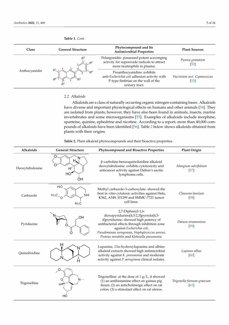
Based on one study, the underlying structure of flavonoids can be subdivided into six major subclasses, namely, flavonols, flavones, flavanones, isoflavones, flavan-3-ols and anthocyanins [39]. Table 1 shows the major classes of flavonoids, their general struc-ture and major sources from where they can be obtained.
Table 1. List of major classes and their phytocompounds with positive bioactivity from plants.
Class General Structure Phytocompound and Its Antimicrobial Properties Plant Sources
Flavanol
Catechin: able to inhibit the growth of methicillin-re-sistant S. aureus ATCC 33591 (MRSA) and methicil-lin-susceptible Staphylococcus aureus (MSSA, ATCC
25923).
Anacardium occidentale [40]
Epicatechin gallate: epicatechin gallate enhanced the antibacterial effect of β-lactam antibiotics against
MRSA in vitro and in vivo.
Fructus crataegi [41]
Epicatechin: high efficacy of phytoisolate compound against the parasitic activity of Paramphistomum
cervi.
Ricinus communis [42]
Figure 1. Structural characteristics are indicated by letters A, B and C; diphenylpropane skeleton.
Based on one study, the underlying structure of flavonoids can be subdivided intosix major subclasses, namely, flavonols, flavones, flavanones, isoflavones, flavan-3-ols andanthocyanins [39]. Table 1 shows the major classes of flavonoids, their general structureand major sources from where they can be obtained.
Table 1. List of major classes and their phytocompounds with positive bioactivity from plants.
Class General Structure Phytocompound and ItsAntimicrobial Properties Plant Sources
Flavanol
Antibiotics 2022, 10, x FOR PEER REVIEW 3 of 24
In this review, a compilation of recent studies on the antimicrobial potentials, extrac-tion methods and application of phytocompounds as alternatives to the usage of drugs in aquaculture is interpreted and discussed, with an emphasis on scholarly works from the period 2000 to 2021.
2. Phytocompounds 2.1. Flavonoids
Flavonoids consist of a large group of polyphenolic compounds having a benzo-γ-pyrone structure and are ubiquitously present in plants; they are synthesized by the phe-nylpropanoid pathway [34]. A recent report estimated that over 9000 different flavonoids have been identified to date throughout the plant kingdom [35], and at least several hun-dred which occur are edible components [36]. Due to their chemical structure, flavonoids (2-phenyl-benzo-γpyrone derivatives) are divided into flavanones, flavonols, flavones, isoflavones, flavonols and anthocyanins. According to one study, the structure of flavo-noids contains a flavan backbone formed of two benzene rings (rings A and B) connected by a heterocyclic ring of pyron or pyran (ring C) (Figure 1) [37]. The classification of fla-vonoid compounds consists of the presence of a carbonyl group at the fourth carbon atom of the C ring, a double bond between the second and third carbon atoms in this ring and a number of hydroxyl groups or other groups. All naturally occurring flavonoids have three hydroxyl groups: two in ring A (positions 5 and 7) and one in ring B (position 3) [38].
Figure 1. Structural characteristics are indicated by letters A, B and C; diphenylpropane skeleton.
Based on one study, the underlying structure of flavonoids can be subdivided into six major subclasses, namely, flavonols, flavones, flavanones, isoflavones, flavan-3-ols and anthocyanins [39]. Table 1 shows the major classes of flavonoids, their general struc-ture and major sources from where they can be obtained.
Table 1. List of major classes and their phytocompounds with positive bioactivity from plants.
Class General Structure Phytocompound and Its Antimicrobial Properties Plant Sources
Flavanol
Catechin: able to inhibit the growth of methicillin-re-sistant S. aureus ATCC 33591 (MRSA) and methicil-lin-susceptible Staphylococcus aureus (MSSA, ATCC
25923).
Anacardium occidentale [40]
Epicatechin gallate: epicatechin gallate enhanced the antibacterial effect of β-lactam antibiotics against
MRSA in vitro and in vivo.
Fructus crataegi [41]
Epicatechin: high efficacy of phytoisolate compound against the parasitic activity of Paramphistomum
cervi.
Ricinus communis [42]
Catechin: able to inhibit the growth ofmethicillin-resistant S. aureus ATCC 33591
(MRSA) and methicillin-susceptibleStaphylococcus aureus (MSSA, ATCC 25923).
Anacardium occidentale[40]
Epicatechin gallate: epicatechin gallateenhanced the antibacterial effect of β-lactam
antibiotics against MRSA in vitro andin vivo.
Fructus crataegi[41]
Epicatechin: high efficacy of phytoisolatecompound against the parasitic activity of
Paramphistomum cervi.
Ricinus communis[42]

Antibiotics 2022, 11, 469 4 of 24
Table 1. Cont.
Class General Structure Phytocompound and ItsAntimicrobial Properties Plant Sources
Flavonol
Antibiotics 2022, 10, x FOR PEER REVIEW 4 of 24
Flavonol
Kaempferol 3-O-α-L-(2″, 3″-di-Z-p-coumaroyl)rham-noside: showed high efficacy against MRSA (IC50 0.4
mg/L) and Streptococcus iniae LA94-426.
Platanus occidentalis [43]
Myricetin 3′-glucoside and myricetin 3-alpha-L-arab-inofuranoside: showed strong antiglycemic activity by inhibiting carbohydrate-hydrolyzing enzymes.
Syzygium malaccense [44]
Quercetin 3-O-glucuronide: significant inhibitory ef-fect of bacterial growth against S. aureus, E. faecalis,
E. coli, P. aeruginosa and Salmonella typhi with inhibi-tion zone diameters greater than 13 mm.
Tamarix gallica [45]
Flavone
5-Hydroxy-3′,4′-dimethoxyflavone-7-O-(rhamno-side) and 5-hydroxy-3′-methoxyflavone-4′-O-(pen-
thenyl-4-one)-7-O-(2″-(rhamnosyl) rhamnoside): able to inhibit the growth of B. subtilis (21.4 mm) and E. faecalis (8.2 mm) compared to tetracycline (22.2 mm
and 9.6 mm, respectively).
Achillea tenuifolia [46]
Apigenin: apigenin (10 µL) had antibacterial effects that were more significant on S. typhimurium and P.
mirabilis when compared with streptomycin as a control (10 µL).
Portulaca oleracea L. [47]
Isoflavone
Genistein: Increased the acetylcholinesterase (AChE) activity and, in contrast, reduced both glutathione
and catalase activity. The results may suggest bene-ficial impacts on cognitive defects related to Alz-
heimer’s disease.
Glycine max [48]
Genistein: methanolic extracts containing genistein displayed antibiotic response against all bacterial strains and maximum zone of inhibition at a low
concentration level at 350 µg/mL.
Rhizophora apiculate [49]
Flavanone
Hesperidin: able to inhibit the growth Streptococcus aureus, Escherichia coli, Enterococcus faecalis and Pseu-domonas auraginosa at a 15% concenteration with in-
hibitory diameter range of 7.65 mm ± 0.36 mm to 9.96 mm ± 0.52 mm, and at a concentration of 20%
with a diameter range of 9.26 mm ± 0.72 mm to 13.39 mm ± 028 mm.
Citrus microparpa [50]
Hesperetin-A: showed a noteworthy cytotoxicity ef-fect (IC50: 2.86 µg/mL) on HeLa cell line, and an in
silico molecular docking study portrayed hesperetin as having a good interaction with the E6 protein of HPV16 cervical carcinoma, which is beneficial for
cancer treatment.
Cordia sebestena [51]
Anthocya-nidin
Pelargonidin: possessed potent scavenging activity for superoxide radicals to attract more neutrophils in
plasma.
Punica granatum [52]
Proanthocyanidins: exhibits anti-Escherichia coli ad-hesion activity with P-type fimbriae on the wall of
the urinary tract.
Vaccinium sect. Cyanococ-cus [53]
Kaempferol 3-O-α-L-(2′′,3′′-di-Z-p-coumaroyl)rhamnoside: showedhigh efficacy against MRSA (IC50 0.4 mg/L)
and Streptococcus iniae LA94-426.
Platanus occidentalis[43]
Myricetin 3′-glucoside and myricetin3-alpha-L-arabinofuranoside: showed strong
antiglycemic activity by inhibitingcarbohydrate-hydrolyzing enzymes.
Syzygium malaccense[44]
Quercetin 3-O-glucuronide: significantinhibitory effect of bacterial growth againstS. aureus, E. faecalis, E. coli, P. aeruginosa and
Salmonella typhi with inhibition zonediameters greater than 13 mm.
Tamarix gallica[45]
Flavone
Antibiotics 2022, 10, x FOR PEER REVIEW 4 of 24
Flavonol
Kaempferol 3-O-α-L-(2″, 3″-di-Z-p-coumaroyl)rham-noside: showed high efficacy against MRSA (IC50 0.4
mg/L) and Streptococcus iniae LA94-426.
Platanus occidentalis [43]
Myricetin 3′-glucoside and myricetin 3-alpha-L-arab-inofuranoside: showed strong antiglycemic activity by inhibiting carbohydrate-hydrolyzing enzymes.
Syzygium malaccense [44]
Quercetin 3-O-glucuronide: significant inhibitory ef-fect of bacterial growth against S. aureus, E. faecalis,
E. coli, P. aeruginosa and Salmonella typhi with inhibi-tion zone diameters greater than 13 mm.
Tamarix gallica [45]
Flavone
5-Hydroxy-3′,4′-dimethoxyflavone-7-O-(rhamno-side) and 5-hydroxy-3′-methoxyflavone-4′-O-(pen-
thenyl-4-one)-7-O-(2″-(rhamnosyl) rhamnoside): able to inhibit the growth of B. subtilis (21.4 mm) and E. faecalis (8.2 mm) compared to tetracycline (22.2 mm
and 9.6 mm, respectively).
Achillea tenuifolia [46]
Apigenin: apigenin (10 µL) had antibacterial effects that were more significant on S. typhimurium and P.
mirabilis when compared with streptomycin as a control (10 µL).
Portulaca oleracea L. [47]
Isoflavone
Genistein: Increased the acetylcholinesterase (AChE) activity and, in contrast, reduced both glutathione
and catalase activity. The results may suggest bene-ficial impacts on cognitive defects related to Alz-
heimer’s disease.
Glycine max [48]
Genistein: methanolic extracts containing genistein displayed antibiotic response against all bacterial strains and maximum zone of inhibition at a low
concentration level at 350 µg/mL.
Rhizophora apiculate [49]
Flavanone
Hesperidin: able to inhibit the growth Streptococcus aureus, Escherichia coli, Enterococcus faecalis and Pseu-domonas auraginosa at a 15% concenteration with in-
hibitory diameter range of 7.65 mm ± 0.36 mm to 9.96 mm ± 0.52 mm, and at a concentration of 20%
with a diameter range of 9.26 mm ± 0.72 mm to 13.39 mm ± 028 mm.
Citrus microparpa [50]
Hesperetin-A: showed a noteworthy cytotoxicity ef-fect (IC50: 2.86 µg/mL) on HeLa cell line, and an in
silico molecular docking study portrayed hesperetin as having a good interaction with the E6 protein of HPV16 cervical carcinoma, which is beneficial for
cancer treatment.
Cordia sebestena [51]
Anthocya-nidin
Pelargonidin: possessed potent scavenging activity for superoxide radicals to attract more neutrophils in
plasma.
Punica granatum [52]
Proanthocyanidins: exhibits anti-Escherichia coli ad-hesion activity with P-type fimbriae on the wall of
the urinary tract.
Vaccinium sect. Cyanococ-cus [53]
5-Hydroxy-3′,4′-dimethoxyflavone-7-O-(rhamnoside) and
5-hydroxy-3′-methoxyflavone-4′-O-(penthenyl-4-one)-7-O-(2′′-(rhamnosyl)
rhamnoside): able to inhibit the growth ofB. subtilis (21.4 mm) and E. faecalis (8.2 mm)
compared to tetracycline (22.2 mmand 9.6 mm, respectively).
Achillea tenuifolia[46]
Apigenin: apigenin (10 µL) had antibacterialeffects that were more significant onS. typhimurium and P. mirabilis when
compared with streptomycin as acontrol (10 µL).
Portulaca oleracea L.[47]
Isoflavone
Antibiotics 2022, 10, x FOR PEER REVIEW 4 of 24
Flavonol
Kaempferol 3-O-α-L-(2″, 3″-di-Z-p-coumaroyl)rham-noside: showed high efficacy against MRSA (IC50 0.4
mg/L) and Streptococcus iniae LA94-426.
Platanus occidentalis [43]
Myricetin 3′-glucoside and myricetin 3-alpha-L-arab-inofuranoside: showed strong antiglycemic activity by inhibiting carbohydrate-hydrolyzing enzymes.
Syzygium malaccense [44]
Quercetin 3-O-glucuronide: significant inhibitory ef-fect of bacterial growth against S. aureus, E. faecalis,
E. coli, P. aeruginosa and Salmonella typhi with inhibi-tion zone diameters greater than 13 mm.
Tamarix gallica [45]
Flavone
5-Hydroxy-3′,4′-dimethoxyflavone-7-O-(rhamno-side) and 5-hydroxy-3′-methoxyflavone-4′-O-(pen-
thenyl-4-one)-7-O-(2″-(rhamnosyl) rhamnoside): able to inhibit the growth of B. subtilis (21.4 mm) and E. faecalis (8.2 mm) compared to tetracycline (22.2 mm
and 9.6 mm, respectively).
Achillea tenuifolia [46]
Apigenin: apigenin (10 µL) had antibacterial effects that were more significant on S. typhimurium and P.
mirabilis when compared with streptomycin as a control (10 µL).
Portulaca oleracea L. [47]
Isoflavone
Genistein: Increased the acetylcholinesterase (AChE) activity and, in contrast, reduced both glutathione
and catalase activity. The results may suggest bene-ficial impacts on cognitive defects related to Alz-
heimer’s disease.
Glycine max [48]
Genistein: methanolic extracts containing genistein displayed antibiotic response against all bacterial strains and maximum zone of inhibition at a low
concentration level at 350 µg/mL.
Rhizophora apiculate [49]
Flavanone
Hesperidin: able to inhibit the growth Streptococcus aureus, Escherichia coli, Enterococcus faecalis and Pseu-domonas auraginosa at a 15% concenteration with in-
hibitory diameter range of 7.65 mm ± 0.36 mm to 9.96 mm ± 0.52 mm, and at a concentration of 20%
with a diameter range of 9.26 mm ± 0.72 mm to 13.39 mm ± 028 mm.
Citrus microparpa [50]
Hesperetin-A: showed a noteworthy cytotoxicity ef-fect (IC50: 2.86 µg/mL) on HeLa cell line, and an in
silico molecular docking study portrayed hesperetin as having a good interaction with the E6 protein of HPV16 cervical carcinoma, which is beneficial for
cancer treatment.
Cordia sebestena [51]
Anthocya-nidin
Pelargonidin: possessed potent scavenging activity for superoxide radicals to attract more neutrophils in
plasma.
Punica granatum [52]
Proanthocyanidins: exhibits anti-Escherichia coli ad-hesion activity with P-type fimbriae on the wall of
the urinary tract.
Vaccinium sect. Cyanococ-cus [53]
Genistein: Increased the acetylcholinesterase(AChE) activity and, in contrast, reduced
both glutathione and catalase activity. Theresults may suggest beneficial impacts on
cognitive defects related toAlzheimer’s disease.
Glycine max[48]
Genistein: methanolic extracts containinggenistein displayed antibiotic response
against all bacterial strains and maximumzone of inhibition at a low concentration
level at 350 µg/mL.
Rhizophora apiculate[49]
Flavanone
Antibiotics 2022, 10, x FOR PEER REVIEW 4 of 24
Flavonol
Kaempferol 3-O-α-L-(2″, 3″-di-Z-p-coumaroyl)rham-noside: showed high efficacy against MRSA (IC50 0.4
mg/L) and Streptococcus iniae LA94-426.
Platanus occidentalis [43]
Myricetin 3′-glucoside and myricetin 3-alpha-L-arab-inofuranoside: showed strong antiglycemic activity by inhibiting carbohydrate-hydrolyzing enzymes.
Syzygium malaccense [44]
Quercetin 3-O-glucuronide: significant inhibitory ef-fect of bacterial growth against S. aureus, E. faecalis,
E. coli, P. aeruginosa and Salmonella typhi with inhibi-tion zone diameters greater than 13 mm.
Tamarix gallica [45]
Flavone
5-Hydroxy-3′,4′-dimethoxyflavone-7-O-(rhamno-side) and 5-hydroxy-3′-methoxyflavone-4′-O-(pen-
thenyl-4-one)-7-O-(2″-(rhamnosyl) rhamnoside): able to inhibit the growth of B. subtilis (21.4 mm) and E. faecalis (8.2 mm) compared to tetracycline (22.2 mm
and 9.6 mm, respectively).
Achillea tenuifolia [46]
Apigenin: apigenin (10 µL) had antibacterial effects that were more significant on S. typhimurium and P.
mirabilis when compared with streptomycin as a control (10 µL).
Portulaca oleracea L. [47]
Isoflavone
Genistein: Increased the acetylcholinesterase (AChE) activity and, in contrast, reduced both glutathione
and catalase activity. The results may suggest bene-ficial impacts on cognitive defects related to Alz-
heimer’s disease.
Glycine max [48]
Genistein: methanolic extracts containing genistein displayed antibiotic response against all bacterial strains and maximum zone of inhibition at a low
concentration level at 350 µg/mL.
Rhizophora apiculate [49]
Flavanone
Hesperidin: able to inhibit the growth Streptococcus aureus, Escherichia coli, Enterococcus faecalis and Pseu-domonas auraginosa at a 15% concenteration with in-
hibitory diameter range of 7.65 mm ± 0.36 mm to 9.96 mm ± 0.52 mm, and at a concentration of 20%
with a diameter range of 9.26 mm ± 0.72 mm to 13.39 mm ± 028 mm.
Citrus microparpa [50]
Hesperetin-A: showed a noteworthy cytotoxicity ef-fect (IC50: 2.86 µg/mL) on HeLa cell line, and an in
silico molecular docking study portrayed hesperetin as having a good interaction with the E6 protein of HPV16 cervical carcinoma, which is beneficial for
cancer treatment.
Cordia sebestena [51]
Anthocya-nidin
Pelargonidin: possessed potent scavenging activity for superoxide radicals to attract more neutrophils in
plasma.
Punica granatum [52]
Proanthocyanidins: exhibits anti-Escherichia coli ad-hesion activity with P-type fimbriae on the wall of
the urinary tract.
Vaccinium sect. Cyanococ-cus [53]
Hesperidin: able to inhibit the growthStreptococcus aureus, Escherichia coli,
Enterococcus faecalis and Pseudomonasauraginosa at a 15% concenteration with
inhibitory diameter range of7.65 mm ± 0.36 mm to 9.96 mm ± 0.52 mm,
and at a concentration of 20% with adiameter range of 9.26 mm ± 0.72 mm to
13.39 mm ± 028 mm.
Citrus microparpa[50]
Hesperetin-A: showed a noteworthycytotoxicity effect (IC50: 2.86 µg/mL) onHeLa cell line, and an in silico moleculardocking study portrayed hesperetin as
having a good interaction with the E6 proteinof HPV16 cervical carcinoma, which is
beneficial for cancer treatment.
Cordia sebestena[51]
Antibiotics 2022, 11, 469 5 of 24
Table 1. Cont.
Class General Structure Phytocompound and ItsAntimicrobial Properties Plant Sources
Anthocyanidin
Antibiotics 2022, 10, x FOR PEER REVIEW 4 of 24
Flavonol
Kaempferol 3-O-α-L-(2″, 3″-di-Z-p-coumaroyl)rham-noside: showed high efficacy against MRSA (IC50 0.4
mg/L) and Streptococcus iniae LA94-426.
Platanus occidentalis [43]
Myricetin 3′-glucoside and myricetin 3-alpha-L-arab-inofuranoside: showed strong antiglycemic activity by inhibiting carbohydrate-hydrolyzing enzymes.
Syzygium malaccense [44]
Quercetin 3-O-glucuronide: significant inhibitory ef-fect of bacterial growth against S. aureus, E. faecalis,
E. coli, P. aeruginosa and Salmonella typhi with inhibi-tion zone diameters greater than 13 mm.
Tamarix gallica [45]
Flavone
5-Hydroxy-3′,4′-dimethoxyflavone-7-O-(rhamno-side) and 5-hydroxy-3′-methoxyflavone-4′-O-(pen-
thenyl-4-one)-7-O-(2″-(rhamnosyl) rhamnoside): able to inhibit the growth of B. subtilis (21.4 mm) and E. faecalis (8.2 mm) compared to tetracycline (22.2 mm
and 9.6 mm, respectively).
Achillea tenuifolia [46]
Apigenin: apigenin (10 µL) had antibacterial effects that were more significant on S. typhimurium and P.
mirabilis when compared with streptomycin as a control (10 µL).
Portulaca oleracea L. [47]
Isoflavone
Genistein: Increased the acetylcholinesterase (AChE) activity and, in contrast, reduced both glutathione
and catalase activity. The results may suggest bene-ficial impacts on cognitive defects related to Alz-
heimer’s disease.
Glycine max [48]
Genistein: methanolic extracts containing genistein displayed antibiotic response against all bacterial strains and maximum zone of inhibition at a low
concentration level at 350 µg/mL.
Rhizophora apiculate [49]
Flavanone
Hesperidin: able to inhibit the growth Streptococcus aureus, Escherichia coli, Enterococcus faecalis and Pseu-domonas auraginosa at a 15% concenteration with in-
hibitory diameter range of 7.65 mm ± 0.36 mm to 9.96 mm ± 0.52 mm, and at a concentration of 20%
with a diameter range of 9.26 mm ± 0.72 mm to 13.39 mm ± 028 mm.
Citrus microparpa [50]
Hesperetin-A: showed a noteworthy cytotoxicity ef-fect (IC50: 2.86 µg/mL) on HeLa cell line, and an in
silico molecular docking study portrayed hesperetin as having a good interaction with the E6 protein of HPV16 cervical carcinoma, which is beneficial for
cancer treatment.
Cordia sebestena [51]
Anthocya-nidin
Pelargonidin: possessed potent scavenging activity for superoxide radicals to attract more neutrophils in
plasma.
Punica granatum [52]
Proanthocyanidins: exhibits anti-Escherichia coli ad-hesion activity with P-type fimbriae on the wall of
the urinary tract.
Vaccinium sect. Cyanococ-cus [53]
Pelargonidin: possessed potent scavengingactivity for superoxide radicals to attract
more neutrophils in plasma.
Punica granatum[52]
Proanthocyanidins: exhibitsanti-Escherichia coli adhesion activity with
P-type fimbriae on the wall of theurinary tract.
Vaccinium sect. Cyanococcus[53]
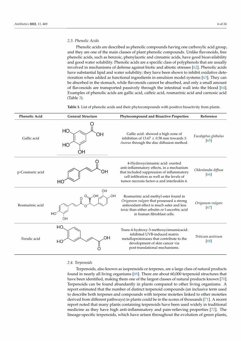
2.2. Alkaloids
Alkaloids are a class of naturally occurring organic nitrogen-containing bases. Alkaloidshave diverse and important physiological effects on humans and other animals [54]. Theyare isolated from plants; however, they have also been found in animals, insects, marineinvertebrates and some microorganisms [55]. Examples of alkaloids include morphine,sparteine, quinine, ephedrine and nicotine. According to a report, more than 40,000 com-pounds of alkaloids have been identified [56]. Table 2 below shows alkaloids obtained fromplants with their origins.
Table 2. Plant alkaloid phytocompounds and their bioactive properties.
Alkaloids General Structure Phytocompound and Bioactive Properties Plant Origin
Deoxytubulosine
Antibiotics 2022, 10, x FOR PEER REVIEW 5 of 24
2.2. Alkaloids Alkaloids are a class of naturally occurring organic nitrogen-containing bases. Alka-
loids have diverse and important physiological effects on humans and other animals [54]. They are isolated from plants; however, they have also been found in animals, insects, marine invertebrates and some microorganisms [55]. Examples of alkaloids include mor-phine, sparteine, quinine, ephedrine and nicotine. According to a report, more than 40,000 compounds of alkaloids have been identified [56]. Table 2 below shows alkaloids obtained from plants with their origins.
Table 2. Plant alkaloid phytocompounds and their bioactive properties.
Alkaloids General Structure Phytocompound and Bioactive Properties Plant Origin
Deoxytubulosine
β-carboline-benzoquinolizidine alkaloid deox-ytubulosine: exhibits cytotoxicity and anti-cancer activity against Dalton’s ascitic lym-
phoma cells.
Alangium salvifolium [57]
Carbazole
Methyl carbazole-3-carboxylate: showed the best in vitro cytotoxic activities against Hela, K562, A549, H1299 and SMMC-7721 tumor
cell lines.
Clausena lansium [58]
Pyridazine
2,7-Diphenyl-1,6-dioxopyridazino[4,5:2,3]pyr-rolo[4,5-d]pyridazine: showed high potency
of antibacterial effects through inhibition zone against Escherichia coli, Pseudomonas euroge-nosa, Staphylococcus aureus, Proteus mirabilis
and Klebsiella pneumonia.
Datura stramonium [59]
Quinolizidine
Lupanine, 13α-hydroxylupanine and albine: alkaloid extracts showed high antimicrobial activity against K. pneumonia and moderate
activity against P. aeruginosa clinical isolates.
Lupinus albus [60]
Trigonelline
Trigonelline: at the dose of 1 g/L, it showed (1) an antihistamine effect on guinea pig il-
eum; (2) an anticholinergic effect on rat colon; (3) a stimulant effect on rat uterus.
Trigonella foenum-graecum [61]
2.3. Phenolic Acids Phenolic acids are described as phenolic compounds having one carboxylic acid
group, and they are one of the main classes of plant phenolic compounds. Unlike flavo-noids, free phenolic acids, such as benzoic, phenylacetic and cinnamic acids, have good bioavailability and good water solubility. Phenolic acids are a specific class of polyphenols that are usually involved in mechanisms of defense against biotic and abiotic stresses [62]. Phenolic acids have substantial lipid and water solubility; they have been shown to inhibit oxidative deterioration when added as functional ingredients in emulsion model systems [63]. They can be absorbed in the stomach, while flavonoids cannot be absorbed, and only a small amount of flavonoids are transported passively through the intestinal wall into the blood [64]. Examples of phenolic acids are gallic acid, caffeic acid, rosmarinic acid and carnosic acid (Table 3).
β-carboline-benzoquinolizidine alkaloiddeoxytubulosine: exhibits cytotoxicity andanticancer activity against Dalton’s ascitic
lymphoma cells.
Alangium salvifolium[57]
Carbazole
Antibiotics 2022, 10, x FOR PEER REVIEW 5 of 24
2.2. Alkaloids Alkaloids are a class of naturally occurring organic nitrogen-containing bases. Alka-
loids have diverse and important physiological effects on humans and other animals [54]. They are isolated from plants; however, they have also been found in animals, insects, marine invertebrates and some microorganisms [55]. Examples of alkaloids include mor-phine, sparteine, quinine, ephedrine and nicotine. According to a report, more than 40,000 compounds of alkaloids have been identified [56]. Table 2 below shows alkaloids obtained from plants with their origins.
Table 2. Plant alkaloid phytocompounds and their bioactive properties.
Alkaloids General Structure Phytocompound and Bioactive Properties Plant Origin
Deoxytubulosine
β-carboline-benzoquinolizidine alkaloid deox-ytubulosine: exhibits cytotoxicity and anti-cancer activity against Dalton’s ascitic lym-
phoma cells.
Alangium salvifolium [57]
Carbazole
Methyl carbazole-3-carboxylate: showed the best in vitro cytotoxic activities against Hela, K562, A549, H1299 and SMMC-7721 tumor
cell lines.
Clausena lansium [58]
Pyridazine
2,7-Diphenyl-1,6-dioxopyridazino[4,5:2,3]pyr-rolo[4,5-d]pyridazine: showed high potency
of antibacterial effects through inhibition zone against Escherichia coli, Pseudomonas euroge-nosa, Staphylococcus aureus, Proteus mirabilis
and Klebsiella pneumonia.
Datura stramonium [59]
Quinolizidine
Lupanine, 13α-hydroxylupanine and albine: alkaloid extracts showed high antimicrobial activity against K. pneumonia and moderate
activity against P. aeruginosa clinical isolates.
Lupinus albus [60]
Trigonelline
Trigonelline: at the dose of 1 g/L, it showed (1) an antihistamine effect on guinea pig il-
eum; (2) an anticholinergic effect on rat colon; (3) a stimulant effect on rat uterus.
Trigonella foenum-graecum [61]
2.3. Phenolic Acids Phenolic acids are described as phenolic compounds having one carboxylic acid
group, and they are one of the main classes of plant phenolic compounds. Unlike flavo-noids, free phenolic acids, such as benzoic, phenylacetic and cinnamic acids, have good bioavailability and good water solubility. Phenolic acids are a specific class of polyphenols that are usually involved in mechanisms of defense against biotic and abiotic stresses [62]. Phenolic acids have substantial lipid and water solubility; they have been shown to inhibit oxidative deterioration when added as functional ingredients in emulsion model systems [63]. They can be absorbed in the stomach, while flavonoids cannot be absorbed, and only a small amount of flavonoids are transported passively through the intestinal wall into the blood [64]. Examples of phenolic acids are gallic acid, caffeic acid, rosmarinic acid and carnosic acid (Table 3).
Methyl carbazole-3-carboxylate: showed thebest in vitro cytotoxic activities against Hela,K562, A549, H1299 and SMMC-7721 tumor
cell lines.
Clausena lansium[58]
Pyridazine
Antibiotics 2022, 10, x FOR PEER REVIEW 5 of 24
2.2. Alkaloids Alkaloids are a class of naturally occurring organic nitrogen-containing bases. Alka-
loids have diverse and important physiological effects on humans and other animals [54]. They are isolated from plants; however, they have also been found in animals, insects, marine invertebrates and some microorganisms [55]. Examples of alkaloids include mor-phine, sparteine, quinine, ephedrine and nicotine. According to a report, more than 40,000 compounds of alkaloids have been identified [56]. Table 2 below shows alkaloids obtained from plants with their origins.
Table 2. Plant alkaloid phytocompounds and their bioactive properties.
Alkaloids General Structure Phytocompound and Bioactive Properties Plant Origin
Deoxytubulosine
β-carboline-benzoquinolizidine alkaloid deox-ytubulosine: exhibits cytotoxicity and anti-cancer activity against Dalton’s ascitic lym-
phoma cells.
Alangium salvifolium [57]
Carbazole
Methyl carbazole-3-carboxylate: showed the best in vitro cytotoxic activities against Hela, K562, A549, H1299 and SMMC-7721 tumor
cell lines.
Clausena lansium [58]
Pyridazine
2,7-Diphenyl-1,6-dioxopyridazino[4,5:2,3]pyr-rolo[4,5-d]pyridazine: showed high potency
of antibacterial effects through inhibition zone against Escherichia coli, Pseudomonas euroge-nosa, Staphylococcus aureus, Proteus mirabilis
and Klebsiella pneumonia.
Datura stramonium [59]
Quinolizidine
Lupanine, 13α-hydroxylupanine and albine: alkaloid extracts showed high antimicrobial activity against K. pneumonia and moderate
activity against P. aeruginosa clinical isolates.
Lupinus albus [60]
Trigonelline
Trigonelline: at the dose of 1 g/L, it showed (1) an antihistamine effect on guinea pig il-
eum; (2) an anticholinergic effect on rat colon; (3) a stimulant effect on rat uterus.
Trigonella foenum-graecum [61]
2.3. Phenolic Acids Phenolic acids are described as phenolic compounds having one carboxylic acid
group, and they are one of the main classes of plant phenolic compounds. Unlike flavo-noids, free phenolic acids, such as benzoic, phenylacetic and cinnamic acids, have good bioavailability and good water solubility. Phenolic acids are a specific class of polyphenols that are usually involved in mechanisms of defense against biotic and abiotic stresses [62]. Phenolic acids have substantial lipid and water solubility; they have been shown to inhibit oxidative deterioration when added as functional ingredients in emulsion model systems [63]. They can be absorbed in the stomach, while flavonoids cannot be absorbed, and only a small amount of flavonoids are transported passively through the intestinal wall into the blood [64]. Examples of phenolic acids are gallic acid, caffeic acid, rosmarinic acid and carnosic acid (Table 3).
2,7-Diphenyl-1,6-dioxopyridazino[4,5:2,3]pyrrolo[4,5-
d]pyridazine: showed high potency ofantibacterial effects through inhibition zone
against Escherichia coli,Pseudomonas eurogenosa, Staphylococcus aureus,
Proteus mirabilis and Klebsiella pneumonia.
Datura stramonium[59]
Quinolizidine
Antibiotics 2022, 10, x FOR PEER REVIEW 5 of 24
2.2. Alkaloids Alkaloids are a class of naturally occurring organic nitrogen-containing bases. Alka-
loids have diverse and important physiological effects on humans and other animals [54]. They are isolated from plants; however, they have also been found in animals, insects, marine invertebrates and some microorganisms [55]. Examples of alkaloids include mor-phine, sparteine, quinine, ephedrine and nicotine. According to a report, more than 40,000 compounds of alkaloids have been identified [56]. Table 2 below shows alkaloids obtained from plants with their origins.
Table 2. Plant alkaloid phytocompounds and their bioactive properties.
Alkaloids General Structure Phytocompound and Bioactive Properties Plant Origin
Deoxytubulosine
β-carboline-benzoquinolizidine alkaloid deox-ytubulosine: exhibits cytotoxicity and anti-cancer activity against Dalton’s ascitic lym-
phoma cells.
Alangium salvifolium [57]
Carbazole
Methyl carbazole-3-carboxylate: showed the best in vitro cytotoxic activities against Hela, K562, A549, H1299 and SMMC-7721 tumor
cell lines.
Clausena lansium [58]
Pyridazine
2,7-Diphenyl-1,6-dioxopyridazino[4,5:2,3]pyr-rolo[4,5-d]pyridazine: showed high potency
of antibacterial effects through inhibition zone against Escherichia coli, Pseudomonas euroge-nosa, Staphylococcus aureus, Proteus mirabilis
and Klebsiella pneumonia.
Datura stramonium [59]
Quinolizidine
Lupanine, 13α-hydroxylupanine and albine: alkaloid extracts showed high antimicrobial activity against K. pneumonia and moderate
activity against P. aeruginosa clinical isolates.
Lupinus albus [60]
Trigonelline
Trigonelline: at the dose of 1 g/L, it showed (1) an antihistamine effect on guinea pig il-
eum; (2) an anticholinergic effect on rat colon; (3) a stimulant effect on rat uterus.
Trigonella foenum-graecum [61]
2.3. Phenolic Acids Phenolic acids are described as phenolic compounds having one carboxylic acid
group, and they are one of the main classes of plant phenolic compounds. Unlike flavo-noids, free phenolic acids, such as benzoic, phenylacetic and cinnamic acids, have good bioavailability and good water solubility. Phenolic acids are a specific class of polyphenols that are usually involved in mechanisms of defense against biotic and abiotic stresses [62]. Phenolic acids have substantial lipid and water solubility; they have been shown to inhibit oxidative deterioration when added as functional ingredients in emulsion model systems [63]. They can be absorbed in the stomach, while flavonoids cannot be absorbed, and only a small amount of flavonoids are transported passively through the intestinal wall into the blood [64]. Examples of phenolic acids are gallic acid, caffeic acid, rosmarinic acid and carnosic acid (Table 3).
Lupanine, 13α-hydroxylupanine and albine:alkaloid extracts showed high antimicrobialactivity against K. pneumonia and moderateactivity against P. aeruginosa clinical isolates.
Lupinus albus[60]
Trigonelline
Antibiotics 2022, 10, x FOR PEER REVIEW 5 of 24
2.2. Alkaloids Alkaloids are a class of naturally occurring organic nitrogen-containing bases. Alka-
loids have diverse and important physiological effects on humans and other animals [54]. They are isolated from plants; however, they have also been found in animals, insects, marine invertebrates and some microorganisms [55]. Examples of alkaloids include mor-phine, sparteine, quinine, ephedrine and nicotine. According to a report, more than 40,000 compounds of alkaloids have been identified [56]. Table 2 below shows alkaloids obtained from plants with their origins.
Table 2. Plant alkaloid phytocompounds and their bioactive properties.
Alkaloids General Structure Phytocompound and Bioactive Properties Plant Origin
Deoxytubulosine
β-carboline-benzoquinolizidine alkaloid deox-ytubulosine: exhibits cytotoxicity and anti-cancer activity against Dalton’s ascitic lym-
phoma cells.
Alangium salvifolium [57]
Carbazole
Methyl carbazole-3-carboxylate: showed the best in vitro cytotoxic activities against Hela, K562, A549, H1299 and SMMC-7721 tumor
cell lines.
Clausena lansium [58]
Pyridazine
2,7-Diphenyl-1,6-dioxopyridazino[4,5:2,3]pyr-rolo[4,5-d]pyridazine: showed high potency
of antibacterial effects through inhibition zone against Escherichia coli, Pseudomonas euroge-nosa, Staphylococcus aureus, Proteus mirabilis
and Klebsiella pneumonia.
Datura stramonium [59]
Quinolizidine
Lupanine, 13α-hydroxylupanine and albine: alkaloid extracts showed high antimicrobial activity against K. pneumonia and moderate
activity against P. aeruginosa clinical isolates.
Lupinus albus [60]
Trigonelline
Trigonelline: at the dose of 1 g/L, it showed (1) an antihistamine effect on guinea pig il-
eum; (2) an anticholinergic effect on rat colon; (3) a stimulant effect on rat uterus.
Trigonella foenum-graecum [61]
2.3. Phenolic Acids Phenolic acids are described as phenolic compounds having one carboxylic acid
group, and they are one of the main classes of plant phenolic compounds. Unlike flavo-noids, free phenolic acids, such as benzoic, phenylacetic and cinnamic acids, have good bioavailability and good water solubility. Phenolic acids are a specific class of polyphenols that are usually involved in mechanisms of defense against biotic and abiotic stresses [62]. Phenolic acids have substantial lipid and water solubility; they have been shown to inhibit oxidative deterioration when added as functional ingredients in emulsion model systems [63]. They can be absorbed in the stomach, while flavonoids cannot be absorbed, and only a small amount of flavonoids are transported passively through the intestinal wall into the blood [64]. Examples of phenolic acids are gallic acid, caffeic acid, rosmarinic acid and carnosic acid (Table 3).
Trigonelline: at the dose of 1 g/L, it showed(1) an antihistamine effect on guinea pigileum; (2) an anticholinergic effect on ratcolon; (3) a stimulant effect on rat uterus.
Trigonella foenum-graecum[61]
Antibiotics 2022, 11, 469 6 of 24
2.3. Phenolic Acids
Phenolic acids are described as phenolic compounds having one carboxylic acid group,and they are one of the main classes of plant phenolic compounds. Unlike flavonoids, freephenolic acids, such as benzoic, phenylacetic and cinnamic acids, have good bioavailabilityand good water solubility. Phenolic acids are a specific class of polyphenols that are usuallyinvolved in mechanisms of defense against biotic and abiotic stresses [62]. Phenolic acidshave substantial lipid and water solubility; they have been shown to inhibit oxidative dete-rioration when added as functional ingredients in emulsion model systems [63]. They canbe absorbed in the stomach, while flavonoids cannot be absorbed, and only a small amountof flavonoids are transported passively through the intestinal wall into the blood [64].Examples of phenolic acids are gallic acid, caffeic acid, rosmarinic acid and carnosic acid(Table 3).
Table 3. List of phenolic acids and their phytocompounds with positive bioactivity from plants.
Phenolic Acid General Structure Phytocompound and Bioactive Properties Reference
Gallic acid
Antibiotics 2022, 10, x FOR PEER REVIEW 6 of 24
Table 3. List of phenolic acids and their phytocompounds with positive bioactivity from plants.
Phenolic Acid General Structure Phytocompound and Bioactive Properties Reference
Gallic acid
Gallic acid: showed a high zone of inhibi-tion of 13.67 ± 0.58 mm towards S. Aureus
through the disc diffusion method.
Eucalyptus globulus [65]
p-Coumaric acid
4-Hydroxycinnamic acid: exerted anti-in-flammatory effects, in a mechanism that in-cluded suppression of inflammatory cell in-filtration as well as the levels of tumor ne-
crosis factor-α and interleukin 6.
Oldenlandia diffusa [66]
Rosmarinic acid
Rosmarinic acid methyl ester found in Ori-ganum vulgare that possessed a strong anti-oxidant effect is much safer and less toxic
than either arbutin or l-ascorbic acid in hu-man fibroblast cells.
Origanum vulgare [67]
Ferulic acid
Trans-4-hydroxy-3-methoxycinnamic acid: inhibited UVB-induced matrix metal-loproteinases that contribute to the devel-
opment of skin cancer via post-translational mechanisms.
Triticum aestivum [68]
2.4. Terpenoids Terpenoids, also known as isoprenoids or terpenes, are a large class of natural prod-
ucts found in nearly all living organisms [69]. There are about 60,000 terpenoid structures that have been identified, making them one of the largest classes of natural products known [70]. Terpenoids can be found abundantly in plants compared to other living or-ganisms. A report estimated that the number of distinct terpenoid compounds (an inclu-sive term used to describe both terpenes and compounds with terpene moieties linked to other moieties derived from different pathways) in plants could be in the scores of thou-sands [71]. A recent report noted that many plants containing terpenoids have been used widely in traditional medicine as they have high anti-inflammatory and pain-relieving properties [72]. The lineage-specific terpenoids, which have arisen throughout the evolu-tion of green plants, have generally been postulated to play a role in the ecological inter-actions of plants with biotic and abiotic aspects of their environment [73]. Such roles have included defense against herbivores and pathogens, including signals and rewards for beneficial organisms, such as pollinators and mycorrhiza [74]. Table 4 below shows the classification and structure of terpenoids with their origins.
Gallic acid: showed a high zone ofinhibition of 13.67 ± 0.58 mm towards S.
Aureus through the disc diffusion method.
Eucalyptus globulus[65]
p-Coumaric acid
Antibiotics 2022, 10, x FOR PEER REVIEW 6 of 24
Table 3. List of phenolic acids and their phytocompounds with positive bioactivity from plants.
Phenolic Acid General Structure Phytocompound and Bioactive Properties Reference
Gallic acid
Gallic acid: showed a high zone of inhibi-tion of 13.67 ± 0.58 mm towards S. Aureus
through the disc diffusion method.
Eucalyptus globulus [65]
p-Coumaric acid
4-Hydroxycinnamic acid: exerted anti-in-flammatory effects, in a mechanism that in-cluded suppression of inflammatory cell in-filtration as well as the levels of tumor ne-
crosis factor-α and interleukin 6.
Oldenlandia diffusa [66]
Rosmarinic acid
Rosmarinic acid methyl ester found in Ori-ganum vulgare that possessed a strong anti-oxidant effect is much safer and less toxic
than either arbutin or l-ascorbic acid in hu-man fibroblast cells.
Origanum vulgare [67]
Ferulic acid
Trans-4-hydroxy-3-methoxycinnamic acid: inhibited UVB-induced matrix metal-loproteinases that contribute to the devel-
opment of skin cancer via post-translational mechanisms.
Triticum aestivum [68]
2.4. Terpenoids Terpenoids, also known as isoprenoids or terpenes, are a large class of natural prod-
ucts found in nearly all living organisms [69]. There are about 60,000 terpenoid structures that have been identified, making them one of the largest classes of natural products known [70]. Terpenoids can be found abundantly in plants compared to other living or-ganisms. A report estimated that the number of distinct terpenoid compounds (an inclu-sive term used to describe both terpenes and compounds with terpene moieties linked to other moieties derived from different pathways) in plants could be in the scores of thou-sands [71]. A recent report noted that many plants containing terpenoids have been used widely in traditional medicine as they have high anti-inflammatory and pain-relieving properties [72]. The lineage-specific terpenoids, which have arisen throughout the evolu-tion of green plants, have generally been postulated to play a role in the ecological inter-actions of plants with biotic and abiotic aspects of their environment [73]. Such roles have included defense against herbivores and pathogens, including signals and rewards for beneficial organisms, such as pollinators and mycorrhiza [74]. Table 4 below shows the classification and structure of terpenoids with their origins.
4-Hydroxycinnamic acid: exertedanti-inflammatory effects, in a mechanismthat included suppression of inflammatory
cell infiltration as well as the levels oftumor necrosis factor-α and interleukin 6.
Oldenlandia diffusa[66]
Rosmarinic acid
Antibiotics 2022, 10, x FOR PEER REVIEW 6 of 24
Table 3. List of phenolic acids and their phytocompounds with positive bioactivity from plants.
Phenolic Acid General Structure Phytocompound and Bioactive Properties Reference
Gallic acid
Gallic acid: showed a high zone of inhibi-tion of 13.67 ± 0.58 mm towards S. Aureus
through the disc diffusion method.
Eucalyptus globulus [65]
p-Coumaric acid
4-Hydroxycinnamic acid: exerted anti-in-flammatory effects, in a mechanism that in-cluded suppression of inflammatory cell in-filtration as well as the levels of tumor ne-
crosis factor-α and interleukin 6.
Oldenlandia diffusa [66]
Rosmarinic acid
Rosmarinic acid methyl ester found in Ori-ganum vulgare that possessed a strong anti-oxidant effect is much safer and less toxic
than either arbutin or l-ascorbic acid in hu-man fibroblast cells.
Origanum vulgare [67]
Ferulic acid
Trans-4-hydroxy-3-methoxycinnamic acid: inhibited UVB-induced matrix metal-loproteinases that contribute to the devel-
opment of skin cancer via post-translational mechanisms.
Triticum aestivum [68]
2.4. Terpenoids Terpenoids, also known as isoprenoids or terpenes, are a large class of natural prod-
ucts found in nearly all living organisms [69]. There are about 60,000 terpenoid structures that have been identified, making them one of the largest classes of natural products known [70]. Terpenoids can be found abundantly in plants compared to other living or-ganisms. A report estimated that the number of distinct terpenoid compounds (an inclu-sive term used to describe both terpenes and compounds with terpene moieties linked to other moieties derived from different pathways) in plants could be in the scores of thou-sands [71]. A recent report noted that many plants containing terpenoids have been used widely in traditional medicine as they have high anti-inflammatory and pain-relieving properties [72]. The lineage-specific terpenoids, which have arisen throughout the evolu-tion of green plants, have generally been postulated to play a role in the ecological inter-actions of plants with biotic and abiotic aspects of their environment [73]. Such roles have included defense against herbivores and pathogens, including signals and rewards for beneficial organisms, such as pollinators and mycorrhiza [74]. Table 4 below shows the classification and structure of terpenoids with their origins.
Rosmarinic acid methyl ester found inOriganum vulgare that possessed a strongantioxidant effect is much safer and less
toxic than either arbutin or l-ascorbic acidin human fibroblast cells.
Origanum vulgare[67]
Ferulic acid
Antibiotics 2022, 10, x FOR PEER REVIEW 6 of 24
Table 3. List of phenolic acids and their phytocompounds with positive bioactivity from plants.
Phenolic Acid General Structure Phytocompound and Bioactive Properties Reference
Gallic acid
Gallic acid: showed a high zone of inhibi-tion of 13.67 ± 0.58 mm towards S. Aureus
through the disc diffusion method.
Eucalyptus globulus [65]
p-Coumaric acid
4-Hydroxycinnamic acid: exerted anti-in-flammatory effects, in a mechanism that in-cluded suppression of inflammatory cell in-filtration as well as the levels of tumor ne-
crosis factor-α and interleukin 6.
Oldenlandia diffusa [66]
Rosmarinic acid
Rosmarinic acid methyl ester found in Ori-ganum vulgare that possessed a strong anti-oxidant effect is much safer and less toxic
than either arbutin or l-ascorbic acid in hu-man fibroblast cells.
Origanum vulgare [67]
Ferulic acid
Trans-4-hydroxy-3-methoxycinnamic acid: inhibited UVB-induced matrix metal-loproteinases that contribute to the devel-
opment of skin cancer via post-translational mechanisms.
Triticum aestivum [68]
2.4. Terpenoids Terpenoids, also known as isoprenoids or terpenes, are a large class of natural prod-
ucts found in nearly all living organisms [69]. There are about 60,000 terpenoid structures that have been identified, making them one of the largest classes of natural products known [70]. Terpenoids can be found abundantly in plants compared to other living or-ganisms. A report estimated that the number of distinct terpenoid compounds (an inclu-sive term used to describe both terpenes and compounds with terpene moieties linked to other moieties derived from different pathways) in plants could be in the scores of thou-sands [71]. A recent report noted that many plants containing terpenoids have been used widely in traditional medicine as they have high anti-inflammatory and pain-relieving properties [72]. The lineage-specific terpenoids, which have arisen throughout the evolu-tion of green plants, have generally been postulated to play a role in the ecological inter-actions of plants with biotic and abiotic aspects of their environment [73]. Such roles have included defense against herbivores and pathogens, including signals and rewards for beneficial organisms, such as pollinators and mycorrhiza [74]. Table 4 below shows the classification and structure of terpenoids with their origins.
Trans-4-hydroxy-3-methoxycinnamicacid:inhibited UVB-induced matrix
metalloproteinases that contribute to thedevelopment of skin cancer viapost-translational mechanisms.
Triticum aestivum[68]
2.4. Terpenoids
Terpenoids, also known as isoprenoids or terpenes, are a large class of natural productsfound in nearly all living organisms [69]. There are about 60,000 terpenoid structures thathave been identified, making them one of the largest classes of natural products known [70].Terpenoids can be found abundantly in plants compared to other living organisms. Areport estimated that the number of distinct terpenoid compounds (an inclusive term usedto describe both terpenes and compounds with terpene moieties linked to other moietiesderived from different pathways) in plants could be in the scores of thousands [71]. A recentreport noted that many plants containing terpenoids have been used widely in traditionalmedicine as they have high anti-inflammatory and pain-relieving properties [72]. Thelineage-specific terpenoids, which have arisen throughout the evolution of green plants,

Antibiotics 2022, 11, 469 7 of 24
have generally been postulated to play a role in the ecological interactions of plants withbiotic and abiotic aspects of their environment [73]. Such roles have included defenseagainst herbivores and pathogens, including signals and rewards for beneficial organisms,such as pollinators and mycorrhiza [74]. Table 4 below shows the classification and structureof terpenoids with their origins.
Table 4. List of common terpenoids and their phytocompounds with positive bioactivity from plants.
Terpenoids Chemical Structure Phytocompound Plant Species
Sesquiterpenoids
Antibiotics 2022, 10, x FOR PEER REVIEW 7 of 24
Table 4. List of common terpenoids and their phytocompounds with positive bioactivity from plants.
Terpenoids Chemical Structure Phytocompound Plant Species
Sesquiterpenoids
Artemisinin: acts as an inhibitor of the production of Flaviviridae viruses, and its effect is additive to interferon-α and rib-
avirin.
Artemisia annua [75]
Monoterpenoids
Linalool: responsible for the antipsoriatic activity of lavender oil as the compound
showed more than 50% recovery in psori-asis area severity index scores and recov-
ery level of Th-17 cell cytokines.
Lavandula angustifolia [76]
Triterpenoids
Stigmasterol: the compound exhibited 29 mm as the zone of inhibition against
Staphylococcus aureus.
Neocarya macrophylla [77]
Diterpenoids
Carnosic acid and carnosol: exhibited a significant increase in antibacterial activ-
ity against Listeria monocytogenes and Staphylococcus aureus strains.
Rosmarinus officinalis [78]
2.5. Saponins Saponins are a class of bioorganic compounds. More specifically, they are naturally
occurring glycosides described by their soap-like foaming property; consequently, they produce foams when shaken in aqueous solutions [79]. Saponins have one or more hydro-philic glycoside sugar moieties combined with a lipophilic triterpene molecule [80]. Sci-entific research reveals that saponins display medicinal properties such as anti-inflamma-tory [81], antidiabetic [82] and antibacterial effects [83] and play a role in cytotoxic activity against tumor cells [84]. As a result, saponins have the potential to provide a platform for the development of drugs based on natural products [85]. Table 5 below show examples of plants that are rich in saponins.
Table 5. Plant-derived saponins and the phytocompounds with positive bioactivity derived from plants.
Saponins Chemical Structure Compound and Bioactive Properties Plant Origin
Quinoa saponins
Compound exerted obvious bacterio-static and bactericidal effects on Gram-positive bacteria such as Staphylococcus
aureus, Staphylococcus epidermidis and Ba-cillus cereus.
Chenopodium quinoa [86]
Artemisinin: acts as an inhibitor of theproduction of Flaviviridae viruses, and its
effect is additive to interferon-αand ribavirin.
Artemisia annua[75]
Monoterpenoids
Antibiotics 2022, 10, x FOR PEER REVIEW 7 of 24
Table 4. List of common terpenoids and their phytocompounds with positive bioactivity from plants.
Terpenoids Chemical Structure Phytocompound Plant Species
Sesquiterpenoids
Artemisinin: acts as an inhibitor of the production of Flaviviridae viruses, and its effect is additive to interferon-α and rib-
avirin.
Artemisia annua [75]
Monoterpenoids
Linalool: responsible for the antipsoriatic activity of lavender oil as the compound
showed more than 50% recovery in psori-asis area severity index scores and recov-
ery level of Th-17 cell cytokines.
Lavandula angustifolia [76]
Triterpenoids
Stigmasterol: the compound exhibited 29 mm as the zone of inhibition against
Staphylococcus aureus.
Neocarya macrophylla [77]
Diterpenoids
Carnosic acid and carnosol: exhibited a significant increase in antibacterial activ-
ity against Listeria monocytogenes and Staphylococcus aureus strains.
Rosmarinus officinalis [78]
2.5. Saponins Saponins are a class of bioorganic compounds. More specifically, they are naturally
occurring glycosides described by their soap-like foaming property; consequently, they produce foams when shaken in aqueous solutions [79]. Saponins have one or more hydro-philic glycoside sugar moieties combined with a lipophilic triterpene molecule [80]. Sci-entific research reveals that saponins display medicinal properties such as anti-inflamma-tory [81], antidiabetic [82] and antibacterial effects [83] and play a role in cytotoxic activity against tumor cells [84]. As a result, saponins have the potential to provide a platform for the development of drugs based on natural products [85]. Table 5 below show examples of plants that are rich in saponins.
Table 5. Plant-derived saponins and the phytocompounds with positive bioactivity derived from plants.
Saponins Chemical Structure Compound and Bioactive Properties Plant Origin
Quinoa saponins
Compound exerted obvious bacterio-static and bactericidal effects on Gram-positive bacteria such as Staphylococcus
aureus, Staphylococcus epidermidis and Ba-cillus cereus.
Chenopodium quinoa [86]
Linalool: responsible for the antipsoriaticactivity of lavender oil as the compound
showed more than 50% recovery inpsoriasis area severity index scores andrecovery level of Th-17 cell cytokines.
Lavandula angustifolia[76]
Triterpenoids
Antibiotics 2022, 10, x FOR PEER REVIEW 7 of 24
Table 4. List of common terpenoids and their phytocompounds with positive bioactivity from plants.
Terpenoids Chemical Structure Phytocompound Plant Species
Sesquiterpenoids
Artemisinin: acts as an inhibitor of the production of Flaviviridae viruses, and its effect is additive to interferon-α and rib-
avirin.
Artemisia annua [75]
Monoterpenoids
Linalool: responsible for the antipsoriatic activity of lavender oil as the compound
showed more than 50% recovery in psori-asis area severity index scores and recov-
ery level of Th-17 cell cytokines.
Lavandula angustifolia [76]
Triterpenoids
Stigmasterol: the compound exhibited 29 mm as the zone of inhibition against
Staphylococcus aureus.
Neocarya macrophylla [77]
Diterpenoids
Carnosic acid and carnosol: exhibited a significant increase in antibacterial activ-
ity against Listeria monocytogenes and Staphylococcus aureus strains.
Rosmarinus officinalis [78]
2.5. Saponins Saponins are a class of bioorganic compounds. More specifically, they are naturally
occurring glycosides described by their soap-like foaming property; consequently, they produce foams when shaken in aqueous solutions [79]. Saponins have one or more hydro-philic glycoside sugar moieties combined with a lipophilic triterpene molecule [80]. Sci-entific research reveals that saponins display medicinal properties such as anti-inflamma-tory [81], antidiabetic [82] and antibacterial effects [83] and play a role in cytotoxic activity against tumor cells [84]. As a result, saponins have the potential to provide a platform for the development of drugs based on natural products [85]. Table 5 below show examples of plants that are rich in saponins.
Table 5. Plant-derived saponins and the phytocompounds with positive bioactivity derived from plants.
Saponins Chemical Structure Compound and Bioactive Properties Plant Origin
Quinoa saponins
Compound exerted obvious bacterio-static and bactericidal effects on Gram-positive bacteria such as Staphylococcus
aureus, Staphylococcus epidermidis and Ba-cillus cereus.
Chenopodium quinoa [86]
Stigmasterol: the compound exhibited29 mm as the zone of inhibition against
Staphylococcus aureus.
Neocarya macrophylla[77]
Diterpenoids
Antibiotics 2022, 10, x FOR PEER REVIEW 7 of 24
Table 4. List of common terpenoids and their phytocompounds with positive bioactivity from plants.
Terpenoids Chemical Structure Phytocompound Plant Species
Sesquiterpenoids
Artemisinin: acts as an inhibitor of the production of Flaviviridae viruses, and its effect is additive to interferon-α and rib-
avirin.
Artemisia annua [75]
Monoterpenoids
Linalool: responsible for the antipsoriatic activity of lavender oil as the compound
showed more than 50% recovery in psori-asis area severity index scores and recov-
ery level of Th-17 cell cytokines.
Lavandula angustifolia [76]
Triterpenoids
Stigmasterol: the compound exhibited 29 mm as the zone of inhibition against
Staphylococcus aureus.
Neocarya macrophylla [77]
Diterpenoids
Carnosic acid and carnosol: exhibited a significant increase in antibacterial activ-
ity against Listeria monocytogenes and Staphylococcus aureus strains.
Rosmarinus officinalis [78]
2.5. Saponins Saponins are a class of bioorganic compounds. More specifically, they are naturally
occurring glycosides described by their soap-like foaming property; consequently, they produce foams when shaken in aqueous solutions [79]. Saponins have one or more hydro-philic glycoside sugar moieties combined with a lipophilic triterpene molecule [80]. Sci-entific research reveals that saponins display medicinal properties such as anti-inflamma-tory [81], antidiabetic [82] and antibacterial effects [83] and play a role in cytotoxic activity against tumor cells [84]. As a result, saponins have the potential to provide a platform for the development of drugs based on natural products [85]. Table 5 below show examples of plants that are rich in saponins.
Table 5. Plant-derived saponins and the phytocompounds with positive bioactivity derived from plants.
Saponins Chemical Structure Compound and Bioactive Properties Plant Origin
Quinoa saponins
Compound exerted obvious bacterio-static and bactericidal effects on Gram-positive bacteria such as Staphylococcus
aureus, Staphylococcus epidermidis and Ba-cillus cereus.
Chenopodium quinoa [86]
Carnosic acid and carnosol: exhibited asignificant increase in antibacterial activity
against Listeria monocytogenes andStaphylococcus aureus strains.
Rosmarinus officinalis[78]
2.5. Saponins
Saponins are a class of bioorganic compounds. More specifically, they are naturallyoccurring glycosides described by their soap-like foaming property; consequently, they pro-duce foams when shaken in aqueous solutions [79]. Saponins have one or more hydrophilicglycoside sugar moieties combined with a lipophilic triterpene molecule [80]. Scientificresearch reveals that saponins display medicinal properties such as anti-inflammatory [81],antidiabetic [82] and antibacterial effects [83] and play a role in cytotoxic activity againsttumor cells [84]. As a result, saponins have the potential to provide a platform for thedevelopment of drugs based on natural products [85]. Table 5 below show examples ofplants that are rich in saponins.

Antibiotics 2022, 11, 469 8 of 24
Table 5. Plant-derived saponins and the phytocompounds with positive bioactivity derived from plants.
Saponins Chemical Structure Compound and Bioactive Properties Plant Origin
Quinoa saponins
Antibiotics 2022, 10, x FOR PEER REVIEW 7 of 24
Table 4. List of common terpenoids and their phytocompounds with positive bioactivity from plants.
Terpenoids Chemical Structure Phytocompound Plant Species
Sesquiterpenoids
Artemisinin: acts as an inhibitor of the production of Flaviviridae viruses, and its effect is additive to interferon-α and rib-
avirin.
Artemisia annua [75]
Monoterpenoids
Linalool: responsible for the antipsoriatic activity of lavender oil as the compound
showed more than 50% recovery in psori-asis area severity index scores and recov-
ery level of Th-17 cell cytokines.
Lavandula angustifolia [76]
Triterpenoids
Stigmasterol: the compound exhibited 29 mm as the zone of inhibition against
Staphylococcus aureus.
Neocarya macrophylla [77]
Diterpenoids
Carnosic acid and carnosol: exhibited a significant increase in antibacterial activ-
ity against Listeria monocytogenes and Staphylococcus aureus strains.
Rosmarinus officinalis [78]
2.5. Saponins Saponins are a class of bioorganic compounds. More specifically, they are naturally
occurring glycosides described by their soap-like foaming property; consequently, they produce foams when shaken in aqueous solutions [79]. Saponins have one or more hydro-philic glycoside sugar moieties combined with a lipophilic triterpene molecule [80]. Sci-entific research reveals that saponins display medicinal properties such as anti-inflamma-tory [81], antidiabetic [82] and antibacterial effects [83] and play a role in cytotoxic activity against tumor cells [84]. As a result, saponins have the potential to provide a platform for the development of drugs based on natural products [85]. Table 5 below show examples of plants that are rich in saponins.
Table 5. Plant-derived saponins and the phytocompounds with positive bioactivity derived from plants.
Saponins Chemical Structure Compound and Bioactive Properties Plant Origin
Quinoa saponins
Compound exerted obvious bacterio-static and bactericidal effects on Gram-positive bacteria such as Staphylococcus
aureus, Staphylococcus epidermidis and Ba-cillus cereus.
Chenopodium quinoa [86]
Compound exerted obviousbacteriostatic and bactericidal effects on
Gram-positive bacteria such asStaphylococcus aureus, Staphylococcus
epidermidis and Bacillus cereus.
Chenopodium quinoa[86]
Soyasaponin
Antibiotics 2022, 10, x FOR PEER REVIEW 8 of 24
Soyasaponin
Soyasaponin Ab: colon shortening, myeloperoxidase activity, the expres-sion of cyclooxygenase-2 (COX-2) and inducible nitric oxide synthase (iNOS)
and activation of the transcription factor nuclear factor-kB (NF-kB).
Glycine max [87]
Ginsenosides
12-One-pseudoginsenoside F11: pre-vented H2O2-stimulated cell damage in A549 cells, which may be strongly re-lated to the antioxidative effects of 12-
one-PF11.
Panax quinquefolium [88]
3. Extraction Methods of Phytochemicals from Plants Phytochemicals, also referred to as phytobiotics or phytogenics, are natural bioactive
compounds that are derived from plants and incorporated into animal feed to enhance productivity [89]. Phytochemicals can be used in solid, dried and ground forms or as ex-tracts (crude or concentrated) and can also be classified as essential oils (EOs). EOs are volatile lipophilic substances (obtained by cold extraction or steam/alcohol distillation) and oleoresins (extracts derived using non-aqueous solvents), depending on the process used to derive the active ingredients [90]. There are benefits of such compounds in plants, and researchers must use and know the appropriate extraction techniques to maximize yield and quality [91].
3.1. Extraction Process Processing is an important step for the use of herbal medicines in traditional Chinese
medicine [92]. There are many extraction techniques including conventional ways such as maceration, Soxhlet extraction, percolation, reflux extraction and distillation; these have all been used to obtain extracts from plants [93]. The disadvantages of these methods in-clude longer extraction times, the use of expensive and high-purity solvents, lower extrac-tion selectivity and thermal decomposition of heat-labile compounds at higher tempera-tures [94].
For the best extraction method, extraction should be realized in a short time and with minimal solvent consumption because a short extraction time reduces power consump-tion and possible decomposition of the active components [95]. Table 6 shows the solvents used for the extraction of phytochemical components. The extraction process can be di-vided into two categories, which are the traditional extraction methods and newly emerg-ing technologies based on energy or mechanisms [96]. New and promising techniques have been introduced such as ultrasound-assisted extraction, microwave-assisted extrac-tion (MAE), enzyme-assisted extraction and supercritical fluid extraction [97].
Soyasaponin Ab: colon shortening,myeloperoxidase activity, the expression
of cyclooxygenase-2 (COX-2) andinducible nitric oxide synthase (iNOS)
and activation of the transcription factornuclear factor-kB (NF-kB).
Glycine max[87]
Ginsenosides
Antibiotics 2022, 10, x FOR PEER REVIEW 8 of 24
Soyasaponin
Soyasaponin Ab: colon shortening, myeloperoxidase activity, the expres-sion of cyclooxygenase-2 (COX-2) and inducible nitric oxide synthase (iNOS)
and activation of the transcription factor nuclear factor-kB (NF-kB).
Glycine max [87]
Ginsenosides
12-One-pseudoginsenoside F11: pre-vented H2O2-stimulated cell damage in A549 cells, which may be strongly re-lated to the antioxidative effects of 12-
one-PF11.
Panax quinquefolium [88]
3. Extraction Methods of Phytochemicals from Plants Phytochemicals, also referred to as phytobiotics or phytogenics, are natural bioactive
compounds that are derived from plants and incorporated into animal feed to enhance productivity [89]. Phytochemicals can be used in solid, dried and ground forms or as ex-tracts (crude or concentrated) and can also be classified as essential oils (EOs). EOs are volatile lipophilic substances (obtained by cold extraction or steam/alcohol distillation) and oleoresins (extracts derived using non-aqueous solvents), depending on the process used to derive the active ingredients [90]. There are benefits of such compounds in plants, and researchers must use and know the appropriate extraction techniques to maximize yield and quality [91].
3.1. Extraction Process Processing is an important step for the use of herbal medicines in traditional Chinese
medicine [92]. There are many extraction techniques including conventional ways such as maceration, Soxhlet extraction, percolation, reflux extraction and distillation; these have all been used to obtain extracts from plants [93]. The disadvantages of these methods in-clude longer extraction times, the use of expensive and high-purity solvents, lower extrac-tion selectivity and thermal decomposition of heat-labile compounds at higher tempera-tures [94].
For the best extraction method, extraction should be realized in a short time and with minimal solvent consumption because a short extraction time reduces power consump-tion and possible decomposition of the active components [95]. Table 6 shows the solvents used for the extraction of phytochemical components. The extraction process can be di-vided into two categories, which are the traditional extraction methods and newly emerg-ing technologies based on energy or mechanisms [96]. New and promising techniques have been introduced such as ultrasound-assisted extraction, microwave-assisted extrac-tion (MAE), enzyme-assisted extraction and supercritical fluid extraction [97].
12-One-pseudoginsenoside F11:prevented H2O2-stimulated cell damage
in A549 cells, which may be stronglyrelated to the antioxidative effects
of 12-one-PF11.
Panax quinquefolium[88]
3. Extraction Methods of Phytochemicals from Plants
Phytochemicals, also referred to as phytobiotics or phytogenics, are natural bioactivecompounds that are derived from plants and incorporated into animal feed to enhanceproductivity [89]. Phytochemicals can be used in solid, dried and ground forms or asextracts (crude or concentrated) and can also be classified as essential oils (EOs). EOs arevolatile lipophilic substances (obtained by cold extraction or steam/alcohol distillation)and oleoresins (extracts derived using non-aqueous solvents), depending on the processused to derive the active ingredients [90]. There are benefits of such compounds in plants,and researchers must use and know the appropriate extraction techniques to maximizeyield and quality [91].
3.1. Extraction Process
Processing is an important step for the use of herbal medicines in traditional Chinesemedicine [92]. There are many extraction techniques including conventional ways such asmaceration, Soxhlet extraction, percolation, reflux extraction and distillation; these have allbeen used to obtain extracts from plants [93]. The disadvantages of these methods includelonger extraction times, the use of expensive and high-purity solvents, lower extraction se-lectivity and thermal decomposition of heat-labile compounds at higher temperatures [94].
For the best extraction method, extraction should be realized in a short time and withminimal solvent consumption because a short extraction time reduces power consumptionand possible decomposition of the active components [95]. Table 6 shows the solvents usedfor the extraction of phytochemical components. The extraction process can be divided

Antibiotics 2022, 11, 469 9 of 24
into two categories, which are the traditional extraction methods and newly emergingtechnologies based on energy or mechanisms [96]. New and promising techniques havebeen introduced such as ultrasound-assisted extraction, microwave-assisted extraction(MAE), enzyme-assisted extraction and supercritical fluid extraction [97].
Table 6. The solvents that are used for active phytochemical extraction.
Water Ethanol Methanol Chloroform Dichloromethanol Ether n-Hexane
TanninsAnthocyanins
TerpenoidsSaponins
TanninsTerpenoids
PolyphenolsFlavonoidsAlkaloids
Phenolic acidsAlkaloids
CarotenoidsFlavonoids
FlavonoidsTerpenoids Terpenoids Alkaloids
Terpenoids Carotenoids
3.1.1. Conventional Extraction MethodSoxhlet Method
One of the traditional methods of extraction is Soxhlet extraction. Soxhlet extractionis the most commonly used method for extracting phenolic compounds due to its lowerprocessing cost, simplicity of operation, suitability for both initial and bulk extraction,and for the total recovery of extracts, lower time consumption and solvent options ascompared to other conventional methods such as maceration or percolation [98]. Table 7shows a comparison of the Soxhlet method and various extraction methods for naturalproducts. However, the disadvantage of the Soxhlet method is that the process is verytime-consuming and requires a large amount of solvents [99]. This method does not requirethe separation of the extraction result, unlike the other extraction methods.
Maceration
Maceration is not only a simple but also the cheapest conventional method because itonly requires a container as the place for extraction; this method, however, requires a longtime for the extraction process [100]. The process takes place only by molecular diffusionwhich needs sufficient time for the solvent to be allowed to pass through the cell wall andsolubilize the constituent present in the plant [101]. Maceration involves soaking plantmaterials (coarse or powdered) in a sealed container with a solvent and keeping them atroom temperature for at least three days with frequent stirring [102]. The advantages ofmaceration are that it does not require any special equipment or a skill-based operator, andit is an energy-saving process. Unfortunately, it comes with a drawback as the process takesup to a few weeks [101].
Percolation
Percolation is more efficient than maceration because it is a continuous process inwhich the saturated solvent is constantly replaced by a fresh solvent [103]. Raw materialsare moisturized with the menstruum for a period of 4 h in a separate closed vessel; thisprocess is called imbibition [104]. An additional solvent is added to form a shallow layerabove the mass, and the mixture is allowed to macerate in the closed percolator (a narrow,cone-shaped vessel open at both ends) for 24 h [101]. An additional solvent is added asrequired, until the percolate measures about three-quarters of the required volume of thefinished product [102]. The extract is then pressed, and the liquid is added to the percolate.A sufficient amount of solvent is added to produce the required volume, and the mixedliquid is clarified by filtration or by standing followed by decanting. The process is repeateduntil a drop of the solvent from the percolator does not leave a residue when evaporated.
Decoction
Decoction is a process that involves continuous hot extraction using a specified volumeof water as a solvent [105]. Dried, ground and powdered plant materials are placed into

Antibiotics 2022, 11, 469 10 of 24
a clean container. Water is then poured and stirred. Heat is then applied throughout theprocess to hasten the extraction [106]. The process usually only takes 15 min. Decoction isthe method of choice when working with tough and fibrous plants, barks and roots, andalso with plants that have water-soluble chemicals [101]. This process, however, cannot beapplied for the extraction of thermolabile or volatile components [103].
3.1.2. Advanced Extraction Method
In recent decades, new and more rapid efficient extraction methodologies have beendeveloped which minimize the use of solvents and shorten extraction times while maxi-mizing extraction yields and preventing the risk of degrading the compounds of interest,which is the main goal of extraction [93]. There are many advanced techniques such asmicrowave-assisted extraction (MAE), ultrasound-assisted extraction (UAE), pressurizedliquid extraction (PLE) and supercritical fluid extraction. Modern methods such as mi-crowaves and ultrasonication facilitate efficient extraction due to their fast removal ofcuticular superficial waxes from plants [107].
Microwave-Assisted Extraction (MAE)
This technique is based on nonionizing electromagnetic radiation, which moves in theform of sinusoidal waves [108]. Radiation causes rotation of the dipoles of the moleculesand the migration of ions, disturbing the hydrogen bonds of the food components. Theions maintain their direction according to the signals of the electric field generated bythe waves. The field oscillates, and therefore the directions of the ions change frequently,sliding from one side to another and/or rotating on themselves, which causes friction andcollision between them, generating heat [109]. A study reported that there are two typesof MAE methods: solvent-free extraction (usually for volatile compounds) and solventextraction (usually for non-volatile compounds) [110]. A microwave that works by assistingelectromagnetic waves makes the extraction time faster, as it only takes a few minutes. Thisis because all electromagnetic waves generated are converted directly into heat [111].
For example, a study concluded that the MAE technique returned a better yield ofextraction compared to heat reflux extraction (HRE) and had a better extraction timecompared to maceration and HRE [91]. Another study also reported that the best methodfor extracting the active compound from soursop leaves was the MAE technique, witha yield of 33.98% compared to maceration (10%) and the Soxhlet method (29.13%) [112].MAE is also regarded as a green technology because it reduces the usage of organicsolvents [103]. A study on the extraction of catechin from Arbutus unedo fruits comparing themaceration, microwave-assisted extraction and ultrasound extraction techniques showedthat microwave-assisted extraction (MAE) was the most effective, since a lower temperaturewas applied in maceration with nearly identical extraction yields to MAE, which can betranslated into economic benefits [113].
3.2. Importance of Extraction of Phytocompounds
The extraction process of the bioactive compounds from plants depends on the type ofextraction process that will be applied along with the parameters such as solvents, polarity,temperature and pressure [114]. These factors influence the types of metabolites found pre-dominantly in the crude extract. The type of solvents used for extraction can be categorizedbased on polarity, ranging from non-polar solvents such as hexane and trichloromethaneto the highly polar solvent of water [115,116]. A study found that methanol, water andacetone extracts yielded positive results for many groups of compounds in a phytochemicalscreening test [117]. Biscaia et al. [118] reported that the extraction yields of methanolextracts were higher than other solvent extracts with a decrease in polarity, which indicatedthat most of the soluble components in seaweeds were high in polarity. It was found thatthe use of ethanol as a co-solvent in SFE increased the extraction yield of propolis by aboutthree times compared to CO2 extraction. It was also stated that the pressure differenceof 100, 150, 200 and 250 bar showed a significant increase in the extraction yield, with

Antibiotics 2022, 11, 469 11 of 24
6.4 ± 0.4, 9.0 ± 0.1, 10 ± 1 and 12 ± 1% w/w, respectively. Hence, it is important to usevarious methods in extraction according to the suitability of the plant.
Table 7. Comparison of extraction methods and their corresponding phytocompounds.
Method Solvent Temperature Duration Compound(s)Extracted Advantages Disadvantages References
Maceration
Water,aqueous andnon-aqueous
solvents
28–30 ◦C 3–4 daysPhenolics,
flavonoids andalkaloids
Cheap, with nospecial toolsrequired andless energyprocesses.
Time-consuming and
high solventusage.
[119]
Percolation
Water,aqueous andnon-aqueous
solvents
25–40 ◦C 24 h Phenolics andflavonoids
Shorter timethan
maceration andis possible to
extractthermolabileconstituents.
Needs skill andtakes longer
time thanSoxhlet
extraction.
[120]
Soxhletextraction
Organicsolvents <60 ◦C 16–20 h
Andrographolideand deoxyan-drographolide
Able to extractlarge sample
materials, lessskill requiredand solvent
savings.
High risk ofthermal
destruction ofcompounds
and time-consuming.
[121]
Decoction Water 70 ◦C 0.5–1 h
Catechoo-tannins,
anthraquinones,phenolics and
alkaloids
Suitable forheat-stablecompoundsand less skill
required.
Unsuitable forheat-sensitivecompounds.
[122]
Microwave-assisted
extraction
Water,aqueous andnon-aqueous
solvents
70–80 ◦C 3–5 minPhenolics,
alkaloids andcarotenoids
Less organicsolvents are
needed, highextraction rate
and noairborne
contamination.
Limitedamount of
sample thatcan be
extracted.
[123]
4. Phytocompounds as Alternatives to Antimicrobial Approach in Aquaculture4.1. Antimicrobial Activities in Aquaculture
In this decade, the development of scientific research has continuously increased thediscovery of phytochemicals from plants (Table 8). Furthermore, the use of plant extractsin the traditional extraction method in aquaculture is considered to be an ecofriendlyapproach and cost-effective method, which does not cause side effects [124].

Antibiotics 2022, 11, 469 12 of 24
Table 8. Phytocompounds and their application in the aquaculture industry.
Bioactivity Plant Species Phytocompound Application References
Antibacterial
Chelidoniummajus
• Chelerythrinechloride
Displayed strong toxicity against Edwarsiellaictaluri with a 24 h LC of 7.3 ± 0.8 mg/L andMIC of 2.1 ± 1.7 mg/L.
[125]
Eichhorniacrassipes
• 17-Pentatriacontene• Octasiloxane• Stigmasterol
Increased bacterial resistance in Channapunctate against Vibrio harveyi infection. [126]
Gelsemiumelegans
• Koumine• Gelsemine
Significantly increased survival rates inMegalobroma amblycephala after the challengewith Aeromonas hydrophila.
[127]
Macleaya cordata • SanguinarineTwo concentrations (1 and 1.5 mg/kg of feed)improved the survival rate and resistance toVibrio parahaemolyticus infection ofLitopenaeus vannamei.
[128]
Platanusoccidentalis
• Kaempferol3-O-α-L-(2′′, 3′′-di-Z-p-coumaroyl)-rhamnoside
High antibacterial efficacy against MRSA(IC50 0.4 mg/L) and Streptococcus iniaeLA94-426.
[129]
Antiparasitic Costus speciosus• Gracillin• Zingibernsis
newsaponin
Can achieve 100% killing with in vitrotreatments of gracillin and zingibernsisnewsaponin. The EC50 values were 0.53 and3.2 mg L−1, respectively.
[130]
Antiviral
Galla chinensis • Pentagalloylglucose
Elimination of all Ichthyophthirius multifiliistheronts at the concentrations of 2.5–20 mg/L,and complete interference of reproduction oftomonts at 40 mg/L. A 93.3% rate of survivalwas achieved in the Ich-infected catfish thatwere treated with pentagalloylglucose at20 mg/L, whereas all infected fish were deadin the negative control group.
[131]
Macleayamicroparpa
• Sanguinarine• β-Allocryptopine• 6-Methoxyl-dihydro-
chelerythrine
Potent anthelmintic activity againstDactylogyrus intermedius in Carassius auratuswith EC50 values of 0.37, 4.64 and3.63 mg L−1, respectively.
[132]
Macleayamicroparpa
• Dihydrosanguinarine• Dihydrochelerythrin
The EC50 values of dihydrosanguinarine anddihydrochelerythrine against I. multifiliis were5.18 and 9.43 mg/L, respectively.
[133]
Polygonumcuspidatum
• Emodin
At 96 min, in vitro treatment of emodin at1 mg/L was able to kill all I. multifiliis.Recovery of the Ich-infected Ctenopharyngodonidella can be achieved by continuously addingemodin for 10 days.
[134]
Avicennia alba• Friedlein• Phytosterols• 1-Triacontanol
Compounds friedlein, phytosterols and1-triacontanol were determined to bepotential drug candidates against WDSVusing molecular docking simulation, withdocking scores of −8.5 kcal/mol,−8.0 kcal/mol and −7.9 kcal/mol,respectively.
[135]
Antibiotics 2022, 11, 469 13 of 24
Table 8. Cont.
Bioactivity Plant Species Phytocompound Application References
Gymnemasylvestre
• Gymnemagenol (3β,16β, 28, 29-tetrahydroxyolean-12-ene)
At 20 µg/mL of gymnemagenol, it inhibited50% of cell viability of grouper nervousnecrosis virus (GNNV) that showedeffectiveness in inhibiting the proliferation ofGNNV in infected SIGE cells.
[136]
Polysiphoniamorrowii
• 3-Bromo-4,5-dihydroxybenzymethyl ether
3-Bromo-4, 5-dihydroxybenzy methyl etherexhibited significant antiviral activitiesshowing selective index values(SI = CC50/EC50) of 20 to 40 against infectioushematopoietic necrosis virus (IHNV) andinfectious pancreatic necrosis virus (IPNV).
[137]
Psidium guajava
• 2,5-Bis(1,1-dimethylethyl)
• Diethyl phthalate• Asarone• Phthalic acid, butyl
dodecyl ester• Phytol
The F1-treated Fennerropenaeus indicussurvived significantly against white shrimpsyndrome virus (p < 0.05) at 80%, and whilesurvival rates of 20, 30, 40, 35, 35 and 25 werefound in the F2 to F7 fraction-treated groups,respectively. Meanwhile, the control groupfaced a 100% mortality rate.
[138]
Rhus verniciflua• Fustin• Fisetin• Sulfurutin
Fisetin showed the highest significantanti-infectious activities against hemorrhagicnecrosis virus and antiviral hemorrhagicsepticemia virus, showing EC50 values of 27.1and 33.3 µM. Fustin and sulfuretin displayedsignificant antiviral activities, showing EC50values of 91.2–197.3 µM against infectioushemorrhagic necrosis virus and viralhemorrhagic septicemia virus.
[139]
Antifungal
Eucalyptuscamaldulensis
• Anethole• 1,8-Cineole
E. camaldolensis at a concentration of 25 ppm andG. herbarium at a concentration of 100 ppm for60 min daily with three repetitions were thebest treatments in Onchorynchus mykiss,represented by the prevention of fungal attack,and the increase in the hatching rate, the eyedegg rate and the final larvae rate.
[140]
Geraniumherbarium
• Geraniol• Dihydrogeraniol
Thymus linearis• Hexadec-2-en-1-ol• Carvacrol
More specificity of hexadec-2-en-1-ol towardsthe V-type ATPase site, and of carvacroltowards TKL protein kinase, of Saprolegniaparasitica responsible for the virulenceof pathogens.
[141]
Ulva lactuta• Palmitic acid• Squalene
Displayed a significant zone of inhibition ofantifungal activity against Aspergillus niger(36 mm).
[142]
Zatariamultiflora
• p-Cymene• Carvacrol
A concentration of 25 ppm for 60 min dailywith three repetitions was the best treatmentin Onchorynchus mykiss, represented by theprevention of fungal attack, and the increasein the hatching rate, the eyed egg rate and thefinal larvae rate.
[140]
4.1.1. Antibacterial Activity
In general, bacteria can be classified based on their cell wall structure, whether it isGram-positive or Gram-negative [143]. Unlike viruses, which are transported by airborne

Antibiotics 2022, 11, 469 14 of 24
migration, most bacteria achieve motility (in a fluid environment) by flagella which arecharacteristically nanomotors powered by ionized hydrogen or protons (H+) [144]. Theantimicrobial activity of various plants has been tested against many Gram-positive andGram-negative bacteria by different research groups worldwide, highlighting the impor-tance of herbs as alternative therapeutic agents in aquaculture [145]. The discovery ofantimicrobial activities in plants through recent research has shown positive outcomes ineliminating such bacterial infections. Quercetin, a type of lipophilic compound, is impor-tant to human health and the development of functional foods [146]; it can also be naturallyfound in many foods. Kim et al. [147] found that a quercetin-pivaloxymethyl conjugate(Q-POM) at 5µg/mL inhibited 70% of biofilm establishment by a vancomycin-resistantEnterococcus faecium isolate.
Another example of a compound that exhibited antibacterial activity is kaempferol3-O-α-L-(2′′,3′′-di-Z-p-coumaroyl)rhamnoside isolated from Platanus occidentalis. The com-pound exhibited high antibacterial efficacy against MRSA (IC50 0.4 mg/L) and Streptococcusiniae LA94-426 [129]. Alkaloids such as koumine, gelsemine and gelsenicine have beendemonstrated to possess antibacterial effects. Ye et al. [127] showed that Wuchang bream(Megalobrama amblycephala) fed with 20 mg/kg or 40 mg/kg of Gelsemium elegans alkaloidspossessed a high survival rate upon Aeromonas hydrophila challenge as well as a highergrowth performance as compared to that of the negative control. Furthermore, no alkaloidresidues were detected in the orally administrated fish [127]. Hence, they can be usedas efficient and environmentally friendly additives in aquaculture. On the other hand,the phytocompound lycorine has been reported to have antibacterial effects against a fishbacterial pathogen, Flavobacterium columnare [148]. The phytocompound gallic acid foundin Eucalyptus globulus showed effective antibacterial activity against seven fish pathogenicbacteria when tested using a growth inhibition test [149].
In a recent study, fishes that were exposed to 1µg/L of quercetin via the immersionmethod with different concentrations of 0.01, 0.1, 1, 10, 100 and 1000 µg/L significantlyshowed the highest acid phosphatase (ACP) and myeloperoxidase (MPO) activities andcomplement (C3) contents in Danio rerio compared to other concentrations [150]. Otherexamples of phytocompounds are 17-pentatriacontene, octasiloxane and stigmasterol thatwere extracted from Eichhornia crassipes, which increased bacterial resistance in Channapunctate against Vibrio harveyi infection [126]. The plant species Chelidonium majus has phy-tocompounds such as chelerythrine chloride that showed strong toxicity against Edwarsiellaictalurid, with a 24 h LC of 7.3 ± 0.8 mg/L and MIC of 2.1 ± 1.7 mg/L [125]. A reportalso stated that Macleaya cordata that contained sanguanarine at two concentrations (1 and1.5 mg/kg of feed) improved the survival rate and resistance to Vibrio parahaemolyticusinfection of Litopenaeus vannamei [128].
Some phytocompounds possess no bactericidal properties but can disrupt the quorumsensing of bacteria. Quorum sensing is a bacterial cell-to-cell communication system thatallows bacteria to share information about fluctuations in cell population density andadjust gene expression accordingly to give rise to the most beneficial phenotypes [151].Plant-derived compounds such as tocopherol and phytol have been shown to interruptthe quorum sensing of aquatic pathogenic microbes. The quorum quenching properties oftocopherol and phytol have been demonstrated to significantly decrease the production ofbiofilm and virulence factors of Vibrio campbellii, as well as increasing the survival rates ofinfected tomato clownfish [152]. The terpene thymol showed the strongest antimicrobialactivity against Streptococcus aureus, and it possessed the highest capacity to increase thepermeability of PC LUVs, together with the ability to migrate across an aqueous mediumand to interact with phospholipidic membranes [153]. Further, Cristani et al. [153] foundthat monoterpenes such as p-cymene and carvacrol are more active against the Gram-negative Escherichia coli; their calorimetric behavior shows that they markedly affect themembrane lipid composition, taking the place of lipid molecules, and are strongly absorbedby lipidic membranes, so that they are not released and transferred to other bilayers.In addition, the detection of the terpene-like molecule S-12-hydroxyfarnesyl-L-cysteine

Antibiotics 2022, 11, 469 15 of 24
indicated that Chlorella sp. may secrete bioactive molecules as a defense mechanism [154].Moreover, catechin, a flavonoid-like compound extracted from the bark and leaves of theplant Combretum albiflorum, has been described to inhibit the production of violacein inC. violaceum CV026 and pyocyanin in P. aeruginosa PAO1 [155].
4.1.2. Antiparasitic Activity
Parasites represent one of the most successful modes of life in nature [156] that havearisen on multiple independent occasions in many phyla [157]. They are a natural com-ponent of the environment and may be viewed as an indicator of the relative health of anecosystem [158]. Most parasite species rarely cause problems in the natural environment,but in aquaculture, parasites often cause serious outbreaks of disease [159]. Most of thecommonly encountered fish parasites are protozoans. They are single-celled organisms,many of which are free-living in the aquatic environment [160]. The obvious effect of theseparasites is irritation of the epithelial surface, causing the fish to stress [161]. Treatmentwith medicinal plants having antimicrobial activity is a potentially beneficial alternativein aquaculture.
Zheng et al. [130] reported that phytocompounds such as gracilin and zingibernsisnewsaponin found in Costus speciosus can kill 100% of Ichthyophthirius multifilis by in vitrotreatments. The EC50 values were recorded to be 0.53 and 3.2 mg/L, respectively. It wasreported by Zhang et al. [131] that Galla chinensis contains pentagalloylglucose. This phyto-compound was responsible for eliminating all Ichthyophthirius multifiliis theronts at concen-trations of 2.5–20 mg/L, and for completely interferring with the reproduction of tomontsat 40 mg/L. A 93.3% rate of survival was achieved in the Ich-infected catfish treated withpentagalloylglucose at 20 mg/L, whereas all infected fish were dead in the negative controlgroup. Other than that, phytocompounds such as sanguinarine, β-allocryptopine and6-methoxyl-dihydro-chelerythrine that were extracted from Macleaya microparpa showedpotent anthelmintic activity against Dactylogyrus intermedius in Carassius auratus, with EC50values of 0.37, 4.64 and 3.63 mg L−1, respectively [132]. It was also reported that Macleayamicroparpa contains dihydrosanguinarine and dihydrochelerythrin. The EC50 values ofthese compounds against I. multifiliis were 5.18 and 9.43 mg/L, respectively [133]. Finally,Zhou et al. [134] reported that Polygonum cuspidatum contains emodin. In vitro treatmentof the compound at 1 mg/L took 96 min to kill all I. multifiliis. The recovery of infectedCtenopharyngodon idella can be achieved by continuously adding emodin for 10 days.
4.1.3. Antiviral Activity
Viruses are obligate intracellular parasites, meaning that they are completely depen-dent upon the internal environment of the cell to create new infectious virus particles, orvirions [162]. The infectious form of a virus, the virus particle or virion, replicates itselfby entering a host cell, disassembling itself and copying its components, which are thenassembled into progeny virus particles [163].
Extracts from mangrove plants and associates have been used worldwide for medicinalpurposes, and having around 349 metabolites recorded, they turn out to be a rich sourceof steroids, diterpenes and triterpenes, saponins, flavonoids, alkaloids and tannins [164].Sudheer et al. [165] found that extracts from Rhizophora mucronata, Sonneratia sp. and Ceriopstagal were found to have virucidal properties against white spot syndrome virus (WSSV).Similarly, Rhus verniciflua contained the phytocompounds fisetin, fustin and sulfurustinthat possessed antiviral activity. Fisetin showed the highest significant anti-infectiousactivities against hemorrhagic necrosis virus and antiviral hemorrhagic septicemia virus,showing EC50 values of 27.1 and 33.3 µM. Fustin and sulfuretin displayed significantantiviral activities, showing EC50 values of 91.2–197.3 µM against IHNV and VHSV [139].The compound polyhydroxy isocopalane, a terpene, was extracted from the marine spongeCallyspongia sp., and the compound was found to exhibit strong antiviral activity againstWSSV at a concentration of 60 mg/L and resulted in 34% survival of the WSSV-infectedLitopenaeus vannamei. The presence of the –OH group in polyhydroxy isocopalane was

Antibiotics 2022, 11, 469 16 of 24
found to be responsible for inhibiting the replication of the DNA virus and amino acids onthe active sites in the host organism cell [166].
Moreover, research by Qian and Zhu [167] showed that hesperetin exhibits vital ef-fects on the protection of Procambarus clarkia against white spot syndrome virus. Anotherexample of a phytocompound with antiviral activity is quercetin, which is extracted fromIllicium verum, a famous medicinal plant also known as a drug homologous food in China.Furthermore, quercetin (50 µg/mL) has the greatest antiviral activity, with a percent inhibi-tion of 99.83%, making it a potential candidate for developing effective drugs for controllingSGIV infection [168]. The plant species Polysiphonia morrowii that contains 3-bromo-4, 5-dihydroxybenzy methyl ether exhibited significant antiviral activities, showing selectiveindex values (SI = CC50/EC50) of 20 to 40 against IHNV and IPNV [137]. Further, thephytocompound gymnemagenol (3β, 16β, 28, 29-tetrahydroxyolean-12-ene) extracted fromGymnema sylvestre showed antiviral activity against WSSV. At 20 µg/mL of gymnemagenol,it inhibited 50% of cell viability of grouper nervous necrosis virus (GNNV), which showedeffectiveness in inhibiting the proliferation of GNNV in infected SIGE cells [136].
4.1.4. Antifungal Activity
Fungal infection is one of the foremost disease problems in shellfish and finfish aqua-culture [169]. It was revealed that an opportunistic fungal pathogen was responsible forthe mortality of broodstocks in aquaculture [170]. The majority of fungi which can causeinfection in fish are opportunistic and not exclusive parasites of fish [171]. For example,chytridiomycosis is an infectious disease caused by the chytrid fungus Batrachochytriumdendrobatidis; it is a non-hyphal zoospore fungus that comes under the phylum Chytrid-iomycota [172] or Blastocladiomycota [173]. Baleta et al. [174] found that phytocompoundssuch as flavonoids, tannins, saponins, phenolics, sterols and terpenoids were obtained fromSargassum oligocystum and Sargassum crassifolium by ethanolic extracts. Ethanolic extractsdemonstrated the phytochemical constituents responsible for the antimicrobial property.Sargassum polycystum and Sargassum tenerrimum showed significant activity against fungalpathogens [175]. Recent research revealed that the use of Magnolia officinalis and Euphorbiafischeriana shows significant antifungal effects against Saprolegnia sp. [176].
Effiong and Sanni [177] found that ethanolic extracts of tannins and steroids that wereextracted from Lemna pauciscostata have antifungal properties against the mycelial growthof fish feed spoilage fungi such as Fusarium oxysporium, Penicillium digiatum, Aspergillusniger, Aspergillus fumigatus, Aspergillus flavus, Rhizopus oryzae and Rhizopus stolonifera. Otherthan those, compounds such as palmitic acid and squalene extracted from Ulva lactutashowcased a significant zone of inhibition of antifungal activity against Aspergillus niger(36 mm) when compared to other antibiotic drugs [142]. Moreover, Zataria multiflora extractcontaining p-cymene and carvacrol at a concentration of 25 ppm, Eucalyptus camaldolensiscontaining anethole and 1, 8-cineole at a concentration of 25 ppm and Geranium herbariumcontaining geraniol and dihydrogeraniol at a concentration of 100 ppm for 60 min dailywith three repetitions were the best treatments in Onchorynchus mykiss, represented by theprevention of fungal attack, and the increase in the hatching rate, the eyed egg rate and thefinal larvae rate [140]. The compounds hexadec-2-en-1-ol and carvacrol found in Thymuslinearis showed significant specificity towards the V-type ATPase site and TKL proteinkinase of Saprolegnia parasitica responsible for the virulence of pathogens [141].
5. Challenges and Future Perspective
Despite numerous studies on the discovery and characterization of new plant-derivedcompounds being on the rise, their successful on-site farm application is limited and theireffective performance is inconsistent [178]. In vivo studies are needed to understand theunderlying mechanism and pharmacodynamics of phytocompounds as well as the safedosage. Translation of in vitro tests into in vivo application is challenging in aquaculture.Some phytocompounds possess very close median effective concentration (EC50) and acute

Antibiotics 2022, 11, 469 17 of 24
toxicity (LC50) values, thus making them dangerous when used by laymen as an overdoseof these compounds tends to occur [133].
Aquaculture leverages the aspects of practicability and profitability. Instead of a puri-fied phytocompound, part or whole of the therapeutic plants are used and administered toaquatic animals orally as feed additives due to the cheaper cost and high practicability [179].This practice may pose a negative impact: for example, a high dietary fiber content stuntsfish growth [180]. Additional steps such as fermentation have been proposed to reduce thefiber content of the medicinal plant before its incorporation into fish feed [180,181]. Exten-sive studies of various aspects including the preservation of bioactive phytocompounds,and fermentation conditions, are necessary for each medicinal plant of interest.
Furthermore, these phytocompounds have proved their potency when acting asantimicrobial agents against fish pathogens including bacteria, viruses and parasites. It isalso important to use various extraction methods that are suitable for certain compoundsas the amount of product compounds can vary. Most importantly, the phytocompoundsextracted from plants are only produced in a small volume which can only be used forresearch purposes. It is necessary to overcome this problem, as further research needs to beconducted to synthesize the compounds for the aquaculture industry.
Phytocompounds have the prospect of becoming an antimicrobial agent alternativein aquaculture. Emerging technology and advanced research have showcased a widerange of potential activities in phytocompounds ranging from medium to high levels ofantimicrobial activity towards freshwater and marine pathogens. Application of thesephytocompounds would definitely be a step forward towards the usage of medicinal plantsand herbs in aquaculture. However, further research on natural products will need tobe conducted in the future not only to maximize the use of medicinal plants, but also tocapitalize on the safety aspects of what phytocompounds can offer which may outweighsynthetically or semi-synthetically derived agents by far. Current technologies in diseasedetection together with effective treatment, prevention and control methods are integral tocontain or slow down the spread of infectious diseases in cultured fish.
Author Contributions: N.N.M.N.R., F.K.E.R. and C.-M.C. wrote the manuscript. K.-S.L., N.F.M.I.and S.-H.E.L. conceptualized the flow and technical aspects of the manuscript. M.G. and J.-Y.L.contributed specialized sections. All authors have read and agreed to the published version ofthe manuscript.
Funding: This work was supported by the International Development Research Center (IDRC),Canada, and the Global AMR Innovation Fund (GAMRIF), the United Kingdom, under the INNOVET-AMR Grant (Project no: 109055), and Higher Colleges of Technology Interdisciplinary Research Grant(Grant No. 97252). The authors would like to thank the Ministry of Higher Education Malaysiafor supporting the research facility via the Higher Institution Centre of Excellence (HICoE) undervote No. 6369100.
Institutional Review Board Statement: Not applicable.
Informed Consent Statement: Not applicable.
Data Availability Statement: Not applicable.
Conflicts of Interest: The authors declare no conflict of interest.
References1. Balunas, M.J.; Kinghorn, A.D. Drug discovery from medicinal plants. Life Sci. 2005, 78, 431–441. [CrossRef] [PubMed]2. FAO. Fisheries and Aquaculture Software. FishStatJ: Software for Fishery and Aquaculture Statistical Time Series; FAO Fisheries Division:
Rome, Italy, 2019.3. The Insight Partners. Aquaculture Market Forecast to 2028—COVID-19 Impact and Global Analysis by Culture Environment
(Freshwater, Marine Water, and Brackish Water) and Product Type (Fish, Aquatic Plants, Crustaceans, Mollusca, and Others).GlobeNewswire. 2022. Available online: https://www.globenewswire.com/news-release/2022/02/10/2382597/0/en/Aquaculture-Market-Size-Worth-357-903-27-Mn-Globally-by-2028-at-4-3-CAGR-Exclusive-Report-by-The-Insight-Partners.html (accessed on 15 February 2022).
4. FAO. The State of World Fisheries and Aquaculture; FAO: Rome, Italy, 2018; pp. 40–41.

Antibiotics 2022, 11, 469 18 of 24
5. FAO. The State of World Fisheries and Aquaculture. Sustainability in Action; FAO: Rome, Italy, 2020; pp. 4–6.6. Bombardelli, R.A.; Mewes, J.K.; Buzzi, A.H.; de Oliveira Pedreira, A.C.; Syperreck, M.A.; Dalmaso, A.C.S.; Chagas, T.V.;
Chiella, R.J.; Meurer, F. Diets containing crude glycerin modify the ovary histology, cause reproductive harm on Nile tilapiafemales and impair the offspring quality. Aquaculture 2021, 533, 736098. [CrossRef]
7. Schrag, S.J.; Wiener, P. Emerging infectious disease: What are the relative roles of ecology and evolution? Trends Ecol. Evol. 1995,10, 319–324. [CrossRef]
8. Patz, J.A.; Patz, J.A.; Graczyk, T.K.; Geller, N.; Vittor, A.Y. Effects of environmental change on emerging parasitic diseases. Int. J.Parasitol. 2000, 30, 1395–1405. [CrossRef]
9. Dobson, A.; Foufopoulos, J. Emerging infectious pathogens of wildlife. Philos. Trans. R. Soc. London. Ser. B Biol. Sci. 2001,356, 1001–1012. [CrossRef]
10. Murray, A.G.; Peeler, E.J. A framework for understanding the potential for emerging diseases in aquaculture. Prev. Vet. Med. 2005,67, 223–235. [CrossRef]
11. Jones, K.E.; Patel, N.G.; Levy, M.A.; Storeygard, A.; Balk, D.; Gittleman, J.L.; Daszak, P. Global trends in emerging infectiousdiseases. Nature 2008, 451, 990–993. [CrossRef]
12. Pulkkinen, K.; Pulkkinen, K.; Suomalainen, L.R.; Read, A.F.; Ebert, D.; Rintamäki, P.; Valtonen, E.T. Intensive fish farming and theevolution of pathogen virulence: The case of columnaris disease in Finland. Proc. R. Soc. B Biol. Sci. 2010, 277, 593–600. [CrossRef]
13. Rodger, H.D. Fish disease causing economic impact in global aquaculture. In Fish Vaccines; Springer: Basel, Switzerland, 2016;pp. 1–34.
14. Stentiford, G.D.; Sritunyalucksana, K.; Flegel, T.W.; Williams, B.A.; Withyachumnarnkul, B.; Itsathitphaisarn, O.; Bass, D. Newparadigms to help solve the global aquaculture disease crisis. PLoS Pathog. 2017, 13, e1006160. [CrossRef]
15. Defoirdt, T.; Sorgeloos, P.; Bossier, P. Alternatives to antibiotics for the control of bacterial disease in aquaculture. Curr. Opin.Microbiol. 2011, 14, 251–258. [CrossRef]
16. Hoseinifar, S.H.; Dadar, M.; Doan, H.V.; Harikrishnan, R. Feed additives impacts on shellfish microbiota, health, and development.In Microbial Communities in Aquaculture Ecosystems; Springer: Cham, Switzerland, 2019; pp. 143–163.
17. Hoque, M.S.; Jacxsens, L.; De Meulenaer, B.; Alam, A.N. Quantitative risk assessment for formalin treatment in fish preservation:Food safety concern in local market of Bangladesh. Procedia Food Sci. 2016, 6, 151–158. [CrossRef]
18. Pham, D.K.; Chu, J.; Do, N.T.; Brose, F.; Degand, G.; Delahaut, P.; De Pauw, E.; Douny, C.; Van Nguyen, K.; Vu, T.D.; et al.Monitoring antibiotic use and residue in freshwater aquaculture for domestic use in Vietnam. EcoHealth 2015, 12, 480–489.[CrossRef] [PubMed]
19. De La Pena, A.M.; Espinosa-Mansilla, A. Analysis of antibiotics in fish samples. Anal. Bioanal. Chem. 2009, 395, 987–1008.20. Won, S.Y.; Lee, C.H.; Chang, H.S.; Kim, S.O.; Lee, S.H.; Kim, D.S. Monitoring of 14 sulfonamide antibiotic residues in marine
products using HPLC-PDA and LC-MS/MS. Food Control 2011, 22, 1101–1107. [CrossRef]21. He, X.; Wang, Z.; Nie, X.; Yang, Y.; Pan, D.; Leung, A.O.; Cheng, Z.; Yang, Y.; Li, K.; Chen, K. Residues of fluoroquinolones in
marine aquaculture environment of the Pearl River Delta, South China. Environ. Geochem. Health 2012, 34, 323–335. [CrossRef]22. World Health Organization. Antimicrobial Resistance: Global Report on Surveillance; World Health Organization: Geneva,
Switzerland, 2014.23. Centers for Disease Control and Prevention. Antibiotic Resistance Threats in the United States; Centers for Disease Control and
Prevention: Atlanta, GA, USA, 2013.24. Bush, K.; Courvalin, P.; Dantas, G.; Davies, J.; Eisenstein, B.; Huovinen, P.; Jacoby, G.A.; Kishony, R.; Kreiswirth, B.N.;
Kutter, E.; et al. Tackling antibiotic resistance. Nat. Rev. Microbiol. 2011, 9, 894–896. [CrossRef]25. Schar, D.; Klein, E.Y.; Laxminarayan, R.; Gilbert, M.; Van Boeckel, T.P. Global trends in antimicrobial use in aquaculture. Sci. Rep.
2020, 10, 21878. [CrossRef]26. Cabello, F.C.; Godfrey, H.P.; Buschmann, A.H.; Dölz, H.J. Aquaculture as yet anoHassther environmental gateway to the
development and globalisation of antimicrobial resistance. Lancet Infect. Dis. 2016, 16, e127–e133. [CrossRef]27. Schar, D.; Zhao, C.; Wang, Y.; Larsson, D.G.; Gilbert, M.; Van Boeckel, T.P. Twenty-year trends in antimicrobial resistance from
aquaculture and fisheries in Asia. Nat. Commun. 2021, 12, 5384. [CrossRef]28. Kragesteen, T.J.; Simonsen, K.; Visser, A.W.; Andersen, K.H. Identifying salmon lice transmission characteristics between Faroese
salmon farms. Aquac. Environ. Interact. 2018, 10, 49–60. [CrossRef]29. Cantrell, D.L.; Groner, M.L.; Ben-Horin, T.; Grant, J.; Revie, C.W. Modeling pathogen dispersal in marine fish and shellfish. Trends
Parasitol. 2020, 36, 239–249. [CrossRef] [PubMed]30. Preena, P.G.; Swaminathan, T.R.; Kumar, V.J.R.; Singh, I.S.B. Antimicrobial resistance in aquaculture: A crisis for concern. Biologia
2020, 75, 1497–1517. [CrossRef]31. Pham, T.T.H.; Dang, K.D.H.; Rohrbach, E.; Breider, F.; Rossi, P. Seasonal and spatial variability of antibiotic resistance genes and
Class I integrons in the rivers of the Mekong delta, Vietnam. bioRxiv 2021, 8–21. [CrossRef]32. Wang, W.; Sun, J.; Liu, C.; Xue, Z. Application of immunostimulants in aquaculture: Current knowledge and future perspectives.
Aquac. Res. 2017, 48, 1–23. [CrossRef]33. Doan, H.V.; Soltani, E.; Ingelbrecht, J.; Soltani, M. Medicinal herbs and plants: Potential treatment of monogenean infections in
fish. Rev. Fish. Sci. Aquac. 2020, 28, 260–282. [CrossRef]34. Kumar, S.; Pandey, A.K. Chemistry and biological activities of flavonoids: An overview. Sci. World J. 2013, 2013, 162750. [CrossRef]

Antibiotics 2022, 11, 469 19 of 24
35. Markham, K.R.; Andersen, O.M. Flavonoids: Chemistry, Biochemistry, and Applications; CRC Press: Boca Raton, FL, USA, 2006.36. Tuli, H.S. Current Aspects of Flavonoids: Their Role in Cancer Treatment; Springer: Singapore, 2019.37. Panche, A.N.; Diwan, A.D.; Chandra, S.R. Flavonoids: An overview. J. Nutr. Sci. 2016, 5, e47. [CrossRef]38. Pietta, P.-G. Flavonoids as antioxidants. J. Nat. Prod. 2000, 63, 1035–1042. [CrossRef]39. Yamagata, K. Metabolic Syndrome: Preventive Effects of Dietary Flavonoids. Stud. Nat. Prod. Chem. 2019, 60, 1–4.40. Sinsinwar, S.; Vadivel, V. Catechin isolated from cashew nut shell exhibits antibacterial activity against clinical isolates of MRSA
through ROS-mediated oxidative stress. Appl. Microbiol. Biotechnol. 2020, 104, 8279–8297. [CrossRef]41. Qin, R.; Xiao, K.; Li, B.; Jiang, W.; Peng, W.; Zheng, J.; Zhou, H. The combination of catechin and epicatechin gallate from Fructus
crataegi potentiates β-lactam antibiotics against methicillin-resistant Staphylococcus aureus (MRSA) in vitro and in vivo. Int. J.Mol. Sci. 2013, 14, 1802–1821. [CrossRef] [PubMed]
42. Zahir, A.A.; Rahuman, A.A.; Bagavan, A.; Geetha, K.; Kamaraj, C.; Elango, G. Evaluation of medicinal plant extracts and isolatedcompound epicatechin from Ricinus communis against Paramphistomum cervi. Parasitol. Res. 2012, 111, 1629–1635. [CrossRef][PubMed]
43. Ibrahim, M.A.; Mansoor, A.A.; Gross, A.; Ashfaq, M.K.; Jacob, M.; Khan, S.I.; Hamann, M.T. Methicillin-resistant Staphylococcusaureus (MRSA)-active metabolites from Platanus occidentalis (American sycamore). J. Nat. Prod. 2009, 72, 2141–2144. [CrossRef][PubMed]
44. Arumugam, B.; Palanisamy, U.D.; Chua, K.H.; Kuppusamy, U.R. Potential antihyperglycaemic effect of myricetin derivativesfrom Syzygium malaccense. J. Funct. Foods 2016, 22, 325–336. [CrossRef]
45. Boulaaba, M.; Snoussi, M.; Saada, M.; Mkadmini, K.; Smaoui, A.; Abdelly, C.; Ksouri, R. Antimicrobial activities and phytochemicalanalysis of Tamarix gallica extracts. Ind. Crops Prod. 2015, 76, 1114–1122. [CrossRef]
46. Shafaghat, A.; Farzaneh, P.; Shafaghatlonbar, M. Luteolin derivatives and antimicrobial activity of Achillea tenuifolia Lam. methanolextract. Ind. Crops Prod. 2014, 62, 533–536. [CrossRef]
47. Nayaka, H.B.; Londonkar, R.L.; Umesh, M.K.; Tukappa, A. Antibacterial attributes of apigenin, isolated from Portulaca oleracea L.Int. J. Bacteriol. 2014, 2014, 175851. [CrossRef]
48. Fallah, D.F.; Arasteh, A. Investigation of antioxidant and Inhibitory properties of soybean seeds extract (Glycine max) on theacetylcholinesterase and the production of Amyloid Nano- biofibrils. Exp. Anim. Biol. 2021, 10, 11–22.
49. Moovendhan, M.; Ramasubburayan, R.; Vairamani, S.; Shanmugam, A.; Palavesam, A.; Immanuel, G. Antibiotic susceptibility ofGenistein and Alkaloids from Rhizophora apiculata. Biocatal. Agric. Biotechnol. 2014, 3, 323–327. [CrossRef]
50. Husni, E.F.I.; Afriyandi, D. Standardization study of simplicia and extract of calamondin (citrus microcarpa bunge) peel,quantification of hesperidin and antibacterial assay. Pharmacogn. J. 2020, 12, 777–783. [CrossRef]
51. Prakash, S.; Elavarasan, N.; Subashini, K.; Kanaga, S.; Dhandapani, R.; Sivanandam, M.; Kumaradhas, P.; Thirunavukkarasu, C.;Sujatha, V. Isolation of hesperetin-A flavonoid from Cordia sebestena flower extract through antioxidant assay guided methodand its antibacterial, anticancer effect on cervical cancer via in vitro and in silico molecular docking studies. J. Mol. Struct. 2020,1207, 127751. [CrossRef]
52. Noda, Y.; Kaneyuki, T.; Mori, A.; Packer, L. Antioxidant activities of pomegranate fruit extract and its anthocyanidins: Delphinidin,cyanidin, and pelargonidin. J. Agric. Food Chem. 2002, 50, 166–171. [CrossRef] [PubMed]
53. Howell, A.B.; Vorsa, N.; Marderosian, A.D.; Foo, L.Y. Inhibition of the adherence of P-fimbriated Escherichia coli to uroepithelial-cell surfaces by proanthocyanidin extracts from cranberries. N. Engl. J. Med. 1998, 339, 1085–1086. [CrossRef] [PubMed]
54. Encyclopedia Britannica. The Editors of Encyclopaedia. Alkaloid. 2020. Available online: https://www.britannica.com/science/alkaloid (accessed on 22 December 2021).
55. Bribi, N. Pharmacological activity of alkaloids: A review. Asian J. Bot. 2018, 1, 6.56. Buckingham, J.; Baggaley, K.H.; Roberts, A.D.; Szabo, L.F. (Eds.) Dictionary of Alkaloids with CD-ROM; CRC Press: Boca Raton, FL,
USA, 2010.57. Venkateshwarlu, R.; Gopal, Y.V.; Raju, A.B.; Prasad, K.B. Antitumor activity of Alangium salvifolium against Dalton’s ascitic
Lymphoma. Med. Chem. Drug Discov. 2012, 3, 122–133.58. Jiang, H.Y.; Wang, C.F.; Fan, L.; Yang, K.; Feng, J.B.; Geng, Z.F.; Xu, J.; Deng, Z.W.; Du, S.S.; Yin, H.B. Cytotoxic constituents from
the stems of Clausena lansium (Lour.) Skeels. Molecules 2013, 18, 10768–10775. [CrossRef]59. Altameme, H.J.; Hameed, I.H.; Kareem, M.A. Analysis of alkaloid phytochemical compounds in the ethanolic extract of Datura
stramonium and evaluation of antimicrobial activity. Afr. J. Biotechnol. 2015, 14, 1668–1674.60. Romeo, F.V.; Fabroni, S.; Ballistreri, G.; Muccilli, S.; Spina, A.; Rapisarda, P. Characterization and antimicrobial activity of alkaloid
extracts from seeds of different genotypes of Lupinus spp. Sustainability 2018, 10, 788. [CrossRef]61. Zhou, J.; Chan, L.; Zhou, S. Trigonelline: A plant alkaloid with therapeutic potential for diabetes and central nervous system
disease. Curr. Med. Chem. 2012, 19, 3523–3531. [CrossRef]62. Dicko, M.H.; Gruppen, H.; Traoré, A.S.; van Berkel, W.J.; Voragen, A.G. Evaluation of the effect of germination on phenolic
compounds and antioxidant activities in sorghum varieties. J. Agric. Food Chem. 2005, 53, 2581–2588. [CrossRef]63. Lisete-Torres, P.; Losada-Barreiro, S.; Albuquerque, H.; Sanchez-Paz, V.; Paiva-Martins, F.; Bravo-Diaz, C. Distribution of
hydroxytyrosol and hydroxytyrosol acetate in olive oil emulsions and their antioxidant efficiency. J. Agric. Food Chem. 2012,60, 7318–7325. [CrossRef] [PubMed]

Antibiotics 2022, 11, 469 20 of 24
64. Chen, J.; Yang, J.; Ma, L.; Li, J.; Shahzad, N.; Kim, C.K. Structure-antioxidant activity relationship of methoxy, phenolic hydroxyl,and carboxylic acid groups of phenolic acids. Sci. Rep. 2020, 10, 2611. [CrossRef] [PubMed]
65. Boulekbache-Makhlouf, L.S.S.; Madani, K. Total phenolic content, antioxidant and antibacterial activities of fruits of Eucalyptusglobulus cultivated in Algeria. Ind. Crops Prod. 2013, 41, 85–89. [CrossRef]
66. Zhu, H.; Liang, Q.H.; Xiong, X.G.; Wang, Y.; Zhang, Z.H.; Sun, M.J.; Lu, X.; Wu, D. Anti-inflammatory effects of p-coumaric acid,a natural compound of Oldenlandia diffusa, on arthritis model rats. Evid. -Based Complementary Altern. Med. 2018, 2018, 5198594.[CrossRef] [PubMed]
67. Ding, H.-Y.; Chou, T.-Z.; Liang, C.-H. Antioxidant and antimelanogenic properties of rosmarinic acid methyl ester from Origanumvulgare. Food Chem. 2010, 123, 254–262. [CrossRef]
68. Mateo, A.N.; van den Berg, R.; Havenaar, R.; Bast, A.; RMM Haenen, G. Ferulic acid from aleurone determines the antioxidantpotency of wheat grain (Triticum aestivum L.). J. Agric. Food Chem. 2008, 56, 5589–5594. [CrossRef]
69. Oldfield, E.; Lin, F.-Y. Terpene biosynthesis: Modularity rules. Angew. Chem. Int. Ed. 2012, 51, 1124–1137. [CrossRef]70. Berthelot, K.; Estevez, Y.; Deffieux, A.; Peruch, F. Isopentenyl diphosphate isomerase: A checkpoint to isoprenoid biosynthesis.
Biochimie 2012, 94, 1621–1634. [CrossRef]71. Chen, F.; Tholl, D.; Bohlmann, J.; Pichersky, E. The family of terpene synthases in plants: A mid-size family of genes for specialized
metabolism that is highly diversified throughout the kingdom. Plant J. 2011, 66, 212–229. [CrossRef]72. Agatonovic-Kustrin, S.; Morton, D.W. The Cosmeceutical Properties of Compounds Derived from Marine Algae. Mar. Macro-
Microalgae Overv. 2018, 198.73. Pichersky, E.; Raguso, R.A. Why do plants produce so many terpenoid compounds? New Phytol. 2018, 220, 692–702. [CrossRef]
[PubMed]74. Heiling, S.; Schuman, M.C.; Schoettner, M.; Mukerjee, P.; Berger, B.; Schneider, B.; Jassbi, A.R.; Baldwin, I.T. Jasmonate and
ppHsystemin regulate key malonylation steps in the biosynthesis of 17-hydroxygeranyllinalool diterpene glycosides, an abundantand effective direct defense against herbivores in Nicotiana attenuata. Plant Cell 2010, 22, 273–292. [CrossRef] [PubMed]
75. Romero, M.R.; Serrano, M.A.; Vallejo, M.; Efferth, T.; Alvarez, M.; Marin, J.J. Antiviral effect of artemisinin from Artemisia annuaagainst a model member of the Flaviviridae family, the bovine viral diarrhoea virus (BVDV). Planta Med. 2006, 72, 1169–1174.[CrossRef] [PubMed]
76. Rai, V.K.; Sinha, P.; Yadav, K.S.; Shukla, A.; Saxena, A.; Bawankule, D.U.; Tandon, S.; Khan, F.; Chanotiya, C.S.; Yadav, N.P.Anti-psoriatic effect of Lavandula angustifolia essential oil and its major components linalool and linalyl acetate. J. Ethnopharmacol.2020, 261, 113127. [CrossRef]
77. Mailafiya, M.M.; Yusuf, A.J.; Abdullahi, M.I.; Aleku, G.A.; Ibrahim, I.A.; Yahaya, M.; Abubakar, H.; Sanusi, A.; Adamu, H.W.;Alebiosu, C.O. Antimicrobial activity of stigmasterol from the stem bark of Neocarya macrophylla. J. Med. Plants Econ. Dev. 2018,2, 1–5.
78. Jordán, M.J.; Lax, V.; Rota, M.C.; Lorán, S.; Sotomayor, J.A. Relevance of carnosic acid, carnosol, and rosmarinic acid concentrationsin the in vitro antioxidant and antimicrobial activities of Rosmarinus officinalis (L.) methanolic extracts. J. Agric. Food Chem. 2012,60, 9603–9608. [CrossRef]
79. El Aziz, M.M.A.; Ashour, A.S.; Melad, A.S.G. A review on saponins from medicinal plants: Chemistry, isolation, and determination.J. Nanomed. Res. 2019, 8, 282–288.
80. Netala, V.R.; Ghosh, S.B.; Bobbu, P.; Anitha, D.; Tartte, V. Triterpenoid saponins: A review on biosynthesis, applications andmechanism of their action. Int. J. Pharm. Pharm. Sci. 2015, 7, 24–28.
81. Liu, W.; Deng, S.; Zhou, D.; Huang, Y.; Li, C.; Hao, L.; Zhang, G.; Su, S.; Xu, X.; Yang, R.; et al. 3, 4-Seco-dammarane triterpenoidsaponins with anti-inflammatory activity isolated from the leaves of Cyclocarya paliurus. J. Agric. Food Chem. 2020, 68, 2041–2053.[CrossRef]
82. Jiang, S.; Xu, L.; Xu, Y.; Guo, Y.; Wei, L.; Li, X.; Song, W. Antidiabetic effect of Momordica charantia saponins in rats induced byhigh-fat diet combined with STZ. Electron. J. Biotechnol. 2020, 43, 41–47. [CrossRef]
83. Zhao, Y.; Su, R.; Zhang, W.; Yao, G.L.; Chen, J. Antibacterial activity of tea saponin from Camellia oleifera shell by novel extractionmethod. Ind. Crops Prod. 2020, 153, 112604. [CrossRef]
84. Kozinska, N.; Tokarska, K.; Chudy, M.; Wojciechowski, K. Cytotoxicity of Quillaja saponaria Saponins towards Lung Cells IsHigher for Cholesterol-Rich Cells. Biophysica 2021, 1, 126–136. [CrossRef]
85. Biswas, T.; Dwivedi, U.N. Plant triterpenoid saponins: Biosynthesis, in vitro production, and pharmacological relevance.Protoplasma 2019, 256, 1463–1486. [CrossRef] [PubMed]
86. Dong, S.; Yang, X.; Zhao, L.; Zhang, F.; Hou, Z.; Xue, P. Antibacterial activity and mechanism of action saponins from Chenopodiumquinoa Willd. husks against foodborne pathogenic bacteria. Ind. Crops Prod. 2020, 149, 112350. [CrossRef]
87. Lee, I.-A.; Park, Y.J.; Joh, E.H.; Kim, D.H. Soyasaponin Ab ameliorates colitis by inhibiting the binding of lipopolysaccharide(LPS) to Toll-like receptor (TLR) 4 on macrophages. J. Agric. Food Chem. 2011, 59, 13165–13172. [CrossRef]
88. Qi, Z.; Wang, Z.; Zhou, B.; Fu, S.; Hong, T.; Li, P.; Liu, J. A new ocotillol-type ginsenoside from stems and leaves of Panaxquinquefolium L. and its anti-oxidative effect on hydrogen peroxide exposed A549 cells. Nat. Prod. Res. 2020, 34, 2474–2481.[CrossRef]
89. Windisch, W.; Schedle, K.; Plitzner, C.; Kroismayr, A. Use of phytogenic products as feed additives for swine and poultry. J. Anim.Sci. 2008, 86, 140–148. [CrossRef]

Antibiotics 2022, 11, 469 21 of 24
90. Gadde, U.; Kim, W.H.; Oh, S.T.; Lillehoj, H.S. Alternatives to Antibiotics for Maximizing Growth Performance and Feed Efficiencyin Poultry: A Review. Anim. Health Res. Rev. 2017, 18, 26–45. [CrossRef]
91. Sik, B.; Hanczné, E.L.; Kapcsándi, V.; Ajtony, Z. Conventional and nonconventional extraction techniques for optimal extractionprocesses of rosmarinic acid from six Lamiaceae plants as determined by HPLC-DAD measurement. J. Pharm. Biomed. Anal. 2020,184, 113173. [CrossRef]
92. Gao, H.; Wang, Z.; Li, Y.; Qian, Z. Overview of the quality standard research of traditional Chinese medicine. Front. Med. 2011,5, 195–202. [CrossRef]
93. Barbero, G.F. Extraction and Analysis of Natural Product in Plant. Agronomy 2021, 11, 415. [CrossRef]94. De Castro, M.L.; Garcıa-Ayuso, L.E. Soxhlet extraction of solid materials: An outdated technique with a promising innovative
future. Anal. Chim. Acta 1998, 369, 1–10. [CrossRef]95. Ameer, K.; Shahbaz, H.M.; Kwon, J. Green extraction methods for polyphenols from plant matrices and their byproducts: A
review. Compr. Rev. Food Sci. Food Saf. 2017, 16, 295–315. [CrossRef] [PubMed]96. Getachew, A.T.; Jacobsen, C.; Holdt, S.L. Emerging technologies for the extraction of marine phenolics: Opportunities and
challenges. Mar. Drugs 2020, 18, 389. [CrossRef] [PubMed]97. Carpentieri, S.; Soltanipour, F.; Ferrari, G.; Pataro, G.; Donsì, F. Emerging green techniques for the extraction of antioxidants from
agri-food by-products as promising ingredients for the food industry. Antioxidants 2021, 10, 1417. [CrossRef]98. Seidel, V. Initial and bulk extraction of natural products isolation. Nat. Prod. Isol. 2012, 864, 27–41.99. De Castro, MD Luque, and Feliciano Priego-Capote. Soxhlet extraction: Past and present panacea. J. Chromatogr. A 2010,
1217, 2383–2389. [CrossRef]100. Naviglio, D.; Scarano, P.; Ciaravolo, M.; Gallo, M. Rapid Solid-Liquid Dynamic Extraction (RSLDE): A powerful and greener
alternative to the latest solid-liquid extraction techniques. Foods 2019, 8, 245. [CrossRef]101. Rasul, M.G. Conventional Extraction Methods Use in Medicinal Plants, their Advantages and Disadvantages. Int. J. Basic Sci. App.
Comput. 2018, 2, 10–14.102. Handa, S.S. An overview of extraction techniques for medicinal and aromatic plants. Extr. Technol. Med. Aromat. Plants 2008,
1, 21–40.103. Zhang, Q.-W.; Lin, L.; Ye, W.-C. Techniques for extraction and isolation of natural products: A comprehensive review. Chin. Med.
2018, 13, 1–26. [CrossRef] [PubMed]104. United Nations Industrial Development Organization; Handa, S.S.; Khanuja, S.P.S.; Longo, G.; Rakesh, D.D. Extraction Technologies
for Medicinal and Aromatic Plants; United Nations Industrial Development Organization and the International Centre for Scienceand High Technology: Trieste, Italiy, 2008; pp. 200–266.
105. Abubakar, A.R.; Haque, M. Preparation of medicinal plants: Basic extraction and fractionation procedures for experimentalpurposes. J. Pharm. Bioallied Sci. 2020, 12, 1. [CrossRef] [PubMed]
106. Ingle, K.P.; Deshmukh, A.G.; Padole, D.A.; Dudhare, M.S.; Moharil, M.P.; Khelurkar, V.C. Phytochemicals: Extraction methods,identification and detection of bioactive compounds from plant extracts. J. Pharmacogn. Phytochem. 2017, 6, 32–36.
107. Alonso-Salces, R.M.; Korta, E.; Barranco, A.; Berrueta, L.A.; Gallo, B.; Vicente, F. Determination of polyphenolic profiles of Basquecider apple varieties using accelerated solvent extraction. J. Agric. Food Chem. 2001, 49, 3761–3767. [CrossRef] [PubMed]
108. Guntero, V.A.; Mancini, P.M.E.; Kneeteman, M.N. Introducing Organic Chemistry Students to the Extraction of Natural ProductsFound in Vegetal Species. World J. Chem. Educ. 2017, 5, 142–147. [CrossRef]
109. Cristianini, M.; Sánchez, J.S.G. Extraction of bioactive compounds from purple corn using emerging technologies: A review.J. Food Sci. 2020, 85, 862–869. [CrossRef]
110. Vinatoru, M.; Mason, T.J.; Calinescu, I. Ultrasonically assisted extraction (UAE) and microwave assisted extraction (MAE) offunctional compounds from plant materials. TrAC Trends Anal. Chem. 2017, 97, 159–178. [CrossRef]
111. Chemat, F.; Cravotto, G. (Eds.) Microwave-Assisted Extraction for Bioactive Compounds: Theory and Practice; Springer Science &Business Media: Berlin/Heidelberg, Germany, 2012; Volume 4.
112. Tambun, R.; Alexander, V.; Ginting, Y. Performance comparison of maceration method, soxhletation method, and microwave-assisted extraction in extracting active compounds from soursop leaves (Annona muricata): A review. IOP Conf. Ser. Mater. Sci.Eng. 2021, 1122, 012095. [CrossRef]
113. Albuquerque, B.R.; Prieto, M.A.; Barreiro, M.F.; Rodrigues, A.; Curran, T.P.; Barros, L.; Ferreira, I. Catechin-based extractoptimization obtained from Arbutus unedo L. fruits using maceration/microwave/ultrasound extraction techniques. Ind. CropsProd. 2017, 95, 404–415. [CrossRef]
114. Stéphane, F.F.Y.; Jules, B.K.J.; Batiha, G.E.S.; Ali, I.; Bruno, L.N. Extraction of Bioactive Compounds from Medicinal Plants and Herbs;IntechOpen: London, UK, 2021.
115. Nn, A. A Review on the Extraction Methods Use in Medicinal Plants, Principle, Strength and Limitation. Med. Aromat. Plants2015, 4, 3–8.
116. Ebada, S.S.; Edrada, R.A.; Lin, W.; Proksch, P. Methods for Isolation, Purification and Structural Elucidation of Bioactive SecondaryMetabolites from Marine Invertebrates. Nat. Protoc. 2008, 3, 1820–1831. [CrossRef] [PubMed]
117. Chigayo, K.; Mojapelo, P.E.L.; Mnyakeni-Moleele, S.; Misihairabgwi, J.M. Phytochemical and antioxidant properties of differentsolvent extracts of Kirkia wilmsii tubers. Asian Pac. J. Trop. Biomed. 2016, 6, 1037–1043. [CrossRef]

Antibiotics 2022, 11, 469 22 of 24
118. Biscaia, D.; Ferreira, S.R.S. Propolis extracts obtained by low pressure methods and supercritical fluid extraction. J. Supercrit.Fluids 2009, 51, 17–23. [CrossRef]
119. Abdullahi, M.I.; Musa, A.M.; Haruna, A.K.; Sule, I.M.; Abdullahi, M.S.; Abdulmalik, M.; Akinwande, Y.; Abimiku, A.G.; Iliya, I.Anti-microbial Flavonoid Diglycoside from the leaves of Ochna schweinfurthiana F Hoffm (Ochnaceae). Niger. J. Pharm. Sci. 2011,10, 1–7.
120. Kamali, H.; Khodaverdi, E.; Hadizadeh, F.; Ghaziaskar, S.H. Optimization of phenolic and flavonoid content and antioxidantscapacity of pressurized liquid extraction from Dracocephalum kotschyi via circumscribed central composite. J. Supercrit. Fluids 2016,107, 307–314. [CrossRef]
121. Kumoro, A.C.; Hasan, M.; Singh, H. Effects of solvent properties on the Soxhlet extraction of diterpenoid lactones fromAndrographis paniculata leaves. Sci. Asia 2009, 35, 306–309. [CrossRef]
122. Zintchem, R.; Fankem, G.O.; Fokunang, C.; Mkounga, P.; Tembe, E.A.; Kamgang, R.; Gatsing, D.; Dimo, T. In vitro evaluation ofthe antimicrobial properties of Mallotus oppositifolium decoction leaf extracts and fractions. Int. J. Biol. Chem. Sci. 2015, 9, 815–824.[CrossRef]
123. Akbari, S.; Abdurahman, N.H.; Yunus, R.M. Optimization of saponins, phenolics, and antioxidants extracted from fenugreekseeds using microwave-assisted extraction and response surface methodology as an optimizing tool. Comptes Rendus Chim. 2019,22, 714–727. [CrossRef]
124. Thanigaivel, S.; Vijayakumar, S.; Gopinath, S.; Mukherjee, A.; Chandrasekaran, N.; Thomas, J. In vivo and in vitro antimicrobialactivity of Azadirachta indica (Lin) against Citrobacter freundii isolated from naturally infected Tilapia (Oreochromis mossambicus).Aquaculture 2015, 437, 252–255. [CrossRef]
125. Schrader, K.K. Plant natural compounds with antibacterial activity towards common pathogens of pond-cultured channel catfish(Ictalurus punctatus). Toxins 2010, 2, 1676–1689. [CrossRef]
126. Verma, V.K.; Prakash, O.; Kumar, R.S.R.; Rani, K.V.; Sehgal, N. Water hyacinth (Eichhornia crassipes) leaves enhances diseaseresistance in Channa punctata from Vibrio harveyi infection. J. Basic Appl. Zool. 2021, 82, 6. [CrossRef]
127. Ye, Q.; Feng, Y.; Wang, Z.; Zhou, A.; Xie, S.; Zhang, Y.; Xiang, Q.; Song, E.; Zou, J. Effects of dietary Gelsemium elegans alkaloidson growth performance, immune responses and disease resistance of Megalobrama amblycephala. Fish Shellfish Immunol. 2019,91, 29–39. [CrossRef] [PubMed]
128. Bussabong, P.; Rairat, T.; Chuchird, N.; Keetanon, A.; Phansawat, P.; Cherdkeattipol, K.; Pichitkul, P.; Kraitavin, W. Effectsof isoquinoline alkaloids from Macleaya cordata on growth performance, survival, immune response, and resistance to Vibrioparahaemolyticus infection of Pacific white shrimp (Litopenaeus vannamei). PLoS ONE 2021, 16, e0251343. [CrossRef] [PubMed]
129. Schrader, K.K.; Hamann, M.T.; McChesney, J.D.; Rodenburg, D.L.; Ibrahim, M.A. Antibacterial activities of metabolites fromPlatanus occidentalis (american sycamore) against fish pathogenic bacteria. J. Aquac. Res. Dev. 2015, 6, 364. [CrossRef]
130. Zheng, W.; Yan, C.-M.; Zhang, Y.-B.; Li, Z.-H.; Li, Z.; Li, X.-Y.; Wang, Z.-W.; Wang, X.; Chen, W.-Q.; Yu, X.-H. AntiparasiticEfficacy of Gracillin and Zingibernsis Newsaponin from Costus Speciosus (Koen Ex. Retz) Sm. against Ichthyophthirius multifiliis.Parasitology 2015, 142, 473–479. [CrossRef]
131. Zhang, Q.; Xu, D.-H.; Klesius, P.H. Evaluation of an Antiparasitic Compound Extracted from Galla Chinensis against Fish ParasiteIchthyophthirius Multifiliis. Vet. Parasitol. 2013, 198, 45–53. [CrossRef]
132. Wang, G.-X.; Zhou, Z.; Jiang, D.X.; Han, J.; Wang, J.F.; Zhao, L.W.; Li, J. In vivo anthelmintic activity of five alkaloids from Macleayamicrocarpa (Maxim) Fedde against Dactylogyrus intermedius in Carassius auratus. Vet. Parasitol. 2010, 171, 305–313. [CrossRef]
133. Yao, J.-Y.; Zhou, Z.-M.; Li, X.-L.; Yin, W.-L.; Ru, H.-S.; Pan, X.-Y.; Hao, G.-J.; Xu, Y.; Shen, J.-Y. Antiparasitic Efficacy of Dihy-drosanguinarine and Dihydrochelerythrine from Macleaya microcarpa against Ichthyophthirius multifiliis in Richadsin (Squaliobarbuscurriculus). Vet. Parasitol. 2011, 183, 8–13. [CrossRef]
134. Zhou, S.-Y.; Liu, Y.-M.; Zhang, Q.-Z.; Fu, Y.-W.; Lin, D.-J. Evaluation of an Antiparasitic Compound Extracted from PolygonumCuspidatum against Ichthyophthirius Multifiliis in Grass Carp. Vet. Parasitol. 2018, 253, 22–25. [CrossRef]
135. Aljahdali, M.O.; Molla, M.H.R.; Ahammad, F. Compounds identified from marine mangrove plant (Avicennia Alba) as potentialantiviral drug candidates against WDSV, an in-silico approach. Mar. Drugs 2021, 19, 253. [CrossRef]
136. Gopiesh, K.V.; Kannabiran, K.; Sarath Babu, V.; Sahul Hameed, A.S. Inhibition of fish nodavirus by gymnemagenol extractedfrom Gymnema sylvestre. J. Ocean Univ. China 2011, 10, 402–408. [CrossRef]
137. Kim, S.-Y.; Kim, S.R.; Oh, M.J.; Jung, S.J.; Kang, S.Y. In vitro antiviral activity of red alga, Polysiphonia morrowii extract and itsbromophenols against fish pathogenic infectious hematopoietic necrosis virus and infectious pancreatic necrosis virus. J. Microbiol.2011, 49, 102–106. [CrossRef] [PubMed]
138. Velmurugan, S.; Babu, M.M.; Punitha, S.M.J.; Viji, V.T.; Citarasu, T. Screening and Characterization of Antiviral Compounds fromPsidium Guajava Linn. Root Bark against White Spot Syndrome Virus. Indian J. Nat. Prod. Resour. 2012, 3, 208–214.
139. Kang, S.Y.; Kang Ji Oh, M.-J. Antiviral activities of flavonoids isolated from the bark of Rhus verniciflua stokes against fishpathogenic viruses In Vitro. J. Microbiol. 2012, 50, 293–300. [CrossRef]
140. Naeini, A.R.; Nazeri, M.; Shokri, H. Antifungal activity of Zataria multiflora, Pelargonium graveolens and Cuminum cyminum essentialoils towards three species of Malassezia isolated from patients with pityriasis versicolor. J. Mycol. Médicale 2011, 21, 87–91.[CrossRef]

Antibiotics 2022, 11, 469 23 of 24
141. Shah, T.K.; Tandel, R.S.; Kumar, A.; Bhat, R.A.H.; Dash, P.; Sarma, D. Chemical composition, antifungal activity and moleculardocking of Himalayan thyme leaf extract (Thymus linearis) against fish pathogenic oomycete Saprolegnia parasitica. Aquaculture2021, 543, 736988. [CrossRef]
142. Barot, M.; Kumar, N.J.I.; Kumar, R.N. Bioactive compounds and antifungal activity of three different seaweed species Ulvalactuca, Sargassum tenerrimum and Laurencia obtusa collected from Okha coast, Western India. J. Coast. Life Med. 2016, 4, 284–289.[CrossRef]
143. Moo, C.-L.; Yang, S.K.; Yusoff, K.; Ajat, M.; Thomas, W.; Abushelaibi, A.; Lim, S.H.E.; Lai, K.S. Mechanisms of antimicrobialresistance (AMR) and alternative approaches to overcome AMR. Curr. Drug Discov. Technol. 2020, 17, 430–447. [CrossRef]
144. Murray, T.S.; Kazmierczak, B.I. Pseudomonas aeruginosa exhibits sliding motility in the absence of type IV pili and flagella.J. Bacteriol. 2008, 190, 2700–2708. [CrossRef]
145. Tkachenko, H.; Buyun, L.; Terech-Majewska, E.; Osadowski, Z. In vitro antimicrobial activity of ethanolic extracts obtained fromFicus spp. leaves against the fish pathogen Aeromonas hydrophila. Fish. Aquat. Life 2016, 24, 219–230. [CrossRef]
146. Bigliardi, B.; Galati, F. Innovation trends in the food industry: The case of functional foods. Trends Food Sci. Technol. 2013,31, 118–129. [CrossRef]
147. Kim, M.K.; Lee, T.G.; Jung, M.; Park, K.H.; Chong, Y. In vitro synergism and anti-biofilm activity of quercetin–pivaloxymethylconjugate against Staphylococcus aureus and Enterococcus Species. Chem. Pharm. Bull. 2018, 66, 1019–1022. [CrossRef] [PubMed]
148. Tan, C.-X.; Schrader, K.K.; Mizuno, C.S.; Rimando, A.M. Activity of lycorine analogues against the fish bacterial pathogenFlavobacterium columnare. J. Agric. Food Chem. 2011, 59, 5977–5985. [CrossRef] [PubMed]
149. Park, J.-W.; Wendt, M.; Heo, G.A. Antimicrobial activity of essential oil of Eucalyptus globulus against fish pathogenic bacteria. Lab.Anim. Res. 2016, 32, 87–90. [CrossRef]
150. Wang, J.; Zhang, C.; Zhang, J.; Xie, J.; Yang, L.; Xing, Y.; Li, Z. The effects of quercetin on immunity, antioxidant indices, anddisease resistance in zebrafish (Danio rerio). Fish Physiol. Biochem. 2020, 46, 759–770. [CrossRef]
151. Rutherford, S.T.; Bassler, B.L. Bacterial quorum sensing: Its role in virulence and possibilities for its control. Cold Spring Harb.Perspect. Med. 2012, 2, a012427. [CrossRef]
152. Alexpandi, R.; Abirami, G.; Satish, L.; Swasthikka, R.P.; Krishnaveni, N.; Jayakumar, R.; Pandian, S.K.; Ravi, A.V. Tocopherol andphytol possess anti-quorum sensing mediated anti-infective behavior against Vibrio campbellii in aquaculture: An in vitro andin vivo study. Microb. Pathog. 2021, 161, 105221. [CrossRef]
153. Cristani, M.; D’Arrigo, M.; Mandalari, G.; Castelli, F.; Sarpietro, M.G.; Micieli, D.; Venuti, V.; Bisignano, G.; Saija, A.; Trombetta, D.Interaction of four monoterpenes contained in essential oils with model membranes: Implications for their antibacterial activity.J. Agric. Food Chem. 2007, 55, 6300–6308. [CrossRef]
154. Piampiano, E.; Pini, F.; Biondi, N.; Garcia, C.J.; Decorosi, F.; Tomàs-Barberàn, F.A.; Giovannetti, L.; Viti, C. Tetraselmis suecicaF&M-M33 phycosphere: Associated bacteria and exo-metabolome characterization. Eur. J. Phycol. 2021, 56, 61–71.
155. Vandeputte, O.M.; Kiendrebeogo, M.; Rajaonson, S.; Diallo, B.; Mol, A.; El Jaziri, M.; Baucher, M. Identification of catechin as oneof the flavonoids from Combretum albiflorum bark extract that reduces the production of quorum-sensing-controlled virulencefactors in Pseudomonas aeruginosa PAO1. Appl. Environ. Microbiol. 2010, 76, 243–253. [CrossRef]
156. Poulin, R.; Morand, S. The diversity of parasites. Q. Rev. Biol. 2000, 75, 277–293. [CrossRef] [PubMed]157. Poulin, R. The many roads to parasitism: A tale of convergence. Adv. Parasitol. 2011, 74, 1–40.158. Adams, A.M.; Murrell, K.D.; Cross, J.H. Parasites of fish and risks to public health. Rev. Sci. Tech. 1997, 16, 652–660. [CrossRef]
[PubMed]159. Roberts, R.J. Fish Pathology; John Wiley & Sons: Hoboken, NJ, USA, 2012.160. Chandra, K.J. Checklist of monogenetic trematodes infesting Bangladesh and Indian freshwater fishes. J. Bangladesh Agric. Univ.
2004, 2, 113–124.161. Faruk, M.A.R. Fish parasite: Infectious diseases associated with fish parasite. In Seafood Safety and Quality; CRC Press: Boca Raton,
FL, USA, 2018; pp. 154–176.162. Louten, J. Poliovirus. In Essential Human Virology; Elsevier: Amsterdam, The Netherlands, 2016; pp. 257–271.163. Lucas, W.; Knipe, D.M. Viral Capsids and Envelopes: Structure and Function; Wiley: Hoboken, NJ, USA, 2001.164. Bandaranayake, W.M. Bioactivities, bioactive compounds and chemical constituents of mangrove plants. Wetl. Ecol. Manag. 2002,
10, 421–452. [CrossRef]165. Sudheer, N.S.; Philip, R.; Singh, I.S.B. In vivo screening of mangrove plants for anti WSSV activity in Penaeus monodon, and
evaluation of Ceriops tagal as a potential source of antiviral molecules. Aquaculture 2011, 311, 36–41. [CrossRef]166. Kurnianda, V.; Khairunnisa, W.A.; Mauliza, A.; Ulfah, M.; Nurfadillah, I.R.; Kobayashi, M. Polyhydroxy Isocopalane from
Indonesian’s Marine Sponge Callyspongia sp. as Anti–White Spot Syndrome Virus from Litopenaeus vannamei. J. Pharmacogn.Nat. Prod. 2017, 3, 2–4. [CrossRef]
167. Qian, X.; Zhu, F. Hesperetin protects crayfish Procambarus clarkii against white spot syndrome virus infection. Fish ShellfishImmunol. 2019, 93, 116–123. [CrossRef]
168. Liu, M.; Yu, Q.; Xiao, H.; Li, M.; Huang, Y.; Zhang, Q.; Li, P. The Inhibitory Activities and Antiviral Mechanism of Medicinal PlantIngredient Quercetin against Grouper Iridovirus Infection. Front. Microbiol. 2020, 11, 2457. [CrossRef]
169. Khoa, L.V.; Hatai, K.; Aoki, T. Fusarium incarnatum isolated from black tiger shrimp, Penaeus monodon Fabricius, with black gilldisease cultured in Vietnam. J. Fish Dis. 2004, 27, 507–515. [CrossRef]

Antibiotics 2022, 11, 469 24 of 24
170. Otta, S.K.; Praveena, P.E.; Raj, R.A.; Saravanan, P.; Priya, M.S.; Amarnath, C.B.; Bhuvaneswari, T.; Panigrahi, A.; Ravichandran, P.Pythium Insidiosum as a New Opportunistic Fungal Pathogen for Pacific White Shrimp, Litopenaeus vannamei; Researchgate: Berlin,Germany, 2018.
171. Gozlan, R.E.; Marshall, W.; Lilje, O.; Jessop, C.; Gleason, F.H.; Andreou, D. Current ecological understanding of fungal-likepathogens of fish: What lies beneath? Front. Microbiol. 2014, 5, 62. [CrossRef] [PubMed]
172. Canter-Lund, H.; Lund, J. Freshwater algae: Their Microscopic World Explored; Bio Press: Devon, UK, 1995.173. James, T.Y.; Letcher, P.M.; Longcore, J.E.; Mozley-Standridge, S.E.; Porter, D.; Powell, M.J.; Griffith, G.W.; Vilgalys, R. A molecular
phylogeny of the flagellated fungi (Chytridiomycota) and description of a new phylum (Blastocladiomycota). Mycologia 2006,98, 860–871. [CrossRef] [PubMed]
174. Baleta, F.N.; Bolaños, J.M.; Ruma, O.C.; Baleta, A.N.; Cairel, J.D. Phytochemicals screening and antimicrobial properties ofSargassum oligocystum and Sargassum crassifolium Extracts. J. Med. Plants 2017, 5, 382–387.
175. Kausalya, M.; Rao, G.M.N. Antimicrobial activity of marine algae. J. Algal Biomass Utln. 2015, 6, 78–87.176. Huang, X.L.; Liu, R.J.; Whyte, S.; Du, Z.J.; Chen, D.F.; Deng, Y.Q.; Wang, K.Y.; Geng, Y. The in vitro antifungal activity of
30 Chinese herb extracts to Saprolegnia sp. J. Appl. Ichthyol. 2015, 31, 681–686. [CrossRef]177. Effiong, B.N.; Sanni, A. Antifungal properties and phytochemical screening of crude extract of Lemna pauciscostata (Helgelm)
against fish feed spoilage fungi. Life Sci. J. 2009, 6, 19–22.178. Lee, P.-T.; Yamamoto, F.Y.; Low, C.F.; Loh, J.Y.; Chong, C.M. Gut immune system and the implications of oral-administered
immunoprophylaxis in finfish aquaculture. Front. Immunol. 2021, 12, 773193. [CrossRef]179. Chong, C.M.; Murthy, A.G.; Choy, C.Y.; Lai, K.S. Phytotherapy in aquaculture: Integration of endogenous application with science.
J. Environ. Biol. 2020, 41, 1204–1214. [CrossRef]180. Zhuo, L.-C.; Yong, A.S.K.; Shapawi, R.; Lin, Y.H. Effects of fermented lemon peel supplementation in diet on growth, immune
responses, and intestinal morphology of Asian sea bass, Lates calcarifer. Aquac. Rep. 2021, 21, 100801. [CrossRef]181. Hassaan, M.S.; Soltan, M.A.; Mohammady, E.Y.; Elashry, M.A.; El-Haroun, E.R.; Davies, S.J. Growth and physiological responses
of Nile tilapia, Oreochromis niloticus fed dietary fermented sunflower meal inoculated with Saccharomyces cerevisiae andBacillus subtilis. Aquaculture 2018, 495, 592–601. [CrossRef]