PHYTOCHEMICAL AND PHARMACOLOGICAL INVESTIGATION OF THE CRUDE EXTRACT OF TYPHONIUM TRILOBATUM (L.)...
22
www.wjpr.net Vol 4, Issue 2, 2015. 167 PHYTOCHEMICAL AND PHARMACOLOGICAL INVESTIGATION OF THE CRUDE EXTRACT OF TYPHONIUM TRILOBATUM (L.) SCHOTT Mohammad Shahriar*, Nazma Akter Tithi, Rumana Akhter, Sonia Kamal, Syeda Najah Narjish and Mohiuddin Ahmed Bhuiyan Phytochemistry Research Laboratory, Department of Pharmacy, University of Asia Pacific, Dhaka, Bangladesh. ABSTRACT Typhonium trilobatum (L.) Schott of the family Aracae has been considered as an important plant in traditional medicine practices in Bangladesh. The aim of the present study was to evaluate the phytochemical constituents of Typhonium trilobatum along with in- vitro antioxidant activity, in-vitro thrombolytic and membrane stabilizing activities, in-vivo anti-spasmodic activity, in-vivo anti- depressant activity and acute toxicity were investigated. Standard qualitative chemical tests were performed as phytochemical screening and the presence of flavonoids, phenols, and alkaloids was detected in various parts of the plant. Six different assays were performed to evaluate antioxidant activity and the leaf extracts showed good potential. The presence of flavonoid and phenolic contents was remarkable in the ethanolic leaf extract of the plant. Methanol (% clot lysis 30.15%) and chloroform (% clot lysis 22.64%) extracts of leaves of the plant showed significant (p<0.05) thrombolytic effect and good human RBC membrane stabilizing effect. Chloroform extracts of 200 mg/kg showed very significant (p<0.01) result in GI motility. All the leaf extracts of 100 and 200 mg/kg demonstrated very potent dose dependent anti-depressant effect. Thus the present study validated the traditional uses of Typhonium trilobatum and demonstrated its potential to contribute in drug discovery from natural resources. KEYWORDS: Typhonium trilobatum, Membrane stabilizing, in-vitro antioxidant activity, GI Motility, Acute Toxicity. World Journal of Pharmaceutical Research SJIF Impact Factor 5.045 Volume 4, Issue 2, 167-188. Research Article ISSN 2277– 7105 Article Received on 30 Nov 2014, Revised on 24 Dec 2014, Accepted on 19 Jan 2015 *Correspondence for Author Mohammad Shahriar Phytochemistry Research Laboratory, Department of Pharmacy, University of Asia Pacific, Dhaka, Bangladesh.
Transcript of PHYTOCHEMICAL AND PHARMACOLOGICAL INVESTIGATION OF THE CRUDE EXTRACT OF TYPHONIUM TRILOBATUM (L.)...
www.wjpr.net Vol 4, Issue 2, 2015.
167
Tithi et al. World Journal of Pharmaceutical Research
PHYTOCHEMICAL AND PHARMACOLOGICAL INVESTIGATION
OF THE CRUDE EXTRACT OF TYPHONIUM TRILOBATUM (L.)
SCHOTT
Mohammad Shahriar*, Nazma Akter Tithi, Rumana Akhter, Sonia Kamal, Syeda
Najah Narjish and Mohiuddin Ahmed Bhuiyan
Phytochemistry Research Laboratory, Department of Pharmacy, University of Asia Pacific,
Dhaka, Bangladesh.
ABSTRACT
Typhonium trilobatum (L.) Schott of the family Aracae has been
considered as an important plant in traditional medicine practices in
Bangladesh. The aim of the present study was to evaluate the
phytochemical constituents of Typhonium trilobatum along with in-
vitro antioxidant activity, in-vitro thrombolytic and membrane
stabilizing activities, in-vivo anti-spasmodic activity, in-vivo anti-
depressant activity and acute toxicity were investigated. Standard
qualitative chemical tests were performed as phytochemical screening
and the presence of flavonoids, phenols, and alkaloids was detected in
various parts of the plant. Six different assays were performed to
evaluate antioxidant activity and the leaf extracts showed good
potential. The presence of flavonoid and phenolic contents was remarkable in the ethanolic
leaf extract of the plant. Methanol (% clot lysis 30.15%) and chloroform (% clot lysis
22.64%) extracts of leaves of the plant showed significant (p<0.05) thrombolytic effect and
good human RBC membrane stabilizing effect. Chloroform extracts of 200 mg/kg showed
very significant (p<0.01) result in GI motility. All the leaf extracts of 100 and 200 mg/kg
demonstrated very potent dose dependent anti-depressant effect. Thus the present study
validated the traditional uses of Typhonium trilobatum and demonstrated its potential to
contribute in drug discovery from natural resources.
KEYWORDS: Typhonium trilobatum, Membrane stabilizing, in-vitro antioxidant activity,
GI Motility, Acute Toxicity.
World Journal of Pharmaceutical Research
SJIF Impact Factor 5.045
Volume 4, Issue 2, 167-188. Research Article ISSN 2277– 7105
Article Received on
30 Nov 2014,
Revised on 24 Dec 2014,
Accepted on 19 Jan 2015
*Correspondence for
Author
Mohammad Shahriar
Phytochemistry Research
Laboratory, Department
of Pharmacy, University
of Asia Pacific, Dhaka,
Bangladesh.
www.wjpr.net Vol 4, Issue 2, 2015.
168
Tithi et al. World Journal of Pharmaceutical Research
INTRODUCTION
Typhonium is a genus in the Araceae family endemic to tropical Asia, the South Pacific, and
Australia. It consists of approximately 50 species that are typically found growing in wooded
area. Seven species of Typhonium grow in Bangladesh among them Typhonium available in
Bangladesh, Typhonium trilobatum (L.) Scott is selected for the current study. It is a
neglected species of Typhonium genus on which very few scientific investigations have been
conducted although it is widely used as traditional medicine. There remains a possibility that
the plant may contain some bioactive compounds essential to treat diseases and so this plant
is considered under the current phytochemical and pharmacological studies. Typhonium
trilobatum is a small tuberous herb, with subglobose tuber up to 4 cm diam. Petiole 25-30 cm
long; lamina hastate-subtrisect, segments all acuminate, front segment ovate, 8-18 cm long,
lateral ones obliquely ovate, shorter, subbilobed at base. Peduncle thin, 5-7 cm long; tube of
spathe oblong, 2.5 cm long, lamina oblong-ovate-lanceolate, acuminate, 15 or more cm long,
5-7 cm broad, inside rose-purple. Spadix nearly 15 cm long. Female inflorescence short-
cylindric, about 7 mm long; male inflorescence 1.25-1.5 cm long, rose-pink, situated above
the female. Flowering and fruiting time: April- October.[1]
As a part of our continuing studies
on medicinal plants of Bangladesh the organic soluble materials of the crude extract of
Typhonium trilobatum were evaluated for phytochemical screenings, In-vitro bioassay study
of leaf extracts of the plant which are antioxidant activity, thrombolytic activity, membrane
stabilizing activity (both hypotonic solution induced and heat induced RBC heamolysis), In-
vivo bioassay study of leaf extracts of the plant; antispasmodic activity or effect on GI
motility; anti-depressant activity (forced swim test); acute toxicity were performed for the
first time.[2- 9]
MATERIALS AND METHODS
Plant Collection and Identification: Whole plant of Typhonim trilobatum (L.) Schott was
collected during June, 2014, from the premises of University of Dhaka. Then the plant
sample was submitted to the National Herbarium of Bangladesh, Mirpur, Dhaka. One week
later its voucher specimen was collected after its identification (Accession No.39584) which
was identified and authenticated by taxonomist of the National Herbarium of Bangladesh,
Mirpur, Dhaka.
Extraction Procedure: The powdered plant parts (22 gm) were successively extracted in a
soxhlet extractor at elevated temperature using 250 ml of distilled methanol (40-60) °C which
www.wjpr.net Vol 4, Issue 2, 2015.
169
Tithi et al. World Journal of Pharmaceutical Research
was followed by ethanol and chloroform. After extraction all extracts kept in refrigerator 4°C
for future investigation with their necessary markings for identification.
Photochemical Screening: Different extracts were screened for the presence of phenols,
flavonoids, tannin, saponin, alkaloids, glycosides, phytosterols and carbohydrate by using
standard protocols.[10]
In-vitro Bioassays
DPPH Free radical scavenging activity
The free radical scavenging capacity of the extracts was determined using DPPH.[11]
1 ml of
plant extract or standard of different diluted (6.25 µg/ml to 800 µg/ml) concentration
solutions was taken in test tube and freshly prepared 2 ml of 0.004% DPPH solution is added
in each test tubes to make the final volume 3 ml. Incubate the mixture in room temperature
for 30 minutes, the absorbance was read at 517 nm using a spectrophotometer. Ascorbic acid
was used as standard. Control sample was prepared containing the same volume without any
extract and standard. The absorbance was read at 517 nm using a spectrophotometer. Ethanol
was served as blank. % inhibition of the DPPH free radical was measured by using the
following equation:
% inhibition = (1-A1/A0) x 100% ………………………. [Equation 1]
where,
Al = Absorbance of the extract or standard
A0 = Absorbance of the control
Cupric reducing antioxidant capacity (CUPRAC)
The assay was conducted as Demiray et al., 2009.[12]
To 0.5 ml of plant extract or standard of
different diluted (5 µg/ml to 200 µg/ml) concentrations solutions was taken in test tube and
added 1 ml of copper (II) chloride solution (0.01 M prepared from CuCl2.2H2O), 1 ml of
ammonium acetate buffer at pH 7.0 , 1 ml of neocaproin solution (0.0075 M) were mixed.
The final volume of the mixture was adjusted to 4.1 ml by adding 0.6 ml of distilled water
and the total mixture was incubated for 1 hour at room temperature. Then the absorbance of
the solution was measured at 450 nm using a spectrophotometer against blank. Ascorbic acid
was used as a standard.
Total phenolics analysis: Total phenolic contents in the extracts were determined by the
Folin-Ciocalteu reagent method.[13]
All of extracts and standard were diluted by serial
www.wjpr.net Vol 4, Issue 2, 2015.
170
Tithi et al. World Journal of Pharmaceutical Research
dilutions as (6.25 μg/ml to 200 μg/ml) then, on each test tube containing 1ml of diluted
solution of sample and standard, following reagent solutions were added 5 ml Folin-Ciocalteu
reagent (previously diluted with water 1:10 v/v) and 4 ml (7.5% sodium carbonate) of sodium
carbonate. Samples were incubated at 200C temperature for 60 minutes and standard diluted
solution–reagent mixture was incubated at 200C temperature for 30 minutes. Absorbance of
samples and standard were measured at 765 nm using spectrophotometer against blank. A
typical blank solution contained the solvent used to dissolve the plant extract. The total
content of phenol compounds in plant extracts in Gallic acid equivalents (GAE) was
calculated using the following equation:
C = (c x V)/m ………………….. [Equation 2]
Where; C = total content of phenol compounds, mg/gm plant extract, in GAE, c = the
concentration of Gallic acid established from the calibration curve (mg/ml), V = the volume
of extract in ml, m = the weight of crude plant extract in gm.
Determination of total flavonoids content
Aluminum chloride colorimetric method was used for flavonoids determination.[13]
To 1 ml
of plant extract or standard of different diluted (6.25 µg/ml to 200 µg/ml) concentrations
solutions was taken in a test tube and added 3 ml of methanol, 0.2 ml of aluminum chloride,
0.2 ml of 1 M potassium acetate and 5.6 ml of distilled water. It incubated at room
temperature for 30 min then absorbance of the reaction mixture was measured at 415 nm with
spectrophotometer against blank. Methanol served as blank. The total content of flavonoid
compounds in plant methanol extracts in quercetin equivalents was calculated with
equation 2.
Determination of total antioxidant capacity: The total antioxidant capacity was evaluated
by the phosphomolybdenum method.[14]
0.3 ml of extract and sub-fraction in methanol,
ascorbic acid used as standard (12.5-200 μg/ml) and blank (methanol) were combined with 3
ml of reagent mixture separately and incubated at 95°C for 90 minutes. After cooling to room
temperature, the absorbance of each sample was measured at 695 nm against the blank. The
antioxidant activity is expressed as the number of equivalents of ascorbic acid and was
calculated by the following equation:
A = (c x V)/m……………….. [Equation 3]
www.wjpr.net Vol 4, Issue 2, 2015.
171
Tithi et al. World Journal of Pharmaceutical Research
Where, A = total content of antioxidant compounds, mg/gm plant extract, in ascorbic acid
equivalent c = the concentration of ascorbic acid established from the calibration curve,
mg/ml, V = the volume of extract in ml, m = the weight of crude plant extract, gm.
Nitric oxide scavenging assay
Nitric oxide scavenging assay was carried by using sodium nitroprussid.[15]
This can be
determined by the use of the Griess Illosvoy reaction. 2 ml of 10 mM sodium nitroprusside in
0.5 ml phosphate buffer saline (pH 7.4) was mixed with 0.5 ml of extract/sub- fraction of
different diluted (6.25 µg/ml to 100 µg/ml) concentrations solution and the mixture was
incubated at 25°C for 150 minutes. From the mixture 0.5 ml was taken out and added into 1.0
ml sulphanilamide solution (0.33% in 20% glacial acetic acid) and further incubated at room
temperature for 5 minutes. Finally, 1.0 ml Naphthyl ethylenediamine dihydrochloride (0.1%
w/v) was mixed and maintained at room temperature for 30 minutes. The absorbance was
measured at 546 nm. Typical control solutions contain the same solution mixture without
plant extract or standard. % inhibition was calculated by using equation 1.
Hydrogen peroxide scavenging assay
The scavenging activity of extract towards hydrogen peroxide radicals was determined by the
modified method.[16]
Solution of hydrogen peroxide (40Mm) was prepared in phosphate
buffer pH 7.4 and its concentration was determined by measuring the absorbance at 560nm
using UV spectrophotometer. 0.1mg/ml of the extract was added to hydrogen peroxide
solution and absorbance measured at 560nm using UV spectrophotometer against a blank
solution containing phosphate buffer without hydrogen peroxide. The percentage of hydrogen
peroxide scavenging by the extract and standard compound was calculated using equation 1.
Thrombolytic Activity
Blood sample
Blood was drawn from healthy human volunteers (n=5) without a history of oral
contraceptive and anticoagulant therapy. 500 l of blood was transferred to previously
weighted micro-centrifuge tubes and was allowed to form clots.
Streptokinase (SK): Commercially available lyophilized Altepase (Streptokinase) vial
(Trade name: S-Kinase from Popular Pharmaceuticals Ltd.) of 15,00,000 I.U. was collected
and 5ml of 0.9% NaCl was added and mixed properly to prepare the concentration 3,00,000
I.U. This suspension was used as stock from which 100 l was used for in-vitro thrombolysis.
www.wjpr.net Vol 4, Issue 2, 2015.
172
Tithi et al. World Journal of Pharmaceutical Research
The thrombolytic activity of all extracts were evaluated by using in-vitro clot lysis method
developed by Prasad et al., 2006 and Ratnasooriya et al., 2008 [17,18]
with slight modification,
using streptokinase (SK) as the standard substance. Human blood was allowed to clot and
different extracts of Typhonium trilobatum leaf were added to the clotted blood.
Thrombolytic activity was evaluated by determining % clot lysis using the following
equation.
% of clot lysis = (W2-W3/W2-W1) ×100
Where, W1= Weight of microcentrifuge tube alone, W2= Weight of microcentrifuge tube with
blood clot and W3= Weight of microcentrifuge tube after clot lysis.
Membrane Stabilizing Activity
Membrane stabilizing activity
The membrane stabilization by hypotonic solution and heat-induced haemolysis method was
used to assess anti-inflammatory activity of the plant extracts by following standard
protocol.[19, 20]
Since the erythrocyte membrane resembles to lysosomal membrane and as
such, the effect of drugs on the stabilization of erythrocyte could be extrapolated to the
stabilization of lysosomal membrane.[21]
The membrane stabilizing activity of the extractives
was assessed by using hypotonic solution-induced and heat-induced human erythrocyte
haemolysis. To prepare the erythrocyte suspension, whole blood was obtained from healthy
human volunteer and was taken in syringes (containing anticoagulant 3.1% Na-citrate). The
blood was centrifuged and blood cells were washed three times with solution (154mM NaCl)
in 10mM sodium phosphate buffer (pH 7.4) through centrifugation for 10min at 3000g.[22, 23]
Hypotonic solution-induced haemolysis
The test sample consisted of stock erythrocyte (RBC) suspension (0.5mL) mixed with 5mL of
hypotonic solution (50mM NaCl) in 10mM sodium phosphate buffered saline (pH 7.4)
containing either the extract (1.0mg/mL) or acetyl salicylic acid (ASA) (0.1mg/mL). The
control sample consisted of 0.5mL of RBCs mixed with hypotonic-buffered saline alone. The
mixture was incubated for 10min at room temperature, centrifuged for 10min at 3000g and
the absorbance of the supernatant was measured at 540nm. The percentage inhibition of
either haemolysis or membrane stabilization was calculated using the following equation:
% inhibition of haemolysis = 100 x (OD1-OD2)/OD1
Where, OD1= optical density of hypotonic-buffered saline solution alone (control).
OD2= optical density of test sample in hypotonic solution.
www.wjpr.net Vol 4, Issue 2, 2015.
173
Tithi et al. World Journal of Pharmaceutical Research
Heat-induced haemolysis
Isotonic buffer containing aliquots (5ml) of the different extractives were put into two
duplicate sets of centrifuge tubes. The vehicle, in the same amount, was added to another tube
as control. Erythrocyte suspension was added to each tube and mixed gently by inversion.
One pair of the tubes was incubated at 56°C for 30min in a water bath, while the other pair
was maintained at (0-5) °C in an ice bath. The reaction mixture was centrifuged for 5min at
2500g and the absorbance of the supernatant was measured at 560 nm. The percentage
inhibition or acceleration of hemolysis in tests and was calculated according to the equation.
% Inhibition of hemolysis = 100 x [1- (OD1-OD2)/ (OD3-OD1)]
Where, OD1= optical density of unheated test sample
OD2= optical density of heated test sample.
OD3= optical density of heated control sample.
In-Vivo Bioassays
Experimental animals
Swiss albino mice (Mus musculus) aged around 4 to 5 weeks and weighing 20-30 g of either
sex was used for the research. The mice were purchased from the Animal Research Branch of
the International Centre for Diarrhoeal Disease and Research, Bangladesh (ICDDR, B)
Mohakhali, Dhaka. The animals were maintained under standard hygienic conditions
(temperature 270C ± 2
0C, relative humidity 55-65% and natural day night cycle) and had free
access to feed and water ad libitum. The animals were acclimatized to laboratory condition
for one week prior to experiments.
Gastrointestinal (GI) motility or anti-spasmodic activity
Forty eight Swiss Albino mice, weighing between 10-20 g were selected and housed properly
for 10 days before performing the experiment. On the test day, the animals were divided into
eight groups of six mice each. They were weighed and deprived of food, with free access to
water. Three hours after food deprivation, the animals in group 1 received orally by gavages
5 ml/kg body weight of 0.9% NaCl (normal saline) as control group, while those in group 2
received 50 mg/kg body weight of butapen as standard group. The other six groups received
their respective doses as shown in the table. After 90 min, 0.3 ml of an aqueous suspension of
5% charcoal in normal saline was administered to each animal orally by gavages (time 90
min). Sixty minutes later they had free access to food (time 150 min). The animals were
observed at 5 min intervals until feces with charcoal were eliminated (maximum time of
www.wjpr.net Vol 4, Issue 2, 2015.
174
Tithi et al. World Journal of Pharmaceutical Research
observation was 300 min). Charcoal was observed on the feces using normal light when it
was easily visible, or using a microscope to help the identification of the black spots. The
results were based on the time for the charcoal to be eliminated.[24]
Anti-depressant activity (forced swimming test, FST)
According to Porsolt et al., 1977 [25]
swimming test was performed with slight modification.
Animals were randomly divided into 8 groups with 6 mice on each group. Group 1 (control)
received 1% Tween 80, 10ml/kg orally. Group 2 received diazepam of 10 mg/kg body weight
which served as standard. Groups 3, 4 received sample where methanol extract of Typhonim
trilobatum 100 mg/kg and 150 mg/kg of body weight. Groups 5, 6 received sample where
ethanol extract of Typhonium trilobatum 100 mg/kg 150 mg/kg of body weight. Group 7, 8
served as sample where chloroform extracts of Typhonim trilobatum has given to mice as
dose of 100mg/kg &150mg/kg of body weight. The forced swim test was carried out on mice
individually forced to swim in an open acquire water tank apparatus (29cm x 19cm x 20cm),
containing 9 cm of water at 25±1 °C. The total duration of immobility during the 6-min test
was scored as described. Each mouse was judged to be immobile when it ceased struggling
and remained floating motionless in the water, making only those movements necessary to
keep its head above water. The duration of immobility was recorded. Decrease in the duration
of immobility during the FST was taken as a measure of antidepressant activity.
Acute toxicity
Acute toxicity describes the effects of a substance which result either from a single exposure
or from multiple exposures in a short space of time (usually less than 24 hours). The acute
toxicity study was conducted to find out LC50 of the test samples. The test samples were
administered orally to the test animals at different concentration (100, 500 & 1000 mg/kg
body weight). After administration of the extract solutions mortality or sign of any toxicity
was observed for one hour. Then the test animals were observed every 1 hour for next 5-6
hours. The animals were kept under observation for 1 week.[26-30]
Statistical Analysis: Data was expressed as Mean ±SEM (Standard Error of Mean).
Significance was measured by t-test (two sample assuming unequal variances). Results below
p<0.05 and p<0.01 were considered as statistically significant.
www.wjpr.net Vol 4, Issue 2, 2015.
175
Tithi et al. World Journal of Pharmaceutical Research
RESULTS AND DISCUSSIONS
Phytochemical Screening: Preliminary phytochemical screening of crude extracts of
different parts of Typhonium trilobatum (L.) Schott revealed the presence of different
bioactive chemical components Different crude extracts of Typhonium trilobatum have been
showed the presence of various chemical groups including flavonoids, carbohydrates, phenols
and alkaloids. Different parts of the plant possess these components in different
concentration. Results show the presence of flavonoids in methanol extract of leaves and
ethanol extract of root in moderate concentration. Methanol extract of root shows high
concentration of carbohydrates whereas chloroform extract of leaves indicates the moderate
presence of phenols and alkaloids. Methanol extract of root showed the presence of very low
concentration of glycosides. No tannin, steroid and saponin were detected in this plant. These
phytochemicals present in the crude extracts may account for their various pharmacological
activities.
In-vitro Bioassays: Oxidation is an imperative process for the generation of energy through
metabolism to sustain our lives. But ironically the oxidation process exerts a great deal of
oxidative stress which is usually managed by our endogenous antioxidants. If the oxidative
stress is not balanced by the endogenous antioxidants, the oxidative damaged may be caused
by Reactive Oxygen Species (ROS) and this may lead to initiation of various diseases like
aging, cancer, inflammation, and heart disease.[31]
Medicinal plants may contain bioactive substances that act as naturally occurring
antioxidants; hence protective biochemical functions of these naturally occurring antioxidants
in plants are gaining more and more attention of scientists as a key to manage oxidative
stress.[32]
A great number of in-vitro methods have been developed to measure the efficacy of
natural antioxidants. It is essential to use more than one method to evaluate antioxidant
capacity of plant extracts because of the complex structures of phytochemicals.[33]
Different
crude extracts of Typhonium trilobatum leaves were subjected to six different methods of
antioxidant activity evaluation.
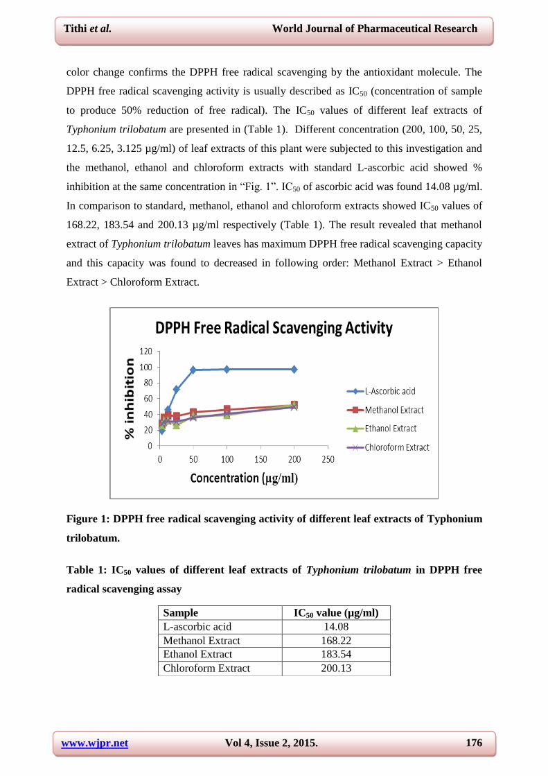
DPPH free radical scavenging activity: DPPH radical scavenging is a popular and reliable
method for screening the antioxidant activity of plant extracts.[34]
DPPH free radical contains
an odd electron and shows maximum absorption at 517 nm. When this odd electron of DPPH
becomes paired with an electron donated by an antioxidant compound, its molar absorptivity
reduces and the color turns from deep purple to yellow or becomes decolorized. This visual
Figure 2: Sun-dried leaves of T. trilobatum
www.wjpr.net Vol 4, Issue 2, 2015.
176
Tithi et al. World Journal of Pharmaceutical Research
color change confirms the DPPH free radical scavenging by the antioxidant molecule. The
DPPH free radical scavenging activity is usually described as IC50 (concentration of sample
to produce 50% reduction of free radical). The IC50 values of different leaf extracts of
Typhonium trilobatum are presented in (Table 1). Different concentration (200, 100, 50, 25,
12.5, 6.25, 3.125 µg/ml) of leaf extracts of this plant were subjected to this investigation and
the methanol, ethanol and chloroform extracts with standard L-ascorbic acid showed %
inhibition at the same concentration in “Fig. 1”. IC50 of ascorbic acid was found 14.08 µg/ml.
In comparison to standard, methanol, ethanol and chloroform extracts showed IC50 values of
168.22, 183.54 and 200.13 µg/ml respectively (Table 1). The result revealed that methanol
extract of Typhonium trilobatum leaves has maximum DPPH free radical scavenging capacity
and this capacity was found to decreased in following order: Methanol Extract > Ethanol
Extract > Chloroform Extract.
Figure 1: DPPH free radical scavenging activity of different leaf extracts of Typhonium
trilobatum.
Table 1: IC50 values of different leaf extracts of Typhonium trilobatum in DPPH free
radical scavenging assay
Sample IC50 value (µg/ml)
L-ascorbic acid 14.08
Methanol Extract 168.22
Ethanol Extract 183.54
Chloroform Extract 200.13
www.wjpr.net Vol 4, Issue 2, 2015.
177
Tithi et al. World Journal of Pharmaceutical Research
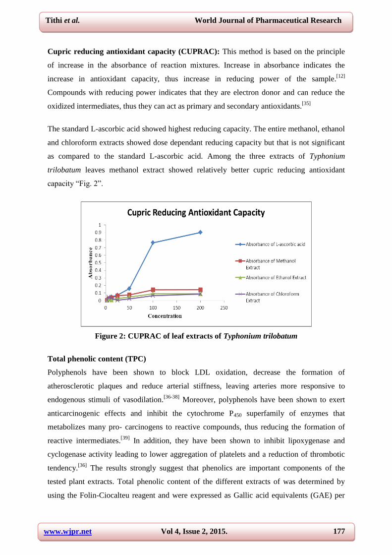
Cupric reducing antioxidant capacity (CUPRAC): This method is based on the principle
of increase in the absorbance of reaction mixtures. Increase in absorbance indicates the
increase in antioxidant capacity, thus increase in reducing power of the sample.[12]
Compounds with reducing power indicates that they are electron donor and can reduce the
oxidized intermediates, thus they can act as primary and secondary antioxidants.[35]
The standard L-ascorbic acid showed highest reducing capacity. The entire methanol, ethanol
and chloroform extracts showed dose dependant reducing capacity but that is not significant
as compared to the standard L-ascorbic acid. Among the three extracts of Typhonium
trilobatum leaves methanol extract showed relatively better cupric reducing antioxidant
capacity “Fig. 2”.
Figure 2: CUPRAC of leaf extracts of Typhonium trilobatum
Total phenolic content (TPC)
Polyphenols have been shown to block LDL oxidation, decrease the formation of
atherosclerotic plaques and reduce arterial stiffness, leaving arteries more responsive to
endogenous stimuli of vasodilation.[36-38]
Moreover, polyphenols have been shown to exert
anticarcinogenic effects and inhibit the cytochrome P450 superfamily of enzymes that
metabolizes many pro- carcinogens to reactive compounds, thus reducing the formation of
reactive intermediates.[39]
In addition, they have been shown to inhibit lipoxygenase and
cyclogenase activity leading to lower aggregation of platelets and a reduction of thrombotic
tendency.[36]
The results strongly suggest that phenolics are important components of the
tested plant extracts. Total phenolic content of the different extracts of was determined by
using the Folin-Ciocalteu reagent and were expressed as Gallic acid equivalents (GAE) per
www.wjpr.net Vol 4, Issue 2, 2015.
178
Tithi et al. World Journal of Pharmaceutical Research
gram of plant extract. Ethanol extract of leaves of the plant was found to contain the highest
amount of phenolic content (13.3. mg/gm) (Table 2). Phenolic contents of the extracts were
found to decrease in the following order: Ethanol Extract > Methanol Extract > Chloroform
Extract.
Table 2: Total phenolic contents of different leaf extracts of Typhonium trilobatum
Total flavonoid content (TFC): Flavonoids play an important role in antioxidant system in
plants. The antioxidative properties of flavonoids are due to several different mechanisms,
such as scavenging of free radicals, chelation of metal ions, such as iron and copper and
inhibition of enzymes responsible for free radical generation.[40]
Depending on their structure,
flavonoids are able to scavenge practically all known ROS. Aluminum chloride colorimetric
method was used to determine the total flavonoid contents of the different extracts of
Typhonium trilobatum. Ethanol extract of Typhonium trilobatum leaves was found to contain
the highest amount of flavonoids content (14.26 mg/gm) (Table 3). Flavonoid contents of the
extracts were found to decrease in the following order: Ethanol Extract > Methanol Extract >
Chloroform Extract.
Table 3: Total flavonoid contents of different leaf extracts of Typhonium trilobatum
Nitric oxide scavenging assay: Nitric oxide (NO) is a physiologically important chemical
mediator generated my endothelial cells and involved in the regulation of various
biochemical processes. Excess generation and accumulation of NO are implicated in
cytotoxic effects observed in various disorders like AIDS, cancer, Alzheimer’s disease and
arthritis.[41]
Overproduction of NO can mediate toxic effects such as DNA fragmentation, cell
damage and neuronal cell death. Different concentration (200, 100, 50, 25, 12.5, 6.25, 3.125
µg/ml) of leaf extracts of Typhonium trilobatum were subjected to investigate the nitric oxide
Extracts Total Phenolic Content
(mg/gm, Gallic Acid equivalents)
Methanol Extract 12.74
Ethanol Extract 13.3
Chloroform Extract 12.64
Extracts Total Flavonoid Content
(mg/gm, Gallic Acid equivalents)
Methanol Extract 13.51
Ethanol Extract 14.26
Chloroform Extract 13.09
www.wjpr.net Vol 4, Issue 2, 2015.
179
Tithi et al. World Journal of Pharmaceutical Research
scavenging activity and the methanol, ethanol and chloroform extracts showed maximum
activity of at low concentration whereas standard L-ascorbic acid showed the same. All the
three extracts showed very good activity (methanol extract showed 90.40% inhibition,
ethanol extract showed 86.91% inhibition and chloroform extract showed 89.53% inhibition)
(Table 5) that is even higher than standard (76.16% inhibition). IC50 of ascorbic acid was
found 40.2 µg/ml. In comparison to standard, methanol, ethanol and chloroform extracts
showed IC50 values of 145.8, 165.67 and 155.45 µg/ml respectively (Table 4). The result
revealed that methanol extract of Typhonium trilobatum leaves has maximum nitric oxide
scavenging capacity and this capacity was found to decrease in following order: Methanol
Extract > Chloroform Extract > Ethanol Extract.
Table 4: IC50 values of different extracts of Typhonium trilobatum leaves in nitric oxide
scavenging assay
Table 5: % inhibition of samples in nitric oxide scavenging activity
Samples % inhibition
L-Ascorbic Acid 76.16
Methanol Extract 90.40
Ethanol Extract 86.91
Chloroform Extract 89.53
Hydrogen peroxide scavenging activity: Hydrogen peroxide, although not a radical species
play a role to contribute oxidative stress. The generation of even low levels of H2O2 in
biological systems may be important. Naturally-occurring iron complexes inside the cell
believed to react with H2O2 in vivo to generate highly reactive hydroxyl radicals and this may
be the origin of many of its toxic effects. % inhibition of hydrogen peroxide of different
extracts of Typhonium trilobatum leaf is presented in figure “Fig. 2”. Among the three
extracts, Chloroform extract showed very good activity that is even higher than standard L-
ascorbic acid. All these extracts possessed with an IC50 value of 141.65, 58.58, and 187.78
µg/ ml respectively whereas standard showed IC50 value of 108.36 µg/ ml (Table 6). The
result revealed that ethanol extract of Typhonium trilobatum leaves has maximum H2O2
Sample IC50 value (µg/ml)
L-Ascorbic Acid 40.2
Methanol Extract 145.8
Ethanol Extract 165.67
Chloroform Extract 155.45
www.wjpr.net Vol 4, Issue 2, 2015.
180
Tithi et al. World Journal of Pharmaceutical Research
scavenging capacity and this capacity was found to decrease in following order: Ethanol
Extract > Methanol Extract > Chloroform Extract.
Table 6: IC50 values of different leaf extracts of Typhonium trilobatum in H2O2
scavenging assay
Figure 2: H2O2 scavenging activity of different leaf extracts of Typhonium trilobatum
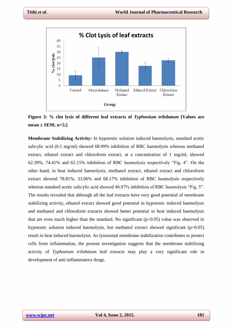
Thrombolytic Activity: The result revealed all the leaf extracts of Typhonium trilobatum
exert better thrombolytic activity.
Blood clot treated with distilled water as control showed negligible clot lysis (9.23%)
whereas Streptokinase as standard showed 25.07% clot lysis. Significant (p<0.05) clot lysis
were visually seen in case of 20mg/ml methanol, ethanol and chloroform extracts of leaves of
Typhonium trilobatum. Methanol extract showed maximum clot lysis (30.15%) that is even
higher than standard activity (25.07%). Ethanol and chloroform extracts also showed better
clot lysis of 17.62 % and 22.64% respectively “Fig. 3”. This is an important finding which
may have important implications in cardiovascular health through the discovery of cardio-
protective drugs from Typhonium trilobatum. In addition, this finding may indicate the
possibility of developing novel thrombolytic agent from Typhonium trilobatum.
Sample IC50 value (µg/ml)
L-Ascorbic Acid 108.36
Methanol Extract 141.65
Ethanol Extract 58.58
Chloroform Extract 187.78
www.wjpr.net Vol 4, Issue 2, 2015.
181
Tithi et al. World Journal of Pharmaceutical Research
Figure 3: % clot lysis of different leaf extracts of Typhonium trilobatum [Values are
mean ± SEM; n=5;]
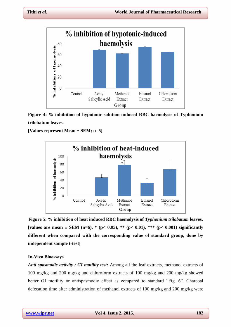
Membrane Stabilizing Activity: In hypotonic solution induced haemolysis, standard acetic
salicylic acid (0.1 mg/ml) showed 68.99% inhibition of RBC haemolysis whereas methanol
extract, ethanol extract and chloroform extract, at a concentration of 1 mg/ml, showed
62.39%, 74.41% and 65.15% inhibition of RBC heamolysis respectively “Fig. 4”. On the
other hand, in heat induced haemolysis, methanol extract, ethanol extract and chloroform
extract showed 78.81%, 33.06% and 68.17% inhibition of RBC heamolysis respectively
whereas standard acetic salicylic acid showed 46.87% inhibition of RBC haemolysis “Fig. 5”.
The results revealed that although all the leaf extracts have very good potential of membrane
stabilizing activity, ethanol extract showed good potential in hypotonic induced haemolysis
and methanol and chloroform extracts showed better potential in heat induced haemolysis
that are even much higher than the standard. No significant (p<0.05) value was observed in
hypotonic solution induced haemolysis, but methanol extract showed significant (p<0.05)
result in heat induced haemolysis. As lysosomal membrane stabilization contributes to protect
cells from inflammation, the present investigation suggests that the membrane stabilizing
activity of Typhonium trilobatum leaf extracts may play a very significant role in
development of anti-inflammatory drugs.
www.wjpr.net Vol 4, Issue 2, 2015.
182
Tithi et al. World Journal of Pharmaceutical Research
Figure 4: % inhibition of hypotonic solution induced RBC haemolysis of Typhonium
trilobatum leaves.
[Values represent Mean ± SEM; n=5]
Figure 5: % inhibition of heat induced RBC haemolysis of Typhonium trilobatum leaves.
[values are mean ± SEM (n=6), * (p< 0.05), ** (p< 0.01), *** (p< 0.001) significantly
different when compared with the corresponding value of standard group, done by
independent sample t-test]
In-Vivo Bioassays
Anti-spasmodic activity / GI motility test: Among all the leaf extracts, methanol extracts of
100 mg/kg and 200 mg/kg and chloroform extracts of 100 mg/kg and 200 mg/kg showed
better GI motility or antispasmodic effect as compared to standard “Fig. 6”. Charcoal
defecation time after administration of methanol extracts of 100 mg/kg and 200 mg/kg were
www.wjpr.net Vol 4, Issue 2, 2015.
183
Tithi et al. World Journal of Pharmaceutical Research
137 minutes and 125 minutes respectively whereas standard butapan took 153 minutes.
Defecation time decreased significantly (p<0.05, p<0.01) in case of chloroform extracts
administration. Chloroform 100 mg/kg and 200 mg/kg extracts needed 116 and 83 minutes
for charcoal defecation. The result revealed that the stimulating effect of the methanol and
chloroform extracts of the plant on GI motility is dependent on its concentration. This
increase in GI motility indicates the potential of antispasmodic effect of the extracts. The
presence of alkaloid in Typhonium trilobatum leaf might be responsible for its increased GI
motility effect.
Figure 6: Charcoal defecation time after administration of samples
[Methanol Extract = ME, Ethanol Extract = EE, Chloroform Extract = ChE, values are
mean ± SEM (n=6), * (p< 0.05), ** (p< 0.01), *** (p< 0.001) significantly different when
compared with the corresponding value of standard group, done by independent sample
t-test]
Anti-depressant activity (forced swimming test, FST): The swimming test has been
validated as a suitable tool to evaluate drugs with anti-depressant effects.[25]
When the subject
is forced to swim in a confined place, they tend to become immobile after struggling. The
depressant behavior of the subject in this stressful inescapable situation can be evaluated by
assessing its immobile period. The development of immobility in that situation reflects the
cessation of persistent escape-directed behavior.[42]
In the present study, methanol, ethanol
and chloroform extracts of 100mg/kg and 200 mg/kg showed immobile duration of 30.33,
30.67, 14.0, 10.67, 6.33 and 0.33 seconds respectively whereas standard diazepam 2mg/kg
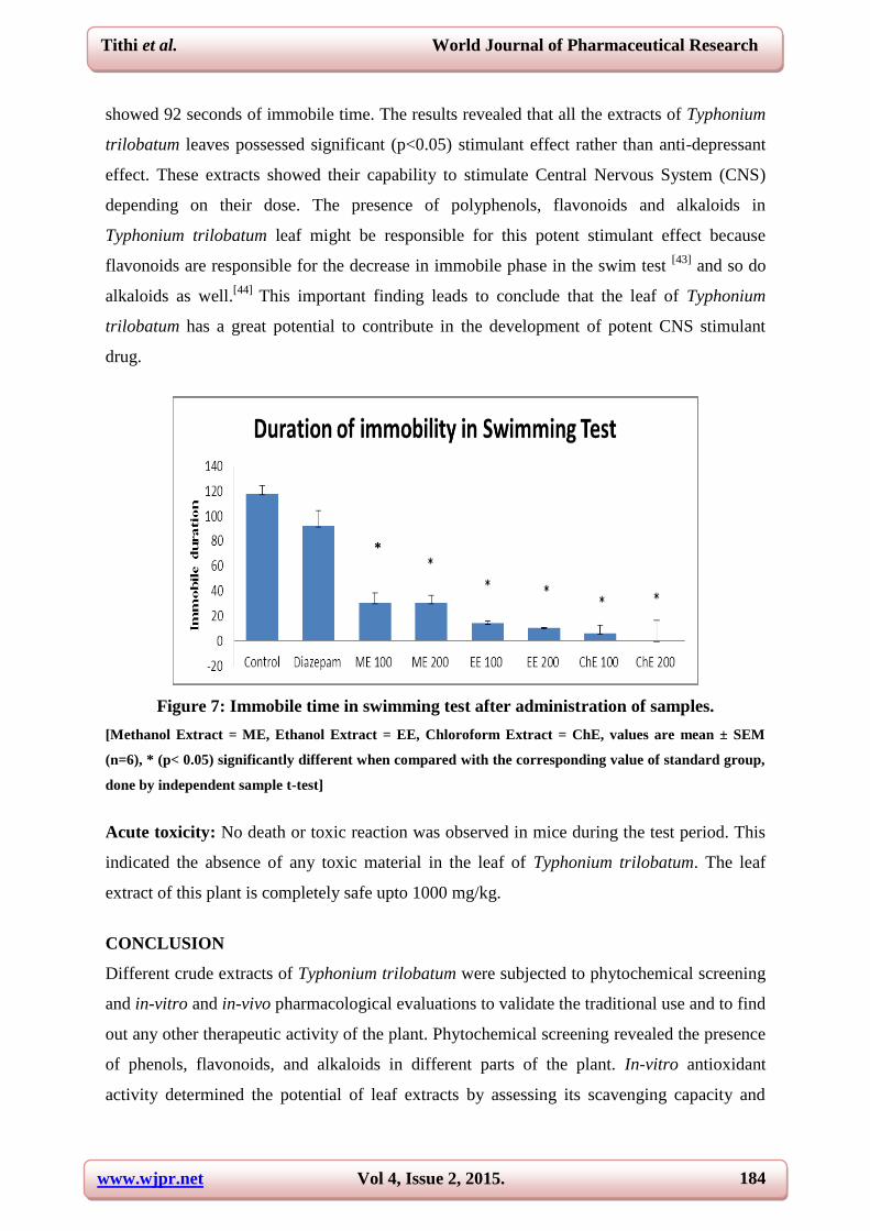
www.wjpr.net Vol 4, Issue 2, 2015.
184
Tithi et al. World Journal of Pharmaceutical Research
showed 92 seconds of immobile time. The results revealed that all the extracts of Typhonium
trilobatum leaves possessed significant (p<0.05) stimulant effect rather than anti-depressant
effect. These extracts showed their capability to stimulate Central Nervous System (CNS)
depending on their dose. The presence of polyphenols, flavonoids and alkaloids in
Typhonium trilobatum leaf might be responsible for this potent stimulant effect because
flavonoids are responsible for the decrease in immobile phase in the swim test [43]
and so do
alkaloids as well.[44]
This important finding leads to conclude that the leaf of Typhonium
trilobatum has a great potential to contribute in the development of potent CNS stimulant
drug.
Figure 7: Immobile time in swimming test after administration of samples.
[Methanol Extract = ME, Ethanol Extract = EE, Chloroform Extract = ChE, values are mean ± SEM
(n=6), * (p< 0.05) significantly different when compared with the corresponding value of standard group,
done by independent sample t-test]
Acute toxicity: No death or toxic reaction was observed in mice during the test period. This
indicated the absence of any toxic material in the leaf of Typhonium trilobatum. The leaf
extract of this plant is completely safe upto 1000 mg/kg.
CONCLUSION
Different crude extracts of Typhonium trilobatum were subjected to phytochemical screening
and in-vitro and in-vivo pharmacological evaluations to validate the traditional use and to find
out any other therapeutic activity of the plant. Phytochemical screening revealed the presence
of phenols, flavonoids, and alkaloids in different parts of the plant. In-vitro antioxidant
activity determined the potential of leaf extracts by assessing its scavenging capacity and
*
www.wjpr.net Vol 4, Issue 2, 2015.
185
Tithi et al. World Journal of Pharmaceutical Research
total phenolic and flavonoid contents. The leaf extracts also revealed significant (p<0.05)
thrombolytic activity and good membrane stabilizing capacity. Effect of leaf extracts on GI
motility was evaluated and it showed significant (p<0.05, p<0.01) anti-spasmodic or laxative
effect. The leaf extracts demonstrated very potent CNS stimulating or anti-depressant effect
(p<0.05). The result clearly indicates that the crude extracts of Typhonium trilobatum may be
a very important contributor in different drug discovery including cardioprotective,
antioxidant, anti-spasmodic and anti-depressant drugs. The present study indicated a better
chance of anti-tumor potential of the plant that might be revealed in near future. Therefore,
further investigation on Typhonium trilobatum to isolate new bioactive compounds might be
the next step to be followed.
REFERENCES
1. Ghani A. Medicinal plants of Bangladesh with chemical constituents and uses. In: 2nd
edition. The Asiatic Society of Bangladesh, Dhaka, Bangladesh: 2003; pp. 31, 39-40, 418,
500-504, 580-589.
2. Shahriar M, Islam S, Aich RK, Haque MA, Sayeed MSB and Kadir FK. Evaluation of the
analgesic and neuropharmacological activities of the bark of Terminala arjuna in mice.
International Journal of Recent Scientific Research, 2013; 4(3):285-289.
3. Akther R, Haque MA, Bhuiyan MA and Shahriar M. In-vivo pharmacological
investigation of leaf extract of Polygonum hydropiper (L.). Dhaka University Journal of
Pharmaceutical Sciences, 2013; 12(2): 165-169.
4. Ahmed T, Akter R, Sharif S, Shahriar M and Bhuiyan MA In vitro antioxidant activities
and In vivo anti-nociceptive and neuropharmacological activities of Mimosa pudica.
International Journal of Pharmacy, 2014; 4(2): 70-78.
5. Shahriar M, Ahmed T, Sharif S, Akter R, Haque MA, Bhuiyan MA. Phytochemical
screenings, membrane stabilizing activity, thrombolytic activity and cytotoxic properties
of leaf extracts of Mimosa pudica. International Journal of Pharmacy, 2014; 4(2): 155-
158.
6. Shahriar M, Alam F, Uddin MMN. Membrane stabilizing and thrombolytic activity of
Withania somnifera root. American Journal of Phytomedicine and Clinical Therapeutics
2014; 2(2): 252-256.
www.wjpr.net Vol 4, Issue 2, 2015.
186
Tithi et al. World Journal of Pharmaceutical Research
7. Uddin MMN, Basak A, Amin MR, Shahriar M. Pharmacological Investigations on
Flowers of Ixora coccinea. International Journal of Pharmacognosy and Phytochemistry,
2014; 29(1): 1208-1213.
8. Biswas S, Ahsan CR, Shahriar M, Khanam JA. Investigation of antioxidant, in-vitro
cytotoxic and in-vivo antitumor effects of leaf extracts of Annona reticulate. Bangladesh
Journal of Microbiology, 2012; 29(2):70-74.
9. Alam F, Shahriar M and Bhuiyan MA. In-vivo pharmacological investigation of bark
extracts of carissa carandas. International Journal of Pharmacy and Pharmaceutical
Sciences; 2014; 6(6): 180-185.
10. Daginawala HF, Prasad S, Kashyap RS, Deopujari JY, Purohit HJ and Taori GM.
Development of an in vitro model to study clot lysis activity of thrombolytic drugs.
Thrombosis Journal, 2006; 4:14.
11. Resat A, Güçlü K, Özyürek M, Karademir SE. Novel Total Antioxidant Capacity Index
for Dietary Poly-phenols and Vitamins C and E, Using Their Cupric Ion Reducing
Capability in the Presence of Neocuproine: CUPRAC Method. J Agric Food Chem, 2004;
52: 7981-7970.
12. Demiray S, Pintado ME, Castro PML. Evaluation of phenolic profiles and antioxidant
activities of Turkish medicinal plants, Tilia argentea, Crataegi folium leaves and
Polygonum bistorta roots. W Aca of Sci Eng and Tech, 2009; 54: 317-312.
13. Velioglu YS, Mazza G, Gao YL, Oomah BD. Antioxidant activity and total phenolics in
selected fruits, vegetables and grain products. J Agric Food Chem, 1998; 46: 4417-4413.
14. Jayaprakasha GK, Rao LJ and Sakariah KK. Antioxidant activities of flavidin in different
in vitro model systems. Bio Med Chem, 2004; 12(19): 5146-5141.
15. Sreejayan and Rao MNA. Nitric Oxide Scavenging by Curcuminoids. J Pharm and
Pharmaco, 1997; 49(1):107-105.
16. Nabavi SM, Ebrahimzadeh MA, Nabavi SF, Hamidinia A, Bekhradnia AR.
Determination of antioxidant activity, phenol and flavonoids content of Parrotia persica
mey. Pharmaco online 2008; 2:567-60.
17. Prasad S, Rajpal SK, Jayant YD, Hemant JP, Girdhar MT, Hatim FD. Development of an
in-vitro model to study clot lysis activity of thrombolytic drugs. Thrombosis Journal,
2006; 4: 14.
18. Ratnasooriya WD, Fernando TSP, Madubashini PP. In-vitro thrombolytic activity of Sri
Lankan black tea, Camellia sinensis O. Kuntze. Journal of National Science Foundation
of Sri Lanka, 2008; 36(2): 179-181.
www.wjpr.net Vol 4, Issue 2, 2015.
187
Tithi et al. World Journal of Pharmaceutical Research
19. Shinde U, Phadke A, Nair A, Mungantiwar A, Dikshit V and Saraf M. Membrane
stabilizing activity-a possible mechanism of action for the anti-inflammatory activity of
Cedrus deodara wood oil. Fitoterapia, 1999; 257-251.
20. Sikder M, Rahman M, Kaisar M, Rahman MS, Hasan C and Rashid M. In vitro
antioxidant, reducing power, free radical scavenging and membrane stabilizing activities
Maisuthisakul P, Suttajit M and Pongsawatmanit R. Assessment of phenolic content and
free radical-scavenging capacity of some Thai indigenous plants. Food Chem, 2007;
100(4):1418-09.
21. Omale J and Okafor P. Comparative antioxidant capacity, membrane stabilization,
polyphenols composition and cytotoxicity of the leaf and stem of Cissus multistriata. Afri
J of Biotech, 2008; 7:3133-29.
22. Ahmed M, Shikha H, Sadhu S, Rahman M and Datta B. Analgesic, diuretic, and anti-
inflammatory principle from Scoparia dulcis. Pharmazie, 2001; 56(8): 660-657.
23. Shahriar M, Alam F and Uddin MMN. Membrane stabilizing and thrombolytic activity of
Withania somnifera root. Am J of Phyt and Cli Ther, 2014; 2(2): 256-252.
24. Marona HRN, Lucchesi MBB. Protocol to refine intestinal motility test in mice. Lab Ani,
2004; 38: 260-257.
25. Porsolt RD, Bertin A, Jalfre M. Behavioral despair in mice: a primary screening test for
antidepressants. Arch Int Pharmacodyn Ther, 1977; 229: 336-327.
26. OECD. Guidance Document on Acute Oral Toxicity Testing. Environmental Health and
Safety Monograph Series on Testing and Assessment N.24. Paris; 2001.
27. OECD. OECD Guidelines for Acute Toxicity of Chemicals. Organization for Economic
Co operation and Development. No. 420. Paris; 2001.
28. OECD. OECD Guidelines for Acute Toxicity of Chemicals. Organization for Economic
Co-operation and Development. No. 423. Paris; 2001.
29. OECD. OECD Guidelines for Acute Toxicity of Chemicals. Organization for Economic
Co-operation and Development. No. 425. Paris: France; 2001.
30. Walum E. Acute oral toxicity Environ. Health Prosp, 1998; 106: 503-497.
31. Meerson FZ, Kagan VE, Kozlov YP, Belkina LM, Arkhipenko YV. The role of lipid
peroxidation in pathogenesis of ischemic damage and the antioxidant protection of the
heart. Basic Research in Cardiology, 1982; 77: 485- 465.
32. Larson RA. The antioxidants of higher plants. Phytochemistry, 1988; 27: 978- 969.
www.wjpr.net Vol 4, Issue 2, 2015.
188
Tithi et al. World Journal of Pharmaceutical Research
33. Salazar R, Pozos ME, Cordero P, Perez J, Salinas MC, Waksman N. Determination of the
antioxidant activity of plants from Northeast Mexico. Pharmaceutical Biology, 2008; 46:
166-170.
34. Mazumder MEH, Rahman S. Pharmacological evaluation of Bangladeshi medicinal
plants for antioxidant activity. Pharmaceutical Biology, 2008; 46(10): 704-709.
35. Yen GC, Chen HY. Antioxidant activity of various tea extracts in relation to their
antimutagenicity. Journal of Agriculture and Food Chemistry, 1995; 43: 32-27.
36. Moline J, Bukharovich IF, Wolff MS. Dietary flavonoids and hypertension: is there a
link? Medical Hypotherapy, 2000; 55(4): 306-309.
37. Arai Y, Wantanabe Kimira M, Shimoi K. Dietary intakes of flavonols, flavones and
isoflavones by Japanese women and the inverse correlation between quercetin intake and
plasma LDL cholesterol Concentration. American Society of Nutritional Science, 2000;
130(9): 2243-2250.
38. Duthie GG, Duthei SJ, Kyle JAM. Plant polyphenols in cancer and heart disease:
implication as nutritional antioxidants. Nutrition Research Review, 2000; 13: 79-106.
39. Stoner GD, Mukhtar H. Polyphenols as cancer chemopreventive agent. Journal of Cell
Biochemistry, 1995; 22: 169-180.
40. Benavente-Gaecia O, Castillo J, Martin FR, Ortuno A, Del-Rio JA. Uses and properties of
Citrus flavonoids. Journal of Agriculture and Food Chemistry, 1997; 45: 4515-4505.
41. Sainani GS, Manika JS, Sainani RG. Oxidative stress- a key factor in pathogenesis of
chronic diseases. Medical Update, 1997; 1: 1.
42. Lucki I. The forced swimming test as a model for core and components behavioral effects
of antidepressant drugs. Behavioural Pharmacology, 1997; 8: 532-523.
43. Butterweck V, Jurgenliemk G, Nahrstedt A. Planta Medica, 2000; 66: 6-3.
44. Silva GN, Martins FR, Matheus ME. Journal of Ethnopharmacology, 2005; 100: 259-254.