Practice-Oriented Validation of Embedded Beam Formulations ...
Upload
independentCategory
view
3download
0
THE JOURNAL OF GENE MEDICINE R E S E A R C H A R T I C L EJ Gene Med 2008; 10: 770–782.Published online 21 April 2008 in Wiley InterScience (www.interscience.wiley.com) DOI: 10.1002/jgm.1199
Physical characterization and in vivo evaluationof poloxamer-based DNA vaccine formulations
Jukka Hartikka1*Andrew Geall1†
Vesselina Bozoukova1
David Kurniadi1
Denis Rusalov1
Joel Enas1
Ji-Hyun Yi2
Antonio Nanci2
Alain Rolland1
1Vical Incorporated, 10390 PacificCenter Court, San Diego, CA92121-4340, USA2Faculty of Dentistry, Universite deMontreal, 2900 BlvdEdouard-Montpetit, Montreal, QC,Canada H3T 1J4
*Correspondence to:Jukka Hartikka, Vical Incorporated,10390 Pacific Center Court, SanDiego, CA 92121-4340, USA.E-mail: [email protected]
†Current address: Novartis Institutesfor Biomedical Research, Inc., 250Massachusetts Ave, Cambridge, MA02139, USA.
Received: 24 November 2007Revised: 25 February 2008Accepted: 13 March 2008
Abstract
Background Plasmid DNA (pDNA) vaccines have generated significantinterest for the prevention or treatment of infectious diseases. Broaderapplications may benefit from the identification of safe and potent vaccineadjuvants. This report describes the development of a novel polymer-basedformulation to enhance the immunogenicity of pDNA-based vaccines.
Methods Plasmid DNA was formulated with a nonionic block copolymer,poloxamer CRL1005, and the cationic surfactant benzalkonium chloride(BAK) to produce a thermodynamically stable, self-assembling system. Theinfluence of parameters such as polymer concentration and BAK compositionon the immune responses was evaluated in mice vaccinated with pDNAencoding influenza nucleoprotein.
Results At concentrations of 7.5 mg/ml CRL1005, 0.3 mM BAK and5 mg/ml pDNA, CRL1005/BAK/pDNA particles had a mean diameter of261 ± 0.2 nm and a surface charge of −11.6 ± 0.9 mV. The negative surfacecharge and atomic force microscopy images suggested that pDNA binds to BAKadsorbed to the surface of poloxamer particles. The CRL1005/BAK/pDNAformulation significantly enhanced antigen-specific cellular and humoralimmune responses, and increased transgene levels in muscle and serum.The complexity of the formulation was reduced by replacing the commercialBAK, which is a mixture of four alkyl chains, with a C14 BAK homolog. Thesubstitution yielded an analytically preferable formulation with equivalentphysical characteristics and immunogenicity.
Conclusions The results suggest that the CRL1005/BAK/pDNA formulationmay enhance immunogenicity by improving the delivery of pDNA-basedvaccines. This formulation is currently being evaluated for the prevention ofCMV-associated disease in a phase 2 clinical trial. Copyright 2008 JohnWiley & Sons, Ltd.
Keywords plasmid DNA; vaccine; poloxamer CRL1005; block copolymer; genedelivery
Introduction
DNA vaccines are generating significant interest for the prevention ortreatment of infectious diseases for human and veterinary applications[1–5]. Although results from preclinical studies of unformulated plas-mid DNA (pDNA) have been encouraging, broader human applicationsmay benefit from the identification of safe and potent adjuvants toenhance the immunogenicity of pDNA-based vaccines. One promising
Copyright 2008 John Wiley & Sons, Ltd.
CRL1005/BAK/pDNA vaccine formulation 771
approach to increase the humoral and/or cellular immuneresponses is the use of micro- or nanoparticulate deliverysystems, which allow delivery of phagocytosable particlesto antigen-presenting cells (APCs) [4]. Amongst variousparticulate delivery systems, adsorption of pDNA ontopolymeric micro- or nanoparticles reduces many of themanufacturing hurdles associated with encapsulation.Several clinical trials using such an approach areunderway [4,6,7].
A microparticulate HIV pDNA vaccine is being testedin a phase 1 clinical trial for safety and immunogenicity[3]. The vaccine candidate employs a strategy of primingthe immune responses using gag and env DNA plasmids,followed by boosting with a recombinant gp140 envelopeprotein formulated with an adjuvant. The pDNA primeuses a gene-delivery system in which the pDNA is loadedonto surface-modified poly(lactide-co-glycolide) (PLG)microparticles. The PLG microparticles are coated withthe cationic surfactant cetyltrimethylammonium bromide(CTAB) during preparation using a standard doubleemulsion followed by solvent evaporation technique[8–10]. A resulting net positive surface charge of themicroparticles allowed the negatively charged plasmidsto bind, via electrostatic interactions, with high efficiencyand loading capacity [4]. Studies performed to elucidatethe mechanism of action of such a formulation haveindicated that, after intramuscular administration, thetransgene is expressed at the injection site, followedby recruitment of mononuclear phagocytes, activation ofAPCs, and subsequent presentation of the antigen-derivedpeptides in the context of MHC class I molecules [4,8,9].
Another prime-boost regimen investigated utilizedpoloxamer CRL1005 adjuvanted pDNA and an adenovirustype 5 vector boost, both expressing the HIV-1 gag gene[11]. Preclinical studies in nonhuman primates havedescribed the immunogenic potential of this approach[6,11–13]. A recent publication [6] described somephysical characteristics and immunological properties ofa poloxamer formulation consisting of CRL1005 andthe cationic surfactant benzalkonium chloride (BAK).The presence of BAK imparts a net positive surfacecharge to the particles, rendering them capable ofbinding pDNA through electrostatic interactions. Between1.5–3% of the total pDNA in the formulation wasshown to be associated with the CRL1005/BAK particles,corresponding to approximately 20–40 plasmids perparticle.
A CRL1005/BAK-based pDNA vaccine formulation iscurrently in a phase 2 clinical trial for the preventionof cytomegalovirus (CMV)-associated disease in theallogeneic hematopoietic cell transplant population [14].This plasmid-based immunotherapeutic vaccine containstwo pDNAs encoding the major surface glycoproteinB (gB) and the tegument phosphoprotein 65 (pp65)of human CMV formulated with the nonionic triblockcopolymer CRL1005 and BAK in phosphate-bufferedsaline (PBS).
CRL1005, a triblock POE-POP-POE copolymer witha formula HO-(CH2CH2O)8-[CH2CHO(CH3)]205-(CH2
CH2O)8-H, contains repeating units comprising a lin-ear chain of hydrophobic polyoxypropylene (POP, Mw =12 kDa), flanked by two linear chains of hydrophilicpolyoxyethylene (POE, 5% of total molecular weight).This poloxamer has inverse solubility characteristics inaqueous media [15–17]. Below the phase transitiontemperature or cloud point (7–12 ◦C), this copolymeris water-soluble and forms a clear solution that canbe sterile filtered. When a solution of copolymer iswarmed and it passes through its cloud point, theincreased thermal motion is sufficient to break thehydrogen bonds and, as the copolymer comes out ofsolution, it self-assembles into micelles or microparti-cles [18]. The process is reversible. In the presence ofthe hydrophobic cationic surfactant BAK, CRL1005 self-assembles into nanoparticles with BAK anchored ontothe surface of the copolymer. BAK is a mixture of alkyl-benzyldimethylammonium chlorides of general formula[C6H5CH2N(CH3)2R]Cl, where R represents a mixtureof alkyls, including n-C12H25 (C12 BAK), n-C14H29 (C14
BAK), n-C16H33 (C16 BAK) and possibly n-C18H37 (C18
BAK).This paper describes the physicochemical characteri-
zation of poloxamer CRL1005-based formulations. Theinfluence of parameters such as polymer concentrationand BAK composition on the immune responses in micevaccinated with pDNA encoding influenza nucleoprotein(NP), as a model immunogen, was evaluated.
Materials and methods
Materials and plasmid constructs
Poloxamer CRL1005 was obtained from CytRx Corpora-tion (Los Angeles, CA, USA). Benzalkonium chloride 50%solution NF (BAK, BTC 50 NF) was obtained from StepanCompany (Northfield, IL, USA). Benzyldimethyldodecy-lammonium chloride (C12 BAK homolog) was obtainedfrom Fluka Chemie (Buchs, Switzerland). Alkyldimethyl-benzylammonium chloride, benzylcetyldimethylammo-nium chloride, and benzyldimethylstearylammoniumchloride (C14, C16, and C18 BAK homologs, respec-tively) were purchased from TCI America (Portland,OR, USA). Plasmid VR4700 was constructed by insert-ing the A/PR/8/34 influenza NP coding sequence intoan expression plasmid containing the CMV immedi-ate early 1 promoter/enhancer and intron A, a mod-ified rabbit β-globin transcriptional terminator and anopen reading frame encoding the kanamycin resistancegene [19,20]. Plasmids VCL6368 encoding a detoxi-fied CMV antigen pp65, VCL6365 encoding a secretedform of the CMV gB antigen lacking the transmem-brane and cytoplasmic domains, VR1255 encoding fire-fly luciferase, and VR2956 encoding feline erythropoi-etin (EPO) were constructed as previously described[7,21].
Copyright 2008 John Wiley & Sons, Ltd. J Gene Med 2008; 10: 770–782.DOI: 10.1002/jgm
772 J. Hartikka et al.
CRL1005/pDNA formulations
The required concentration of VR4700 pDNA (to producea final concentration of 0.1 mg/ml) in PBS (0.9%sodium chloride + 10 mM sodium phosphate, pH 7.2)was stirred on ice and the required amount of CRL1005(to produce a final concentration of 0.15 to 75 mg/ml)was added using a positive displacement pipette. Thesolution was then thermocycled through the cloud point(7–12 ◦C) several times to ensure homogeneity, filtersterilized through a Millipore Steriflip disposable vacuumfiltration system (Millipore, Billerica, MA, USA) at 4 ◦Cand stored frozen. CRL1005 formulations without pDNAwere made as described above by dissolving CRL1005in PBS.
CRL1005/BAK/pDNA formulations
The required concentration of VR4700 pDNA (to producea final concentration of 5 mg/ml) in PBS was stirredon ice and the required amount of CRL1005 (toproduce a final concentration of 7.5 mg/ml) was addedusing a positive displacement pipette. The solutionwas stirred on ice until the poloxamer dissolved andthen the required concentration of BAK dissolved inPBS was added (to produce a final concentration of0.3 mM). The mixture was then thermocycled throughthe cloud point several times to ensure homogeneity,filter sterilized through a Millipore Steriflip disposablevacuum filtration system at 4 ◦C and stored frozen.Prior to injection, the vaccine was thawed at ambienttemperature (25 ◦C) and diluted to the required pDNAconcentration with PBS above the cloud point ofCRL1005. The particle sizes and surface charges offormulations did not change upon dilution (data notshown). CRL1005/BAK formulations without pDNA weremade as described above by dissolving the CRL1005 andBAK in PBS.
CRL1005/BAK/pDNA formulations withdifferent particle sizes
The size of CRL1005/BAK/pDNA particles was found todepend on the formulation concentration during storage.The CRL1005/BAK/pDNA formulations with differentparticle sizes (Z average diameter of 250, 350, 550,1000, or >1000 nm) tested for immunogenicity wereachieved by thermocycling the formulation through thecloud point as described above (at 7.5 mg/ml CRL1005 +0.3 mM BAK + 5 mg/ml pDNA), diluting the formulationabove the cloud point with PBS to various concentrations,and storing them frozen at −80 ◦C in glass vials. Priorto injections, the formulations were thawed at ambienttemperature and diluted with PBS to the final concen-tration (0.15 mg/ml CRL1005 + 6 µM BAK + 0.1 mg/mlpDNA).
CRL1005/BAK homolog formulations
CRL1005/BAK and CRL1005/BAK/pDNA formulationscontaining only the C14 BAK homolog at a concentrationof 0.3 mM were prepared as described in the sectionbefore last. CRL1005/BAK formulations containing C12,C16 or C18 BAK homologs were also prepared as describedabove (without pDNA) by dissolving the CRL1005 andBAK homologs in PBS.
Particle size and zeta potentialmeasurements
Particle size measurements were made using photoncorrelation spectroscopy (3000HS Zetasizer, MalvernInstruments, Worcestershire, UK). The instrument wascalibrated using a 200 nm latex standard (200 nm latexnanospheres; Duke Scientific, Palo Alto, CA, USA) dilutedin PBS at pH 7.2. Prior to analysis, samples were dilutedinto PBS as per the instrument manufacturer’s guidelines.The Z average or the hydrodynamic diameter (nm)and polydispersity of the size range were calculatedusing cumulants analysis. Zeta potential measurementswere made using micro-electrophoresis (Malvern 3000HSZetasizer). The instrument was calibrated using a −50 mVMalvern zeta potential transfer standard (MalvernInstruments). Prior to analysis, samples were dilutedinto 20 mM Tris acetate (pH 7.2) as per the instrumentmanufacturer’s guidelines. The zeta potential was derivedfrom the electrophoretic mobility by applying theSmoluchowsky relationship and was quoted in millivolts(mV).
Fluorescence microscopy
Poloxamer particles were visualized with Nile Red(Molecular Probes Inc., Eugene, OR, USA), a dye whichbecomes fluorescent in nonpolar environments [22]. NileRed was dissolved in dimethylformamide (Sigma, St.Louis, MO, USA) at 8 mg/ml, and added into CRL1005 orCRL1005/BAK formulations in PBS (final concentration ofNile Red 42 µg/ml). Formulations were placed on poly-L-lysine-coated microscope slides (Electron MicroscopySciences, Ft. Washington, PA, USA) and examined usinga 100× oil immersion objective with a Nikon Optiphotmicroscope fitted with a Nikon narrow band pass G-1B fluorescence filter (excitation wavelength 546 nm,barrier filter 590 nm), which allowed visualization of the628 nm emission of Nile Red. Digital phase contrast andfluorescent photomicrographs were taken using a SpotRT color camera (Diagnostic Instruments Inc., SterlingHeights, MI, USA). The size of CRL1005 particles wasestimated by measuring the diameter of all poloxamerparticles in seven randomly selected phase contrastimages (a total of 427 particles were measured) using theImage-Pro Plus software (version 5.1.0.20; BioImagingSolutions Inc., San Diego, CA, USA). The imaging software
Copyright 2008 John Wiley & Sons, Ltd. J Gene Med 2008; 10: 770–782.DOI: 10.1002/jgm
CRL1005/BAK/pDNA vaccine formulation 773
was calibrated using a stage micrometer (Fisher Scientific,Hampton, NH, USA).
Atomic force microscopy (AFM)
Plasmid DNA was diluted to 2.5 µg/ml with 2.4 mMNiCl2. A 10 µl drop was applied to a freshly cleavedmica surface. After incubation for 5 min in a closedenvironment at ambient temperature, the substrate waswashed with nano-pure water (Barnstead Diamond ROwater purification system, Garner, NC, USA) and thendried with a stream of nitrogen. The CRL1005/BAK/pDNAformulation was diluted with a solution of 5 mM NiCl2 inPBS, to a final pDNA concentration of 50 µg/ml, anda 40 µl drop was applied to a freshly cleaved micasurface. The preparation was incubated for 120 minand similarly processed for AFM imaging. AFM imageswere taken in tapping mode using a JEOL JSPM-5200 inair. Topographic and phase images were simultaneouslycollected. Phase imaging AFM can detect variations incomposition, adhesion, friction, viscosity, and is used tosupplement information on surface structure.
Vaccination regimen
Groups of 6- to 10-week-old female BALB/c mice (Harlan-Sprague-Dawley, Indianapolis, IN, USA) received bilateralintramuscular injections (on days 0, 20, and 48) into therectus femoris with 5 µg of VR4700 pDNA ± formulationin 50 µl PBS/leg (total dose of 10 µg of pDNA/injection)as previously described [19]. Mice were bled via theophthalmic venous plexus 61 days after the first injection,and sera were stored at −20 ◦C until assayed for NP-specific antibodies by enzyme-linked immunosorbentassay (ELISA). Splenocytes were harvested on threeconsecutive days, 9 weeks after the first injection (onday 62 through 64) and NP-specific CD8+ and CD4+T-cell responses were measured by IFN-γ ELISPOT assay.
In studies assessing immunogenicity of C14 BAKhomolog formulations, 8- to 11-week-old female BALB/cmice were vaccinated with the bivalent CMV vaccinecontaining equal amounts of pp65 pDNA (VCL6368) andgB pDNA (VCL6365). On days 0 and 14, mice receivedbilateral injections of CRL1005/BAK/pDNA formulationsinto the rectus femoris muscles (20 µg total pDNA in 100 µlPBS/mouse/injection). Splenocytes were harvested onthree consecutive days (on days 27 through 29) 4 weeksafter the first injections. In each collection, spleens fromtwo mice per test group were pooled, and the poolswere assayed for CMV pp65-specific IFN-γ -secreting T-cells using an ELISPOT assay. Mice were bled on day26, and CMV gB-specific antibodies were analyzed byELISA. All animal procedures were approved by the VicalInstitutional Animal Care and Use Committee (IACUC)and complied with the standards set forth in the Guidefor the Care and Use of Laboratory Animals (ILAR, 1996)and the Animal Welfare Act and Animal Care Regulations[23].
Antibody ELISA assays
Serum anti-NP IgG titers were measured in an indi-rect ELISA using 96-well plates (Corning Incorporated,Corning, NY, USA) coated with 71 ng/well of influenzaA/PR/8/34 nucleoprotein (NP) purified from recombi-nant baculoviral extracts as previously described [19,21].The anti-gB ELISA was performed as previously described[7]. Endpoint titers were determined as the reciprocal ofthe last dilution at which the absorbance value of the testserum was at least twice that of the absorbance value ofthe background established with pre-immune serum.
IFN-γ ELISPOT assays
Mice were sacrificed over three consecutive days. In eachcollection, splenocytes from three mice out of each testgroup were isolated, pooled, and the pools were assayedfor NP-specific IFN-γ -secreting T-cells. ImmunoSpotplates (Millipore, Billerica, MA, USA) were coated with5 µg/ml of rat anti-mouse IFN-γ monoclonal antibody(BD Pharmingen, San Diego, CA, USA) and blocked withRPMI-1640 medium containing 10% (v/v) defined fetalbovine serum (FBS, Hyclone, Logan, UT, USA). Spleno-cyte suspensions were seeded in quadruplicate wells ofImmunoSpot plates at 1 × 10⁶ cells/well in RPMI-1640medium containing 25 mM HEPES buffer and L-glutamine(Invitrogen, Carlsbad, CA, USA) supplemented with 10%(v/v) FBS, 55 µM β-mercaptoethanol (Invitrogen), 100U/mL of penicillin G sodium salt and 100 µg/mL ofstreptomycin sulfate (Invitrogen). For detection of NP-specific IFN-γ -secreting CD8+ T-cells, splenocytes werestimulated with the NP class I peptide, TYQRTRALV[24], at 4 µg/ml with 1 unit/ml of recombinant mouseIL-2 (Roche, Indianapolis, IN, USA). For detection ofNP-specific IFN-γ -secreting CD4+ T-cells, splenocyteswere stimulated with a cocktail of three NP class IIpeptides, FWRGENGKTRSAYERMCNILKGK, AVKGVGT-MVMELIRMIKRGINDRN, RLIQNSLTIERMVLSAFDERRNK[24–26], at 10 µg/ml each. To assess nonspecific stim-ulation, splenocytes were plated in the absence of NPpeptides, with or without added IL-2. After overnightincubation at +37 ◦C in 5% CO2, captured IFN-γ fromstimulated cells was detected by the sequential additionof biotin-labeled rat anti-mouse IFN-γ monoclonal anti-body (1 : 1000 dilution, BD Pharmingen) and horseradishperoxidase-labeled avidin D (Vector Labs, Burlingame,CA, USA). Spots produced by the conversion of 3-amino-9-ethylcarbazole substrate (AEC, Vector Labs) were quanti-fied using an ImmunoSpot analyzer (Cellular TechnologyLtd., Cleveland, OH, USA). For each test group, the aver-age background count derived from unstimulated cellswas subtracted from counts obtained from each wellof stimulated cells. Data were presented as the numberof antigen-specific IFN-γ -producing T-cells, designatedas spot forming units (SFU) per million splenocytes(SFU/106 cells). ELISPOT assays for the quantitation of
Copyright 2008 John Wiley & Sons, Ltd. J Gene Med 2008; 10: 770–782.DOI: 10.1002/jgm

774 J. Hartikka et al.
pp65-specific IFN-γ -secreting T-cells were performed aspreviously described [7].
Reporter gene assays
To quantitate transgene expression levels in the injectedtissue, muscles were harvested, pulverized and extractedas previously described [19]. Luciferase activity inmuscle extracts was assayed using a Wallac model 1420Victor2 (PerkinElmer, Waltham, MA, USA) microplateluminometer as previously described [21]. Erythropoietin(EPO) levels in serum or muscle extracts were assayedusing a commercially available kit (R&D Systems,Minneapolis, MN, USA) following the instructionsprovided in the kit.
Statistical analysis
Statistical analysis was performed using either Kruskal-Wallis one-way analysis of variance (ANOVA), Student’st-test, or the nonparametric Mann-Whitney rank sumtest (SigmaStat version 2.03; Systat Software, Inc., PointRichmond, CA, USA).
Results
Particle size and zeta potential ofCRL1005/BAK/pDNA formulations
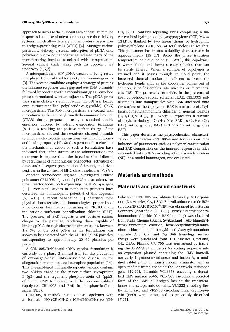
To better isolate the physical interactions of thecomponents of the CRL1005/BAK-based pDNA vaccineformulations, the copolymer alone was characterized first.When CRL1005 was dissolved in PBS and cycled throughthe cloud point, the copolymer self-assembled intoparticles at room temperature. The size of these sphericalparticles was dependent on poloxamer concentration,with a trend of increasing particle sizes with increasingCRL1005 concentration (Figure 1A). At 7.5 mg/ml,CRL1005 produced micron-sized particles with a meanhydrodynamic diameter of 1445 ± 32 nm and a negativezeta potential of −4.1 ± 0.5 mV. The size and thecharge of the particles could be manipulated with thecationic surfactant BAK (or with other cationic lipids,data not shown) to produce uniform-sized submicronparticles with a positive surface charge. Increasing BAKconcentration from 0.3 to 0.9 mM had little effect on thesize of CRL1005/BAK particles (Figure 1B), but increasedthe surface charge (Figure 1C). CRL1005/BAK particles,at 7.5 mg/ml CRL1005 and 0.3 mM BAK concentration,had a mean hydrodynamic diameter of 186 ± 0.6 nmand a positive zeta potential of +8.3 ± 0.8 mV (Table 1).Formulating these particles in the presence of 5 mg/mlpDNA produced CRL1005/BAK/pDNA particles with amean hydrodynamic diameter of 261 ± 0.2 nm and anegative zeta potential of −11.6 ± 0.9 mV. The negativesurface charge of CRL1005/BAK/pDNA particles is
0
200
400
600
800
1.000
1.200
1.400
1.600
1.800
0.0 0.2 0.4 0.6 0.8 1.0BAK concentration (mM)
Z A
vera
ge
dia
met
er (
nm
)
B
0
200
400
600
800
1.000
1.200
1.400
1.600
1.800
1 10 100CRL1005 concentration (mg/ml)
Z A
vera
ge
dia
met
er (
nm
)
A
-5
0
5
10
15
20
0.0 0.2 0.4 0.6 0.8 1.0BAK concentration (mM)
Zet
a p
ote
nti
al (
mV
)
C
Figure 1. Particle size and zeta potential of CRL1005 andCRL1005/BAK formulations. CRL1005 poloxamer, at concen-trations ranging from 1 to 50 mg/ml, was dissolved in PBSand the particle sizes at ambient temperature were deter-mined after cycling through the cloud point (A). The effectof varying concentrations of the cationic surfactant BAK onthe size (B; photon correlation spectroscopy) and zeta poten-tial (C; micro-electrophoresis) of CRL1005/BAK particles wasdetermined at a CRL1005 concentration of 7.5 mg/ml
consistent with pDNA binding to the cationic surfactantanchored onto the surface of the poloxamer particles.Attempts to prepare CRL1005/BAK/pDNA formulationsusing BAK concentration from 0.52 to 1.2 mM resulted
Copyright 2008 John Wiley & Sons, Ltd. J Gene Med 2008; 10: 770–782.DOI: 10.1002/jgm
CRL1005/BAK/pDNA vaccine formulation 775
in precipitation of insoluble material. Thus the 0.3 mMBAK/CRL1005 formulation was chosen to be tested in thein vivo studies.
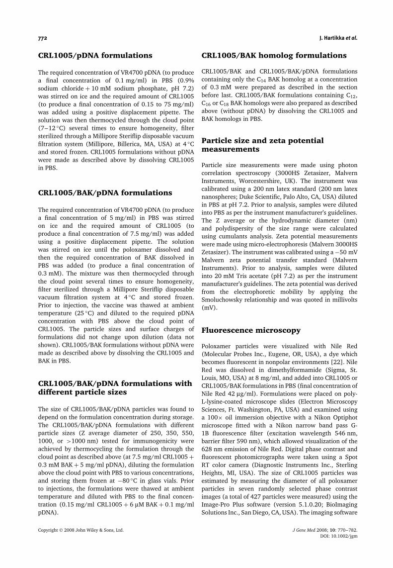
Microscopy of CRL1005/BAK/pDNAformulations
Phase contrast and fluorescence microscopy confirmedthat, in the absence of BAK, CRL1005 particles had
a variable diameter (Figures 2A and 2B). The particlesize was estimated from phase contrast images and,at 7.5 mg/ml CRL1005 concentration, the averagemeasured diameter was 1274 ± 32 nm (n = 427, range230–3201 nm), in good agreement with the data obtainedwith photon correlation spectroscopy (Figure 1A). Incontrast, formulating CRL1005 in the presence of BAKeither without (Figures 2C and 2D) or with pDNA(Figure 2E) resulted in more uniform particle sizes with atypical particle diameter in the submicron range.
Figure 2. Phase contrast and fluorescence photomicrographs of CRL1005, CRL1005/BAK and CRL1005/BAK/pDNA formulations.CRL1005 at 7.5 mg/ml was dissolved in PBS and cycled through the cloud point in the absence (A, B) or presence (C, D) of 0.3 mMBAK. CRL1005/BAK/pDNA formulation (E) was prepared at final concentrations of 7.5 mg/ml CRL1005, 0.3 mM BAK and 5 mg/mlpDNA. Images were obtained at room temperature, either by phase contrast microscopy (A, C, E) or fluorescence microscopy, aftervisualizing the poloxamer particles with Nile Red (B, D). Images were taken using a 100 × oil immersion objective. The scale barcorresponds to 2 µm in length
Copyright 2008 John Wiley & Sons, Ltd. J Gene Med 2008; 10: 770–782.DOI: 10.1002/jgm
776 J. Hartikka et al.
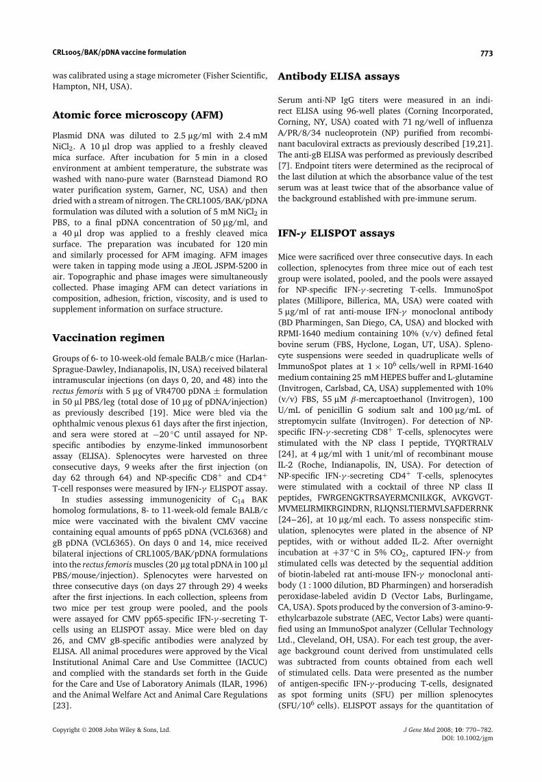
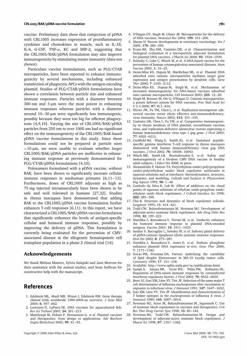
Atomic force microscopy images revealed pDNAmolecules mainly in a supercoiled state (Figure 3A).The measured height ranged from 0.4 to 0.8 nm, andthe approximate length ranged from one to severalmicrometers. The width of pDNA molecules measuredusing height profiles in AFM images were in the20–30 nm range. An AFM topographic image of theCRL1005/BAK/pDNA formulation (Figure 3B) showedthat the entire mica surface was covered with entangledpDNA molecules. Rounded particles associated withentangled pDNAs were also present. Their morphologywas clearly distinct from that of pDNA, and their diameterwas in the 200–250 nm range. These particles werehomogenously distributed on the mica surface with adensity of about 0.4/µm2. Increasing the concentration ofpDNA 20-fold, to 1 mg/ml, resulted in higher numbers ofimmobilized molecules on the mica substrate. The AFMphase image of the CRL1005/BAK/pDNA formulation(Figure 3C), taken simultaneously with the topographicimage, showed a similar surface morphology, composedof entangled pDNA and round particles. However, theparticles were more contrasted in the phase image thanin the topographic image. CRL1005 and CRL1005/BAKsamples which did not contain pDNA could not beimmobilized on mica using Ni2+ ions. After incubation andwashing, AFM imaging confirmed that nothing remainedon the mica surface.
Immunogenicity of CRL1005/pDNAformulations
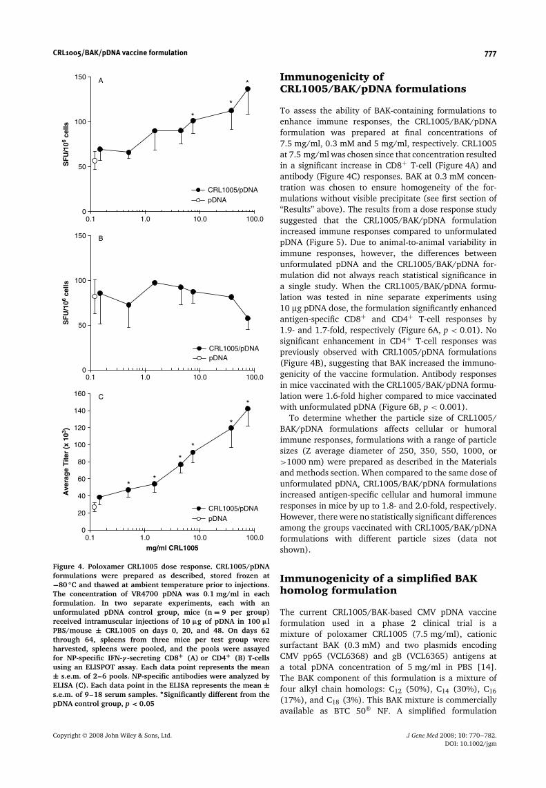
The immunogenicity of CRL1005/pDNA formulations,in the absence of BAK, were evaluated first sincesimilar poloxamer/pDNA formulations have previouslybeen reported to increase immune responses in non-human primates [12,13]. The particle size and zetapotential of CRL1005/pDNA formulations at 0.1 mg/mlpDNA concentration varied from 462 nm and −3.7 mV(obtained with 0.15 mg/ml CRL1005) to 2030 nm and+0.2 mV (obtained with 75 mg/ml CRL1005), respec-tively. Compared to unformulated pDNA injected inPBS, CRL1005/pDNA formulations enhanced antigen-specific CD8+ T-cell (Figure 4A) and antibody (Figure 4C)responses by up to 2- and 5-fold, respectively, in a polox-amer concentration-dependent manner. CRL1005/pDNA
Table 1. Physical characteristics of CRL1005/BAK formulationsprepared using BAK homologs
FormulationaZ average
diameter (nm)Poly-
dispersityAverage surface
charge (mV)
BTC 50 NF 186 0.02 +8.3C12 BAK 924 0.27 +1.2C14 BAK 207 0.05 +0.8C16 BAK 471 0.27 +3.4C18 BAK 760 0.58 +15.2
aThe CRL1005 concentration was 7.5 mg/ml, and the BAK(BTC 50 NF) or BAK homolog concentration for all formulations was0.3 mM.
13.44 Å
0.00 Å200 nm
-82 deg
-132 deg500 nm
4.61 nm
0.00 nm500 nm
B
A
C
Figure 3. Atomic force microscopy of pDNA and CRL1005/BAK/pDNA formulations. AFM topographic image of pDNA(2.5 µg/ml) molecules immobilized by Ni2+ on mica sub-strate (A). AFM topographic (B) and phase (C) image ofCRL1005/BAK/pDNA formulation immobilized on mica surfaceat pDNA concentration of 50 µg/ml. The scale bar correspondsto 200 nm (A) or 500 nm (B, C) in length
formulations did not have a significant effect on thenumber of antigen-specific IFN-γ -secreting CD4+ T-cellscompared to vaccination with pDNA alone (Figure 4B).
Copyright 2008 John Wiley & Sons, Ltd. J Gene Med 2008; 10: 770–782.DOI: 10.1002/jgm
CRL1005/BAK/pDNA vaccine formulation 777
0
50
100
150
0.1 1.0 10.0 100.0
SF
U/1
06 ce
lls
CRL1005/pDNApDNA
B
*
*
*
0
50
100
150
0.1 1.0 10.0 100.0
SF
U/1
06 ce
lls
CRL1005/pDNA
pDNA
A
*
*
**
**
0
20
40
60
80
100
120
140
160
0.1 1.0 10.0 100.0
mg/ml CRL1005
Ave
rag
e T
iter
(x
103 )
CRL1005/pDNA
pDNA
C
Figure 4. Poloxamer CRL1005 dose response. CRL1005/pDNAformulations were prepared as described, stored frozen at−80 ◦C and thawed at ambient temperature prior to injections.The concentration of VR4700 pDNA was 0.1 mg/ml in eachformulation. In two separate experiments, each with anunformulated pDNA control group, mice (n = 9 per group)received intramuscular injections of 10 µg of pDNA in 100 µlPBS/mouse ± CRL1005 on days 0, 20, and 48. On days 62through 64, spleens from three mice per test group wereharvested, spleens were pooled, and the pools were assayedfor NP-specific IFN-γ -secreting CD8+ (A) or CD4+ (B) T-cellsusing an ELISPOT assay. Each data point represents the mean± s.e.m. of 2–6 pools. NP-specific antibodies were analyzed byELISA (C). Each data point in the ELISA represents the mean ±s.e.m. of 9–18 serum samples. ∗Significantly different from thepDNA control group, p < 0.05
Immunogenicity ofCRL1005/BAK/pDNA formulations
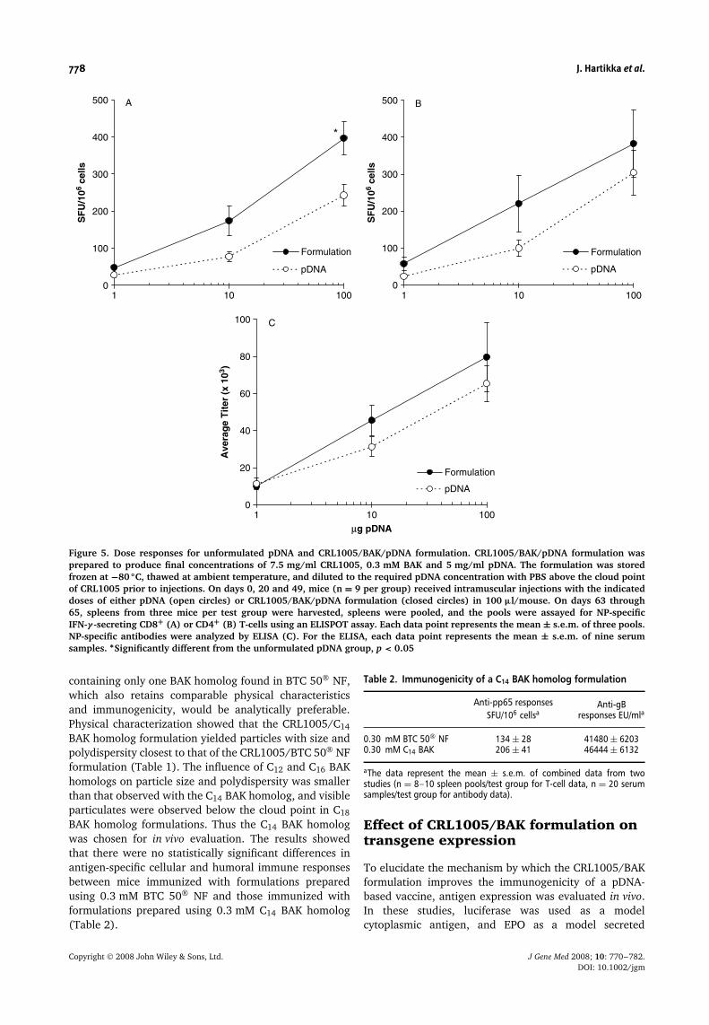
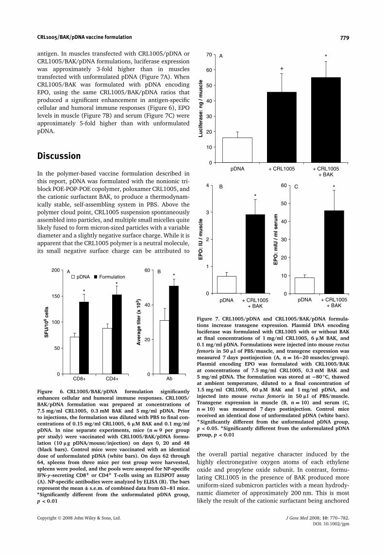
To assess the ability of BAK-containing formulations toenhance immune responses, the CRL1005/BAK/pDNAformulation was prepared at final concentrations of7.5 mg/ml, 0.3 mM and 5 mg/ml, respectively. CRL1005at 7.5 mg/ml was chosen since that concentration resultedin a significant increase in CD8+ T-cell (Figure 4A) andantibody (Figure 4C) responses. BAK at 0.3 mM concen-tration was chosen to ensure homogeneity of the for-mulations without visible precipitate (see first section of‘‘Results’’ above). The results from a dose response studysuggested that the CRL1005/BAK/pDNA formulationincreased immune responses compared to unformulatedpDNA (Figure 5). Due to animal-to-animal variability inimmune responses, however, the differences betweenunformulated pDNA and the CRL1005/BAK/pDNA for-mulation did not always reach statistical significance ina single study. When the CRL1005/BAK/pDNA formu-lation was tested in nine separate experiments using10 µg pDNA dose, the formulation significantly enhancedantigen-specific CD8+ and CD4+ T-cell responses by1.9- and 1.7-fold, respectively (Figure 6A, p < 0.01). Nosignificant enhancement in CD4+ T-cell responses waspreviously observed with CRL1005/pDNA formulations(Figure 4B), suggesting that BAK increased the immuno-genicity of the vaccine formulation. Antibody responsesin mice vaccinated with the CRL1005/BAK/pDNA formu-lation were 1.6-fold higher compared to mice vaccinatedwith unformulated pDNA (Figure 6B, p < 0.001).
To determine whether the particle size of CRL1005/BAK/pDNA formulations affects cellular or humoralimmune responses, formulations with a range of particlesizes (Z average diameter of 250, 350, 550, 1000, or>1000 nm) were prepared as described in the Materialsand methods section. When compared to the same dose ofunformulated pDNA, CRL1005/BAK/pDNA formulationsincreased antigen-specific cellular and humoral immuneresponses in mice by up to 1.8- and 2.0-fold, respectively.However, there were no statistically significant differencesamong the groups vaccinated with CRL1005/BAK/pDNAformulations with different particle sizes (data notshown).
Immunogenicity of a simplified BAKhomolog formulation
The current CRL1005/BAK-based CMV pDNA vaccineformulation used in a phase 2 clinical trial is amixture of poloxamer CRL1005 (7.5 mg/ml), cationicsurfactant BAK (0.3 mM) and two plasmids encodingCMV pp65 (VCL6368) and gB (VCL6365) antigens ata total pDNA concentration of 5 mg/ml in PBS [14].The BAK component of this formulation is a mixture offour alkyl chain homologs: C12 (50%), C14 (30%), C16
(17%), and C18 (3%). This BAK mixture is commerciallyavailable as BTC 50 NF. A simplified formulation
Copyright 2008 John Wiley & Sons, Ltd. J Gene Med 2008; 10: 770–782.DOI: 10.1002/jgm
778 J. Hartikka et al.
*
0
100
200
300
400
500
1 10 100
SF
U/1
06 ce
lls
Formulation
pDNA
A
0
100
200
300
400
500
1 10 100
SF
U/1
06 ce
lls
Formulation
pDNA
B
0
20
40
60
80
100
1 10 100
µg pDNA
Ave
rag
e T
iter
(x
103 )
Formulation
pDNA
C
Figure 5. Dose responses for unformulated pDNA and CRL1005/BAK/pDNA formulation. CRL1005/BAK/pDNA formulation wasprepared to produce final concentrations of 7.5 mg/ml CRL1005, 0.3 mM BAK and 5 mg/ml pDNA. The formulation was storedfrozen at −80 ◦C, thawed at ambient temperature, and diluted to the required pDNA concentration with PBS above the cloud pointof CRL1005 prior to injections. On days 0, 20 and 49, mice (n = 9 per group) received intramuscular injections with the indicateddoses of either pDNA (open circles) or CRL1005/BAK/pDNA formulation (closed circles) in 100 µl/mouse. On days 63 through65, spleens from three mice per test group were harvested, spleens were pooled, and the pools were assayed for NP-specificIFN-γ -secreting CD8+ (A) or CD4+ (B) T-cells using an ELISPOT assay. Each data point represents the mean ± s.e.m. of three pools.NP-specific antibodies were analyzed by ELISA (C). For the ELISA, each data point represents the mean ± s.e.m. of nine serumsamples. ∗Significantly different from the unformulated pDNA group, p < 0.05
containing only one BAK homolog found in BTC 50 NF,which also retains comparable physical characteristicsand immunogenicity, would be analytically preferable.Physical characterization showed that the CRL1005/C14
BAK homolog formulation yielded particles with size andpolydispersity closest to that of the CRL1005/BTC 50 NFformulation (Table 1). The influence of C12 and C16 BAKhomologs on particle size and polydispersity was smallerthan that observed with the C14 BAK homolog, and visibleparticulates were observed below the cloud point in C18
BAK homolog formulations. Thus the C14 BAK homologwas chosen for in vivo evaluation. The results showedthat there were no statistically significant differences inantigen-specific cellular and humoral immune responsesbetween mice immunized with formulations preparedusing 0.3 mM BTC 50 NF and those immunized withformulations prepared using 0.3 mM C14 BAK homolog(Table 2).
Table 2. Immunogenicity of a C14 BAK homolog formulation
Anti-pp65 responsesSFU/106 cellsa
Anti-gBresponses EU/mla
0.30 mM BTC 50 NF 134 ± 28 41480 ± 62030.30 mM C14 BAK 206 ± 41 46444 ± 6132
aThe data represent the mean ± s.e.m. of combined data from twostudies (n = 8–10 spleen pools/test group for T-cell data, n = 20 serumsamples/test group for antibody data).
Effect of CRL1005/BAK formulation ontransgene expression
To elucidate the mechanism by which the CRL1005/BAKformulation improves the immunogenicity of a pDNA-based vaccine, antigen expression was evaluated in vivo.In these studies, luciferase was used as a modelcytoplasmic antigen, and EPO as a model secreted
Copyright 2008 John Wiley & Sons, Ltd. J Gene Med 2008; 10: 770–782.DOI: 10.1002/jgm
CRL1005/BAK/pDNA vaccine formulation 779
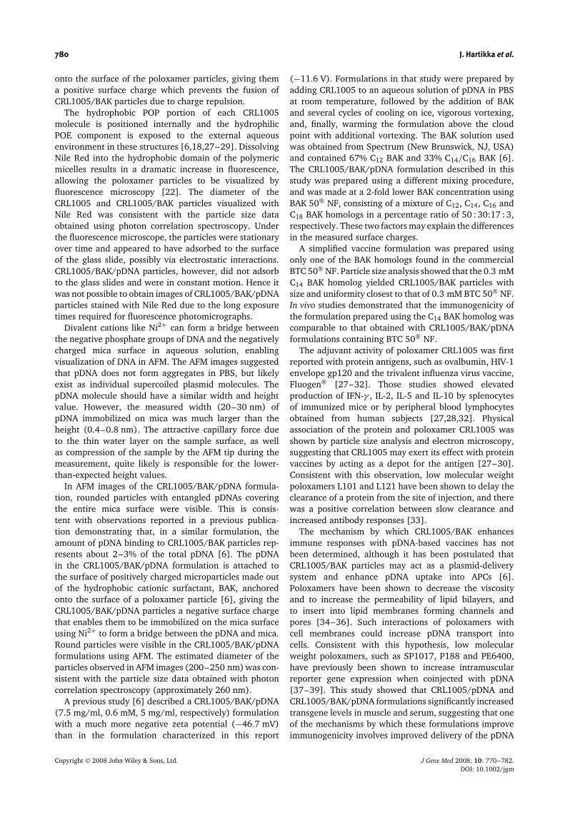
antigen. In muscles transfected with CRL1005/pDNA orCRL1005/BAK/pDNA formulations, luciferase expressionwas approximately 3-fold higher than in musclestransfected with unformulated pDNA (Figure 7A). WhenCRL1005/BAK was formulated with pDNA encodingEPO, using the same CRL1005/BAK/pDNA ratios thatproduced a significant enhancement in antigen-specificcellular and humoral immune responses (Figure 6), EPOlevels in muscle (Figure 7B) and serum (Figure 7C) wereapproximately 5-fold higher than with unformulatedpDNA.
Discussion
In the polymer-based vaccine formulation described inthis report, pDNA was formulated with the nonionic tri-block POE-POP-POE copolymer, poloxamer CRL1005, andthe cationic surfactant BAK, to produce a thermodynam-ically stable, self-assembling system in PBS. Above thepolymer cloud point, CRL1005 suspension spontaneouslyassembled into particles, and multiple small micelles quitelikely fused to form micron-sized particles with a variablediameter and a slightly negative surface charge. While it isapparent that the CRL1005 polymer is a neutral molecule,its small negative surface charge can be attributed to
**
0
50
100
150
200
CD8+ CD4+
SF
U/1
06 ce
lls
pDNA FormulationA
*
0
20
40
60
Ab
Ave
rag
e ti
ter
(x 1
03 )
B
Figure 6. CRL1005/BAK/pDNA formulation significantlyenhances cellular and humoral immune responses. CRL1005/BAK/pDNA formulation was prepared at concentrations of7.5 mg/ml CRL1005, 0.3 mM BAK and 5 mg/ml pDNA. Priorto injections, the formulation was diluted with PBS to final con-centrations of 0.15 mg/ml CRL1005, 6 µM BAK and 0.1 mg/mlpDNA. In nine separate experiments, mice (n = 9 per groupper study) were vaccinated with CRL1005/BAK/pDNA formu-lation (10 µg pDNA/mouse/injection) on days 0, 20 and 48(black bars). Control mice were vaccinated with an identicaldose of unformulated pDNA (white bars). On days 62 through64, spleens from three mice per test group were harvested,spleens were pooled, and the pools were assayed for NP-specificIFN-γ -secreting CD8+ or CD4+ T-cells using an ELISPOT assay(A). NP-specific antibodies were analyzed by ELISA (B). The barsrepresent the mean ± s.e.m. of combined data from 63–81 mice.∗Significantly different from the unformulated pDNA group,p < 0.01
+
*
0
10
20
30
40
50
60
70
pDNA + CRL1005 + CRL1005+ BAK
Lu
cife
rase
: n
g /
mu
scle
A
*
0
1
2
3
4
pDNA + CRL1005+ BAK
EP
O:
IU /
mu
scle
B *
0
10
20
30
40
50
60
pDNA + CRL1005+ BAK
EP
O:
mIU
/ m
l ser
um
C
Figure 7. CRL1005/pDNA and CRL1005/BAK/pDNA formula-tions increase transgene expression. Plasmid DNA encodingluciferase was formulated with CRL1005 with or without BAKat final concentrations of 1 mg/ml CRL1005, 6 µM BAK, and0.1 mg/ml pDNA. Formulations were injected into mouse rectusfemoris in 50 µl of PBS/muscle, and transgene expression wasmeasured 7 days postinjection (A, n = 16–20 muscles/group).Plasmid encoding EPO was formulated with CRL1005/BAKat concentrations of 7.5 mg/ml CRL1005, 0.3 mM BAK and5 mg/ml pDNA. The formulation was stored at −80 ◦C, thawedat ambient temperature, diluted to a final concentration of1.5 mg/ml CRL1005, 60 µM BAK and 1 mg/ml pDNA, andinjected into mouse rectus femoris in 50 µl of PBS/muscle.Transgene expression in muscle (B, n = 10) and serum (C,n = 10) was measured 7 days postinjection. Control micereceived an identical dose of unformulated pDNA (white bars).+Significantly different from the unformulated pDNA group,p < 0.05. ∗Significantly different from the unformulated pDNAgroup, p < 0.01
the overall partial negative character induced by thehighly electronegative oxygen atoms of each ethyleneoxide and propylene oxide subunit. In contrast, formu-lating CRL1005 in the presence of BAK produced moreuniform-sized submicron particles with a mean hydrody-namic diameter of approximately 200 nm. This is mostlikely the result of the cationic surfactant being anchored
Copyright 2008 John Wiley & Sons, Ltd. J Gene Med 2008; 10: 770–782.DOI: 10.1002/jgm
780 J. Hartikka et al.
onto the surface of the poloxamer particles, giving thema positive surface charge which prevents the fusion ofCRL1005/BAK particles due to charge repulsion.
The hydrophobic POP portion of each CRL1005molecule is positioned internally and the hydrophilicPOE component is exposed to the external aqueousenvironment in these structures [6,18,27–29]. DissolvingNile Red into the hydrophobic domain of the polymericmicelles results in a dramatic increase in fluorescence,allowing the poloxamer particles to be visualized byfluorescence microscopy [22]. The diameter of theCRL1005 and CRL1005/BAK particles visualized withNile Red was consistent with the particle size dataobtained using photon correlation spectroscopy. Underthe fluorescence microscope, the particles were stationaryover time and appeared to have adsorbed to the surfaceof the glass slide, possibly via electrostatic interactions.CRL1005/BAK/pDNA particles, however, did not adsorbto the glass slides and were in constant motion. Hence itwas not possible to obtain images of CRL1005/BAK/pDNAparticles stained with Nile Red due to the long exposuretimes required for fluorescence photomicrographs.
Divalent cations like Ni2+ can form a bridge betweenthe negative phosphate groups of DNA and the negativelycharged mica surface in aqueous solution, enablingvisualization of DNA in AFM. The AFM images suggestedthat pDNA does not form aggregates in PBS, but likelyexist as individual supercoiled plasmid molecules. ThepDNA molecule should have a similar width and heightvalue. However, the measured width (20–30 nm) ofpDNA immobilized on mica was much larger than theheight (0.4–0.8 nm). The attractive capillary force dueto the thin water layer on the sample surface, as wellas compression of the sample by the AFM tip during themeasurement, quite likely is responsible for the lower-than-expected height values.
In AFM images of the CRL1005/BAK/pDNA formula-tion, rounded particles with entangled pDNAs coveringthe entire mica surface were visible. This is consis-tent with observations reported in a previous publica-tion demonstrating that, in a similar formulation, theamount of pDNA binding to CRL1005/BAK particles rep-resents about 2–3% of the total pDNA [6]. The pDNAin the CRL1005/BAK/pDNA formulation is attached tothe surface of positively charged microparticles made outof the hydrophobic cationic surfactant, BAK, anchoredonto the surface of a poloxamer particle [6], giving theCRL1005/BAK/pDNA particles a negative surface chargethat enables them to be immobilized on the mica surfaceusing Ni2+ to form a bridge between the pDNA and mica.Round particles were visible in the CRL1005/BAK/pDNAformulations using AFM. The estimated diameter of theparticles observed in AFM images (200–250 nm) was con-sistent with the particle size data obtained with photoncorrelation spectroscopy (approximately 260 nm).
A previous study [6] described a CRL1005/BAK/pDNA(7.5 mg/ml, 0.6 mM, 5 mg/ml, respectively) formulationwith a much more negative zeta potential (−46.7 mV)than in the formulation characterized in this report
(−11.6 V). Formulations in that study were prepared byadding CRL1005 to an aqueous solution of pDNA in PBSat room temperature, followed by the addition of BAKand several cycles of cooling on ice, vigorous vortexing,and, finally, warming the formulation above the cloudpoint with additional vortexing. The BAK solution usedwas obtained from Spectrum (New Brunswick, NJ, USA)and contained 67% C12 BAK and 33% C14/C16 BAK [6].The CRL1005/BAK/pDNA formulation described in thisstudy was prepared using a different mixing procedure,and was made at a 2-fold lower BAK concentration usingBAK 50 NF, consisting of a mixture of C12, C14, C16 andC18 BAK homologs in a percentage ratio of 50 : 30:17 : 3,respectively. These two factors may explain the differencesin the measured surface charges.
A simplified vaccine formulation was prepared usingonly one of the BAK homologs found in the commercialBTC 50 NF. Particle size analysis showed that the 0.3 mMC14 BAK homolog yielded CRL1005/BAK particles withsize and uniformity closest to that of 0.3 mM BTC 50 NF.In vivo studies demonstrated that the immunogenicity ofthe formulation prepared using the C14 BAK homolog wascomparable to that obtained with CRL1005/BAK/pDNAformulations containing BTC 50 NF.
The adjuvant activity of poloxamer CRL1005 was firstreported with protein antigens, such as ovalbumin, HIV-1envelope gp120 and the trivalent influenza virus vaccine,Fluogen [27–32]. Those studies showed elevatedproduction of IFN-γ , IL-2, IL-5 and IL-10 by splenocytesof immunized mice or by peripheral blood lymphocytesobtained from human subjects [27,28,32]. Physicalassociation of the protein and poloxamer CRL1005 wasshown by particle size analysis and electron microscopy,suggesting that CRL1005 may exert its effect with proteinvaccines by acting as a depot for the antigen [27–30].Consistent with this observation, low molecular weightpoloxamers L101 and L121 have been shown to delay theclearance of a protein from the site of injection, and therewas a positive correlation between slow clearance andincreased antibody responses [33].
The mechanism by which CRL1005/BAK enhancesimmune responses with pDNA-based vaccines has notbeen determined, although it has been postulated thatCRL1005/BAK particles may act as a plasmid-deliverysystem and enhance pDNA uptake into APCs [6].Poloxamers have been shown to decrease the viscosityand to increase the permeability of lipid bilayers, andto insert into lipid membranes forming channels andpores [34–36]. Such interactions of poloxamers withcell membranes could increase pDNA transport intocells. Consistent with this hypothesis, low molecularweight poloxamers, such as SP1017, P188 and PE6400,have previously been shown to increase intramuscularreporter gene expression when coinjected with pDNA[37–39]. This study showed that CRL1005/pDNA andCRL1005/BAK/pDNA formulations significantly increasedtransgene levels in muscle and serum, suggesting that oneof the mechanisms by which these formulations improveimmunogenicity involves improved delivery of the pDNA
Copyright 2008 John Wiley & Sons, Ltd. J Gene Med 2008; 10: 770–782.DOI: 10.1002/jgm
CRL1005/BAK/pDNA vaccine formulation 781
vaccine. Preliminary data show that coinjection of pDNAwith CRL1005 increases expression of proinflammatorycytokines and chemokines in muscle, such as IL-1ß,IL-6, G-CSF, TNF-α, KC and MIP-2, suggesting thatthe CRL1005/BAK/pDNA formulation may also improveimmunogenicity by stimulating innate immunity (data notshown).
Particulate vaccine formulations, such as PLG/CTABmicroparticles, have been reported to enhance immuno-genicity by several mechanisms, including enhancedtransfection of phagocytic APCs with the antigen-encodingplasmid. Studies of PLG/CTAB/pDNA formulations haveshown a correlation between particle size and enhancedimmune responses. Particles with a diameter between300 nm and 3 µm were the most potent in enhancingimmune responses whereas particles with a diameteraround 10–30 µm were significantly less immunogenic,possibly because they were too big for efficient phagocy-tosis [4,9,10]. Varying the size of CRL1005/BAK/pDNAparticles from 250 nm to over 1000 nm had no significanteffect on the immunogenicity of the CRL1005/BAK-basedpDNA vaccine formulations in this study. Since theseformulations could not be prepared at particle sizes>10 µm, we were unable to evaluate whether largerCRL1005/BAK/pDNA particles are ineffective in enhanc-ing immune response as previously demonstrated forPLG/CTAB/pDNA formulations [4,10].
Poloxamers formulated with a pDNA vaccine, withoutBAK, have been shown to significantly increase cellularimmune responses in nonhuman primates [6,11–13].Furthermore, doses of CRL1005 adjuvant as high as75 mg injected intramuscularly have been shown to besafe and well tolerated in humans [27,32]. Studiesin rhesus macaques have demonstrated that addingBAK to the CRL1005/pDNA vaccine formulation furtherenhances T-cell responses [6,11]. In this report, we havecharacterized a CRL1005/BAK/pDNA vaccine formulationthat significantly enhances the levels of antigen-specificcellular and humoral immune responses, possibly byimproving the delivery of pDNA. This formulation iscurrently being evaluated for the prevention of CMV-associated disease in the allogeneic hematopoietic celltransplant population in a phase 2 clinical trial [14].
Acknowledgements
We thank Melissa Masters, Sylvia Salapski and Jane Morrow fortheir assistance with the animal studies, and Sean Sullivan forconstructive help with the manuscript.
References
1. Edelstein ML, Abedi MR, Wixon J, Edelstein RM. Gene therapyclinical trials worldwide 1989-2004-an overview. J Gene Med2004; 6: 597–602.
2. Lorenzen N, LaPatra SE. DNA vaccines for aquacultured fish.Rev Sci Technol 2005; 24: 201–213.
3. Manthorpe M, Hobart P, Hermanson G, et al. Plasmid vaccinesand therapeutics: from design to applications. Adv BiochemEngin/Biotechnol 2005; 99: 41–92.
4. O’Hagan DT, Singh M, Ulmer JB. Microparticles for the deliveryof DNA vaccines. Immunol Rev 2004; 199: 191–200.
5. Shams H. Recent developments in veterinary vaccinology. Vet J2005; 170: 289–299.
6. Evans RK, Zhu DM, Casimiro DR, et al. Characterization andbiological evaluation of a microparticle adjuvant formulationfor plasmid DNA vaccines. J Pharm Sci 2004; 93: 1924–1939.
7. Selinsky C, Luke C, Wloch M, et al. A DNA-based vaccine for theprevention of human cytomegalovirus-associated diseases. HumVaccine 2005; 1: 16–23.
8. Denis-Mize KS, Dupuis M, MacKichan ML, et al. Plasmid DNAadsorbed onto cationic microparticles mediates target geneexpression and antigen presentation by dendritic cells. GeneTher 2000; 7: 2105–2112.
9. Denis-Mize KS, Dupuis M, Singh M, et al. Mechanisms ofincreased immunogenicity for DNA-based vaccines adsorbedonto cationic microparticles. Cell Immunol 2003; 225: 12–20.
10. Singh M, Briones M, Ott G, O’Hagan D. Cationic microparticles:a potent delivery system for DNA vaccines. Proc Natl Acad SciU S A 2000; 97: 811–816.
11. Shiver JW, Fu TM, Chen L, et al. Replication-incompetent ade-noviral vaccine vector elicits effective anti-immunodeficiency-virus immunity. Nature 2002; 415: 331–335.
12. Casimiro DR, Chen L, Fu TM, et al. Comparative immunogenic-ity in rhesus monkeys of DNA plasmid, recombinant vacciniavirus, and replication-defective adenovirus vectors expressing ahuman immunodeficiency virus type 1 gag gene. J Virol 2003;77: 6305–6313.
13. Caulfield MJ, Wang S, Smith JG, et al. Sustained peptide-specific gamma interferon T-cell response in rhesus macaquesimmunized with human immunodeficiency virus gag DNAvaccines. J Virol 2002; 76: 10038–10043.
14. Wloch MK, Smith LR, Boutsaboualoy S, et al. Safety andimmunogenicity of a bivalent CMV DNA vaccine in healthyadult subjects. J Infect Dis 2008; in press.
15. Alexandridis P, Hatton TA. Poly(ethylene oxide)-poly(propyleneoxide)-poly(ethylene oxide) block copolymer surfactants inaqueous solutions and at interfaces: thermodynamics, structure,dynamics, and modeling. Colloids and Surfaces A: PhysicochemEng Aspects 1995; 96: 1–46.
16. Cardodo da Silva R, Loh W. Effect of additives on the cloudpoints of aqueous solutions of ethylene oxide-propylene oxide-ethylene oxide block copolymers. J Colloid Interface Sci 1998;202: 385–390.
17. Chu B. Structure and dynamics of block copolymer colloids.Langmuir 1995; 11: 414–421.
18. Todd CW, Balusubramanian M, Newman MJ. Development ofadjuvant-active nonionic block copolymers. Adv Drug Deliv Rev1998; 32: 199–223.
19. Hartikka J, Bozoukova V, Ferrari M, et al. Vaxfectin enhancesthe humoral immune response to plasmid DNA-encodedantigens. Vaccine 2001; 19: 1911–1923.
20. Sankar V, Baccaglini L, Sawdey M, et al. Salivary gland deliveryof pDNA-cationic lipoplexes elicits systemic immune responses.Oral Dis 2002; 8: 275–281.
21. Hartikka J, Bozoukova V, Jones D, et al. Sodium phosphateenhances plasmid DNA expression in vivo. Gene Ther 2000;7: 1171–1182.
22. Gocze PM, Freeman DA. Factors underlying the variabilityof lipid droplet fluorescence in MA-10 Leydig tumor cells.Cytometry 1994; 17: 151–158.
23. Available: http://www.aphis.usda.gov/ac/publications.html.24. Sasaki S, Amara RR, Yeow WS, Pitha PM, Robinson HL.
Regulation of DNA-raised immune responses by cotransfectedinterferon regulatory factors. J Virol 2002; 76: 6652–6659.
25. Brett SJ, Gao XM, Liew FY, Tite JP. Selection of the same major Tcell determinants of influenza nucleoprotein after vaccination orexposure to infectious virus. J Immunol 1991; 147: 1647–1652.
26. Gao XM, Liew FY, Tite JP. Identification and characterization ofT helper epitopes in the nucleoprotein of influenza A virus. JImmunol 1989; 143: 3007–3014.
27. Newman MJ, Actor JK, Balusubramanian M, Jagannath C. Useof nonionic block copolymers in vaccines and therapeutics. CritRev Ther Drug Carrier Syst 1998; 15: 89–142.
28. Newman MJ, Todd CW, Balusubramanian M. Design anddevelopment of adjuvant-active nonionic block copolymers. JPharm Sci 1998; 87: 1357–1362.
Copyright 2008 John Wiley & Sons, Ltd. J Gene Med 2008; 10: 770–782.DOI: 10.1002/jgm
782 J. Hartikka et al.
29. Todd CW, Pozzi LA, Guarnaccia JR, et al. Development of anadjuvant-active nonionic block copolymer for use in oil-freesubunit vaccines formulations. Vaccine 1997; 15: 564–570.
30. McNicholl JM, Bond KB, Ruhadze ER, Olsen MR, Takayama K,Hunter RL. Enhancement of HIV type 1 vaccine immunogenicityby block copolymer adjuvants. I. Induction of high-titer, long-lasting, cross-reactive antibodies of broad isotype. AIDS Res HumRetroviruses 1998; 14: 1457–1471.
31. Newman MJ, Todd CW, Lee EM, Balusubramanian M, Didier PJ,Katz JM. Increasing the immunogenicity of a trivalent influenzavirus vaccine with adjuvant-active nonionic block copolymersfor potential use in the elderly. Mech Ageing Dev 1997; 93:189–203.
32. Triozzi PL, Stevens VC, Aldrich W, Powell J, Todd CW,Newman MJ. Effects of a beta-human chorionic gonadotropinsubunit immunogen administered in aqueous solution witha novel nonionic block copolymer adjuvant in patients withadvanced cancer. Clin Cancer Res 1997; 3: 2355–2362.
33. Hunter R, Strickland F, Kezdy F. The adjuvant activity ofnonionic block polymer surfactants. I. The role of hydrophile-lipophile balance. J Immunol 1981; 127: 1244–1250.
34. Krylova OO, Pohl P. Ionophoric activity of pluronic blockcopolymers. Biochemistry 2004; 43: 3696–3703.
35. Zhirnov AE, Demina TV, Krylova OO, Grozdova ID, Melik-Nubarov NS. Lipid composition determines interaction ofliposome membranes with Pluronic L61. Biochim Biophys Acta2005; 1720: 73–83.
36. Gau-Racine J, Lal J, Zeghal M, Auvray L. PEO-PPO blockcopolymer vectors do not interact directly with DNA but withlipid membranes. J Phys Chem B 2007; 111: 9900–9907.
37. Lemieux P, Guerin N, Paradis G, et al. A combination ofpoloxamers increases gene expression of plasmid DNA in skeletalmuscle. Gene Ther 2000; 7: 986–991.
38. Hartikka J, Sukhu L, Buchner C, et al. Electroporation-facilitateddelivery of plasmid DNA in skeletal muscle: plasmid dependenceof muscle damage and effect of poloxamer 188. Mol Ther 2001;4: 407–415.
39. Pitard B, Pollard H, Agbulut O, et al. A nonionic amphiphileagent promotes gene delivery in vivo to skeletal and cardiacmuscles. Hum Gene Ther 2002; 13: 1767–1775.
Copyright 2008 John Wiley & Sons, Ltd. J Gene Med 2008; 10: 770–782.DOI: 10.1002/jgm
Copyright © 2022 FDOKUMEN