Multi-path quenchers: efficient quenching of common fluorophores

AQUATIC BIOLOGYAquat Biol
Vol. 7: 143–152, 2009doi: 10.3354/ab00164
Published online October 22, 2009
INTRODUCTION
Seagrass meadows dominate many shallow estuariesand marine embayments, yet at a global scale sea-grasses are in decline (Orth et al. 2006). While thisdecline has been largely attributed to human activitythrough increased light attenuation caused largely byhigher sediment loads and algal densities, other factorssuch as eutrophication, heavy metals and petrochemi-cal pollution have a significant impact on seagrass sur-vival (Orth et al. 2006, Ralph et al. 2007). The capacityfor a seagrass species to conduct photosynthesis such
that the net carbon balance is positive is vital for thesurvival and resilience of a meadow. Thus consider-able research effort over the last half-century hasmaintained a focus on seagrass growth and photosyn-thesis (e.g. Larkum 1976, Lee et al. 2007).
The seagrass Halophila stipulacea is an integralcomponent of the the Gulf of Aqaba coral reef ecosys-tem in the Red Sea (Edwards & Head 1987), where itcan be found in extensive monospecific beds. H. stipu-lacea has a generally wide ecological range, growingfrom the intertidal to depths >50 to 70 m (Hulings1979, Lipkin 1979, Beer & Waisel 1982). This is in con-
© Inter-Research 2009 · www.int-res.com*Email: [email protected]
Photosynthetic responses of Halophila stipulaceato a light gradient. I. In situ energy partitioning of
non-photochemical quenching
John W. Runcie1,*, Diogo Paulo2, Rui Santos2, Yoni Sharon3, 4, Sven Beer4, João Silva2
1School of Biological Sciences, University of Sydney, New South Wales 2006, Australia 2ALGAE-Marine Plant Ecology Research Group, Center of Marine Sciences (CCMAR), Universidade do Algarve,
Campus de Gambelas, 8005-139 Faro, Portugal3The Interuniversity Institute for Marine Sciences, POB 469, Eilat 88103, Israel
4Department of Plant Sciences, Tel Aviv University, Tel Aviv 60078, Israel
ABSTRACT: The quantum yield of photosystem II (φII, also termed ΔF/Fm’ or Fv/Fm in light- or dark-acclimated plants, respectively) of the tropical seagrass Halophila stipulacea was measured in situusing modulated fluorescence techniques over diel periods at a range of depths. Photosynthetic elec-tron transport rates (ETRs), as derived from φII values at specific ambient photosynthetically availableradiation (PAR) irradiances, increased in direct proportion to increasing irradiance in the morningand, at shallow sites (7 to 10 m), reached saturating rates and then declined in the afternoon withlower PAR-specific ETRs. On the other hand, plants at 32 to 33 m showed no saturation even at mid-day, and the percentage reduction in PAR-specific afternoon ETRs was less than that of the shallowerplants. The use of an automated shutter in the measuring device enabled non-photochemicalquenching due to down-regulation and basal intrinsic non-radiative decay to be distinguished. Whilemidday values of down-regulation were lower in deeper water, basal intrinsic non-radioactive decayremained fairly constant at 30 to 40% at all depths, with more variation in shallow waters. The maxi-mal φII (i.e. Fv/Fm) reached similar values at midnight regardless of depth. H. stipulacea acclimates tothe widely varying irradiances across this depth gradient by regularly modulating down-regulation-based non-photochemical quenching processes, while dissipating a large proportion of light energythrough intrinsic decay regardless of depth.
KEY WORDS: Depth gradient · Halophila stipulacea · Irradiance · Nonphotochemical quenching ·PE curves · Photosynthesis · Pigments
Resale or republication not permitted without written consent of the publisher
Contribution to the Theme Section ‘Primary production in seagrasses and macroalgae’ OPENPEN ACCESSCCESS

Aquat Biol 7: 143–152, 2009
trast with the seagrass Halodule uninervis that coexistsin shallow waters in the northern Red Sea, Gulf ofAqaba, but does not extend as deeply (Beer & Waisel1982). Mechanisms that enable the survival of H. stip-ulacea in shallow and deep waters may include aninsensitivity to pressure due to its lack of substantialgas lacunae (Beer & Waisel 1982) and its capacityfor chloroplast clumping (Schwarz & Hellblom 2002,Sharon & Beer 2008).
Since the advent of submersible modulated fluoro-meters in the 1990s, chlorophyll fluorescence techniqueshave been widely used in the examination of in situseagrass photophysiology (e.g. Beer et al. 1998, 2000),and the effects of varying irradiance with increasingturbidity or depth have been examined in some detail(Lee et al. 2007). However, most in situ fluorescencestudies have been constrained by the necessity fordivers to make the measurements, limiting studies to acombination of maximum depth, frequency and dura-tion of measurements at these depths. Fewer studieshave taken advantage of logging functions available incommercially available instruments (e.g. Winters etal. 2003, Schwarz et al. 2003); other studies have devel-oped custom devices for continuous measurementsover time of multiple replicate samples (Runcie &Riddle 2004, 2006, Runcie & Durako 2004).
The quantum yield (φ) of a light-dependent processis defined as the ratio of the rate of that process to therate of photon absorption. In unstressed green plantsand algae, the light-dependent process of photo-chemical energy conversion uses approximately 83%of light absorbed, thus the quantum yield of photosyn-thesis is 0.83. Modulated fluorescence techniques areparticularly useful for measuring the quantum yield ofphotochemical energy conversion in photosystem II(PSII), φII, and by difference, the quantum yield of non-photochemical energy conversion in PSII, φNPQ.
Diel changes in φII describe how a phototrophresponds to irradiance and can be used with othermeasurements to estimate net carbon gain (Beer et al.2000, Longstaff et al. 2002). However, irradianceenergy absorbed by PSII that cannot be directed tophotochemistry is (by definition) directed to severalclasses of non-photochemical quenching (NPQ) pro-cesses. Of these, the generation of singlet oxygen cancause intensive damage to internal structures if theabsorbed energy is not effectively dissipated throughother NPQ processes (Müller et al. 2001).
Kornyeyev & Holaday (2008) separated NPQ into 4components: (1) fluorescence is generally a very smallproportion of NPQ (a few percent at most) and isgenerally disregarded; (2) regulated, dark-reversibledissipation is generally activated in response to irra-diance stress; (3) constitutive thermal dissipation canbe regarded as the (assumed) invariant ~27% of light
directed to non-photochemical processes when ab-sorbed by non-stressed phototrophs; and (4) inducedthermal dissipation is associated with PSII photo-inactivation and is sometimes termed photoinhibition.Kramer et al. (2004) pooled the 2 thermal dissipationcomponents together and termed them basal intrinsicnon-radiative decay. For simplicity we adopt this latterterm to describe thermal dissipation.
As part of the recent Group for Aquatic PrimaryProductivity (GAP) workshop in Eilat (April 2008, seewww.gap-aquatic.org/), we studied various photo-physiological aspects of the seagrass Halophila stipu-lacea, in situ. In the present study, we determined howirradiance energy absorbed by PSII is allocated tophotochemistry versus non-photochemical processes inplants exposed to very different irradiances along awide depth gradient. By determining diel changes inelectron transport rates at various depths (8 to 33 m),we aimed to distinguish differential responses in termsof photochemical and non-photochemical use of thecontrasting ambient photosynthetically available radi-ation (PAR) irradiances at each depth. The specificobjectives of the present study were to determine howexcitation energy is allocated within H. stipulacea tophotochemistry, down-regulation and basal intrinsicnon-radiative decay processes when exposed to differ-ent irradiances (at various depths), and how the alloca-tion to these processes varies during the onset of irra-diance stress and recovery. A parallel study was alsoconducted to examine acclimatory responses of H. stip-ulacea transplanted across the 3 depths (Sharon et al.2009, this Theme Section).
MATERIALS AND METHODS
Study sites and seagrass species. Halophila stipu-lacea (Forsskål) Ascherson was examined in situ at 8,18 and 33 m depth just south of the Inter-UniversityInstitute (IUI) in Eilat, the Gulf of Aqaba, northern RedSea (29° 30’ N, 34°55’E) during April 2008. Automatedchlorophyll fluorescence measurements were set up ateach depth using SCUBA. Individual leaves separatedfrom each other by ≥10 cm were examined. Selectedsamples were taken to the laboratory for leaf ab-sorbance determinations.
In situ irradiance and Kd measurements. Irradiancewas measured in situ during fluorometer deploymentsusing cosine-corrected irradiance sensors incorporatedin the custom fluorometers, and is termed incident irra-diance or Ii. Data was compared with measurements inW m–2 obtained at the nearby research station and con-verted to µmol photons m–2 s–1 by multiplying by 4.6(McCree 1981). Occasional in-air measurements anddepth profiles using a LiCor 190SA quantum sensor
144

Runcie et al.: Depth-dependent seagrass photosynthesis
were also taken from a boat in the vicinity of the studysite; those in-air irradiance values were similar to val-ues measured at the research station. All light sensorswere calibrated against the LiCor 190SA quantum sen-sor. The light attenuation coefficient Kd was calculatedfrom depth profile data.
Absorbance measurements. The absorption factor(AF) of 12 different leaves was assessed and averagedat each depth (8, 18 and 33 m) throughout the day(06:00, 10:00, 12:30, 16:00 and 18:00 h) by determiningthe reduction of ambient irradiance (or artificial irradi-ance at dawn and dusk) caused by a single leaf placedover the light sensor of a Diving-PAM fluorometer(Walz, Effeltrich). This reduction in irradiance was con-verted to a proportion. For example, a 50% reduction inirradiance caused by placing a leaf over the light sensorcorresponded to an AF value for that leaf of 0.5 (Sharon& Beer 2008). It was assumed that absorbance by photo-synthetic pigments contributed 96% of the overallabsorbance in Halophila stipulacea (as found earlier,Sharon & Beer 2008); the absorbance of other com-pounds was therefore ignored. The measured AF val-ues were regressed against time separately for morningand afternoon measurements and thus 2 linear relation-ships were determined for each depth. The trajectory ofthese regression lines indicated the direction and rateof change in absorbance throughout the day (a proxyfor chloroplast clumping, Sharon & Beer 2008), andenabled the estimation of AF by interpolation at eachdepth for each fluorescence measurement.
Twelve absorbance values were obtained at eachdepth at (approximately) each time; differences be-tween mean AF values at each depth at dawn anddusk were compared using ANOVA after testing forhomogeneity of variances.
In situ modulated fluorescence measurements. Twodesigns of custom submersible fluorometer (Aquation,Australia) were used that excite chlorophyll a fluo-rescence with a modulated blue light (470 nm light-emitting diode, LED) before and during a 0.8 s blue-enriched white saturating pulse provided by a 10 WLED (Luxeon, Lumileds Lighting). A far-red LED(735 nm) incorporated into the fluorometers was acti-vated for 5 s prior to saturating pulse measurements tofacilitate the opening of PSII centres and hence deter-mine Fo' (i.e. minimal fluorescence intensity with all PSII reaction centers open in a light-acclimated state).The sensor head of one fluorometer design closed overthe sample when taking a measurement, thus placingthe sample in darkness (Fig. 1). The sensor head couldthen be left in place for a predetermined interval,thereby dark-acclimating the sample. Fluorescencemeasurements made immediately prior to closing ofthe automated shutter provided background fluores-cence values which could then be subtracted from sub-
sequent values obtained from the seagrass sampleswhen the sensor head was in a closed position. Theother fluorometer design simply positioned the sensingpart of the fluorometer close to the sample which washeld in a clear acrylic sample holder; samples wereexposed to ambient irradiance throughout the courseof measurements. Each fluorometer was connected bycable to an independent submersible data-logger andpower supply; previous experiments (authors’ unpubl.data) demonstrated that the system can operate for atleast 2 d on a single charge. Several fluorometers ofboth designs were deployed at each site and a singleseagrass blade was held in the sample holder of thefluorometer with an upwards orientation; the irradi-ance sensor incorporated into each fluorometer like-wise faced upwards. Care was taken during thedeployments to minimise any disturbance of the sea-grasses. The fluorometers were left in position for atleast 24 h. While the second design of fluorometer wasprogrammed simply to measure ΔF/Fm’ every 15 min,the dark-acclimating shutter fluorometers were addi-tionally programmed to remain closed for a 15 mininterval once every 2 h, thereby also allowing 12 mea-surements of Fv/Fm during a 24 h interval. Examinationof the φII data directly after the Fv/Fm measurementssuggested no apparent effect of the 15 min dark treat-ment on subsequent φII measurements, which wouldhave manifested as an increase in φII values directlyafter each Fv/Fm measurement.
Diel photosynthesis–irradiance curves. ETRs werecalculated according to Genty et al. (1989) as the prod-uct of ambient irradiance, φII, AF at the time of mea-surement and 0.5 to account for electron sharingbetween photosystem I (PSI) and PSII. Diel photosyn-thesis–irradiance (P-I) curves (Longstaff et al. 2002),(where ‘I ’ represents non-wavelength-specific irradi-
145
Fig. 1. Custom fluorometer holding a Halophila stipulacealeaf with automated shutter (left) and battery data/acquisition
unit (right)

Aquat Biol 7: 143–152, 2009146
ance) were plotted using absorbed irradiance (Ia,Saroussi & Beer 2007) for the abscissa; Ia was calcu-lated as the product of incident irradiance (Ii), AF and0.5, which describes the proportion of exciton energyshared between PSI and PSII. P-I curves were gener-ated using data acquired over 24 h intervals. The expo-nential model of Webb et al. (1974) was used to derivevalues for ETRmax and the photon to electron conver-sion efficiency (α) using non-linear least squares min-imisation techniques and Sigmaplot software (JandelScientific). The saturation irradiance, Ik, was calcu-lated from ETRmax and α by extrapolating a linear P-Icurve with slope α to ETRmax, and errors associatedwith Ik were determined by propagation from ETRmax
and α. Non-saturating curves generated from the deeperplants were analysed using the Webb model, with ini-tial parameter seed values obtained from the saturat-ing curves of shallow plants. Thus values of ETRmax
were extrapolated from the non-saturating data ob-tained in the field.
As the above curves were calculated using datapooled from both the morning and afternoon, esti-mates of ETRmax necessarily could not reflect the ob-served maximum ETR at noon. Therefore, observedETRmax and Ik calculated using observed ETRmax werealso presented. Values of α were assumed to be similarfor both calculated and observed curves.
Fluorescence parameters and quenching analysis.Fluorescence parameters follow the notation of VanKooten & Snel (1990). F or Ft denote steady state fluo-rescence under ambient irradiance. Fm’ and Fm aremaximal fluorescence obtained during a 0.8 s period ofsaturating light for samples exposed to ambient light ordarkness, respectively. Fo and Fo’ represent minimalfluorescence of (respectively) dark-acclimated samples,or light-acclimated samples measured after a briefdark exposure after the shutter had closed. Kramer etal. (2004) described the analytical techniques thatenabled the examination of the allocation of excitationenergy to photochemistry (φII) and 2 competing non-photochemical pathways (downregulation, φNPQ) andother energy losses (basal intrinsic nonradiative decayin the present study, φNO).
NPQ = (Fm – Fm’)/Fm’qP = (Fm’ – Ft)/(Fm’ – Fo’)φNO = 1/{NPQ + 1 + qP[(Fm/Fo) – 1]}1 = φNO + φNPQ + φII
qcu = (Fm’ – Ft)/[(Ft – Fo’) + Fm’ – Fo’]
where qP represents photochemical quenching, andqcu represents the proportion of open reaction centres(Kramer et al. 2004), assuming a value of 0.5 as theexciton transfer probability parameter, and assumingan intermediate model for reaction centre connectivity(Lavergne & Trissl 1995).
RESULTS
Absorbance
AF values of Halophila stipulacea at 8 m declinedsignificantly from ~0.60 to a midday minimum of 0.34,rising again in the afternoon to values similar to,but slightly lower than, predawn values (Fig. 2). Weascribe this difference in AF to the chloroplast clump-ing phenomenon as described by Sharon & Beer(2008). In contrast, AF values of the deeper growingplants only declined slightly during the day, with mar-ginally greater predawn and dusk values compared tothe 8 m plants.
Time (h)
06:00 09:00 12:00 15:00 18:00
Ab
so
rbance
0.0
0.1
0.2
0.3
0.4
0.5
0.6
0.7
33 m
18 m
8 m
Fig. 2. Halophila stipulacea. Relative absorbance of leaves at8, 18 and 33 m depth measured from dawn to dusk. Values
are mean ± SD of 12 replicate measurements
Depth (m)8 18 33
Ab
so
rbance v
elo
city (A
F h
–1)
0.00
0.01
0.02
0.03
0.04
Morning
Afternoon
Fig. 3. Halophila stipulacea. Rate of change of absorption fac-tor (AF) at 8, 18 and 33 m before and after midday. Valuespresented are absolute; morning values are negative and
afternoon values are positive

Runcie et al.: Depth-dependent seagrass photosynthesis 147
At each depth, the absolute value of the rate of declinein AF during the morning was similar to that during theafternoon (Fig. 3). The magnitude was greatest for shal-low seagrasses and least for the deepest seagrasses, in-dicative of a more marked response in shallow water.Data describing AF acquired at dawn satisfied the as-sumptions of homogeneity, and AF at 8 m (0.558 ± 0.03;all reported values are mean ± SD) was significantly lessthan values at the deeper sites which did not differ sig-nificantly (33 m: 0.604 ± 0.05; 18 m: 0.632 ± 0.03; F = 14.2,p > 0.001). AF measured at dusk was significantly differ-ent at the 3 sites (8 m: 0.509 ± 0.03; 18 m: 0.642 ± 0.04;33 m: 0.598 ± 0.06; F = 26.6, p > 0.000).
Morning and afternoon fluctuation in φφ II, Fv/Fm
and ETR
Minimum midday values of φII at 8, 18 and 33 mdropped to less than 0.1, 0.2 and 0.3, respectively,
reflecting the decline in midday irradiance with depth(Figs. 4–6). After sunset, φII of plants at all depthsincreased rapidly to values around 0.6, followed by aslow gradual increase throughout the night. Fv/Fm, asmeasured after in situ dark-acclimation for 15 min,declined during the day with declining φII and recov-ered rapidly at dusk assuming the same values as φII
during the night. Fv/Fm continued to rise slightly afterdawn while φII commenced its decline
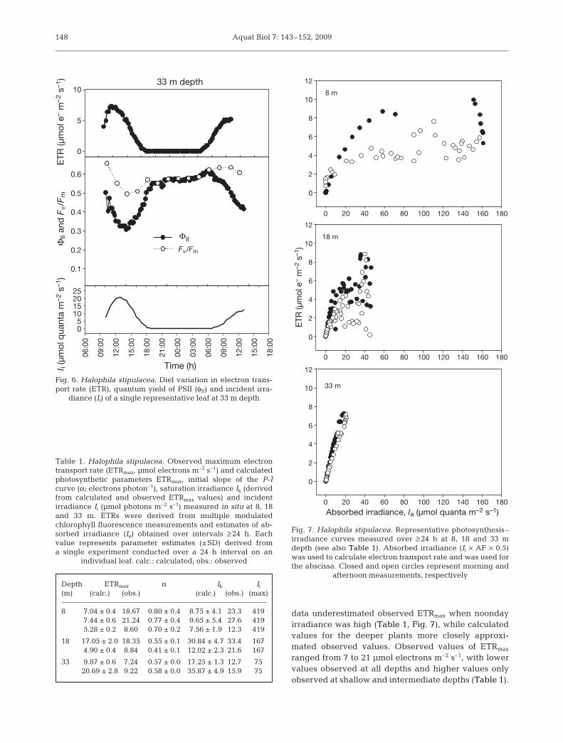
ETRs tended to increase rapidly during the morningand decline more slowly in the afternoon. This can beseen more clearly in Fig. 7 where morning values ofETRs for shallow plants are far greater than afternoonvalues for the same absorbed irradiance. The reduceddifferences between morning and afternoon ETRs ofdeeper plants is largely a consequence of the reducedirradiance, as can be seen when comparing ETRswhere Ia is ~20 to 160 µmol photons m–2 s–1 for ex-periments conducted at the 3 depths (Fig. 7). Clearly,ETRmax calculated from pooled morning and afternoon
0.1
0.2
0.3
0.4
0.5
0.6
8 m depth
0
5
10
15
20
Time (h)
12:0
0
15:0
0
18
:00
21
:00
00
:00
03
:00
06
:00
09
:00
12
:00
15:0
0
18
:00
21
:00
0100200300400
ΦII a
nd
Fv/F
m
ΦII
Fv/Fm
ET
R (µ
mo
l e
– m
–2 s
–1)
I i (µ
mo
l quanta
m–2 s
–1)
Fig. 4. Halophila stipulacea. Diel variation in electron trans-port rate (ETR), quantum yield of PSII (φII) and incident irra-
diance (Ii) of a single representative leaf at 8 m depth
0.2
0.4
0.6
18 m depth
0
5
10
15
20 06
:00
09
:00
12:0
0
15:0
0
18
:00
21
:00
00
:00
03
:00
06
:00
09:0
0
12
:00
15
:00
18
:00
21:0
0
00
:00
03:0
0
06
:00
09
:00
12
:00
0
50
100
150
Time (h)
ΦII a
nd
Fv/F
mE
TR
(µ
mo
l e
– m
–2 s
–1)
I i (µ
mo
l q
uan
ta m
–2 s
–1)
ΦII
Fv/Fm
Fig. 5. Halophila stipulacea. Diel variation in electron trans-port rate (ETR), quantum yield of PSII (φII) and incident irra-
diance (Ii) of a single representative leaf at 18 m depth
Aquat Biol 7: 143–152, 2009
data underestimated observed ETRmax when noondayirradiance was high (Table 1, Fig. 7), while calculatedvalues for the deeper plants more closely approxi-mated observed values. Observed values of ETRmax
ranged from 7 to 21 µmol electrons m–2 s–1, with lowervalues observed at all depths and higher values onlyobserved at shallow and intermediate depths (Table 1).
148
0.1
0.2
0.3
0.4
0.5
0.6
33 m depth
0
5
10 06:0
0
09:0
0
12:0
0
15:0
0
18:0
0
21:0
0
00:0
0
03:0
0
06:0
0
09:0
0
12:0
0
15:0
0
18:0
0
05
10152025
ΦII
Fv/Fm
Time (h)
ΦII a
nd
Fv/F
mE
TR
(µ
mo
l e
– m
–2 s
–1)
I i (µ
mo
l q
uan
ta m
–2 s
–1)
Fig. 6. Halophila stipulacea. Diel variation in electron trans-port rate (ETR), quantum yield of PSII (φII) and incident irra-
diance (Ii) of a single representative leaf at 33 m depth
8 m
0
2
4
6
8
10
12
0
2
4
6
8
10
12
0 20 40 60 80 100 120 140 160 180
0 20 40 60 80 100 120 140 160 180
0 20 40 60 80 100 120 140 160 180
0
2
4
6
8
10
12
18 m
33 m
Absorbed irradiance, Ia (µmol quanta m–2 s–1)
ET
R (µ
mo
l e– m
–2 s
–1)
Fig. 7. Halophila stipulacea. Representative photosynthesis–irradiance curves measured over ≥ 24 h at 8, 18 and 33 mdepth (see also Table 1). Absorbed irradiance (Ii × AF × 0.5)was used to calculate electron transport rate and was used forthe abscissa. Closed and open circles represent morning and
afternoon measurements, respectively
Table 1. Halophila stipulacea. Observed maximum electrontransport rate (ETRmax, µmol electrons m–2 s–1) and calculatedphotosynthetic parameters ETRmax, initial slope of the P-Icurve (α; electrons photon–1), saturation irradiance Ik (derivedfrom calculated and observed ETRmax values) and incidentirradiance Ii (µmol photons m–2 s–1) measured in situ at 8, 18and 33 m. ETRs were derived from multiple modulatedchlorophyll fluorescence measurements and estimates of ab-sorbed irradiance (Ia) obtained over intervals ≥ 24 h. Eachvalue represents parameter estimates (±SD) derived froma single experiment conducted over a 24 h interval on an
individual leaf. calc.: calculated; obs.: observed
Depth ETRmax α Ik Ii
(m) (calc.) (obs.) (calc.) (obs.) (max)
8 7.04 ± 0.4 18.67 0.80 ± 0.4 8.75 ± 4.1 23.3 4197.44 ± 0.6 21.24 0.77 ± 0.4 9.65 ± 5.4 27.6 4195.28 ± 0.2 8.60 0.70 ± 0.2 7.56 ± 1.9 12.3 419
18 17.05 ± 2.0 18.35 0.55 ± 0.1 30.84 ± 4.7 33.4 1674.90 ± 0.4 8.84 0.41 ± 0.1 12.02 ± 2.3 21.6 167
33 9.87 ± 0.6 7.24 0.57 ± 0.0 17.25 ± 1.3 12.7 7520.69 ± 2.8 9.22 0.58 ± 0.0 35.87 ± 4.9 15.9 75

Runcie et al.: Depth-dependent seagrass photosynthesis
The daily mean initial slope of the P-I curves, α,ranged from 0.41 to 0.80, with greater values at theshallow site. While calculated values of saturationirradiance (Ik) were higher for deeper plants, valuesderived from observed ETRmax values showed no obvi-ous differences between depths and ranged between12 and 33 µmol photons m–2 s–1.
Non-photochemical quantum yields
At 8 m depth, φNPQ declined from midday values ofaround 0.6 to near zero at night (Fig. 8). At 18 m depth,φNPQ similarly dropped to near zero at night, but wasgenerally around 0.4 at midday, except for an (presum-ably erroneous) anomaly where φNPQ reached its maxi-mal value of 1 at midday (Fig. 9) and φNO was underes-timated at <0. At 33 m depth, φNPQ only rose to about0.3 at midday, also dropping to zero at night. φNO wassteady at about 0.4 in plants at all depths during thenight. At 8 m depth, this value declined with lightonset, and then increased at midday to about 0.5(Fig. 8). A similar pattern could be seen at 18 m wherea strong midday decline was evident (Fig. 9). Thedecline was also apparent, but far less marked, forplants at 33 m depth (Fig. 10). The range of φNO valuesdeclined with reduced irradiance from about 20% forthe shallow plants to about 10% for the deep plants.
149
8 m depth
Quantu
m y
ield
(a.u
.)
0.0
0.2
0.4
0.6
0.8
1.0
Time (h)
12:0
0
15:0
0
18:0
0
21:0
0
00:0
0
03:0
0
06:0
0
09:0
0
12:0
0
15:0
0
18:0
0
21:0
00
100
200
300
400
I (µm
ol q
uanta
m–2
s–1
)
ΦNO
IIΦ
ΦNPQ
Fig. 8. Halophila stipulacea. Diel variation in quantum yields(atomic units, a.u.) of PSII fluorescence (φII), non-photochemi-cal quenching by down-regulation (φNPQ) and other non-pho-tochemical quenching (φNO) of a single representative leaf at8 m depth. Incident irradiance at the depth of the experiment
is presented in µmol quanta m–2 s–1
18 m depth
0.0
0.2
0.4
0.6
0.8
1.0
Time (h)
06:0
0
09:0
0
12:0
0
15:0
0
18:0
0
21:0
0
00:0
0
03:0
0
06:0
0
09:0
0
12:0
0
15:0
0
18:0
0
21:0
0
00:0
0
03:0
0
06:0
0
09:0
0
12:0
0
0
50
100
150
ΦNO
Φ
ΦNPQ
II
Qu
an
tum
yie
ld (a.u
.)I (
µm
ol q
uanta
m–2 s
–1)
Fig. 9. Halophila stipulacea. Diel variation in quantum yields(atomic units, a.u.) of PSII fluorescence (φII), non-photochemi-cal quenching by down-regulation (φNPQ) and other non-pho-tochemical quenching (φNO) of a single representative leaf at18 m depth. Incident irradiance at the depth of the experi-
ment is presented in µmol quanta m–2 s–1
0.0
0.2
0.4
0.6
0.8
1.0
09:0
0
12:0
0
15:0
0
18:0
0
21:0
0
00:0
0
03:0
0
06:0
0
09:0
0
12:0
0
15:0
0
0
20
40
60
80
33 m depth
Time (h)
ΦNO
Φ
ΦNPQ
II
Quantu
m y
ield
(a.u
.)I (
µm
ol q
uanta
m–2
s–1
)
Fig. 10. Halophila stipulacea. Diel variation in quantum yields(atomic units, a.u.) of PSII fluorescence (φII), non-photochemi-cal quenching by down-regulation (φNPQ) and other non-photochemical quenching (φNO) of a single representativeleaf at 33 m depth. Incident irradiance at the depth of the
experiment is presented in µmol quanta m–2 s–1

Aquat Biol 7: 143–152, 2009
DISCUSSION
The rate of decline in absorbance of Halophila stipu-lacea was greatest in shallow waters, supporting previ-ous observations that high light exposure causes aphysiological response in this species (Sharon & Beer2008). This response was less marked for the deeperplants, with progressively decreasing rates of changeover the day with increasing depth. These data sug-gest that, with increasing depth, there is a diminishingrequirement for chloroplast translocation throughoutthe day. Consequently, resources used for chloroplastmovement in shallow plants can be reallocated to otherprocesses. This capacity to acclimate during the day toelevated irradiance appears to be inducible, as shownby Sharon et al. (2009), where translocated seagrassesassumed similar responses to those resident at eachdepth.
P-I curves are commonly used in aquatic photosyn-thesis research to characterise the response of meta-bolic rates to changing irradiance, and ETRs are oftenpresented because chlorophyll fluorescence (fromwhich ETR is derived) can be easily measured. How-ever, recent research has demonstrated the impor-tance of diel changes in parameters often assumed tobe invariant (Belshe et al. 2007). For example, dielchanges in absorbance of Halophila stipulacea due tochloroplast clumping clearly influenced calculatedETRs (Sharon & Beer 2008), and Saroussi & Beer (2007)argued that Ia rather than Ii should be used in P-Icurves when calculating α and Ik. Both studies demon-strated that apparent differences in ETR due to sea-sonal or depth effects may be non-significant when theP-I curve is constructed using Ia rather than Ii. Theresults of the present study show wide variation inmaximum ETRs of P-I curves measured across thedepth range (Table 1). The differences in values ofETRmax and α derived from curves using pooled dataversus observed values of ETRmax demonstrate theadditional complexity of analysing diel P-I curveswhen representative parameter values are desired,and the use of observed data may be generally advisedif representative parameters are required.
The wide variation in parameter estimates across thedepth gradient in the present study may in part bedue to variability in leaf condition and orientation, asleaves were fixed in a horizontal position to enablemeasurement. As the propagated errors associatedwith Ik values were considerably greater than thoseassociated with ETRmax and α, Ik should be treated withcaution when used to compare treatments.
At 8 m depth, φII declined below 0.1 (Fig. 4). When φII
reaches this critical value (due to increasing irradi-ance), electron transport estimates become less suit-able as descriptors of photosynthetic rates (Beer &
Axelsson 2004) as more electrons are diverted fromphotochemistry. Thus, Halophila stipulacea at 8 m hasmore than enough light to conduct photosynthesis dur-ing the day, and accordingly diverts the excess energyabsorbed into NPQ (Fig. 8). Also, the fact that ETRswere lower in the afternoon than before noon at com-parable irradiances points towards a dynamic photo-inhibition taking place after several hours of highirradiance. A similar phenomenon of hysteretic diurnalETRs was also described for shallow-growing corals inthe Gulf of Aqaba (Winters et al. 2003) and the sea-grass Thalassia testudium (Belshe et al. 2007, 2008). Atthe 18 and 33 m sites, φII does not exceed this thresholdvalue (Figs. 5 & 6) and accordingly diverts less excessenergy into NPQ. Even at 33 m there is substantialNPQ, with 30% of exciton energy diverted to this sinkat noon (Fig. 10). However, this may not be surprisingas H. stipulacea has been observed at depths farin excess of 33 m (Lipkin 1979).
The automated dark-acclimation function enablesthe measurement of dark-acclimated fluorescence inthe field without user intervention, as well as the appli-cation of a far-red light. Until now this not been possi-ble and, as pointed out by Maxwell & Johnson (2000)and others, this enables the significant limitations ofdark-acclimating samples in the field to be overcome.Using this technique, our field measurements enableus to determine the extent that long-term NPQ (qI, orphotoinhibitory quenching; Müller et al. 2001) con-tributes to the decline in photon conversion efficiencyrelative to the other more rapidly relaxing NPQ com-ponents qE (energy-dependent quenching) and qT(state-transition quenching). As expected, all 3 formsof NPQ are active during the day, hence the differencebetween effective and maximal quantum yield values.However, at night, short-term relaxation of qE and qThas occurred and the remaining NPQ (qI) is evident forboth effective and maximal quantum yields. Here, qIcan be seen as the difference between the predawnmaximum quantum yield and nighttime quantum yield.
In addition to this ability to quantify these 3 differentphases of NPQ according to relaxation kinetics, in situdark-acclimation also enables a detailed deconvolu-tion of in situ-derived NPQ components over a dielcycle and enables the direct estimation of NPQ-parameters described in the literature (Kramer et al.2004, Kornyeyev & Holaday 2008). Here, the relativelystraightforward deconvolution described by Kramer etal. (2004) is employed, where the term for basal intrin-sic non-radiative decay, φNO, includes both constitutivethermal dissipation and thermal dissipation associatedwith PSII photo-inactivation (Kornyeyev & Holaday2008).
φNO remained fairly constant at between 30 and 40%at all 3 depths, with slightly more variation in shallow
150

Runcie et al.: Depth-dependent seagrass photosynthesis
waters and slightly lower values in deep waters. Thus,Halophila stipulacea dissipates a large proportion oflight energy through intrinsic decay apparently re-gardless of depth within the depth range examined.The water clarity in the Gulf of Aqaba is such that pho-tosynthetic organisms at 33 m are unlikely to be lightlimited (Winters et al. 2003). As φNO describes the dis-sipation of excess energy and is relatively constantthroughout the diel period in the deepwater plants, itpartly represents a constitutive property of H. stipu-lacea rather than a high-light-induced acclimatorymechanism. The reduced capacity for photosyntheticphoton conversion during the day in deep water leavesoccurs at a depth where one might consider irradianceto be limiting; clearly it is not and the daytime declinein φII of deepwater plants is apparently an acceptabletrade-off for the capacity to rapidly divert excess lightenergy. If one were to extrapolate the daily maximumφNO of H. stipulacea measured over a diel period to H.stipulacea at deeper depths, the less-constitutive por-tion may become negligible at the depth limit of thisspecies; such an approach might be used to suggesta species’ depth limit. The present study invites thequestion of the cost of maintaining a constitutivecapacity to dissipate excitons versus the cost of main-taining a down-regulatory capacity for dissipation.
Acknowledgements. This work was conducted in conjunctionwith the Bat Sheva de Rothchild seminar on Gross and NetPrimary Productivity within the framework of the 8th Interna-tional workshop of Group for Aquatic Primary Productivity(GAP). The authors thank the Inter-University Institute forMarine Sciences of Eilat for logistic support, in particular I.Berman-Frank, and O. Ben Shaprut and D. Tobias for divingsupport.
LITERATURE CITED
Beer S, Axelsson L (2004) Limitations in the use of PAM fluo-rometry for measuring photosynthetic rates of macroalgaeat high irradiances. Eur J Phycol 39:1–7
Beer S, Waisel Y (1982) Effects of light and pressure on photo-synthesis in two seagrasses. Aquat Bot 13:331–337
Beer S, Vilenkin B, Weil A, Veste M, Susel L, Eshel A (1998)Measuring photosynthetic rates in seagrasses by pulseamplitude modulated (PAM) fluorometry. Mar Ecol ProgSer 174:293–300
Beer S, Larsson C, Poryan O, Axelsson L (2000) Photosyn-thetic rates of Ulva (Chlorophyta) measured by pulseamplitude modulated (PAM) fluorometry. Eur J Phycol 35:69–74
Belshe EF, Durako MJ, Blum JE (2007) Photosynthetic rapidlight curves (RLC) of Thalassia testudinum exhibit diurnalvariation. J Exp Mar Biol Ecol 342:253–268
Belshe EF, Durako MJ, Blum JE (2008) Diurnal light curvesand landscape-scale variation in photosynthetic charac-teristics of Thalassia testudinum in Florida Bay. Aquat Bot89:16–22
Edwards AJ, Head SM (eds) (1987) Key environments: RedSea. Pergamon Press, Oxford
Genty B, Briantias JM, Baker NR (1989) The relationshipbetween the quantum yield of photosynthetic electrontransport and quenching of chlorophyll fluorescence.Biochim Biophys Acta 990:87–92
Hulings NC (1979) The ecology, biometry and biomass of theseagrass Halophila stipulacea along the Jordanian coast ofthe Gulf of Aqaba. Bot Mar 22:425–430
Kornyeyev D, Holaday AS (2008) Corrections to currentapproaches used to calculate energy partitioning in photo-system 2. Photosynthetica 46:170–178
Kramer DM, Johnson G, Kiirats O, Edwards GE (2004) Newfluorescence parameters for the determination of QA redoxstate and excitation energy fluxes. Photosynth Res 79:209–218
Larkum AWD (1976) Ecology of Botany Bay. I. Growth of Posi-donia australis (Brown) Hook. f. in Botany Bay and otherbays of the Sydney basin. Aust J Mar Freshw Res 27:117–127
Lavergne J, Trissl HW (1995) Theory of fluorescence induc-tion in photosystem II: derivation of analytical expressionsin a model including exciton-radical-pair equilibrium andrestricted energy transfer between photosynthetic units.Biophys J 68:2474–2492
Lee KS, Park SR, Kim YK (2007) Effects of irradiance, temper-ature, and nutrients on growth dynamics of seagrasses: areview. J Exp Mar Biol Ecol 350:144–175
Lipkin Y (1979) Quantitative aspects of seagrass communitiesparticularly those dominated by Halophila stipulacea, inSinai (northern Red Sea). Aquat Bot 7:119–128
Longstaff BJ, Kildea T, Runcie JW, Cheshire A and others(2002) An in situ study of photosynthetic oxygen exchangeand electron transport rate in the marine macroalga Ulvalactuca (Chlorophyta). Photosynth Res 74:281–293
Maxwell K, Johnson GN (2000) Chlorophyll fluorescence—a practical guide. J Exp Bot 51:659–668
McCree KJ (1981) Photosynthetically active radiation. In:Lang OL, Novel P, Osmond B, Ziegler H (eds) Physiologi-cal plant ecology. Encyclopedia of Plant Physiology, Vol12A. Springer-Verlag, Berlin
Müller P, Li XP, Niyogi KK (2001) Non-photochemical quench-ing: a response to excess light energy. Plant Physiol 125:1558–1566
Orth RJ, Carruthers TJB, Dennison WC, Duarte CM andothers (2006) A global crisis for seagrass ecosystems. Bio-science 56:987–996
Ralph PJ, Durako MJ, Enríquez S, Collier CJ, Doblin MA(2007) Impact of light limitation on seagrasses. J Exp MarBiol Ecol 350:176–193
Runcie JW, Durako MJ (2004) Among-shoot variability andleaf-specific absorptance characteristics affect diel esti-mates of in situ electron transport of Posidonia australis.Aquat Bot 80:209–220
Runcie JW, Riddle MJ (2004) Measuring variability in chlo-rophyll-fluorescence-derived photosynthetic parametersin situ with a programmable multi-channel fluorometer.Funct Plant Biol 31:559–562
Runcie JW, Riddle MJ (2006) Photosynthesis of marinemacroalgae in ice-covered and ice-free environments inEast Antarctica. Eur J Phycol 41:223–233
Saroussi S, Beer S (2007) Alpha and quantum yield of aquaticplants derived from PAM fluorometry: uses and misuses.Aquat Bot 86:89–92
Schwarz AM, Hellblom F (2002) The photosynthetic lightresponse of Halophila stipulacea growing along a depthgradient in the Gulf of Aqaba, the Red Sea. Aquat Bot 74:263–272
Schwarz AM, Hawes I, Andrew N, Norkko A, Cummings V,Thrush S (2003) Macroalgal photosynthesis near the
151

Aquat Biol 7: 143–152, 2009
southern global limit for growth; Cape Evans, Ross Sea,Antarctica. Polar Biol 26:789–799
Sharon Y, Beer S (2008) Diurnal movements of chloroplasts inHalophila stipulacea and their effect on PAM fluorometricmeasurements of photosynthetic rates. Aquat Bot 88:273–276
Sharon Y, Silva J, Santos R, Runcie J (2009) Photosynthetic re-sponses of Halophila stipulacea to a light gradient. II. Accli-mations following transplantations. Aquat Biol 7:153–157
Van Kooten O, Snel JFH (1990) The use of chlorophyll fluores-cence nomenclature in plant stress physiology. PhotosynthRes 27:121–133
Webb WL, Newton M, Starr D (1974) Carbon dioxide ex-change of Alnus rubra: a mathematical model. Oecologia17:281–291
Winters G, Loya Y, Rottgers R, Beer S (2003) Photoinhibitionin shallow-water colonies of the coral Stylophora pistillataas measured in situ. Limnol Oceanogr 48:1388–1393
152
Submitted: November 28, 2008; Accepted: April 24, 2009 Proofs received from author(s): June 9, 2009
Copyright © 2022 FDOKUMEN