Local charge transfer doping in suspended graphene nanojunctions
Upload
independentCategory
view
0download
0
Photocatalyst TiO2–Co: the effect of doping depth profileon methylene blue degradation
Hudson W. P. Carvalho • Ana P. L. Batista •
Roberto Bertholdo • Celso V. Santilli •
Sandra H. Pulcinelli • Teodorico C. Ramalho
Received: 8 October 2009 / Accepted: 21 May 2010 / Published online: 16 June 2010
� Springer Science+Business Media, LLC 2010
Abstract In this work, we study the effect of doping
depth profile on the photocatalytic and surface properties of
TiO2 films. Two thin film layers of TiO2 (200 nm) and Co
(5 nm), respectively, were deposited by physical evapora-
tion on glass substrate. These films were annealed for 1 s at
100 and 400 �C and the Co layer was removed by chemical
etching. Atomic force microscopy (AFM) phase images
showed changes in the surface in function of thermal
treatment. The grazing-incidence X-ray fluorescence
(GIXRF) measurements indicated that the thermal treat-
ment caused migration of Co atoms to below the surface,
the depths found were between 19 and 29 nm. The contact
angle showed distinct values in function of the doped
profile or Co surface concentration. The UV–vis spectra
presented a red shift with the increasing of thermal treat-
ment. Photocatalytical assays were performed by methy-
lene blue discoloration and the higher activity was found
for TiO2–Co treated at 400 �C, the ESI-MS showed
the fragments formed during the methylene blue
decomposition.
Introduction
There are many treatment techniques for wastewater
remediation, among them, we can highlight advanced
oxidative processes that produce hydroxyl radicals (OH•)
[4]. One way to produce OH• radicals consists of irradia-
tion by UV light on semiconductors. When irradiated with
enough energy these materials can generate electron–hole
pairs, which may react with the surface hydroxyl groups,
OH, oxygen molecules and O2 absorbed on the surface of
the catalyst, thus yielding hydroxyl radical and the super
oxide radical ion, respectively. Such species have high
activity and can oxidize organic compounds. Another
pathway consists of a direct reaction between holes and
organic pollutant, depending on the properties of both the
substance and the photocatalyst. In this case, the radical
derived from the pollutant molecule can react both with
OH• radicals and dissolved oxygen [17].
The TiO2 is largely utilized as a photocatalyst, and
several works have employed doping with Co atoms to
improve the photocatalityc properties [11, 13]. The TiO2
have a band gap around 3.20 eV, that corresponds to
388 nm, and many efforts have been made to decrease the
band gap energy. The doping process with transition metals
seems to be efficient in reducing the energy necessary for
these transitions [1].
Another approach consists of the creation of other
reaction mechanism on TiO2 surfaces; the doping process of
semiconductors with transitions metals could lead to Fen-
ton-like mechanisms in systems containing H2O2. Then, the
doping process could cause a red shift on TiO2 and supply
other reactive sites for H2O2 decomposition [12].
Cobalt is a relatively abundant metal and is frequently
found with other ones such as iron and nickel, thus it could
be an attractive dopant [7, 10]. Therefore, in this work, we
H. W. P. Carvalho (&) � R. Bertholdo � C. V. Santilli �S. H. Pulcinelli
Departamento de Fısico-Quımica, Instituto de Quımica,
Universidade Estadual Paulista, CP 355, Araraquara,
SP 14800-900, Brazil
e-mail: [email protected]
A. P. L. Batista
Departamento de Quımica Fundamental, Instituto de Quımica,
Universidade de Sao Paulo, CEP 05508-000 Sao Paulo,
SP, Brazil
T. C. Ramalho
Departamento de Quımica, Universidade Federal de Lavras,
CP 3037, Lavras, MG 37200-000, Brazil
123
J Mater Sci (2010) 45:5698–5703
DOI 10.1007/s10853-010-4639-5

aim to understand the effects of the depth profile and Co
concentration on the surface properties of TiO2.
Experiments
Sample preparation
In the first step of the sample preparation process, shown in
Fig. 1, 200 nm thick TiO2 films were deposited on glass
substrates (2 9 2 cm, borosilicate, Corning 7070) by DC
sputtering (Balzers BA510). The main chamber base
pressure was kept at 2 9 10-7 mbar, Ti (99.999%) was
employed as target, and the reactive gas mixture consisted
of O2 (99.99%) and Ar (99.99%). On the top of the TiO2
film, a 5 nm thick Co layer was deposited by DC sputtering
using a Co target (99.999%) and Ar (99.99%) working gas.
In both depositions, the adjusted power was 800 W.
The photocatalyst materials were obtained by thermally
induced diffusion of Co atoms into the TiO2 films. For this
purpose, the samples were submitted to thermal treatment
for 1 s at 100 and 400 �C in argon atmosphere, using a
rapid thermal processing system (RTA AG Heat Pulse
410). A set of non-annealed samples were used as refer-
ence. After annealing, the Co films were removed by
chemical etching (10 mL hydrogen peroxide (30% v/v),
50 mL of sulfuric acid, and 440 mL of water; corrosion
rate of 1 lm min-1), leaving only a Co-doped TiO2 surface
region. The samples were named following the thermal
treatment: TiO2–Co-WT, TiO2–Co-100, and TiO2–Co-400.
Characterization
The surface morphology was investigated by atomic force
microscopy (AFM). The images were collected under
ambient conditions using an Agilent 5500 microscope
operating in acoustic AC mode. Silicon tips (NanoWorld)
with spring constants of 42 N m-1 and 320 kHz resonance
frequency were used.
Contact angle measurements with the sessile drop
method were recorded and analyzed using an OCA-20
Contact Angle System (DataPhysics Instruments). Testing
liquids with different polarity and surface tension were
used: glycerol (rL,V = 65.20 mN/m), NaCl 3.5% solution
(rL,V = 73.76 mN/m), and ultra pure water (rL,V =
72.30 mN/m, MilliQ, Millipore). A drop of liquid (10 lL)
was placed, at 1 lL/s velocity, on the thin films, and the
contact angle was measured. Measurements were per-
formed at three different points on each film and the mean
value was calculated. The accuracy of the contact angles
was estimated as ±1% [16]. The UV–vis spectra were
recorded by using a Cary 500 Scan spectrophotometer. The
absorption (a/S) data were calculated from the reflectivity
using the Kubelka–Munk transformation. Usually, optical
gaps are determined after a Kubelka–Munk transformation
of the reflectivity spectrum as the intersection point
between the energy axis and the line extrapolated from the
linear portion of the absorption threshold.
The grazing-incidence X-ray fluorescence (GIXRF)
measurements were performed at the XRF beamline of the
Brazilian Synchrotron Light Laboratory (LNLS). The syn-
chrotron radiation (SR), produced by the DO9B (15�)
bending magnet of the storage ring, was monochromatized
by double crystal ‘‘channel-cut’’ monochromator equipped
with a Si (111) crystal. The beam intensity was monitored
with an ionization chamber. The detection system consists
of a Ultra-LEGe solid state detector to collect the X-ray
fluorescence and scattered radiation coming from the sam-
ple. Samples were placed on a special holder attached to a
high-resolution goniometer (0.001� angular resolution). The
monochromatic beam was collimated with orthogonal slits
to 4 mm 9 0.2 mm in the horizontal and vertical direction,
respectively. A 8.5 keV beam energy was selected, which is
slightly above the Co Ka absorption edge.
Photoactivity measurements
The measurements were carried out with 1 mL of hydrogen
peroxide solution, H2O2 (30% v/v) (Merck), and 50 mL
(mg L-1) of methylene blue (Petroquımios), at pH 6.5. The
films were put on a reactor and the degradation of meth-
ylene blue solution was evaluated under UV light, provided
by a high-pressure mercury lamp (HPK 125 W, Philips).
All reactions were carried out at 25 �C. Liquid aliquots
were removed at intervals of 10, 25, and 45 min; dye
photodegradation was monitored by measuring the absor-
bance at 665 nm with a ShimadzuUVPC 1600 spectro-
photometer (UV/vis).
ESI-MS study
To identify the intermediates formed during the methylene
blue decomposition, mass spectrometry with electron spray
ionization interface at positive mode of an Agilent MS ion-
trap mass spectrometer was used. After 10 min of reaction
glass
200 nm TiO2
5 nm Co
glass
glass glass
Thermaltreatment
Etching
glass
200 nm TiO2
5 nm Co
glass
glass glass
Fig. 1 Sample preparation
J Mater Sci (2010) 45:5698–5703 5699
123

time, one aliquot was collected and injected into the ESI
source with a syringe pump at a flow rate of 5 mL min-1.
The spectra were obtained as an average of 50 scans of
0.2 s. The ESI conditions were as follows: heated capillary
temperature of 325 �C; sheath gas (N2) at a flow rate of
(6 L min-1); spray voltage of 4 kV; capillary tension of
25 V; tube lens off set tension of 25 V.
Results and discussion
Figure 2 shows the AFM phase images of the TiO2–Co
photocatalysts. Phase imaging is useful to differentiate the
component phases of materials through sensing local
variations in hardness, viscoeleasticity, or adhesion [3].
Figure 2 clearly shows the light spot diminution with the
thermal treatment evolution. These spots can be attributed
to Co particles that are spread out to the TiO2 layer under
high temperatures.
The contact angle is a surface property consequent from
intermolecular interactions. Contact angle measurements
are related to surface tensions or energies via Young’s
equation: csv = csl ? clv cosh, where h is the measured
contact angle and c is the surface energy of the solid–vapor
(sv), solid–liquid (sl), and liquid–vapor (lv) interface. From
contact angle measurements, we have extracted the solid
surface energy employing the Owens–Wendt–Rabel–
Kaelble method [8]. It is an important property influencing
the wettability, since that photocatalytic reaction occurs at
an interface between solution and photocatalyst surface.
Surface energy depends on the chemical structure of the
solid; the wettability also depends on the surface mor-
phology and on the contact liquid [8].
Table 1 shows the contact angle of samples. Apparently
TiO2–Co-100 presents more hydrophilic features in NaCl
3.5% than water. When compared TiO2-100 and TiO2-400,
the first one has shown more affinity by the polar solvents,
but these differences were close of experimental error. In
glycerol medium the TiO2–Co-WT was the more wettable
surface. The interesting fact is that the decrease at the
contact angle is not linearly dependent on the Co
concentration, and seemingly the thermal treatment could
also played a role on the wettability. A similar behavior
was described for TiO2–Mn films, where doping reduces
and increases the contact angle, depending on the dopant
concentration increase [15]. The influence of Co could be
related to changes in the cation equilibrium at the surface,
the presence of Co metal dopant can change the relation of
Ti4? and Ti3? at the surface, Ti4?–O2- $ Ti3?–O- [1].
When Ti3? is formed the excess oxygen can be ejected
from the surface and the oxygen vacancies filled by water
molecules forming the OH groups, therefore increasing the
wettability [15].
In a GIXRF experiment, a high-collimated monochro-
matic beam excites the sample at shallow angles, usually
less than 1�. The angular dependence of the fluorescence
signal was measured below and above the critical angle for
total external reflection of the incident X-rays, covering an
angular range from 0� to 0.6�.
Figure 3 shows the experimental and calculated GIXRF
data, it was possible to find good concordance among the
data. The fitting data TiO2–Co-WT (Fig. 3a) showed Co up
to 19–20 nm below the TiO2 surface. It is possible for
metal self-diffusion to occur at room temperature, or during
the sputtering deposition (around 70 �C). Zhou and Chen
studied the diffusion of Cu in the TiO2. In this work the
authors monitored the diffusion of Cu in function of time at
room temperature and reported a depth up to 4.8 A after
30 min [18]. The GIXRF measured in this work was per-
formed 20 days after deposition of Cu thin film, so it is
possible that the Co atoms were diffused to this depth. Still
in the matter of Co diffusion, Co ion implantation (25 keV)
in the silicon substrate showed presence of Co up to 60 nm.
Fig. 2 AFM phase images:
a TiO2–Co-WT, b TiO2–Co-
100, and c TiO2–Co-400
Table 1 Contact angle with different solvents and surface free
energy
Thin film Surface
energy (mN/m)h (�)
Water NaCl 3.5% Glycerol
TiO2–Co-WT 62 33 39 34
TiO2–Co-100 86 32 25 45
TiO2–Co-400 68 36 35 39
5700 J Mater Sci (2010) 45:5698–5703
123

In fact this study compares the GIXRF, RBS, and ion
sputtering to determine the depth profile concentration, and
the GIXRF showed good concordance with other tech-
niques [9].
In the TiO2–Co-100, Co atoms were found around 23–
25 nm (Fig. 3b). This result is coherent with the thermal
treatment and for TiO2–Co-400 (Fig. 3c), Co atoms were
found at a 27–29 nm depth. As expected, the depth pene-
tration increases with temperature of the thermal treatment,
because of the diffusion. So far the concentration of Co has
not been determined, only the maximum depth.
The diffusion processes are dependent on the tempera-
ture and are the activated processes. The depth profiles of
ion implantation or diffusion present a concentration gra-
dient, which decreases in function of depth. Other mea-
surements by XPS performed by our research group have
demonstrated that in the first 3 nm the doping concentra-
tion is higher for films treated at higher temperatures than
at lower temperatures.
One interesting point is that the variation of the diffusion
depth found in this work is not so contrasting to that observed
in the diffusion of Cu and Au, in silicon, for example. In the
other study, the non-thermally treated material, presented
metal doping around 4 nm below the surface, while for the
thermally treated metal doping can be found up to 45 nm. It
seems that the temperature range employed is not enough to
cause meaningful differences [12].
UV light can excites the electrons from the valence to
the conduction bands creating electron/hole pairs. The
excited electrons are high reductive species, while the
holes are oxidants. In this way, the doping process can
influences at least two points: the energy necessary for
transitions to take place, and the life time of the electron–
hole pair. The UV–vis spectra (Fig. 4) showed a slight
bathochromic shift for the TiO2–Co-100 sample. The
optical band gap found from extrapolation of the UV-vis
spectra of TiO2 anatase, TiO2–Co-WT, TiO2–Co-100,
TiO2–Co-400, were 3.20, 3.20, 3.13 and 3.18 eV, respec-
tively. Therefore, although small, we can infer that the
doping process had some influence on decreasing the band
gap. In the literature, other transition metals have been
employed to promote red shifts on TiO2. Apparently, the
Experimental TiO2-Co-WT Fitted 19 nm depth Fitted 20 nm depth
Incidence angle (degree)
Flu
ore
cen
ce in
ten
sity
Co
-Kα
(a.
u.)
(a)F
luo
rece
nce
inte
nsi
ty C
o-K
α (
a.u
.)
(b)
Incidence angle (degree)
Experimental TiO2-Co-100 Fitted 23 nm depth Fitted 25 nm depth
0.2 0.3 0.4 0.5 0.6
0.2 0.3 0.4 0.5 0.6
0.2 0.3 0.4 0.5 0.6
Flu
ore
cen
ce in
ten
sity
Co
-Kα
(a.
u.)
(c)
Incidence angle (degree)
Experimental TiO2-Co-400 Fitted 27 nm depth Fitted 29 nm depth
Fig. 3 Experimental and fitted GIXRF curves appointing the depth
of Co bellow the TiO2 surface: a TiO2–Co-WT, b TiO2–Co-100, and
c TiO2–Co-400
400 480 560
Ab
sorb
ance
(a.
u.)
Wavelenght (nm)
TiO2-Co-WT
TiO2-Co-100
TiO2-Co-400
Fig. 4 UV–vis spectra obtained in reflectance mode and converted to
absorbance with the Kulbelka–Munk function
J Mater Sci (2010) 45:5698–5703 5701
123

efficiency in reaching this objective has the following
sequence V [ Cr [ Mn [ Fe [ Ni [1].
Other works in the literature have demonstrated more
significant shifts, but in samples obtained by sol–gel
methods and with higher amounts of Co still, in our study,
the main objective is the relation between depth profile and
its effect on the surface activity for photocatalytical
employment.
The photocatalytic activity was evaluated through the
methylene blue degradation. Figure 5 shows the absor-
bance in function of time. As the essays took place under
UV light methylene blue is subjected to photolysis. The
addition of H2O2 to the medium accelerated the methylene
blue decomposition, H2O2 is decomposed forming 2OH•,
and it is well known that this radical is a strong oxidant.
The TiO2–Co-400 had the major activity, and all TiO2–
Co had a meaningful difference when compared to the
methylene blue/H2O2 leading to the conclusion that
the photocatalyst surface reacts with the H2O2 or/and the
methylene blue. The TiO2–Co-400 demonstrated the
highest photoactivity, on a first analysis it could be related
to the concentration of Co ions at the surface. We suppose
there are at least two active regions at the surface, the TiO2
and Co species sites. At first, on TiO2 the reaction among
the electron/hole pair and H2O2, H2O, O2, or methylene
blue leading to oxide or reduced species can occur. These
mechanisms are well explored in several works [14].
Another hypothesis consists of the reaction between Co0,
Co2?, or Co3? with the H2O2 resulting in a kind of Fenton-
like mechanism, according to the following equation:
Co0; Co1þ; or Co2þ þ H2O2 ! Co1þ; Cu2þ; or Co3þ
þ OH� þ OH�
TiO2 þ hm! hþ=e�
Co1þ; Co2þ; or Co3þ þ e� ! Co0; Co1þ; or Co2þ:
This suggestion is consistent with recent studies, where
H2O2 has been catalytically decomposed by surface
materials [6].
The formation of OH• radical and subsequent reaction
hypothesis is reinforced by ESI-MS spectra (Fig. 6).
Figure 6a shows the mass spectra of pure methylene blue
before reaction, the signal at m/z = 284 corresponds to
methylene blue molecule. However, in Fig. 6b, we can see
the signal of the methylene blue molecule m/z = 284, and
other signals that correspond to fragments, which arose
during the decomposition of the methylene blue. The signal
at m/z = 284 could be seen because the spectrum was
taken 10 min after the decomposition start, then at that
moment both structures were present in solution.
Previous studies developed by our research group, have
shown that the signals at m/z = 300 and m/z = 316 can be
attributed to successive hydroxylation of colorant structure
due to OH• attack [2]. The other signals correspond to
fragmentation of the molecular structure; some studies on
0 1 0 2 0 3 0 4 0 5 0
1.1
1.2
1.3
1.4
1.5
1.6
1.7
Ab
sorb
ance
Time (min)
MB + UV ligth MB + UV ligth + H
2O
2
MB + UV ligth + H2O
2 + TiO
2-Co-400
MB + UV ligth + H2O
2 + TiO
2-Co-100
MB + UV ligth + H2O
2 + TiO
2-Co-WT
Fig. 5 Methylene blue discoloration by photocatalyst thin films
m/z
Inte
nsi
ty (
a.u
.)
284(a)
120 150 180 210 240 270 300 330 120 150 180 210 240 270 300 330
Inte
nsi
ty (
a.u
.)
m/z
284130
301317
(b)Fig. 6 ESI-MS spectra:
a standard methylene blue,
b obtained 10 min after onset
of the discoloration reaction
5702 J Mater Sci (2010) 45:5698–5703
123
the decomposition of methylene blue by hydroxyl radicals
have shown that the colorant is completely mineralized [5].
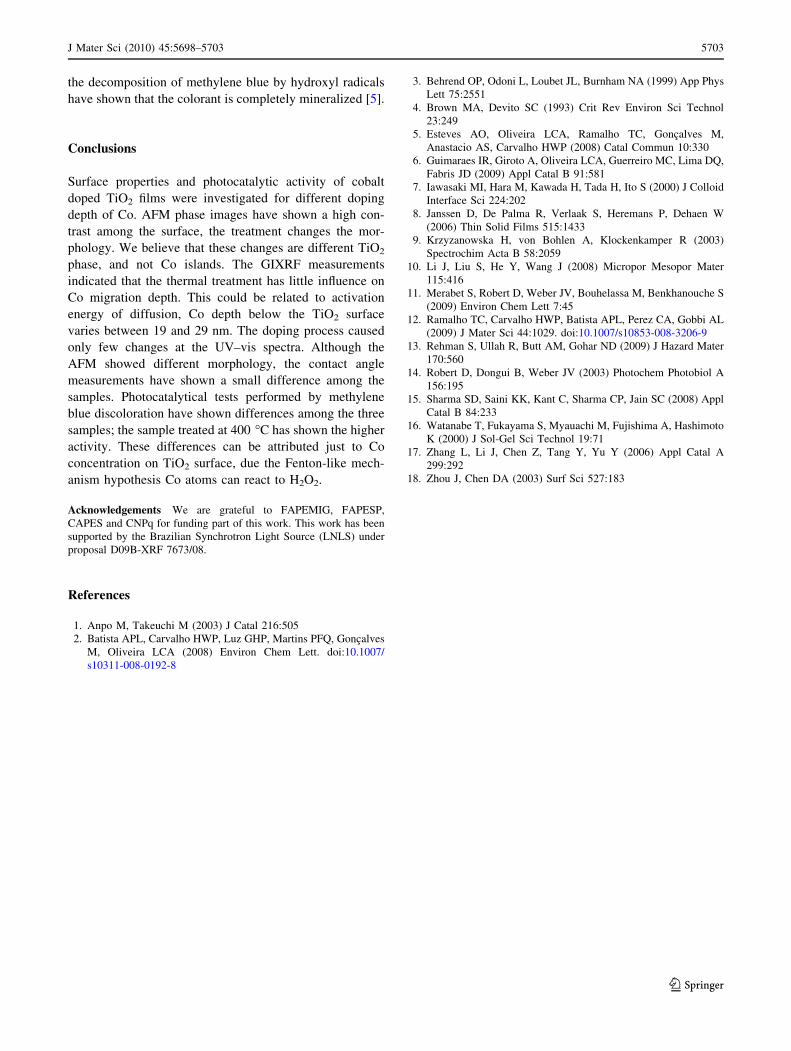
Conclusions
Surface properties and photocatalytic activity of cobalt
doped TiO2 films were investigated for different doping
depth of Co. AFM phase images have shown a high con-
trast among the surface, the treatment changes the mor-
phology. We believe that these changes are different TiO2
phase, and not Co islands. The GIXRF measurements
indicated that the thermal treatment has little influence on
Co migration depth. This could be related to activation
energy of diffusion, Co depth below the TiO2 surface
varies between 19 and 29 nm. The doping process caused
only few changes at the UV–vis spectra. Although the
AFM showed different morphology, the contact angle
measurements have shown a small difference among the
samples. Photocatalytical tests performed by methylene
blue discoloration have shown differences among the three
samples; the sample treated at 400 �C has shown the higher
activity. These differences can be attributed just to Co
concentration on TiO2 surface, due the Fenton-like mech-
anism hypothesis Co atoms can react to H2O2.
Acknowledgements We are grateful to FAPEMIG, FAPESP,
CAPES and CNPq for funding part of this work. This work has been
supported by the Brazilian Synchrotron Light Source (LNLS) under
proposal D09B-XRF 7673/08.
References
1. Anpo M, Takeuchi M (2003) J Catal 216:505
2. Batista APL, Carvalho HWP, Luz GHP, Martins PFQ, Goncalves
M, Oliveira LCA (2008) Environ Chem Lett. doi:10.1007/
s10311-008-0192-8
3. Behrend OP, Odoni L, Loubet JL, Burnham NA (1999) App Phys
Lett 75:2551
4. Brown MA, Devito SC (1993) Crit Rev Environ Sci Technol
23:249
5. Esteves AO, Oliveira LCA, Ramalho TC, Goncalves M,
Anastacio AS, Carvalho HWP (2008) Catal Commun 10:330
6. Guimaraes IR, Giroto A, Oliveira LCA, Guerreiro MC, Lima DQ,
Fabris JD (2009) Appl Catal B 91:581
7. Iawasaki MI, Hara M, Kawada H, Tada H, Ito S (2000) J Colloid
Interface Sci 224:202
8. Janssen D, De Palma R, Verlaak S, Heremans P, Dehaen W
(2006) Thin Solid Films 515:1433
9. Krzyzanowska H, von Bohlen A, Klockenkamper R (2003)
Spectrochim Acta B 58:2059
10. Li J, Liu S, He Y, Wang J (2008) Micropor Mesopor Mater
115:416
11. Merabet S, Robert D, Weber JV, Bouhelassa M, Benkhanouche S
(2009) Environ Chem Lett 7:45
12. Ramalho TC, Carvalho HWP, Batista APL, Perez CA, Gobbi AL
(2009) J Mater Sci 44:1029. doi:10.1007/s10853-008-3206-9
13. Rehman S, Ullah R, Butt AM, Gohar ND (2009) J Hazard Mater
170:560
14. Robert D, Dongui B, Weber JV (2003) Photochem Photobiol A
156:195
15. Sharma SD, Saini KK, Kant C, Sharma CP, Jain SC (2008) Appl
Catal B 84:233
16. Watanabe T, Fukayama S, Myauachi M, Fujishima A, Hashimoto
K (2000) J Sol-Gel Sci Technol 19:71
17. Zhang L, Li J, Chen Z, Tang Y, Yu Y (2006) Appl Catal A
299:292
18. Zhou J, Chen DA (2003) Surf Sci 527:183
J Mater Sci (2010) 45:5698–5703 5703
123
Copyright © 2022 FDOKUMEN