Phenotypic robustness conferred by apparently redundant transcriptional enhancers
12
Phenotypic robustness conferred by apparently redundant transcriptional enhancers Nicolás Frankel 1 , Gregory K. Davis 2 , Diego Vargas 1 , Shu Wang 1 , François Payre 3 , and David L. Stern 1,4 1 Howard Hughes Medical Institute and Department of Ecology and Evolutionary Biology, Princeton University, Princeton, NJ 08544, USA 2 Department of Biology, Bryn Mawr College, 101 N. Merion Ave, Bryn Mawr, PA 19010, USA. 3 Université de Toulouse and Centre National de la Recherche Scientifique, Centre de Biologie du Développement, UMR5547, Toulouse, F-31062, France. Abstract Genes include cis-regulatory regions that contain transcriptional enhancers. Recent reports have shown that developmental genes often possess multiple discrete enhancer modules that drive transcription in similar spatio-temporal patterns1 - 4: primary enhancers located near the basal promoter and secondary, or “shadow”, enhancers located at more remote positions. It has been hypothesized that the seemingly redundant activity of primary and secondary enhancers contributes to phenotypic robustness1 , 5. We tested this hypothesis by generating a deficiency that removes two newly-discovered enhancers of shavenbaby (svb), a gene encoding a transcription factor that directs development of larval trichomes6. At optimal temperatures for embryonic development, this deficiency causes minor defects in trichome patterning. In embryos that develop at both low and high extreme temperatures, however, absence of these secondary enhancers leads to extensive loss of trichomes. These temperature-dependent defects can be rescued by a transgene carrying a secondary enhancer driving transcription of the svb cDNA. Finally, removal of one copy of wingless, a gene required for normal trichome patterning7, causes a similar loss of trichomes only in flies lacking the secondary enhancers. These results support the hypothesis that secondary enhancers contribute to phenotypic robustness in the face of environmental and genetic variability. The cis-regulatory region of the svb gene integrates inputs from multiple gene regulatory networks to generate a complex pattern of transcription in the embryonic epidermis of insect species6 , 8. Svb protein then activates many downstream genes, ultimately resulting in trichome morphogenesis9 , 10. Three enhancer modules located in a 50 Kb region upstream of the svb transcription start site (called 7, E, and A) together recapitulate the complete svb epidermal expression pattern11. Partial loss of function of all three enhancers led to the evolutionary loss of the long, thin quaternary trichomes (indicated in Fig. 1a and 2a) on first- instar larvae of D. sechellia, a species that is closely related to D. melanogaster11. Evolution of svb expression patterns has probably also contributed to parallel loss of quaternary Users may view, print, copy, download and text and data- mine the content in such documents, for the purposes of academic research, subject always to the full Conditions of use: http://www.nature.com/authors/editorial_policies/license.html#terms 4 corresponding author: [email protected]. Author contributions N.F., G.K.D. and D.L.S. designed the experiments. N.F., G.K.D., D.V., S.W., F.P. and D.L.S. performed the experimental work. N.F. and D.L.S. wrote the manuscript. G.K.D and F.P. commented on the manuscript at all stages. NIH Public Access Author Manuscript Nature. Author manuscript; available in PMC 2011 January 1. Published in final edited form as: Nature. 2010 July 22; 466(7305): 490–493. doi:10.1038/nature09158. NIH-PA Author Manuscript NIH-PA Author Manuscript NIH-PA Author Manuscript
-
Upload
independent -
Category
Documents
-
view
3 -
download
0
Transcript of Phenotypic robustness conferred by apparently redundant transcriptional enhancers
Phenotypic robustness conferred by apparently redundanttranscriptional enhancers
Nicolás Frankel1, Gregory K. Davis2, Diego Vargas1, Shu Wang1, François Payre3, andDavid L. Stern1,4
1 Howard Hughes Medical Institute and Department of Ecology and Evolutionary Biology,Princeton University, Princeton, NJ 08544, USA2 Department of Biology, Bryn Mawr College, 101 N. Merion Ave, Bryn Mawr, PA 19010, USA.3 Université de Toulouse and Centre National de la Recherche Scientifique, Centre de Biologiedu Développement, UMR5547, Toulouse, F-31062, France.
AbstractGenes include cis-regulatory regions that contain transcriptional enhancers. Recent reports haveshown that developmental genes often possess multiple discrete enhancer modules that drivetranscription in similar spatio-temporal patterns1-4: primary enhancers located near the basalpromoter and secondary, or “shadow”, enhancers located at more remote positions. It has beenhypothesized that the seemingly redundant activity of primary and secondary enhancerscontributes to phenotypic robustness1,5. We tested this hypothesis by generating a deficiency thatremoves two newly-discovered enhancers of shavenbaby (svb), a gene encoding a transcriptionfactor that directs development of larval trichomes6. At optimal temperatures for embryonicdevelopment, this deficiency causes minor defects in trichome patterning. In embryos that developat both low and high extreme temperatures, however, absence of these secondary enhancers leadsto extensive loss of trichomes. These temperature-dependent defects can be rescued by a transgenecarrying a secondary enhancer driving transcription of the svb cDNA. Finally, removal of onecopy of wingless, a gene required for normal trichome patterning7, causes a similar loss oftrichomes only in flies lacking the secondary enhancers. These results support the hypothesis thatsecondary enhancers contribute to phenotypic robustness in the face of environmental and geneticvariability.
The cis-regulatory region of the svb gene integrates inputs from multiple gene regulatorynetworks to generate a complex pattern of transcription in the embryonic epidermis of insectspecies6,8. Svb protein then activates many downstream genes, ultimately resulting intrichome morphogenesis9,10. Three enhancer modules located in a 50 Kb region upstreamof the svb transcription start site (called 7, E, and A) together recapitulate the complete svbepidermal expression pattern11. Partial loss of function of all three enhancers led to theevolutionary loss of the long, thin quaternary trichomes (indicated in Fig. 1a and 2a) on first-instar larvae of D. sechellia, a species that is closely related to D. melanogaster11. Evolutionof svb expression patterns has probably also contributed to parallel loss of quaternary
Users may view, print, copy, download and text and data- mine the content in such documents, for the purposes of academic research,subject always to the full Conditions of use: http://www.nature.com/authors/editorial_policies/license.html#terms
4 corresponding author: [email protected] contributionsN.F., G.K.D. and D.L.S. designed the experiments. N.F., G.K.D., D.V., S.W., F.P. and D.L.S. performed the experimental work. N.F.and D.L.S. wrote the manuscript. G.K.D and F.P. commented on the manuscript at all stages.
NIH Public AccessAuthor ManuscriptNature. Author manuscript; available in PMC 2011 January 1.
Published in final edited form as:Nature. 2010 July 22; 466(7305): 490–493. doi:10.1038/nature09158.
NIH
-PA Author Manuscript
NIH
-PA Author Manuscript
NIH
-PA Author Manuscript
trichomes in the D. virilis group, species of which are distantly related to D.melanogaster12.
We noticed that a 41 kb region upstream of the three known svb enhancers displays highconservation among drosophilids, but contains only one small gene named SIP3 (Fig. 1b andS1). To test whether this region contained additional svb enhancers, we assayed reporterconstructs encompassing the entire region (Fig. S1). Two constructs drove expression in thedorso-lateral epidermis in patterns that reproduced part of the native svb expression pattern(Fig. 1c, f, and S2). To characterize the precise expression domains driven by these newly-discovered enhancers, we performed co-immunodetection of the β-galactosidase reporterand of the Dusky-like protein, an early component of developing trichomes10.
The Z enhancer drove expression in many cells that produce quaternary trichomes (Fig. 1c).This expression overlaps the patterns driven by the three previously identified enhancers: 7,E, and A (Fig. 1b). The DG2 enhancer drove expression in a more restricted region (Fig. 1f)that overlaps the domain of expression driven by the E enhancer. Both Z and DG2 driveexpression starting at stage 14 of embryogenesis (Fig. S2), which is similar to the time whensvb mRNA can be detected in epidermal cells.
Given the redundant expression patterns of Z and DG2 with the three previously identifiedenhancers, we sought further evidence that Z and DG2 encode functional svb enhancers. Wereasoned that if the Z and DG2 enhancers contribute to trichome patterning, then they shouldhave evolved in a similar way to the previously discovered 7, E, and A enhancers; theyshould retain expression in species that also produce quaternary trichomes (such as D.simulans), and show reduced expression in D. sechellia, which has lost quaternarytrichomes. We therefore assayed Z and DG2 enhancer constructs made with orthologousregions from D. simulans and D. sechellia. These regions were straightforward to identifybecause the genomes of these species are 3-5% divergent from D. melanogaster. The D.simulans Z and DG2 enhancers drove an expression pattern similar to that of theorthologous D. melanogaster enhancers (Fig.1c, d, f, and g), which suggests that Z and DG2contribute to the production of quaternary trichomes both in D. melanogaster and in D.simulans. In contrast, the Z and DG2 enhancers from D. sechellia drove low levels ofexpression in only a few cells (Fig. 1e and h). The weak expression driven by the D.sechellia Z and DG2 constructs is consistent with the partial loss of expression driven by theD. sechellia A, E, and 7 enhancers and with the loss of quaternary trichomes in thisspecies11.
To further assess the functional importance of the Z and DG2 enhancers, we generated a 32kbp chromosomal deficiency on the X chromosome that removes both enhancers, calledDf(X)svb108 (Fig. 1b). As a control, we used strain C108, which carries both of the parentaltransposable elements that were used to generate the deletion. Df(X)svb108 flies are viableand display no gross abnormalities. We examined first-instar larvae in detail and found that,when Df(X)svb108 embryos developed at the optimal temperature for development (25°C),larvae exhibited slightly fewer quaternary trichomes (Fig. 2b) and a reduction in the size ofthe lateral sensory bristles (Fig. S3). These results suggest that, under optimal conditions, Zand DG2 are functional enhancers of the svb gene that contribute to fine details of trichomepatterning and perhaps to bristle morphogenesis. Despite this evidence that the Z and DG2enhancers contribute to svb activity, their loss-of-function phenotype was considerablyweaker than one would have expected, given the strong expression driven by theseenhancers. We reasoned that this resulted from the fact that the Z and DG2 enhancers driveoverlapping expression with the enhancers 7, E, and A, and that the latter three enhancersdrive expression levels that are sufficient to generate most larval trichomes when embryosdevelop under optimal conditions11.
Frankel et al. Page 2
Nature. Author manuscript; available in PMC 2011 January 1.
NIH
-PA Author Manuscript
NIH
-PA Author Manuscript
NIH
-PA Author Manuscript

We therefore considered the hypothesis that Z and DG2 contribute to phenotypic robustness.Natural populations experience repeated stresses over evolutionary time, including variabletemperatures. Temperature influences membrane fluidity, enzymatic activity, proteinfolding, protein-protein interactions, and protein-DNA interactions13,14. Organisms haveevolved developmental mechanisms to buffer the phenotype in the face of temperature-induced cellular changes. We reasoned that sub-optimal temperatures might destabilize thetranscriptional output of genes during embryogenesis and that secondary enhancers mayconfer a selective advantage by maintaining transcription above a required minimumthreshold. We therefore tested the effect of Df(X)svb108 in embryos that had developed at17°C and 32°C, temperatures close to the extremes at which Drosophila embryos survive15.We counted the number of quaternary trichomes in the regions where Z and DG2 areexpressed strongly (Fig. 2a). The svb gene is an ideal target for this analysis, becausequantitative changes in svb transcription influence trichome density, size and shape16.
Control embryos reared at all temperatures produced similar numbers of trichomes,implying that the number of trichomes is canalized against temperature variation17. Thenumber of trichomes on Df(X)svb108 larvae reared at 25°C was similar to the number oncontrol C108 larvae at all temperatures (Fig. 2b). In contrast, Df(X)svb108 larvae displayed ahighly significant decrease in trichome numbers when reared at extreme temperatures (Fig.2b). The primary and tertiary trichomes look normal on Df(X)svb108 larvae at alltemperatures (data not shown), which is expected, because the Z and DG2 enhancers do notdrive expression in cells producing primary and tertiary trichomes.
In principle, the loss of trichomes observed on Df(X)svb108 larvae reared at extremetemperatures may have resulted from mechanisms acting independently of the Z and DG2enhancers. If the effects observed with Df(X)svb108 resulted from loss of the Z and DG2enhancers, then reintroducing a functional Z or DG2 enhancer into a Df(X)svb108
background should rescue some trichomes. We tested this hypothesis for the Z enhancer. Wegenerated a transgene carrying the svb cDNA under the transcriptional control of the Zenhancer and introduced it onto the third chromosome of Df(X)svb108 flies. At extremetemperatures, the Z::svb cDNA transgene completely rescued wild type trichome numbers inthe lateral patch (Fig. 3a and S3). However, in the region dorsal to the lateral patch, therescue is very weak or absent (Fig. 3b and S3). This is consistent with the fact that Z drivesexpression at high levels in the lateral region, where rescue is observed, and only weakly ina small number of cells of the dorsal region (Fig. 1). The loss of canalization in the dorsalregion of Df(X)svb108 larvae may be caused by loss of DG2, which drives expressionmainly in this dorsal region. These results demonstrate that Z contributes to phenotypicrobustness. Moreover, the rescue of trichome numbers by a transgene introduced onto adifferent chromosome from the svb locus suggests that Z does not need to be in intimatecontact with other svb enhancers or with the svb basal promoter to buffer svb function.Instead, we hypothesize that Z contributes to phenotypic robustness simply by boostinglevels of svb transcription in the cells in which Z drives expression.
Given this evidence that the Z enhancer, and possibly also DG2, contribute to robustnessagainst environmental perturbations, we asked whether these enhancers also buffer againstgenetic perturbations. For example, Boettiger & Levine18 have reported that two Dorsaltarget genes that possess “shadow” enhancers maintain synchronous transcriptionalactivation across Dorsal+/- embryos, whereas two Dorsal target genes that seem to lack such“shadow” enhancers display less synchrony in Dorsal+/- embryos. Therefore, we tested theeffect of reducing Wingless signaling, which is required for normal development ofquaternary trichomes7, by crossing the Df(X)svb108 allele and the C108 control allele into abackground heterozygous for a wingless null allele. At 25°C, the Df(X)svb108;wg-/+
embryos produced significantly fewer trichomes than C108;wg-/+ embryos, Df(X)svb108
Frankel et al. Page 3
Nature. Author manuscript; available in PMC 2011 January 1.
NIH
-PA Author Manuscript
NIH
-PA Author Manuscript
NIH
-PA Author Manuscript
embryos, and C108 embryos (Fig. 4). The combined results suggest that the Z and DG2enhancers buffer against both environmental and genetic perturbations.
These results indicate that the production of larval trichomes is normally canalized and thatthis is accomplished, at least in part, through transcriptional activation mediated by the svbsecondary enhancers that are removed in Df(X)svb108.
The svb locus contains multiple enhancers with overlapping expression patterns. Similarpatterns of overlapping enhancer activity have been found for the cis-regulatory regions ofthe Drosophila genes sog1, vnd3, and brinker1 and for the cis-regulatory regions of themouse genes sonic hedgehog4 and sox102. Moreover, it has been estimated that 50% of thetarget genes of the transcription factor Dorsal contain shadow enhancers5. Therefore, thepresence of additional enhancers in cis-regulatory regions may be a common signature ofdevelopmental regulators. This may explain why, in previous reports, animals carryingdeletions of highly conserved enhancers have not displayed observable phenotypic defectswhen reared in standard laboratory conditions19,20.
Developmental buffering is likely to result from many molecular mechanisms. For example,deletion of the conserved miRNA miR7 in D. melanogaster has no obvious phenotypiceffect in normal laboratory conditions, but it is required to canalize the expression of thegene atonal under fluctuating temperatures21. Similarly, our results indicate that svbsecondary enhancers have a minimal role at optimal conditions for development, but thatthey are essential to buffer the trichome phenotype under genetic or environmentalvariability. Secondary enhancers are likely to be evolutionarily maintained by selection forrobustness against temperature fluctuation, genetic background effects22, and expressionnoise23.
Methods summaryThe target regions were PCR-amplified from genomic DNA from D. melanogaster, D.simulans, and D. sechellia. These PCR fragments were cloned into pCaSpeR-hs43-lacZ orplacZattB and integrated into the D.melanogaster genome to test their enhancer activity. Theprecise expression domains of the enhancer constructs were determined by double stainingwith a mouse anti-βGal antibody (Promega) and a rabbit anti-Dusky-like antibody10 andthen by examining stained embryos with a confocal microscope. Df(X)svb108 was generatedvia flipase-induced deletion of the DNA between two FRTs present in C108. We made 0-3hour embryo collections and reared embryos to hatching at different temperatures. First-instar larvae were mounted in 1:1 Hoyer's:lactic acid mixture and cuticles were imaged withphase-contrast microscopy. Trichomes were counted using ImageJ. A null allele of wingless(wgIG22)24 was used to obtain males with the genotypes Df(X)svb108 /Y, +/wgIG22 andC108/Y, +/wgIG22.
Supplementary MaterialRefer to Web version on PubMed Central for supplementary material.
AcknowledgmentsWe thank D. Chien and D. Erezyilmaz for assistance with early experiments, L. Kruglyak, S. Levin, and S.Tavazoie for helpful comments on the manuscript, and E. Wieschaus for providing the wg mutant flies. This workwas supported by The Pew Charitable Trusts Latin American Fellows Program in the Biomedical SciencesFellowship to N. F., Agence Nationale de la Recherche (Blanc 2008, Netoshape) to F. P., and NIH(GM063622-06A1) and NSF (IOS-0640339) grants to D.L.S.
Frankel et al. Page 4
Nature. Author manuscript; available in PMC 2011 January 1.
NIH
-PA Author Manuscript
NIH
-PA Author Manuscript
NIH
-PA Author Manuscript
Methods
Reporter constructsGenomic DNA from D. melanogaster, D. simulans, and D. sechellia (see table below) wasamplified using the Expand HiFi PCR system (Roche) and cloned into pGEMT Easy(Promega). Fragments Z from D. melanogaster and Zprox from D. simulans were subclonedinto pCaSpeR-hs43-lacZ using NotI. This plasmid was co-injected with pTURBO33 into D.melanogaster w1118 embryos using standard conditions. At least three independenttransgenic lines were established for each construct. The remaining fragments weresubcloned into placZattB using NotI and injected into line M{3xP3-RFP.attP} ZH-51D(with M{vas-int.Dm}ZH-2A)25.
Region name Forward primer Reverse primer
D.melanogaster DGO TGGCCTGTGCCATGTGTGCGAGTACG TGGGTGCGCAATTATGCCGCCAGAGC
D.melanogaster DG1 CTGGGTGTGTGTGCAATATGTGAGC GTGAGGGTACAAGGCGAAATCGAAA
D.melanogaster DG2 AATTGTTCGCACGCTTCGCTCTAA GATTGGTGCCGAGAGGTGAAAGTG
D.melanogaster DG3 GGCCACAACTCAATGGCAAAAATG CAGCAGCGAATCAAGACGAAAGGT
D.melanogaster DG4 CCCCCGTCTTTGTCTGTTTGTCTG GGAACACAATCTGCCTGCCTGACT
D.melanogaster DG5 TATCCTTTTACGACGCCCCTGTGTC GATTCGGTTCCTTGGGATTGGATTT
D.melanogaster Z ATTGCTTCGGCTCTCCCGTTA TTGTGTGGCTCACTTGGCAC
D.simulans Zprox GTGAAAGATCGGATCCGTCT GTTCGTATCGCCCACTTGAAT
D.simulans Z ATTGCTTCGGCTCTCCCGTTA TTATGTGGCTCACTTGGCAC
D.sechellia Z ATTGCTTCGGCTCTCCCGTTA TTGTGTGGCTCACTTGGCAC
D.simulans DG2 TGCTTTTCCAACCCCTCAGTT GGGGGTGCAGGCTATTTTGTTC
D.sechellia DG2 TGCTTTTCCAACCCCTCAGTT GAGGGTGCAGGCTATTTTGTTC
Only transgenes containing the Z and DG2 regions drove expression in the dorso-lateralepidermis. DG3, which is contained within the region deleted by Df(X)svb108, drove weakexpression in the ventral epidermis, but no phenotypic changes in the ventral denticles wereobserved at any temperature. Zprox was analyzed from D. simulans DNA, as this regionlacked a large roo element that is present in the D.melanogaster genome.
Immunohistochemistry and ImmunofluorescenceEmbryos were fixed using standard conditions. To determine the precise expression domainsof the enhancer constructs we performed fluorescent double staining with a mouse anti-βGalantibody (Promega) and a rabbit anti-Dusky-like antibody10. Alexa-488 anti-rabbit andAlexa-647 anti-mouse (Molecular Probes) were used as secondary antibodies. The embryoswere examined on a Leica TCS SPE confocal microscope. For immuno-histochemistry, weused a rabbit anti-βGal antibody (Cappel) and anti-rabbit antibody coupled to HRP (SantaCruz Biotech) and staining was developed with DAB/Nickel.
Generation of Df(X)svb108
pBac{WH}Ptp4E[f02952] and pBac{RB}e03292 were recombined onto the same Xchromosome and a homozygous stock was generated (named C108). This stock was crossedto a line containing a hs::flipase and larvae were heat shocked at 37C for 1 hour each day
Frankel et al. Page 5
Nature. Author manuscript; available in PMC 2011 January 1.
NIH
-PA Author Manuscript
NIH
-PA Author Manuscript
NIH
-PA Author Manuscript
during larval development. After crossing these adults to white- flies, we selected adults thathad lost one copy of the white+ transgene (originating on one of the pBac transgenes), whichis expected if the two FRT sites recombined to generate a deletion. The deletion wasconfirmed by a PCR experiment, which amplified a fragment containing a chimericpiggyBac element. The primer used (TGCATTTGCCTTTCGCCTTAT) amplified theexpected 7.3 kb fragment26. We then generated a stock homozygous for the deletion. Thisallele is named Df(X)svb108.
Embryo collection and cuticle microscopyWe made 0-3 hour embryo collections (many hours before the onset of svb expression inepidermal tissues) and transferred embryos to dishes with water at the differenttemperatures. Two days later, we collected first instar larvae and incubated them at 60C for4 hours. Subsequently, larvae were mounted on a microscope slide with a drop of 1:1Hoyer's:lactic acid mixture. After overnight drying, the cuticles were imaged with phase-contrast microscopy.
Trichome countingA spiracle below the lateral patch was used as a landmark to position the green box (shownin Fig.2a). The blue box was positioned directly above the green box (shown in Fig.2a).Both boxes were programmed as macros in Image J software (Rasband, W.S., ImageJ, U. S.National Institutes of Health, Bethesda, Maryland, USA, http://rsb.info.nih.gov/ij/,1997-2009). The trichomes were counted using the cell-counter option of Image J.
Rescue experimentsThe cDNA of svb was amplified from the plasmid pUAS-svb6 with primers NsiI-svbcDNAfw (ATGCATTTAACTCACCTGGGCGAATCC) and NdeI-svbcDNArv(CATATGTTGCAGCTTGTTCGGTTGGTA) and cloned into pCR-Blunt II-TOPO(Invitrogen). The svb cDNA was subcloned with NsiI and NdeI into a version ofplacZattB25 that had the lacZ removed (by cutting with PstI and NdeI). We named thisplasmid pRSQsvb. The Z enhancer was amplified with the primers used previously (seereporter constructs) that had the addition of 3’ XbaI sites. This PCR fragment was clonedinto pGEMT (Promega) and subcloned into pRSQsvb using XbaI. This plasmid was injectedinto the recipient line M{3xP3-RFP.attP}ZH- 86Fb (with M{vas-int.Dm}ZH-2A)25. A thirdchromosome carrying the Z::svb transgene was introduced into the Df(X)svb108 line toobtain a stock homozygous for both the deficiency, on the X chromosome, and Z::svb, onthe third chromosome, and is referred to as Df(X)svb108; Z::svb.
Wingless experimentA null allele of wingless (wgIG22)24 was used to obtain males of the genotype FM7c,actin::GFP/Y; CyO, actin::GFP/wgIG22. These males were crossed to females with eitherDf(X)svb108/Df(X)svb108 or C108/C108 genotypes. We selected for non-fluorescentprogeny, which were male first-instar larvae heterozygous for the wingless null allele:Df(X)svb108/Y; wgIG22/+ or C108/Y; wgIG22/+.
References1. Hong JW, Hendrix DA, Levine MS. Shadow enhancers as a source of evolutionary novelty.
Science. 2008; 321:1314. [PubMed: 18772429]
Frankel et al. Page 6
Nature. Author manuscript; available in PMC 2011 January 1.
NIH
-PA Author Manuscript
NIH
-PA Author Manuscript
NIH
-PA Author Manuscript
2. Werner T, Hammer A, Wahlbuhl M, Bosl MR, Wegner M. Multiple conserved regulatory elementswith overlapping functions determine Sox10 expression in mouse embryogenesis. Nucleic AcidsRes. 2007; 35:6526–6538. [PubMed: 17897962]
3. Zeitlinger J, et al. Whole-genome ChIP-chip analysis of Dorsal, Twist, and Snail suggestsintegration of diverse patterning processes in the Drosophila embryo. Genes Dev. 2007; 21:385–390. [PubMed: 17322397]
4. Jeong Y, El-Jaick K, Roessler E, Muenke M, Epstein DJ. A functional screen for sonic hedgehogregulatory elements across a 1 Mb interval identifies long-range ventral forebrain enhancers.Development. 2006; 133:761–772. [PubMed: 16407397]
5. Perry MW, Cande JD, Boettiger AN, Levine M. Evolution of Insect Dorsoventral PatterningMechanisms. Cold Spring Harb Symp Quant Biol. 2009
6. Payre F, Vincent A, Carreno S. ovo/svb integrates Wingless and DER pathways to control epidermisdifferentiation. Nature. 1999; 400:271–275. [PubMed: 10421370]
7. Bokor P, DiNardo S. The roles of hedgehog, wingless and lines in patterning the dorsal epidermis inDrosophila. Development. 1996; 122:1083–1092. [PubMed: 8620835]
8. Overton PM, Chia W, Buescher M. The Drosophila HMG-domain proteins SoxNeuro and Dichaetedirect trichome formation via the activation of shavenbaby and the restriction of Wingless pathwayactivity. Development. 2007; 134:2807–2813. [PubMed: 17611224]
9. Chanut-Delalande H, Fernandes I, Roch F, Payre F, Plaza S. Shavenbaby couples patterning toepidermal cell shape control. PLoS Biol. 2006; 4:e290. [PubMed: 16933974]
10. Fernandes I, et al. A scaffold of ZP proteins remodels the apical compartment for localized cellshape changes. Developmental Cell. 2010
11. McGregor AP, et al. Morphological evolution through multiple cis-regulatory mutations at a singlegene. Nature. 2007; 448:587–590. [PubMed: 17632547]
12. Sucena E, Delon I, Jones I, Payre F, Stern DL. Regulatory evolution of shavenbaby/ovo underliesmultiple cases of morphological parallelism. Nature. 2003; 424:935–938. [PubMed: 12931187]
13. Crane-Robinson C, Dragan AI, Read CM. Defining the thermodynamics of protein/DNAcomplexes and their components using micro-calorimetry. Methods Mol Biol. 2009; 543:625–651.[PubMed: 19378190]
14. Hochachka, PW.; Somero, GN. Biochemical adaptation : mechanism and process in physiologicalevolution. Oxford University Press; 2002.
15. Powsner L. The effects of temperature on the durations of the developmental stages of Drosophilamelanogaster. Physiological Zoology. 1935; 8:474–520.
16. Delon I, Chanut-Delalande H, Payre F. The Ovo/Shavenbaby transcription factor specifies actinremodelling during epidermal differentiation in Drosophila. Mech Dev. 2003; 120:747–758.[PubMed: 12915226]
17. Nijhout, HF.; Davidowitz, G. Developmental Instability (DI): Causes and Consequences. Polak,M., editor. Oxford University Press; 2003.
18. Boettiger AN, Levine M. Synchronous and stochastic patterns of gene activation in the Drosophilaembryo. Science. 2009; 325:471–473. [PubMed: 19628867]
19. Cretekos CJ, et al. Regulatory divergence modifies limb length between mammals. Genes Dev.2008; 22:141–151. [PubMed: 18198333]
20. Xiong N, Kang C, Raulet DH. Redundant and unique roles of two enhancer elements in theTCRgamma locus in gene regulation and gammadelta T cell development. Immunity. 2002;16:453–463. [PubMed: 11911829]
21. Li X, Cassidy JJ, Reinke CA, Fischboeck S, Carthew RW. A microRNA imparts robustnessagainst environmental fluctuation during development. Cell. 2009; 137:273–282. [PubMed:19379693]
22. Crickmore MA, Ranade V, Mann RS. Regulation of Ubx expression by epigenetic enhancersilencing in response to Ubx levels and genetic variation. PLoS Genet. 2009; 5:e1000633.[PubMed: 19730678]
23. Raser JM, O'Shea EK. Noise in gene expression: origins, consequences, and control. Science (NewYork, N.Y. 2005; 309:2010–2013.
Frankel et al. Page 7
Nature. Author manuscript; available in PMC 2011 January 1.
NIH
-PA Author Manuscript
NIH
-PA Author Manuscript
NIH
-PA Author Manuscript

24. van den Heuvel M, Harryman-Samos C, Klingensmith J, Perrimon N, Nusse R. Mutations in thesegment polarity genes wingless and porcupine impair secretion of the wingless protein. EMBO J.1993; 12:5293–5302. [PubMed: 8262072]
25. Bischof J, Maeda RK, Hediger M, Karch F, Basler K. An optimized transgenesis system forDrosophila using germ-line-specific phiC31 integrases. Proceedings of the National Academy ofSciences of the United States of America. 2007; 104:3312–3317. [PubMed: 17360644]
26. Parks AL, et al. Systematic generation of high-resolution deletion coverage of the Drosophilamelanogaster genome. Nat Genet. 2004; 36:288–292. [PubMed: 14981519]
Frankel et al. Page 8
Nature. Author manuscript; available in PMC 2011 January 1.
NIH
-PA Author Manuscript
NIH
-PA Author Manuscript
NIH
-PA Author Manuscript
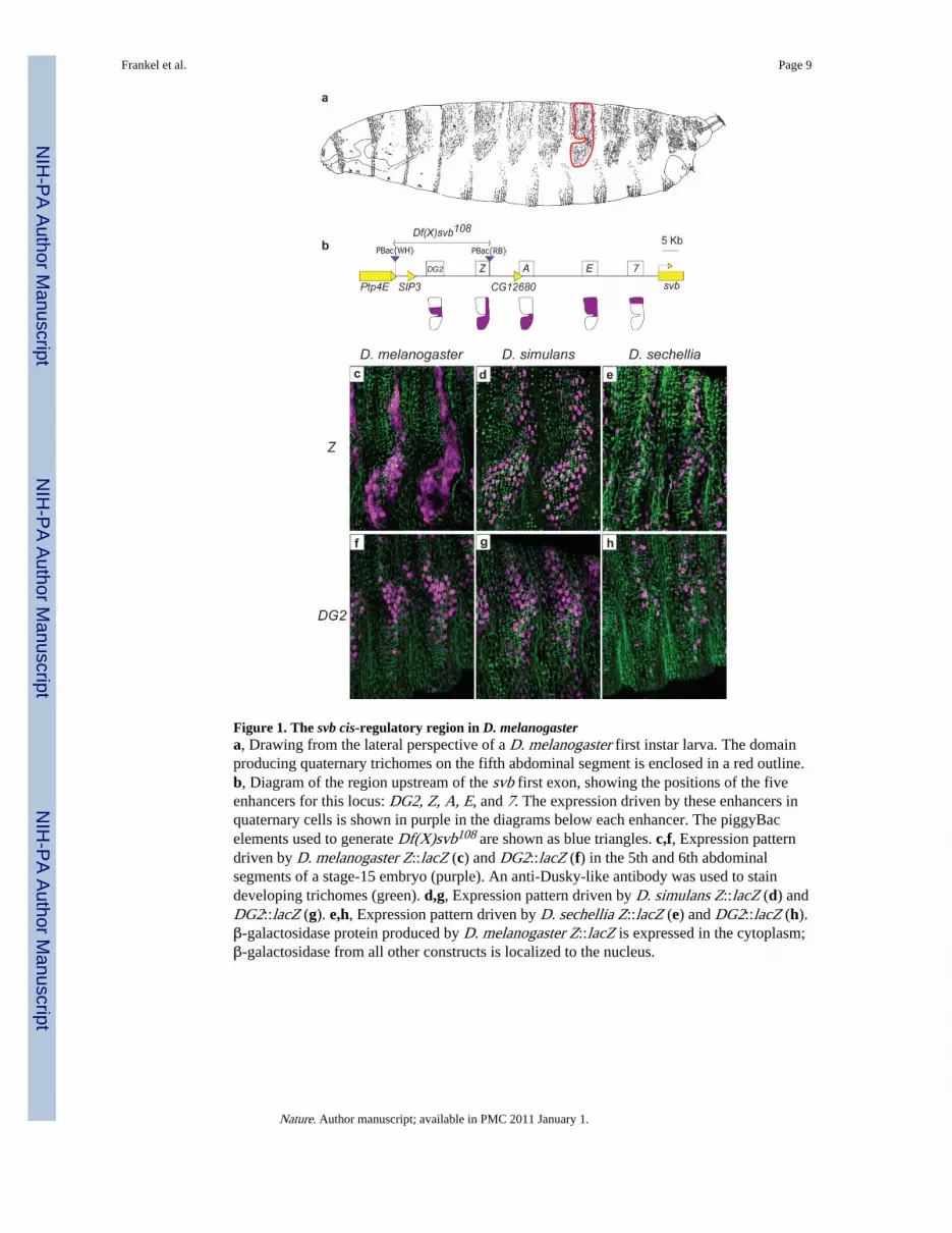
Figure 1. The svb cis-regulatory region in D. melanogastera, Drawing from the lateral perspective of a D. melanogaster first instar larva. The domainproducing quaternary trichomes on the fifth abdominal segment is enclosed in a red outline.b, Diagram of the region upstream of the svb first exon, showing the positions of the fiveenhancers for this locus: DG2, Z, A, E, and 7. The expression driven by these enhancers inquaternary cells is shown in purple in the diagrams below each enhancer. The piggyBacelements used to generate Df(X)svb108 are shown as blue triangles. c,f, Expression patterndriven by D. melanogaster Z::lacZ (c) and DG2::lacZ (f) in the 5th and 6th abdominalsegments of a stage-15 embryo (purple). An anti-Dusky-like antibody was used to staindeveloping trichomes (green). d,g, Expression pattern driven by D. simulans Z::lacZ (d) andDG2::lacZ (g). e,h, Expression pattern driven by D. sechellia Z::lacZ (e) and DG2::lacZ (h).β-galactosidase protein produced by D. melanogaster Z::lacZ is expressed in the cytoplasm;β-galactosidase from all other constructs is localized to the nucleus.
Frankel et al. Page 9
Nature. Author manuscript; available in PMC 2011 January 1.
NIH
-PA Author Manuscript
NIH
-PA Author Manuscript
NIH
-PA Author Manuscript
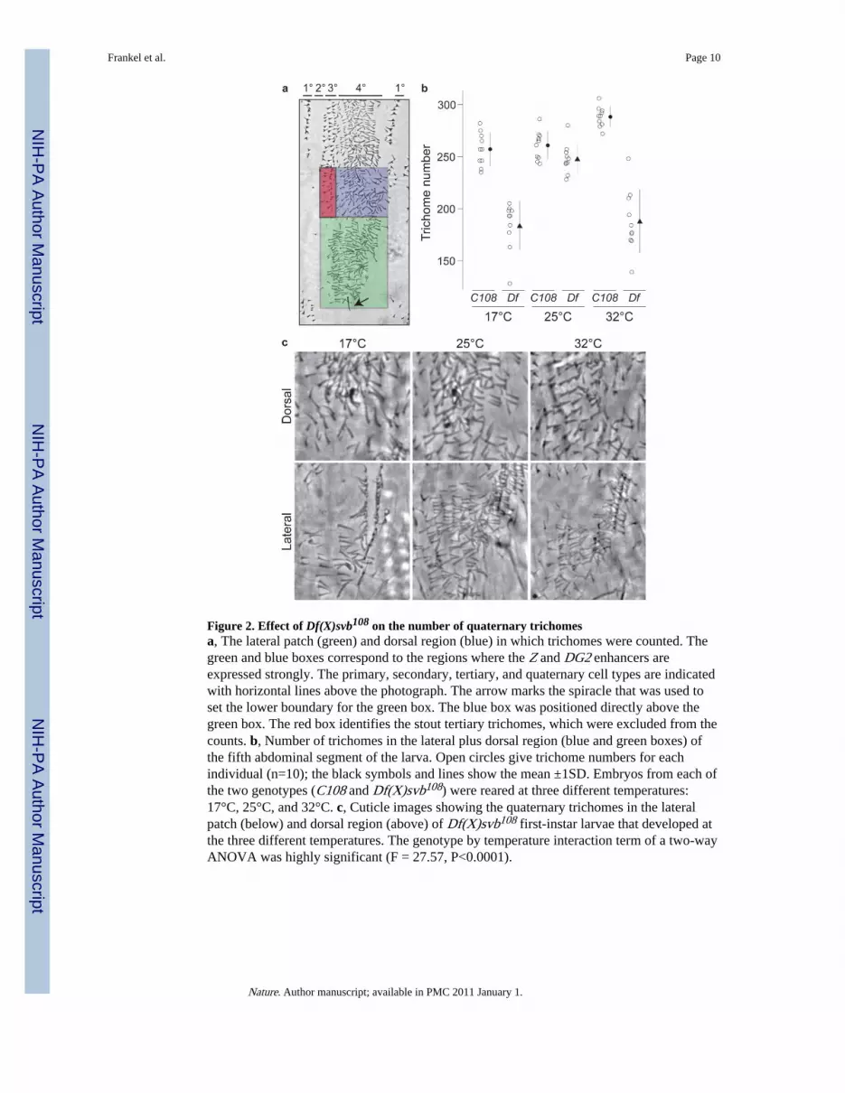
Figure 2. Effect of Df(X)svb108 on the number of quaternary trichomesa, The lateral patch (green) and dorsal region (blue) in which trichomes were counted. Thegreen and blue boxes correspond to the regions where the Z and DG2 enhancers areexpressed strongly. The primary, secondary, tertiary, and quaternary cell types are indicatedwith horizontal lines above the photograph. The arrow marks the spiracle that was used toset the lower boundary for the green box. The blue box was positioned directly above thegreen box. The red box identifies the stout tertiary trichomes, which were excluded from thecounts. b, Number of trichomes in the lateral plus dorsal region (blue and green boxes) ofthe fifth abdominal segment of the larva. Open circles give trichome numbers for eachindividual (n=10); the black symbols and lines show the mean ±1SD. Embryos from each ofthe two genotypes (C108 and Df(X)svb108) were reared at three different temperatures:17°C, 25°C, and 32°C. c, Cuticle images showing the quaternary trichomes in the lateralpatch (below) and dorsal region (above) of Df(X)svb108 first-instar larvae that developed atthe three different temperatures. The genotype by temperature interaction term of a two-wayANOVA was highly significant (F = 27.57, P<0.0001).
Frankel et al. Page 10
Nature. Author manuscript; available in PMC 2011 January 1.
NIH
-PA Author Manuscript
NIH
-PA Author Manuscript
NIH
-PA Author Manuscript

Figure 3. Rescue of the temperature-dependent trichome loss in the lateral patch by a Z::svbtransgenea,b, Trichome number in the lateral patch (a) and dorsal region (b) of the 5th abdominalsegment of larvae with the genotypes C108, Df(X)svb108, and Df(X)svb108; Z::svb. Opencircles represent trichome numbers for each individual (n=10); the black symbols and linesshow the mean ±1SD.
Frankel et al. Page 11
Nature. Author manuscript; available in PMC 2011 January 1.
NIH
-PA Author Manuscript
NIH
-PA Author Manuscript
NIH
-PA Author Manuscript
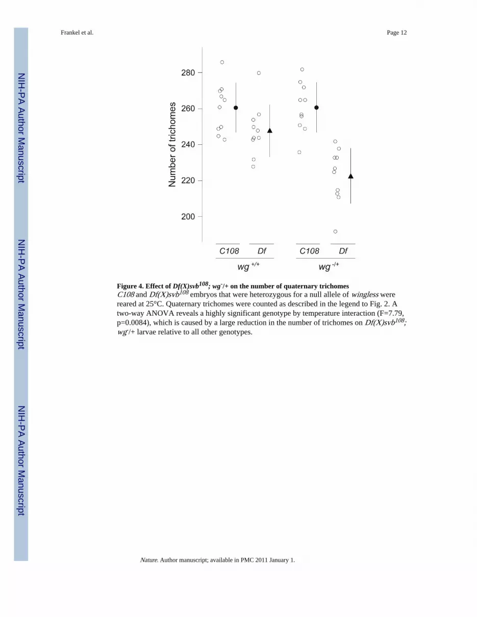
Figure 4. Effect of Df(X)svb108; wg-/+ on the number of quaternary trichomesC108 and Df(X)svb108 embryos that were heterozygous for a null allele of wingless werereared at 25°C. Quaternary trichomes were counted as described in the legend to Fig. 2. Atwo-way ANOVA reveals a highly significant genotype by temperature interaction (F=7.79,p=0.0084), which is caused by a large reduction in the number of trichomes on Df(X)svb108;wg-/+ larvae relative to all other genotypes.
Frankel et al. Page 12
Nature. Author manuscript; available in PMC 2011 January 1.
NIH
-PA Author Manuscript
NIH
-PA Author Manuscript
NIH
-PA Author Manuscript
![[Hepatic steatosis, visceral fat and metabolic alterations in apparently healthy overweight/obese individuals]](https://static.fdokumen.com/doc/165x107/6324f8237fd2bfd0cb03375f/hepatic-steatosis-visceral-fat-and-metabolic-alterations-in-apparently-healthy.jpg)