Persistence of deposited metals in the lungs after stainless steel and mild steel welding fume...
12
INORGANIC COMPOUNDS Persistence of deposited metals in the lungs after stainless steel and mild steel welding fume inhalation in rats James M. Antonini • Jenny R. Roberts • Samuel Stone • Bean T. Chen • Diane Schwegler-Berry • Rebecca Chapman • Patti C. Zeidler-Erdely • Ronnee N. Andrews • David G. Frazer Received: 23 June 2010 / Accepted: 22 September 2010 / Published online: 6 October 2010 Ó Springer-Verlag (outside the USA) 2010 Abstract Welding generates complex metal fumes that vary in composition. The objectives of this study were to compare the persistence of deposited metals and the inflammatory potential of stainless and mild steel welding fumes, the two most common fumes used in US industry. Sprague–Dawley rats were exposed to 40 mg/m 3 of stain- less or mild steel welding fumes for 3 h/day for 3 days. Controls were exposed to filtered air. Generated fume was collected, and particle size and elemental composition were determined. Bronchoalveolar lavage was done on days 0, 8, 21, and 42 after the last exposure to assess lung injury/ inflammation and to recover lung phagocytes. Non-lavaged lung samples were analyzed for total and specific metal content as a measure of metal persistence. Both welding fumes were similar in particle morphology and size. Fol- lowing was the chemical composition of the fumes— stainless steel: 57% Fe, 20% Cr, 14% Mn, and 9% Ni; mild steel: 83% Fe and 15% Mn. There was no effect of the mild steel fume on lung injury/inflammation at any time point compared to air control. Lung injury and inflammation were significantly elevated at 8 and 21 days after exposure to the stainless steel fume compared to control. Stainless steel fume exposure was associated with greater recovery of welding fume-laden macrophages from the lungs at all time points compared with the mild steel fume. A higher concentration of total metal was observed in the lungs of the stainless steel welding fume at all time points compared with the mild steel fume. The specific metals present in the two fumes were cleared from the lungs at different rates. The potentially more toxic metals (e.g., Mn, Cr) present in the stainless steel fume were cleared from the lungs more quickly than Fe, likely increasing their translocation from the respiratory system to other organs. Keywords Welding fume Á Inhalation Á Lung burden Á Lung clearance Á Pulmonary toxicity Introduction Welding is a common industrial process used to join metals. Over 300,000 workers in the United States (Bureau of Labor Statistics 2007) and millions of workers world- wide are exposed to welding aerosols on a daily basis. Welding fume is a complex aerosol of different metals. The composition of the fume varies depending on the welding processes and materials used. Welding aerosols are mostly generated from the consumption of an electrode, wire, or rod. Two of the most common types of consumables used in welding are mild steel and stainless steel electrodes. Mild steel fume is composed of a complex of iron (Fe) with a smaller percentage of manganese (Mn), whereas stainless steel fume contains both Fe and Mn, but with significant amounts of chromium (Cr) and nickel (Ni) that are not present in mild steel fume. The metals present in welding fumes are of interest toxicologically due to their potential effects on worker health. Significant amounts of Fe have been observed to deposit and persist in the lungs of full-time welders, J. M. Antonini (&) Á J. R. Roberts Á S. Stone Á B. T. Chen Á D. Schwegler-Berry Á R. Chapman Á P. C. Zeidler-Erdely Á D. G. Frazer Health Effects Laboratory Division, National Institute for Occupational Safety and Health, 1095 Willowdale Road, Mailstop 2015, Morgantown, WV 26505, USA e-mail: [email protected] R. N. Andrews Division of Applied Research and Technology, National Institute for Occupational Safety and Health, Cincinnati, OH 45213, USA 123 Arch Toxicol (2011) 85:487–498 DOI 10.1007/s00204-010-0601-1
-
Upload
universitassemarang -
Category
Documents
-
view
1 -
download
0
Transcript of Persistence of deposited metals in the lungs after stainless steel and mild steel welding fume...
INORGANIC COMPOUNDS
Persistence of deposited metals in the lungs after stainless steeland mild steel welding fume inhalation in rats
James M. Antonini • Jenny R. Roberts • Samuel Stone • Bean T. Chen •
Diane Schwegler-Berry • Rebecca Chapman • Patti C. Zeidler-Erdely •
Ronnee N. Andrews • David G. Frazer
Received: 23 June 2010 / Accepted: 22 September 2010 / Published online: 6 October 2010
� Springer-Verlag (outside the USA) 2010
Abstract Welding generates complex metal fumes that
vary in composition. The objectives of this study were to
compare the persistence of deposited metals and the
inflammatory potential of stainless and mild steel welding
fumes, the two most common fumes used in US industry.
Sprague–Dawley rats were exposed to 40 mg/m3 of stain-
less or mild steel welding fumes for 3 h/day for 3 days.
Controls were exposed to filtered air. Generated fume was
collected, and particle size and elemental composition were
determined. Bronchoalveolar lavage was done on days 0, 8,
21, and 42 after the last exposure to assess lung injury/
inflammation and to recover lung phagocytes. Non-lavaged
lung samples were analyzed for total and specific metal
content as a measure of metal persistence. Both welding
fumes were similar in particle morphology and size. Fol-
lowing was the chemical composition of the fumes—
stainless steel: 57% Fe, 20% Cr, 14% Mn, and 9% Ni; mild
steel: 83% Fe and 15% Mn. There was no effect of the mild
steel fume on lung injury/inflammation at any time point
compared to air control. Lung injury and inflammation
were significantly elevated at 8 and 21 days after exposure
to the stainless steel fume compared to control. Stainless
steel fume exposure was associated with greater recovery
of welding fume-laden macrophages from the lungs at all
time points compared with the mild steel fume. A higher
concentration of total metal was observed in the lungs of
the stainless steel welding fume at all time points compared
with the mild steel fume. The specific metals present in the
two fumes were cleared from the lungs at different rates.
The potentially more toxic metals (e.g., Mn, Cr) present in
the stainless steel fume were cleared from the lungs more
quickly than Fe, likely increasing their translocation from
the respiratory system to other organs.
Keywords Welding fume � Inhalation � Lung burden �Lung clearance � Pulmonary toxicity
Introduction
Welding is a common industrial process used to join
metals. Over 300,000 workers in the United States (Bureau
of Labor Statistics 2007) and millions of workers world-
wide are exposed to welding aerosols on a daily basis.
Welding fume is a complex aerosol of different metals. The
composition of the fume varies depending on the welding
processes and materials used. Welding aerosols are mostly
generated from the consumption of an electrode, wire, or
rod. Two of the most common types of consumables used
in welding are mild steel and stainless steel electrodes.
Mild steel fume is composed of a complex of iron (Fe) with
a smaller percentage of manganese (Mn), whereas stainless
steel fume contains both Fe and Mn, but with significant
amounts of chromium (Cr) and nickel (Ni) that are not
present in mild steel fume.
The metals present in welding fumes are of interest
toxicologically due to their potential effects on worker
health. Significant amounts of Fe have been observed to
deposit and persist in the lungs of full-time welders,
J. M. Antonini (&) � J. R. Roberts � S. Stone �B. T. Chen � D. Schwegler-Berry � R. Chapman �P. C. Zeidler-Erdely � D. G. Frazer
Health Effects Laboratory Division,
National Institute for Occupational Safety and Health,
1095 Willowdale Road, Mailstop 2015,
Morgantown, WV 26505, USA
e-mail: [email protected]
R. N. Andrews
Division of Applied Research and Technology, National Institute
for Occupational Safety and Health, Cincinnati, OH 45213, USA
123
Arch Toxicol (2011) 85:487–498
DOI 10.1007/s00204-010-0601-1
possibly leading to a condition called siderosis (Antonini
2003; Sferlazza and Beckett 1991). Manganese is a known
neurotoxicant. Neurobehavioral changes have been repor-
ted in exposed welders (Bowler et al. 2007; Ellingsen et al.
2008). Welding fume has been classified as ‘‘possibly
carcinogenic’’ by the International Agency for Research on
Cancer (IARC) due to the presence of known human car-
cinogens, Cr and Ni, in stainless steel fume (IARC 1990).
However, epidemiological studies have been unable to
correlate chronic adverse lung effects, such as cancer,
solely with exposure to stainless steel welding fume when
compared with mild steel fume (Moulin et al. 1993; Moulin
1997; Langard 1994).
Numerous animal toxicology studies have been
performed in recent years to evaluate the lung effects
associated with welding fume exposure. Stainless steel
welding fume was observed to induce a chronic lung
inflammatory response with a trend for increased lung
tumor incidence in a mouse lung tumor-susceptible model
compared with a mild steel fume (Zeidler-Erdely et al.
2008). Short-term inhalation exposure to stainless steel,
but not mild steel, fume caused lung injury and inflam-
mation in rats (Antonini et al. 2007, 2009a). However, the
same studies did show that both stainless and mild steel
fumes suppressed lung immune responses after pulmonary
inoculation with a bacterial pathogen. In a sub-chronic
inhalation exposure study in rats, Yu et al. (2001)
observed the appearance of interstitial fibrosis by 60 days
that becomes prominent at 90 days after exposure to a
high concentration of stainless steel welding fume
(107 mg/m3). Recovery studies indicated that the fibrosis
observed in the rats exposed to the highest dose of
stainless steel welding fume for 60–90 days did not fully
resolve (Yu et al. 2003; Sung et al. 2004).
The objective of the current study was to compare the
lung toxicity and persistence of the deposited particles after
inhalation of different welding fumes using an animal
model. A completely automated, robotic welding fume
generation and inhalation exposure system has been con-
structed by NIOSH (Antonini et al. 2006). Unique to this
current study was the examination of the persistence and
clearance of individual metals associated with each type of
fume. We hypothesize that one mechanism by which
stainless steel fume may be more toxic than mild steel
fume is that metal particles generated during stainless steel
welding persist in the lungs longer than mild steel fume due
to differences in metal composition. This potential increase
in residence time of the metals associated with stainless
steel fume in the lung would allow for a longer exposure
period between the welding fume metals and pulmonary
cells. Lung toxicity, pulmonary clearance of the metals
associated with welding fume, and macrophage phagocy-
tosis of inhaled particles were examined over a 42-day
period after a 3-day inhalation of mild steel and stainless
steel welding fumes by rats during gas metal arc welding.
Methods
Experimental design
Rats were exposed by inhalation for 3 h/day for 3 days to
40 mg/m3 of fume generated during gas metal arc welding
using either a mild steel or stainless steel welding
electrode. Control animals were exposed to filtered air for
3 h/day for 3 days. At 1 h (day 0), 8, 21, and 42 days after
the last exposure, lung injury, inflammation, and particle/
metal lung deposition and clearance were assessed in the
exposed animals.
Welding fume generation system
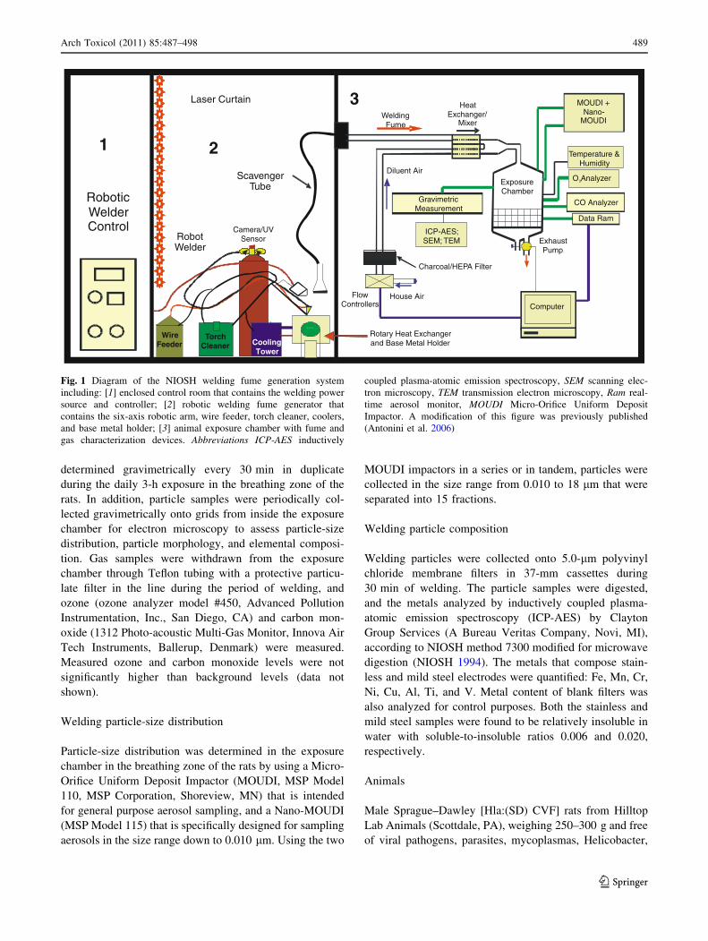
The welding fume generation system (Fig. 1) consisted of a
welding power source (Power Wave 455, Lincoln Electric,
Cleveland, OH), an automated, programmable six-axis
robotic arm (Model 100 Bi, Lincoln Electric), a water-
cooled arc welding torch (WC 650 amp, Lincoln Electric),
a wire feeder that supplied the wire to the torch at a pro-
grammed rate up to 300 inches/min, and an automatic
welding torch cleaner that kept the welding nozzle free of
debris and spatter (Antonini et al. 2006). Gas metal arc
welding was performed using either a mild steel electrode
(carbon steel ER70S-6, Lincoln Electric) or a stainless steel
electrode (Blue Max E308LSi wire, Lincoln Electric,
Cleveland, OH). Welding took place on A36 carbon steel
plates for daily exposures of 3 h at 25 V and 200 amps.
During welding, a shielding gas combination of 95% Ar
and 5% CO2 (Airgas Co., Morgantown, WV) was contin-
ually delivered to the welding nozzle at an air flow rate of
20 l/min.
Exposure chamber fume and gas determinations
A flexible trunk was positioned approximately 18 inches
from the arc to collect the generated fume and transport it
to the exposure chamber. The generated welding fume was
mixed with dry HEPA-filtered air. Continuous records of
chamber fume concentration, temperature, and humidity
were maintained during welding fume generation. The
mass concentration in the chamber was monitored by a
real-time aerosol monitor (DataRAM, Thermo Electron
Co., DR-4000, Franklin, MA). Depending on the desired
concentration, the diluent air in this system was normally
controlled between 20 and 80 l/min. Fume was collected
onto 37-mm Teflon filters at a rate of 1 l/min, and the
particle mass delivered to the exposure chamber was
488 Arch Toxicol (2011) 85:487–498
123
determined gravimetrically every 30 min in duplicate
during the daily 3-h exposure in the breathing zone of the
rats. In addition, particle samples were periodically col-
lected gravimetrically onto grids from inside the exposure
chamber for electron microscopy to assess particle-size
distribution, particle morphology, and elemental composi-
tion. Gas samples were withdrawn from the exposure
chamber through Teflon tubing with a protective particu-
late filter in the line during the period of welding, and
ozone (ozone analyzer model #450, Advanced Pollution
Instrumentation, Inc., San Diego, CA) and carbon mon-
oxide (1312 Photo-acoustic Multi-Gas Monitor, Innova Air
Tech Instruments, Ballerup, Denmark) were measured.
Measured ozone and carbon monoxide levels were not
significantly higher than background levels (data not
shown).
Welding particle-size distribution
Particle-size distribution was determined in the exposure
chamber in the breathing zone of the rats by using a Micro-
Orifice Uniform Deposit Impactor (MOUDI, MSP Model
110, MSP Corporation, Shoreview, MN) that is intended
for general purpose aerosol sampling, and a Nano-MOUDI
(MSP Model 115) that is specifically designed for sampling
aerosols in the size range down to 0.010 lm. Using the two
MOUDI impactors in a series or in tandem, particles were
collected in the size range from 0.010 to 18 lm that were
separated into 15 fractions.
Welding particle composition
Welding particles were collected onto 5.0-lm polyvinyl
chloride membrane filters in 37-mm cassettes during
30 min of welding. The particle samples were digested,
and the metals analyzed by inductively coupled plasma-
atomic emission spectroscopy (ICP-AES) by Clayton
Group Services (A Bureau Veritas Company, Novi, MI),
according to NIOSH method 7300 modified for microwave
digestion (NIOSH 1994). The metals that compose stain-
less and mild steel electrodes were quantified: Fe, Mn, Cr,
Ni, Cu, Al, Ti, and V. Metal content of blank filters was
also analyzed for control purposes. Both the stainless and
mild steel samples were found to be relatively insoluble in
water with soluble-to-insoluble ratios 0.006 and 0.020,
respectively.
Animals
Male Sprague–Dawley [Hla:(SD) CVF] rats from Hilltop
Lab Animals (Scottdale, PA), weighing 250–300 g and free
of viral pathogens, parasites, mycoplasmas, Helicobacter,
Robotic Welder Control
1
Laser Curtain
````
2
Scavenger Tube
Robot Welder
Camera/UV Sensor
Wire Feeder
Torch Cleaner Cooling
Tower
Rotary Heat Exchanger and Base Metal Holder
Welding Fume
Heat Exchanger/
Mixer
MOUDI + Nano-
MOUDI
Flow Controllers
House Air
3
Exposure Chamber
Exhaust Pump
Diluent Air
Gravimetric Measurement
ICP-AES; SEM; TEM
Computer
Charcoal/HEPA Filter
Temperature & Humidity
O Analyzer3
CO Analyzer
Data Ram
Fig. 1 Diagram of the NIOSH welding fume generation system
including: [1] enclosed control room that contains the welding power
source and controller; [2] robotic welding fume generator that
contains the six-axis robotic arm, wire feeder, torch cleaner, coolers,
and base metal holder; [3] animal exposure chamber with fume and
gas characterization devices. Abbreviations ICP-AES inductively
coupled plasma-atomic emission spectroscopy, SEM scanning elec-
tron microscopy, TEM transmission electron microscopy, Ram real-
time aerosol monitor, MOUDI Micro-Orifice Uniform Deposit
Impactor. A modification of this figure was previously published
(Antonini et al. 2006)
Arch Toxicol (2011) 85:487–498 489
123
and CAR Bacillus, were used for all exposures. The rats
were acclimated for at least 6 days after arrival, were
housed in ventilated polycarbonate cages on Alpha-Dri
cellulose chips and hardwood Beta-chips as bedding, and
were provided HEPA-filtered air, irradiated Teklad 2918
diet, and tap water ad libitum when not being exposed.
During the daily 3-h exposures to welding fume or air in
the inhalation chamber, food and water were withheld from
the animals. Body weight was monitored before and after
each exposure. No significant changes were observed in
animal body weight from any treatment group during the
exposure regimen used in the study (data not shown).
Temperature and humidity were measured in the animal
exposure chamber to be 21�C and 38%, respectively, and
remained constant in the chamber during the exposure
period.
During exposure to welding fume, no animal showed
any outward signs or symptoms of labored breathing or
respiratory distress. The animal facilities are specific
pathogen-free, environmentally controlled, and accredited
by the Association for Assessment and Accreditation of
Laboratory Animal Care International (AAALAC). All
animal procedures used during the study have been
reviewed and approved by the institution’s Animal Care
and Use Committee.
Bronchoalveolar lavage
At different time points after exposure, control and welding
fume-exposed rats were deeply anesthetized with an
intraperitoneal injection of Sleepaway ([100 mg/kg body
weight of sodium pentobarbital, Fort Dodge Animal
Health, Fort Dodge, IA, USA) and then exsanguinated by
severing the abdominal aorta. The left lungs were tied off
to be saved for metal analysis, and the lungs were lavaged
with a 1 ml/100 g body weight aliquot of calcium- and
magnesium-free phosphate-buffered saline (PBS), pH 7.4.
The first fraction of recovered bronchoalveolar lavage fluid
(BALF) was centrifuged at 5009g for 10 min, and the
resultant cell-free supernatant was analyzed for various
biochemical parameters and cytokine levels. The right
lungs were further lavaged with 6 ml aliquots of PBS until
30 ml were collected. These samples were also centrifuged
for 10 min at 5009g, and the cell-free BALF discarded.
The cell pellets from all washes for each rat were com-
bined, washed, and resuspended in 1 ml of PBS buffer and
evaluated as described in the next section.
Cellular evaluation
Total cell numbers recovered by BAL were determined
using a Coulter Multisizer II and AccuComp software
(Coulter Electronics, Hialeah, FL, USA). Cells were
differentiated using a Cytospin 3 centrifuge (Shandon Life
Sciences International, Cheshire, England). Cell suspen-
sions (5 9 104 cells) were spun for 5 min at 800 rpm and
pelleted onto a slide. Cells (200/rat) were identified after
labeling with Leukostat stain (Fisher Scientific, Pittsburgh,
PA, USA) as alveolar macrophages (AMs) and neutrophils
(PMNs).
Another portion of the cells was preserved with
Karnovsky’s fixative for analysis by transmission electron
microscopy (TEM). Cells were post-fixed in osmium
tetroxide and embedded in epoxy resin. Welding particles
within the lung macrophages were photographed on a
JEOL 1220 transmission electron microscope (JEOL, Inc.,
Tokyo, Japan) at 80 kV.
Biochemical parameters of injury
Using the acellular first fraction of BALF collected from
the right lungs, albumin content, an index to quantify
increased permeability of the bronchoalveolar-capillary
barrier, and lactate dehydrogenase (LDH) activity, an
indicator of general cytotoxicity, were measured. Albumin
content was determined colorimetrically at 628 nm based
on albumin binding to bromcresol green using an albumin
BCG diagnostic kit (Sigma Chemical Co., St. Louis, MO,
USA). LDH activity was determined by measuring the
oxidation of lactate to pyruvate coupled with the formation
of NADH at 340 nm. Measurements were taken with a
COBAS MIRA auto-analyzer (Roche Diagnostic Systems,
Montclair, NJ, USA).
Pulmonary deposition and clearance of particles
To assess the clearance of deposited welding particles from
the lungs, the percentage of recovered AMs that contained
particles was counted on days 0, 8, 21, and 42 after BAL on
cytospin-prepared slides. For air control and fume treat-
ment groups, 200–300 AMs were counted for each animal
using a Zeiss ICS Standard 25 light microscope (Carl Zeiss,
Inc., Thornwood, NY, USA).
In addition, the metal content present in the left lungs on
days 0, 8, 21, and 42 after exposure to the stainless steel
and mild steel welding fumes was determined to assess
particle clearance from the lungs. Non-lavaged left lungs
from each animal were excised, weighed, and freeze-dried
after an overnight lyophilization. The amount of Fe, Cr,
Mn, and Ni deposited in the lung at each time point for
each welding fume was determined at NIOSH-DART
(Cincinnati, OH), according to NIOSH method 7300
(NIOSH 1994). The lung tissue samples were transferred to
beakers for digestion. The sample containers were rinsed
with concentrated nitric acid and three washings of
deionized water, and the washings were transferred to the
490 Arch Toxicol (2011) 85:487–498
123
sample. The samples were treated with 25 ml of concen-
trated nitric acid and 2 ml of concentrated perchloric acid,
covered, and refluxed at 150�C until complete dissolution.
The sample residues were dissolved in a dilute solution of
4% nitric acid/1% perchloric acid and then analyzed for
trace metals by ICP-AES.
Statistical analysis
Results are expressed as means ± standard error of mea-
surement. Statistical analysis was performed using JMP
statistical software (SAS, Inc., Belmont, CA). Because we
have only one welding fume generator and inhalation
exposure system, animals were exposed to one type of
fume at a time, either stainless steel or mild steel fume.
Thus, exposures to stainless welding fume and mild steel
welding fume were performed on different days. Corre-
sponding control animals were exposed to filtered air
during welding for both types of fume. For comparison of
specific lung damage parameters (albumin and LDH
activity) between the stainless steel and mild steel fume
treatment groups, data are presented as a percentage of air
control value for each parameter. The significance of dif-
ference between treatment groups within a time point was
analyzed using a one-way analysis of variance (ANOVA)
and the Tukey–Kramer post-hoc test. For all analyses, the
criterion of significance was set at P \ 0.05.
Results
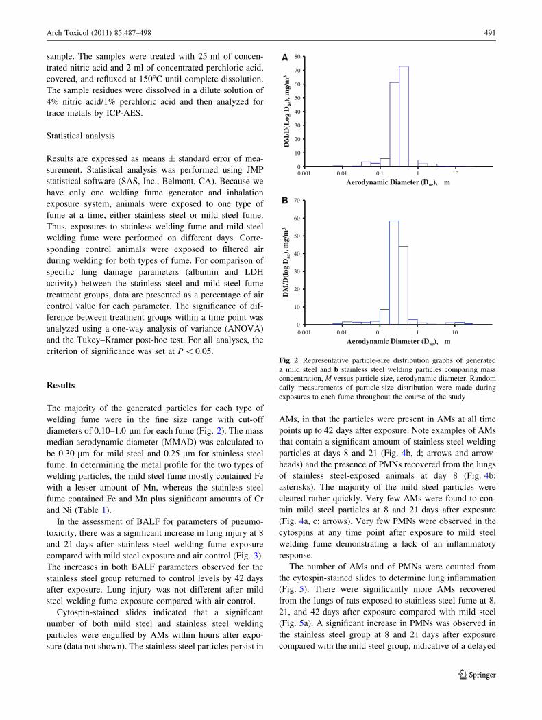
The majority of the generated particles for each type of
welding fume were in the fine size range with cut-off
diameters of 0.10–1.0 lm for each fume (Fig. 2). The mass
median aerodynamic diameter (MMAD) was calculated to
be 0.30 lm for mild steel and 0.25 lm for stainless steel
fume. In determining the metal profile for the two types of
welding particles, the mild steel fume mostly contained Fe
with a lesser amount of Mn, whereas the stainless steel
fume contained Fe and Mn plus significant amounts of Cr
and Ni (Table 1).
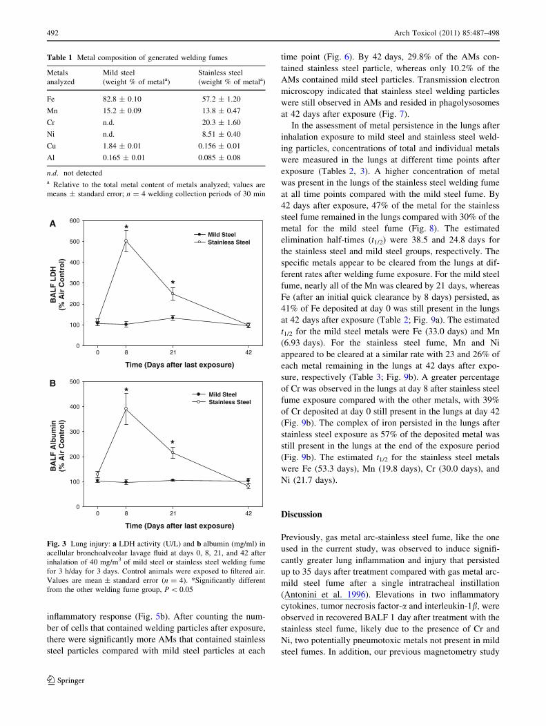
In the assessment of BALF for parameters of pneumo-
toxicity, there was a significant increase in lung injury at 8
and 21 days after stainless steel welding fume exposure
compared with mild steel exposure and air control (Fig. 3).
The increases in both BALF parameters observed for the
stainless steel group returned to control levels by 42 days
after exposure. Lung injury was not different after mild
steel welding fume exposure compared with air control.
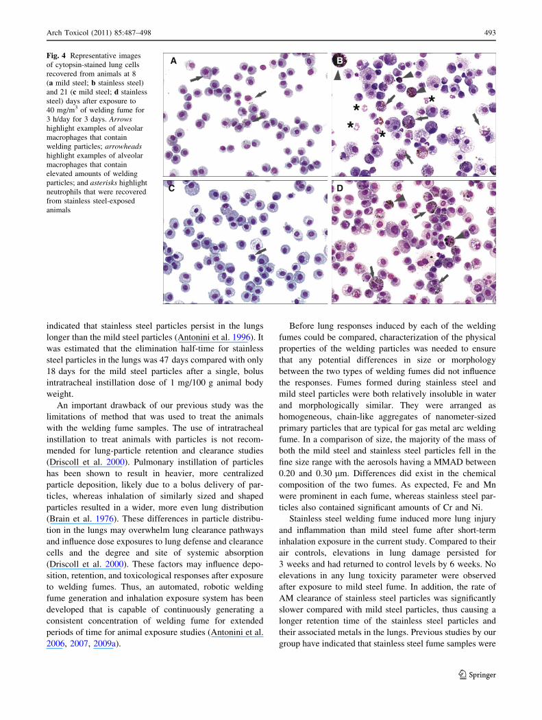
Cytospin-stained slides indicated that a significant
number of both mild steel and stainless steel welding
particles were engulfed by AMs within hours after expo-
sure (data not shown). The stainless steel particles persist in
AMs, in that the particles were present in AMs at all time
points up to 42 days after exposure. Note examples of AMs
that contain a significant amount of stainless steel welding
particles at days 8 and 21 (Fig. 4b, d; arrows and arrow-
heads) and the presence of PMNs recovered from the lungs
of stainless steel-exposed animals at day 8 (Fig. 4b;
asterisks). The majority of the mild steel particles were
cleared rather quickly. Very few AMs were found to con-
tain mild steel particles at 8 and 21 days after exposure
(Fig. 4a, c; arrows). Very few PMNs were observed in the
cytospins at any time point after exposure to mild steel
welding fume demonstrating a lack of an inflammatory
response.
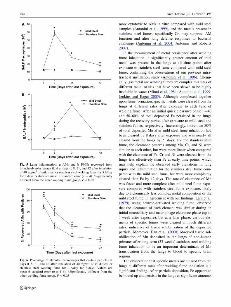
The number of AMs and of PMNs were counted from
the cytospin-stained slides to determine lung inflammation
(Fig. 5). There were significantly more AMs recovered
from the lungs of rats exposed to stainless steel fume at 8,
21, and 42 days after exposure compared with mild steel
(Fig. 5a). A significant increase in PMNs was observed in
the stainless steel group at 8 and 21 days after exposure
compared with the mild steel group, indicative of a delayed
0
10
20
30
40
50
60
70
80
0.001 0.01 0.1 1 10
DM
/D(L
og D
ae),
mg/
m3
Aerodynamic Diameter (Dae), µm
A
0
10
20
30
40
50
60
70
0.001 0.01 0.1 1 10
DM
/D(l
og D
ae),
mg/
m3
Aerodynamic Diameter (Dae), µm
B
Fig. 2 Representative particle-size distribution graphs of generated
a mild steel and b stainless steel welding particles comparing mass
concentration, M versus particle size, aerodynamic diameter. Random
daily measurements of particle-size distribution were made during
exposures to each fume throughout the course of the study
Arch Toxicol (2011) 85:487–498 491
123
inflammatory response (Fig. 5b). After counting the num-
ber of cells that contained welding particles after exposure,
there were significantly more AMs that contained stainless
steel particles compared with mild steel particles at each
time point (Fig. 6). By 42 days, 29.8% of the AMs con-
tained stainless steel particle, whereas only 10.2% of the
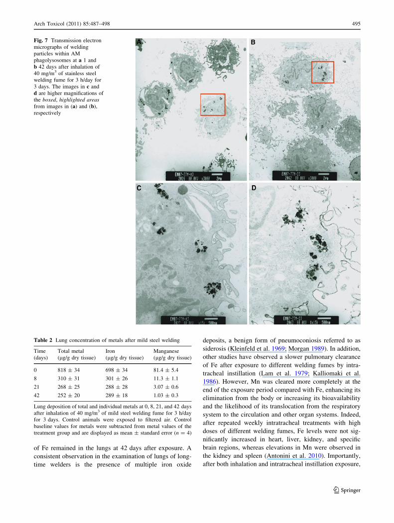
AMs contained mild steel particles. Transmission electron
microscopy indicated that stainless steel welding particles
were still observed in AMs and resided in phagolysosomes
at 42 days after exposure (Fig. 7).
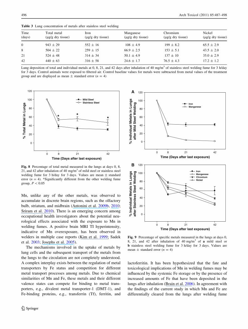
In the assessment of metal persistence in the lungs after
inhalation exposure to mild steel and stainless steel weld-
ing particles, concentrations of total and individual metals
were measured in the lungs at different time points after
exposure (Tables 2, 3). A higher concentration of metal
was present in the lungs of the stainless steel welding fume
at all time points compared with the mild steel fume. By
42 days after exposure, 47% of the metal for the stainless
steel fume remained in the lungs compared with 30% of the
metal for the mild steel fume (Fig. 8). The estimated
elimination half-times (t1/2) were 38.5 and 24.8 days for
the stainless steel and mild steel groups, respectively. The
specific metals appear to be cleared from the lungs at dif-
ferent rates after welding fume exposure. For the mild steel
fume, nearly all of the Mn was cleared by 21 days, whereas
Fe (after an initial quick clearance by 8 days) persisted, as
41% of Fe deposited at day 0 was still present in the lungs
at 42 days after exposure (Table 2; Fig. 9a). The estimated
t1/2 for the mild steel metals were Fe (33.0 days) and Mn
(6.93 days). For the stainless steel fume, Mn and Ni
appeared to be cleared at a similar rate with 23 and 26% of
each metal remaining in the lungs at 42 days after expo-
sure, respectively (Table 3; Fig. 9b). A greater percentage
of Cr was observed in the lungs at day 8 after stainless steel
fume exposure compared with the other metals, with 39%
of Cr deposited at day 0 still present in the lungs at day 42
(Fig. 9b). The complex of iron persisted in the lungs after
stainless steel exposure as 57% of the deposited metal was
still present in the lungs at the end of the exposure period
(Fig. 9b). The estimated t1/2 for the stainless steel metals
were Fe (53.3 days), Mn (19.8 days), Cr (30.0 days), and
Ni (21.7 days).
Discussion
Previously, gas metal arc-stainless steel fume, like the one
used in the current study, was observed to induce signifi-
cantly greater lung inflammation and injury that persisted
up to 35 days after treatment compared with gas metal arc-
mild steel fume after a single intratracheal instillation
(Antonini et al. 1996). Elevations in two inflammatory
cytokines, tumor necrosis factor-a and interleukin-1b, were
observed in recovered BALF 1 day after treatment with the
stainless steel fume, likely due to the presence of Cr and
Ni, two potentially pneumotoxic metals not present in mild
steel fumes. In addition, our previous magnetometry study
Table 1 Metal composition of generated welding fumes
Metals
analyzed
Mild steel
(weight % of metala)
Stainless steel
(weight % of metala)
Fe 82.8 ± 0.10 57.2 ± 1.20
Mn 15.2 ± 0.09 13.8 ± 0.47
Cr n.d. 20.3 ± 1.60
Ni n.d. 8.51 ± 0.40
Cu 1.84 ± 0.01 0.156 ± 0.01
Al 0.165 ± 0.01 0.085 ± 0.08
n.d. not detecteda Relative to the total metal content of metals analyzed; values are
means ± standard error; n = 4 welding collection periods of 30 min
Time (Days after last exposure)
422180
BA
LF
LD
H(%
Air
Co
ntr
ol)
0
100
200
300
400
500
600
Mild SteelStainless Steel
*
*
A
Time (Days after last exposure)
422180
BA
LF
Alb
um
in(%
Air
Co
ntr
ol)
0
100
200
300
400
500
Mild Steel Stainless Steel
*
*
B
Fig. 3 Lung injury: a LDH activity (U/L) and b albumin (mg/ml) in
acellular bronchoalveolar lavage fluid at days 0, 8, 21, and 42 after
inhalation of 40 mg/m3 of mild steel or stainless steel welding fume
for 3 h/day for 3 days. Control animals were exposed to filtered air.
Values are mean ± standard error (n = 4). *Significantly different
from the other welding fume group, P \ 0.05
492 Arch Toxicol (2011) 85:487–498
123
indicated that stainless steel particles persist in the lungs
longer than the mild steel particles (Antonini et al. 1996). It
was estimated that the elimination half-time for stainless
steel particles in the lungs was 47 days compared with only
18 days for the mild steel particles after a single, bolus
intratracheal instillation dose of 1 mg/100 g animal body
weight.
An important drawback of our previous study was the
limitations of method that was used to treat the animals
with the welding fume samples. The use of intratracheal
instillation to treat animals with particles is not recom-
mended for lung-particle retention and clearance studies
(Driscoll et al. 2000). Pulmonary instillation of particles
has been shown to result in heavier, more centralized
particle deposition, likely due to a bolus delivery of par-
ticles, whereas inhalation of similarly sized and shaped
particles resulted in a wider, more even lung distribution
(Brain et al. 1976). These differences in particle distribu-
tion in the lungs may overwhelm lung clearance pathways
and influence dose exposures to lung defense and clearance
cells and the degree and site of systemic absorption
(Driscoll et al. 2000). These factors may influence depo-
sition, retention, and toxicological responses after exposure
to welding fumes. Thus, an automated, robotic welding
fume generation and inhalation exposure system has been
developed that is capable of continuously generating a
consistent concentration of welding fume for extended
periods of time for animal exposure studies (Antonini et al.
2006, 2007, 2009a).
Before lung responses induced by each of the welding
fumes could be compared, characterization of the physical
properties of the welding particles was needed to ensure
that any potential differences in size or morphology
between the two types of welding fumes did not influence
the responses. Fumes formed during stainless steel and
mild steel particles were both relatively insoluble in water
and morphologically similar. They were arranged as
homogeneous, chain-like aggregates of nanometer-sized
primary particles that are typical for gas metal arc welding
fume. In a comparison of size, the majority of the mass of
both the mild steel and stainless steel particles fell in the
fine size range with the aerosols having a MMAD between
0.20 and 0.30 lm. Differences did exist in the chemical
composition of the two fumes. As expected, Fe and Mn
were prominent in each fume, whereas stainless steel par-
ticles also contained significant amounts of Cr and Ni.
Stainless steel welding fume induced more lung injury
and inflammation than mild steel fume after short-term
inhalation exposure in the current study. Compared to their
air controls, elevations in lung damage persisted for
3 weeks and had returned to control levels by 6 weeks. No
elevations in any lung toxicity parameter were observed
after exposure to mild steel fume. In addition, the rate of
AM clearance of stainless steel particles was significantly
slower compared with mild steel particles, thus causing a
longer retention time of the stainless steel particles and
their associated metals in the lungs. Previous studies by our
group have indicated that stainless steel fume samples were
A B
C D
**
*
*
Fig. 4 Representative images
of cytopsin-stained lung cells
recovered from animals at 8
(a mild steel; b stainless steel)
and 21 (c mild steel; d stainless
steel) days after exposure to
40 mg/m3 of welding fume for
3 h/day for 3 days. Arrowshighlight examples of alveolar
macrophages that contain
welding particles; arrowheadshighlight examples of alveolar
macrophages that contain
elevated amounts of welding
particles; and asterisks highlight
neutrophils that were recovered
from stainless steel-exposed
animals
Arch Toxicol (2011) 85:487–498 493
123
more cytotoxic to AMs in vitro compared with mild steel
samples (Antonini et al. 1999), and the metals present in
stainless steel fumes, specifically Cr, may suppress AM
function and alter lung defense responses to bacterial
challenge (Antonini et al. 2004; Antonini and Roberts
2007).
In the measurement of metal persistence after welding
fume inhalation, a significantly greater amount of total
metal was present in the lungs at all time points after
exposure to stainless steel fume compared with mild steel
fume, confirming the observations of our previous intra-
tracheal instillation study (Antonini et al. 1996). Chemi-
cally, gas metal arc welding fumes are complex mixtures of
different metal oxides that have been shown to be highly
insoluble in water (Minni et al. 1984; Antonini et al. 1999;
Jenkins and Eagar 2005). Although complexed together
upon fume formation, specific metals were cleared from the
lungs at different rates after exposure to each type of
welding fume. After an initial quick clearance phase, *40
and 50–60% of total deposited Fe persisted in the lungs
during the recovery period after exposure to mild steel and
stainless fumes, respectively. Interestingly, more than 80%
of total deposited Mn after mild steel fume inhalation had
been cleared by 8 days after exposure and was nearly all
cleared from the lungs by 21 days. For the stainless steel
fume, the clearance patterns among Mn, Cr, and Ni were
similar to each other, but were more linear when compared
with the clearance of Fe. Cr and Ni were cleared from the
lungs less effectively than Fe at early time points, which
may help explain the observed early elevations in lung
injury and inflammation for the stainless steel fume com-
pared with the mild steel fume, but were more completely
cleared than Fe by 42 days. The rate of clearance of Mn
was faster and more complete after mild steel fume expo-
sure compared with stainless steel fume exposure, likely
due to a chemically less complex metal composition of the
mild steel fume. In agreement with our findings, Lam et al.
(1979), using neutron-activated welding fume, observed
that the clearance of each element was similar during an
initial mucociliary and macrophage clearance phase (up to
1 week after exposure), but at a later phase, various ele-
ments of specific fumes were cleared at much different
rates, indicative of tissue solubilization of the deposited
particle. Moreover, Han et al. (2008) observed tissue sol-
ubilization of Mn deposited in the lungs of non-human
primates after long-term (33 weeks) stainless steel welding
fume inhalation to be an important determinant of Mn
translocation from the lungs to blood to specific brain
regions.
The observation that specific metals are cleared from the
lungs at different rates after welding fume inhalation is a
significant finding. After particle deposition, Fe appears to
be bound up and persists in the lungs as significant amounts
Time (Days after last exposure)422180
BA
LF
Mac
rop
hag
es (
106 )
0
10
20
30
40
50
60
70
Mild Steel Stainless Steel
*
*
*
A
Time (Days after last exposure)422180
BA
LF
Neu
tro
ph
ils (
106 )
0
1
2
3
4
Mild Steel Stainless Steel
*
*
B
Fig. 5 Lung inflammation: a AMs and b PMNs recovered from
bronchoalveolar lavage fluid at days 0, 8, 21, and 42 after inhalation
of 40 mg/m3 of mild steel or stainless steel welding fume for 3 h/day
for 3 days. Values are mean ± standard error (n = 4). *Significantly
different from the other welding fume group, P \ 0.05
Time (Days after last exposure)422180
% R
eco
vere
d A
Ms
wit
h P
arti
cles
0
20
40
60
80
100
Mild Steel Stainless Steel
*
*
* *
Fig. 6 Percentage of alveolar macrophages that contain particles at
days 0, 8, 21, and 42 after inhalation of 40 mg/m3 of mild steel or
stainless steel welding fume for 3 h/day for 3 days. Values are
mean ± standard error (n = 4–6). *Significantly different from the
other welding fume group, P \ 0.05
494 Arch Toxicol (2011) 85:487–498
123
of Fe remained in the lungs at 42 days after exposure. A
consistent observation in the examination of lungs of long-
time welders is the presence of multiple iron oxide
deposits, a benign form of pneumoconiosis referred to as
siderosis (Kleinfeld et al. 1969; Morgan 1989). In addition,
other studies have observed a slower pulmonary clearance
of Fe after exposure to different welding fumes by intra-
tracheal instillation (Lam et al. 1979; Kalliomaki et al.
1986). However, Mn was cleared more completely at the
end of the exposure period compared with Fe, enhancing its
elimination from the body or increasing its bioavailability
and the likelihood of its translocation from the respiratory
system to the circulation and other organ systems. Indeed,
after repeated weekly intratracheal treatments with high
doses of different welding fumes, Fe levels were not sig-
nificantly increased in heart, liver, kidney, and specific
brain regions, whereas elevations in Mn were observed in
the kidney and spleen (Antonini et al. 2010). Importantly,
after both inhalation and intratracheal instillation exposure,
Fig. 7 Transmission electron
micrographs of welding
particles within AM
phagolysosomes at a 1 and
b 42 days after inhalation of
40 mg/m3 of stainless steel
welding fume for 3 h/day for
3 days. The images in c and
d are higher magnifications of
the boxed, highlighted areasfrom images in (a) and (b),
respectively
Table 2 Lung concentration of metals after mild steel welding
Time
(days)
Total metal
(lg/g dry tissue)
Iron
(lg/g dry tissue)
Manganese
(lg/g dry tissue)
0 818 ± 34 698 ± 34 81.4 ± 5.4
8 310 ± 31 301 ± 26 11.3 ± 1.1
21 268 ± 25 288 ± 28 3.07 ± 0.6
42 252 ± 20 289 ± 18 1.03 ± 0.3
Lung deposition of total and individual metals at 0, 8, 21, and 42 days
after inhalation of 40 mg/m3 of mild steel welding fume for 3 h/day
for 3 days. Control animals were exposed to filtered air. Control
baseline values for metals were subtracted from metal values of the
treatment group and are displayed as mean ± standard error (n = 4)
Arch Toxicol (2011) 85:487–498 495
123
Mn, unlike any of the other metals, was observed to
accumulate in discrete brain regions, such as the olfactory
bulb, striatum, and midbrain (Antonini et al. 2009b, 2010;
Sriram et al. 2010). There is an emerging concern among
occupational health investigators about the potential neu-
rological effects associated with the exposure to Mn in
welding fumes. A positive brain MRI TI hyperintensity,
indicative of Mn overexposure, has been observed in
welders in multiple case reports (Kim et al. 1999; Sadek
et al. 2003; Josephs et al. 2005).
The mechanisms involved in the uptake of metals by
lung cells and the subsequent transport of the metals from
the lungs to the circulation are not completely understood.
A complex interplay exists between the regulation of metal
transporters by Fe status and competition for different
metal transport processes among metals. Due to chemical
similarities of Mn and Fe, these metals and their different
valence states can compete for binding to metal trans-
porters, e.g., divalent metal transporter-1 (DMT-1), and
Fe-binding proteins, e.g., transferrin (Tf), ferritin, and
lactoferritin. It has been hypothesized that the fate and
toxicological implications of Mn in welding fumes may be
influenced by the systemic Fe storage or by the presence of
increased amounts of Fe that have been deposited in the
lungs after inhalation (Brain et al. 2006). In agreement with
the findings of the current study in which Mn and Fe are
differentially cleared from the lungs after welding fume
Table 3 Lung concentration of metals after stainless steel welding
Time
(days)
Total metal
(lg/g dry tissue)
Iron
(lg/g dry tissue)
Manganese
(lg/g dry tissue)
Chromium
(lg/g dry tissue)
Nickel
(lg/g dry tissue)
0 943 ± 29 552 ± 16 108 ± 4.9 199 ± 8.2 65.5 ± 2.9
8 504 ± 22 259 ± 15 66.9 ± 2.5 153 ± 5.1 43.5 ± 2.0
21 524 ± 48 314 ± 34 50.1 ± 4.9 137 ± 10 35.0 ± 2.9
42 440 ± 63 316 ± 58 24.6 ± 1.7 76.5 ± 4.3 17.2 ± 1.2
Lung deposition of total and individual metals at 0, 8, 21, and 42 days after inhalation of 40 mg/m3 of stainless steel welding fume for 3 h/day
for 3 days. Control animals were exposed to filtered air. Control baseline values for metals were subtracted from metal values of the treatment
group and are displayed as mean ± standard error (n = 4)
Time (Days after last exposure)422180
% T
ota
l Met
al in
Lu
ng
s
0
20
40
60
80
100
120
Mild Steel Stainless Steel
* **
Fig. 8 Percentage of total metal measured in the lungs at days 0, 8,
21, and 42 after inhalation of 40 mg/m3 of mild steel or stainless steel
welding fume for 3 h/day for 3 days. Values are mean ± standard
error (n = 4). *Significantly different from the other welding fume
group, P \ 0.05
Time (Days after last exposure)422180
% In
div
idu
al M
etal
s in
Lu
ng
saf
ter
Mild
Ste
el W
eld
ing
0
20
40
60
80
100
120
Iron Manganese
A
Time (Days after last exposure)422180
% In
div
idu
al M
etal
s in
Lu
ng
saf
ter
Sta
inle
ss S
teel
Wel
din
g
0
20
40
60
80
100
120
Iron Manganese Chromium Nickel
B
Fig. 9 Percentage of specific metals measured in the lungs at days 0,
8, 21, and 42 after inhalation of 40 mg/m3 of a mild steel or
b stainless steel welding fume for 3 h/day for 3 days. Values are
mean ± standard error (n = 4)
496 Arch Toxicol (2011) 85:487–498
123
inhalation, recent studies have indicated that the in vivo
pharmacokinetics and the mechanisms by which intrat-
racheally instilled Mn and Fe cross the pulmonary epithe-
lium are remarkably different (Heilig et al. 2005, 2006).
Solubilized Fe was readily oxidized to bind to transferrin,
and the elements of the Tf/Tf receptor/DMT-1 pathway
were involved in its transport from the lungs. In contrast,
depending on valence state, the transport of Mn may
involve L-type Ca2? channels and TRPM7, a member of
the transient receptor potential melastatin subfamily.
In summary, the two welding fumes generated for the
study were nearly identical in particle size, solubility, and
morphology. Stainless steel welding fume was observed to
induce more lung injury and inflammation compared with
mild steel fume after a short-term inhalation exposure in
rats. No evidence of lung toxicity was observed after
exposure to the mild steel fume. As we hypothesized, the
rate of pulmonary clearance of deposited stainless steel
particles was significantly slower compared with mild steel
particles, thus causing a longer retention time of the
stainless steel particles and their associated metals in the
lungs. However, the specific metals present in the two
fumes were cleared from the lungs at different rates after
welding fume inhalation. The potentially more toxic metals
(e.g., Mn, Cr, Ni) present in the stainless steel fume were
cleared more completely than Fe by the end of the 42-day
exposure period, likely increasing their translocation from
the respiratory system to other organs. Ongoing studies in
the NIOSH welding laboratory are examining the effects of
these metals and their transport/uptake mechanisms after
welding fume inhalation in other organs, specifically the
central nervous and cardiovascular systems.
Acknowledgments The authors thank Amy Moseley, Jared
Cumpston, and Donny Leonard from the inhalation exposure team for
their expert technical assistance during the project. Funding for the
project was provided by the National Institute for Occupational Safety
and Health (NIOSH) and the National Occupational Research Agenda
(NORA). The authors also thank the National Toxicology Program
for additional support during the development of the welding fume
generator and exposure system.
Conflict of interest The authors declare that there are no conflicts
of interest.
Disclaimer The findings and conclusions of this paper have not
been formally disseminated by NIOSH and should not be construed to
represent any agency determination or policy.
References
Antonini JM (2003) Health effects of welding. Crit Rev Toxicol
33:61–103
Antonini JM, Roberts JR (2007) Chromium in stainless steel welding
fume suppresses lung defense responses against bacterial
infection in rats. J Immunotoxicol 4:117–127
Antonini JM, Krishna Murthy GG, Rogers RA, Albert R, Ulrich GD,
Brain JD (1996) Pneumotoxicity and pulmonary clearance of
different welding fumes after intratracheal instillation in the rat.
Toxicol Appl Pharmacol 140:188–199
Antonini JM, Lawryk NJ, Krishna Murthy GG, Brain JD (1999)
Effect of welding fume solubility on lung macrophage viability
and function in vitro. J Toxicol Environ Health 58:343–363
Antonini JM, Taylor MD, Millecchia L, Bebout AR, Roberts JR
(2004) Suppression in lung defenses after bacterial infection in
rats pretreated with different welding fumes. Toxicol Appl
Pharmacol 200:206–218
Antonini JM, Afshari AA, Stone S, Chen B, Schwegler-Berry D,
Fletcher WG, Goldsmith WT, Vandestouwe KH, McKinney W,
Castranova V, Frazer DG (2006) Design, construction, and
characterization of a novel robotic welding fume generator and
inhalation exposure system for laboratory animals. J Occup
Environ Hyg 3:194–203
Antonini JM, Stone S, Roberts JR, Chen B, Schwegler-Berry D,
Afshari A, Frazer DG (2007) Effect of short-term stainless steel
welding fume inhalation exposure on lung inflammation, injury,
and defense responses in rats. Toxicol Appl Pharmacol 223:234–
245
Antonini JM, Roberts JR, Stone S, Chen BT, Schwegler-Berry D,
Frazer DG (2009a) Short-term inhalation exposure to mild steel
welding fume had no effect on lung inflammation and injury, but
did alter defense responses to bacteria in rats. Inhal Toxicol
21:182–192
Antonini JM, Sriram K, Benkovic SA, Roberts JR, Stone S, Chen BT,
Schwegler-Berry D, Jefferson AM, Billig BK, Felton CM,
Hammer MA, Ma F, Frazer DG, O’Callaghan JP, Miller DB
(2009b) Mild steel welding fume causes manganese accumula-
tion and subtle neuroinflammatory changes but not overt neuro-
nal damage in discrete brain regions of rats after short-term
inhalation exposure. Neurotoxicol 30:915–925
Antonini JM, Roberts JR, Chapman RS, Soukup JM, Ghio AJ, Sriram
K (2010) Pulmonary toxicity and extrapulmonary tissue distri-
bution of metals after repeated exposure to different welding
fumes. Inhal Toxicol 22:805–816
Bowler RM, Roels HA, Nakagawa S, Drezgic M, Diamond E, Park R,
Koller W, Bowler RP, Mergler D, Bouchard M, Smith D,
Gwiazda R, Doty RL (2007) Dose–effect relationships between
manganese exposure and neurological, neuropsychological and
pulmonary function in confined space bridge welders. Occup
Environ Med 64:167–177
Brain JD, Knudson DW, Sorokin SP, Davis MA (1976) Pulmonary
distribution of particles given by intratracheal instillation or by
aerosol inhalation. Environ Res 11:13–33
Brain JD, Heilig E, Donaghey TC, Knutson MD, Wessling-Resnick
M, Molina RM (2006) Effects of iron status on transpulmonary
transport and tissue distribution of Mn and Fe. Am J Resir Cell
Mol Biol 34:330–337
Bureau of Labor Statistics, Occupational Employment Statistics:
Occupational Employment and Wages (2007) Welders, Cutter,
Solders, and Brazers, U.S. Department of Labor. Available at:
http://www.bls.gov/oes/current/oes514121.htm. Accessed 11/10/
2008
Driscoll KE, Costa DL, Hatch G, Henderson R, Oberdorster G, Salem
H, Schlesinger RB (2000) Intratracheal instillation as an exposure
technique for the evaluation of respiratory tract toxicity: uses and
limitations. Toxicol Sci 55:24–35
Ellingsen DG, Konstantinov R, Bast-Pettersen R, Merkurjeva L,
Chashchin M, Thomassen Y, Chashchin V (2008) A neurobehav-
ioral study of current and former welders exposed to manganese.
Neurotoxicol 29:48–59
Han JH, Chung YH, Park JD, Kim CY, Yang SO, Khang HS, Cheong
HK, Lee JS, Ha CS, Song C-W, Kwon IH, Sung JH, Heo JD,
Arch Toxicol (2011) 85:487–498 497
123
Kim N-Y, Huang M, Ch MH, Yu IJ (2008) Recovery from
welding-fume-exposure-induced MRI T1 signal intensities after
cessation of welding-fume exposure in brains of Cynomolgus
monkeys. Inhal Toxicol 20:1075–1083
Heilig E, Molina R, Donaghey T, Brain JD, Wessling-Resnick M (2005)
Pharmacokinetics of pulmonary manganese loading in iron-
deficient rats. Am J Physiol Lung Cell Mol Physiol 288:L887–
L893
Heilig EA, Thompson KJ, Molina RM, Ivanov AR, Brain JD,
Wessling-Resnick M (2006) Manganese and iron transport
across pulmonary epithelium. Am J Physiol Lung Cell Mol
Physiol 290:L1247–L1259
IARC (1990) Chromium, nickel, and welding. In: IARC monographs
on the evaluation of carcinogenic risks to humans. World Health
Organization, Geneva, pp 447–525
Jenkins NT, Eagar TW (2005) Chemical analysis of welding fume
particles. Welding J 84:87s–93s
Josephs KA, Ahlskog JE, Klos KJ, Kumar N, Fealey RD, Trenerry
MR, Cowl CT (2005) Neurologic manifestations in welders with
pallidal MRI T1 hyperintensity. Neurol 64:2033–2039
Kalliomaki PL, Hyvarinen H-K, Aitio A, Lakoma E-L, Kalliomaki K
(1986) Kinetics of the metal components of intratracheally
instilled stainless steel welding fume suspensions in rats. Br J Ind
Med 43:112–119
Kim Y, Kim J-W, Ito K, Lim H-S, Cheong H-K, Kim JY, Shin YC,
Kim SK, Moon Y (1999) Idiopathic parkinsonism with super-
imposed manganese exposure: utility of positron emission
tomography. Neurotoxicol 20:249–252
Kleinfeld M, Messite J, Kooyman O, Spiro J (1969) Welders’
siderosis: a clinical roentgenographic and physiological study.
Arch Environ Health 19:70–73
Lam HF, Hewitt PJ, Hicks R (1979) A study of pulmonary deposition
and the elimination of some constituent metals from welding
fume in laboratory animals. Ann Occup Hyg 21:363–373
Langard S (1994) Nickel-related cancer in welders. Sci Total Environ
148:303–309
Minni E, Gustafsson TE, Koponen M, Kalliomaki P-L (1984) A study
of the chemical structure of particles in the welding fumes of
mild and stainless steel. J Aerosol Sci 15:57–68
Morgan WKC (1989) On welding, wheezing, and whimsy. Am Ind
Hyg Assoc J 50:59–69
Moulin JJ (1997) A meta-analysis of epidemiologic studies of lung
cancer in welders. Scand J Work Environ Health 23:104–113
Moulin JJ, Wild P, Haguenoer JM et al (1993) A mortality study
among mild steel and stainless steel welders. Br J Ind Med
50:234–243
NIOSH (1994) Elements (ICP): Method 7300. In: NIOSH manual of
analytical methods, 4th Edition, Issue 2, U.S. Department of Health
and Human Services, Publication No. 98–119. Washington, DC
Sadek AH, Rauch R, Schulz PE (2003) Parkinsonism due to
manganism in a welder. Int J Toxicol 22:393–401
Sferlazza SJ, Beckett WS (1991) The respiratory health of welders.
Am Rev Respir Dis 143:1134–1148
Sriram K, Lin GX, Jefferson AM, Roberts JR, Chapman RS, Chen
BT, Soukup JM, Ghio AJ, Antonini JM (2010) Dopaminergic
neurotoxicity following pulmonary exposure to manganese-
containing welding fumes. Arch Toxicol 84:521–540
Sung JH, Choi B-G, Maeng S-H, Kim S-J, Chung YH, Han JH, Song
KS, Lee YH, Cho YB, Cho M-H, Kim KJ, Hyun JS, Yu IJ (2004)
Recovery from welding-fume-exposure-induced lung fibrosis
and pulmonary function changes in Sprague Dawley rats.
Toxicol Sci 82:608–613
Yu IJ, Song KS, Chang HK, Han JH, Kim KJ, Chung YH, Maeng SH,
Han KT, Chung KH, Chung HK (2001) Lung fibrosis in
Sprague-Dawley rat, induced by exposure to manual metal arc-
stainless steel welding fumes. Toxicol Sci 63:99–106
Yu IJ, Song KS, Chang HK, Han JH, Kim KJ, Chung YH, Han KT,
Chung KH, Chung HK (2003) Recovery from manual metal arc-
stainless steel welding-fume exposure induced lung fibrosis in
Sprague-Dawley rats. Toxicol Lett 143:247–259
Zeidler-Erdely PC, Kashon ML, Battelli LA, Young S-H, Erdely A,
Roberts JR, Reynolds SH, Antonini JM (2008) Pulmonary
inflammation and tumor induction in lung tumor susceptible A/J
and resistant C57BL/6J mice exposed to welding fume. Part
Fibre Toxicol 5:12
498 Arch Toxicol (2011) 85:487–498
123