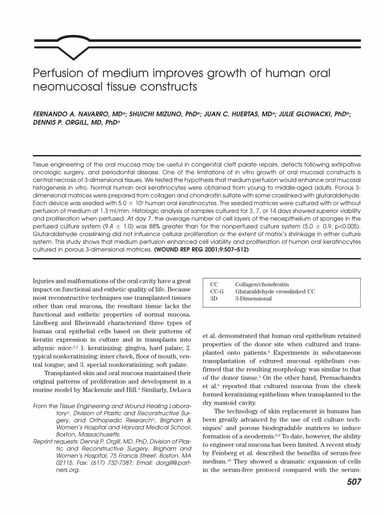
Perfusion of medium improves growth of human oral neomucosal tissue constructs
6
Perfusion of medium improves growth of human oral neomucosal tissue constructs FERNANDO A. NAVARRO, MD a ; SHUICHI MIZUNO, PhD b ; JUAN C. HUERTAS, MD a ; JULIE GLOWACKI, PhD b ; DENNIS P. ORGILL, MD, PhD a Tissue engineering of the oral mucosa may be useful in congenital cleft palate repairs, defects following extirpative oncologic surgery, and periodontal disease. One of the limitations of in vitro growth of oral mucosal constructs is central necrosis of 3-dimensional tissues. We tested the hypothesis that medium perfusion would enhance oral mucosal histogenesis in vitro. Normal human oral keratinocytes were obtained from young to middle-aged adults. Porous 3- dimensional matrices were prepared from collagen and chondroitin sulfate with some crosslinked with glutaraldehyde. Each device was seeded with 5.0 10 5 human oral keratinocytes. The seeded matrices were cultured with or without perfusion of medium at 1.3 ml/min. Histologic analysis of samples cultured for 3, 7, or 14 days showed superior viability and proliferation when perfused. At day 7, the average number of cell layers of the neoepithelium of sponges in the perfused culture system (9.4 1.0) was 88% greater than for the nonperfused culture system (5.0 0.9, p<0.005). Glutaraldehyde crosslinking did not influence cellular proliferation or the extent of matrix’s shrinkage in either culture system. This study shows that medium perfusion enhanced cell viability and proliferation of human oral keratinocytes cultured in porous 3-dimensional matrices. (WOUND REP REG 2001;9:507–512) Injuries and malformations of the oral cavity have a great CC Collagen/chondroitin impact on functional and esthetic quality of life. Because CC-G Glutaraldehyde crosslinked CC most reconstructive techniques use transplanted tissues 3D 3-Dimensional other than oral mucosa, the resultant tissue lacks the functional and esthetic properties of normal mucosa. Lindberg and Rheinwald characterized three types of human oral epithelial cells based on their patterns of et al. demonstrated that human oral epithelium retained keratin expression in culture and in transplants into properties of the donor site when cultured and trans- athymic mice: 1,2 1. keratinizing: gingiva, hard palate; 2. planted onto patients. 4 Experiments in subcutaneous typical nonkeratinizing: inner cheek, floor of mouth, ven- transplantation of cultured mucosal epithelium con- tral tongue; and 3. special nonkeratinizing: soft palate. firmed that the resulting morphology was similar to that Transplanted skin and oral mucosa maintained their of the donor tissue. 5 On the other hand, Premachandra original patterns of proliferation and development in a et al. 6 reported that cultured mucosa from the cheek murine model by Mackenzie and Hill. 3 Similarly, DeLuca formed keratinizing epithelium when transplanted to the dry mastoid cavity. From the Tissue Engineering and Wound Healing Labora- The technology of skin replacement in humans has tory a , Division of Plastic and Reconstructive Sur- been greatly advanced by the use of cell culture tech- gery, and Orthopedic Research b , Brigham & Women’s Hospital and Harvard Medical School, niques 7 and porous biodegradable matrices to induce Boston, Massachusetts. formation of a neodermis. 8,9 To date, however, the ability Reprint requests: Dennis P. Orgill, MD, PhD, Division of Plas- to engineer oral mucosa has been limited. A recent study tic and Reconstructive Surgery, Brigham and by Feinberg et al. described the benefits of serum-free Women’s Hospital, 75 Francis Street. Boston, MA medium. 10 They showed a dramatic expansion of cells 02115. Fax: (617) 732-7387; Email: dorgill@part- ners.org. in the serum-free protocol compared with the serum- 507
-
Upload
independent -
Category
Documents
-
view
0 -
download
0
Transcript of Perfusion of medium improves growth of human oral neomucosal tissue constructs
Perfusion of medium improves growth of human oralneomucosal tissue constructs
FERNANDO A. NAVARRO, MDa; SHUICHI MIZUNO, PhDb; JUAN C. HUERTAS, MDa; JULIE GLOWACKI, PhDb;DENNIS P. ORGILL, MD, PhDa
Tissue engineering of the oral mucosa may be useful in congenital cleft palate repairs, defects following extirpativeoncologic surgery, and periodontal disease. One of the limitations of in vitro growth of oral mucosal constructs iscentral necrosis of 3-dimensional tissues. We tested the hypothesis that medium perfusion would enhance oral mucosalhistogenesis in vitro. Normal human oral keratinocytes were obtained from young to middle-aged adults. Porous 3-dimensional matrices were prepared from collagen and chondroitin sulfate with some crosslinkedwith glutaraldehyde.Each device was seeded with 5.0 � 105 human oral keratinocytes. The seeded matrices were cultured with or withoutperfusion of medium at 1.3 ml/min. Histologic analysis of samples cultured for 3, 7, or 14 days showed superior viabilityand proliferation when perfused. At day 7, the average number of cell layers of the neoepithelium of sponges in theperfused culture system (9.4 � 1.0) was 88% greater than for the nonperfused culture system (5.0 � 0.9, p<0.005).Glutaraldehyde crosslinking did not influence cellular proliferation or the extent of matrix’s shrinkage in either culturesystem. This study shows that medium perfusion enhanced cell viability and proliferation of human oral keratinocytescultured in porous 3-dimensional matrices. (WOUND REP REG 2001;9:507–512)
Injuries and malformations of the oral cavity have a great CC Collagen/chondroitinimpact on functional and esthetic quality of life. Because CC-G Glutaraldehyde crosslinked CCmost reconstructive techniques use transplanted tissues 3D 3-Dimensionalother than oral mucosa, the resultant tissue lacks thefunctional and esthetic properties of normal mucosa.Lindberg and Rheinwald characterized three types ofhuman oral epithelial cells based on their patterns of
et al. demonstrated that human oral epithelium retainedkeratin expression in culture and in transplants into
properties of the donor site when cultured and trans-athymic mice:1,2 1. keratinizing: gingiva, hard palate; 2.
planted onto patients.4 Experiments in subcutaneoustypical nonkeratinizing: inner cheek, floor of mouth, ven-
transplantation of cultured mucosal epithelium con-tral tongue; and 3. special nonkeratinizing: soft palate.
firmed that the resulting morphology was similar to thatTransplanted skin and oral mucosa maintained their
of the donor tissue.5 On the other hand, Premachandraoriginal patterns of proliferation and development in a
et al.6 reported that cultured mucosa from the cheekmurine model by Mackenzie and Hill.3 Similarly, DeLuca
formed keratinizing epithelium when transplanted to thedry mastoid cavity.From the Tissue Engineering and Wound Healing Labora-
The technology of skin replacement in humans hastorya, Division of Plastic and Reconstructive Sur-been greatly advanced by the use of cell culture tech-gery, and Orthopedic Researchb, Brigham &
Women’s Hospital and Harvard Medical School, niques7 and porous biodegradable matrices to induceBoston, Massachusetts. formation of a neodermis.8,9 To date, however, the ability
Reprint requests: Dennis P. Orgill, MD, PhD, Division of Plas- to engineer oral mucosa has been limited. A recent studytic and Reconstructive Surgery, Brigham and
by Feinberg et al. described the benefits of serum-freeWomen’s Hospital, 75 Francis Street. Boston, MAmedium.10 They showed a dramatic expansion of cells02115. Fax: (617) 732-7387; Email: dorgill@part-
ners.org. in the serum-free protocol compared with the serum-
507
WOUND REPAIR AND REGENERATIONNOVEMBER–DECEMBER 2001508 NAVARRO, ET AL
containing protocol at 96 hours. The elimination of che- They were expanded from cryopreserved stocks aspreviously described1,7 by cocultivation with mitomycin-lated fetal bovine serum may have preselected a more
undifferentiated and actively proliferative primary kera- treated Swiss 3T3J2 cells in medium consisting of Dulbec-co’s modified Eagle’s medium/Ham’s F12 (1:1 v/v) mediumtinocyte that was able to grow more rapidly. High-density
cell culture in three-dimensional (3-D) matrices, which (GIBCO/BRL, Grand Island, NY), supplemented with 5%calf serum (Hyclone Laboratories, Logan, UT), 10 ng/mlmore closely mimics in vivo cell distribution, has been
shown to result in limited cell proliferation and central epidermal growth factor (Sigma Chemical Co.), 5 �g/mlhuman insulin (Eli Lilly Co, Indianapolis, IN), 0.4 �g/mlnecrosis due to limitations of nutrient and waste ex-
change. Recently a system of medium perfusion for 3-D hydrocortisone, 1.0 � 10�10 M cholera toxin, 2 � 10�11
M triiodothyronine, and 1.8 � 10�4 M adenine (all fromscaffolds was developed to enhance viability of cellsand histogenesis in vivo. Medium perfusion of porous Sigma Chemical Co.). Human oral keratinocytes were
cultured to approximately 80% confluence and collectedcollagen sponges seeded with murine bone marrow cellsenhanced cell viability and hematopoietic function.11 In for experimentation.addition, perfusion was shown to enhance 3-D culture
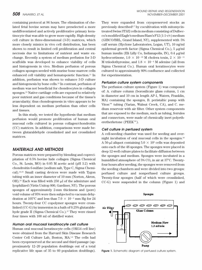
Perfusion culture system componentsand histogenesis by bone cells.12 In contrast, perfusion ofThe perfusion culture system (Figure 1) was composedmedium was not beneficial for chondrocytes in collagenof: A. culture column (borosilicate glass column, 1 cmsponges.13 Native cartilage cells are exposed to relativelyin diameter and 10 cm in length AP-1, Waters, Franklin,poor nutrient and gas conditions because of the tissue’sMA) containing the sponges, B. peristaltic pump withavascularity; thus chondrogenesis in vitro appears to beViton� tubing (Varian, Walnut Creek, CA), and C. me-less dependent on medium perfusion than other cellsdium reservoir with air filter. Other system componentsand tissues.that are exposed to the medium, such as tubing, ferrulesIn this study, we tested the hypothesis that mediumand connectors, were made of chemically inert polyeth-perfusion would promote proliferation of human oraleretherketone (PEEK�).mucosal cells cultured in porous collagen/chondroitin
(CC) matrices. In addition, comparisons were made be- Cell culture in perfused systemtween glutaraldehyde crosslinked and not crosslinked A cell-seeding chamber was used for seeding and over-matrices. night incubation of oral mucosal cells in the sponges.16
A 50-�l aliquot containing 5.0 � 105 cells was depositedonto each of the 48 sponges. The sponges were placed in
MATERIALS AND METHODS deep 12-well culture plates to facilitate diffusion betweenPorous matrices were prepared by blending and copreci- the sponges and medium. Sponges were incubated in apitation of 0.5% bovine hide collagen (Sigma Chemical humidified atmosphere of 5% CO2 in air at 37�C. Twenty-Co., St. Louis, MO) in 0.05 M acetic acid (pH 3.2) with four hours after seeding, the sponges were removed fromchondroitin 6-sulfate (sodium salt, Type C, Sigma Chemi- the seeding chambers and were divided into two groups:cal).14,15 Small casting devices were made with Tygon perfused culture and nonperfused culture groups.tubing with an inner diameter of 10 mm (Norton, Akron, Twenty-four sponges (half of which were crosslinked,OH).16 Each was filled with 250 �l of the admixture and CC-G) were suspended in the column (Figure 1) andlyophilized (Virtis Unitop 800, Gardiner, NY). The poroussponges of approximately 2-mm thickness and (pore)void volume of 95% were then subjected to vacuum dehy-dration at 105�C and less than 7.0 � 10�2 mm Hg for 24hours. Twenty-four CC copolymer sponges were cross-linked (CC-G) by immersion in a bath of 0.25% glutaralde-hyde grade II (Sigma Chemical Co.).17 They were rinsedfour times with 100 ml of distilled water.
Human oral mucosal keratinocyte cell cultureHuman oral mucosal keratinocyte cells (OKG4 cell line)were obtained from the Harvard Skin Disease ResearchCenter Cell Culture Lab, Boston, MA.1,2 The cells hadbeen cryopreserved at the second and third passage (ap-proximately 12–20 population doublings out of a total
Figure 1. Schematic diagram of perfused culture system.replicative life span of 35 to 60 population doublings).
WOUND REPAIR AND REGENERATIONVOL. 9, NO. 6 NAVARRO, ET AL 509
maintained horizontally at 37�C with 5% CO2 in air. The number of nucleated cell layers forming the neoepithe-lium at 10 different locations at uniform intervals alongmedium flow rate of 1.3 ml/min (1.6 cm/min within the
column) produces laminar flow with Reynolds number each cross-section.The statistical analysis we used was a mixed-effectsRe � 2.07. The calculated wall shear stress was 1.57
� 10�3 dynes/cm2, which is 0.1–1% of the shear stress regression model.18,19 Because the outcomes of interestwere measured multiple times on different sponges atachieved in veins.12 The system maintained a medium
volume of 10 ml per sponge, and culture medium was each time point (10 different sections of each sponge)for both the control and treatment groups, we analyzedchanged every 7 days.
For the control, nonperfused group, twenty-four the data using a general linear model for a group-repeatedmeasures design. This assumes that the removed spongessponges (half of which were crosslinked, CC-G) were
maintained in six-well plates (one sponge per 4 ml me- represented a random sample from the population ofinterest and that the values of the outcomes have a multi-dium per well) with half volumes of media changed bi-
weekly. variate normal distribution. The statistical procedureused was a mixed-effects regression model and the soft-Groups of eight perfused and nonperfused sponges
were harvested at day 3, 7, and 14. Each sponge was ware to fit the model was SAS PROC MIXED20 (SAS,Cary, NC). Quantial plots were generated to assess themeasured, fixed in formalin, embedded in paraffin, sec-
tioned, and stained with hematoxylin and eosin. normality assumption of the data.
Cell analysisRESULTSTwo observers, who were blind to the groupings, evalu-
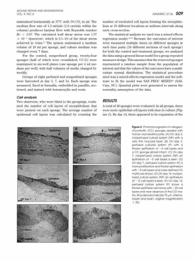
ated the number of cell layers of neoepithelium that A total of 48 sponges were evaluated. In all groups, therewere more epithelial cell layers with time in culture (Fig-were present on each sponge. The average number of
epidermal cell layers was calculated by counting the ure 2). By day 14, there appeared to be expansion of the
Figure 2. Photomicrographs of collagen/chondroitin (CC) sponges seeded withhuman oral keratinocytes. (A) On day 3,nonperfused culture system (NP) with avery thin mucosal layer. (B) On day 3,perfused cultured system (P) with athicker epithelium of �4 cell layers anda CC sponge almost intact. (C) On day7, nonperfused culture system (NP) anepithelium of �5 cell layers is seen. (D)On day 7, perfused culture system (P) amore proliferative and thicker epitheliumwith �9 cell layers and a less defined CGmatrix are shown. (E) On day 14, nonper-fused culture system (NP) an epitheliumof �13 cell layers is seen. (F) On day 14,perfused culture system (P) shows athicker epithelial cell clump with �20 celllayers and near absence of the CG ma-trix. All scale bars indicate 75 �m. (Hema-toxylin and eosin; original magnification� 20).
WOUND REPAIR AND REGENERATIONNOVEMBER–DECEMBER 2001510 NAVARRO, ET AL
tissue with replacement of the CC and CC-G matrices. were perfused decreased in diameter 15% by day 3, 24%by day 7, and 46% by day 14; there was an average of 4.6More epithelial cells were observed at each time point
in the group of perfused devices than in the nonperfused mm reduction in diameter of the perfused CC devicesthroughout the experiment. The CC-G sponges that wereones. At 3, 7, and 14 day after seeding the cells, there
were 250%, 90% and 50% more epithelial cell layers re- not perfused decreased in diameter 6% by day 3, 13% byday 7, and 31% by day 14; there was an average of 3.1spectively, in the CC matrices that were perfused than in
the nonperfused matrices (Table 1). In the CC-G matrices mm reduction for nonperfused sponges throughout theexperiment. At each time point, the average diameter wasperfusion also significantly enhanced cell layers at day
3 (122%) and at day 7 (75%) (the increase at day 14 did similar between the perfused and nonperfused groups ofcrosslinked sponges. This analysis shows that the en-not reach statistical significance) (Table 1). Comparison
of sponges that were crosslinked with those that were hancements of shrinkage by perfusion were abrogatedin the CC-G matrices.not crosslinked indicated no significant differences at
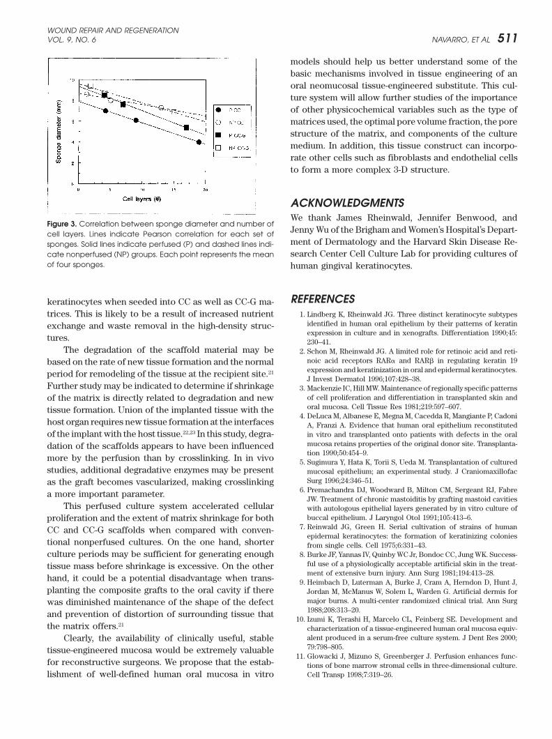
any time point. Analysis of correlations between cellularity andsponge diameter revealed significance for both perfusedMeasurements of device diameter indicated that
there was shrinkage of each type with time in vitro. The groups (Figure 3). The perfused CC and CC-G spongesachieved the greatest cellularity and the maximal shrink-extent of shrinkage was greater for the perfused CC
sponges than for those that were not perfused (Table age. There was a significant correlation between spongediameter and number of cell layers for the perfused CC2). At each time point, the perfused CC sponges were
significantly smaller than those that were not perfused. sponges (Pearson r � –0.9996, p � 0.017) and for theperfused CC-G sponges (r � –0.9980, p � 0.044). TheAll sponges were 10 mm at the beginning of incubation.
The CC sponges that were perfused decreased in diame- correlations for nonperfused CC sponges (r � –0.9875,p � 0.1008) and CC-G sponges (r � –0.9930, p � 0.0755)ter 30% by day 3, 39% by day 7, and 60% by day 14; there
was an average of 6.0 mm reduction in diameter of the did not reach statistical significance. This is due to thefact that those two groups achieved the lowest cellularityperfused CC devices throughout the experiment. The CC
sponges that were not perfused decreased in diameter and shrinkage.14% by day 3, 20% by day 7, and 27% by day 14; therewas an average of 2.7 mm reduction for nonperfused
DISCUSSIONmatrices throughout the experiment. At each time point,shrinkage was significantly greater in the perfused than Normal human keratinocytes develop stratified epithelial
structures in 2-dimensional cultures. The use of 3D-cul-in the nonperfused groups.The extent of shrinkage was greater for the perfused ture systems may more closely approximate in vivo con-
ditions. This study shows that a 3-D perfusion cultureCC-G matrices than for those that were not perfused,but not significantly so (Table 2). The CC-G sponges that system allows for greater proliferation of human oral
Table 1. Effect of perfusion on the number of cell layers in CC sponges with and without glutaraldehyde crosslinking
CC Sponges CC-G Sponges
Time (d) Perfused Nonperfused P value Perfused Nonperfused P value
3 4.2 � 0.5* 1.2 � 0.5 0.04 4.0 � 0.1 1.8 � 0.4 0.0467 9.0 � 1.0 5.0 � 0.9 0.001 7.0 � 0.5 4.0 � 1.5 0.04
14 19 � 1.2 13 � 2.0 0.0005 17 � 2.0 15 � 2.0 0.07
N � 4 samples per group; P values compare �/� perfusion.*Values indicate the number of cell layers.
Table 2. Effect of perfusion on sponge diameter in CC sponges with and without glutaraldehyde crosslinking
CC Sponges CC-G Sponges
Time (d) Perfused Nonperfused P value Perfused Nonperfused P value
3 7.0 � 1.5* 8.6 � 1.7 0.04 8.5 � 0.7 9.4 � 0.2 0.087 6.1 � 1.2 8.0 � 1.5 0.01 7.6 � 1.5 8.7 � 0.9 0.08
14 4.0 � 1.0 7.3 � 1.5 0.001 5.4 � 1.0 6.9 � 2.0 0.07
*Values indicate diameter in mm.N � 4 samples per group; P values compare �/� perfusion.
WOUND REPAIR AND REGENERATIONVOL. 9, NO. 6 NAVARRO, ET AL 511
models should help us better understand some of thebasic mechanisms involved in tissue engineering of anoral neomucosal tissue-engineered substitute. This cul-ture system will allow further studies of the importanceof other physicochemical variables such as the type ofmatrices used, the optimal pore volume fraction, the porestructure of the matrix, and components of the culturemedium. In addition, this tissue construct can incorpo-rate other cells such as fibroblasts and endothelial cellsto form a more complex 3-D structure.
ACKNOWLEDGMENTSWe thank James Rheinwald, Jennifer Benwood, and
Figure 3. Correlation between sponge diameter and number ofJenny Wu of the Brigham and Women’s Hospital’s Depart-cell layers. Lines indicate Pearson correlation for each set ofment of Dermatology and the Harvard Skin Disease Re-sponges. Solid lines indicate perfused (P) and dashed lines indi-search Center Cell Culture Lab for providing cultures ofcate nonperfused (NP) groups. Each point represents the mean
of four sponges. human gingival keratinocytes.
REFERENCESkeratinocytes when seeded into CC as well as CC-G ma-trices. This is likely to be a result of increased nutrient 1. Lindberg K, Rheinwald JG. Three distinct keratinocyte subtypes
identified in human oral epithelium by their patterns of keratinexchange and waste removal in the high-density struc-expression in culture and in xenografts. Differentiation 1990;45:
tures. 230–41.The degradation of the scaffold material may be 2. Schon M, Rheinwald JG. A limited role for retinoic acid and reti-
noic acid receptors RAR� and RAR� in regulating keratin 19based on the rate of new tissue formation and the normalexpression and keratinization in oral and epidermal keratinocytes.period for remodeling of the tissue at the recipient site.21
J Invest Dermatol 1996;107:428–38.Further study may be indicated to determine if shrinkage 3. Mackenzie IC, Hill MW. Maintenance of regionally specific patterns
of cell proliferation and differentiation in transplanted skin andof the matrix is directly related to degradation and neworal mucosa. Cell Tissue Res 1981;219:597–607.tissue formation. Union of the implanted tissue with the
4. DeLuca M, Albanese E, Megna M, Cacedda R, Mangiante P, Cadonihost organ requires new tissue formation at the interfaces A, Franzi A. Evidence that human oral epithelium reconstituted
in vitro and transplanted onto patients with defects in the oralof the implant with the host tissue.22,23 In this study, degra-mucosa retains properties of the original donor site. Transplanta-dation of the scaffolds appears to have been influencedtion 1990;50:454–9.
more by the perfusion than by crosslinking. In in vivo 5. Sugimura Y, Hata K, Torii S, Ueda M. Transplantation of culturedstudies, additional degradative enzymes may be present mucosal epithelium; an experimental study. J Craniomaxillofac
Surg 1996;24:346–51.as the graft becomes vascularized, making crosslinking6. Premachandra DJ, Woodward B, Milton CM, Sergeant RJ, Fabrea more important parameter.
JW. Treatment of chronic mastoiditis by grafting mastoid cavitiesThis perfused culture system accelerated cellular with autologous epithelial layers generated by in vitro culture of
buccal epithelium. J Laryngol Otol 1991;105:413–6.proliferation and the extent of matrix shrinkage for both7. Reinwald JG, Green H. Serial cultivation of strains of humanCC and CC-G scaffolds when compared with conven-
epidermal keratinocytes: the formation of keratinizing coloniestional nonperfused cultures. On the one hand, shorter from single cells. Cell 1975;6:331–43.
8. Burke JF, Yannas IV, Quinby WC Jr, Bondoc CC, Jung WK. Success-culture periods may be sufficient for generating enoughful use of a physiologically acceptable artificial skin in the treat-tissue mass before shrinkage is excessive. On the otherment of extensive burn injury. Ann Surg 1981;194:413–28.
hand, it could be a potential disadvantage when trans- 9. Heimbach D, Luterman A, Burke J, Cram A, Herndon D, Hunt J,planting the composite grafts to the oral cavity if there Jordan M, McManus W, Solem L, Warden G. Artificial dermis for
major burns. A multi-center randomized clinical trial. Ann Surgwas diminished maintenance of the shape of the defect1988;208:313–20.and prevention of distortion of surrounding tissue that
10. Izumi K, Terashi H, Marcelo CL, Feinberg SE. Development andthe matrix offers.21
characterization of a tissue-engineered human oral mucosa equiv-alent produced in a serum-free culture system. J Dent Res 2000;Clearly, the availability of clinically useful, stable79:798–805.tissue-engineered mucosa would be extremely valuable
11. Glowacki J, Mizuno S, Greenberger J. Perfusion enhances func-for reconstructive surgeons. We propose that the estab- tions of bone marrow stromal cells in three-dimensional culture.
Cell Transp 1998;7:319–26.lishment of well-defined human oral mucosa in vitro
WOUND REPAIR AND REGENERATIONNOVEMBER–DECEMBER 2001512 NAVARRO, ET AL
12. Mueller S, Mizuno S, Gerstenfeld L, Glowacki J. Medium perfusion 18. Morrison DF. Multivariate statistical methods. New York: McGraw-Hill Publishing, 1990;100–25.enhances osteogenesis by murine osteosarcoma cells in three-
dimensional collagen sponges. J Bone Miner Res 1999;14:2118–26. 19. SAS/STAT. Users guide. Cary NC: SAS Institute Inc, 1990;25–30.20. Laird N, James H. Random effects models for longitudinal data.13. Mizuno S, Allemann F, Glowacki J. Effects of medium perfusion
on matrix production by bovine chondrocytes in the three-dimen- New York: Biometrics, 1982;963–74.21. Spector M. Basic principles of tissue engineering. In: Lynch SE,sional collagen sponges. J Biomed Mater Res 2001;56:368–75.
14. Yannas IV, Burke JF. Design of an artificial skin. I. Basic design Genco RJ, Marx RE, editors. Tissue engineering: Applications inmaxillofacial surgery and periodontics. Carol Stream: Quintes-principles. J Biomed Mater Res 1980;14:65–81.
15. Yannas IV, Burke JF, Gordon PL, Huang C, Rubenstein RH. Design sence Publishing 1998;3–16.22. Murphy GF, Orgill DP, Yannas IV. Partial dermal regeneration isof an artificial skin. II. Control of chemical composition. J Biomed
Mater Res 1980;14:107–32. induced by biodegradable collagen-glycosaminoglycan grafts. LabInvest 1990;63:305–13.16. Mizuno S, Glowacki J. Three-dimensional composite of demineral-
ized bone powder and collagen for in vitro analysis of chondroin- 23. Nehrer S, Breinan HA, Ramapa A, Hsu HP, Minas T, Shortkroff S,Sledge CP, Yannas IV, Spector M. Chondrocyte-seeded collagenduction of human dermal fibroblasts. Biomaterials 1996;17:
1819–25. matrices implanted in a chondral defect in a canine model. Bioma-terial 1998;19:2313–28.17. Dagalakis N, Flink J, Stasikelis P, Burke JF, Yannas IV. Design of
an artificial skin. Part III. Control of pore structure. J BiomedMater Res 1980;14:511–28.