Effects of Medium Perfusion Rate on Cell-Seeded Three-Dimensional Bone Constructs in Vitro
7
1197 INTRODUCTION I N THE ABSENCE of a vascular blood supply in vitro, nu- trient delivery to cells throughout three-dimensional (3D) tissue-engineered constructs grown in static culture must occur by diffusion. As a result, thin tissues (e.g., skin) and tissues that are naturally avascular (e.g., carti- lage) have been more readily grown in vitro than thicker, vascular tissues such as bone. 1–3 When 3D cellular con- structs are grown in static culture, cells on the outer sur- face of the constructs are typically viable and proliferate readily while cells within the construct may be less ac- tive or necrotic. 4–6 For example, rat calvarial osteoblasts cultured statically within demineralized trabecular bone scaffolds (6.0 mm in diameter and 3.0 mm thick) for 56 days produce a thin (less than 500 mm) layer of miner- alized matrix around the construct periphery (Fig. 1A). Tissue culture systems that provide dynamic medium flow conditions around or within tissue-engineered con- structs are designed to enhance nutrient exchange and cell growth in vitro. 7–9 Such tissue culture systems may be useful as bioreactors to engineer thicker, more uniform tissues for implantation or as test bed models that simu- late aspects of the in vivo environment. TISSUE ENGINEERING Volume 9, Number 6, 2003 © Mary Ann Liebert, Inc. Effects of Medium Perfusion Rate on Cell-Seeded Three-Dimensional Bone Constructs in Vitro* SARAH H. CARTMELL, Ph.D., BLAISE D. PORTER, B.A., ANDRÉS J. GARCÍA, Ph.D., and ROBERT E. GULDBERG, Ph.D. ABSTRACT Cellular activity at the center of tissue-engineered constructs in static culture is typically decreased relative to the construct periphery because of transport limitations. We have designed a tissue cul- ture system that perfuses culture medium through three-dimensional (3D) porous cellular constructs to improve nutrient delivery and waste removal within the constructs. This study examined the ef- fects of medium perfusion rate on cell viability, proliferation, and gene expression within cell-seeded 3D bone scaffolds. Human trabecular bone scaffolds were seeded with MC3T3-E1 osteoblast-like cells and perfused for 1 week at flow rates of 0.01, 0.1, 0.2, and 1.0 mL/min. Confocal microscopy after 1 week of culture indicated that a flow rate of 1.0 mL/min resulted in substantial cell death throughout the constructs whereas lowering the flow rate led to an increasing proportion of viable cells, particularly at the center of the constructs. DNA analysis showed increases in cell prolifera- tion at a flow rate of 0.01 mL/min relative to 0.2 mL/min and static controls. Conversely, mRNA expressions of Runx2, osteocalcin, and alkaline phosphatase were upregulated at 0.2 mL/min com- pared with lower flow rates as quantified by real-time RT-PCR. These data suggest that medium perfusion may benefit the development of 3-D tissues in vitro by enhancing transport of nutrients and waste within the constructs and providing flow-mediated mechanical stimuli. School of Mechanical Engineering, Georgia Institute of Technology, Atlanta, Georgia. *Previously presented at the 3rd Biennial Meeting of the Tissue Engineering Society (November 30–December 3, 2000, Or- lando, Florida), the 47th Annual Meeting of the Orthopaedic Research Society (February 25–28, 2001, San Francisco, Califor- nia), and the Biomedical Engineering Society (October 4–7, 2001, Durham, North Carolina).
-
Upload
independent -
Category
Documents
-
view
0 -
download
0
Transcript of Effects of Medium Perfusion Rate on Cell-Seeded Three-Dimensional Bone Constructs in Vitro
1197
INTRODUCTION
IN THE ABSENCE of a vascular blood supply in vitro, nu-trient delivery to cells throughout three-dimensional
(3D) tissue-engineered constructs grown in static culturemust occur by diffusion. As a result, thin tissues (e.g.,skin) and tissues that are naturally avascular (e.g., carti-lage) have been more readily grown in vitro than thicker,vascular tissues such as bone.1–3 When 3D cellular con-structs are grown in static culture, cells on the outer sur-face of the constructs are typically viable and proliferatereadily while cells within the construct may be less ac-
tive or necrotic.4–6 For example, rat calvarial osteoblastscultured statically within demineralized trabecular bonescaffolds (6.0 mm in diameter and 3.0 mm thick) for 56days produce a thin (less than 500 mm) layer of miner-alized matrix around the construct periphery (Fig. 1A).Tissue culture systems that provide dynamic mediumflow conditions around or within tissue-engineered con-structs are designed to enhance nutrient exchange and cellgrowth in vitro.7–9 Such tissue culture systems may beuseful as bioreactors to engineer thicker, more uniformtissues for implantation or as test bed models that simu-late aspects of the in vivo environment.
TISSUE ENGINEERINGVolume 9, Number 6, 2003© Mary Ann Liebert, Inc.
Effects of Medium Perfusion Rate on Cell-Seeded Three-Dimensional Bone Constructs in Vitro*
SARAH H. CARTMELL, Ph.D., BLAISE D. PORTER, B.A., ANDRÉS J. GARCÍA, Ph.D., and ROBERT E. GULDBERG, Ph.D.
ABSTRACT
Cellular activity at the center of tissue-engineered constructs in static culture is typically decreasedrelative to the construct periphery because of transport limitations. We have designed a tissue cul-ture system that perfuses culture medium through three-dimensional (3D) porous cellular constructsto improve nutrient delivery and waste removal within the constructs. This study examined the ef-fects of medium perfusion rate on cell viability, proliferation, and gene expression within cell-seeded3D bone scaffolds. Human trabecular bone scaffolds were seeded with MC3T3-E1 osteoblast-likecells and perfused for 1 week at flow rates of 0.01, 0.1, 0.2, and 1.0 mL/min. Confocal microscopyafter 1 week of culture indicated that a flow rate of 1.0 mL/min resulted in substantial cell deaththroughout the constructs whereas lowering the flow rate led to an increasing proportion of viablecells, particularly at the center of the constructs. DNA analysis showed increases in cell prolifera-tion at a flow rate of 0.01 mL/min relative to 0.2 mL/min and static controls. Conversely, mRNAexpressions of Runx2, osteocalcin, and alkaline phosphatase were upregulated at 0.2 mL/min com-pared with lower flow rates as quantified by real-time RT-PCR. These data suggest that mediumperfusion may benefit the development of 3-D tissues in vitro by enhancing transport of nutrientsand waste within the constructs and providing flow-mediated mechanical stimuli.
School of Mechanical Engineering, Georgia Institute of Technology, Atlanta, Georgia.*Previously presented at the 3rd Biennial Meeting of the Tissue Engineering Society (November 30–December 3, 2000, Or-
lando, Florida), the 47th Annual Meeting of the Orthopaedic Research Society (February 25–28, 2001, San Francisco, Califor-nia), and the Biomedical Engineering Society (October 4–7, 2001, Durham, North Carolina).

In addition to enhancing mass transport, bioreactor sys-tems may be used to deliver controlled mechanical stim-uli such as flow-mediated shear stresses, matrix strains,or hydrostatic pressures to tissue constructs.10–13 Thisgeneral approach has been used to culture a variety of3D constructs including bone, cartilage, muscle, liver,and blood vessels.14–18 Tissue culture systems that in-corporate dynamic medium flow conditions for develop-ing 3D bone and cartilage constructs include spinnerflasks,19 rotary cell bioreactors,20–23 and perfusion sys-tems.24 In general, improved cell viability, proliferation,and extracellular matrix production have been demon-strated in such systems relative to static controls.
The perfusion of culture medium through porous 3Dcellular constructs for bone and cartilage tissue engi-neering purposes has only recently been analyzed.Glowacki et al. perfused culture medium through stro-mal cell seeded 90% porous type I collagen sponges at1.3 mL/min.9 The perfused constructs, in comparisonwith the static controls, had increased cell viability andproliferation, especially in the center. Bujia et al. dem-onstrated successful long-term culture of chondrocytesseeded onto a polylactic acid fleece, using perfusion ofmedium.7 In addition, Goldstein et al. described an in-crease in alkaline phosphatase (ALP) production from os-teoblasts seeded on 80% porous polyglycolic acid foamsthat were perfused with medium at 0.03 mL/s in com-parison with static controls.8 Although medium perfusionhas been shown previously to significantly influence cell-seeded constructs, no previous study has specifically ad-dressed the effects of varying perfusion rate.
The goals of this study were to develop a novel 3Dperfusion tissue culture system and investigate the influ-ence of different continuous flow rates on bone constructscultured for 1 week. Using MC3T3-E1 osteoblast-likecells seeded on cylindrical human trabecular bone scaf-folds, we demonstrate the effects of varying the medium
CARTMELL ET AL.
perfusion rate on cell viability, proliferation, and geneexpression within the constructs.
MATERIALS AND METHODS
Freeze-dried, g irradiation-sterilized human trabecularbone (Georgia Tissue Bank, Atlanta, GA) was rehydratedby submersion in sterile phosphate-buffered saline (PBS)for 1 h before sizing. A trephine drill bit was used understerile conditions to produce cylindrical scaffolds mea-suring 0.25 in. in diameter and length from the trabecu-lar bone of several femoral metaphyses. The cylinderswere hydrated in sterile PBS at 4°C for 24 h before seed-ing with cells. Microcomputed tomography (micro-CT)imaging was used to evaluate trabecular bone scaffold ar-chitecture (Fig. 1B). Average porosity was 82% and av-erage pore size was 645 mm as determined by 3D stere-ological analysis of micro-CT data.
Hydrated trabecular bone scaffolds were seeded in 24-well non-tissue culture-treated plates with 2 millionMC3T3-E1 immature osteoblast-like cells suspended in100 mL of culture medium (a-minimum essential medium)(GIBCO, Grand Island, NY) containing 1% penicillin–streptomycin and 10% fetal bovine serum (FBS; HyClone,Logan, UT) per scaffold. MC3T3-E1 cells were selectedfor their ability to express proteins specific to the osteo-blast phenotype and to produce mineralized nodules whencultured in vitro. After 20 min, 2 mL of culture mediumwas added to each sample. The cells were then allowed toadhere to the constructs over a period of 24 h before place-ment into the 3D tissue culture system.
A novel 3D tissue culture system was developed thatfacilitates controlled perfusion of a defined culturemedium through cell-seeded constructs. For each perfu-sion rate experiment, eight cylindrical constructs wereloaded into individual chambers of the 316L stainless
1198
FIG. 1. (A) Microcomputed tomography image showing mineralization at periphery of 8-week stromal cell-seeded demineralizedtrabecular bone matrix construct (6 mm in diameter by 3 mm thick). (B) Microcomputed tomography image of typical human freeze-dried trabecular bone scaffold used. Bone was cleaned free of marrow and cells before use and sterilized by irradiation.
steel perfusion block shown in Fig. 2. Side holes in theperfusion block allowed the entrance and exit of definedculture medium to each of the independent chambers. Theculture medium flow rate was controlled by a Masterflexconsole drive pump (Cole-Parmer, Vernon Hills, IL), andthe flow loop was completed by a multichannel reservoircontaining 15 mL of culture medium per chamber.Medium perfusion rates of 1.0, 0.2, 0.1, and 0.01 mL/minwere applied continuously to the constructs for 7 days.Static controls consisted of cell-seeded constructs cul-tured unconfined in non-tissue culture-treated six-wellplates with 8 mL of complete culture medium as well ascell-seeded constructs encased in stainless steel cubeswith two lateral ports, simulating the interior of the per-fusion system. Culture medium was changed on day 2and day 4 and contained standard osteogenic supplementsof 3 mM sodium b-glycerophosphate, ascorbic acid (50mg/mL), and 1028 M dexamethasone. Acellular controlswere also employed for each analysis group.
After 1 week, a live/dead fluorescent cell stain (L3224;Molecular Probes, Eugene, OR) that labels viable cellsgreen (calcein) and dead cells red (ethidium homodimer)was used to assess cell viability within the perfused con-structs and static controls. The cellular constructs werecut in half before staining to allow any cells in the cen-ter of the construct to be viewed. Cell viability withinconstructs taken down after 1 day in static culture wasalso assessed. The stained cellular constructs wereviewed with a laser scanning confocal microscope (LSM510UV; Carl Zeiss, Thornwood, NY).
At 1 week, constructs for each perfusion rate (n 5 6)were retrieved for total DNA content analysis in order toassess cell proliferation. Each cellular construct wasplaced into 1 mL of PBE (phosphate-buffered ethylene-diaminetetraacetic acid [EDTA]) and stored at 280°C.Two freeze-thaw cycles were completed before crushingeach construct in a mortar and pestle. Each sample wasthen sonicated to rupture cell membranes and release the
EFFECTS OF MEDIUM PERFUSION RATE
cellular DNA into solution. After centrifugation at10,000 3 g, 10 mL of each sample was added to 200 mLof working dye solution containing 10 ng of Hoechst dye(Sigma, St. Louis, MO) per milliliter of Tris–EDTA–so-dium buffer. A standard of calf thymus DNA (Sigma)was also prepared at 10 mg/mL in PBE. The fluorescenceof the samples was read on a 96-well black plate at anexcitation wavelength of 365 nm and an emission wave-length of 458 nm.
One-week experiments were repeated at each flow rateto assess the effects of perfusion rate on gene expression.RNA from constructs was isolated with an RNeasy minikit (Qiagen, Hilden, Germany) according to the manu-facturer’s description. First-strand cDNA synthesis wasperformed according to a well-established procedure us-ing SuperScript II as described by the manufacturer(GIBCO-BRL, Gaithersburg, MD). Real-time quantitativereverse transcription-polymerase chain reaction (RT-PCR) was performed on five of the constructs at each per-fusion rate, using an ABI PRISM 7700 (Applied Biosys-tems, Foster City, CA) sequence detector. This techniqueprovides quantitative comparisons of gene expressionamong experimental groups by determining the real-timefluorescence emitted at each PCR amplification. Runx2,osteocalcin (OCN), and ALP were quantified by real-timeRT-PCR and were normalized to 18S (Table 1). Data fromsample groups were analyzed for statistical significancewith a one-way analysis of variance (ANOVA) test andthe Tukey least significance test for post-hoc comparisons,with a significance level of p , 0.05.
RESULTS
Cell viability
Confocal microscopy revealed a clear influence of per-fusion rate on cell viability. At 1 day, constructs showedadherence of MC3T3-E1 cells on the surface of the tra-
1199
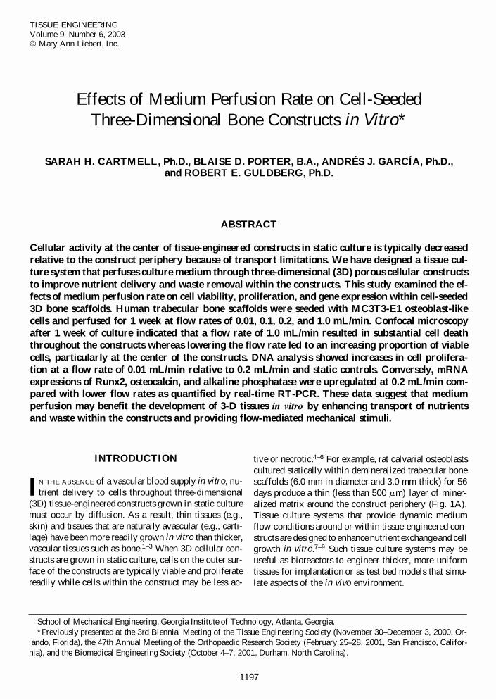
FIG. 2. Left: Top view of stainless steel perfusion block. Eight cell-seeded constructs can be placed into the chamber (of whichfour entries can be seen here; one entry is indicated by the arrow). Right: Perfusion chamber and individual reservoir for culturemedium are placed into an incubator at 37°C, 5% CO2. Tubing is attached to a peristaltic pump (not shown).

beculae within the constructs. A high proportion of thecells was viable throughout the constructs on day 1. Af-ter 1 week in static culture, viable cells were nearly con-fluent on the periphery of the constructs; however, con-sistent with previous reports, only a few viable cells werefound at the construct center. The highest flow rate, 1mL/min, resulted in nearly complete cell death through-out the construct after 7 days. Lowering the flow rate in-creased cell viability at both the periphery and the cen-ter of the constructs after 1 week of continuous perfusion.Flow rates of 0.2 and 0.1 mL/min resulted in a mixtureof viable and dead cells on the surface of the constructs,with limited cell viability in the center. However, a flowrate of 0.01 mL/min resulted in a high proportion of vi-able cells both on the outer surface of the constructs andwithin the construct interior.
Cell proliferation
The effects of medium perfusion rate on cell prolifer-ation, as measured by total DNA content within the con-structs, was consistent with the qualitative assessment ofcell viability by confocal microscopy. High DNA levels
CARTMELL ET AL.
were measured after 7 days of continuous perfusion at0.1 mL/min in comparison with both the 0.2-mL/min andstatic control groups (Fig. 3, p , 0.05). There was alsosignificantly greater cell proliferation in the 0.1-mL/mingroup than in the 0.2-ml/min group; however, only thelowest perfusion rate of 0.01 mL/min enhanced cell pro-liferation relative to static controls. As expected, mini-mal DNA was recovered from acellular control samples.
Osteoblastic gene expression
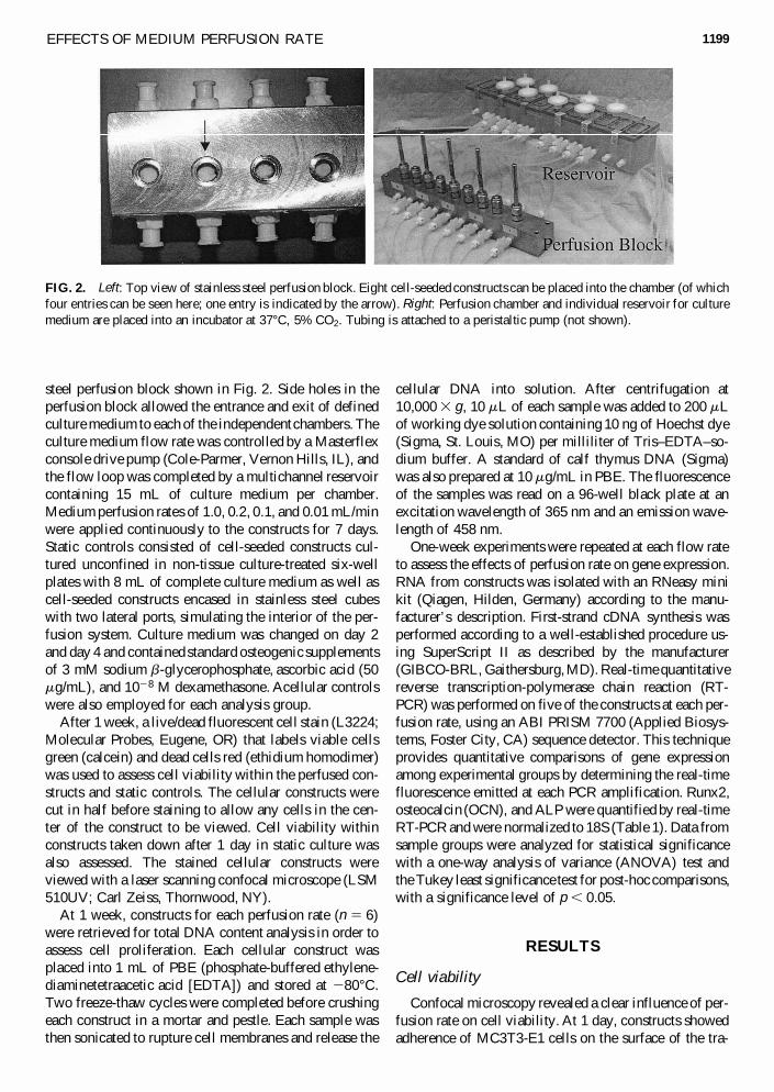
Real-time RT-PCR showed that all three bone-relatedgenes analyzed were detected in cellular constructs per-fused at less than or equal to 0.2 mL/min. Expression ofRunx2, ALP, and OCN was enhanced within cell-seededconstructs perfused at 0.2 mL/min relative to lower flowrates (Fig. 4, p , 0.05). However, the 0.2-mL/min per-fusion treatment increased only Runx2 expression rela-tive to unconfined static controls. There was not enoughRNA recovered from the control constructs encased inthe stainless steel to produce reliable measurements.Likewise, there was no RNA recovered from acellularcontrol samples.
1200
TABLE 1. PCR PRIMERS
Product GenBankGene Forward primer Reverse primer size (bp) accession no.
Runx2 59-TGC TTC ATT CGC 59-TTG CAG TCT TCC TGG 70 AF134836CTC ACA AA AGA AAG TT (copy
complement)OCN 59-GCC GGA GTC TGT 59-CGG CCC TGA GTC TGA 68 BM876746
TCA CTA CCT T CAA A (copy complement)ALP 59-GGG ACT GGT ACT 59-CTG ATA TGC GAT GTC 71 AF285233
CGG ATA ACG A CTT GCT (copy complement)
FIG. 3. DNA assay after 1 week of perfusion. 1 day: 1 day seeded scaffold before perfusion. static 7 day: static sample (notperfused). 0.01, 0.1, and 0.2 ml/min 7 day: flow rates, perfused for 1 week. Brackets: p , 0.05.
DISCUSSION
A critical barrier to the development of 3D tissues invitro is the limited transport of nutrients and metaboliteswithin the construct interior. As a result, reduced cell vi-ability or activity is typicaly observed at the center of tis-sue constructs grown in static culture. In the presentstudy, a perfusion tissue culture system has been devel-oped and used to achieve enhanced cell viability and in-
EFFECTS OF MEDIUM PERFUSION RATE
creased cell proliferation in 3D bone constructs at lowcontinuous perfusion rates compared with static controls.Higher continuous perfusion rates activated expression ofgenes associated with osteoblast differentiation but alsoled to increased levels of cell death due to flow-inducedshear stresses.
Various fluid flow conditions have been shown previ-ously to regulate cell activity in 2D culture systems. Con-tinuous fluid flow applied to osteoblasts in vitro has beenshown to upregulate bone-related gene and phenotype ex-pression.25–29 Pulsatile and oscillatory flow conditionsapplied to osteoblasts in in vitro parallel-plate flow cham-bers have also been shown to increase gene expression,intracellular calcium concentration, and the production ofnitric oxide and prostaglandin E2 in comparison with sta-tic controls.30,31 Furthermore, cell responsiveness hasbeen reported to vary with fluid flow rate and fre-quency.32,33 Proposed mechanisms for the stimulation ofcells by fluid flow include increased mass transport, gen-eration of streaming potentials, and application of shearstresses to the cell membranes.26,31 Although these pre-vious studies were performed in 2D cell culture systems,they suggest that variable flow conditions may also havedifferential effects in 3D tissue culture systems.
Total DNA results after 7 days of continuous perfu-sion were consistent with qualitative assessments of cellviability by confocal microscopy. Significantly moreDNA was isolated from samples perfused at 0.01 mL/minin comparison with both static controls and samples per-fused at 0.2 mL/min. Differences in the number of viablecells were most evident at the center of the constructs,suggesting that slow perfusion may have improved masstransport throughout the constructs. Perfusing medium atlower rates may also have allowed for autocrine effects,as soluble proteins released by cells may have beenflushed away at higher flow rates before exerting an in-fluence on neighboring cells. Continuous perfusion at lowrates for 1 week did not significantly increase the ex-pression of Runx2, ALP, or OCN bone marker genescompared with static controls.
A higher continuous perfusion rate, 0.2 mL/min, sig-nificantly upregulated expression of Runx2, ALP, andOCN relative to lower rates. Runx2 expression, but notALP or OCN expression, was also upregulated comparedwith static controls. Runx2, ALP, and OCN are impor-tant markers of different stages of bone matrix produc-tion.34 Runx2 is a transcriptional activator essential forinitial osteoblast differentiation and subsequent bone for-mation.35 ALP is expressed by preosteoblasts and os-teoblasts before the expression of OCN.36–38 Finally,OCN is a late marker that binds hydroxyapatite and isproduced by osteoblasts just before and during matrixmineralization.34
Although this study measured responses at only onerelatively early time point, the RT-PCR results in con-
1201
FIG. 4. Top: Osteocalcin (OCN) gene expression as measuredby quantitative real-time RT-PCR at 7 days. Bracket: p , 0.05.Middle: Runx 2 gene expression as measured by quantitativereal time RT-PCR at 7 days. Brackets: p , 0.05. Bottom: Al-kaline phosphatase (ALP) gene expression as measured byquantitative real time RT-PCR at 7 days. Brackets: p , 0.05.
cert with DNA content data indicate that lower continu-ous flow rates promoted cell proliferation whereas ahigher perfusion rate of 0.2 mL/min may have acceler-ated the differentiation of cells within the construct to-ward an osteoblast phenotype. These results suggest thatit may be possible to direct cellular behavior within 3Dconstructs via modulation of perfusion flow rate. Longerterm studies that measure the effects of perfusion rate onmatrix mineralization are ongoing to test this hypothesis.
Live/dead staining and confocal microscopy showedan increasing level of cell death within the constructs as-sociated with increasing the perfusion rate from 0.1 to1.0 mL/min. Increased shear stresses at the higher per-fusion rates were likely responsible for the observed de-crease in cell viability. The DNA present after 1 week ofperfusion at 0.2 mL/min was lower than the 1-day seededsample, indicating that the original cell number had ac-tually decreased. The loss in cell number may be due tocells being sheared off the construct as a result of thehigher flow rates as well as decreased cell proliferation.Although continuous perfusion at 0.1–1.0 mL/min wasnot beneficial to construct development, transient in-creases in flow rate superimposed on continuous slowperfusion may simultaneously enhance mass transportand accelerate osteoblast differentiation.
The flow rates used in this study (0.01–1.0 mL/min)are lower than those described in other 3D perfusion sys-tems (1.3–1.8 mL/min).8,9,39 Even if the flow rates hadbeen matched, however, differences in the architectureand porosity of the scaffolds used in the various perfu-sion experiments would result in different shear stressesapplied to cells on the scaffold surfaces. The scaffoldsused in this experiment were in general larger and lessporous than those used in previous perfusion experi-ments, and local shear stresses at a given rate of perfu-sion would therefore be higher. Although modeling theflow of medium through 3D porous scaffolds with com-plex microarchitecture to estimate local shear stresses isa challenging computational problem, such analyses arenecessary to compare results between different perfusionexperiments. Computational fluid dynamics simulationsthat specifically model scaffold microarchitecture arecurrently under development to directly estimate localfluid velocities and shear stresses throughout perfusedconstructs.
A perfusion tissue culture system has been developedand used to demonstrate that altering the flow ratethrough 3D cell-seeded scaffolds affects cell viability,cell proliferation, and the expression of bone markergenes. Further characterization and optimization of per-fusion systems may provide an effective strategy to over-come current diffusion limitations associated with cul-turing 3D tissues in vitro while simultaneously providingflow-mediated mechanical stimuli known to be potentregulators of cell function.
CARTMELL ET AL.
ACKNOWLEDGMENTS
This work made use of ERC Shared Facilities sup-ported by the National Science Foundation under AwardEEC-9731643. We thank the Georgia Tissue bank forkindly donating trabecular bone for our studies.
REFERENCES
1. Vunjak-Novakovic, G., Freed, L.E., Biron, R., and Langer,R. Effects of mixing on the composition and morphologyof tissue engineered cartilage. J. Am. Inst. Chem. Eng. 42,850, 1996.
2. Freed, L.E., Marquis, J.C., Nohria, A., Emmanual, J.,Mikos, A.G., and Langer, R. Neocartilage formation in vitroand in vivo using cells cultured on synthetic biodegradablepolymers. J. Biomed. Mater. Res. 27, 11, 1993.
3. Freed, L.E., Marquis, J.C., Vunjak-Novakovic, G., Em-manual, J., and Langer, R. Composition of cell–polymercartilage implants. Biotechnol. Bioeng. 43, 605, 1994.
4. Ishaug, S.L., Crane, G.M., Miller, M.J., Yasko, A.W.,Yaszemski, M.J., and Mikos, A.G. Bone formation by three-dimensional stromal osteoblast culture in biodegradablepolymer scaffolds. J. Biomed. Mater. Res. 36, 17, 1997.
5. Ishaug-Riley, S.L., Crane-Kruger, G.M., Yaszemski, M.J.,and Mikos, A.G. Three-dimensional culture of rat calvar-ial osteoblasts in porous biodegradable polymers. Bioma-terials, 19, 1405, 1998.
6. Holy, C.E., Shoichet, M.S., and Davies, J.E. Engineeringthree-dimensional bone tissue in vitro using biodegradablescaffolds: Investigating initial cell-seeding density and cul-ture period. J. Biomed. Mater. Res. 51, 376, 2000.
7. Bujia, J., Sittinger, M., Minuth, W.W., Hammer, C.,Burmester, G., and Kastenbauer, E. Engineering of carti-lage tissue using bioresorbable polymer fleeces and perfu-sion culture. Acta Otolaryngol. 115, 307, 1995.
8. Goldstein, A.S., Juarez, T.M., Helmke, C.D., Gustin, M.C.,and Mikos, A.G. Effect of convection on osteoblastic cellgrowth and function in biodegradable polymer foam scaf-folds. Biomaterials, 22, 1279, 2001.
9. Glowacki, J., Mizuno, S., and Greenberger, J.S. Perfusionenhances functions of bone marrow stromal cells in three-dimensional culture. Cell Transplant 7, 319, 1998.
10. Freed, L.E., and Vunjak-Novakovic, G. Cultivation ofcell–polymer constructs in simulated microgravity.Biotechnol. Bioeng. 46, 306, 1995.
11. Kim, S.S., Utsunomiya, H., Koski, J.A., Wu, B.M., Cima,M.J., Sohn, J., Mukai, K., Griffith, L.G., and Vacanti, J.P.Survival and function of hepatocytes on a novel three-di-mensional synthetic biodegradable polymer scaffold withan intrinsic network of channels. Ann. Surg. 228, 8, 1998.
12. Vacanti, J.P., Langer, R., Upton, J., and Marler, J.J. Trans-plantation of cells in matrices for tissue regeneration. Adv.Drug Deliv. Rev. 33, 165, 1998.
13. Vandenburgh, H., Del Tatto, M., Shansky, J., Lemaire, J.,Chang, A., Payumo, F., Lee, P., Goodyear, A., and Raven,L. Tissue-engineered skeletal muscle organoids for re-versible gene therapy. Hum. Gene Ther. 7, 2195, 1996.
1202

14. Rothenburger, M., Vischer, P., Volker, W., Glasmacher,B., Berendes, E., Scheld, H.H., and Deiwick, M. In vitromodelling of tissue using isolated vascular cells on a syn-thetic collagen matrix as a substitute for heart valves. Tho-rac. Cardiovasc. Surg. 49, 204, 2001.
15. Seliktar, D., Black, R.A., Vito, R.P., and Nerem, R.M. Dy-namic mechanical conditioning of collagen–gel blood ves-sel constructs induces remodeling in vitro. Ann. Biomed.Eng. 28, 351, 2000.
16. Qiu, Q., Ducheyne, P., Gao, H., and Ayyaswamy, P. For-mation and differentiation of three-dimensional rat marrowstromal cell culture on microcarriers in a rotating-wall ves-sel. Tissue Eng. 4, 19, 1998.
17. Ziegler, T., and Nerem, R.M. Tissue engineering a bloodvessel: Regulation of vascular biology by mechanicalstresses. J. Cell. Biochem. 56, 204, 1994.
18. Takezawa, T., Inoue, M., Aoki, S., Sekiguchi, M., Wada,K., Anazawa, H., and Hanai, N. Concept for organ engi-neering: A reconstruction method of rat liver for in vitroculture. Tissue Eng. 6, 641, 2000.
19. Freed, L.E., and Vunjak-Novakovic, G. Microgravity tissueengineering. In Vitro Cell. Dev. Biol. Anim. 33, 381, 1997.
20. Botchwey, E.A., Pollack, S.R., Levine, E.M., and Lau-rencin, C.T. Bone tissue engineering in a rotating bioreac-tor using a microcarrier matrix system. J. Biomed. Mater.Res. 55, 242, 2001.
21. Freed, L.E., Hollander, A.P., Martin, I., Barry, J.R., Langer,R., and Vunjak-Novakovic, G. Chondrogenesis in a cell-polymer-bioreactor system. Exp. Cell Res. 240, 58, 1998.
22. Qiu, Q.Q., Ducheyne, P., and Ayyaswamy, P.S. 3D bonetissue engineered with bioactive microspheres in simulatedmicrogravity. In Vitro Cell. Dev. Biol. Anim. 37, 157, 2001.
23. Duray, P.H., Hatfill, S.J., and Pellis, N.R. Tissue culture inmicrogravity. Sci. Med. (Phila.) 4, 46, 1997.
24. Dunkelman, N.S., Zimber, M.P., LeBaron, R.G., Pavelec,R., Kwan, M., and Purchio, A.F. Cartilage production byrabbit articular chondrocytes on polyglycolic acid scaffoldsin a closed bioreactor system. Biotechnol. Bioeng. 46, 299,1994.
25. Johnson, D.L., McAllister, T.N., and Frangos, J.A. Fluidflow stimulates rapid and continuous release of nitric ox-ide in osteoblasts. Am. J. Physiol. 271, E205, 1996.
26. McAllister, T.N., and Frangos, J.A. Steady and transientfluid shear stress stimulate NO release in osteoblaststhrough distinct biochemical pathways. J. Bone Miner Res.14, 930, 1999.
27. Ogata, T. Fluid flow-induced tyrosine phosphorylation andparticipation of growth factor signaling pathway in osteo-blast-like cells. J. Cell. Biochem. 76, 529, 2000.
28. Reich, K.M., and Frangos, J.A. Effect of low on prosta-glandin E2 and inositol trisphosphate levels in osteoblasts.Am. J. Physiol. 26, C428, 1991.
29. Owan, I., Burr, D.B., Turner, C.H., Qiu, J., Tu, Y., Onyia,
EFFECTS OF MEDIUM PERFUSION RATE
J.E., and Duncan, R.L. Mechanotransduction in bone: Os-teoblasts are more responsive to fluid forces than mechan-ical strain. Am. J. Physiol. 273, C810, 1997.
30. Klein-Nulend, J., Burger, E.H., Semeins, C.M., Raisz, L.G.,and Pilbeam, C.C. Pulsating fluid flow stimulates prosta-glandin release and inducible prostaglandin G/H synthasemRNA expression in primary mouse bone cells. J. BoneMiner. Res. 12, 45, 1997.
31. Bakker, A.D., Soejima, K., Klein-Nulend, J., and Burger,E.H. The production of nitric oxide and prostaglandin (E2)by primary bone cells is shear stress dependent. J. Biomech.34, 671, 2001.
32. Jacobs, C.R., Yellowley, C.E., Davis, B.R., Zhou, Z., Cim-bala, J.M., and Donahue, H.J. Differential effect of steadyversus oscillating flow on bone cells. J. Biomech. 31, 969,1998.
33. Edlich, M., Yellowley, C.E., Jacobs, C.R., and Donahue,H.J. Oscillating fluid flow regulates cytosolic calcium con-centration in bovine articular chondrocytes. J. Biomech. 34,59, 2001.
34. Sandberg, M.M., Aro, H.T., and Vuorio, E.I. Gene ex-pression during bone repair. Clin. Orthop. 289, 292, 1993.
35. McCarthy, T.L., Ji, C., Chen, Y., Kim, K.K., Imagawa, M.,Ito, Y., and Centrella, R. Runt domain factor (Runx)-de-pendent effects on CCAAT/enhancer-binding protein deltaexpression and activity in osteoblasts. J. Biol. Chem. 275,21746, 2000.
36. Bronckers, A.L., Gay, S., Finkelman, R.D., and Butler,W.T. Developmental appearance of Gla proteins (osteo-calcin) and alkaline phosphatase in tooth germs and bonesof the rat. Bone Miner. 2, 361, 1987.
37. Aronow, M.A., Gerstenfeld, L.C., Owen, T.A., Tassinari,M.S., Stein, G.S., and Lian, J.B. Factors that promote pro-gressive development of the osteoblast phenotype in cul-tured fetal rat calvaria cells. J. Cell. Physiol. 143, 213, 1990.
38. Malaval, L., Modrowski, D., Gupta, A.K., and Aubin, J.E.Cellular expression of bone-related proteins during in vitroosteogenesis in rat bone marrow stromal cell cultures. J.Cell. Physiol. 158, 555, 1994.
39. Mueller, S.M., Mizuno, S., Gerstenfeld, L.C., and Glo-wacki, J. Medium perfusion enhances osteogenesis bymurine osteosarcoma cells in three-dimensional collagensponges. J. Bone Miner. Res. 14, 2118, 1999.
Address reprint requests to:Robert E. Guldberg, Ph.D.
School of Mechanical Engineering, IBB Building315 Ferst Drive
Georgia Institute of TechnologyAtlanta, GA 30332
E-mail: [email protected]
1203