Paratrichodina africana (Ciliophora): A pathogenic gill parasite in farmed Nile tilapia
6
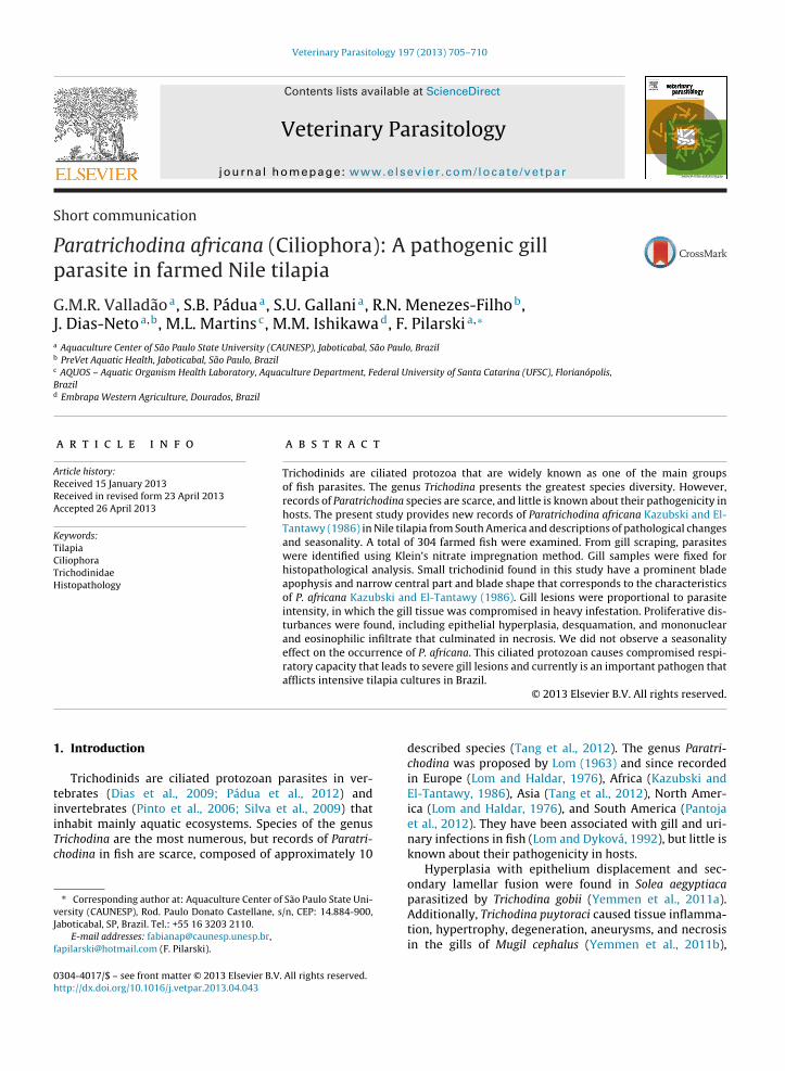
Veterinary Parasitology 197 (2013) 705–710 Contents lists available at ScienceDirect Veterinary Parasitology jo u r nal homep age: www.elsevier.com/locate/vetpar Short communication Paratrichodina africana (Ciliophora): A pathogenic gill parasite in farmed Nile tilapia G.M.R. Valladão a , S.B. Pádua a , S.U. Gallani a , R.N. Menezes-Filho b , J. Dias-Neto a,b , M.L. Martins c , M.M. Ishikawa d , F. Pilarski a,∗ a Aquaculture Center of São Paulo State University (CAUNESP), Jaboticabal, São Paulo, Brazil b PreVet Aquatic Health, Jaboticabal, São Paulo, Brazil c AQUOS – Aquatic Organism Health Laboratory, Aquaculture Department, Federal University of Santa Catarina (UFSC), Florianópolis, Brazil d Embrapa Western Agriculture, Dourados, Brazil a r t i c l e i n f o Article history: Received 15 January 2013 Received in revised form 23 April 2013 Accepted 26 April 2013 Keywords: Tilapia Ciliophora Trichodinidae Histopathology a b s t r a c t Trichodinids are ciliated protozoa that are widely known as one of the main groups of fish parasites. The genus Trichodina presents the greatest species diversity. However, records of Paratrichodina species are scarce, and little is known about their pathogenicity in hosts. The present study provides new records of Paratrichodina africana Kazubski and El- Tantawy (1986) in Nile tilapia from South America and descriptions of pathological changes and seasonality. A total of 304 farmed fish were examined. From gill scraping, parasites were identified using Klein’s nitrate impregnation method. Gill samples were fixed for histopathological analysis. Small trichodinid found in this study have a prominent blade apophysis and narrow central part and blade shape that corresponds to the characteristics of P. africana Kazubski and El-Tantawy (1986). Gill lesions were proportional to parasite intensity, in which the gill tissue was compromised in heavy infestation. Proliferative dis- turbances were found, including epithelial hyperplasia, desquamation, and mononuclear and eosinophilic infiltrate that culminated in necrosis. We did not observe a seasonality effect on the occurrence of P. africana. This ciliated protozoan causes compromised respi- ratory capacity that leads to severe gill lesions and currently is an important pathogen that afflicts intensive tilapia cultures in Brazil. © 2013 Elsevier B.V. All rights reserved. 1. Introduction Trichodinids are ciliated protozoan parasites in ver- tebrates (Dias et al., 2009; Pádua et al., 2012) and invertebrates (Pinto et al., 2006; Silva et al., 2009) that inhabit mainly aquatic ecosystems. Species of the genus Trichodina are the most numerous, but records of Paratri- chodina in fish are scarce, composed of approximately 10 ∗ Corresponding author at: Aquaculture Center of São Paulo State Uni- versity (CAUNESP), Rod. Paulo Donato Castellane, s/n, CEP: 14.884-900, Jaboticabal, SP, Brazil. Tel.: +55 16 3203 2110. E-mail addresses: [email protected], [email protected] (F. Pilarski). described species (Tang et al., 2012). The genus Paratri- chodina was proposed by Lom (1963) and since recorded in Europe (Lom and Haldar, 1976), Africa (Kazubski and El-Tantawy, 1986), Asia (Tang et al., 2012), North Amer- ica (Lom and Haldar, 1976), and South America (Pantoja et al., 2012). They have been associated with gill and uri- nary infections in fish (Lom and Dyková, 1992), but little is known about their pathogenicity in hosts. Hyperplasia with epithelium displacement and sec- ondary lamellar fusion were found in Solea aegyptiaca parasitized by Trichodina gobii (Yemmen et al., 2011a). Additionally, Trichodina puytoraci caused tissue inflamma- tion, hypertrophy, degeneration, aneurysms, and necrosis in the gills of Mugil cephalus (Yemmen et al., 2011b), 0304-4017/$ – see front matter © 2013 Elsevier B.V. All rights reserved. http://dx.doi.org/10.1016/j.vetpar.2013.04.043
-
Upload
independent -
Category
Documents
-
view
5 -
download
0
Transcript of Paratrichodina africana (Ciliophora): A pathogenic gill parasite in farmed Nile tilapia
S
Pp
GJa
b
c
Bd
ARRA
KTCTH
1
tiiTc
vJ
f
0h
Veterinary Parasitology 197 (2013) 705– 710
Contents lists available at ScienceDirect
Veterinary Parasitology
jo u r nal homep age: www.elsev ier .com/ locate /vetpar
hort communication
aratrichodina africana (Ciliophora): A pathogenic gillarasite in farmed Nile tilapia
.M.R. Valladãoa, S.B. Páduaa, S.U. Gallania, R.N. Menezes-Filhob,. Dias-Netoa,b, M.L. Martinsc, M.M. Ishikawad, F. Pilarskia,∗
Aquaculture Center of São Paulo State University (CAUNESP), Jaboticabal, São Paulo, BrazilPreVet Aquatic Health, Jaboticabal, São Paulo, BrazilAQUOS – Aquatic Organism Health Laboratory, Aquaculture Department, Federal University of Santa Catarina (UFSC), Florianópolis,razilEmbrapa Western Agriculture, Dourados, Brazil
a r t i c l e i n f o
rticle history:eceived 15 January 2013eceived in revised form 23 April 2013ccepted 26 April 2013
eywords:ilapiailiophorarichodinidaeistopathology
a b s t r a c t
Trichodinids are ciliated protozoa that are widely known as one of the main groupsof fish parasites. The genus Trichodina presents the greatest species diversity. However,records of Paratrichodina species are scarce, and little is known about their pathogenicity inhosts. The present study provides new records of Paratrichodina africana Kazubski and El-Tantawy (1986) in Nile tilapia from South America and descriptions of pathological changesand seasonality. A total of 304 farmed fish were examined. From gill scraping, parasiteswere identified using Klein’s nitrate impregnation method. Gill samples were fixed forhistopathological analysis. Small trichodinid found in this study have a prominent bladeapophysis and narrow central part and blade shape that corresponds to the characteristicsof P. africana Kazubski and El-Tantawy (1986). Gill lesions were proportional to parasiteintensity, in which the gill tissue was compromised in heavy infestation. Proliferative dis-
turbances were found, including epithelial hyperplasia, desquamation, and mononuclearand eosinophilic infiltrate that culminated in necrosis. We did not observe a seasonalityeffect on the occurrence of P. africana. This ciliated protozoan causes compromised respi-ratory capacity that leads to severe gill lesions and currently is an important pathogen thatafflicts intensive tilapia cultures in Brazil.. Introduction
Trichodinids are ciliated protozoan parasites in ver-ebrates (Dias et al., 2009; Pádua et al., 2012) andnvertebrates (Pinto et al., 2006; Silva et al., 2009) that
nhabit mainly aquatic ecosystems. Species of the genusrichodina are the most numerous, but records of Paratri-hodina in fish are scarce, composed of approximately 10∗ Corresponding author at: Aquaculture Center of São Paulo State Uni-ersity (CAUNESP), Rod. Paulo Donato Castellane, s/n, CEP: 14.884-900,aboticabal, SP, Brazil. Tel.: +55 16 3203 2110.
E-mail addresses: [email protected],[email protected] (F. Pilarski).
304-4017/$ – see front matter © 2013 Elsevier B.V. All rights reserved.ttp://dx.doi.org/10.1016/j.vetpar.2013.04.043
© 2013 Elsevier B.V. All rights reserved.
described species (Tang et al., 2012). The genus Paratri-chodina was proposed by Lom (1963) and since recordedin Europe (Lom and Haldar, 1976), Africa (Kazubski andEl-Tantawy, 1986), Asia (Tang et al., 2012), North Amer-ica (Lom and Haldar, 1976), and South America (Pantojaet al., 2012). They have been associated with gill and uri-nary infections in fish (Lom and Dyková, 1992), but little isknown about their pathogenicity in hosts.
Hyperplasia with epithelium displacement and sec-ondary lamellar fusion were found in Solea aegyptiaca
parasitized by Trichodina gobii (Yemmen et al., 2011a).Additionally, Trichodina puytoraci caused tissue inflamma-tion, hypertrophy, degeneration, aneurysms, and necrosisin the gills of Mugil cephalus (Yemmen et al., 2011b),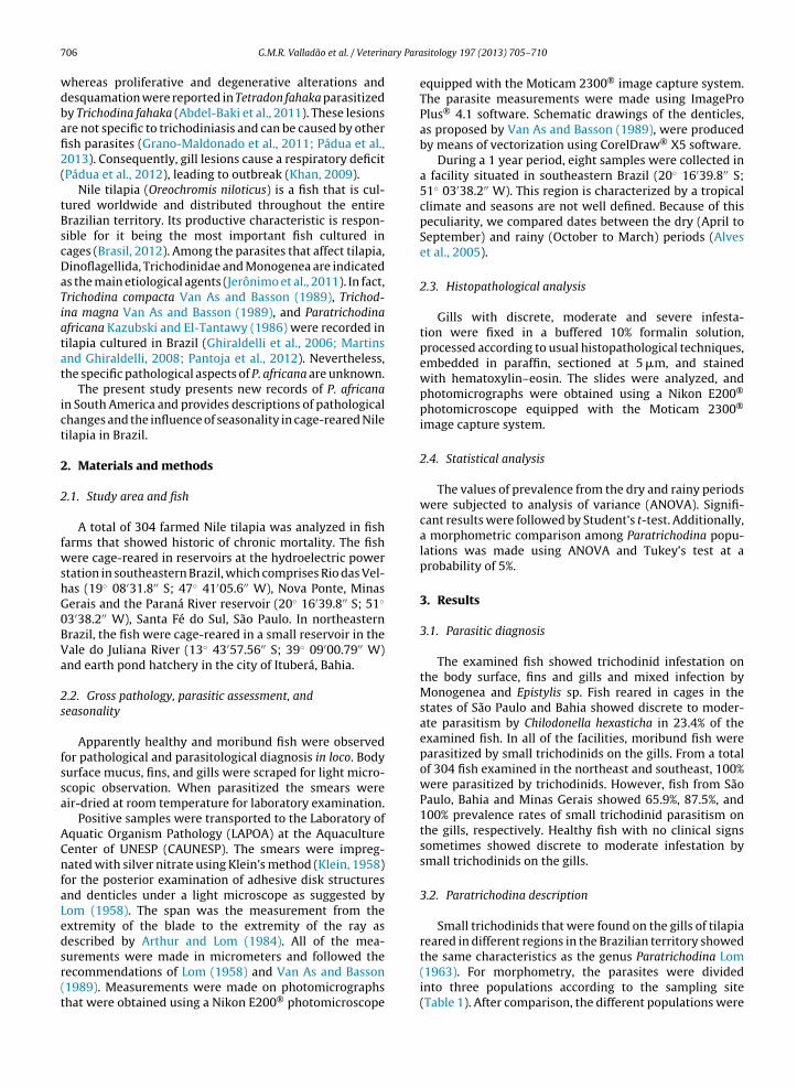
ary Para
706 G.M.R. Valladão et al. / Veterinwhereas proliferative and degenerative alterations anddesquamation were reported in Tetradon fahaka parasitizedby Trichodina fahaka (Abdel-Baki et al., 2011). These lesionsare not specific to trichodiniasis and can be caused by otherfish parasites (Grano-Maldonado et al., 2011; Pádua et al.,2013). Consequently, gill lesions cause a respiratory deficit(Pádua et al., 2012), leading to outbreak (Khan, 2009).
Nile tilapia (Oreochromis niloticus) is a fish that is cul-tured worldwide and distributed throughout the entireBrazilian territory. Its productive characteristic is respon-sible for it being the most important fish cultured incages (Brasil, 2012). Among the parasites that affect tilapia,Dinoflagellida, Trichodinidae and Monogenea are indicatedas the main etiological agents (Jerônimo et al., 2011). In fact,Trichodina compacta Van As and Basson (1989), Trichod-ina magna Van As and Basson (1989), and Paratrichodinaafricana Kazubski and El-Tantawy (1986) were recorded intilapia cultured in Brazil (Ghiraldelli et al., 2006; Martinsand Ghiraldelli, 2008; Pantoja et al., 2012). Nevertheless,the specific pathological aspects of P. africana are unknown.
The present study presents new records of P. africanain South America and provides descriptions of pathologicalchanges and the influence of seasonality in cage-reared Niletilapia in Brazil.
2. Materials and methods
2.1. Study area and fish
A total of 304 farmed Nile tilapia was analyzed in fishfarms that showed historic of chronic mortality. The fishwere cage-reared in reservoirs at the hydroelectric powerstation in southeastern Brazil, which comprises Rio das Vel-has (19◦ 08′31.8′′ S; 47◦ 41′05.6′′ W), Nova Ponte, MinasGerais and the Paraná River reservoir (20◦ 16′39.8′′ S; 51◦
03′38.2′′ W), Santa Fé do Sul, São Paulo. In northeasternBrazil, the fish were cage-reared in a small reservoir in theVale do Juliana River (13◦ 43′57.56′′ S; 39◦ 09′00.79′′ W)and earth pond hatchery in the city of Ituberá, Bahia.
2.2. Gross pathology, parasitic assessment, andseasonality
Apparently healthy and moribund fish were observedfor pathological and parasitological diagnosis in loco. Bodysurface mucus, fins, and gills were scraped for light micro-scopic observation. When parasitized the smears wereair-dried at room temperature for laboratory examination.
Positive samples were transported to the Laboratory ofAquatic Organism Pathology (LAPOA) at the AquacultureCenter of UNESP (CAUNESP). The smears were impreg-nated with silver nitrate using Klein’s method (Klein, 1958)for the posterior examination of adhesive disk structuresand denticles under a light microscope as suggested byLom (1958). The span was the measurement from theextremity of the blade to the extremity of the ray asdescribed by Arthur and Lom (1984). All of the mea-
surements were made in micrometers and followed therecommendations of Lom (1958) and Van As and Basson(1989). Measurements were made on photomicrographsthat were obtained using a Nikon E200® photomicroscopesitology 197 (2013) 705– 710
equipped with the Moticam 2300® image capture system.The parasite measurements were made using ImageProPlus® 4.1 software. Schematic drawings of the denticles,as proposed by Van As and Basson (1989), were producedby means of vectorization using CorelDraw® X5 software.
During a 1 year period, eight samples were collected ina facility situated in southeastern Brazil (20◦ 16′39.8′′ S;51◦ 03′38.2′′ W). This region is characterized by a tropicalclimate and seasons are not well defined. Because of thispeculiarity, we compared dates between the dry (April toSeptember) and rainy (October to March) periods (Alveset al., 2005).
2.3. Histopathological analysis
Gills with discrete, moderate and severe infesta-tion were fixed in a buffered 10% formalin solution,processed according to usual histopathological techniques,embedded in paraffin, sectioned at 5 �m, and stainedwith hematoxylin–eosin. The slides were analyzed, andphotomicrographs were obtained using a Nikon E200®
photomicroscope equipped with the Moticam 2300®
image capture system.
2.4. Statistical analysis
The values of prevalence from the dry and rainy periodswere subjected to analysis of variance (ANOVA). Signifi-cant results were followed by Student’s t-test. Additionally,a morphometric comparison among Paratrichodina popu-lations was made using ANOVA and Tukey’s test at aprobability of 5%.
3. Results
3.1. Parasitic diagnosis
The examined fish showed trichodinid infestation onthe body surface, fins and gills and mixed infection byMonogenea and Epistylis sp. Fish reared in cages in thestates of São Paulo and Bahia showed discrete to moder-ate parasitism by Chilodonella hexasticha in 23.4% of theexamined fish. In all of the facilities, moribund fish wereparasitized by small trichodinids on the gills. From a totalof 304 fish examined in the northeast and southeast, 100%were parasitized by trichodinids. However, fish from SãoPaulo, Bahia and Minas Gerais showed 65.9%, 87.5%, and100% prevalence rates of small trichodinid parasitism onthe gills, respectively. Healthy fish with no clinical signssometimes showed discrete to moderate infestation bysmall trichodinids on the gills.
3.2. Paratrichodina description
Small trichodinids that were found on the gills of tilapiareared in different regions in the Brazilian territory showed
the same characteristics as the genus Paratrichodina Lom(1963). For morphometry, the parasites were dividedinto three populations according to the sampling site(Table 1). After comparison, the different populations were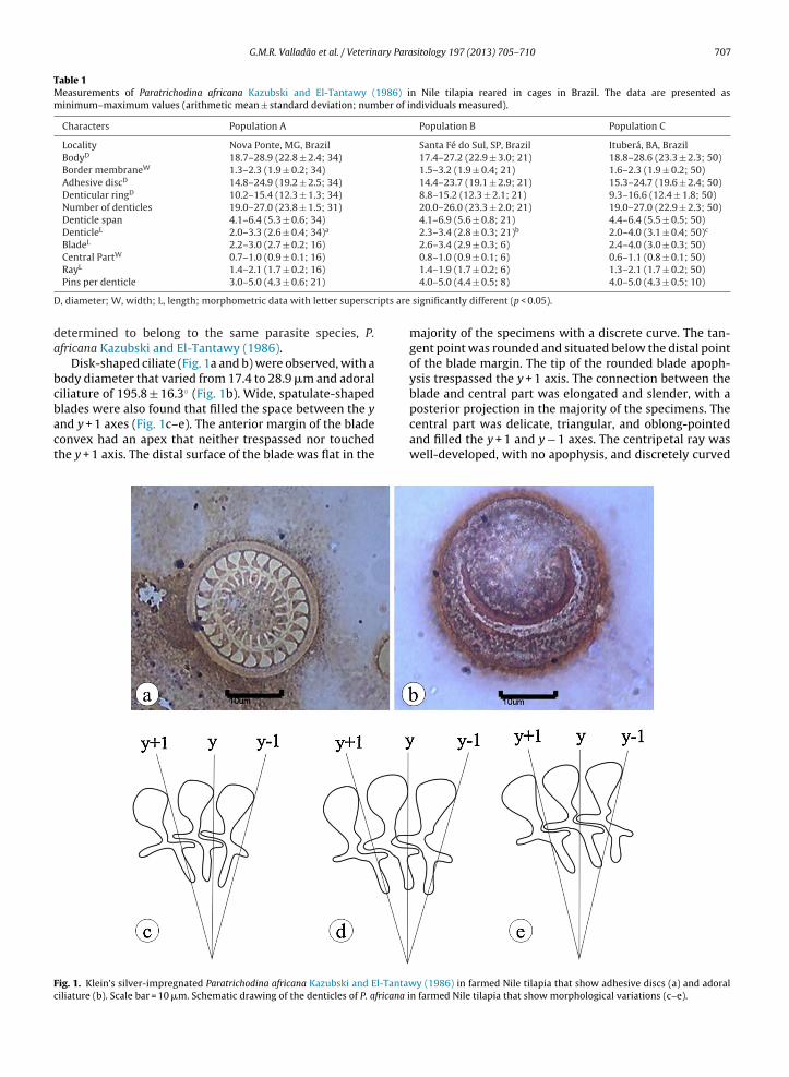
G.M.R. Valladão et al. / Veterinary Parasitology 197 (2013) 705– 710 707
Table 1Measurements of Paratrichodina africana Kazubski and El-Tantawy (1986) in Nile tilapia reared in cages in Brazil. The data are presented asminimum–maximum values (arithmetic mean ± standard deviation; number of individuals measured).
Characters Population A Population B Population C
Locality Nova Ponte, MG, Brazil Santa Fé do Sul, SP, Brazil Ituberá, BA, BrazilBodyD 18.7–28.9 (22.8 ± 2.4; 34) 17.4–27.2 (22.9 ± 3.0; 21) 18.8–28.6 (23.3 ± 2.3; 50)Border membraneW 1.3–2.3 (1.9 ± 0.2; 34) 1.5–3.2 (1.9 ± 0.4; 21) 1.6–2.3 (1.9 ± 0.2; 50)Adhesive discD 14.8–24.9 (19.2 ± 2.5; 34) 14.4–23.7 (19.1 ± 2.9; 21) 15.3–24.7 (19.6 ± 2.4; 50)Denticular ringD 10.2–15.4 (12.3 ± 1.3; 34) 8.8–15.2 (12.3 ± 2.1; 21) 9.3–16.6 (12.4 ± 1.8; 50)Number of denticles 19.0–27.0 (23.8 ± 1.5; 31) 20.0–26.0 (23.3 ± 2.0; 21) 19.0–27.0 (22.9 ± 2.3; 50)Denticle span 4.1–6.4 (5.3 ± 0.6; 34) 4.1–6.9 (5.6 ± 0.8; 21) 4.4–6.4 (5.5 ± 0.5; 50)DenticleL 2.0–3.3 (2.6 ± 0.4; 34)a 2.3–3.4 (2.8 ± 0.3; 21)b 2.0–4.0 (3.1 ± 0.4; 50)c
BladeL 2.2–3.0 (2.7 ± 0.2; 16) 2.6–3.4 (2.9 ± 0.3; 6) 2.4–4.0 (3.0 ± 0.3; 50)Central PartW 0.7–1.0 (0.9 ± 0.1; 16) 0.8–1.0 (0.9 ± 0.1; 6) 0.6–1.1 (0.8 ± 0.1; 50)
L
D ipts are
da
bcbact
Fc
Ray 1.4–2.1 (1.7 ± 0.2; 16)
Pins per denticle 3.0–5.0 (4.3 ± 0.6; 21)
, diameter; W, width; L, length; morphometric data with letter superscr
etermined to belong to the same parasite species, P.fricana Kazubski and El-Tantawy (1986).
Disk-shaped ciliate (Fig. 1a and b) were observed, with aody diameter that varied from 17.4 to 28.9 �m and adoraliliature of 195.8 ± 16.3◦ (Fig. 1b). Wide, spatulate-shaped
lades were also found that filled the space between the ynd y + 1 axes (Fig. 1c–e). The anterior margin of the bladeonvex had an apex that neither trespassed nor touchedhe y + 1 axis. The distal surface of the blade was flat in theig. 1. Klein’s silver-impregnated Paratrichodina africana Kazubski and El-Tantawiliature (b). Scale bar = 10 �m. Schematic drawing of the denticles of P. africana i
1.4–1.9 (1.7 ± 0.2; 6) 1.3–2.1 (1.7 ± 0.2; 50)4.0–5.0 (4.4 ± 0.5; 8) 4.0–5.0 (4.3 ± 0.5; 10)
significantly different (p < 0.05).
majority of the specimens with a discrete curve. The tan-gent point was rounded and situated below the distal pointof the blade margin. The tip of the rounded blade apoph-ysis trespassed the y + 1 axis. The connection between theblade and central part was elongated and slender, with a
posterior projection in the majority of the specimens. Thecentral part was delicate, triangular, and oblong-pointedand filled the y + 1 and y − 1 axes. The centripetal ray waswell-developed, with no apophysis, and discretely curvedy (1986) in farmed Nile tilapia that show adhesive discs (a) and adoraln farmed Nile tilapia that show morphological variations (c–e).
708 G.M.R. Valladão et al. / Veterinary Parasitology 197 (2013) 705– 710
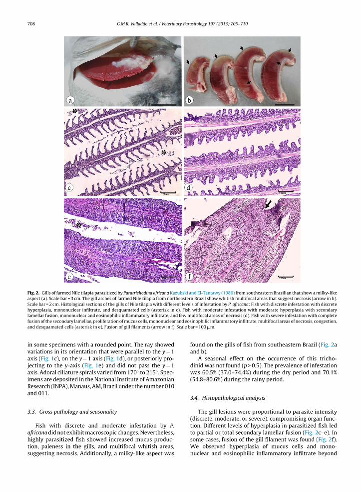
Fig. 2. Gills of farmed Nile tilapia parasitized by Paratrichodina africana Kazubski and El-Tantawy (1986) from southeastern Brazilian that show a milky-likeaspect (a). Scale bar = 3 cm. The gill arches of farmed Nile tilapia from northeastern Brazil show whitish multifocal areas that suggest necrosis (arrow in b).Scale bar = 2 cm. Histological sections of the gills of Nile tilapia with different levels of infestation by P. africana: Fish with discrete infestation with discretehyperplasia, mononuclear infiltrate, and desquamated cells (asterisk in c). Fish with moderate infestation with moderate hyperplasia with secondary
d few m and eos
. Scale b
lamellar fusion, mononuclear and eosinophilic inflammatory infiltrate, anfusion of the secondary lamellae, proliferation of mucus cells, mononuclearand desquamated cells (asterisk in e). Fusion of gill filaments (arrow in f)
in some specimens with a rounded point. The ray showedvariations in its orientation that were parallel to the y − 1axis (Fig. 1c), on the y − 1 axis (Fig. 1d), or posteriorly pro-jecting to the y-axis (Fig. 1e) and did not pass the y − 1axis. Adoral ciliature spirals varied from 170◦ to 215◦. Spec-imens are deposited in the National Institute of AmazonianResearch (INPA), Manaus, AM, Brazil under the number 010and 011.
3.3. Gross pathology and seasonality
Fish with discrete and moderate infestation by P.
africana did not exhibit macroscopic changes. Nevertheless,highly parasitized fish showed increased mucus produc-tion, paleness in the gills, and multifocal whitish areas,suggesting necrosis. Additionally, a milky-like aspect wasultifocal areas of necrosis (d). Fish with severe infestation with completeinophilic inflammatory infiltrate, multifocal areas of necrosis, congestion,ar = 100 �m.
found on the gills of fish from southeastern Brazil (Fig. 2aand b).
A seasonal effect on the occurrence of this tricho-dinid was not found (p > 0.5). The prevalence of infestationwas 60.5% (37.0–74.4%) during the dry period and 70.1%(54.8–80.6%) during the rainy period.
3.4. Histopathological analysis
The gill lesions were proportional to parasite intensity(discrete, moderate, or severe), compromising organ func-tion. Different levels of hyperplasia in parasitized fish led
to partial or total secondary lamellar fusion (Fig. 2c–e). Insome cases, fusion of the gill filament was found (Fig. 2f).We observed hyperplasia of mucus cells and mono-nuclear and eosinophilic inflammatory infiltrate beyond
ary Para
dmonpb
4
rdhd1B2bi(Bdo(ea
hltlBtti(
tBtc(ei
iraHceSlfsfie
om
G.M.R. Valladão et al. / Veterin
esquamation in the spaces between the gill fila-ents. Congestion and interstitial hemorrhages were also
bserved in the gills. Multifocal to coalescent areas of gillecrosis were observed in severely parasitized fish. Theresence of small trichodinids on the gills was confirmedy the histopathological analysis.
. Discussion
The present study presented new records of the occur-ence of P. africana in South America and revealed a wideistribution throughout the Brazilian territory. This speciesas been sparsely reported in the literature. It was initiallyescribed in Egypt and Kenya (Kazubski and El-Tantawy,986) and later in India (Mitra and Bandyopadhyay, 2006),angladesh (Kibria et al., 2009) and Brazil (Pantoja et al.,012). Comparisons of these findings showed that theody of our specimens was shorter than that observed
n the original description by Kazubski and El-Tantawy1986) but coincided with the descriptions of Mitra andandyopadhyay (2006) and Kibria et al. (2009). Adhesiveisk and denticulate ring measurements and the numberf denticles were consistent with specimens from AfricaKazubski and El-Tantawy, 1986) and Bangladesh (Kibriat al., 2009) and slightly lower than the description of Mitrand Bandyopadhyay (2006).
Our specimens that were collected from differentydrographic basins showed similarities among the popu-
ations. The few differences included a denticle lengthhat was higher in population C than in the other popu-ations but coincided with the description of Mitra andandyopadhyay (2006). The border membrane in popula-ion B had a maximum value of 3.2 �m, but this was similaro the arithmetic mean of populations A and C and the stud-es by Mitra and Bandyopadhyay (2006) and Kibria et al.2009).
Our specimens showed the same denticle features ashe findings of Kazubski and El-Tantawy (1986), Mitra andandyopadhyay (2006), and Kibria et al. (2009), includinghe presence of a posterior projection in the blade. Thisharacteristic can be absent in Paratrichodina yangtzeusHu, 2009) or discrete in Paratrichodina rotundiformis (Tangt al., 2012). The present study contributed additionalnformation on the ray position relative to the y-axes.
Little is known about the pathogenicity of Paratrichod-na species in hosts. However, the pathological changesevealed in this study showed the pathogenic potential of P.fricana in Nile tilapia in intensive culture systems in cages.owever, the alterations described herein are not spe-ific and also found in other parasitic (Grano-Maldonadot al., 2011; Pádua et al., 2013) and bacterial (Starliper andchill, 2011) infections. Additionally, the aggravation of gillesions observed in the present study possibly occurredrom opportunistic bacterial infection, in which the abra-ive action exerted by P. africana on the gills of the host mayavor infection by bacterial agents. In fact, ciliate parasitesncrease the susceptibility of fish to bacterial infection (Xu
t al., 2012a,b).In the present study, no seasonal influence was foundn P. africana infestation in tilapia, similar to studies of T.agna and T. compacta in southern Brazil (Jerônimo et al.,
sitology 197 (2013) 705– 710 709
2011). These latter studies, however, found a relationshipbetween parasitism and the culture system. In Turkey, noinfluence was observed in the prevalence of Trichodinamodesta, but higher parasitism intensity was observed inthe warmest months (Özer, 2007). A water temperature of29 ◦C inhibited the proliferation of T. puytoraci in fish fromTunisia, in which the greatest prevalence and intensitywere observed at 18 ◦C (Yemmen et al., 2011b). These vari-ations indicate that the seasonality effect on trichodinidinfestation might be related to environmental conditions,the culture system, and the ecology of the parasite.
P. africana is an exotic parasite that was possiblyintroduced into Brazil by imports of cichlid fish for aqua-culture. This might be true when analyzing Trichodinaheterodentata, T. magna, and T. compacta. The parasiteis widely distributed throughout Brazilian territories,including the northern region (Pantoja et al., 2012) andnortheastern and southeastern regions (present study).This ciliate found favorable conditions in the cage-rearedtilapia system and is currently an important etiologic agentthat afflicts tilapia cultures in Brazil.
Conflict of interest
The authors declare no conflicts of interest.
Acknowledgements
The authors thank PreVet Aquatic Health and Nutreco-FriRibe for support in the field samplings, GeneseasFish Farm for providing fish for the seasonality study,Pedro Henrique de Oliveira Viadanna, M.Sc. (CAUNESP),for histopathological analysis, the AQUABRASIL project– Technological Bases for Sustainable Development ofAquaculture – EMBRAPA, the Ministry of Fisheries andAquaculture, CNPq for a Grant 302493/2010-7 awarded toM.L. Martins and BioMed Proofreading for English editing.
References
Abdel-Baki, A.S., Sakran, T., Fayed, H., Zayed, E., 2011. Trichodina fahaka(Ciliophora: Peritrichia) in Tetradon fahaka from Nile River, Egypt:seasonality and histopathology. Sci. Res. Essays 6, 1583–1587.
Alves, L.M., Marengo, J.A., Camargo Jr., H., Castro, C., 2005. Início da estac ãochuvosa na região Sudeste do Brasil: Parte 1 – Estudos observacionais.Rev. Bras. Meteorol. 20, 385–394.
Arthur, J.R., Lom, J., 1984. Trichodinid protozoa (Ciliophora: Peritrichida)from freshwater fishes of Rybinsk Reservoir, USSR. J. Protozool. 31,82–91.
Brasil. Ministério da Pesca e Aquicultura, 2012. Boletim estatístico dapesca e aquicultura: Brasil 2010. Brasília, DF, 2012, 129 p., http://www.mpa.gov.br/images/Docs/Informacoes e Estatisticas/Boletim%20Estat%C3%ADstico%20MPA%202010.pdf (accessed 30.09.12).
Dias, R.J.P., Fernandes, N.M., Sartini, B., Silva-Neto, I.D., D’Agosto, M.,2009. Occurrence of Trichodina heterodentata (Ciliophora: Tricho-dinidae) infesting tadpoles of Rhinella pombali (Anura: Bufonidae) inthe Neotropical area. Parasitol. Int. 58, 471–474.
Ghiraldelli, L., Martins, M.L., Adamante, W.B., Yamashita, M., 2006. Firstrecord of Trichodina compacta Van As & Basson, 1989 (Protozoa: Cilio-phora) from cultured Nile tilapia in the State of Santa Catarina, Brazil.Int. J. Zool. Res. 2, 369–375.
Grano-Maldonado, M.I., Gisbert, E., Hirt-Chabbert, J., Paladini, G., Roque,A., Bron, J.E., Shinn, A.P., 2011. An infection of Gyrodactylus anguillaeErgens, 1960 (Monogenea) associated with the mortality of glass eels(Anguilla anguilla L.) on the north-western Mediterranean Sea boardof Spain. Vet. Parasitol. 180, 323–331.
ary Para
710 G.M.R. Valladão et al. / VeterinHu, Y., 2009. Description of Paratrichodina yangtzeus sp. n. (Ciliophora:Trichodinidae) from the freshwater fishes in the Yangtze River, China.Wiad Parazytol. 55, 53–57.
Jerônimo, G.T., Speck, G.M., Cechinel, M.M., Gonc alves, E.L.T., Martins, M.L.,2011. Seasonal variation on the ectoparasitic communities of Niletilapia cultured in three regions in southern Brazil. Braz. J. Biol. 71,365–373.
Kazubski, S.L., El-Tantawy, S.A.M., 1986. The ciliate Paratrichodina africanasp. n. (Peritricha, Trichodinidae) from Tilapia fish (Cichlidae) fromAfrica. Acta Protozool. 25, 433–438.
Khan, R.A., 2009. Parasites causing disease in wild and cultured fish inNewfoundland. Icel. Agric. Sci. 22, 29–35.
Kibria, Md.M., Sultana, N., Habib, M.M.A., Sharmin, N., Akhter, N., Ahmed,M.K., Asmat, G.S.M., 2009. Two trichodinid ciliates (Ciliophora: Tri-chodinidae) recorded from Oreochromis mossambicus (Peters, 1852) inBangladesh. Bangladesh J. Mar. Sci. Fish. 1, 63–70.
Klein, B.M., 1958. The dry silver method and its proper use. J. Protozool.5, 99–103.
Lom, J., 1958. A contribution to the systematics and morphology ofendoparasitic trichodinids from amphibians, with a proposal of uni-form specific characteristics. J. Protozool. 5, 251–263.
Lom, J., 1963. The ciliates of the family Urceolariidae inhabiting gills offishes (Trichodinella group). Acta Soc. Zool. Bohemoslav. 27, 7–19.
Lom, J., Dyková, I., 1992. Protozoan Parasites of Fishes. Developments inAquaculture and Fisheries Science, vol. 26. Elsevier Science, Amster-dam, 315 p.
Lom, J., Haldar, D.P., 1976. Observations on trichodinids endocommensalin fishes. Trans. Amer. Microsc. Soc. 95, 527–541.
Martins, M.L., Ghiraldelli, L., 2008. Trichodina magna Van As & Basson,1989 (Ciliophora: Peritrichia) from cultured Nile tilapia in the State ofSanta Catarina, Brazil. Braz. J. Biol. 68, 169–172.
Mitra, A.K., Bandyopadhyay, P.K., 2006. First record of ectoparasiticAfrican trichodinids (Ciliophora: Peritrichida) in a cichlid fish Ore-ochromis mossambicus (Peters, 1852) from the Churni river system,West Bengal, India. Anim. Biol. 56, 323–333.
Özer, A., 2007. Trichodina modesta Lom, 1970 (Ciliophora: Peritrichia)infestations of an endemic Toothcarp Aphanius danfordii Boulenger,1890 (Pisces: Cyprinodontidae) in Sinop, Turkey. J. Nat. Hist. 41,
2543–2549.Pádua, S.B., Martins, M.L., Carraschi, S.P., Cruz, C., Ishikawa, M.M., 2012.Trichodina heterodentata (Ciliophora: Trichodinidae): a new para-
site for Piaractus mesopotamicus (Pisces: Characidae). Zootaxa 3422,62–68.
sitology 197 (2013) 705– 710
Pádua, S.B., Martins, M.L., Carrijo-Mauad, J.R., Ishikawa, M.M., Jerôn-imo, G.T., Dias-Neto, J., Pilarski, F., 2013. First record of Chilodonellahexasticha (Ciliophora: Chilodonellidae) in Brazilian cultured fish:a morphological and pathological assessment. Vet. Parasitol. 191,154–160.
Pantoja, W.M.F., Neves, L.R., Dias, M.K.R., Marinho, R.G.B., Montagner, D.,Tavares-Dias, M., 2012. Protozoan and metazoan parasites of Niletilapia Oreochromis niloticus cultured in Brazil. Rev. MVZ Córdoba 17,2812–2819.
Pinto, H.A., Wieloch, A.H., Melo, A.L., 2006. Uma nova espécie deTrichodina Ehrenberg, 1838 (Ciliophora: Trichodinidae) em Biom-phalaria schrammi (Crosse, 1864) (Mollusca: Planorbidae). Lundiana7, 121–124.
Silva, W.M., Roche, K.F., Vicente, F.S., Delben, A.A.S.T., 2009. First Recordof the Peritrich Trichodina diaptomi Basson and Van As, 1991 (Proto-zoa: Ciliophora) on a South American Calanoid Notodiaptomus deitersi(Poppe, 1890) (Crustacea: Copepoda). J. Euk. Microbiol. 56, 385.
Starliper, C.E., Schill, W.B., 2011. Flavobacterial diseases: columnaris dis-ease, coldwater disease and bacterial gill disease. In: Woo, P.T.K.,Bruno, D.W. (Eds.), Fish Diseases and Disorders. Vol. 3: Viral, Bacterialand Fungal Infections. , pp. 606–631.
Tang, F., Zhao, Y., Liu, C., 2012. Two trichodinids of Paratrichodina (Cilio-phora, Peritrichida, Trichodinidae) infecting gills of Ietalurus punetausfrom Chongqing, China. Afr. J. Microbiol. Res. 6, 2145–2149.
Van As, J.G., Basson, L., 1989. A further contribution to the taxonomy ofthe Trichodinidae (Ciliophora: Peritrichia) and a review of the taxo-nomic status of some fish ectoparasitic trichodinids. Syst. Parasitol.14, 157–179.
Xu, D.-H., Pridgeon, J.W., Klesius, P.H., Shoemaker, C.A., 2012a. Para-sitism by protozoan Ichthyophthirius multifiliis enhanced invasion ofAeromonas hydrophila in tissues of channel catfish. Vet. Parasitol. 184,101–107.
Xu, D.-H., Shoemaker, C.A., Martins, M.L., Pridgeon, J.W., Klesius, P.H.,2012b. Enhanced susceptibility of channel catfish to the bacteriumEdwardsiella ictaluri after parasitism by Ichthyophthirius multifiliis. Vet.Microbiol. 158, 216–219.
Yemmen, C., Quilichini, Y., Ktari, M.H., Marchand, B., Bahri, S., 2011a.Morphological, ecological and histopathological studies of Trichodina
gobii Raabe, 1959 (Ciliophora: Peritrichida) infecting the gills of Soleaaegyptiaca. Protistology 6, 258–263.Yemmen, C., Ktari, M.H., Bahri, S., 2011b. Seasonality and histopathologyof Trichodina puytoraci Lom, 1962, a parasite of flathead mullet (Mugilcephalus) from Tunisia. Acta Adriat. 52, 15–20.