"Ciliophora ". In: Encyclopedia of Life Science
11
Ciliophora Denis H Lynn, University of Guelph, Ontario, Canada Ciliophora is the name for a phylum of protists commonly called the ciliates or infusorians. These protists are the most complex of cells, having an elaborate cytoskeleton, cilia and two different kinds of nuclei. Introduction Humans have probably observed protozoa assigned to the phylum Ciliophora for hundreds of thousands of years. This is because some of the common marine and freshwater species, like Mesodinium rubrum and Stentor polymorphus, can grow to populations so dense that they colour the sea- water or the pond water red or green, respectively. How- ever, Anton van Leeuwenhoek was probably the first person actually to see ciliates as individual cells when he peered down his revolutionary ‘new’ microscope in the seventeenth century. In those days, ciliates were called In- fusoria, because they were often the most obvious organ- isms arising in infusions of vegetation. Once modern microscopes became common laboratory instruments in the nineteenth century, there was an explosion of study on the ciliates. During this period, the major groups were identified (Corliss, 1979). See also: Leeuwenhoek, Antoni van; Protozoa The phylum Ciliophora is one of the most homogeneous of protozoan groups, long recognized as monophyletic. There are some 8000 species with about two-thirds of these being free-living and the remainder symbiotic. The free- living forms can be found all over the world (Finlay et al., 1998) in almost any habitat where water might accumulate. Ciliates are common in sediments or the benthos of marine and freshwater habitats (Finlay et al., 1998), in the plank- ton of marine (Pierce and Turner, 1992) and freshwater (Fenchel, 1987) environments and in soils (Foissner, 1987). They are even found in some extreme environments, like hot springs and in ice flows from the Arctic and Antarctica. Ciliates are commonly the ‘top’ organisms in the microbial food web. They feed on bacteria, flagellates and phyto- plankton, and are, in their own turn, fed upon by zoo- plankton, jellyfish and small fish. Thus, they perform a role of transferring energy through aquatic food chains. Symbiotic ciliates can be commensals, mutualists or parasites. Unique assemblages of commensal forms are found in places as diverse as the intestine of sea urchins and the fermenting stomachs of ruminants. Mutualists are not so common: Nyctotherus, which in its turn harbours met- hanogenic bacteria, lives in the intestinal tract of cock- roaches, whose growth is promoted by the presence of these methanogen-carrying ciliates. Parasitic ciliates are rare: Ichthyophthirius or ‘Ich’ creeps into the epithelium of the skin and gills of fishes and causes ‘white spot’ disease while Balantidium can be found invading the intestinal ep- ithelium of pigs and humans. Parasitic invasions by ciliates are being reported more frequently in recent years as fish and shellfish farming become more common, and mortali- ties have been reported. See also: Protozoan Pathogens of domestic and companion animals; Protozoan Symbiosis Brief Description and Characterization Ciliates were probably the pinnacle of unicellular eukar- yote evolution over 1000 million years ago, before the ev- olution of the metazoans. They are still some of the most complex and beautiful heterotrophic protists, although some forms (e.g. Mesodinium, Stentor polymorphus) are autotrophic because they harbour photosynthetic sym- bionts in their cytoplasm. See also: Universal tree of life Ciliates are characterized by three major features. 1. They exhibit nuclear dimorphism, i.e. they have two different kinds of nuclei in their cytoplasm. The mac- ronucleus is the transcriptionally active nucleus, syn- thesizing messenger ribonucleic acid (mRNA) that controls the functions of the cell. The macronucleus typically contains many copies of the ciliate genome, which is often highly modified when it develops from the micronucleus following conjugation. The micro- nucleus is a typical diploid protistan nucleus, i.e. it contains two sets of chromosomes. It functions as the germline deoxyribonucleic acid (DNA) for each spe- cies, and undergoes meiosis during conjugation. 2. Conjugation is the second distinctive feature of cili- ates. Conjugation is the temporary fusion of two cil- iates during which the partners exchange gametic nuclei, which are products of the meiosis of the mi- cronucleus of each partner (Figure 1). 3. Ciliates typically have cilia at some time during their life cycle (Figures 1 and 2). In some species, these cilia cover the entire cell; in other species, the cilia form compound ciliary organelles called cirri; in some sessile species, the cilia are restricted to the oral region with Article Contents Advanced article . Introduction . Brief Description and Characterization . Place in Overall Taxonomic Scheme . Major Subtaxa and Well-known Species . Subphylum Intramacronucleata . Phylogenetic and Evolutionary Considerations doi: 10.1038/npg.els.0004264 1 ENCYCLOPEDIA OF LIFE SCIENCES & 2006, John Wiley & Sons, Ltd. www.els.net
-
Upload
independent -
Category
Documents
-
view
4 -
download
0
Transcript of "Ciliophora ". In: Encyclopedia of Life Science

CiliophoraDenis H Lynn, University of Guelph, Ontario, Canada
Ciliophora is the name for a phylum of protists commonly called the ciliates or infusorians.
These protists are the most complex of cells, having an elaborate cytoskeleton, cilia and
two different kinds of nuclei.
Introduction
Humans have probably observed protozoa assigned to thephylum Ciliophora for hundreds of thousands of years.This is because someof the commonmarine and freshwaterspecies, likeMesodinium rubrum and Stentor polymorphus,can grow to populations so dense that they colour the sea-water or the pond water red or green, respectively. How-ever, Anton van Leeuwenhoek was probably the firstperson actually to see ciliates as individual cells when hepeered down his revolutionary ‘new’ microscope in theseventeenth century. In those days, ciliates were called In-fusoria, because they were often the most obvious organ-isms arising in infusions of vegetation. Once modernmicroscopes became common laboratory instruments inthe nineteenth century, there was an explosion of study onthe ciliates. During this period, the major groups wereidentified (Corliss, 1979). See also: Leeuwenhoek, Antonivan; Protozoa
The phylumCiliophora is one of themost homogeneousof protozoan groups, long recognized as monophyletic.There are some 8000 species with about two-thirds of thesebeing free-living and the remainder symbiotic. The free-living forms can be found all over the world (Finlay et al.,1998) in almost anyhabitatwherewatermight accumulate.Ciliates are common in sediments or the benthos ofmarineand freshwater habitats (Finlay et al., 1998), in the plank-ton of marine (Pierce and Turner, 1992) and freshwater(Fenchel, 1987) environments and in soils (Foissner, 1987).They are even found in some extreme environments, likehot springs and in ice flows from theArctic andAntarctica.Ciliates are commonly the ‘top’ organisms in themicrobialfood web. They feed on bacteria, flagellates and phyto-plankton, and are, in their own turn, fed upon by zoo-plankton, jellyfish and small fish. Thus, they perform a roleof transferring energy through aquatic food chains.
Symbiotic ciliates can be commensals, mutualists orparasites. Unique assemblages of commensal forms arefound in places as diverse as the intestine of sea urchins andthe fermenting stomachs of ruminants. Mutualists are notso common: Nyctotherus, which in its turn harbours met-hanogenic bacteria, lives in the intestinal tract of cock-roaches, whose growth is promoted by the presence ofthese methanogen-carrying ciliates. Parasitic ciliates arerare: Ichthyophthirius or ‘Ich’ creeps into the epithelium ofthe skin and gills of fishes and causes ‘white spot’ disease
while Balantidium can be found invading the intestinal ep-ithelium of pigs and humans. Parasitic invasions by ciliatesare being reported more frequently in recent years as fishand shellfish farming become more common, and mortali-ties have been reported. See also: Protozoan Pathogens ofdomestic and companion animals; Protozoan Symbiosis
Brief Description and Characterization
Ciliates were probably the pinnacle of unicellular eukar-yote evolution over 1000 million years ago, before the ev-olution of the metazoans. They are still some of the mostcomplex and beautiful heterotrophic protists, althoughsome forms (e.g. Mesodinium, Stentor polymorphus) areautotrophic because they harbour photosynthetic sym-bionts in their cytoplasm. See also: Universal tree of lifeCiliates are characterized by three major features.
1. They exhibit nuclear dimorphism, i.e. they have twodifferent kinds of nuclei in their cytoplasm. The mac-ronucleus is the transcriptionally active nucleus, syn-thesizing messenger ribonucleic acid (mRNA) thatcontrols the functions of the cell. The macronucleustypically contains many copies of the ciliate genome,which is often highly modified when it develops fromthe micronucleus following conjugation. The micro-nucleus is a typical diploid protistan nucleus, i.e. itcontains two sets of chromosomes. It functions as thegermline deoxyribonucleic acid (DNA) for each spe-cies, and undergoes meiosis during conjugation.
2. Conjugation is the second distinctive feature of cili-ates. Conjugation is the temporary fusion of two cil-iates during which the partners exchange gameticnuclei, which are products of the meiosis of the mi-cronucleus of each partner (Figure 1).
3. Ciliates typically have cilia at some time during theirlife cycle (Figures 1 and 2). In some species, these ciliacover the entire cell; in other species, the cilia formcompoundciliary organelles called cirri; in some sessilespecies, the cilia are restricted to the oral region with
Article Contents
Advanced article
. Introduction
. Brief Description and Characterization
. Place in Overall Taxonomic Scheme
. Major Subtaxa and Well-known Species
. Subphylum Intramacronucleata
. Phylogenetic and Evolutionary Considerations
doi: 10.1038/npg.els.0004264
1ENCYCLOPEDIA OF LIFE SCIENCES & 2006, John Wiley & Sons, Ltd. www.els.net

Figure 1 Life cycles of ciliatedprotozoa. (a) A generalized life cycle of a ciliate showing threemajor phases: (1) growth and reproductionduringwhich theciliate feeds and undergoes binary fission; (2) conjugation usually stimulated by starvation conditions, during which the ciliates undergo meiosis and
exchange gametic nuclei before separating and (3) encystment and excystment during which the ciliate secretes a cyst wall about itself to survive harshconditions, like desiccation or the absence of food. (b) The life cycle of the hymenostome Tetrahymena patula, which has a microstome phase that eats
bacteria, a macrostome phase that eats microstomes and other smaller ciliates when the bacterial food supply is depleted and a tomont or dividing phasethat undergoes sequential binary fissions in a cyst toproduce tomites. The tomites escape the cystwhenbacteria are again abundant. (c) The life cycleof the
hymenostome Ichthyophthirius multifiliis, the parasite that causes white-spot disease of the skin of fishes. The theront, a small cell, burrows into the skin of afish to become a phoront that begins feeding as a trophont stage. The trophontmay reach over 1mm in diameter at which time it falls off the fish on to the
bottom to become a tomont. Here, the tomont divides sequentially by binary fission to produce sometimes 1000 tomites, which break out of the divisioncyst to become the next generation of theronts. Based on Lynn and Small (1989) and Lynn and Small (2002).
Ciliophora
2

body or somatic cilia only appearing when the ciliatedisperses, while in other sessile species, such as thesuctorians, cilia are absent at all stages but the disper-sal stage.
Thus, if a protist exhibits these three features – nucleardimorphism, conjugation and presence of cilia – it must bea ciliate.
Life cycles
Ciliates have life cycles that vary depending upon theadaptive strategy of the species. A typical life cycle hasthree major phases (Figure 1a). If there are prey organismspresent, the ciliate feeds and undergoes growth and repro-duction. Reproduction is typically by transverse binaryfission (cytokinesis, division of the cell) accompanied by
mitosis of the micronucleus and bipartitioning of the mac-ronucleus (Raikov, 1996). Often, when food becomes limi-ting, this is a signal for ciliates to become sexually activeand enter the conjugation phase of the life cycle (Figure 1a).Ciliate geneticists have taken advantage of this adaptivestrategy to induce sexual processes in the laboratory so thatthey can be explored as cellular andmolecular processes. Ifthere are no partners nearby, ciliates – like many otherprotists – can undergo encystment, forming a protectivewall around the cell to inhibit desiccation, and then un-dergoing excystment when optimal conditions again arise(Figure 1a). It is in the cyst stage that ciliates are mostprobably dispersed to distant habitats by wind and ani-mals. See also: Binary fission in bacteria; Protozoan cystsand sporesThere are many variations on this basic life cycle (see
Corliss, 1979). Two interesting ones are demonstrated byTetrahymena patula and Ichthyophthiriusmultifiliis (Figure1b
Figure 2 Generalized drawing of the ventral surface of a ciliated protozoon. The body can be divided into somatic and oral regions. The oral regionillustratedherehas twooral polykinetids andoneparoral kinety, andadjacent to this are somespecializedperioral kineties that aid in feeding. Refer to Figure
6 for some illustrations of more oral structures. The somatic cortex is covered by a pellicle that includes the alveoli (see Figure 3). The pellicle is part of thecortex, the outer portionof the cell inwhich are embedded the somaticmono- anddikinetids and themitochondria. The cortexmaybe separated from the
endoplasmby a filamentous layer. There are typically three openings to the outside: the cytostome or cell mouth throughwhich food passes into the foodvacuole; the cytoproct throughwhich undigested food passes from the food vacuole and the contractile vacuole pore out of which excesswater and some
wastes are passed from the contractile vacuole. Based on Lynn and Small (1989).
Ciliophora
3

and 1c). T. patula can develop into a large-mouthed form ormacrostome (Figure 1b) if small-sized prey organisms disap-pear and only larger-sized ones abound. It can even becomecannibalistic, eating its ‘brothers and sisters’. Ichthyophthir-ius is a parasite of the epithelium of fishes. A small-sizedtheront or hunter searches out a fish and burrows beneathits surface epithelial cells where it ingests fluids and cells ofthe fish, growing into a large trophont. The trophont falls offthe fish to the bottom of the pond, becomes a tomont, anddivides many times to yield sometimes over 1000 tomites(Figure 1c). Sessile ciliates, like the suctorians and Vorticella,have life cycles characterized by dispersal stages calledswarmers. See also: Tetrahymena
Cell structure
There is a considerable diversity in cell morphology amongthe different groups or classes of ciliates. Nevertheless,the vast majority of ciliates exhibit similar basic features(Figure 2). The cilia distributed over the cell surface can bedivided into somatic and oral types.
The somatic cilia cover the cell surface or body, usuallyin longitudinal files or rows called kineties. The unit struc-ture of a kinety is the kinetid, which can be composed ofone kinetosome and its cilium, a monokinetid, or twokinetosomes and their cilia, a dikinetid (Figure 2). The so-matic kinetids are used to propel the ciliate through thewater in search of food or to escape predators. Some so-matic kinetids, the perioral kinetids, becomemore complexnear the oral region, functioning to aid in food capture(Figure 2). See also: Cilia and flagella
The oral cilia can be organized as simple dikinetids sur-rounding the cell mouth or cytostome. Sometimes, these
dikinetids can be restricted to one side of the oral region,often the right side; they are called the paroral kinety orparoral membrane, whose cilia are used to filter food par-ticles from the water. The water is drawn to the oral regionby the beating of the cilia of compound ciliary organelles,called oral polykinetids (Figure 2). In ciliates like Euplotesand Stentor these oral polykinetids can number over 30,forming a conspicuous adoral zone that spirals around theanterior end of the cell. Food particles are driven throughthe cytostome into a forming food vacuole, which buds offfrom the base of the oral cavity and circulates through thecytoplasm. After the ingested food has been digested, itis egested through a permanent cell anus or cytoproct(Figure 2). During ingestion, water from the environment issequestered in the food vacuole. This water, together withwater that continuously crosses the cell membranes, issequestered by a tubular system in the cytoplasm, accu-mulated in the contractile vacuole, and expelled throughthe permanent contractile vacuole pore.The cell surface of a ciliate is covered by a cell membrane
or plasmalemma, which is underlain by unit membrane-bound sacs, called alveoli (Figures 2 and 3). A layer ofmicrotubules and a fibrous epiplasmic layer can be foundbeneath the alveoli. Together these form the pellicle(Figure 3). A more detailed examination in the electron mi-croscope reveals the substructure of the kinetids. The so-matic kinetids are typically composed of a kinetosome orbasal body of nine triplets of microtubules, two of whichextend as the nine doublets of the ciliary shaft. Three fi-brillar structures are associated with the kinetosomes, andtogether with it form a kinetid. (1) A transverse ribbonof microtubules typically extends leftwards across thelongitudinal axis of the ciliate. (2) A postciliary ribbon of
Figure 3 Apatch of the cortex of a ciliate that shows the subcomponents of the pellicle and the kinetid. Themicrotubular and filamentous components of
the cortex are elements of the cytoskeleton that provide support for the form of the ciliate. Based on Lynn and Small (1989).
Ciliophora
4

microtubules typically extends posteriorly along the lengthof the kinety. (3) A kinetodesmal fibril extends anteriorlyor laterally to the right (Figure 3). These fibrillar struc-tures provide support for the cell surface and maintainthe characteristic form of each ciliate species. All thesefibrillar elements together with those of the pellicle form
the cortex of a ciliate cell.See also: Alveolates; Tubulin andmicrotubulesThe structure of the fibrillar associates of the somatic
kinetid differs from one group of ciliates to another(Figure 4). It is this variation in structure that has servedas the basis for the modern characterization of the major
Figure 4 Transverse sections of the somatic kinetids of representative genera from themajor classes of ciliates. (a, b) SubphylumPostciliodesmatophora.(a) Class Karyorelictea – Geleia. (b) Class Heterotrichea – Climacostomum. (c–p) Subphylum Intramacronucleata. (c, d) Class Spirotrichea – Protocruzia (c)
and Stylonychia (d). (e, f) Class Colpodea – Sorogena (e) and Pseudoplatyophrya (f). (g, h) Class Phyllopharyngea – Hypocoma (g) and the suctorianTrichophrya (h). (i, j) Class Nassophorea – monokinetid (i) and dikinetid of Nassula (j). (k, l) Class Oligohymenophorea – dikinetid of Colpidium (k) and
monokinetid of Ichthyophthirius (l). (m, n) Class Prostomatea – monokinetid (m) and dikinetid of Coleps (n). (o, p) Class Litostomatea –
Lepidotrachelophyllum (o) and Isotricha (p). kd, kinetodesmal fibril; pc, postciliarymicrotubular ribbon; t1 and t2, transversemicrotubular ribbons. Basedon
Lynn (1996a).
Ciliophora
5

lineages of ciliates (Lynn and Corliss, 1991; Lynn andSmall, 1989; Lynn and Small, 2002). Some lineages arecharacterized as having somaticmonokinetidswhile othershave somatic dikinetids (Figure 4).
Place in Overall Taxonomic Scheme
The ciliates have long been recognized as a monophyleticgroup. Formany years, the suctorians were not consideredciliates (Corliss, 1979). However, along with other ciliates,they are known to shownuclear dimorphism, to conjugate,and to have cilia during their dispersal phase. The structureof suctorian kinetids has the fibrillar associates typical ofciliates (Figure 4).
Ciliates are now acknowledged to constitute a phylumtaxonomically, related to two other phyla that have mem-brane-bound alveoli in the pellicle. Along with the ciliates
the two sister phyla, theApicomplexa and theDinozoa, arecalled the alveolates. In addition to the presence of alveoli,these three phyla are highly similar with respect to se-quences of the small subunit ribosomal RNA (SS-rRNA)genes (Figure 5). See also: Alveolates; Apicomplexa;Phylogeny based on 16S rRNA/DNA; Protozoan taxon-omy and Systematics
Major Subtaxa and Well-known Species
The ciliates are now divided into 11 major lineages orclasses (Table 1). These classes were initially character-ized by differences in the pattern of the somatic kinetid(Figure 4). To a lesser extent, oral structures and the pat-terns of cell division (Foissner, 1996; Lynn and Small,2002) can also be used to characterize these classes. How-ever, evenwithin one class, for example the classColpodea,
SubphylumClassPhylogeny of ciliates based on gene sequence data
Oligohymenophorea
Plagiopylea
Colpodea
Prostomatea
Phyllopharyngea
Litostomatea
Armophorea
Spitrotrichea
Heterotrichea
Karyorelictea
Apicomplexa
Dinozoa
TheileriaSarcocystisSymbiodiniumProrocentrum
Climacostomum
BlepharismaTracheloraphisLoxodes
ProtocruziaEuplotesOxytrichaOnychodromusNyctotheroidesNyctotherusMetopusOphryoscolexEntodiniumDidiniumSpathidiumTrithigmostomaDiscophryaPseudomicrothorax
NassophoreaObertrumiaColepsHolophrya BursariaPseudoplatyophrya
ColpodaTrimyemaPlagiopylaParameciumUronemaCyclidiumIchthyophthiriusColpidium
Tetrahymena
Alv
eola
tes
Intramacronucleata
Postciliodesmatop
hora
Figure 5 A phylogeny based on phylogenies derived from comparisons of the nucleotide sequences of ss-rRNA genes. The other two alveolate phyla
Apicomplexa and Dinozoa serve as outgroups to root the ciliate portion of the tree.
Ciliophora
6

oral structures can be found that are reminiscent of the oralstructures found in other classes (Figure 6).
Subphylum Postciliodesmatophora
Gene sequences of both SS-rRNA and large subunit rib-osomalRNA(LS-rRNA)have confirmed these classes, butalso strongly suggest that there are two major subphyletic
radiations (Lynn, 1996b; Lynn, 2004). The subphylumPostciliodesmatophora, which includes the heterotrichgenera Stentor and Climacostomum and the karyorelic-tean genera Loxodes and Tracheloraphis (Figure 5), is char-acterized by having somatic kinetids whose postciliaryribbons of microtubules typically form conspicuous over-lapping fibres. The macronuclei of karyorelicteans do notdivide but are formed anew at each cell division from aproduct of micronuclear division. On the other hand, themacronuclei of heterotrichs do divide, but use bundles ofmicrotubules that assemble outside the macronuclear en-velope, the so-called extramacronuclearmicrotubules. Thepresence of these extramacronuclear microtubules has ledsome to argue that division of macronuclei evolved twicewithin the phylum (Lynn, 1996b).The classKaryorelictea (Figure5, Table1) includes genera
such asTracheloraphis andLoxodes.Tracheloraphis and itsrelatives are typically found in marine sands where theycrawl between the sand grains ingesting bacteria and otherfood particles.Loxodes is an unusual karyorelictean as it isfound in freshwaters, but typically in habitatswith very lowoxygen concentrations (Fenchel, 1987).The class Heterotrichea (Figure 5, Table 1) includes both
marine and freshwater species. These typically large cili-ates, such as Blepharisma, Stentor and Spirostomum, havean extensive spiralling zoneof oral polykinetids that extendout over the anterior end. The polykinetidal cilia createfeeding currents that bring prey, such as flagellates andother ciliates, into the oral cavity for ingestion (Fenchel,1987). See also: Stentor
Subphylum Intramacronucleata
The second subphylum of the ciliates, the Intramacronu-cleata, includes the remaining nine classes of ciliates, whichdivide their macronuclei using microtubules that assembleinside the macronuclear envelope, the so-called intramac-ronuclearmicrotubules (Lynn, 1996b; Lynn, 2004).Unlikethe subphylum Postciliodesmatophora in which there issome similarity in somatic kinetid structure (Figure 4), in-tramacronucleates exhibit a tremendous diversity in so-matic kinetid structure (Figure 4) and show a rapid burst ofdiversification in molecular phylogenies (Figure 5). It is pri-marily on the basis of these somatic kinetid features to-gether with features of the oral region and the pattern oforal development or stomatogenesis that the classes withinthe subphylum Intramacronucleata are characterized(Lynn and Corliss, 1991; Lynn and Small, 1997; Lynnand Small, 2002).The class Spirotrichea (Figure 5, Table 1) includes a di-
verse assemblage of forms found in a wide variety of habi-tats from ponds to lakes to the oceans, and even in soils.Spirotrichs are found on the bottom of these water bodiesand swimming in the water column as plankton. Euplotes,
Table 1 Classification of the phylum Ciliophora with repre-sentative genera, some of which are available from culturecollections or biological supply houses
Phylum Ciliophora (Doflein, 1901)
Subphylum Postciliodesmatophora, Gerassimova and Sera-vin, 1976
Class Karyorelictea, Corliss, 1974Loxodes, Tracheloraphis
Class Heterotrichea, Stein, 1859Blepharisma, Spirostomum, Stentor
Subphylum Intramacronucleata, Lynn, 1996Class Spirotrichea, Butschli, 1889
ProtocruziaEuplotes, Oxytricha, StylonychiaHalteria, Strombidium, Tintinnopsis
Class Armophorea, Jankowski, 1964Caenomorpha, MetopusClevelandella, Nyctotherus, Nyctotheroides
Class Litostomatea, Small and Lynn, 1981Didinium, Dileptus, Protospathidium, SpathidiumBalantidium, Entodinium, Ophryoscolex
Class Phyllopharyngea, de Puytorac et al., 1974Chilodonella, Trithigmostoma, SpirochonaDiscophrya, Heliophrya, Tokophrya
Class Nassophorea, Small and Lynn, 1981Nassula, Pseudomicrothorax
Class Colpodea, Small and Lynn, 1981Bursaria, Colpoda, Platyophrya, Sorogena
Class Prostomatea, Schewiakoff, 1896Coleps, Cryptocaryon, Holophrya
Class Plagiopylea, Small and Lynn, 1985Plagiopyla, Trimyema
Class Oligohymenophorea, de Puytorac et al., 1974Scuticociliates
Anophryoides, Cinetochilum, Cyclidium, Glauconema,Homalogastra,Metanophrys,Miamiensis,Parauronema,Potomacus, Pseudocohnilembus
PeniculinesParamecium
PeritrichsCarchesium, Epistylis, Opisthonecta, Rhabdostyla,Vorticella
HymenostomesColpidium, Glaucoma, Ichthyophthirius, Tetrahymena
Ciliophora
7
Oxytricha and Stylonychia are some common genera,which have oral polykinetids arranged like the heterot-richs. However, their somatic cilia are combined into cirri,compound ciliary structures composed of 50 or more cilia,which are used like little legs by these ciliates as they ‘walk’along the substrate. Other spirotrichs, like Halteria,Strombidium and Tintinnopsis, are common in the plank-ton, where they feed upon bacteria and phytoplankton,and are in turn ingested by zooplankton. Tintinnopsis is arepresentative of an unusual group of ciliates called thetintinnids. These ciliates secrete a lorica or ‘house’, inwhich they sit and which they propel using their oral cilia.Tintinnids are one group of ciliates to have been fossilized;fossil tintinnid loricae date back over 200 million years(Lynn and Small, 1989). Another spirotrich ciliate is Pro-tocruzia associated with the spirotrichs by its SS-rRNAsequence. This unusual ciliate has a dividingmacronucleusthat appears to exhibit chromosome-like structures whenit divides, reminiscent of a micronucleus (Corliss, 1979;Raikov, 1982). Protocruzia may represent the ancestralstate of the intramacronucleate macronucleus. See also:Euplotes (dorsoventrally-flattened Ciliates)
The class Armophorea (Table 1) is one of the two ribo-classes in the phylum (Lynn, 2004). ‘Riboclasses’ aregroups that do not demonstrate any obvious morpholog-ical shared-derived characters, and yet they are stronglysupported by gene sequence data. The class is divided intotwo major groups. One group, represented by Caeno-morpha andMetopus, includes free-living ciliates found inhabitats with low or no oxygen. The other group includesClevelandella, Nyctotherus and Nyctotheroides end-osymbionts in the intestinal tract of insects, frogs andother invertebrates. The ciliates in this class are all anaer-obes or microaerophils, and use hydrogenosomes as ‘res-piratory organelles’ rather than mitochondria (Boxmaet al., 2005).
The class Litostomatea (Figure 5, Table 1) includes twovery different groups of ciliates:Didinium andDileptus arefree-living ciliates found in marine and freshwater habitatswhile Entodinium and Ophryoscolex represent a diverseassemblage of ciliates found in the intestinal tracts of ver-tebrates, such as deers, cows, horses, elephants and evengorillas. The only ciliate to cause disease in humans, Ba-lantidium, is placed in this class. These ciliates typicallyhave an anterior oral region with simple oral ciliature,typically composed of mono- or dikinetids. Didinium andDileptus capture their prey, which can be other ciliates likeParamecium, using organelles called toxicysts. Toxicystsare rod-like organelles that are ejected from the ciliate intoits prey, much like a harpoon. Not only do toxicysts cap-ture the prey, but they also have hydrolytic enzymes thatimmobilize the prey and begin to digest its cytoplasm. Seealso: Balantidiosis; Paramecium
The class Phyllopharyngea (Figure 5, Table 1) is namedbecause the cytopharynx of these ciliates is lined with leaf-like ribbonsorphyllae (5 phyllos inGreek) ofmicrotubules.
Phyllopharyngeans are divided into twomajor groups, free-swimming forms like Chilodonella and Trithigmostoma,which are typically associated with the sediments and othersubstrates in marine and freshwater habitats, and sessileforms like the chonotrichs and suctorians. Chonotrichs,such asSpirochona, are ectosymbionts on crustaceanswherethey attach to appendages andmouthparts of their host andobtain foodparticles andbacteria from the host. Suctorians,such as Discophrya, Heliophrya and Tokophrya, attach tosubstrates and to hosts, like crustaceans, insects and eventurtles. These ciliates were named because they have longtentacles supported by the phyllae. The tentacles have ex-trusive organelles called haptocysts at their tips, which cap-ture prey, usually other ciliates, and ‘suck’ or transport theircytoplasm into the cell bodyof the suctorian. Suctorians andchonotrichs produce ciliated swarmers by cell division, andthese swarmers swim to other habitats or hosts to dispersethe species. See also: SuctoriansThe class Nassophorea (Figure 5, Table 1) includesmarine
and freshwater ciliates that are typically associatedwith thesediments. They are named after a very prominent basketof microtubular bundles that supports the cytopharynx,called the nasse in French (Corliss, 1979; Lynn and Small,2002). Nassophoreans, such as Nassula and Pseudomicro-thorax, use the nasse to ingest filamentous cyanobacteria,which are transported into the cytoplasm by the action ofsmall arms attached to the microtubules of the nasse.The class Colpodea (Figures 5 and 6; Table 1) includes
species that are typically able to form protective capsulescalled cysts, in which the ciliate can remain dormant,often for periods of years. Colpodeans, such as Colpoda,Platyophrya and Sorogena, are primarily restricted tofreshwater habitats and soils where they feed on bacteria,flagellates and other ciliates. Sorogena is unusual amongciliates in that individual ciliates aggregate when theirfood, Colpoda, becomes scarce to form a complex aggre-gate cyst called a sorocarp that sits on a small stalk. Thesorocarp is then blown by the wind, taking its hundreds of‘passengers’ to a new habitat. One of the largest ciliates,Bursaria, is found in this class.Bursariawas formany yearsthought to be a heterotrich, like Stentor, because it hasmany oral polykinetids spiralling towards its cytostome(Figure 6). However, its ultrastructure and SS-rRNA genesequences clearly demonstrate that it is a colpodean (Lynn,1996b; Lynn and Corliss, 1991; Lynn and Small, 2002). Infact, a number of colpodeans were misclassified by earlierresearchers because their oral structures (Figure6) appearedsimilar to the oral structures of ciliates that we now knowbelong to different classes based on the structure ofthe somatic kinetid (Figure 4) and gene sequences (Figure 5).See also: StentorThe class Prostomatea (Figure 5, Table 1) includes marine
and freshwater ciliates that are associated with the bottomand are also found in the water column as plankton. Theprostomes, as the name suggests, have an oral region atthe anterior end of the cell body. Oral ciliature is simple,
Ciliophora
8
usually of dikinetids, but complemented sometimes byspecialized kineties, called brosse (5brush in French)kineties, which may be used in detecting prey. Prostomes,such asColeps,Holophrya and their relatives, eat bacteria,flagellates and other ciliates. However, they can alsofeed on dead or dying invertebrates and are called his-tophagous, or tissue-eating (5 histos in Greek) for thisreason. A prostome, Cryptocaryon, causes a white-spot disease of marine fishes. This is similar to the fishdisease caused by the freshwater oligohymenophoreanIchthyophthirius (see below).
The class Plagiopylea, the second riboclass in the phylum(Figure 5, Table 1), includes a small assemblage of ciliateswhosephylogenetic affinitieswereunclear until sequences ofSS-rRNA suggested that they were related to the class
Oligohymenophorea, described below. Plagiopyleans aretypically found in marine and freshwater sediments, partic-ularlywhere oxygen concentrations are lowor absent. Someare even symbionts in the intestines of sea urchins, anotheranaerobic habitat. These ciliates are able to survive herebecause their mitochondria have evolved to generate hy-drogen, and are now called hydrogenosomes. The hydro-gen, in turn, is consumed bymethanogenic bacteria that livein association with the hydrogenosomes, providing somecarbon for their ciliate host.The class Oligohymenophorea (Figure 5, Table 1) is prob-
ably the most speciose class of ciliates. These ciliates arefound virtually anywhere there is moisture – in soils, hotsprings, temporary puddles, small ponds, lakes and oceans.The fourmajor subclasses in this taxonare the scuticociliates,
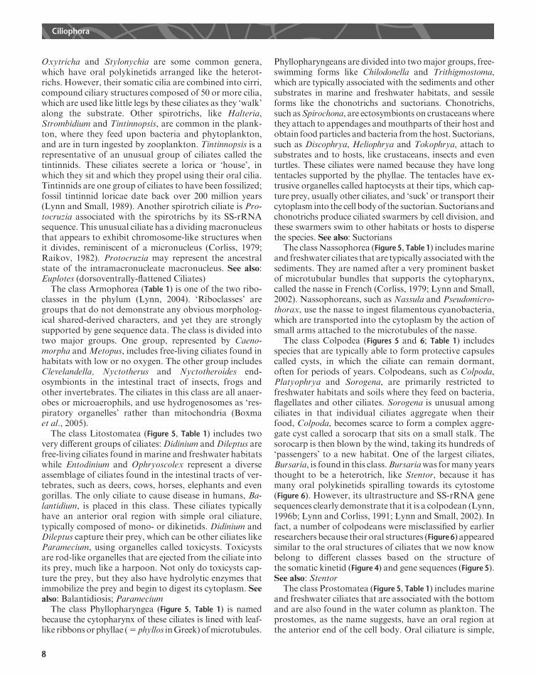
Figure 6 A demonstration of the diversity of oral structures of genera in the class Colpodea. Some of these ciliates represent oral features that areconvergent on oral features of ciliates from other classes. (a) Colpoda with two oral polykinetids. (b) Hausmanniella also with two oral polykinetids. (c)
Bardeliellawith a left oral polykinetid that extendsout over the anterior end as a series of rows of cilia. (d)Grossglockneriawhose everted cytopharynx is usedto pierce fungal cells and ingest their cytoplasm, like a suctorian’s tentacle. (e, f) Bursaria (e) and Bursaridium (f) whose extensive series of left oral
polykinetids placed them in the class Heterotrichea formany years. (g) Cyrtolophosiswith its small number of oral polykinetids and small cell size was onceconsidered amember of the classOligohymenophorea. (h) Platyophryawith paroral andmultiple left oral polykinetids. (i) Sagittaria similar to Platyophrya.
(j) Rostrophrya with small polykinetids extending out on to the somatic surface was initially thought to be a member of the class Nassophorea. (k)Pseudochlamydonellawhose ventral oral cavity andbasket-like cytopharynx are similar to those structures in the class Phyllopharyngea. (l) Sorogenawith its
simple, ring-like anterior oral apparatus was initially classified as a member of the class Prostomatea. (m) Bryophrya shows some similarities to Rostrophryaand nassophoreans although it has a deep oral cavity. (n) Trihymena, a colpodean with a simple oligohymenophorean-like oral apparatus. Based on Lynn
(1996a) and Foissner (1993).
Ciliophora
9

peniculines, peritrichs and hymenostomes. Many of theseciliates are easily cultivated and this has made them favour-ite research subjects: two of the most researched genera ofciliates belong to this class – the peniculineParamecium andthe hymenostome Tetrahymena (Nanney, 1980). Oligohy-menophoreans are named because they have only a few(5 oligos inGreek)membranes (5 hymen inGreek) in theiroral cavity. Typically they have a paroral kinety or mem-braneon the right side and three oral polykinetids on the leftside of the oral cavity, hence Tetrahymena or four mem-branes. Most oligohymenophoreans are bacteriovorous.Peritrich ciliates, such asCarchesium andVorticella, can befound abundantly in sewage treatment plants where theyconsume faecal coliforms. However, some Tetrahymenaspecies can be histophagouswhile others can be parasitic, ascan some scuticociliates, such as Philasterides. Probablythe most famous parasitic ciliate, although most people donot recognize it as a protozoon, is the hymenostomeIchthyophthirius, the parasite of fish that causes white-spotdisease of the epithelium and gills. Diseases caused by cil-iates may be on the increase as the aquaculture industryexpands andprovidesmoreopportunities for ciliates to takeadvantage of overcrowded invertebrates and vertebrates,which also may be less physically fit than their free-rangingrelatives. See also: Paramecium; Tetrahymena; Protozoanpathogens of domestic and companion animals; Vorticella
This brief survey of the diversity of ciliates has omittedmention of a number of smaller, but no less interestinggroups. More detailed information can be found in Corliss(1979), Lynn andCorliss (1991) andLynn and Small (2002).
Phylogenetic and EvolutionaryConsiderations
There is probably more known about the comparativemorphology and molecular evolution of ciliate groupsthan any other group of protists. Thus, our confidence inthe phylogeny and evolution within this group is fairlyhigh, even though there still remain many unansweredquestions. Ciliates have a fossil record that certainly datesback over 200 million years: some fossil tintinnids are thisold (Lynn and Small, 1989). However, the phylum is un-doubtedly much older. Reid and John (1981) argued thatchitinozoa, which are found in Proterozoic deposits over600 million years old, may be tintinnid cysts. Wright andLynn (1997) used a molecular clock calibrated on the ev-olution of the SS-rRNA genes and calculated that ciliatesmight be 1980–2200 million years old, dating back to thePalaeoproterozoic. If true, the ciliates were undoubtedlythe ‘masters of the seas’ in that microbial age. The molec-ular evidence suggests that the major lineages, now sub-phyla and classes, radiated very shortly after the emergenceof the ‘protociliate’. A burst of evolutionary diversificationis a common feature of the emergence of most groups of
organisms and unfortunately confounds our understand-ing of how evolution might have occurred. See also: Fossilrecord;Molecular phylogeny reconstruction; Origin of lifeCiliates are alveolates, and so related to the dinoflagel-
lates. Thus, dinoflagellates and ciliates undoubtedly sharea common ancestor. Given this, what might the ancestralciliate have looked like? Eisler (1992) has argued that thesomatic kineties of ciliates arose from the migration of thekinetosomes of a paroral kinety of dikinetids associatedwith the cytostome of the ‘protociliate’. This ancestorwould also probably have evolved some kind of nucleardimorphism with a macronucleus, probably nondividinglike the karyorelicteans, and a dividingmicronucleus.Howthis evolution proceeded is still not resolved, but it is ourhope that further comparative study of other genes of cil-iates will reveal more clearly the sequence of evolutionof the major groups in this intriguing phylum of protists.See also: Protozoan evolution and phylogeny
References
Boxma B, deGraaf RM, van der StaayGWM et al. (2005) An anaerobic
mitochondrion that produces hydrogen. Nature 434: 74–79.
Corliss JO (1979) The Ciliated Protozoa: Characterization, Classifica-
tion, and Guide to the Literature, 2nd edn.NewYork: Pergamon Press.
Eisler K (1992) Somatic kineties or paroral membrane: which came first
in ciliate evolution? BioSystems 26: 239–254.
Fenchel T (1987) Ecology of Protozoa. The Biology of Free-living
Phagotrophic Protists. Berlin: Springer.
Finlay BJ, Esteban GF and Fenchel T (1998) Protozoan diversity: Con-
verging estimates of the global number of free-living ciliate species.
Protist 149: 29–37.
Foissner W (1987) Soil protozoa: fundamental problems, ecological sig-
nificance, adaptations in ciliates and testaceans, bioindicators, and
guide to the literature. In: Corliss JO and Patterson DJ (eds) Progress
in Protistology, vol. 2, pp. 69–212. Bristol: Biopress Ltd.
Foissner W (1993) Colpodea (Ciliophora). Stuttgart: Gustav Fischer.
Foissner W (1996) Ontogenesis in ciliated protozoa, with emphasis on
stomatogenesis. In: Hausmann K and Bradbury PC (eds) Ciliates.
Cells as Organisms, pp. 95–177. Stuttgart: Gustav Fischer.
LynnDH(1996a) Systematics of ciliates. In:HausmannKandBradbury
PC (eds) Ciliates. Cells as Organisms, pp. 51–72. Stuttgart: Gustav
Fischer.
LynnDH (1996b)My journey in ciliate systematics. Journal of Eukaryo-
tic Microbiology 43: 253–260.
Lynn DH (2004) Morphology or molecules: how do we identify the
major lineages of ciliates (Phylum Ciliophora)? European Journal of
Protistology 39: 356–364.
Lynn DH and Corliss JO (1991) Ciliophora. In: Harrison FW and Cor-
liss JO (eds)Microscopic Anatomy of Invertebrates. Protozoa, pp. 333–
467. New York: Wiley-Liss.
Lynn DH and Small EB (1989) Phylum Ciliophora. In: Margulis L,
Corliss JO, Melkonian M and Chapman DJ (eds) Handbook of Pro-
toctista, pp. 498–523. Boston: Jones and Bartlett Publishers.
Lynn DH and Small EB (1997) A revised classification of the Phylum
CiliophoraDoflein, 1901.Revista de la SociedadMexicana deHistoria
Natural 47: 65–78.
LynnDHand Small EB (2002) PhylumCiliophora. In: Lee JJ, Bradbury
PCandLeedaleGF (eds)An IllustratedGuide to theProtozoa, pp. 371–
656. Lawrence, KS: Society of Protozoologists.
Ciliophora
10

Nanney DL (1980) Experimental Ciliatology. An Introduction to Genetic
and Developmental Analysis in Ciliates. New York: Wiley.
PierceRWandTurner JT (1992) Ecology of planktonic ciliates inmarine
food webs. Reviews in Aquatic Sciences 6: 139–181.
Raikov IB (1982) The Protozoan Nucleus. Morphology and Evolution.
New York: Springer.
Raikov IB (1996) Nuclei of ciliates. In: Hausmann K and Bradbury PC
(eds) Ciliates. Cells as Organisms, pp. 221–242. Stuttgart: Gustav
Fischer.
ReidPCandJohnAWG(1981)Apossible relationshipbetweenchitinozoa
and tintinnids. Review of Paleobotany and Palynology 34: 251–262.
Wright A-DG and Lynn DH (1997) Maximum ages of ciliate lineages
estimated using a small subunit rRNA molecular clock: crown
eukaryotes date back to the Paleoproterozoic. Archiv fur Pro-
tistenkunde 148: 329–341.
Further Reading
Buchmann K, Lindenstrom T and Bresciani J (2001) Defence mecha-
nisms against parasites in fish and the prospect for vaccines. Acta
Parasitologica 46: 71–81.
Buchmann K, Sigh J, Nielsen CV and Dalgaard (2001) Host responses
against the fish parasitizing ciliate Ichthyophthirius multifiliis. Veter-
inary Parasitology 100: 105–116.
Ferry T, BouhourD,DeMonbrisonF et al. (2004) Severe peritonitis due
to Balantidium coli acquired in France. European Journal of Clinical
Microbiology and Infectious Diseases 23: 393–395.
Frankel J (1989)Pattern Formation. Ciliate Studies andModels.Oxford:
Oxford University Press.
HausmannKandBradbury PC (eds) (1996)Ciliates. Cells asOrganisms.
Stuttgart: Gustav Fisher.
Jee BY, Kim KH, Park SI and Kim YC (2000) A new strain of Crypt-
ocaryon irritans from the cultured olive flounder Paralichthys
olivaceus. Diseases of Aquatic Organisms 43: 211–215.
Jones AR (1974) The Ciliates. London: Hutchinson University Library.
ParamaA, Iglesias R, AlvarezMF et al. (2003)Philasterides dicentrarchi
(Ciliophora, Scuticociliatida): experimental infection and possible
routes of entry in farmed turbot (Scophthalmusmaximus).Aquaculture
217: 73–80.
Puytorac P de (ed.) (1994) Infusoires Cilies. Traite de Zoologie, vol. II.
Paris: Masson.
Ciliophora
11