Papillonema danieli gen. et sp.n. and Papillonema clavatum (Gerlach, 1957) comb.n. (Nematoda,...
13
Hydrobiologia 316 : 225-237,1995 . ©1995KluwerAcademicPublishers. PrintedinBelgium. Papillonemadanieli gen .et sp .n.and Papillonemaclavatum (Gerlach,1957) comb.n .(Nematoda,Desmodoridae)fromthe Ceriops mangrovesediments ofGaziBay,Kenya DominickVerschelde l ,AgnesMuthumbi 2 & MagdaVincxl 1 UniversityofGhent,ZoologyInstitute,MarineBiologySection,K .L .Ledeganckstraat35,B-9000Gent,Belgium 2 KenyaMarine&FisheriesResearchInstitute,P.0 .Box81651Mombasa,Kenya Received11October 1994 ;in revisedform 15 February 1995 ; accepted 21 February 1995 Keywords : Marinenematodes,Desmodoridae, Papillonema gen .n .,taxonomy Abstract Anewgenus, Papillonema gen .n .,iserectedtoaccomodatethetwospecies P . danieli g en .e t sp .n .and P . clavatum (Gerlach,1957)comb .n .fromintertidalsedimentsofatropicalmangrove . Papillonema gen .n .is characterized byprominentpapilliformlabialsensillae,anelongatemuscularterminalbulb(upto60%ofpharyngeallength), andthreeprecloacalsupplements .Commentsaregivenontheuseoftheterms`headcapsule',`headregion',and `cervicalsetae' . Introduction In1957Gerlachdescribed Metachromadoraclava- ta fromBrazil .Later,FurstenbergandVincx(1988) transferredthespeciestothegenus Chromadoropsis Thepresenceofprominentpapilliformlabialsen- sillaeincontrasttoshortsetiformsensillaeinboth Metachromadora and Chromadoropsis, theabsenceof cervicalsetae(seeremarkfurtheron),andthepres- enceofthree(seldenfour)strangelyshapedprecloacal supplements(manyandshapeddifferentlyintheother twogenera)areonlyafewexamplesofthedifferences betweenthisspeciesandthetwogenera .Thediscov- eryofanewspecies, Papillonemadanieli g en .et sp .n ., closelyrelatedto P . clavatum (Gerlach,1957)comb .n ., nowjustifiestheerectionofanewgenus . Materialandmethods Samplesweretakenfromtheintertidalsandysedi- mentsofGaziBay(Kenya)usingacoreof3 .5cm diameterpushedintothesedimenttoadepthof10-12 cm.Thesampleswereimmediatelyfixedinawarm (70°C)4%formaldehyde-seawatersolution .Nema- todesweretransferredtoglycerinebythemethodof Seinhorst(1959) . Drawingsweremadewiththeaidofacameralucida onaLeitzDialux20EBmicroscope . Scanningelectronmicroscopicpicturesweretak- enfromformalinfixedanimals,transferredinOs04, dehydrated,driedandcoatedwith20-25nmofgold (SEM :JEOLJSM840) . Abbreviations a:bodylengthdividedbymaximumbodydiameter ; abd :analbodydiameter ;amph% :diameterofthe amphidasapercentageofthecorrespondinghead diameter ;aw :amphidialwidth ;b :bodylengthdivid- edbypharyngeallength ;bdcs :bodydiameteratlevel ofthecephalicsetae ;bdnr:bodydiameteratlevelof nervering ;c :bodylengthdividedbytaillength ;cs : 225
Transcript of Papillonema danieli gen. et sp.n. and Papillonema clavatum (Gerlach, 1957) comb.n. (Nematoda,...

Hydrobiologia 316 : 225-237, 1995 .© 1995 Kluwer Academic Publishers. Printed in Belgium.
Papillonema danieli gen. et sp.n. and Papillonema clavatum (Gerlach, 1957)comb.n. (Nematoda, Desmodoridae) from the Ceriops mangrove sedimentsof Gazi Bay, Kenya
Dominick Verschelde l, Agnes Muthumbi 2 & Magda Vincxl1 University of Ghent, Zoology Institute, Marine Biology Section, K . L. Ledeganckstraat 35, B-9000 Gent, Belgium2 Kenya Marine & Fisheries Research Institute, P. 0. Box 81651 Mombasa, Kenya
Received 11 October 1994 ; in revised form 15 February 1995 ; accepted 21 February 1995
Key words : Marine nematodes, Desmodoridae, Papillonema gen.n., taxonomy
Abstract
A new genus, Papillonema gen .n., is erected to accomodate the two species P. danieli gen . e t sp . n . andP. clavatum(Gerlach, 1957) comb.n. from intertidal sediments of a tropical mangrove . Papillonema gen.n . is characterizedby prominent papilliform labial sensillae, an elongate muscular terminal bulb (up to 60% of pharyngeal length),and three precloacal supplements . Comments are given on the use of the terms `head capsule', `head region', and`cervical setae' .
Introduction
In 1957 Gerlach described Metachromadora clava-ta from Brazil . Later, Furstenberg and Vincx (1988)transferred the species to the genus Chromadoropsis
The presence of prominent papilliform labial sen-sillae in contrast to short setiform sensillae in bothMetachromadora and Chromadoropsis, the absence ofcervical setae (see remark furtheron), and the pres-ence of three (selden four) strangely shaped precloacalsupplements (many and shaped differently in the othertwo genera) are only a few examples of the differencesbetween this species and the two genera . The discov-ery of a new species, Papillonema danieli gen . et sp.n .,closely related to P. clavatum (Gerlach, 1957) comb .n .,now justifies the erection of a new genus .
Material and methods
Samples were taken from the intertidal sandy sedi-ments of Gazi Bay (Kenya) using a core of 3 .5 cmdiameter pushed into the sediment to a depth of 10-12cm. The samples were immediately fixed in a warm(70°C) 4% formaldehyde-seawater solution . Nema-todes were transferred to glycerine by the method ofSeinhorst (1959) .
Drawings were made with the aid of a camera lucidaon a Leitz Dialux 20EB microscope .
Scanning electron microscopic pictures were tak-en from formalin fixed animals, transferred in Os04,dehydrated, dried and coated with 20-25 nm of gold(SEM: JEOL JSM 840) .
Abbreviations
a: body length divided by maximum body diameter ;abd: anal body diameter; amph % : diameter of theamphid as a percentage of the corresponding headdiameter ; aw: amphidial width ; b: body length divid-ed by pharyngeal length ; bdcs: body diameter at levelof the cephalic setae ; bdnr: body diameter at level ofnerve ring ; c : body length divided by tail length ; cs :
225

226
length of cephalic setae; da: distance from anteriorto anus ; dcs : distance from anterior edge to cephalicsetae ; dnr: distance from anterior edge to nerve ring ;dv : distance from anterior to vulva ; gub : length of thegubernaculum ; hw: head width ; L: body length ; lsp :length of sperm cells; mbd : maximum body diameter;mbd ph: body diameter at level of pharynx ; ph: pharyn-geal length ; spic : length of spicules measured along thearc ; t : tail length ; tmr: length of non-annulated tail end ;V: position of vulva as a percentage of the total bodylength from anterior; wsp: width of sperm cells .
Remark
In the genus diagnosis of Chromadoropsis, (and also ofmany other genera of the Desmodoridae which do nothave a so called head capsule ('helmet' or `rostrum',Allen and Noffsinger, 1978) i.e. a non-annulated, wellset off, head region with extra thickening of inner lay-ers of the body cuticle, but an annualted head regionwhere the annuli extend to anterior, as far as the lipregion), the term subcephalic setae is used for spe-cial-looking setae in the head region in the vicinity ofthe amphids. As the term 'subcephalic' should, with-in the Desmodoroidea, be restricted to non-labial andnon-cephalic setae located on a head capsule, anteriorto the first body annule (see also Verschelde & Vincx(1994)), it is desirable not to use it for, again withinthe Desmodoroidea, the description of special look-ing (i.e. not the same as other somatic setae) setae onthe anterior part of the pharyngeal region, posteriorto the head capsule or in species without a true headcapsule ; in such cases the term cervical setae offersan acceptable alternative : 'subcephalic setae' is to beused only when the special looking setae are located ona head capsule, `cervical setae' when they are locatedposterior to the head capsule on the first annuli or onan annulated head region .
Descriptions
Familia Desmodoridae Filipjev, 1922Genus Papillonema gen.n .
DiagnosisCylindrical body with blunt conical head region andshort conical tail . Body cuticle striated with fine, faintannuli extending from the cephalic setae, anterior to the
fovea amphidialis, as far as the short non-annulated tailend. Tiny somatic setae in eight longitudinal rows inthe pharyngeal region, possibly in less further on .
Annulated head region. Lip region protrusible . Sixinternal (inner) and six external (outer) labial papil-lae (real papillae, not small setae) ; four thick cephalicsetae located at the level of the external labial papil-lae; no cervical setae . Multispiral, seldom loop-shapedamphids in which sexual dimorphism can occur.
Buccal cavity with a large dorsal tooth and twominute subventral teeth, a crown of denticles (locat-ed at the anterior level of the dorsal tooth) can bepresent but is only clear when lips are protruded. Two-part pharynx long with slender cylindrical corpus andlong, broad cylindrical postcorpus (elongated termi-nal bulb), thick lumen cuticle; postcorpus with two orthree slim partitions, two of them located more pos-teriorly in the postcorpus ; partitions in lumen cuticleinconspicuous .
Males with three (seldom four) precloacal supple-ments: each one consisting of a papilliform base intowhich a protrusible trunk-shaped distal duct is retract-ed. Short, arcuate spicules with large capitulum andfine velum . Slightly bent gubernaculum .
Type species : Papillonema danieli gen . e t sp.n .
Etymology : From Latin : papilla = nipple; referringto the shape of the internal and external labial sensil-lae .
Differential diagnosisPapillonema gen .n. differs from all other genera ofthe Desmodoridae by the presence of its internal andprominent external labial papillae . Papillonema gen.n .resembles Metachromadora and Chromadoropsis butdiffers from them in shape of (internal and) externallabial sensillae (setae in M. and C.), and in number(three (to four) in P, more in M. and C.) and shape ofthe precloacal supplements .
Papillonema danieli gen. et sp. n. (Fig. 1 ; Table 1)
Material: 5 males, 3 females.Holotype male: slide RIT 449 (KBIN) .Allotype female: slide RIT 450 (KBIN) .Other paratypes : slide RIT 451 (KBIN), slides BN244-245 (MNHN) .

Table 1 . Measurements (in µm) of Papillonema danieli gen . et sp .n .
Type locality: The specimens were collected fromthe intertidal muddy sand sediments in the Ceriopsmangroves of Gazi Bay, Kenya; 17-06-1992 .
Etymology : The species has been named in honourof Done Daniel .
Measurements: See Table 1 .
DescriptionMales: The body is cylindrical and measures 1136-1260,am in length (Fig . 1A). The cuticle is very finelystriated (24 striations over 10 gm) . Eight longitudinalrows of very short and fine somatic setae .
Head region blunt with body annuli extending toanterior (Fig . 1E). The six internal and six externallabial sensillae are (true) papillae ; internal labial papil-lae situated on the lip edge (inconspicuous under lightmicroscope), external ones prominent and located justin front of the cephalic setae (Fig . 1E) . Four cephal-ic setae . Multispiral fovea amphidialis, 1 .5 whirls(Fig. 1 E) .
227
Stoma with one large dorsal tooth and two smallsubventral ones (Fig . 1F) . Pharynx anteriorly slight-ly swollen to incorporate the muscles supporting thetooth . Short cylindrical corpus with strong elongat-ed terminal bulb (postcorpus 60% of pharynx length)(Fig. 1B, C) ; terminal bulb shows three unclear parti-tions (Fig . 1C, arrows) . Nerve ring located just in frontof elongated bulb .
Reproductive system monorchic, outstretched andlocated at the right of the intestine (Fig . IA). Spiculesarcuate with large capitulum (40-49 µm) and finevelum (Fig . I I, J) . Gubernaculum 25-28 µm long . Usu-ally three, seldom four (one specimen) ventral precloa-cal supplements, equidistant from each other (Fig . 11,J); they have a papilliform base with a pointed tip thatcan be protruded and retracted . Normally, two supple-ments are located within the spicule region, and oneanterior to it (Fig . I I, J) ; in one specimen all three sup-plements were located within the range of the spiculeregion . Anterior to the precloacal supplements, thereis a ventral row of about ten local thickenings of thecuticle resembling inplantation sites of setae, but nosomatic setae are visible .
Hol . d'
min
Par d' d'n=4max avg std
All . q Par g gn=2
L 1306 1136 1260 1194 51 1313 1130 1193Cs 6 4 6 6 1 5 6 7amph % 27 27 29 26 24aw 7 6 7 7 6hw 26 21 26 25 2 27 25 27dnr 87 85 91 88 3 99 85ph 279 260 282 268 9 346 281 288mbd ph 49 36 43 40 2 48 41 42mbd 52 39 46 42 3 55 47 49bdnr 44 36 37 37 1 42 40spic 45 40 49 44 5gub 25 26 28 26 1dv 791 632 689V 60 58 58da 1250 1114 1193 1154 40 1250 1057abd 43 33 39 36 2 34 30 33t 62 57 69 62 5 52 54 55a 23 24 31,6 28,5 2,9 23 22 25,5b 4,3 4,3 4,7 4,5 0,2 3,8 3,9 4,1c 19,4 16,8 22 19,3 2,7 23 20 21,6

228
1001Jm , A,o 50um 8.0
50UmE .F.G,H,I,J.K
A: d 1, habitus of holotype maleB : d 1, pharynxC: d 2, pharynx of paratype maleD: 9 1 , reproductive system of allotype femaleE: d ~, head regionF : d t , buccal cavity
Fig. 1 Papillonema danieli gen . et sp .n .G : 4 , head regionH : g i , buccal cavityI : d j, tailJ : e2, tailK : q 1 , tail

Table 2 . Measurements (in µm) of Papillonema clavatum (Gerlach, 1957) comb .n .
Short, conical tail (t 1 .5-2 times abd) with a ventralindentation just in front of the non-annulated tail tip .Clear spinneret, three caudal glands .
Females : Females similar to males in general bodyshape (1130-1193 lam), anterior sensilla, pattern ofsomatic setae, stoma (Fig . 1 H) and tail shape (Fig . 1 K) .Multispiral fovea amphidialis (1 .5 whirls ; Fig. 1 G) .
Buccal cavity with large dorsal tooth, and two tinysubventral teeth ; denticles could be present but weretoo hard to distinguish with certainty . Pharyngeal shapesimilar to males .
Reproductive system didelphic, amphidelphic withreflexed ovaries (Fig . 1D) . Reproductive system locat-ed at the left of the intestine . Small vulva, cuticularvagina vera and short vagina uterina .
Tail without indentation (Fig . 1 K) . Prominent spin-neret .
Juveniles: not found .
DiagnosisPapillonema danieli gen. et sp.n. is characterizedby multispiral amphids, elongated terminal bulb withthree partitions lined with thick lumen cuticle . Malesare characterized by their three, seldom four, equidis-tant precloacal supplements and indented tail .
Papillonema clavatum (Gerlach, 1957) comb .n. (Fig .2-4, Plate 1, 2 ; Table 2,3)
Material: 13 males, 11 females, 7 juveniles .Specimens : slide RIT 452 (KBIN ; 2 d' d' , 1slide RIT 453 (KBIN; 1 d', 4 yy, 3 J), slide 10279(MBRUG; 1 o', 1 y) .Locality : Kenya, Gazi, 07-08-1989 : sediment sample(coarse coral sand) taken at the foot of a Bruguieratree, near the field lab hut .
229
min
d' d'n=7max aver std min
44n=7max aver std
L 1061 1295 1210 84,27 1074 1256 1175 54,88cardia 8 13 11 2,00 8 12 10 4,16cs 3 6 4 0,98 4 7 5 1,13dcs 1 3 2 1,00 2 4 3 0,69amph % 38 45 41 2,65 32 38 35 2,23Isp 11 16 13 1,62 8 12 10 1,62wsp 6 8 7 0,79 6 8 7 0,69dnr 73 92 83 7,21 73 87 80 5,28ph 189 227 212 14,87 197 228 215 10,75mbd ph 30 33 31 1,07 33 36 34 1,13mbd 30 34 32 1,51 34 41 36 2,58bdnr 29 33 30 1,25 30 32 31 0,76bdes 12 14 13 0,76 13 15 14 0,82spic 36 41 37 1,89gub 18 23 21 1,90dv 573 633 615 19,20V 50 53 52 1,21da 1000 1233 1146 83,50 1020 1206 1120 56,08abd 31 34 32 1,35 28 34 30 2,34t 45 60 55 5,22 48 58 54 3,21tmr 7 14 10 2,31 8 13 10 1,90a 32 .1 43,2 38 .4 3,92 29 .3 36.9 32.7 2,66b 5 .3 6 .1 5 .7 0,27 5 .2 6.1 5.4 0,34c 17 .7 28 .6 22 .0 3,33 20 .5 23 .7 22.0 1,11
230
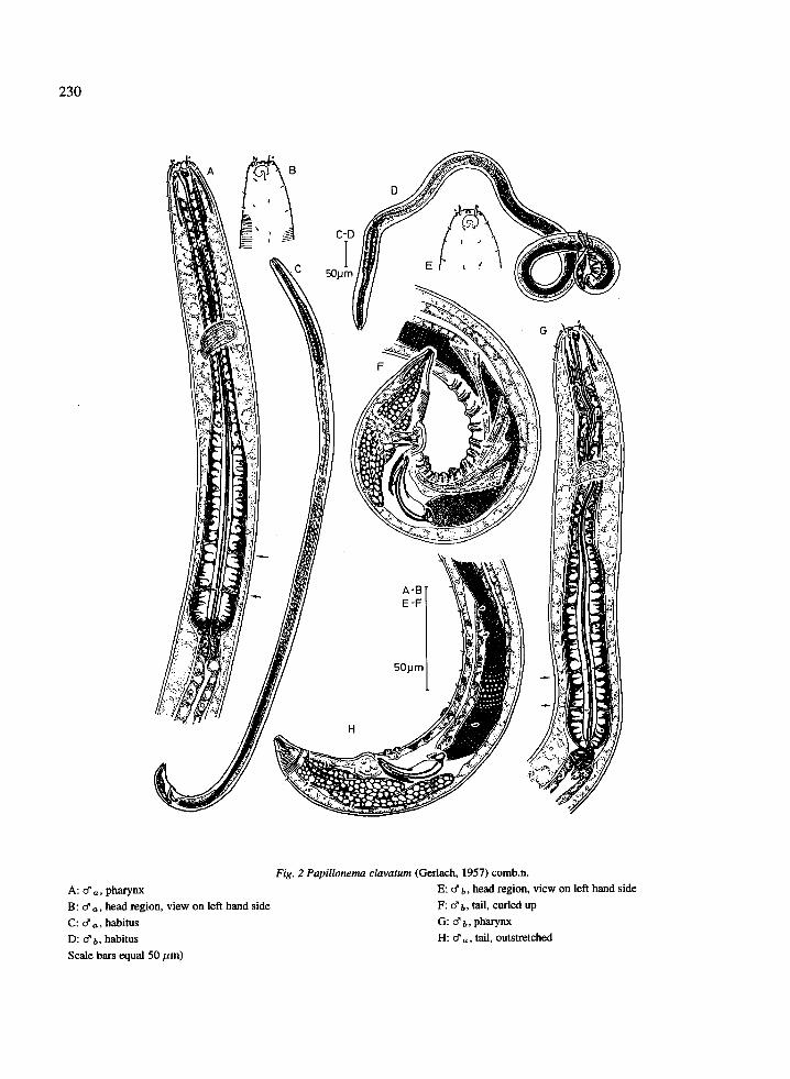
Fig. 2 Papillonema clavatum (Gerlach, 1957) comb .n .A: d' a , pharynx
E: d b, head region, view on left hand sideB : d' a , head region, view on left hand side
F: d b, tail, curled upC : d' a , habitus
G: d' b , pharynxD : d' b, habitus
H: (f d , tail, outstretchedScale bars equal 50 µm)
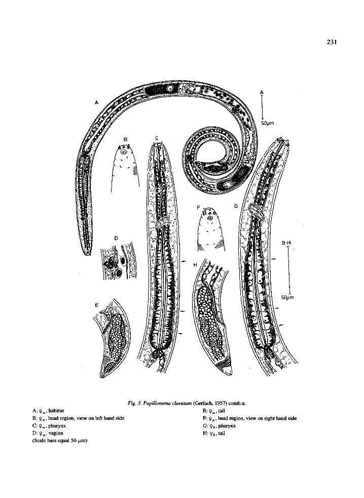
A: 9 a , habitusB : 9 a , head region, view on left hand sideC : 9 Q , pharynxD: 9 a , vagina(Scale bars equal 50 µm)
Fig. 3. Papillonema clavatum (Gerlach, 1957) comb .n .E: 9 a , tailF : 9 a , head region, view on right hand sideG: 4t, pharynxH : 9 b , tail
23 1
232
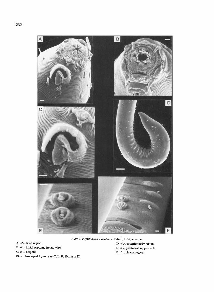
Plate 1 . Papillonema clavatum (Gerlach, 1957) comb.n.A : d' - head region
D: d' d, posterior body regionB : d' d, labial papillae, frontal view
E: &,, precloacal supplementsC: d' ., amphid
F: o'- cloacal region(Scale bars equal 1 µm in A-C, E, F ; 10 pm in D)

Plate 2. Papillonema clavatum (Gerlach, 1957) comb .n .A : d' anterior precloacal supplement
E: 9b, buccal cavity with dorsal tooth and denticlesB : d'~, precloacal supplements and 'cushions' of tiny papillae
F: 9 ., egg caught up in curled bodyC : 9 b , head region
G: 9b , tailD : 9 b , labial papillae, frontal view
H: 9b, vulva(Scale bars equal 1 pm in A-a, H ; 10 pm in F, G)
233

Additional descriptionMales: Body cylindrical with blunt conical headregion and conical tail (Fig . 2C, D) . Body cuticle striat-ed with numerous, very slender body annuli extendingto the anterior edge of the fovea amphidialis (Plate IA,C); annuli visible in tail region, faintly visible in pha-ryngeal region, almost invisible along the rest of thebody. Tiny, hyalin somatic setae are very hard to dis-tinguish : in the pharyngeal region, they are arrangedin eight longitudinal rows of three (to four) setae (Fig .2B, E) ; the first are located at the posterior edge (orhalf) of the amphids, therefore Gerlach (1957) previ-ously named these setae 'subcephalic setae' (i .e. cer-vical setae, see remark above), but we regard themas somatic setae because there is no head capsule andthey do not differ one bit from the rest of the somaticsetae . Anterior to the precloacal supplements, there is aventral row of eight to ten characteristic somatic setaewhich are only visible when the posterior body regionis not curled up (compare Fig . 2F with Fig . 2H), thesesetae are connected with gland cells (not drawn) .
No head capsule . Head region with six small inter-nal lip papillae, six larger external lip papillae andfour cephalic setae (Plate 1A, B) . External lip papil-
lae and cephalic setae situated at anterior edge ofthe fovea amphidialis. Fovea amphidialis broad, openloop-shaped (Fig . 2B, E ; Plate 1C).
Lip region protrusible . Buccal cavity (stoma) withlarge dorsal tooth, two minute subventral teeth, anda crown of denticles (Fig. 2G); when lips are closed(not protruded) denticles are hardly visible by meansof light microscope . In the stomatal region there areeight epidermal glands . Pharynx (clavate) with slenderbuccal bulb (stoma), slender corpus and long, broadcylindrical postcorpus (terminal bulb, about 47% ofthe total pharyngeal length) ; the pharynx shows threefine partitions : one at the transition from corpus topostcorpus and two distally in the posterior half of thepostcorpus (so terminal bulb itself with two partitions ;Fig. 2G, arrows); lumen lined with a thick cuticle .Nerve ring situated anterior to the postcorpus at about39% of the total pharyngeal length (Fig . 2A). Longcardia (8-13 µm) .
Reproductive system monorchic : long outstretchedtestis with large sperm cells (l = 13µm, w = 7µm)which were also found in the uterus of each female .Testis located at the right of the intestine in all males ;vas deferens located ventrally to the intestine in mostmales, at the left of the intestine in one male . Archedspicules with birdhead-shaped capitulum and broad butthin velum (Fig . 2F, H). Thick gubernaculum whichproximally embraces the spicule tips .
Three ventral precloacal supplements consistingof a button-shaped to conical base (h = lµm, w =3 .5µm) with a protrusible slender, trunk-shaped exten-sion retracted into it (Plates 1E, F and 2A, B) ; two outof three are located 5 µm anterior to the cloaca, thethird 39 µm in front of it (Fig . 2F, H) . Anterior to thethird precloacal supplement, there is a ventral row ofeight to ten short setae which are not visible when thetail is curled up (Fig . 2F). A series of muscles (Fig . 2F)are present to curl up the tail region during copulation ;when this happens, the ventral precloacal region folds(Fig. 2F; Plate 1D) ; one each fold there are paired sub-ventral `cushions' of tiny papillae, clearly visible bymeans of SEM pictures (Plate 2A, B), but only visi-ble as gland-enriched areas in the epidermis by meansof light microscope; when the tail region is straight,only pairs of local, slightly risen cuticle and the epi-dermal glands give away the position of the cuticularpapillae .
Conical tail with three pairs of latero-ventral somat-ic setae and three (four) pairs of dorso-lateral setae .Large hyalin caudal glands ; spinneret with clear valve,valve muscle attached to dorsal body wall . Non-
234
Table 3 . Measurements (in /.Lm) of Papillonemaclavatum (Gerlach, 1957) comb .n .
J4 J3 J3' J2
L 1045 1016 972 760aw 6 4 4 4cs 4 3 4 4dcs 3 2 2 2amph % 40 28 32dnr 76 59 74 63ph 198 174 202 157mbd ph 32 33 30 28mbd 34 34 28 29bdnr 31 31 29 28bdcs 12 12 12da 991 955 911 709abd 28 31 26 24t 54 56 54 51tmr 11 12 11 9a 30 .7 29 .9 34 .7 26 .2b 5 .3 5 .8 4 .8 4 .8c 19 .4 18 .1 18 .0 14 .9
Measurements: See Table 2, 3 .

annulated tail end with one pair of short dorso-lateralsomatic setae (Fig. 2H) .
Females : Body shape, body annuli, pattern of somat-ic setae, pattern of labial papillae (Plate 2D), cephalicsetae, pharynx, cardia and tail (Plate 2G) similar tomales .
Head region as in males . Fovea amphidialis multi-spiral (1 .5 whirls, sexual dimorphism : loop-shaped inmales; Fig. 3B, F; Plate 2C) .
Stoma with large dorsal tooth, two small subventralteeth and a crown of denticles (located anterior to thedorsal tooth; Plate 2E) . Partitions of pharynx and post-corpus more clear than in males (postcorpus makes out47% of the total pharyngeal length ; Fig. 3C, G, seearrows). Posterior in the intestine there are two cellsforming a sort of valve . Rectal valve present .
Reproductive system didelphic, amphidelphic withreflected ovaries (Fig . 3A, D) ; position to intestinevariable: in some females both branches located to theleft of the intestine ; in others, antepudendum located tothe right and postpudendum to the left of the intestine .Vulva inconspicuous (not distinct by means of lightmicroscope ; Plate 2H); vagina vera consisting of twocuticular plates, vagina uterina with clear sphinctermuscle (Fig . 3D). Sperm cells (1 = 10µm, w = 7pm)are always found within the uterus . In one female, anegg was caught between the coils of her curled upbody (Plate 2F), what opens the chance of some kindof brood protection .
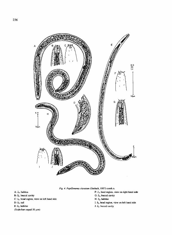
Juveniles: Body shape, annuli, pattern of somaticsetae, labial papillae, and cephalic setae as in adults .
Fourth stage juvenile : (Fig. 4A-D). Fovea amphidi-alis (6 ,um) multispiral (1 .5 whirls) . Stoma with largedorsal tooth, in which the tooth of the next stage isalready present. Nerve ring clear.
Reproductive system of a juvenile female is alreadypresent : primordium for vagina consisting out of twogroups of three cells. Didelphic, amphidelphic uterusand oviducts with straight ovaries with a few germcells .
Third stage juveniles : (Fig. 4E-G). Multispiral (1 .5whirls) fovea amphidialis (4 am) . Stoma with largedorsal tooth, in which the tooth of the next (possibly
two next) juvenile stage is present . Nerve ring notclear.
235
Developing reproductive system in both sexes con-sists out of a slender hyalin cell strand in which indi-vidual cells cannot be recognized .
Second stage juvenile : (Fig. 4H-J). Multispiral foveaamphidialis (4 am) . Buccal cavity with large dorsaltooth in which the tooth of the next stage is alreadypresent. Nerve ring not clear .
Genital primordium consists out of a small groupof a few cells .
First stage juvenile : not found .
RemarkPapillonema clavatum (Gerlach, 1957) comb.n. wasfirst described from the mangrove bushes of Canandia(Brazilian coast; Gerlach, 1957) . Later it was alsofound on the Maldive Islands (Gerlach, 1963) andfinally on the Sarso Islands (Red Sea ; Gerlach, 1967) .The Kenyan specimens resemble mostly the specimensfound on the Sarso Islands, although these specimensare larger than the Kenyan specimens. Kenyan maleshave similar labial papillae and fovea amphidialis asthe Sarso Islands males, but differ from those of theMaldives (papillae and amphids) and from those ofCanandia (amphids) . Spicules from Kenyan specimensshow a broad, but very thin velum, which has not been
described by Gerlach, although this could just havebeen overlooked because the velum is really thin .
DiscussionPapillonema clavatum (Gerlach, 1957) comb.n .resembles P danieli gen . et sp.n. but differs fromit in shape of the amphids (sexual dimorphism in Pclavatum, multispiral in P. danieli), relative length ofthe pharyngeal postcorpus (47% in P clavatum com-pared to 60% in P danieli), and the arrangement ofthe precloacal supplements in males (in P danieli allthree precloacal supplements are arranged at an equaldistance for one another) .
Acknowledgements
The authors would like to thank ABOS (Belgium),Kenyan Belgian Project and KMFRI (Mombasa) forfinancial and technical assistance . Thanks also toYvette Vermeulen and Rita Van Driessche for theirassistance . Research supported by National Science
236
A: J4, habitusB : J4, buccal cavityC : J4, head region, view on left hand sideD : J4, tailE : J3, habitus(Scale bars equal 50 µm)
Fig . 4. Papillonema clavatum (Gerlach, 1957) comb .n .F : J3, head region, view on right hand sideG: J3, buccal cavityH: J2, habitusI : J2, head region, view on left hand sideJ : J2, buccal cavity

Foundation of Belgium (32 .0009.92) and ConcertedActions in Oceanography (12 .0501 .92) .
References
Allen, M . W. & E. M. Noffsinger, 1978. A revision of themarine nematodes of the superfamily Draconematidae Filip-
jev, 1918 (Nematoda : Draconematina) . Berkely-Los Angeles-London, Univ . of Calif. Press, Univ. Calif. Publis . Zool ., 109 :i-viii, 133 pp .
Furstenberg, J . & M. Vincx, 1988. Three new Chromadoropsisspecies (Nematoda: Desmodoridae) from southern Africa andthe North Sea. S . Aft. J . Zool ., 23 : 215-223 .
Gerlach, S . A., 1957. Marine nematoden aus dem mangrove-Gebietvon Cananeia . Abh . Math . -Naturw . Kl . Akad . Wiss . Main z ., 5 :129-176 .
237
Gerlach, S. A ., 1963 . Freilebende Meeresnematoden von den Male-diven II . Kieler Meeresforsch. 19: 67-103 .
Gerlach, S. A., 1967 . Freilebende Meeres-Nematoden von denSarso-Inseln (Rotes Meer) 3 . Beitrag der Arbeitsgruppe Litoral-forschung . `Meteor' Forschergebn ., (D) (2) : 19-43 .
Seinhorst, J . W., 1959 . A rapid method for the transfer of nematodesfrom fixative of anhydrous glycerin. Nematologica 4 : 67-69 .
Verschelde, D . & M. Vincx, 1994. Epsilonematidae (Nematoda :Desmodoroidea) from the East African coast, with a discussion onthe external morphology of cuticular appendages . Nematologica40: 78-105 .
Wieser, W. & B . Hopper, 1967 . Marine nematodes of the east coastof North America. I . Florida. Bull. Mus. Comp. Zool . Harv. 135 :239-344.