Pamukkale Tıp Dergisi - DergiPark
180
Pamukkale Tıp Dergisi Cilt/Vol: 14 Sayı/No: 4 Ekim/October 2021 e-ISSN 1308-0865 ISSN 1309-9833 Sahibi Pamukkale Üniversitesi Tıp Fakültesi adına Dekan Prof. Dr. Zekiye Melek Küçükatay Baş Editör Dr. Selçuk Yüksel Baş Editör Yardımcısı Dr. Eylem Değirmenci Bölüm Editörleri Dr. Eylem Değirmenci Dr. Süleyman Erkan Alataş Dr. Çağdaş Erdoğan Dr. Ebru Nevin Çetin Dr. Nida Kaçar Dr. Gülfizar Varma Dr. Özmert Muhammet Ali Özdemir Dr. Nuray Akkaya Dr. Nilay Şen Türk Dr. Gamze Gököz Doğu Dr. Murat Özban Dr. Bayram Özhan Dr. Harun Reşit Güngör Dr. Tuğba Sarı Dr. Samet Yılmaz Dr. Gökhan Ozan Çetin Dr. Yusuf Özlülerden Dr. Esin Avcı Dr. Başak Ünver Koluman Dr. Emrah Egemen Dr. Mert Özen Yayın/ Danışma Kurulu Dr. Selçuk Yüksel, Pamukkale Üniversitesi, Denizli Dr. Eylem Değirmenci, Pamukkale Üniversitesi, Denizli Dr. Banu Çelikel Acar, Sağlık Bilimleri Üniversitesi, Ankara Dr. Murat Bülent Rabuş, Sağlık Bilimleri Üniversitesi, İstanbul Dr. Mehmet Uludağ, Şişli Hamidiye Etfal Eğitim ve Araştırma Hastanesi, İstanbul Dr. Güven Çetin, Bezmiâlem Vakfı Üniversitesi, İstanbul Dr. Cengiz Candan, Medeniyet Üniversitesi, İstanbul Yayın Koordinatörü Dr. Eylem Değirmenci Sekreter Bil.İşl. Kutsel Onaç Memur Burcu Ateş İngilizce Redaktör Öğr. Gör. Ayşe Yavuz Grafik Tasarım/Dizgi Cansu Ekinci Dergi Adı: Pamukkale Tıp Dergisi Web Adresi: http://dergipark.gov.tr/patd/writing-rules Dergi Statüsü: Hakemli Süreli Yayın Yayınlanma Süresi: Yılda 4 Sayı ISSN: 1309-9833 e-ISSN: 1308-0865 Adres: Pamukkale Tıp Dergisi, Pamukkale Üniversitesi Tıp Fakültesi Dekanlığı, Yunusemre mah. no:3/F, Kınıklı Kampüsü, 200070 Pamukkale, Denizli. E-posta: [email protected] Tel: +902582961619 Fax: +902582961765 Pamukkale Tıp Dergisi, TÜBİTAK/ULAKBİM Türk Tıp Dizini, Türkiye Atıf Dizini, TÜRK MEDLINE, ve CrossRef tarafından indekslenmektedir.
-
Upload
khangminh22 -
Category
Documents
-
view
0 -
download
0
Transcript of Pamukkale Tıp Dergisi - DergiPark

Pamukkale Tıp DergisiCilt/Vol: 14 Sayı/No: 4 Ekim/October 2021
e-IS
SN 1
308-
0865
ISSN
130
9-98
33
SahibiPamukkale Üniversitesi Tıp Fakültesi adına Dekan
Prof. Dr. Zekiye Melek Küçükatay
Baş EditörDr. Selçuk Yüksel
Baş Editör YardımcısıDr. Eylem Değirmenci
Bölüm EditörleriDr. Eylem Değirmenci
Dr. Süleyman Erkan AlataşDr. Çağdaş ErdoğanDr. Ebru Nevin Çetin
Dr. Nida KaçarDr. Gülfizar Varma
Dr. Özmert Muhammet Ali ÖzdemirDr. Nuray AkkayaDr. Nilay Şen Türk
Dr. Gamze Gököz DoğuDr. Murat Özban
Dr. Bayram ÖzhanDr. Harun Reşit Güngör
Dr. Tuğba Sarı Dr. Samet Yılmaz
Dr. Gökhan Ozan ÇetinDr. Yusuf Özlülerden
Dr. Esin Avcı Dr. Başak Ünver Koluman
Dr. Emrah EgemenDr. Mert Özen
Yayın/ Danışma KuruluDr. Selçuk Yüksel, Pamukkale Üniversitesi, Denizli
Dr. Eylem Değirmenci, Pamukkale Üniversitesi, DenizliDr. Banu Çelikel Acar, Sağlık Bilimleri Üniversitesi, Ankara
Dr. Murat Bülent Rabuş, Sağlık Bilimleri Üniversitesi, İstanbulDr. Mehmet Uludağ, Şişli Hamidiye Etfal Eğitim ve Araştırma Hastanesi,
İstanbulDr. Güven Çetin, Bezmiâlem Vakfı Üniversitesi, İstanbul
Dr. Cengiz Candan, Medeniyet Üniversitesi, İstanbul
Yayın KoordinatörüDr. Eylem Değirmenci
SekreterBil.İşl. Kutsel OnaçMemur Burcu Ateş
İngilizce RedaktörÖğr. Gör. Ayşe Yavuz
Grafik Tasarım/DizgiCansu Ekinci
Dergi Adı: Pamukkale Tıp DergisiWeb Adresi: http://dergipark.gov.tr/patd/writing-rules Dergi Statüsü: Hakemli Süreli YayınYayınlanma Süresi: Yılda 4 SayıISSN: 1309-9833 e-ISSN: 1308-0865
Adres: Pamukkale Tıp Dergisi, Pamukkale Üniversitesi Tıp Fakültesi Dekanlığı, Yunusemre mah. no:3/F, Kınıklı Kampüsü, 200070 Pamukkale, Denizli.E-posta: [email protected]: +902582961619Fax: +902582961765
Pamukkale Tıp Dergisi, TÜBİTAK/ULAKBİM Türk Tıp Dizini, Türkiye Atıf Dizini, TÜRK MEDLINE, ve CrossRef tarafından indekslenmektedir.


Pamukkale Tıp DergisiPamukkale Medical Journal
Cilt/Vol:14 Sayı/Issue:4 Ekim/October 2021
İÇİNDEKİLER - CONTENTS
784-791
792-802
804-810
812-817
818-827
828-834
836-845
846-853
Distal radial artery access in the anatomical snuffbox for coronary angiography and percutaneous coronary intervention Koroner anjiyografi ve perkütan koroner girişimlerde, anatomik enfiye çukurundan distal radyal arter girişimi
Gökhan Alıcı, Alaa Quisi
Effects of BDNF deficiency and endoplasmic reticulum stress on the GABAergic system BDNF eksikliği ve endoplazmik retikulum stresinin GABAerjik sistem üzerindeki etkileri
Gülay Hacıoğlu Dervişoğlu, Selma Cırrık, Hakan Yüzüak, Selcen Aydın, İsmail Abidin
Çocuk yoğun bakım ünitesinde izlenen olguların elektroensefalografi sonuçlarının geriye dönük olarak değerlendirilmesiEvaluation of electroencephalogram results in patients hospitalized in the pediatric intensive care unit
Müge Ayanoğlu, Uluç Yiş, Ayşe İpek Polat, Alper Köker, Gazi Arslan, Ayşe Semra Hız Kurul
Follow-Up of endovascular coil embolization applied for the treatment of intracranial aneurysms by magnetic resonance angiographyIİntrakraniyal anevrizmaların tedavisi için uygulanan endovasküler koil embolizasyonunun manyetik rezonans anjiyografi ile takibi
Murat Dökdök, Kutlay Karaman, Selçuk Göçmen
Myocardial performance index is associated with Systematic Coronary Risk Estimation (SCORE) system in chronic coronary syndromeKronik koroner sendromda miyokardial performans indeks Sistematik Koroner Risk Tahmini (SCORE) ile ilişkilidir
Hazar Harbalıoğlu, Ömer Genç, Abdullah Yıldırım
Clinical and prognostic factors in children with dilated cardiomyopathyDilate kardiyomyopatili çocuk hastalarda klinik ve prognostik faktörler
Ayça Koca Yozgat, Selmin Karademir
Annelerin tamamlayıcı beslenme hakkındaki tutum ve yaklaşımları Approaches and attitudes of mothers about complementary feeding
Gizem Şenyazar, Şule Gökçe, Feyza Koç
Metotreksat kaynaklı beyin hasarına karşı bromelainin potansiyel faydalı etkilerinin araştırılması
Investigation of potential beneficial effects of bromelain against methotrexate-induced brain injury Kürşat Kaya, Ali Gürel, Volkan İpek
Araştırma Makaleleri - Research Articles
Pamukkale Tıp DergisiPamukkale Medical Journal
Cilt/Vol:14 Sayı/Issue:4 Ekim/October 2021
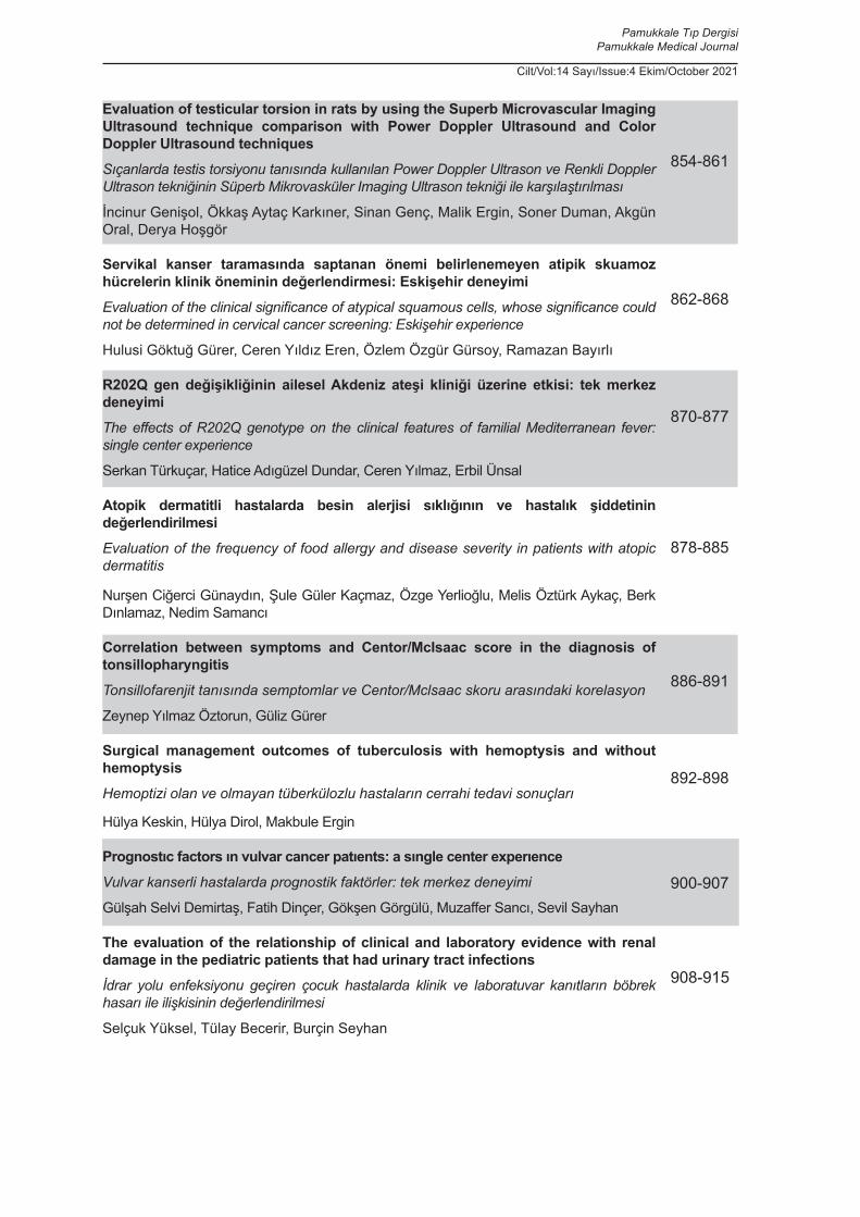
Evaluation of testicular torsion in rats by using the Superb Microvascular Imaging Ultrasound technique comparison with Power Doppler Ultrasound and Color Doppler Ultrasound techniques Sıçanlarda testis torsiyonu tanısında kullanılan Power Doppler Ultrason ve Renkli Doppler Ultrason tekniğinin Süperb Mikrovasküler Imaging Ultrason tekniği ile karşılaştırılması
İncinur Genişol, Ökkaş Aytaç Karkıner, Sinan Genç, Malik Ergin, Soner Duman, Akgün Oral, Derya Hoşgör Servikal kanser taramasında saptanan önemi belirlenemeyen atipik skuamoz hücrelerin klinik öneminin değerlendirmesi: Eskişehir deneyimi Evaluation of the clinical significance of atypical squamous cells, whose significance could not be determined in cervical cancer screening: Eskişehir experience
Hulusi Göktuğ Gürer, Ceren Yıldız Eren, Özlem Özgür Gürsoy, Ramazan Bayırlı
R202Q gen değişikliğinin ailesel Akdeniz ateşi kliniği üzerine etkisi: tek merkez deneyimiThe effects of R202Q genotype on the clinical features of familial Mediterranean fever: single center experience
Serkan Türkuçar, Hatice Adıgüzel Dundar, Ceren Yılmaz, Erbil Ünsal
Atopik dermatitli hastalarda besin alerjisi sıklığının ve hastalık şiddetinin değerlendirilmesi Evaluation of the frequency of food allergy and disease severity in patients with atopic dermatitis
Nurşen Ciğerci Günaydın, Şule Güler Kaçmaz, Özge Yerlioğlu, Melis Öztürk Aykaç, Berk Dınlamaz, Nedim Samancı
Correlation between symptoms and Centor/McIsaac score in the diagnosis of tonsillopharyngitisTonsillofarenjit tanısında semptomlar ve Centor/Mclsaac skoru arasındaki korelasyon
Zeynep Yılmaz Öztorun, Güliz Gürer
Surgical management outcomes of tuberculosis with hemoptysis and without hemoptysisHemoptizi olan ve olmayan tüberkülozlu hastaların cerrahi tedavi sonuçları
Hülya Keskin, Hülya Dirol, Makbule Ergin
Prognostıc factors ın vulvar cancer patıents: a sıngle center experıence Vulvar kanserli hastalarda prognostik faktörler: tek merkez deneyimi
Gülşah Selvi Demirtaş, Fatih Dinçer, Gökşen Görgülü, Muzaffer Sancı, Sevil Sayhan
The evaluation of the relationship of clinical and laboratory evidence with renal damage in the pediatric patients that had urinary tract infectionsİdrar yolu enfeksiyonu geçiren çocuk hastalarda klinik ve laboratuvar kanıtların böbrek hasarı ile ilişkisinin değerlendirilmesi
Selçuk Yüksel, Tülay Becerir, Burçin Seyhan
854-861
862-868
870-877
878-885
886-891
892-898
900-907
908-915

Pamukkale Tıp DergisiPamukkale Medical Journal
Cilt/Vol:14 Sayı/Issue:4 Ekim/October 2021
26 yaşındaki genç maden işçisinde eş zamanlı iki taraflı femur boyun stres kırığıConcurrent two-sided femur neck stress fracture in 26-year-old young mine worker
Murat Saylık, Kemal Gökkuş
Secondary bladder stone formation on polypropylene suture after burch colposuspension and abdominal hysterectomyBurch kolposüspansiyonu ve abdominal histerektomi sonrası polipropilen sütüre ikincil mesane taşı oluşumu
Mert Hamza Özbilen, Batuhan Ergani, Taha Çetin, Mehmet Yiğit Yalçın, Çağdaş Bildirici, Erkin Karaca, Orçun Çelik, Yusuf Özlem İlbey
Splenic artery aneurysm in kidney transplant recipientBöbrek nakli alıcısında splenik arter anevrizması
Utku Özgen, Murat Özban, Muhammet Arslan, Onur Birsen, Mevlüt Çeri, Sevda Yılmaz, Ezgi Doğa Yoran, Çağadaş Aydın
Postural hematürinin değerlendirilmesi: posterior nutcracker sendromuEvaluation of postural hematuria: posterior nutcracker syndrom
Murat Yaşar Taş, Belda Dursun, Muhammet Arslan
Coğrafi Bilgi Sistemleri-mekânsal epidemiyoloji çerçevesinde SARS CoV-2 (COVID-19)SARS CoV-2 (COVID-19) in the framework of Geographic Information System spatial epidemiology
Ömer Barış İnce, Murat Şevik, Ahmet Sait
916-920
922-925
926-929
930-932
934-943
Olgu Sunumu - Case Report
Derleme - Review
Pamukkale Tıp DergisiPamukkale Medical Journal
Cilt/Vol:14 Sayı/Issue:4 Ekim/October 2021
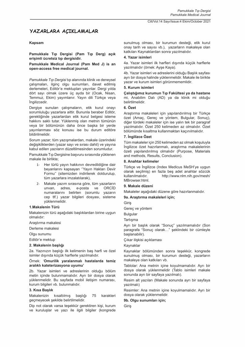
YAZARLARA AÇIKLAMALAR
Kapsam
Pamukkale Tıp Dergisi (Pam Tıp Derg) açık erişimli ücretsiz tıp dergisidir. Pamukkale Medical Journal (Pam Med J) is an open-access free medical journal.
Pamukkale Tıp Dergisi tıp alanında klinik ve deneysel çalışmaları, ilginç olgu sunumları, davet edilmiş derlemeleri, Editör’e mektupları yayınlar. Dergi yılda dört sayı olmak üzere üç ayda bir (Ocak, Nisan, Temmuz, Ekim) yayımlanır. Yayın dili Türkçe veya İngilizcedir.Dergiye sunulan çalışmaların, etik kurul onayı sorumluluğu yazarlara aittir. Bununla beraber Editör, gerektiğinde yazarlardan etik kurul belgesi isteme hakkını saklı tutar. Yüklenmiş olan metnin tümünün veya bir bölümünün daha önce başka bir yerde yayınlanması söz konusu ise bu durum editöre bildirilmelidir.Sorum yazar; tüm yazışmalardan, makale üzerindeki değişikliklerden (yazar sayı ve sırası dahil) ve yayına kabul edilen yazıların düzeltilmesinden sorumludur.Pamukkale Tıp Dergisine başvuru sırasında yüklenen makale ile birlikte;
1- Her türlü yayın hakkının devredildiğine dair beyanlarını kapsayan ‘’Yayın Hakları Devir Formu’’ (sitemizden indirilerek doldurulup, tüm yazarlara imzalatılarak),
2- Makale yazım sırasına göre, tüm yazarların unvan, adres, e-posta ve ORCİD numaralarını belirten (sorumlu yazarın cep tlf.) yazar bilgileri dosyası, sisteme yüklenmelidir.
1.Makalenin TürüMakalenizin türü aşağıdaki başlıklardan birine uygun olmalıdır:Araştırma makalesiDerleme makalesiOlgu sunumuEditör’e mektup2. Makalenin başlığı2a. Yazınızın başlığı ilk kelimenin baş harfi ve özel isimler dışında küçük harflerle yazılmalıdır.Örnek: ‘Omurilik yaralanmalı hastalarda temiz aralıklı kateterizasyona uyumu’2b. Yazar isimleri ve adreslerinin olduğu bölüm metin içinde bulunmamalıdır. Ayrı bir dosya olarak yüklenmelidir. Bu sayfada mobil iletişim numarası, kurum bilgileri vb. bulunmalıdır.3. Kısa BaşlıkMakalenizin kısaltılmış başlığı 75 karakteri geçmeyecek şekilde belirtilmelidir. Dip not olarak varsa teşekkür gerektiren kişi, kurum ve kuruluşlar ve yazı ile ilgili bilgiler (kongrede
sunulmuş olması, bir kurumun desteği, etik kurul onay tarih ve sayısı vb.), yazarların makaleye olan katkıları Kaynaklardan sonra yazılmalıdır.4. Yazar isimleri4a. Yazar isimleri ilk harfleri dışında küçük harflerle yazılmalıdır (örnek: Ayşe Kaya).4b. Yazar isimleri ve adreslerini olduğu Başlık sayfası ayrı bir dosya halinde yüklenmelidir. Makale ile birikte yazar ve kurum isimleri görünmememlidir.5. Kurum isimleriÇalıştığımız kurumun Tıp Fakültesi ya da hastane mi, Anabilim Dalı (AD) ya da klinik mi olduğu belirtilmelidir.6. ÖzetAraştırma makaleleri için yapılandırılmış bir Türkçe özet (Amaç, Gereç ve yöntem, Bulgular, Sonuç), diğer türdeki makaleler için ise yalın tek bir paragraf yazılmalıdır. Özet 250 kelimeden az olmalıdır. Özet bölümünde kısaltma kullanmaktan kaçınılmalıdır.7. İngilizce ÖzetTüm makaleler için 250 kelimeden az olmak koşuluyla İngilizce özet hazırlanmalı, araştırma makalelerinin özeti yapılandırılmış olmalıdır (Purpose, Materials and methods, Results, Conclusion).8. Anahtar kelimelerTürkçe ve İngilizce (Index Medicus MeSH’ye uygun olarak seçilmiş) en fazla beş adet anahtar sözcük kullanılmalıdır. http://www.nlm.nih.gov/mesh/MBrowser.html.9. Makale düzeniMakaleler aşağıdaki düzene göre hazırlanmalıdır.9a. Araştırma makaleleri için;GirişGereç ve yöntemBulgularTartışmaAyrı bir başlık olarak “Sonuç” yazılmamalıdır (Son paragrafa “Sonuç olarak…” şeklindeki bir cümleyle başlanabilir).Çıkar ilişkisi açıklamasıKaynaklar Kaynaklar bölümünden sonra teşekkür, kongrede sunulmuş olması, bir kurumun desteği, yazarların makaleye olan katkıları vb.Tablolar: Ana metnin içine koyulmamalıdır. Ayrı bir dosya olarak yüklenmelidir (Tablo isimleri makale sonunda ayrı bir sayfaya yazılmalı).Resim alt yazıları (Makale sonunda ayrı bir sayfaya yazılmalı)Resimler: Ana metnin içine koyulmamalıdır. Ayrı bir dosya olarak yüklenmelidir.9b. Olgu sunumları için;Giriş

Pamukkale Tıp DergisiPamukkale Medical Journal
Cilt/Vol:14 Sayı/Issue:4 Ekim/October 2021
Olgu sunumuTartışmaAyrı bir başlık olarak “Sonuç” yazmamalıdır (Son paragrafa “Sonuç olarak…” şeklindeki bir cümleyle başlanabilir).Çıkar ilişkisi açıklamasıKaynaklar Kaynaklar bölümünden sonra teşekkür, kongrede sunulmuş olması, bir kurumun desteği, yazarların makaleye olan katkıları vb.Tablolar: Ana metnin içine koyulmamalıdır. Ayrı bir dosya olarak yüklenmelidir (Tablo isimleri makale sonunda ayrı bir sayfaya yazılmalı).Resim alt yazıları (Makale sonunda ayrı bir sayfaya yazılmalı)Resimler: Ana metnin içine koyulmamalıdır. Ayrı bir dosya olarak yüklenmelidir.9c. Derleme makaleler için;GirişMetin 4000 kelime, 50 kaynak, Tablo ve Resim sayısı en fazla 4 adet olacak şekilde düzenlenmelidir.Metnin gövdesi istenildiği şekilde başlık ve alt başlıklarla yapılandırılabilir.Ayrı bir başlık olarak “Sonuç” yazmamalıdır (Son paragrafa “Sonuç olarak…” şeklindeki bir cümleyle başlanabilir).Çıkar ilişkisi açıklamasıKaynaklarKaynaklar bölümünden sonra teşekkür, kongrede sunulmuş olması, bir kurumun desteği, yazarların makaleye olan katkıları vb.Tablolar: Ana metnin içine koyulmamalıdır. Ayrı bir dosya olarak yüklenmelidir (Tablo isimleri makale sonunda ayrı bir sayfaya yazılmalı).Resim alt yazıları (Makale sonunda ayrı bir sayfaya yazılmalı)Resimler: Ana metnin içine koyulmamalıdır. Ayrı bir dosya olarak yüklenmelidir.9d. Editöre Mektup makaleler için;GirişMetin 1000 kelime, 10 kaynak, Tablo ve Resim sayısı 1 adet olacak şekilde düzenlenmelidir.Metnin gövdesi istenildiği şekilde başlık ve alt başlıklarla yapılandırılabilir.Ayrı bir başlık olarak “Sonuç” yazmamalıdır. (Son paragrafa “Sonuç olarak…” şeklindeki bir cümleyle başlanabilir.)Çıkar ilişkisi açıklamasıKaynaklarKaynaklar bölümünden sonra teşekkür, kongrede sunulmuş olması, bir kurumun desteği, yazarların makaleye olan katkıları vb.Tablolar: Ana metnin içine koyulmamalıdır Ayrı bir dosya olarak yüklenmelidir. (Tablo isimleri makale sonunda ayrı bir sayfaya yazılmalı).Resim alt yazıları (Makale sonunda ayrı bir sayfaya yazılmalı)
Resimler: Ana metnin içine koyulmamalıdır. Ayrı bir dosya olarak yüklenmelidir.10. Makale metni Metin Arial puntosu (boyut 12), 1.5 satır aralığı ve paragraf aralığı Önce: 0 nk ve Sonra: 0 nk. kullanılarak yazılmalıdır. Başlık, özet, abstract metin içerisine yazılmamalıdır. Ayrı dosya olarak yüklenmelidir. Tüm yüklenen dosyalar, dosya uzantısı ile değil isimleri ile yüklenmelidir. Örnek: Makale metni, Abstract, Resim gibi.10a. Paragraf başlarında girinti olmalıdır (içerden başlamalıdır.)10b. Başlık, Alt başlıklar, Kaynaklar, Resim alt yazıları normal sola dayalı bold olmalıdır.10c. Kısaltmalar metin içinde ilk kullanıldığı yerde açık olarak tanımlanmalıdır.10d. Metin içindeki her kaynak, şekil, resim ve tabloya atıf yapılmalıdır.10e. Mikroorganizma cins, tür ve gen isimlerinde eğik (italik) karakterde harfler kullanılmalıdır: ”… Schistosoma haematobium“.10f. İstatistiksel analizler için kulanılan ‘p’ için italik karakterde ve küçük harf kullanılmalıdır. p’den sonraki “=, >, <” işaretlerinden önce ve sonra boşluk bırakılmamalıdır. p<0.05. Bu kural ayrıca tablo ve şekiller için kullanılan ‘p’ için de geçerlidir.10g. Başka durumlarda da “>”, “<”, “=” veya “±” işaretlerinden önce veya sonra boşlukbırakılmamalıdır.10h. Birimler için SI birimleri kullanılmalıdır. Ör: “mL” ( “cc”değil ), “dL” gibi. Litre büyük harf kullanılarak kısaltılmalıdır.10ı. Kimliğinizin çalıştığınız kurum veya daha önceden yaptığınız yayınlar vs. belirtilerekeleştirmenlere açıklanmadığından emin olunmalıdır. Eğer bunun yapılması gerekiyorsakırmızı renkli ve koyu karakterde yazılmalı ve resimlerinizin bir kurum ya da hasta adını açıklamadığından emin olunmalıdır.10i. Bir ilacın, donanımın veya yazılımın üreticisini parantez içinde ve sonuna virgül koyarak belirtilip, daha sonra şirketin bulunduğu şehir ve ülke ismi virgül ile ayrılmalıdır: “…Şirketi, Ankara, Türkiye”.10j. Tartışma bölümünde araştırma makalenizdeki kısıtlılıklar, sınırlılıklar ya da eksikler belirtilmelidir.10k. Makalede ondalık sayılar ifade edilecek ise Türkçe yazımlarda , (virgül) ile yazılmalıdır. Yazım İngilizce ise. (nokta) ile ifade edilmelidir. Örneğin: 12,17 (Türkçe yazım), 12.17 (İngilizce yazım). 11. Metin içerisinde kaynak kullanımı:11a. Tüm kaynakların yazı içinde sıralı şekilde belirtilmiş olmasına dikkat edilmelidir.11b. Sadece ilgili ve gerekli olan kaynaklar belirtilmelidir.11c. Kaynaklar metinde kullanım sırasına göre numaralandırılmalı, numaraları metinde cümlenin sonunda veya yazar adı geçmişse isimden hemen sonra köşeli parantez ([]) içinde virgül ile ayrılarak ve arada boşluk bırakılarak yazılmalıdır: [1, 4, 7-9].

Pamukkale Tıp DergisiPamukkale Medical Journal
Cilt/Vol:14 Sayı/Issue:4 Ekim/October 2021
11d. İkiden fazla ardışık kaynak için “kısa tire, -” kullanılmalıdır. “[7-9]”.11e. Eğer kullanılan kaynak tek yazarlı ise, metin içinde yazarın isminden sonra ‘ark.’ veya ‘et al.’ kullanılmamalıdır. Örnek: “Abban [7] çalışmasında bu sıklığı…” veya “Yüksel [7] stated that…”.11f. Eğer kullanılan kaynak iki yazarlı ise, metin içinde yazarın isminden sonra ‘ark.’ veya ‘et al.’ kullanılmamalıdır. Örnek: “Saçar ve Karabulut [7] bu sıklığı…” veya “Herek and Ergin [7] stated that…”.11g. Eğer kullanılan kaynak ikiden fazla yazarlı ise, metin içinde yazarın isminden sonra ‘ark.’ veya ‘et al.’ kullanılmalıdır. Örnek: “Baki ve ark. [7] bu sıklığı…” veya “Aybek et al. [7] stated that…”.11h. Bir resim ya da tablo için kullanılan cümle bir kaynak ile bitiyorsa, kaynağı resim ya da tablo parantezinden sonra belirtilmelidir. (önce değil): “…(Tablo 1) [7].12. Araştırma EtiğiTüm araştırma makalelerinde, çalışma için etik kurul onamının alındığı Gereç ve yöntem bölümünde belirtilmelidir. Kaynaklar bölümünden sonra izinin hangi kurumdan, hangi tarihte ve hangi karar veya sayı numarası ile alındığı açıkça sunulmalıdır.Dergimizde yayınlanacak olan makalelerle ilgili etik uygulamalar TR Dizin TÜBİTAK ULAKBİM, Cahit Arf Bilgi Merkezi tarafından önerilen aşağıdaki kurallar doğrultusunda gerçekleştirilmektedir.Etik Kurul izni gerektiren araştırmalar: . Anket, mülakat, odak grup çalışması, gözlem, deney, görüşme teknikleri kullanılarak katılımcılardan veri toplanmasını gerektiren nitel ya da nicel yaklaşımlarla yürütülen her türlü araştırmalar,· İnsan ve hayvanların (materyal/veriler dahil) deneysel ya da diğer bilimsel amaçlarla kullanılması, · İnsanlar üzerinde yapılan klinik araştırmalar,· Hayvanlar üzerinde yapılan araştırmalar,· Kişisel verilerin korunması kanunu gereğince retrospektif çalışmalar, Ayrıca;· Olgu sunumlarında “Aydınlatılmış onam formu”nun alındığının belirtilmesi,· Başkalarına ait ölçek, anket, fotoğrafların kullanımı için sahiplerinden izin alınması ve belirtilmesi,. Kullanılan fikir ve sanat eserleri için telif hakları düzenlemelerine uyulduğunun belirtilmesiGeçmiş yıllarda tamamlanmış çalışma ve tezden üretilen yayınlar için geriye dönük Etik Kurul izni:2020 yılı öncesi araştırma verileri kullanılmış, yüksek lisans/doktora çalışmalarından üretilmiş (makalede belirtilmelidir), bir önceki yıl dergiye yayın başvurusunda bulunulmuş, kabul edilmiş ama henüz yayımlanmamış makaleler için geriye dönük etik kurul izni gerekmemektedir. Üniversite mensubu olmayan araştırmacılar için etik izin:Üniversite mensubu olmayan araştırmacılar da bölgelerinde bulunan Etik Kurul’lara başvurabilir ve oradan izin alabilirler.
Ayrıca; Dergiler “Yayın Etiği”, “Araştırma Etiği” ve “Yasal/Özel izin belgesi alınması” ile ilgili kurallara uyduğunu uluslararası standartlara atıf yaparak, hem web sayfasında hem de basılı dergide herbiri için ayrı başlık açarak belirtmelidir.· Dergilerde yayın etiğine uygunluk konusu sadece yazarların sorumluluğuna bırakılmamalı, dergi yayın etiği konusunda izleneceği yolu açık olarak tanımlanmış olmalıdır.· Dergimizde yayımlanacak araştırma makalelerinde etik kurul izini ve/veya yasal/özel izin alınmasının gerekip gerekmediği makalede belirtilmiş olmalıdır. Eğer bu izinlerin alınması gerekli ise, izinin hangi kurumdan, hangi tarihte ve hangi karar veya sayı numarası ile alındığı açıkça sunulmalıdır.· Çalışma insan ve hayvan deneklerinin kullanımını gerektiriyor ise çalışmanın uluslararası deklerasyon, kılavuz vb. uygun gerçekleştirildiği beyan edilmelidir.13. Yayın Etiği PolitikamızPamukkale Tıp Dergisi editör, editör yardımcısı ve alan editörleri, Davranış Kuralları ve Dergi Editörleri İçin En İyi Uygulama Kuralları (COPE Code of Conductand Best Practice Guidelines for Journal Editors) ve Committee on Publication Ethics (COPE) ‘nin yayınladığı Dergi Editörleri İçin En İyi Uygulama Kuralları (COPE Best Practice Guidelines for Journal Editors) ilkelerine dayanarak etik görev ve sorumluluklarını yerine getirmektedirler.14. Çıkar İlişkisi (Conflict of interest)Her yazar yazıyı yükleme aşamasında yazıda sunulan bilgiler hakkında çıkar ilişkisi oluşturabilecek ticari veya finansal ilişkilerini açıklamalıdır. Bu tür ilişkiler danışmanlık, hissedarlık veya araştırma için harcamaları içerir. Yazarlar bu çalışma için maddi destek almışlarsa bunu belirtmelidir. Bu tür bir ilişki yoksa Kaynaklar bölümünden önceki Çıkar İlişkisi açıklaması bölümüne;Türkçe makalelerde: Çıkar İlişkisi: ‘Yazarlar çıkar ilişkisi olmadığını beyan eder’. İngilizce makalelerde: Conflict of Interest: No conflict of interest was declared by the authors. şeklinde yazılmalıdır.15. Kaynaklar1. Tüm kaynakların yazı içinde sıralı şekilde belirtilmiş olmasına dikkat edilmelidir.2. Sadece ilgili ve gerekli olan kaynaklar belirtilmelidir.3. Eğer altı ya da daha az yazar varsa hepsi listelenmelidir. Eğer yedi veya daha fazla yazar varsa ilk üç yazarın isminden sonra “ve ark. (et al.)” yazılmalıdır.4. Kaynaklar metinde kullanım sırasına göre numaralandırılmalıdır,5. Dergilerin adları Index Medicus’da (www.ncbi.nlm.nih.gov/journals) kullanılan biçimde kısaltılmalıdır.16. Kaynakta kullanılan Makaleler için:
Kaynak bir kitap ise aşağıdakilerden birisi gibi yazılmalıdır. Eğer online bir kitap ise, basılı değil ise erişim adresi ve tarihi ayrıntılı olarak verilmelidir.

Pamukkale Tıp DergisiPamukkale Medical Journal
Cilt/Vol:14 Sayı/Issue:4 Ekim/October 2021
1. Watanabe M, Takeda S, Ikeuchi H. Atlas of arthroscopy. 2nd ed. Tokyo: Igaku Shoin, 1969;57-59.
2. Hull RD, Hirsh J. Comparative value of tests for the diagnosis of venous thrombosis. In: Bernstein EF, ed. Noninvasive diagnostic techniques in vascular disease. 3rd ed. St. Louis: Mosby, 1985;779-796.
Basılı dergilerdeki kaynak yazılımı Örnek1. Doi olmayan makalelerde; Berkman ND, Sheridan SL, Donahue KE, Halpern DJ, Crotty K. Low health literacy and health outcomes: an updated systematic review. Ann Intern Med 2011;155:97-107.2. Doi olan makaleler:
Berkman ND, Sheridan SL, Donahue KE, Halpern DJ, Crotty K. Low health literacy and health outcomes: an updated systematic review. Ann Intern Med 2011;155:97-107. https://doi.org/10.7326/0003-4819-155-2-201107190-00005
Henüz Basılmamış makaleler;Örnek
Call JE, Mann JA, Linos KD, Perry A, Yost J. Linear lipoatrophy following intra-articular triamcinolone acetonide injection mimicking linear scleroderma. Pediatr Dermatol 2018. https://doi.org/10.1111/pde.13736 [Epub ahead of print]Elektronik Dergiler;Örnek
1. Kuah CY, Koleva E, Gan JJL, Iqbal T. Parry-Romberg syndrome in a patient with scleroderma. BMJ Case Rep 2018. pii: bcr-2018-226754. https://doi.org/10.1136/bcr-2018-226754
2. Rambon S, Brian J, AneskievichJ. TNIP1 in autoimmune diseases: regulation of toll-like receptor signaling. Immunol Res 2018;2018:3491269. https://doi.org/10.1155/2018/3491269
3. Chen Y, Yan H, Song Z. et al. Downregulation of TNIP1 expression leads to increased proliferation of human keratinocytes and severer psoriasis-like conditions in an imiquimod-induced mouse model of dermatitis. Plos One 2015;10:e0127957. https://doi.org/10.1371/ journal.pone.0127957
a. Kaynak gösterilen makalenin ilk harfi dışındaki kelimeleri küçük harfle yazılmalıdır.b. Kaynakta iki nokta üst üsteden sonra küçük harf kullanılmalıdır.c. Dergi kısaltmasından sonra nokta işareti kullanmamalıdır.d. Yayınlanma yılından önce veya sonra ay belirten kısaltma yapılmamalıdır.
e. Yayınlanma yılından sonraki noktalı virgülden sonra boşluk bırakılmamalıdır.f. Yayının cilt numarasından sonra sayı numarası yazılmamalıdır.g. Yayının cilt numarasından sonra kullanılan iki nokta üst üste işaretinden sonra boşluk bırakılmamalıdır.h. Kaynaklarda varsa doi numarası yazılmalıdır.ı. Sayfa numaraları aralarında küçük tire işareti “-” kullanılmalıdır.i. Son sayfa numarası tam olarak yazılmalıdır: “166-171.“. Lütfen “166-9”. ”166-69” yazmayınız.j. Kaynağın sonuna nokta koyulmalıdır.17. Kaynakta kullanılan kitap ve kitap bölümü için:Örnek: 1. Watanabe M, Takeda S, Ikeuchi H. Atlas of
arthroscopy. 2nd ed. Tokyo: Igaku
Shoin, 1969;57-59.2. Hull RD, Hirsh J. Comparative value of tests for
the diagnosis of venous thrombosis. In: Bernstein EF, ed. Noninvasive diagnostic techniques in vascular disease. 3rded. St. Louis: Mosby, 1985;779-796.
17a.Kaynak gösterilen kitap veya bölüm adının ilk harfi dışındaki kelimeler küçük harfle yazılmalıdır.17b. Yayınlanan şehrin isminden sonra iki nokta üst üste (:) kullanılmalıdır.17c. Yayınevi isminden sonra virgül kullanılmalıdır.17d. Yayınlanma yılından sonra noktalı virgül (;) kullanılmalıdır.17e. Yayınlanma yılından sonraki noktalı virgül işaretinden sonra boşluk bırakılmamalıdır.17f. Sayfa numaraları aralarında kısa tire işareti “-” kullanılmalıdır.17g. Son sayfa numarası tam olarak yazılmalı: “914-916.”17h. Kaynağın sonuna nokta koyulmalıdır.18. İnternet (ağ) kaynakları için: Erişim tarihiniz belirtilmelidir.Örnek: Musculoskeletal MRI Atlas. Available at: http://www.gla.med.va.gov/mriatlas/Index.html. Erişim tarihi 14 Eylül 2010 (yazarın makalesinin yazım dili Türkçe ise)Accessed September 14, 2010 (yazarın makalesinin yazım dili İngilizce ise)19. Poster veya bildiri için:Örnek: Karabulut N, Çakmak V. Diffusion-weighted MR imaging of pulmonary lesions.Paper presented at:ISMRM-ESMRMB Joint Annual Meeting 01-07 Mayıs 2010; Stockholm, Sweden.

Pamukkale Tıp DergisiPamukkale Medical Journal
Cilt/Vol:14 Sayı/Issue:4 Ekim/October 2021
20. Tez çalışmaları için:Örnek: Gündüz B. Hemşirelerde stresle başa çıkma biçimleri ile tükenmişlik arasındaki ilişkilerin incelenmesi. Yayınlanmamış Yüksek Lisans Tezi. Karadeniz Teknik Üniversitesi Sosyal Bilimler Enstitüsü, Eğitim Bilimleri Anabilim Dalı Rehberlik ve Psikolojik Danışmanlık Programı, Trabzon, 2000.Tablolara. Tüm tabloların yazınızın içinde belirtilmesi gerekmektedir.b. Grafik, diyagram ve algoritmaların tablo değil, şekil olduğu unutulmamalıdır.c. Bütün tabloların üstünde numarası ve başlığı olmalıdır.d.Tabloların başlığından sonra nokta koyulmamalıdır.d. Tablolardaki kısaltmalar tablo altında açıklanmalıdır.e. Tablolar, Word’de tablo kurallarına uygun şekilde yüklenmelidir. Resimler ve şekillera. Tüm resimlere yazı içerisinde atıf yapılmış olmalıdır.b. Görüntülerin üzerinde herhangi bir kurumun veya hastanın bilgileri olmamalıdır. Yüz fotoğraflarında gizliliği korumak için gözler kapatılmalıdır.c. Görüntüler en az 300 vpi çözünürlükte, 1280x960 piksel boyutunda çekilmiş, jpg veya tiff formatlarında kaydedilmiş olmalıdır.d. Her resim, şekil veya grafik ayrı bir belge olarak hazırlanmalı, yazının ekleri olarak yüklenmelidir. Ana metin içine yerleştirilmemelidir.e. Resim üzerinde ok vb. işaretler kullanılmış ise resim açıklamasında bu belirtilmelidir.f. Resim Yazısı: makalenizle birlikte yükleyeceğiniz resimlerde dikkat çekmek istediğiniz noktaya lütfen işaret koyunuz (Oklar ince, yerine göre beyaz veya siyah renk olmalıdır). Resimlerin açıklama bölümüne ayrıntılı açıklama yazınız. Bazı resimlerde tedavi sonrası düzelme ifade ediliyorsa, resim yazısı olarak tedavi sonrası düzelmiş olan grafi, sintigrafi vs bulguları şeklinde bilgi ilave ediniz. Resimlerin orijinal haliyle 300 vpi çözünürlüğünde yüklenmelidir. NOT: Hakem tarafından istenen düzeltmelere 2(iki) ay içerisinde cevap verilmemesi durumunda makaleler red’de alınacaktır.İntihal KontrolüOcak 2015’den itibaren gönderilen makalelerin tümü IThenticate® intihal belirleme yazılımı kullanılarak kontrol edilmektedir. Yazılım tarafından üretilen benzerlik raporu doğrultusunda, Editörler Kurulu makalenin hakem değerlendirmesine alınmasına veya doğrudan ret edilmesine karar vermektedir.

784
Distal radial artery access in the anatomical snuffbox for coronary angiography and percutaneous coronary intervention
Koroner anjiyografi ve perkütan koroner girişimlerde, anatomik enfiye çukurundan distal radyal arter girişimi
Gokhan Alici, Alaa Quisi
Pamukkale Tıp DergisiPamukkale Medical Journal
doi:https://dx.doi.org/10.31362/patd.846438
Araştırma Makalesi
Gokhan Alıcı, MD. Prof.Dr.Cemil Taşçıoğlu City Hospital, Department of Cardiology, 34384 Istanbul, Turkey, e-mail: [email protected] (https://orcid.org/0000-0002-4589-7566) (Corresponding Author)Alaa Quisi, MD. Medline Adana Hospital, Department of Cardiology, Adana, Turkey, e-mail: [email protected] (https://orcid.org/0000-0002-5862-5789)
AbstractPurpose: Compared with transfemoral access, transradial access (TRA) has been shown to reduce major adverse cardiac events, major bleeding, and access site-related vascular complications. This study aimed to investigate the safety and feasibility of the novel distal TRA in the anatomical snuffbox (AS) for coronary angiography and percutaneous coronary intervention (PCI).Materials and methods: This cross-sectional study included a total of 102 consecutive patients (67 male; mean age: 56.1±13.2 years) who underwent coronary angiography and/or PCI via distal TRA in the AS. Results: Distal TRA was successfully performed in 98% of the patients. The crossover rate was very low (2%). The right distal TRA was the preferred approach and was used in 90.2% of the patients. Mean artery puncture time was 3.9±1.6 min. Mean compression time to achieve hemostasis at puncture site was 17.0±6.9 min. The post-procedural hematoma rate was very low (1%). One-month follow-up Doppler ultrasound showed zero cases of arteriovenous fistula and pseudo-aneurysm. However, proximal radial artery occlusion was observed in 1 patient (1%) and it was asymptomatic. Artery puncture time, unfractionated heparin dose, time to sheath removal, procedural numerical rating scale (NRS) score and post-procedural NRS score at 6 h were significantly different between diagnostic catheterization and PCI procedures (p<0.001).Conclusion: The distal TRA in the AS is safe and feasible for coronary angiography and PCI. However, further studies are warranted.
Key words: Anatomical snuffbox, coronary angiography, distal transradial access, percutaneous coronary intervention.
Alici G, Quisi A. Distal radial artery access in the anatomical snuffbox for coronary angiography and percutaneous coronary intervention. Pam Med J 2021;14:784-791.
ÖzAmaç: Transfemoral erişim ile karşılaştırıldığında, transradyal erişimin majör advers kardiyak olayları, majör kanamayı ve girişim bölgesine bağlı vasküler komplikasyonları azalttığı gösterilmiştir. Bu çalışmada koroner anjiyografi ve perkütan koroner girişim için anatomik enfiye çukurundaki distal transradyal erişimin güvenilirliği ve fizibilitesi araştırıldı.Gereç ve yöntem: Bu kesitsel çalışmaya anatomik enfiye çukurundaki distal transradyal erişim yoluyla yapılan koroner anjiyografi ve/veya perkütan koroner girişim uygulanan toplam 102 ardışık hasta (67 erkek, ortalama yaş: 56,1±13,2 yıl) dahil edildi.Bulgular: Hastaların %98'inde distal transradyal erişim başarıyla gerçekleştirildi. Başarısızlık oranı çok düşüktü (%2). Sağ distal transradyal erişim tercih edilen yaklaşımdı ve hastaların %90,2'sinde kullanıldı. Ortalama arter ponksiyon süresi 3,9±1,6 dakika idi. Ponksiyon bölgesinde hemostaz elde etmek için ortalama kompresyon süresi 17,0±6,9 dakika idi. İşlem sonrası hematom oranı çok düşüktü (%1). Bir aylık takipte Doppler ultrasonografide arteriyovenöz fistül ve/veya psödo-anevrizma saptanmadı. Ancak 1 hastada (%1) proksimal radyal arter oklüzyonu izlendi ve asemptomatik seyretti. Arter ponksiyon süresi, fraksiyone olmayan heparin dozu, kılıf çıkarılma süresi, işlem sırasındaki NRS skoru ve işlemden 6 saat sonraki NRS skoru tanısal kateterizasyon ve perkütan koroner girişim prosedürleri arasında anlamlı olarak farklıydı (p<0,001).Sonuç: Anatomik enfiye çukurundaki distal transradyal erişim koroner anjiyografi ve perkütan koroner girişim için güvenli ve uygulanabilirdir. Bununla birlikte, bu teknik için daha fazla araştırma gerekmektedir.
Anahtar kelimeler: Anatomik enfiye çukuru, koroner anjiyografi, distal transradyal erişim, perkütan koroner girişim.
Alıcı G, Quisi A. Koroner anjiyografi ve perkütan koroner girişimlerde, anatomik enfiye çukurundan distal radyal arter girişimi. Pam Tıp Derg 2021;14:784-791.
Research Article
Gönderilme tarihi:24.12.2020 Kabul tarihi:11.02.2021

785
Introduction
Coronary artery disease (CAD) is still the main cause of death worldwide. Coronary angiography and percutaneous coronary intervention (PCI) are important tools for the diagnosis and treatment of CAD [1]. Cardiac interventions are performed using several access routes, including femoral, brachial, radial, and ulnar arteries. Compared with transfemoral access, transradial access (TRA) has been shown to reduce major adverse cardiac events [2], major bleeding, access site-related vascular complications [3], patient discomfort, and allow early mobilization. However, TRA is not without challenges and complications. Transradial access is technically more difficult and is associated with radial artery spasm and radial artery occlusion (RAO) particularly in females and elderly patients [4, 5]. Transradial access has grown to become the default access site in Europe, Asia, and is rapidly growing in the United States [2, 6-8]. Also, the European Society of Cardiology guidelines gave class I recommendation to use TRA as the preferred method of access [9].
A novel, safe, and feasible technique of accessing the distal TRA in the anatomical snuffbox (AS) was first described by Kiemeneij [10]. Compared with conventional TRA, distal TRA may yield some advantages, including preserving antegrade blood flow in the hand and thus minimizing hand ischemia risk, as well as obtaining faster hemostasis due to smaller vessel size beyond the bifurcation. However, there is a lack of data examining the routine use of distal TRA.
The AS is a surface anatomy feature described as a triangular depression on the dorsum of the hand at the base of the thumb. The AS is visible with ulnar deviation of the wrist and extension and abduction of the thumb. Anatomically, the AS is bordered medially by the tendon of the extensor pollicis longus muscle, and laterally by the tendons of the extensor pollicis brevis and the abductor pollicis longus muscles. The floor of the AS is formed by the scaphoid bone and trapezium bone of the wrist, as well as the tendons of the extensor carpi radialis longus and the extensor carpi radialis brevis muscles. The base of the first metacarpal bone can be palpated distally, and the styloid process of the radius can be palpated proximally.
The distal part of the radial artery, the superficial branches of the radial nerve, and the cephalic vein pass within the AS [11]. In this study, we aimed to investigate the safety and feasibility of the novel distal TRA in the AS for coronary angiography and PCI.
Materials and methods
Study population and design
This cross-sectional study included a total of 102 consecutive patients who underwent coronary angiography and/or PCI via distal TRA in the AS. Patients with an absent arterial pulse in the AS, history of previous coronary artery bypass grafting and concomitant radial artery use, history of forearm arterial malformation, severe chronic kidney disease, chronic liver disease, and abnormal coagulation function, previous ipsilateral radial access were excluded. The study was conducted following the Declaration of Helsinki. Prof.Dr. Cemil Taşçıoğlu City Hospital Clinical Research Ethics Committee approved the study protocol (No: 121, 05.05.2020). Each participant provided written informed consent.
Procedure
The patient was positioned supine on the angiography table. For both left and right distal TRA, the patient’s upper arm was positioned comfortably next to on a side-board. The patient was asked to grasp his thumb under the other four fingers to bring the distal radial artery on the surface of the radial fossa. After subcutaneous injection of 1 ml lidocaine, Seldinger’s technique puncture was performed in the AS using a 21-gauge open needle and a 0.025” wire. We do not recommend a through-and-through puncture to avoid the pain caused by the needle tip touching the periosteum of the scaphoid or trapezium bones. A 6-French sheath was used in all diagnostic catheterization and PCI procedures (Figure 1). A spasmolytic cocktail consisting of 200 mcg of nitroglycerine and weight-based unfractionated heparin (50 IU/kg) was given intraarterially after the successful insertion of the sheath. If PCI was performed, an additional dose of unfractionated heparin was administered. Angiogram with distal TRA was performed by two different experienced operators.
Pamukkale Medical Journal 2021;14(4):784-791 Alici and Quisi

786
Distal radial artery access for percutaneous coronary intervention
Figure 1. The introduction of a 6-French hydrophilic radial sheath into the right distal radial artery in the anatomical snuffbox
The success rate was defined as successful cannulation of the sheath and completion of the angiogram and/or PCI via distal TRA. Access time was defined as the time between the subcutaneous local anesthetic to the administration of a spasmolytic cocktail. The numerical rating scale (NRS) score was used to describe pain intensity during and 6 h after the procedure. Since it was before discharge, we evaluated the pain score at the 6th hour.(0–10 numeric rating scales; higher scores = greater pain; 0: painless; 1–3: mild pain; 4–6: moderate pain; 7–10 severe pain).
After the completion of the procedure, the radial sheath was pulled out and early hemostasis
was obtained by manual compression on the puncture site. Manual compression was applied as we did not have a vascular closure device.
Until hemostasis was achieved then a slightly compressive bandage with gauze was applied over the access site. The puncture site was checked for the presence of radial pulse and absence of hematoma or bleeding before discharge. All patients underwent follow-up Doppler ultrasound one month following the procedure.
Statistical analysis
Data analyses were performed using SPSS version 22.0 statistical software package (SPSS Inc., Chicago, IL, USA). Continuous variables were expressed as mean±standard deviation or median (minimum-maximum). Categorical variables were expressed as number (percentage). The normal distribution of continuous variables was assessed using the Kolmogorov-Smirnov test. The independent samples t-test was used to compare continuous variables and the Chi-square test was used to compare categorical variables. A two-tailed p-value of less than 0.05 was considered significant.
Results
A total of 102 consecutive patients (67 male and 35 females; mean age: 56.1±13.2 years) who underwent coronary angiography and/or PCI via distal TRA in the AS were included in this study. The demographic characteristics of the study population are shown in Table 1. Diabetes mellitus, hypertension, hyperlipidemia,
Table 1. Demographic characteristics of the study population
Demographic feature Mean ± SD, Median (Min-Max), N (%)Age (year) 56.1±13.2
Gender, (male) 67 (65.7)
Body mass index (kg/m2) 25.5 (22.8-31.6)
Diabetes mellitus 33 (32.4)
Hypertension 20 (19.6)
Hyperlipidemia 6 (5.9)
Family history of CAD 38 (37.3)
Smoking status 59 (57.8)
Acute coronary syndrome 22 (21.6)
Left ventricular ejection fraction (%) 60 (35-65)
CAD: Coronary artery disease, SD: Standard deviation

787
Table 2. Procedural and post-procedural characteristics of the study population
Variable Mean ± SD, Median (Min-Max), N (%)Success rate 100 (98.0)
Crossover rate 2 (2.0)
Radial artery spasm 2 (2.0)
Crossover access site
Contralateral proximal radial artery 2 (2.0)
Right distal TRA 92 (90.2)
Artery puncture time (min) 3.9±1.6
Number of puncture attempts 1 (1-3)
Compression time (min) 17.0±6.9
Procedural NRS score
0: Painless 0 (0.0)
1-3: Mild pain 64 (62.8)
4-6: Moderate pain 35 (34.3)
7-10: Severe pain 3 (2.9)
Post-procedural NRS score at 6 h
0: Painless 21 (20.6)
1-3: Mild pain 81 (79.4)
4-6: Moderate pain 0 (0.0)
7-10: Severe pain 0 (0.0)
Diagnostic catheterization 50 (49)
Percutaneous coronary intervention 52 (51)
Coronary artery treated
Left anterior descending artery 21 (40.4)
Left circumflex artery 20 (38.5)
Right coronary artery 11 (21.1)
Unfractionated heparin (unit) 8137.3±2298.5
Early postoperative complication
Hematoma 1 (1.0)
Arm movement disability 0 (0.0)
One-month follow-up Doppler ultrasound
Radial artery occlusion 1 (1.0)
Arteriovenous fistula 0 (0.0)
Pseudo-aneurysm 0 (0.0)
Radial sheath (6-French) 102 (100.0)
Time to sheath removal (min) 18.3±7.7
Contrast volume (ml) 104.2±32.8
NRS: Numeric rating scale (Scoring system used to assess pain intensity), SD: Standard deviation, TRA: Transradial access
family history of CAD and smoking were present in 32.4%, 19.6%, 5.9%, 37.3% and 57.8% of the patients respectively. Almost one-fifth (21.6%) of the patients underwent coronary angiography and/or PCI due to acute coronary syndrome.
The procedural and post-procedural characteristics of the study population are shown in Table 2. Distal TRA was successfully
performed in 98% of the patients. The crossover rate was very low (2%). Two patients sustained radial artery spasm and the procedure was completed via the contralateral conventional TRA in these patients. The right distal TRA was the preferred approach and it was used in 90.2% of the patients. Mean artery puncture time was 3.9±1.6 min. The median number of puncture
Pamukkale Medical Journal 2021;14(4):784-791 Alici and Quisi

788
Distal radial artery access for percutaneous coronary intervention
Table 3. Comparison of some characteristics between diagnostic catheterization and percutaneous coronary intervention procedures
Variable Diagnostic
catheterization
(n=50)
Percutaneous coronary
intervention
(n=52)
p-value
Artery puncture time (min) 3.1±1.1 4.6±1.6 <0.001Unfractionated heparin (unit) 6450±2148 9759±744 <0.001Time to sheath removal (min) 14.8±5.0 21.7±8.3 <0.001Compression time (min) 15.8±8.6 18.3±4.1 0.076
Procedural NRS score
0: Painless 0 (0.0) 0 (0.0)
<0.001 1-3: Mild pain 38 (76.0) 26 (50.0)
4-6: Moderate pain 10 (20.0) 25 (48.0)
7-10: Severe pain 2 (4.0) 1 (2.0)
Post-procedural NRS score at 6 h
0: Painless 16 (32.0) 5 (9.6)
<0.001 1-3: Mild pain 34 (68.0) 47 (90.4)
4-6: Moderate pain 0 (0.0) 0 (0.0)
7-10: Severe pain 0 (0.0) 0 (0.0)
Data are presented as mean ± standard deviation or number (%).NRS: Numeric rating scale (Scoring system used to assess pain intensity).p-value was calculated using the Independent-Samples T test for continuous variables and the Chi-Square test for categorical variables as appropriate. p-value <0.05 was considered significant.
attempts for distal TRA was 1 (1-3) attempt. Mean compression time to achieve hemostasis at the puncture site was 17.0±6.9 min. Most patients had mild pain during the procedure. However, most of them were painless 6 h after the procedure. Almost half of the patients (51%) underwent PCI. The mean heparin dose was 8137.3±2298.5 units. Post-procedural hematoma rare was very low (1%), and arm movement disability was not seen in any patient. One-month follow-up Doppler ultrasound showed zero cases of arteriovenous fistula and
pseudo-aneurysm. However, proximal RAO was observed in 1 patient (1%), who was managed conservatively with anticoagulation.
A comparison of some characteristics between diagnostic catheterization and PCI procedures is shown in Table 3. Artery puncture time, unfractionated heparin dose, time to sheath removal, procedural NRS score and post-procedural NRS score at 6 h were significantly different between diagnostic catheterization and PCI procedures (p<0.001).
Discussion
The main finding of our study is that distal TRA in the AS is feasible and safe, and can be used to perform coronary angiography and PCI with a very high success rate (98%), and very low access site-related vascular complications. It is noteworthy that our study was not limited to left distal TRA, which offers several advantages including a more natural route of the aortic arch to engage the coronary arteries.
There are substantial advantages of distal TRA over conventional TRA, which may contribute to decreasing the risk of RAO
and subsequently potential hand ischemia [12]. Distal TRA preserves superficial palmar archflow because the puncture site is beyond the bifurcation into the deep palmar arch. Also, distal TRA achieves early hemostasis due to smaller vessel size [12], and anatomical position over a bony basement. In male patients, the diameters of the conventional radial artery and distal radial artery are 2.62±0.60 mm and 2.04±0.43 mm, respectively. However, in females, these diameters are 2.44±0.51 mm and 1.96±0.44 mm, respectively [13].

789
Distal TRA in the AS is more challenging compared to conventional TRA and there is a learning curve to overcome. A recent study investigated the learning curve for distal TRA and artery puncture time demonstrated stabilization after approximately 150 cases [14]. A recent study by Aoi et al. [12], reported that the mean puncture time of distal TRA was 7.3±5.7 min. Al Azizi et al. [15], reported that mean lidocaine injection-to-sheath time was 4.32 min. In our study, the mean puncture time of distal TRA was 3.9±1.6 min. In our study, puncture attempts made for distal TRA were reported in all patients. Distal TRA was successfully performed at the first attempt in most patients. However, some cases required more than one to three attempts. Lee et al. [14] suggested keeping the puncture angle to be less than 30°to maximize the chance of successful puncture. As the distal radial artery is smaller and pulsation is less apparent compared to the conventional radial artery, in some patients. The ultrasound-guided technique may increase the success rate and minimize the risk of puncture-mediated vasospasm in Patients whose distal radial artery is too weak to attempt a puncture. However, this can lead to longer artery puncture time which maybe not favorable in time-sensitive situations such as primary PCI. In our study, ultrasound guidance was not required in any patient. There is strong evidence for improved outcomes with TRA over transfemoral access in ACS. However, distal TRA may not be the best choice for access compared to conventional TRA in this setting, especially for non-experienced operators. In our study, 21.6% of the patients underwent coronary angiography due to acute coronary syndrome.
Once the radial artery was successfully cannulated, the rate of the crossover rate was very low. Prior meta-analysis comparing right and left radial approaches showed no significant differences in total procedure time and crossover rate with a small benefit in the left radial approach in terms of fluoroscopy time and contrast use [16]. These aspects may be related to anatomical variations.
Aoi et al. [12] reported that artery compression device removal time was 104.6±40.6 min in Patients undergone distal TRA. Although we did not use an artery compression device, the mean compression time to achieve hemostasis
at the puncture site was 17.0±6.9 min. Also, our study interestingly showed that there was no significant difference between diagnostic catheterization and PCI procedures regarding hemostasis time. However, PCI cases had a higher dose of heparin. We think that smaller distal artery size in the AS and anatomical position over a bony basement had contributed to early hemostasis.
Radial artery occlusion is a quiescent complication of TRA that rarely leads to critical hand ischemia requiring intervention because of the dual vascular supply of the hand from the palmar arch. Once the radial artery is occluded, its future use as an access site for coronary angiography, as a conduit for coronary bypass grafting, or fistula formation in hemodialysis patients is precluded. The reported incidence of RAO varies widely, from 0.8% to as high as 38% [17-21]. Some baseline patient-related characteristics such as body mass index and diabetes have been reported to influence RAO [22]. Also, several procedural variables such as sheath size [23], use of anticoagulants [19-24], and patent hemostasis [19], have also been shown to reduce the incidence of RAO. In our study, one-month follow-up Doppler ultrasound revealed a very low incidence of access site-related vascular complications. The RAO rate was very low (1%). Proper techniques such as puncture in the AS, radial cocktail, a relatively high dose of heparin and short duration of manual compression were important factors in our study that may explain the low incidence of RAO.
The distal radial artery should be punctured in the AS and not further distal to the tendon of extensor pollicis longus muscles due to less bony structure underneath the more distal part of the artery outside of the AS. This hint should be taken into consideration to minimize potential post-procedural complications.
Limitations of the study
Our study has several limitations. First, it should be noted that our results are based on a study including a relatively small number of patients. A multi-center study involving more patients could have more significant results and data. Second, all the procedures were performed by a highly experienced operator in radial access. Third, due to the non-randomized
Pamukkale Medical Journal 2021;14(4):784-791 Alici and Quisi

790
Distal radial artery access for percutaneous coronary intervention
nature of the study and the lack of a control group, conclusions should be made with caution. Fourth, the artery compression device was not used to achieve hemostasis at the puncture site.
In conclusion, the distal TRA in the AS is safe and feasible for coronary angiography and PCI. Despite the difficulty in cannulation, this technique yields less arterial occlusion and earlier hemostasis. However, more studies, especially randomized studies and meta-analyses, are needed to be a guideline in the future.
Conflict of interest: No conflict of interest was declared by the authors.
References1. Thiele H, Desch S, de Waha S. Acute myocardial
infarction in patients with ST-segment elevation myocardial infarction: ESC guidelines 2017. Herz 2017;42:728-738. https://doi.org/10.1007/s00059-017-4641-7
2. Mamas MA, Anderson SG, Carr M, et al. Baseline bleeding risk and arterial access site practice in relation to procedural outcomes after percutaneous coronary intervention. J Am Coll Cardiol 2014;64:1554-1564. https://doi.org/10.1016/j.jacc.2014.05.075
3. Bertrand OF, Bernat I. Radial artery occlusion: still the Achille’s heel of transradial approach or is it? Coron Artery Dis 2015;26:97-98. https://doi.org/10.1097/MCA.0000000000000229
4. Mamas MA, Fraser DG, Ratib K, et al. Minimising radial injury: prevention is better than cure. EuroIntervention 2 0 1 4 ; 1 0 : 8 2 4 - 8 3 2 . h t t p s : / / d o i . o r g / 1 0 . 4 2 4 4 /EIJV10I7A142
5. Abdelaal E, Brousseau Provencher C, Montminy S, et al. Risk score, causes, and clinical impact of failure of transradial approach for percutaneous coronary interventions. JACC Cardiovasc Interv 2013;6:1129-1137. https://doi.org/10.1016/j.jcin.2013.05.019
6. Ratib K, Mamas MA, Anderson SG, et al. Access site practice and procedural outcomes in relation to clinical presentation in 439,947 patients undergoing percutaneous coronary intervention in the United kingdom. JACC Cardiovasc Interv 2015;8:20-29. https://doi.org/10.1016/j.jcin.2014.06.026
7. Bertrand OF, Rao SV, Pancholy S, et al. Transradial approach for coronary angiography and interventions: results of the first international transradial practice survey. JACC Cardiovasc Interv 2010;3:1022-1031. https://doi.org/10.1016/j.jcin.2010.07.013
8. Rao SV, Cohen MG, Kandzari DE, Bertrand OF, Gilchrist IC. The transradial approach to percutaneous coronary intervention: historical perspective, current concepts, and future directions. J Am Coll Cardiol 2010;55:2187-2195. https://doi.org/10.1016/j.jacc.2010.01.039
9. Roffi M, Patrono C, Collet JP, et al. 2015 ESC guidelines for the management of acute coronary syndromes in patients presenting without persistent ST-segment elevation: task force for the management of acute coronary syndromes in patients presenting without persistent ST-segment elevation of the European Society of Cardiology (ESC). Eur Heart J 2016;37:267-315. https://doi.org/10.1093/eurheartj/ehv320
10. Kiemeneij F. Left distal transradial access in the anatomical snuffbox for coronary angiography (ldTRA) and interventions (ldTRI). EuroIntervention 2017;13:851-857. https://org/10.4244/IJ-D-17-000079
11. Hallett S, Ashurst JV. Anatomy, shoulder and upper limb, hand anatomical snuff box. In: StatPearls. Treasure Island (FL); NLM 2020.Available at: https://www.ncbi.nlm.nih.gov/books/NBK482228/. Accessed October 30, 2020
12. Aoi S, Htun WW, Freeo S, et al. Distal transradial artery access in the anatomical snuffbox for coronary angiography as an alternative access site for faster hemostasis. Catheter Cardiovasc Interv 2019;94:651-657. https://doi.org/10.1002/ccd.28155
13. Naito T, Sawaoka T, Sasaki K, et al. Evaluation of the diameter of the distal radial artery at the anatomical snuff box using ultrasound in Japanese patients. Cardiovasc Interv Ther 2019;34:312-316. https://doi.org/10.1007/s12928-018-00567-5
14. Lee JW, Park SW, Son JW, Ahn SG, Lee SH. Real-world experience of the left distal transradial approach for coronary angiography and percutaneous coronary intervention: a prospective observational study (LeDRA). EuroIntervention 2018;14:995-1003. https://doi.org/10.4244/EIJ-D-18-00635
15. Al Azizi KM, Grewal V, Gobeil K, et al. The left distal transradial artery access for coronary angiography and intervention: a US experience. Cardiovasc Revasc Med 2019;20:786-789. https://doi.org/10.1016/j.carrev.2018.10.023
16. Shah RM, Patel D, Abbate A, Cowley MJ, Jovin IS. Comparison of transradial coronary procedures via right radial versus left radial artery approach: a meta-analysis. Catheter Cardiovasc Interv 2016;88:1027-1033. https://doi.org/10.1002/ccd.26519
17. Stella PR, Kiemeneij F, Laarman GJ, Odekerken D, Slagboom T, van der Wieken R. Incidence and outcome of radial artery occlusion following transradial artery coronary angioplasty. Cathet Cardiovasc Diagn 1997;40:156-158. https://doi.org/10.1002/(sici)1097-0304(199702)40:2<156::aid-ccd7>3.0.co;2-a

791
Pamukkale Medical Journal 2021;14(4):784-791 Alici and Quisi
18. Sanmartin M, Gomez M, Rumoroso JR, et al. Interruption of blood flow during compression and radial artery occlusion after transradial catheterization. Catheter Cardiovasc Interv 2007;70:185-189. https://doi.org/10.1002/ccd.21058
19. Pancholy S, Coppola J, Patel T, Roke Thomas M. Prevention of radial artery occlusion-patent hemostasis evaluation trial (PROPHET study): a randomized comparison of traditional versus patency documented hemostasis after transradial catheterization. Catheter Cardiovasc Interv 2008;72:335-340. https://doi.org/10.1002/ccd.21639
20. Cubero JM, Lombardo J, Pedrosa C, et al. Radial compression guided by mean artery pressure versus standard compression with a pneumatic device (RACOMAP). Catheter Cardiovasc Interv 2009;73:467-472. https://doi.org/10.1002/ccd.21900
21. Chugh SK, Chugh S, Chugh Y, Rao SV. Feasibility and utility of pre-procedure ultrasound imaging of the arm to facilitate transradial coronary diagnostic and interventional procedures (PRIMAFACIE-TRI). Catheter Cardiovasc Interv 2013;82:64-73. https://doi.org/10.1002/ccd.24585
22. Garg N, Madan BK, Khanna R, et al. Incidence and predictors of radial artery occlusion after transradial coronary angioplasty: Doppler-guided follow-up study. J Invasive Cardiol 2015;27:106-112.
23. Saito S, Ikei H, Hosokawa G, Tanaka S. Influence of the ratio between radial artery inner diameter and sheath outer diameter on radial artery flow after transradial coronary intervention. Catheter Cardiovasc Interv 1999;46:173-178. https://doi.org/10.1002/(SICI)1522-726X(199902)46:2<173::AID-CCD12>3.0.CO;2-4
24. Pancholy SB. Comparison of the effect of intra-arterial versus intravenous heparin on radial artery occlusion after transradial catheterization. Am J Cardiol 2009;104:1083-1085. https://doi.org/10.1016/j.amjcard.2009.05.057
Ethics committee approval: The study was conducted following the Declaration of Helsinki. Prof.Dr. Cemil Taşçıoğlu City Hospital Clinical Research Ethics Committee approved the study protocol (No: 121, 05.05.2020). Each participant provided written informed consent.
Contributions of the authors to the article
G.A. and A.Q. constructed the main idea and hypothesis of the study. G.A. and A.Q. they developed the theory and organized the material method section. G.A. and A.Q. made the evaluation of the data in the results section. Discussion section of the article written by G.A. has reviewed and made the necessary corrections and approved. In addition, all authors discussed the entire study and confirmed its final version.

792
Effects of BDNF deficiency and endoplasmic reticulum stress on the GABAergic system
BDNF eksikliği ve endoplazmik retikulum stresinin GABAerjik sistem üzerindeki etkileri
Gülay Hacıoğlu Dervişoğlu, Selma Cırrık, Hakan Yüzüak, Selcen Aydın, İsmail Abidin
Pamukkale Tıp DergisiPamukkale Medical Journal
doi:https://dx.doi.org/10.31362/patd.834832
Araştırma Makalesi
Gülay Hacıoğlu Dervişoğlu, Assoc. Prof. Giresun University Faculty of Medicine Department of Physiology, Giresun, Turkey, e-mail: [email protected] (https://orcid.org/0000-0002-8528-2371) (Corresponding Author)Selma Cırrık, Assoc. Dr. Ordu University Faculty of Medicine Department of Physiology, Ordu, Turkey, e-mail: [email protected] (https://orcid.org/0000-0001-8474-0185)Hakan Yüzüak, Ass. Prof. Giresun University Faculty of Medicine Department of Physiology, Giresun, Turkey, e-mail: [email protected] (https://orcid.org/0000-0002-9783-0451)Selcen Aydin, Prof. Karadeniz Technical University, Department of Biophysics, Faculty of Medicine, Trabzon, Turkey, e-mail: [email protected] (https://orcid.org/0000-0002-5843-5539)İsmail Abidin, Prof. Karadeniz Technical University, Department of Biophysics, Faculty of Medicine, Trabzon, Turkey, e-mail: [email protected] (https://orcid.org/0000-0003-2510-9718)
AbstractPurpose: Protective pathways against endoplasmic reticulum stress in neurons are activated via brain-derived neurotrophic factor. However, it is not known how the inhibitory intermediate neuron types expressing the different Ca2+-binding proteins of GABAergic system will be affected with changes in Ca2+ homeostasis, in conditions of chronic reduction of brain-derived neurotrophic factor and endoplasmic reticulum stress. The study was planned to reveal the interaction of these factors.Materials and methods: 6-8 months old (30-40 g), wild-type (WT) and brain-derived neurotrophic factor heterozygous (BDNF(+/-)) male mice were used and 4 groups were formed. Groups 3 and 4 were treated with a single dose of tunicamycin to induce endoplasmic reticulum stress. On the 3rd day of tunicamycin injection, animals were sacrificed and blood and brain tissues were taken. In serum samples BDNF, in tissue homogenates GRP78, CHOP, Caspase-12, parvalbumin, calretinin, calbindin, GAD65 and GAD67 levels were investigated by ELISA method. One-way ANOVA and Tukey post-hoc tests were used for statistical evaluation.Results: Serum BDNF levels were significantly lower in BDNF(+/-) and tunicamycin-treated BDNF(+/-) groups. Caspase-12 and CHOP levels significantly increased with tunicamycin injection. Calbindin level decreased significantly with endoplasmic reticulum stress. GAD65 and GAD67 levels were similar in WT and BDNF(+/-) groups. However, GAD65 level was significantly decreased during endoplasmic reticulum stress in WT and BDNF(+/-) groups.Conclusion: Endoplasmic reticulum stress caused a significant decrease in glutamic acid decarboxylase GAD65 isoform and caldindin levels. This result indicates that the sensitivity of varied intermediate neurons in GABAergic system to endoplasmic reticulum stress may be different.
Key words: BDNF, endoplasmic reticulum stress, calcium homeostasis, GABA.
Hacioglu Dervisoglu G, Cirrik S, Yuzuak H, Aydin S, Abidin I. Effects of BDNF deficiency and endoplasmic reticulum stress on the GABAergic system. Pam Med J 2021;14:792-802.
ÖzAmaç: Nöronlarda endoplazmik retikulum stresine karşı koruyucu yollar, beyin kaynaklı nörotrofik faktör aracılığıyla etkinleştirilir. Bununla birlikte, GABAerjik sistemin farklı Ca2+-bağlayıcı proteinlerini eksprese eden inhibitör ara nöron tiplerinin, beyin kaynaklı nörotrofik faktörün kronik azalması ve endoplazmik retikulum stres koşullarında Ca2+ homeostazındaki değişikliklerden nasıl etkileneceği bilinmemektedir. Çalışma, bu faktörlerin etkileşimini ortaya çıkarmak için planlandı.Gereç ve yöntem:6-8 aylık (30-40 gr), yabanıl tip (WT) ve beyin kaynaklı nörotrofik faktör heterozigot (BDNF(+/-)) erkek fareler kullanılmış ve 4 grup oluşturuldu. Grup 3 ve 4, endoplazmik retikulum stresini indüklemek için tek doz tunikamisin ile muamele edildi. Tunikamisin enjeksiyonunun 3. gününde hayvanlar feda edilerek kan ve beyin dokuları alındı. Serum örneklerinde BDNF, doku homojenatlarında GRP78, CHOP, Kaspaz-12, parvalbumin, kalretinin, kalbindin, GAD65 ve GAD67 düzeyleri ELISA yöntemi ile analiz edildi. İstatistiksel değerlendirme için tek yönlü ANOVA ve Tukey post-hoc testleri kullanıldı.Bulgular: Serum BDNF seviyeleri, BDNF(+/-) ve tunikamisin ile tedavi edilen BDNF(+/-) gruplarında anlamlı olarak daha düşüktü. Tunikamisin enjeksiyonu ile Kaspaz-12 ve CHOP seviyeleri önemli ölçüde artdı. Kalbindin düzeyi endoplazmik retikulum stresi ile önemli ölçüde azaldı. GAD65 ve GAD67 seviyeleri WT ve BDNF(+/-) gruplarında yakındı. Ancak WT ve BDNF(+/-) gruplarında endoplazmik retikulum stresi sırasında GAD65 düzeyi anlamlı olarak azaldı.
Research Article
Gönderilme tarihi:02.12.2020 Kabul tarihi:19.02.2021

793
Sonuç: Endoplazmik retikulum stresi glutamik asit dekarboksilazın GAD65 izoformunda ve kalbindin düzeylerinde anlamlı bir düşüşe sebep olmuştu. Bu sonuç, GABAerjik sistemdeki çeşitli ara nöronların endoplazmik retikulum stresine duyarlılıklarının farklı olabileceğini göstermektedir.
Anahtar kelimeler: BDNF, endoplazmik retikulum stresi, kalsiyum homeostazı, GABA.
Hacıoğlu Dervişoğlu G, Cırrık S, Yüzüak H, Aydın S, Abidin İ. BDNF eksikliği ve endoplazmik retikulum stresinin GABAerjik sistem üzerindeki etkileri. Pam Tıp Derg 2021;14:792-802.
Introduction
The major neurotransmitter of the inhibitory system in the brain is gamma- aminobutyric acid (GABA) and is secreted by the inhibitor intermediate neurons mainly present in cortical structures [1]. GABA plays an essential role in regulating the stimulating activity of pyramidal cells and thus specifying the function of cortical networks. Cortical GABA is synthesized at the terminals with 67- and 65-kDa glutamic acid decarboxylase (GAD) isoforms, which are products of separate genes and are subjected to different post-translational changes. It is thought that under steady-state conditions GAD65 is largely ineffective and provides the optional GABA pool, whereas GAD67 is active and provides the basal pool. Constituting the majority of GABA synthesis the half-life of GAD67 is short (~2 hours), within the cortex this enzyme is dispersed across intermediate neurons and its activity is mainly regulated by transcription. In contrast, GAD65 has a long half-life (>24 hours) and is efficiently transported to the axon terminals; cofactor-dependent activity is highly regulated in response to GABA concentration. Both enzymes have been shown to be expressed depending on neuronal activity, however, only post-translational regulation of GAD65 is thought to be associated with rapid functional adaptation of GABA release [2, 3].
In the inhibitory system, non-adaptive, fast-spiking chandelier-type neurons that synthesize parvalbumin (PVALB), a Ca2+ binding protein, usually determine cortical excitability and synchronization of pyramidal cells via perisomatic synapses 4,5). Inhibitory neurons that express other Ca2+ binding proteins -calbindin (CB) or calretinin (CR), but do not express PVALB, preferably control the dendritic integration of synaptic inputs into pyramidal neurons or the activity of other intermediate neurons. These neurons with low frequency are adaptable cells and do not have fast spiking properties. PVALB positive neurons with fast
spiking feature express GAD67, but CB and CR positive neurons preferentially express GAD65 [4-6].
In addition to its classic role on neuronal growth and differentiation, brain-derived neurotrophic factor (BDNF) is a rapid regulator of excitability, synaptic conduction and inhibition, and activity-dependent synaptic plasticity [7, 8]. Synaptic inhibition plays an important role in the regular operating of brain cortex functions. During the performance of sensory, motor, memory and high cognitive functions, the stability and proper running of all cortical tasks can be regulated by the correct working of these inhibitory mechanisms [9]. BDNF deficiency, which is specific to different regions of the brain, causes an irregularity in the expression of various neuropeptides and Ca2+-binding proteins.
As part of the cellular reticular network, endoplasmic reticulum (ER) plays an important role in many processes such as Ca2+ balance, lipid and protein biosynthesis, translational modification and regulation of gene expression [10]. Disorders of cell Ca2+ and redox balance, hypoxia or glucose deprivation affect post-translational modifications in ER and cause abnormal protein folding. Accumulation of misfolded or unfolded proteins in ER initiates a process defined as ‘‘ER stress’’, and prolongation of this process triggers apoptotic mechanisms leading to cell death [10-12]. Cellular defense mechanisms activated during ER stress are associated with survival and coping with stress.
Ca2+ homeostasis in cells is provided by the integrated and coordinated function of Ca2+-transport molecules, Ca2+-tampons and Ca2+-binding proteins. For normal cellular functions, the flow of Ca2+ to cells and organelles should be regulated continuously in equilibrium. In this process, the majority of ER-associated proteins are involved in maintaining Ca2+ homeostasis. In ER stress deterioration of Ca2+ balance and
Pamukkale Medical Journal 2021;14(4):792-802 Hacioglu Dervisoglu et al.

794
BDNF, endoplasmic reticulum stress and GABAergic system interaction
changes in expression of different Ca2+-binding proteins were observed [13, 14].
Studies have shown that protective pathways against ER stress in neurons are activated via BDNF [15-17]. It has been reported that under conditions where ER stress is induced, BDNF specifically blocks the CCAAT/enhancer-binding protein homologous protein (CHOP) pathway, preventing apoptosis and preventing nerve cell damage and death [17]. Nevertheless, under conditions where BDNF is chronically reduced and with changes in Ca2+ homeostasis in ER stress, it is not known how the types of inhibitory intermediate neurons of the GABAergic system expressing different Ca2+-binding proteins will be affected. Current study was designed to demonstrate the interaction between these factors.
Materials and methods
Experimental animals and grouping: This study was approved by the Local Institutional Animal Care and Use Committee of the Faculty of Medicine, Karadeniz Technical University. 6-8 months old, wild type (WT) and BDNF heterozygous (BDNF(+/-)) male adult mice were used and 4 groups were formed: Group 1: Control-Wild-type mice WT (n=7); Group 2: Control-BDNF heterozygous mice BDNF(+/-)
(n=8); Group 3: Wild type-Tunicamycin (Tm) mice WT+Tm (n=8); Group 4: BDNF heterozygous-Tunicamycin mice BDNF(+/-)+Tm (n=8). A BDNF heterozygous (knockdown) mouse model has been developed to better understand the functions of BDNF and to characterize the consequences of chronic BDNF deficiency under physiological conditions [18]. In this model, one of the alleles in the coding region of the BDNF gene was replaced with the neomycin-resistant gene. As a result, completely healthy and fertile animals were obtained. Polymerase chain reaction (PCR) analysis was performed to determine the type of animal from the tail tissues of mice [19].
ER stress generation and application of Tunicamycin: Tm is an agent that induces ER stress by inhibiting N-glycosylation in ER [20, 21]. In our study, ER stress was induced by intraperitoneal administration of 0.5 mg/kg Tm intraperitoneally to groups 3 and 4. In the control groups, the same volume of saline was administered intraperitoneally. Mice under general anesthesia (ketamine, 50 mg/kg,
intramuscular) were sacrificed on the 3rd day of injection, then blood and tissue samples were taken. Brain tissues were placed in -80°C freezer.
Biochemical parameters: Serum BDNF levels were determined by ELISA. This parameter was used as an indicator of BDNF deficiency in BDNF heterozygous groups. 78-kDa glucose-regulated protein (GRP78), CHOP and cleaved-Caspase-12 levels in tissue homogenates were studied by ELISA method. These parameters, which are seen as evidence of ER stress [22, 23], showed the difference between wild and heterozygous groups in both basal and stress induced conditions in the brain. In addition to Ca2+-binding proteins (PVALB, CB and CR) synthesized by inhibitory intermediate neurons, GAD65 and GAD67 enzymes were also examined by ELISA method in order to see how GABA synthesis has changed.
Statistical evaluation: Results are given as mean±standard deviation. Differences were analyzed via parametric one way analysis of variance (ANOVA) followed by Tukey’s Post Hoc Test for normally distributed variables using GraphPad Prism 4.0 software. p<0.05 was considered statistically significant.
Results
The circulating BDNF level in BDNF(+/-) and BDNF(+/-)+Tm groups showed a statistically significant decrease compared to WT and WT+Tm groups (p<0.05) (Table 1).
The concentration of GRP78 as an ER stress marker was measured with ELISA in mice treated with saline solution or Tm. GRP78 level increased slightly in BDNF(+/-) group compared to WT group, but it was not statistically significant (0.77±0.22 and 0.57±0.06 ng/mg prot, respectively). Tm injection did not cause a statistically significant change in the level of GRP78 (Figure 1).
CHOP level was similar in WT and BDNF(+/-)
groups (3.44±0.20 and 3.67±0.63 ng/mg prot, respectively). ER stress produced a significant increase in CHOP levels in both WT+Tm and BDNF(+/-)+Tm groups (4.60±0.78 and 6.26±0.78 ng/mg prot, respectively). However, CHOP increase was more prominent in BDNF(+/-)+Tm group, compared to WT+Tm group (p<0.01) (Figure 2).

795
Table 1. Serum BDNF levels
WT (n=7)
BDNF(+/-)
(n=8)WT+Tm (n=8)
BDNF(+/-)+Tm (n=8)
Serum BDNF(ng/ml) 0.21±0.02 0.11±0.03* 0.24±0.02 0.14±0.03^
*p<0.05 (difference from WT), ^p<0.05 (difference from WT+Tm)BDNF: brain-derived neurotrophic factor; WT: wild type; BDNF(+/-): BDNF heterozygous; Tm: tunicamycin
Figure 1. Brain GRP78 levelsGRP78: 78-kDa glucose-regulated protein; BDNF: brain-derived neurotrophic factor; WT: wild type; BDNF(+/-): BDNF heterozygous; Tm: tunicamycin
Figure 2. Brain CHOP levels*p<0.05, **p<0.001 (difference from WT), #p<0.05, ##p<0.001 (difference from BDNF(+/-)),ɸp<0.01(difference from WT+Tm)CHOP: CCAAT/enhancer-binding protein homologous protein; BDNF: brain-derived neurotrophic factor; WT: wild type; BDNF(+/-): BDNF heterozygous; Tm: tunicamycin
Caspase-12 level was 0.68±0.08 in the WT group and 0.8±0.23 ng/mg prot in the BDNF(+/-)
group. Tm injection significantly increased Caspase-12 levels, this increase was more evident in BDNF(+/-)+Tm group, but did not show statistical significance compared to WT+Tm group (1.41±0.51 and 1.15±0.05 ng/mg prot, respectively). Again, Tm injection caused a
significant increment in Caspase-12 level in brain tissue of BDNF heterozygous animals, compared to saline applied BDNF(+/-) group (p<0.01) (Figure 3).
Under basal conditions, levels of PVALB (0.68±0.14 and 0.85±0.38 µg/mg prot, for WT and BDNF(+/-) groups, respectively), CR
Pamukkale Medical Journal 2021;14(4):792-802 Hacioglu Dervisoglu et al.

796
BDNF, endoplasmic reticulum stress and GABAergic system interaction
Figure 3. Brain Caspase-12 levels*p<0.05, **p<0.001 (difference from WT), #p<0.01 (difference from BDNF(+/-))BDNF: brain-derived neurotrophic factor; WT: wild type; BDNF(+/-): BDNF heterozygous; Tm: tunicamycin
(0.86±0.21 and 1.17±0.51 ng/mg prot, for WT and BDNF(+/-), respectively) and CB (3.36±0.67 and 3.96±1.64 µg/mg prot, for WT and BDNF(+/-)
groups, respectively) were higher in the BDNF(+/-) group, although this difference was not statistically significant (Figure 4). Changes induced by Tm application in PVALB and CR values were not statistically important. However, CB level decreased significantly with ER stress (2.37±0.87 and 2.26±0.31 µg/mg prot, for WT+Tm and BDNF(+/-)+Tm groups, respectively (Figure 4).
GAD65 (0.23±0.06 and 0.27±0.12 ng/mg prot, respectively) and GAD67 (0.21±0.04 and 0.26±0.12 ng/mg prot, respectively) levels were close to each other in WT and BDNF(+/-) groups. Reduction was observed in both groups after Tm injection (Figures 5 and 6). GAD65 level in BDNF(+/-) group decreased from 0.27±0.12 ng/mg prot to 0.16±0.05 ng/mg prot in BDNF(+/-)
+Tm group (p<0.05) (Figure 5).
Discussion
Important findings of the current study are: (i) under basal conditions, endogenous BDNF deficiency did not affect the parameters associated with ER stress in the brain, as well as different types of inhibitory intermediate neurons expressing different Ca2+-binding proteins of the GABAergic system, and two different enzyme isoforms that synthesize GABA neurotransmitter; ii) a significant decrease in the levels of CB and GAD65 isoform of glutamic acid decarboxylase during ER stress showed that the sensitivity of different intermediate neurons
in the GABAergic system to ER stress may be different; iii) endogenous BDNF deficiency did not alter the sensitivity of these intermediate neurons to ER stress.
Transgenic heterozygous mice show reduction in the expression of BDNF to nearly 50% in the brain cortex, but exhibit similar phenotypic characteristics with normal wild type mice [24]. In this study, chronic BDNF deficiency was also confirmed by BDNF measurements in serum of BDNF(+/-) groups. Previously, it was observed that decreased BDNF level induces hyperphagia, obesity, hyperglycemia, insulin resistance and behavioral abnormalities, including increased aggression and hyperactivity in these transgenic mice [25, 26]. Consistent with these findings, an increase in food intake, body weight, fat mass and aggressive behavior pattern was observed during the experiments in non-stressed heterozygous mice (data not shown).
ER coordinates synthesis, folding and post-translational modification of proteins, cytoplasmic and mitochondrial metabolism, Ca2+ storage and cell death. The protein folding capacity of ER is impaired under different physiological and pathological conditions that induce ER stress. During ER stress, the unfolded proteins deposited in the ER lumen trigger an adaptive cell response called unfolded protein response (UPR) to maintain cell homeostasis. Tm, an agent that inhibits N-glycosylation in ER, was used in this study to induce ER stress. Three days after the injection of Tm, known to cross the blood-brain barrier

797
Figure 4. Brain Ca2+-binding proteins levels#p<0.05 (difference from BDNF(+/-))PVALB: parvalbumin; CR: calretinin; CB: calbindin; BDNF: brain-derived neurotrophic factor; WT: wild type; BDNF(+/-): BDNF heterozygous; Tm: tunicamycin
Pamukkale Medical Journal 2021;14(4):792-802 Hacioglu Dervisoglu et al.
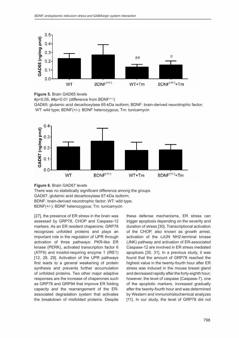
798
BDNF, endoplasmic reticulum stress and GABAergic system interaction
Figure 5. Brain GAD65 levels#p<0.05, ##p<0.01 (difference from BDNF(+/-))GAD65: glutamic acid decarboxylase 65-kDa isoform; BDNF: brain-derived neurotrophic factor; WT: wild type; BDNF(+/-): BDNF heterozygous; Tm: tunicamycin
Figure 6. Brain GAD67 levelsThere was no statistically significant difference among the groups GAD67: glutamic acid decarboxylase 67-kDa isoform; BDNF: brain-derived neurotrophic factor; WT: wild type; BDNF(+/-): BDNF heterozygous; Tm: tunicamycin
[27], the presence of ER stress in the brain was assessed by GRP78, CHOP and Caspase-12 markers. As an ER resident chaperone, GRP78 recognizes unfolded proteins and plays an important role in the regulation of UPR through activation of three pathways: PKR-like ER kinase (PERK), activated transcription factor 6 (ATF6) and inositol-requiring enzyme 1 (IRE1) [12, 28, 29]. Activation of the UPR pathways first leads to a general weakening of protein synthesis and prevents further accumulation of unfolded proteins. Two other major adaptive responses are the increase of chaperones such as GRP78 and GRP94 that improve ER folding capacity and the rearrangement of the ER-associated degradation system that activates the breakdown of misfolded proteins. Despite
these defense mechanisms, ER stress can trigger apoptosis depending on the severity and duration of stress [30]. Transcriptional activation of the CHOP, also known as growth arrest, activation of the cJUN NH2-terminal kinase (JNK) pathway and activation of ER-associated Caspase-12 are involved in ER stress mediated apoptosis [30, 31]. In a previous study, it was found that the amount of GRP78 reached the highest value in the twenty-fourth hour after ER stress was induced in the mouse breast gland and decreased rapidly after the forty-eighth hour; however, the level of caspase (Caspase-7), one of the apoptotic markers, increased gradually after the twenty-fourth hour and was determined by Western and immunohistochemical analyzes [11]. In our study, the level of GRP78 did not

799
differ significantly between the groups on the third day of ER stress, possibly due to the major neuronal loss caused by stress, while the amount of Caspase-12 increased significantly in the brain tissue of both control and heterozygous animals exposed to Tm.
Our BDNF analysis results showed that Tm-induced ER stress did not alter serum BDNF levels. Since BDNF is a secretory protein, a decrease in BDNF levels during ER dysfunction can be expected. Wei et al. [32] demonstrated that homocysteine-induced ER stress modeling was associated with a reduction in BDNF expression with induction of ER stress in the hippocampus after seven days of intracerebroventricular homocysteine administration. Although there are no studies in the literature that can be interpreted with our results, we can suggest that the short test period (3 days) may not be sufficient for such an effect. It is well known that BDNF, a neurotrophic factor affecting the central nervous system, prevents neuronal cell death induced by various stimulants. Molecular mechanisms involved in the protective effects of BDNF against ER stress have been scrutinized. Shimoke et al. [16] showed that BDNF limits ER stress-induced apoptosis by suppressing the activation of Caspase-12 via a phosphatidylinositol 3-kinase (PI3-K) dependent mechanism; Chen et al. [17] suggested that suppression of CHOP activation contributes to BDNF-mediated neuroprotection during ER stress. The current study showed that Tm administration significantly increased Caspase-12 and CHOP levels in both wild-type and BDNF heterozygous mice. Although the Tm-induced increase of Caspase-12 levels were similar in both groups, the increase in CHOP was greater in the BDNF heterozygous mice than the wild-type ones. This result suggests that endogenous BDNF may protect the brain against apoptosis due to ER stress by reducing CHOP elevation.
Inhibitor intermediate neurons make up approximately 20-30% of cortical neurons. Based on the expression of one of the three Ca2+-binding proteins (PVALB, CB or CR) most GABAergic neurons within the prefrontal cortex can be divided into non-overlapping subtypes [33]. CB, CR and PVALB neurons play different roles in the local cortical network. For instance, PVALB and CB neurons offer different strategies
for regulating feedforward or feedback inhibition within and between cortical layers, as well as neuronal input-output transformations in cortical circuits. Thus, the amount and type of GABA inhibition that each cortical layer receives directly affects the integration of inputs into that layer (inhibition provided by CB and CR neurons) or the functional output of neurons present in the layer (inhibition provided by PVALB neurons).
In differentiated neurons, BDNF directly regulates synaptic transmission. In hippocampal cultures, BDNF induces expansion of soma of GABAergic neurons, increases GABAA receptor subunits and GAD expression, and facilitates high K+ mediated GABA release. Data to date have been controversial, but BDNF has many roles, particularly for the development and maintenance of interneurons. In general, however, BDNF deficiency does not seem to be of great importance for the development and maturation of presynaptic terminals [34]. Our study revealed that endogenous BDNF deficiency did not affect two different enzyme isoforms that synthesize inhibitory intermediate neuron types and GABA neurotransmitters in basal conditions. The significant decrease in levels of CB and GAD65 isoform during ER stress indicated that the sensitivity of different intermediate neurons in the GABAergic system to ER stress may be different, but endogenous BDNF deficiency did not increase the sensitivity of these intermediate neurons to stress.
According to our results, less loss was observed in CR and PVALB expressing neurons with ER stress, than in neurons expressing CB. This finding may suggest that neurons expressing CB are more sensitive and vulnerable to ER stress. In previous reports it was observed that Ca2+-binding proteins protect GABAergic neurons from an increase of Ca2+ overload in various pathological states such as hypoxia and ischemia [35], amyotrophic lateral sclerosis [36] and glutamate toxicity [37], thus maintain Ca2+ homeostasis. In different stress models, different subtypes of Ca2+-binding proteins stand out as protective of neurons. For instance, in ischemic conditions neuroprotective efficacy was demonstrated in a descending order like: CB≥CR>PVALB [35]. Therefore, in our study, we can speculate that especially PVALB and CR proteins, rather than CB, make GABAergic neurons more resistant to ER stress.
Pamukkale Medical Journal 2021;14(4):792-802 Hacioglu Dervisoglu et al.

800
BDNF, endoplasmic reticulum stress and GABAergic system interaction
Dysfunction of inhibitor GABAergic interneurons have been implicated in various brain diseases with neurodevelopmental origin. It has been suggested that altered GABA neurotransmission in the subgroups of intermediate neurons in the prefrontal cortex contributes to the pathophysiology of psychiatric disorders including schizophrenia [38], mental retardation [39], and epilepsy [40], as well as to the pathobiology of major depression [41] and autism [42]. Inhibitor GABAergic interneuron abnormalities cause excitation-inhibition imbalance in neural circuits. Among these subtypes, PVALB-positive GABAergic intermediate neurons in the neocortex, were strongly associated with brain disorders. Therefore, further molecular, cellular and experimental animal model studies should be performed to increase our understanding of the pathophysiology of various neurodevelopmental and neuropsychiatric disorders associated with inhibitory intermediate neurons.
The limitation of the study is that the biochemical parameters were studied in brain tissue homogenates. If these parameters were supported by immunohistochemical analysis, the localization of GABAergic intermediate neurons could be demonstrated exactly.
Conflict of interest: No conflict of interest was declared by the authors.
References1. DeFelipe J. Chandelier cells and epilepsy. Brain
1999;122:1807-1822. https://doi.org/10.1093/brain/122.10.1807
2. Martin DL, Rimvall K. Regulation of gamma-aminobutyric acid synthesis in the brain. J Neurochem 1993;60:395-407. https://doi.org/10.1111/j.1471-4159.1993.tb03165.x
3. Esclapez M, Tillakaratne NJ, Kaufman DL, Tobin AJ, Houser CR. Comparative localization of two forms of glutamic acid decarboxylase and their mRNAs in rat brain supports the concept of functional differences between the forms. J Neurosci 1994;14:1834-1855. https://doi.org/10.1523/JNEUROSCI.14-03-01834.1994
4. Kawaguchi Y, Kubota Y. Neurochemical features and synaptic connections of large physiologically-identified GABAergic cells in the rat frontal cortex. Neuroscience 1998;85:677-701. https://doi.org/10.1016/S0306-4522(97)00685-4
5. Markram H, Rodriguez M, Wang Y, Gupta A, Silberberg G, Wu C. Interneurons of the neocortical inhibitory system. Nat Rev Neurosci 2004;5:793-807. https://doi.org/10.1038/nrn1519
6. Blatow M, Rozov A, Katona I, et al. A novel network of multipolar bursting interneurons generates theta frequency oscillations in neocortex. Neuron 2003;38:805-817. https://doi.org/10.1016/s0896-6273(03)00300-3
7. Lewin GR, Barde YA. 1996. Physiology of neurotrophins. Annu Rev Neurosci 1996;19:289-317. https://doi.org/10.1146/annurev.ne.19.030196.001445
8. Nieto Gonzalez JL, Jensen K. BDNF depresses excitability of parvalbumin -positive interneurons through an M-like current in rat dentate gyrus. PLoS One 2013;8:e67318. https://doi.org/10.1371/journal.pone.0067318
9. Benes FM, Berretta S. GABAergic interneurons: implications for understanding schizophrenia and bipolar disorder. Neuropsychopharmacol 2001;25:1-27. https://doi.org/10.1016/S0893-133X(01)00225-1
10. Bravo R, Parra V, Gatica D, et al. Endoplasmic reticulum and the unfolded protein response: dynamics and metabolic integration. Int Rev Cell Mol Biol 2013;301:215-290. https://doi.org/10.1016/B978-0-12-407704-1.00005-1
11. Wärri A, Cook KL, Hu R, et al. Autophagy and unfolded protein response (UPR) regulate mammary gland involution by restraining apoptosis-driven irreversible changes. Cell Death Discov 2018;15;4:40. https://doi.org/10.1038/s41420-018-0105-y
12. Zhang K, Kaufman RJ. The unfolded protein response: a stress signaling pathway critical for health and disease. Neurology 2006;24:102-109. https://doi.org/10.1212/01.wnl.0000192306.98198.ec
13. Groenendyk J, Peng Z, Dudek E, et al. Interplay between the oxidoreductase PDIA6 and microRNA-322 controls the response to disrupted endoplasmic reticulum calcium homeostasis. Sci Signal 2014;7:54. https://doi.org/10.1126/scisignal.2004983
14. Yang Y, Ma F, Liu Z, et al. The ER-localized Ca2+-binding protein calreticulin couples ER stress to autophagy by associating with microtubule-associated protein 1A/1B light chain 3. J Biol Chem 2019;294:772-782. https://doi.org/10.1074/jbc.RA118.005166
15. Xiao F, Zhang P, Chen AH, et al. Hydrogen sulfide inhibits MPP+-induced aldehyde stress and endoplasmic reticulum stress in PC12 cells: involving upregulation of BDNF. Exp Cell Res 2016;348:106-114. https://doi.org/10.1016/j.yexcr.2016.09.006
16. Shimoke K, Utsumi T, Kishi S, et al. Prevention of endoplasmic reticulum stress-induced cell death by brain-derived neurotrophic factor in cultured cerebral cortical neurons. Brain Res 2004;1028:105-111. https://doi.org/10.1016/j.brainres.2004.09.005
17. Chen G, Fan Z, Wang X, et al. Brain-derived neurotrophic factor suppresses tunicamycin-induced upregulation of CHOP in neurons. J Neurosci Res 2007;85:1674-1684. https://doi.org/10.1002/jnr.21292

801
18. Korte M, Carroll P, Wolf E, Brem G, Thoenen H, Bonhoeffer T. Hippocampal long-term potentiation is impaired in mice lacking brain-derived neurotrophic factor. Proc Natl Acad Sci USA 1995;92:8856-8860. https://doi.org/10.1073/pnas.92.19.8856
19. Abidin I, Yildirim M, Aydin Abidin S, et al. Penicillin-induced epileptiform activity and EEG spectrum analysis of BDNF heterozygous mice: an in vivo electrophysiological study. Brain Res Bull 2011;86:159-164. https://doi.org/10.1016/j.brainresbull.2011.06.015
20. De Miguel C, Hamrick WC, Hobbs JL, Pollock DM, Carmines PK, Pollock JS. Endothelin receptor-specific control of endoplasmic reticulum stress and apoptosis in the kidney. Sci Rep 2017;7:43152. https://doi.org/10.1038/srep43152
21. Hodeify R, Megyesi J, Tarcsafalvi A, Mustafa HI, Lar Seng NS, Price PM. Gender differences control the susceptibility to ER stress-induced acute kidney injury. Am J Physiol Renal Physiol 2013;304:875-882. https://doi.org/10.1152/ajprenal.00590.2012
22. Jiao G, Hao L, Wang M, et al. Upregulation of endoplasmic reticulum stress is associated with diaphragm contractile dysfunction in a rat model of sepsis. Mol Med Rep 2017;15:366-374. https://doi.org/10.3892/mmr.2016.6014
23. Huang Y, Leng TD, Inoue K, et al. TRPM7 channels play a role in high glucose-induced endoplasmic reticulum stress and neuronal cell apoptosis. J Biol Chem 2018;293:14393-14406. https://doi.org/10.1074/jbc.RA117.001032
24. Abidin I, Köhler T, Weiler E, et al. Reduced presynaptic efficiency of excitatory synaptic transmission impairs LTP in the visual cortex of BDNF-heterozygous mice. Eur J Neurosci 2006;24:3519-3531. https://doi.org/10.1111/j.1460-9568.2006.05242.x
25. Lyons WE, Mamounas LA, Ricaurte GA, et al. Brain-derived neurotrophic factor-deficient mice develop aggressiveness and hyperphagia in conjunction with brain serotonergic abnormalities. Proc Natl Acad Sci USA 1999;9:15239-15244. https://doi.org/10.1073/pnas.96.26.15239
26. Duan W, Guo Z, Jiang H, Ware M, Mattson MP. Reversal of behavioral and metabolic abnormalities, and insulin resistance syndrome, by dietary restriction in mice deficient in brain-derived neurotrophic factor. Endocrinology 2003;144:2446-2453. https://doi.org/10.1210/en.2002-0113
27. Steele KE, Seth P, Lebaron KM, et al. Tunicamycin enhances neuroinvasion and encephalitis in mice infected with Venezuelan equine encephalitis virus. Vet Pathol 2006;43:904-913. https://doi.org/10.1354/vp.43-6-904
28. Xu C, Bailly Maitre B, Reed JC. Endoplasmic reticulum stress: cell life and death decisions. J Clin Invest 2005;115:2656-2664. https://doi.org/10.1172/JCI26373
29. Cao SS, Kaufman RJ. Unfolded protein response. Curr Biol 2012;22:622-626. https://doi.org/10.1016/j.cub.2012.07.004
30. Breckenridge DG, Germain M, Mathai JP, Nguyen M, Shore GC. Regulation of apoptosis by endoplasmic reticulum pathways. Oncogene 2003;22:8608-8618. https://doi.org/10.1038/sj.onc.1207108
31. Oyadomari S, Mori M. Roles of CHOP/GADD153 in endoplasmic reticulum stress. Cell Death Differ 2004;11:381-389. https://doi.org/10.1038/sj.cdd.4401373
32. Wei HJ, Xu JH, Li MH, et al. Hydrogen sulfide inhibits homocysteine-induced endoplasmic reticulum stress and neuronal apoptosis in rat hippocampus via upregulation of the BDNF-TrkB pathway. Acta Pharmacol Sin 2014;35:707-715. https://doi.org/10.1038/aps.2013.197
33. Fish KN, Rocco BR, Lewis DA. Laminar distribution of subsets of GABAergic axon terminals in human prefrontal cortex. Front Neuroanat 2018;12:9. https://doi.org/10.3389/fnana.2018.00009
34. Grosse G, Djalali S, Deng DR, et al. Area-specific effects of brain-derived neurotrophic factor (BDNF) genetic ablation on various neuronal subtypes of the mouse brain. Dev Brain Res 2005;156:111-126. https://doi.org/10.1016/j.devbrainres.2004.12.012
35. Turovsky EA, Zinchenko VP, Gaidin SG, Turovskaya MV. Calcium-binding proteins protect GABAergic neurons of the hippocampus from hypoxia and ischemia in vitro. Biochem (Moscow) 2018;12:74-84. https://doi.org/10.1134/S1990747818010105
36. Elliott JL, Snider WD. Parvalbumin is a marker of ALS-resistant motor neurons. Neuroreport 1995;6:449-452. https://doi.org/10.1097/00001756-199502000-00011
37. D’Orlando C, Fellay B, Schwaller B, et al. Calretinin and calbindin D-28k delay the onset of cell death after excitotoxic stimulation in transfected P19 cells. Brain Res 2001;909:145-158. https://doi.org/10.1016/S0006-8993(01)02671-3
38. Lewis DA, Hashimoto T, Volk DW. Cortical inhibitory neurons and schizophrenia. Nat Rev Neurosci 2005;6:312-324. https://doi.org/10.1038/nrn1648
39. Cramer NP, Best TK, Stoffel M, Siarey RJ, Galdzicki Z. GABAB-GIRK2- mediated signaling in Down syndrome. Adv Pharmacol 2010;58:397-426. https://doi.org/10.1016/S1054-3589(10)58015-3
40. Rossignol E. Genetics and function of neocortical GABAergic interneurons in neurodevelopmental disorders. Neural Plast 2011;2011:649325. https://doi.org/10.1155/2011/649325
41. Lee B, Zhang Y, Kim Y, Kim S, Lee Y, Han K. Age-dependent decrease of GAD65/67 mRNAs but normal densities of GABAergic interneurons in the brain regions of Shank3-overexpressing manic mouse model. Neurosci Lett 2017;649:48-54. https://doi.org/10.1016/j.neulet.2017.04.016
Pamukkale Medical Journal 2021;14(4):792-802 Hacioglu Dervisoglu et al.

802
BDNF, endoplasmic reticulum stress and GABAergic system interaction
42. Gogolla N, Leblanc JJ, Quast KB, Südhof TC, Fagiolini M, Hensch TK. Common circuit defect of excitatory-inhibitory balance in mouse models of autism. J Neurodev Disord 2009;1:172-181. https://doi.org/10.1007/s11689-009-9023-x
Ethics committee approval: This study was approved by the Local Institutional Animal Care and Use Committee of the Faculty of Medicine, Karadeniz Technical University (number: 2018/04, date: 06.02.2018).
Funding: This research was supported by the grant from Giresun University with project number: SAĞ-BAP-A-230218-85.
This article was presented as an oral presentation at the 17th National Neuroscience Congress held in Trabzon on April 04-07, 2019.
Contributions of the authors to the article
G.H.D. built the main idea, hypothesis and scientific infrastructure of the study. S.C. developed the idea. S.A. and İ.A. developed the method part of the study. G.H.D., S.C. and H.Y. conducted the experiments. G.H.D. and S.C. made the evaluation of the data in the results section. The discussion part of the article was written by G.H.D., then S.C., S.A. and İ.A. reviewed and made the necessary corrections. In addition, all authors discussed the entire study and approved its final version.


804
Pamukkale Tıp DergisiPamukkale Medical Journal
doi:https://dx.doi.org/10.31362/patd.822260
Araştırma Makalesi Research Article
Gönderilme tarihi:05.11.2020 Kabul tarihi:29.03.2021
Çocuk yoğun bakım ünitesinde izlenen olguların elektroensefalografi sonuçlarının geriye dönük olarak değerlendirilmesi
Evaluation of electroencephalogram results in patients hospitalized in the pediatric intensive care unit
Müge Ayanoğlu, Uluç Yiş, Ayşe İpek Polat, Alper Köker, Gazi Arslan, Ayşe Semra Hız Kurul
ÖzAmaç: Çocuk yoğun bakım ünitesinde izlenen ve elektroensefalogram (EEG) çekilen olguların, çekim nedenlerinin ve EEG sonuçlarının değerlendirilmesi.Gereç ve yöntem: Nisan 2013-Şubat 2015 tarihleri arasında çocuk yoğun bakım ünitemizde EEG çekilen 51 olgunun demografik özellikleri, yatış ve EEG istem nedenleri, sonuçları ve çekim süreleri geriye dönük olarak kaydedildi.Bulgular: Elli bir (32’si erkek) olguya toplam 72 EEG çekimi yapıldı. EEG’lerin 14’ünün (%19,4) uzun süreli monitorizasyon, 58’inin (%80,6) standart süreli (20dk) olarak çekildiği görüldü. EEG çekilen olguların en sık yatış nedenleri kardiyak cerrahi sonrası izlem (%23,5), sepsis (%13,7) ve bilinç değişikliğiydi (%17,6). Standart süre çekim endikasyonları nöbet ve sonrasında bilinç değişikliği (%36,2), akut nörolojik olay ve beraberinde bilinç değişikliği (%34,5), bilinmeyen nedenle birlikte bilinç değişikliği (%8,6), nöbet ya da santral sinir sistemi bulgusu olmaksızın daha önceden nöbet öyküsü olması (%20,7) idi. Uzun süreli monitorizasyon endikasyonlarının %35,7’si nöbet sonrası bilinç değişikliği, %50’si santral sinir sistemini akut etkileyen patoloji ve beraberinde bilinç değişikliği, %14,2’si nedeni bilinmeyen bilinç değişikliği idi. Uzun süreli monitorizasyonların 2’sinde non-konvülzif status epileptikus (NKSE) ve 3’ünde non-konvülzif nöbet (NKN) saptandı. EEG anormalliği anlamlı olarak erkek cinsiyette daha fazla sıklıkta görülürken (p=0,035), yaş ile EEG sonuçları arasında anlamlı fark saptanmadı (p=0,32).Sonuç: EEG, motor fenomenin olmadığı NKN ve NKSE olgularının saptanmasında ve tedaviye yanıtlarını değerlendirmede önemli bir araçtır. EEG çekimi ve özellikle uzun süreli monitorizasyonlar çocuk yoğun bakım ünitelerinin vazgeçilmez bir parçası olmalıdır.
Anahtar kelimeler: Çocuk yoğun bakım, nonkonvülzif status epileptikus, nonkonülzif nöbet, elektroensefalografi monitorizasyon.
Ayanoğlu M, Yiş U, Polat Aİ, Köker A, Arslan G, Hız Kurul AS. Çocuk yoğun bakım ünitesinde izlenen olguların elektroensefalografi sonuçlarının geriye dönük olarak değerlendirilmesi. Pam Tıp Derg 2021;14:804-810.
AbstractPurpose: To evaluate the electroencephalogram (EEG) monitoring reasons and the results of the EEG in patients hospitalized in the pediatric intensive care unit (PICU).Materials and methods: Demographic features, hospitalization and EEG monitoring indications, monitoring results, and durations were analyzed retrospectively in patients hospitalized in PICU between April 2013 and February 2015.Results: Seventy-two times EEG monitoring was performed for 51 patients (32 male). EEG abnormality was determined in males more than females (p=0.035). There was no significant difference between ages and EEG results (p=0.32). Fourteen (19.4%) of the EEG monitorings were performed as long-duration and 58 (80.6%) of them were performed as standard-duration (20 min). The most frequent hospitalization etiologies were post-operative follow-up after cardiac surgery (23.5%), sepsis (13.7%), and altered state of consciousness (17.6%). Standard-duration EEG monitoring indications were altered mental status after a seizure (36.2%), altered mental status with an acute primary neurological condition (34.5%), altered mental status of unknown etiology (8.6%), and history of a seizure without acute neurological condition (20.7%). Long-duration EEG monitoring indications were altered mental status after a seizure (35.7%), altered mental status with an acute neurological
Müge Ayanoğlu, Dr. Öğr. Üye. Dokuz Eylül ÜniversitesiTıp Fakültesi, Çocuk Sağlığı ve Hastalıkları Anabilim Dalı, Çocuk Nöroloji Bilim Dalı, İzmir, Türkiye, e-posta: [email protected] (https://orcid.org/0000-0002-0556-1435) (Sorumlu Yazar)Uluç Yiş, Prof. Dr. Dokuz Eylül Üniversitesi Tıp Fakültesi, Çocuk Sağlığı ve Hastalıkları Anabilim Dalı, Çocuk Nöroloji Bilim Dalı, İzmir, Türkiye, e-posta: [email protected] (https://orcid.org/0000-0001-8355-1411) Ayşe İpek Polat, Uzm. Dr. Dokuz Eylül Üniversitesi Tıp Fakültesi, Çocuk Sağlığı ve Hastalıkları Anabilim Dalı, Çocuk Nöroloji Bilim Dalı, İzmir, Türkiye, e-posta: [email protected] (https://orcid.org/0000-0002-8270-3249) Alper Köker, Dr. Öğr. Üye. Dokuz Eylül Üniversitesi Tıp Fakültesi, Çocuk Sağlığı ve Hastalıkları Anabilim Dalı, Çocuk Yoğun Bakım Bilim Dalı,İzmir, Türkiye, e-posta: [email protected] (https://orcid.org/0000-0003-1231-3023)Gazi Arslan, Öğr. Gör. Dokuz Eylül Üniversitesi Tıp Fakültesi , Çocuk Sağlığı ve Hastalıkları Anabilim Dalı, Çocuk Yoğun Bakım Bilim Dalı, İzmir, Türkiye, e-posta: [email protected] (https://orcid.org/0000-0002-8616-3761)Ayşe Semra Hız Kurul, Prof. Dr. Dokuz Eylül Üniversitesi Tıp Fakültesi, Çocuk Sağlığı ve Hastalıkları Anabilim Dalı, Çocuk Nöroloji Bilim Dalı, İzmir, Türkiye, e-posta: [email protected] (https://orcid.org/0000-0003-0669-1909)

805
Pamukkale Tıp Dergisi 2021;14(4):804-810 Ayanoğlu ve ark.
Giriş
Nöbet akut motor, duysal ya da otonomik fenomenlere eşlik eden beyinde anormal elektriksel aktivite olarak tanımlanır. Uzun sürdüğünde hayatı tehdit edebilen bir sürece dönüşebilmektedir. Nadiren, bilinç değişikliğine motor aktivite eşlik etmeyebilir ve elektroensefalogramda (EEG) eş zamanlı elektrografik nöbet aktivitesi saptanabilir. Bu nöbetler, nöbet süresine göre non-konvülzif nöbet (NKN) ve non-konvülzif status epileptikus (NKSE) olarak adlandırılır. Nöbeti olan bir olgunun tedavi sonrası sonuçları, tanıya ulaşma süresine, erken ve etkin tıbbi tedavinin uygulanmasına bağlıdır [1]. Gelişmekte olan sinir sistemine sahip olan çocuk olguların klinik izlemleri ve tedavileri daha farklı olduğu için, çocuk hastaların çocuk yoğun bakım ünitelerinde çocuk yoğun bakım uzmanları ve çocuk nörologları tarafından değerlendirilmesi gereklidir [2].
EEG, iktal ve interiktal epileptik anormalliklerin saptanmasında önemli bir araçtır. Hem NKN, NKSE ve bilinç değişikliğinin saptanmasındaki ve hem de tedaviye yanıtın izlenmesindeki rolü önemlidir [3, 4]. EEG, aynı zamanda ensefalopatinin ve prognozun değerlendirilmesinde, nöroşirurjikal girişimlerin izleminde ve çocuk yoğun bakım ünitelerindeki paroksizmal olayların sınıflandırılmasında da değerli bilgiler verir [3, 5-8]. Hem NKN hem de NKSE’nin negatif semptomlarının olması nedeniyle, EEG’nin bu tip nöbetleri yakalama duyarlılığı çekim süresine bağlıdır [2]. Bu durum her yoğun bakım ünitesinde 7/24 EEG çekimi yapılmasını destekler niteliktedir. Ancak ne yazık ki, her ünitede bu imkân mevcut değildir. Hasta başı sürekli monitorizasyon yapılması mümkün olmasa bile standart ve uzun süreli
EEG çekimlerinin de oldukça yol gösterici olduğu bilinmektedir. Bu çalışmada çocuk yoğun bakım ünitemizdeki tüm EEG sonuçlarının sunulması amaçlandı.
Gereç ve yöntem
Çalışma için 28.09.2020 tarih ve 2020/23-01 karar no ile etik kurul onayı alındı. Nisan 2013-Şubat 2015 tarihleri arasında Dokuz Eylül Üniversitesi Çocuk Yoğun Bakım Ünitesi’nde EEG çekimi yapılmış olan olguların demografik özellikleri, kraniyal görüntüleme sonuçları, yoğun bakımda yatış nedenleri ve hangi nedenlerle EEG istendiği geriye dönük olarak incelendi. Zemin ritmi, epileptik anormallik, NKSE ya da NKN olup olmadığı kaydedildi. Çekim süreleri ise standart (20 dk) ve uzun süreli monitorizasyon (maksimum 72 saat süren) olarak 2 gruba ayrılarak incelendi. Olguların EEG çekim nedenleri; i ) nöbet ve sonrasında bilinç değişikliği ii ) akut nörolojik olayla birlikte bilinç değişikliği iii ) bilinmeyen bir nedenle olan bilinç değişikliği iv ) ensefalopati ya da akut santral sinir sistemi (SSS) bulgusu olmaksızın daha önceden nöbet öyküsü varlığı şeklinde 4 grupta sınıflandırıldı [9].
● EEG çekimi
EEG çekimi, 10-20 sisteminde, Nihon-Cohden Neurofax EEG-1200 marka cihazla, 21 elektrot kullanılarak yapıldı. EEG’ler Çocuk Nöroloji Bilim Dalı’nda çalışan iki çocuk nöroloğu tarafından değerlendirildi. Uzun süreli monitorizasyon sonucunda NKN ya da NKSE saptanan olguların çekimleri, nörolojik hasarın derecesi dikkate alınarak son 24 saat içinde NKN ya da NKSE ile uyumlu bulguların olmadığı görüldükten sonra sonlandırıldı.
condition (50%), and altered mental status with unknown etiology (14.2%). Two EEGs were compatible with, non-convulsive status epilepticus (NCSE), 3 EEGs were compatible with non-convulsive seizure (NCS) among the long-duration EEG monitorings. Conclusion: EEG is a useful instrument for the diagnosis of NKSE and NKN as well as the evaluation of treatment responses. EEG monitoring rates in PICU should be increased and should be an essential part of PICU.
Key words: Pediatric intensive care unit, non-convulsive status epilepticus, non-convulsive seizure, EEG monitoring.
Ayanoglu M, Yis U, Polat AI, Koker A, Arslan G, Hız Kurul AS. Evaluation of electroencephalogram results in patients hospitalized in the pediatric intensive care unit. Pam Med J 2021;14:804-810.

Çocuk yoğun bakım ünitelerinde elektroensefalografik monitorizasyon
806
● Tanımlar
NKN; davranış veya mental durumda açıklanamayan bir değişikliğe, EEG’de ani başlangıçlı organize ritmisite ve stereotipik doğadaki epileptik deşarjların en az 10 saniye sürmesi olarak tanımlandı. NKSE; klinik bulgulara eşlik eden EEG’deki iktal paternin 30 dakikadan fazla sürmesi veya 1 saat içinde %50’den fazla oranda görülmesi olarak tanımlandı [10]. Burst-supresyon paterni ise; 2-5 saniyelik burst aktivitesi ve onu izleyen yaklaşık 5-20 saniye süren elektrodekrementer yanıt alanları ile tanındı [11].
İstatistiksel yöntem
Araştırma verileri SPSS 21.0 istatistik programı kullanılarak değerlendirilmiştir. Araştırmanın tanımlayıcı istatistikleri sayı, yüzde ve normal dağılıma uyan verilerde ortalama ve standart sapma, normal dağılıma uymayan verilerde ortanca, çeyrekler arası mesafe kullanılarak gösterilmiştir. Değişkenlerin normal dağılıma uygunluğu Kolmogorov – Smirnov testi ile değerlendirilmiştir. Kategorik veriler Pearson Chi-quare testi ile değerlendirilmiş ve p değerinin 0,05 altında olması istatistiksel olarak anlamlı kabul edilmiştir.
Bulgular
Ortanca yaşı 52 (çeyrekler arası mesafe: 48) ay olan, 19’u (%37,3) kız, 32’si (%62,7) erkek olmak üzere 51 olgu çalışmaya alındı. Çocuk yoğun bakıma yatış nedenleri: 12 (%23,5) olguda kardiyak cerrahi sonrası izlem, 7 (%13,7) olguda sepsis, 9 (%17,6) olguda bilinç bulanıklığı, 3 (%5,9) olguda status epileptikus, 4 (%7,8) olguda metabolik hastalık ve 3 (%5,9) olguda travma idi. On üç (%25,4) olgu ise diğer nedenlerle yatırıldı. Elli bir olguya toplam 72 EEG çekimi yapıldı. EEG istemlerinin; 32’sinde (%44) nöbet geçirme, 13’ünde (%18,1) ensefalopati, 10’unda (%13,9) NKSE, 15’inde (%20,8) daha önce nöbet öyküsü olan olguların değerlendirilmesi, 2’sinde (%2,8) beyin ölümü ön tanıları mevcuttu. EEG’lerin 22’si acil (%30,6), 50’si ise (%69,4) elektif olarak çekildi. Elli sekiz tanesi standart
süre (%80,6), 14 tanesi ise (%19,4) uzun süre monitorizasyon şeklinde çekildi. Standart süre EEG çekim endikasyonları incelendiğinde; 21’inin (%36,2) nöbet ve sonrasında bilinç değişikliği, 20’sinin (%34,5) akut nörolojik olay ve beraberinde bilinç değişikliği, 5’inin (%8,6) bilinmeyen nedenle birlikte bilinç değişikliği, 12’sinin (%20,7) ise nöbet ya da santral sinir sistemi bulgusu olmaksızın daha önceden nöbet öyküsü varlığı nedeniyle çekildiği görüldü (Tablo 1). Uzun süreli monitorizasyon nedenleri incelendiğinde ise 5’inin (%35,7) nöbet ve sonrasında bilinç değişikliği, 7’sinin (%50) akut nörolojik olay ve beraberinde bilinç değişikliği, 2’sinin (%14,2) bilinmeyen nedenle bilinç değişikliği nedeniyle yapılmış olduğu görüldü (Tablo 2). EEG kayıtlarının zemin ritimleri incelendiğinde standart süre EEG çekimlerinin 36’sında (%62,1), uzun süreli monitorizasyonların ise 13’ünde (%92,9) zemin ritmi anormalliği saptandı. Standart süre EEG çekimlerinin 43’ünde (%74,1) epileptik anormallik saptanmazken, 5’inde (%8,6) fokal epileptik anormallik, 9’unda (%15,5) jeneralize epileptik anormallik, 1’inde (%1,7) burst-supresyon patterni izlendi (Tablo 3). Uzun süreli EEG monitorizasyon sonuçları epileptik aktivite açısından değerlendirildiğinde; 2’sinde (%14,2) NKSE, 3’ünde (%21,4) NKN, 6’sında (%42,8) fokal epileptik anormallik, 1’inde (%7,14) jeneralize epileptik anormallik saptandı. İkisinde (%14,2) ise epileptik anormallik izlenmedi (Tablo 4). Tüm EEG’lerin 31’inde (%43,1) zemin ritmi normaldi ve epileptik anormallik yoktu. Standart süre çekim ve uzun süreli monitorizasyon yapılan olguların yaşları karşılaştırıldığında anlamlı farklılık saptanmadı (p=0,586, Tablo 5). Kırk iki olguya kraniyal MRG çekimi yapılmıştı. Beyin MRG sonuçlarından 9’u normal (%12,5), 33’ü anormal (%45,8) saptandı. Otuz dokuz olguya kraniyal BT çekimi yapıldı. BT sonuçlarından 14’ü normal (%19,4), 25’i anormal (%34,7) saptandı. Beyin MRG ve beyin BT sonuçları EEG sonuçları ile karşılaştırıldığında aralarında ilişki saptanmadı (p=0,11, p=0,986). EEG sonuçları ve yaş arasında anlamlı farklılık saptanmazken (p=0,32) (Tablo 6), EEG anormalliği anlamlı olarak erkek cinsiyette daha fazla sıklıkta görüldü (p=0,035) (Tablo 7).

807
Pamukkale Tıp Dergisi 2021;14(4):804-810 Ayanoğlu ve ark.
Tablo 1. Standart süre EEG çekim nedenleri
N %Nöbet ve sonrasında olan bilinç değişikliği
21 36,2
Santral sinir sistemini etkileyen patoloji ve beraberinde bilinç değişikliği
20 34,5
Bilinmeyen bir durumla olan bilinç değişikliği
5 8,6
Nöbet ya da SSS bulgusu olmaksızın, daha önceden nöbet öyküsü olması nedeniyle
12 20,7
Toplam 58 100
Tablo 2. Uzun süreli EEG çekimlerinin nedenleri
N %Nöbetin öncesi ya da sonrasında olan bilinç değişikliği
5 35,7
Santral sinir sistemini etkileyen patoloji ve beraberinde bilinç değişikliği
7 50
Bilinmeyen bir durumla olan bilinç değişikliği
2 14,2
Toplam 14 100
Tablo 3. Standart süre EEG çekimlerinde epileptik anormallik açısından sonuçları
N %Epileptik anormallik yok 43 74,1
Fokal epileptik anormallik 5 8,6
Jeneralize epileptik anormallik 9 15,5
Burst-suppresyon patterni 1 1,7
Toplam 58 100
Tablo 4. Uzun süreli EEG monitorizasyonlarının epileptik anormallik sonuçların %
NKSE 2 14,2
NKN 3 21,4
Fokal epileptik anormallik 6 42,8
Jeneralize epileptik anormallik 1 7,14
Epileptik anormallik yok 2 14,2
Toplam 14 100NKSE; non-konvülziv satatus epileptikus, NKN; non-konvüzif nöbet
Tablo 5. Yaş ve EEG çekim sürelerinin karşılaştırılması
Ortanca Yaş (ay) Çeyrekler arası mesafeRutin süreli çekim 29 18,75
Uzun süreli
Monitorizasyon
27,5 26,75
Yaş ve çekim süreleri arasında anlamlı farklılık saptanmamıştır (p=0,586)
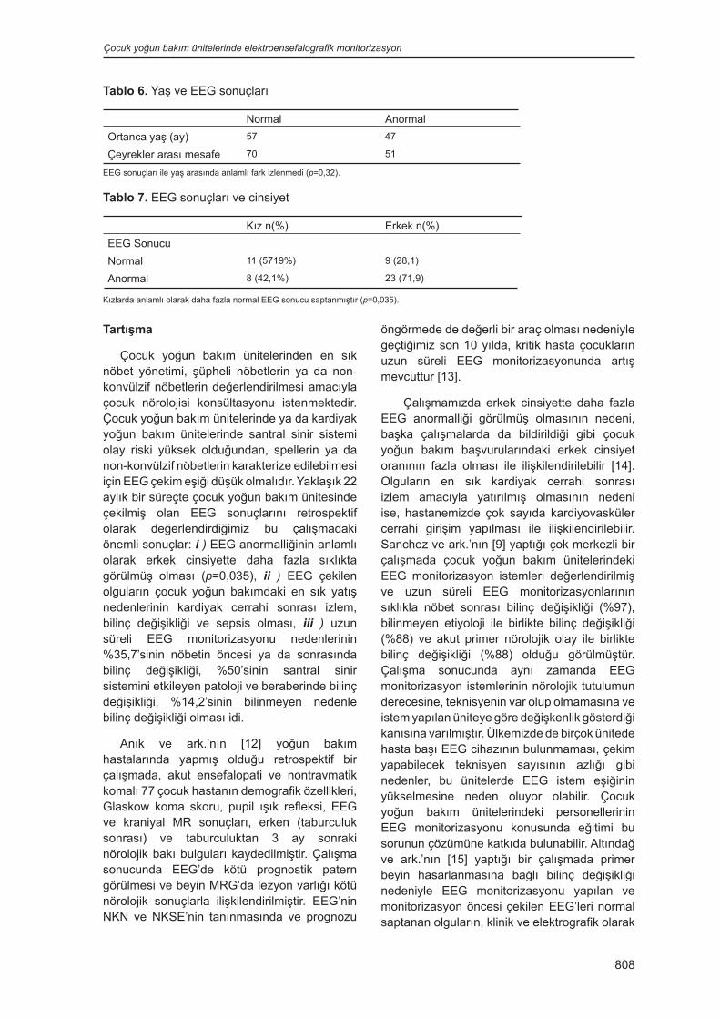
Çocuk yoğun bakım ünitelerinde elektroensefalografik monitorizasyon
808
Tartışma
Çocuk yoğun bakım ünitelerinden en sık nöbet yönetimi, şüpheli nöbetlerin ya da non-konvülzif nöbetlerin değerlendirilmesi amacıyla çocuk nörolojisi konsültasyonu istenmektedir. Çocuk yoğun bakım ünitelerinde ya da kardiyak yoğun bakım ünitelerinde santral sinir sistemi olay riski yüksek olduğundan, spellerin ya da non-konvülzif nöbetlerin karakterize edilebilmesi için EEG çekim eşiği düşük olmalıdır. Yaklaşık 22 aylık bir süreçte çocuk yoğun bakım ünitesinde çekilmiş olan EEG sonuçlarını retrospektif olarak değerlendirdiğimiz bu çalışmadaki önemli sonuçlar: i ) EEG anormalliğinin anlamlı olarak erkek cinsiyette daha fazla sıklıkta görülmüş olması (p=0,035), ii ) EEG çekilen olguların çocuk yoğun bakımdaki en sık yatış nedenlerinin kardiyak cerrahi sonrası izlem, bilinç değişikliği ve sepsis olması, iii ) uzun süreli EEG monitorizasyonu nedenlerinin %35,7’sinin nöbetin öncesi ya da sonrasında bilinç değişikliği, %50’sinin santral sinir sistemini etkileyen patoloji ve beraberinde bilinç değişikliği, %14,2’sinin bilinmeyen nedenle bilinç değişikliği olması idi.
Anık ve ark.’nın [12] yoğun bakım hastalarında yapmış olduğu retrospektif bir çalışmada, akut ensefalopati ve nontravmatik komalı 77 çocuk hastanın demografik özellikleri, Glaskow koma skoru, pupil ışık refleksi, EEG ve kraniyal MR sonuçları, erken (taburculuk sonrası) ve taburculuktan 3 ay sonraki nörolojik bakı bulguları kaydedilmiştir. Çalışma sonucunda EEG’de kötü prognostik patern görülmesi ve beyin MRG’da lezyon varlığı kötü nörolojik sonuçlarla ilişkilendirilmiştir. EEG’nin NKN ve NKSE’nin tanınmasında ve prognozu
öngörmede de değerli bir araç olması nedeniyle geçtiğimiz son 10 yılda, kritik hasta çocukların uzun süreli EEG monitorizasyonunda artış mevcuttur [13].
Çalışmamızda erkek cinsiyette daha fazla EEG anormalliği görülmüş olmasının nedeni, başka çalışmalarda da bildirildiği gibi çocuk yoğun bakım başvurularındaki erkek cinsiyet oranının fazla olması ile ilişkilendirilebilir [14]. Olguların en sık kardiyak cerrahi sonrası izlem amacıyla yatırılmış olmasının nedeni ise, hastanemizde çok sayıda kardiyovasküler cerrahi girişim yapılması ile ilişkilendirilebilir. Sanchez ve ark.’nın [9] yaptığı çok merkezli bir çalışmada çocuk yoğun bakım ünitelerindeki EEG monitorizasyon istemleri değerlendirilmiş ve uzun süreli EEG monitorizasyonlarının sıklıkla nöbet sonrası bilinç değişikliği (%97), bilinmeyen etiyoloji ile birlikte bilinç değişikliği (%88) ve akut primer nörolojik olay ile birlikte bilinç değişikliği (%88) olduğu görülmüştür. Çalışma sonucunda aynı zamanda EEG monitorizasyon istemlerinin nörolojik tutulumun derecesine, teknisyenin var olup olmamasına ve istem yapılan üniteye göre değişkenlik gösterdiği kanısına varılmıştır. Ülkemizde de birçok ünitede hasta başı EEG cihazının bulunmaması, çekim yapabilecek teknisyen sayısının azlığı gibi nedenler, bu ünitelerde EEG istem eşiğinin yükselmesine neden oluyor olabilir. Çocuk yoğun bakım ünitelerindeki personellerinin EEG monitorizasyonu konusunda eğitimi bu sorunun çözümüne katkıda bulunabilir. Altındağ ve ark.’nın [15] yaptığı bir çalışmada primer beyin hasarlanmasına bağlı bilinç değişikliği nedeniyle EEG monitorizasyonu yapılan ve monitorizasyon öncesi çekilen EEG’leri normal saptanan olguların, klinik ve elektrografik olarak
Tablo 6. Yaş ve EEG sonuçları
Normal AnormalOrtanca yaş (ay) 57 47
Çeyrekler arası mesafe 70 51
EEG sonuçları ile yaş arasında anlamlı fark izlenmedi (p=0,32).
Tablo 7. EEG sonuçları ve cinsiyet
Kız n(%) Erkek n(%)EEG SonucuNormal 11 (5719%) 9 (28,1)
Anormal 8 (42,1%) 23 (71,9)
Kızlarda anlamlı olarak daha fazla normal EEG sonucu saptanmıştır (p=0,035).

809
Pamukkale Tıp Dergisi 2021;14(4):804-810 Ayanoğlu ve ark.
en az 24 saat daha monitorize edilmesi gerektiği sonucuna varılmıştır. Bizim çalışmamızda da NKN ya da NKSE olan hastaların EEG monitorizasyonları, bu paternlerin 24 saat boyunca izlenmediği görüldükten sonra sonlandırılmıştır. Hyllienmark ve ark.’nın [16] çocuk yoğun bakım ünitesinde yaptığı bir çalışmada epilepsi nedeniyle izlenen 42 ve intrakranial basınç artışı nedeniyle izlenen 12 olguya uzun süreli EEG monitorizasyonu yapılmıştır. NKN ve/veya NKSE şüphesi ile monitorizasyon yapılan 14 (%33) olgunun EEG kayıtları normal saptanmıştır. Bizim çalışmamızda ise 14 olguya uzun süreli monitorizasyon yapıldı. Bu olguların yalnızca 2’sinde (%14,2) iktal ya da interiktal epileptik anormallik saptanmadı (Tablo 4). Nöbet sonrası bilinç değişikliği ile başvuran bir olguda ve herhangi bir motor fenomen olmaksızın travma sonrası bilinç değişikliği ile izlenen bir olguda NKSE saptandı. NKN saptanan 3 olgudan biri kardiyak cerrahi sonrası yoğun bakımda izlenirken yaşadığı serebrovaskuler olay sonrasında nöbet ve uzun süren bilinç değişikliği, biri sepsis nedeniyle izlenirken status epileptikus gelişimi sonrası uzun süren bilinç değişikliği, diğeri ise status epileptikus sonrası uzun süren bilinç değişikliği nedeniyle monitorize edildi. Ross ve ark.’nın [17] 7’si erkek toplam 8 çocuk hastada, hasta başı EEG monitorizasyonu yaptığı çalışmada; deserebre postürü olan ve nöbet olduğu düşünülen bir olgunun EEG’sinde iktal ya da interiktal deşarj saptanmadığı, kardiyak arrest sonrası her iki üst ekstiremitede klonik hareketleri olan bir olgunun EEG’sinde elektroserebral sessizlik saptandığı için tiopental tedavisinin kesildiği, EEG monitorizasyonunda devam eden nöbet aktivitesi olan bir olguda ise, daha yüksek tiopental dozları uygulanarak NKN’lerin kontrol altına alındığı bildirilmiştir. Elektrografik olarak nöbet aktivitesinin varlığının özellikle hipokampüste nöronal ölüme yol açtığı daha önce hayvan deneyleriyle kanıtlanmıştır [18-20]. Bu nedenle status epileptikus tedavisindeki ana hedef, hem motor hem de elektrografik nöbetlerin durdurulmasıdır. Tanısı, tedavisi ve izlemi ancak EEG ile mümkün olan NKSE ve NKN önemli antitelerdir [21]. Erişkin yaş grubunda hafif bilinç değişikliği olan hastaların monitorize edildiği çalışmalar da mevcuttur [2]. Böylesi hastalar çocuk yoğun bakımlarda
daha fazlaca görülebilmektedir. Bu nedenle EEG monitorizasyonu daha fazla oranlarda yapılmalıdır. Bizim çalışmamızda 2 olguda NKSE, 3 olguda NKN saptanmış olup, bu hastalardan birisinde travma sonrası motor aktivite olmaksızın ensefalopati olması nedeniyle EEG monitorizasyonu yapıldı. EEG ile tanı alan bu olgu ve tedavi yanıtı ancak EEG ile değerlendirilebilen diğer olgular göstermektedir ki, EEG monitorizasyonu çocuk yoğun bakım üniteleri için vazgeçilmez bir tanı ve izlem aracıdır.
Çalışmamızın retrospektif dizaynı ve tek merkez olması sınırlılıklarındandır. Ancak merkezimizin önemli bir üçüncü düzey çocuk yoğun bakım ünitesine sahip olması ve hasta başı monitorizasyona uygun EEG cihazının bulunması, çok sayıda kritik hastayı EEG sonuçları ile birlikte değerlendirme fırsatını bize sunmuştur.
EEG, kritik hastaların bilinç değişikliklerinin tanı ve tedaviye yanıtlarını değerlendirmede önemli bir araçtır. Çalışmamızın sonuçları literatürde bu alanda çocuk hastalar üzerinde yapılmış olan az sayıdaki çalışmaları destekler niteliktedir.
Sonuç olarak, EEG çekimi ve özellikle de uzun süreli monitorizasyonlar çocuk yoğun bakım ünitelerinin bir parçası olmalıdır.
Çıkar ilişkisi: Yazarlar çıkar ilişkisi olmadığını beyan eder.
Kaynaklar1. Phillips SA, Shanahan RJ. Etiology and mortality
of status epilepticus in children: a recent update. Arch Neurol 1989;46:74-76. https://doi.org/10.1001/archneur.1989.00520370076023
2. Claassen J, Mayer SA, Kowalski RG, Emerson RG, Hirsch LJ. Detection of electrographic seizures with continuous EEG monitoring in critically ill patients. Neurology 2004;62:1743-1748. https://doi.org/10.1212/01.wnl.0000125184.88797.62
3. Towne AR, Waterhouse EJ, Boggs JG, et al. Prevalence of non convulsive status epilepticus in comatose patients. Neurology 2000;54:340-345. https://doi.org/10.1212/wnl.54.2.340
4. Hosain SA, Solomon GE, Kobylarz EJ. Electroencephalographic patterns in unresponsive pediatric patients. Pediatr Neurol 2005;32:162-165. https://doi.org/10.1016/j.pediatrneurol.2004.09.008

Çocuk yoğun bakım ünitelerinde elektroensefalografik monitorizasyon
810
5. Abend NS, Topjian A, Ichord R, et al. Electroencephalographic monitoring during hypothermia after pediatric cardiac arrest. Neurology 2009;72:1931-1940. https://doi.org/10.1212/WNL.0b013e3181a82687
6. Abend NS, Chapman KE, Gallentine WB, et al. Electroencephalographic monitoring in the pediatric intensive care unit. Curr Neurol Neurosci Rep 2013;13:330. https://doi.org/10.1007/s11910-012-0330-3
7. Trinka E, Cock H, Hesdorffer D, et al. A definition and classification of status epilepticus – Report of the ILAE Task Force on Classification of Status Epilepticus. Epilepsia 2015;56:1515-1523. https://doi.org/10.1111/epi.13121
8. Vespa PM, Nuwer MR, Nenov V, et al. Increased incidence and impact of nonconvulsive and convulsive seizures after traumatic brain injury as detected by continuous electroencephalography monitoring. J Neurosurg 1999;91:750-760. https://doi.org/10.3171/jns.1999.91.5.0750
9. Sanchez SM, Carpenter J, Chapman KE, et al. Pediatric ICU EEG monitoring: current resources and practice in the United States and Canada. J Clin Neurophysiol 2013;30:156-160. https://doi.org/10.1097/WNP.0b013e31827eda27
10. Foreman B, Claassen J, Abou Khaled K, et al. Generalized periodic discharges in the critically ill: a case-control study of 200 patients. Neurology 2012;79:1951-1960. https://doi.org/10.1212/WNL.0b013e3182735cd7
11. Bauer G, Trinka E, Kaplan PW. EEG patterns in hypoxic encephalopathies (post-cardiac arrest syndrome): fluctuations, transitions, and reactions. J Clin Neurophysiol 2013;30:477-489. https://doi.org/10.1097/WNP.0b013e3182a73e47
12. Anık A, Tekgül H, Yılmaz S, et al. The prognostic role of clinical, electroencephalographic and neuro-radiological parameters in predicting outcome in pediatric non-traumatic coma. Pamukkale Tıp Derg 2020;13:509-518. https://doi.org/10.31362/patd.685215
13. Abend NS, Arndt DH, Carpenter JL, et al. Electrographic seizures in pediatric ICU patients: cohort study of risk factors and mortality. Neurology 2013;81:383-391. https://doi.org/10.1212/WNL.0b013e31829c5cfe
14. Abebe T, Girmay M, Michael G, Tesfaye M. The epidemiological profile of pediatric patients admitted to the general intensive care unit in an Ethiopian university hospital. Int J Gen Med 2015;8:63-67. https://doi.org/10.2147/IJGM.S76378
15. Altındağ E, Okudan ZV, Özkan Tavukçu S, Krespı Y, Baykan B. Nöroloji yoğun bakım ünitesinde bilinç değişikliği nedeni ile izlenen hastaların devamlı EEG monitorizasyonunda saptanan elektroensefalografik paternler. Arch Neuro Psychiatry 2017;54:168-178. https://doi.org/10.5152/npa.2016.14822
16. Hyllienmark L, Amark P. Continuous EEG monitoring in a paediatric intensive care unit. Eur J Paediatr Neurol 2007;11:70-75. https://doi.org/10.1016/j.ejpn.2006.11.005
17. Ross C, Blake A, Whitehouse WP. Status epilepticus on the paediatric intensive care unit—the role of EEG monitoring. Seizure 1999;8:335-338 https://doi.org/10.1053/seiz.1999.0300
18. Meldrum BS, Vigouroux RA, Brierley JB. Systemic factors andepileptic brain damage: prolonged seizures in paralyzed, artificially ventilated baboons. Arch Neurol 1973;29:82-87. https://doi.org/10.1001/archneur.1973.00490260026003
19. Fujikawa DG. The temporal evolution of neuronal damage frompilocarpine-induced status epilepticus. Brain Res 1996;725:11-22. https://doi.org/10.1016/0006-8993(96)00203-x
20. Nevander G, Ingvar M, Auer R, Siesjö BK. Status epilepticus in well-oxygenated rats causes neuronal necrosis. Ann Neurol 1985;18:281-290. https://doi.org/10.1002/ana.410180303
21. Rüegg SJ, Dichter MA. Diagnosis and treatment of nonconvulsive status epilepticus in an intensive care unit setting. Curr Treat Options Neurol 2003;5:93-110. https://doi.org/10.1007/s11940-003-0001-4
Çalışma, 17. Ulusal Çocuk Nöroloji Kongresi’nde poster bildiri olarak sunulmuştur (PS:170, 06-09 Mayıs 2015, Çeşme, İzmir).
Etik kurul onayı: Dokuz Eylül Üniversitesi Girişimsel Olmayan Araştırmalar Etik Kurulu tarafından 28.09.2020 tarih ve 2020/23-01 karar no ile onaylanmıştır.
Yazarların makaleye olan katkıları
U.Y. çalışmanın dizaynı ve makalenin kontrolü; M.A. çalışma dizaynı, dosya tarama ve verilerin toplanması, istatistiksel analizin yapılması ve makalenin yazılması; A.İ.P., A.K. ve G.A. verilerin toplanması ve makalenin kontrolü; A.S.H. çalışma dizaynı ve makalenin kontrolü alanlarında çalışmaya katkı sunmuşlardır.


812
Pamukkale Tıp DergisiPamukkale Medical JournalAraştırma Makalesi Research Article
Gönderilme tarihi:06.01.2021 Kabul tarihi:15.04.2021
Follow-Up of endovascular coil embolization applied for the treatment of intracranial aneurysms by magnetic resonance angiography
İntrakraniyal anevrizmaların tedavisi için uygulanan endovasküler koil embolizasyonunun manyetik rezonans anjiyografi ile takibi
Murat Dökdök, Kutlay Karaman, Selçuk Göçmen
AbstractPurpose: To assess the usefulness and reproducibility of magnetic resonance angiography (MRA) for the evaluation of intracranial aneurysms treated by endovascular coiling.Materials and methods: 51 patients (18 men, 33 women; 21-86 year-old, mean age 50.2) with 54 aneurysms treated by detachable platinum coils were included in this study. The patients were scheduled for follow-up MRA imaging at three to six months after endovascular treatment. Two interventional neuroradiologists, evaluated the TOF-MRA images blindly. Source MR images and volume rendering techniques of 3D TOF images such as maximum intensity projection (MIP) and multiplanar reformation (MPR) were utilized for this purpose. Follow-up digital subtraction angiography (DSA) was performed as a reference test on a biplane angiographic unit on the sixth month and the first year depending on the patient's characteristics.Results: DSA confirmed residual or recurrent aneurysmal flow in 5 patients out of 54 treated cerebral aneurysms; two with tip of basilary artery, one with left MCA bifurcation, one with anterior communicating artery and one with right supraclinoid internal carotid artery. Then the DSA images were compared with the MRA data. Total agreement was 0.94 for MRA. Kappa coefficient was 0.74 for indicating good concordance of the readers for MRA (95% CI was 0.44 to 1.04). Conclusion: MRA is a reproducible technique for the follow-up of aneurysms after endovascular coil embolization. Conventional catheter angiography as a gold standard test could be used to solve uncertain cases.
Key words: Cerebral aneurysm, endovascular treatment, coil embolization, magnetic resonance angiography, digital subtraction angiography.
Dokdok M, Karaman K, Gocmen S. Follow-Up of endovascular coil embolization for intracranial aneurysms using magnetic resonance angiography. Pam Med J 2021;14:812-817.
ÖzAmaç: Manyetik rezonans anjiyografinin (MRA) endovasküler koil ile tedavi edilen intrakraniyal anevrizmaların takiplerinde kullanışlılığını ve tekrarlanabilirliğini değerlendirmek. Gereç ve yöntem: Platin koil ile tedavi edilen 51 hasta (18 erkek, 33 kadın; yaş 21-86 yıl, ort. yaş 50,2 yıl) bu çalışmaya alındı. Hastalara, endovasküler tedaviden 3-6 ay sonra takip MRA planlandı. İki girişimsel nöroradyolog, TOF-MRA görüntülerini körleme değerlendirdi. Bu amaçla, maksimum yoğunluk projeksiyonu (MIP) ve multiplanar reformasyon (MPR) gibi 3D TOF görüntülerinin kaynak MR görüntüleri ve hacimsel işleme teknikleri kullanılmıştır. Altı ay ile birinci yılda hastanın özelliklerine bağlı olarak biplanar anjiografi ünitesinde bir referans testi olarak takip dijital subtraksiyon anjiyografi (DSA) yapılmıştır.Bulgular: Tedavi edilen 54 serebral anevrizmalı hastadan, 5’inde rezidü veya rekürren anevrizma olduğu DSA ile doğrulandı; ikisi baziler tepe, biri sol MCA bifurkasyosu, biri anterior komünikan arter ve biri sağ supraklinoid internal karotid arterdi. Daha sonra DSA, MRA verileriyle karşılaştırıldı. MRA için toplam uzlaşma 0,94 idi. MRA için, okuyucuların iyi uyumunu göstermek için Kappa katsayısı 0,74 idi (%95 CI 0,44 ile 1,04 idi). Sonuç: MRA, endovasküler koil embolizasyonundan sonra anevrizmaların takibi için tekrarlanabilir bir tekniktir. Altın standart bir test olarak konvansiyonel kateter anjiyografi şüpheli olguları göstermek için kullanılabilir.
Anahtar kelimeler: Serebral anevrizma, endovasküler tedavi, koil embolizasyon, manyetik rezonans anjiyografi, dijital subtraksiyon anjiyografi.
Dökdök M, Karaman K, Göçmen S. İntrakraniyal anevrizmalar için endovasküler koil embolizasyonun manyetik rezonans anjiyografisi kullanılarak takibi. Pam Tıp Derg 2021;14:812-817.
Murat Dokdok, MD, Anadolu Medical Center, Department of Radiology, Kocaeli, Turkey, e-mail: [email protected] (https://orcid.org/0000-0002-1313-8557) (Corresponding Author)Kutlay Karaman, MD, Anadolu Medical Center, Department of Radiology, Kocaeli, Turkey, e-mail: [email protected] (https://orcid.org/0000-0001-7654-8038)Selcuk Gocmen, MD. Assac Prof., Anadolu Medical Center, Department of Neurosurgery, Kocaeli, Turkey, e-mail: [email protected] (https://orcid.org/0000-0003-0532-3549
doi:https://dx.doi.org/10.31362/patd.854579

813
Introduction
Although it has been increasingly used as a first-line treatment of intracranial aneurysms [1, 2], endovascular treatment carries a potential risk of aneurysm recanalization. The risk factors for recanalization is not well defined. Therefore, all patients should be followed up closely after endovascular treatment. Digital Subtraction Angiography (DSA) is still the reference imaging modality in follow-up; however, it is invasive and carries a risk of morbidity [2, 3]. Magnetic resonance angiography (MRA) is a non-invasive and radiation-free technique, and it can be used to reduce the need for follow-up DSA [4-7].
The purpose of our study was to assess the usefulness and reproducibility of MRA for the evaluation of intracranial aneurysms treated with coils endovascularly.
Material and methods
A total number of 51 patients (18 men, 33 women; 21-86 year-old, mean age 50.2) with 54 aneurysms treated by detachable platinum coils were retrospectively included in this study. The patients with stent-assisted coils were excluded from the study if the stents had interfered MRA evaluation. At the end of the embolization, the treatment was documented by DSA.
The patients who had no contraindications for MR were scheduled for the follow-up imaging three to six months after endovascular treatment. MRA was performed on 1.5 Tesla (Magnetom Avanto, Siemens AG, Erlangen, Germany) or 3 Tesla (Magnetom Skyra, Siemens AG, Erlangen, Germany) MR device using a 16 channel phased array head coil. Three-dimensional time-of-flight (3D-TOF) sequence was utilized in the axial plane, and a saturation band was placed above the acquisition volume to eliminate the venous signal. 3D-TOF parameters for 3T MR were as follows: TE 18 ms; TE 3.24 ms; flip angle 20°; section thickness 0.77 mm; field of view 210 mm; matrix 256x256, and for 1.5 Tesla MR as follows: TE 7 ms; TR 36 ms; flip angle 25°; field of view 210 mm; slab effective thickness 0.65-1.5; matrix 256x512.
Follow-up DSA was performed using a biplane angiographic unit (Axiom Artis, Siemens Medical Systems, Erlangen, Germany) at six months to one year, depending on the patient’s characteristics. It consisted of multiple
projections and 3D rotational angiography of the aneurysm parent artery. Through a 5F standard diagnostic catheter, iodinated contrast material was administrated with a power injector.
The study was approved by Anadolu Medical Center Ethical Committee and all the procedures were performed with the written informed consent.
Evaluation of images
Two interventional neuroradiologists (K.K and M.D), who did not perform the above embolization, evaluated the TOF-MRA images blindly. They were unaware of the DSA results before and after coil embolization.
The quality of TOF-MRA images was adequate in all patients. Minor artifacts were observed predominantly 3T MRA images that were related to the coils. However, they did not interfere with image interpretation. Visualization of the parent arteries was optimal in all cases. MRA data were evaluated on a dedicated workstation (Syngo, Siemens AG, Berlin, Germany). Source MR images and volume rendering techniques of 3D TOF images such as maximum intensity projection (MIP) and multiplanar reformation (MPR) were utilized for this purpose.
Based on the DSA, the observers assessed the occlusion status of the treated aneurysm according to the following Raymond classification [8]: Class 1, excluded aneurysm; Class 2, persistence of residual neck; and Class 3, persistence of residual aneurysm. Images were analyzed on a served workstation (Syngo, Siemens AG, Berlin, Germany) using InSpace 3D software. The maximum size of the residual lumen was also measured.
Results
The study included 54 treated cerebral aneurysms. DSA as a gold standard confirmed residual or recurrent aneurysmal flow in 5 patients out of 54 treated cerebral aneurysms; two with tip of basilar artery (one Class 2, one Class 3) (Figure 1), one with left MCA bifurcation (Class 2) (Figure 2), one with anterior communicating artery (Class 2) and one with right supraclinoid internal carotid artery (Class 2) classified according to Raymond et al [8]. 49 patients out of 56 were classified as Class 1 (excluded
Pamukkale Medical Journal 2021;14(4):812-817 Dokdok et al.

Follow-up of coil embolization for cerebral aneurysms
814
aneurysm). Incomplete aneurysm occlusions consisted of four recurrences (7.4%) and one residual aneurysm (1.8%), mean aneurysm diameter was 3.7 mm. Complete aneurysmal occlusion was seen at 49 aneurysms (90%). The patients’ profile and aneurysm locations are shown in Table 1.
MRA images were evaluated with the presence and absence of an aneurysm only without any classification. All 5 aneurysms cases confirmed with DSA were identified with MRA by both readers. There were three false-positive cases on MRA compared to gold standard DSA.
Figure 1. Follow-up at 3-6 months after basilar top aneurysm coil embolization. A) TOF-MRA raw data showed recurrent filling of basilar top aneurysm sac posterior to the coils (white arrow). B) 3D TOF-MRA depicted aneurysmal sack filling of 6x3 mm. C) DSA confirmed the above findings (black arrow)
Figure 2. Follow-up 6 months after left MCA aneurysm coil embolization. A) TOF-MRA raw data showed recurrent filling of left MCA aneurysm at neck (white arrow). B) 3D TOF-MRA depicted aneurysmal neck filling of 2 mm. C) DSA confirmed the above findings (white arrow)
Table 1. Patient profile and aneurysm location before coiling
Total number of patients 51Age range 21-86 mean 50.2
Female 33 (65%)
Male 18 (35%)
Aneurysm numbers 54
ACA and ACoAICA and PCoAMCABA and PCA
927126
Subarachnoid hemorrhage 17 (33%)
Headache 28 (55%)
Incidental 6 (12%)
ACA: Anterior Cerebral Artery, AcoA: Anterior Communicating Artery, ICA: Internal Carotid Artery, PcoA: Posterior Communicating Artery, MCA: Middle Cerebral Artery; BA: Basilar Artery, PCA: Posterior Cerebral Artery

815
Statistical analysis
At the main step, the interobserver agreement for TOF-MRA is evaluated. For this purpose, k statistical test was calculated with corresponding 95% confidence intervals (CI). For the categorical data, k values 0.6-0.79 indicated substantial agreement [9]. Then the DSA is analyzed and compared with the MRA data.
Due to small number of selected patients during a short term follow-up, accuracy studies including negative and positive predictive values and sensitivity and specificity of MRA compared to DSA were not in this study’s scope. Similarly, subgroup analyses were not performed for the test characteristics.
Interobserver agreement
Total agreement was 0.94 for MRA. Kappa coefficient was 0.74 for indicating good concordance of the readers for MRA (95% CI was 0.44 to 1.04). Interobserver agreement for DSA was not obtained, as it was regarded reference test. We did not observe any adverse event during or after the MR examination. Discrepancies between both examiners were noted in three cases. An additional peer reading was performed to have a consensus in these cases.
Discussion
Due to best efforts and technical advancements, coiled aneurysms may demonstrate persistent filling and re-growth of the aneurysm sac. Therefore, follow-up imaging is crucial for aneurysms treated with endovascular coiling. In our study, TOF MR angiography at both 1.5 T and 3.0 T showed comparable results to the gold standard DSA with a high interobserver agreement, as shown by others [10]. Hence, DSA did not always confirm a suspected incomplete occlusion at MRA due to low positive predictive value as implied in a multicenter study conducted by Schaafsma et al. [10]. We observed such cases, although there was an interval up to three months between two tests. It is not certain when in which risk groups to perform follow-up angiography to detect for reopening. The probability of reopening is minimal in aneurysms with complete or near-complete occlusion, as shown at 6-month follow-up [10-12]. Nevertheless, it might be reasonable
to pursue it in the mid and long term, while the monitoring is adapted on a case-by-case basis [11].
Although the recurrence rates might be as high as 33.6%, retreatment is necessary in about 10% to 15% of cases with embolized aneurysms [8, 13]. There are 26 reports of late bleeding after coil embolization of previously unruptured aneurysms reported in the literature, while sixteen of them occurred in large and giant aneurysms [14]. Such aneurysms tend to have a high aspect ratio and should be meticulously observed. All delayed ruptures were observed in aneurysms with dome filling in the residual aneurysm (Raymond class 3).
DSA is still the gold standard to evaluate aneurysm occlusion, but it is invasive. However, a non-invasive image modality is required, such as MRA, to replace DSA. In MRA, the blood demonstrates high signal while coils have low signal. MRA can clearly show residual high signal aneurysm in a 3D fashion with reconstruction capabilities. There is no significant difference between DSA and MRA in the follow-up of coiled embolized brain aneurysms, as shown by many authors [4, 15, 16]. Besides, it might be of limited use because pulsation artifacts caused by the coils may interfere with images in three-dimensional rotational angiography. However, DSA may still be necessary, especially in aneurysms treated with stent remodeling. In one of our basilar tip aneurysm cases; remodeling stent did not interfere with interpretation of MRA due to residual filling of large aneurysm. However, the decision should be made depending on each case.
The sensitivity and specificity for detecting residual flow within the coil mass were reported between 90% and 100%, respectively, with different MRA techniques [4, 7, 16]. In an early study with 26 patients, there was a positive predictive value of 100% and a negative predictive value of 96% [17].Gonner et al. [18] compared different TOF MRA protocols with two different TE (6 ms vs 2.4 ms). These investigators concluded that coil related artifacts on MRA were reduced while visualization of the adjacent parent vessel was improved by 36% with the short TE sequences. As they demonstrated, the shortest possible TE was the key factor in improving perianeurysmal visualization [18]. Various types of coils are used for the occlusion
Pamukkale Medical Journal 2021;14(4):812-817 Dokdok et al.
Follow-up of coil embolization for cerebral aneurysms
816
of intracranial aneurysms. The Nexus coils were found to have inherently larger artifacts with MRA compared to other types of coils [19]. We observed minor artifacts related to the used new generation coils were observed on TOF-MRA images in two patients, which did not interfere with interpretation. There was a good correlation between DSA and MRA by the readers.
Since Majoie et al. [20], many others have demonstrated TOF-MRA might be utilized as a first-line exam for coiled aneurysm follow-up [21-23]. In a meta-analysis in the follow-up of embolized aneurysms with contrast-enhanced MRA and TOF-MRA, there were no significant statistical differences [24].
In our study, MRA was shown as a reproducible method for the assessment of intracranial aneurysms after coil embolization. Although TOF-MRA and DSA techniques showed similar results of aneurysm occlusion after embolization, catheter angiography remains the gold standard for evaluating treated aneurysms and should be used in cases of diagnostic uncertainty.
Conflict of interest: No conflicts of interest was declared by the authors.
References1. Molyneux AJ, Kerr RS, Birks J, et al. Risk of recurrent
subarachnoid hemorrhage, death, or dependence and standardised mortality ratios after clipping or coiling of an intracranial aneurysm in the international subarachnoid aneurysm trial (ISAT): long-term follow-up. Lancet Neurol 2009;8:427-433. https://doi.org/10.1016/S1474-4422(09)70080-8
2. Molyneux AJ, Kerr RS, Yu LM, et al. International subarachnoid aneurysm trial (ISAT) of neurosurgical clipping versus endovascular coiling in 2143 patients with ruptured intracranial aneurysms: a randomized comparison of effects on survival, dependency, seizures, rebleeding, subgroups, and aneurysm occlusion. Lancet 2005;366:809-817. https://doi.org/10.1016/S0140-6736(05)6721-5
3. Willinsky RA, Taylor SM, TerBrugge K, Farb RI, Tomlinson G, Montanera W. Neurologic complications of cerebral angiography: prospective analysis of 2,899 procedures and review of the literature. Radiology 2003;227:522-528. https://doi.org/10.1148/radiol.2272012071
4. Anzalone N, Righi C, Simionato F, et al. Three-dimensional time-of-flight MR angiography in the evaluation of intracranial aneurysms treated with Guglielmi detachable coils. AJNR Am J Neuroradiol 2000;21:746-752.
5. Boulin A, Pierot L. Follow-up of intracranial aneurysms treated with detachable coils: comparison of gadolinium-enhanced 3D time-of-flight MR angiography and digital subtraction angiography. Radiology 2001;219:108-113. https://doi.org/10.1148/radiology.219.1.r01mr06108
6. Derdeyn CP, Graves VB, Turski PA, Masaryk AM, Strother CM. MR angiography of saccular aneurysms after treatment with Guglielmi detachable coils: preliminary experience. AJNR Am J Neuroradiol 1997;18:279-286.
7. Kahara VJ, Seppanen SK, Ryymin PS, Mattila P, Kuurne T, Laasonen EM. MR angiography with three-dimensional time-of-flight and targeted maximum-intensity-projection reconstructions in the follow-up of intracranial aneurysms embolized with Guglielmi detachable coils. AJNR Am J Neuroradiol 1999;20:1470-1475.
8. Raymond J, Guilbert F, Weill A, et al. Long-term angiographic recurrences after selective endovascular treatment of aneurysms with detachable coils. Stroke 2003;34:1398-1403. https://doi.org/10.1161/01.STR.0000073841.88563.E9
9. Landis JR, Koch GG. The measurement of observer agreement for categorical data. Biometrics 1977;33:159-174.
10. Schaafsma JD, Velthuis BK, Majoie CB, et al. Intracranial aneurysms treated with coil placement: test characteristics of follow-up MR angiography--multicenter study. Radiology 2010;256:209-218. https://doi.org/10.1148/radiol.10091528
11. Soize S, Gawlitza M, Raoult H, Pierot L. Imaging follow-up of intracranial aneurysms treated by endovascular means: Why, when, and how? Stroke 2016;47:1407-1412. https://doi.org/10.1161/STROKEAHA.115.011414
12. Piotin M, Spelle L, Mounayer C, et al. Intracranial aneurysms: treatment with bare platinum coils--aneurysm packing, complex coils, and angiographic recurrence. Radiology 2007;243:500-508. https://doi.org/10.1148/radiol.2431060006
13. Sprengers ME, Schaafsma J, Rooij WJV, et al. Stability of intracranial aneurysms adequately occluded 6 months after coiling: a 3T MR angiography multicenter long-term follow-up study. AJNR Am J Neuroradiol 2008;29:1768-1774. https://doi.org/10.3174/ajnr.A1181
14. Tsurumi A, Tsurumi Y, Negoro M, et al. Delayed rupture of a basilar artery aneurysm treated with coils: case report and review of the literature. J Neuroradiol 2013;40:54-61. https://doi.org/10.1016/j.neurad.2012.08.005
15. Yamada N, Hayashi K, Murao K, Higashi M, Lihara K. Time-of flight MR angiography targeted to coiled intracranial aneurysms is more sensitive to residual flow than is digital subtraction angiography. AJNR Am J Neuroradiol 2004;25:1154-1157.

817
16. Grunwald IQ, Papanagiotou P, Struffert T, et al. Recanalization after endovascular treatment of intracerebral aneurysms. Neuroradiology 2007;49:41-47. https://doi.org/10.1007/s.00234-006-0153-5
17. Brunereau L, Cottier JP, Sonier CB, et al. Prospective evaluation of time-of flight MR angiography in the follow-up of intracranial saccular aneurysms treated with Guglielmi detachable coils. J Comput Assist Tomogr 1999;23:216-223. https://doi.org/10.1097/00004728-199903000-00009
18. Gönner F, Heid O, Remonda L, et al. MR angiography with ultrashort echo time in cerebral aneurysms treated with Guglielmi detachable coils. AJNR Am J Neuroradiol 1998;19:1324-1328.
19. Kang HS, Moon WJ, Roh HG, et al. MR angiographic evaluation is limited in intracranial aneurysms embolized with Nexus coils. Neuroradiology 2008;50:171-178. https://doi.org/10.1007/s00234-007-0320-3
20. Majoie CB, Sprengers ME, van Rooij WJ, et al. MR angiography at 3T versus digital subtraction angiography in the follow-up of intracranial aneurysms treated with detachable coils. AJNR Am J Neuroradiol 2005;26:1349-1356.
21. Urbach H, Dorenbeck U, von Falkenhausen M, et al. Three-dimensional time-of-flight MR angiography at 3T compared to digital subtraction angiography in the follow-up of ruptured and coiled intracranial aneurysms: a prospective study. Neuroradiology 2008;50:383-389. https://doi.org/10.1007/s00234-007-0355-5
22. Buhk JH, Kallenberg K, Mohr A, Dechent P, Knauth M. No advantage of time-of-flight magnetic resonance angiography at 3 Tesla compared to 1.5 Tesla in the follow-up after endovascular treatment of cerebral aneurysms. Neuroradiology 2008;50:855-861. https://doi.org/10.1007/s00234-008-0413-7
23. Ferre JC, Carsin Nicol B, Morandi X, et al. Time-of-flight MR angiography at 3T versus digital subtraction angiography in the imaging follow-up of 51 intracranial aneurysms treated with coils. Eur J Radiol 2009;72:365-369. https://doi.org/10.1016/j.ejrad.2008-08-005
24. Kwee TC, Kwee RM. MR angiography in the follow-up of intracranial aneurysms treated with Guglielmi detachable coils: systematic review and meta-analysis. Neuroradiology 2007;49:703-713. https://doi.org/10.1007/s00234-007-0266-5
Acknowledgements: This research did not receive any specific grant from funding agencies in the public, commercial, or not-for-profit sectors.
Ethics committee approval: The study was approved by Anadolu Medical Center Ethical Committee (ASM-EK-21/135 12, January, 2021) and all the procedures were performed with the written informed consent.
Contributions of the authors to the article
M.D. and K.K. designed the main idea and hypothesis of 'Follow-Up of Endovascular Coil Embolization for Intracranial Aneurysms Using Magnetic Resonance Angiography' study. M.D., K.K developed the hypothesis and arranged the materials and methods section. M.D., K.K., and S.G executed analysis and interpretation of data in the results section. M.D., K.K., and S.G. wrote the discussion section of the manuscript; M.D., K.K, and S.G. reviewed made necessary corrections, and approved. All authors also discussed and approved the final version.
Pamukkale Medical Journal 2021;14(4):812-817 Dokdok et al.

818
Pamukkale Tıp DergisiPamukkale Medical Journal
doi:https://dx.doi.org/10.31362/patd.910961
Araştırma Makalesi Research Article
Gönderilme tarihi:07.04.2021 Kabul tarihi:28.04.2021
Myocardial performance index is associated with Systematic Coronary Risk Estimation (SCORE) system in chronic coronary syndrome
Kronik koroner sendromda miyokardial performans indeks Sistematik Koroner Risk Tahmini (SCORE) ile ilişkilidir
Hazar Harbalıoğlu, Ömer Genç, Abdullah Yıldırım
AbstractPurpose: The Systematic Coronary Risk Estimation (SCORE) system is used to determine 10-year cardiovascular risk. Myocardial performance index (MPI), a well-accepted echocardiographic parameter, is used to provide any information about the global function of the ventricle. We aimed to investigate the relationship between MPI and SCORE.Materials and methods: A total of 168 participants with chronic coronary syndrome (CCS) presented to cardiology outpatient clinic between 1 June and 31 December 2020 were retrospectively enrolled in the study. Cardiovascular risk estimation was calculated by SCORE assessment. The participants were divided into two groups as 46 patients with low cardiovascular risk, and 122 patients with moderate-high-very high risk, according to the SCORE. Laboratory parameters and echocardiography findings of the patients were recorded. Results: There was no difference in body mass index, office systolic, and diastolic blood pressure between the groups. While blood glucose was 94.5 (83.0-110.3) in the low-risk group, it was 101.0 (90.0-130.5) in the moderate-high risk patient group (p=0.007). Similarly, Urea, creatinine, lactate dehydrogenase, total cholesterol, NT pro-BNP, and Troponin T in the moderate-high risk group were significantly higher than was the low-risk group. MPI was significantly higher in the moderate-high risk patient group (p<0.001). MPI (OR=2.358, 95% CI:1.033-5.382, p=0.042), urea (OR=1.090, 95% CI:1.019-1.166, p=0.012), and glucose (OR=1.023, 95% CI:1.003-1.043, p=0.025) were independently associated with the moderate-high SCORE.Conclusion: We found that MPI predicted a moderate-high SCORE system. Further studies are warranted to better clarify the association of SCORE with echocardiographic parameters including MPI.
Key words: Myocardial performance index, Systematic Coronary Risk Estimation (SCORE) system, chronic coronary syndrome, cardiovascular risk.
Harbalioglu H, Genc O, Yildirim A. Myocardial performance index is associated with Systematic Coronary Risk Estimation (SCORE) system in chronic coronary syndrome. Pam Med J 2021;14:818-827.
ÖzAmaç: Sistematik Koroner Risk Tahmin (SCORE) sistemi, 10 yıllık kardiyovasküler riski belirlemek için kullanılır. Kabul gören bir ekokardiyografik parametre olan miyokardiyal performans indeksi (MPI), ventrikülün global işlevi hakkında bilgi sağlamak için kullanılır. Biz; MPI ve SCORE arasındaki ilişkiyi araştırmayı amaçladık.Gereç ve yöntem: 1 Haziran-31 Aralık 2020 tarihleri arasında kardiyoloji polikliniğine başvuran kronik koroner sendromlu (KKS) toplam 168 hasta retrospektif olarak çalışmaya alındı. Kardiyovasküler risk tahmini SCORE değerlendirmesi ile hesaplandı. SCORE'a göre hastalar düşük kardiyovasküler riskli 46 hasta ve orta-yüksek-çok yüksek riskli 122 hasta olarak iki gruba ayrıldı. Hastaların laboratuvar parametreleri ve ekokardiyografi bulguları kaydedildi.Bulgular: Gruplar arasında vücut kitle indeksi, ofis sistolik ve diyastolik kan basıncı açısından fark yoktu. Kan şekeri düşük risk grubunda 94,5 (83,0-110,3) iken, orta-yüksek riskli hasta grubunda 101,0 (90,0-130,5) idi (p=0,007). Benzer şekilde, orta-yüksek risk grubundaki Üre, kreatinin, laktat dehidrogenaz, total kolesterol, NT pro-BNP ve Troponin T, düşük risk grubuna göre anlamlı olarak daha yüksekti. Orta-yüksek riskli hasta grubunda MPI anlamlı olarak daha yüksekti (p<0,001). MPI (OR=2,358, %95 CI:1,033-5,382, p=0,042), üre (OR=1,090, %95 CI:1,019-1,166, p=0,012) ve glikoz (OR=1,023, %95 CI:1,003 -1,043, p=0,025) bağımsız olarak orta-yüksek SCORE ile ilişkilendirildi.Sonuç: MPI'nin orta-yüksek SCORE sistemini öngördüğünü bulduk. SCORE ile MPI de dahil olmak üzere ekokardiyografik parametreler arasındaki ilişkiyi daha iyi açıklığa kavuşturmak için ileri çalışmalara ihtiyaç vardır.
Anahtar kelimeler: Miyokardiyal performans indeks, Sistematik Koroner Risk Tahmin (SCORE) sistemi, kronik koroner sendrom, kardiyovasküler risk.
Hazar Harbalıoğlu, MD, Specialist of Cardiology, Duzce Ataturk State Hospital, Department of Cardiology, Duzce, Turkey, e-mail: [email protected] (https://orcid.org/0000-0002-6694-814X) (Corresponding Author)Ömer Genç, MD, Specialist of Cardiology, Ağrı Training and Research Hospital, Department of Cardiology, Ağrı, Turkey, e-mail: [email protected] (https://orcid.org/0000-0002-9097-5391)Abdullah Yıldırım, MD, Specialist of Cardiology, University of Health Sciences - Adana Health Practices and Research Center, Department of Cardiology, Adana, Turkey, e-mail: [email protected] (https://orcid.org/0000-0002-7071-8099)

819
Introduction
Cardiovascular diseases, the leading cause of mortality in both men and women, account for 46% of deaths in Europe [1]. There are many risk factors associated with cardiovascular events such as male gender [2], advanced age [2], high blood pressure [3], high low-density lipoprotein (LDL) [4], smoking [5], and diabetes mellitus [6]. Various risk scores have been developed to select patients at moderate-high risk to receive primary preventive treatment approaches including lipid-lowering therapy and lifestyle change. The Systematic Coronary Risk Estimation (SCORE) scoring tool, calculated using age, sex, total cholesterol, systolic blood pressure, and smoking parameters, is recommended by the European Society of Cardiology to determine 10-year cardiovascular risk [7]. This scoring aims to reduce cardiovascular deaths via measures and treatment protocols to be practiced according to the determined risk level.
Myocardial performance index (MPI), which is a doppler-derived echocardiographic parameter easy to obtain, is calculated by summing the isovolumetric relaxation time and the isovolumetric contraction time and dividing it by the ejection time [8]. The ratio of isovolumetric contraction time to ejection time reflects the systolic function of the ventricle, while the ratio of isovolumetric relaxation time to ejection time reveals the diastolic function of the ventricle [9]. Many studies reported that the increase in MPI, which provides information about both systolic and diastolic function of the ventricle, is associated with diseases that affect the cardiovascular system, such as hypertension [10], coronary artery disease [11], congestive heart failure [12], pulmonary hypertension [13], chronic obstructive pulmonary disease [14], and cardiac amyloidosis [15]. Since MPI is less affected by the image quality and also demonstrates diastolic dysfunction, it enables us to better evaluate global left ventricular (LV) function, compared to ejection fraction [16]. There are few studies on echocardiographic parameters that may be related to the SCORE and even might predict a higher SCORE. We, therefore, believe this point should be clarified.
Thus, in order to overcome this deficiency, to some extent, we aimed to investigate the relationship between MPI and SCORE, which reflects cardiovascular risk.
Materials and methods
Study population and design
168 patients with chronic coronary syndrome (CCS) who admitted to Düzce Atatürk State Hospital cardiology outpatient clinic between 1 June and 31 December 2020 were retrospectively included in the study. The diagnosis of CCS was made after the confirmation by imaging procedures (myocardial perfusion scintigraphy (MPS) or coronary CT angiography) or a positive exercise test for patients with effort-related typical chest pain (localized to retrosternal/anterior chest, arm, shoulder, jaw, and relieved by rest or nitrate or shortness of breath, equivalent to angina) [17]. Of the study population, 95 patients were positive for exercise test, 38 patients for MPS, and 35 patients for coronary computed tomography. The participants were divided into two groups as 46 patients with low cardiovascular risk, and 122 patients with moderate-high-very high risk according to the SCORE assessment tool. Demographic data of the study population consisting of age, gender, comorbidities, smoking, and body mass index were recorded. Laboratory parameters including hemogram, glucose, urea, creatinine, lipid profile, Troponin T both on admission and peak, N-terminal pro-BNP, C-reactive protein (CRP), and lactate dehydrogenase (LDH) were analyzed. Glomerular filtration rate (GFR) was calculated by the Modification of Diet in Renal Disease (MDRD) method [18]. Echocardiography findings of the patients were recorded. Cardiovascular risk estimation was calculated by SCORE assessment. The study was conducted in accordance with the Declaration of Helsinki. Patients were excluded if they had coronary artery disease, diabetes mellitus, cerebrovascular disease, peripheral artery disease, moderate-severe chronic renal failure (GFR<60 ml/min /1.73 m2), heart failure with reduced EF (EF<50%), severe valve insufficiency, active infection or malignancy.
Harbalıoğlu H, Genç Ö, Yıldırım A. Kronik koroner sendromda miyokardial performans indeks ile Sistematik Koroner Risk Tahmini (SCORE) arasındaki ilişki. Pam Tıp Derg 2021;14:818-827.
Pamukkale Medical Journal 2021;14(4):818-827 Harbalioglu et al.

820
An institutional ethics committee, Çukurova University Faculty of Medicine Clinical Research Ethics Committee, approved the study protocol. The need for written informed consent was waived due to the retrospective nature of the study.
SCORE algorithm
In the European Society of Cardiology’s guideline on cardiovascular disease prevention in clinical practice, the risk algorithm of countries has been determined by a 10-year cardiovascular risk assessment [7]. SCORE was calculated for each patient using age, sex, smoking status, systolic blood pressure, and total cholesterol levels according to the European high-risk chart. Determination of cardiovascular risk algorithm was as follows; those with a SCORE of < 1 as low-risk, those with a SCORE of 1 ≤ to <5 as intermediate-risk, those with a SCORE of 5 ≤ to <10 as high-risk, and those with a SCORE of >10 as very high-risk patients.
Echocardiographic findings
Measurements were taken from the participants in the left decubitus position with a VIVID 7 (GE-Vingmed, Horten, Norway) echocardiography device using a 3.5 MHz cardiac probe. Ejection fraction (EF) was measured by using the modified Simpson method [19]. In echocardiographic evaluation, left atrial diameter, left ventricular systolic volume, left ventricular diastolic volume, interventricular wall thickness, and posterior wall thickness were recorded. MPI was calculated by measuring isovolumetric contraction time (IVCT), isovolumetric relaxation time (IVRT), and ejection time (ET), with the (IVCT + IVRT) / ET formula [20] (Figure 1).
Statistical analysis
An analytical (Kolmogorov–Smirnov test) method and visual methods (histograms and probability plots) were used to test the normality of distribution. Categorical variables were
Figure 1. Illustration of how to calculate MPI in echocardiographic evaluation. IVRT: isovolumetric relaxation time, IVCT: isovolumetric contraction time, ET: ejection time
SCORE and MPI

821
expressed as numbers and percentages (%), while continuous variables were expressed as mean ± standard deviation (SD) or median (interquartile range). Fisher’s exact test and The Chi-square test were utilized to compare categorical variables. The Student t-test and the Mann-Whitney U test were used to compare continuous variables where appropriate. All of the significant parameters in the univariate analysis with p<0.1 were selected for the multivariable model and stepwise logistic regression analysis with backward selection was used to identify the independent association of a high SCORE. The odds ratio (OR) and 95% confidence interval (CI) of each independent variable were calculated. Receiver operating characteristic (ROC) curve analysis was used to determine the cut-off value of MPI level for determining high SCORE with the Youden index (Youden index = Max ([sensitivity] + [specificity] − 1). A 2-tailed p-value of <0.05 was considered significant throughout the study. In all statistical analyses; SPSS 20.0 Statistical Package Program for Windows (SPSS Inc., Chicago, IL, USA) and MedCalc statistical software v19.5.6 (Ostend, Belgium) were utilized.
Results
A total of 168 individuals participated in our retrospective cohort, 46 of whom were at low risk in SCORE assessment. The mean age in the low-risk group was lower than in the moderate-high risk group (45.4±5.9 vs 57.0±10.4, p<0.001). There was no difference in body mass index, office systolic, and diastolic blood pressure between the groups. In comorbidities, Hypertension and chronic obstructive pulmonary disease were similar between the two groups. While blood glucose was 94.5 (83.0-110.3) in the low-risk group, it was 101.0 (90.0-130.5) in the moderate-high risk patient group (p=0.007). Similarly, Urea, creatinine, lactate dehydrogenase, total cholesterol, NT pro-BNP, and Troponin T in the moderate-high risk group were significantly higher than the low-risk group. SCORE was 2 (1-9) in the study population and 4 (1-15) in the moderate-high SCORE group. The demographic data and laboratory parameters of the patients are shown in detail in Table 1. EF was higher in the low-risk patient group (p=0.02). MPI was significantly higher in the moderate-high risk patient group (p<0.001). However, no difference was observed in other
echocardiographic parameters (Table 2). SCORE system was positively correlated with MPI (r:0.325, p<0.001), fasting blood glucose (r:0.369, p<0.001), and urea (r:0.311, p<0.001) (Table 3). These relationships are demonstrated more clearly in the scatter plot analysis (Figure 2).
In the stepwise logistic regression analysis; MPI (OR=2.358, 95% CI:1.033-5.382, p=0.042), urea (OR=1.090, 95% CI:1.019-1.166, p=0.012), and glucose (OR=1.023, 95% CI: 1.003-1.043, p=0.025) were independently associated with the moderate-high SCORE assessment (Table 4). The association of NT pro-BNP with SCORE was close to statistical significance after adjustment (OR=1.006, 95% CI:0.999-1.013, p=0.074). In receiving operating characteristics (ROC) curve analysis; MPI value of >0.57, based on the youden-index, determined moderate-high SCORE system, with 93.2% sensitivity and 41.3% specificity (AUC:0.709, 95% CI, p<0.001) (Figure 3).
Discussion
This is the first study to reveal the relationship between SCORE assessment, which is used to determine estimated cardiovascular risk, and MPI, a popular doppler-derived echocardiographic parameter for demonstrating global LV function. The main result of our study was that MPI was independently associated with moderate-high SCORE system.
In a study with a mean age of 70 years, co-morbidities and echocardiographic findings of the patients were evaluated and patients were followed up for an average of 6.8 years. As a result of the study, it has been found that MPI predicted cardiovascular mortality [21]. This inference has been attributed to the association of MPI with left ventricular hypertrophy, arterial compliance, and peripheral resistance as well as left ventricular functions. In another study by Kılıç et al. [22], compared cardiac syndrome X, a syndrome which is characterized by abnormal coronary flow reserve, insulin resistance, abnormal autonomic control, increased sodium hydrogen exchange, angina-like chest pain, microvascular spasm, positive cardiac stress test, and normal coronary arteries, with the control group, EF was similar between the two groups, while MPI was significantly higher in the cardiac syndrome X group. That conclusion
Pamukkale Medical Journal 2021;14(4):818-827 Harbalioglu et al.

822
Table 1. Demographic and laboratory parameters of the study population
Low SCORE (n:46) Moderate-high SCORE (n:122)
All patients (n:168) p value
Age (years) 45.43±5.99 57.00±10.41 53.83±10.72 <0.001
Sex (male), n (%) 17 (36.9) 62 (50.8) 79 (47.0) 0.108
Body mass index (kg/m2)
29.36±5.15 29.37±5.68 29.37±5.52 0.993
Systolic blood pressure (mmHg)
115.66±13.55 119.41±13.61 118.44±13.65 0.126
Diastolic blood pressure (mmHg)
75.14±9.98 74.79±8.87 74.88±9.14 0.831
Current smoker, n (%) 10 (21.7) 37 (30.3) 47 (28.0) 0.170
Hypertension n (%) 16 (34.8) 54 (44.3) 70 (41.7) 0.266
COPD, n (%) 2 (4.3) 13 (10.7) 15 (8.9) 0.243
Laboratory parametersFasting blood glucose (mg/dL)
94.50 (83.00-110.25) 101.00 (90.00-130.50) 99.00 (88.00-123.00) 0.007
Urea (mg/dL) 23.00 (19.00-27.00) 29.50 (25.10-35.95) 27.50 (23.00-35.00) <0.001
Creatinine (mg/dL) 0.70 (0.60-0.90) 0.80 (0.70-0.90) 0.80 (0.70-0.90) 0.022
Lactate dehydrogenase (U/L)
212.00 (187.00-303.75) 263.00 (207.00-338.00) 252.00 (196.50-335.50) 0.043
Total cholesterol (mg/dL)
179.58±35.12 200.81±46.71 194.79±44.67 0.002
HDL (mg/dL) 44.04±22.31 45.98±12.46 45.43±15.84 0.485
LDL (mg/dL) 118.73±30.78 130.00±37.62 126.80±36.08 0.073
Triglyceride (mg/dL) 159.22±72.72 179.06±87.77 173.43±84.03 0.176
CRP (mg/L) 0.20 (0.10-0.50) 0.30 (0.10-0.57) 0.20 (0.10-0.50) 0.273
Hemoglobin (g/dL) 14.00 (12.45-15.00) 13.95 (13.00-15.25) 14.00(12.97-15.02 0.814
White blood cell (103/uL) 7.23 (6.49-8.60) 7.27 (5.80-9.06) 7.21 (5.90-8.76) 0.581
Platelet count (103/uL) 259.90±65.47 251.83±66.98 253.94±66.45 0.517
NT pro-BNP (pg/ml) 68.10 (36.00-110.00) 84.56 (55.57-173.70) 77.77 (50.87-147.50) 0.003
Troponin T (ng/mL) 3.92 (0.00-6.80) 8.00 (3.90-11.47) 6.39 (3.47-10.38) 0.001
Drugs, n (%)Angiotensin converting enzyme inhibitor
5 (11.6) 7 (6.2) 12 (7.7) 0.314
Angiotensin receptor blocker
5 (11.6) 22 (19.5) 27 (17.3) 0.247
Beta blocker 5 (11.6) 13 (11.5) 18 (11.5) 1.000
Calcium channel blocker 3 (7.0) 8 (7.1) 11 (7.1) 1.000
Diuretic 5 (11.6) 18 (15.9) 23 (14.7) 0.498
Acetylsalicylic acid 4 (9.3) 12 (10.6) 16 (10.3) 1.000
COPD, chronic obstructive pulmonary disease; HDL, high-density lipoprotein; LDL, low-density lipoprotein; CRP, C-reactive protein; NT pro-BNP, N-terminal pro-Brain natriuretic peptide. Note: those with a SCORE of < 1 are at low SCORE group, those with a SCORE of ≥ 1 are at moderate-high SCORE group
SCORE and MPI

823
Table 2. Echocardiography parametres of the study population
Low SCORE (n:46)
Moderate-high SCORE (n:122)
All patients (n:168)
p value
Ejection fraction (%) 62.97±3.90 60.53±6.45 61.21±5.94 0.020
Left atrium diameter (cm)
3.45±0.44 3.49±0.36 3.48±0.38 0.570
Left ventricular systolic volume (ml)
35.37±10.94 39.63±17.09 38.78±16.11 0.130
Left ventricular diastolic volume (ml)
107.80±34.87 101.49±36.36 103.33±35.93 0.328
Interventricular wall thickness (cm)
1.04±0.17 1.00±0.20 1.01±0.19 0.344
Posterior wall thickness(cm)
0.97±0.21 0.91±0.19 0.93±0.19 0.100
Myocardial perfomance index
0.66±0.18 0.80±0.18 0.76±0.19 <0.001
Note: those with a SCORE of < 1 are at low SCORE group, those with a SCORE of ≥ 1 are at moderate-high SCORE group
Table 3. Association of SCORE assessment tool with glucose, MPI and urea
r* p
MPI 0.325 <0.001
Urea (mg/dL) 0.311 <0.001
Glucose (mg/dL) 0.369 <0.001*Spearman’s rho correlation coefficient. MPI: Myocardial performance index
Table 4. Univariate and multivariate regression analysis of independent risk factors for SCORE system
Variable Univariate Analysis Multivariate AnalysisOR (95% CI) p-value OR (95% CI) p-value
Glucose (mg/dL) 1.017(1.004-1.030)
0.011 1.023(1.003-1.043)
0.025
Urea (mg/dL) 1.119(1.061-1.180)
<0.001 1.090(1.019-1.166)-
0.012
Lactate dehydrogenase (U/L)B-type natriuretic peptide (pg/ml)
1.002(0.999-1.005)1.008(1.001-1.015)
0.126
0.018
-1.006(0.999-1.013)
-0.074
Troponin T (ng/mL) 1.094(1.018-1.176)
0.014 - -
Ejection Fraction (%) 0.918(0.852-0.987)
0.022
MPI 3.875(2.021-7.430)
<0.001 2.358(1.033-5.382)
0.042
p-value <0.05 was considered significant. MPI: Myocardial performance index. Nagelkerke R2:0.172. p<0.001
Pamukkale Medical Journal 2021;14(4):818-827 Harbalioglu et al.

824
Figure 2. Scatter plot graphs that indicate the association of SCORE system with MPI, urea and, fasting blood glucose
Figure 3. ROC curve analysis of MPI for determining moderate-high SCORE system in patients with chronic coronary syndrome. AUC: area under the curve, CI: confidence interval, MPI: myocardial performance index
SCORE and MPI

825
is not surprising since myocardial ischemia caused by increased oxidative stress and radical oxygen species in cardiac syndrome X patients impairs cardiac functions, which is associated with an increase in MPI. As stated before, the fact that MPI shows both systolic and diastolic functions and hereby provides more insight into total cardiac performance compared to ejection fraction has made its relationship with SCORE more important. Therefore, the association of SCORE, which is determined by demographic and laboratory parameters, with that echocardiographic parameter may give us a clinical and pathophysiological approach.
In one study Al Daydamony et al. [23], 24-hour Holter monitoring was applied to type-2 DM patients without a history of heart failure or coronary artery disease, and ST changes were observed. Patients with ST-elevation or depression were diagnosed with silent ischemia and MPI was significantly higher in patients with a diagnosis of silent ischemia. Prolonged IVCT and shortened ET have been reported in heart failure patients with myocardial infarction (MI). In addition, MPI predicted the severity of the disease and the development of heart failure following MI [24]. Similarly, MPI is successful in predicting in-hospital major adverse cardiac events consisting of death, heart failure, post-MI angina, and arrhythmia in patients with MI [25]. After percutaneous coronary intervention, a significant decrease in MPI, which could be associated with improvement in LV systolic function, was detected in patients with stable coronary artery disease [26].
Despite systolic functions at the physiological limit, diastolic dysfunction is frequently reported in diabetic patients [27, 28]. Underlying neurohumoral changes, cardiovascular diseases, microangiopathies, interstitial fibrosis, and extracellular collagen deposition may impair myocardial relaxation in DM [29, 30]. Consistent with this result, there is a positive correlation between albuminuria and MPI [31].
In hypertensive patients, deterioration in myocardial functions might be associated with other reasons such as left ventricular mass increase without hypertrophy, change in collagen matrix density in myocardial tissue and left ventricular architecture, and increased ‘afterload’, apart from classical
physiopathological explanations [32]. The study by Yılmaz et al. [33] that compares hypertensive patients with different left ventricular geometries with healthy individuals revealed that MPI was significantly higher in all hypertensive patient groups. The fact that hypertension, one of the parameters used in SCORE calculation, is also responsible for the increase in MPI supports the theory of our study. Smoking is a firmly established risk factor for coronary and peripheral vascular disease. Most significantly, smoking is associated with impaired coronary blood flow responses during increased myocardial demand and might thereby contribute to myocardial ischemia in patients with CAD [34]. Studies indicating that MPI increases with smoking are also in line with our conclusion [35, 36]. This increase in MPI is thought to be due to impaired diastolic filling and prolonged IVRT. Taking precautions according to the SCORE assessment is considerable in preventing cardiovascular diseases. In this regard, the association of increased MPI with SCORE provides us with a different perspective for identifying a population at moderate-high cardiovascular risk and offers a new approach in determining treatment priority.
As a result; we found that MPI was independently associated with SCORE, a well-established tool in the assessment of 10-year cardiovascular risk. This result is crucial in terms of providing a perspective that an echocardiographic parameter could also be used for risk assessment in outpatient clinic conditions. Comprehensive prospective investigations, however, are required to test this conclusion.
Limitations
The limitations of the study could be listed as follows; (i) Although we documented ischemia by various imaging methods, we did not confirm this via coronary angiography, which may be considered as the major limitation of our study, (ii) The retrospective observational study design prevented us from obtaining follow-up data, (iii) The present study with a small sample size makes statistical analysis poor, (iv) Because of retrospective nature, as stated above, echocardiographic evaluation was performed by different echocardiographers in our cardiology outpatient clinic, not by a previously determined
Pamukkale Medical Journal 2021;14(4):818-827 Harbalioglu et al.

826
independent echocardiographer, intra and inter-observer analysis, therefore, could not be conducted.
Conflict of interest: No conflict of interest was declared by the authors.
References
1. Townsend N, Nichols M, Scarborough P, Rayner M. Cardiovascular disease in Europe 2015: epidemiological update. Eur Heart J 2015;36:2673-2679.
2. Conroy RM, Pyorala K, Fitzgerald AP, et al. Estimation of ten-year risk of fatal cardiovascular disease in Europe: the SCORE project. Eur Heart J 2003;24:987-1003. https://doi.org/10.1016/s0195-668x(03)00114-3
3. Lewington S, Clarke R, Qizilbash N, Peto R, Collins R. Prospective Studies Collaboration. Age-specific relevance of usual blood pressure to vascular mortality: a meta-analysis of individual data for one million adults in 61 prospective studies. Lancet 2002;360:1903-1913. https://doi.org/10.1016/s0140-6736(02)11911-8
4. Neaton JD, Blackburn H, Jacobs D, et al. Serum cholesterol level and mortality findings for men screened in the multiple risk factor intervention trial. Multiple risk factor intervention trial research group. Arch Intern Med 1992;152:1490-1500.
5. Prescott E, Hippe M, Schnohr P, Hein HO, Vestbo J. Smoking and risk of myocardial infarction in women and men: longitudinal population study. BMJ 1998;316:1043-1047. https://doi.org/10.1136/bmj.316.7137.1043
6. Jousilahti P, Vartiainen E, Tuomilehto J, Puska P. Sex, age, cardiovascular risk factors, and coronary heart disease: a prospective follow-up study of 14 786 middle-aged men and women in Finland. Circulation 1999;99:1165-1172. https://doi.org/10.1161/01.cir.99.9.1165
7. Piepoli MF, Hoes AW, Agewall S, et al. 2016 European Guidelines on cardiovascular disease prevention in clinical practice: the sixth joint task force of the European Society of Cardiology and other societies on cardiovascular disease prevention in clinical practice (constituted by representatives of 10 societies and by invited experts) developed with the special contribution of the European Association for Cardiovascular Prevention & Rehabilitation (EACPR). Eur Heart J 2016;37:2315-2381. https://doi.org/10.1093/eurheartj/ehw106
8. Tei C, Nishimura RA, Seward JB, Tajik AJ. Noninvasive Doppler-derived myocardial performance index: correlation with simultaneous measurements of cardiac catheterization measurements. J Am Soc Echocardiogr 1997;10:169-178. https://doi.org/10.1016/s0894-7317(97)70090-7
9. Arnlöv J, Ingelsson E, Risérus U, Andrén B, Lind L. Myocardial performance index, a Doppler-derived index of global left ventricular function, predicts congestive heart failure in elderly men. Eur Heart J 2004;25:2220-2225. https://doi.org/10.1016/j.ehj.2004.10.021
10. Masugata H, Senda S, Goda F, et al. Independent determinants of the Tei index in hypertensive patients with preserved left ventricular systolic function. Int Heart J 2009;50:331-340. https://doi.org/10.1536/ihj.50.331
11. Bruch C, Schmermund A, Marin D, et al. Tei-index in patients with mild-to-moderate congestive heart failure. Eur Heart J 2000;21:1888-1895. https://doi.org/10.1053/euhj.2000.2246
12. Tei C, Ling LH, Hodge DO, et al. New index of combined systolic and diastolic myocardial performance: a simple and reproducible measure of cardiac function--a study in normals and dilated cardiomyopathy. J Cardiol 1995;26:357-366.
13. Tei C, Dujardin KS, Hodge DO, et al. Doppler echocardiographic index for assessment of global right ventricular function. J Am Soc Echocardiogr 1996;9:838-847. https://doi.org/10.1016/s0894-7317(96)90476-9
14. Yilmaz R, Gencer M, Ceylan E, Demirbag R. Impact of chronic obstructive pulmonary disease with pulmonary hypertension on both left ventricular systolic and diastolic performance. J Am Soc Echocardiogr 2005;18:873-881. https://doi.org/10.1016/j.echo.2005.01.016
15. Kim WH, Otsuji Y, Yuasa T, Minagoe S, Seward JB, Tei C. Evaluation of right ventricular dysfunction in patients with cardiac amyloidosis using Tei index. J Am Soc Echocardiogr 2004;17:45-49. https://doi.org/10.1016/j.echo.2003.09.006
16. Bennett S, Wong CW, Griffiths T, et al. The prognostic value of Tei index in acute myocardial infarction: a systematic review. Echo Res Pract 2020;7:49-58. https://doi.org/10.1530/ERP-20-0017
17. Knuuti J, Wijns W, Saraste A, et al. 2019 ESC Guidelines for the diagnosis and management of chronic coronary syndromes. Eur Heart J 2020;41:407-477. https://doi.org/10.1093/eurheartj/ehz425
18. Prigent A. Monitoring renal function and limitations of renal function tests. Semin Nucl Med 2008;38:32-46. https://doi.org/10.1053/j.semnuclmed.2007.09.003
19. Lang RM, Badano LP, Mor Avi V, et al. Recommendations for cardiac chamber quantification by echocardiography in adults: an update from the American Society of Echocardiography and the European Association of Cardiovascular Imaging. Eur Heart J Cardiovasc Imaging 2015;16:233-271. https://doi.org/10.1093/ehjci/jev014
SCORE and MPI

20. Tei C, Ling LH, Hodge DO, et al. New index of combined systolic and diastolic myocardial performance: a simple and reproducible measure of cardiac function--a study in normals and dilated cardiomyopathy. J Cardiol 1995;26:357-366.
21. Arnlöv J, Lind L, Andren B, Risérus U, Berglund L, Lithell H. A Doppler-derived index of combined left ventricular systolic and diastolic function is an independent predictor of cardiovascular mortality in elderly men. Am Heart J 2005;149:902-907. https://doi.org/10.1016/j.ahj.2004.07.022
22. Kılıç A, Yarlioglues M, Ercan EA, et al. Association of total serum antioxidant capacity with the Tei index in echocardiography in patients with microvascular angına. Coron Artery Dis 2015;26:620-625. https://doi.org/10.1097/MCA.0000000000000293
23. Al Daydamony MM, El Tahlawi MA, Shawky A. Can myocardial performance index predict the presence of silent ischemia in asymptomatic type 2 diabetic patients? Echocardiography 2016;33:1823-1827. https://doi.org/10.1111/echo.13359
24. Poulsen SH, Jensen SE, Tei C, Seward JB, Egstrup K. Value of the Doppler index of myocardial performance in the early phase of acute myocardial infarction. J Am Soc Echocardiogr 2000;13:723-730. https://doi.org/10.1067/mje.2000.105174
25. Ascione L, Michele MD, Accadia M, et al. Myocardial global performance index as a predictor of in-hospital cardiac events in patients with first myocardial infarction. J Am Soc Echocardiogr 2003;16:1019-1023. https://doi.org/10.1016/S0894-7317(03)00589-3
26. Sikora Frac M, Zaborska B, Maciejewski P, Budaj A, Bednarz B. Improvement of left ventricular function after percutaneous coronary intervention in patients with stable coronary artery disease and preserved ejection fraction: Impact of diabetes mellitus. Cardiol J 2019. https://doi.org/10.5603/CJ.a2019.0066
27. Attali JR, Sachs R, Valensi P, et al. Asymptomatic diabetic cradiomyopathy: a noninvasive study. Diabetes Res Clin Pract 1988;4:183-190. https://doi.org/10.1016/s0168-8227(88)80016-0
28. Di Bonito P, Cuomo S, Moio N, et al. Diastolic dysfunction in patients with noninsulin-dependent diabetes mellitus of short duration. Diabet Med 1996;13:321-324. https://doi.org/10.1002/(SICI)1096-
9136(199604)13:4<321::AID-DIA3>3.0.CO;2-7
29. Schaffer SW, Mozaffari MS, Artman M, Wilson GL. Basis for myocardial mechanical defects associated with noninsulin-dependent diabetes. Am J Physiol 1989;256:25-30. https://doi.org/10.1152/ajpendo.1989.256.1.E25
30. Flarsheim CE, Grupp IL, Matlib MA. Mitochondrial dysfunction accompaines diastolic dysfunction in diabetic rat heart. Am J Physiol 1996;271:192-202. https://doi.org/10.1152/ajpheart.1996.271.1.H192
31. Orem C, Küçükosmanoğlu M, Hacıhasanoğlu A, et al. Association of Doppler-derived myocardial performance index with albuminuria in patients with diabetes. J Am Soc Echocardiogr 2004;17:1185-1190. https://doi.org/10.1016/j.echo.2004.07.006
32. Brutsaert DL, Sys SU, Gillebert TH. Diastolic failure: pathophysiology and therapeutic implications. J Am Coll Cardiol 1993;22:318-325. https://doi.org/10.1016/0735-1097(93)90850-z
33. Yılmaz R, Seydaliyeva T, Ünlü D, Uluçay A. The effect of left ventricular geometry on myocardial performance index in hypertensive patients. Anadolu Kardiyol Derg 2004;4:217-222.
34. Deanfield JE, Shea MJ, Wilson RA, Horlock P, Landsheere CMD, Selwyn AP. Direct effects of smoking on the heart: silent ischemic disturbances of coronary flow. Am J Cardiol 1986;57:1005-1009. https://doi.org/10.1016/0002-9149(86)90665-x
35. Barutcu I, Esen AM, Kaya D, et al. Effect of acute cigarette smoking on left and right ventricle filling parameters: a conventional and tissue Doppler echocardiographic study in healthy participants. Angiology 2008;59:312-316. https://doi.org/10.1177/0003319707304882
36. Karakaya O, Barutcu I, Esen AM, et al. Acute smoking-induced alterations in Doppler echocardiographic measurements in chronic smokers. Tex Heart Inst J 2006;33:134-138.
Ethics committee approval: An institutional ethics committee, Çukurova University Faculty of Medicine Clinical Research Ethics Committee, approved the study protocol. (Meeting number:107, decision number 75, date:22 Jan 2021)
Contributions of the authors to the article
H.H. constructed the main idea and hypothesis of the study. H.H. and Ö.G. developed the theory and organized the material method section. H.H., Ö.G. and A.Y. analyzed the data in the results section. The discussion section of the article was written by H.H., Ö.G. and A.Y. reviewed, made the necessary corrections, and approved. Also, all authors discussed the whole study and approved its final version.
Pamukkale Medical Journal 2021;14(4):818-827 Harbalioglu et al.
827
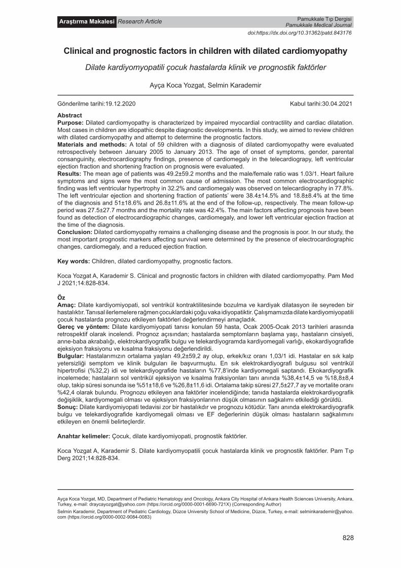
828
Pamukkale Tıp DergisiPamukkale Medical Journal
doi:https://dx.doi.org/10.31362/patd.843176
Araştırma Makalesi Research Article
Gönderilme tarihi:19.12.2020 Kabul tarihi:30.04.2021
Clinical and prognostic factors in children with dilated cardiomyopathy
Dilate kardiyomyopatili çocuk hastalarda klinik ve prognostik faktörler
Ayça Koca Yozgat, Selmin Karademir
AbstractPurpose: Dilated cardiomyopathy is characterized by impaired myocardial contractility and cardiac dilatation. Most cases in children are idiopathic despite diagnostic developments. In this study, we aimed to review children with dilated cardiomyopathy and attempt to determine the prognostic factors.Materials and methods: A total of 59 children with a diagnosis of dilated cardiomyopathy were evaluated retrospectively between January 2005 to January 2013. The age of onset of symptoms, gender, parental consanguinity, electrocardiography findings, presence of cardiomegaly in the telecardiograpy, left ventricular ejection fraction and shortening fraction on prognosis were evaluated.Results: The mean age of patients was 49.2±59.2 months and the male/female ratio was 1.03/1. Heart failure symptoms and signs were the most common cause of admission. The most common electrocardiographic finding was left ventricular hypertrophy in 32.2% and cardiomegaly was observed on telecardiography in 77.8%. The left ventricular ejection and shortening fraction of patients’ were 38.4±14.5% and 18.8±8.4% at the time of the diagnosis and 51±18.6% and 26.8±11.6% at the end of the follow-up, respectively. The mean follow-up period was 27.5±27.7 months and the mortality rate was 42.4%. The main factors affecting prognosis have been found as detection of electrocardiographic changes, cardiomegaly, and lower left ventricular ejection fraction at the time of the diagnosis.Conclusion: Dilated cardiomyopathy remains a challenging disease and the prognosis is poor. In our study, the most important prognostic markers affecting survival were determined by the presence of electrocardiographic changes, cardiomegaly, and a reduced ejection fraction.
Key words: Children, dilated cardiomyopathy, prognostic factors.
Koca Yozgat A, Karademir S. Clinical and prognostic factors in children with dilated cardiomyopathy. Pam Med J 2021;14:828-834.
ÖzAmaç: Dilate kardiyomiyopati, sol ventrikül kontraktilitesinde bozulma ve kardiyak dilatasyon ile seyreden bir hastalıktır. Tanısal ilerlemelere rağmen çocuklardaki çoğu vaka idiyopatiktir. Çalışmamızda dilate kardiyomiyopatili çocuk hastalarda prognozu etkileyen faktörleri değerlendirmeyi amaçladık.Gereç ve yöntem: Dilate kardiyomiyopati tanısı konulan 59 hasta, Ocak 2005-Ocak 2013 tarihleri arasında retrospektif olarak incelendi. Prognoz açısından; hastalarda semptomların başlama yaşı, hastaların cinsiyeti, anne-baba akrabalığı, elektrokardiyografik bulgu ve telekardiyogramda kardiyomegali varlığı, ekokardiyografide ejeksiyon fraksiyonu ve kısalma fraksiyonu değerlendirildi. Bulgular: Hastalarımızın ortalama yaşları 49,2±59,2 ay olup, erkek/kız oranı 1,03/1 idi. Hastalar en sık kalp yetersizliği semptom ve klinik bulguları ile başvurmuştu. En sık elektrokardiyografi bulgusu sol ventrikül hipertrofisi (%32,2) idi ve telekardiyografide hastaların %77,8’inde kardiyomegali saptandı. Ekokardiyografik incelemede; hastaların sol ventrikül ejeksiyon ve kısalma fraksiyonları tanı anında %38,4±14,5 ve %18,8±8,4 olup, takip süresi sonunda ise %51±18,6 ve %26,8±11,6 idi. Ortalama takip süresi 27,5±27,7 ay ve mortalite oranı %42,4 olarak bulundu. Prognozu etkileyen ana faktörler incelendiğinde; tanıda hastalarda elektrokardiyografik değişiklik, kardiyomegali olması ve ejeksiyon fraksiyonlarının düşük olmasının sağkalımı etkilediği görüldü.Sonuç: Dilate kardiyomiyopati tedavisi zor bir hastalıkdır ve prognozu kötüdür. Tanı anında elektrokardiyografik bulgu ve telekardiyografide kardiyomegali olması ve EF değerlerinin düşük olması hastaların sağkalımını etkileyen en önemli belirteçlerdir.
Anahtar kelimeler: Çocuk, dilate kardiyomiyopati, prognostik faktörler.
Koca Yozgat A, Karademir S. Dilate kardiyomyopatili çocuk hastalarda klinik ve prognostik faktörler. Pam Tıp Derg 2021;14:828-834.
Ayça Koca Yozgat, MD, Department of Pediatric Hematology and Oncology, Ankara City Hospital of Ankara Health Sciences University, Ankara, Turkey, e-mail: [email protected] (https://orcid.org/0000-0001-6690-721X) (Corresponding Author) Selmin Karademir, Department of Pediatric Cardiology, Düzce University School of Medicine, Düzce, Turkey, e-mail: [email protected] (https://orcid.org/0000-0002-9084-0083)

829
Introduction
Dilated cardiomyopathy (DCM) is the most common cardiyomyopathy and a considerable cause of mortality in children [1]. Dilated cardiomyopathy is a myocardial disorder characterized by impaired myocardial contractility and cardiac dilatation that causes congestive failure of the heart. The annual incidence of DCM is 0.34 to 1.13 per 1 million children [2]. The etiology is varied but most of the cases in children are idiopathic; the remainder are secondary to various factors, such as familial, infectious myocarditis, mitochondrial, metabolic or malformation syndromes and neuromuscular disease [3]. Descripiton of familial cardiomyopathy with or without a specific genetic mutation may be important for the treatment of family members [4].
The prognosis of patients with DCM is in a wide spectrum between sudden recovery and severe heart failure. In prior studies from the Pediatric Cardiomyopathy Registry, the survival rates of patients range from 75 to 80% and from 60 to 75%, at one to five years, respectively [4, 5].
Despite advances in treatment, such as β-blockers, diuretics and angiotensin converting enzyme inhibitors, the prognosis of DCM in children remains poor, and organ survival has changed little over the past 20 years. Currently the only effective treatment in patients who unresponsive to medical treatment is cardiac transplantation [6].
In pediatric patients, prognostic factors of DCM are not well identified until now because of the diseases heterogeneity. In children the published data are rare and little is known about long-term clinical course and factors that may influenced prognosis, better or worse. Identifying the prognostic factors for patients who have a poor outcome is necessary and thus, cardiac transplantation could be offered to those patients as a therapeutic option.
In this study we aimed to review the children with dilated cardiomyopathy and attempt to determine prognostic factors.
Material and methods
This retrospective study comprised of 59 children with dilated cardiomyopathy at Dr.
Sami Ulus Maternity, Child Health and Diseases Training and Research Hospital between January 2005 to January 2013 and this is the first authors’ pediatric thesis because of a retrospective study, there is no ethics committee approval. The diagnosis of DCM was based on the presence of ventricular enlargement and systolic dysfunction with normal left ventricles wall thickness in echocardiography determined by the American Heart Association in 2006 [7].
Patients with congenital cardiac disease, arrhythmogenic and other cardiomyopathies, Kawasaki’s disease, ischaemic cardiac diseases, systemic arterial hypertension, cardiomyopaties due to cardiotoxic agents and inborn errors of metabolism, neuromuscular disease, rheumatic heart disease, septicaemia, Chagas’ disease were all excluded.
Clinical records of 59 patients were analyzed for data on age, gender, consanguinity between parents, familial history, complaints on presentation, physical examination findings, electrocardiography (ECG) and echocardiography findings at the time of diagnosis and prognosis.
The age of onset of symptoms, gender, consanguinity between parents, ECG findings, presence of cardiomegaly in the telecardiography, left ventricular ejection fraction (LVEF) and shortening fraction (LVFS) measurements on prognosis were evaluated. The survival of the patients was evaluated from the time of diagnosis.
Statistical analysis
Statistical analyses were performed using the SPSS 15.0 programme (Statistical package for the social sciences IBM Corp., Armonk, NY, USA). Comparative variables were reported as mean and range. Whether continuous variables fit the normal distribution was evaluated using visual (histogram and probability plots) and analytical tests (Kolmogorov-Smirnov/Shapiro-Wilk tests). Independent sample t-test was used for comparative analysis between the two groups, and the Mann-Whitney U test was used for data that did not conform to the normal distribution. Pearson chi-square test was used in comparison analysis for categorical variables between independent groups. The differences with a p-value of less than 0.05 were considered as statistically significant and the confidence
Pamukkale Medical Journal 2021;14(4):828-834 Koca Yozgat and Karademir

interval was 95%. The survival times of the patients were evaluated using Kaplan-Meier method and Cox regression analysis.
Results
The mean age of patients was 49.2±59.2 months (10 days-183 months) and 32 patients (54.2%) were under, and 27 patients (45.8%) above two years of age. Thirty (50.8%) of the patients were female and 29 (49.2%) were male. Parental consanguinity and familial history of DCM were determined in 24 (40.7%) and in 1 (1.6%) patients, respectively. A history of pneumonia or upper respiratory tract infection was defined in 41 (69.4%) patients within a month before diagnosis.
Heart failure symptoms and signs were the most common cause of admission in both age groups (n=40, 67.7%). The most common physical examination findings were 2–3/6 degree systolic murmur heard at apical localization (n=43, 72.8%) and tachycardia (n=38, 64.3%). Table 1 shows the complaints and physical examination findings on the presentation of the 59 patients.
On electrocardiography, the most common findings were left ventricular hypertrophy in 19 patients (32.2%), right ventricular hypertrophy in 6 (10.1%), ST-T changes in 5 (10.1%), voltage suppression in 4 (8.4%), arrhytmia in 3 (6.7%), and biventricular hypertrophy in 1 (1.6%) patients. Of 4 patients with arrhythmia, 1 had supraventricular tachycardia and 3 had ventricular extrasystoles. Cardiomegaly was present in 46 (77.8%) of the patients in the telecardiography.
At the time of the diagnosis, in the transthoracic echocardiographic examinations, mean left ventricle end-diastole diameter (LVEDd) was determined as 41.57±2.5 mm, mean LVEF as 38.4±14.5%, mean LVFS as 18.8±8.4%. At the end of the follow-up period, the mean LVEF was 51±18.6% and mean LVFS was 26.8±11.6%. According to age groups of 2 years LVEF and LVFS values are shown in Table 2.
The median follow-up period of our patients was 21.2 months, ranged from 1 day to 86 months. Two (3.3%) patients underwent cardiac
830
Prognostic factors in children with dilated cardiomyopathy
Table 1. Complaints and physical examination findings at the time of diagnosis
Number %Complaints*Cough 38 64.3
Shortness of breath 32 54
Anorexia 29 49.1
Weakness 27 45.6
Fever 22 36
Nausea-vomiting 17 28.7
Palpitations 14 23.6
Sweating 12 20.2
Bruising 10 16.8
Other complaints** 19 32
Physical examination findings*Cardiac murmur 43 72.8
Tachycardia 38 64.3
Tachypnea 27 45.7
Hepatomegaly 24 40.6
Galo rhythm 10 16.8
Edema 7 11.8
Other physical examination findings*** 13 22
*Some patients have more than one complaint and physical examination finding** Chest pain, growth failure, abdominal pain, headache*** Splenomegaly, retarded neuromotor development, venous distension, clubbing

831
transplantation. Mortality was seen in 25 (42.4%) patients at median 7.5 months (1-26 months) after diagnosis, and 34 (57.6%) survived. One of our patients who underwent transplantation died.
The patients included in the study were divided into two groups, Group I as survivors and Group II as non-survivors. Age, gender, parental consanguinity, presence of ECG changes, presence of cardiomegaly on telecardiography, LVEF, LVFS measurement were compared in terms of patients’ prognosis.
Mortality was seen in 11 patients aged <2 years and in 14 patients aged 2 years or older. The mortality rate was found to be higher in female patients (n=14, 46.7%) than in male patients (n=11, 38%) and mortality was seen in 14 (58.4%) patients with and eleven patients (31.5%) without parental consanguinity. In the present study, no statistically significant difference was found between the groups in age, gender and parental consanguinity (p>0,05).
While the mortality rate was 50% in patients with ECG changes at the time of the diagnosis and twenty-two patients (47.8%) with cardiomegaly as determined on telecardiography were died. Electrocardiographic changes and rate of cardiomegaly was defined at a higher rate in group II (p<0.05).
In the evaluation of the echocardiography findings at diagnosis, the mean LVEF values were lower in Group II than in Group I and the difference between survivors and non-survivors was statistically significant (p=0.04). The characteristics of Group I and Group II are shown in Table 3.
Discussion
Dilated cardiomyopathy is a myocardial disease which produces a dilated left ventricle with systolic dysfunction and it is the main indication for a heart transplant in children and adults [8]. A significant proportion of pediatric patients with DCM remain classified as idiopathic, despite advances in diagnosis [9]. Jr Dec et al. [10] showed that, when myocardial biopsy is performed in patients with DCM, the disease is secondary to myocarditis in most patients who are thought to be idiopathic. Although there are many reasons other than idiopathic, post-myocarditis dilated cardiomyopathy being
the most prevalent form [11]. In our study, 41 patients (69.4%) had a history of pneumonia or upper respiratory tract infection within the previous month and this suggests myocarditis secondary to infection is important in etiology.
In our study, the parental consanguinity rate was 40.7% and one of our patients had a family history of DCM. Cardiological evaluations of the family members of this patient were found to be normal. In patients with dilated cardiomyopathy, screening cases among family members is substantial in terms of reducing the mortality of the disease and early diagnosis. As in our study, because of the high rate of consanguineous marriage in our country, we think that a detailed cardiological examination should be done on family members.
The clinical presentation may vary from asymptomatic to acute congestive heart failure and tachypnea, tachycardia, hepatomegaly in children with DCM and a systolic murmur of mitral regurgitation are the most frequent symptoms and signs. In our study we also found that the most common physical examination findings were 2–3/6 degree systolic murmur and tachycardia.
The 5-year transplant free survival rate in pediatric patients with DCM ranges from 50-60% [12]. In our study group, the mortality rate was determined as 42.4% at a median of 7.5 months after diagnosis, similar to the rates in the literature.
The relationship between age, gender and parental consanguinity and prognosis has been evaluated. But we found that age, gender and parental consanguinity did not have any effect on mortality in patients with DCM. Burch et al. [13] stated that mortality was higher in those over 2 years of age. Similarly, Daubenay et al. [6] reported that the prognosis was poor in patients over 5 years of age at the time of diagnosis. Although in our study, we found high mortality rate in patients over 2 years of age but we think about that we could not find a significant difference due to the limited number of patients.
Arrhythmia and left ventricular hypertrophy cause a major risk in children with DCM and ST and T wave changes, pathologic Q wave which are ischemia findings of electrocardiography were also seen [14]. In our study, the most
Pamukkale Medical Journal 2021;14(4):828-834 Koca Yozgat and Karademir

832
Table 2. Echocardiographic findings of the patients at the time of the diagnosis, and at the end of the follow-up period
<2 years (n:32) ≥ 2 years (n:27)LVEF LVFS LVEF LVFS p1 ‡ p2 ‡
At diagnosis 38.2±11.2 18.2±5.7 38.6±17.9 19.5±10.9 0.92 0.56
At the end of the follow-up
52.2±19.8 27.3±12.5 49.5±17.3 26.2±10.7 0.58 0.71
LVEF, left ventricular ejection fraction; LVFS, left ventricular shortening fraction; ‡: Independent T test; p1, p value between LVEF, p2, p value between LVFS
Table 3. Characteristics of Group I and Group II
Group I (Survivors) (n:34)
Group II (Non-survivors) (n:25) Total
n % n % p value
Age < 2 years 21 65.6 11 34.4 32 >0.05 ¥
≥ 2 years 13 48.1 14 51.9 27
Gender >0.05 ¥
Male 18 62 11 38 29
Female 16 53.3 14 46.7 30
Consanguinity >0.05 ¥
Yes 10 41.6 14 58.4 24
No 24 68.5 11 31.5 35
Electrographic changes 0.04 ¥
Yes 19 50 19 50 38
No 15 71.5 6 28.5 21
Presence of cardiomegaly 0.03 ¥
Yes 24 52.2 22 47.8 46
No 10 77 3 23 13
Echocardiography findings LVEF 41.7±13.8% 34±14.5% 0.04 ‡
LVFS 20.3±8.2% 16.8±8.4% >0.05 ‡
LVEF, left ventricular ejection fraction; LVFS, left ventricular shortening fraction; ¥: Pearson chi-square test; ‡:Independent T-Test
Prognostic factors in children with dilated cardiomyopathy

833
common electrocardiography finding was left ventricular hypertrophy and we observed that the mortality rate was statistically higher in patients with ECG findings. Türe et al. [15] reported that, the significant difference was seen between the electrocardiographic findings of deceased and surviving patients with DCM and they suggest that detailed ECG wave measurements should be evaluated in children to define the risk of mortality.
In our study, 77.8% of our patients had cardiomegaly as detected in telecardiography and the mortality of patients with cardiomegaly was higher than those without. Similarly, in the literature, cardiomegaly has been shown to be a predictor of prognosis in patients with DCM [16].
Many studies in the literature have investigated the relationship between left ventricular function and prognosis in patients with DCM. Zecchin et al. [17] reported that patients whose LVEF lower than 30% and a longer left ventricular end-diastolic diameter had a poor prognosis. Daubeney et al. [6] stated that risk factors for a poorer outcome included a lower LVFS at presentation. In our study group, the mean LVEF at presentation was statistically significantly lower in non-survivors. Also, the mean LVFS at the time of diagnosis was lower in non-survivors, but no statistical difference was found.
Our study has some limitations due to the fact that the study was retrospective and study sample size was small. Consequently, statistical significance could not be reached in some data and larger prospective studies should be required to support the results of our study.
Despite medical treatment and efforts to advance patient care, pediatric DCM remains a challenging disease and the prognosis is poor. Cardiac transplantation should be considered in patients with intractable advanced symptomatic heart failure and require multiple inotropes and mechanical ventilation [18]. Adwani et al. [19] reported that the survival rate of pediatric patients who underwent cardiac transplantation for DCM was 95% at one year and 87% at three years. In our study, cardiac transplantation was performed in two (3.3%) patients and one died after transplantation.
In conclusion, there were no significant difference between survivors and non-survivors in respect of age, gender and parental consanguinity. Whereas, in our study the main factors affecting prognosis have been found as detection of ECG changes on electrocardiography, cardiomegaly on telecardiography and lower LVEF at the time of the diagnosis in patients with DCM. Further studies are needed to determine the factors predicting mortality in patients with dilated cardiomyopathy.
Conflict of interest: No conflict of interest was declared by the authors.
References1. Nugent AW, Daubeney PEF, Chondros P, et al.
National Australian Childhood Cardiomyopathy Study. The epidemiology of childhood cardiomyopathy in Australia. N Engl J Med 2003;348:1639-1646. https://doi.org/10.1056/NEJMoa021737
2. Richardson P, McKenna W, Bristow M, et al. Report of the 1995 World Health Organization/International Society and Federation of Cardiology task force on the definition and classification of cardiomyopathies. Circulation 1996;93:841-842. https://doi.org/10.1161/01.cir.93.5.841
3. Lipshultz SE, Sleeper LA, Towbin JA, et al. The incidence of pediatric cardiomyopathy in two regions of the United States. N Engl J Med 2003;348:1647-1655. https://doi.org/10.1056/NEJMoa021715
4. Cox GF. Diagnostic approaches to pediatric cardiomyopathy of metabolic genetic etiologies and their relation to therapy. Prog Pediatr Cardiol 2007;24:15-25. https://doi.org/10.1016/j.ppedcard.2007.08.013
5. Towbin JA, Lowe AM, Colan SD, et al. Incidence, causes, and outcomes of dilated cardiomyopathy in children. JAMA 2006;296:1867-1876. https://doi.org/10.1001/jama.296.15.1867
6. Daubeney PE, Nugent AW, Chondros P, et al. National Australian Childhood Cardiomyopathy Study. Clinical features and outcomes of childhood dilated cardiomyopathy: results from a national population-based study. Circulation 2006;114:2671-2678. https://doi.org/10.1161/CIRCULATIONAHA.106.635128
7. Maron BJ, Towbin CJA, Thiene FG, et al. Contemporary definitions and classification of the cardiomyopathies: an American Heart Association scientific statement from the Council on Clinical Cardiology, Heart Failure and Transplantation Committee; quality of care and outcomes research and functional genomics and translational biology interdisciplinary working groups; and council on epidemiology and prevention. Circulation 2006;113:1807-1816. https://doi.org/10.1161/CIRCULATIONAHA.106.174287
Pamukkale Medical Journal 2021;14(4):828-834 Koca Yozgat and Karademir
834
8. Naudi D, Rossano JW. Epidemiology and Cost of heart failure in children. Cardiol Young 2015;25:1460-1468. https://doi.org/10.1017/S1047951115002280
9. Puggia I, Merlo M, Barbati G, et al. Natural history of dilated cardiomyopathy in children. J Am Heart Assoc 2016;5:e003450. https://doi.org/10.1161/JAHA.116.003450
10. Dec GW Jr, Palacios IF, Fallon JT, et al. Active myocarditis in the spectrum of acute dilated cardiomyopathies. Clinical features, histologic correlates, and clinical outcome. N Engl J Med 1985;312:885-890. https://doi.org/10.1056/NEJM198504043121404
11. Lipshultz SE, Law YM, Asante Korang A, et al. Cardiomyopathy in children: classification and diagnosis: a scientific statement from the American Heart Association. Circulation 2019;140:9-68. https://doi.org/10.1161/CIR.0000000000000682
12. Alvarez JA, Orav EJ, Wilkinson JD, et al. Competing risks for death and cardiac transplantation in children with dilated cardiomyopathy: results from the pediatric cardiomyopathy registry. Circulation 2011;124:814-823. https://doi.org/10.1161/CIRCULATIONAHA.110.973826
13. Burch M, Siddiqi SA, Celermajer DS, Scott C, Bull C, Deanfield JE. Dilated cardiomyopathy in children: determinants of outcome. Br Heart J 1994;72:246-250. https://doi.org/10.1136/hrt.72.3.246
14. Muller G, Ulmer HE, Hagel KJ, Wolf D. Cardiac dysrhythmias in children with idiopathic dilated or hypertrophic cardiomyopathy. Pediatr Cardiol 1995;16:56-60. https://doi.org/10.1007/BF00796818
15. Türe M, Balık H, Akın A, Bilici M, Nergiz A. The relationship between electrocardiographic data and mortality in children diagnosed with dilated cardiomyopathy. Eur J Pediatr 2020;179:813-819. https://doi.org/10.1007/s00431-020-03569-9
16. Manolio TA, Baughman KL, Rodeheffer R, et al. Prevalence and etiology of idiopathic dilated cardiomyopathy (summary of a National Heart, Lung, and Blood Institute workshop. Am J Cardiol 1992;69:1458-1466. https://doi.org/10.1016/0002-9149(92)90901-a
17. Zecchin M, Lenarda AD, Bonin M, et al. Incidence and predictors of sudden cardiac death during long-term follow-up in patients with dilated cardiomyopathy on optimal medical therapy. Ital Heart J 2001;2:213-221.
18. Jefferies JL, Towbin JA. Dilated cardiomyopathy. Lancet 2010;375:752-762. https://doi.org/10.1016/S0140-6736(09)62023-7
19. Adwani SS, Whitehead BF, Rees PG, et al. Heart transplantation for dilated cardiomyopathy. Arch Dis Child 1995;73:447-452. https://doi.org/10.1136/adc.73.5.447
Acknowledgement: This manuscript is the thesis of the first author and it was produces under the supervision of the second author.
Ethics committee approval: This article is the first authors’ pediatric thesis, which was made in 2013. Since it is a retrospective study, there is no ethics committee approval.
Contributions of the authors to the article
A.K.Y. conceptualized and designed the study, drafted the initial manuscript, collected data, carried out the initial analyses, and reviewed and revised the manuscript. S.K. conceptualized and designed the study, coordinated and supervised data collection. All authors approved the final manuscript as submitted and agree to be accountable for all aspects of the work.
Prognostic factors in children with dilated cardiomyopathy

836
Pamukkale Tıp DergisiPamukkale Medical Journal
doi:https://dx.doi.org/10.31362/patd.862333
Araştırma Makalesi Research Article
Gönderilme tarihi:16.02.2021 Kabul tarihi:21.05.2021
Annelerin tamamlayıcı beslenme hakkındaki tutum ve yaklaşımları
Approaches and attitudes of mothers about complementary feeding
Gizem Şenyazar, Şule Gökçe, Feyza Koç
ÖzAmaç: Beslenme özellikle ilk bir yaş için sağlıklı büyüme ve gelişmede en önemli unsurdur. Çalışmada annelerin bebeklerini ne kadar süre sadece anne sütüyle beslediklerini, tamamlayıcı beslenmeye ne zaman başladıklarını ve tamamlayıcı beslenmeye geçişte kararlarını etkileyen faktörleri saptamak amaçlanmıştır. Gereç ve yöntem: Çalışmaya, 12-36 ay arasında Ege Üniversitesi Tıp Fakültesi’nde Sosyal Pediatri, Genel Pediatri ve diğer polikliniklerde takibe alınmış toplam 300 olgu dahil edildi. Yazılı onam formu alındıktan sonra anneler ile tek tek ve yüz yüze görüşülerek hazırlanmış anket formu dolduruldu. Bulgular: Çalışmaya 174 (%58) erkek, 126 (%42) kız olmak üzere toplam 300 çocuk dahil edildi. Olguların yaş ortalaması 22,5±7,8 ay idi. Çalışmada yer alan 293 (%97,7) bebek doğumdan sonraki ilk 24 saat içinde anne sütü almıştı, fakat tek başına anne sütü ile başlama oranı %78,3 idi (n=235). Sadece anne sütü ile beslenme süresi ortalama 4±2,5 aydı. İlk 6 ayda sadece anne sütü ile beslenme oranı ise %47,7 idi. Annelerin ilk 24 saat içinde anne sütü verme eğilimi yüksek iken, sadece anne sütü verme oranı yeterli düzeyde değildi. Anne sütüne ek olarak doğumda formül mamaların da yüksek düzeyde verildiği gözlendi. Toplam anne sütü alma süresi 14±7,8 ay, tamamlayıcı beslenmeye başlama yaş ortalaması ise 5,6±1,2 aydı ve %29,3'üne 6. aydan önce tamamlayıcı beslenme başlanmıştı. Annelere 6. aydan önce niçin tamamlayıcı beslenmeye başladıkları sorulduğunda, en yaygın cevap “Tattırmak amaçlı” (%37,5) idi. Sonuç: Çalışmamız annelerin anne sütü konusunda istekli olduklarını fakat tatlandırma isteği, bebeğin emmeyi reddettiği düşüncesi, anne sütünün yetmediği düşüncesi, bebeğin sürekli ağlaması gibi çeşitli nedenler ile farklı beslenme şekillerini denediklerini göstermiştir. Çalışmamızda her ne kadar oran düşük gözükse de tamamlayıcı beslenmeye hala erken başlanmaktadır. Anne eğitiminin doğumdan önce ve sonrasında da devam edilmesi, beslenme konusunda annelerin bilgi düzeylerinin arttırılması gerekmektedir.
Anahtar kelimeler: Anne sütü, bebek, tamamlayıcı beslenme, beslenme.
Şenyazar G, Gökçe Ş, Koç F. Annelerin tamamlayıcı beslenme hakkındaki tutum ve yaklaşımları. Pam Tıp Derg 2021;14:836-845.
AbstractPurpose: Nutrition is the most important factor in healthy growth and development, particularly for the first age. The aim of our study was to determine how long mothers fed their babies only with breast milk, when they started complementary food and the factors that affect their decision to start complementary food. Materials and methods: A total of 300 cases between 12-36 months that were monitored in social pediatrics, general pediatrics, and other outpatient clinics at Ege University Faculty of Medicine were included in the study. After receiving the written consent form, the questionnaire prepared by meeting the mothers individually and face-to-face was filled out. Results: A total of 300 children, 174 boys (58%) and 126 girls (42%) were included in the study. The mean age of the children was 22.5±7.8 months. Two hundred ninety-three of the cases (97.7%) have been fed with breast milk within the first 24 hours after delivery; however, only breast milk percent was 78.3% (n=235). The mean time of only breast milk feeding was 4±2.5 months. The rate of nutrition only with breast milk in the first 6 months was 47.7%. Although the mothers have a high tendency of giving breast milk in the first 24 hours, the rate of giving only breast milk is not at sufficient level. It is observed that the formula was also given at a high level at birth in addition to breast milk. The total period of taking breast milk was 14±7.8 months and the mean duration of starting the complementary feeding was 5.6±1.2 months and 29.3% of the cases have been started the complementary feeding before the 6th month. When the mothers were asked why they started complementary feeding before the 6th month, the most common answer was “for tasting” (37.5%).Conclusions: Our study showed that mothers are willing to breastfeed, but they try different forms of nutrition for various reasons, such as to sweeten, the idea that the baby refuses the breastfeeding, the idea that breast milk is not enough, and the baby's constant crying. Although the rate seems low in our study, complementary
Gizem Şenyazar, Uzm. Dr. Ege Üniversitesi Tıp Fakültesi, Çocuk Sağlığı ve Hastalıkları Anabilim Dalı, İzmir, Türkiye, e-posta: [email protected] (https://orcid.org/0000-0003-1313-9426)Şule Gökçe, Öğr. Gör. Ege Üniversitesi Tıp Fakültesi, Çocuk Sağlığı ve Hastalıkları Anabilim Dalı, İzmir, Türkiye, e-posta: [email protected] (https://orcid.org/0000-0003-3392-4990) (Corresponding Author)Feyza Koç, Doç Dr. Ege Üniversitesi Tıp Fakültesi, Çocuk Sağlığı ve Hastalıkları Anabilim Dalı, Sosyal Pediatri Bilim Dalı, İzmir, Türkiye, e-posta: [email protected] (https://orcid.org/0000-0002-5891-8506)

837
Giriş
Beslenme; çocukluk çağında sağlıklı büyüme ve gelişme için gerekli en temel faktördür. Bu konuda yapılan hatalı ve bilinçsiz uygulamalara bağlı gelişen olumsuz etkiler; sadece çocukluk çağında değil, yaşamın bütün dönemlerinde gözlenebilmektedir. Dünya Sağlık Örgütü (DSÖ) ve Amerikan Pediatri Akademisi (AAP) bebeklerin ilk 6 ay sadece anne sütü ile beslenmelerini, altıncı ayda mutlaka tamamlayıcı beslenmeye geçilmesini ve emzirmenin 2 yıl sürdürülmesini tavsiye etmektedir [1, 2].
Anne sütü ile beslenme; bebek mortalite ve morbidite oranlarını azaltır, bebeğin sağlıklı büyüme ve gelişmesini sağlar. Anne sütü bebeğin gereksinim duyduğu besin içeriği ve enfeksiyonlara karşı koruyucu özellikleri nedeniyle yaşamın ilk anından itibaren verilmesi gereken tek fizyolojik besindir. Yeterli süre ve miktarda verilen anne sütü ile zamanında uygun tamamlayıcı beslenme başlanması ve çeşitlendirilerek devam edilmesi temel çocuk gelişimi açısından son derece önemlidir. Dünya Sağlık Örgütü verilerine göre çocuk ölümlerinin %7’sinin birinci, %46’sının ise ikinci nedeni yetersiz ve dengesiz beslenmedir [2]. Türkiye Nüfus ve Sağlık Araştırmaları (TNSA) verilerine göre bebek formül mamaları ve diğer sıvılara erken başlama ülkemizde de oldukça yaygın bir uygulamadır. Türkiye Nüfus ve Sağlık Araştırmaları 2008 verilerinde bebeklerin %8’i 6. aydan önce tamamlayıcı beslenmeye geçerken bu oran 2013 TNSA verilerinde artarak %12 olarak tespit edilmiştir. Yine TNSA 2018 verilerine göre de bebeklerin %23’ü anne sütü dışı süt almakta ve %12’si anne sütüne ilave tamamlayıcı beslenme almaktadır. Diğer taraftan bebeklere verilen tamamlayıcı gıdaların ayına uygun verilmediği de saptanmıştır. Bu durum tamamlayıcı gıdaya geçiş sürecinin hala önemli bir sorun olduğunu göstermektedir [3].
Bu çalışma ile annelerin tamamlayıcı beslenme hakkındaki bilgi düzeylerinin ölçülmesi, ne kadar sadece anne sütü ile beslediklerini, ne
zaman tamamlayıcı beslenmeye geçtiklerini, tamamlayıcı beslenmeye geçiş kararını vermelerinde etkili olan faktörlerin belirlenmesi amaçlanmıştır.
Gereç ve yöntem
Ege Üniversitesi Tıp Fakültesi Çocuk Sağlığı ve Hastalıkları Anabilim Dalı, Sağlıklı Çocuk İzlem Polikliniği, Genel Pediatri Polikliniği, diğer yan dal polikliniklerine başvuran ve/veya serviste yatarak izlenen 12-36 ay arasındaki çocuklar çalışmaya alındı. Kesitsel anket çalışması olarak planlanan bu çalışma Ege Üniversitesi Tıp Fakültesi Girişimsel Olmayan Klinik Araştırmalar Etik Kurulu tarafından onaylandı. Çalışmaya alınan çocukların ailelerine anket öncesi bilgi verildi ve yazılı bilgilendirilmiş onam formu alındı.
“Ekonomik düzey” Türk-iş tarafından Ağustos 2017 de yayınlanan sınıflamaya göre düzenlenmiştir. Bu sınıflamaya göre aylık gelir 1500 TL altı “Kötü”, 1500-4900 TL arası “Orta” ve 4900 TL ve üzeri “İyi” olarak nitelendirilmiştir [4].
Çalışmaya 12-36 ay arasında, kronik hastalık, prematüre doğum (≤37 hafta), hastanede uzun süre yatış (>1ay) ve besin alerjisi öyküsü olmayan çocuklar alındı. Çalışma için toplam 374 aile ile görüşme yapıldı; 36 tanesi çalışma koşullarını sağlamadığı için, 15 aile çalışmayı kabul etmediği için, 23 aile de anne bilgileri yeterli hatırlanmadığı için çalışma dışı bırakıldı ve toplam 300 anket ile çalışma tamamlandı.
İstatistiksel analiz
Çalışmaya alınan olguların ve ailelerinin sosyodemografik özelliklere ilişkin verileri tanımlayıcı tablolara dönüştürüldü. Araştırmadan elde edilen veriler SPSS (Statistics 22) programında analiz edildi. Öncelikle tüm değişkenler için tanımlayıcı istatistikler yapıldı. Kategorik değişkenler arasındaki fark için X2 kare testi, sayısal değişkenler arasındaki ilişki için Spearman’s korelasyon analizi uygulandı.
nutrition is still started early. It is necessary to continue maternal education before and after delivery and to increase the knowledge level of mothers about nutrition.
Key words: Breast milk, infant, complementary feeding, nutrition.
Senyazar G, Gokce S, Koc F. Approaches and attitudes of mothers about complementary feeding. Pam Med J 2021;14:836-845.
Pamukkale Tıp Dergisi 2021;14(4):836-845 Şenyazar ve ark.

Tamamlayıcı beslenme
838
İstatiksel olarak verilerin karşılaştırılmasında iki bağımsız grup varsa Mann Whitney U testi ve ikiden fazla grup varsa Kruskal Wallis testi uygulandı. P<0,05 istatistiksel olarak anlamlı kabul edildi.
Bulgular
Olguların %58’i erkek, %42’si kız idi. Çalışmaya alınan olguların %44,7’si (n=134) ailenin ilk çocuğuydu. Olguların %38,3’ü (n=115) ailenin ikinci çocuğu, %12,7’si (n=38) ailenin üçüncü çocuğu, %4,3’ü (n=13) ise 3. gebelikten sonra doğmuş çocuklardı. Çalışmaya dahil edilen olguların/ailelerinin sosyo-ekonomik verileri ve olguların beslenme şekilleri Tablo 1’de özetlenmiştir.
Çalışmamıza dahil edilen ailelere anne sütü alımı ile ilgili sorula yöneltildiğinde; Hiç anne sütü almayan %2,3 (n=7) olgununun anne sütü alamama nedeni olarak anneler “sütüm gelmedi” cevabını vermişti. İlk 6 ayda sadece anne sütü ile beslenme oranı %47,7 (n=143) idi. Olguların %4’ü (n=14) 7. aya kadar, %2’si (n=6) 11. aya kadar, %0,3’ü (n=1) 12 aya kadar sadece anne sütü ile beslenmeye devam etmişti. Bebeklerin 293’ü (%97,6) doğumdan sonra ilk 24 saat içinde anne sütü almıştı, ancak sadece anne sütü alanlara bakıldığında bu oran %78,3 (n=235)’tü. Doğumdan sonra ilk 24 saatte anne sütü alanların %19,7’si (n=58) anne sütü ile birlikte formül mama ile beslenmişti. Olguların %75’i (n=225) ilk 2 ayda sadece anne sütü ile beslenirken yaşla birlikte bu oran azalmaktaydı.
İlk 6 ay sadece anne sütü ile beslenen grup ile diğer beslenme şekillerini alan gruplar arasında bu duruma etki eden faktörler irdelendiğinde ailenin kaçıncı çocuğu olması ile beslenme arasında ilişki görülmedi (p>0,05). Anne ve babaların eğitim düzeyi ile ilk 6 ay sadece anne sütü verme arasındaki ilişkiye bakıldığında, annelerin okuryazar olmayanlarının %33,3’ü (n=6), ortaokul mezunu olanların %36,4’ü (n=24), üniversite mezunu olanların %57,3’ü (n=47) bebeklerini ilk 6 ay sadece anne sütü ile beslemişti. Anne ve baba eğitim düzeyi ile beslenme şekli arasında anlamlı bir ilişki saptanmadı (p>0,05) (Tablo 2).
Tablo 3’te toplam anne sütü alma süresine etki eden faktörler irdelenmiştir. Anne eğitim düzeyine göre toplam anne sütü alma süresi arasındaki ilişkiye bakıldığında, okuryazar olmayanların toplam anne sütü alma süresi
ortalama 15,1±8,3 ay, ortaokul mezunu olanların 14,4±7,2 ay, üniversite mezunu olanların 14,6±7,3 ay saptandı. Annenin eğitim düzeyi ile toplam anne sütü alma süresi arasında ilişki saptanmadı (p>0,05). Ekonomik düzeyini “iyi” olarak değerlendiren ailelerin toplam anne sütü alma süresi ortalama 14,5±8 ay, “orta” olarak değerlendirenlerin 13,9±7,7 ay, kötü olarak değerlendirenlerin 15±8,2 ay saptandı. Ailelerin ekonomik düzeyi ile toplam anne sütü alma süresi arasında istatistiksel bir ilişki bulunamadı (p>0,05).
Çalışma grubunun tamamlayıcı beslenmeye başlama yaşı ortalama 5,6±1,2 ay idi. Olguların 88'ine (%29,3) 6. aydan önce ve bunların 9'una (%3) 4. aydan önce tamamlayıcı beslenme verilmişti. Dokuz (%3) olguya 8. ayda, 1 (%0,3) olguya 9. ayda, 2 (%0,7) olguya ise 12. ayda tamamlayıcı beslenme başlanmıştı. Anne eğitim düzeyine göre tamamlayıcı beslenme başlama zamanı arasındaki ilişkiye bakıldığında, okuryazar olmayanların ek gıdaya başlama zamanı ortalama 6±1,9 ay, ortaokul mezunu olanların 5,3±1 ay, üniversite mezunu olanların 5,7±0,8 ay idi fakat istatistiksel anlamlı bir ilişki saptanmadı (p>0,05). Ev hanımı olan annelerin ek gıdaya başlama zamanı ortalama 5,6±1,2 ay, çalışan annelerin 5,7±1,1 ay idi. Annenin çalışma durumu ile ek gıdaya başlama zamanı arasında da anlamlı bir ilişki yoktu (p>0,05). Benzer şekilde ailelerin ekonomik düzeyi ile ek gıdaya başlama zamanı arasında anlamlı farklılık saptanmadı (p>0,05). Annelerin tamamlayıcı beslenmeye geçişte hangi besinler ile başlandığı değerlendirildiğinde; %99,6’sı yoğurt ile %86,3’ü peynir ile %57,3’ü ise muhallebi ile başladıklarını belirtmişlerdir. Olguların tamamlayıcı beslenmeye geçiş özellikleri, diğer tamamlayıcı beslenme ürünleri ve ürünlerin başlanma zamanları Tablo 4’te belirtilmiştir.
Tamamlayıcı beslenmeye başlama zamanına etki eden faktör olarak çocuğun cinsiyeti, kaçıncı çocuk olması, annenin doğum yaptığındaki yaşı, anne-baba eğitim, annenin çalışma durumu, ailenin ekonomik durumu değerlendirildi (Tablo 5). Anne eğitim düzeyine göre tamamlayıcı beslenmeye başlama zamanı arasındaki ilişkiye bakıldığında, okuryazar olmayanların tamamlayıcı beslenmeye başlama zamanı ortalama 6±1,9 ay, ortaokul mezunu olanların 5,3±1 ay, üniversite mezunu olanların 5,7±0,8 ay saptandı. Annenin eğitim düzeyi

839
Tablo 1. Olguların/ailelerin temel demografik özellikleri ve olguların beslenme şekilleriHasta özellikleri n %
Yaş (ortalama ± SS), ay 22,5±7,8
Cinsiyet Erkek 174 58
Kız 126 42
Doğum şekli NSVY 113 37,7
C/S 187 62,3
Doğum ağırlığı, gr <2500 25 8,3
2500-3500 194 64,7
>3500 81 27
Ailenin kaçıncı çocuğu İlk 134 44,7
2. 115 38,3
3. 38 12,7
>3 13 4,3
Beslenme şekilleri
Sadece anne sütü 143 47,7
Anne sütü ve formül mama 66 22
Anne sütü, formül mama, Tamamlayıcı beslenme 49 16,3
Anne sütü ve Tamamlayıcı beslenme 32 10,7
Formül mama ve Tamamlayıcı beslenme 4 1,3
Sadece formül mama 3 1
Anne sütü, formül mama, ek gıda, inek sütü 3 1
Aile özellikleri n %
Anne yaşı (ortalama±SS), yaş 30,9±5,6Annenin doğum yaşı (ortalama±SS), yaş 29±5,6
Anne eğitim (mezuniyet) Okuryazar değil 18 6
İlkokul 62 20,7
Ortaokul 66 22
Lise 72 24
Üniversite 82 27,3
Baba eğitim (mezuniyet) Okuryazar değil 6 2
İlkokul 62 20,7
Ortaokul 56 18,6
Lise 93 31
Üniversite 83 27,7
Annenin iş durumu Ev hanımı 227 75,7
Çalışıyor 73 24,3
Babanın iş durumu İşsiz 14 4,7
Çalışıyor 286 95,3
Ailenin ekonomik düzeyi İyi 63 21
Orta 203 67,7
Kötü 34 11,3
SS: Standard sapma
Pamukkale Tıp Dergisi 2021;14(4):836-845 Şenyazar ve ark.

840
Tablo 2. İlk 6 ay sadece anne sütü alan grup ile diğer beslenme şekillerini alan grubun karşılaştırılması
İlk 6 ay beslenme pSadece anne sütü ile beslenen grup(n=143)
Diğer beslenme şekillerini alan grup(n=157)
n (%) n (%)CinsiyetErkek 77 (44,3) 97 (55,7) 0,164
Kız 66 (52,4) 60 (47,6)
Doğum ağırlığı, gr<2500 9 (36) 16 (64) 0,231
2500-3500 90 (46,4) 104 (53,6)
>3500 44 (54,3) 37 (45,7)
Kaçıncı çocukİlk 65 (48,5) 69 (51,5) 0,950
2. 53 (46,1) 62 (53,9)
3. 18 (47,4) 20 (52,6)
>3 7 (53,8) 6 (46,2)
Anne eğitim (mezuniyet)Okuryazar değil 6 (33,3) 12 (66,7) 0,081
İlkokul 32 (51,6) 30 (48,4)
Ortaokul 24 (36,4) 42 (63,6)
Lise 34 (47,2) 38 (52,8)
Üniversite 47 (57,3) 35 (42,7)
Baba eğitim (mezuniyet)Okuryazar değil 2 (33,3) 4 (66,7) 0,380
İlkokul 29 (46,8) 33 (53,2)
Ortaokul 25 (44,6) 31 (55,4)
Lise 40 (43) 53 (57)
Üniversite 47 (56,6) 36 (43,4)
Annenin iş durumuEv hanımı 104 (45,8) 123 (54,2) 0,257
Çalışıyor 39 (53,4) 34 (46,6)
Ailenin ekonomik düzeyiİyi 32 (50,8) 31 (49,2) 0,790
Orta 94 (46,3) 109 (53,7)
Kötü 17 (50) 17 (50)
Tamamlayıcı beslenme

841
Tablo 3. Toplam anne sütü alma sürelerini etkileyebilecek faktörlerin irdelenmesi
Toplam anne sütü alma süresi (ortalama ± SS), ay p
Cinsiyet 0,289
Erkek 13,7±7,9
Kız 14,7±7,7
Kaçıncı çocuk 0,393
İlk çocuk 13,3±7,5
2. çocuk 14,3±8,2
3. çocuk 16±7,7
>3 14,5±1,5
Anne eğitim (mezuniyet) 0,720
Okuryazar değil 15,1±8,3
İlkokul 13,9±8,3
Ortaokul 14,4±7,2
Lise 13,3±8,2
Üniversite 14,6±7,3
Baba eğitim (mezuniyet) 0,512
Okuryazar değil 17,5±12,6
İlkokul mezunu 13,2±8
Ortaokul mezunu 14,8±8,3
Lise mezunu 13,6±7,5
Üniversite mezunu 14,5±7,1
Annenin iş durumu 0,515
Ev hanımı 14,3±7,9
Çalışıyor 13,6±7,5
Ailenin ekonomik düzeyi 0,648
İyi 14,5±8
Orta 13,9±7,7
Kötü 15±8,2
SS: Standart sapma
Pamukkale Tıp Dergisi 2021;14(4):836-845 Şenyazar ve ark.

Tablo 4. Olguların tamamlayıcı beslenmeye geçiş özellikleri, tamamlayıcı beslenme ögeleri ve başlangıç zamanları
Tamamlayıcı beslenmeye geçiş özellikleri n (%)
Tamamlayıcı beslenmeye geçiş, (ortalama ± SS), ay 5,6±1,2
0-4 9 (3)
4-6 79 (26,3)
>6 212 (70,7)
Tamamlayıcı beslenmeye erken geçiş nedenleri
Tattırmak amaçlı 33 (37,5)
Bebeğin emmeyi reddetmesi 24 (27,2)
Sütümün yetersiz olduğunu düşüyorum 20 (22,7)
Sürekli ağlıyordu 8 (9)
Bebeğin yetersiz kilo alımı, doktor tavsiyesi 2 (2,3)
Bebeğin sağlık sorunu 1 (1,13)
Tamamlayıcı beslenme ürünleri Başlangıç süre (Ortalama ± SS), ay
Yoğurt (n=299) 6±1,4
Muhallebi (n=172) 6,8±2
Yumurta sarısı (n=287) 7,1±2,2
Peynir (n=259) 7,3±2,3
Et (n=290) 9,5±3,1
Yumurta beyazı (n=275) 9,6±2,7
Tavuk (n=276) 10±3,3
Balık (n=247) 10,3±3,5
Köfte (n=278) 10,8±3,1
Keçi sütü (n=24) 11,3±4
İnek sütü (n=239) 12,2±4,3
SS: Standart sapma
842
Tamamlayıcı beslenme

843
ile tamamlayıcı beslenmeye başlama zamanı arasında ilişki saptanmadı (p>0,05).
Tartışma
Sağlıklı beslenme-özellikle bebeklik döneminde- büyüme ve gelişme için çok önemli rol oynamaktadır. Anne sütü ile beslenme sağlıklı beslenmenin ilk ve en önemli adımıdır. İlk 6 ay sadece anne sütü ile beslenme önerilirken, 6. aydan itibaren bebeğin besin gereksinimlerini anne sütü tek başına karşılayamaz ve tamamlayıcı besinlerin başlanması gerekir. Ancak 2 yaşına kadar emzirmeye de devam edilmelidir [2]. Dünya genelinde, düşük emzirme ve erken tamamlayıcı beslenmeye geçiş uygulamaları ne yazık ki yaygındır. Dünyada ilk 6 ay sadece anne sütü alma oranı
%38 olarak bildirilmektedir. Dünya Emzirme Eğilimleri Girişimi (World Breastfeeding Trends İnitiative)’nin verilerine göre ise ilk 6 ayda sadece anne sütü alma oranı %41 olarak açıklanmıştır [5]. Norveç’te yapılan çalışmada anne sütü alanların oranı 1. ayda %90, 4. ayda %44 ve 6. ayda %7 saptanmıştır [6]. Ülkemizde ise TNSA 2018 verilerine göre 6. ayda sadece anne sütü alma oranı %41’dir ve sadece anne sütü alan çocukların ortanca emzirilme süresi 1,8 aydır [3]. Çalışmamızda doğumdan sonra ilk 24 saatte anne sütü alma oranı oldukça yüksek (%97,7) bulunmasına rağmen sadece anne sütü alma oranına bakıldığında bu oran %78,3 saptandı ve ilk 24 saatte anne sütü alanların %19,7'sinin anne sütüne ek olarak formül mama da aldığı saptandı. Yaşla birlikte
Tablo 5. Tamamlayıcı beslenmeye geçişi etkileyebilecek faktörler
Tamamlayıcı beslenme başlama zamanı (ortalama ± SS), ay
p
Cinsiyet 0,419
Erkek 5,6±1,3
Kız 5,7±0,9
Kaçıncı çocuk 0,483
İlk 5,6±1,2
2. 5,5±1
3. 5,7±1
>3 6,1±2,1
Anne eğitim (mezuniyet) 0,173
Okuryazar değil 6±1,9
İlkokul 5,6±1,1
Ortaokul 5,3±1
Lise 5,7±1,4
Üniversite 5,7±0,8
Baba eğitim (mezuniyet) 0,063
Okuryazar değil 6,2±2,9
İlkokul 5,6±1,2
Ortaokul 5,6±1,1
Lise 5,4±1,3
Üniversite 5,8±1,2
Annenin iş durumu 0,680
Ev hanımı 5,6±1,2
Çalışıyor 5,7±1,1
Ailenin ekonomik düzeyi 0,763
İyi 5,6±1
Orta 6,7±1,1
Kötü 5,6±1,5
SS: Standart sapma
Pamukkale Tıp Dergisi 2021;14(4):836-845 Şenyazar ve ark.

844
sadece anne sütü alma oranı da azalmaktaydı. Olguların, 2. ayda %75'i, 6. ayda %47,7'si sadece anne sütü almaktaydı. Sadece anne sütü ile beslenmedeki başarısızlığın en önemli nedeni, doğumdan hemen sonra anne sütünün verilmesinin gecikmesi, emzirme sorunu yaşayan annelerde bu sorunun çözümünde de hemen formül mamaya geçilmesidir [7]. Türkiye Nüfus ve Sağlık Araştırmaları 2018 verilerinde ilk bir gün içinde emzirilme oranı %71, 2. ayda %58, 6. ayda %30,1 olarak bulunmuştur [3]. Yine İzmir'de Ünsal ve ark.’nın [8] toplam 5003 anneyi içeren çalışmalarında, annelerin %93,7’sinin doğumda bebeklerini emzirmeye başladıkları, %6,3 bebeğin ise hiç anne sütü almadığı belirtilmiştir. Yine aynı çalışmada bebeklerini emziren annelerin tek başına anne sütü verme durumlarına bakıldığında; doğumda %97 bebek sadece anne sütü alırken 2 aylık olduklarında bu oran %81,5; 4. ayda oran %46,2; 6. ayda ise %8,7’ye düşmüştür. Ülkemizde emzirmeye başlama oranları yüksek olmasına rağmen ilk 6 ay sadece anne sütü verme ve emzirmeye devam etme konusunda eksikler mevcuttur.
Dünya Sağlık Örgütü, UNICEF, ESPGHAN tamamlayıcı besinlere 6. ayda başlanılmasını önermektedir [9-11]. Buna karşın ülkemiz dahil, gelişmiş ve gelişmekte olan birçok ülkede yapılan çalışmalarda, önerilerin aksine tamamlayıcı besinlere erken başlandığı belirlenmiştir. İngiltere’de 4. aydan önce %51 oranında tamamlayıcı beslenme başlandığı, Almanya’da %16’sının 3. ayda başladığı bildirilmiştir [12, 13]. Türkiye Nüfus ve Sağlık Araştırmaları- 2018 verilerinde ise 6. aydan önce tamamlayıcı beslenme başlama oranı %14,6 olarak bulunmuştur [3]. Köksal ve ark.’nın [14] yaptığı bir çalışmada 12-23 aylık çocukların %45,9’una 6. aydan önce tamamlayıcı beslenme başlanmış, Ünalan ve ark.’nın [15] çalışmasında ise bebeklerin %58'ine 6. aydan önce, %33'üne 0-3 ayda, %25'ine ise 3-6 ayda tamamlayıcı beslenme başlanmıştı. Tamamlayıcı beslenmeye erken başlama için en sık neden "sütüm yetmiyordu’’ ve "doymuyordu, ağlıyordu" şeklinde cevaplardı. Nepal’de yapılan bir çalışmada da anneler tamamlayıcı beslenme erken başlama nedeni olarak ‘’sütümün yetersiz olduğunu düşünüyordum’’ olarak belirtmişti [16].
Çalışmamızda çocuğun cinsiyeti, kaçıncı çocuk olması ile tamamlayıcı beslenmeye erken başlama arasında ilişki saptanmamıştır. Beş Avrupa ülkesinde yapılmış çalışmada genç
annelerin tamamlayıcı besinlere erken dönemde başladığı saptanmıştır [17]. Anne ve baba eğitim düzeyinin tamamlayıcı besinlere uygun zamanda başlamayı etkileyen bir faktör olduğu çalışmalarla belirtilmiştir, anne eğitim düzeyi arttıkça tamamlayıcı beslenmeye zamanında başlandığı, baba eğitimi arttıkça tamamlayıcı beslenme başlama zamanı ile ilgili bilginin arttığı saptanmıştır [17-19]. Bizim çalışmamızda tamamlayıcı beslenmeye başlama zamanının anne yaşından etkilenmediği belirlenmiştir.
Çalışmamızın en büyük kısıtlılığı kesitsel olması ve az sayıda katılım ile gerçekleşmiş olmasıdır. Sonuçlar Türkiye genelini yansıtamadığı gibi anneler tarafından verilen bilgilerin yetersiz ve yanlış olma olasılığı da mevcuttur. Annelere tamamlayıcı beslenme konusundaki davranışları ile ilgili sorular yöneltilmiş olsa da çevresel ailevi faktörler ülkemiz için göz ardı edilmemelidir. Tamamlayıcı beslenmeye geçiş sırasında aile büyüklerinin hala çok önemli bir faktör olabileceğini düşünmekteyiz.
Sonuç olarak; annelerin ilk 24 saat içinde anne sütü verme eğilimleri yüksekken, yalnızca anne sütü verme oranı düşüktür ve yüksek oranda doğumda anne sütüne ek olarak formül mama da verilmektedir. Her ne kadar oran düşük gözükse de tamamlayıcı beslenmeye erken başlanmaktadır ve uygun zamana göre uygun tamamlayıcı beslenme konusunda eksikler mevcuttur. İlk 6 ay sadece anne sütü alma, toplam anne sütü alma ve tamamlayıcı beslenme başlama zamanının bebek ve aile sosyodemografik özelliklerinin arasında ilişki saptanmamıştır. Ayrıca sonuçlarımız annelerin anne sütü konusunda istekli olduklarını fakat yanlış inanış ve uygulamalar ile farklı beslenme şekillerini denediklerini göstermiştir. Altıncı ayda bebeklerin gereksinimlerini anne sütünün karşılayamadığı tamamlayıcı beslenme ihtiyacının olduğu bu dönem sağlıklı büyüme ve gelişmenin sağlanmasında büyük önem arz etmektedir. Anne eğitiminin doğumdan önce ve sonrasında da devam edilmesi, annelerin bu dönemde desteklenmesi ve emzirmeye teşvik edilmesi, ilk 6 ay sadece anne sütü konusunda ısrar edilmesi ve tamamlayıcı beslenme konusunda annelerin bilgi düzeylerinin arttırılması gerekmektedir.
Çıkar ilişkisi: Yazarlar çıkar ilişkisi olmadığını beyan eder.
Tamamlayıcı beslenme

845
Kaynaklar1. Pérez Lizaur AB. Complementary feeding. Gac Med
Mex 2011;147:39-45.PMID: 22352127
2. PAHO. Guiding principles for complementary feeding of the breastfed child. Washington: Pan American Health Organization, World Health Organization, 2003. Available at: http://www.who.int/child_adolescent_health/documents/a85622/en/index.htm. Accessed December, 2003
3. Hacettepe Üniversitesi Nüfus Etütleri Enstitüsü. Türkiye Nüfus ve Sağlık Araştırması 2018. Erişim adresi: http://www.hips.hacettepe.edu.tr/tnsa2018/rapor/TNSA2018_ana_Rapor.pdf. Erişim tarihi Kasım, 2019
4. Türk-İş Açlık ve Yoksulluk sınırı. Erişim adresi: http://www.turkis.org.tr/dosya/pBDme2chn8Tg.pdf. Erişim tarihi 27 Mart 2017
5. WBTi Evaluation Report-2020 Launched Webinar’s Video-Recording on Launch of WBTi Evaluation Report 2020. Available at: https://www.worldbreastfeedingtrends.org/article/wbti-evaluation-report-2020-launched. Accessed June 12, 2020
6. Lande B, Andersen LF, Baerug A, et al. Infant feeding practices and associated factors in the first six months of life: The Norwegian infant nutrition survey. Acta Paediatr 2007;92:152-161. https://doi.org/10.1111/j.1651-2227.2003.tb00519.x
7. Aydın A. Anne eğitim düzeyi ve bebek dostu hastane uygulamasının altı ay tek başına anne sütü kullanımı üzerine etkileri. Ege Pediatri Bülteni 2009;16:71-78.
8. Ünsal H, Atlihan F, Özkan H, Targan Ş, Hassoy H. Toplumda anne sütü verme eǧilimi ve buna etki eden faktörler. Çocuk Sağlığı ve Hast Derg 2005;48:226-233.
9. Fewtrell M, Bronsky J, Campoy C, et al. Complementary feeding: a position paper by the European Society for Paediatric Gastroenterology, Hepatology, and Nutrition (ESPGHAN) Committee on Nutrition. J Pediatr Gastroenterol Nutr 2017;64:119-132. https://doi.org/10.1097/MPG.0000000000001454
10. WHO | Complementary feeding of young children in developing countries. WHO 2014. Available at: http://www.who.int/nutrition/publications/infantfeeding/WHO_NUT_98.1/en. Accessed May 28, 2017
11. American Academy of Pediatrics, Kleinman RE, Greer FR. Pediatric nutrition: Policy of the American Academy of Pediatrics. Available at: https://ebooks.aappublications.org/content/pediatric-nutrition-8th-ed. Accessed Jun 11, 2017
12. Bolling K, Grant C, Hamlyn B, Thornton A. Infant feeding survey 2005. Available at: http://data.parliament.uk/DepositedPapers/Files/DEP2008-0853/DEP2008-0853.pdf. Accessed September 06, 2016
13. Koletzko B, Dokoupil K, Reitmayr S, Weimert Harendza B, Keller E. Dietary fat intakes in infants and primary school children in Germany. Am J Clin Nutr 2000;72:1392-1400. https://doi.org/10.1093/ajcn/72.5.1392s
14. Köksal E, Yalçın SS, Pekcan G, Özbaş S, Tezel B, Köse MR. Complementary feeding practices of children aged 12-23 months in Turkey. Cent Eur J Public Health 2015;23:149-154. https://doi.org/10.21101/cejph.a3988
15. Ünalan PC, Akgün T, Çiftçili S, Boler İ, Akman M. Bebek dostu bir ana çocuk sağlığı merkezinden hizmet alan anneler neden bebeklerine erken ek gıda vermeye başlıyor? Türk Ped Arş 2008;43:59-64.
16. Karkee R, Lee AH, Khanal V, Binns CW. Infant feeding information, attitudes and practic: A longitudinal survey in central Nepal. Int Breastfeed J 2014;9:1-5. https://doi.org/10.1186/1746-4358-9-14
17. Schiess S, Grote V, Scaglioni S, et al. Introduction of complementary feeding in 5 European countries. J Pediatr Gastroenterol Nutr 2010;50:92-98. https://doi.org/10.1097/MPG.0b013e31819f1ddc
18. Battaloğlu İnanç B. 15-49 yaş arası annelerin anne sütü ile ilgili uygulamaları ve etki eden faktörleri. Türkiye Aile Hek Derg 2013;1:51-55. https://doi.org/10.2399/tahd.13.63835
19. Kaya Z, Yiğit Ö, Erol M, Gayret ÖB. Altı-yirmi dört ay arası yaş grubunda beslenmeyle ilgili anne ve babaların bilgi ve deneyimlerinin değerlendirilmesi. Med Bull Haseki 2016;54:70-75. https://doi.org/10.4274/haseki.2756
“Çalışmamız 2. Avrasya Sosyal Pediatri Kongresi’nde (2020) sözlü sunum olarak kabul edilmiştir”.
Etik kurul onayı: Ege Üniversitesi Tıp Fakültesi Girişimsel Olmayan Klinik Araştırmalar Etik Kurulu tarafından onaylandı (tarih: 18.04.2017 ve no: 17-4.1/15).
Yazarların makaleye olan katkıları
F.K. çalışmanın ana fikrini ve hipotezini kurgulamışlardır. F.K. ve G.Ş. teoriyi geliştirmiş, gereç ve yöntem bölümünü düzenlemişlerdir. Sonuçlar kısmındaki verilerin değerlendirmesini F.K., Ş.G., ve G.Ş. yapmışlardır. Makalenin tartışma bölümü F.K., Ş.G. ve G.Ş. tarafından yazılmış, F.K. gözden geçirip gerekli düzeltmeleri yapmış ve onaylamıştır. Ayrıca tüm yazarlar çalışmanın tamamını tartışmış ve son halini onaylamıştır.
Pamukkale Tıp Dergisi 2021;14(4):836-845 Şenyazar ve ark.
846
Pamukkale Tıp DergisiPamukkale Medical Journal
doi:https://dx.doi.org/10.31362/patd.912469
Araştırma Makalesi Research Article
Gönderilme tarihi:09.04.2021 Kabul tarihi:20.05.2021
Metotreksat kaynaklı beyin hasarına karşı bromelainin potansiyel faydalı etkilerinin araştırılması
Investigation of potential beneficial effects of bromelain against methotrexate-induced brain injury
Kürşat Kaya, Ali Gürel, Volkan İpek
ÖzAmaç: Metotreksat, bazı kanser türleri ve romatoid artrit başta olmak üzere çeşitli hastalıkların tedavisinde kullanılan bir folik asit antagonistidir. Ancak, metotreksat kullanımıyla birlikte görülen ve nörotoksisiteyi de içeren birçok yan etki önemli bir klinik problemdir. Ananas bitkisinden elde edilen bromelain, radikal süpürücü ve lipit peroksidasyonu önleyici etkileri olan enzimatik bir karışımdır.Gereç ve yöntem: 28 adet sıçan rastgele 4 gruba bölündü (n=7). Birinci grup kontrol grubu olarak tutuldu. İkinci grup BRM grubu olarak belirlendi ve bu gruptaki ratlara 14 gün boyunca 200 mg /kg/gün bromelain verildi. Üçüncü grup MTX grubu olarak belirlendi ve bu gruptaki sıçanlara üçüncü gün tek doz 20 mg/kg metotreksat verildi. Dördüncü grup MTX+BRM grubu olarak belirlendi ve gruptaki sıçanlara metotreksat ve bromelain eşdeğer dozlarda birlikte verildi.Bulgular: Metotreksat uygulaması gruplar arasında tiyobarbitürik asit reaktif substans seviyeleri açısından herhangi bir değişikliğe neden olmadı ancak redükte glutatyon seviyesinde ve süperoksit dismutaz, glutatyon peroksidaz ve katalaz aktivitelerinde kontrol grubu ile karşılaştırıldığında istatistiksel olarak anlamlı bir azalmaya neden oldu. Metotreksat ile birlikte bromelain uygulanması redükte glutatyon seviyesini ve süperoksit dismutaz aktivitesini metotreksat grubu ile karşılaştırıldığında anlamlı şekilde arttırdı ancak glutatyon peroksidaz ve katalaz aktivitelerindeki artış istatistiki olarak anlamlı değildi. Beyin dokusu histolojik açıdan da değerlendirildi. Metotreksat uygulamasının beyinde histopatolojik olarak belirgin bir lezyon oluşumuna yol açmadığı görüldü.Sonuç: Bulgularımız bromelain kullanımının nörolojik hasara karşı metotreksat ile tedavi edilen hastalarda fayda sağlayabileceğini desteklemektedir.
Anahtar kelimeler: Metotreksat, bromelain, beyin hasarı, oksidatif stres.
Kaya K, Gürel A, İpek V. Metotreksat kaynaklı beyin hasarına karşı bromelainin potansiyel faydalı etkilerinin araştırılması. Pam Tıp Derg 2021;14:846-853.
AbstractPurpose: Methotrexate is a folic acid antagonist used in the treatment of various diseases, especially some types of cancer and rheumatoid arthritis. However, many side effects associated with methotrexate use, including neurotoxicity, are an important clinical problem. Bromelain obtained from the pineapple plant is an enzymatic mixture with radical scavenging and anti-lipid peroxidation effects.Materials and methods: 28 rats were randomly divided into 4 groups (n=7). The first group was kept as the control group. The second group was determined as the bromelain group and 200 mg/kg/day bromelain was given to the rats in this group for 14 days. The third group was determined as methotrexate group and a single dose of 20 mg/kg methotrexate was given to the rats in this group on the third day. The fourth group was determined as methotrexate + bromelain group, and the rats in the group were given methotrexate and bromelain in equivalent doses together.Results: Methotrexate administration did not cause any change in thiobarbituric acid reactive levels among groups, but caused a statistically significant decrease in reduced glutathione level and superoxide dismutase, glutathione and catalase activities compared to the control group. The administration of bromelain with methotrexate significantly increased the reduced glutathione level and superoxide dismutase activity compared to the methotrexate group, but the increase in glutathione peroxidase and catalase activities was not statistically significant. Brain tissue was also evaluated histologically. It was observed that methotrexate administration did not cause a significant lesion formation in the brain histopathologically.Conclusion: Our findings support that the use of bromelain may benefit patients treated with methotrexate against neurological damage.
Kürşat Kaya, Dr. Öğr. Üye. Pamukkale Üniversitesi Tıp Fakültesi, Tıbbi Biyokimya Anabilim Dalı, Denizli, Türkiye, e-posta: [email protected] (https://orcid.org/0000-0002-6353-7791) (Sorumlu Yazar)Ali Gürel, Doç. Dr. Fırat Üniversitesi Tıp Fakültesi, İç Hastalıkları Anabilim Dalı, Nefroloji Bilim Dalı, Elâzığ, Türkiye, e-posta: [email protected] (https://orcid.org/0000-0001-8087-8814)Volkan İpek, Dr. Öğr. Üye. Mehmet Akif Ersoy Üniversitesi Veteriner Fakültesi, Veterinerlik Patolojisi Anabilim Dalı, Burdur, Türkiye, e-posta: [email protected] (https://orcid.org/0000-0001-5874-7797)

847
Giriş
Metotreksat (MTX), kanser tedavisinde ve romatoid artrit, sedef hastalığı ve iltihaplı bağırsak hastalıkları dahil olmak üzere çeşitli otoimmün ve inflamatuar hastalıklarda, gestasyonel trofoblast hastalığının tedavisinde ve ayrıca ektopik gebelikte yaygın olarak kullanılan bir folik asit antagonistidir [1, 2]. Metotreksat hücreye girdikten sonra poliglutamatlanır ve dihidrofolat redüktaz enzimine büyük bir ilgi ile bağlanarak bu enzimin dihidrofolatı tetrahidrofolata dönüştürmesini engeller. DNA sentezi için gerekli olan timidin ve purinlerin biyosentezi için tetrahidrofolat gerektiğinden, MTX tarafından tetrahidrofolat sentezinin bloke edilmesi hücrelerin proteinleri üretememesine sebep olur [3].
Bununla birlikte, MTX tedavisi, doz sınırlamasını ve bazen tedavinin tamamen kesilmesini gerektiren çeşitli ciddi toksik yan etkilere neden olur [4]. MTX kaynaklı toksisitenin mekanizması tam olarak aydınlatılamamışsa da, oksidatif stress, tümör nekrozis faktör alfa ve indüklenebilir nitrik oksit sentaz gibi inflamatuar madiatörlerin salınımı gibi bazı muhtemel mekanizmalar bu toksik etkilerden sorumlu tutulmaktadır [1]. Oksidanlar ve antioksidanlar arasındaki dengesizlikten kaynaklan oksidatif hasar, MTX ile ilişkili doku hasarının ana nedensel faktörlerinden biri olarak değerlendirilmektedir [1]. Metotreksat uygulaması, gastrointestinal toksisite, hepatik toksisite, nefrotoksisite, hematolojik toksisite ve nörotoksisite gibi bir çok doku ve organla ilişkili ciddi toksik etkiler ile ilişkilendirilmektedir [5]. Metotreksat'ın neden olduğu nörotoksisite, akut, subakut veya geç formda meydana gelebilir ve gerek intratekal ve gerekse intravenöz uygulamadan sonra gözlemlenebilir [6].
Ananas comosus (ananas), büyük ölçüde Ekvator bölgelerinde yetiştirilen ve antioksidan, antikanser, antiinflamatuar ve anti-platelet faydalı etkileri ile bilinen bromeliaceae familyasına ait bir bitkidir [7]. Ananas bitkisinin sapından ve meyvesinden elde edilen ve kimyasal olarak 1875’den beri bilinen bromelain (BRM) ise çeşitli tiol endopeptidazlar ile fosfatazlar,
glukozidazlar, peroksidazlar, selülazlar, glikoproteinler, karbonhidratlar ve çeşitli proteaz inhibitörlerinin bir karışımıdır [8]. Biyolojik aktivitesini kaybetmeden bağırsaklardan emilebilen BRM için, trombosit agregasyonunun geri dönüşümlü inhibisyonu, sinüzit, cerrahi travmalar, tromboflebit, piyelonefrit, anjina pektoris ve bronşit gibi çeşitli hastalıklara karşı ve antibiyotikler başta olmak üzere bazı ilaçların emilimini arttırması gibi çok çeşitli terapötik faydaları öne sürülmüştür [8-10]. Bununla birlikte, BRM’in serbest radikal süpürücü ve lipit peroksidasyonu önleyici antioksidan etkileri pek çok çalışmada gösterilmiştir [7, 11, 12].
Tarafımızca yapılan literatür taramasında MTX kaynaklı oksidatif hasara karşı BRM’in etkilerinin araştırıldığı herhangi bir çalışmaya rastlanmamıştır. Bundan dolayı bu çalışmada MTX nedeniyle oluşan oksidatif beyin hasarına karşı BRM’in potansiyel faydalı etkilerinin araştırılması amaçlanmıştır.
Gereç ve yöntem
Kimyasallar
MTX (Koçak Farma, 500 mg/20 ml, İstanbul, Türkiye) ve BRM (Solgar, 500 mg, Leonia, New Jersey) eczaneden satın alındı. Kullanılan diğer tüm kimyasallar analitik saflıkta veya mevcut en yüksek saflıktaydı ve Sigma-Aldrich’den satın alındı.
Hayvanlar ve deney dizaynı
Çalışma için, Adıyaman Üniversitesi Hayvan Deneyleri Yerel Etik Kurulu’ndan etik onayı alınmıştır. Çalışmada Adıyaman Üniversitesi Deney Hayvanları Üretim, Uygulama ve Araştırma Merkezinden temin edilen toplam 28 adet erkek Spraque dawley rat kullanıldı. Polipropilen rat kafeslerine yerleştirilen ratlar çalışma süresince, 21°C ortam sıcaklığında ve 12 saatlik aydınlık-karanlık döngüsünde tutuldu. Yem ve su “ad libitum” olarak verildi. 28 adet rat her grupta 7 adet olacak şekilde rastgele 4 gruba bölündü. Birinci gruptaki ratlar kontrol grubu olarak tutuldu ve bu gruptaki ratlara 14 gün boyunca günde 1 kez gavaj ile distile su ve üçüncü gün tek doz serum fizyolojik
Key words: Methotrexate, bromelain, brain injury, oxidative stress.
Kaya K, Gurel A, Ipek V. Investigation of potential beneficial effects of bromelain against methotrexate-induced brain injury. Pam Med J 2021;14:846-853.
Pamukkale Tıp Dergisi 2021;14(4):846-853 Kaya ve ark.

Bromelain metotreksat kaynaklı beyin hasarını önler
848
intraperitoneal enjeksiyon ile uygulandı. İkinci grup BRM grubu olarak belirlendi ve bu gruptaki ratlara 14 gün boyunca günde bir kez 200 mg/kg dozda BRM gavaj ile verildi ve üçüncü gün tek doz serum fizyolojik enjeksiyonu intra peritoneal enjeksiyon ile uygulandı. Üçüncü grup MTX grubu olarak belirlendi ve bu gruptaki ratlara 14 gün boyunca günde 1 kez gavaj ile distile su ve üçüncü gün tek doz 20 mg/kg MTX intraperitoneal enjeksiyon ile uygulandı. Dördüncü grup MTX+BRM grubu olarak belirlendi ve bu gruptaki ratlara 14 gün boyunca günde 1 kez 200 mg/kg BRM gavaj ile ve üçüncü gün tek doz 20 mg/kg MTX intraperitoneal enjeksiyon ile uygulandı. Çalışmanın sonunda ksilazin-ketamin kombinasyonu ile anesteziye alınan ratlara eksanguinasyon yöntemi ile ötenazi yapıldı.
Hızlı bir şekilde bütün olarak çıkarılan beyin dokusu soğuk cam üzerinde sagittal düzlemden kesilerek iki yarım beyin küresine ayrıldı. Yarım kürelerden biri histopatolojik inceleme için tespit edildi. Diğer yarım küre biyokimyasal incelemeler yapılıncaya kadar -86°C’de saklandı.
Biyokimyasal ölçümler
Dokular 0,2 M Tris-HCl tamponu (pH: 7,4) içerisinde soğuk zincir şartları altında 1:10 (w/v) seyreltme ile homojenize edildi. Tiyobarbitürik asit reaktif substans (TBARS) ölçümleri direk homojenattan yapıldı. Homojenatlar, süperoksit dismutaz (SOD), katalaz (CAT), indirgenmiş glutatyon (GSH) ve glutatyon peroksidaz (GPx) ölçümlerinin yapılması için 3220 rpm’de 30 dk. (4°C) santrifüj edilerek üstte kalan süpernatan kısımları alındı.
Lipit peroksidasyonunun bir göstergesi olan TBARS seviyeleri Yagi’nin [13] yöntemine göre spektrofotometrik olarak 532 nm’de ölçülerek belirlendi. Sonuçlar nmol/g doku olarak ifade edildi. Hücresel enzimatik antioksidan savunma sistemi üyeleri olan SOD, CAT ve GPx serbest oksijen radikallerine karşı en güçlü savunmayı yapan ilk antioksidan savunma hattı enzimleridir. SOD, süperoksit anyonunu hidrojen peroksite dönüştürerek temizler ve oluşan hidrojen peroksit CAT ve mitokondride GPx tarafından suya dönüştürülerek uzaklaştırılır [14]. SOD, CAT ve GPx aktiviteleri spektrofotometrik yöntemle ölçüldü ve ünite/mg doku proteini olarak gösterildi. SOD aktivitesi Sun ve ark. [15] yöntemine göre 560 nm’de, CAT aktivitesi Aebi
[16] yöntemine göre 240 nm’de ve GPx aktivitesi Paglia ve Valentine’in [17] yöntemine göre 340 nm’de ölçüm yapılarak belirlendi. GSH, serbest radikallere elektron vererek onları nötralize eden ve temizleyen ikinci hat antioksidan savunma sistemi üyelerinden biridir [14]. Beyin dokusu GSH içeriği Sedlak ve Lindsay’ın [18] yöntemine göre belirlendi. GSH düzeyleri 412 nm’de ölçüldü ve nmol/mg doku proteini olarak ifade edildi. Beyin dokusu protein miktarı Lowry metoduna göre belirlendi [19].
Histopatolojik değerlendirme
Nekropsi sonrası alınan beyin dokuları %10’luk tamponlu formaldehitte daldırma yöntemi ile tespiti takiben rutin doku takip prosedürlerinden geçirilerek parafine gömüldü ve 5 mikron kalınlığında kesitler alındı. Kesitler hematoksilen-eozin ile boyanarak ışık mikroskobu altında değerlendirildi. Histopatolojik olarak telensefalon, diensefalon, mesensefalon, metensefalon ve myelensefalonda; dejenerasyon, nekroz, kromatoliz, satellitozis, demyelinasyon, myelin solgunluğu, nöropil vakuolizasyonu, aksonal şişkinlik ve sferoid oluşumu, oligodendroglial şişkinlik, intremyelinik ödem, makrofaj infiltrasyonları, gemistositik astrositler ve glial aktivasyon varlığı açısından değerlendirmeler yapıldı.
İstatistik analizler
İstatistik analizler için IBM SPSS 21 programı (IBM Corporation, Armonk, New York, Amerika Birleşik Devletleri) kullanıldı. Verilerin normallik analizi Shapiro-Wilk testi kullanılarak yapıldı. Gruplar arası farkların önemliliği tek yönlü varyans analizi (ANOVA) testi ile analiz edildi. Post hoc testi olarak, Levene testi sonucunda homojen varyans gösteren veriler için Tukey HSD ve homojen varyans göstermeyen veriler için ise Games-Howell testi kullanıldı. Sonuçlar ortalama±standart hata (SEM) olarak gösterildi. Güven aralığı %95 olarak seçildi ve p<0,05 düzeyi istatistiksel olarak anlamlı kabul edildi.
Bulgular
Biyokimyasal bulgular
Beyin dokusu TBARS ve GSH seviyeleri ile SOD, GPx ve CAT aktiviteleri Tablo 1’de verilmiştir. Çalışmamızda MTX uygulaması TBARS seviyeleri açısından herhangi bir değişikliğe neden olmadı ancak GSH
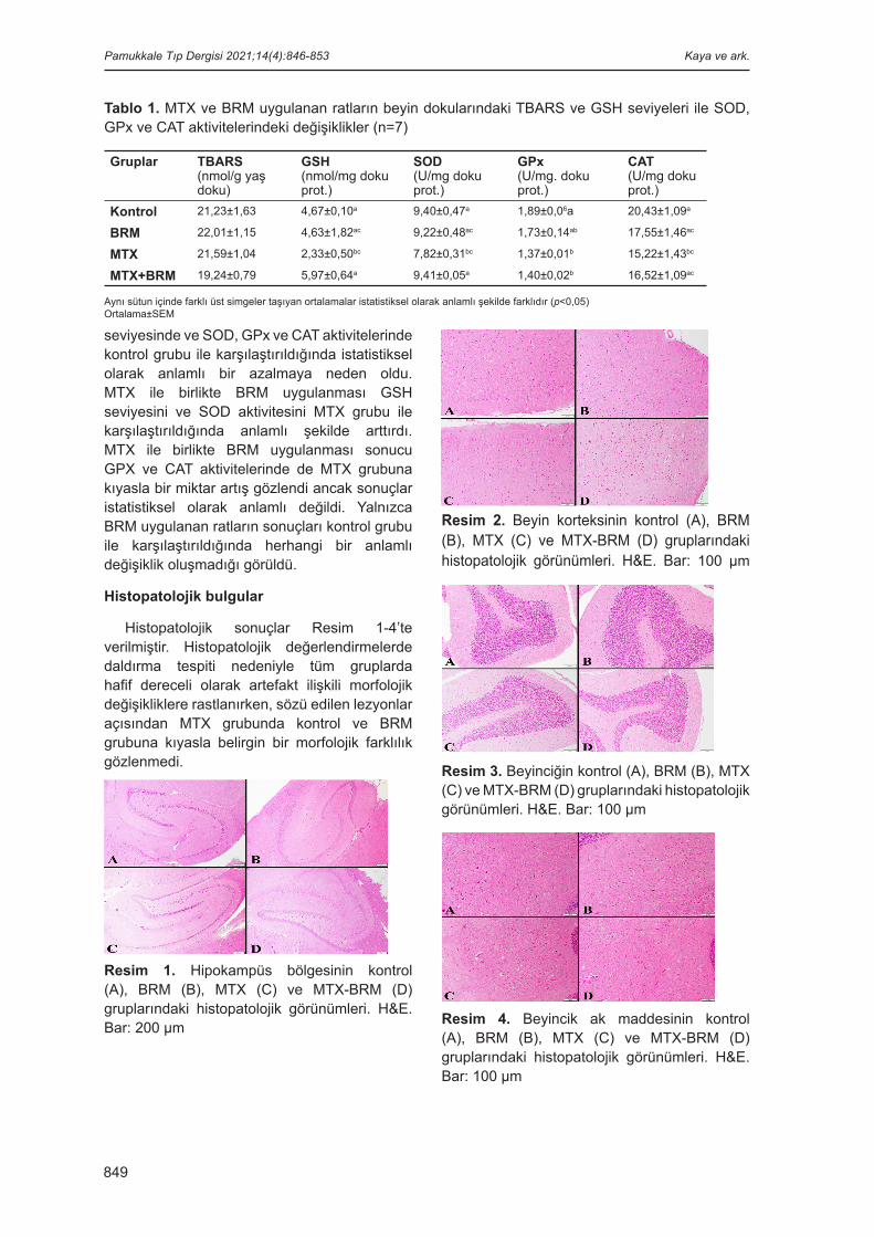
849
seviyesinde ve SOD, GPx ve CAT aktivitelerinde kontrol grubu ile karşılaştırıldığında istatistiksel olarak anlamlı bir azalmaya neden oldu. MTX ile birlikte BRM uygulanması GSH seviyesini ve SOD aktivitesini MTX grubu ile karşılaştırıldığında anlamlı şekilde arttırdı. MTX ile birlikte BRM uygulanması sonucu GPX ve CAT aktivitelerinde de MTX grubuna kıyasla bir miktar artış gözlendi ancak sonuçlar istatistiksel olarak anlamlı değildi. Yalnızca BRM uygulanan ratların sonuçları kontrol grubu ile karşılaştırıldığında herhangi bir anlamlı değişiklik oluşmadığı görüldü.
Histopatolojik bulgular
Histopatolojik sonuçlar Resim 1-4’te verilmiştir. Histopatolojik değerlendirmelerde daldırma tespiti nedeniyle tüm gruplarda hafif dereceli olarak artefakt ilişkili morfolojik değişikliklere rastlanırken, sözü edilen lezyonlar açısından MTX grubunda kontrol ve BRM grubuna kıyasla belirgin bir morfolojik farklılık gözlenmedi.
Resim 1. Hipokampüs bölgesinin kontrol (A), BRM (B), MTX (C) ve MTX-BRM (D) gruplarındaki histopatolojik görünümleri. H&E. Bar: 200 µm
Resim 2. Beyin korteksinin kontrol (A), BRM (B), MTX (C) ve MTX-BRM (D) gruplarındaki histopatolojik görünümleri. H&E. Bar: 100 µm
Resim 3. Beyinciğin kontrol (A), BRM (B), MTX (C) ve MTX-BRM (D) gruplarındaki histopatolojik görünümleri. H&E. Bar: 100 µm
Resim 4. Beyincik ak maddesinin kontrol (A), BRM (B), MTX (C) ve MTX-BRM (D) gruplarındaki histopatolojik görünümleri. H&E. Bar: 100 µm
Tablo 1. MTX ve BRM uygulanan ratların beyin dokularındaki TBARS ve GSH seviyeleri ile SOD, GPx ve CAT aktivitelerindeki değişiklikler (n=7)
Gruplar TBARS(nmol/g yaş doku)
GSH(nmol/mg doku prot.)
SOD(U/mg doku prot.)
GPx(U/mg. doku prot.)
CAT(U/mg doku prot.)
Kontrol 21,23±1,63 4,67±0,10a 9,40±0,47a 1,89±0,06a 20,43±1,09a
BRM 22,01±1,15 4,63±1,82ac 9,22±0,48ac 1,73±0,14ab 17,55±1,46ac
MTX 21,59±1,04 2,33±0,50bc 7,82±0,31bc 1,37±0,01b 15,22±1,43bc
MTX+BRM 19,24±0,79 5,97±0,64a 9,41±0,05a 1,40±0,02b 16,52±1,09ac
Aynı sütun içinde farklı üst simgeler taşıyan ortalamalar istatistiksel olarak anlamlı şekilde farklıdır (p<0,05)Ortalama±SEM
Pamukkale Tıp Dergisi 2021;14(4):846-853 Kaya ve ark.

850
Tartışma
DNA sentezini, onarımını ve hücresel replikasyonu inhibe eden bir antifolat metaboliti olan MTX ilk kez 1951 yılında romatoid artritin ve sedef hastalığının tedavisinde kullanılmıştır [20]. Bugün için lösemi başta olmak üzere birçok malignitede yaygın olarak kullanılan sitotoksik bir kemoterapotik ajandır. Bunun yanında romatoid artrit ve diğer romatizmal hastalıkların tedavisinde kullanılan en yaygın antiromatizmal ilaçtır [21]. MTX, indirgenmiş folat taşıyıcıları ile taşınarak hücreye girer ve hücreye girdikten sonra folil glutamat sentaz enzimi tarafından poliglutamasyona uğratılırarak hücrelerde uzun süre tutulur [20]. MTX poliglutamatlar hücre içinde büyük bir ilgi ile dihidrofolat redüktaz enzimine bağlanır ve dihidrofolatın tetrahidrofolata dönüşmesini engelleyerek timidin ve pürinlerin sentezini ve böylece protein sentezini durdurur [3]. Uzun süreli MTX kullanımı MTX-poliglutamatların hücre içinde birikimine neden olur [22].
MTX’in sitotoksik etkisi sadece kanser hücreleri için seçici değildir ve yüksek proliferasyona sahip kemik iliğinin hematopoietik hücreleri ve bağırsak mukozasının aktif olarak bölünen hücreleri başta olmak üzere çeşitli hücre ve dokuları da etkiler [21]. Bu nedenle, MTX tedavisi sırasında doz sınırlamasını ve bazen tedavinin tamamen kesilmesini gerektiren çeşitli ciddi toksik yan etkiler ortaya çıkabilir [4]. MTX kaynaklı hematopoietik, karsinojenisitik, hepatotoksik, nefrotoksik, pulmoner, gastrointestinal, testikular, kardiyak ve nörotoksik yan etkiler önceki çeşitli çalışmalarda ortaya konulmuştur [20, 22-24].
Nörotoksisite, MTX tedavisi sırasında sıklıkla görülen önemli yan etkilerden biridir. Hem yüksek doz intravenöz MTX hem de intratekal MTX, demiyelinizasyon, beyaz madde nekrozu, oligodendroglia kaybı, aksonal şişme, mikrokistik ensefalomalazi ve derin serebral beyaz madde atrofisi ile ilişkilendirilmiştir [25]. Yüksek doz MTX uygulanan hastaların %11 kadarında kafa karışıklığı, nöbetler, uyku hali ve radyografik lökoensefalopati kanıtı olan veya olmayan baş ağrıları gibi merkezi sinir sistemi bulguları görülebilir [3]. Akut MTX nörotoksisitesi genellikle afazi, halsizlik, duyu eksiklikleri, ataksi ve nöbetler gibi felç benzeri semptomlarla sonuçlanır [25]. Akut MTX nörotoksisitesi insidansı MTX'in dozuna, uygulama yoluna,
verilme sıklığına ve beraberinde folinik asit kullanımına göre değişir [25]. Yang ve ark. [24] meme kanser modeli oluşturulmuş farelere akut MTX uygulamasının zihinsel depresyon ve hafıza kayıplarına neden olduğunu göstermiş ve bu durumu hipokampustaki nörogenezin inhibisyonu ve proinflamatuar enzimlerin artışı ile ilişkilendirmiştir. MTX'in beyin dokusundaki toksisitesinin yeri ve derecesinin belirlenmesi zordur ve manyetik rezonans görüntüleme, elektroensefalografi ve nöropsikolojik testler gibi yardımcı testlerin sonuçları ile klinik belirtiler arasında net bir ilişki gösterilememiştir [6].
MTX, muhtemel merkezi sinir sistemi folat homeostazını bozarak veya doğrudan nöronal hasara yol açarak akut, subakut ve uzun vadeli nörotoksisitelere neden olur [26]. MTX kaynaklı nörotoksisite için çeşitli potansiyel mekanizmalar açıklanmıştır. Bu mekanizmaların en önemlilerinden birinin MTX nedeniyle azalan pürin sentezine bağlı olarak oluşan adenozin birikimi olduğu düşünülmektedir [3]. MTX ve MTX-poliglutamatlar, 5-aminoimidazol-4-karboksamid ribonükleotid formiltransferazı inhibe ederek ve adenosin salımını teşvik ederek adenosin yoluna müdahale eder. Adenosin reseptörü polimorfizmleri, MTX ile ilişkili lökoensefalopati ile ilişkilendirilmiştir [5]. MTX ile indüklenen toksisitenin, hücre içi folat homeostazı ve metabolizması için anahtar bir enzim olan metilenetetrahidrofolat redüktaz genindeki mutasyonlarla ilişkili olduğu da varsayılmıştır [4].
MTX kaynaklı doku hasarında ana nedensel faktörlerden biri de oksidatif strestir [1]. MTX hücre içi sitozolik NADPH seviyelerini azaltırken lipit peroksidasyonunu arttırır. NADPH seviyesinin azalması hücreler için önemli bir serbest radikal temizleyici antioksidan olan glutatyon seviyelerinde bir azalmaya yol açar [27]. Ayrıca, MTX, 5-metiltetrahidrofolat seviyesini düşürerek homosistein metabolizmasını bozar ve S-adenozil metiyonin (SAM) seviyesini düşürür. SAM antioksidan etkili bir bileşik olduğundan SAM azalması reaktif oksijen türlerinin artmasına neden olabilir [28]. MTX, oksidatif stres ve DNA hasarına neden olarak çeşitli hipokampal nörojenez tiplerini engeller [29].
Beyin vücut ağırlığının %2’sinden daha azını oluşturmakta ancak tüm vücudun kullandığı oksijenin %20’sini tüketmektedir. Bu nedenle
Bromelain metotreksat kaynaklı beyin hasarını önler

851
beyin büyük bir potansiyel oksidatif kapasiteye sahiptir ancak oksidatif strese karşı koyma yeteneği sınırlıdır. Bu durum beyni oksidatif hasara karşı en duyarlı organ haline getirir [30]. Önceki pek çok çalışmada beyin dokusu da dahil çeşitli doku ve organlarda MTX nedeniyle oluşan oksidatif hasar açıkça ortaya konulmuştur. Pınar ve ark. [27] çalışmalarında; tek doz 20 mg/kg MTX uygulaması hem rat karaciğer ve böbrek dokularında hem de testis dokusunda [23] MDA seviyelerini kontrol grubuna göre anlamlı şekilde arttırmış, SOD, CAT ve GPx aktivitelerini anlamlı şekilde azaltmıştır. Fikry ve ark. [31] çalışmasında MTX uygulaması rat kalp dokusunda MDA seviyesini anlamlı şekilde arttırırken, GSH seviyesinin ve CAT aktivitesinin anlamlı şekilde azalmasına neden olmuştur. Vardi ve ark. [32] MTX uygulanan ratların serebellum dokusunda MDA düzeyinin anlamlı şekilde arttığını ve bununla birlikte GSH seviyesi ile birlikte SOD ve CAT aktivitelerinin anlamlı şekilde azaldığını gözlemlemiştir. Kushwaha ve ark. [33] çalışmasında, 28 gün boyunca farelere 0,5, 1 ve 2 mg/kg dozlarında oral MTX verilmiş ve hem 1 hem de 2 mg/kg MTX verilen farelerin beyin dokularında anlamlı şekilde MDA seviyesinde yükselme ve 2 mg/kg MTX verilen farelerde anlamlı şekilde GSH azalması rapor edilmiştir. Çalışmamızda MTX uygulanması ratların beyin dokusunda TBARS seviyeleri açısından gruplar arasında herhangi bir değişikliğe neden olmadı ancak hem GSH seviyesinde hem de CAT, SOD ve GPx aktivitelerinde kontrol grubu ile karşılaştırıldığında anlamlı şekilde azalma tespit edildi. Bu yönüyle çalışmamızın sonuçları geçmiş çalışmaların sonuçlarıyla büyük oranda uyumluluk gösterdi.
MTX nedeniyle oluşan oksidatif hasarı önlemek amacıyla pek çok anti-oksidan ve anti-inflamatuar madde araştırmacıların ilgisini çekmiş ve pek çok maddenin potansiyel faydalı etkileri çeşitli çalışmalarda araştırılmıştır. Klorojenik asit [32], kafeik asit [28], alfa-lipoik asit [23], tempol [27] ve gallik asit [34] bunlardan bazılarıdır. Tarafımızca yapılan literatür taramasında BRM’in MTX nedeniyle oluşan sinir sistemi toksisitesine karşı potansiyel etkilerinin araştırıldığı herhangi bir çalışmaya rastlanmamıştır. Bu nedenle çalışmamız MTX kaynaklı oksidatif hasara karşı bromelainin potansiyel faydalı etkilerinin araştırıldığı ilk çalışmadır.
BRM, Ananas comosus’tan izole edilen kükürt içeren çeşitli enzimlerin bir karışımıdır. Oral uygulamadan sonra biyolojik aktivitesini kaybetmeden ve bozulmadan emilen ve uzun süreli kullanımda bile yan etkisi bulunmayan BRM, antiinflamatuar, antitrombotik ve antikanser özelliklere sahiptir [35]. Ayrıca BRM’in antioksidan etkileri önceki çeşitli çalışmalarda ortaya konulmuştur. Aliminyum kaynaklı oksidatif hasara karşı 250 mg/g oral BRM uygulaması, El Demerdash ve ark. [7] çalışmasında rat böbrek dokusunda ve Jebur ve ark. [12] çalışmasında rat testis dokusunda TBARS ve H2O2 seviyelerini anlamlı şekilde azaltmış, bununla birlikte GSH seviyesini ve SOD, CAT ve GPx aktivitelerini anlamlı şekilde arttırarak düzeltmiştir. Agarwal ve ark. [35] diklorvos toksisitesine maruz bırakılmış farelere 70 mg/kg BRM uygulanmasının serum TBARS ve diğer bir oksidatif stres göstergesi olan protein karbonil içeriği seviyesini anlamlı şekilde azalttığını ve GSH seviyesi ile SOD, CAT ve GPx aktivitelerini yükselttiğini göstermiştir. Çalışmamızda 200 mg/kg oral BRM uygulaması GSH seviyesini ve SOD aktivitesini MTX uygulanan gruba göre istatistiksel olarak anlamlı şekilde arttırdı. GPx ve CAT aktivitelerinde de bir miktar düzelme gözlendi ancak sonuçlar istatistiksel olarak anlamlı değildi. Sonuç olarak BRM uygulanması MTX kaynaklı oksidatif beyin hasarına karşı kısmi olmakla birlikte net bir düzelme sağladı.
MTX’in sinir sistemine olan toksik etkileri histopatolojik olarak da önceki pek çok çalışmada değerlendirilmiştir [2, 32, 36, 37]. Vardi ve ark. [32], çalışmasında MTX uygulamasının ratlarda purkinje hücrelerinin şekillerini kaybedip küçülmesine ve Nissl cisimciklerinin yoğunluğunun azalmasına neden olduğu rapor edilmiştir. Sun ve ark. [2] çalışmasında MTX’in rat fetal beyin dokusunda telensefalik duvar boyunca bol miktarda piknotik nöroepitelyal hücreler oluşturduğu gösterilmiştir. Elens ve ark. [38] çalışmasında 20 mg/kg MTX enjeksiyonunun dorsal hipokampüste hücre proliferasyonunu baskılamıştır. Çalışmamızda, MTX’in çalışmanın 3. gününde 20 mg/kg dozda tek sefer intraperitoneal uygulamasının beyinde histopatolojik olarak belirgin bir lezyon oluşumuna yol açmadığı görülmüştür.
Çalışmamızda kullanılan BRM’in saf olarak temin edilememiş olması ve kullanılan tablet
Pamukkale Tıp Dergisi 2021;14(4):846-853 Kaya ve ark.

852
formda etken maddenin yanında bazı yardımcı maddeler bulunması çalışmamız için bir kısıtlılık oluşturmaktadır.
Sonuç olarak, bu çalışmada MTX’in neden olduğu nörooksidatif etki doğrulanmıştır. Ayrıca, MTX ile birlikte uygulanan BRM’in bu toksik etkiyi tersine çevirebilme potansiyeli olduğu gösterilmiştir. Bu nedenle MTX ile tadavi edilen hastalarda oluşabilecek istenmeyen nörolojik yan etkilere karşı BRM kullanımı koruyucu bir tamamlayıcı alternatif olarak değerlendirilebilir.
Çıkar ilişkisi: Yazarlar çıkar ilişkisi olmadığını beyan eder.
Kaynaklar 1. Hafez HM, Ibrahim MA, Ibrahim SA, Amin EF, Goma
W, Abdelrahman AM. Potential protective effect of etanercept and aminoguanidine in methotrexate-induced hepatotoxicity and nephrotoxicity in rats. Eur J Pharmacol 2015;768:1-12. https://doi.org/10.1016/j.ejphar.2015.08.047
2. Sun J, Sugiyama A, Inoue S, Takeuchi T, Furukawa S. Effect of methotrexate on neuroepithelium in the rat fetal brain. J Vet Med Sci 2014;76:347-354. https://doi.org/10.1292/jvms.13-0457
3. Howard SC, McCormick J, Pui CH, Buddington RK, Harvey RD. Preventing and managing toxicities of high-dose methotrexate. Oncologist 2016;21:1471-1482. https://doi.org/10.1634/theoncologist.2015-0164
4. Campbell JM, Bateman E, Peters MD, Bowen JM, Keefe DM, Stephenson MD. Fluoropyrimidine and platinum toxicity pharmacogenetics: an umbrella review of systematic reviews and meta-analyses. Pharmacogenomics 2016;17:435-451. https://doi.org/10.2217/pgs.15.180
5. Bernsen EC, Hagleitner MM, Kouwenberg TW, Hanff LM. Pharmacogenomics as a tool to limit acute and long-term adverse effects of chemotherapeutics: an update in pediatric oncology. Front Pharmacol 2020;11:1184. https://doi.org/10.3389/fphar.2020.01184
6. Leite RA, Vosgrau JS, Cortez Neto L, et al. Brainstem auditory pathway of children with acute lymphoid leukemia on chemotherapy with methotrexate. Arq Neuropsiquiatr 2020;78:63-69. https://doi.org/10.1590/0004-282X20190139
7. El Demerdash FM, Baghdadi HH, Ghanem NF, Mhanna ABA. Nephroprotective role of bromelain against oxidative injury induced by aluminium in rats. Environ Toxicol Pharmacol 2020;80:103509. https://doi.org/10.1016/j.etap.2020.103509
8. Pavan R, Jain S, Shraddha, Kumar A. Properties and therapeutic application of bromelain: a review. Biotechnol Res Int 2012;2012:1-6. https://doi.org/10.1155/2012/976203
9. White RR, Crawley FE, Vellini M, Rovati LA. Bioavailability of 125I bromelain after oral administration to rats. Biopharm Drug Dispos 1988;9:397-403. https://doi.org/10.1002/bod.2510090408
10. Castell JV, Friedrich G, Kuhn CS, Poppe GE. Intestinal absorption of undegraded proteins in men: presence of bromelain in plasma after oral intake. Am J Physiol 1997;273:139-146. https://doi.org/10.1152/ajpgi.1997.273.1.G139
11. Ataide JA, De Carvalho NM, Rebelo MDA, et al. Bacterial nanocellulose loaded with bromelain: assessment of antimicrobial, antioxidant and physical-chemical properties. Sci Rep 2017;7:2-10. https://doi.org/10.1038/s41598-017-18271-4
12. Jebur AB, El Demerdash FM, Kang W. Bromelain from Ananas comosus stem attenuates oxidative toxicity and testicular dysfunction caused by aluminum in rats. J Trace Elem Med Biol 2020;62:126631. https://doi.org/10.1016/j.jtemb.2020.126631
13. Yagi K. Simple assay for the level of total lipid peroxides in serum or plasma. Methods Mol Biol 1998;108:101-106. https://doi.org/10.1385/0-89603-472-0:101
14. Ighodaro OM, Akinloye OA. First line defence antioxidants-superoxide dismutase (SOD), catalase (CAT) and glutathione peroxidase (GPX): their fundamental role in the entire antioxidant defence grid. Alexandria J Med 2018;54:287-293. https://doi.org/10.1016/j.ajme.2017.09.001
15. Sun Y, Oberley LW, Li Y. A simple method for clinical assay of superoxide dismutase. Clin Chem 1988;34:497-500.
16. Aebi H. Catalase. Methods Enzym Anal 1974;673-684.
17. Paglia DE, Valentine WN. Studies on the quantitative and qualitative characterization of erythrocyte glutathione peroxidase. J Lab Clin Med 1967;70:158-169.
18. Sedlak J, Lindsay RH. Estimation of total, protein-bound, and nonprotein sulfhydryl groups in tissue with Ellman’s reagent. Anal Biochem 1968;25:192-205. https://doi.org/10.1016/0003-2697(68)90092-4
19. Lowry OH, Rosebrough NJ, Farr AL, Randall RJ. Protein measurement with the folin phenol reagent. J Biol Chem 1951;193:265-275.
20. Wang W, Zhou H, Liu L. Side effects of methotrexate therapy for rheumatoid arthritis: a systematic review. Eur J Med Chem 2018;158:502-516. https://doi.org/10.1016/j.ejmech.2018.09.027
21. Sener G, Ekşioğlu Demiralp E, Çetiner M, Ercan F, Yeğen BC. β-glucan ameliorates methotrexate-induced oxidative organ injury via its antioxidant and immunomodulatory effects. Eur J Pharmacol 2006;542:170-178. https://doi.org/10.1016/j.ejphar.2006.02.056
Bromelain metotreksat kaynaklı beyin hasarını önler

853
22. Abdel Daim MM, Khalifa HA, Abushouk AI, Dkhil MA, Al Quraishy SA. Diosmin attenuates methotrexate-ınduced hepatic, renal, and cardiac ınjury: a biochemical and histopathological study in mice. Oxid Med Cell Longev 2017;2017:1-10. https://doi.org/10.1155/2017/3281670
23. Pınar N, Çakırca G, Özgür T, Kaplan M. The protective effects of alpha lipoic acid on methotrexate induced testis injury in rats. Biomed Pharmacother 2018;97:1486-1492. https://doi.org/10.1016/j.biopha.2017.11.078
24. Yang M, Kim JS, Kim J, et al. Acute treatment with methotrexate induces hippocampal dysfunction in a mouse model of breast cancer. Brain Res Bull 2012;89:50-56. https://doi.org/10.1016/j.brainresbull.2012.07.003
25. Rollins N, Winick N, Bash R, Booth T. Acute methotrexate neurotoxicity: findings on diffusion-weighted imaging and correlation with clinical outcome. Am J Neuroradiol 2004;25:1688-1695.
26. Bhojwani D, Sabin ND, Pei D, et al. Methotrexate-induced neurotoxicity and leukoencephalopathy in childhood acute lymphoblastic leukemia. J Clin Oncol 2014;32:949-959. https://doi.org/10.1200/JCO.2013.53.0808
27. Pınar N, Kaplan M, Özgür T, Özcan O. Ameliorating effects of tempol on methotrexate-induced liver injury in rats. Biomed Pharmacother 2018;102:758-764. https://doi.org/10.1016/j.biopha.2018.03.147
28. Uzar E, Koyuncuoglu HR, Uz E, et al. The activities of antioxidant enzymes and the level of malondialdehyde in cerebellum of rats subjected to methotrexate: protective effect of caffeic acid phenethyl ester. Mol Cell Biochem 2006;291:63-68. https://doi.org/10.1007/s11010-006-9196-5
29. Sirichoat A, Krutsri S, Suwannakot K, et al. Melatonin protects against methotrexate-induced memory deficit and hippocampal neurogenesis impairment in a rat model. Biochem Pharmacol 2019;163:225-233. https://doi.org/10.1016/j.bcp.2019.02.010
30. Kumar GP, Khanum F. Neuroprotective potential of phytochemicals. Pharmacogn Rev 2012;6:81-90. https://doi.org/10.4103/0973-7847.99898
31. Fikry EM, Hassan WA, Gad AM. Bone marrow and adipose mesenchymal stem cells attenuate cardiac fibrosis induced by methotrexate in rats. J Biochem Mol Toxicol 2017;31:e21970. https://doi.org/10.1002/jbt.21970
32. Vardi N, Parlakpinar H, Ates B. Beneficial effects of chlorogenic acid on methotrexate-induced cerebellar Purkinje cell damage in rats. J Chem Neuroanat 2012;43:43-47. https://doi.org/10.1016/j.jchemneu.2011.09.003
33. Kushwaha S, Tripathi DN, Vikram A, Ramarao P, Jena GB. Evaluation of multi-organ DNA damage by comet assay from 28 days repeated dose oral toxicity test in mice: a practical approach for test integration in regulatory toxicity testing. Regul Toxicol Pharmacol 2010;58:145-154. https://doi.org/10.1016/j.yrtph.2010.05.004
34. Asci H, Ozmen O, Ellidag HY, Aydin B, Bas E, Yilmaz N. The impact of gallic acid on the methotrexate-induced kidney damage in rats. J Food Drug Anal 2017;25:890-897. https://doi.org/10.1016/j.jfda.2017.05.001
35. Agarwal S, Chaudhary B, Bist R. Bacoside a and bromelain relieve dichlorvos induced changes in oxidative responses in mice serum. Chem Biol Interact 2016;254:173-178. https://doi.org/10.1016/j.cbi.2016.05.017
36. Sugiyama A, Sun J, Ueda K, Furukawa S, Takeuchi T. Effect of methotrexate on cerebellar development in infant rats. J Vet Med Sci 2015;77:789-797. https://doi.org/10.1292/jvms.14-0475
37. Hirako A, Furukawa S, Takeuchi T, Sugiyama A. Effect of methotrexate exposure at late gestation on development of telencephalon in rat fetal brain. J Vet Med Sci 2016;78:213-220. https://doi.org/10.1292/jvms.15-0389
38. Elens I, Dekeyster E, Moons L, D’Hooge R. Methotrexate affects cerebrospinal fluid folate and tau levels and ınduces late cognitive deficits in mice. Neuroscience 2019;404:62-70. https://doi.org/10.1016/j.neuroscience.2019.01.024
Etik kurul onayı: Bu çalışma için, Adıyaman Üniversitesi Hayvan Deneyleri Yerel Etik Kurulu’ndan 04.06.2020 tarih ve 04 sayılı kurul toplantısında 2020/038 protokol numarası ile etik kurul onayı alınmıştır.
Yazarların makaleye olan katkıları
K.K. çalışmanın ana fikrini ve hipotezini kurgulamıştır. K.K., A.G. ve V.İ. teoriyi geliştirmiş, gereç ve yöntem bölümünü düzenlemişlerdir. K.K. ve A.G. deney hayvanları ile ilgili çalışmayı gerçekleştirmiştir. K.K. biyokimyasal analizleri gerçekleştirmiştir. V.İ. histopatolojik incelemeleri gerçekleştirmiştir. Makalenin yazımı K.K. tarafından gerçekleştirilmiştir. A.G. ve V.İ. makaleyi gözden geçirip gerekli düzeltmeleri yapmıştır. Ayrıca tüm yazarlar çalışmanın tamamını tartışmış ve son halini onaylamıştır.
Pamukkale Tıp Dergisi 2021;14(4):846-853 Kaya ve ark.

854
Evaluation of testicular torsion in rats by using the Superb Microvascular Imaging Ultrasound technique comparison with Power Doppler Ultrasound
and Color Doppler Ultrasound techniques Sıçanlarda testis torsiyonu tanısında kullanılan Power Doppler Ultrason ve Renkli Doppler Ultrason tekniğinin Süperb Mikrovasküler Imaging Ultrason tekniği ile karşılaştırılması
İncinur Genişol, Ökkaş Aytaç Karkıner, Sinan Genç, Malik Ergin, Soner Duman, Akgün Oral, Münevver Hoşgör
Pamukkale Tıp DergisiPamukkale Medical Journal
doi:https://dx.doi.org/10.31362/patd.907776
Araştırma Makalesi
İncinur Genişol, MD, Pamukkale University Medical School, Pediatric Surgery, Denizli, Turkey, e-mail: [email protected] (https://orcid.org/0000-0003-3843-5942) (Corresponding Author)Ökkaş Aytaç Karkıner, MD, Dr. Behçet Uz Children Hospital, Pediatric Surgery, Izmir, Turkey, e-mail: [email protected] (https://orcid.org/0000-0003-1504-2628)Sinan Genç, MD, Dr. Behçet Uz Children Hospital, Radiology, Izmir, Turkey, e-mail: [email protected] (https://orcid.org/0000-0002-0516-1028)Malik Ergin, MD, Dr. Behçet Uz Children Hospital, Pathology, Izmir, Turkey, e-mail: [email protected] (https://orcid.org/0000-0002-1927-9609)Soner Duman, Prof. Ege University Medical School, Internal Medicine, Izmir, Turkey, e-mail: [email protected] (https://orcid.org/0000-0002-7232-9660)Akgün Oral, Prof. Dr. Behçet Uz Children Hospital, Pediatric Surgery, Izmir, Turkey, e-mail: [email protected] (https://orcid.org/0000-0002-5250-7335)Münevver Hoşgör, Prof, Dr. Behçet Uz Children Hospital, Pediatric Surgery, İzmir, Turkey, e-mail: mü[email protected] (https://orcid.org/0000-0002-9078-187X)
AbstractAim: Testicular torsion is a surgical emergency. This study aims to evaluate the superiorities of Power Doppler Imaging, Color Doppler Imaging, and Superb Microvascular Imaging to each other in the diagnosis of testicular torsion in rats. Material and methods: This study included thirty pubertal, 26-30 days old Wistar-Albino male rats weighing 210-262 g. Rats were randomized to 5 groups, including group A-sham, group B-orchiectomy after one hour of torsion, group C-orchiectomy after two hours of torsion, group D-orchiectomy after three hours of torsion, group E-orchiectomy after four hours of torsion. All the rats were followed with Power Doppler Imaging, Color Doppler Imaging, and Superb Microvascular Imaging before torsion, but also group B was done the three ultrasound techniques after torsion's first hour, group C was done ultrasound after torsion's first and second hours, group D was done ultrasound after the first, second and third hours of torsion, group E was done ultrasound after the first, second, third and fourth hours of torsion. Ultrasound scanning was performed for all rats by the same doctor using a 7-18 MHz linear probe (Aplio 500, Toshiba Medical System Company Tokyo, Japan). The images were assessed with regard to the degree of vascularity and the presence of artifacts. Histologic examination was performed at the designated conclusion of each hour. Results: Superb Microvascular Imaging had higher staging scores in the torsioned testicles compared to Color Doppler Imaging and Power Doppler Imaging, without a statistical significant difference. After 4-hour ischemia, all three methods gave the same scores. When looking at histopathological staging, a statistical significant difference was found between 1-hour ischemia and 2, 3, and 4-hours ischemia durations. No statistical significant difference was found in histopathological staging performed after 2, 3, and 4-hour ischemia. All three devices were found to be compatible with each other for each hour. Conclusions: Comparing Superb Microvascular Imaging used to detect testicular torsion, with Power Doppler Imaging and Color Doppler Imaging; all three techniques were found as effective and safe diagnostic methods.
Key words: Testicular torsion, superb microvascular imaging, power doppler imaging, color doppler imaging, animal study.
Genisol I, Karkiner OA, Genc S, Ergin M, Duman S, Oral A, Hosgor M. Evaluation of testicular torsion in rats by using the Superb Microvascular Imaging Ultrasound technique comparison with Power Doppler Ultrasound and Color Doppler Ultrasound techniques. Pam Med J 2021;14:854-861.
ÖzAmaç: Testis torsiyonu cerrahi bir acildir. Bu çalışmada sıçanlarda testis torsiyonu tanısında Power Doppler Ultrason, Renkli Doppler Ultrason ve Superb Mikrovasküler Imaging Ultrason tekniklerinin birbirlerine üstünlüklerini değerlendirmeyi amaçlamaktadır.Gereç ve yöntem: 210-262 gr ağırlığında, 26-30 günlük, 30 pubertal Wistar-Albino erkek sıçanı deneye dahil edildi. Sıçanlar, grup A-sham, bir saatlik torsiyondan sonra grup B-orşiektomi, iki saatlik torsiyondan sonra grup
Research Article
Gönderilme tarihi:01.04.2021 Kabul tarihi:27.05.2021

855
Introduction
Testicular torsion is a surgical emergency that requires rapid diagnosis and treatment, leading to the acute scrotum in childhood and causes permanent ischemic damage to the testicle [1]. This condition is caused by the blocked blood flow of the testicles and its appendages due to the rotation of the spermatic cord around its axis [2]. The cause is testicular torsion in 15% of patients who apply with acute scrotum [3, 4]. It peaks in two age groups, generally in the neonatal period and 12-18 years [5]. If correct diagnosis and proper treatment cannot be achieved within the first 6 hours, irreversible ischemic damage causes the testicle on that side to be lost [6, 7]. Therefore, a rapid diagnosis should be made, and manual detorsion and/or surgical exploration should be performed for treatment [6, 8]. Although imaging techniques used in the diagnosis of diseases that cause acute scrotum development, it is not always easy to make a definite diagnosis. Power Doppler Imaging (PDI) is the most frequently used, rapidly responsive, non-invasive, gold standard method for the imaging of torsioned testicles [9-11]. However, the rate of misdiagnosis was found to be 7-10% by pediatric urologists and 45% by family physicians.
The absence of blood flow in PDI is in favor of testicular torsion. There are new publications
reporting that Superb Microvascular Imaging (SMI), which has been used in recent years, gives more information about parenchymal perfusion [8, 12]. The reason for preferring SMI is because it shows the tissue perfusion better, responds in a shorter time, is non-invasive, and does not emit radiation, and it is used to detect small vessels and low-velocity blood flow [8, 13]. This study aims to compare the superiorities of PDI, Color Doppler Imaging (CDI), and SMI to each other in the diagnosis of testicular torsion. The study was carried out in the form of animal experiments since the comparison of all three techniques will increase the response time and damage in the tissue in patients with testicular torsion. Rats were preferred as experimental animals since the size of the rat testicle is appropriate, easy surgical intervention can be performed, and imaging can be easily performed with the ultrasound probe.
Materials and methods
Study Groups, anesthesia and surgical intervention
Ethical approval was obtained in line with the decision taken in the session dated 23 August 2016, and decision number is 01/16/2016 of Dokuz Eylül University Multidisciplinary Laboratory Animal Experiments Local Ethics Committee.
D-orşiektomi, üç saatlik torsiyondan sonra grup C-orşiektomi, dört saat sonra grup C-orşiektomi grubu olmak üzere 5 gruba randomize olarak ayrıldı. Tüm sıçanlar torsiyondan önce Power Doppler Görüntüleme, Renkli Doppler Görüntüleme ve Süper Mikrovasküler Görüntüleme ile takip edildi, ancak torsiyonun birinci saatinden sonra B grubuna üç ultrason tekniği, torsiyonun birinci ve ikinci saatlerinden sonra grup C'ye üç ultrason tekniği ile, grup D torsiyonun birinci, ikinci ve üçüncü saatlerinden sonra her üç ultrason tekniği ile, grup E'ye ise torsiyonun birinci, ikinci, üçüncü ve dördüncü saatlerinden sonra her üç ultrason tekniği ile ultrason yapıldı. Tüm sıçanlara aynı doktor tarafından 7-18 MHz lineer prob kullanılarak ultrason taraması yapıldı. Görüntüler, damarlanma derecesi ve artefaktların varlığı açısından değerlendirildi. Her grubun torsiyon süresi sonunda orşiektomi yapılması ile histopatolojik değerlendirme yapıldı. Bulgular: Superb Mikrovasküler Imaging Ultrason, torsiyonlu testislerde Renkli Doppler Ultrason ve Power Doppler Ultrasona göre kıyasla istatistiksel olarak anlamlı bir fark olmaksızın daha yüksek evreleme puanlarına sahipti. 4 saatlik iskemiden sonra, her üç yönteme de aynı puanları verdi. Histopatolojik evrelemeye bakıldığında 1 saatlik iskemi ile 2, 3 ve 4 saatlik iskemi süreleri arasında anlamlı fark bulundu. 2, 3 ve 4 saatlik iskemiden sonra yapılan histopatolojik evrelemede anlamlı bir fark bulunmadı. Her üç cihazın da her saat için birbiriyle uyumlu olduğu görüldü.Sonuç: Testis torsiyonunu saptamak için kullanılan Superb Mikrovasküler Imaging Ultrason, Power Doppler Ultrason ve Renkli Doppler Ultrason ile karşılaştırılmasında her üç teknik de etkili ve güvenli tanı yöntemleri olarak bulundu.
Anahtar kelimeler: Testis torsiyonu, süperb mikrovasküler imaging ultrason, power doppler ultrason, renkli doppler ultrason, sıçan deneyi.
Genişol İ, Karkıner ÖA, Genç S, Ergin M, Duman S, Oral A, Hoşgör M. Sıçanlarda testis torsiyonu tanısında kullanılan Power Doppler Ultrason ve Renkli Doppler Ultrason tekniğinin Süperb Mikrovasküler Imaging Ultrason tekniği ile karşılaştırılması. Pam Tıp Derg 2021;14:854-861.
Pamukkale Medical Journal 2021;14(4):854-861 Genişol ve ark.

856
Comparison of ultrasound techniques in testicular torsion
Pubertal, 26-30 days old Wistar-Albino male rats weighing 210-262 g, produced by Dokuz Eylül University Multidisciplinary Laboratory, were used. Subjects were kept at 22±1°C at Dokuz Eylül University Multidisciplinary Laboratory, in an environment that was darkened for 12 hours, lightened for 12 hours, and humidified at a ratio of 50-60%. Standard pellet feed and city potable water were used to feed the rats until the day of the experiment. No support was received for expenses. The histopathology of the tissues excised at the end of the experiment was studied by Dr. Behcet Uz Children’s Hospital Pathology Laboratory.
General anesthesia was applied to the subjects by administering 5-10 mg/kg dose of xylazine (Rompun, Bayer-İstanbul) and 50-70 mg/kg dose of ketamine hydrochloride (Ketalar, Pfizer-İstanbul) intramuscularly. Scrotum skin was cleaned with a 10% povidone-iodine solution. Ultrasound was performed on all scrotums in the supine position by the same pediatric radiologist with a 7-18 MHz linear probe. The blood flow was staged by PDI, CDI, and SMI as follows: Stage 0, no blood flow;
stage 1, blood flow in one or two focal areas; stage 2, blood flow in one linear or more focal areas; stage 3, multiple linear blood flow [12] (Table 1).
After imaging procedures, two cm long vertical skin and subcutaneous incisions were applied to the subjects on the midline of the scrotum. After the first ultrasonographic imaging of the testes of the rats in all groups, orchiectomy was applied to the testicles of the rats in the Group A-sham group. The experimental extravaginal testicular torsion model was created by rotating the left testicles of the other subjects 720 degrees clockwise with the cord elements (Figure 1). The torsioned testicle was fixed in the inner scrotum surface from two locations with 6/0 polydioxanone (PDS) sutures. The testicle was placed back into the scrotum. At the end of one-hour torsion, re-imaging was obtained from the remaining 24 subjects, the incision was opened, and bilateral orchiectomy was applied to 6 of the subjects (Group B) (Figure 2). During the next two hours of torsion, second-hour images were taken from 18 subjects, and bilateral orchiectomy was
Table 1. Ultrasonography grading
Testicular Vascularity Grading Stage 0 No blood flow
Stage 1 Blood flow in one or two focal areas
Stage 2 Blood flow in one linear or more focal area
Stage 3 Multiple linear blood flow
Figure 1. Extravaginal testicular torsion in the rat testis

857
by staining with Hematoxylin-Eosin (H&E). All preparations were examined and staged by the same pathology specialist under the light microscope (NikonEclipse E400, Tokyo, Japan) with x100 and x200 magnifi cations as follows: Stage 1; normal testicular tissue with regular sequenced germ cells. Stage 2; less regular germ cells and irregularly narrowed seminiferous tubules. Stage 3; shrunken pyknotic nucleus and seminiferous tubules with distorted borders. Stage 4; seminiferous tubules fi lled with irregular germ cells with coagulated necrosis [14] (Table 2).
Statistical analysis
SPSS Windows v.21.0 was used for the analysis. Data were given as mean ± standard deviation (SD). Histopathological data were evaluated by Kruskal-Wallis variance analysis. Mann-Whitney U test was used for inter-group comparisons. Bonferroni correction was made. Flow staging scores between three USG devices were compared by using the Friedman test. Wilcoxon test was used in paired comparisons.
Compatibility analysis of the three devices was performed with weighted kappa. P<0.05 was considered statistically signifi cant.
Results
The subjects were divided into 5 groups and there was no loss of rats from any group. Before the torsion, the echogenicity of the testes of all rats was homogeneous. Average testicular volumes were found to be 1756±423 mm3. The blood supply of all testicles in PDI was evaluated as Stage 3. In CDI, the left testicle of two of the rats was evaluated as Stage 2(one of Group D and the other one is Group E), and the blood supply of the remaining testicles was evaluated as Stage 3. In SMI, blood supply of all testicles was evaluated as Stage 3.
At the 1st hour of the torsion, the blood supply of the testicle of 1 rat in Group C in PDI is Stage 1, the blood supply of the torsion testicle of a rat in Group B is Stage 2, and the blood supply of the 22 testicles is Stage 0. In CDI, the blood supply of the same testicle, which was in
performed on six subjects (Group C) at the end of the second hour. During the three-hour torsion, third-hour images were taken from 12 subjects, and bilateral orchiectomy was performed on six subjects (Group D) at the end of the third hour. The last six subjects (Group E) within four-hour torsion; bilateral orchiectomy was applied to all of them at the end of the fourth hour. At the end of the surgical procedures, cervical dislocation was performed on all subjects.
Histopathological examination
Testicular tissues were fi xated in 10% formaldehyde solution after orchiectomy for histopathological examination and sent to Dr. Behçet Uz Children’s Hospital Pathology Laboratory. Samples that were followed-up after fi xation were blocked with paraffi n. Standard sections with a thickness of four microns were prepared for microscopic examination
Table 2. Histopathological grading
Stage 1 Normal testicular tissue with regular sequenced germ cells
Stage 2 Less regular germ cells and irregularly narrowed seminiferous tubules
Stage 3 Shrunken pyknotic nucleus and seminiferous tubules with distorted borders
Stage 4 Seminiferous tubules filled with irregular germ cells with coagulated necrosis
Figure 2. PDI, CDI and SMI images in the same testis at one clock after torsion
Pamukkale Medical Journal 2021;14(4):854-861 Genişol ve ark.

858
Table 3. Comparison of grading scores among PDI, CDI and SMI
PDI Mean±SD CDI Mean±SD SMI Mean±SD p* value
Baseline all testes (n=60) 3.0±0.0 2.9±0.1 3.0±0.0 0.135
1 hour ischemia torsioned testis (n=24) 0.1±0.4 0.2±0.6 0.3±0.6 0.368
2 hours ischemia torsioned testis (n=18) 0.0±0.0 0.0±0.0 0.1±0.3 0.146
3 hours ischemia torsioned testis (n=12) 0.0±0.0 0.0±0.0 0.1±0.3 0.135
4 hours ischemia torsioned testis (n=6) 0.0±0.0 0.0±0.0 0.0±0.0 0.986*Friedman testPDI: Power Doppler Imaging, CDI: Color Doppler Imaging, SMI: Superb Microvascular Imaging, SD: Standard Deviation
Group B and had testicular blood in Stage 2 in PDI, came as Stage 2, in PDI, the blood supply of the same testicle with testicular blood in Stage 0 came in Stage 2, and the blood supply of the remaining 20 testicles came as Stage 0. Testicular blood in PDI and CDI blood in the same testicle in Group B with Stage 2 SMI in Stage 3. Stage 2 in CDI blood in the testicle in Group C, SMI in Stage 3. PDI in Stage 1, the blood in the testicle in Group D came as Stage 2. In PDI and CDI, 1 testicle in Group B came as Stage 0, 3 testicles were Stage 1, and 1 testicle was Stage 2. In PDI and CDI, the blood supply of 1 testicle in SMI from the testicles in Group C, which is at Stage 0, Stage 0, the blood supply of 3 testicles Stage 1, the blood supply of 1 testicle Stage 2 came. PDI and CDI were evaluated as Stage 0 in Group D, 2 testes in SMI were Stage 0, 2 testicles were Stage 1 and 1 testicle was Stage 2. 4 testes of the testicles evaluated as Stage 0 in PDI and CDI came as Stage 0 in SMI, and 2 testes as Stage 1.
At the second hour of the torsion, the blood supply of all testicles (n=18) in PDI and CDI came as Stage 0. However, in SMI in Group C, 1 testicle was Stage 3, 2 testicles in Group D was Stage 1, 2 testes in Group E were Stage 1, and the blood supply of 13 testicles was Stage 0.
At the third hour of the torsion, the blood supply of all testicles (n=12) in PDI and CDI came as Stage 0. However, in SMI in Group D, the blood supply of 2 testicles was Stage 1, and the blood supply of 10 testicles was Stage 0.
At the 4th hour of the torsion, the blood supply of all testicles in PDI, CDI and SMI was Stage 0.
A total of 30 testes in Group A, B, C, D, E were staged histopathologically. All testicles in Group A were pathologically Stage 1. 2 testicles in Group B were Stage 2, and 4 testicles were Stage 3. All testicles in Group C and D were Stage 3. 4 testicles in the group were Stage 3, and 2 testes were Stage 4.
When staging of three devices was compared, it was observed that the evaluations made with SMI had higher scores in the torsioned testicles compared to CDI and PDI, except for 4-hour ischemia. However, this difference was not considered to be statistically significant. When the staging scores after 4- hour ischemia were compared, all three methods yielded the same scores. Staging scores made with CDI and PDI devices were always found similar.
When looking at histopathological staging, a statistical significant difference was found between 1-hour ischemia and 2, 3, and 4-hours ischemia durations (p=0.02). No statistical significant difference was found in histopathological staging performed after 2, 3, and 4-hour ischemia. When the compatibility analysis of the three devices was evaluated, all three devices were found to be compatible with each other for each hour (Table 3, 4).
Discussion
Testicular torsion is a surgical emergency,
Comparison of ultrasound techniques in testicular torsion

859
and treatment delay often results in the loss of a testicle. In this study, the superiority of PDI, CDI, and SMI techniques was compared in the experimental unilateral testicular torsion model. This study is the first randomized and controlled study to evaluate the use and effectiveness of SMI in the diagnosis of testicular torsion.
While establishing our experimental model, we used 26-30 day old rats that match the testicular torsion age group with no sexual reproduction capacity, since testicular torsion is common before puberty. In our study, it was observed that there was an infarct in the testicles at the first hour of the torsion, there was no increase in congestion at the second hour, but disorganization started in the seminiferous tubules. The venous congestion increased, and the infarct in the testicle became evident. No change was found in volume at the third hour of the torsion due to the arterial blockage along with venous congestion. Infarction occurred in the entire testicle in 4 hours of torsion, and seminiferous tubules and basal membrane integrity were impaired. The testicle was heterogeneous due to the extensive infarct that occurred at the fourth hour of the torsion. However, due to venous congestion and arterial blockage, no blood flow was detected after the second hour. Cosentino et al. [15] found that the most severe changes occurred after three hours and later when they examined the testicles after torsion for different periods. In our study, we found that histopathologically findings are worsened after the second hour of testicular torsion in rats.
The SMI USG device used in this study is a non-invasive technique showing tissue blood flow [16]. It has been used as a vascular imaging technique for breast, thyroid, brain masses, and undescended testicle in various
publications [12, 16-19]. The SMI technique is used by loading a different program in the same ultrasonography device. Unlike Doppler ultrasonography, it has been reported in publications that it prevents movement-related artifacts and shows blood supply better [12]. Since we know that ultrasonography is difficult to do because children are very mobile, we aimed to get more accurate results with this technique.
Although SMI USG is a USG technique is capable of showing even micro blood flows, there is not enough data on the effectiveness of this technology in determining the blood flow of the testicle [17]. Therefore, we aimed to show even minimal blood flow in torsioned testicle tissue. In the USG evaluation, SMI and PDI and CDI were compared, and stage 1-2-3 blood flow from SMI USG was considered significant for the testicle, which was accepted as Stage 0 (no blood build-up) in Doppler examinations, and histopathological evaluation after orchiectomy was performed according to the classification described by Cosentino et al. [15]. When looking at the results of our study, no statistical significant difference was found between SMI and PDI and CDI in torsioned testicles in the four experimental groups. However, in testicles with extensive infarction and coagulation necrosis in histopathological evaluation, it was thought that tissue blood flow could be misinterpreted as normal due to the minimal blood flow imaging with SMI. Therefore, with SMI, which can be used in the diagnosis of testicular torsion, we think that it should not be perceived that the testicle that has gone into necrosis can be left in its place during the surgical procedure. At the end of the experiment, color changes due to hemorrhage secondary to trauma during orchiectomy were observed in macroscopy of all of the torsioned
Table 4. Agreement of three devices
PDI vs CDI PDI vs SMI CDI vs SMIKappa value p-value Kappa value p-value Kappa value p-value
1 hour ischemia 0.916 <0.01 0.702 <0.01 0.673 <0.01
2 hours ischemia 0.944 <0.01 0.87 <0.01 0.815 <0.01
3 hours ischemia 0.944 <0.01 0.889 <0.01 0.944 <0.01
4 hours ischemia 0.944 <0.01 0.944 <0.01 1 <0.01 Correlation strengths interpreted as: <0 less than chance agreement,0.01-0.20 slight agreement 0.21-0.40 fair agreement,0.41-0.60 moderate agreement, 0.61-0.80 substantial agreement,0.81-0.99 almost perfect agreementPDI: Power Doppler Imaging, CDI: Color Doppler Imaging, SMI: Superb Microvascular Imaging
Pamukkale Medical Journal 2021;14(4):854-861 Genişol ve ark.

testicles. Because of the sudden changes in circulation, edema and vascular congestion were also observed in the early period and even in the non-torsioned testicle. The reason for this may be that when the testicular torsion occludes the veins, the arteries do not accompany, and finally, edema that occurs, widespread ischemia, or hemorrhagic infarction in the testicle can be seen [20]. Degenerate seminiferous tubules and germ cells with pyknotic nucleus were noteworthy in a few of the torsioned testicles. The clinical study by Puri et al. [21] in which prepubertal patients were subjected; examined the testicles after puberty, in which severe necrosis was detected but left in the scrotum with the hope of showing a little testicular function, they observed severe atrophy in the detorsioned testicles, sperm examinations of these testicles were found to be normal, and all married people had children. In our study, since orchiectomy was performed at the early histopathological stage, it does not give an idea about the future damage and autoimmune damage to the contralateral testicle. However, due to hypoxia and ischemia, germ cell and Leydig cell loss are observed, even in the early period, and this situation gets worse within hours. Therefore, it is very important to make an accurate diagnosis in the early period.
Comparing SMI used to detect testicular torsion, which is a surgical emergency requiring rapid evaluation and intervention with PDI and CDI, all three techniques were found as effective and safe diagnostic methods. As testicular torsion in rats results in necrosis after 2nd hour for 720 degrees, there is no need to continue the experiment for 3th and 4th hours for this study.
This study has several limitations. Firstly, since our study was conducted under anesthesia, there was no need for screening in PDI to prevent artifacts caused by movement, and even low currents could be displayed. Therefore, we recommend the study to be used in active boys and re-evaluate the results. The second limitation is the histologic assessment of the torsed testicle was based on the pathologist’s reading, not on an objective evaluation using a grading system. The third one is that we used 7-18 MHz linear probe for ultrasonography, but the rat testicle is smaller than newborn child testicle. At the same time other differential diagnoses that caused acute
scrotum were not included in the study. The last limitation we found in this article is to use the SMI technique in other organs and to determine which tissue shows better blood supply.
Conflict of interest: No conflict of interest was declared by the authors.
References1. Knight PJ, Vassy LE. The diagnosis and treatment
of the acute scrotum in children and adolescents. 1984;200:664-673. https://doi.org/10.1097/00000658-198411000-00019
2. Başaklar ACJB. Palme Yayıncılık, Ankara, Bebek ve çocukların cerrahi ve ürolojik hastalıkları. 2006:965-968.
3. Liang T, Metcalfe P, Sevcik W, Noga M. Retrospective review of diagnosis and treatment in children presenting to the pediatric department with acute scrotum. AJR Am J Roentgenol 2013;200:444-449. https://doi.org/10.2214/AJR.12.10036
4. Atallah MW, Mazzarino AF, Horton BF. Testicular scan, diagnosis and followup for torsion of testis. J Urol 1977;118:120-121. https://doi.org/10.1016/s0022-5347(17)57916-1
5. Jefferson RH, Perez LM, Joseph DB. Critical analysis of the clinical presentation of acute scrotum: a 9-year experience at a single institution. J Urol 1997;158:1198-200. https://doi.org/10.1097/00005392-199709000-00134
6. Kapoor S. Testicular torsion: a race against time. Int J Clin Pract 2008;62:821-827. https://doi.org/10.1111/j.1742-1241.2008.01727.x
7. Hayn MH, Herz DB, Bellinger MF, Schneck FX. Intermittent torsion of the spermatic cord portends an increased risk of acute testicular infarction. J Urol 2008;180:1729-1732. https://doi.org/10.1016/j.juro.2008.03.101
8. Vasdev N, Chadwick D, Thomas D. The acute pediatric scrotum: presentation, differential diagnosis and management. Curr Urol 2012;6:57-61. https://doi.org/10.1159/000343509
9. Burks DD, Markey BJ, Burkhard TK, Balsara ZN, Haluszka MM, Canning DA. Suspected testicular torsion and ischemia: evaluation with color Doppler sonography. Radiology 1990;175:815-821. https://doi.org/10.1148/radiology.175.3.2188301
10. Atkinson GO, Patrick LE, JrBall TI, Stephenson CA, Broecker BH, Woodard JR. The normal and abnormal scrotum in children: evaluation with color Doppler sonography. AJR Am J Roentgenol 1992;158:613-617. https://doi.org/10.2214/ajr.158.3.1739005
11. Liu CC, Huang SP, Chou YH, et al. Clinical presentation of acute scrotum in young males. Kaohsiung J Med Sci 2007;23:281-286. https://doi.org/10.1016/S1607-
860
Comparison of ultrasound techniques in testicular torsion

551X(09)70410-3
12. Lee YS, Kim MJ, Han SW, et al. Superb microvascular imaging for the detection of parenchymal perfusion in normal and undescended testes in young children. Eur J Radiol 201;85:649-656. https://doi.org/10.1016/j.ejrad.2015.12.023
13. Zhan J, Diao XH, Jin JM, Chen L, Chen Y. Superb microvascular imaging-a new vascular detecting ultrasonographic technique for avascular breast masses: a preliminary study. Eur J Radiol 2016;85:915-921. https://doi.org/10.1016/j.ejrad.2015.12.011
14. Geiger J, Epelman M, Darge K. The fountain sign: a novel color Doppler sonographic finding for the diagnosis of acute idiopathic scrotal edema. J Ultrasound Med 2010;29:1233-1237. https://doi.org/10.7863/jum.2010.29.8.1233
15. Cosentino MJ, M Nishida M, Rabinowitz R, Cockett AT. Histological changes occurring in the contralateral testes of prepubertal rats subjected to various durations of unilateral spermatic cord torsion. J Urol 1985;133:906-911. https://doi.org/10.1016/s0022-5347(17)49278-0
16. Bonacchi G, Becciolini M, Seghieri M. Superb microvascular imaging: a potential tool in the detection of FNH. J Ultrasound 2017;20:179-180. https://doi.org/10.1007/s40477-017-0240-y
17. Karaca L, Oral A, Kantarci M, et al. Comparison of the superb microvascular imaging technique and the color Doppler techniques for evaluating children’s testicular blood flow. Eur Rev Med Pharmacol Sci 2016;20:1947-1953.
18. Ohno Y, Fujimoto T, Shibata Y. A new era in diagnostic ultrasound, superb microvascular imaging: preliminary results in pediatric hepato-gastrointestinal disorders. Eur J Pediatr Surg 2017;27:20-25. https://doi.org/10.1055/s-0036-1593381
19. Boettcher M, Krebs T, Bergholz R, Wenke K, Aronson D, Reinshagen AK. Clinical and sonographic features predict testicular torsion in children: a prospective study. BJU Int 2013;112:1201-1206. https://doi.org/10.1111/bju.12229
20. Heindel RM, Pakyz RE, Reinking LN, Cosentino MJ. The effect of various degrees of unilateral spermatic cord torsion on fertility in the rat. J Urol 1990;144:366-369. https://doi.org/10.1016/s0022-5347(17)39462-4
21. Puri, P, Barton D, O’Donnell B. Prepubertal testicular torsion: subsequent fertility. J Pediatr Surg 1985;20:598-601. https://doi.org/10.1016/s0022-3468(85)80006-3
Ethics committee approval: Ethical approval was obtained in line with the decision taken in the session dated 23 August 2016, and
decision number is 01/16/2016 of Dokuz Eylül University Multidisciplinary Laboratory Animal Experiments Local Ethics Committee.
Contrıbutıon of the authors
Study conception and design: A.K., İ.G., A.O. Acquisition of data: İ.G., S.G., M.E., S.D. Analysis and interpretation of data: İ.G., S.D. Drafting of manuscript: İ.G., A.O., M.H. Critical revision: İ.G. All the authors approved the final version to be published; all authors agreed to all aspects of the work.
861
Pamukkale Medical Journal 2021;14(4):854-861 Genişol ve ark.

862
Servikal kanser taramasında saptanan önemi belirlenemeyen atipik skuamoz hücrelerin klinik öneminin değerlendirmesi: Eskişehir deneyimi Evaluation of the clinical significance of atypical squamous cells, whose significance
could not be determined in cervical cancer screening: Eskişehir experience
Hulusi Göktuğ Gürer, Ceren Yıldız Eren, Özlem Özgür Gürsoy, Ramazan Bayırlı
Pamukkale Tıp DergisiPamukkale Medical JournalAraştırma Makalesi
ÖzAmaç: Çalışmamızın amacı, atipik squamoz hücreler tanısının klinikteki önemini belirlemektir. Gereç ve yöntem: Araştırmaya Ocak 2011-Mayıs 2020 tarihleri arasında pap smear test sonucu atipik squamoz hücreler çıkan, kolposkopik biyopsi ile Human Papilloma Virüs (HPV) DNA genotip tayini yapılan 177 hasta dahil edilmiştir. Olguların kolposkopik bulguları Reid Kolposkopi İndeksine göre sınıflandırıp histopatoloji sonuçları ile karşılaştırılmıştır. Bulgular: İncelenen 177 hastada yaş ortalaması 37,1±1,41 yıl, parite 2,8±1,07 (0-11) ve ortalama sigara kullanımı 6 adet/gündür (0-40 adet/gün). Olguların 148'i (%83,6) premenopoz, 29'u (%16,3) postmenopoz dönemdedir. Kolposkopi eşliğinde biyopsi yapılan 68 olguya (%38,4) kronik servisit teşhisi konmuş olup, geriye kalan 108 (%61,0) hastada servikal intraepitelyal lezyonlar ve 1 (%0,5) olguda da servikal karsinom tespit edilmiştir. Hastaların HPV DNA tiplendirme ve Reid Kolposkopi İndeksi ile biyopsi sonuçları karşılaştırıldığında Fisher Kesin Olasılık Testi ile gruplar arasında istatiksel olarak anlamlı farklılık bulunmuştur (p<0,001). Sonuç: Araştırmamızda atipik skuamoz hücreler (ASCUS) saptanan ve HPV DNA testi negatif bulunan olgularda yüksek dereceli lezyon (servikal intraepitelyal lezyon 2 ve 3) saptanmamıştır. Bununla birlikte bunların yaklaşık %25’inde düşük dereceli lezyon (servikal intraepitelyal lezyon 1) tespit edilmesinin dikkat çekici olduğu değerlendirilmiştir. Servikal smear’ın sitolojik incelenmesi ile HPV DNA analizinin eş zamanlı yapılmasının, tanı ve tedavi planı açısından fayda sağlayacağı düşünülmektedir.
Anahtar kelimeler: ASCUS, kolposkopi, HPV, reid indeksi.
Göktuğ Gürer H, Yıldız Eren C, Özgür Gürsoy Ö, Bayırlı R. Servikal kanser taramasında saptanan önemi belirlenemeyen atipik skuamoz hücrelerin klinik öneminin değerlendirmesi: Eskişehir deneyimi. Pam Tıp Derg 2021;14:862-868.
AbstractPurpose: The aim of our study is to determine the clinical importance of the diagnosis of atypical squamous cells. Materials and methods: Between January 2011 and May 2020, 177 patients with atypical squamous cells as a result of pap smear test and Human Papilloma Virus (HPV) DNA genotype determination by colposcopic biopsy were included in the study. The colposcopic results of the cases were classified according to the Reid Colposcopy Index and compared with the histopathology results.Results: In the 177 patients examined, the mean age was 37.1±1.41 year, parity was 2.8±1.07 (0-11), and the mean smoking was 6 units/day (0-40 units/day). 148 of the cases (83.6%) were in the premenopausal period and 29 (16.3%) were in the postmenopausal period. Chronic cervicitis was diagnosed in 68 cases (38.4%) who underwent colposcopy-guided biopsy, cervical intraepithelial lesions were detected in the remaining 108 (61.0%) patients, and cervical carcinoma was detected in 1 (0.5%) case. When the biopsy results of the patients were compared with HPV DNA typing and Reid Colposcopy Index, statistically significant difference was found between the groups with Fisher Exact Test. (p<0.001). Conclusion: In our study, no high-grade lesion (cervical intraepithelial lesion 2 and 3) was found in patients with atypical squamous cells (ASCUS) and negative HPV DNA test. However, low-grade lesions (cervical intraepithelial lesion 1) were detected in approximately 25% of them, which was considered remarkable. It is thought that performing the cytological screening of the cervical smear and HPV DNA analysis simultaneously will be beneficial in terms of diagnosis and treatment plan.
Key words: ASCUS, colposcopy, HPV, reid index.
Research Article
Gönderilme tarihi:07.01.2021 Kabul tarihi:24.08.2021
Hulusi Göktuğ Gürer, Uzm. Dr. Eskişehir Acıbadem Hastanesi Kadın Hastalıkları ve Doğum Kliniği, Eskişehir, Türkiye, e-posta: [email protected] (https://orcid.org/0000-0001-8821-9469)Ceren Yıldız Eren, Uzm. Dr. Eskişehir Acıbadem Hastanesi Kadın Hastalıkları ve Doğum Kliniği, Eskişehir, Türkiye, e-posta: [email protected] (https://orcid.org/0000-0003-1648-3345)Özlem Özgür Gürsoy, Uzm. Dr. Eskişehir Acıbadem Hastanesi Kadın Hastalıkları ve Doğum Kliniği, Eskişehir, Türkiye, e-posta: [email protected] (https://orcid.org/0000-0001-8978-9105) (Corresponding Author)Ramazan Bayırlı, Op. Dr. Eskişehir Acıbadem Hastanesi Kadın Hastalıkları ve Doğum Kliniği, Eskişehir, Türkiye, e-posta: [email protected] (https://orcid.org/0000-0002-2002-5382)
doi:https://dx.doi.org/10.31362/patd.856068

Goktug Gurer H, Yildiz Eren C, Ozgur Gursoy O, Bayirli R. Evaluation of the clinical significance of atypical squamous cells, whose significance could not be determined in cervical cancer screening: Eskişehir experience. Pam Med J 2021;14:862-868.
Giriş
Serviks kanseri 20. yüzyıl başlarında kadınlarda kansere bağlı ölüm sebepleri arasında ikinci sırada yer alırken, etyopatogenezindeki Human Papilloma Virüs (HPV)’ün saptanması ile rutin sitolojik tarama programları hayata geçirilmiş, bunun sonucunda hastalığın insidans ve mortalitesi yıllar içinde gittikçe azalmıştır. Globocan 2018 verilerine göre serviks kanseri insidansı sıklık açısından dördüncü sırada yer almasına rağmen, gelişmiş ülkelerde insidans olarak 11, mortalitede ise 13. sıraya gerilemiştir. Serviks kanseri, tarama ve erken tanı ile eradike edilebileceği ortaya konan ilk kanserdir [1-5].
Buna rağmen her yıl dünyada yaklaşık 600.000 yeni serviks kanseri tanısı konulmakta ve hastalık nedenli 300.000’den fazla ölüm gerçekleşmektedir. Bu olguların yaklaşık %80’i gelişmekte olan veya az gelişmiş ülkelerde görülmektedir. Ülkemizde serviks kanseri, kanser vakaları arasında sekizinci sırada olup her yıl 1500 civarında yeni vaka tespit edilmekte, bunlardan yaklaşık 600’ü ilk yıl hayatını kaybetmektedir [6, 7].
Servikal intraepitelyal lezyonların (CIN) serviks kanserinden yaklaşık 10 yıl önce tespit edilebilir olması, erken tanı imkânı sunmaktadır. Ülkemizin kanser tarama programına göre 30-65 yaş arasında her kadına 5 yılda bir HPV testi yapılması, pozitif çıkan olguların da pap smear ile takibi önerilmektedir [8].
Ancak yeni kılavuzlar servikal smear’ın sitolojik olarak incelenmesi ve eş zamanlı HPV DNA çalışılması olan “co-testin” yapılmasını desteklemektedir. Yüksek riskli onkojenik HPV DNA (HR-HPV DNA) (tip 16, 18, 26, 31, 33, 35, 39, 45, 51, 53, 56, 58, 59, 66, 68, 73, 82) testinin pozitifliği ve/veya konvansiyonel smearde atipik hücrelerin izlenmesi kolposkopi eşliğinde servikal biyopsi endikasyonudur [9].
Kolposkopide serviksin doğrudan incelenmesini sağlayan stereoskopik mikroskop, biyopsi yapılacak alanların tespitine olanak sağlamakta, kullanılan asetik asit ile renk değişikliği ortaya çıkmaktadır. Kolposkopik incelemede bulguların Reid skorlama sistemine
göre sınıflandırılıp, histopatolojik sonuçlarla birlikte değerlendirilmesi servikal lezyonların tanı değerini arttırmaktadır. Reid Kolposkopi İndeksi (RCI); lezyonun rengi, marjini, atipik damarlanma varlığı ve tipi, iyodin tutma derecesine göre hesaplanmaktadır [10].
Retrospektif nitelikteki çalışmamızda, smear sonucu ASCUS olan olguların kolposkopi bulgularını RCI göre sınıflandırılması, histopatoloji sonuçları ile karşılaştırarak, ASCUS tanısının klinikteki öneminin ortaya konması amaçlanmıştır.
Gereç ve yöntem
Kliniğimizde Ocak 2011-Mayıs 2020 tarihleri arasında alınan 10,832 pap smear testinden, sonucu ASCUS çıkan 381 olgunun retrospektif analizi yapılmıştır. Bu hastalardan kolposkopik biyopsi yapılıp, HPV DNA genotip tayini olan 24-74 yaş arasındaki 177 hasta çalışmaya dahil edilmiştir. Hastaların yaş, parite, sigara kullanımı, menopoz durumu gibi demografik özellikleri kaydedilmiştir.
Yapılan co-testler ThinPrep solusyonunda (Cytyc’s ThinPrep PreservCyt medium- Hologic USA) saklanıp, Aptima Panther testi (Thinprep 5000 processor Aptima® HPV 16 18/45 genotype assay- Hologic, USA) ile HPV tip tayini yapılmıştır. PAP smearler modifiye Bethesda sistemine göre raporlanmıştır. Çalışmamızda kullanılan Aptima testi; 14 yüksek riskli (16, 18, 31, 33, 35, 39, 45, 51, 52, 56, 58, 59, 66, 68) HPV tipini E6/E7 mRNA ile tarayan testtir. Tip 16, tip 18/45 ayrı ayrı; geri kalan 11 tip ise topluca raporlanmıştır. Vaka sayısının az olması nedeniyle Tip 18/45 beraber sunulmuştur. Kliniğimizin genel yaklaşımı ASCUS olgularında biyopsi yapılması şeklindedir. Bu nedenle yapılan testte ASCUS sonucu alınan hastalara HPV testi negatif olsa da kolposkopik servikal biyopsi işlemi yapılmıştır.
10 yıl üzerinde deneyimi olan dört uzman hekim tarafından yapılan kolposkopide (Karl Kaps GmbH and Co. KG, Asslar/Wetzlar, Germany), 40x büyütme altında %5’lik asetik asit ve lugol solüsyonu kullanılarak orijinal skuamoz epitel, kolumnar epitel ve transformasyon zonu
863
Pamukkale Tıp Dergisi 2021;14(4):862-868 Gürer ve ark.
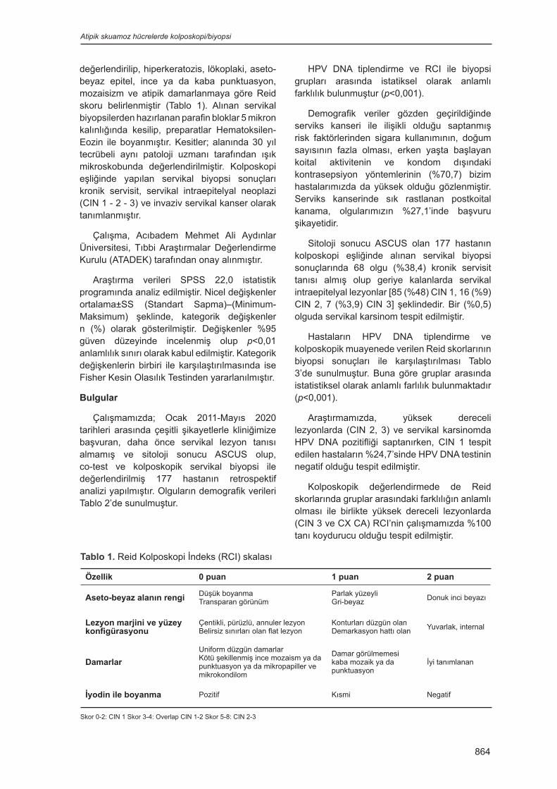
değerlendirilip, hiperkeratozis, lökoplaki, aseto-beyaz epitel, ince ya da kaba punktuasyon, mozaisizm ve atipik damarlanmaya göre Reid skoru belirlenmiştir (Tablo 1). Alınan servikal biyopsilerden hazırlanan parafin bloklar 5 mikron kalınlığında kesilip, preparatlar Hematoksilen-Eozin ile boyanmıştır. Kesitler; alanında 30 yıl tecrübeli aynı patoloji uzmanı tarafından ışık mikroskobunda değerlendirilmiştir. Kolposkopi eşliğinde yapılan servikal biyopsi sonuçları kronik servisit, servikal intraepitelyal neoplazi (CIN 1 - 2 - 3) ve invaziv servikal kanser olarak tanımlanmıştır.
Çalışma, Acıbadem Mehmet Ali Aydınlar Üniversitesi, Tıbbi Araştırmalar Değerlendirme Kurulu (ATADEK) tarafından onay alınmıştır.
Araştırma verileri SPSS 22,0 istatistik programında analiz edilmiştir. Nicel değişkenler ortalama±SS (Standart Sapma)–(Minimum-Maksimum) şeklinde, kategorik değişkenler n (%) olarak gösterilmiştir. Değişkenler %95 güven düzeyinde incelenmiş olup p<0,01 anlamlılık sınırı olarak kabul edilmiştir. Kategorik değişkenlerin birbiri ile karşılaştırılmasında ise Fisher Kesin Olasılık Testinden yararlanılmıştır.
Bulgular
Çalışmamızda; Ocak 2011-Mayıs 2020 tarihleri arasında çeşitli şikayetlerle kliniğimize başvuran, daha önce servikal lezyon tanısı almamış ve sitoloji sonucu ASCUS olup, co-test ve kolposkopik servikal biyopsi ile değerlendirilmiş 177 hastanın retrospektif analizi yapılmıştır. Olguların demografik verileri Tablo 2’de sunulmuştur.
HPV DNA tiplendirme ve RCI ile biyopsi grupları arasında istatiksel olarak anlamlı farklılık bulunmuştur (p<0,001).
Demografik veriler gözden geçirildiğinde serviks kanseri ile ilişikli olduğu saptanmış risk faktörlerinden sigara kullanımının, doğum sayısının fazla olması, erken yaşta başlayan koital aktivitenin ve kondom dışındaki kontrasepsiyon yöntemlerinin (%70,7) bizim hastalarımızda da yüksek olduğu gözlenmiştir. Serviks kanserinde sık rastlanan postkoital kanama, olgularımızın %27,1’inde başvuru şikayetidir.
Sitoloji sonucu ASCUS olan 177 hastanın kolposkopi eşliğinde alınan servikal biyopsi sonuçlarında 68 olgu (%38,4) kronik servisit tanısı almış olup geriye kalanlarda servikal intraepitelyal lezyonlar [85 (%48) CIN 1, 16 (%9) CIN 2, 7 (%3,9) CIN 3] şeklindedir. Bir (%0,5) olguda servikal karsinom tespit edilmiştir.
Hastaların HPV DNA tiplendirme ve kolposkopik muayenede verilen Reid skorlarının biyopsi sonuçları ile karşılaştırılması Tablo 3’de sunulmuştur. Buna göre gruplar arasında istatistiksel olarak anlamlı farlılık bulunmaktadır (p<0,001).
Araştırmamızda, yüksek dereceli lezyonlarda (CIN 2, 3) ve servikal karsinomda HPV DNA pozitifliği saptanırken, CIN 1 tespit edilen hastaların %24,7’sinde HPV DNA testinin negatif olduğu tespit edilmiştir.
Kolposkopik değerlendirmede de Reid skorlarında gruplar arasındaki farklılığın anlamlı olması ile birlikte yüksek dereceli lezyonlarda (CIN 3 ve CX CA) RCI’nin çalışmamızda %100 tanı koydurucu olduğu tespit edilmiştir.
Tablo 1. Reid Kolposkopi İndeks (RCI) skalası
Özellik 0 puan 1 puan 2 puan
Aseto-beyaz alanın rengi Düşük boyanmaTransparan görünüm
Parlak yüzeyliGri-beyaz Donuk inci beyazı
Lezyon marjini ve yüzey konfigürasyonu
Çentikli, pürüzlü, annuler lezyonBelirsiz sınırları olan flat lezyon
Konturları düzgün olan Demarkasyon hattı olan Yuvarlak, internal
DamarlarUniform düzgün damarlarKötü şekillenmiş ince mozaism ya da punktuasyon ya da mikropapiller ve mikrokondilom
Damar görülmemesikaba mozaik ya da punktuasyon
İyi tanımlanan
İyodin ile boyanma Pozitif Kısmi Negatif
Skor 0-2: CIN 1 Skor 3-4: Overlap CIN 1-2 Skor 5-8: CIN 2-3
Atipik skuamoz hücrelerde kolposkopi/biyopsi
864
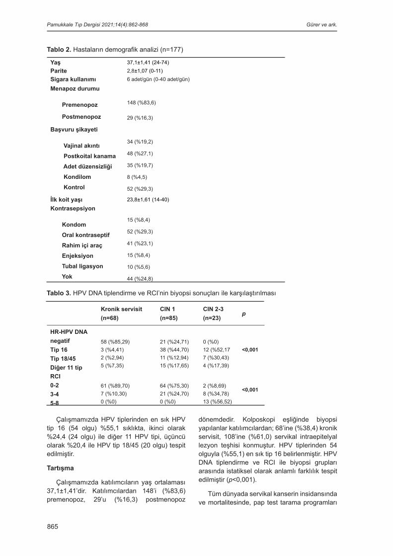
865
Çalışmamızda HPV tiplerinden en sık HPV tip 16 (54 olgu) %55,1 sıklıkta, ikinci olarak %24,4 (24 olgu) ile diğer 11 HPV tipi, üçüncü olarak %20,4 ile HPV tip 18/45 (20 olgu) tespit edilmiştir.
Tartışma
Çalışmamızda katılımcıların yaş ortalaması 37,1±1,41’dir. Katılımcılardan 148’i (%83,6) premenopoz, 29’u (%16,3) postmenopoz
dönemdedir. Kolposkopi eşliğinde biyopsi yapılanlar katılımcılardan; 68’ine (%38,4) kronik servisit, 108’ine (%61,0) servikal intraepitelyal lezyon teşhisi konmuştur. HPV tiplerinden 54 olguyla (%55,1) en sık tip 16 belirlenmiştir. HPV DNA tiplendirme ve RCI ile biyopsi grupları arasında istatiksel olarak anlamlı farklılık tespit edilmiştir (p<0,001).
Tüm dünyada servikal kanserin insidansında ve mortalitesinde, pap test tarama programları
Tablo 2. Hastaların demografik analizi (n=177)
Yaş 37,1±1,41 (24-74)Parite 2,8±1,07 (0-11)Sigara kullanımı 6 adet/gün (0-40 adet/gün)
Menapoz durumu
Premenopoz
Postmenopoz
148 (%83,6)
29 (%16,3)
Başvuru şikayeti
Vajinal akıntı Postkoital kanama Adet düzensizliği Kondilom Kontrol
34 (%19,2)
48 (%27,1)
35 (%19,7)
8 (%4,5)
52 (%29,3)
İlk koit yaşı 23,8±1,61 (14-40)
Kontrasepsiyon
Kondom Oral kontraseptif Rahim içi araç Enjeksiyon Tubal ligasyon Yok
15 (%8,4)
52 (%29,3)
41 (%23,1)
15 (%8,4)
10 (%5,6)
44 (%24,8)
Tablo 3. HPV DNA tiplendirme ve RCI’nin biyopsi sonuçları ile karşılaştırılması
Kronik servisit(n=68)
CIN 1(n=85)
CIN 2-3(n=23)
p
HR-HPV DNAnegatifTip 16Tip 18/45Diğer 11 tip
58 (%85,29)3 (%4,41)2 (%2,94)5 (%7,35)
21 (%24,71)38 (%44,70)11 (%12,94)15 (%17,65)
0 (%0)12 (%52,177 (%30,43)4 (%17,39)
<0,001
RCI0-23-45-8
61 (%89,70)7 (%10,30)0 (%0)
64 (%75,30)21 (%24,70)0 (%0)
2 (%8,69)8 (%34,78)13 (%56,52)
<0,001
Pamukkale Tıp Dergisi 2021;14(4):862-868 Gürer ve ark.

866
sayesinde ciddi bir azalma izlenmiştir. Ancak smear testinin sensivitesi %30-87 arasında değişmesi, gereksiz kolposkopi, biyopsi ve tedavilere neden olmaktadır. Bu nedenle HPV DNA testinin kullanımı gündeme gelmiştir. ACS’nin 2012 yılı kılavuzunda sitoloji taramasına HPV DNA tiplendirilmesinin eklenmesi önerilmektedir [11].
Kolposkopi; servikal lezyonların değerlendirilmesine, doğru yerden biyopsi alınmasına ve serviks kanserinin erken evrede tanı konmasına yardımcı olduğu gibi gereksiz invaziv girişimlerden kaçınılmasına olanak sağlamıştır. Her ne kadar kolposkopiyi yapan jinekoloğun deneyimine bağlı değişkenler söz konusu olsa da Reid ve Scalzi’nin skorlama sistemi sayesinde ortak bir dil oluşturulmuştur [11].
Mitchell ve ark. [12] tarafından yapılan metaanalizde kolposkopinin servikal preinvaziv ve invaziv lezyonlara tanı koymada sensitivitesi %95, spesifitesi %45, pozitif prediktif değeri %82, negatif prediktif değeri %79 olarak bulunmuştur.
Kolposkopinin tarama ve tanı testi olarak doğruluğunun karşılaştırıldığı bir çalışmada araştırmacılar tarafından, kolposkopinin servikal preinvaziv ve invaziv hastalıklarda tanı aracı olarak yüksek duyarlılığa sahip olduğunu ancak tarama testi olarak kullanılmaması gerektiğini bildirilmiştir [13].
Auger ve ark.’nın [14] çalışmasında ASCUS grubunda vakaların %54’ünde histolojik olarak patoloji tespit edilmemiş, %27 LSIL ve %10 HSIL tespit edilmiştir.
ASCUS/LSIL Triage Study for Cervical Cancer Group (ALTS grup) [15] çalışmasında 3488 ASCUS tanılı hastada yapılan randomize kontrollü çalışmanın sonucunda smear testi ile takibin yeterli olduğu, CIN 3 ve servikal karsinomun tespitinin aynı oranda başarılı olduğu gösterilmiştir.
Ancak çalışmamızda ASCUS tanısı alan hastalarında %9 CIN 2, %3,9 CIN 3 ve %0,5 serviks karsinomunu tespit etmiş olmak, takip kararını kolposkopik biyopsi sonucuna göre vermek uygun gibi görünse de klinik uygulamaya geçirmek için daha çok olgu serili çalışmalara ihtiyaç vardır.
Ülkemizde yapılan Tamburacı Demir ve ark.’nın [16] çalışmasında en sık görülen HPV tipi 16 olarak tespit edilmiş olup, literatür çalışmamızla benzerlik göstermektedir. Çalışmamızda HPV tiplerinden HPV tip 16 birinci sıklıkta, ikinci olarak diğer 11 HR-HPV tipleri, üçüncü sırada HPV tip 18/45 tespit edilmiştir.
Castle ve ark.’nın [17] çalışmasında sigara kullanımı servikal karsinom riskini yaklaşık %50 arttırdığı ortaya konulmuştur. Aynı şekilde Sarian ve ark.’nın [18] çalışmasında sigara, invaziv serviks kanseri ile ilişkili bulunmuştur. Çalışmamızın analizinde de sigara kullanımının premalign ve malign lezyonu olan hastalarda literatür ile uyumlu olarak daha yüksek oranda olduğu bulunmuştur.
Çalışmamızda servikal sitoloji sonucu ASCUS, HR-HPV DNA tarama sonucu pozitif olan 177 hastanın retrospektif analizini yaparak, Reid indeks puanları ile histopatolojik sonuçlar karşılaştırılmalı olarak değerlendirmeye alınmıştır. Özellikle serviks karsinomların tespitinde smear yetersiz kalabilmektedir. Pap smearın atladığı karsinom vakalarını HPV ile saptamak mümkündür. Bu hastalarda HPV saptama oranı %93’tür. Rijkaart ve ark.’nın [19] POBASCAM çalışmasında; HPV DNA pozitif olan 724 olgunun sitolojisi negatif tespit edilmiştir. Bu vakalara yapılan kolposkopi sonuçlarında %4,28’inde CIN 2, %4’ünde CIN 3 bulunmuştur. Amerikan Servikal Patolojiler ve Kolposkopi Derneği (ASCCP, 2019), sitolojisi ASCUS veya daha ileri displazik lezyon olan hastalarda HR-HPV varlığında doğrudan kolposkopik incelemeyi önermektedir [20].
Analizimizde Pap smear sonucu ASCUS olan olgularda HP-HPV DNA testi pozitifliğinde kolposkopik değerlendirme kaçınılmaz olarak yapılmıştır. Bununla beraber HR-HPV DNA negatif olgularda da preinvazif lezyonla karşılaştığımız için co-testteki her iki parametreden birinin pozitif çıkması durumunda kolposkopik incelemenin endike olduğu değerlendirilmektedir.
Buna ilişkin olarak yapılan güncel bir çalışmada CIN 1 tespit edilen 511 kadınlardan yaklaşık %7’sinde HPV DNA analizinin negatif bulunduğu bildirilmiştir. Araştırma sonucunda CIN 1 tespit edilen vakalardan yaklaşık %2’si CIN 3’e ilerlemiştir. Aynı konuda yapılan retrospektif bir kohort çalışmasında ise CIN 1 tespit edilen
Atipik skuamoz hücrelerde kolposkopi/biyopsi

867
434 kadınlardan yaklaşık %8,5’i HPV DNA analizinin negatif bulunduğu bildirilmiştir. CIN 1 tespit edilen vakalardan yaklaşık %7,4’ü CIN 2 ve 3’e ilerlemiştir. Burada da görüldüğü gibi her iki çalışmada da CIN 1 tespit edilen kadınlarda HPV DNA analizinin negatif olma sıklığı %7-8 bandında seyretmektedir. Bizim çalışmamızda CIN 1 tespit edilen kadınlarda HPV DNA negatifliğinin sıklığı yaklaşık %25 civarındadır. Aradaki bu farkın katılımcı sayısı ile açıklanabileceği düşünülmektedir. Her iki çalışmanın katılımcı sayısı da çalışmamızın yaklaşık üç katıdır. Bunun yanında çalışmaların her ikisinin de Avrupa’da yürütülmüş olması dolayısıyla katılımcıların sosyo-demografik özelliklerinin birbirine benzememesi söz konusu farkın kaynağı olabilir [21, 22].
Yaptığımız değerlendirmelerin tek merkezden elde edilen sonuçlara dayalı olması, HPV DNA tiplendirmesinde sadece 16, 18 ve 45’e bakılıp, diğer onkojenik HPV tiplerin genotiplendirilmemiş olması ve hasta sayısının sınırlı olması, araştırmanın kısıtlayıcı faktörleridir. Ayrıca kontrol grubu olmaması nedeniyle sensitivite, spesifisite, pozitif ve negatif prediktif değer hesaplanması istatistiksel olarak mümkün olmamıştır.
Sonuç olarak araştırmamızda ASCUS saptanan ve HPV DNA testi negatif bulunan olgularda yüksek dereceli lezyon (CIN 2, 3) saptanmamıştır. Bununla birlikte bunların yaklaşık %25’inde düşük dereceli lezyon (CIN 1) tespit edilmesi dikkat çekicidir. Servikal smear’ın sitolojik olarak incelenmesi ve eş zamanlı HPV DNA analizi anlamına gelen “co-test”in yapılmasının tanı ve tedavi planı açısından fayda sağlayacağı düşünülmektedir. Bununla birlikte CIN 1 sadece HPV negatif olgularda Pap-smear ile takip edilebilir. Kolposkopik biyopsi yapılmasına rutin klinik pratikte ihtiyaç olduğunu söylemek için daha fazla çalışmaya ihtiyaç vardır.
Çıkar ilişkisi: Yazarlar çıkar ilişkisi olmadığını beyan eder.
Kaynaklar1. Zekioglu O, Ozdemir N. Serviks kanserleri patolojisi.
Turkiye Klinikleri J Med Oncol Special Topics 2011;4:14-19.
2. Ortac F, Taskın S. Serviksin malign hastalıkları. Turkiye Klinikleri J Surg Med Sci 2007;3:55-58.
3. GCO Cancer, IARC Globocan 2018: estimated cancer incidence, mortality and prevalence worldwide in 2018. Available at: http://gco.iarc.fr/today/explore. Accessed Dec 14, 2020
4. Qiao YL. Perspective of cervical cancer prevention and control in developing countries and areas. Chin J Cancer 2010;29:1-3. https://doi.org/10.5732/cjc.009.10570
5. Ozan H. Servikal karsinogeneziste HPV’nin rolü. Turkiye Klinikleri J Gynecol Obst-Special Topics 2009;2:9-12.
6. Kitchener HC, Castle PE, Cox JT. Chapter 7: achievements and limitations of cervical cytology screening. Vaccine 2006;24:63-70. https://doi.org/10.1016/j.vaccine.2006.05.113
7. ACOG practice bulletin no 99: management of abnormal cervical cytology and histology. Obstet Gynecol 2008;112:1419-1444. https://doi.org/10.1097/AOG.0b013e318192497c
8. Türkiye Kanser Kontrol Programı. T.C. Sağlık Bakanlığı Türkiye Halk Sağlığı Kurumu Kanser Daire Başkanlığı. Erişim adresi: https://hsgm.saglik.gov.tr/depo/birimler/kanser-db/yayinlar/raporlar/Ulusal_Kanser_Kontrol_Plani_2013_2018.pdf. Erişim tarihi 14 Aralık 2020
9. Huh WK, Ault KA, Chelmow D, et al. Use of primary high-risk human papillomavirus testing for cervical cancer screening: interim clinical guidance. Gynecol Oncol 2015;136:178-182. https://doi.org/10.1016/j.ygyno.2014.12.022
10. Burghardt E, Histopathologic basis of colposcopy. In: Colposcopy Cervical Pathology Textbook and Atlas, 4th ed. New York, NY: Georg Thieme Verlag 2015;24-56.
11. Saslow D, Solomon D, Lawson HW, et al. American cancer society, American society for colposcopy and cervical pathology, and American society for clinical pathology screening guidelines for the prevention and early detection of cervical cancer. Am J Clin Pathol 2012;137:516-542. https://doi.org/10.1309/AJCPTGD94EVRSJCG
12. Mitchell MF, Schottenfeld D, Tortolero Luna G, Cantor SB, Richards Kortum R. Colposcopy for the diagnosis of squamous intraepithelial lesions: a meta-analysis. Obstet Gynecol 1998;91:626-631. https://doi.org/10.1016/S0029-7844(98)00006-4
13. Cantor SB, Cárdenas Turanzas M, Cox DD, et al. Accurary of colposcopy in the diagnostic setting compared with the screening setting. Obstet Gynecol 2008;111:7-14. https://doi.org/10.1097/01.AOG.0000295870.67752.b4
14. Auger M, Charbonneau M, Arseneau J. Atypical squamous cells of undetermined significance. A cytohistologic study of 52 cases. Acto Cytol 1997;41:1671-1675. https://doi.org/10.1159/000333166
Pamukkale Tıp Dergisi 2021;14(4):862-868 Gürer ve ark.
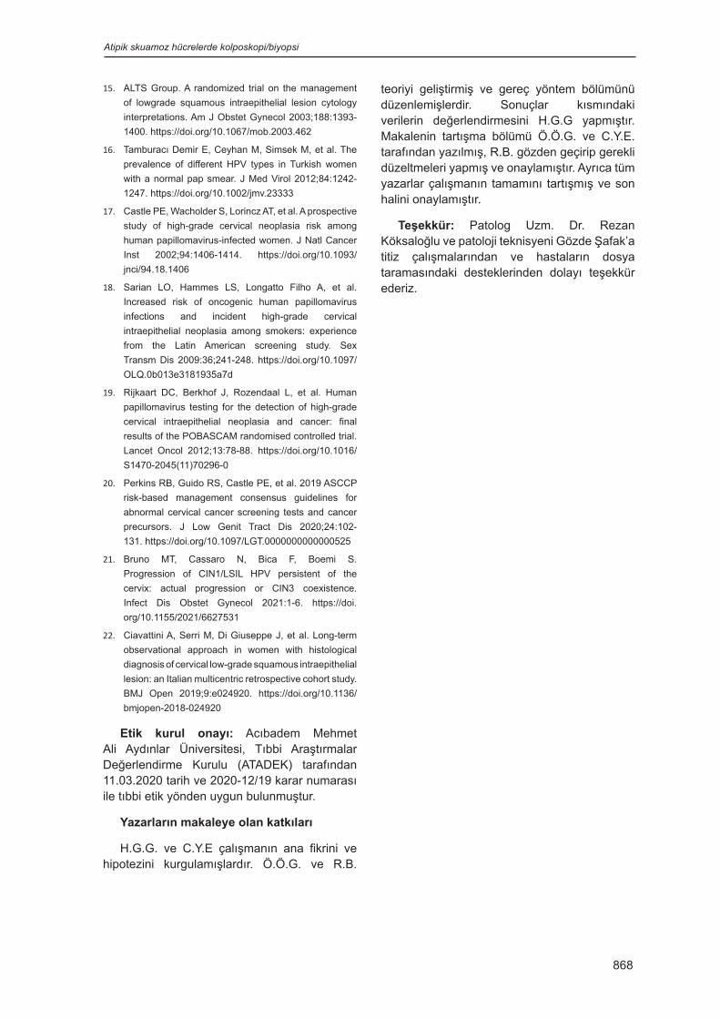
868
15. ALTS Group. A randomized trial on the management of lowgrade squamous intraepithelial lesion cytology interpretations. Am J Obstet Gynecol 2003;188:1393-1400. https://doi.org/10.1067/mob.2003.462
16. Tamburacı Demir E, Ceyhan M, Simsek M, et al. The prevalence of different HPV types in Turkish women with a normal pap smear. J Med Virol 2012;84:1242-1247. https://doi.org/10.1002/jmv.23333
17. Castle PE, Wacholder S, Lorincz AT, et al. A prospective study of high-grade cervical neoplasia risk among human papillomavirus-infected women. J Natl Cancer Inst 2002;94:1406-1414. https://doi.org/10.1093/jnci/94.18.1406
18. Sarian LO, Hammes LS, Longatto Filho A, et al. Increased risk of oncogenic human papillomavirus infections and incident high-grade cervical intraepithelial neoplasia among smokers: experience from the Latin American screening study. Sex Transm Dis 2009:36;241-248. https://doi.org/10.1097/OLQ.0b013e3181935a7d
19. Rijkaart DC, Berkhof J, Rozendaal L, et al. Human papillomavirus testing for the detection of high-grade cervical intraepithelial neoplasia and cancer: final results of the POBASCAM randomised controlled trial. Lancet Oncol 2012;13:78-88. https://doi.org/10.1016/S1470-2045(11)70296-0
20. Perkins RB, Guido RS, Castle PE, et al. 2019 ASCCP risk-based management consensus guidelines for abnormal cervical cancer screening tests and cancer precursors. J Low Genit Tract Dis 2020;24:102-131. https://doi.org/10.1097/LGT.0000000000000525
21. Bruno MT, Cassaro N, Bica F, Boemi S. Progression of CIN1/LSIL HPV persistent of the cervix: actual progression or CIN3 coexistence. Infect Dis Obstet Gynecol 2021:1-6. https://doi.org/10.1155/2021/6627531
22. Ciavattini A, Serri M, Di Giuseppe J, et al. Long-term observational approach in women with histological diagnosis of cervical low-grade squamous intraepithelial lesion: an Italian multicentric retrospective cohort study. BMJ Open 2019;9:e024920. https://doi.org/10.1136/bmjopen-2018-024920
Etik kurul onayı: Acıbadem Mehmet Ali Aydınlar Üniversitesi, Tıbbi Araştırmalar Değerlendirme Kurulu (ATADEK) tarafından 11.03.2020 tarih ve 2020-12/19 karar numarası ile tıbbi etik yönden uygun bulunmuştur.
Yazarların makaleye olan katkıları
H.G.G. ve C.Y.E çalışmanın ana fikrini ve hipotezini kurgulamışlardır. Ö.Ö.G. ve R.B.
teoriyi geliştirmiş ve gereç yöntem bölümünü düzenlemişlerdir. Sonuçlar kısmındaki verilerin değerlendirmesini H.G.G yapmıştır. Makalenin tartışma bölümü Ö.Ö.G. ve C.Y.E. tarafından yazılmış, R.B. gözden geçirip gerekli düzeltmeleri yapmış ve onaylamıştır. Ayrıca tüm yazarlar çalışmanın tamamını tartışmış ve son halini onaylamıştır.
Teşekkür: Patolog Uzm. Dr. Rezan Köksaloğlu ve patoloji teknisyeni Gözde Şafak’a titiz çalışmalarından ve hastaların dosya taramasındaki desteklerinden dolayı teşekkür ederiz.
Atipik skuamoz hücrelerde kolposkopi/biyopsi


870
R202Q gen değişikliğinin ailesel Akdeniz ateşi kliniği üzerine etkisi: tek merkez deneyimi
The effects of R202Q genotype on the clinical features of familial Mediterranean fever: single center experience
Serkan Türkuçar, Hatice Adıgüzel Dundar, Ceren Yılmaz, Erbil Ünsal
Pamukkale Tıp DergisiPamukkale Medical JournalAraştırma Makalesi
Serkan Türkuçar, Uzm. Dr. Dokuz Eylül Üniversitesi Tıp Fakültesi, Çocuk Sağlığı ve Hastalıkları Anabilim Dalı, Çocuk Romatolojisi Bilim Dalı, İzmir, Türkiye, e-posta: [email protected] (https://orcid.org/0000-0003-4700-1361) (Sorumlu Yazar)Hatice Adıgüzel Dundar, Uzm. Dr. Dokuz Eylül Üniversitesi Tıp Fakültesi, Çocuk Sağlığı ve Hastalıkları Anabilim Dalı, Çocuk Romatolojisi Bilim Dalı, İzmir, Türkiye, e-posta: [email protected] (https://orcid.org/0000-0003-1469-9900)Ceren Yılmaz, Uzm. Dr. Dokuz Eylül Üniversitesi Tıp Fakültesi, Çocuk Sağlığı ve Hastalıkları Anabilim Dalı, Çocuk Genetik Bilim Dalı, İzmir, Türkiye, e-posta: [email protected] (https://orcid.org/0000-0002-0853-3253)Erbil Ünsal, Prof. Dr. Dokuz Eylül Üniversitesi Tıp Fakültesi, Çocuk Sağlığı ve Hastalıkları Anabilim Dalı, Çocuk Romatolojisi Bilim Dalı, İzmir, Türkiye, e-posta: [email protected] (https://orcid.org/0000-0002-8800-0800)
ÖzAmaç: Ailesel Akdeniz ateşinin kliniğinde görülen tekrarlayan febril poliserozit atakları, MEFV genindeki mutasyonlara bağlı kontrolsüz interlökin 1-β salınımından kaynaklanmaktadır ve hastalığın seyri mutasyonlara göre değişkenlik gösterebilmektedir. Bu çalışmada R202Q gen değişiminin klinik ve laboratuvar bulguları üzerindeki etkisinin değerlendirmesi amaçlanmıştır. Gereç ve yöntem: 122 pediatrik AAA hastasının demografik, klinik ve laboratuvar verileri kıyaslanmıştır. Hastalar MEFV mutasyonlarına göre R202Q değişimini heterozigot, homozigot, ekzon-10 mutasyonları ile bileşik heterozigot bulunduranlar ve sadece ekzon-10 mutasyonu bulunduranlar olarak gruplandırılarak karşılaştırılmıştır.Bulgular: Demografik, antropometrik, klinik ve laboratuvar bulgular açısından değerlendirildiğinde, gruplar arasında herhangi bir istatistiksel anlamlı fark saptanmamıştır.Sonuç: R202Q gen değişikliğine sahip hastalarda da ekzon 10 mutasyonlu hastalar gibi tipik AAA klinik bulguları gözlenebilmektedir. Ailesel Akdeniz ateşi, genotipik ve fenotipik özelliklerinin birlikte değerlendirilmesi gereken bir hastalıktır.
Anahtar kelimeler: MEFV mutasyonu, ekzon-2, ailesel Akdeniz ateşi, R202Q.
Türkuçar S, Adıgüzel Dundar H, Yılmaz C, Ünsal E. R202Q gen değişikliğinin ailesel Akdeniz ateşi kliniği üzerine etkisi: tek merkez deneyimi. Pam Tıp Derg 2021;14:870-877.
AbstractPurpose: Recurrent febrile polyserositis attacks of familial Mediterranean fever are caused by uncontrolled interleukin 1-β release due to mutations in the MEFV gene and the course of the disease may vary according to mutations. In this study, it was aimed to evaluate the effect of R202Q genotypes on clinical and laboratory findings. Materials and methods: Demographic, clinical and laboratory data of 122 pediatric FMF patients were compared. The patients were grouped according to the MEFV mutations as heterozygous and homozygous R202Q, compound heterozygous with exon-10 mutations and R202Q genotypes, and those with only exon-10 mutations compared.Results: In terms of demographic, anthropometric, clinical and laboratory findings, there was no statistically significant difference between the groups.Conclusion: Typical clinical findings can be observed in patients with R202Q gene alterations, as in patients with exon 10 mutations. Familial Mediterranean fever is a disease that should be evaluated together with its genotypic and phenotypic characteristics.
Key words: MEFV mutations, exon-2, familial Mediterranean fever, R202Q.
Turkucar S, Adiguzel Dundar H, Yilmaz C, Unsal E. The effects of R202Q genotype on the clinical features of familial Mediterranean fever: single center experience. Pam Med J 2021;14:870-877.
Research Article
Gönderilme tarihi:23.02.2021 Kabul tarihi:15.04.2021
doi:https://dx.doi.org/10.31362/patd.885049

871
Giriş
Tek gen geçişli herediter otoinflamatuvar hastalıkların en sık alt tipi olan ailesel Akdeniz ateşi (AAA), ülkemiz başta olmak üzere Doğu Akdeniz Bölgesi’nden köken alan toplumlarda oldukça sık görülür [1]. Kliniğinde kendini sınırlandıran ve tekrarlayan ataklar halinde gelen ateşin eşlik ettiği karın, göğüs ya da eklem ağrısı şikayetleri görülür. Bu ataklara neden olan abartılı inflamasyonun altında interlökin 1-β (IL 1-β) nın kontrolsüz salgılanması yatar [2].
Hastalığa neden olan genetik mutasyon 1997 yılında 16. kromozomun kısa kolunda bulunan MEFV (MEditerranean FeVer) geninde tanımlanmıştır [3, 4]. Bu gen pro-IL 1-β nın, aktif olan formuna dönüşme basamaklarında kısıtlayıcı rol oynayan pyrin adlı proteini kodlar. Günümüze kadar MEFV geninde ekzon 2, 3, 5 ve 10 da olmak üzere 379 farklı değişiklik bildirilmiştir [5]. Bu genetik değişikliklerin dağılımı ve çeşitliliği toplumlara göre farklılık gösterebilirken; ülkemizde en sık rastlananlar ekzon 10 da bulunan M694V, M680I ve V726A mutasyonları ile ekzon 2 de bulunan R202Q ve E148Q değişiklikleridir [6-8]. Onuncu ekzon mutasyonları ile hastalık belirtileri ve ciddiyeti arasında ilişkiler literatürde iyi tanımlanmış olmakla birlikte, R202Q gibi sık rastlanan değişikliklerin hastalık üzerine etkisi net bilinmemektedir [9-11]. İkinci ekzonun 605. pozisyondaki Guanin yerine Adenin geçmesi ile oluşan (c.605G>A) R202Q gen değişimi (p.Arg202Gln), ilk olarak 1998 de Bernot ve ark. [12] tarafından polimorfizm olarak tanımlanmıştır. Fakat daha sonraki araştırmalarda bu polimorfizmin sağlıklı popülasyonla karşılaştırıldığında AAA hastalarında daha sık gözlenmesi, hastalık yapıcı bir genetik değişiklik olabileceğini düşündürmüştür [13-15]. Bununla birlikte, günümüzde infevers genetik veri tabanında R202Q’nun M694V mutasyonu ile bağlantısının genetik dengesizliğe yol açabileceği bildirilmektedir [16].
Bu çalışmanın amacı, toplumumuzda sıklıkla görülmekle birlikte, hastalığın seyri üzerindeki etkisi net tanımlanmamış olan R202Q değişimini, çocuk AAA hastalarımızın klinik ve laboratuvar bulguları üzerinden değerlendirmektir.
Gereç ve yöntem
Çalışma için, Dokuz Eylül Üniversitesi Girişimsel Olmayan Etik Kurulu’ndan etik onayı alınmıştır.
Hastalar
Üçüncü Basamak Sağlık Merkezimizin Çocuk Romatoloji Bilim Dalı’nda 1990 ve 2020 yılları arasında AAA tanısı ile takip edilen 122 pediatrik yaş grubu hastanın kayıtları geriye dönük olarak incelenmiştir. 6-72 saat arası süren tipik ateş, karın ağrısı, göğüs ağrısı ve artrit ataklarına ek olarak AAA aile öyküsünden oluşan Ankara klinik tanı kriterlerinden en az ikisini karşılayanlar tanı alarak çalışmaya dahil edilmiştir [17]. Çalışmaya dâhil edilen hastalar, daha önceden çalışılmış MEFV gen analizi değerlendirmesinde R202Q değişimi bulundurmalarına göre gruplara ayrılmıştır. Toplam 1100 pediatrik AAA hasta dosyasının içinden 20 adet R202Q heterozigot, 20 adet R202Q homozigot, 52 adet R202Q ve ekzon-10 mutasyonunu birlikte bulunduran ve 30 adet sadece ekzon 10 mutasyonu bulunduran olmak üzere değerlendirilmiştir. Toplam 122 hasta dosyası, her bir grup içinden rastgele seçilerek çalışmaya alınmıştır.
Hastalar MEFV genlerindeki ekzon 10 mutasyonları ve R202Q gen değişimlerine göre, R202Q heterozigot hastalar: grup 1; R202Q homozigot hastalar: grup 2, R202Q ve ekzon 10 mutasyonlarının birlikte bulunduğu hastalar: grup 3 ve ekzon 10 mutasyonu olup R202Q değişimi bulunmayanlar ise: grup 4 olarak sınıflandırılmıştır.
Klinik ve demografik veriler
Tüm hastaların dosyalarından ve elektronik veri kayıt sistemindeki verilerden elde edilen yaş, cinsiyet, son ziyaretlerindeki vücut ağrılığı ve boy, ilk atak yaşı, kolşisin başlanma zamanı, tanıda gecikme süresi ve ataklardaki şikâyetleri gibi demografik ve klinik bilgileri not edilmiştir. Vücut ağırlığı ve boy değerlerinin yaşa uygun şekilde Neyzi ve ark. [18] verilerine göre persentilleri hesaplanmıştır. Ayrıca tüm hastaların ataklar arasında eklem şikâyeti olup olmadığı, tekrarlayan tonsillit ve apendektomi öyküleri, Pras ve ark.’nın [19] skorlama sistemine göre hesaplanmış AAA ciddiyet skorları ve kullanılan günlük kolşisin dozları kaydedilmiştir. Elektronik hasta kayıt sisteminden elde edilen veriler üzerinden, tüm hastaların atak arası dönemde bakılan en son hemogram parametreleri [hemoglobin (Hb), beyaz küre (BK) ve platelet (Plt) sayıları ile nötrofil/lenfosit oranları (NLR)] kaydedilmiştir. Ayrıca, eş zamanlı bakılan akut faz yanıtları [eritrosit sedimantasyon hızı (ESH)
Pamukkale Tıp Dergisi 2021;14(4):870-877 Türkuçar ve ark.

R202Q gen değişikliğinin ailesel Akdeniz ateşi kliniği üzerine etkisi
872
ve C-reaktif protein düzeyleri (CRP)], karaciğer ve böbrek fonksiyon testleri [kreatinin (Cr) ve alanin aminotransferaz (ALT)] ve idrarda protein atılımları da not edilmiştir.
Genetik analiz
Ailesel Akdeniz ateşi klinik tanısı ile çalışmaya dâhil ettiğimiz 122 hastanın tamamının MEFV gen analiz sonuçları değerlendirmeye alınmıştır. Genetik analizlerin tümü, periferik kandan elde edilen DNA örneklerini Real Time Polimeraz Zincir Reaksiyon (RT-) PCR yöntemiyle, Cobas z480 RT-PCR (Roche, Almanya) cihazı ve LightSNIP Assay (TIB Molbiol, Almanya) kiti ile değerlendirilerek yapılan analizlerdir. Mevcut yöntemle, AAA ile ilişkili olarak değerlendirilenden en sık 9 mutasyon (E148Q, R202Q, M680I, M694V, M694I, K695R, V726A, R761H ve A744S) kaydedilmiştir.
İstatistiksel değerlendirme
İstatistiksel analiz SPSS 22 yazılımı kullanılarak yapılmış olup, değerlerin homojen dağılıp dağılmadığı Kolmogorov-Smirnov testi ile incelenmiştir. Homojen dağılmış değerler ortalama ± standart sapma olarak belirtilirken, heterojen dağılmış değerler medyan ve yüzde 25 ve 75 değerleri ile belirtilmiştir. Gruplar arasındaki kategorik farklılıkları tanımlamak için Ki-Kare testi kullanılmıştır. Parametrik değerleri karşılaştırmak için bağımsız örneklem-T testi, parametrik olmayan değerler için ise Mann-Whitney U testi kullanılmıştır. P<0,05 istatistiksel olarak anlamlı kabul edilmiştir.
Bulgular
Çalışmaya dâhil ettiğimiz 122 hastanın 60’ı kız (%49,2), 62’si erkek (%50,8) iken; ortalama yaşları 12,25 (9,75-13,45) yıldı. Hastaların ortanca ilk atak yaşı 4,5 (2,5-7,5) yıl, ortanca kolşisin başlama yaşı 6 (4-10) yıl ve ortalama tanıda gecikme süreleri 12 (6-24) ay olarak hesaplanmıştır.
Demografik veriler açısından bakıldığında hastaların hiçbirinde gelişim geriliği saptanmazken; ortanca vücut ağırlık persentilleri %56,5 (28,2-88,2) ve boy persentilleri %75 (39-89) olarak hesaplanmıştır.
Atakların en sık semptomu olan ateş hastaların 104’ünde (%85,2) görülürken, takiben 93 hastada (%76,2) karın ağrısı, 50 hastada (%41) artralji, 23 hastada (%18,9)
artrit ve yalnızca 8 hastada (%6,6) göğüs ağrısı belirtilmiştir. Hastaların %28,7'sinde (n=35) is ataklar arasında artralji ve miyalji gibi kas iskelet şikâyetleri bildirilirken, ortalama günlük kullanılan kolşisin dozu 1,0 mg olarak saptanmıştır. Ayrıca hastaların tüm laboratuvar verileri Tablo 1’de verilmiştir. Hastaların taşıdıkları MEFV gen değişiklikleri Tablo 2’de detaylı olarak belirtilmişitr.
Demografik, antropometrik, klinik ve laboratuvar bulgular açısından değerlendirildiğinde, MEFV genlerine göre oluşturulan gruplar arasında herhangi bir istatistiksel anlamlı fark saptanmadı, C-reaktif protein değeri üçüncü grupta diğerlerine göre yüksek saptansa da normal sınırlar içinde (0,5-5 mg/L) olduğundan anlamlı kabul edilmedi (Tablo 3).
Tartışma
Ailesel Akdeniz ateşi, MEFV genindeki çok sayıdaki genetik değişikliğin patogenezde rol alabileceği ve farklı genotipik özelliklerin farklı klinik bulgulara yol açabileceği bir hastalıktır. Bu çalışmamızda, klinik üzerindeki etkisi tartışmalı olan R202Q gen değişikliğinin AAA belirtilerine neden olabildiğini; hatta ekzon 10 mutasyonlarıyla kıyaslandığında atak belirtileri, hastalık ciddiyet skorları, atak arası inflamasyon parametreleri ve tedavi dozu gereksinimleri açısından benzer özelliklere sahip olduğu saptanmıştır.
İlk saptandığı dönemlerde genetik polimorfizm olarak tanımlanan R202Q değişikliğinin önemi ile ilgili bildirilen çalışmalarda birbiriyle çelişen bilgiler raporlanmıştır. Ritis ve ark. [13], 26 AAA’lı Yunan hastanın 4’ünde homozigot R202Q değişikliği saptarken, 60 sağlıklı kontrolün hiçbirinde R202Q saptamamışlar ve bu genetik değişikliğin bir mutasyon olabileceğini öne sürmüşlerdir. Ülkemizden Öztürk ve ark.’nın [14] bildirdiği bir çalışmada ise, hasta ve sağlıklı kontrol gruplarında benzer oranda heterozigot R202Q değişikliği saptanmasına rağmen, homozigot değişikliğin sağlıklı grupta daha az olduğu raporlanmıştır. Mevcut sonuçlarla heterozigot değişikliklerin polimorfizm, homozigot değişikliklerin ise en azından bazı hastalarda klinik ile ilişkili olduğu iddia edilmiştir. Benzer şekilde Yiğit ve ark.’ları [15], R202Q heterozigot değişiminin AAA ve sağlıklı gruplarda benzer oranda olduğunu,

873
Tablo 1. Hastaların demografik, klinik ve laboratuvar verileri (n=122)
Cinsiyet (kız/erkek) 60/62
Yaş (yıl) 8,4 (6,7-11,5)
İlk atak yaşı (yıl) 4,5 (2,5-7,5)
Tedavi başlama yaşı (yıl) 6 (4-10)
Tanıda gecikme süresi (ay) 12 (6-24)
Ailede AAA öyküsü (1° ve 2° akrabalarda) 49 (%40,2)
Akraba evliliği öyküsü (1° ve 2° akrabalık) 7 (%5,7)
VA (kg) 25,5 (19,4-46,3)
Boy (cm) 128,5 (111-153)
VA (persentil) (%) 56,5 (28,2-88,2)
Boy (persentil) (%) 75 (39-89)
Atak belirtileri
• Ateş 104 (%85,2)
• Karın ağrısı 93 (%76,2)
• Artrit 23 (%18,9)
• Artralji 50 (%41)
• Göğüs ağrısı 8 (%6,6)
Atak arası kas iskelet şikâyetleri 35 (%28,7)
Tekrarlayan tonsillit öyküsü 21 (%17,2)
Apendektomi öyküsü 4 (%3,3)
Laboratuvar Parametreleri
• BK (103/uL) 7625±2043*
• Hb (g/dL) 12,6±1,2*
• Plt (103/uL) 320,2±78,9*
• CRP (mg/L) 0,9 (0,4-3,7)
• ESH (mm/sa) 7 (3-14)
• ALT (U/L) 18 (13-25)
• Cr (mg/dL) 0,4 (0,3-0,5)
Ortanca (25-75, persentiller),*Ortalama ± standart sapmaAAA: Ailesel Akdeniz ateşi, VA: vücut ağırlığı, BK: beyaz küre, Hb: hemoglobin, Plt: platelet sayısı, CRP: C-reaktif protein, ESH: eritrosit sedimantasyon hızı, ALT: alanin aminotransferaz, Cr: kreatinin
Pamukkale Tıp Dergisi 2021;14(4):870-877 Türkuçar ve ark.

874
Tablo 2. MEFV gen mutasyonlarının dağılımı
M694V/R202Q 26 (%21,3)
R202Q/R202Q 20 (%16,4)
R202Q 20 (%16,4)
M694V 8 (%6,5)
M694V/M694V/R202Q/R202Q 6 (%4,9)
M694V/M694V/R202Q 5 (%4,1)
M694V/M680I/R202Q 5 (%4,1)
V726A 5 (%4,1)
M694V/M694V 4 (%3,3)
M694V/R202Q/R202Q 3 (%2,5)
M694V/R202Q/E148Q 2 (%1,6)
A744S 3 (%2,5)
R761H 2 (%1,6)
M694V/M680I 1 (%0,8)
M694V/R761H 1 (%0,8)
M694V/V726A/R202Q 1 (%0,8)
M694V/A744S/R202Q 1 (%0,8)
M680I /A744S/E148Q 1 (%0,8)
M680I/R761H 1 (%0,8)
M680I 1 (%0,8)
M680I/V726A 1 (%0,8)
M680I/E148Q 1 (%0,8)
M680I/R202Q 1 (%0,8)
K695R 1 (%0,8)
A744S/E148Q 1 (%0,8)
V726A/R202Q 1 (%0,8)
homozigot değişikliğin ise sağlıklı grupta daha az (%14,7 ye karşı %2,7) olduğunu bildirmişlerdir. Sönmezgöz ve ark.’nın [20] 2019’da yayınlanan bir çalışmasında da R202Q’nun M694V mutasyonu ile birlikte en sık görülen MEFV gen değişikliği olduğunu ve bu genotipin göğüs ağrısı görülme riskini arttırdığını bildirmişlerdir. Sonuç olarak R202Q gen değişikliğinin hastalık yapıcı bir mutasyon olduğunu ileri sürmüşlerdir. Celep ve ark.’nın [7] Anadolu’nun kuzey bölgesinden bildirdiği benzer bir çalışmada da, AAA klinik tanısı ile takip ettikleri 379 çocuk hastada en sık saptanan MEFV gen değişikliğinin R202Q olduğunu rapor etmişlerdir (%50,1). Mevcut çalışmamızın sonuçları da Ankara kriterleri ile AAA tanısı koyarak takip ettiğimiz ve atakları kolşisin tedavisi ile kontrol altına alınan R202Q gen değişimi olan çocuk hastalarımızın anlamlı klinik bulguları olduğunu göstermektedir. Ayrıca, hem homozigot hem de heterozigot R202Q değişikliğinin kliniğe benzer oranda yansıyan bulgulara neden olabileceği gösterilmiştir.
Hastalığın genotipik özelliklerindeki değişiklikler, çevresel etmenler ve epigenetik farklılıklarla birleştiğinde fenotipik özelliklerde çeşitliliğe yol açmaktadır [21]. Literatürde özellikle ekzon 10 mutasyonlarının erken yaşta şiddetli ataklar, ağır eklem şikâyetleri ve artmış amiloidoz riski ile ilişkilendirildiği gösterilmiştir [6-11]. Ekzon 2’deki R202Q ve E148Q gibi değişikliklerin de klinik bulgular üzerinde etkisi olduğu, fakat daha hafif hastalık seyrine yol açtığını bildiren birçok çalışma literatürde mevcuttur. Çomak ve ark. [22] bir çalışmasında, homozigot R202Q değişikliği bulunan çocuk hastalarda AAA kliniği görüldüğü, fakat belirtilerin daha geç yaşlarda başladığı ve hafif kliniğe sebep olduğu rapor edilmiştir. Yakın zamanda Milenkoviç ve ark. [23] bildirdiği diğer bir çalışmada da homozigot R202Q değişikliğinin, E148Q (ekzon-2) ve K695R (ekzon-10) gen değişikliklerine göre daha sık ateşli karın ağrısı ataklarına ve daha fazla halsizlik belirtilerine neden olduğu gösterilmiştir. Mevcut çalışmadan
R202Q gen değişikliğinin ailesel Akdeniz ateşi kliniği üzerine etkisi

875
Tablo 3. MEFV gen analiz sonuçlarına göre oluşturulan grupların demografik, antropometrik, klinik ve laboratuvar verileri açısından kıyaslanması
Grup 1R202Q +/- n=20
Grup 2R202Q +/+n=20
Grup 3R202Q (+)/ekzon 10 (+) n=52
Grup 4Ekzon 10 (+)/R202Q (-)n=30
p değeri
Cinsiyet (k/e) (n) 10/10 7/13 28/24 15/15 0,56
İlk atak yaşı (yıl) Median (%25-75)
4 (2-7,5) 3,5 (3-10) 5 (2,5-8) 4,2 (3-5,6) 0,12
Kolşisin başlama yaşı (yıl)Median (%25-75)
5 (3,5-11) 6,5 (4,3-11) 6,6 (3,5-10) 5,5 (4-9) 0,22
Tanıda gecikme süresi (ay)Median (%25-75)
12 (6-26) 20 (12-33) 10 (4-18) 12 (8-32) 0,42
VA persentil (%)Median (%25-75)
58 (36-96) 40 (12,3-80) 52 (27-82) 55 (36-78) 0,28
Boy persentil (%) Median (%25-75)
85 (55-99) 65 (24-85) 72 (36-89) 72 (35-88) 0,27
AAA aile öyküsü [n (%)]
8 (%40) 4 (%25) 23 (%44) 14 (%46) 0,23
Atak belirtileri
• Ateş [n (%)] 20 (%70) 16 (%80) 46 (%88) 28 (%93) 0,15
• Karın ağrısı [n (%)] 13 (%65) 16 (%80) 40 (%76) 24 (%80) 0,14
• Artrit [n (%)] 5 (%20) 3 (%15) 9 (%17) 6 (%25) 0,67
• Artralji [n (%)] 9 (%45) 6 (%30) 21 (%40) 14 (%47) 0,91
• Göğüs ağrısı [n (%)] 2 (%10) 1 (%5) 1 (%2) 4 (%13) 0,21
Atak arası kas-iskelet şikâyetleri [n (%)]
7 (%35) 3 (%15) 14 (%27) 11 (%36) 0,77
Tekrarlayan tonsillit öyküsü[n (%)]
6 (%30) 4 (%20) 7 (%13) 4 (%13) 0,10
Apendektomi öyküsü [n (%)] 1 (%5) 0 3 (%5) 0 0,78
AAA ciddiyet skoru Median (%25-75)
6 (4-7,3) 5 (4-6,4) 6 (5-7,2) 6 (5-6,5) 0,62
Kolşisin dozu (mg/gün)Median (%25-75)
1,0 (0,6-1,0) 1,0 (1,0-1,0) 1,0 (0,5-1,0) 1,0 (1,0-1,0) 0,44
Laboratuvar
• BK [Mean±SD] 7506±1510 6050±1397 7713±2406 7470±1760 0,50
• Hb [Mean±SD] 12,78±0,95 12,62±1,62 12,6±1,32 12,7±1,0 0,41
• Plt [Mean±SD] 313,2±77,1 335±44,8 321,2±89,2 310,7±72,9 0,42
• NLR Median (%25-75) 1,5 (0,85-1,87) 1,1(0,75-1,6) 1,2 (0,9-1,6) 1,2 (0,7-1,4) 0,38
• CRP Median (%25-75) 0,4 (0,3-0,9)a 0,55 (0,2-1,8)a 1,55 (0,62-4,57) b 0,7 (0,3-3,1)a 0,03
• ESH Median (%25-75) 4 (2-12,75) 8,5 (8-11,25) 7 (4-17,75) 5 (3-10) 0,23
• ALT Median (%25-75) 17 (13,25-25) 17,5 (12,2-34,75) 18 (13-25) 20 (13-24) 0,99
• Cr Median (%25-75) 0,4 (0,3-0,5) 0,46 (0,43-0,75) 0,4 (0,33-0,5) 0,44 (0,34-0,54) 0,36
AAA: Ailesel Akdeniz ateşi, VA: vücut ağırlığı, BK: beyaz küre, Hb: hemoglobin, Plt: platelet sayısı, NLR: nötrofil/lenfosit oranı, CRP: C-reaktif protein, ESH: eritrosit sedimantasyon hızı, ALT: alanin aminotransferaz, Cr: kreatinin “a” ve “b” grupları arasında istatistiksel anlamlı fark vardır.
Pamukkale Tıp Dergisi 2021;14(4):870-877 Türkuçar ve ark.

876
elde ettiğimiz veriler bize R202Q değişikliğini en az bir allelinde bile taşıyan hastaların, her iki allelinde bulunduran ve ekzon-10 mutasyonu olan hastalara benzer atak ve atak arası belirtilerine ve hastalık ciddiyet skorlarına sahip olduğunu göstermektedir. Literatür verilerinin aksine çalışmamızın verileri, bu hasta grubunun ekzon-10 mutasyonu taşıyan grup ile benzer hastalık başlangıç yaşına sahip olduğunu göstermektedir.
Hastalığın kliniğinde görülen ateşli poliserozit ataklarının buzdağının görünen yüzü olduğu, bunun dışında ataklar arası dönemde de süreğen bir inflamasyonun olduğu son yıllardaki çalışmalarda öne sürülmüştür [24, 25]. Tedaviye direnç, ataklar arası yüksek akut faz yanıtları gibi risk faktörleri sıklıkla M694V homozigot mutasyonlu hastalarda süreğen inflamasyon ile ilişkilendirilmektedir [24]. Bununla birlikte Balkarlı ve ark. [26], R202Q gen değişimi bulunan hastalarında artmış metabolik sendrom riski saptamışlar (atfedilmiş risk: 4,42) ve bu durumu süreğen inflamasyon ile ilişkilendirmişlerdir. Benzer şekilde, Fentoğlu ve ark. [27] çalışmasında da, R202Q nun M694V mutasyonu ile birlikte oluşturduğu süreğen inflamasyonun, kronik periodontit riskinde istatistiksel anlamlı bir artışa neden olduğu öne sürülmüştür. Bu süreğen inflamasyonun daha geniş ve çok merkezli hasta popülasyonunda değerlendirilmesi, R202Q mutasyonun klinik üzerine etkisi ile ilgili kapsamlı bilgi edinmemizi sağlayacaktır.
Sonuç olarak, R202Q gen değişikliğine sahip hastalarda da ekzon 10 mutasyonlu hastalar gibi tipik AAA klinik bulguları gözlenebilmektedir. Ailesel Akdeniz ateşi, genotipik ve fenotipik özelliklerinin birlikte değerlendirilmesi gereken bir hastalıktır. Genetik odaklı çalışmalarda elde edilen farklı sonuçlar çevresel etkenler ve epigenetik faktörlerin de etkin değişkenler olabileceğini düşündürmektedir. Konu ile ilgili daha kapsamlı çalışmalar, klinik çeşitliliği anlamamızda fayda sağlayacaktır.
Çıkar ilişkisi: Yazarlar arasında çıkar ilişkisi olmadığını beyan eder.
Kaynaklar 1. Özen S. Update on the epidemiology and disease
outcome of familial Mediterranean fever. Best Pract Res Clin Rheumatol 2018;32:254-260. https://doi.org/10.1016/j.berh.2018.09.003
2. Özen S, Batu ED, Demir S. Familial Mediterranean Fever: recent developments in pathogenesis and new recommendations for management. Front Immunol 2017;8:253. https://doi.org/10.3389/fimmu.2017.00253
3. Ancient missense mutations in a new member of the RoRet gene family are likely to cause familial Mediterranean fever. The International FMF Consortium. Cell 1997;90:797-807. https://doi.org/10.1016/s0092-8674(00)80539-5
4. French FMF Consortium. A candidate gene for familial Mediterranean fever. Nat Genet 1997;17:25-31. https://doi.org/10.1038/ng0997-25
5. Infevers genetical database. Available at: https://https://infevers.umai-montpellier.fr/web/search.php?n=1. Accessed Jan 14, 2021
6. Yaşar Bilge Ş, Sarı İ, Solmaz D, et al. The distribution of MEFV mutations in Turkish FMF patients: multicenter study representing results of Anatolia. Turk J Med Sci 2019;49:472-477. https://doi.org/10.3906/sag-1809-100”10.3906/sag-1809-100
7. Celep G, Durmaz ZH, Erdogan Y, Akpinar S, Kaya SA, Guckan R. The Spectrum of MEFV gene mutations and genotypes in the Middle Northern Region of Turkey. Eurasian J Med 2019;51:252-256. https://doi.org/10.5152/eurasianjmed.2019.18396
8. Akin H, Onay H, Turker E, Cogulu O, Ozkinay F. MEFV mutations in patients with familial Mediterranean fever from the Aegean region of Turkey. Mol Biol Rep 2010;37:93-98. https://doi.org/10.1007/s11033-009-9543-1
9. Akpolat T, Özkaya O, Özen S. Homozygous M694V as a risk factor for amyloidosis in Turkish FMF patients. Gene 2012;492:285-289. https://doi.org/10.1016/j.gene.2011.10.012
10. Lidar M, Yonath H, Shechter N, et al. Incomplete response to colchicine in M694V homozygote FMF patients. Autoimmun Rev 2012;12:72-76. https://doi.org/10.1016/j.autrev.2012.07.025
11. Akpolat T, Yilmaz E, Ozen S, Akpolat I, Danaci M, Kandemir B. M680I(Arm2)/M694V(Med) mutations in a patient with familial Mediterranean fever and polyarteritis nodosa. Nephrol Dial Transplant 1998;13:2633-2635. https://doi.org/10.1093/ndt/13.10.2633
12. Bernot A, da Silva C, Petit JL, et al. Non-founder mutations in the MEFV gene establish this gene as the cause of familial Mediterranean fever (FMF). Hum Mol Genet 1998;7:1317-1325. https://doi.org/10.1093/hmg/7.8.1317
13. Ritis K, Giaglis S, Spathari N, et al. Non-isotopic RNase cleavage assay for mutation detection in MEFV, the gene responsible for familial Mediterranean fever, in a cohort of Greek patients. Ann Rheum Dis 2004:63:438-443. https://doi.org/10.1136/ard.2003.009258
14. Ozturk A, Ozcakar B, Ekim M, Akar N. Is MEFV gene Arg202Gln (605 G>A) A disease-causing mutation? Turk J Med Sci 2008;38:205-208.
R202Q gen değişikliğinin ailesel Akdeniz ateşi kliniği üzerine etkisi

877
15. Yigit S, Karakus N, Tasliyurt T, Uzun Kaya S, Bozkurt N, Kisacik B. Significance of MEFV gene R202Q polymorphismin Turkish familial Mediterranean fever patients. Gene 2012;506:43-45. https://doi.org/10.1016/j.gene.2012.06.074
16. Infevers genetical database. Available at: https://https://infevers.umai-montpellier.fr/web/detail_mutation.php. Accessed Jan 14, 2021
17. Yalçinkaya F, Ozen S, Ozçakar ZB, et al. A new set of criteria for the diagnosis of familial Mediterranean fever in childhood. Rheumatology (Oxford) 2009;48:395-398. https://doi.org/10.1093/rheumatology/ken509
18. Nevzi O, Günöz H, Furman A, et al. Türk çocuklarında vücut ağırlığı, boy uzunluğu, baş çevresi ve vücut kitle indeksi referans değerleri. Çocuk Sağlığı ve Hastalıkları Dergisi 2008;51:1-14.
19. Pras E, Livneh A, Jr Balow JE, et al. Clinical differences between North African and Iraqi Jews with familial Mediterranean fever. Am J Med Genet 1998;13;75:216-2 1 9 . h t t p s : / / d o i . o r g / 1 0 . 1 0 0 2 / ( s i c i ) 1 0 9 6 -8628(19980113)75:2<216::aid-jmg20>3.0.co;2-r
20. Sönmezgöz E, Özer S, Gül A, et al. Clinical and demographic evaluation according to MEFV genes in patients with familial mediterranean fever. Biochem Genet 2019;57:289-300. https://doi.org/10.1007/s10528-018-9889-y
21. Kirectepe AK, Kasapcopur O, Arisoy N, et al. Analysis of MEFV exon methylation and expression patterns in familial Mediterranean fever. BMC Med Genet 2011;12:105. https://doi.org/10.1186/1471-2350-12-105
22. Comak E, Akman S, Koyun M, et al. Clinical evaluation of R202Q alteration of MEFV genes in Turkish children. Clin Rheumatol 2014;33:1765-1771. https://doi.org/10.1007/s10067-014-2602-6
23. Milenković J, Vojinović J, Debeljak M, et al. Distribution of MEFV gene mutations and R202Q polymorphism in the Serbian population and their influence on oxidative stress and clinical manifestations of inflammation. Pediatr Rheumatol Online J 2016;14:39. https://doi.org/10.1186/s12969-016-0097-1
24. Bayram MT, Çankaya T, Bora E, et al. Risk factors for subclinical inflammation in children with familial Mediterranean fever. Rheumatol Int 2015;35:1393-1398. https://doi.org/10.1007/s00296-015-3227-z
25. Özer S, Yılmaz R, Sönmezgöz E, et al. Simple markers for subclinical inflammation in patients with familial Mediterranean fever. Med Sci Monit 2015;21:298-303. https://doi.org/10.12659/MSM.892289
26. Balkarli A, Akyol M, Tepeli E, Elmas L, Cobankara V. MEFV gene variation R202Q is associated with metabolic syndrome. Eur Rev Med Pharmacol Sci 2016;20:3255-3261.
27. Fentoğlu Ö, Dinç G, Bağcı Ö, et al. R202Q/M694V as novel MEFV gene mutations in chronic periodontitis and familial Mediterranean fever. J Periodontal Res 2017;52:994-1003. https://doi.org/10.1111/jre.12467
Etik kurul onayı: Dokuz Eylül Üniversitesi Girişimsel Olmayan Etik Kurulu’ndan 15/02/2021 tarihli ve 2021/05-27 onay numaralı etik kurul onayı alınmıştır.
Yazarların makaleye olan katkıları
S.T., C.Y. ve E.U. çalışmanın ana fikrini ve hipotezini kurgulamışlardır. S.T. teoriyi geliştirmiş, gereç ve yöntem bölümünü düzenlemiştir. Sonuçlar kısmındaki verilerin değerlendirmesini E.U., S.T. ve H.A.D. yapmışlardır. Makalenin tartışma bölümü S.T. tarafından yazılmış, E.U. gözden geçirip gerekli düzeltmeleri yapmış ve onaylamıştır. Ayrıca tüm yazarlar çalışmanın tamamını tartışmış ve son halini onaylamıştır.
Pamukkale Tıp Dergisi 2021;14(4):870-877 Türkuçar ve ark.

878
Atopik dermatitli hastalarda besin alerjisi sıklığının ve hastalık şiddetinin değerlendirilmesi
Evaluation of the frequency of food allergy and disease severity in patients with atopic dermatitis
Nurşen Ciğerci Günaydın, Şule Güler Kaçmaz, Özge Yerlioğlu, Melis Öztürk Aykaç, Berk Dınlamaz, Nedim Samancı
Pamukkale Tıp DergisiPamukkale Medical JournalAraştırma Makalesi
Nurşen Ciğerci Günaydın, Dr. Öğr. Üye. Namık Kemal Üniversitesi Tıp Fakültesi, Çocuk Sağlığı ve Hastalıkları Anabilim Dalı, Çocuk Alerji ve İmmünoloji Bölümü, Süleymanpaşa, Tekirdağ, Türkiye, e-posta: [email protected] (https://orcid.org/0000-0003-4059-829X) (Sorumlu Yazar)Şule Güler Kaçmaz, Arş. Gör. Dr. Namık Kemal Üniversitesi Tıp Fakültesi, Çocuk Sağlığı ve Hastalıkları Anabilim Dalı, Süleymanpaşa, Tekirdağ, Türkiye, e-posta: [email protected] (https://orcid.org/0000-0001-7044-8837)Özge Yerlioğlu, Arş. Gör. Dr. Namık Kemal Üniversitesi Tıp Fakültesi, Çocuk Sağlığı ve Hastalıkları Anabilim Dalı, Süleymanpaşa, Tekirdağ, Türkiye, e-posta: [email protected] (https://orcid.org/0000-0003-0879-3300)Melis Öztürk Aykaç, Arş. Gör. Dr. Namık Kemal Üniversitesi Tıp Fakültesi, Çocuk Sağlığı ve Hastalıkları Anabilim Dalı, Süleymanpaşa, Tekirdağ, Türkiye, e-posta: [email protected] (https://orcid.org/0000-0003-0269-7529)Berk Dınlamaz, Arş. Gör. Dr. Namık Kemal Üniversitesi Tıp Fakültesi, Çocuk Sağlığı ve Hastalıkları Anabilim Dalı, Süleymanpaşa, Tekirdağ, Türkiye, e-posta: [email protected] (https://orcid.org/0000-0003-2175-1690)Nedim Samancı, Prof. Dr. Namık Kemal Üniversitesi Tıp Fakültesi, Çocuk Sağlığı ve Hastalıkları Anabilim Dalı, Süleymanpaşa, Tekirdağ, Türkiye, e-posta: [email protected] (https://orcid.org/0000-0002-3947-3492)
ÖzAmaç: Atopik dermatit çocukluk çağının en sık görülen kronik enflamatuar deri hastalığıdır. Bu çalışmanın amacı atopik dermatitli çocuklarda besin alerjisi varlığının ve diğer faktörlerin atopik dermatit şiddeti üzerine etkisinin araştırılmasıdır. Gereç ve yöntem: Çalışmada atopik dermatitli 130 hasta retrospektif olarak değerlendirildi; klinik, demografik özellikleri ve laboratuvar değerleri kaydedildi. Sonuçların karşılaştırılmasında hastalar, atopik dermatit şiddetine göre ‘hafif’ ve ‘orta veya ağır’ şiddette olmak üzere iki grupta değerlendirildi. Besin duyarlılığı varlığı deri prik testi ve besin spesifik IgE testi ile değerlendirildi. Besin alerjisi tanısı ise açık besin yükleme testi ile kondu.Bulgular: Hastaların yaş ortalaması 20,9±16,3 (2-70) aydı, %54,6 (n=71)’si erkekti. Şikâyet başlangıç zamanı 5,7±5 (0,5-34) aydı. Atopik dermatit şiddetine göre hastaların %37’si hafif, %37'si orta, %26'sı ağır idi. Hastaların %65,4’ünde besin, %13,8'inde akar duyarlılığı mevcuttu; %38,5 hastada besin alerjisi saptandı. Klinik ile ilişkili sorumlu besinler %62 yumurta, %22 inek sütü ve yumurta, %12 sadece inek sütü, %4 fındıktı. Besin alerjisi olan hastaların olmayanlara göre yaş ortalaması daha küçük (p<0,01), total IgE düzeyi daha yüksek (p=0,04), D vitamini düzeyleri daha düşük (p=0,03), şikâyetlerin 6 aylıktan önce başlama oranı daha yüksekti (p=0,03). Orta/ağır atopik dermatitli hastalarda semptomların ilk 6 ayda ve sadece anne sütü ile beslenirken görülme oranları (sırasıyla, p=0,02 ve p=0,01), atopi ve besin alerjisi oranları (sırasıyla, p=0,04 ve p<0,001) ve Total IgE düzeyleri (p<0,01) hafif hastalardan daha yüksekti.Sonuç: Orta/ağır şiddette atopik dermatitli çocuklarda semptomların sıklıkla erken süt çocukluğu döneminde geliştiği, besin alerjisinin daha sık eşlik ettiği ve total IgE’nin daha yüksek olduğu göz önünde bulundurulmalı, hafif ve besin alerjisinin eşlik etmediği atopik dermatitli hastalarda gereksiz eliminasyon diyetinden kaçınılmalıdır.
Anahtar kelimeler: Atopik dermatit, hastalık şiddeti, besin alerjisi.
Ciğerci Günaydın N, Güler Kaçmaz Ş, Yerlioğlu Ö, Öztürk Aykaç M, Dınlamaz B, Samancı N. Atopik dermatitli hastalarda besin alerjisi sıklığının ve hastalık şiddetinin değerlendirilmesi. Pam Tıp Derg 2021;14:878-885.
AbstractPurpose: Atopic dermatitis is the most common chronic inflammatory skin disease in childhood. Aim of this study is to investigate the effects of food allergy and other factors on severity of atopic dermatitis. Materials and methods: Clinical, demographic characteristics and laboratory values of 130 patients with atopic dermatitis were evaluated. The patients were grouped according to disease severity as “mild” and “moderate/severe”. Food sensitivity was evaluated with skin test and specific-IgE levels. Food allergy was diagnosed with oral food challenge.Results: Mean age was 20.9±16.3 (2-70) months, 54.6% were male. The onset age of complaints was 5.7±5 (0.5-34) months. According to the severity of atopic dermatitis, 37% of patients were mild, 37% moderate, and 26% severe. While 65.4% of patients had food and 13.8% mite sensitivity, food allergy was detected in 38.5%. Responsible foods were eggs (62%), cow's milk-eggs (22%), cow's milk (12%) and hazelnut (4%). Patients with food allergy were younger (p<0.01), had a higher rate of onset before 6 months of age (p=0.03), higher total IgE (p=0.04) and lower vitamin D levels (p=0.03). In moderate/severe atopic dermatitis rates of symptoms in
Research Article
Gönderilme tarihi:16.01.2021 Kabul tarihi:19.04.2021
doi:https://dx.doi.org/10.31362/patd.861596
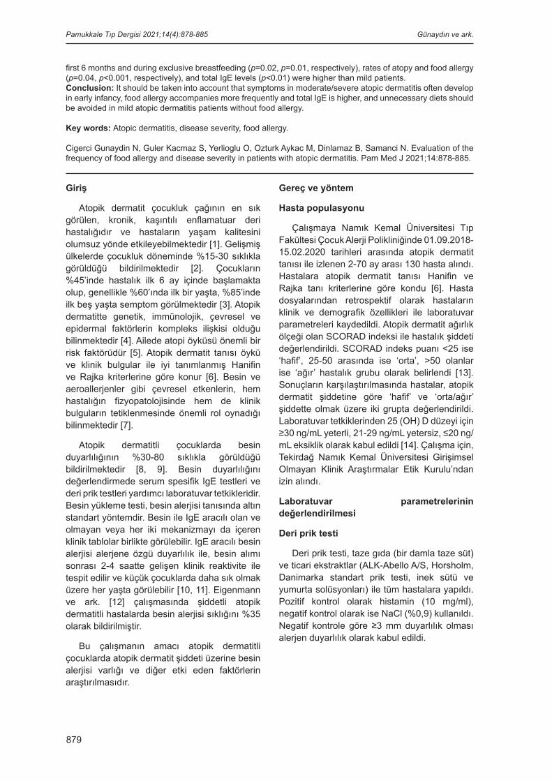
879
first 6 months and during exclusive breastfeeding (p=0.02, p=0.01, respectively), rates of atopy and food allergy (p=0.04, p<0.001, respectively), and total IgE levels (p<0.01) were higher than mild patients.Conclusion: It should be taken into account that symptoms in moderate/severe atopic dermatitis often develop in early infancy, food allergy accompanies more frequently and total IgE is higher, and unnecessary diets should be avoided in mild atopic dermatitis patients without food allergy.
Key words: Atopic dermatitis, disease severity, food allergy.
Cigerci Gunaydin N, Guler Kacmaz S, Yerlioglu O, Ozturk Aykac M, Dinlamaz B, Samanci N. Evaluation of the frequency of food allergy and disease severity in patients with atopic dermatitis. Pam Med J 2021;14:878-885.
Giriş
Atopik dermatit çocukluk çağının en sık görülen, kronik, kaşıntılı enflamatuar deri hastalığıdır ve hastaların yaşam kalitesini olumsuz yönde etkileyebilmektedir [1]. Gelişmiş ülkelerde çocukluk döneminde %15-30 sıklıkla görüldüğü bildirilmektedir [2]. Çocukların %45’inde hastalık ilk 6 ay içinde başlamakta olup, genellikle %60’ında ilk bir yaşta, %85’inde ilk beş yaşta semptom görülmektedir [3]. Atopik dermatitte genetik, immünolojik, çevresel ve epidermal faktörlerin kompleks ilişkisi olduğu bilinmektedir [4]. Ailede atopi öyküsü önemli bir risk faktörüdür [5]. Atopik dermatit tanısı öykü ve klinik bulgular ile iyi tanımlanmış Hanifin ve Rajka kriterlerine göre konur [6]. Besin ve aeroallerjenler gibi çevresel etkenlerin, hem hastalığın fizyopatolojisinde hem de klinik bulguların tetiklenmesinde önemli rol oynadığı bilinmektedir [7].
Atopik dermatitli çocuklarda besin duyarlılığının %30-80 sıklıkla görüldüğü bildirilmektedir [8, 9]. Besin duyarlılığını değerlendirmede serum spesifik IgE testleri ve deri prik testleri yardımcı laboratuvar tetkikleridir. Besin yükleme testi, besin alerjisi tanısında altın standart yöntemdir. Besin ile IgE aracılı olan ve olmayan veya her iki mekanizmayı da içeren klinik tablolar birlikte görülebilir. IgE aracılı besin alerjisi alerjene özgü duyarlılık ile, besin alımı sonrası 2-4 saatte gelişen klinik reaktivite ile tespit edilir ve küçük çocuklarda daha sık olmak üzere her yaşta görülebilir [10, 11]. Eigenmann ve ark. [12] çalışmasında şiddetli atopik dermatitli hastalarda besin alerjisi sıklığını %35 olarak bildirilmiştir.
Bu çalışmanın amacı atopik dermatitli çocuklarda atopik dermatit şiddeti üzerine besin alerjisi varlığı ve diğer etki eden faktörlerin araştırılmasıdır.
Gereç ve yöntem
Hasta populasyonu
Çalışmaya Namık Kemal Üniversitesi Tıp Fakültesi Çocuk Alerji Polikliniğinde 01.09.2018- 15.02.2020 tarihleri arasında atopik dermatit tanısı ile izlenen 2-70 ay arası 130 hasta alındı. Hastalara atopik dermatit tanısı Hanifin ve Rajka tanı kriterlerine göre kondu [6]. Hasta dosyalarından retrospektif olarak hastaların klinik ve demografik özellikleri ile laboratuvar parametreleri kaydedildi. Atopik dermatit ağırlık ölçeği olan SCORAD indeksi ile hastalık şiddeti değerlendirildi. SCORAD indeks puanı <25 ise ‘hafif’, 25-50 arasında ise ‘orta’, >50 olanlar ise ‘ağır’ hastalık grubu olarak belirlendi [13]. Sonuçların karşılaştırılmasında hastalar, atopik dermatit şiddetine göre ‘hafif’ ve ‘orta/ağır’ şiddette olmak üzere iki grupta değerlendirildi. Laboratuvar tetkiklerinden 25 (OH) D düzeyi için ≥30 ng/mL yeterli, 21-29 ng/mL yetersiz, ≤20 ng/mL eksiklik olarak kabul edildi [14]. Çalışma için, Tekirdağ Namık Kemal Üniversitesi Girişimsel Olmayan Klinik Araştırmalar Etik Kurulu’ndan izin alındı.
Laboratuvar parametrelerinin değerlendirilmesi
Deri prik testi
Deri prik testi, taze gıda (bir damla taze süt) ve ticari ekstraktlar (ALK-Abello A/S, Horsholm, Danimarka standart prik testi, inek sütü ve yumurta solüsyonları) ile tüm hastalara yapıldı. Pozitif kontrol olarak histamin (10 mg/ml), negatif kontrol olarak ise NaCl (%0,9) kullanıldı. Negatif kontrole göre ≥3 mm duyarlılık olması alerjen duyarlılık olarak kabul edildi.
Pamukkale Tıp Dergisi 2021;14(4):878-885 Günaydın ve ark.

Total IgE ve alerjen (inek sütü, yumurta sarısı, yumurta beyazı, kuruyemiş) spesifik IgE ölçümü
Total serum IgE, inek sütü spesifik IgE (sIgE), yumurta beyazı-sIgE, yumurta sarısı-sIgE, fındık, yer fıstığı ve diğer besin spesifik IgE ölçümleri ELİSA yöntemi ile yapıldı. Spesifik IgE için 0,35 kUA/l üzeri değerler pozitif kabul edildi.
Oral besin yükleme testi
Besin alerjisi varlığının değerlendirilmesinde hastalara besin yükleme testi (BYT) yapıldı. Besin yükleme testi şüpheli gıdanın 2-4 hafta eliminasyonu sonrası, sistemik ve topikal ilaçları kesilerek, açık besin yükleme testi olarak yapıldı [15, 16]. Sadece anne sütü alan hastalar ise anneye eliminasyon sonrası provakasyon yapılarak değerlendirildi. Besin yükleme testi sırasında atopik dermatitte alevlenme, ürtiker, anjiyoödem, hava yolu obstrüksiyon bulguları (dispne, ronküs, vizing v.b), kusma ve anafilaksi gelişmesi gibi objektif bulgular varlığında test pozitif kabul edildi ve sonlandırıldı. Tüm basamakları reaksiyon gelişmeden tüketebilen hasta BYT testinde erken reaksiyon negatif olarak kabul edildi ve geç reaksiyonlar açısından 5-7 gün süre ile izlendi. İnek sütü alerjisi (İSA) şüphesi olan hastalarda tanı için açık BYT’de hasta yaşına uygun olarak pastörize inek sütü veya formula mama kullanılarak seyreltilmiş 0,1 mL ile başlatıldı ve 200 mL süt miktarına ulaşıncaya veya bir reaksiyon kaydedilinceye kadar her 15-30 dakikada bir artan dozlarla sürdürüldü. Besin yükleme testinde reaksiyon görülmeyen hastalar sonrasında geç reaksiyonlar açısından izlenerek reaksiyon durumu kaydedildi [16]. Yumurta alerjisi şüphesi olan hastalarda tanı için ağız yoluyla 0,5-1 gr ile başlanarak 15-30 dakikada bir artan dozlarla 16 gr yumurtaya ulaşıncaya veya bir reaksiyon kaydedilinceye kadar yumurta ile açık besin yükleme testi yapıldı; erken ve geç reaksiyonlar kaydedildi [17].
İstatistiksel analiz
Veriler bilgisayarda SPSS 24.0 (Statistical Packages of Social Sciences) programı kullanılarak analiz edildi. Verilerin normal dağılıma uygunluğu Kolmogorov-Smirnov testi ile değerlendirildi; sürekli değişkenler için ortalama±standart sapma ve medyan şeklinde kategorik değişkenler için frekans
ve yüzde şeklinde gösterildi. Normal dağılım gösteren verilerin analizinde t testi, normal dağılım göstermeyen verilerin analizinde Mann-WhitneyU testi, kategorik değişkenler arasındaki farkın değerlendirilmesinde ise ki-kare testi yapıldı. P<0,05 değeri istatistiksel anlamlı kabul edildi.
Bulgular
Çalışmada yaş ortalaması 20,9±16,3 (6-70) ay olan 130 hasta değerlendirildi. Hastaların %45,4 (n=59)’ü kız, %54,6 (n=71)’sı erkek idi. Şikayetlerin başlama zamanı ortalama 5,7±5 (0,5-34) ay idi; %63,8 (n=83) hastada şikayetler sadece anne sütü ile beslendiği dönemde görüldü. Şikayet başlangıç zamanı hastaların %80 (n=104)’inde ilk 6 ay, %10,8 (n=14)’ünde 6-12 aylıkken, %9,2 (n=12)’sinde 12 aydan sonra idi. SCORAD indeksine göre atopik dermatit şiddeti değerlendirildiğinde %37 (n=48) hafif, %37 (n=48) orta, %26 (n=34) ağır idi.
Eşlik eden semptomlar, hastaların %24,6 (n=32)’sında hırıltılı solunum, %9,2 (n=12)’sinde kanlı mukuslu kaka, %4,6 (n=6)’sında kusma idi.
Ek gıda başlama yaşı yoğurt için: 6±1,4 (4-14) ay, yumurta sarısı için 6,2±1,6 (4-14 ) ay, yumurta beyazı için 8,7±2,4 (4-17) ay idi.
Laboratuvar parametrelerinde, tam kan sayımı ve biyokimyasal parametreleri normaldi [eozinofil sayısı: 397±257 (43-1370)x103/mm3, eozinofil yüzdesi: 9±3,3 (0,5-20)].
Hastaların demografik özellikleri ve laboratuvar bulguları Tablo 1’de özetlenmiştir.
Eşlik eden hastalık varlığı %36,2 (n=47) hastada mevcuttu; bu hastalıklar %20,7 (n=27) reaktif hava yolu hastalığı, %10 (n=13) alerjik rinit, %8,5 (n=11) alerjik proktokolit ve %6,1 (n=8) gastroözefageal reflü %0,7 (n=1) alerjik konjonktivit idi. Atopik dermatit şiddetine göre eşlik eden hastalar arasında fark yoktu (p>0,05).
Hastaların %65,4 (n=85)’ünde süt, yumurta ve kuruyemişler ile besin duyarlılığı, %13,8 (n=18)’inde ev tozu akarları duyarlılığı mevcuttu. Hastaların %38,5 (n=50)’inde besin alerjisi saptandı. Klinik ile ilişkili sorumlu besinler %62 (n=31) yumurta, %22 (n=11) inek sütü ve yumurta, %12 (n=6) sadece inek sütü, %4 (n=2) fındık idi. Deri prik testinde %2,3 (n=3) yer fıstığı, %2,3 (n=3) badem, %1,5 (n=2) susam,
Atopik dermatitte besin alerjisi ve hastalık şiddetinin değerlendirilmesi
880

881
%1,5 (n=2) ceviz, %0,07 (n=1)’sinde kakao ile duyarlılık saptandı.
Besin duyarlılığı olan hastalarda (n=85, %65,3) besin duyarlılığı olmayan hastalara (n=45, %34,7) göre hastaların tanı yaşı daha düşük (18±13,5 ay; 26,2±19 ay, sırasıyla, p=0,03), şikayet başlangıç zamanı daha erkendi (8,6±6,7; 13,7±11,8 ay, sırasıyla, p=0,02); Total IgE (p=0,1), eozinofil sayısı (p=0,98), eozinofil yüzdesi (p=0,5) benzerdi.
İnek sütü alerjisi olan hastalarda ortanca süt spesifik IgE: 6,4 (0-100) kUA/l, deri prik testte kabarıklık çapı inek sütü ile uzun çap: 8 (0-20) mm, inek sütü ile kısa çap: 5 (0-7) mm idi. Yumurta alerjisi olan hastalarda ise ortanca yumurta beyazı spesifik IgE: 6,3 (0-100) kUA/l, yumurta sarısı spesifik IgE: 6,3 (0-100) kUA/l, deri prik testte kabarıklık çapı yumurta beyazı ile uzun çap: 10 (0-29) mm, yumurta beyazı ile kısa çap: 5 (0-10) mm, yumurta sarısı ile uzun çap: 0 (0-10) mm, yumurta sarısı ile kısa çap: 0 (0-6) mm idi.
Besin alerjisi olan hastaların besin alerjisi olmayan hastalara göre yaşları daha küçük (p<0,01), 6 aylıktan önce şikayetlerin başlaması daha sık (p=0,03), Total IgE düzeyi daha yüksek (p=0,04) ve D vitamini düzeyi daha düşüktü (p=0,03). Besin alerjisi varlığı ile cinsiyet (p=0,59), doğum şekli (p=0,47), ek gıda başlama zamanı (p=0,57), eşlik eden hastalık varlığı (p=0,1), eozinofil sayısı ve eozinofil yüzdesi arasında ilişki saptanmadı (p>0,05) (Tablo 2).
Atopik dermatit şiddetine göre hastalar değerlendirildiğinde; cinsiyet, doğum şekli, besin duyarlılığı varlığı, eşlik eden hastalık
varlığı ile atopik dermatit şiddeti arasında fark saptanmadı. Orta veya ağır atopik dermatitli çocuklarda semptom başlangıç zamanı ilk 6 ayda ve sadece anne sütü ile beslendiği dönemde daha sıktı; atopi ve besin alerjisi sıklığı daha yüksekti. Orta veya ağır atopik dermatit tanılı hastaların Total IgE düzeyi hafif atopik dermatitli hastalara göre yüksekti; eozinofil sayısı ve yüzdesi ile D vitamini düşüklüğü varlığı ise benzerdi (Tablo 3).
Hasta öyküsünde çocuk alerji polikliniğine başvuru öncesinde hastaların %91,5 (n=119)’inde en az bir gıda ile eliminasyon diyeti aldığı saptandı. Eliminasyon diyeti yapılan gıdalar ise; hastaların %49,2 (n=64)’sinde sadece inek sütü, %30 (n=39)’unda inek sütü ve yumurta, %4,6 (n=6) sadece yumurta, %7,7 (n=10)’sında inek sütü, yumurta, dana eti ve kuruyemişler idi.
Tartışma
Bu çalışmada atopik dermatit tanısı ile izlenen %37 (n=48) hafif, %37 (n=48) orta, %26 (n=34) ağır şiddette olan 130 hastanın klinik ve laboratuvar özellikleri retrospektif olarak değerlendirildi. Atopik dermatit tanılı hastalarda besin alerjisi varlığı %38,5 olarak saptandı.
Bieber [18] çalışmasında, hastaların %45’inde yaşamın ilk 6 ayında semptomların görüldüğünü bildirilmiştir. Bu çalışmada ise hastaların büyük çoğunluğunda (n=83, %63,8) hayatın ilk 6 ayında semptomların başladığı saptandı. Ayrıca atopik dermatit şiddeti orta/ciddi olanlarda semptomların ilk 6 ayda ve sadece anne sütü aldığı dönemde başlaması daha sık olarak görüldü.
Tablo 1. Hastaların demografik özellikleri ve laboratuvar bulguları
Yaş ortalaması 20,9±16,3 (6-70) ay
Cinsiyet %54,6 (n=71) erkek
Doğum şekli %57,7 (n=75) c/s (sezeryan)
Ortalama şikayet başlama zamanı (ay) 5,7±5 (0,5-34) ay
Şikayetlerin başladığı dönemde beslenme şekli %63,8 (n=83) anne sütü alırken, %36,2 (n=47) ek gıdaya geçtikten sonra
Ailede atopi öyküsü %56,9 (n=74) var
Total IgE (kU/l) (ortanca) 48 (2-2420)
Eozinofil sayısı (mm3) 397±257 (43-1370)x103
Eozinofil yüzdesi (%) 4,9±3,3 (0,5-20)
D vitamini (ng/mL) 29,1±13,7 (8-63)
D vitamini eksikliği (<20 ng/mL) olan hasta yüzdesi %28,7 (n=23)
Pamukkale Tıp Dergisi 2021;14(4):878-885 Günaydın ve ark.

882
Tablo 2. Besin alerjisi varlığına göre hastaların özellikleri
Besin alerjisi var (n=50) Besin alerjisi yok (n=80) pTanı yaşı (ay) 15±11,6 (6-57) 24,6±17,7 (6-79) <0,01
Cinsiyet %58 (n=29) erkek %52,5 (n=42) erkek 0,59
Atopi varlığı %41 (n=31) var %53,8 (n=43) var 0,37
Şikayet başlama zamanı %92 (n=60) hastada 6 aylıktan önce
%75 (n=60) hastada 6 aylıktan önce
0,03
Total IgE (kU/l) (ortanca) 80 (24-1102) 35 (2-2420) 0,04
Eozinofil sayısı 391±275 (50-1150) 385±247 (43-1470) 0,81
Eozinofil yüzdesi (%) 4,9±3,7 (0,7-20) 4,6±3,1 (0,5-17,6) 0,85
25 (OH) D vitamini düzeyi (ng/mL) 25±11 (9-57) 31,2±15 (8-63) 0,03
Tablo 3. Atopik dermatit şiddetinin klinik ve laboratuvar bulgular ile ilişkisi
Hafif Atopik Dermatit(n=48)
Orta/Ağır Atopik Dermatit (n=82)
p
Tanı yaşı (ay) 24,7±16,6 (6-68) 18,7±15 (7-70) 0,01
Şikayet başlama yaşı (ay) 8,3±7 (0,5-34) 4,4±3,7 (0,5-24) 0,04
Cinsiyet (n=71,%54,6 erkek)
%52,1 (n=25) kız%47,9 (n=23) erkek
%63 (n=34) kız%48 (n=37) erkek
0,27
Doğum şekli(n=75, %57,7 sezeryan)
%54,2 (n=36) sezeryan %59,8 (n=49) sezeryan 0,58
Atopi(n=74, %56,9 var)
%28,3 (n=21) var %71,6 (n=53) var 0,04
Şikayet Başlangıç zamanı<6 aylıkken: n=104, %81,5>6 aylıkken: n=24, %18,5
%70,8 (n=34) (<6 aylıkken)
%87,8 (n=72)(<6 aylıkken)
0,02
Sadece anne sütü alırkensemptom başlangıcı varlığı(n=83, %63,8)
%28,9 (n=24) var %72,1 (n=59) var 0,01
Besin Duyarlılığı varlığı (n=85, %65,4)
%34,1 (n=29) var %65,9 (n=56) var 0,44
Besin Alerjisi varlığı (n=50, %38,6)
%14 (n=7) var %51,3 (n=41) var <0,001
Çoklu Besin Alerjisi (n=13, %10)
%4,2 (n=2) %13,4 (n=11) 0,07
Eşlik Eden Hastalık varlığı(n=47, %36,2 var)
%38,3 (n=18) var %61,7 (n=29) 0,85
Total IgE (ortanca) 30 (2-553) 65 (2-2420) <0,01
Eozinofil sayısı (mm3) 392±292 (43-1370) 384±236 (50-1130) 0,1
Eozinofil yüzdesi (%) 4,4±3 (0,5-14) 4,9±3,5 (0,7-20) 0,5
25 (OH) D vitamini düzeyi (ng/mL) 30,6±16 28,7±12,7 0,80
D vitamini eksikliği varlığı(n=26, %21)
%14,5 (n=7) var %23,2 (n=19) var 0,18
Literatürde atopik dermatit varlığının cinsiyete göre değişmediği bildirilmektedir [19]. Bu çalışmada da literatürle uyumlu olarak cinsiyete göre atopik dermatit sıklığı benzerdir.
Ailede atopi öyküsü ise Roos ve ark. [20] çalışmasında %30-50 olarak bildirilmektedir. Bu çalışmada ailede atopi öyküsü literatüre benzer şekilde %56,9 olarak saptanmış, bununla
Atopik dermatitte besin alerjisi ve hastalık şiddetinin değerlendirilmesi

883
birlikte orta/ağır atopik dermatitli hastalarda daha sık olmak üzere atopik dermatit şiddeti ile ilişkili bulunmuştur.
Atopik dermatitte olguların çoğunda serum IgE düzeyinin yükseldiği ve periferik kanda eozinofili bildirilmektedir [5]. Çalışmamızda Total IgE düzeyi için besin duyarlılığı olan ve olmayan hastalar arasında fark saptanmazken (p=0,1), besin alerjisi olan hastalarda olmayanlara göre daha yüksekti (p=0,04). Bununla birlikte atopik dermatit şiddeti orta veya ciddi olan hastalarda serum Total IgE düzeyi hafif olgulara göre yüksek saptanmıştır. Periferik kanda eozinofil sayısı ve yüzdesinde ise atopik dermatit şiddeti, besin duyarlılığı veya besin alerjisi varlığına göre fark saptanmamıştır (p>0,05).
Ricci ve ark. [21] çalışmasında atopik dermatitin astım ve alerjik rinit gibi alerjik hastalıklarla birlikteliği bildirilmektedir. Atopik dermatitli hastaların uzun süre izleminin yapıldığı Ekback ve ark. [22] çalışmasında hastaların %47’sinde alerjik rinit, %29’unda astım geliştiği bildirilmiş; yüksek hastalık şiddetinin astım ve alerjik rinit açısından risk faktörü olduğu vurgulanmıştır. Çalışmamızda eşlik eden hastalık varlığı %36,2 (n=47) hastada mevcuttu; bu hastalıklar sıklıkla %20,7 reaktif hava yolu hastalığı, %10 alerjik rinit, %8,5 alerjik proktokolit idi.
Atopik dermatitte, alerjik duyarlanma sürecinin erken çocukluk döneminde besin alerjisi şeklinde ortaya çıktığı düşünülmektedir [23, 24]. Çocukluk çağında atopik dermatit patofizyolojisinde ve akut alevlenmelerinde besinlerin rolü bilinmektedir. Yapılan bir kohort çalışmada ilk 6 yaşta atopik dermatit görülen çocukların %52’sinde en az bir besin ile duyarlılık saptanırken, hastaların %15’inde besin yükleme testi ile kanıtlanmış besin alerjisi varlığı gösterilmiştir [25]. Hill ve ark. [26] çalışmasında ise atopik dermatitte besin veya aeroallerjenler ile duyarlılık %80 olarak bildirilmiş, bunların küçük bir kısmında klinik reaktivite ile ilişkili olduğu bildirilmiştir. Ülkemizde Mısırlıoğlu ve ark. [27] çalışmasında alerjen duyarlılaşma %37,8 olarak bildirilmiştir; bizim çalışmamızda ise %65,4 olarak yüksek saptanmış, ancak besin alerjisi varlığı ise hastaların %38,5 (n=50)’sinde gösterilmiştir. Literatürde atopik dermatitli infantlarda besin alerjisi sıklığı çeşitli çalışmalarda ise %33-66 olarak bildirilmektedir [28-31]. İnek sütü, yumurta, buğday, soya,
kuruyemişler, balık, kabuklu deniz ürünleri en sık suçlanan besinlerdir [10, 32]. Bergmann ve ark.’nın [33] çalışmasında atopik dermatitli hastalarda en çok bulunan besin alerjisi yumurta olarak bildirilmiştir. Bu çalışmada da en sık sorumlu besinler yumurta ve inek sütü idi.
Çalışmamızda hasta öykülerinde, hastaların %92’sinin çocuk alerji polikliniğine başvuru öncesi en az bir gıda ile eliminasyon diyeti aldığı bildirilmiştir; bu da gereksiz besin eliminasyonunun yapılmaması için hastaların değerlendirilmesinin önemini ortaya koymaktadır.
Son yıllarda yapılan çalışmalar D vitaminin atopik dermatit üzerine olası rolü dikkat çekmektedir. Kim ve ark. [34] çalışmasında D vitamini desteğinin atopik dermatit tedavisinde yardımcı olabileceğini vurgulamışlardır. Çalışmamızda D vitamini eksikliği hastaların %28,7 (n=23)’sinde saptandı; besin alerjisi olan hastalarda D vitamini düzeyinin besin alerjisi olmayanlara göre daha düşüktü (25±11; 31,2±15 ng/mL, sırasıyla, p=0,03). Ancak atopik dermatit şiddetine göre değerlendirildiğinde D vitamini düzeyi benzer saptanmıştır (p=0,8).
Sonuç olarak, atopik dermatitli çocuklarda besin alerjisi sıklığı %38,5 saptandı. Orta veya ağır şiddette atopik dermatitli çocuklarda şikayetlerin erken süt çocuğu döneminde sıklıkla başladığı ve bulguların daha erken yaşta geliştiği; Total IgE’nin daha yüksek ve besin alerjisi varlığının daha sık olduğu gösterildi. Hastalığın şiddetinin değerlendirilmesi ve orta/ağır şiddette atopik dermatit varlığında besin alerjisinin daha sık görüldüğünün göz önünde bulundurulması; hafif ve besin alerjisinin eşlik etmediği atopik dermatitli hastalarda gereksiz eliminasyon diyetinden kaçınmak için hastaların değerlendirilmesi önemlidir.
Çıkar ilişkisi: Yazarlar çıkar ilişkisi olmadığını beyan eder.
Kaynaklar 1. Leung AKC, Hon KLE, Robson WLM. Atopic dermatitis.
Adv Pediatr 2007;54:241-273. https://doi.org/10.1016/j.yapd.2007.03.013
2. Williams HC. Clinical practice. Atopic dermatitis. N Engl J Med 2005;352:2314-2324. https://doi.org/10.1056/NEJMcp042803
Pamukkale Tıp Dergisi 2021;14(4):878-885 Günaydın ve ark.

3. Kay J, Gawkrodger DJ, Mortimer MJ, Jaron AG. The prevalance of childhood atopic eczema in general population. J Am Acad Dermatol 1994;30:35-39. https://doi.org/10.1016/s0190-9622(94)70004-4
4. Cork MJ, Danby SG, Vasilopoulos Y, et al. Epidermal barrier disfunction in atopic dermatitis. J Invest Dermatol 2009;129:1892-1908. https://doi.org/10.1038/jid.2009.133
5. Sapan N. Atopik dermatit. In: Şekerel BE, ed. Çocukluk çağında alerji astım immünoloji, İstanbul: Ada Basın Yayın, 2015;541-548.
6. Hanifin JM, Rajka G. Diagnostic features of atopic dermatitis. Acta Derm Venereol (Stockh) 1980;92:44-47.
7. Untersmayr E, Jensen Jarolim E. The effect of gastric digestion on food allergy. Curr Opin Allergy Clin Immunol 2006;6:214-219. https://doi.org/10.1097/01.all.0000225163.06016.93
8. Peroni DG, Piacentini GL, Bodini A, Rigotti E, Pigozzi E, Boner AL. Prevalence and risk factors for atopic dermatitis in preschool children. Br J Dermatol 2008;158:539-543. https://doi.org/10.1111/j.1365-2133.2007.08344.x
9. Schneider L, Tilles S, Lio P, et al. Atopic dermatitis: a practice parameter update 2012. J Allergy Clin Immunol 2013;131:295-299. https://doi.org/10.1016/j.jaci.2012.12.672
10. Sicherer SH, Sampson HA. Food hypersensitivity and atopic dermatitis: pathophysiology, epidemiology, diagnosis, and management. J Allergy Clin Immunol 1999;104:114-122. https://doi.org/10.1016/s0091-6749(99)70053-9
11. Harmancı K. Atopik dermatit tanı ve tedavisine yaklaşım: Ulusal rehber 2018 ve ürtiker tanı ve tedavisi güncel durum raporu. Astım Allerji İmmünoloji 2018;16:1-95. https://doi.org/10.21911/aai.2018.2
12. Eigenmann PA, Sicherer SH, Borkowski TA, Cohen BA, Sampson HA. Prevalence of IgE mediated food allergy among children with atopic dermatitis. Pediatrics 1998;101:8. https://doi.org/10.1542/peds.101.3.e8
13. Severity scoring of atopic dermatitis: the SCORAD index. Consensus report of the European task force on atopic dermatitis. Dermatology 1993;186:23-31. https://doi.org/10.1159/000247298
14. Holick MF, Binkley NC, Bischoff Ferrari HA, et al. Evaluation, treatment, and prevention of vitamin D deficiency: an endocrine society clinical practice guideline. J Clin Endocrinol Metab 2011;96:1911-1930. https://doi.org/10.1210/jc.2011-0385
15. Bindslev Jensen C, Ballmer Weber BK, Bengtsson U, et al. Standardization of food challenges in patients with immediate reactions to foods position paper from the European Academy of Allergology and Clinical Immunology. Allergy 2004:59:690-697. https://doi.org/10.1111/j.1398-9995.2004.00466.x
16. Vandenplas Y, Koletzko S, Isolauri E, et al. Guidelines for the diagnosis and management of cow’s milk protein allergy in infants. Arch Dis Child 2007;92:902-908. https://doi.org/10.1136/adc.2006.110999
17. Sampson HA, Gerth van Wijk R, Bindslev Jensen C, et al. Standardizing double-blind, placebo controlled oral food challenges: American Academy of Allergy, Asthma & Immunology-European Academy of Allergy and Clinical Immunology PRACTALL consensus report. J Allergy Clin Immunol 2012;130:1260-1274. https://doi.org/10.1016/j.jaci.2012.10.017
18. Bieber T. Atopic dermatitis. N Engl J Med 2008;358:1483-1494. https://doi.org/10.1056/NEJMra074081
19. Boguniewicz M, Leung DYM. Atopic dermatitis. In: Adkinson NF, Holgate ST, Bochner BS, Lemanske RF, Buse W, Simons FE, eds. Middleton’s allergy principles and practice. 7th ed. China Elsevier, 2009;1083-1103.
20. Roos TC, Geuer S, Roos S, Brost H. Recent advances in treatment strategies for atopic dermatitis. Drugs 2004;64:2639-2666. https://doi.org/10.2165/00003495-200464230-00003
21. Ricci G, Patrizzi A, Baldi E, Menna G, Tabanelli M, Masi M. Long term follow up of atopic dermatitis: retrospective analysis of related risk factors and association with concomitant allergic diseases. J Am Acad Dermatol 2006;55:765-771. https://doi.org/10.1016/j.jaad.2006.04.064
22. Ekback M, Tedner M, Devenney I, et al. Severe eczema in ınfancy can predict asthma development a prospective study to the age of 10 years. PLoS One 2014;9:e99609. https://doi.org/10.1371/journal.pone.0099609
23. Akdis CA, Akdis M, Bieber T, et al. Diagnosis and treatment of atopic dermatitis in children and adults: European Academy of Allergology and Clinical Immunology/American Academy of Allergy, Asthma and Immunology / PRACTALL Consensus Report. J Allergy Clin Immunol 2006;118:152-169. https://doi.org/10.1016/j.jaci.2006.03.045
24. Tsakok T, Marss T, Mohsin M, et al. Does atopic dermatitis cause food allergy? A systematic review. J Allergy Clin Immunol 2016;137:1071-1078. https://doi.org/10.1016/j.jaci.2015.10.049
25. Eller E, Kjaer HF, Host A, Andersen KE, Bindslev Jensen C. Food allergy and food sensitization in early childhood: results from the DARC cohort. Allergy 2009;64:1023-1029. https://doi.org/10.1111/j.1398-9995.2009.01952.x
26. Hill DJ, Heine RG, Hosking CS, et al. IgE food sensitization in infants with eczema attending a dermatology department. J Pediatr 2007;151:359-363. https://doi.org/10.1016/j.jpeds.2007.04.070
27. Dibek Mısırlıoglu E, Güngör Ş, Nacaroğlu HT, et al. An evaluation of characteristics and concomitant allergic diseases in children with atopic dermatitis. Asthma Allergy Immunol 2014;12:97-103.
884
Atopik dermatitte besin alerjisi ve hastalık şiddetinin değerlendirilmesi

885
28. Eigenmann PA, Calza AM. Diagnosis of IgE-mediated food allergy among Swiss children with atopic dermatitis. Pediatr Allergy Immunol 2000;11:95-100. https://doi.org/10.1034/j.1399-3038.2000.00071.x
29. Sampson HA. The immunopathogenic role of food hypersensitivity in atopic dermatitis. Acta Derm Venereol Suppl (Stockh) 1992;176:34-37.
30. Niggemann B, Sielaff B, Beyer K, Binder C, Wahn U. Outcome of double-blind, placebo-controlled food challenge tests in 107 children with atopic dermatitis. Clin Exp Allergy 1999;29:91-96. https://doi.org/10.1046/j.1365-2222.1999.00454.x
31. Breuer K, Heratizadeh A, Wulf A, et al. Late eczematous reactions to food in children with atopic dermatitis. Clin Exp Allergy 2004;34:817-824. https://doi.org/10.1111/j.1365-2222.2004.1953.x
32. Boyce JA, Assa’ad A, Burks AW, et al. Guidelines for the diagnosis and management of food allergy in the United States: report of the NIAID - sponsored expert panel. J Allergy Clin Immunol 2010;126:1105-1118. https://doi.org/10.1016/j.jaci.2010.10.008
33. Bergmann MM, Caubet JC, Boguniewicz M, Eigenmann PA. Evaluation of food allergy in patients with atopic dermatitis. J Allergy Clin Immunol Pract 2013;1:22-28. https://doi.org/10.1016/j.jaip.2012.11.005
34. Kim MJ, Kim SN, Lee YW, Choe YB, Ahn KJ. Vitamin D status and efficacy of vitamin D supplementation in atopic dermatitis: a systematic review and meta-analysis. Nutrients 2016;8:789. https://doi.org/10.3390/nu8120789
Etik kurul onayı: Çalışmaya başlamadan önce, Tekirdağ Namık Kemal Üniversitesi Girişimsel Olmayan Klinik Araştırmalar Etik Kurulu’ndan 25.02.2020 tarih ve 2020.42.02.16 protokol numarası ile izin alındı.
Yazarların makaleye olan katkıları
N.C.G., S.G.K. ve N.S. çalışmanın ana fikrini ve hipotezini kurgulamışlardır. N.C.G., Ö.Y. ve B.D. teoriyi geliştirmiş, gereç ve yöntem bölümünü düzenlemişlerdir. Sonuçlar kısmınındaki verilerin değerlendirmesini N.C.G., Ş.G.K. ve M.Ö.A. yapmışlardır. Makalenin tartışma bölümü N.C.G., Ö.Y. ve B.D. tarafından yazılmış N.S., N.C.G. gözden geçirip gerekli düzeltmeleri yapmış ve onaylamıştır. Ayrıca tüm yazarlar çalışmanın tamamını tartışmış ve son halini onaylamıştır.
Pamukkale Tıp Dergisi 2021;14(4):878-885 Günaydın ve ark.

886
Correlation between symptoms and Centor/McIsaac score in the diagnosis of tonsillopharyngitis
Tonsillofarenjit tanısında semptomlar ve Centor/Mclsaac skoru arasındaki korelasyon
Zeynep Yılmaz Öztorun, Güliz Gürer
Pamukkale Tıp DergisiPamukkale Medical JournalAraştırma Makalesi
Zeynep Yılmaz Oztorun, Ass. Prof. Nigde Omer Halisdemir University Faculty of Medicine, Department of Child Health and Diseases, Nigde, Turkey, e-mail: [email protected] (https://orcid.org/0000-0001-8017-3048) (Corresponding Author)Guliz Gurer, Ass. Prof. Nigde Omer Halisdemir University Faculty of Medicine, Department of Child Health and Diseases, Nigde, Turkey, e-mail: [email protected] (https://orcid.org/0000-0003-2922-430X)
Abstract Purpose: Group A beta hemolytic streptococcus (GABHS) is identified in 20-40% of cases of childhood pharyngitis. The Centor/McIsaac score is an important scoring method used in the early diagnosis of patients presenting with symptoms of Group A beta hemolytic streptococcus (GABHS) pharyngitis. In this study, we aimed to determine the relationship between Centor / McIsaac score and patients’ symptoms, and to investigate any conformity between Rapid Antigen Test positivity and high Centor/McIsaac score.Materials and methods: Data were obtained retrospectively by scanning the files of patients (age 2 to 18 years) diagnosed with GAData were obtained retrospectively by scanning the files of patients (age 2 to 18 years) diagnosed with GABHS who were hospitalized between May and December 2017. Inclusion criteria were, being between the ages of 2 and 18 years and to be diagnosed with group A beta hemolytic streptococcal pharyngitis. Receiving antibiotic treatment within the last 14 days, being under 2 years of age and over 18 years of age are exclusion criteria. Daily clinical findings and test results (complete blood count, neutrophil and lymphocyte percentages, rapid antigen test, throat culture) were collected from medical records. The Centor/McIsaac scores calculated according to symptoms and rapid antigen test results used for GABHS antigen detection from throat swab samples, used for the initiation of effective treatment and to shorten hospitalization, were recorded from patient files. Student's t test was used to analyze the data that showed normal distribution among independent groups. The chi-square test was used for the analysis of categorical variables. P<0.05 was considered statistically significant. Results: A total of 236 patients were included in the study. The average age of the patients was 7 years. The frequency of pharyngitis symptoms was similar in patients younger and older than 7 years of age. There was no statistically significant difference between those younger and older than 7 years of age in terms of throat culture positivity (p=0.059). There was no statistically significant difference between Centor/McIsaac scores of the two age groups (p=0.063). The Centor/McIsaac score (3.62±1.01) of those with positive rapid antigen test was significantly higher than that of those with negative rapid antigen test (2.78±1.28) (p=0.001). Centor/McIsaac scores of those with and without fever, those with and without tonsillar hypertrophy, those with and without cough were also compared. Centor/McIsaac scores of those with fever (3.53±1.02) were higher compared to those without (1.63±0.89) (p=0.001), scores were higher in those with tonsillar hypertrophy (3.68±1.01) compared to those without (2.10±0.97) (p=0.001), and scores were again higher in those without cough (3.67±1.07) compared to those with cough (2.22±0.99) (p<0.001).Conclusion: The intensity of symptoms seen in GABHS infection leads to higher Centor/McIsaac scores. The correlation between rapid antigen test results and Centor/McIsaac score shows the increasing importance of Centor/McIsaac score in early diagnosis of GABHS pharyngitis.
Key words: Tonsillopharyngitis, Centor/McIsaac score, rapid antigen test.
Yilmaz Oztorun Z, Gurer G. Correlatıon between symptoms and Centor/McIsaac score ın the dıagnosıs of tonsıllopharyngıtıs. Pam Med J 2021;14:886-891.
ÖzAmaç: A grubu beta hemolitik streptokok (GABHS) çocukluk çağı farenjit vakalarının %20-40’ında tanımlanır. Centor/McIsaac skoru A grubu beta hemolitik streptokok farenjiti semptomları ile başvuran hastaların erken tanısında kullanılan önemli bir skorlama yöntemidir. Bu çalışmada, Centor/McIsaac skor ile hastaların semptomları arasındaki ilişkiyi saptamayı ve Hızlı Antijen Test pozitifliği ile yüksek Centor/McIsaac skoru arasındaki herhangi bir uygunluğu incelemeyi amaçladık. Gereç ve yöntem: Mayıs ile Aralık 2017 arasında hastanede yatmış olan GABHS tanısı alan hastaların (2 ila 18 yaş) dosyaları taranarak retrospektif olarak veriler elde edildi. Dahil edilme kriterleri 2 ve 18 yaş arasında olmak, A grubu beta hemolitik streptokok farenjit tanısı almasıdır. Son 14 gün içinde antibiyotik tedavisi almak, 2 yaş altında ve 18 yaş üstünde olmak dışlanma kriterleridir. Günlük klinik bulguları ve test sonuçları (tam kan sayımı, nötrofil ve lenfosit yüzdeleri, Hızlı antijen test, boğaz kültür) tıbbi kayıtlardan toplandı. Boğaz sürüntü örneklerinden GABHS antijeninin saptanması, etkin tedavi başlanması ve hastanede kalış süresinin kısaltılması için kullanılan; semptomlar ve hızlı antijen test sonuçlarına göre hesaplanan Centor/McIsaac skorları hasta
Research Article
Gönderilme tarihi:19.01.2021 Kabul tarihi:19.04.2021
doi:https://dx.doi.org/10.31362/patd.864610

887
Introduction
‘Group A beta hemolytic Streptococcus’ (GABHS) infections most often manifest in the form of acute pharyngotonsillitis [1]. GABHS are identified in 20-40% of childhood pharyngitis cases [2]. In addition to fever, sore throat and dysphagia; headache, vomiting and abdominal pain may develop. The other symptoms of GABHS pharyngitis are pharyngotonsillitis, pharyngeal exudates and anterior cervical adenitis [3, 4].
Definitive diagnosis is made by culture isolation of GABHS from the pharynx, which requires around 48 hours for conclusive results. The rapid antigen detection test (RADT) is another method to assess such infections, and it can be completed in only a few minutes [5, 6].
The Centor/McIsaac score is comprised of the following parameters: greater than 38°C fever, no cough, painful or tender anterior cervical lymphadenopaty, occurrence of tonsillar exudate and being aged from 3 to 14 years old [7]. RADT accompanied with Centor/McIsaac score to detect GABHS enables differential diagnosis (viral vs. bacterial), thereby allowing early administration of necessary treatment [8, 9]. By comparing the Centor/McIsaac score (which is an easy and non-invasive evaluation) with the rapid antigen test, it will be possible to determine whether the Centor/McIsaac can provide more reliable information about the course of infection before performing a throat culture.
In this study we aimed to assess the pharyngitis symptoms and Centor/McIsaac scores, and to examine whether there was any relationship between RADT positivity and high Centor/McIsaac score.
Materials and methods
A retrospective study of patients examined at the Nigde Omer Halisdemir University (OHU) Faculty of Medicine between the dates of May 2017-December 2017 was made by researchers. Inclusion criteria were, being between the ages of 2 and 18 years and to be diagnosed with group A beta hemolytic streptococcal pharyngitis. Receiving antibiotic treatment within the last 14 days, being under 2 years of age and over 18 years of age are exclusion criteria. All patients aged 2 to 18 years who were hospitalized after being diagnosed with GABHS pharyngitis were included in the study. Ethics approval was given by Nigde Omer Halisdemir University School of Medicine, Clinical Research Ethics Committee (approval no: 2018/08-15). All procedures of the study conformed to the Declaration of Helsinki and its most recent amendments.
Medical records were reviewed to obtain the following information: visit time, demographic data, symptoms, physical exam findings (temperature, cervical lymphadenopathy, tonsillar hypertrophy), laboratory results (RADT, pharyngeal swab culture, complete blood count (CBC)). We used the RADT Quik test Strep-A Plus (BioMérieux) which is based on lateral_flow immunoassay for GAS antigen detection from throat swab samples. Throat swab culture
dosyalarından kaydedildi. Bağımsız gruplar arasında normal dağılım gösteren verilerin analizinde Student t test kullanıldı. Kategorik değişkenlerin analizi için kikare testi kullanıldı. P<0,05 istatiksel olarak anlamlı kabul edildi.Bulgular: Çalışmaya toplam 236 hasta dahil edildi. Hastaların ortalama yaşı 7 yaştı. 7 yaşından büyük ve küçük olan hastalarda farenjit semptomlarının sıklığı benzerdi. 7 yaş altındakiler ve üstündekiler arasında boğaz kültürü pozitifliği açısından istatiksel olarak anlamlı fark yoktu (p=0,059). İki yaş grubunun Centor/McIsaac skorları arasında istatiksel olarak anlamlı bir fark yoktu(p=0,063). Hızlı antijen testi pozitif olanların Centor/McIsaac skoru (3,62±1,01), hızlı antijen test negatif olanlarınkinden (2,78±1,28) anlamlı düzeyde yüksekti (p=0,001). Ateşi olanlar ve ateşi olmayanların, tonsiller hipertrofi olanların ve olmayanların, öksürük olmayanlar ve öksürük olanların Centor/McScorları da karşılaştırılmıştır. Ateşi olanların (3,53±1,02) ateşi olmayanlara göre (1,63±0,89) (p=0,001), tonsiller hipertrofi olanların (3,68±1,01) tonsiller hipertrofi olmayanlara göre (2,10±0,97) (p=0,001) Centor/McIsaac skorları daha yüksekti ve yine öksürük olmayanların (3,67±1,07) öksürük olanlara (2,22±0,99) (p<0,001) göre skorları daha yüksekti.Sonuç: GABHS infeksiyonunda görülen semptomların yoğunluğu, daha yüksek Centor/McIsaac skorlara neden olmaktadır. Hızlı antijen test sonuçları ile Centor/McIsaac skorun arasındaki korelasyon, GABS farenjitinin erken tanısında Centor/McIsaac skorun öneminin arttığını göstermektedir.
Anahtar kelimeler: Tonsillofarenjit, Centor/McIsaac skor, hızlı antijen test.
Yılmaz Öztorun Z, Gürer G. Tonsillofarenjit tanısında Centor/McIsaac skoru arasındaki korelasyon. Pam Tıp Derg 2021;14:886-891.
Pamukkale Medical Journal 2021;14(4):886-891 Oztorun and Gurer

Symptoms and Centor/McIsaac score
888
was performed using standard methods via streaking on 5% sheep blood agar plates incubated anaerobically at 350C and examined at 48 hours with confirmation of GABHS using latex agglutination.
We used the Centor/McIsaac clinical score to evaluate GABHS in each patient. This scoring system is comprised of determining the presence or absence of the following criteria: painful and tender anterior cervical lymphadenopathy, absence of cough, a temperature of >38°C, being aged 3-14 years, occurrence of tonsillar exudate. One point is given for each criterion that is present in the Centor/McIsaac scoring. Then these points are summed and the final score is calculated; thus, overall score ranges from 0 to 5 points.
SPSS version 22 was used for all statistical analyses. Descriptive statistics were given using mean ± standard deviation for normally distributed variables. Student’s t test was used to analyze the data that showed normal distribution among independent groups. Mann Whitney U test was used for the variables who did not show normal distribution. The chi-square test was used for the analysis of categorical variables. P<0.05 was considered statistically significant. P<0.05 values were defined as statistically significant.
Results
Among the 236 patients included in the study, 115 were girls and, 121 were boys. Mean age was 7 years. 109 children were under 7 years, 127 children were over 7 years. There was no statistically significant difference between the Centor/McIsaac scores of boy’s and girl’s (p>0.05). The Centor/McIsaac scores and throat
culture results of the patients were evaluated with respect to the 7-year age threshold. There was no statistically significant difference between children younger than and older than 7 years old in terms of Centor/McIsaac scores (p=0.063) and symptoms (p>0.05). Also, there was no significant difference between the frequency of throat culture positivity in those under 7 years old and those over 7 years old (p=0.059).
The summary of Centor/McIsaac criteria and the number of patients with <2 and ≥2 points are given in Table 1. The most common symptom in this group of patients was having a fever of than >38°C (84%). Comparison of Centor/McIsaac scores of patients with and without pharyngitis symptoms is shown in Table 2. The Centor/McIsaac scores of hospitalized patients with fever over 38°C were compared with those with fever below 38°C. Accompaniment of other scoring criteria was more frequent in those with fever above exceeding 38°C. As such, the mean Centor/McIsaac score of those with a fever over 38°C (3.53±1.02) was found to be significantly higher than the mean score of those with a fever below 38°C (1.63±0.89) (p<0.05). Higher Centor/McIsaac score is defined as having a score of ≥2 points. The frequency of having a Centor/McIsaac score of 2 and above was significantly higher in patients with a fever of >38°C.
The mean Centor/McIsaac score of patients without cough (3.67±1.07) was significantly higher than those with cough (2.22±0.99) (p<0.05). There was a significant difference in Centor/McIsaac scores with respect to tonsillar swelling (p<0.05). Patients with tonsillar hypertrophy had higher Centor/McIsaac scores (3.68±1.01) than patients without tonsillar
Table 1. The frequency of clinical characteristics of the patients and the distribution of Centor/McIsaac score
Symptoms and Centor/McIsaac score n %Cough 101 42.8
>38°C fever 175 74.2
Tonsillar enlargement or exudate 142 60.2
Cervical lymphadenopathy 74 31.4
Sore throat 170 72
Centor/McIsaac score<2 32 13.6
Centor/McIsaac score≥2 204 86.4

889
hypertrophy (2.10±0.97). The percentage of patients with a Centor/McIsaac score of 2 or above was significantly higher in those with tonsillar hypertrophy compared to those without.
The mean Centor/McIsaac score of the patients was 3.05 in the study. The comparison of Centor/McIsaac scores with respect to rapid antigen test and throat culture results are shown in Table 3. The mean Centor/McIsaac score of
patients with positive RADT result (3.62±1.01) was significantly higher than the mean Centor/McIsaascore of patients with negative rapid test result (2.78±1.28) (p<0.001). Also, the mean level of having a Centor/McIsaac Score of 2 or greater above was significantly more common among patients with rapid antigen test positivity (p<0.001). Significantly higher Centor/McIsaac scores were determined in patients with positive
throat culture (3.75±0.95) compared to those with negative culture (2.76±1.26) (p<0.05).
Overall, mean neutrophil percentage was 54% in this study group. There was no correlation between RADT positivity and neutrophil dominance (p>0.05). Patients with negative throat culture result were found to have significantly higher neutrophil percentage compared to those with positive culture.
Discussion
In this study, the occurrence of GABHS positivity in throat culture was found to be 33%. In Egyptian children GABHS frequency was 42.2% [10], and in other studies from Arab countries, the rate of GABHS pharyngitis was reported to be 40% and 41.5% [11, 12]. The frequency of GABHS was similar in Egypt and Arab-population studies. Since we included
patients under 3 years of age, the likelihood of pharyngitis due to viral etiology was higher. Therefore, in the present study, GABHS frequency was lower in comparison to studies performed in Egyptian and Arabian populations.
In the present study, the Centor/McIsaac score of 68 patients with GABHS positive throat culture were found to be significantly higher than those with negative throat culture. Throat culture positivity appears to increase the likelihood of higher Centor/McIsaac score. Similar to our study, Stefaniuk et al. [13] stated that GABHS positive throat culture was present in 48% of those with a Centor/McIsaac score of 3, and in 50% of those with a Centor/McIsaac score of 4 or 5. There was a strong correlation between the results of positive throat culture result and Centor/McIsaac score (r=0.81). Fine et al. [14] compared patient’s Centor/McIsaac scores
Table 2. Comparison of Centor/McIsaac scores of patients with and without pharyngitis symptoms
Coughn sd t
-10.67
p
0.000*
Yes 101 2.22 0.99
No 135 3.67 1.07
>380C FeverYes 175 3.53 1.02
9.2 0.000*
No 61 1.63 0.89
Tonsillar hypertrophy
Yes 142 3.68 1.0111.96 0.001*
No 94 2.10 0.97
*p<0.05
Table 3. Comparison of Centor/McIsaac scores of those with and without throat culture positive and those with or without rapid antigen test positive
Throat culture
n sd t
3.14
p
0.001*
Positive 68 3.75 0.95
Negative 168 2.76 1.26
Rapid antigen
test
Positive 76 3.62 1.01 5.05 0.001*
Negative 160 2.78 1.28
*p<0.05
Pamukkale Medical Journal 2021;14(4):886-891 Oztorun and Gurer

with the frequency of GABHS isolates and they were unable to find a correlation between Centor/McIsaac score and Streptococcus pyogenes isolation. Interestingly, Felsenstein et al. [15] stated that patients who had GABHS positivity in throat culture demonstrated lower Centor/McIsaac scores. The differences in the literature on this topic suggests that the criteria used in McIsaac scoring may also occur in viral infections.
Increased Centor/McIsaac score was in correlation with increased sensitivity of RADT. Dimatteo et al. [16] stated that the sensitivity of RADT increased markedly with increasing modified Centor scores; for patients with a Centor Score of 4, the sensitivity was 97%. Tanz et al. [17] stated that, among patients with McIsaac scores of >2, RADT sensitivity was 78%. Parallel to these studies, in our study, the mean Centor/McIsaac score of 76 patients with rapid test positivity were found to be significantly higher than those with negative rapid test result.
Çamurdan et al. [18] stated that GABHS prevalence varies greatly in patients younger than 72 months (25.2%) and those older than 72 months (53.9%). In our study, throat culture positivity was 33% in patients under 7 years and 31.5% in those older than 7 years of age. There was no statistically significant difference between these two age groups. The difference between two studies may be associated with the sample size (close to each other in both age groups in our study), throat swab technique, and high false positives.
We also investigated whether Centor/McIsaac scores were associated with fever, absence of cough and cervical lymphadenopathy findings. Other studies in the literature did not investigate the relationships between specific symptoms and the overall Centor/McIsaac score. This is another feature that distinguishes our study from the literature.
Our study has some limitations, including its retrospective nature. Secondly, the number of patients is relatively low for the evaluation of such a common pathogen, which represents it is another limitation of the study. Furthermore, since the specific microorganisms grown in culture were not evaluated, we could not perform any investigations pertaining to the type of microorganism.
In conclusion, the intensity of symptoms seen in GABHS infection causes higher Centor/McIsaac scores. The correlation between rapid antigen test results and Centor/McIsaac score appears to increase the value and reliability of the Centor/McIsaac score in the early diagnosis of GABHS. We can conclude that the relationship between the rapid antigen test and Centor /McIsaac score will contribute to the correct management of GABHS pharyngitis.
Conflict of interest: No conflict of interest was declared by the authors.
References1. Küçük Ö, Biçer S, Giray T, et al. Validity of rapid
antigen detection testing in group a beta-hemolytic streptococcal tonsillopharyngitis. Indian J Pediatr 2014;81:138-142. https://doi.org/10.1007/s12098-013-1067-y
2. Shaikh N, Leonard E, Martin JM. Prevalence of streptococcal pharyngitis and streptococcal carriage in children: a meta-analysis. Pediatrics 2010;126:557-564. https://doi.org/10.1542/peds.2009-2648
3. Borchardt RA. Diagnosis and management of group a beta hemolytic streptococcal pharyngitis. JAAPA 2013;26:53-54. https://doi.org/10.1097/01.jaa.0000433876.39648.52
4. Shulman ST, Bisno AL, Clegg HW, et al. Clinical practice guideline for the diagnosis and management of group a streptococcal pharyngitis: 2012 update by the infectious diseases society of America. Clin Infect Dis 2012;55:86-102. https://doi.org/10.1093/cid/cis629
5. Lindbaek M, Hoiby EA, Lermark G, Steinsholt MI, Hjortdahl P. Which is the best method to trace group a streptococci in sore throat patients: culture or GAS antigen test? Scand J Prim Health Care 2004;22:233-238. https://doi.org/10.1080/02813430410006675
6. Fontes MJ, Bottrel FB, Fonseca MT, Lasmar LB, Diamante R, Camargos PA. Early diagnosis of streptococcal pharyngotonsillitis: assessment by latex particle agglutination test. J Pediatr (Rio J) 2007;83:465-470. https://doi.org/10.2223/JPED.1695
7. Hryniewicz W, Albrecht P, Radzikowski A. Guidelines for the management of community acquired-respiratory tract infection. Recommendations of approach in respiratory tract infections. In National Program of Antibiotics Protection. National Medicines Institute 2016. Available at: https://www.antybiotyki.edu.pl/Rekomendacje2016.pdf. Accessed May 9, 2019
8. Centor RM, Witherspoon JM, Dalton HP, Brody CE, Link K. The diagnosis of strep throat in adults in the emergency room. Med Decis Making 1981;1:239-246. https://doi.org/10.1177/0272989X8100100304
890
Symptoms and Centor/McIsaac score

891
9. McIsaac WJ, Goel V, To T, Low DE. The validity of a sore throat score in family practice. CMAJ 2000;163:811-815.
10. El Ghany SMA, Abdelmaksoud AA, Saber SM, Abd El Hamid DH. Group a beta-hemolytic streptococcal pharyngitis and carriage rate among egyptian children: a case-control study. Ann Saudi Med 2015;35:377-382. https://doi.org/10.5144/0256-4947.2015.377
11. Telmesani AM, Ghazi HO. A study of group a streptococcal bacteria isolation from less than 12 years with acute tonsillitis, pharyngitis and healthy primary school chidren. J Family Community Med 2002;9:23-26.
12. Ba Saddik IA, Munibari AA, Alhilali AM, et al. Prevalance of group a beta hemolytic streptococcus isolated from children with acute pharyngotonsillitis in Aden, Yemen. Trop Med Int Health 2014;19:431-439. https://doi.org/10.1111/tmi.12264
13. Stefaniuk E, Bosacka K, Wanke Rytt M, Hryniewicz W. The use of rapid test quikread go strep a in bacterial pharyngotonsillitis diagnosing and therapeutic decisions. Eur J Clin Microbiol Infect Dis 2017;36:1733-1738. https://doi.org/10.1007/s10096-017-2986-8
14. Fine AM, Nizet V, Mandl KD. Large-scale validation of the centor and mcisaac scores to predict group a streptococcal pharyngitis. Arch Intern Med 2012;172:847-852. https://doi.org/10.1001/archinternmed.2012.950
15. Felsenstein S, Faddoul D, Sposto R, Battoon K, Polanco CM, Dien Bard J. Molecular and clinical diagnosis of group a streptococcal pharyngitis in children. J Clin Microbiol 2014;52:3884-3889. https://doi.org/10.1128/JCM.01489-14
16. Dimatteo LA, Lowenstein SR, Brimhall B, Reiquam W, Gonzales R. The relationship between the clinical features of pharyngitis and the sensitivity of a rapid antigen test: evidence of spectrum bias. Ann Emerg Med 2001;38:648-652. https://doi.org/10.1067/mem.2001.119850
17. Tanz RR, Gerber MA, Kabat W, Rippe J, Seshadri R, Shulman ST. Performance of a rapid antigen-detection test and throat culture in community pediatric offices: implications for management of pharyngitis. Pediatrics 2009;123:437-444. https://doi.org/10.1542/peds.2008-0488
18. Çamurdan AD, Çamurdan OM, Ok I, Sahin F, Ilhan MN, Beyazova U. Diagnostic value of rapid antigen detection test for streptococcal pharyngitis in a pediatric population. Int J Pediatr Otorhinolaryngol 2008;72:1203-1206. https://doi.org/10.1016/j.ijporl.2008.04.008
Ethics committee approval: Ethical approval for this study was obtained from Nigde Omer Halisdemir University School of Medicine Clinical Research Ethics Committee (number:2018/08-15, date:25/04/2018).
Acknowledgments: This research did not receive any specific grant from funding agencies in the public, commercial, or not-for-profit sectors. The statistical analyses were performed by Keldal G who made substantial contribution to the study.
Contributions of authors
Z.Y.Ö. designed the main idea and hypothesis of the study. Z.Y.Ö. and G.G. developed the theory and organised the material method section. G.G. and Z.Y.Ö. performed the evaluation of the data in the results section. The discussion section of the article was written by Z.Y.Ö. and G.G. reviewed the article, made the necessary corrections and approved. In addition, all authors discussed the entire study and approved its final version.
Pamukkale Medical Journal 2021;14(4):886-891 Oztorun and Gurer

892
Surgical management outcomes of tuberculosis with hemoptysis and without hemoptysis
Hemoptizi olan ve olmayan tüberkülozlu hastaların cerrahi tedavi sonuçları
Hakan Keskin, Hülya Dirol, Makbule Ergin
Pamukkale Tıp DergisiPamukkale Medical JournalAraştırma Makalesi
Hakan Keskin, Prelector, Akdeniz University Faculty of Medicine Department of Thoracic Surgery, Antalya, Turkey, e-mail:[email protected] (https://orcid.org/0000-0002-5736-5954) (Corresponding Author)Hülya Dirol, Prelector, Akdeniz University Faculty of Medicine Department of Respiratory Diseases, Antalya, Turkey, e-mail: [email protected] (https://orcid.org/0000-0002-7712-6467)Makbule Ergin, Assoc. Prof. Akdeniz University Faculty of Medicine Department of Thoracic Surgery, Antalya, Turkey, e-mail: [email protected] (https://orcid.org/0000-0002-4373-0009)
AbstractPurpose: Increased complication risk of surgical interventions performed in patients with hemoptysis is a common concern among surgeons. Therefore, here, we investigated the surgical outcomes of tuberculosis patients with hemoptysis.Materials and methods: Patients, between 1985 and 2018, diagnosed with tuberculosis surgically, were analyzed retrospectively. The patients were divided into two groups; those without hemoptysis (Group 1) and those with hemoptysis (Group 2). The analysis included age, gender, symptoms, comorbidities, Positron Emission Tomography-Computed Tomography (PET-CT) findings, surgical interventions, perioperative complications, lengths of hospital stay, recurrence of hemoptysis, and comparison of Group 1 and 2.Results: 541 patients with pulmonary tuberculosis diagnosed surgically were analyzed. 448 patients were in Group 1 and 93 patients were in Group 2. The patients in Group 2 were significantly younger than the patients in Group 1 (Group 1 mean age 57.8±6.2, Group 2 mean age 31.2±5.3, p<0.01). The most frequent comorbidity was malignancy in both groups. Cavitation was more common in Group 2 (Group 1 92 (20.5%), Group 2 79 (84.9%), p<0.01). Postoperative complications and hospital stay length were similar in both groups and there was no hemoptysis recurrence in Group 2. There was no perioperative death in Group 1 while 3 (3%) patients died in Group 2.Conclusion: The surgical morbidity and mortality of patients diagnosed with tuberculosis after the surgical intervention performed for the diagnosis or treatment of the hemoptysis were not significantly different from those of the patients without hemoptysis
Key words: Complications, hemoptysis, mycobacterium tuberculosis, pulmonary tuberculosis, surgery.
Keskin H, Dirol H, Ergin M. Surgical management outcomes of tuberculosis with hemoptysis and without hemoptysis. Pam Med J 2021;14:892-898.
ÖzAmaç: Hemoptizi hastalarında uygulanan cerrahi girişimlerin artmış komplikasyon riski cerrahlar arasında yaygın bir endişedir. Bu nedenle burada hemoptizi olan tüberküloz hastalarının cerrahi sonuçlarını araştırdık.Gereç ve yöntem: 1985-2018 yılları arasında cerrahi olarak tüberküloz tanısı alan hastalar retrospektif olarak incelendi. Hastalar iki gruba ayrıldı; hemoptizi olmayanlar (Grup 1) ve hemoptizi olanlar (Grup 2). Analiz yaş, cinsiyet, semptomlar, komorbiditeler, Pozitron Emisyon Tomografisi-Bilgisayarlı Tomografi (PET-CT) bulguları, cerrahi müdahaleler, perioperatif komplikasyonlar, hastanede kalış süreleri, hemoptizi nüksü ve Grup 1 ve 2'nin karşılaştırmasını içeriyordu.Bulgular: Cerrahi olarak teşhis edilen akciğer tüberkülozu olan 541 hasta incelendi. Grup 1'de 448 hasta, Grup 2'de 93 hasta vardı. Grup 2'deki hastalar Grup 1'deki hastalara göre anlamlı olarak daha gençti (Grup 1 ortalama yaş 57,8±6,2, Grup 2 ortalama yaş 31,2±5,3, p<0,01). Her iki grupta da en sık komorbidite maligniteydi. Grup 2'de kavitasyon daha sıktı (Grup 1 92 (%20,5), Grup 2 79 (%84,9), p<0,01). Postoperatif komplikasyonlar ve hastanede kalış süresi her iki grupta benzerdi ve Grup 2'de hemoptizi rekürrensi yoktu. Grup 1'de perioperatif ölüm görülmezken Grup 2'de 3 (%3) hasta öldü.Sonuç: Hemoptizi tanısı veya tedavisi için yapılan cerrahi girişim sonrası tüberküloz tanısı alan hastaların cerrahi morbidite ve mortalitesi, hemoptizi olmayan hastalardan anlamlı olarak farklı değildi.
Anahtar kelimeler: Komplikasyonlar, hemoptizi, mycobacterium tuberculosis, akciğer tüberkülozu, cerrahi.
Keskin H, Dirol H, Ergin M. Hemoptizi olan ve olmayan tüberkülozlu hastaların cerrahi tedavi sonuçları. Pam Tıp Derg 2021;14:892-898.
Research Article
Gönderilme tarihi:19.01.2021 Kabul tarihi:03.05.2021
doi:https://dx.doi.org/10.31362/patd.864790

Introduction
Tuberculosis is an infectious disease that affects one third of the world’s population and causes 1-3 million deaths each year [1]. It is one of the main global struggle issues of the World Health Organization (WHO) [2]. The gold standard method in diagnosis of tuberculosis is microbiological. However, sometimes, the diagnosis cannot be achieved microbiologically via noninvasive methods. In these patients, surgery can be applied for diagnostic purposes.
Patients with tuberculosis can apply with various symptoms. One of the most important symptoms is hemoptysis, that is also one of the most fatal complications. It is also the symptom that forces the patient to seek medical attention in a short time. Early diagnosis and treatment is important for the global control of tuberculosis [3-6]. In this regard, even if it is a major concern for the patient and even if it is a fatal complication, hemoptysis may actually be a chance for early diagnosis and the global control of the disease. On the other hand, hemoptysis can also be confusing, since cancer is almost always found in differential diagnosis of hemoptysis. Moreover, sometimes hemoptysis can not be controlled by medical treatment and even the diagnosis is certain microbiologically, resection of the cavity becomes inevitable. So surgery takes place sometimes for the diagnosis and sometimes for the therapy in tuberculosis.
Here in this study, we evaluated all the patients diagnosed with tuberculosis who had undergone surgery for diagnosis or treatment. We compared surgical morbidity and mortality in patients with hemoptysis.
Participants and methods
A retrospective database search was performed for patients with pulmonary tuberculosis diagnosed surgically at University Faculty of Medicine Thoracic Surgery Department between 1985 and 2018. The patients were divided into two groups; those without hemoptysis (Group 1, 448 patients), and those with hemoptysis (Group 2, 93 patients). Analysis included age, gender, symptoms, comorbidities, Positron Emission Tomography-Computed Tomography (PET-CT) imaging, surgical interventions, complications, lengths of hospital stay, postoperative anti-tuberculosis treatment implementations of both Group 1 and 2.
We performed pleural biopsy in patients with pleural effusion, bronchoscopy in patients with mass in the hilum, mediastinoscopy in patients with mediastinal lymph nodes in PET/CT or CT. If samples obtained through mediastinoscopy or bronchoscopy were not diagnostic, then we performed surgical mass resection. Primarily we preferred wedge resection or segmentectomy. We performed lobectomy and pneumonectomy in patients with suspicious-frozen or in whom complete mass resection was impossible by segmentectomy or wedge resection.
Medical treatment was prioritized in patients with minor hemoptysis. In cases responsive to medical treatment, we performed bronchoscopy electively. In cases of unresponsiveness to this therapy, or in cases with major and massive hemoptysis, we performed bronchoscopy to stop bleeding. If the bleeding did not stop, then according to the bronchoscopic and radiological findings, we performed the surgery.
The study was conducted in accordance with the Helsinki Declaration Rules after approval from the Clinical Researches Ethics Committee of Akdeniz University Faculty of Medicine. The study was approved by the local ethics committee.
Statistical analysis
SPSS 21.0 statistical package program was used for the statistical analysis. P value<0.05 was excepted statistically significant. Descriptive statistics were presented with frequency, percentage, mean, standard deviation (SD), median (median), minimum (min.) and maximum (max.) values. Fisher’s Exact Test or Pearson chi-square test was used to analyze the relationships between categorical variables and Kolmogorv Smirnov, T test, Mann Whitney U test, ANOVA and Sidak test were used for the distribution of numerical measurements. Statistical analysis of the study was performed by Akdeniz University Statistical Consultancy Application and Research Center.
Results
A total of 541 patients with pulmonary tuberculosis diagnosed surgically were included in this study. 448 of them were in Group 1 (patients without hemoptysis) and 93 patients were in Group 2 (patients with hemoptysis). 254 (56%) of the patients in Group 1 and 69 (74%) of the patients in Group 2 were male (Table 1).
893
Pamukkale Medical Journal 2021;14(4):892-898 Keskin et al.

The mean age of the patients was 57.8±6.2 in Group 1 and 31.2±5.3 in Group 2. The male gender was significantly more frequent in Group 2 than in Group 1 and the patients in Group 2 were younger than those in Group 1 (p<0.01, p<0.01, respectively).
The most common symptom in Group 1 was dyspnea (173, 38%). Cough (141, 31%) and loss of weight (134, 29%) were the other common symptoms in Group 1. 62 (67%) patients had massive or major hemoptysis in Group 2 while, minor hemoptysis was present in 31 (33%) patients.
The most common comorbidity was malignancy (n:68) in group 1. The other most common diseases were rheumatoid arthritis (n:30), ulcerative colitis (n:23), and diabetes mellitus (n:23). The most common disease in group 2 was malignancy also (n:16). The comorbidity incidence was higher in group 2 than in group 1, but the difference was not statistically significant.
The mean diameter of nodules was 2.1 cm (range:0.8 to 3.7 cm) in Group 1 and 2.8 cm (range:0.7 to 4.5 cm) in Group 2. The mean maximum standardized uptake value (SUVmax) were 7.8 (range:2.1 to 26) in Group 1 and 9.3 (range:3.2 to 30) in Group 2.
Although right lung involvement was excessive in both groups, this was not statistically significant. Cavitation was present in 92 (20.5%) of the patients in Group 1 and
79 (84.9%) of the patients in Group 2 and this radiological finding was significantly higher in Group 2 (p<0.01) (Table 1).
The most common procedure performed was wedge resection (214; 47.7%) in group 1 and lobectomy (45;48.3%) in group 2. There was no pleural biopsy or mediastinoscopy in Group 2. The most frequent postoperative complication was prolonged air leakage in both groups. There was no significant difference between the groups in terms of the frequency of the complications (Table 2).
The mean hospital stay length was 6.8 days in Group 1 (range: 1 to 22) and 11 days in Group 2 (range: 7 to 28). When patients who underwent mediastinoscopy were excluded from the study, the average length of hospital stay in Group 1 increased to 9.9 days. There was no perioperative death in Group 1 while 3 (3,2%) patients died in Group 2. Twenty-three (5.1%) patients in Group 1 and 5 (5.5%) patients in Group 2 died due to tuberculosis during the follow up. No hemoptysis recurrence was reported during follow up.
Discussion
In this study, we investigated the surgical morbidity and mortality of patients diagnosed with tuberculosis after the surgical intervention performed for the diagnosis or treatment and compared the patients with hemoptysis and those without hemoptysis. We found that the patients with hemoptysis were younger than
Table 1. Demographic and clinical characteristics of patients
Grup 1(n=448)
Grup 2(n=93)
p
GenderMaleFemale
254 (56.7%)194 (43.3%)
69 (74.2%)24 (25.8%)
<0.01
Age (Mean±SD) 57.8±6.2 31.2±5.3 <0.01
ComorbidityMalignancyOthers
68 (15.1%)120 (26.7%)
16 (17.2%)20 (21.5%)
0.620.29
PET-CT/CT (Mean±SD)Diameter of nodules cmSUVmax
2.1±0.5 cm7.8±2.4
2.8±0.4 cm9.3±2.9
0.170.24
Cavitation 92 (20.5%) 79 (84.9%) <0.01
PET-CT: Positron Emission Tomography-Computed TomographySUVmax: The maximum standardized uptake value
894
Surgical management outcomes in tuberculosis

those without hemoptysis. The ratio of males in the group of patients with hemoptysis was significantly higher than those without hemoptysis. There was no significant difference between the groups in terms of nodule metabolic activity and diameter. There was no significant difference between the groups in terms of perioperative complications and mortality either. Based on these findings, first of all, we suggest
keeping in mind tuberculosis in the cases of young and male patients with hemoptysis and a cavitary lesion in the lung. In cases of tuberculosis not diagnosed by noninvasive methods or hemoptysis not treated medically, we recommend surgical intervention because surgical interventions for both diagnosis and treatment have negligible complication and mortality rates.
Table 2. Analysis of complications between two groups
Grup 1 n Complication n % Grup2 n Complication n % p
BRONCHOSCOPY 14 24
Pneumonia 0 0 Pneumonia 1 4.1 1
Arrhythmia 2 14.2 Arrhythmia 0 0.12
PLEURAL BIOPSY 76 0
Pneumonia 3 3.9 Pneumonia 0 0 +
Wound infection
4 5.2 Wound infection
0 0 +
MEDIASTINOSCOPY 112 0
Wound infection
2 1.7 Wound infection
0 0 +
WEDGE RESECTION 214 9
Prolonged air leakage
75 35.0 Prolonged air leakage
6 66.6 0.07
Atelectasis 39 18.2 Atelectasis 2 22.2 0.06
Pneumonia 18 8.4 Pneumonia 1 11.1 0.55
Wound infection
8 3.7 Wound infection
1 11.1 0.31
Empyema 7 3.2 Empyema 0 0 1
Arrhythmia 3 1.4 Arrhythmia 0 0 1
SEGMENTECTOMY 6 8
Prolonged air leakage
3 50 Prolonged air leakage
5 62.5 1
Atelectasis 1 16.6 Atelectasis 1 12.5 1
Pneumonia 2 33.3 Pneumonia 1 12.5 0.53
Wound infection
2 33.3 Wound infection
1 12.5 0.53
Empyema 3 50 Empyema 1 12.5 0.24
LOBECTOMY 25 45
Prolonged air leakage
11 44 Prolonged air leakage
30 66.6 0.08
Atelectasis 6 24 Atelectasis 4 8.8 0.15
Pneumonia 2 8 Pneumonia 2 4.4 0.61
Wound infection
1 4 Wound infection
2 4.4 1
Empyema 5 20 Empyema 2 4.4 0.08
PNEUMONECTOMY 1 7
Pneumonia 0 Pneumonia 1 14.2 1
Wound infection
0 Wound infection
1 14.2 1
Arrhythmia 1 100 Arrhythmia 1 14.2 0.25
+ Statistical analysis could not be performed between the two groups
895
Pamukkale Medical Journal 2021;14(4):892-898 Keskin et al.

Turkey is one of the countries where tuberculosis is endemic [7]. The gold standard method in the diagnosis of tuberculosis is the detection of the bacilli [2]. However, sometimes noninvasive methods may not be enough for the microbiological diagnosis. In this situation, surgical interventions become inevitable [4]. In our study, all the patients had already been investigated for tuberculosis via noninvasive methods and not diagnosed with tuberculosis.
Hemoptysis, one of the common symptoms of that disease, is also the major symptom forcing patients more urgently apply healthcare professionals [3]. The early diagnosis of tuberculosis is extremely crucial in terms of the worldwide control of this disease. Hemoptysis may be a chance for disease control in this regard [6]. But, sometimes medical treatments are not enough for the control of hemoptysis. So, surgical interventions are required in cases with medically uncontrolled hemoptysis also [4]. Here in this study, we investigated the patients who could not be diagnosed via noninvasive methods or required surgical intervention for either tuberculosis diagnosis or treatment of hemoptysis.
The risk of tuberculosis is higher in patients aged below 50 and above 70 [8]. Previously, the incidence of tuberculosis has been reported to be higher in patients aged 20-24 years [9]. In our study, the patients’ ages were very variable in both groups but the patients with hemoptysis were younger than those without hemoptysis. In this study, we observed that tuberculosis may occur at every age and young patients with hemoptysis should be vigorously investigated for tuberculosis.
Cough, dyspnea, weight loss, anorexia, hemoptysis, and night sweats are the common symptoms of tuberculosis. In our study, the most common symptoms were dyspnea (173, 38%) cough (141, 31%), and loss of weight (134, 29%) in patients without hemoptysis. The patients with hemoptysis had various amounts of daily hemorrhage and about 2/3 of them had major or massive hemoptysis. Rasmussen aneurysms, which is a reason for massive hemoptysis, were detected in 45 (4%) of 114 autopsies with chronic tuberculosis with cavitation in a study before [10]. In our study, the patients with hemoptysis had cavitation more frequently than those without hemoptysis
also. Probably hemoptysis is associated with the presence of the cavity. With tuberculosis reaching the adventitia and media of the vascular wall, a vein hernia occurs in the cavity lumen, and massive bleeding occurs from time to time from that site [11]. Moreover, hemoptysis in any amount may be present in tuberculosis. In our study, among the patients with hemoptysis, most of them had major or massive hemoptysis. So in patients with tuberculosis if hemoptysis is present, most of the time, it is major or massive and intervenes the management and therefore surgical interventions may be required not just for the diagnosis, but also for the management.
Tuberculosis is more common in patients with cancer [12, 13]. Malignancy was the most common comorbidity in both groups in our study also. In patients with malignancy, the lung lesions cause the suspicion of metastasis most of the time. However, the togetherness of these diseases is common, thus in such cases, tuberculosis should be kept in mind in the differential diagnosis and investigated. Diabetes mellitus, rheumatoid arthritis, and ulcerative colitis are the other most common diseases accompanying tuberculosis. Acquired immune deficiency syndrome, cancer, immunosuppression, diabetes, malnutrition, cirrhosis, and kidney diseases were reported as the most common comorbidities in patients with tuberculosis [13]. In our study, the number of patients without any comorbidity was very high also. So, tuberculosis is also possible in patients without any apparent health problems.
PET/CT may not be sensitive enough for distinguishing between lung malignancies and benign diseases [14, 15]. Due to the low diagnostic accuracy of using a SUVmax cutoff of 2.5 in TB-endemic regions, Du Toit et al. [16] derived using an SUV cutoff of 5.0 in a TB-endemic area. Subotic et al. [13] have stated that SUVmax <2 has a high specificity for the differentiation of lung cancer from tuberculosis, while SUVmax ≥2.5 has a high sensitivity for the differentiation of lung cancer from tuberculosis. In our study, many of the patients had SUVmax above 2.5, which was offered cut-off value for the differentiation of tuberculosis from lung cancer. According to this finding, we suggest that this cut off value is not useful for the differention between tuberculosis and malignancy in tuberculosis endemic areas.
896
Surgical management outcomes in tuberculosis

Moreover, in our study we observed that right lung involvement was more prominent in both groups. With respect to the lateralization of the involvement, there was no difference between the groups. But cavitation was a significant radiological finding which was more common in patients with hemoptysis. Previously, the most frequent symptom in patients with tuberculosis-cavitation was hemoptysis [5]. Surgical interventions improved patient outcomes and enabled a cure in a high portion, even in cases of bilateral cavitation due to advanced tuberculosis also [17]. We also acchieved a good symptom (hemoptysis) control and no hemoptysis recurrence was reported during follow-up in our study.
Although some authors suggested that the complication risk of surgical interventions performed in patients with hemoptysis is very high, there was no significant difference between the groups in our study [3, 7, 12]. The perioperative mortality rate in patients with surgical management of tuberculosis-related hemoptysis was reported as 6.8% [4]. In our study, the perioperative mortality rate was 3.2% (n:3). Besides medical treatment and pulmonary artery embolization, surgical intervention may be inevitable. especially in the case of massive hemoptysis. Surgical resection may be a crucial part of the MDR-TB treatment [3, 6, 12, 18, 19]. The most common postoperative complication was a prolonged air leak in both groups in our study. So, we suggest that the presence of hemoptysis seem not to increase the risk of prolonged air leakage.
This study has also some limitations. First of all, it was a single-centered, retrospective database search. Secondly, the patient number in group 2 was significantly lower compared to group 1. Lastly, in this study, long-term data were evaluated, thus there may be differences in the preferred surgical approaches.
In conclusion, in our study, the surgical morbidity and mortality of patients diagnosed with tuberculosis after the surgical intervention performed for the diagnosis or treatment of the hemoptysis were not significantly different from those of the patients without hemoptysis. We suggest if the noninvasive methods are not enough for the diagnosis, surgical intervention can be performed for both the diagnosis and
even treatment of hemoptysis with an affordable complication risk and complete resection hinders the recurrence.
Conflict of interest: No conflict of interest was declared by the authors.
References 1. Albay A, Güney M, Tekin K, Kısa Ö, Sığ AK. Pulmoner
ve ekstrapulmoner örneklerde tüberkülozun erken tanısı ve rifampisin direncinin tespiti için geneXpert MTB/RIF testinin değerlendirilmesi. Cukurova Med J 2016;41:548-553. https://doi.org/10.17826/cukmedj.237514
2. Furin J, Cox H, Pai M. Tuberculosis. Lancet 2019;393:1642-1656. https://doi.org/10.1016/S0140-6736(19)30308-3
3. Zhang Y, Chen C, Jiang GN. Surgery of massive hemoptysis in pulmonary tuberculosis: immediate and long-term outcomes. J Thorac Cardiovasc Surg 2014;148:651-656. https://doi.org/10.1016/j.jtcvs.2014.01.008
4. Erdogan A, Yegin A, Gürses G, Demircan A. Surgical management of tuberculosis-related hemoptysis. Ann Thorac Surg 2005;79:299-302. https://doi.org/10.1016/j.athoracsur.2004.05.016
5. Cortés Julián G, Valencia LC, Ríos Pascual S, de la Rosa Abarroa MA, Guzmánde Alba E. Complications of surgery for infectious lung cavities. Asian Cardiovasc Thorac Ann 2018;26:120-126. https://doi.org/10.1177/0218492318754740
6. Halezeroğlu S, Okur E. Thoracic surgery for haemoptysis in the context of tuberculosis: what is the best management approach? J Thorac Dis 2014;6:182-185. https://doi.org/10.3978/j.issn.2072-1439.2013.12.25
7. Evman S, Baysungur V, Alpay L, et al. Management and surgical outcomes of concurrent tuberculosis and lung cancer. Thorac Cardiovasc Surg 2017;65:542-545. https://doi.org/10.1055/s-0036-1583167
8. Reid MJA, Arinaminpathy N, Bloom A, et al. Building a tuberculosis-free world: the lancet commission on tuberculosis. Lancet 2019;393:1331-1384. https://doi.org/10.1016/S0140-6736(19)30024-8
9. Echazarreta A, Zerbini E, De Sandro J, et al. Tuberculosis and comorbidities in urban areas in Argentina. A gender and age perspective. Biomedica 2018;15;38:180-188. http://dx.doi.org/10.7705/biomedica.v38i0.3904
10. Gandhi S, Jaiswal A, Joshi S, Shetty N, Shah I. Rasmussen’s aneurysm in a child with multidrug resistant pulmonary tuberculosis. Indian J Pediatr 2020;87:1-3. https://doi.org/10.1007/s12098-020-03211-4
897
Pamukkale Medical Journal 2021;14(4):892-898 Keskin et al.

11. Garay Stuart M. Pulmonary tuberculosis. In: N Rom William, Garay Stuart M. editors. Tuberculosis. 2nd ed. Lippincott Williams & Wilkins; Philadelphia 2004:367-368.
12. Dewan RK, Pezzella AT. Surgical aspects of pulmonary tuberculosis: an update. Asian Cardiovasc Thor Ann 2016;24:835-846. http://dx.doi.org/10.1177/0218492316661958
13. Subotic D, Yablonskiy P, Sulis G, et al. Surgery and pleuro-pulmonary tuberculosis: a scientific literature review. J Thorac Dis 2016;8:474-485. http://dx.doi.org/10.21037/jtd.2016.05.59
14. Lowe VJ, Fletcher JW, Gobar L, et al. Prospective investigation of positron emission tomography in lung nodules. J Clin Oncol 1998;16:1075-1084. http://dx.doi.org/10.1200/JCO.1998.16.3.1075
15. Hashimoto Y, Tsujikawa T, Kondo C, et al. Accuracy of PET for diagnosis of solid pulmonary lesions with 18F-FDG uptake below the standardized uptake value of 2.5. J Nucl Med 2006;47:426-431. Available at: https://jnm.snmjournals.org/content/47/3/426.long. Accessed December 17, 2020
16. Du Toit R, Shaw JA, Irusen EM, Von Groote Bidlingmaier F, Warwick JM, Koegelenberg CFN. The diagnostic accuracy of integrated positron emission tomography/computed tomography in the evaluation of pulmonary mass lesions in a tuberculosis-endemicarea. S Afr Med J 2015;105:1049-1052. http://dx.doi.org/10.7196/SAMJ.2015.v105i12.10300
17. Marfina GY, Vladimirov KB, Avetisian AO, et al. Bilateral cavitary multidrug- or extensively drug-resistant tuberculosis: role of surgery. Eur J Cardiothorac Surg 2018;4:618-624. http://dx.doi.org/10.1093/ejcts/ezx350
18. Sathekge MM, Maes A, Pottel H, Stoltz A, Van de Wiele C. Dual time-point FDG PET/CT for differentiating benign from malignant solitary pulmonary nodules in a TB endemic area. S Afr Med J 2010;100:598-601. https://doi.org/10.7196/samj.4082
19. Yun JS, Song SY, Na KJ, et al. Surgery for hemoptysis in patients with benign lung disease. J Thorac Dis 2018;10:3532-3538. https://doi.org/10.21037/jtd.2018.05.122
Ethics committee approval: The study was approval by the Clinical Researches Ethics Committee of Akdeniz University Faculty of Medicine (protocol number: 2012-KAEK-20 / date: 02.10.2019).
Authors’ contributions
H.K. and H.D. designed the study. H.K. and M.E. collected the data. M.E. and H.D. performed the analysis and wrote the manuscript. All authors read and approved the final manuscript.
898
Surgical management outcomes in tuberculosis


900
Prognostic factors in vulvar cancer patients: a single center experienceVulvar kanserli hastalarda prognostik faktörler: tek merkez deneyimi
Gülşah Selvi Demirtaş, Fatih Dinçer, Gökşen Görgülü, Muzaffer Sancı, Sevil Sayhan
Pamukkale Tıp DergisiPamukkale Medical JournalAraştırma Makalesi
Gulsah Selvi Demirtas, MD. Tepecik Education and Research Hospital, Gynecologic Oncology Department, Izmir, Turkey, e-mail: [email protected] (https://orcid.org/0000-0002-1634-8365) (Corresponding Author)Fatih Dincer, MD. Tepecik Education and Research Hospital, Gynecologic Oncology Department, Izmir, Turkey, e-mail: [email protected] (https://orcid.org/0000-0003-0796-6257)Goksen Gorgulu, MD. Tepecik Education and Research Hospital, Gynecologic Oncology Department, Izmir, Turkey, e-mail: [email protected] (https://orcid.org/0000-0003-2837-8497)Muzaffer Sanci, Assoc. Prof. Tepecik Education and Research Hospital, Gynecologic Oncology Department, Izmir, Turkey, e-mail: [email protected] (https://orcid.org/0000-0001-6209-0003)Sevil Sayhan, Assoc. Prof. Tepecik Education and Research Hospital, Pathology Department, Izmir, Turkey, e-mail: [email protected] (https://orcid.org/0000-0003-4783-5550)
AbstractPurpose: Many prognostic factors have been investigated in vulvar cancer patients. In our study, we aimed to compare clinical and pathological prognostic factors and investigate the factors that affect the recurrence and survival in patients with recurrent and non-recurrent squamous cell vulvar cancer.Materials and methods: This retrospective study was conducted in Tepecik Research Hospital, Izmir, Turkey between 1999-2020. The descriptive features, prognostic factors and survival in recurrent patients were evaluated.Results: Tumor invasion depth, surgical margin positivity, lymphovascular space invasion (LVSI), are statistically significant for recurrence (p=0.03, p=0.00, p=0.04). Variables such as age, tumor size, surgical margin distance (mm), tumor localization, presence of metastatic lymph node, operation type (surgery with lymph node dissection) and adjuvant therapy were not found to be statistically significant in terms of recurrent patients with vulvar cancer. There was correlation between recurrence, LVSI, surgical margin positivity, stage and survival in Kaplan Meier survival analysis.Conclusion: As in other studies, many prognostic factors like tumor invasion depth, surgical margin positivity, LVSI are risk factors for recurrence in our study. Stage, LVSI, recurrence, surgical margin positivity were significant factors for survival as well.
Key words: Vulvar cancer, adjuvant therapy, recurrence.
Selvi Demirtas G, Dincer F, Gorgulu G, Sanci M, Sayhan S. Prognostic factors in vulvar cancer patients: a single center experience. Pam Med J 2021;14:900-907.
ÖzAmaç: Vulvar kanserli hastalarda birçok prognostik faktör araştırılmıştır. Çalışmamızda nüks ve nüks olmayan skuamöz hücreli vulvar kanserli hastalarda klinik ve patolojik prognostik faktörleri karşılaştırmayı, nüks ve sağkalımı etkileyen faktörleri araştırmayı amaçladık.Gereç ve yöntem: Bu prospektif çalışma 1999-2020 yılları arasında İzmir, Tepecik Araştırma Hastanesi’nde yapıldı. Tekrarlayan hastalarda tanımlayıcı özellikler, prognostik faktörler ve sağkalım değerlendirildi.Bulgular: Tümör invazyon derinliği, cerrahi sınır pozitifliği, lenfovasküler boşluk invazyonu (LVSI) nüks açısından istatiksel olarak anlamlıdır (p=0,03, p=0,00, p=0,04). Yaş, tümör boyutu, cerrahi sınır mesafesi (mm), tümör lokalizasyonu, metastatik lenf nodu varlığı, operasyon tipi (lenf nodu diseksiyonlu cerrahi) ve adjuvan tedavi gibi değişkenler nüks vulvar kanseri hastaları açısından istatiksel olarak anlamlı bulunmadı. Kaplan Meier sağkalım analizinde nüks, lenfovasküler boşluk invazyonu, cerrahi sınır pozitifliği, evre ve sağkalım arasında korelasyon vardı.Sonuç: Diğer çalışmalarda olduğu gibi bizim çalışmamızda da tümör invazyon derinliği, cerrahi sınır pozitifliği, lenfovasküler boşluk invazyonu gibi birçok prognostik faktör nüks için risk faktörüdür. Evre, lenfovasküler boşluk invazyonu, nüks, cerrahi sınır pozitifliği de sağkalım için önemli faktörlerdendir.
Anahtar kelimeler: Vulva kanseri, adjuvan tedavi, nüks.
Selvi Demirtaş G, Dinçer F, Görgülü G, Sancı M, Sayhan S. Vulvar kanserli hastalarda prognostik faktörler: tek merkez deneyimi. Pam Tıp Derg 2021;14:900-907.
Research Article
Gönderilme tarihi:04.08.2021 Kabul tarihi:02.09.2021
doi:https://dx.doi.org/10.31362/patd.978808

Introduction
Vulvar cancer is the fourth most common gynecologic malignancy with approximately 3-5% incidence [1, 2] . Squamous cell carcinoma is the most common type of vulvar cancer and squamous cell vulvar carcinomas devided into two main categories; usual and differentiated types [3]. Usual type is human papilloma virus (HPV) related type. Differentiated type does not have an assocation with HPV. Basal cell carcinoma, Paget’s disease and vulvar melanomas are the other types of vulvar carcinomas [4]. The most common prognostic factor is lymph node metastasis [5]. Surgery is the gold standart treatment in patients with early stages. Butterfly excision with separate inguinal lymphnode dissection is preferred to wide local excisons. In decades, although the surgical morbidity decreased local and distant recurrences still have a high ratio [6].
Lesion size, depth of stromal invasion, lymphovascular space invasion (LVSI), and the pathological resection margin, positive groin nodes are considered to be associated with local and distant recurrences [7]. The 5-year survival rate of vulvar cancer was reported as 60-70% [8]. The reported incidence of recurrence in vulvar cancers is approximately 25% [9]. In surgical treatment removal of the tumor with a tumor margin of at least 8 mm is recommended to prevent local recurrences [10].
In this study, we aim to investigate the prognostic factors and the effects of recurrence on survival.
Materials and methods
This retrospective study was conducted in Tepecik Research and Education Hospital, Izmir in between 1999-2020. The medical records of all patients who were operated in our hospital with vulvar squamous cell carcinoma diagnosis were retrieved from the hospital database. Approvals of the study were obtained from the Tepecik Research and Education Hospital Ethics Board. 92 patients’ records were examined and analysed by the same author. From records, 90 cases had available records. Two were excluded from the study because of unavailable medical records or being operated in other hospitals. 90 patients were included into the study. FIGO 2009 staging was used for classification and patients
classified with the FIGO 1988 were restaged based on the FIGO 2009 staging. The following variables were evaluated; age, operation type (only excision or excision plus inguinal lymph node dissection), recurrence, tumor localization, tumor size, surgical margin, surgical margin tumor free distance, LVSI, adjuvant therapy, lymph node metastasis, overall survival (OS) and disease free survival (DFS). Patients were followed every 3 months for the first 2 years, every 6 months for the next 3 years, and then annually.
Statistical analysis was performed with SPSS version 22 (SPSS for Mac Inc., Chicago, IL, USA). P-value <0.05 was considered statistically significant. For descriptive analyses frequency counts, percentages and 95% confidence interval (CI) were used. Binary logistic regression analyses were used to define the risk factors for recurrence. Kaplan Meier graphics were used to analyse survival. Cox regression analyses were used to define factors that affected the OS and DFS.
Results
All patients had vulvar squamous cell carcinoma. Mean age was 65.5±11.2 years. 60.0% (n=54) of the patients were at stage 1, 37.8% (n=34) were at stage 3, 2.2% (n=2) were at stage 4, 83.3% of the patients underwent tumor excisition and bilateral lymph node dissection. In 28.9% (n=26) of the patients recurrence occured. In 83 (92.2%) patients tumor locations were < 2 cm from midline and in 7 (7.8%) patients >2 cm from midline. In 24 (26.7%) (n=24) patients tumor involved the surgical margins. 12 (13.3%) patients had LVSI. Adjuvant treatment was administered to 59 (65,6%) patients. 5 (19.2%) patients had surgical intervention and adjuvant chemoradiotherapy (CRT). 9 (34,6%) patients were treated with chemotherapy. In 2 (7.7%) chemoradiotherapy, and in 6 (23.1%) radiotherapy was used. In 14 (53.8%) patients local recurrence, in 9 (34.6%) patients inguinal recurrence was occured (Table 1). Between recurrent and non recurrent groups the mean tumor size was statistically significant (p=0.03) (Table 2). Binary logistic regression analyses revealed that surgical margin positivity and LVSI were independent risk factors for recurrence (p=0.00, p=0.04) (Table 3). Univariate and Multivariate Cox Regression Analyses revealed
901
Pamukkale Medical Journal 2021;14(4):900-907 Selvi Demirtas et al.

that recurrence, surgical margin, tumor size and adjuvant treatment were significant for overall survival and for disease free survival surgical margin, tumor size and adjuvant teratment were significant (Table 4, 5).
Discussion
In this study we investigated the clinicopathologic prognostic factors and risk factors for recurrence and survival. 90 patients with squamous cell vulvar carcinoma underwent
Vulvar cancers
902
Table 1. The descriptive features of patients
n (%) 95% CI Mean ± STD
Age 90 (100%) 65.5±11.2
OperationTumor Excision(Radical or local excision)
7 (7.8%) 2.2-13.3
Tumor Excision+Unilateral lymph node dissection
8 (8.9%) 3.3-15.6
Tumor Excision+Bilateral lymph node dissection
75 (83.3%) 75.6-90.0
RecurrencePositive 26 (28.9%) 20.0-38.9
Negative 64 (71.1%) 61.1-80.0
Stage 1 54 (60.0%) 48.9-71.1
3 34 (37.8%) 27.8-48.9
4 2 (2.2%) 0.0-5.6
Tumor Localization˂2 cm from Midline 83 (92.2%) 86.7-97.8
>2 cm from midline 7 (7.8%) 2.2-13.3
Tumor Size 3.04±1.42
Surgical Margin Positive 24 (26.7%) 17.8-35.6
Negative 66 (73.3%) 64.4-82.2
Surgical Margin distance (mm) 5.71±5.03
LVSI
No 78 (86.7%) 78.9-93.3
Yes 12 (13.3%) 6.7-21.1
Adjuvant TreatmentNo 31 (34.4%) 25.6-43.3
Yes 59 (65.6%) 56.7-74.4
Nux LocationLocal 14 (53.8%) 34.6-73.1
Inguinal 9 (34.6%) 15.4-53.8
Systemic 3 (11.5%) 0-23.1
Recurrence Treatment Surgical 4 (15.4%) 3.8-30.8
RT 6 (23.1%) 7.7-38.5
CRT 2 (7.7%) 0-19.2
CT 9 (34.6%) 15.4-53.8
Surgery+CRT 5 (19.2%) 7.7-34.6
RT: Radiotherapy, CRT: Chemoradiotherapy, CT: Chemotherapy

903
Table 2. Comparision of means between recurrent and non-recurrent groups.
Recurrent groupmean ± SD
Non-Recurrent Group mean ± SD
p Value (2 -tailed)
Tumor Size (cm) 3.23±1.42 2.98±1.39 0.45
Invasion Depth(mm) 6.40±5.71 4.39±4.50 0.03
Surgical Margin Distance(mm)
4.53±5.28 6.18±4.88 0.16
Independent T- test, p˂0,05 is significant
Table3. Risk Factors for recurrence. Binary Logistic Regression Analyses
p Value OR 95% CIAge 0.24 1.03 0.97-1.08
Surgical Margin positivity 0.00 6.80 1.60-29.2
LVSI 0.04 0.11 0.01-0.90
Adjuvant treatment 0.84 0.86 0.19-3.85
Tumor localization 0.63 0.60 0.07-4.83
Lymph node metastasis 1.00 0.00 0.00-0.93
Operation Type(excision and Lymph node dissection groups) 0.08 0.10 0.00-1.40
LVSI: Lymphovascular space invasion
Table 4. Univariate and Multivariate Cox Regression Analyses of OVS
Univariate Analyses Multivariate Analysesp Value OR 95% CI p Value OR 95% CI
Recurrence 0.00 0.30 0.15-0.60 0.00 0,24 0.11-0.52
Stage 0.06 0.91
Surgical margin 0.00 0.30 0.14-0.63 0.48
Lymph node metastasis 0.07 0.91
Tm size 0.00 1.32 1.08-1.62 0.81
Adjuvant Treatment 0.02 0.39 0.17-0.87 0.36
OVS: Overall survival
Table 5. Univariate and Multivariate Cox Regression Analyses of DFS
Univariate Analyses Multivariate Analysesp Value OR 95% CI p Value OR 95% CI
Stage 0.06 7.27 0.87-60.8 0.50
Surgical margin 0.00 0.32 0.16-0.67 0.05 0.44 0.19-1.01
Lymph node metastasis 0.14 0.59 0.29-1.19 0.93 0.00
Tumor size 0.01 1.30 1.05-1.60 0.64 0.78 0.27-2.22
Adjuvant Treatment 0.04 0.43 0.19-0.97 0.32 1.12 0.88-1.43
DFS: Disease free survival
Pamukkale Medical Journal 2021;14(4):900-907 Selvi Demirtas et al.

Vulvar cancers
904
vulvectomy ± lymph node dissection. In line with literature, this study revealed that there are several risk factors and clinicopathologic prognostic factors associated with recurrence and survival in this rare gynecologic malignancy.
The reported rate for recurrence in a study was 25%- 33% [6]. In this study 28.9% of patients had a recurrent disease. In literature, the most significant factor was described as lymph node metastasis [11]. Although in literature it was described that lymphnode metastasis was the most significant factor, in our study, lymph node metastasis was not found to be statistically significant for recurrence (Figure 1) . Zach et al reported the recurrence rate as 22.3% in their study [12]. They also reported that stage 1 patients had an excellent prognosis. Similarly in our study for stage 1 patients had statistically
significant overall survival than stage 3 and 4 patients (Figure 2).
In this study independent t-test analysis between recurrent and non-recurrent group revealed that, mean tumor size, invasion depth and tumor free surgical margin, was statistically significant in the recurrent group (p=0.00, p=0.03, p=0.04).
In literature, pathologic tumor free surgical margin distance is described as a predictor factor for local vulvar recurrence. In our study tumor free surgical margin distance (mm) mean was not statistically significant between two groups. Heaps et al emphasized <8 mm tumor free surgical margin as a risk factor for recurrence [7]. Perez et al reported that post operative RT improves the local control at the primary site
Figure 1. Kaplan-Meier survival analyses of lymph node metastasis (p=0.06)
Figure 2. Kaplan-Meier survival analyses of stages (p=0.03)

[13]. In this study authors stated that irridation is an alternative treatment to radical vulvectomy and when used postoperatively it improves tumor control at the primary site. In our study, adjuvant RT was not statistically significant for recurrence but in the univariate and multivariate cox regression analysis it improved OS and DFS.
In this study in the univariate cox regression analysis surgical margin, tm size, adjuvant treatments were independent risk factors both for OS and DFS. In a study reported by Imoto, age > 70 years, close surgical resection margin
were found as independent prognostic factors for OS but not for DFS [14].
In a study age, nodal status, tumor classification, depth of invasion, surgical margin involvement were found to be statistically significant parameters that affecting OS and DFS [11]. The authors also found that survival parameters decreased with tumor size, depth of invasion and margin involvement. In our study tumor size, recurrence, stage, LVSI, surgical margin, adjuvant treatment were statistically significant for survival parameters (Figure 3, 4, 5, 6).
Figure 3. Kaplan-Meier survival analyses of recurrent and non-recurrent groups (p=0.00)
Figure 4. Kaplan-Meier survival analyses of LVSI (p=0.00)
Pamukkale Medical Journal 2021;14(4):900-907 Selvi Demirtas et al.
905

Vulvar cancers
Figure 5. Kaplan-Meier survival analyses of surgical margin (p=0.00)
Figure 6. Kaplan-Meier survival analyses of adjuvant treatment (p=0.01)
906
Although there were several risk factors mentioned in literature Stankevica et al revealed that midline involvement was a risk factor for vulvar cancer recurrence [15]. However in this study we found that tumor localization (<2 cm from midline, >2cm from midline) was not statistically significant for recurrence (p=0.63). The main limitations of this study are retrospective design and small sample size.
In conclusion, vulvar cancer is a rare gynecologic malignancy. The present study demonstrated that stage, LVSI, recurrence, surgical margin positivity were significant factors for survival in this rare malignacy. Larger prospective series are needed to further evaluate women with vulvar cancer.
In conclusion, vulvar cancer is a rare gynecologic malignancy. The present study demonstrated that stage, LVSI, recurrence, surgical margin positivity were significant factors for survival in this rare malignacy. Larger prospective series are needed to further evaluate women with vulvar cancer.
Conflict of interest: No conflict of interest was declared by the authors.
References1. Siegel RL, Miller KD, Jemal A. Cancer statistics,
2019. CA Cancer J Clin 2019;69:7-34. https://doi.org/10.3322/caac.21551
2. de Hullu JA, van der Zee AG. Surgery and radiotherapy in vulvar cancer. Crit Rev Oncol Hematol 2006;60:38-58. https://doi.org/10.1016/j.critrevonc.2006.02.008

Pamukkale Medical Journal 2021;14(4):900-907 Selvi Demirtas et al.
907
3. Zweizig S, Korets S, Cain J. Key concepts in management of vulvar cancer. Best Pract Res Clin Obstet Gynaecol 2014;28:959-966. https://doi.org/10.1016/j.bpobgyn.2014.07.001
4. Tan A, Bieber AK, Stein JA, Pomeranz MK. Diagnosis and management of vulvar cancer: a review. J Am Acad Dermatol. 2019;81:1387-1396. https://doi.org/10.1016/j.jaad.2019.07.055
5. Baiocchi G, Silva Cestari FM, Rocha RM, et al. Prognostic value of the number and laterality of metastatic inguinal lymph nodes in vulvar cancer: revisiting the FIGO staging system. Eur J Surg Oncol 2013;39:780-785. https://doi.org/10.1016/j.ejso.2013.03.004
6. Te Grootenhuis NC, van der Zee AG, van Doorn HC, et al. Sentinel nodes in vulvar cancer: long-term follow-up of the GROningen International Study on Sentinel nodes in Vulvar cancer (GROINSSV). Gynecol Oncol 2016;140:8-14. https://doi.org/10.1016/j.ygyno.2015.09.077
7. Heaps JM, Fu YS, Montz FJ, Hacker NF, Berek JS. Surgical-pathologic variables predictive of local recurrence in squamous cell carcinoma of the vulva. Gynaecol Oncol 1990;38:309-314. https://doi.org/10.1016/0090-8258(90)90064-r
8. Iversen T, Aalders JG, Christensen A. Squamous cell carcinoma of the vulva: a review of 424 patients, 1956–1974. Gynecol Oncol1980;9:271-279. https://doi.org/10.1016/0090-8258(80)90038-4
9. Gadducci A, Tana R, Barsotti C, Guerrieri ME, Genazzani AR. Clinico-pathological and biological prognostic variables in squamous cell carcinoma of the vulva. Crit Rev Oncol Hematol 2012;83:71-83. https://doi.org/10.1016/j.critrevonc.2011.09.003
10. Viswanathan AN, Pinto AP, Schultz D, Berkowitz R, Crum CP. Relationship of margin status and radiation dose to recurrence in post-operative vulvar carcinoma. Gynecol Oncol 2013;130(3):545-549. https://doi.org/10.1016/j.ygyno.2013.05.036
11. Woelber L, Mahner S, Voelker K, et al. Clinicopathological prognostic factors and patterns of recurrence in vulvar cancer. Anticancer Res 2009;29:545-552.
12. Zach D, Avall Lundqvist, Falconer H, Hellman K, Johansson H, Flöter Radestad A. Patterns of recurrence and survival in vulvar cancer: a nationwide population based study. 2021;161:748-754. https://doi.org/10.1016/j.ygyno.2021.03.013
13. Perez CA, Grigsby PW, Chao C, et al. Irradiation in carcinoma of the vulva: factors affecting outcome. Int J Radiat Oncol Biol Phys 1998:42:335-344. https://doi.org/10.1016/s0360-3016(98)00238-7
14. Imoto S, Inamine M, Kudaka W, et al. Prognostic factors in patients with vulvar cancer treated with primary surgery: a single-center experience. 2016;5:125. https://doi.org/10.1186/s40064-016-1767-7
15. Stankevica J, Macuks R, Baidekalna I, Donina S. Midline rnvolvement as a Risk factor for vulvar cancer recurrence. Asian Pac J Cancer Prev 2012;13:5237-5240. https://doi.org/10.7314/apjcp.2012.13.10.5237
Ethics Committee Aproval: The study was conducted in accordance with the declaration of Helsinki, and the protocol was approved by the ethics committee of number 2020/10-13 on 12.08.2020.
Author Contribution
G.S.D.: Designed the study, analysed the data, wrote the study.
F.D.: Analysed the data
G. G.: Collected the data, analysed the data
M. S. : Analysed the data
S. S.: Analysed the data

908
The evaluation of the relationship of clinical and laboratory evidence with renal damage in the pediatric patients that had urinary tract
infectionsİdrar yolu enfeksiyonu geçiren çocuk hastalarda klinik ve laboratuvar kanıtların böbrek
hasarı ile ilişkisinin değerlendirilmesi
Selcuk Yuksel, Tulay Becerir, Burcin Seyhan
Pamukkale Tıp DergisiPamukkale Medical JournalAraştırma Makalesi
Selcuk Yuksel, Prof. Pamukkale University Faculty of Medicine, Department of Pediatrics, Department of Pediatric Rheumatology and Pediatric Nephrology, Kinikli Campus, Denizli, Turkey, e-mail: [email protected] (https://orcid.org/0000-0001-9415-1640)Tulay Becerir, Ass. Prof. Pamukkale University Faculty of Medicine, Department of Pediatrics, Department Pediatric Nephrology, Kinikli Campus, Denizli, Turkey, e-mail: [email protected] (https://orcid.org/0000- 0001-6277-1458) (Corresponding Author)Burcin Seyhan, MD. Denizli State Hospital, Department of Pediatrics, Denizli, Turkey, e-mail: [email protected] (https://orcid.org/0000-0001-9177-7461)
AbstractPurpose: Urinary tract infection (UTI) is one of the important infectious diseases of childhood age. It causes serious late-term complications by leading to development of renal scarring in some pediatric patients. In the present study, it was aimed to determine the risk factors in development of renal damage in the pediatric patients that admitted with clinical of acute pyelonephritis. Materials and methods: In our study, the medical files of the patients were admitted to the pediatric nephrology polyclinic of Pamukkale University Medical Faculty and followed-up with the diagnosis of urinary tract infection were retrospectively evaluated. The study included 197 patients diagnosed with acute pyelonephritis (confirmed by urine culture and clinical evidence) and undergoing dimercaptosuccinic acid (DMSA) scintigraphic imaging.The clinical evidence (fever, recurrent UTI, voiding dysfunction etc.), laboratory evidence (C-reactive protein (CRP) and erythrocyte sedimentation rate (ESR), white blood cell (WBC) count, polymorphonuclear leukocyte (PNL) count, mean platelet volume (MPV), platelet count, serum urea and creatinine levels) and imaging evidence (urinary tract system ultrasonography (USG), voiding cystourethrography (VCUG) and DMSA scintigraphy) of the patients detected at admission and obtained by anamnesis were evaluated.Results: The number of the female patients (n=153) was found significantly higher than number of the male patients (n=44) (p<0.001). It was detected that pretreatment fever of 38ºC and over, persisting clinical evidence for 2 days or longer, the presence of recurrent UTI and high levels of WBC, PNL, ESR and CRP significantly increased renal damage in the patients (p<0.001). It was determined that USG and VCUG have low sensitivity regarding prediction of renal damage.Conclusion: Determination of renal damage rate by clinical and laboratory data detected at patient admission may contribute to a reduction in morbidity and mortality rates by applying an appropriate follow-up and treatment modality.
Key words: Urinary tract infection, child, risk factors, renal scar, fever.
Yuksel S, Becerir T, Seyhan B. The evaluation of the relationship of clinical and laboratory evidence with renal damage in the pediatric patients that had urinary tract infections. Pam Med J 2021;14:908-915.
ÖzAmaç: İdrar yolu enfeksiyonu (İYE) çocukluk çağının önemli enfeksiyon hastalıklarından birisidir. Bazı çocuklarda renal skar gelişimine neden olarak uzun dönemde ciddi komplikasyonlara neden olmaktadır. Bu çalışmada akut piyelonefrit kliniği ile başvuran çocuk hastalarda renal hasar gelişimindeki risk faktörlerinin belirlenmesi amaçlanmıştır. Gereç ve yöntem: Çalışmamızda Pamukkale Üniversitesi Tıp Fakültesi çocuk nefroloji polikliniğine başvuran idrar yolu enfeksiyonu tanısı ile takipli hastaların dosyaları retrospektif olarak değerlendirildi. Akut piyelonefrit tanısı idrar kültürü ve klinik bulgular ile kanıtlanmış ve Dimerkaptosuksinik asit renal sintigrafisi (DMSA) çekilmiş 197 hasta çalışmaya dahil edildi. Hastaların başvuru sırasında saptanan ve anamnezden elde edilen klinik (ateş, tekrarlayan İYE, işeme disfonksiyonu vb.), laboratuar (C-reaktif protein (CRP) ve eritrosit sedimentasyon hızı (ESH), beyaz küre sayısı (BK), polimorfonükleer lökosit sayısı (PNL), ortalama platalet hacmi (MPV), trombosit sayısı, serum üre ve kreatinin düzeyleri) ve görüntüleme (üriner sistem ultrasonografi (USG), voiding sistoüretrografi (VSUG) ve DMSA sintigrafisi) bulguları incelendi. Bulgular: Kız hastaların sayısı (n:153) erkek hastaların sayısından (n:44) anlamlı olarak daha yüksek saptandı (p<0,001). Hastaların tedavi öncesi ateş düzeyinin 38ºC ve üzerinde olması, klinik bulguların iki gün ve daha fazla süredir devam ediyor olması, tekrarlayan İYE varlığı, BK, PNL, ESH ve CRP yüksekliğinin renal hasarlanmayı artırdığı tespit edildi (p<0,001). USG ve VSUG’ nin renal hasarlanmayı gösterme açısından düşük sensitiviteye sahip olduğu görüldü.
Research Article
Gönderilme tarihi:03.09.2021 Kabul tarihi:20.09.2021
doi:https://dx.doi.org/10.31362/patd.990677

909
Sonuç: Hastaların başvuru sırasında tespit edilen klinik ve laboratuar verileri ile renal hasarlanma oranı belirlenmesi bu hastalarda uygun takip ve tedavi ile morbidite ve mortalite oranının azaltılmasını sağlayabilir.
Anahtar kelimeler: İdrar yolu enfeksiyonu, çocuk, risk faktörleri, renal skar, ateş.
Yüksel S, Becerir T, Seyhan B. İdrar yolu enfeksiyonu geçiren çocuk hastalarda klinik ve laboratuvar kanıtların böbrek hasarı ile ilişkisinin değerlendirilmesi. Pam Tıp Derg 2021;14:908-915.
Introduction
Urinary tract infection (UTI) is one of the important infectious diseases of childhood age [1, 2]. Although, the frequency varies depending on age and gender, particularly upper urinary tract infections are known to cause serious renal damage and late-term serious morbidity [1-5]. Urinary tract infection is defined as the infections due to localization and growth of most common bacteria and rarely viruses and fungi in any site of the urinary tract system [1, 2]. The infection of renal parenchyma and collecting system is considered as acute pyelonephritis while cystitis and urethritis are the terms indicating the infections of lower urinary tract system [1].
It has been reported that renal damage develops in 10-64% of the pediatric patients with acute pyelonephritis [1]. The factors responsible for development of renal damage in UTI are not yet clear. The development of renal damage may cause hypertension in the late term as well as it may manifest a clinical course progressing to gestational complications and chronic kidney failure [1, 5].
At the present time, renal cortical scintigraphy is the best technique that reveals the renal damage due to acute pyelonephritis [6, 8]. It has been reported that renal damage encountered by scintigraphic techniques performed during or a while after acute pyelonephritis is closely related with clinical and laboratory evidence detected at the beginning of the disease. Low age, high levels of C-reactive protein (CRP) and erythrocyte sedimentation rate (ESR), the presence of vesicoureteral reflux (VUR), high fever, delayed treatment and the presence of recurrent pyelonephritis are the risk factors suggested for development of renal parenchymal damage [8-19].
In the present study, it was aimed to investigate the risk factors in development of renal scarring in the patients that applied with clinical evidence of acute pyelonephritis and to compare the sensitivity of dimercaptosuccinic
acid (DMSA) renal scintigraphy marked with Tc99m to ultrasonography and voiding cystourethrography (VCUG) in determination of renal damage.
Materials and methods
Data of the patients that admitted to pediatric nephrology polyclinic of Pamukkale University and that were diagnosed with urinary tract infection were retrospectively evaluated based on polyclinic patient files. The medical files of totally 480 patients followed-up with diagnosis of UTI were evaluated. The patients in whom diagnosis of urinary tract infection was not confirmed by urine culture, DMSA scintigraphy was not performed and the patients with an additional anomaly (major urogenital system anomaly, anorectal malformation, neurological and obstructive urological anomaly) were excluded from the study.
The study was approved by “Ethics Committee of Pamukkale University” of the hospital before the start of the study.
High fever (≥38ºC) detected by anamnesis and during examination, vomiting, restlessness, malnutrition and abdominal distention in the young pediatric patients, abdominal pain, side pain and costovertebral angle tenderness in the older pediatric patients were considered as the clinical evidence of acute pyelonephritis. The recurrent UTI, constipation and voiding dysfunction were questioned.
The definition of infection was based on the presence of pyuria alone or accompanied by hematuria, nitrite positivity and growth of ≥100,000 colonies/ml from one bacterium in the urine culture samples obtained by attaching urine drainage bag and transurethral catheter techniques in the young pediatric patients and midstream urine samples in the older pediatric patients. In addition to these findings, CRP positivity by immunoturbidimetric method (>0.5 mg/L), ESR value greater than 20 mm/hour by Westergren method and white blood cell count >10,000 K/µL in complete blood count test (by
Pamukkale Medical Journal 2021;14(4):908-915 Yuksel et al.

Kidney damage risk in childhood urinary tract infections
910
Cell-Dyn 3700 device) were evaluated as the evidence of acute pyelonephritis.
Anthropometric data, clinical evidence, fever duration, voiding dysfunction, constipation, recurrent UTI, laboratory evidence, CRP, ESR, white blood cell (WBC) count, polymorphonuclear leukocyte (PNL) count, mean platelet volume (MPV), platelet count, serum urea, serum creatinine, proteinuria, leukocytouria, the presence of nitrite, erythrocytouria, urine culture, the findings of urinary system USG, VCUG and DMSA of all the patients were recorded.
VUR grading was performed according to the recommendation of International Reflux Study Committee (37). In the study, the patients with Grade 1 and 2 VUR, Grade 3 VUR and those with Grade VUR 4 and 5 according to VCUG results were grouped as mild, moderate and severe patients, respectively.
Statistical analysis
Statistical analysis of the study data was performed using SPSS Version 10.0 (Statistical Package for Social Sciences) software package. Frequency table was used for the descriptive statistics while paired quantitative values were compared by t-test, ANOVA test was performed for comparison between triple quantitative values and qualitative data was compared by carrying out Chi-Square analysis.
Results
The study included 197 patients. Of those, 44 (22.3%) were male and 153 (77.7%) were female. The ages of the pediatric patients ranged between 1 month and 16 years (mean 4.7±4.05 years). Mean age of the male patients was lower than that of female patients (4.7±4 years vs 5.45±3.9 years, respectively) (p<0.001).
Urine cultures revealed growth of E. coli, Klebsiella pneumoniae, Enterobacter spp, Pseudomonas spp, Proteus mirabilis and other bacteria (Morganella morganii, Serratia spp, Citrobacter spp, Candida spp) with rates of 79.2%, 8.6%, 5.6%, 2.5%, 2% and 2%, respectively.
It was determined that DMSA scintigraphy was performed on average 47±13 days (7-100 days) after treatment of acute pyelonephritis. DMSA results were normal in 53.8% (n=106) of those patients whereas acute changes and renal scarring were encountered in 37.6% (n=74) and 8.6% (n=17) patients, respectively.
The evaluation of DMSA results in terms of age groups showed abnormal scintigraphic findings in 35%, 56.7% and 42% of 0-1, 1-5 and 5≤ age group patients, respectively. No statistically significant difference was found between the age groups regarding the frequency of abnormal scintigraphic findings (p=0.053) (Figure 1).
Figure 1. Distribution of DMSA scintigraphy findings by age groupsDMSA: Dimercaptosuccinic acid

911
Table 1. Relationship between clinical findings and scintigraphic changes
DMSA scintigraphyNormal
DMSA scintigraphyAbnormal
Total p
Fever
<37 0 (n, %) 65 (73.9) 23 (26.1) 88 (100)
370-380 (n, %) 19 (48.7) 20 (51.3) 39 (100) <0.001
> 38 0 (n, %) 22 (31.4) 48 (68.6) 70 (100)
Total (n, %) 106 (53.8) 91 (46.2) 197 (100)
Duration of fever
less than 2 days (n, %) 100 (63.7) 57 (36.3) 157 (100)
2 days or longer (n, %) 6 (15) 34 (85) 40 (100) <0.001
Total (n, %) 106 (53.8) 91 (46.2) 197 (100)
Voiding dysfunction
Yes (n, %) 27 (46.6) 31 (53.4) 58 (100)
No (n, %) 40 (61.5) 25 (38.5) 65 (100) >0.05
Total (n, %) 67 (54.5) 56 (45.5) 123 (100)
Recurrent UTI
Yes (n, %) 28 (32.6) 58 (67.4) 86 (100)
No (n, %) 78 (70.3) 33 (29.7) 111 (100) <0.001
Total (n, %) 106 (53.8) 91 (46.2) 197 (100)
Constipation
Yes (n, %) 16 (53.3) 14 (46.7) 30 (100)
No (n, %) 90 (53.9) 77 (46.1) 167 (100) >0.05
Total (n, %) 106 (53.8) 91 (46.2) 197 (100)
DMSA; Dimercaptosuccinic acid
It was found that body temperature of 38ºC and above detected during clinical course of acute pyelonephritis, persisting clinical evidence for 2 days or longer and the presence of recurrent UTI history significantly increased the probability of abnormal scintigraphic findings (p<0.001). DMSA scintigraphy results showed
no significant correlation with voiding dysfunction and the presence of constipation (p>0.05) (Table 1). It was encountered that elevated white blood cell count, polymorphonuclear leukocyte count, CRP and ESR increased risk for development of renal damage (p<0.001) (Table 2).
Table 2. Relationship between laboratory data and scintigraphic findings
DMSA Normal DMSA Abnormal p
Urea (mg/dl) 20.6±7.5 22.4±8.3 >0.05
Creatinine (mg/dl) 0.40±0.11 0.44±0.15 =0.087
PNL (%) 55.7±9.4 62±10.1 <0.001
Platelet (K/µL) 335179.2±91070.4 341714.3±107141.9 >0.05
ESH (mm/saat) 20±16.4 50.9±20 <0.001
White Blood cell (K/µL) 11104.7±4547.1 13723.4±4952.9 <0.001
CRP (mg/L) 1.40±3.5 3.63±5.6 =0.001
PNL: polymorphonuclear leukocytes, ESR: Erythrocyte sedimentation rate, CRP: C-reactive protein
Pamukkale Medical Journal 2021;14(4):908-915 Yuksel et al.
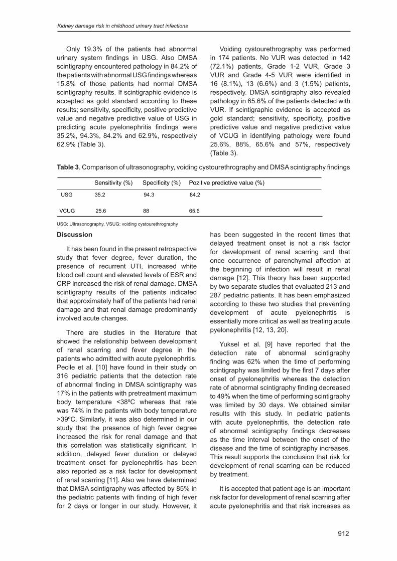
Kidney damage risk in childhood urinary tract infections
912
Only 19.3% of the patients had abnormal urinary system findings in USG. Also DMSA scintigraphy encountered pathology in 84.2% of the patients with abnormal USG findings whereas 15.8% of those patients had normal DMSA scintigraphy results. If scintigraphic evidence is accepted as gold standard according to these results; sensitivity, specificity, positive predictive value and negative predictive value of USG in predicting acute pyelonephritis findings were 35.2%, 94.3%, 84.2% and 62.9%, respectively 62.9% (Table 3).
Voiding cystourethrography was performed in 174 patients. No VUR was detected in 142 (72.1%) patients, Grade 1-2 VUR, Grade 3 VUR and Grade 4-5 VUR were identified in 16 (8.1%), 13 (6.6%) and 3 (1.5%) patients, respectively. DMSA scintigraphy also revealed pathology in 65.6% of the patients detected with VUR. If scintigraphic evidence is accepted as gold standard; sensitivity, specificity, positive predictive value and negative predictive value of VCUG in identifying pathology were found 25.6%, 88%, 65.6% and 57%, respectively (Table 3).
Table 3. Comparison of ultrasonography, voiding cystourethrography and DMSA scintigraphy findings
Sensitivity (%) Specificity (%) Pozitive predictive value (%)
USG 35.2 94.3 84.2
VCUG 25.6 88 65.6
USG: Ultrasonography, VSUG: voiding cystourethrography
Discussion
It has been found in the present retrospective study that fever degree, fever duration, the presence of recurrent UTI, increased white blood cell count and elevated levels of ESR and CRP increased the risk of renal damage. DMSA scintigraphy results of the patients indicated that approximately half of the patients had renal damage and that renal damage predominantly involved acute changes.
There are studies in the literature that showed the relationship between development of renal scarring and fever degree in the patients who admitted with acute pyelonephritis. Pecile et al. [10] have found in their study on 316 pediatric patients that the detection rate of abnormal finding in DMSA scintigraphy was 17% in the patients with pretreatment maximum body temperature <38ºC whereas that rate was 74% in the patients with body temperature >39ºC. Similarly, it was also determined in our study that the presence of high fever degree increased the risk for renal damage and that this correlation was statistically significant. In addition, delayed fever duration or delayed treatment onset for pyelonephritis has been also reported as a risk factor for development of renal scarring [11]. Also we have determined that DMSA scintigraphy was affected by 85% in the pediatric patients with finding of high fever for 2 days or longer in our study. However, it
has been suggested in the recent times that delayed treatment onset is not a risk factor for development of renal scarring and that once occurrence of parenchymal affection at the beginning of infection will result in renal damage [12]. This theory has been supported by two separate studies that evaluated 213 and 287 pediatric patients. It has been emphasized according to these two studies that preventing development of acute pyelonephritis is essentially more critical as well as treating acute pyelonephritis [12, 13, 20].
Yuksel et al. [9] have reported that the detection rate of abnormal scintigraphy finding was 62% when the time of performing scintigraphy was limited by the first 7 days after onset of pyelonephritis whereas the detection rate of abnormal scintigraphy finding decreased to 49% when the time of performing scintigraphy was limited by 30 days. We obtained similar results with this study. In pediatric patients with acute pyelonephritis, the detection rate of abnormal scintigraphy findings decreases as the time interval between the onset of the disease and the time of scintigraphy increases. This result supports the conclusion that risk for development of renal scarring can be reduced by treatment.
It is accepted that patient age is an important risk factor for development of renal scarring after acute pyelonephritis and that risk increases as

913
the patient age decreases. It has been reported that infants (below one year of age) carry the highest risk while the pediatric patients aged over 5 years carry lower risk [21, 22]. On the contrary, some studies have reported that age is positively correlated with the development of renal scarring [9, 10]. Yuksel et al. [9] have carried out a study on 201 patients with acute pyelonephritis aged between 0-14 years and identified abnormal scintigraphy findings with the rates of 22%, 47% and 53% in the pediatric patients aged below one-year-old, between 1-5 years and over 5 years, respectively. Also in our study, the rates of renal damage were 35%, 56.7% and 42% in the pediatric patients aged below one-year-old, between 1-5 years and over 5 years, respectively. Even though, the detection rate of abnormal scintigraphy finding was found higher than the other groups, however, no statistically significant difference was found between the age groups regarding detection rate of abnormal scintigraphic finding.
It has been demonstrated that irreversible renal damage occurs due to inflammation, interstitial injury and fibrosis that develop during acute pyelonephritis. This state is defined as the development of renal scarring displayed with renal scintigraphic examination. In addition, some authors have shown that inflammatory process continues even after recovery of urinary tract infection [23, 24]. Febrile recurrent UTI has been identified to be an independent risk factor for renal scarring in a prospective study conducted on 565 pediatric patients with febrile UTI and/or VUR. It has been observed in the same study that DMSA scintigraphy findings were normal during follow-up process in 76% of the patients despite recurrent febrile UTI episodes [25]. In also our study, normal scintigraphic findings were monitored in 32.6% of the patients with recurrent urinary tract infection. Although, no reason for nonoccurrence of renal scarring despite recurrent UTIs in some pediatric patients, it has been suggested that genetically protective factors may be present. It is considered that immune response of the host has a critical role in development of renal scarring [20]. DMSA is gold standard in the diagnosis of acute pyelonephritis. However, scintigraphy is most commonly not performed during diagnosis since it is an invasive procedure. The fact that some patients followed-up with diagnosis
of pyelonephritis may not be a true case of pyelonephritis may also affect these results.
High white blood cell count and increased levels of CRP and ESR are the important laboratory parameters. Several studies have reported that there is a positive correlation between development of renal damage and high white blood cell account. Doganis et al. [12] have ascertained in their study on 278 patients that mean white blood cell count was significantly higher in the patients with abnormal scintigraphic findings. Similar results were obtained also in our study. However, there are also some studies that demonstrated that there is no relationship between white blood cell count and renal damage [26, 27]. In our study, mean CRP level was found significantly higher in the patients with abnormal scintigraphic findings compared to those with normal scintigraphic findings. CRP is one of the most important laboratory parameters in differentiation between upper and lower urinary tract infections. Therefore, many studies have addressed the relationship between CRP and renal damage. The studies have shown that the patients with abnormal findings in DMSA scintigraphy after acute pyelonephritis had significantly higher CRP levels. In contrast to other studies, Jakobsson et al. [8] have found no difference between CRP levels of the patients detected and not detected with renal scarring in DMSA scintigraphy. ESR is also one of the distinctive criteria like CRP in diagnosis of acute pyelonephritis. Garin et al. [16] have reported that ESR has higher sensitivity than CRP in diagnosis of acute pyelonephritis. The studies have shown that ESR values of the patients with renal damage than those without renal damage [9, 12, 16]. Similarly, with the studies in the literature, mean ESR values of the group with abnormal scintigraphy findings were found statistically higher than that of the group with normal scintigraphy findings in also our study.
Even though, ultrasonography does not provide adequate information about renal functions, it is still the primarily performed imaging method in diagnosis of the pediatric patients. There are studies in the literature that shows the efficacy of ultrasonography in detection of renal damage [5, 6, 28]. Christian et al. [28] have carried out a study on 990 patients and obtained
Pamukkale Medical Journal 2021;14(4):908-915 Yuksel et al.

Kidney damage risk in childhood urinary tract infections
914
a low sensitivity rate of ultrasonography such as 21.7% in detection of renal cortical scarrings. Moorthy et al. [29] have determined a sensitivity rate of 47.2% and a specificity rate of 91.8% for ultrasonography regarding detection of diffuse renal scarring. Yuksel et al. [9] have carried out a study on 201 patients with urinary system infections and the sensitivity and specificity rates of ultrasonography in detection of renal cortical scarrings were found 29% and 90% in this study, respectively. Compared with the results of DMSA scintigraphy scan accepted as the gold standard; our results demonstrated that ultrasonography has low sensitivity although it has a high specificity in detection of renal scarring. Currently, we have concluded that ultrasonography cannot be replaced with DMSA scintigraphy.
The relationship between UTI, VUR and development of renal damage has been demonstrated in many studies. The prevalence of VUR has been reported to be approximately 30% in the pediatric patients who had febrile UTI for the first time. However, the frequency of VUR in the healthy population is not known [1]. The present study showed that VUR was detected in 18.4% of the patients that had pyelonephritis. That rate was reported to be 22% by Yuksel et al. [9]. In our study, sensitivity, specificity, positive predictive value and negative predictive value of VCUG in identifying pathology were found 25.6%, 88%, 65.6% and 57%, respectively. In the study of Moorthy et al. [4], senstivity and specificity rates of VCUG regarding detection of diffuse renal scarring to be 50% and 89.9%, respectively. Yuksel et al. [9] have reported the sensitivity and specificity rates of VUCG in detection of renal cortical scarrings to be 33% and 94%, respectively.
In conclusion, urinary tract infection is an important infectious disease that should be considered seriously and treated meticulously. The rate of renal damage can be determined by the clinical and laboratory data obtained at baseline admission of the patients. Taking late-term serious complications into consideration, morbidity and mortality rates can be reduced by appropriate management and treatment
Conflict of interest: No conflict of interest was declared by the authors.
References1. Tullus K, Shaikh N. Urinary tract infections in children.
Lancet 2020;395:1659-1668. https://doi.org/10.1016/S0140-6736(20)30676-0
2. Millner R, Becknell B. Urinary tract infections. Pediatr Clin North Am 2019;66:1-13. https://doi.org/10.1016/j.pcl.2018.08.002
3. Foxman B. Epidemiology of urinary tract infections: incidence, morbidity, and economic costs. Am J Med 2002;113;5-13. https://doi.org/10.1016/s0002-9343(02)01054-9
4. Moorthy I, Easty M, McHung K, Ridout D, Biassoni D, Gordon I. The presence of vesicoureteric reflux does not identify a population at risk for renal scarring following a first urinary tract infection. Arch Dis Child 2005;90:733-736. https://doi.org/10.1136/adc.2004.057604
5. Yüksel S, Yüksel G, Çakar N. Urinary tract infection in children. T Klin Pediatri 2002;11:41-49.
6. Sinha MD, Gibson P, Kane T, Lewis MA. Accuracy of ultrasonic detection of renal scarring in different centres using DMSA as the gold standart. Nephrol Dial Transplant 2007;22:2213-2216. https://doi.org/10.1093/ndt/gfm155
7. Benador D, Benador N, Slosman D, Mermillod B, Girardin E. Are younger children at highest risk of renal sequelae after pyelonephritis? Lancet 1997;349:17-19. https://doi.org/10.1016/S0140-6736(96)06126-0
8. Jakobsson B, Berg U, Svensson L. Renal scarring after acute pyelonephritis. Arch Dis Child 1994;70:111-115. https://doi.org/10.1136/adc.70.2.111
9. Yüksel S, Çakar N, Çakmak FN, et al. Risk factors of renal damage in children with acut pyelonephritis. Turkiye Klinikleri J Pediatr 2007;16:158-164.
10. Pecile P, Miorin E, Romanello C, et al. Age-related renal paranchymal lesions in children with first febrile urinary tract infections. Pediatrics 2009;124:23-29. https://doi.org/10.1542/peds.2008-1192
11. Hiraoka M, Hashimoto G, Tsuchida S, Tsukahara H, Ohshima Y, Mayumi M. Early treatment of urinary infection prevents renal damage on cortical scintigraphy. Pediatr Nephrol 2003;18:115-118. https://doi.org/10.1007/s00467-002-1023-y
12. Doganis D, Siafas K, Mavrikou M, et al. Does early treatment of urinary tract infection prevent renal damage? Pediatrics 2007;120:922-928. https://doi.org/10.1542/peds.2006-2417
13. Hewitt IK, Zucchetta P, Rigon L, et al. Early treatment of acute pyelonephritis in children fails to reduce renal scarring: data from the Italian renal ınfection study trials. Pediatrics 2008;122:486-490. https://doi.org/10.1542/peds.2007-2894
14. Smellie JM, Poulton A, Prescod NP. Retrospective study of children with renal scarring associated with reflux and urinary infection. BMJ 1994;308:1193-1196. https://doi.org/10.1136/bmj.308.6938.1193

Pamukkale Medical Journal 2021;14(4):908-915 Yuksel et al.
915
15. Chang SL, Shortliffe LD. Pediatric urinary tract infections. Pediatr Clin North Am 2006;53:379-400. https://doi.org/10.1016/j.pcl.2006.02.011
16. Garin EH, Olovarria F, Araya C, Broussain M, Barrea C, Young L. Diagnostic significance of clinical and laboratory findings to localize site of urinary infection. Pediatr Nephrol 2007;22:1002-1006. https://doi.org/10.1007/s00467-007-0465-7
17. Lai SW, Ng KC. Retrospective analysis of inflammatory parameters in acute pyelonephritis. Scand J Urol Nephrol 2003;37:250-252. https://doi.org/10.1080/00365590310008145
18. Soylu A, Kasap B, Demir K, Türkmen M, Kavukçu S. Predictive value of clinical and laboratory variables for vesicoureteral reflux in children. Pediatr Nephrol 2007;22:844-848. https://doi.org/10.1007/s00467-006-0418-6
19. Oostenbrink R, Van der Heijden AJ, Moons KG, Moll HA. Prediction of vesico-ureteric reflux in childhood urinary tract infection: a multivariate approach. Acta Paediatr 2000;89:806-810.
20. Kosmeri C, Kalaitzidis R, Siomou E. An update on renal scarring after urinary tract infection in children: what are the risk factors? J Pediatr Urol 2019;15:598-603. https://doi.org/10.1016/j.jpurol.2019.09.010
21. Spencer JR, Schaeffer AJ. Paediatric urinary tract infections. Urol Clin North Am 1986;13:661-672.
22. Gleeson FV, Gordon I. Imaging in urinary tract infection. Arch Dis Child 1991;66:1282-1283.
23. Kaack MB, Dowling KJ, Patterson GM, Roberts JA. Immunology of pyelonephritis VIII. E. coli causes granulocytic aggregation and renal ischemia. J Urol 1986;136:1117-1122. https://doi.org/10.1016/s0022-5347(17)45235-9
24. Roberts JA. Mechanisms of renal damage in chronic pyelonephritis (reflux nephropathy). Curr Top Pathol 1995;88:265-287. https://doi.org/10.1007/978-3-642-79517-6_9
25. Snodgrass WT, Shah A, Yang M, et al. Prevalence and risk factors for renal scars in children with febrile UTI and/or VUR: a cross-sectional observational study of 565 consecutive patients. J Pediatr Urol 2013;9:856-863. https://doi.org/10.1016/j.jpurol.2012.11.019
26. Ferreiro C, Piepsz A, Nogarède C, Tondeur M, Hainaut M, Levy J. Late renal sequelae in intravenously treated complicated urinary tract infection. Eur J Pediatr 2013;172:1243-1248. https://doi.org/10.1007/s00431-013-2024-5
27. Keren R, Shaikh N, Pohl H, et al. Risk factors for recurrent urinary tract infection and renal scarring. Pediatrics 2015;136:13-21. https://doi.org/10.1542/peds.2015-0409
28. Christian MT, McColl JH, MacKenzie JR, Beattie TJ. Risk assesment of renal cortical scarring with urinary tract infection by clinical features and ultrasonography. Arch Dis Child 2000;82:376-380. https://doi.org/10.1136/adc.82.5.376
29. Moorthy I, Wheat D, Gordon I. Ultrasonography in the evaluation of renal scarring using DMSA scan as the gold standard. Pediatr Nephrol 2004;19:153-156. https://doi.org/10.1007/s00467-003-1363-2
Ethics committee approval: The study was approved by the ethic committee of Pamukkale University (date: 14.01.2011 and number: B.30.2.PAÜ.0.20.05.09/11).
Authors’ contributions
S.Y. built the main idea and hypothesis. B.S. arranged the material and method section and evaluated the data. The article was written, reviewed and necessary corrections were made by T.B. and S.Y. In addition, all authors discussed the entire study and approved the final version.

916
26 yaşındaki genç maden işçisinde eş zamanlı iki taraflı femur boyun stres kırığı
Concurrent two-sided femur neck stress fracture in 26-year-old young mine worker
Murat Saylık, Kemal Gökkuş
Pamukkale Tıp DergisiPamukkale Medical JournalOlgu Sunumu
Murat Saylık, Dr. Öğr. Gör. İstinye Üniversitesi, Medikalpark Bursa Hastanesi, Ortopedi ve Travmatoloji Anabilim Dalı, Bursa, Türkiye, e-posta: [email protected] (https://orcid.org/0000-0002-1023-4164) (Sorumlu Yazar)Kemal Gökkuş, Doç. Dr. Başkent Üniversitesi Tıp Fakültesi Alanya Araştırma ve Uygulama Hastanesi, Ortopedi ve Travmatoloji Anabilm Dalı, Alan-ya-Antalya, Türkiye, e-posta: [email protected] (https://orcid.org/0000-0002-4916-3471)
ÖzGenç hastalarda eş zamanlı iki taraflı femur boyun stres kırığı (FBSK) çok nadir görülür. Bununla ilgili literatürde sınırlı sayıda olgu bildirilmiştir. Bu olgular çoğunlukla yüksek fiziksel aktivite altında kalan askerler ve atletler, epilepsi gibi nöbet geçirenler, yaşlı veya metobolik kemik hastalığı olan orta yaş hastalardır. Bizim olgumuz sporcu olmayan sağlıklı genç bir maden işçisinin geç tanı konulan eş zamanlı bilateral femur boyun kırığı nedeniyle literatürden farklıdır. Bu olguyu genç maden işçilerindeki iki taraflı femur boyun stres kırığında, tanı koymanın zorluğunu ve acil müdahele süresi (24-48 saat) geçmiş olsa da, stabil kırık fiksasyonu ile femur başı avasküler nekrozu gelişmeden kırığın iyileşebileceğini vurgulamak için sunduk.
Anahtar kelimeler: Femur, boyun, kırık, iki taraf.
Saylık M, Gökkuş K. 26 yaşındaki genç maden işçisinde eş zamanlı iki taraflı femur boyun stres kırığı. Pam Tıp Derg 2021;14:916-920.
AbstractConcurrent two-sided femoral neck stress fracture (FNSK) is very rare in young patients. A limited number of cases have been reported in the literature. The majority of them are; soldiers, athletes, epileptic patients, and elderly/middle-aged patients with bone pathology. Our case differs from literature due to a concurrent bilateral femur neck fracture diagnosed late of a healthy young mine worker who is not an athlete. This report aims to emphasize the challenge of diagnosing bilateral femoral neck fractures in young miners and demonstrating excellent healing (without avascular necrosis) of the head with stable fixation even though the critical period of 48 hours has been spent.
Key words: Femur, neck, fracture, bilateral.
Saylik M, Gokkus K. Concurrent two-sided femur neck stress fracture in 26-year-old young mine worker. Pam Med J 2021;14:916-920.
Case Report
Gönderilme tarihi:24.12.2020 Kabul tarihi:17.02.2021
Giriş
Stres kırıklarının %8’ini oluşturan femur boyun stres kırığı (FBSK) ilk olarak 1905’te Blecher tarafından bildirilmiştir [1]. FBSK biomekanik oluşma ve yer değiştirme derecesine göre sınıflandırılmıştır. Tip-1; kompresyon tipi ve ayrılmamış kırık. Tip-2; gerilim tipi ve ayrılmamış kırık. Tip-3; ayrılmış kırık [2].
Genç popülasyonda iki taraflı FBSK nadir görülür. Gençlerde iki taraflı FBSK genellikle askerlerde, atletlerde, patolojik kemik hastalıklarında, tekrarlayan ağır yük taşımada, epilepsi benzeri nöbet geçirenlerde oluşabilir [3-5]. Gebelik sonrası gelişen transient kalça osteoporozuna sekonder bilateral FBSK olgusu bildirilmiştir [5, 6]. Olgular kasık ağrısı
ve geçmeyen uzun süreli ağrı şikayeti ile başvurur. Ayırıcı tanıda pubis stress kırığı, adduktör tendinit, bel fıtığı ve ürolojik problemler düşünülmelidir.
Bu olguyu genç maden işçilerinde iki taraflı FBSK’da tanı koymanın zorluğunu ve kabül görmüş cerrahi müdahele süresi (24-48 saat) geçmiş olsa da, stabil kırık fiksasyonu ile kırığın iyileşebileceğini vurgulamak için sunduk.
Olgu sunumu
26 yaşında erkek hasta, travma öyküsü olmaksızın her iki kasıkta ağrı ve yürüyememe şikayetiyle, ağrı başlangıcından 17 gün sonra tekerlekli sandalyede polikliniğimize başvurdu. Hasta bir kömür madeninde 2 yıldır çalıştığını ifade etti. Hastanın klinik sorgusunda, alkol,
doi:https://dx.doi.org/10.31362/patd.846177

917
Pamukkale Tıp Dergisi 2021;14(4):916-920 Saylık ve Gökkuş
kortikosteroid, antikoagülan veya antipsikotik ilaç kullanımı gibi kemik kırılganlığıyla ilişkili risk faktörünün olmadığı tespit edildi. Herhangi bir sistemik hastalık öyküsü yoktu. Nörolojik ve vasküler muayenede anormal bir bulgu saptanmadı.
Metabolik kemik hastalıkları, renal ve endokrinolojik hastalıkların tespiti için endokrinoloji ve nefroloji bölümlerinden konsultasyonlar istendi. Konsültasyonlar ve laboratuvar sonuçlarına göre Tip II diyabet, kronik böbrek hastalığı, romatoid artrit saptanmadı. Orta seviyede D vitamini eksikliği bulundu (Tablo 1).
Direk kalça radyografi sinde sağ femur boynunda şüpheli bir radyolusen hat, sol fémur boynunda patolojik görünüm saptanmadı. FBSK’na sebep olabilecek, asetabuler displazi, femoroasetabular sıkışma, asetabular retroversiyon saptanmadı [7]. Manyetik
rezonans görüntülemede, her iki femur boynunda kemik iliği ödemi ve Koronal T2 yağ baskılı MR görüntüde, her iki femur boynunda sağda komplet, solda inkomplet FBSK görüldü (Şekil 1).
Şekil 1. Preoperatif yağ baskılayıcı koronal MR kesitte her iki kalçada sağda komplet solda inkomplet FBSK ve boyun bölgesindeki ödem
Tablo 1. Olgunun ameliyat öncesi laboratuvar bulguları
Tetkik Değer Yorum Normal Değer Sınırı Birim
WBC 7,7 N 4,4-11,0 10/uL
AKŞ 142 H 70-15 mg/dL
BUN 15,2 N 8,9-22,6 mg/dL
Creatinin 0,81 N 0,72-1,31 mg/dL
Albumin 4,3 N 3,5,05 g/dL
AST (SGOT 19,4 N 5,34 U/L
ALT (SGPT) 31,7 N 0-55 U/L
Biluribin (Total) 0,3 N 0,2-1,2 mg/dL
Sodyum 136,8 N 135-145 mmol/L
Potasyum 4 N 3,5-5,1 mmol/L
Kalsiyum 8,9 N 8,4-10,5 mg/dL
Demir 76 N 65-175 mg/dL
ALP 109,8 N 40-150 U/L
Testesteron 337 N 240,24-870,68 ng/dL
Ürikasid 6,5 N 3,5-7,2 mg/dL
Kolesterol (Total) 117,7 N 0-200 mg/dL
İnorganik fosfor 3,8 N 2,3-4,7 mg/dL
Ferritin 130,4 N 21,81-274,66 ng/mL
TSH 3061 N 0,35-4,94 yIU/ml
PTH 23,4 N 15-68,3
Vitamin D3 (25-OH- Kolekalsiferol) 17 Orta derece eksiklik 10-20 ng/ml ng/mL

Eş zamanlı iki tarafl ı femur boyun stres kırığı
918
Hastamıza bilateral FBSK tanısı konularak, ameliyat öncesi tetkikler ve anestezi konsultasyonu yapıldı. Spinal anestezi altında skopi kontrolünde, sol kalçaya inkomplet kırık olduğu için 2 adet kanüllü vida ile sağ kalçaya komplet kırık olduğu için 3 adet kanüllü vida ile tespit uygulandı. Ameliyat sonrası 1. günde kısmi ağırlık verilerek destek ile mobilize edildi. Birinci aydaki klinik takipte, her iki kalça AP ve kurbağa bacağı pozisyonunda radyografi de stabil tespiti (Şekil 2a, 2b) görülerek çift koltuk değneği ile kısmi yük vermesine izin verildi.
Şekil 2a. Postoperatif 1.ay pelvis ön-arka radyografi sinde sağ kalçaya 3 adet ve sol kalçaya 2 adet kanüllü vida ile osteosentezin radyografi k görüntüsü
Şekil 2b. Postoperatif 1.ay her iki kalça kurbağa bacağı pozisyonu radyografi sinde osteosentezin görüntüsü
İkinci aydaki klinik takipte, radyografi de kaynamanın görülmesi üzerine, hastanın desteksiz kısa yürüyüşüne izin verildi. On ikinci aydaki klinik takipte radyografi de yeterli kırık iyileşmesi görüldü. Kalça hareketleri tam ve ağrısızdı. Ameliyathane ortamında spinal anestezi altında kanüllü vidalar çıkarıldı.
18. ayda çekilen MR’da (Şekil 3a, 3b) kırıkta tam iyileşme görülürken avasküler nekrozun gelişmediği teyit edildi.
Şekil 3a. 18. aydaki Coronal yağ baskılayıcı MR görüntüsü
Şekil 3b. 18. aydaki aksiyel yağ baskılayıcı MR görüntüsü
Tartışma
Genç maden işçisinde, iki tarafl ı ve eş zamanlı gelişen kompresyon tipi kaymamış FBSK’da, 17. günde skopi kontrolünde perkütan kanüllü vida ile tespit uygulandı. Tedavi sonucunda femur başında avaskuler nekroz gelişmeden kırıkta iyileşme sağlandı.
FBSK’larının konservatif veya cerrahi tedavisi, kırığın kompresyon veya gerilme tipine, kırığın ayrılma derecesiyle beraber hastanın genel durumuna göre verilir. Cerrahi tedavide; fi ksasyon, valgus osteotomisi yapılarak fi ksasyon, parsiyel ve total kalça protezi uygulanabilir [8].
FBSK’da; kırığın ayrılma riski ve femur başı avasküler nekrozu gelişim riski olduğu için cerrahi tedavi ön plandadır. Özellikle gerilim tipi FBSK’ları vertikale yakın kırıklardır ve avasküler nekroz riski fazladır [9]. Kompresyon

919
veya gerilim tipi FBSK’da, kırık hattı boynun %50‘sinden fazlaysa perkütan kanüllü vida fiksasyonu düşünülmelidir [10]. Bu makalede sunulan olguda sağ femur boynunda %100, sol femur boynunda %35 oranında kompressif kırık mevcuttu. Sol kalça fiksasyon nedenimiz: iki taraflı FBSK olduğundan, erken mobilizasyon ve yük verebilme avantajıydı.
Bazı yayınlarda FBSK’da internal tespite rağmen başarısızlık oranı %59 gibi yüksek oranlarda bildirilmiştir [11]. Avasküler nekroz (%12-86), kırıkta kaynamama (%10-59) ve erken evre osteoartrit (%14-66) tedavide başarısızlığın en önemli sebepleridir [12]. Literatür araştırmamızda ise iki taraflı kompresyon türü FBSK olan ve kanüllü vida ile tedavi edilmiş hastalarda, sadece bir hastada kırıkta kaynamama bildirilmişti (Tablo 2).
Stres kırıkları iki guruba ayrılmıştır. Normal stres altında patolojik kemik yapısında oluşan kırığa yetersizlik kırığı ve anormal stres altında normal kemik yapısında oluşan kırığa yorgunluk kırığı denmiştir [13]. Hastamızda orta derecede D vitamini eksikliği ve madenci olması nedeniyle anormal stres yükü olduğundan, kanımızca iki fizyopatolojik yolak beraber işlemiş olabilir.
FBSK tanısında MR ve kemik sintigrafisi diğer radyolojik tetkiklerden daha değerlidir [14, 15]. Olgumuzda tanı koyarken ve tedavimizi planlarken radyografi ve MR görüntülerini kullandık. MR ile tanı belirginleştiğinden sintigrafiye gerek kalmadı.
Bir çalışmada serum 25-hidroksi vitamin D ile güneş ışığına maruz kalma arasında anlamlı bir pozitif korelasyon olduğunu, ancak serum 25-hidroksi vitamin D’nin günlük D vitamini alımı ile korele olmadığı gösterilmiştir [16].
Olgumuz, maden işçisi olduğundan güneş enerjisinden nispeten az faydalanıyordu ve laboratuvar tetkiklerinde orta derecede D vitamini eksikliği tespit edildi. D vitamini eksikliğinin osteoporoz için risk faktörlerinden biri olduğu ve altta yatan başka risk faktörü olmayan genç hastalarda FBSK’na yol açabileceği bildirilmiştir [17]. Olgumuzda orta derece D vitamini eksikliği dışında bir bulgu elde edilemedi. Özellikle genç hastalardaki iki taraflı FBSK’da, D vitamini eksikliği etkisinin anlaşılması için ileri araştırmalar yapılmalıdır.
Sonuç olarak, maden işçilerinde iki taraflı kasık ağrısı şikayeti, muhtemel D vitamini eksikliğini ve buna bağlı FBSK olasılığını akla getirmelidir. Farkındalık tanıyı kolaylaştıracaktır. Kırk sekiz saatten daha uzun zaman geçmiş olsa bile stabil kırık tespitiyle avasküler nekroz gelişmeden kırık iyileşmesi sağlanabileceği akılda tutulmalıdır.
Çıkar ilişkisi: Yazarlar çıkar ilişkisi olmadığını beyan eder.
Tablo 2. Kompresyon tipi FBSK tanısıyla kanullü vida metodu uygulanan makaleler
FBSK Tipi Osteosentez Takip Süresi
Komplikasyon Yaş ve Cinsiyet
Naik et al. Kompresyon-Sol Kanüllü vida 12 ay Yok 38-Kadın
Oliviera et al. Kompresyon-Sağ ve sol Kanüllü vida Bilgi yok Bilgi yok 43-Erkek
Vaishya at al. Kompresyon-Sol ve sağ Kanüllü vida Bilgi yok Bilgi yok 50-Erkek
Baki et al. Kompresyon-Sol ve sağ Kanüllü vida 6 ay Yok 22-Kadın
Webber et al. Kompresyon-Sağ ve sol Kanüllü vida Bilgi yok Yok 23-Erkek
Nemoto et al. Kompresyon-Sağ ve sol Kanüllü vida 24 ay Yok 24-Erkek
Voss et al. Kompresyon-sağ Kanüllü vida 24 ay Nonunion 30 Kadın
Romero et al. Kompresyon-sağ Kanüllü vida 6 ay Yok 19-Erkek
Kalaci et al. Kompresyon-Sağ ve sol Kanüllü vida 6 ay Yok 18-Kadın
Saylik et al. (mevcut makale) Kompresyon-Sağ ve sol Kanüllü vida 18 ay Yok 26- Erkek
Pamukkale Tıp Dergisi 2021;14(4):916-920 Saylık ve Gökkuş
Eş zamanlı iki taraflı femur boyun stres kırığı
920
Kaynaklar1. Blecher A. Uber den einfluss des parademarsches
auf die entstehungder fussgeschwulst. Med Klin 1905;1:305-306. https://doi.org/10.1155/2015/306246
2. Cody JP, Evans KN, Kluk MW, Andersen RC. Occult femoral neck fracture associated with vitamin D deficiency diagnosed by MRI: case report. Mil Med 2012;177:605-608. https://doi.org/10.7205/MILMED-D-11-00321
3. Shah HM, Grover A, Gadi D. Bilateral neck femur fracture following a generalized seizure - a rare case report. Arch Bone Jt Surg 2014;2:255-257.
4. Cagirmaz T, Yapici C, Orak MM, Guler O. Bilateral femoral neck fractures after an epileptic attack: a case report. International Journal of Surgery Case Reports 2015;6:107-110. https://doi.org/10.1016/j.ijscr.2014.12.003
5. Ly TV, Swiontkowski MF. Treatment of femoral neck fractures in young adults. J Bone Joint Surg Am 2008;90:2254-2266.
6. Kezunovic M, Bulatovic N, Kadic V. Bilateral subcapital femoral neck fracture in a 28 year old postpartum woman. Med Arch 2017;71:151-153. https://doi.org/10.5455/medarh.2017.71.151-153
7. Wassilew GI, Heller MO, Janz V, Perka C, Müller M, Renner L. High prevalence of acetabular retroversion in asymptomatic adults: a 3D CT-based study. Bone Joint J 2017;99:1584-1589. https://doi.org/10.1302/0301-620X.99B12.37081
8. Moerenhout K, Gkagkalis G, Laflamme GY, Rouleau DM, Leduc S, Benoit B. Total hip arthroplasty for bilateral femoral neck stress fracture: a case report and review of the literatüre. Hindawi 2019;2019:8. https://doi.org/10.1155/2019/2720736
9. Nemoto O, Kawaguchi M, Katou T. Simultaneous bilateral femoral neck stress fractures in a 24-year old male recruit: a case report. West Indian Med J 2013;62:552-553. https://doi.org/10.7727/wimj.2012.103
10. Fullerton LR, Snowdy JrHA. Femoral neck stress fractures. The Am J Sports Med 1988;16:365-377. https://doi.org/10.1177/036354658801600411
11. Rogmark C, Kristensen MT, Viberg B, Rönnquist SS, Overgaard S, Palm H. Hip fractures in the non-elderly-who, why and whither? Injury 2018;49:1445-1450. https://doi.org/10.1016/j.injury.2018.06.028
12. Henari S, Leonard M, Hamadto M, Cogley D. Review of a single contemporary femoral neck fracture fixation method in young patients. Orthopedics 2011,34:171. https://doi.org/10.3928/01477447-20110124-09
13. Bailie DS, Lamprecht DE. Bilateral femoral neck stress fractures in an adolescent male runner: a case report. The American Journal of Sports Medicine 2001;29:811-813. https://doi.org/10.1177/03635465010290062301
14. Egol KA, Koval KJ, Kummer F, Frankel VH. Stress fractures of the femoral neck. Clin Orthop Relat Res 1998;348:72-78.
15. Rizzo PF, Gould ES, Lyden JP, Asnis SE. Diagnosis of occult fractures about the hip. Magnetic resonance imaging compared with bone-scanning. J Bone Joint Surg Am 1993;75:395-401. https://doi.org/10.2106/00004623-199303000-00011
16. Wang J, Yang D, Yu Y, Shao G, Wang Q. Vitamin D and sunlight exposure in newly-diagnosed Parkinson’s disease. Nutrients 2016;8:142. https://doi.org/10.3390/nu8030142
17. Palacios C, Gonzalez L. Is vitamin D deficiency a major global public health problem? J Steroid Biochem Mol Biol 2014;144:138-145. https://doi.org/10.1016/j.jsbmb.2013.11.003
Hasta onamı: Hastadan klinik bilgiler ve resimlerin kullanımı için yazılı onay alınmıştır.
Yazarların makaleye olan katkısı
M.S. vakaya tanıyı koyan, ameliyatını ve takiplerini yapan, yazının taslağını ve kaynakçasını hazırladı. K.G. yazının literatür taramasında katkıda bulunan kaynakçanın yazılımında destek oldu.


922
Secondary bladder stone formation on polypropylene suture after burch colposuspension and abdominal hysterectomy
Burch kolposüspansiyonu ve abdominal histerektomi sonrası polipropilen sütüre ikincil mesane taşı oluşumu
Mert Hamza Ozbilen, Batuhan Ergani, Taha Cetin, Mehmet Yigit Yalcin, Cagdas Bildirici, Erkin Karaca, Orcun Celik, Yusuf Ozlem Ilbey
Pamukkale Tıp DergisiPamukkale Medical JournalOlgu Sunumu
AbstractBladder calculies are stones or calcified materials found in the bladder. Foreign bodies that come into contact with the urine may cause bladder stones. Examples of foreign body include suture materials, clips, catheters, ureteral stents, urinary incontinence meshes or migration of an intrauterine device. A 63-year-old female patient was admitted to our urology clinic with dysuria, frequent urination and mixed type urinary incontinence for a year. The patient had a history of abdominal hysterectomy and Burch colposuspension two years ago. Computerized tomography of the patient revealed a bladder stone measuring 2.9x2.6 cm.The patient underwent cystolithotripsy with Holmium (Ho: YAG) laser. The bladder stone was found to be attached to the bladder wall by a polypropylene suture. The suture material was removed endoscopically. The patient has no complaints during the 2-year follow-up. The presence of nonabsorbable suture material in the bladder is a well-known cause of urinary tract infection and stone formation and defined complication of hysterectomy and colposuspension. Information on the lithogenic properties of various suture materials is unfortunately limited to only in vitro and in vivo animal studies. In humans, experience with stone formation related to suture materials is limited to case reports. It has been observed that lithogenicity is related to the longevity of the suture material and in the studies carried out it has been observed that the rate of calcification with non-absorbable sutures is higher. Treatment includes disintegration of the stone and removal of the foreign body causing the stone.
Key words: Bladder stone, polypropylene, hysterectomy, colposuspension.
Ozbilen MH, Ergani B, Cetin T, Yalcin MY, Bildirici C, Karaca E, Celik O, Ilbey YO. Secondary bladder stone formation on polypropylene suture after burch colposuspension and abdominal hysterectomy. Pam Med J 2021;14:922-925.
ÖzMesane içi kalküller mesanede bulunan taşlar veya kalsifiye olmuş materyallerdir. İdrarla temas eden yabancı cisimler mesane taşlarına neden olabilmektedir. Yabancı cisim örnekleri arasında dikiş materyali, klipsler, kateterler, üreteral stentler, üriner inkontinans meşleri veya intrauterin cihazın göçü sayılabilir. 63 yaşında kadın hasta üroloji kliniğimize yaklaşık 1 yıldır var olan idrar yaparken yanma, sık idrara çıkma ve mikst tip idrar kaçırma şikâyeti ile başvurdu. Hastanın iki yıl önce geçirilmiş abdominal histerektomi ve Burch kolposüspansiyon öyküsü mevcuttu. Çekilen bilgisayarlı tomografide 2,9x2,6 cm ebatlarında mesane taşı saptandı. Hastaya Holmium (Ho: YAG) lazer ile sistolitotripsi yapıldı. Mesane taşının mesane duvarına bir polipropilen sütür ile bağlı olduğu görüldü. Sütür materyali endoskopik olarak çıkarıldı. Hastanın 2 yıllık takibinde herhangi bir şikâyeti bulunmamaktadır. Mesanede emilmeyen sütür materyalinin varlığı, idrar yolu enfeksiyonu ve taş oluşumunun iyi bilinen bir nedenidir ve histerektomi ve kolposüspansiyon operasyonlarının tanımlanmış komplikasyonlarındandır. Çeşitli dikiş malzemelerinin litojenik özelliklerine ilişkin bilgiler, ne yazık ki, sadece in vitro ve hayvansal in vivo çalışmalarla sınırlıdır. İnsanlarda dikiş materyallerine bağlı taş oluşumuyla ilgili
Case Report
Gönderilme tarihi:22.12.2020 Kabul tarihi:12.03.2021
Mert Hamza Ozbilen, Dr. Department of Urology, Health Sciences University Tepecik Education and Research Hospital, Izmir, Turkey, e-mail: [email protected] (https://orcid.org/0000-0002-5733-6790) (Corresponding Author)Batuhan Ergani, Dr. Department of Urology, Konya Beyhekim State Hospital,Konya, Turkey, e-mail: [email protected] (https://orcid.org/0000-0002-4667-855X)Taha Cetin, Dr. Department of Urology, Health Sciences University Bozyaka Education and Research Hospital, Izmir, Turkey, e-mail: [email protected] (https://orcid.org/0000-0003-0330-4854)Mehmet Yigit Yalcin, Dr. Department of Urology, Health Sciences University Tepecik Education and Research Hospital, Izmir, Turkey, e-mail: [email protected] (https://orcid.org/000-0001-9943-7453)Cagdas Bildirici, Dr. Department of Urology, Health Sciences University Tepecik Education and Research Hospital, Izmir, Turkey, e-mail: [email protected] (https://orcid.org/0000-0002-4220-8147)Erkin Karaca, Dr. Department of Urology, Health Sciences University Tepecik Education and Research Hospital, Izmir, Turkey, e-mail: [email protected] (https://orcid.org/0000-0002-9123-4069)Orcun Celik, Assoc. Prof. Dr. Department of Urology, Health Sciences University Tepecik Education and Research Hospital, Izmir, Turkey, e-mail: [email protected] (https://orcid.org/0000-0001-6612-1831)Yusuf Ozlem Ilbey, Prof. Dr. Department of Urology, Health Sciences University Tepecik Education and Research Hospital, Izmir, Turkey, e-mail: [email protected] (https://orcid.org/0000-0002-1483-9160)
doi:https://dx.doi.org/10.31362/patd.845469

923
Introduction
Bladder calculies are stones or calcifi ed materials found in the bladder (or in a structure that acts as a urinary reservoir instead of a bladder). Bladder outlet obstruction is the most common cause of bladder valculi in adults. In addition, foreign bodies that come into contact with the urine may cause bladder stones. Examples of foreign body include suture materials, clips, catheters, ureteral stents, urinary incontinence meshes or migration of an intrauterine device [1]. Approximately 2% of all bladder calculi occur in women. The presence of bladder stones requires careful consideration of the cause [2]. Recently the number of patients undergoing obstetric surgery with the implantation of foreign bodies into the bladder increases and this situation increases bladder stone formation. In this article, we present a 63-year-old female patient who had calculus over suture material in bladder following prior abdominal hysterectomy and Burch colposuspension.
Case report
A 63-year-old female patient was admitted to our urology clinic with dysuria, frequent urination and mixed type urinary incontinence for a year. The patient had a history of abdominal hysterectomy and Burch colposuspension two years ago. Physical examination of the abdomen and external genitalia did not reveal any abnormal fi ndings. Urine culture yielded no bacterial growth. Complete blood count and blood biochemistry were within normal range. The KUB (kidney, ureter and bladder) fi lm revealed a radiopaque shadow of 2.4cmx1.7 cm size over the pelvis (Figure 1) and computerized tomography of the patient revealed a bladder stone measuring 2.9x2.6 cm (Figure 2). Thereafter the patient underwent cystolithotripsy with Holmium (Ho: YAG) laser under spinal anesthesia. The bladder stone was found to be attached to the bladder wall by a polypropylene
suture. The suture material was removed endoscopically (Figure 3a, 3b, 3c). The next day the patient was discharged. The patient was asymptomatic at 1-month follow-up and urine culture was sterile. On postoperative 1-month cystoscopy, the suture region was completely healed. The patient has no complaints during the 2-year follow-up.
Figure 1. Abdominal X-ray showing a radiopaque shadow of 2.4cmx1.7 cm over the pelvis
Figure 2. Image of a transverse plane of CT-scan shows 2.9x2.6 cm bladder stone
deneyimlerse vaka raporlarından ibarettir. Litojenitenin sütür materyalinin uzun ömürlülüğü ile ilişkili olduğu ve yapılan çalışmalarda, emilmeyen sütürlerle taşlaşmanın daha yüksek oranda gerçekleştiği görülmüştür. Tedavi ise taşın parçalanmasını ve taşa sebebiyet veren yabancı cismin çıkarılmasını içerir.
Anahtar kelimeler: Mesane taşı, polipropilen, histerektomi, kolposüspansiyon.
Özbilen MH, Ergani B, Çetin T, Yalçın MY, Bildirici Ç, Karaca E, Çelik O, İlbey YÖ. Burch kolposüspansiyonu ve abdominal histerektomi sonrası polipropilen sütüre ikincil mesane taşı oluşumu. Pam Tıp Derg 2021;14:922-925.
Pamukkale Medical Journal 2021;14(4):922-925 Ozbilen et al.

924
Secondary bladder stone formation on polypropylene suture
Figure 3a. The bladder stone attached to the bladder wall by polypropylene suture
Figure 3b. Suture material after removal of stone around it
Figure 3c. Post-operative suture material and some stone fragments
Discussion
Bladder calculi constitute 5% of the urinary tract calculi and usually occur due to foreign bodies, obstructions or infections [3]. Approximately 2% of all bladder calculi occur in women. The presence of bladder stones requires careful consideration of the cause [2]. As in our patient in a patient with intravesical stone, lower urinary tract symptoms such as pain, urgency and / or intermittent urine stream are usually observed. However, some patients may not have symptoms and urine analysis may be normal or symptoms may occur only as recurrent dysuria and pyuria [4]. The detailed past medical history and physical examination remain the most valuable tools in the diagnosis despite the new advances in diagnostic methods. The presence of nonabsorbable suture material in the bladder is a well-known cause of urinary tract infection and stone formation [5] and defined complication of hysterectomy and colposuspension. Information on the lithogenic properties of various suture materials is unfortunately limited to only in vitro and in vivo animal studies [6, 7]. In humans, experience with stone formation related to suture materials is limited to case reports. It has been observed that lithogenicity is related to the longevity of the suture material and in the studies carried out it has been observed that the rate of calcification with non-absorbable sutures is higher [8]. The nonabsorbable suture we found in the bladder was possibly as a result of the accidental insertion into the bladder while suturing or subsequent erosion of a loop of suture in the bladder lumen, creating a nidus for stone formation [3]. The main stimulus in the formation of calcules is crystal formation. Nucleation occurs around a foreign object as a result of infection or secondary to obstruction with resulting super saturation and then aggregation, proliferation for crystal growth and eventual stone formation [2]. Intravesical stone formation caused by extravesical surgical procedures is rare. Such complications can be avoided by the usage of absorbable sutures such as polyglycaprone, polyglactin, polydioxanone and careful passage of the needles and sutures from the bladder layers. In addition, intraoperative cystoscopy during incontinence surgery is necessary to avoid complications [9]. The main principle of treating a bladder stone is to eliminate the underlying cause of stone formation such as bladder outlet obstruction or bladder infection.

When encrustation or stone formation occurs on the foreign body, the stone forceps can be used to crush the stone to facilitate removal of the foreign body or endoscopic laser lithotripsy can be used as we did. However, open surgery may be necessary because sometimes the foreign body is too large or its nature makes this mandatory.
Bladder stones are rare in women. Suspicion is important for early diagnosis. Previous gynecological and other pelvic surgeries should be questioned. Investigations should include plain radiography, urinalysis, ultrasonography or computed tomography. The diagnosis is confirmed by cystoscopy. Treatment includes disintegration of the stone and removal of the foreign body causing the stone.
Conflict of interest: The authors have no conflicts of interest to declare.
References1. Rafique M. Intravesical foreign bodies: review and
current management strategies. Urol J 2008;5:223-231. Available at: www.uj.unrc.ir. Accepted July, 2008
2. Drach GW. Urinary lithiasis: etiology, diagnosis, and medical treatment. In: Walsh RF, Retik AB, Stamey TA, Vaughan ED. (eds). Campbell’s Urology. 6th ed. Philadelphia, PA: WB Saunders Co; 1992;2085-2156.
3. Schwartz BF, Stoller ML. The vesical calculus. Urol Clin North Am 2000;27:333-346. https://doi.org/10.1016/s0094-0143(05)70262-7
4. Litschgi MS, Benz JJ, Glatthaar E. Bladder stones as a complication of gynecologic surgery. Fortschr Med 1975;7:1627-1628.
5. Zderic SA, Burros HM, Hanno PH, Dudas N, Whitmore KE. Bladder calculi in women after urethrovescial suspension. J Urol 1988;139:1047-1048. https://doi.org/10.1016/s0022-5347(17)42766-2
6. Portilla E, Ramos A, Ramos L, et al. A model of suture-induced urolithiasis with urographic control in the bladder of the rat. J Invest Surg 1999;12:205-211. https://doi.org/10.1080/089419399272476
7. Pal KM. Urinary bladder wall repair: what suture to use? Br J Urol 1998;82:196-198. https://doi.org/10.1046/j.1464-410x.1998.00722.x
8. Morris MC, Baquero A, Redovan E, Mahoney E, Bannett AD. Urolithiasis on absorbable and nonabsorbable suture materials in the rabbit bladder. J Urol 1986;135:602-603. https://doi.org/10.1016/s0022-5347(17)45758-2
9. Timmons MC, Addison WA. Suprapubic teloscopy: extraperitoneal intraoperative technique to demonstrate ureteral patency. Obstet Gynecol 1990;75:137-139.
Informed consent: Written informed consent was obtained from the patient.
Acknowledgements: We wish to thank the patient for allowing us to publish his clinical story. Informed consent to publication was obtained. This manuscript received no specific grant from any funding agency in the public, commercial, or not-for-profit sectors.
Contributions of authors
O.Ç. and Y.Ö.İ. sets up the main idea and hypothesis of the study. Ç.B. and E.K. operated the patient and edited the photographs. The discussion section of the article was written by M.H.Ö., B.E, T.Ç. and M.Y.Y. critically reviewed and revised the manuscript.All authors approved the final manuscript as submitted and agree to be accountable for all aspects of the work.
925
Pamukkale Medical Journal 2021;14(4):922-925 Ozbilen et al.
926
Splenic artery aneurysm in kidney transplant recipientBöbrek nakli alıcısında splenik arter anevrizması
Utku Ozgen, Murat Ozban, Muhammet Arslan, Onur Birsen, Mevlut Ceri, Sevda Yilmaz, Ezgi Doga Yoran, Cagatay Aydin
Pamukkale Tıp DergisiPamukkale Medical JournalOlgu Sunumu
AbstractIn this article, we present the first case of SAA (Splenic Artery Aneurysm) in a kidney transplantation recipient, which was incidentally detected during the preparation of the transplantation and successfully treated. A 25-mm SAA was detected at kidney transplant recipient during the preparation examination by abdominal ultrasonography. Splenic artery ligation and aneurysmectomy was performed before the transplantation. Intraoperative Doppler ultrasonography revealed that there were no abnormalities of the spleen blood supply. During the first month of follow-up, the patient remained asymptomatic and laboratory investigations revealed no abnormalities. Selective ligation and resection of SAAs combined with spleen preservation is a safe treatment modality for selected cases with favorable short and long-term results, allowing the permanent treatment of the SAA, while preserving the splenic function.
Key words: Splenic artery aneurysm, kidney transplantation, aneurysmectomy.
Ozgen U, Ozban M, Arslan M, Birsen O, Ceri M, Yilmaz S, Yoran ED, Aydin C. Splenic artery aneurysm in kidney transplant recipient. Pam Med J 2021;14:926-929.
ÖzBu yazıda, böbrek nakli alıcısında, nakil hazırlığı sırasında tesadüfen tespit edilen ve başarıyla tedavi edilen ilk SAA (Splenik Arter Anevrizması) vakasını sunuyoruz. Karın ultrasonografi ile yapılan hazırlık incelemesinde böbrek nakli alıcılarında 25 mm SAA saptandı. Nakil öncesi splenik arter ligasyonu ve anevrizmektomi yapıldı. İntraoperatif Doppler ultrasonografi ile dalak kan akışında herhangi bir anormallik olmadığı gösterildi. Takibin ilk ayında hasta asemptomatik kaldı ve laboratuvar incelemelerinde herhangi bir anormallik görülmedi. Dalak korunarak SAA'ların seçici ligasyonu ve rezeksiyonu, splenik fonksiyonu korurken SAA'nın kalıcı tedavisine izin veren, olumlu kısa ve uzun vadeli sonuçları olan seçilmiş vakalar için güvenli bir tedavi yöntemidir.
Anahtar kelimeler: Splenik arter anevrizması, böbrek nakli, anevrizmektomi.
Özgen U, Özban M, Arslan M, Birsen O, Çeri M, Yılmaz S, Yoran ED, Aydın Ç. Böbrek nakli alıcısında splenik arter anevrizması. Pam Tıp Derg 2021;14:926-929.
Case Report
Gönderilme tarihi:26.02.2021 Kabul tarihi:06.04.2021
Utku Ozgen, MD. Asis. Prof. Pamukkale University, School of Medicine, Denizli, Turkey, e-mail: [email protected] (https://orcid.org/0000-0002-6481-1473) (Corresponding Author)Murat Ozban, MD. Assoc. Prof. Department of Surgery, Pamukkale University, School of Medicine, Denizli, Turkey, e-mail: [email protected] (https://orcid.org/0000-0003-4974-8442)Muhammet Arslan, MD. Assoc. Prof. Department of Interventional Radiology, Pamukkale University, School of Medicine, Denizli, Turkey, e-mail: [email protected] (https://orcid.org/0000-0001-5565-0770)Onur Birsen, MD, Assoc. Prof. Department of Surgery, Pamukkale University, School of Medicine, Denizli, Turkey, e-posta: [email protected] (https://orcid.org/0000-0001-6064-5693) Mevlut Ceri, MD, Assoc. Prof. Department of Nephrology, Pamukkale University, School of Medicine, Denizli, Turkey, e-mail: [email protected] (https://orcid.org/0000-0002-2276-5157)Sevda Yilmaz, MD, Asis. Prof. Department of Surgery, Pamukkale University, School of Medicine, Denizli, Turkey, e-mail: [email protected] (https://orcid.org/0000-0002-1309-0805)Ezgi Doğa Yoran, MD. Department of Surgery, Pamukkale University, School of Medicine, Denizli, Turkey, e-mail: [email protected] (https://orcid.org/0000-0003-4091-7537)Cagatay Aydin, MD, Prof. Department of Surgery, Medipol University, School of Medicine, Istanbul, Turkey, e-mail: [email protected] (https://orcid.org/0000-0001-8036-8021)
doi:https://dx.doi.org/10.31362/patd.855146
Introduction
The splenic artery is the most commonly affected visceral artery by aneurysms and pseudoaneurysms, although less often than only the aorta and iliac arteries. The incidence of splenic artery aneurysms (SAAs) ranges between 0.02% and 10.4% and are
usually diagnosed incidentally during imaging investigations [1]. An SAA is typically saccular and often localized in the one-third distal of the artery, followed by the medial section and, rarely, in the proximal or intrasplenic area [2]. Computed tomography (CT) angiography is very sensitive for the diagnosis and characterization of SAAs, and three-dimensional reconstructions
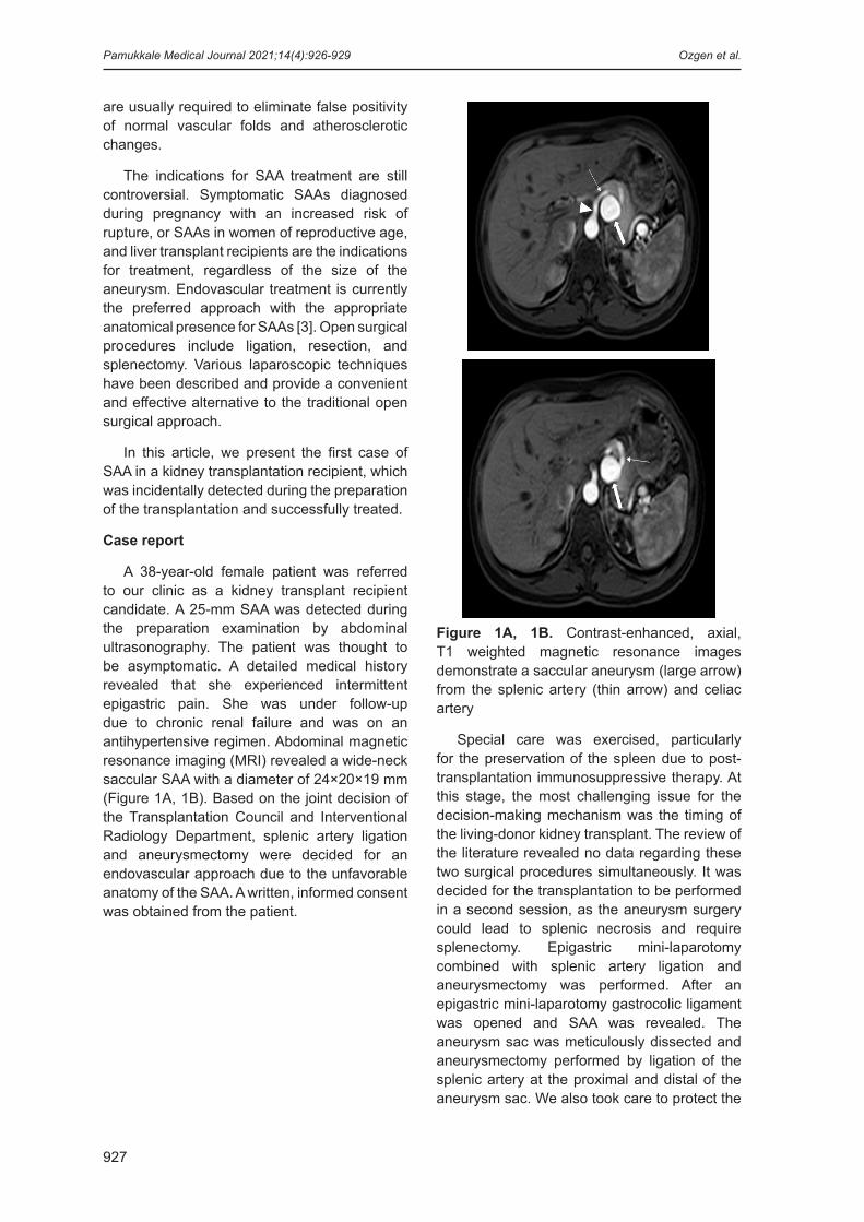
are usually required to eliminate false positivity of normal vascular folds and atherosclerotic changes.
The indications for SAA treatment are still controversial. Symptomatic SAAs diagnosed during pregnancy with an increased risk of rupture, or SAAs in women of reproductive age, and liver transplant recipients are the indications for treatment, regardless of the size of the aneurysm. Endovascular treatment is currently the preferred approach with the appropriate anatomical presence for SAAs [3]. Open surgical procedures include ligation, resection, and splenectomy. Various laparoscopic techniques have been described and provide a convenient and effective alternative to the traditional open surgical approach.
In this article, we present the first case of SAA in a kidney transplantation recipient, which was incidentally detected during the preparation of the transplantation and successfully treated.
Case report
A 38-year-old female patient was referred to our clinic as a kidney transplant recipient candidate. A 25-mm SAA was detected during the preparation examination by abdominal ultrasonography. The patient was thought to be asymptomatic. A detailed medical history revealed that she experienced intermittent epigastric pain. She was under follow-up due to chronic renal failure and was on an antihypertensive regimen. Abdominal magnetic resonance imaging (MRI) revealed a wide-neck saccular SAA with a diameter of 24×20×19 mm (Figure 1A, 1B). Based on the joint decision of the Transplantation Council and Interventional Radiology Department, splenic artery ligation and aneurysmectomy were decided for an endovascular approach due to the unfavorable anatomy of the SAA. A written, informed consent was obtained from the patient.
Figure 1A, 1B. Contrast-enhanced, axial, T1 weighted magnetic resonance images demonstrate a saccular aneurysm (large arrow) from the splenic artery (thin arrow) and celiac artery
Special care was exercised, particularly for the preservation of the spleen due to post-transplantation immunosuppressive therapy. At this stage, the most challenging issue for the decision-making mechanism was the timing of the living-donor kidney transplant. The review of the literature revealed no data regarding these two surgical procedures simultaneously. It was decided for the transplantation to be performed in a second session, as the aneurysm surgery could lead to splenic necrosis and require splenectomy. Epigastric mini-laparotomy combined with splenic artery ligation and aneurysmectomy was performed. After an epigastric mini-laparotomy gastrocolic ligament was opened and SAA was revealed. The aneurysm sac was meticulously dissected and aneurysmectomy performed by ligation of the splenic artery at the proximal and distal of the aneurysm sac. We also took care to protect the
927
Pamukkale Medical Journal 2021;14(4):926-929 Ozgen et al.

short gastric arteries through the gastroepiploic arch in order to preserve splenic vascularization. Intraoperative Doppler ultrasonography revealed that there were no abnormalities of the spleen blood supply. The postoperative course was uneventful, and the drain was removed on the second postoperative day. On Day 2, Doppler ultrasonography revealed that the blood supply of the spleen was normal. A living-donor kidney transplant was successfully performed one week after the operation. There weren’t any complications that developed after the transplantation. The patient was discharged on postoperative day 10 with complete recovery. The pathological examination of the resected aneurysm material confirmed the diagnosis and revealed a 25×22×19 mm saccular aneurysm with a mural thrombus.
During the first month of follow-up, the patient remained asymptomatic and laboratory investigations revealed no abnormalities. Therefore, no further interventions or follow-ups were scheduled. The patient is still under follow-up in the organ transplantation clinic.
Discussion
Although there are several surgical approaches described for SAA, there are very few published cases of SAA ligation and spleen preservation in the literature. In recent years, the incidence of SAA has been increasing due to the introduction of diagnostic tools for earlier detection and the frequent use of cross-sectional imaging modalities [4]. Despite the low incidence and often asymptomatic course, SAA rupture can be fatal [4]. Although it is usually defined as 2 cm [5], there is no consensus on the limits of the size of SAAs for the treatment. However, surgery is indicated in SAAs and symptomatic SAAs that are detected during pregnancy, in women of reproductive age, and in liver transplant recipients [6].
To the best of our knowledge, there are no guidelines or consensus for the treatment of SAAs. Endovascular approaches with transcatheter embolization are usually used due to their low morbidity and mortality rates. However, not all aneurysms are appropriate for this technique. The ideal form of treatment is to maintain flow in the splenic artery and to exclude the aneurysm from circulation [7]. Based on the joint decision of the Council,
the open surgical approach was decided, as the artery folds and distal localization of the aneurysm made the endovascular approach less feasible. Several laparoscopic approaches have been described [8]. Initially, tangential stapler resection of saccular aneurysms was described to preserve the splenic flow; however, the chance of recurrence exists [9]. Proximal and distal ligations are safer approaches for SAAs, with or without resection and splenectomy [10]. Reconstruction of the vascular continuity of the splenic artery is not usually required due to the collateral circulation provided by the mini-gastric vessels. Nevertheless, an end-to-end anastomosis was used in the literature, particularly in young patients for whom spleen preservation was recommended [6]. Based on our experience, in this case, we recommend ligation and resection with spleen preservation in the surgical treatment of SAAs. To the best of our knowledge, there is only a limited number of case reports regarding aneurysmectomy with spleen preservation [11]. Dissection of distally located aneurysms is more challenging and, at this location, the splenic vein can be damaged, as the small branches may be in communication with the arterial branches. However, most of the SAAs are distal and less convenient to endovascular therapy [12]. Doppler ultrasonography is not essential; however, it may be helpful for the evaluation of the splenic arterial flow indications before and after the SAA ligation and resection [6]. Condensation of the splenic surface does not infer to the possibility of a future splenic infarction, and there is evidence which shows that improved splenic perfusion and collateral circulation can restore the flow to the spleen after the operation. Indeed, of these patients, 22% had zones of splenic infarction, although only 2% required surgery [13]. In our case, there was no evidence of an infarction zone, confirmed visibly or by ultrasonography.
In conclusion, SAAs have been increasingly detected thanks to the recent advancements in the field of imaging studies. Currently, there are no guidelines or consensus for the treatment of SAAs. A multidisciplinary approach is of utmost importance in the management of all visceral aneurysms. Predominantly distal location and the tortuous course of the splenic artery do not always indicate endovascular treatment of SAAs. Surgery is an important treatment method and several laparoscopic approaches
Splenic artery aneurysm in kidney transplant recipient
928

have been described in the literature. Selective ligation and resection of SAAs combined with spleen preservation is a safe treatment modality for selected cases with favorable short and long-term results, allowing the permanent treatment of the SAAs, while preserving the splenic function.
Conflict of interest: No conflict of interest was declared by the authors.
References1. Jiang J, Ding X, Su Q, et al. Therapeutic management
of superior mesenteric artery aneurysms. J VascSurg 2011;53:1619-1624. https://doi.org/10.1016/j.jvs.2011.02.004
2. Spanos CP, Ktenidis K, Saratzis N, Lazaridis I, Kiskinis D. Laparoscopic ligation of splenic artery aneurysms: report of two cases and literature overview. Updates Surg 2014;66:73-76. https://doi.org/10.1007/s13304-012-0180-1
3. Erben Y, Brownstein AJ, Rajaee S, et al. Natural history and management of splanchnic artery aneurysms in a single tertiary referral center. J Vasc Surg 2018;68:1079-1087. https://doi.org/10.1016/j.jvs.2017.12.057
4. Iida A, Katayama K, Yamaguchi A. Laparoscopic resection for splenic artery aneurysm using the lateral approach: report of two cases. Asian J Endosc Surg 2013;6:147-150. https://doi.org/10.1111/ases.12000
5. He MX, Zheng JM, Zhang SH, Wang JJ, Liu WQ, Zhu MH. Rupture of splenic artery aneurysm in pregnancy: a review of the literature and report of two cases. Am J Forensic Med Pathol 2010;31:92-94. https://doi.org/10.1097/PAF.0b013e3181c65da2
6. Xu WL, Li SL, Wang Y, et al. Laparoscopic splenectomy: color Doppler flow imaging for preoperative evaluation. Chin Med J (Engl) 2009;122:1203-1208.
7. İnan B, Teker ME, Ay Y, Aydın C, Zeybek R. Our experience on splenic artery aneurysms. Vasc Sur J 2013;22:257-262. https://doi.org/10.9739/uvcd.2013-37769
8. Małczak P, Wysocki M, Major P, et al. Laparoscopic approach to splenic aneurysms. Vascular 2017;25:346-350. https://doi.org/10.1177/1708538116682164
9. Müller Eschner M, Kortes N, Rehnitz C, et al. Endovascular repair of a complex splenic artery aneurysm using a multilayer stent. Cardiovasc Intervent Radiol 2015;38:494-497.
10. Lee SY, Florica O. Laparoscopic resection of splenic artery aneurysm with preservation of splenic function. Singapore Med J 2008;49:303-304.
11. LaBella GD, Muck P, Kasper G, Welling R, Schlueter F, Vaughan A. Operative management of an aberrant splenic artery aneurysm: utility of the medial visceral rotation approach: a case report and review of the literature. Vasc Endovascular Surg 2006;40:331-333. https://doi.org/10.1177/1538574406292005
12. Moriyama H, Ishikawa N, Kawaguchi M, Inaki N, Watanabe G. Laparoscopic approaches for splenic artery aneurysms. Minim Invasive Ther Allied Technol 2012;21:362-365. https://doi.org/10.3109/13645706.2011.636051
13. Toyoda Y, Igami T, Ochiai Y, et al. Single-Incision laparoscopic splenectomy for an unruptured aneurysm of the splenic artery. Med Princ Pract 2018;27:95-98. https://doi.org/10.1159/000485836
Informed consent: A written, informed consent was obtained from the patient.
Authors’ contributions to the article
U.O., M.O., M.A. and M.C. have constructed the main idea and hypothesis of the study. O.B., S.Y., and E.D.Y. developed the theory and organized the material method section. The evaluation of the data in the results section was made by M.C. and M.O. The discussion part of the article was written by U.O., M.O., and H.C.A. reviewed, made the necessary corrections and approved. In addition, all authors discussed the entire study and approved its final version.
Pamukkale Medical Journal 2021;14(4):926-929 Ozgen et al.
929

930
Postural hematürinin değerlendirilmesi: posterior nutcracker sendromuEvaluation of postural hematuria: posterior nutcracker syndrome
Murat Yaşar Taş, Belda Dursun, Muhammet Arslan
Pamukkale Tıp DergisiPamukkale Medical JournalOlgu Sunumu
ÖzNutcracker sendromu sol renal venin abdominal aorta ile süperior mezenterik arter arasında ya da abdominal aorta ile vertebral kolon arasında sıkışmasından kaynaklanmaktadır. Sol renal venden inferior vena kavaya olan kan akımının dıştan bası nedeniyle azalması; artmış venöz basınç, böbrek konjesyonu, hematüri ve proteinüriye sebep olur. Ortostatik hematüri ile başvuran hastalarda akılda tutulmalıdır. Hematüriyle başvuran hastalarda öyküde hematürinin ortostatik yani ayakta kalmakla artıp artmadığının ve mesleğin sorgulanması son derece önemlidir. Dikkatli anemnez alınmaması sonucunda böbrek biyopsisini de içeren birçok gereksiz tetkik yapılması ve sonucunda net tanı konulamaması durumu hastalarda endişeye neden olabilir. Tanısal yöntemler doppler ultrasonografi, bilgisayarlı tomografi, MR ve altın standart yöntem olarak sol renal ven ile inferior vena cava arasındaki basınç farkının venografi ile ölçülmesidir. Nutcracker sendromunun yönetimi ise tartışmalıdır ve spontan remisyon nedeniyle özellikle genç ve hafif hematürisi olan kişilerde izlem tercih edilir. Ortostatik hematüri ile başvuran ve Posterior Nutcracker Sendromu tanısı alan genç bir kadın hasta vaka sunumu olarak tartışılmıştır.
Anahtar kelimeler: Hematüri, nutcracker sendromu, postural hematüri.
Taş MY, Dursun B, Arslan M. Postural hematürinin değerlendirilmesi: posterior nutcracker sendromu. Pam Tıp Derg 2021;14:930-932.
AbstractNutcracker sydrome is associated with extrinsic left renal vein compression between abdominal aorta and superior mesenteric vein or between abdominal aorta and vertebral column. Decreased blood flow from the left renal vein to the inferior vena cava due to external compression results in increased venous pressure, kidney congestion, hematuria and proteinuria. Patients with orthostatic hematuria should be screened for nutcracker syndrome. In patients presenting with hematuria, it is extremely important to question whether orthostatic hematuria increases with standing and the profession. As a result of not taking a careful anamnesis, many unnecessary tests, including kidney biopsy, and the inability to make a clear diagnosis as a result may cause anxiety in patients. Diagnostic methods are Doppler ultrasonography, computed tomography, MRI and, as the gold standard method, measuring the pressure difference between the left renal vein and the inferior vena cava by venography. The management of Nutcracker syndrome is controversial and because of spontaneous remission, follow-up is preferred especially in young people with mild hematuria. A young female patient who presented with ortostatic hematuria and diagnosed with posterior nutcracker syndrome is discussed hereby in the case report.
Key words: Hematuria, nutcracker syndrome, postural hematuria.
Tas MY, Dursun B, Arslan M. Evaluation of postural hematuria: posterior nutcracker syndrome. Pam Med J 2021;14:930-932.
Case Report
Gönderilme tarihi:23.02.2021 Kabul tarihi:20.05.2021
Murat Yaşar Taş, Uzm. Dr. Pamukkale Üniversitesi Tıp Fakültesi, İç Hastalıkları Anabilim Dalı, Nefroloji Bilim Dalı, Denizli, Türkiye, e-posta: [email protected] (https://orcid.org/0000-0003-1459-1836) (Sorumlu Yazar)Belda Dursun, Prof. Dr. Pamukkale Üniversitesi Tıp Fakültesi, İç Hastalıkları Anabilim Dalı, Nefroloji Bilim Dalı, Denizli, Türkiye, e-posta: [email protected] (https://orcid.org/0000-0003-3235-0577)Muhammet Arslan, Doç. Dr. Pamukkale Üniversitesi Tıp Fakültesi, Radyoloji Anabilim Dalı, Denizli, Türkiye, e-posta: [email protected] (https://orcid.org/0000-0001-5565-0770)
Doi no: https://dx.doi.org/10.31362/patd.885463
Giriş
Nutcracker sendromu (NS), sol renal venin (SRV) abdominal aorta ile süperior mezenterik arter arasında (anterior nutcracker) ya da abdominal aorta ile vertebral kolon arasında (posterior nutcracker) sıkışmasından kaynaklanmaktadır [1-4]. Sol renal venden inferior vena kavaya olan kan akımının dıştan
bası nedeniyle azalması; artmış venöz basınç, böbrek konjesyonu, hematüri ve proteinüriye sebep olur [2, 4]. Posterior nutcracker sendromu (PNS) daha seyrek görülmektedir [5].
Tanı konulması birçok vakada zorlayıcı olabilir [1]. Hematüri şikâyetiyle başvuran her hastada hematürinin ortostatik yani ayakta kalmakla artıp artmadığı, meslek bilgilerini de

içeren ayrıntılı öykü alınması çok önemlidir. Yeterli öykü alınmadığı durumda yanlış ön tanılar nedeniyle böbrek biyopsisi dahil olmak üzere gereksiz birçok tetkik ve girişimsel işlem yapılmış olabilir. Bu yazıda postural hematüri nedeniyle başvuran bir vakamızı özetledik.
Olgu sunumu
22 yaşında kadın hasta 4 yıldır devam eden makroskopik hematüri ve sol yan ağrısı şikâyetiyle başvurdu. Kuaförlük yaptığını, günde 12-16 saat ayakta iş yaptığını belirtti. Hematüri yakınmasının ayakta uzun süre kaldıktan sonra arttığını, sabah uyandığında idrar renginin normale döndüğünü ifade etti. Ateş ya da idrar yaparken yanma yakınması yoktu. Daha önce başvurduğu merkezde yapılan ve 38 glomerülü içeren böbrek biyopsisinin mikroskopi sonucu ‘Tübülointerstisyel, hafif tübüler dejenerasyon dışında olağan sınırdadır. Vasküler yapılar olağandır. İmmünfloresans incelemede 7 glomerül izlenmiş olup, hafif C3 birikimi saptandı.’ olarak raporlanmıştı. Yine aynı merkezde yapılan sistoskopi tetkiki normal olarak değerlendirilmiş ve hastaya herhangi bir tedavi önerilmemişti. Hematüri şikâyeti devam eden hasta, net tanı konulamadığı için endişeliydi. Postural hematüri nedeniyle NS ön tanısıyla yatırılarak tetkik edildi. Yatışı sırasında fizik muayenesinde patolojik bulgu yoktu. Laboratuvar tetkiklerinde böbrek fonksiyon testleri normal, eGFR CKD-EPİ: 123 ml/dk, TİT: Lökosit ++, nitrit (-), protein +, idrar-Hb: +++, lökosit mikroskopisi: 23, eritrosit mikroskopisi: 4492, immünolojik tetkikleri negatif olarak sonuçlandı. İdrar sedimentinde yoğun izomorfik eritrositler izlendi, silendir görülmedi. Postural hematürisi olan hastanın intravenöz kontrastlı trifazik bilgisayarlı tomografisi ‘Sol ana renal ven aorta ve SMA arasından geçerken normal kalibre izlendi. Solda aksesuar renal ven izlenmekte olup, retroaortik olarak vena kavaya dökülmektedir. Aorta posteriorunda belirgin ince kalibredir.’ olarak raporlandı. Hasta PNS olarak değerlendirildi ve uzun süre ayakta kalmaması, mümkünse meslek değiştirmesinin faydalı olabileceği önerildi. Girişimsel radyoloji bölümünce stent uygulaması önerilmedi. Üroloji bölümünce hastada yoğun hematüri, proteinüri ve pelvik konjesyon sendromu olmaması nedeniyle cerrahi işlem önerilmedi.
Tartışma
Posterior nutcracker sendromu anatomik varyasyonla SRV’nin retroaortik yerleşimi sonucunda spinal kolonla aorta arasında kısmi sıkışmasından kaynaklanır ve renal venöz hipertansiyon, sol yan ağrısı, hematüri ve proteinüriye sebep olabilir [4]. Hematüriyle başvuran hastalarda öykü son derece önemlidir. İdrar renginde ayakta kalmakla hematürinin artması ve dinlenmekle bu durumun azalması postural hematüriyi akla getirmelidir. Meslek ve çalışma koşullarının ayrıntılı not edilmesi ve basit idrar analizinin dikkatli değerlendirilmesi, idrar sedimentinin incelenmesi elzemdir. Makroskopik hematüri yakınması olan hastalar için sorunun psikososyal boyutunun da dikkatle ele alınarak irdelenmesi gerekir. Hematürinin nedeninin açıklığa kavuşturulamaması hastada endişe oluşturabilir.
Sol renal venin retroaortik yerleşimi sıklığının %1-3 arasında olduğu belirtilse de bunların çoğu asemptomatiktir ve PNS sıklığı net olarak bilinmemektedir [6]. Posterior nutcracker sendromu tanısı için henüz tanı kriterleri belirlenmemiştir [5]. Nutcracker sendromu, mikroskopik ya da makroskopik hematüri dışında sol yan ve karın ağrısı yapabilir [2]. Disparoni, dismenore, karın ağrısı, pelvik varisler ile karakterize pelvik konjesyon sendromuna sebep olabilir [2]. Hematürinin sebebi, artmış SRV basıncıyla renal forniks toplayıcı sistem ile varisler arasındaki ince duvarlı septumun rüptürüdür [2]. Ortostatik proteinüri eşlik edebilir [4]. Bizim vakamızda uzun süre ayakta kalmanın, yerçekimi etkisiyle konjesyona katkıda bulunması sonucunda makroskopik hematüri olması, hasta yatar pozisyona geçtiğinde konjesyonun azalmasıyla hematürinin kaybolması NS açısından tipiktir. Nutcracker sendromu genellikle kadınlarda hayatın üçüncü ve dördüncü dekatında tespit edilir. Diğer hematüri yapan sebeplerin dışlanması gerekir [1]. Tanısal yöntemler doppler ultrasonografi, bilgisayarlı tomografi, MR ve altın standart yöntem olarak SRV ile inferior vena cava arasındaki basınç farkının venografi ile ölçülmesidir [5, 6]. Nutcracker sendromunun yönetimi ise tartışmalıdır ve spontan remisyon nedeniyle özellikle genç ve hafif hematürisi olan kişilerde izlem tercih edilir. 2 yıllık izlem sonunda persistan hematüri, ciddi yan ağrısı olanlarda stent uygulaması ya da cerrahi işlem düşünülebilir [1, 6].
931
Pamukkale Tıp Dergisi 2021;14(4):930-932 Taş ve ark.

Sonuç olarak, PNS henüz tanı kriterleri belirlenmemiş, nadir görülen, hematüri sebepleri araştırılırken dikkatli öykü alınarak hastaların gereksiz tekik ve invaziv girişimlerden korunması sağlanabilecek bir sendromdur. Hematüri yakınmasıyla başvuran hastalarda ön tanılar arasında düşünülmelidir. [1].
Çıkar ilişkisi: Yazarlar çıkar ilişkisi olmadığını beyan eder.
Kaynaklar1. Ali El Dein B, Osman Y, Shehab El Din AB, El Diasty
T, Mansour O, Ghoneim MA. Anterior and posterior nutcracker syndrome: a report on 11 cases. Transplant Proc 2003;35:851-853. https://doi.org/10.1016/s0041-1345(02)04026-5
2. Kurklinsky AK, Rooke TW. Nutcracker phenomenon and nutcracker syndrome. Mayo Clin Proc 2010;85:552-559. https://doi.org/10.4065/mcp.2009.0586
3. Park JH, Lee GH, Lee SM, et al. Posterior nutcracker syndrome - a systematic review. Vasa 2018;47:23-29. https://doi.org/10.1024/0301-1526/a000670
4. Granata A, Clementi A, Floccari F, Di Lullo L, Basile A. An unusual case of posterior nutcracker syndrome. Clin Exp Nephrol 2014;18:670-671. https://doi.org/10.1007/s10157-014-0932-y
5. Chen Y, Xing J, Liu F. Left renal vein transposition is effective for posterior nutcracker syndrome. Int J Clind Exp Med 2014;7:5925-5927.
6. Marone EM, Psacharopulo D, Kahlberg A, Coppi G, Chiesa R. Surgical treatment of posterior nutcracker syndrome. J Vasc Surg 2011;54:844-847. https://doi.org/10.1016/j.jvs.2011.01.038
Hasta onamı açıklaması: Hastadan yazılı olarak hasta onamı alınmıştır.
Yazarların makaleye olan katkıları
B.D. tarafından ortostatik hematüri ile başvuran hastada öntanıda Nutcracker Sendromu düşünülmüş ve gerçek tanıya gidilmesi sağlanmıştır. Olgu sunumunun planlanması ve yazılmasına katkıda bulunmuştur. M.Y.T. tarafından literatür taranmış, olgu vaka sunumu olarak yazılmıştır. Vakanın yönetimi sürecinde M.A. tarafından radyolojik görüntülemeleri değerlendirilmiş, uygun görülen radyolojik görüntüler yazımıza eklenmiştir. Ayrıca tüm yazarlar çalışmanın tamamını tartışmış ve son halini onaylamıştır.
932
Postural hematürinin değerlendirilmesi: posterior nutcracker sendromu


934
Coğrafi Bilgi Sistemleri-mekânsal epidemiyoloji çerçevesinde SARS CoV-2 (COVID-19)
SARS CoV-2 (COVID-19) in the framework of Geographic Information System spatial epidemiology
Ömer Barış İnce, Murat Şevik, Ahmet Sait
Ömer Barış İnce, Dr. Pamukkale Üniversitesi Hayvan Islahı ve Genetik Uygulama ve Araştırma Merkezi, Kınıklı Yerleşkesi Pamukkale/Denizli, Türkiye, e-posta: [email protected] (https://orcid.org/0000-0001-8302-9607) (Sorumlu Yazar)Murat Şevik, Doç. Dr. Necmettin Erbakan Üniversitesi, Veteriner Fakültesi, Viroloji Anabilim Dalı, 42310, Ereğli, Konya, Türkiye, e-posta: [email protected] (https://orcid.org/0000-0002-9604-3341)Ahmet Sait, Dr. Pendik Veteriner Kontrol Enstitüsü Müdürlüğü, Viral Teşhis Laboratuvarı, 34890, İstanbul, Türkiye, e-posta: [email protected] (https://orcid.org/0000-0001-7658-8793)
Gönderilme tarihi: 02.01.2021 Kabul tarihi: 03.03.2021
ÖzDünyayı etkisi altına alan şiddetli akut solunum yolu sendromu coronavirusu 2 (SARS-CoV-2) salgını, pek çok ülkede ölümcül sonuçlara neden olan önemli bir halk sağlığı sorunudur. Pandemiye yol açacak hastalık yayılımlarının erken dönemde tespit edilebilmesi hastalık kontrol ve eradikasyonunun önemli bir bileşenidir. Hastalık verilerinin ve mekânsal analiz yöntemlerinin birlikte kullanılması, daha etkili hastalık kontrolü ve çözüm stratejileri geliştirmek için büyük bir fırsat sunmaktadır. Bu derlemede coğrafi bilgi sistemlerinin (CBS) epidemiyolojideki uygulamalarını ve salgın hastalıkların kontrolü ve eradikasyonundaki ilişkisini değerlendirmek için özelde COVID-19’u içeren literatüre dayalı bir inceleme yapılmıştır. Epidemiyoloji alanındaki araştırmalarda, araştırılan hastalık verilerinin nasıl bir dağılım ve kümelenme gösterdiği, kısa, orta ve uzun vadede yapılacak kontrol ve eradikasyon müdahalelerini planlama açısından CBS temelli analizler ve modeller giderek önem kazanmaktadır. COVID-19'un kontrol ve eradikasyonunda yaşanan zorluklar, güçlü bulaşıcılık özelliği, uzun bir kuluçka dönemi, nüfus akış ve hareketliliği ve diğer faktörlerle birleştiğinde, hastalığın yayılmasını kontrol etmek ve önlemek için bilimsel ve teknolojik desteğe gereksinim duyulmaktadır. Bu derlemenin amacı, CBS temelli araçların gelişimini anlamak ve COVID-19 pandemisi yönetiminde CBS kullanımı hakkında güncel bilgiler vermektir.
Anahtar kelime: COVID-19, SARS CoV-2, CBS, mekânsal epidemiyoloji.
İnce ÖB, Şevik M, Sait A. Coğrafi Bilgi Sistemleri-mekânsal epidemiyoloji çerçevesinde SARS CoV-2 (COVID-19).Pam Tıp Derg 2021;14:934-943.
Abstract Severe acute respiratory syndrome coronavirus 2 (SARS-CoV-2) pandemic which has affected all over the world is a major public health concern that causes fatal consequences in many countries. The early detection of disease spread that will cause a pandemic is an important component of disease control and eradication. The combination of disease data and spatial analysis methods provides a great opportunity to develop solution strategies for more effective disease control. In this review, to evaluate the practices of geographic information system (GIS) in epidemiology and its relation in the control and eradication of pandemic, a literature-based review which includes COVID-19 in particular has been made. GIS-based analyses and models are of increasing importance in the epidemiological studies; in terms of how the researched disease data show distribution and clustering, and planning control and eradication interventions in the short, medium, and long terms. Scientific and technological supports are requirement to control and avoid the spread of the disease when the challenges in the control and eradication of COVID-19, features of strong contagiousness, a long incubation period, population flow, and mobility combined with other factors. The aim of this review is to understand the development of GIS-based tools and to provide up-to-date information about the use of GIS in COVID-19 pandemic management.
Key words: COVID-19, SARS CoV-2, GIS, spatial epidemiology.
Ince OB, Sevik M, Sait A. SARS CoV-2 (COVID-19) in the framework of GIS-spatial epidemiology. Pam Med J 2021;14:934-943.
Pamukkale Tıp DergisiPamukkale Medical JournalReviewDerleme Makalesi
doi:https://dx.doi.org/10.31362/patd.852259

Giriş
Coronaviruslara genel bakış
Coronavirus’lar (CoV), insan, çiftlik hayvanları, kuşlar, yarasa, fare ve birçok vahşi hayvan türünde solunum, sindirim, karaciğer ve merkezi sinir sistemi enfeksiyonlarına neden olmaktadır. CoV’lar, Nidovirales takımının, Cornidovirineae alt takımının Coronaviridae ailesinde yer alan yaklaşık 30 kb. genom büyüklüğüne sahip zarflı, segmentsiz ve pozitif polariteli RNA viruslarıdır. Coronoviridae familyası genetik ve serolojik özelliklerine göre 4 genusa sahiptir; Alphacoronavirus, Betacoronavirus, Gammacoronavirus ve Deltacoronavirus. İnsanları enfekte eden koronaviruslar Alphacoronavirus (Human coronavirus 229E ve NL63) veya Betacoronavirus (Human coronavirus HKU1 (HKU1) Human coronavirus OC43 (OC43), Severe Acute Respiratory Syndrome (SARS-CoV), Middle East respiratory syndrome coronavirus (MERS-CoV) ve Severe Acute Respiratory Syndrome coronavirus 2 (SARS-CoV-2)) genuslarında yer almaktadırlar [1, 2]. CoV’lar hücre sitoplazmasında çoğalırlar. CoV’un konakçıda enfeksiyon oluşturabilmesinde ve doku tropizminde ana etken virus spike (S) proteini S1 bölgesindeki reseptör bağlanma bölgesi ile hücre yüzey reseptörü arasındaki ilişkidir. Alphacoronaviruslarının çoğunun hücresel reseptörü aminopeptidaz N ve MERS-CoV’un dipeptidil peptidaz-4 iken SARS-CoV ve SARS-CoV-2 konak hücre yüzey reseptörü olarak anjiyotensin dönüştürücü enzim 2 (ACE-2)’i kullanmaktadır [3-5]. Doğal enfeksiyonlarda CoV’a karşı spesifik nötralizan antikorların çoğu S proteinin N terminal kısmında bulunan epitoplara yönelik olarak oluşmaktadır. Hücresel immun yanıt, özellikle S ve nükleokapsid (N) proteinlerine karşı gelişmektedir. CoV’ların yapısal proteinlerinin yanı sıra, genomun farklı bölgelerinden kodlanan farklı sayılarda yapısal olmayan proteinleri de bulunmaktadır. CoV’lar, 56˚C sıcaklıkta 30 dakika da inaktive olabilmektedirler ve dietil eter, %75 etanol, klor, perasetik asit, kloroform ve diğer yağ çözücüler gibi birçok dezenfektana karşı duyarlıdırlar [6].
Yarasa CoV’ları mutasyon geçirmeleri ve rekombinasyon özellikleri sayesinde insanları enfekte edebilmektedir. CoV’lar genel olarak insanlar için ölümcül olmayan patojenler olarak kabul edilir ve temelde soğuk algınlığının
yaklaşık %15’ine neden olur. İnsan coronavirus HKU1, NL63, 229E ve OC43 suşları insanlarda hafif solunum yolu enfeksiyonları neden olmaktadır [4]. Bununla birlikte, bu zamana kadar insanlar için ölümcül olabilen CoV’larıyla üçüncü kez karşılaşılmış bulunulmaktadır. İlk karşılaşma 2003'te Çin'de ortaya çıkan SARS-CoV enfeksiyonudur. Dünya Sağlık Örgütü (DSÖ) tarafından 2003 yılında bildirilen 8000’in üzerinde SARS-CoV tanısı konulmuş hasta bulunmaktadır [7]. İkinci karşılaşma, 2012’de Suudi Arabistan’da mortalitesi yüksek (%35) ve kısa süre sonra birçok ülkeye yayılan MERS-CoV salgını olarak ortaya çıkmış ve yaklaşık 26 ülkeyi etkisi altına alarak 700’ün üzerinde insanın ölümüne sebep olmuştur [8]. 2019 yılı sonunda ortaya çıkan son salgın ilk kez Çin’in Wuhan eyaletinde ortaya çıkmıştır. Yerel bir Çin sağlık kuruluşunda pnömoni semptomları gösteren bir kişide tespit edilen etken, yeni coronavirus 2019 (nCov-2019) olarak adlandırılmıştır [5]. Uluslararası Virus Taksonomisi Komitesi’nin Coronavirus Çalışma Grubu tarafından, 11 Şubat 2020’de virusun adı SARS-CoV-2 olarak belirlenmiştir [2]. DSÖ, virusun neden olduğu hastalığı tanımlamak için Coronavirus Disease 2019 (COVID-19) terimini kullanmıştır. Son dönemdeki araştırmalar, SARS-CoV-2’nin, genetik dizisinin diğer CoV’lere benzerlik oranına (yarasa SARS ilişkili coronavirus RaTG13 suşu ile benzerlik %96,2) dayanarak, virusun muhtemelen yarasalardan köken aldığını ileri sürmektedir [5]. COVID-19, insanlarda klinik tablo olarak ateş, öksürük, nefes darlığı gibi solunum yolu ve gastrointestinal hastalık belirtilerine neden olabilmektedir [3, 5]. Dünyayı etkisi altına alan, bu salgının kontrol altına alınması amacıyla karantina önlemlerine ilaveten kişisel hijyen ve sosyal mesafenin korunması ve kişisel koruyucu ekipman kullanımı ana kural olarak benimsenmiştir [6, 9-11].
Mekânsal epidemiyoloji
Epidemiyolojide Coğrafi Bilgi Sistemleri (CBS) genel olarak; hastalıkların ve vakaların tespit edilmesi, izlenmesi, dağılımı, riskli alanların ve risk altındaki popülasyonun belirlenmesi, hastalık verilerinin çevresel özellikler arasındaki mekân-zaman ilişkilerinin kurulması, coğrafi koşulların etkilerinin araştırılması ve yetkili otoritece kontrol stratejilerinin geliştirilmesine yardımcı olmak amacıyla yaygın olarak
935
Pamukkale Tıp Dergisi 2021;14(4):934-943 İnce ve ark.

kullanılmaktadır. Mekânsal epidemiyoloji, geçtiğimiz on yılda önemli bir faaliyet alanı haline gelmiştir [12]. Hastalık prevalans araştırmalarında küresel konumlandırma sistemlerinin artan kullanımı, sağlık haritalaması gibi faktörlerin yönlendirmesiyle, mekânsal olarak epidemiyolojik veriler artık daha yaygın hale gelmektedir. Ayrıca mekânsal epidemiyoloji, sağlık verilerinin haritalanması, uydulardan epidemiyolojik olarak ilgili değişkenlerin artan kullanılabilirliği ve istatistiksel yöntemlerin sürekli geliştirilmesiyle hızla büyüyen bir alan olmaya devam etmektedir. Dr. John Snow 1854’de Londra kolera salgını epidemiyolojik vakalarını haritalaması ile mekânsal epidemiyolojinin temelini atmıştır. Dr. Snow Londra’da kolera sonuçlu ölümlerin mekânsal dağılımını yaparak kirli su kaynakları ile ölümler arasındaki ilişkiyi analiz etmeye çalışmıştır [13].
Hastalıkların analizi, çevreyle olan ilişkisi, hastalık kümelerinin nerelerde yoğunlaştığını, karar vericilerin kontrol ve eradikasyon planlamaları hazırlamalarında kaynakların etkin kullanımı, salgın vakalarının yayılımı, kontrol stratejilerinin değerlendirilmesinde mekânsal epidemiyolojinin rolü büyüktür [14, 15]. CBS’nin epidemiyolojide kullanılmasıyla birlikte hastalık haritalaması gündeme gelmiştir. Hastalık haritaları, hastalığa müdahale stratejisinde öncelikli konumlarının zamanla olan ilişkisini göz önünde bulundurarak nokta, çizgi ve alansal olarak hazırlanabilmektedir [16, 17].
Mekânsal epidemiyolojinin ilgilendiği diğer konu ise kümelenme analizleridir. Kümelenme analizleri hastalık etkeninin nerelerde kümelendiğini tespit etmesi, oluşan kümelenmenin istatistiki açıdan anlamlı olup olmadığını göstermesi açısından önem arz etmektedir. Ek olarak kümelenme analizi hastalıkla mücadele stratejilerinin belirlenmesinde, kontrol ve eradikasyona ayrılan bütçenin etkin kullanılmasında, gözlenen-beklenen vaka sayıları arasındaki ilişkilerde önemli bir rol üstlenmektedir [18, 19]. Salgınlar genellikle vakaların mekânsal yayılma derecesi ile karakterize edildiğinden, mekân-zamansal sürveyans algoritmaları ülke bazlı geliştirilmeye çalışılmaktadır.
Coğrafi bilgi sistemi yazılımlarına genel bir bakış
CBS, bilimsel bilgiler yoluyla salgının hafifletilmesine yardımcı olmak, diğer değişkenlerle mekânsal korelasyonlar bulmak ve iletim dinamiklerini tanımlamak için kullanılmaktadır. Ayrıca, CBS salgın tespit algoritmalarını değerlendirmek için gerçekçi vaka dağılımları üretebilen, farklı lokasyonlara kolayca uyarlanabilen ve farklı altta yatan popülasyon dağılımlarını temsil eden hastalık salgınlarını simüle eden bir teknoloji içermektedir. Simüle edilmiş salgın verileri, halk sağlığı sürveyansına yönelik algoritmaları değerlendirmek için kullanılabilir. Değerlendirme genel olarak, algoritmadan türetilmiş salgın göstergeleri ile salgınların belirli zaman ve mekandaki konumlarını gösteren önceden belirlenmiş kriterleri karşılaştırarak gerçekleştirilir. Günümüzde hastalık vakalarının zamansal-mekânsal analizlerinde CBS yazılımı olarak QGIS™, GeoDa™, SaTScan™, ArcGIS TM ve R programı vb. yazılımlar kullanılmaktadır. Hastalık kümelenmeleri ise mekân-zaman permutasyon modeli (STPM) analiziyle tespit edilmektedir. Hastalık verilerinin zamansal-mekânsal analizleri, verilerin mekânsal değişimi ve verilerin konum ilişkisinin inceleme temeline dayanmaktadır. Zaman olarak, her yıl için cinsiyet, yaş gruplarına göre yıllık insidans, hastalık vakaları için yıl bazlı veriler iklim verileriyle ilişkilendirilmektedir. Daha sonra vakaların zamansal desenleri ve görülme sıklığı analiz edilmektedir. Yıl ve yıl içindeki ay için iklimsel veriler (yağış-sıcaklık-bağıl nem) arasındaki ilişki analiz edilmektedir. Ayrıca hastalık vakalarının, zaman serileriyle analizi ve uygun modelin tespit edilerek ileriki yıllara ait vaka sayısı tahmininin yapılması öngörülmektedir. Bu amaçla Moran I, Geary Oranı, Global G İstatistiği, sıcak bölge analizleri (hot spot), Knox indeks, mantel indeks ve lokal mekânsal otokorelasyon (LISA) testi hesaplamaları kullanılmaktadır. Analizlerin anlamlılık düzeyi için dağılım desenlerini tespit etmede, dağılımın çalışma alanında nerelerde yoğunlaştığının bulunması için Kuadrat analizi, Point pattern analizi ve Kernel fonksiyonundan, olaylar arasındaki mekânsal bağımlılığın saptanmasında ise en yakın komşu mesafesi (nearest neighbor analysis) ve K-fonksiyonu ve konumsal ağırlık matrislerinden yararlanılmaktadır.
936
Coğrafi Bilgi Sistemleri-mekânsal epidemiyoloji çerçevesinde SARS CoV-2

Quantum geographic information system (QGIS™)
QGIS™, verilerin depolanması, analizi, görselleştirilmesinde kullanılan ve mekânsal analizler yapmaya imkân veren açık kaynak kodlu bir programdır [20]. Hastalık risk haritaları, sınırları belirlenmiş ve birbirleriyle örtüşmeyen mekân üzerinden riskin mekânsal dağılımında hazırlanmaktadır. Ayrıca hangi konumların risk teşkil edebileceği de bu haritalar üzerinden yorumlanabilmektedir. Epidemiyolojide hastalıkların dağılım haritaları önem arz etmektedir. Bir bölgede meydana gelen hastalık sayısı, bazen riski tanımlamada yetersiz kalmaktadır. Bu çerçevede, hastalık riskine ilişkin bir öngörüde bulunmak için gözlemlenen vaka sayısı ile beklenen vaka sayısı ilişkilendirilerek çıkarım yapılması gerekmektedir. İstatistiksel değişkenlerin ölçümü ile ilişkili olarak mekânların renklendirildiği ve desenlediği salgın hastalık mortalite, morbidite ve vaka-ölüm hızı haritaları, hastalık dağılımı haritaları ve standartlaştırılmış ölüm oranı (SMR) haritaları QGIS™ de hazırlanabilmektedir. SaTScan™ da elde edilen kümeler QGIS™ de görüntülenmektedir.
GeoDaTM
Salgın verilerinin mekânsal otokorelasyon temelindeki analizlerinde GeoDa™ programı kullanılmaktadır. Programda verilerin haritalarla görselleştirilmesi, mekânsal otokorelasyon, regresyon hesaplaması ve tahminleri yapılmaktadır [21]. Kümelenmelerde mekânsal ağırlık matrisi oluşturularak persentil haritası kullanılmaktadır Çalışmalarda hastalığın mekânsal dağılımdaki oranlarda persentil haritasına bakılmaktadır. Diğer taraftan Kuadrat analizi verinin düzenli olarak şekillendirilmiş bölgelerdeki mekânsal dağılımının incelenmesinde kullanılmaktadır. Burada amaç; matematiksel bir formülle dağılımın nasıl olduğunu hesaplamaktır [22].
SaTScan™ -mekân-zaman permutasyon modeli (STPM)
SaTScan, mekânsal veya mekân-zaman hastalık kümelerini tespit etmek, hastalığın coğrafi dağılımını yapmak ve istatistiksel olarak anlamlı olup olmadığını görmek, hastalığın uzaya, zamana ya da uzaya ve zamana rastgele dağılım gösterip göstermediğini ve hastalık kümelenmelerinin istatistiksel olarak
anlamlı olup olmadığının değerlendirilmesinde kullanılmaktadır [23].
Yazılımın tarama istatistikleri, mekânsal-zamansal olgu kümelerini tespit etmek ve değerlendirmek için kullanılmaktadır. Bu, zaman içinde ve / veya alanda hastalık verisini kademeli olarak tarayarak, her bir konumdaki pencerede gözlenen ve beklenen gözlemlerin sayısını dairesel pencerede göstermektedir. Çalışma alanı içerisinde farklı boyutlarda tarama pencereleri oluşturulmaktadır. Sonuçta her pencereye ait gözlenen-beklenen vaka sayıları, kümeleri oluşmakta, olasılıkları ve analiz sonucunda vaka sayıları ve p değeri hesaplanmaktadır. Yazılımla, verilerin özelliklerine göre normal-sürekli, ayrık Poison, Bernoulli modelleri de uygulanabilmektedir [23, 24].
Mekân-zaman permutasyon modelinin avantajı konum-zaman verisine göre çalışmasıdır. Vakaların Poisson dağılımı varsayımından her küme Monte Carlo hipotez yöntemiyle istatistiksel anlamlılığı test etmektedir. Risk altında bulunan nüfus veya kontrol grubunun olmadığında kümelerin oluşturulmasında yine STPM kullanılmaktadır. Model yıllara ilişkin verilerden retrospektif veya her dönem için yapılan periyodik taramalar içinde kullanılmaktadır. Bu çerçevede hastalık salgınlarının ileriye dönük tahmin etmeye yönelik algoritma çalıştırmaktadır [23-25].
ArcGIS TM
ArcGIS, coğrafi bilgi verilerinin derlenerek işlenmesine, haritaların oluşturulmasına, görselleştirilmesine, analiz edilmesine ve kütüphanesindeki mekânsal bilgileri kullanma araçları içeren, sağlık, tarım ve bölgesel planlama gibi birçok alanda kullanıma olanak tanıyan yazılım programıdır. Çevresel Sistemleri Araştırma Enstitüsü (Environmental Systems Research Institute (ESRI)) tarafından tasarlanmış ve geliştirilmiştir. Vektörel ve grafik türündeki coğrafi verilerin bilgisayar ortamında işlenerek ve depolanması sayesinde bir araya getirilerek araştırma kapsamında istenilen şekilde bilgilerin sayısallaştırılarak görselleştirilmesi ve analizi sağlanır. Vektör ve raster tarzı verileri analiz etmek ve anlamak için ihtiyaç duyulan araçları, haritaları ve demografik verileri sağlar. ArcGIS, haritalar oluşturmasına
937
Pamukkale Tıp Dergisi 2021;14(4):934-943 İnce ve ark.

ve düzenlenmesine verilerinizi haritalara eklemesine, coğrafi verilerin analiz edilmesine ve kurumsal veri kaynaklarına bağlanmayı kolaylaştıran birçok yeni ve gelişmiş özellik sunmaktadır [26].
R programı
R (https://www.r-project.org), çok detaylı istatistiki (doğrusal ve doğrusal olmayan modelleme, klasik istatistik testleri, zaman serileri analizi, sınıflandırma, kümeleme) ve grafik çizim teknikleri sunan bilgisayar programı ve program dilidir. R açık kaynak kodlu olduğundan çalışma ekibi (R Core Team) tarafından sürekli geliştirilmektedir. Analizlerde R programında kullanılan algoritmalarda temel kurulumun yanında ek paketler ile birlikte kullanıcılara kolaylık sağlanmaktadır. Kullanıcılara, matrislerde hesaplamalar yapabilme imkânı vermektedir. Programlama dillerine hâkim kod yazan kullanıcılar R programında da aynı kodları yazabilme imkânı bulmaktadırlar. R programı, C ve Java gibi birçok programlama dili ile uyumlu çalışabilmektedir. R, verilerin işlenmesi, hesaplanması ve analizlerinde özet çıktı verebilmekte ve program, veri tabanında ileriki çalışmalar için sonuçları saklamaktadır. Program kullanıcıları kod bilmemelerine karşın R paketlerini kullanarak analizlerini gerçekleştirebilmektedir. Ancak ilgili paketin istatistiksel teorilerin varsayım ve algoritma mantalitesinin bilinmesi gerekmektedir. Çünkü model veya hipotez oluştururken konuya ilişkin kavram ve formüllerin anlaşılması açısından faydalı olacaktır [27].
“spacetime” paketi
R “spacetime” paketi farklı mekânsal-zamansal veri türleri için tasarlanmış bölüm ve yöntemleri açıklar ve bunları analiz etmek için örnekler sağlar. Paket “sp”den gelen mekânsal veriler için ve “xts” paketinden gelen zaman serisi verileri için sınıflar ve yöntemler üzerine inşa edilir. Paketin amacı mekânsal-zamansal verileri için bir dizi yararlı sunumu kapsamak ve bunların tahmin edilmesinden (mekânsal ve/veya zamansal), bir araya getirilmesinden veya alt kümelenmesinden elde edilen sonuçları kapsamaktır. Paket, hangi analiz ve görselleştirme yöntemlerinin
yararlı ve uygulanabilir olduğunu bulmaktadır. Paket yardımı ile hedef parametre (bağımlı-bağımsız değişkeni) seçildikten sonra işlemlere başlayarak oluşturulmak istenen kümeleme, sınıflandırma ve analizleri yapılabilmektedir [28].
“gstat” paketi
R “gstat” paketi mekânsal-zamansal jeoistatistiksel modelleme ve enterpolasyon, tahmin, simülasyon ve mekânsal verileri tanımlama, içe/dışa aktarma ve görselleştirme işlevi görmektedir. Paket ile çeşitli mekânsal-zamansal kovaryans modeller (metrik-toplam metrik modeller) gerçekleştirilebilmektedir. Ayrıca, paket koordinat polinomlarının veya kullanıcı tanımlı temel fonksiyonların doğrusal bir fonksiyonu olarak modellenen eğilimler ve bağımlı/bağımsız jeoistatistiksel modeller ile çok değişkenli doğrusal modelleri uygulama imkânı sunmaktadır [29].
COVID-19 ile ilişkili CBS çalışmalarına genel bakış
Mekânsal epidemiyoloji çerçevesinde SARS CoV-2 enfeksiyonu ile ilgili olarak gerçekleştirilen güncel çalışmalar ve elde edilen verilerin hastalık kontrolüyle ilgili önerileri bu bölümde incelenmiştir. COVID-19’a ilişkin CBS ile ilgili analizler dünya geneli ve ülke, il, bölge, eyalet seviyelerinde haritalandırılmıştır [30-32].
Baker ve ark. [33] SARS-CoV-2 pandemisini simüle etmek için iklime bağlı salgın modeli kullanarak CoV biyolojisine ilişkin üç farklı senaryoyu araştırmışlardır. İlk senaryoda, SARS-CoV-2’nin, influenza gibi iklime aynı duyarlılığa sahip; ikinci ve üçüncü senaryolarda, SARS-CoV-2’nin sırasıyla HCoV-OC43 ve HCoV-HKU1 ile aynı iklim bağımlılığına ve bağışıklık süresine sahip olduğunu varsaymışlardır. İklim bağımlılığının bu üç enfeksiyonla aynı olduğunu varsaymalarına rağmen, simülasyon senaryolarında günlük virus bulaştırma katsayısının (R0) SARS-CoV-2’nin güncel tahminlerine dayandırmışlardır. Çalışmalarında iklimin, pandemik salgının boyutunda ve zaman ölçeğinde rol aldığını tespit etmişlerdir. Ayrıca iklim faktörlerinin daha ayrıntılı bir şekilde anlaşılması ve kontrol önlemlerinin sonuçlarını
938
Coğrafi Bilgi Sistemleri-mekânsal epidemiyoloji çerçevesinde SARS CoV-2

939
anlamak için önemini vurgulamışlardır. Hava durumu ve iklim tahminlerinin, ilk pandemi aşaması geçtikten sonra ikincil salgınları tahmin etmek için yardımcı olabileceğini bildirmişlerdir.
Xun ve ark. [34], Amerika Birleşik Devletleri COVID-19 Atlası (https://geodacenter.github.io/covid/) oluşturmuşlar ve bu atlasta COVID-19 vaka verilerini ve toplum göstergelerini birbirleriyle ilişkilendirmişlerdir. Atlas, toplumdaki yayılmayı daha iyi anlamak ve planlama çabalarını desteklemek için güncel, doğrulanmış il düzeyinde verilere ve mekânsal analize erişmeye yardımcı olmaktadır. ABD’de, Desjardins ve ark. [35], ilçe düzeyinde mevcut ve yeni ortaya çıkan COVID-19 salgınlarını belirlemek için 22 Ocak ile 27 Mart 2020 arasındaki mevcut veriler ile kümelenmeleri tespit etmek amacıyla mekân-zaman istatistiklerini kullanmışlardır. Çalışmalarında halk sağlığı departmanları ve araştırmacılar için mevcut olan veri setinde mekân-zaman tarama istatistiklerinin kullanılması gerektiğini ve COVID-19’un yüksek riskli alanlarını daha iyi anlamak için mevcut vaka ve hastalık oranı haritalarının mekânsal analizde yardımcı olabileceğini bildirmişlerdir. Tang ve Wang [36], COVID-19 salgınını izlemek için COVID-19 verilerini kullanarak matematik model geliştirmişler ve modeli ABD’de üç eyalette (California, Michigan, New York) uygulamışlardır. Çalışmada, bir salgının nasıl geliştiğini ve belirli bir zamanda ne kadar büyük bir mekânı kapsayacağını model üzerinden tahmin etmişlerdir.
Lakhani [37], Queensland (Avustralya)’da 65 yaşın üzerindeki nüfusun sağlık hizmetlerine erişimde yaşanılan zorlukları ve öncelikli kırsal alanları belirlemek için Epidoros-V2 uzamsal platformunda kullanılmak üzere toplanan verileri kullanarak incelemiştir. Metodoloji olarak, insanların sağlık kuruluşlarına erişim ve kuruluş kapasitelerini, demografik dağılımı da dikkate alan yüzde, sayı, kullanılabilirlik ve kapasite (PNAC) mekânsal yöntemini kullanmıştır. Çalışmasında, PNAC yönteminin gelecekteki uygulamalarının COVID-19 temelinde kırsal alanda sağlık planlamasına fayda sağlayacağını ve bu yöntemle belirlenen bölgelerdeki sağlık kuruluşlarına erişilebilirlik durumunun COVID-19 planlama ve uygulama stratejileri açısından önem arz ettiğini bildirmiştir.
Dagnino ve ark. [38] Brezilya’da Mart 2020 COVID-19 vaka verilerini kullanarak QGIS platformunda mekânsal analiz ile incelemişlerdir. Mekânsal analiz platformunda epidemiyolojik veriler belediye sınırları ve nüfus ile ilişkilendirilerek vaka sayısı ve vakaların nerelerde kümelendiğine dair grafikleri, haritaları hazırlayarak doğrulanmış güncel verileri bildirmişlerdir. Rex ve ark. [39], São Paulo Eyaleti (Brezilya) Mayıs 2020 COVID-19 vaka verilerini Kernel yoğunluk tahmini ile incelemişlerdir. Eyaletteki şehirlerin vaka dağılımlarını yüksek-orta-düşük yoğunluk alanlarını mekânsal analiz ile göstermişlerdir. Bu nedenle, mekânsal analizler virusun yayılmasını anlamak için çok önemli olduğunu ve diğer mekânsal verilerle ilişkisinin kontrol önlemlerine rehberlik etmek için gerekli olabileceğini bildirmişlerdir.
Guan ve ark. [40] Aralık 2019 ve 29 Ocak 2020’ye kadar Çin’de hastanelerde laboratuvarca doğrulanmış COVID-19 vakası olan 1099 hasta hakkındaki verileri, ortalama yaş, cinsiyet, semptomlar ve bunların mekân-zamansal özelliklerini incelemişlerdir. Çalışmada, hasta vaka veri setinin mekânsal dağılımı ArcGIS yazılımında ve istatistiki analizleri R programında gerçekleştirilmiştir. Hastalığın eyalete göre dağılımını, Wuhan’da ikamet eden ve yerleşik olmayanlar arasındaki özelliklerini değerlendiklerinde, Wuhan’da ikamet etmeyenlerin %72,3’ünün Wuhan’da ikamet edenlerle temas kurduğu ve bunların %31,3’ünün şehri ziyaret ettiğini bildirmişlerdir. Zhang ve ark. [41], Çin’de Mart 2020 COVID-19 verileri ile ve Ağustos 2003 SARS verilerinin benzer mekânsal-zamansal özellikler gösterip göstermediğini Moran’ I ve LISA gibi mekânsal istatistikleri kullanarak araştırmışlardır. Sonuç olarak, SARS ile karşılaştırıldığında, COVID-19’un daha yüksek bir insidansa sahip olduğunu, daha hızlı ve geniş bir alana yayılım gösterdiğini tespit etmişlerdir. Ayrıca farklı mekân-zamansal kümelenme modellerinin halk sağlığı acil durum hazırlık ve müdahale stratejilerinin yanı sıra Çin’deki CoV’ların bulaşma modelleri ve mekanizmalarındaki farklılıkları yansıtabileceğini bildirmişlerdir.
Kucharski ve ark. [42], Ocak ve Şubat 2020 tarihleri arasında Wuhan kentinde COVID-19 vakaları arasında bulaşma hızının nasıl değiştiğini stokastik bulaşma modeli (https://github.com/adamkucharski/2020-ncov) ile
Pamukkale Tıp Dergisi 2021;14(4):934-943 İnce ve ark.

940
incelemişlerdir. Modelde, bireyler dört sınıfa ayrılmıştır: duyarlı, maruz kalmış (ancak henüz bulaşıcı değil), bulaşıcı ve iyileşmiş (bulaşıcı olmayan). Gerçekleştirilen çalışmada bulaşma, geometrik rastgele yürüyüş süreci olarak modellenmiş ve zaman içindeki bulaşma oranını anlamak için sıralı Monte Carlo simülasyonu kullanılmıştır. Ayrıca, kullanılan modelde vaka gözlem belirsizliğini tespit etmek için yeni semptomatik vakaların Poisson modellemesi kullanılmıştır. Çalışmada, R programı ile analizlerini gerçekleştirmişlerdir. Çalışmalarında seyahat kısıtlamalarından önce Wuhan kentinde R0 yaklaşık 2,35 iken seyahat kısıtlamaları ile bu oranın 1,05’e düştüğünü tespit etmişler ve seyahat kısıtlamalarının hastalığın kontrolünde etkin olabileceğini bildirmişlerdir.
Tang ve ark. [43], Aralık 2019 ve Şubat 2020 tarihleri arasında Çin’de COVID-19’un merkezi olan Hubei eyaletinde ve Hubei eyaleti dışındaki bölgelerde COVID-19 salgının coğrafi dağılımını ve değişen modellerini mekânsal analiz yöntemi ile incelemişlerdir. Çalışmada, haritalar ve mekansal analizlerde ArcGIS yazılımı istatistiki analizler R programı kullanılarak gerçekleştirilmiştir. Çalışmada, yeni ve kümülatif vakaların eğilimlerini analiz etmek için birleşim noktası regrasyonu uygulanmıştır. Ayrıca, salgın kümelerin varlığını incelemek ve yüksek risk altındaki Hubei dışı alanları tespit etmek için mekânsal otokorelasyon analizi ve sıcak nokta analizlerini kullanmışlardır. Uzamsal otokorelasyon analizinde Moran I indeksi >0 ve p<0,05 ise COVID-19 kümelenmesinin mevcut olduğu değerlendirilmiştir. Mekânsal analiz yöntemi ile Şangay, Pekin, Jining, Çongçing, Guangzhou, Shenzhen, Çangsha, Nanchang, Wenzhou ve Xinyang eyaletlerinin de epidemik sıcak bölgeler olduğunu tespit etmişlerdir. Bu şehirlerin bir kısmının Hubei eyaletine komşu iken bir kısmı metropoliten şehirlerdir (özellikle Çongçing, Shenzhen ve Şangay). Hubei eyaletinde yetkili otoritelerinin, virusun potansiyelini hafife almaları ve yüzlerce vakadan sonra bile tam olarak tepki vermemelerine bağlı olarak hastalığın komşu eyaletlere ve insan hareketleriyle daha büyük şehirlere taşındığını bildirmişlerdir.
Roy ve ark. [44], Ocak ve Mayıs 2020 tarihleri arasında Hindistan’da CBS platformunda ağırlıklı örtüşme analizini kullanarak hastalık risk analizinin mekânsal dağılımı, ARIMA
modelleri kullanarak hastalığın prevalansı ve insidansını araştırmışlardır. Çalışmada, COVID-19 salgınının mekânsal dağılımı, vaka sayısı, nüfus yoğunluğu ve Hindistan’ın 734 ilçesinin bölgesel durumu dikkate alınarak bölge düzeyindeki analizi CBS ile gerçekleştirilmiştir. ARIMA modelinde, ayrıştırma yöntemine dayalı entegrasyon ile otoregresif (AR) model ve hareketli ortalama (MA) yöntemi dahil edilmiştir. Mekansal analizlerini QGIS yazılımı ile gerçekleştirmişlerdir. Analizler sonucunda, Hindistan’ın batısının ve güneyinin COVID-19 için oldukça savunmasız olduğunu bildirmişler ve hastalık mücadele programları kapsamında CBS ve ARIMA analizleri kullanılarak daha etkin stratejiler geliştirilebileceğini bildirmişlerdir.
Orea ve Álvarez [45], matematiksel modelleme çalışması ile 14 Mart 2020 tarihinde İspanya’da gerçekleştirilen karantinanın etkisini araştırmışlardır. Geliştirdikleri modelde, salgının başlangıç tarih ve ilin ilk coronavirus vakasını rapor ettiği tarih parametrelerini kullanarak, R programı ile analizlerini gerçekleştirmişlerdir. Yaptıkları çalışma sonucunda karantina tedbirinin eyaletler arasında hastalığın yayılmasının engellenmesinde önemli derecede etkili olduğunu, eyaletlerdeki COVID-19 vakalarının ortalama olarak %5,8 azalmasına neden olduğunu ve karantina uygulaması ile potansiyel COVID-19 vakalarının sayısını %79,5 oranında azaltıldığını bildirmişlerdir.
Rossman ve ark. [46], İsrail’de popülasyondaki semptomların ülke çapında gerçek zamanlı bir görünümünü elde etmek için virusun yayıldığı coğrafi kümelerin erken ve geçici olarak tespit edilmesini amaçlayan bir dakikalık basit bir çevrim içi anket geliştirmişlerdir. Ankette yaş, cinsiyet, coğrafi konum (şehir ve cadde), izolasyon durumu ve sigara içme alışkanlıkları hakkında sorular sorulmuştur. Ayrıca katılımcılardan mevcut literatüre dayanarak, COVID-19 hastalarında yaygın olarak tanımlanan semptomları gösterip göstermedikleri hakkında bilgiler istenmiştir. Elde edilen verilerden, İsrail’deki coğrafi konuma göre katılımcıların semptom oranları analiz edilmiş ve farklı şehirlerden veya coğrafi olarak birbirine yakın olan farklı mahallelerden gelen katılımcılarda bildirilen semptomların oranındaki farklılıkları ortaya konulmuştur. Ayrıca ankette bildirilen semptomların prevalansı ile COVID-19 hastalarında aynı semptomların prevalansı
Coğrafi Bilgi Sistemleri-mekânsal epidemiyoloji çerçevesinde SARS CoV-2

941
arasındaki ilişkiyi de analiz etmişlerdir. Çalışma sonucunda doğrulanmış COVID-19 vakalarında olduğu gibi ankete katılan katılımcıların da öksürük gibi hastalıkla ilgili belirtileri yoğun olarak işaretledikleri, ayrıca evde tecrit edilmiş kişilerde, olmayanlara göre daha yüksek bir semptom yüzdesi tespit etmişlerdir. Bu verilerin yüksek coğrafi çözünürlükte hastalık kümelerini tespit etme potansiyelini sahip olduğunu ve hastalığın görüldüğü ülkelerde benzer anket çalışmalarının yapılmasının hastalığın takip edilmesine katkı sağlayacağını bildirmişlerdir.
Giuliani ve ark. [47], İtalya’da, COVID-19’un mekânsal-zamansal dağılım seviyesini araştırmışlar ve hastalığın uzay-zamansal difüzyonu tahmin etmek için alansal hastalık sayıları için endemik-epidemik zaman serisi karma etkiler genelleştirilmiş doğrusal modeli uygulamışlardır. Günlük enfeksiyon sayısının değişimini, genelleştirilmiş doğrusal mekânsal modellinde önerilen sayım modeli aracılığıyla incelemişlerdir. Modelde, belirli bir günde bir ilde kaydedilen enfeksiyon sayısı, önceki dönemlerde görülen enfeksiyonlara bağlı olarak, negatif binom dağılımı ile gerçekleşmesi olarak ele alınmıştır. Çalışmada, R programı ile analizlerini gerçekleştirmişlerdir. Çalışmaları sonucunda bazı illerde uygulanan sıkı kontrol önlemlerinin hastalığın yayılma döngüsünü etkili bir şekilde kırdığına ve yakın alanlara yayılmayı sınırladığına dair veriler elde etmişlerdir. Hastalığın kontrol edilmesi için ulusal seviyede etkili ve homojen kontrol önlemlerinin alınmasının faydalı olacağını bildirmişlerdir.
Sonuç olarak, bu derlemede, CBS ve mekânsal epidemiyoloji hakkında kullanılan bazı yazılımların ana hatları vurgulanmış, genel çerçevede bilgi verilerek özetlenmeye çalışılmıştır. COVID-19’un mekânsal-zamansal analizi ve yayılım dinamiklerini anlamak, hastalığın kontrol ve eradikasyonu için kritik öneme sahiptir. Bu kapsamda, karar vericilerin COVID-19’un kontrol stratejilerinde kaynakların etkin kullanımı için CBS ve mekânsal epidemiyolojinin kullanılmasının faydalı olacağı düşünülmektedir.
Çıkar ilişkisi: Yazarlar çıkar ilişkisi olmadığını beyan eder.
Kaynaklar1. Siddell SG, Walker PJ, Lefkowitz EJ, et al. Additional
changes to taxonomy ratified in a special vote by the international committee on taxonomy of viruses. Arch Virol 2019;164:943-946. https://doi.org/10.1007/s00705-018-04136-2
2. Gorbalenya A, Baker S, Baric R, et al. The species severe acute respiratory syndrome related coronavirus: classifying 2019-nCoV and naming it SARS-CoV-2. Nat Microbiol 2020;5:536-544. https://doi.org/10.1038/s41564-020-0695-z
3. Nieto Torres JL, DeDiego ML, Verdiá Báguena C, et al. Severe acute respiratory syndrome coronavirus envelope protein ion channel activity promotes virus fitness and pathogenesis. PLoS Pathogens 2014;10:e1004077. https://doi.org/10.1371/journal.ppat.1004077
4. Harapan H, Itoh N, Yufika A, et al. Coronavirus disease 2019 (COVID-19): a literature review. J Infect Public Health 2020;13:667-673. https://doi.org/10.1016/j.jiph.2020.03.019
5. Zhou P, Yang XL, Wang XG, et al. Discovery of a novel coronavirus associated with the recent pneumonia outbreak in humans and its potential bat origin. Nature 2020;579:270-273. https://doi.org/10.1101/2020.01.22.914952
6. Yi Y, Lagniton PNP, Ye S, Li E, Xu RH. COVID-19: what has been learned and to be learned about the novel coronavirus disease. Int J Biol Sci 2020;16:1753-1766. https://doi.org/10.7150/ijbs.45134
7. Luk HK, Li X, Fung J, Lau SKP, Woo PCY. Molecular epidemiology, evolution and phylogeny of SARS coronavirus. Infect Genet Evol 2019;71:21-30. https://doi.org/10.1016/j.meegid.2019.03.001
8. Badawi A, Ryoo SG. Prevalence of comorbidities in the Middle East respiratory syndrome coronavirus (MERSCoV): a systematic review and meta-analysis. Int J Infect Dis 2016;49:129-133. https://doi.org/10.1016/j.ijid.2016.06.015
9. De Wit E, van Doremalen N, Falzarano D, Munster VJ. SARS and MERS: recent insights into emerging coronaviruses. Nat Rev Microbiol 2016;14:523-534. https://doi.org/10.1038/nrmicro.2016.81
10. Tu YF, Chien CS, Yarmishyn AA, et al. A review of SARS-CoV-2 and the ongoing clinical trials. Int J Mol Sci 2020;21:2657. https://doi.org/10.3390/ijms21072657
11. Ministry of Health 2019. Covid 19 Information page. Available at: https://covid19.saglik.gov.tr/. Accessed December 12, 2020
12. Kirby RS, Delmelle E, Eberth JM. Advances in spatial epidemiology and geographic information systems. Ann Epidemiol 2017;27:1-9. https://doi.org/10.1016/j.annepidem.2016.12.001
Pamukkale Tıp Dergisi 2021;14(4):934-943 İnce ve ark.

942
13. Frerichs RR. History, maps and the internet: UCLA’s John Snow site. Soc Bulletin Available at: https://www.ph.ucla.edu/epi/snow/socbulletin34(2)3_7_2001.pdf. Accessed September 10, 2020
14. Carpenter T. The spatial epidemiologic (r)evolution: a look back in time and forward tothe future. Spat Spatiotemporal Epidemiol 2011;2:119-124. https://doi.org/10.1016/j.sste.2011.07.002
15. Prates MO, Kulldorff M, Assunção RM. Relative risk estimates from spatial and space-time scan statistics: are they biased?. Stat Med 2014;33:2634-2644. https://doi.org/10.1002/sim.6143
16. Bailey TC. Spatial statistical methods in health. Cad Saude Publica 2001;217:1083-1098. https://doi.org/10.1590/s0102-311x2001000500011
17. Uluğtekin N, Doğru AÖ. Coğrafi bilgi sistemi ve harita: kartografya, Ege CBS Sempozyumu, 27-29 Nisan 2005, İzmir, Erişim adresi: https://web.itu.edu.tr/~dogruahm/Cografi%20Bilgi%20Sistemi%20Ve%20Harita_Kartografya.pdf. Erişim tarihi 18 Aralık 2020
18. Karabulut E, Alpar R, Özayar E. Hastalıkların yere göre kümelenmesinde kullanılan yöntemler. İnö Üni Tıp Fak Der 2006;13:37-43.
19. Tonini M, Tuia D, Ratle F. Detection of clusters using space–time scan statistics. Int J of Wildland Fire 2009;18:830-836. https://doi.org/10.1071/WF07167
20. Quantum Geographic Information Systems™ (QGIS). QGIS home page. Avaible at: https://www.qgis.org/tr/site/. Accessed December 07, 2020
21. Anselin L, Nancy LG, Julia K. Rate transformations and smoothing-2006. Technical Report. Urbana, Spatial Analysis Laboratory, Department of Geography, University of Illinois. Avaible at: https://pdfs.semanticscholar.org/88d8/b02de84f97f556cfe0ef5a91a7df229cf363.pdf. Accessed May 06, 2020
22. Gayır B, Arslan O. Orman yangınlarının CBS tabanlı konumsal istatistik analizi: 2011-2015 yılları arasında Muğla orman bölge sınırları içerisinde çıkan yangınlar. Anadolu Orman Araşt Derg 2018;4:44-60.
23. Kulldorff M. SaTScan™ user guide for version 9.6- 2018. SatscanTM Home Page. Avaible at: https://www.satscan.org/cgi-bin/satscan/register.pl/SaTScan_Users_Guide.pdf? todo=process_usergu ide_download. Accessed October 11, 2020
24. Suzuki K, Pereira JAC, López R, et al. Descriptive spatial and spatio-temporal analysis of the 2000–2005 canine rabies endemic in Santa Cruz de la Sierra, Bolivia. Acta Trop 2007;103:157-162. https://doi.org/10.1016/j.actatropica.2007.06.003
25. Waller LA, Gotway CA. Applied spatial statistics for public health data. New Jersey: John Wiley&Sons Publications, 2004;118-155. Available at: https://libgen.is/book/index.php?md5=73DFE52B330F23BBBE58C3EDAA2E35C7. Accessed April 3, 2020
26. ESRI Company. ArcGIS Available at: https://www.esri.com.tr/tr-tr/urunler/arcgis-for-personal-use. Accessed May 5, 2020
27. R Core Team. The R project for statistical computing. Available at: https://www.r-project.org/. Accessed December 5, 2019
28. Pebesma E. Spacetime: spatio-temporal data in R. J Stat Softw 2012;51:11-30. https://doi.org/10.18637/jss.v051.i07
29. Pebesma E, Graeler B. Gstat package. Available at: https://cran.r-project.org/web/packages/gstat/gstat.pdf. Accessed May 15, 2020
30. Rezaei M, Nouri AA, Park GS, Kim DH. Application of geographic information system in monitoring and detecting the COVID-19 outbreak. Iran J Public Health 2020;49:114-116. https://doi.org/10.18502/ijph.v49iS1.3679
31. Franch Pardo I, Napoletano BM, Rosete Verges F, Billa L. Spatial analysis and GIS in the study of COVID-19. A review. Sci Total Environ 2020;739:140033. https://doi.org/10.1016/j.scitotenv.2020.140033
32. Saha A, Gupta K, Patil M, Urvashi. Monitoring and epidemiological trends of coronavirus disease (COVID-19) around the world. Matrix Sci Med 2020;4:121-126. Available at: https://www.matrixscimed.org/text.asp?2020/4/4/121/297630. Accessed October 12, 2020
33. Baker RE, Yang W, Vecchi GA, Metcalf CE, Grenfell BT. Susceptible supply limits the role of climate in the early SARS-CoV-2 pandemic. Science 2020;369:315-319. https://doi.org/10.1126/science.abc2535
34. Xun L, Qinyun L, Marynia K, et al. GeoDaCenter/covid: beta 2020 (Version beta). Zenodo. Avaible at: http://doi.org/10.5281/zenodo.4081869. Accessed September 7, 2020
35. Desjardins MR, Hohl A, Delmelle EM. Rapid surveillance of COVID-19 in the United States using a prospective space-time scan statistic: detecting and evaluating emerging clusters. Appl Geogr 2020;118:102202. https://doi.org/10.1016/j.apgeog.2020.102202
36. Tang Y, Wang S. Mathematic modeling of COVID-19 in the United States. Emerging Microbes Infec 2020;9:827-829. https://doi.org/10.1080/22221751.2020.1760146
37. Lakhani A. Introducing the percent, number, availability, and capacity (PNAC) spatial approach to identify priority rural areas requiring targeted health support in light of COVID-19: a commentary and application. J Rural Health 2020;37:149-152. https://doi.org/10.1111/jrh.12436
38. Dagnino R, Weber EJ, Panitz LM. Monitoramento do Coronavirus (Covid-19) nos municípios do Rio Grande do Sul, Brasil. SocArXiv 2020:1-8. https://doi.org/10.31235/osf.io/3uqn5
Coğrafi Bilgi Sistemleri-mekânsal epidemiyoloji çerçevesinde SARS CoV-2

943
39. Rex FE, Borges CAS, Käfer PS. Spatial analysis of the COVID-19 distribution pattern in São Paulo State, Brazil. Ciência & Saúde Coletiva 2020;25:3377-3384.
40. Guan W, Ni Z, Hu Y, et al. Clinical characteristics of coronavirus disease 2019 in China. N Engl J Med 2020. https://doi.org/10.1056/NEJMoa2002032
41. Zhang X, Rao H, Wu Y, Huang Y, Dai H. Comparison of spatiotemporal characteristics of the COVID-19 and SARS outbreaks in mainland China. BMC Infect Dis 2020;20:805. https://doi.org/10.1186/s12879-020-05537-y
42. Kucharski AJ, Russell TW, Diamond C, et al. Early dynamics of transmission and control of COVID-19: a mathematical modelling study. Lancet Infect Dis 2020;20:553-558. https://doi.org/10.1016/S1473-3099(20)30144-4
43. Tang T, Huipeng L, Gifty M, et al. The changing patter of COVID-19 in China: a tempo-geographic analysis of the SARS-CoV-2 epidemic. Clin Infect Dis 2020;71:818-824. https://doi.org/10.1093/cid/ciaa423
44. Roy S, Bhunia GS, Shit PK. Spatial prediction of COVID-19 epidemic using ARIMA techniques in India. Model Earth Syst Environ 2020:1-7. https://doi.org/10.1007/s40808-020-00890-y
45. Orea L, Álvarez IC, da Silva CHF. How effective has the Spanish lockdown been to battle COVID-19? A spatial analysis of the coronavirus propagation across provinces. Documento de Trabajo 2020;3:1-33.
46. Rossman H, Keshet A, Shilo S, et al. A framework for identifying regional outbreak and spread of COVID-19 from one-minute population-wide surveys. Nat Med 2020;26:634-638. https://doi.org/10.1038/s41591-020-0857-9
47. Giuliani D, Dickson MM, Espa G, Santi F. Modelling and predicting the spatio-temporal spread of Coronavirus Disease 2019 (COVID-19) in Italy. 2020. SSRN Electron J https://doi.org/10.2139/ssrn.3559569
Yazarların makaleye olan katkıları
Fikir/Kavram: Ö.B.İ. Tasarım: Ö.B.İ., A.S., M.Ş. Kaynak Taraması: Ö.B.İ., A.S., M.Ş. Makalenin Yazımı: Ö.B.İ., M.Ş., A.S. Eleştirel İnceleme ve düzeltme: M.Ş. Ayrıca tüm yazarlar makalenin tamamını tartışmış ve son halini onaylamıştır.
Pamukkale Tıp Dergisi 2021;14(4):934-943 İnce ve ark.

Pamukkale Tıp DergisiCilt/Vol: 14 Sayı/No: 4 Ekim/October 2021
e-IS
SN 1
308-
0865
ISSN
130
9-98
33
YAZAR İNDEKSİ (Cilt 14, 2021)
YAZAR İSMİ SAYFA NO
Abdullah İhsan Gürler Abdullah Yıldırım Ahmet Çalışkan Ahmet Ergin Ahmet Sait Ahmet Sarohan Akgün Oral Alaa Quisi Ali Arslan Ali Gürel Ali İhsan Ökten Ali Vefa Özcan Alper Köker Anıl Türköz Arife Azak Arzu Yaren Atakan Yılmaz Atike Atalay Atike Gökçen Demiray Ayça Koca Yozgat Ayça Taş Aydın Demiray Aydın SeherAyhan AtıganAykut BaşerAylin ÖztürkAyşe İpek Polat Ayşe Nedret Koç Ayşe Semra Hız Kurul Ayşe Ülgen Aytaç Ökkeş Karkıner Ayten Üstündağ Babür Kaleli Bachar Memet Bahtiyar Şahinoğlu Banu Taşkın Barış Altaylı Batuhan Ergani Bekir Aras Belda DursunBengü Demirağ Berfin Can Gök Berfin Uysal
620-62557-62, 818-827706-716666-674934-943131-140854-861784-791354-360846-853354-360281-283804-810281-283538-547666-674131-140, 141-15321-29666-674828-834474-480 638-644764-767271-273209-213, 388-394, 598-603428-437, 646-652 804-810584-596804-810654-665854-861201-207167-174452-459620-625452-459298-304, 760-763922-925388-394, 598-603518-521, 930-932 362-370215-221, 253-261 460-465

Berk Dınlamaz Burcu Öztürk Hişmi Burcu Yapar Taşköylü Burçin Çakan Demirel Burçin Sağıroğlu Burçin Seyhan Bülent Altunkaynak Bülent Atik Bülent Erdur Bürge Kabukçu Başay Can Cemal Cingi Canan Karan Ceren Çıralı Ceren Oğuz Ceren Yıldız Eren Ceren Yılmaz Cihan Erbay Cihan Kabukçu Çağatay Aydın Çağdaş Bildirici Çiçek Hocaoğlu Çiğdem Ömür Ecevit Danış Aygün Deniz Aydın Ceylan Deniz Kılıç Derya Hoşgün Derya Karaer Derya Kılıç Dilek İnce Dilek Özbeyli Doğan Köse Doğangün Yüksel Duygu Zorlu Karayiğit Ebru Tunçez Egem Burcu Tuzcu Elif Avşaroğlu Elif Bilsin Kocamaz Elif Güdeloğlu Elif Nursel ÖzmertElif Sağsak Elif Selvioğlu Emine Aslan Telci Emine Demir Emine Esra Güner Emrah Egemen Emre Baskan Emre Ekmekci Enver Arpacı Erbil Ünsal Ercan Arca Erdal Kurtoğlu Eren Vurgun Eren Zileligil Ergun Mete
878-885692-705 666-674 666-674402-415908-915155-165306-311131-140402-415191-200666-674510-513574-583862-868870-877402-415175-183, 233-241, 748-759 926-929922-925500-504604-610566-572 253-26143-4831-41620-625167-174, 666-674362-370103-112764-767496-498191-200125-129444-450, 776-783 167-174734-741604-610530-53789-93263-266548-554125-129, 185-190 263-266 396-40071-77 125-129, 185-190372-378870-877428-437322-329452-459768-773742-746

Ergün Erdem Erhan Uğurlu Erkan Alataş Erkan Barış Erkan Doğan Erkan Kural Erkan Tomatır Erkin Karaca Erol Nizamoğlu Eser Ördek Esin Avcı Esra Bihter Gürler Esra Büyük Esra Özgül Esra Vural Evin İlter Bahadur Evrim Eylem Akpınar Ezgi Doğa Yoran Fatih Dinçer Fatih Tekin Fatih Yakar Fedi Ercan Feride Yarar Fesih Aktar Fethiye Akgül Feyza Koç Filiz Alkan Baylan Filiz Özsoy Furkan Kaya Furkan Ufuk Füruzan Kaçar Döğer Gamze Albayrak Gamze Gököz Doğu Gamze Türkoğlu Gazi Arslan Gizem Şenyazar Gökce Celep Gökhan Alıcı Gökhan Ozan Çetin Göksel Altınışık Gökşen Görgülü Gözde Songut Gül İ̇rem Kanberoğlu Gülay Hacıoğlu Dervişoğlu Gülcan Bakan Gülçin Özalp Gerçeker Güliz Gürer Gülşah Selvi Demirtaş Hacer ErginHafize Öksüz Hakan Akça Hakan Keskin Hakan Yüzüak Halil Güner Halil İbrahim İvelik
718-725706-716167-174574-5831-10 298-304, 760-763 774-775922-925482-493632-637666-674103-11231-41466-47243-48530-537 31-41926-929900-907 223-232396-400125-129223-232, 548-554 49-56768-773836-845312-321103-112466-472522-528, 706-716 506-50943-48666-67479-88804-810836-84563-70784-791215-22706-716900-907 522-528604-610792-802538-547734-741 886-891900-907 338-345, 510-513 113-118638-644892-898 792-802518-521 388-394
Halil Sağınç Halil Serdar Aslan Halil Yılmaz Hande Karabaş Hande Şenol Hatice Adıgüzel Dündar Hatice Arzu UçarHatice Deniz İlhanHatice Meral Ekşioğlu Havane Asuman Kaftan Tellioğlu Hazar Harbalıoğlu Hilal Aydın Hilmiye Pakyürek Hulusi Göktuğ Gürer Hülya Dirol Hülya Duran Hülya Okumuş Hülya Özkan Hüseyin Gökhan Yavaş Hüseyin Kılıç Işıl Göğem İmren İbrahim Güven Kartal İbrahim Hakan Bucak İbrahim Türkçüer İhsan Furkan Ertuğrul İlkay Peker İlknur Hatice Akbudak İnci Gökalan Kara İncinur Genişol İpek Büber İpek Çarpan İrem Akdemir Kalkan İrfan Yavaşoğlu İshak Abdurrahman Işık İsmail Abidin İsmail Hakkı Akbudak İsmail İştemen İsmail Yağmur Kadri Karaer Kamile Akça Kemal Gökkuş Kemal Paksoy Kenan Dağlıoğlu Kıvılcım Eren Erdoğan Koray Soytürk Köksal Deveci Kutlay Karaman Kürşat Kaya Mahmut Erkam Arslan Makbule Ergin Makbule Gülbin Erdem Malik Ergin Mehmet Burak Özkan Mehmet Demir
285-287522-528380-387416-427131-140, 612-619 870-877506-509289-297267-270612-61957-62, 818-827330-336444-450, 776-783 862-868892-898 306-31179-88201-207706-716584-596267-270388-394330-336131-140556-564155-165113-118, 444-450, 776-783 760-763854-861281-283223-23249-56263-266, 506-50921-29792-802113-118, 380-387, 438-442, 684-690354-360632-637 620-625 734-741916-920346-352354-360354-360500-504312-321812-817846-85343-48892-898 298-304854-86121-29632-637

Mehmet Furkan Özen Mehmet Kılınç Mehmet Koray Adalı Mehmet SevimMehmet Yiğit Yalçın Melek Bor Küçükatay Melek Özdemir Melek Tunç Ata Melike Akdam Melis Gonülal Melis Öztürk Aytaç Meriç Emre Bostancı Merlinda Aluş Tokat Mert Hamza Özbilen Mert Özen Merve Ayhan Merve Bıyıklı Meryem Merve Ören Mesut Göçer Mevlut Çeri Mirza Yıldırım Muhammed Efe Muhammed Raşid Aykota Muhammed Tekinhatun Muhammet Arslan Murat Bektaş Murat Dökdök Murat Muhtar Yılmazer Murat ÖzbanMurat Saylık Murat Seyit Murat Şevik Murat Yaşar Taş Mustafa Altay Atalay Mustafa Atabey Mustafa Ay Mustafa Çelik Mustafa Çeliktaş Mustafa Kemal Çelen Mustafa Yılmaz Muzaffer Sancı Müberra Kulu Mücahit Öztop Müge Ayanoğlu Münevver Hoşgör Nazlı Çetin Nazlı Çil Nedim Karagenç Nedim Samancı Nergiz Sevinç Neslihan Yılmaz Nesrin Yağcı Neşe Çallı Demirkan Neşe Dursunoğlu
612-619 514-517281-283, 514-517388-394922-925444-450, 776-783666-674482-493223-232428-437, 646-652878-885474-48079-88922-925131-140, 141-15349-56263-26649-56322-329926-929298-304522-528141-153, 474-480, 718-725522-528522-528, 626-631, 926-929, 930-932734-741812-817 362-370926-929916-920131-140, 141-153934-943518-521, 930-932 584-596474-480522-528380-387354-36049-56380-387 900-907 103-11271-77804-810854-861706-716175-183, 748-759638-644878-8851-10279-28071-77275-278706-716

Nida Kaçar Nihan Çeken Nilay Şen Türk Nilüfer Emre Nilüfer Yiğit Nural Bekiroğlu Nurşen Ciğerci Günaydın Okan Alkış Onur Birsen Onur Coşkun Onur Tokgün Orçun Çelik Ömer Barış İnce Ömer Başay Ömer Çanacık Ömer Genç Ömer Karaşahin Özge Yerlioğlu Özgen Kılıç Erkek Özgen Kıvanç Özgür Sevinç Özlem Bağ Özlem Çiçek Özlem Öz Özlem Özgür Gürsoy Özmert M.A. Özdemir Pervin Elvan Tokgün Pınar Atabey Pınar Çakmak Pınar Sağıroğlu Pınar Zengin Akkuş Rahmi Özdemir Ramazan Bayırlı Ramazan Hakan Özcan Ramazan Sabırlı Raziye Vergi̇n Rıza AltınelSalih Bütün Samet Türel Sara Çetin Şanlıalp Sare Gülfem Özlü Savaş Saldıray Seçil Topal Seçim Kolak Sefa Kelekçi Selcen Aydın Selçuk Göçmen Selçuk Yüksel Selim Badur Selma Cırrık Selma Tekin Selmin Karademir Semih Ekici Semih Kıvanç Olguner
275-278306-311338-345675-683, 726-733706-716654-665878-885 388-394926-929402-415638-644 922-925934-943402-415131-14057-62, 818-827 49-56878-885444-450, 776-783298-304, 760-763 574-583604-61079-88125-129862-868 338-345, 510-513638-644768-77395-101584-596530-537362-370862-868279-280131-140362-370482-493209-213638-644612-619243-252338-345223-232706-716185-190792-802812-817279-280, 908-915 49-56792-802675-683828-834566-572354-360

Serap Yaşaroğlu Toksoy Serbay Şekeröz Serkan Akbulut Serkan Değirmencioğlu Serkan Türkuçar Sevda Yılmaz Sevil Sayhan Sevin Başer Sezai Kantar Sibel Alper Sibel Pekcan Sinan Çelen Sinan Genç Soner Duman Soner Gök Sultan Aydın Köker Sümeyye Alparslan Bekir Şafak Şi̇mşek Şahika Pınar Akyer Şeniz Duygulu Şule Gökçe Şule Gökşin Şule Güler Kaçmaz Şükran Metin Tabanoğlu Taha Çetin Taner Atılgan Taner Durak Timur Meşe Tolga Doğan Tolga Güler Tuba Dal Tuba Hilkay Karapınar Tuğba Sarı Tuğçe Karahan Tülay Becerir Uğur Cavlak Ulas Emre Akbulut Uluç Yiş Umut Pamukçu Utku Aygüneş Utku Özgen Ümit Aydoğmuş Ümit Çabuş Ümran Özdemir Vedat AçıkVolkan İpek Vural Küçükatay Yakup Demir Yaşar Enli Yeliz Arman KarakayaYeşim Oymak Yeşim Yıldız Yıldırım Erdoğan Yurdal Gezercan
79-88 548-554626-63166-674870-877 416-427, 718-725, 726-733, 926-929900-907 706-716494-495452-459706-716119-124, 209-213854-861854-861215-221, 253-261, 271-273 362-37031-41281-283566-572267-270, 275-278836-845275-278878-885243-252922-925510-513638-644362-370666-674167-17449-56362-370522-528530-537908-91571-7721-29 804-810 155-165312-321718-725, 926-92911-20233-241, 748-759538-547354-360846-853482-49349-56338-345167-174, 271-273, 298-304, 416-427, 718-725362-37049-56556-564354-360

Yusuf Özlem İlbey Yusuf Özlülerden Yüksel Güleryüzlü Zehra Şule Haskoloğlu Zeynep Dündar Ök Zeynep Hülya Durmaz Zeynep Yılmaz Öztorun
922-925119-124, 209-213380-387312-321380-38763-70886-891

Pamukkale Tıp DergisiCilt/Vol: 14 Sayı/No: 4 Ekim/October 2021
e-IS
SN 1
308-
0865
ISSN
130
9-98
33
2021 yılı içerisinde Pamukkale Tıp Dergisi’ne gönderilen makaleleri değerlendiren hakemlerimiz
Abdullah Barış AkcanAdem Köse Adil ÇoşkunAhmet ErginAhmet GüzelAhmet Nadir AydemirAli Ersin ZümrütbaşAltay KandemirAndaç SalmanArda BorluAsan ÖnderAsiye KartalAslı Kavaz TufanAslı MeteAtakan CombaAyça KurtAykut BaşerAylin YamanAysel AğarAysun ÖzşahinAysun ToramanAyşegül ÇörtAytaç TohmaAyten EraydınBabür KaleliBanu AcarBaşak Ünver KolumanBelgin GülhanBeytullah YağızBeyza ŞahinBirnur YılmazBurcu BelenBurcu Yapar TaşköylüBurhan EnginCem Somer AtabekoğluCengiz CandanCenk SunuCevdet ÇalışkanCüneyt Orhan KaraÇağrı ÖnerDeniz KorDeniz Turgut Çoban
Derya KaraerDerya KılıçDidem AltınerDidem DinçerDilek KaradoğanDilek YekenkurulDolunay GürsesEbru Arık YılmazEcem Çiçek GümüşEda KöksalEmine AlataşEmrah ÇeltikçiEmrah EgemenEnder CeylanEsin AvcıEsin OktayFaik Mümtaz KoyuncuFatih Mehmet KışlalıFatih TemoçinFatih YakarFatma Pelin CengizFerah TuncelFerdi BaşkurtFiliz Topaloğlu DemirFurkan UfukFüsun ŞahinGöksel AltınışıkGülay HacıoğluGülay GüngörGülçin Otar YenerGülfizar Sözeri VarmaGülseren ElayGülsüm Akgün ÇağlıyanGülten ErkenGürbüz AkçayGürdal DağoğluGürdeniz SerinH.Volkan KaraHacı BolatHakan AlkanHakan SarbayHale Musapaşaoğlu Aydın
Halil Gürsoy PalaHalil KocamazHalil Serdar AslanHande ŞenolHasan BarmanHasan Coşkun ŞenolHasan YükselHavva Nur Peltek KendirciHikmet Tekin NacaroğluHilal AksoyHilmi DemirkıranHilmi TozkırHülya CenkHüseyin KayaIşıl Deniz Oğuzİbrahim Ferhat Ürünsakİbrahim Toprakİbrahim Türkçüerİdil Ünalİlknur Girişgenİlknur Kaleliİnci Gökalan Karaİpek Büberİpek Perçinel Yazıcıİsa Dedeİsa Yıldızİsmail Kırbaşİsmail Küçükerİsmail SarıKadir GünserenKemal Can TertemizKürşat GündoğanMehmet Ali KılıçarslanMehmet DurayMehmet Güney ŞenolMehmet KılınçMehmet Koray AdalıMehmet Muhittin YalçınMehmet Okan ÖzkayaMehmet YılmazerMehmet YücelMelahat Melek Oğuz

Melda SağlamMelek DemirMelih ÖzdedeMete ÇağlarMete TanırMuhammed Fatih DoğanMuhammed İkbal ŞaşmazMuhammed Raşid AykotaMuhammet ArslanMurat ÖzbanMustafa AkkayaMustafa AtabeyMustafa Gökhan ÜnsalMuzaffer Onur Turan Müge Güler ÖzdenMünevver YılmazNazmi Mutlu KarakaşNedim OngunNeslihan YılmazNeşe DemirkanNevin Alayvaz AslanNevin İnceNihal BükerNilay TürkNur ArslanNural CevahirNusret ÖkOkan AlkışOkan DilekOlcay GüngörOlga YaylalıOnur KaypaklıOrçin Telli AtalayOsman AkdenizOya TopuzÖmer Tolga GülerÖnder SürgitÖzden ClarkÖzer ÖztekinÖzge GüngörÖzge Sevil KarstarlıÖzgen KıvançÖzgür KızılcaÖzgür TanrıverdiÖzlem BağÖzlem YersalÖzmert M.A. ÖzdemirÖznur BilaçPelin Duru ÇetinkayaPervin HancıPınar ÇakmakPınar SerçekuşRecep DursunSacide Pehlivan
Sadettin ÇalışkanSamet YılmazSebile KılavuzSefa TürkoğluSelda Ayça AltıncıkSelim ÖncelSelma CırrıkSemih GüderSerdal ÇelikSevda YılmazSevim TüraySibel HacıoğluSibel Öner YalçınSinan ÇelenSinan ÖzkavukçuSinan SözütokSüleyman DemirSüleyman Utku UzunŞirin ÇetinŞule Subaşı GökşinTaha BahsiTahir TuranTaner DurakTolga AtakulTuba DalTuğba KocaTuğba SarıTuğçe Toker UğurluTuna CakTülay BecerirTürkan TuranUmut ÖzyerUtku CenilkiÜlkü BulutÜmit Akın DereÜmit AydoğmuşVedat AslanVefa ÇakmakVesile AltınyazarYalın Tolga YaylalıYaşar IssıYılmaz Ay