İstanbul Tıp Fakültesi Dergisi - DergiPark
166
İstanbul Tıp Fakültesi Dergisi Journal of Istanbul Faculty of Medicine EISSN 1305-6441 iupress.istanbul.edu.tr/en/journal/jmed/home Volume: 84 • Issue: 4 • 2021 Indexed in Web of Science
-
Upload
khangminh22 -
Category
Documents
-
view
0 -
download
0
Transcript of İstanbul Tıp Fakültesi Dergisi - DergiPark
İstanbul Tıp Fakültesi
Dergisi
Journal of Istanbul Faculty of Medicine
EISSN 1305-6441
iupress.istanbul.edu.tr/en/journal/jmed/home
Volume: 84 • Issue: 4 • 2021
Indexed in
Web of Science

I
Journal of Istanbul Faculty of Medicineİstanbul Tıp Fakültesi Dergisi
INDEXING AND ABSTRACTINGWeb of Science - Emerging Sources Citation Index (ESCI)
TÜBİTAK-ULAKBİM TR Dizin
CABI Global Health Database
EBSCO-Academic Search Complete

II
Journal of Istanbul Faculty of Medicineİstanbul Tıp Fakültesi Dergisi
OWNER
Prof. Dr. Tufan TÜKEKIstanbul University, Istanbul Faculty of Medicine, Istanbul, Turkey
RESPONSIBLE MANAGER
Prof. Dr. Bülent BAYRAKTARIstanbul University, Istanbul Faculty of Medicine, Istanbul, Turkey
CORRESPONDENCE ADDRESS
Istanbul University, Istanbul Faculty of Medicine Dean's Office,
Publication Commission, 34093 Capa, Fatih, Istanbul, Turkey
Phone: +90 (212) 414 21 61
E-mail: [email protected]
https://dergipark.org.tr/tr/pub/iuitfd
https://iupress.istanbul.edu.tr/en/journal/jmed/home
PUBLISHER
Istanbul University Press
Istanbul University Central Campus,
34452 Beyazit, Fatih, Istanbul, Turkey
Phone: +90 212 440 00 00
Authors bear responsibility for the content of their published articles.
The publication languages of the journal is English.
This is a scholarly, international, peer-reviewed and open-access journal published quarterly in January, April, July and October.
Publication Type: Periodical

III
Journal of Istanbul Faculty of Medicineİstanbul Tıp Fakültesi Dergisi
EDITORIAL MANAGEMENT BOARD
Editor-in-ChiefBirsen KARAMAN – Istanbul University, Istanbul Faculty of Medicine, Istanbul, Turkey – [email protected]
Ayşe KUBAT ÜZÜM – Istanbul University, Istanbul Faculty of Medicine, Istanbul, Turkey – [email protected]
Co-Editors-in-ChiefFunda GÜNGÖR UĞURLUCAN – Istanbul University, Istanbul Faculty of Medicine, Istanbul, Turkey – [email protected]
Tzevat TEFİK – Istanbul University, Istanbul Faculty of Medicine, Istanbul, Turkey – [email protected]
Section EditorsAchmet ALİ – Istanbul University, Istanbul Faculty of Medicine, Istanbul, Turkey – [email protected]
Aydın AYDOSELİ – Istanbul University, Istanbul Faculty of Medicine, Istanbul, Turkey – [email protected]
Zafer CEBECİ – Istanbul University, Istanbul Faculty of Medicine, Istanbul, Turkey – [email protected]
Nalan ÇAPAN – Istanbul University, Istanbul Faculty of Medicine, Istanbul, Turkey – [email protected]
Ali Fuat Kaan GÖK – Istanbul University, Istanbul Faculty of Medicine, Istanbul, Turkey – [email protected]
Mine KARAGÜLLE – Istanbul University, Istanbul Faculty of Medicine, Istanbul, Turkey – [email protected]
Çiğdem KEKİK ÇINAR – Istanbul University, Istanbul Faculty of Medicine, Istanbul, Turkey – [email protected]
Bengüsu MİRASOĞLU – Istanbul University, Istanbul Faculty of Medicine, Istanbul, Turkey – [email protected]
Lütfiye ÖKSÜZ – Istanbul University, Istanbul Faculty of Medicine, Istanbul, Turkey – [email protected]
Nuray ÖZGÜLNAR – Istanbul University, Istanbul Faculty of Medicine, Istanbul, Turkey – [email protected]
Bilge Şadan ÖZSAİT SELÇUK – Istanbul University, Istanbul Faculty of Medicine, Istanbul, Turkey – [email protected]
Şule ÖZTÜRK SARI – Istanbul University, Istanbul Faculty of Medicine, Istanbul, Turkey – [email protected]
Ayşe PALANDUZ – Istanbul University, Istanbul Faculty of Medicine, Istanbul, Turkey – [email protected]
Beldan POLAT – Istanbul University, Istanbul Faculty of Medicine, Istanbul, Turkey – [email protected]
Zeynep SOLAKOĞLU – Istanbul University, Istanbul Faculty of Medicine, Istanbul, Turkey – [email protected]
İsmail Cem SORMAZ – Istanbul University, Istanbul Faculty of Medicine, Istanbul, Turkey – [email protected]
Nermin Görkem ŞİRİN İNAN – Istanbul University, Istanbul Faculty of Medicine, Istanbul, Turkey – [email protected]
Deniz TUĞCU – Istanbul University, Istanbul Faculty of Medicine, Istanbul, Turkey – [email protected]
Yasemin YALÇINKAYA – Istanbul University, Istanbul Faculty of Medicine, Istanbul, Turkey – [email protected]
Halil YAZICI – Istanbul University, Istanbul Faculty of Medicine, Istanbul, Turkey – [email protected]
Alev YILMAZ – Istanbul University, Istanbul Faculty of Medicine, Istanbul, Turkey – [email protected]
Cafer Sadık ZORKUN – Istanbul University, Istanbul Faculty of Medicine, Istanbul, Turkey – [email protected]
Statistics EditorHalim İŞSEVER – Istanbul University, Istanbul Faculty of Medicine, Istanbul, Turkey – [email protected]
Publicity ManagerTzevat TEFİK – Istanbul University, Istanbul Faculty of Medicine, Istanbul, Turkey – [email protected]
Editorial AssistantBirgül TAŞTEMİR – Istanbul University, Istanbul Faculty of Medicine, Publishing Office, Istanbul, Turkey – [email protected]
Language EditorsElizabeth Mary EARL – Istanbul University, Department of Foreign Languages, Istanbul, Turkey – [email protected]
Alan James NEWSON – Istanbul University, Department of Foreign Languages, Istanbul, Turkey – [email protected]
IV
Journal of Istanbul Faculty of Medicineİstanbul Tıp Fakültesi Dergisi
EDITORIAL BOARD
Atilla ARINCI – Istanbul University, Istanbul Faculty of Medicine, Istanbul, Turkey – [email protected]
Pınar Bayrak TOYDEMİR – University of Utah School of Medicine, ARUP Laboratories, Salt Lake USA – [email protected]
Nilgün BOZBUĞA – Istanbul University, Istanbul Faculty of Medicine, Istanbul, Turkey – [email protected]
Şükrü H. EMRE – Yale University, Yale School of Medicine, New Haven, CT, USA – [email protected]
Haluk ERAKSOY – Istanbul University, Istanbul Faculty of Medicine, Istanbul, Turkey – [email protected]
Simin GÖRAL – Perelman School of Medicine University of Pennsylvania, USA – [email protected]
Nilüfer GÖZÜM – Istanbul University, Istanbul Faculty of Medicine, Istanbul, Turkey – [email protected]
Hülya GÜL – Istanbul University, Istanbul Faculty of Medicine, Istanbul, Turkey – [email protected]
Fahrettin KELEŞTEMUR – Yeditepe Universityi Faculty of Medicine, Istanbul, Turkey – [email protected]
Abdullah KUTLAR – Augusta University, Medical College, Georgia, Augusta, USA – [email protected]
Sacit Bülent OMAY – Yale University, Yale School of Medicine, New Haven, CT, USA – [email protected]
Betigül ÖNGEN – Istanbul University, Istanbul Faculty of Medicine, Istanbul, Turkey – [email protected]
Beyza ÖZÇINAR – Istanbul University, Istanbul Faculty of Medicine, Istanbul, Turkey – [email protected]
Altay SENCER – Istanbul University, Istanbul Faculty of Medicine, Istanbul, Turkey – [email protected]
Yasemin ŞANLI – Istanbul University, Istanbul Faculty of Medicine, Istanbul, Turkey – [email protected]
M. Öner ŞANLI – Istanbul University, Istanbul Faculty of Medicine, Istanbul, Turkey – [email protected]
Reha TOYDEMİR – University of Utah, School of Medicine, Salt Lake City, USA – [email protected]
E. Murat TUZCU – Cleveland Clinic, Abu Dhabi, UAE – [email protected]
Bernd WOLLNIK – Göttingen University, Gottingen, Germany – [email protected]
Pınar YAMANTÜRK ÇELİK – Istanbul University, Istanbul Faculty of Medicine, Istanbul, Turkey – [email protected]

V
Journal of Istanbul Faculty of Medicine (J Ist Faculty Med) an international, scientific, open access periodical published in accordance with independent, unbiased, and double-blinded peer-review principles. The journal is the official publication of Istanbul University, Istanbul Faculty of Medicine and it is published quarterly on January, April, July and October. The publication language of the journal is English.
Journal of Istanbul Faculty of Medicine (J Ist Faculty Med) aims to contribute to the literature by publishing manuscripts at the highest scientific level on all fields of medicine. The journal publishes original experimental and clinical research articles, reports of rare cases, reviews articles by invited researchers who have a reputable place in the international literature in their field, and letters to the editors as well as brief reports on a recently established method or technique or preliminary results of original studies related to all disciplines of medicine from all countries.
The journal’s target audience includes researchers, physicians and healthcare professionals who are interested or working in all medical disciplines.
The editorial and publication processes of the journal are shaped in accordance with the guidelines of the International Committee of Medical Journal Editors (ICMJE), World Association of Medical Editors (WAME), Council of Science Editors (CSE), Committee on Publication Ethics (COPE), European Association of Science Editors (EASE), and National Information Standards Organization (NISO). The journal is in conformity with the Principles of Transparency and Best Practice in Scholarly Publishing (doaj.org/bestpractice).
Journal of Istanbul Faculty of Medicine is currently indexed in Web of Science-Emerging Sources Citation Index, TUBITAK ULAKBIM TR Index, CABI Global Health Database and EBSCO-Academic Search Complete.
Processing and publication are free of charge with the journal. No fees are requested from the authors at any point throughout the evaluation and publication process.
All expenses of the journal are covered by the Istanbul University.
Statements or opinions expressed in the manuscripts published in Journal of Istanbul Faculty of Medicine reflect the views of the author(s) and not the opinions of the editors, the editorial board, or the publisher; the editors, the editorial board, and the publisher disclaim any responsibility or liability for such materials. The final responsibility in regard to the published content rests with the authors.
All published content is available online, free of charge.
Editor: Birsen KaramanAddress: Istanbul University, Istanbul Faculty of Medicine Deanery, Turgut Özal Cad. 34093, Çapa, Fatih, Istanbul, TurkeyPhone: +90 212 414 21 61E-mail: [email protected]
Publisher: Istanbul University PressAddress: İstanbul Üniversitesi Merkez Kampüsü, 34452 Beyazıt, Fatih / Istanbul - TurkeyPhone: +90 212 440 00 00
AIMS SCOPE AND PUBLICATION STANDARDS
Journal of Istanbul Faculty of Medicineİstanbul Tıp Fakültesi Dergisi

VI
Journal of Istanbul Faculty of Medicine (J Ist Faculty Med) is an international, scientific, open access periodical published in accordance with independent, unbiased, and double-blinded peer-review principles. The journal is the official publication of Istanbul Faculty of Medicine of Istanbul University and it is published quarterly on January, April, July and October. The publication languages of the journal are English and Turkish.
Journal of Istanbul Faculty of Medicine (J Ist Facul-ty Med) aims to contribute to the literature by pub-lishing manuscripts at the highest scientific level on all fields of medicine. The journal publishes original experimental and clinical research articles, reports of rare cases, reviews articles by invited researchers who have a reputable place in the international liter-ature in their field, and letters to the editors as well as brief reports on a recently established method or technique or preliminary results of original studies related to all disciplines of medicine from all coun-tries.
EDITORIAL POLICIES AND PEER REVIEW PROCESS
The editorial and publication processes of the jour-nal are shaped in accordance with the guidelines of the International Council of Medical Journal Editors (ICMJE), the World Association of Medical Editors (WAME), the Council of Science Editors (CSE), the Committee on Publication Ethics (COPE), the Euro-pean Association of Science Editors (EASE), and Na-tional Information Standards Organization (NISO). The journal conforms to the Principles of Transpar-ency and Best Practice in Scholarly Publishing (doaj.org/bestpractice).
Originality, high scientific quality, and citation poten-tial are the most important criteria for a manuscript to be accepted for publication. Manuscripts submit-ted for evaluation should not have been previously presented or already published in an electronic or printed medium. The journal should be informed of manuscripts that have been submitted to another journal for evaluation and rejected for publication. The submission of previous reviewer reports will ex-pedite the evaluation process. Manuscripts that have been presented in a meeting should be submitted
with detailed information on the organization, includ-ing the name, date, and location of the organization.
Manuscripts submitted to Journal of Istanbul Faculty of Medicine will go through a double-blind peer-re-view process. Each submission will be reviewed by at least two external, independent peer reviewers who are experts in their fields in order to ensure an unbiased evaluation process. The editorial board will invite an external and independent editor to manage the evaluation processes of manuscripts submitted by editors or by the editorial board members of the journal. The Editor in Chief is the final authority in the decision-making process for all submissions.
An approval of research protocols by the Ethics Com-mittee in accordance with international agreements (World Medical Association Declaration of Helsinki “Ethical Principles for Medical Research Involving Human Subjects,” amended in October 2013, www.wma.net) is required for experimental, clinical, and drug studies and for some case reports. If required, ethics committee reports or an equivalent official document will be requested from the authors. For manuscripts concerning experimental research on humans, a statement should be included that shows that written informed consent of patients and vol-unteers was obtained following a detailed explana-tion of the procedures that they may undergo. For studies carried out on animals, the measures taken to prevent pain and suffering of the animals should be stated clearly. Information on patient consent, the name of the ethics committee, and the ethics com-mittee approval number should also be stated in the Materials and Methods section of the manuscript. It is the authors’ responsibility to carefully protect the patients’ anonymity. For photographs that may re-veal the identity of the patients, signed releases of the patient or of their legal representative should be enclosed.
All submissions are screened by a similarity detection software (iThenticate by CrossCheck).
In the event of alleged or suspected research mis-conduct, e.g., plagiarism, citation manipulation, and data falsification/fabrication, the Editorial Board will follow and act in accordance with COPE guidelines.
INSTRUCTION TO AUTHORS
Journal of Istanbul Faculty of Medicineİstanbul Tıp Fakültesi Dergisi

VII
Each individual listed as an author should fulfill the authorship criteria recommended by the Internation-al Committee of Medical Journal Editors
(ICMJE - www.icmje.org). The ICMJE recommends that authorship be based on the following 4 criteria:
1 Substantial contributions to the conception or design of the work; or the acquisition, analysis, or interpretation of data for the work; AND
2 Drafting the work or revising it critically for import-ant intellectual content; AND
3 Final approval of the version to be published; AND
4 Agreement to be accountable for all aspects of the work in ensuring that questions related to the accuracy or integrity of any part of the work are appropriately investigated and resolved.
In addition to being accountable for the parts of the work he/she has done, an author should be able to identify which co-authors are responsible for specific other parts of the work. In addition, authors should have confidence in the integrity of the contributions of their co-authors.
All those designated as authors should meet all four criteria for authorship, and all who meet the four cri-teria should be identified as authors. Those who do not meet all four criteria should be acknowledged in the title page of the manuscript.
Journal of Istanbul Faculty of Medicine requires cor-responding authors to submit a signed and scanned version of the authorship contribution form (avail-able for download through http://jmed.istanbul.edu.tr/en/content/manuscript-submission-guide/manuscript-submission-guide) during the initial submission process in order to act appropriately on authorship rights and to prevent ghost or honorary authorship. If the editorial board suspects a case of “gift authorship,” the submission will be rejected without further review. As part of the submission of the manuscript, the corresponding author should also send a short statement declaring that he/she accepts to undertake all the responsibility for author-ship during the submission and review stages of the manuscript.
Journal of Istanbul Faculty of Medicine requires and encourages the authors and the individuals involved in the evaluation process of submitted manuscripts to disclose any existing or potential conflicts of inter-ests, including financial, consultant, and institutional, that might lead to potential bias or a conflict of in-terest. Any financial grants or other support received for a submitted study from individuals or institutions should be disclosed to the Editorial Board. To dis-close a potential conflict of interest, the ICMJE Po-tential Conflict of Interest Disclosure Form should be filled in and submitted by all contributing authors. Cases of a potential conflict of interest of the editors, authors, or reviewers are resolved by the journal’s Ed-itorial Board within the scope of COPE and ICMJE guidelines.
The Editorial Board of the journal handles all ap-peal and complaint cases within the scope of COPE guidelines. In such cases, authors should get in di-rect contact with the editorial office regarding their appeals and complaints. When needed, an ombud-sperson may be assigned to resolve cases that can-not be resolved internally. The Editor in Chief is the final authority in the decision-making process for all appeals and complaints.
Journal of Istanbul Faculty of Medicine requires each submission to be accompanied by a Copyright Agreement Form (available for download at https://iupress.istanbul.edu.tr/en/journal/jmed/information/author-guidelines). When using previously published content, including figures, tables, or any other material in both print and electronic formats, authors must obtain permission from the copyright holder. Legal, financial and criminal liabilities in this regard belong to the author(s).
Statements or opinions expressed in the manuscripts published in Journal of Istanbul Faculty of Medicine reflect the views of the author(s) and not the opinions of the editors, the editorial board, or the publisher; the editors, the editorial board, and the publisher disclaim any responsibility or liability for such materi-als. The final responsibility in regard to the published content rests with the authors.
INSTRUCTION TO AUTHORS
Journal of Istanbul Faculty of Medicineİstanbul Tıp Fakültesi Dergisi

VIII
MANUSCRIPT PREPARATION The manuscripts should be prepared in accordance with ICMJE-Recommendations for the Conduct, Re-porting, Editing, and Publication of Scholarly Work in Medical Journals (updated in December 2015 - http://www.icmje.org/icmje-recommendations.pdf). Authors are required to prepare manuscripts in accor-dance with the CONSORT guidelines for randomized research studies, STROBE guidelines for observa-tional original research studies, STARD guidelines for studies on diagnostic accuracy, PRISMA guidelines for systematic reviews and meta-analysis, ARRIVE guide-lines for experimental animal studies, and TREND guidelines for non-randomized public behavior.
Manuscripts can only be submitted through the journal’s online manuscript submission and evalua-tion system, available at http://jmed.istanbul.edu.tr/en/content/manuscript-submission-guide/manu-script-submission-guide Manuscripts submitted via any other medium will not be evaluated.
Manuscripts submitted to the journal will first go through a technical evaluation process where the editorial office staff will ensure that the manuscript has been prepared and submitted in accordance with the journal’s guidelines. Submissions that do not conform to the journal’s guidelines will be returned to the submitting author with technical correction requests.
Authors are required to submit the following:
• Copyrigt Agreement Form,• Author Form and ICMJE Potential Conflict of In-
terest Disclosure Form (should be filled in by all contributing authors) during the initial submission. These forms are available for download at http://jmed.istanbul.edu.tr/en/content/manuscript-sub-mission-guide/manuscript-submission-guide
Title page: A separate title page should be sub-mitted with all submissions and this page should in-clude:
• The full title of the manuscript as well as a short title (running head) of no more than 50 characters,
• Name(s), affiliations, highest academic degree(s) and ORCID ID(s) of the author(s),
• Grant information and detailed information on the other sources of support,
• Name, address, telephone (including the mobile phone number) and fax numbers, and email ad-dress of the corresponding author,
• Acknowledgment of the individuals who contrib-uted to the preparation of the manuscript but who do not fulfil the authorship criteria.
Abstract: An English and aTurkish abstract should be submitted with all submissions except for Letters to the Editor. Submitting a Turkish abstract is not compulsory for international authors. The abstract of Research articles should be structured with subhead-ings (Objective, Materials and Methods, Results, and Conclusion). Abstracts of Case Reports and Reviews should be unstructured. Please check Table 1 below for word count specifications.
Keywords: Each submission must be accompanied by a minimum of three to a maximum of six keywords for subject indexing at the end of the abstract. The key-words should be listed in full without abbreviations. The keywords should be selected from the National Library of Medicine, Medical Subject Headings data-base (http://www.nlm.nih.gov/mesh/MBrowser.html).
Manuscript typesResearch articles: This is the most important type of article since it provides new information based on original research. The main text of research articles should be structured with Introduction, Material and Method, Results, Discussion, and Conclusion sub-headings. Please check Table 1 for the limitations for research articles.
Statistical analysis to support conclusions is usually necessary. Statistical analyses must be conducted in accordance with international statistical report-ing standards (Altman DG, Gore SM, Gardner MJ, Pocock SJ. Statistical guidelines for contributors to medical journals. Br Med J 1983: 7; 1489-93). Infor-mation on statistical analyses should be provided with a separate subheading under the Materials and Methods section and the statistical software that was used during the process must be specified.
INSTRUCTION TO AUTHORS
Journal of Istanbul Faculty of Medicineİstanbul Tıp Fakültesi Dergisi

IX
Units should be prepared in accordance with the In-ternational System of Units (SI).
Editorial comments: Editorial comments aim to provide a brief critical commentary by reviewers with expertise or with high reputation in the topic of the research article published in the journal. Authors are selected and invited by the journal to provide such comments. Abstract, Keywords, and Tables, Figures, Images, and other media are not included.
Invited review articles: Invited reviews prepared by authors who have extensive knowledge on a partic-ular field and whose scientific background has been translated into a high volume of publications with a high citation potential are welcomed. The invited reviews should describe, discuss, and evaluate the current level of knowledge of a topic in clinical prac-tice and should guide future studies. The main text should contain Introduction, Clinical and Research Consequences, and Conclusion sections. Please check Table 1 for the limitations for Invited Review Articles.
Case reports: There is limited space for case reports in the journal and reports on rare cases or condi-tions that constitute challenges in diagnosis and treatment, those offering new therapies or revealing knowledge not included in the literature, and inter-esting and educative case reports are accepted for publication. The text should include Introduction, Case Presentation, Discussion, and Conclusion sub-headings. Please check Table 1 for the limitations for Case Reports.
Letters to the editor: This type of manuscript discusses important parts, overlooked aspects, or lacking parts of a previously published article. Ar-ticles on subjects within the scope of the journal that might attract the readers’ attention, partic-ularly educative cases, may also be submitted in the form of a “Letter to the Editor.” Readers can also present their comments on the published manuscripts in the form of a “Letter to the Editor.” Abstract, Keywords, and Tables, Figures, Images, and other media should not be included. The text should be unstructured. The manuscript that is be-ing commented on must be properly cited within this manuscript.
TablesTables should be included in the main document, presented after the reference list, and they should be numbered consecutively in the order they are referred to within the main text. A descriptive title must be placed above the tables. Abbreviations used in the tables should be defined below the ta-bles by footnotes (even if they are defined within the main text). Tables should be created using the “insert table” command of the word processing software and they should be arranged clearly to provide easy reading. Data presented in the tables should not be a repetition of the data presented within the main text but should be supporting the main text.
Figures and figure legendsFigures, graphics, and photographs should be sub-mitted as separate files (in TIFF or JPEG format)
INSTRUCTION TO AUTHORS
Table 1. Limitations for each manuscript type
Type of manuscript Word limitAbstract
word limitReference
limit Table limit Figure limit
Research Article 3500 250 (Structured) 50 6 7 or tatal of 15 images
Invited Review Article 5000 250 50 6 10 or total of 20 images
Case Report 1000 200 15 No tables 10 or total of 20 images
Technical Note 1500 No abstract 15 No tables 10 or total of 20 images
Letter to the Editor 500 No abstract 5 1 1
Journal of Istanbul Faculty of Medicineİstanbul Tıp Fakültesi Dergisi

X
through the submission system. The files should not be embedded in a Word document or the main document. When there are figure subunits, the sub-units should not be merged to form a single im-age. Each subunit should be submitted separately through the submission system. Images should not be labeled (a, b, c, etc.) to indicate figure subunits. Thick and thin arrows, arrowheads, stars, asterisks, and similar marks can be used on the images to support figure legends. Like the rest of the submis-sion, the figures too should be blind. Any informa-tion within the images that may indicate an individ-ual or institution should be blinded. The minimum resolution of each submitted figure should be 300 DPI. To prevent delays in the evaluation process, all submitted figures should be clear in resolution and large in size (minimum dimensions: 100 × 100 mm). Figure legends should be listed at the end of the main document.
All acronyms and abbreviations used in the manu-script should be defined at first use, both in the ab-stract and in the main text. The abbreviation should be provided in parentheses following the definition.
When a drug, product, hardware, or software pro-gram is mentioned within the main text, product information, including the name of the product, the producer of the product, and city and the country of the company (including the state if in USA), should be provided in parentheses in the following format: “Discovery St PET/CT scanner (General Electric, Mil-waukee, WI, USA)”
All references, tables, and figures should be referred to within the main text, and they should be num-bered consecutively in the order they are referred to within the main text.
Limitations, drawbacks, and the shortcomings of re-search articles should be mentioned in the Discus-sion section before the conclusion paragraph.
REVISIONS
When submitting a revised version of a paper, the author must submit a detailed “Response to the re-viewers” that states point by point how each issue
raised by the reviewers has been covered and where it can be found (each reviewer’s comment, followed by the author’s reply and line numbers where the changes have been made) as well as an annotated copy of the main document. Revised manuscripts must be submitted within 30 days from the date of the decision letter. If the revised version of the manu-script is not submitted within the allocated time, the revision option may be canceled. If the submitting author(s) believe that additional time is required, they should request this extension before the initial 30-day period is over.
Accepted manuscripts are copy-edited for grammar, punctuation, and format. Once the publication pro-cess of a manuscript is completed, it is published on-line on the journal’s webpage as an ahead-of-print publication before it is included in its scheduled is-sue. A PDF proof of the accepted manuscript is sent to the corresponding author and their publication approval is requested within 2 days of their receipt of the proof.
REFERENCES
While citing publications, preference should be giv-en to the latest, most up-to-date publications. If an ahead-of-print publication is cited, the DOI number should be provided. Authors are responsible for the accuracy of references. Journal titles should be ab-breviated in accordance with the journal abbrevia-tions in Index Medicus/ MEDLINE/PubMed. When there are six or fewer authors, all authors should be listed. If there are seven or more authors, the first six authors should be listed followed by “et al.” In the main text of the manuscript, references should be cited using Arabic numbers in parentheses. The reference styles for different types of publications are presented in the following examples.
Journal article: Blasco V, Colavolpe JC, Antonini F, Zieleskiewicz L, Nafati C, Albanèse J, et al. Long-ter-moutcome in kidneyrecipientsfromdonorstreated-withhydroxyethylstarch 130/0.4 andhydroxyethyl-starch 200/0.6. Br J Anaesth 2015;115(5):797-8.
Book section: Suh KN, Keystone JS. Malaria and babesiosis. Gorbach SL, Barlett JG, Blacklow NR,
INSTRUCTION TO AUTHORS
Journal of Istanbul Faculty of Medicineİstanbul Tıp Fakültesi Dergisi

XI
editors. Infectious Diseases. Philadelphia: Lippincott Williams; 2004.p.2290-308.
Books with a single author: Sweetman SC. Martin-dale the Complete Drug Reference. 34th ed. Lon-don: Pharmaceutical Press; 2005.
Editor(s) as author: Huizing EH, de Groot JAM, ed-itors. Functional reconstructive nasal surgery. Stutt-gart-New York: Thieme; 2003.
Conference proceedings: Bengisson S. Sothemin BG. Enforcement of data protection, privacy and se-curity in medical informatics. In: Lun KC, Degoulet P, Piemme TE, Rienhoff O, editors. MEDINFO 92. Proceedings of the 7th World Congress on Medical Informatics; 1992 Sept 6-10; Geneva, Switzerland. Amsterdam: North-Holland; 1992. pp.1561-5.
Scientific or technical report: Cusick M, Chew EY, Hoogwerf B, Agrón E, Wu L, Lindley A, et al. Early Treatment Diabetic Retinopathy Study Research Group. Risk factors for renal replacement therapy in the Early Treatment Diabetic Retinopathy Study (ET-DRS), Early Treatment Diabetic Retinopathy Study KidneyInt: 2004. Report No: 26.
Thesis: Yılmaz B. Ankara Üniversitesindeki Öğrencil-erin Beslenme Durumları, Fiziksel Aktivitelerive Be-den Kitle İndeksleri Kan Lipidleri Arasındaki Ilişkiler. H.Ü. SağlıkBilimleriEnstitüsü, DoktoraTezi. 2007.
Manuscripts accepted for publication, not pub-lished yet: Slots J. The microflora of black stain on human primary teeth. Scand J Dent Res. 1974.
Epub ahead of print articles: Cai L, Yeh BM, West-phalen AC, Roberts JP, Wang ZJ. Adult living donor liver imaging. DiagnIntervRadiol. 2016 Feb 24. doi: 10.5152/dir.2016.15323. [Epub ahead of print].
Manuscripts published in electronic format: Morse SS. Factors in the emergence of infectious diseases. Emerg Infect Dis (serial online) 1995 Jan-Mar (cited 1996 June 5): 1(1): (24 screens). Available from: URL: http:/ www.cdc.gov/ncidodlElD/cid.htm.
SUBMISSION CHECKLIST
• Cover letter to the editor - The category of the manuscript - Confirming that “the paper is not under con-
sideration for publication in another journal”. - Including disclosure of any commercial or fi-
nancial involvement. - Confirming that the statistical design of the re-
search article is reviewed. - Confirming that last control for fluent English
was done. - Confirming that journal policies detailed in In-
formation for Authors have been reviewed. - Confirming that the references cited in the text
and listed in the references section are in line with NLM.
• Copyright Agreement Form• Author Form• Permission of previous published material if used
in the present manuscript - Acknowledgement of the study “in accordance
with the ethical standards of the responsible committee on human experimentation (institu-tional and national) and with the Helsinki Dec-laration.
- Statement that informed consent was obtained after the procedure(s) had been fully explained. Indicating whether the institutional and nation-al guide for the care and use of laboratory an-imals was followed as in “Guide for the Care and Use of Laboratory Animals”.
• Title page - The category of the manuscript - The title of the manuscript both in Turkish and
in English - Short title (running head) both in Turkish and in
English - All authors’ names and affiliations (institution,
faculty/department, city, country), e-mail ad-dresses
- Corresponding author’s email address, full postal address, telephone and fax number
- ORCIDs of all authors.
INSTRUCTION TO AUTHORS
Journal of Istanbul Faculty of Medicineİstanbul Tıp Fakültesi Dergisi
XII
• Main Manuscript Document - The title of the manuscript both in English and
in Turkish - Abstracts both in Turkish and in English (250
words). (Case report’s abstract limit is 200 words) - Key words: 3 - 6 words both in Turkish and in
English - Main article sections - References - Grant support (if exists) - Conflict of interest (if exists) - Acknowledgement (if exists) - All tables, illustrations (figures) (including title,
description, footnotes)
Editor: Birsen KaramanAddress: Istanbul University, Istanbul Faculty of Medicine Deanery, Turgut Özal Cad. 34093, Çapa, Fatih, Istanbul, TurkeyPhone: +90 212 414 21 61E-mail: [email protected] Publisher: Istanbul University PressAddress: İstanbul Üniversitesi Merkez Kampüsü, 34452 Beyazıt, Fatih / Istanbul - TurkeyPhone: +90 212 440 00 00
INSTRUCTION TO AUTHORS
Journal of Istanbul Faculty of Medicineİstanbul Tıp Fakültesi Dergisi

XIII
CONTENTS
Journal of Istanbul Faculty of Medicineİstanbul Tıp Fakültesi Dergisi
457
482
488
464
508
514
472
495
502
Volume: 84 • Issue: 4 • 2021
RESEARCH ARTICLE
COMBINED ANALYSIS OF LINKAGE AND WHOLE EXOME SEQUENCING REVEALS CIC AS A CANDIDATE GENE FOR ISOLATED DYSTONIA
BAĞLANTI VE TÜM EKZOM DİZİLEME ANALİZLERİNİN BİRLİKTE DEĞERLENDİRİLMESİYLE CIC GENİNİN İZOLE DİSTONİ ADAYI OLARAK BELİRLENMESİ
Barış SALMAN, Emrah YÜCESAN, Bedia SAMANCI, Başar BİLGİÇ, Haşmet HANAĞASI, Hakan GÜRVİT, Uğur ÖZBEK,Sibel UĞUR İŞERİ
EFFECTS OF ERYTHROPOIETIN PRETREATMENT ON LIVER, KIDNEY, HEART TISSUE IN PENTYLENTETRAZOL-INDUCED SEIZURES; EVALUATION IN TERMS OF OXIDATIVE MARKERS, PROLIDASE AND SIALIC ACID
PENTİLENTETRAZOL-İNDÜKLÜ NÖBETLERDE ERİTROPOİETİN ÖN TEDAVİSİNİN KARACİĞER, BÖBREK, KALP DOKUSU ÜZERİNE ETKİLERİ; OKSİDATİF MARKIRLAR, PROLİDAZ VE SİALİK ASİT AÇISINDAN DEĞERLENDİRME
Ayşegül KAPUCU, Zülal KAPTAN, Kadriye AKGÜN DAR, İslim KALELER, Gülay ÜZÜM
THE RELATIONSHIP BETWEEN THE EXPRESSION LEVELS OF TISSUE INHIBITOR OF METALLOPROTEINASES-3 (TIMP3) AND SEVERITY OF ATHEROSCLEROSIS
METALLOPROTEİNAZ-3 DOKU İNHİBİTÖRÜNÜN (TIMP3) İFADE DÜZEYLERİ İLE ATEROSKLEROZUN ŞİDDETİ ARASINDAKİ İLİŞKİ
Gizem ÇELEBİ, Filiz GÜÇLÜ GEYİK, Dilek YILMAZBAYHAN, Deniz ÖZSOY, Cenk Eray YILDIZ, Mustafa YILDIZ, Doğaç ÖKSEN, Mehmet CAVLAK, Evrim KÖMÜRCÜ BAYRAK
A NOVEL TRAINING ALTERNATIVE IN ORTHOGNATHIC MANDIBULAR OSTEOTOMY: AIR DRIED CLAY MODEL
ORTOGNATİK MANDİBULA OSTEOTOMİSİNDE YENİ BİR EĞİTİM SEÇENEĞİ: HAVA KURUTMALI KİL MODELİ
Erol KOZANOĞLU, Bora Edim AKALIN, Hayri Ömer BERKÖZ, Soner KARAALİ, Nermin MAMMADOVA, Erman AK,Ahmet Faruk YÜCEL, Ufuk EMEKLİ
EVALUATION OF OUTCOMES OF PRIMARY PSEUDOPHAKIC RETINAL DETACHMENT SURGERY
PRİMER PSÖDOFAKİK RETİNA DEKOLMANI CERRAHİSİ SONUÇLARININ DEĞERLENDİRİLMESİ
Zeynep YILMAZABDURRAHMANOĞLU, Kemal Turgay ÖZBİLEN, Mehmet Selim KOCABORA, Osman ÇEKİÇ
FIRST BRANCH OF OPHTHALMIC ARTERY AND ITS CLINICAL IMPORTANCE
ARTERIA OPHTHALMICA’NIN İLK DALI VE KLİNİK ÖNEMİ
Özcan GAYRETLİ, Ayşin KALE, Osman COŞKUN, Adnan ÖZTÜRK, Bülent BAYRAKTAR
COMPARISON OF SWALLOWING IN DIFFERENT TYPES OF PARTIAL LARYNGECTOMIES
FARKLI PARSİYEL LARENJEKTOMİ TEKNİKLERİNDE YUTMANIN KARŞILAŞTIRILMASI
Zeynep ERDOĞAN ÇETİN, Sibel EYİGÖR, Kerem ÖZTÜRK, Göksel TURHAL, Serdar AKYILDIZ, Atilla YAVUZER
THE PRONATING RADIUS OSTEOTOMY FOR CORRECTING THE SUPINATION DEFORMITY IN BRACHIAL PLEXUS BIRTH PALSY
DOĞUMSAL BRAKİYAL PLEKSUS PARALİZİSİNDE SUPİNASYON DEFORMİTESİNİ DÜZELTMEK İÇİN RADİUS ROTASYON OSTEOTOMİSİ UYGULAMASI
Hayri Ömer BERKÖZ, Bora Edim AKALIN, Erol KOZANOĞLU, Safiye ÖZKAN, Atakan AYDIN
PROXIMAL FEMORAL NAILING VERSUS DYNAMIC HIP SCREW IN MANAGEMENT OF STABLE INTERTROCHANTERIC FEMUR FRACTURES: A COMPARISON OF CLINICAL AND RADIOLOGICAL OUTCOMES
STABİL İNTERTROKANTERİK FEMUR KIRIKLARININ TEDAVİSİNDE PROKSİMAL FEMORAL ÇİVİ İLE DİNAMİK KALÇA VİDASI: KLİNİK VE RADYOLOJİK SONUÇLARIN KARŞILAŞTIRILMASI
Tuna PEHLİVANOĞLU, Serkan BAYRAM, Mehmet DEMİREL, Mehmet CHODZA, Emre KOCAZEYBEK, Ahmet SALDUZ,Turgut AKGÜL, Önder YAZICIOĞLU

XIV
CONTENTS
Journal of Istanbul Faculty of Medicineİstanbul Tıp Fakültesi DergisiVolume: 84 • Issue: 4 • 2021
RESEARCH ARTICLE
NEUROENDOCRINE CARCINOMA OF THE BREAST: 18 CASES WITH LONG-TERM FOLLOW-UP
MEMENİN NÖROENDOKRİN KARSİNOMU: 18 HASTA VE UZUN DÖNEM SONUÇLARI
Enver ÖZKURT, Mehmet İLHAN, Ömer Cenk CÜCÜK, Mustafa TÜKENMEZ, Neslihan CABİOĞLU, Mahmut MÜSLÜMANOĞLU
EVALUATION OF 68Ge/68Ga GENERATOR PRE-ELUTION EFFICIENCY ON METALLIC IMPURITIES IN THE COMPOSITION OF 68GaPSMA-11 RADIOPHARMACEUTICAL IN NUCLEAR MEDICINE PET CHEMISTRY
NÜKLEER TIP PET KİMYASINDAKİ 68GAPSMA-11 RADYOFARMASÖTİK BİLEŞİMİNDEKİ METALİK KİRLİLİKLER ÜZERİNDE 68Ge/68Ga JENERATÖRÜNÜN ÖN ELÜSYON ETKİNLİĞİNİN DEĞERLENDİRİLMESİ
Ayşe UĞUR, Doğangün YÜKSEL
MDR1 C3435T POLYMORPHISM: A PRELIMINARY STUDY ON ITS RELATIONSHIP WITH THE RISK OF COLORECTAL CANCER
MDR1 C3435T POLİMORFİZMİ: KOLOREKTAL KANSER RİSKİ İLE İLİŞKİSİ ÜZERİNE BİR ÖN ÇALIŞMA
Gülçin ÖZKARA, İlhan YAYLIM, Soykan ARIKAN, Turgay İŞBİR, Hülya YILMAZ AYDOĞAN
HEARING STATUS OF CHILDREN WITH BEHÇET’S DISEASE: A PROSPECTIVE PRELIMINARY STUDY
BEHÇET HASTALIĞI OLAN ÇOCUKLARIN İŞİTME DURUMU: PROSPEKTİF BİR ÖN ÇALIŞMA
Nuray AKTAY AYAZ, Gonca KESKİNDEMİRCİ, Zehra ÇINAR, Özgür YİĞİT, Çiğdem KALAYCIK ERTUGAY, Mustafa ÇAKAN,Şerife Gül KARADAĞ, Esat ALKAYA
THE EFFECT OF SMARTPHONE APPS AND TECHNOLOGY COMPATIBILITY ON DIABETES CONTROL IN DIABETIC PATIENTS USING INSULIN
İNSÜLİN KULLANAN DİYABET HASTALARINDA AKILLI TELEFON UYGULAMALARI VE TEKNOLOJİYE UYUMUN DİYABET KONTROLÜ ÜZERİNE ETKİSİ
Mustafa KAHRAMAN, Ramazan ÇAKMAK, İlhan SATMAN, Kubilay KARŞIDAĞ, Şükrü ÖZTÜRK, Meryem Merve ÖREN, Mehmet Akif KARAN
A CROSS-SECTIONAL STUDY ON FACTORS AFFECTING DIETARY QUALITY OF ADOLESCENTS WITH TYPE 1 DIABETES
TİP 1 DİYABETLİ ADÖLESANLARIN DİYET KALİTESİNİ ETKİLEYEN FAKTÖRLER ÜZERİNE KESİTSEL BİR ARAŞTIRMA
Gülsüm ŞAHİN BODUR, Alev KESER, Zeynep ŞIKLAR, Merih BERBEROĞLU
EPIDEMIOLOGICAL AND CLINICAL CHARACTERISTICS, CAUSATIVE DRUGS, AND DIAGNOSTIC CHALLENGES OF FIXED DRUG ERUPTION IN ELDERLY PATIENTS: IS BULLOUS TYPE A MORE COMMON CLINICAL PHENOTYPE?
YAŞLI HASTALARDA FİKS İLAÇ ERÜPSİYONUNUN EPİDEMİYOLOJİK VE KLİNİK ÖZELLİKLERİ, ETKEN İLAÇLAR VE TANISAL ZORLUKLAR: BÜLLÜ TİP DAHA SIK GÖRÜLEN BİR KLİNİK FENOTİP MİDİR?
Goncagül BABUNA KOBANER, Esen ÖZKAYA
A POSSIBLE RELATIONSHIP BETWEEN SERUM HOMOCYSTEINE LEVEL AND IgA NEPHROPATHY IN CHILDREN
ÇOCUKLARDA SERUM HOMOSİSTEİN DÜZEYİ İLE IgA NEFROPATİSİ ARASINDAKİ OLASI İLİŞKİ
Cemile PEHLİVANOĞLU, Zeynep YÜRÜK YILDIRIM, Alev YILMAZ, Asuman GEDİKBAŞI, Nurinisa KARAGÖZ, Nurver AKINCI,Aysel KIYAK, Gül ÖZÇELİK, Yasemin ÖZLÜK, Işın KILIÇASLAN, Ayşe Ayşim ÖZAĞARI, Bağdagül YAVAŞ AKSU, Sevinç EMRE
THE USE OF CONTRACEPTIVE METHOD PATTERNS: EVALUATION AT FAMILY HEALTH CENTERS
KONTRASEPTİF YÖNTEM KULLANIM DURUMU: AİLE SAĞLIĞI MERKEZİNDE DEĞERLENDİRME
Özden GÖKDEMİR, Halil PAK, Olgu AYGÜN, Ülkü BULUT, Sabire İlke EKİM YARDIM, Gürcan BALIK, Seval YAPRAK,Nilgün ÖZÇAKAR
526
531
537
543
552
559
568
574
521

XV
CONTENTS
Journal of Istanbul Faculty of Medicineİstanbul Tıp Fakültesi DergisiVolume: 84 • Issue: 4 • 2021
RESEARCH ARTICLE
AN EVALUATION OF CONTINUOUS, SELECTIVE ATTENTION, REASONING AND COLLISION TIME PREDICTION SKILLS IN SECURITY GUARDS 582
GÜVENLİK GÖREVLİLERİNDE SÜREKLİ, SEÇİCİ DİKKAT, MUHAKEME VE ÇARPIŞMA ZAMANI TAHMİN BECERİLERİNİN DEĞERLENDİRİLMESİ
Halim İŞSEVER, Elif EZİRMİK, Nefise ŞEKER, Zeynep Betül SAĞLAM, Gözde ÖZTAN, Fatma CANATAR
REVIEW
SARS-COV-2 INFECTION AND THYROID DISEASES
SARS-COV-2 ENFEKSİYONU VE TİROİD HASTALIKLARI
Özlem ÇELİK
CASE REPORT
A CASE WITH SYSTEMIC LUPUS ERITHEMATOSUS MIMICKING COVID-19 PNEUMONIA
COVID-19 PNÖMONİSİNİ TAKLİT EDEN SİSTEMİK LUPUS ERİTHEMATOSUS VAKASI
Ece ŞAHİNOĞLU, Serap ARGUN BARIŞ, Sevtap DOĞAN, Aynur KARADENİZLİ, İlknur BAŞYİĞİT
EARLY-ONSET CENTRAL DIABETES INSIPIDUS IN A NEWBORN WITH HOLOPROSENCEPHALY
HOLOPROZENSEFALİ TANILI BİR YENİDOĞANDA ERKEN BAŞLANGIÇLI SANTRAL DİABETES İNSİPİDUS
Mustafa Törehan ASLAN, Zeynep İNCE, Asuman ÇOBAN
ERUPTIVE XANTHOMA: A MARKER OF HYPERTRIGLYCERIDEMIA
BİR HİPERTRİGLİSERİDEMİ BULGUSU: ERÜPTİF KSANTOM
Ramazan ÇAKMAK, Özge TELCİ ÇAKLILI, Özlem SOYLUK SELÇUKBİRİCİK
599
603
595
582
590

457
Potential splice variant in CIC gene for dystoniaİstanbul Tıp Fakültesi Dergisi • J Ist Faculty Med 2021;84(4):457-63
Corresponding author/İletişim kurulacak yazar: [email protected]
Submitted/Başvuru: 26.04.2021 • Revision Requested/Revizyon Talebi: 25.05.2021 •Last Revision Received/Son Revizyon: 27.05.2021 • Accepted/Kabul: 28.05.2021 • Published Online/Online Yayın: 31.08.2021
RESEARCH / ARAŞTIRMADOI: 10.26650/IUITFD.2021.913346
İst Tıp Fak Derg 2021 / J Ist Faculty Med 2021
COMBINED ANALYSIS OF LINKAGE AND WHOLE EXOME SEQUENCING REVEALS CIC AS A CANDIDATE GENE FOR ISOLATED DYSTONIA
BAĞLANTI VE TÜM EKZOM DİZİLEME ANALİZLERİNİN BİRLİKTE DEĞERLENDİRİLMESİYLE CIC GENİNİN İZOLE DİSTONİ ADAYI OLARAK BELİRLENMESİ
Barış SALMAN1,2 , Emrah YÜCESAN1,5 , Bedia SAMANCI3 , Başar BİLGİÇ3 , Haşmet HANAĞASI3 ,Hakan GÜRVİT3 , Uğur ÖZBEK1,4 , Sibel UĞUR İŞERİ1
1Istanbul University, Aziz Sancar Institute of Experimental Medicine, Department of Genetics, Istanbul, Turkey 2Istanbul University, Graduate School of Health Sciences, Istanbul, Turkey3Istanbul University, Istanbul Faculty of Medicine, Department of Neurology, Istanbul, Turkey4Acibadem University, Faculty of Medicine, Department of Medical Genetics, Istanbul, Turkey5Bezmialem Vakif University, Department of Medical Biology, Istanbul, Turkey
ORCID IDs of the authors: B.S. 0000-0002-7657-8576; E.Y. 0000-0003-4512-8764; B.S. 0000-0003-0667-2329; B.B. 0000-0001-6032-0856; H.H. 0000-0001-9645-7707; H.G. 0000-0003-2908-8475; U.Ö. 0000-0001-7031-3932; S.U.İ. 0000-0002-5790-6853
Cite this article as: Salman B, Yucesan E, Samanci B, Bilgic B, Hanagasi H, Gurvit H, et al. Combined analysis of linkage and whole exome sequencing reveals cic as a candidate gene for isolated dystonia. J Ist Faculty Med 2021;84(4):457-63. doi: 10.26650/IUITFD.2021.913346
ABSTRACT
Objective: To explore the underlying genetic variations and mechanisms in a family affected by isolated dystonia.
Material and Method: We employed whole genome Single Nu-cleotide Polymorphism (SNP) based linkage analysis along with whole exome sequencing (WES) in a consanguineous family pre-senting with isolated dystonia. An in-house pipeline compiled for WES analysis along with in-depth in silico prediction algo-rithms were used to assess the associated data produced in this study. Sanger sequencing was used for variant confirmation and segregation.
Results: Data analysis included locus oriented WES variant prior-itization and cryptic splicing predictions. We detected a homo-zygous and synonymous variation rs748449895 (NM_015125.4: c.4143C>T; p.(Thr1381=)) in the capicua transcriptional repres-sor, CIC. This variation disrupts the YB-1 RNA recognition motif and creates an alternative SRp20 RNA recognition motif.
Conclusion: The resulting variant might cause the dystonia phe-notype by affecting the alternative splicing of CIC transcript and altering the exon inclusion motif which may disrupt the ATXN1–CIC complex.
Keywords: Autosomal recessive dystonia, whole genome genotyp-ing, linkage analysis, whole exome sequencing, alternative splicing
ÖZET
Amaç: İzole distoni hastalığından etkilenmiş bir ailede hastalığa neden olan genetik varyasyonları ve mekanizmaları keşfetmek.
Gereç ve Yöntem: İzole distoni hastalığı tanısı konmuş ve ak-raba evliliği bulunan bir ailede, tüm genom Single Nucleotide Polymorphism (SNP) temelli bağlantı analizi ile beraber tüm ek-zom dizileme (TED) gerçekleştirildi. TED analizleri için laboratu-varımızda geliştirilen akış hattı ve in siliko tahmin algoritmaları bu çalışmada üretilen verinin ilişkilendirilmesinde kullanıldı. Sanger dizileme varyantların doğrulanması ve ayrımı için kullanıldı.
Bulgular: SNP dizimi ile genotipleme, bağlantı analizi ve ek-zom dizileme analizleri sonucu rs748449895 (NM_015125.4: c.4143C>T;p.(Thr1381=)) homozigot sinonim varyantı tespit edildi. Devamındaki biyoinformatik analizler varyantın YB-1 RNA tanıma motifi olduğunu gösterdi. Bu varyant YB-1 RNA tanıma motifini bozarak, SRp20 RNA tanıma motifi oluşturmaktadır.
Sonuç: Bulunan varyant, ekzon katılma motifini değiştirerek CIC transkriptinin alternatif kırpılmasını etkileyip ATXN1-CIC komp-leksini bozarak distoni fenotipine yol açabilir.
Anahtar Kelimeler: Otozomal resesif distoni, tüm genom geno-tipleme, bağlantı analizi, tüm ekzom dizileme, alternatif kırpılma
Content of this journal is licensed under a Creative Commons Attribution-NonCommercial 4.0 International License.
458
Potential splice variant in CIC gene for dystoniaİstanbul Tıp Fakültesi Dergisi • J Ist Faculty Med 2021;84(4):457-63
INTRODUCTION
Dystonia is a group of movement disorders that is charac-terized by involuntary, chronic, twisting muscle contrac-tions and which causes repetitive involuntary movements with temporary or permanent abnormal postures (1). It is a highly heterogeneous condition both in genetic and clinical dimensions (2). Nevertheless, a number of genes have been implicated in isolated dystonia, including THAP1, GNAL, ANO3 and TOR1A (3, 4).
Linkage analysis is an old, but still useful and reliable ap-proach to map the chromosomal coordinates of disease genes, particularly in extended families with monogenic conditions (5). This method has had a tremendous im-pact on autosomal recessive conditions in consanguin-eous families. In today’s reality, where next generation sequencing (NGS) techniques seem to be the gold stan-dard for disease gene identification, linkage analysis still has a role to serve: Linkage analysis pinpoints candidate chromosomal regions as localization filters for NGS data analysis. In this way, instead of evaluating a large num-ber of samples and variants with unknown significance, a limited number of patients with targeted variants can be examined (6, 7). Whole exome sequencing (WES) is a popular tool among all NGS approaches because of its relatively low variant content and easy to handle anal-ysis features compared to whole genome applications (8). Therefore, in the last decade there have been several reports that have combined linkage analysis with WES as a powerful tool to determine genes and variants associ-ated with the specific diseases (9-11).
Herein, we present genetic studies including Single Nu-cleotide Polymorphism (SNP) array based linkage analysis
and WES performed in a first degree consanguineous fam-ily from Turkey with three patients afflicted with isolated dystonia. This effort has led us to identify a novel variation that possibly disrupts an alternative splicing motif in CIC.
MATERIAL AND METHOD
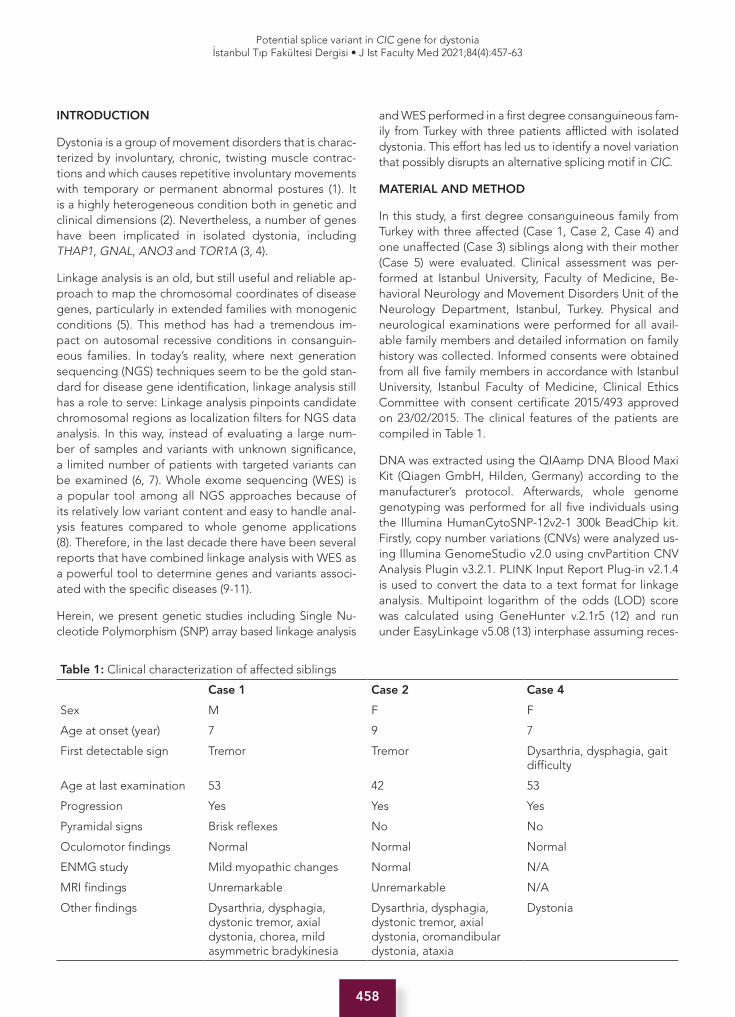
In this study, a first degree consanguineous family from Turkey with three affected (Case 1, Case 2, Case 4) and one unaffected (Case 3) siblings along with their mother (Case 5) were evaluated. Clinical assessment was per-formed at Istanbul University, Faculty of Medicine, Be-havioral Neurology and Movement Disorders Unit of the Neurology Department, Istanbul, Turkey. Physical and neurological examinations were performed for all avail-able family members and detailed information on family history was collected. Informed consents were obtained from all five family members in accordance with Istanbul University, Istanbul Faculty of Medicine, Clinical Ethics Committee with consent certificate 2015/493 approved on 23/02/2015. The clinical features of the patients are compiled in Table 1.
DNA was extracted using the QIAamp DNA Blood Maxi Kit (Qiagen GmbH, Hilden, Germany) according to the manufacturer’s protocol. Afterwards, whole genome genotyping was performed for all five individuals using the Illumina HumanCytoSNP-12v2-1 300k BeadChip kit. Firstly, copy number variations (CNVs) were analyzed us-ing Illumina GenomeStudio v2.0 using cnvPartition CNV Analysis Plugin v3.2.1. PLINK Input Report Plug-in v2.1.4 is used to convert the data to a text format for linkage analysis. Multipoint logarithm of the odds (LOD) score was calculated using GeneHunter v.2.1r5 (12) and run under EasyLinkage v5.08 (13) interphase assuming reces-
Table 1: Clinical characterization of affected siblings
Case 1 Case 2 Case 4
Sex M F F
Age at onset (year) 7 9 7
First detectable sign Tremor Tremor Dysarthria, dysphagia, gait difficulty
Age at last examination 53 42 53
Progression Yes Yes Yes
Pyramidal signs Brisk reflexes No No
Oculomotor findings Normal Normal Normal
ENMG study Mild myopathic changes Normal N/A
MRI findings Unremarkable Unremarkable N/A
Other findings Dysarthria, dysphagia, dystonic tremor, axial dystonia, chorea, mild asymmetric bradykinesia
Dysarthria, dysphagia, dystonic tremor, axial dystonia, oromandibular dystonia, ataxia
Dystonia
459
Potential splice variant in CIC gene for dystoniaİstanbul Tıp Fakültesi Dergisi • J Ist Faculty Med 2021;84(4):457-63
sive inheritance with full penetrance. Computation was adjusted in sets of 100 markers and spacing 0.1 cM. In addition, Haplopainter v1.043 (14) was used to draw the pedigree diagram and visualize the resulting haplotypes.
Whole exome sequencing was performed for the two af-fected siblings (Case 2 and Case 4) from the family on an Illumina HiSeq2000 platform. Exonic DNA was cap-tured using Agilent SureSelect Human All Exon V5 (Agi-lent Technologies, Santa Clara, CA, USA). Samples were sequenced for the targeted regions with a mean cover-age of 53× and 88% of the reads were covered over 20×. Alignment to reference genome hg19 was done using Burrows-Wheeler Aligner v0.7.16a (15), sorting, marking duplicate reads and other bam manipulations were car-ried out using Picard tools v2.12.0 (16), variant calling was performed with the Genome Analysis Toolkit v3.6.0 Hap-lotypeCaller (17). The variants were annotated using the Ensembl Variant Effect Predictor (VEP) v101 (18). Variants with gnomAD and 1kG allele frequency less than 0.001 were filtered using VEP filter script. Filtering of the high LOD regions shared by two siblings was performed using python v3.9 script. Validation and segregation analyses for candidate variants were carried-out using Sanger se-quencing. SpliceAid 2 and Splice AI were used to predict the splicing impacts of these variations (19, 20).
RESULTS
A consanguineous family with four siblings and their mother were studied. Three of the siblings (two of which were female and one was male), were affected and the other sibling was unaffected. All the affected siblings (Case 1, Case 2, Case 4), the unaffected sibling (Case 3), and the mother (Case 5), were examined at the De-partment of Neurology at Istanbul University Faculty of Medicine. Case 1 was admitted to our clinic at the age of 49 for the first time due to dystonia and involuntary movements in the whole body. He started having trem-ors in his right hand at the age of seven, and after two years, he also had tremor in his left hand, and his writing had gradually deteriorated. At the age of 12, gait diffi-culty started, and at the age of 15, he had dystonia in his whole body, mostly in his waist and neck, and rarely dysphagia. His complaints increased in cold and crowd-ed environments. He had no other diagnosed diseases. His parents were cousins. He was the second of four sib-lings, and his two sisters had similar complaints (Case 2, Case 4). In his examination, he was disorientated, and his speech was dysarthric. Myerson was positive. There were diffuse dystonic and choreiform movements, (these were more prominent on the right side of the body), and dystonic tremors in the bilateral upper extremity. There was bilateral mild bradykinesia which was more promi-nent on the left side. Deep tendon reflexes were brisk. He had gait difficulty due to severe dystonia in the whole
body. Cranial MRI and EEG examinations were normal. EMG and muscle biopsy revealed mild myopathic chang-es. Serum and urine copper and serum ceruloplasmin levels were normal. Ophthalmic examination was unre-markable. Bilateral p100 latencies were prolonged in the visual evoked potential test. Genetic investigation for Huntington’s disease was negative. Despite the L-dopa, clonazepam, biperiden, baclofen, tetrabenazine and bor-naprine treatments, his complaints were ongoing in his last examination at the 4th year of his follow-up. Case 2 was admitted to our clinic for the first time at the age of 38 with gait difficulty, tremors in the hands, and speech disorder. At the age of nine, she first started having trem-ors in her right hand and, after a while, in her left hand, and deterioration in writing was observed. Ten years later, speech and swallowing disturbances were added, and ten years later, ataxia and gait difficulties were ob-served. Her past medical history was unremarkable. Her speech was dysarthric in the neurological examination. There were oromandibular dystonia and bilateral upper extremity dystonic tremor, more prominent on the right. She had gait difficulty due to ataxia. Vitamin E and se-rum AFP levels were normal. Cranial MRI and EMG were unremarkable. The neurological examination findings of the patient, who did not attend follow-up examinations regularly and did not respond to L-dopa treatment, were the same in the 4th year follow-up. Case 4, who was ad-mitted for the first time at the age of 53, had gradually progressing gait, speech, and swallowing difficulty, which started at the age of seven. The patient, who had dysar-thria, generalized dystonia, and was unable to walk due to dystonia at the first examination, did not attend main-tain their examinations. The unaffected sibling (Case 3) and the mother (Case 5) had no neurological complaints, and neurological examinations were normal.
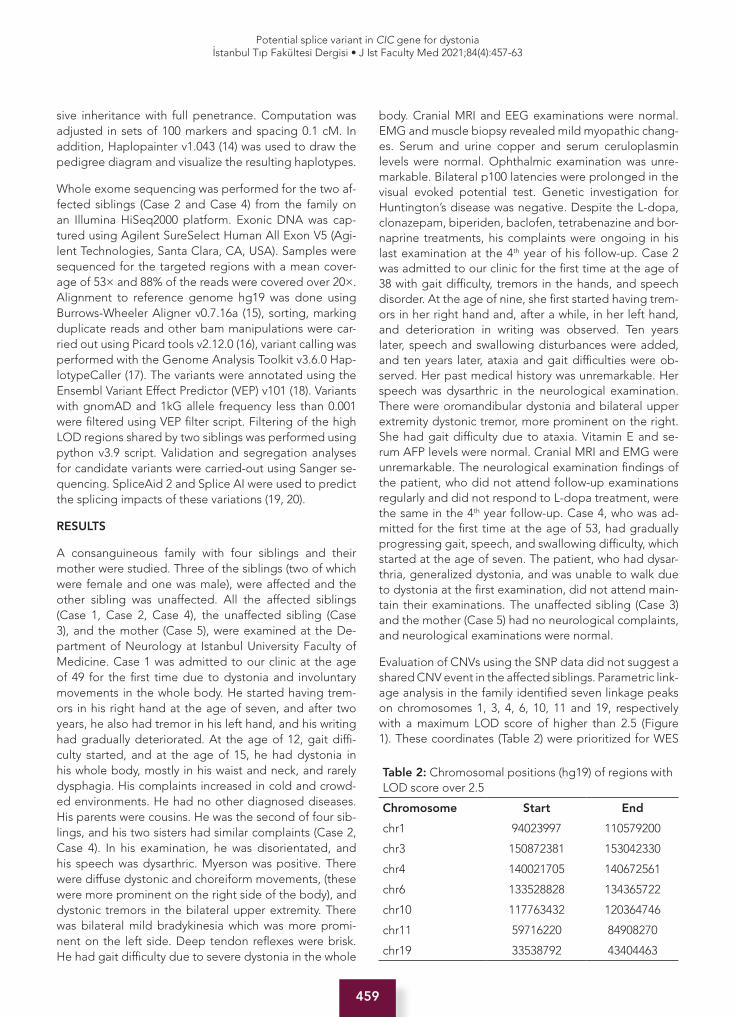
Evaluation of CNVs using the SNP data did not suggest a shared CNV event in the affected siblings. Parametric link-age analysis in the family identified seven linkage peaks on chromosomes 1, 3, 4, 6, 10, 11 and 19, respectively with a maximum LOD score of higher than 2.5 (Figure 1). These coordinates (Table 2) were prioritized for WES
Table 2: Chromosomal positions (hg19) of regions with LOD score over 2.5
Chromosome Start End
chr1 94023997 110579200
chr3 150872381 153042330
chr4 140021705 140672561
chr6 133528828 134365722
chr10 117763432 120364746
chr11 59716220 84908270
chr19 33538792 43404463
460
Potential splice variant in CIC gene for dystoniaİstanbul Tıp Fakültesi Dergisi • J Ist Faculty Med 2021;84(4):457-63
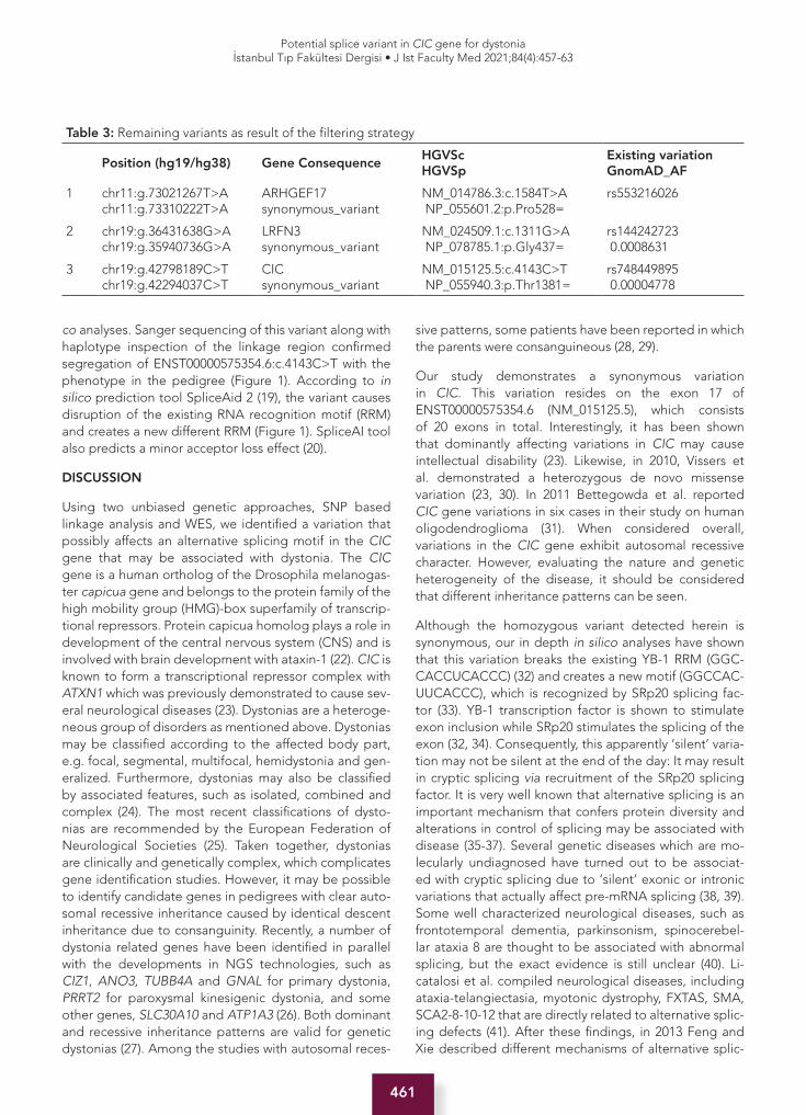
analysis. Filtering process started with the evaluation of these regions and then continued with successive steps as presented in Figure 1. This filtering strategy led us to identify three synonymous candidate variants as annotat-ed with VEP and presented in Table 3. None of these vari-ants were present in the homozygous form in the gnomAD database. Among these genes, Capicua transcriptional
repressor-CIC encodes a member of high mobility group-box (HMG-box) superfamily of transcriptional repressors and has shown to be a critical regulator of neuronal dif-ferentiation (21). Although the variant was annotated as synonymous with the VEP pipeline, the role of CIC gene product in neuronal differentiation has prompted us to perform familial variant segregation and in depth in sili-
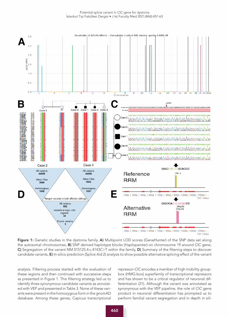
Figure 1: Genetic studies in the dystonia family. A) Multipoint LOD scores (GeneHunter) of the SNP data set along the autosomal chromosomes, B) SNP derived haplotype blocks (Haplopainter) on chromosome 19 around CIC gene, C) Segregation of the variant NM 015125.4:c.4143C>T within the family, D) Summary of the filtering steps to discover candidate variants, E) In-silico prediction (Splice Aid 2) analysis to show possible alternative splicing effect of the variant
461
Potential splice variant in CIC gene for dystoniaİstanbul Tıp Fakültesi Dergisi • J Ist Faculty Med 2021;84(4):457-63
co analyses. Sanger sequencing of this variant along with haplotype inspection of the linkage region confirmed segregation of ENST00000575354.6:c.4143C>T with the phenotype in the pedigree (Figure 1). According to in silico prediction tool SpliceAid 2 (19), the variant causes disruption of the existing RNA recognition motif (RRM) and creates a new different RRM (Figure 1). SpliceAI tool also predicts a minor acceptor loss effect (20).
DISCUSSION
Using two unbiased genetic approaches, SNP based linkage analysis and WES, we identified a variation that possibly affects an alternative splicing motif in the CIC gene that may be associated with dystonia. The CIC gene is a human ortholog of the Drosophila melanogas-ter capicua gene and belongs to the protein family of the high mobility group (HMG)-box superfamily of transcrip-tional repressors. Protein capicua homolog plays a role in development of the central nervous system (CNS) and is involved with brain development with ataxin-1 (22). CIC is known to form a transcriptional repressor complex with ATXN1 which was previously demonstrated to cause sev-eral neurological diseases (23). Dystonias are a heteroge-neous group of disorders as mentioned above. Dystonias may be classified according to the affected body part, e.g. focal, segmental, multifocal, hemidystonia and gen-eralized. Furthermore, dystonias may also be classified by associated features, such as isolated, combined and complex (24). The most recent classifications of dysto-nias are recommended by the European Federation of Neurological Societies (25). Taken together, dystonias are clinically and genetically complex, which complicates gene identification studies. However, it may be possible to identify candidate genes in pedigrees with clear auto-somal recessive inheritance caused by identical descent inheritance due to consanguinity. Recently, a number of dystonia related genes have been identified in parallel with the developments in NGS technologies, such as CIZ1, ANO3, TUBB4A and GNAL for primary dystonia, PRRT2 for paroxysmal kinesigenic dystonia, and some other genes, SLC30A10 and ATP1A3 (26). Both dominant and recessive inheritance patterns are valid for genetic dystonias (27). Among the studies with autosomal reces-
sive patterns, some patients have been reported in which the parents were consanguineous (28, 29).
Our study demonstrates a synonymous variation in CIC. This variation resides on the exon 17 of ENST00000575354.6 (NM_015125.5), which consists of 20 exons in total. Interestingly, it has been shown that dominantly affecting variations in CIC may cause intellectual disability (23). Likewise, in 2010, Vissers et al. demonstrated a heterozygous de novo missense variation (23, 30). In 2011 Bettegowda et al. reported CIC gene variations in six cases in their study on human oligodendroglioma (31). When considered overall, variations in the CIC gene exhibit autosomal recessive character. However, evaluating the nature and genetic heterogeneity of the disease, it should be considered that different inheritance patterns can be seen.
Although the homozygous variant detected herein is synonymous, our in depth in silico analyses have shown that this variation breaks the existing YB-1 RRM (GGC-CACCUCACCC) (32) and creates a new motif (GGCCAC-UUCACCC), which is recognized by SRp20 splicing fac-tor (33). YB-1 transcription factor is shown to stimulate exon inclusion while SRp20 stimulates the splicing of the exon (32, 34). Consequently, this apparently ‘silent’ varia-tion may not be silent at the end of the day: It may result in cryptic splicing via recruitment of the SRp20 splicing factor. It is very well known that alternative splicing is an important mechanism that confers protein diversity and alterations in control of splicing may be associated with disease (35-37). Several genetic diseases which are mo-lecularly undiagnosed have turned out to be associat-ed with cryptic splicing due to ‘silent’ exonic or intronic variations that actually affect pre-mRNA splicing (38, 39). Some well characterized neurological diseases, such as frontotemporal dementia, parkinsonism, spinocerebel-lar ataxia 8 are thought to be associated with abnormal splicing, but the exact evidence is still unclear (40). Li-catalosi et al. compiled neurological diseases, including ataxia-telangiectasia, myotonic dystrophy, FXTAS, SMA, SCA2-8-10-12 that are directly related to alternative splic-ing defects (41). After these findings, in 2013 Feng and Xie described different mechanisms of alternative splic-
Table 3: Remaining variants as result of the filtering strategy
Position (hg19/hg38) Gene ConsequenceHGVScHGVSp
Existing variation GnomAD_AF
1 chr11:g.73021267T>A chr11:g.73310222T>A
ARHGEF17 synonymous_variant
NM_014786.3:c.1584T>A NP_055601.2:p.Pro528=
rs553216026
2 chr19:g.36431638G>A chr19:g.35940736G>A
LRFN3 synonymous_variant
NM_024509.1:c.1311G>A NP_078785.1:p.Gly437=
rs144242723 0.0008631
3 chr19:g.42798189C>T chr19:g.42294037C>T
CIC synonymous_variant
NM_015125.5:c.4143C>T NP_055940.3:p.Thr1381=
rs748449895 0.00004778

462
Potential splice variant in CIC gene for dystoniaİstanbul Tıp Fakültesi Dergisi • J Ist Faculty Med 2021;84(4):457-63
ing that are related to neurological diseases (42). Alter-native splicing variants are also shown to be a part of the disease mechanisms of neurological disorders with complex genetic etiologies including ataxia and dystonia (43, 44). In sum, although the mechanism of occurrence of many neurological diseases has been reported to be al-ternative splicing, there may be other many neurological diseases that are still not clarified.
CONCLUSION
In our findings we demonstrated that CIC may be associ-ated with dystonia and other neurological phenotypes by mechanism of alternative splicing. Therefore, our report requires further functional studies both for in vitro and in vivo to define the exact features of the CIC gene.
Ethics Committee Approval: This study was approved by the Clinical Ethical Committee of the Istanbul University, Istanbul Fa-culty of Medicine (Date: 23/02/2015, No: 2015/493).
Informed Consent: Written consent was obtained from the par-ticipants.
Peer Review: Externally peer-reviewed.
Author Contributions: Conception/Design of Study- B.S., S.U.İ., E.Y., B.B., H.H.; Data Acquisition- E.Y., B.S., B.B., H.H., H.G.; Data Analysis/Interpretation- B.S., E.Y., B.S., B.B., H.H., S.U.İ.; Drafting Manuscript- B.S., E.Y., B.S., B.B., S.U.İ.; Critical Revision of Manuscript- B.S., E.Y., B.S., B.B., H.H., H.G., U.Ö., S.U.İ.; Final Approval and Accountability- B.S., E.Y., B.S., B.B., H.H., H.G., U.Ö., S.U.İ
Conflict of Interest: Authors declared no conflict of interest.
Financial Disclosure: This study was supported by the grants of Scientific Research Projects Coordination Unit of Istanbul Uni-versity (Project numbers: TYL-2018-30315 and ONAP-11021). EY and BS had been fellows of TUBITAK-113S331 and TUBI-TAK-214S222 projects.
Acknowledgement: The authors are grateful to the family for participating in this study. We also thank to Turkish Academy of Sciences for the 2019 Distinguished Young Scientist Award to SAUI.
Etik Komite Onayı: Bu çalışma için etik komite onayı İstanbul Üniversitesi, İstanbul Tıp Fakültesi Klinik Etik Kurulu’ndan alın-mıştır (Tarih: 23/02/2015, No: 2015/493).
Bilgilendirilmiş Onam: Katılımcılardan bilgilendirilmiş onam alınmıştır.
Hakem Değerlendirmesi: Dış bağımsız.
Yazar Katkıları: Çalışma Konsepti/Tasarım- B.S., S.U.İ., E.Y., B.B., H.H.; Veri Toplama- E.Y., B.S., B.B., H.H., H.G.; Veri Analizi/Yo-rumlama- B.S., E.Y., B.S., B.B., H.H., S.U.İ.; Yazı Taslağı- B.S., E.Y.,
B.S., B.B., S.U.İ.; İçeriğin Eleştirel İncelemesi- B.S., E.Y., B.S., B.B., H.H., H.G., U.Ö., S.U.İ.; Son Onay ve Sorumluluk- B.S., E.Y., B.S., B.B., H.H., H.G., U.Ö., S.U.İ
Çıkar Çatışması: Yazarlar çıkar çatışması beyan etmemişlerdir.
Finansal Destek: Bu çalışma, İstanbul Üniversitesi Bilimsel Araştırma Projeleri Koordinasyon Birimi’nin hibeleri ile destek-lenmiştir (Proje numaraları: TYL-2018-30315 ve ONAP-11021). EY ve BS, TÜBİTAK-113S331 ve TÜBİTAK-214S222 projelerinin bursiyeriydi.
Teşekkür: Yazarlar, bu çalışmaya katıldığı için aileye minnettardır. SAUI’ye verilen 2019 Seçkin Genç Bilim İnsanı Ödülü için Türkiye Bilimler Akademisi’ne de teşekkür ederiz.
REFERENCES
1. Jinnah HA. Diagnosis & Treatment of dystonia. Neurol Clin 2015;33(1):77-100. [CrossRef]
2. Balint B, Mencacci NE, Valente EM, Pisani A, Rothwell J, Jankovic J, et al. Dystonia. Nature Reviews Disease Primers. 2018;4(1):1-23. [CrossRef]
3. Domingo A, Yadav R, Ozelius LJ. Isolated dystonia: clinical and genetic updates. J Neural Transm (Vienna) 2021;128(4):405-16. [CrossRef]
4. Charlesworth G, Bhatia KP, Wood NW. The genetics of dystonia: new twists in an old tale. Brain 2013;136(Pt 7):2017-37. [CrossRef]
5. Pulst SM. Genetic linkage analysis. Arch Neurol 1999;56(6):667. [CrossRef]
6. Yucesan E, Ugur Iseri SA, Bilgic B, Gormez Z, Bakir Gungor B, Sarac A, et al. SYNE1 related cerebellar ataxia presents with variable phenotypes in a consanguineous family from Turkey. Neurol Sci 2017;38(12):2203-7. [CrossRef]
7. Ugur Iseri SA, Yucesan E, Tuncer FN, Calik M, Kesim Y, Altiokka Uzun G, et al. Biallelic loss of EEF1D function links heat shock response pathway to autosomal recessive intellectual disability. Journal of Human Genetics 2019;64(5):421-6. [CrossRef]
8. Zech M, Jech R, Boesch S, Škorvánek M, Weber S, Wagner M, et al. Monogenic variants in dystonia: an exome-wide sequencing study. The Lancet Neurology 2020;19(11):908-18. [CrossRef]
9. Mulder R, Lisman T, Meijers JCM, Huntington JA, Mulder AB, Meijer K. Linkage analysis combined with whole-exome sequencing identifies a novel prothrombin (F2) gene mutation in a Dutch Caucasian family with unexplained thrombosis. 1. 2020;105(7):e370-2. [CrossRef]
10. Mescheriakova JY, Verkerk AJ, Amin N, Uitterlinden AG, van Duijn CM, Hintzen RQ. Linkage analysis and whole exome sequencing identify a novel candidate gene in a Dutch multiple sclerosis family. Mult Scler 2019;25(7):909-17. [CrossRef]
11. Choi YJ, Ohn JH, Kim N, Kim W, Park K, Won S, et al. Family-based exome sequencing combined with linkage analyses identifies rare susceptibility variants of MUC4 for gastric cancer. PLOS ONE 2020;15(7):e0236197. [CrossRef]
12. Markianos K, Daly MJ, Kruglyak L. Efficient multipoint linkage analysis through reduction of inheritance space. The American Journal of Human Genetics 2001;68(4):963-77. [CrossRef]

463
Potential splice variant in CIC gene for dystoniaİstanbul Tıp Fakültesi Dergisi • J Ist Faculty Med 2021;84(4):457-63
13. Hoffmann K, Lindner TH. easyLINKAGE-Plus-automated linkage analyses using large-scale SNP data. Bioinformatics 2005;21(17):3565-7. [CrossRef]
14. Thiele H, Nürnberg P. HaploPainter: a tool for drawing pedigrees with complex haplotypes. Bioinformatics 2005;21(8):1730-2. [CrossRef]
15. Li H, Durbin R. Fast and accurate short read alignment with Burrows-Wheeler transform. Bioinformatics 2009;25(14):1754-60. [CrossRef]
16. A set of command line tools (in Java) for manipulating high-throughput sequencing (HTS) data and formats such as SAM/BAM/CRAM and VCF.: broadinstitute/picard [Internet]. Broad Institute; 2019 [cited 2019 May 7]. Available from: https://github.com/broadinstitute/picard
17. McKenna A, Hanna M, Banks E, Sivachenko A, Cibulskis K, Kernytsky A, et al. The Genome Analysis Toolkit: a MapReduce framework for analyzing next-generation DNA sequencing data. Genome Res 2010;20(9):1297-303. [CrossRef]
18. McLaren W, Gil L, Hunt SE, Riat HS, Ritchie GRS, Thormann A, et al. The ensembl variant effect predictor. Genome Biology 2016;17(1):122. [CrossRef]
19. Piva F, Giulietti M, Burini AB, Principato G. SpliceAid 2: a database of human splicing factors expression data and RNA target motifs. Hum Mutat 2012;33(1):81-5. [CrossRef]
20. Jaganathan K, Panagiotopoulou SK, McRae JF, Darbandi SF, Knowles D, Li YI, et al. Predicting splicing from primary sequence with deep learning. Cell 2019;176(3):535-548.e24. [CrossRef]
21. Inah Hwang, Heng Pan, Yao J, Olivier Elemento, Hongwu Zheng, Paik J. CIC is a critical regulator of neuronal differentiation. JCI Insight [Internet]. 2020 [cited 2021 Feb 22];5(9). Available from: https://insight.jci.org/articles/view/135826 [CrossRef]
22. Lee C-J, Chan W-I, Cheung M, Cheng Y-C, Appleby VJ, Orme AT, et al. CIC, a member of a novel subfamily of the HMG-box superfamily, is transiently expressed in developing granule neurons. Brain Res Mol Brain Res 2002;106(1-2):151-6. [CrossRef]
23. Lu H-C, Tan Q, Rousseaux MWC, Wang W, Kim J-Y, Richman R, et al. Disruption of the ATXN1-CIC complex causes a spectrum of neurobehavioral phenotypes in mice and humans. Nature Genetics 2017;49(4):527-36. [CrossRef]
24. Klein C, Lohmann K, Marras C, Münchau A. Hereditary Dystonia Overview. In: Adam MP, Ardinger HH, Pagon RA, Wallace SE, Bean LJ, Mirzaa G, et al., editors. GeneReviews® [Internet]. Seattle (WA): University of Washington, Seattle; 1993 [cited 2021 Feb 14]. Available from: http://www.ncbi.nlm.nih.gov/books/NBK1155/
25. Albanese A, Asmus F, Bhatia KP, Elia AE, Elibol B, Filippini G, et al. EFNS guidelines on diagnosis and treatment of primary dystonias. Eur J Neurol 2011;18(1):5-18. [CrossRef]
26. Charlesworth G, Bhatia KP, Wood NW. The genetics of dystonia: new twists in an old tale. Brain 2013;136(Pt 7):2017-37. [CrossRef]
27. Németh AH. The genetics of primary dystonias and related disorders. Brain 2002;125(Pt 4):695-721. [CrossRef]
28. Almasy L, Bressman SB, Raymond D, Kramer PL, Greene PE, Heiman GA, et al. Idiopathic torsion dystonia linked to chromosome 8 in two Mennonite families. Ann Neurol 1997;42(4):670-3. [CrossRef]
29. Ozelius LJ, Bressman SB. Genetic and clinical features of primary torsion dystonia. Neurobiol Dis 2011;42(2):127-35. [CrossRef]
30. Vissers LELM, de Ligt J, Gilissen C, Janssen I, Steehouwer M, de Vries P, et al. A de novo paradigm for mental retardation. Nat Genet 2010;42(12):1109-12. [CrossRef]
31. Bettegowda C, Agrawal N, Jiao Y, Sausen M, Wood LD, Hruban RH, et al. Mutations in CIC and FUBP1 contribute to human oligodendroglioma. Science 2011;333(6048):1453-5. [CrossRef]
32. Wei WJ, Mu SR, Heiner M, Fu X, Cao LJ, Gong XF, et al. YB-1 binds to CAUC motifs and stimulates exon inclusion by enhancing the recruitment of U2AF to weak polypyrimidine tracts. Nucleic Acids Res 2012;40(17):8622-36. [CrossRef]
33. Hargous Y, Hautbergue GM, Tintaru AM, Skrisovska L, Golovanov AP, Stevenin J, et al. Molecular basis of RNA recognition and TAP binding by the SR proteins SRp20 and 9G8. EMBO J 2006;25(21):5126-37. [CrossRef]
34. Änkö ML, Morales L, Henry I, Beyer A, Neugebauer KM. Global analysis reveals SRp20- and SRp75-specific mRNPs in cycling and neural cells. Nature Structural & Molecular Biology. 2010;17(8):962-70. [CrossRef]
35. Tazi J, Bakkour N, Stamm S. Alternative splicing and disease. Biochim Biophys Acta. 2009;1792(1):14-26. [CrossRef]
36. Webster NJG. Alternative RNA splicing in the pathogenesis of liver disease. Front Endocrinol (Lausanne).;8:133. [CrossRef]
37. Kremer LS, Bader DM, Mertes C, Kopajtich R, Pichler G, Iuso A, et al. Genetic diagnosis of Mendelian disorders via RNA sequencing. Nature Communications 2017;8(1):15824. [CrossRef]
38. Poulos MG, Batra R, Charizanis K, Swanson MS. Developments in RNA splicing and disease. Cold Spring Harb Perspect Biol 2011;3(1):a000778. [CrossRef]
39. Cartegni L, Chew SL, Krainer AR. Listening to silence and understanding nonsense: exonic mutations that affect splicing. Nature Reviews Genetics 2002;3(4):285-98. [CrossRef]
40. Dredge BK, Polydorides AD, Darnell RB. The splice of life: alternative splicing and neurological disease. Nat Rev Neurosci 2001;2(1):43-50. [CrossRef]
41. Licatalosi DD, Darnell RB. Splicing regulation in neurologic disease. Neuron 2006;52(1):93-101. [CrossRef]
42. Feng D, Xie J. Aberrant splicing in neurological diseases. Wiley Interdiscip Rev RNA 2013;4(6):631-49. [CrossRef]
43. Shohet A, Cohen L, Haguel D, Mozer Y, Shomron N, Tzur S, et al. Variant in SCYL1 gene causes aberrant splicing in a family with cerebellar ataxia, recurrent episodes of liver failure, and growth retardation. European Journal of Human Genetics 2019;27(2):263-8. [CrossRef]
44. Xiao J, Zhao Y, Bastian RW, Perlmutter JS, Racette BA, Tabbal SD, et al. Novel THAP1 sequence variants in primary dystonia. Neurology 2010;74(3):229-38. [CrossRef]

464
Oxidant effect on kidney of EPO therapy in seizureİstanbul Tıp Fakültesi Dergisi • J Ist Faculty Med 2021;84(4):464-71
Corresponding author/İletişim kurulacak yazar: [email protected]
Submitted/Başvuru: 19.02.2021 • Revision Requested/Revizyon Talebi: 30.03.2021 •Last Revision Received/Son Revizyon: 19.04.2021 • Accepted/Kabul: 19.04.2021 • Published Online/Online Yayın: 14.09.2021
RESEARCH / ARAŞTIRMADOI: 10.26650/IUITFD.2021.883402
İst Tıp Fak Derg 2021 / J Ist Faculty Med 2021
EFFECTS OF ERYTHROPOIETIN PRETREATMENT ON LIVER, KIDNEY, HEART TISSUE IN PENTYLENTETRAZOL-INDUCED SEIZURES; EVALUATION IN TERMS OF OXIDATIVE MARKERS, PROLIDASE AND SIALIC ACID
PENTİLENTETRAZOL-İNDÜKLÜ NÖBETLERDE ERİTROPOİETİN ÖN TEDAVİSİNİN KARACİĞER, BÖBREK, KALP DOKUSU ÜZERİNE ETKİLERİ; OKSİDATİF MARKIRLAR, PROLİDAZ VE SİALİK ASİT AÇISINDAN DEĞERLENDİRME
Ayşegül KAPUCU1 , Zülal KAPTAN2 , Kadriye AKGÜN DAR1 , İslim KALELER3 , Gülay ÜZÜM4
1Istanbul University, Faculty of Science, Department of Biology, Istanbul, Turkey2Beykent University, Faculty of Medicine, Department of Physiology, Istanbul, Turkey3Istanbul University- Cerrahpasa, Cerrahpaşa Faculty of Medicine, Department of Medical Biochemistry, Istanbul, Turkey4Istanbul University, Istanbul Faculty of Medicine, Department of Physiology, Istanbul, Turkey
ORCID IDs of the authors: A.K. 0000-0002-0946-1407; Z.K. 0000-0002-2641-9534; K.A.D. 0000-0003-2060-1199; İ.K. 0000-0002-2712-7955; G.Ü. 0000-0003-2329-3689
Cite this article as: Kapucu A, Kaptan Z, Akgun Dar K, Kaleler I, Uzum G. Effects of erythropoietin pretreatment on liver, kidney, heart tissue in pentylentetrazol-induced seizures; evaluation in terms of oxidative markers, prolidase and sialic acid. J Ist Faculty Med 2021;84(4):464-71. doi: 10.26650/IUITFD.2021.883402
ABSTRACT
Objective: The effects of erythropoietin (EPO) which has been frequently studied as an anti-epileptic agent, on peripheral tis-sues have not been investigated. This study investigated the ef-fects on malondialdehyde (MDA), advanced protein oxidation products (AOPP), superoxide dismutase (SOD), prolidase and sialic acid (SA) levels in the heart, kidney and liver tissues of EPO pretreatment in pentylenetetrazole (PTZ)-induced seizures.
Material and Method: Thirty three male adult rats were divided into three groups. A saline-injected control group, a 60 mg/kg PTZ-injected group to induce seizures and a 3000 IU/kg EPO-in-jected group 24 hours before seizures. After seizure severity and seizure latency were scored, the rats sacrificed, the tissues were immediately removed for biochemical analyses.
Results: The PTZ-induced seizures increased MDA in kidney (p<0.01) and AOPP in liver (p<0.05) but didn’t alter these mark-ers in heart tissue. In all three tissues, SOD didn’t change due to seizures. The SA levels increased in the heart (p<0.001), de-creased in the kidney (p<0.001), and were unchanged in liver. Prolidase increased (p<0.05) only in kidney, and was unchanged in other tissues. EPO-pretreatment decreased seizure severi-ty and increased seizure latency. It prevented the increase in MDA in the kidney (p<0.01) but increased AOPP (p<0.05) and
ÖZET
Amaç: Antiepileptik ajan olarak sıklıkla çalışılan eritropoietinin (EPO)’nun periferik dokular üzerindeki etkileri araştırılmamıştır. Bu çalışmada pentilentetrazol (PTZ) ile indüklenen nöbetlerde EPO ön tedavisinin kalp, böbrek ve karaciğer dokularında ma-londialdehit (MDA), ileri protein oksidasyon ürünleri (AOPP), su-peroksid dismutaz (SOD), prolidaz ve sialik asit (SA) seviyelerine etkisi araştırıldı.
Gereç ve Yöntem: Otuz üç erişkin erkek sıçan üç gruba ayrıldı. Salin enjekte edilmiş kontrol grubu, nöbetleri indüklemek için 60 mg/kg PTZ enjekte edilmiş grup, nöbetlerden 24 saat önce 3000 IU/kg EPO enjekte edilmiş grup. Nöbet şiddeti ve nöbet gecikmesi puanlandıktan sonra, sıçanlar sakrifiye edildi, dokular biyokimyasal analizler için hemen çıkarıldı.
Bulgular: Pentilentetrazol ile indüklenen nöbetler, böbrekte MDA (p<0,01) ve karaciğerde AOPP’yi arttırdı (p<0,05), ancak kalp dokusunda bu markırları değiştirmedi. Her üç dokuda da SOD nöbetler nedeniyle değişmedi. Kalpte SA arttı (p<0,001), böbrekte azaldı (p<0,001), karaciğerde değişmedi. Prolidaz sa-dece böbrekte arttı (p<0,05), diğer dokularda değişmedi. EPO ön tedavisi nöbet şiddetini azalttı ve nöbet latansını artırdı. EPO böbrekte MDA artışını engelledi (p<0,01), ancak AOPP’yi artırdı (p<0,05) ve SOD’u azalttı (p<0,01) ve prolidazı nöbetlerin arttırdı-
Content of this journal is licensed under a Creative Commons Attribution-NonCommercial 4.0 International License.

465
Oxidant effect on kidney of EPO therapy in seizureİstanbul Tıp Fakültesi Dergisi • J Ist Faculty Med 2021;84(4):464-71
RESEARCH / ARAŞTIRMADOI: 10.26650/IUITFD.2021.883402
İst Tıp Fak Derg 2021 / J Ist Faculty Med 2021
EFFECTS OF ERYTHROPOIETIN PRETREATMENT ON LIVER, KIDNEY, HEART TISSUE IN PENTYLENTETRAZOL-INDUCED SEIZURES; EVALUATION IN TERMS OF OXIDATIVE MARKERS, PROLIDASE AND SIALIC ACID
PENTİLENTETRAZOL-İNDÜKLÜ NÖBETLERDE ERİTROPOİETİN ÖN TEDAVİSİNİN KARACİĞER, BÖBREK, KALP DOKUSU ÜZERİNE ETKİLERİ; OKSİDATİF MARKIRLAR, PROLİDAZ VE SİALİK ASİT AÇISINDAN DEĞERLENDİRME
Ayşegül KAPUCU1 , Zülal KAPTAN2 , Kadriye AKGÜN DAR1 , İslim KALELER3 , Gülay ÜZÜM4
1Istanbul University, Faculty of Science, Department of Biology, Istanbul, Turkey2Beykent University, Faculty of Medicine, Department of Physiology, Istanbul, Turkey3Istanbul University- Cerrahpasa, Cerrahpaşa Faculty of Medicine, Department of Medical Biochemistry, Istanbul, Turkey4Istanbul University, Istanbul Faculty of Medicine, Department of Physiology, Istanbul, Turkey
ORCID IDs of the authors: A.K. 0000-0002-0946-1407; Z.K. 0000-0002-2641-9534; K.A.D. 0000-0003-2060-1199; İ.K. 0000-0002-2712-7955; G.Ü. 0000-0003-2329-3689
Cite this article as: Kapucu A, Kaptan Z, Akgun Dar K, Kaleler I, Uzum G. Effects of erythropoietin pretreatment on liver, kidney, heart tissue in pentylentetrazol-induced seizures; evaluation in terms of oxidative markers, prolidase and sialic acid. J Ist Faculty Med 2021;84(4):464-71. doi: 10.26650/IUITFD.2021.883402
INTRODUCTION
It is demonstrated that epileptic seizures cause oxida-tive stress (OS) not only in the central nervous system but also in peripheral tissues (1). OS is an issue caused by an imbalance between the production of reactive oxygen species (ROS) in tissues and the capacity of endogenous antioxidant defense systems to remove these reactive products. Superoxide dismutase (SOD), one of the im-portant antioxidant enzymes in the body, plays a key role in detoxifying superoxide anions and may prevent OS-in-duced cellular damage (2). It is shown that experimental induced epileptic seizures increase lipid peroxidation and decrease SOD enzyme activity in liver and kidney (2). On the other hand, it is reported that various anti-epileptic drugs (AEDs) are insufficient in preventing seizures and may trigger OS in brain and peripheral tissues, impair the endogenous antioxidative ability. Thus, epilepsy pa-tients are suffering from hepatic and renal dysfunctions due to their current anti-epileptic drug treatment (1). For instance, it is shown that commonly used drugs such as sodium valproate is associated with serious hepatotoxicity (3). Thus, novel AED investigations that suppress seizures more efficiently with no or minimum adverse effects draw attention. Erythropoietin (EPO) is a hypoxia inducible he-matopoietic factor, which is predominantly expressed in the kidneys. However, EPO and its receptors are widely expressed in many tissues including brain, liver, skeletal, heart, muscle and lungs (4). There are many studies show-ing the anti-epileptic effect of EPO (5-7). In our previous studies, we have shown its antioxidant and anti-inflam-matory effects on brain tissue in addition to antiseizure effect of EPO treatment in a generalized acute tonic-clon-ic epilepsy model induced by pentylenetetrazole (PTZ) in rats (6, 7). However, although EPO’s anti-epileptic proper-ty is shown, it is an important deficiency that its effects on peripheral tissues have not been investigated yet. EPO has been shown to have an antioxidant effect and a protective effect on heart tissue and kidney ischemia/reperfusion injury models (4, 8, 9). These reports led us to ask what effect EPO treatment might have on the OS that may occur in peripheral tissues in epileptic seizures.
Because of detoxification and excretion functions, the liver and kidney are two important organs that have been investi-gated in the context of side effects such as OS in experimen-tal seizure models (2, 10). On the other hand, it is reported that drug refractory epilepsy patients have cardiovascular abnormalities and epilepsy is associated with the risk of cardiac ischemia (11). Also, it is known that sudden car-diac arrest and heart rhythm disorders are considered the most common cause of epilepsy-related deaths (12). However, the effects of seizures and anti-epileptic drugs on heart tissue have not been investigated. Voltage-gat-ed sodium channels which include negatively charged sialic acid (SA) is vital for neuronal signal conduction and regular heart rhythms (13). Sialic acid can directly activate the volt-age dependent sodium channel at lower depolarization via contributing to negative potential (14). In this context, if SA may affect heart muscle excitability, how epileptic seizures and anti-epileptic drugs affect SA levels in heart muscle is an issue that needs to be investigated, but as far as we know, no such study has been conducted before. Besides the effects of epileptic seizures and anti-epileptic drugs on heart tissue have not been investigated in terms of other biomarkers.
Prolidase is a metalloenzyme that is found in many tissues including the kidney, brain, heart, lungs, and pancreas (15). It plays a role in the recycling of pro-line from imidodipeptides for the resynthesis of col-lagen, the main component of the connective tissue and therefore it provides collagen turnover, and ma-trix remodeling (16). It is also reported that prolidase enzyme activity may be induced with OS and inflam-mation, also disturbances in prolidase enzyme activity can contribute to the damaging effects of free radicals through collagen breakdown and may play a role in the progress of various diseases (17). Besides, it is shown that serum prolidase enzyme activity and OS values increased in epileptic patients taking anti-epileptic drug and it is reported that this may be a risk factor for vascular damage because of the increase in the colla-gen cycle (18). To the best of our knowledge, there is no data on how the epileptic seizures affect prolidase
decreased SOD (p<0.01) and further increased prolidase more than the seizures increased (p<0.01). EPO-pretreatment pre-vented the increase in AOPP in the liver (p<0.05) but was inef-fective in PTZ-induced SA changes in the heart and kidney.
Conclusion: We think that the increase in the heart SA level in seizures is an original finding and deserves investigation in the context of seizure-related cardiac arrhytmias. Also, despite the EPO’s anti-seizure effect, increased protein oxidaiton and proli-dase, especially in the kidney, is an other important finding that needs further research.
Keywords: Erythropoietin, PTZ-induced seizures, oxidative stress markers, sialic acid, prolidase, peripheral tissues
ğından daha fazla artırdı (p<0,01). EPO ön tedavisi, karaciğerde AOPP artışını önledi (p<0,05), ancak kalp ve böbrekte PTZ’nin neden olduğu SA değişikliklerinde etkisizdi.
Sonuç: Nöbetlerde kalp dokusundaki SA artışının nöbet-ilişkili kardiak aritmiler bağlamında araştırmayı hak eden orijinal bulgu olduğunu düşünüyoruz. Ayrıca EPO ön tedavisinin nöbet engel-leyici etkisine rağmen özellikle böbrek dokusunda artmış prote-in oksidasyonu ve prolidaz da ileri araştırmayı gerektiren diğer önemli bulgudur.
Anahtar Kelimeler: Eritropoietin, PTZ-indüklü nöbetler, oksida-tif stres belirteçleri, sialik asit, prolidaz, periferik dokular

466
Oxidant effect on kidney of EPO therapy in seizureİstanbul Tıp Fakültesi Dergisi • J Ist Faculty Med 2021;84(4):464-71
activity in kidney, liver, and heart tissues and how the EPO treatment changes it.
In light of this information, in this study, we essentially fo-cused the effects of PTZ-induced acute generalized tonic clonic seizures as well as EPO pretreatment 24 hours before PTZ administration on liver, kidney and heart tissues. We evaluated these effects in terms of SA levels, prolidase activity, advanced oxidation protein products (AOPP; protein oxidation indicator), malondialdehyde (MDA; lip-id peroxidaiton indicator) and SOD levels.
MATERIAL AND METHOD
Animals and experimental designMale Wistar albino rats (200–250g) were housed in cages and 12 h light–dark cycle and an ad libitum feeding were maintained. Experiments were conducted in the morning to avoid circadian variations. All procedures were done in accordance with the guidelines of Bezmialem Vakif University Animal Experiments Local Ethics Committee (Date: 22.04.2016, No:128).
Thirty three rats were randomly divided into three groups (n=11/each group); 1: Control group (administered with 0.9% saline), 2: PTZ group (administered with 60 mg/kg PTZ to induce generalized tonic-clonic seizures), 3: EPO+PTZ group (administered with 3000 IU/kg EPO 24 h before PTZ injection). PTZ (Sigma, St. Louis, MO, USA), and Recombinant human EPO (r-HuEPO, Eprex; Epoetin alfa, Santa Farma, Turkey) were dissolved in 0.5 ml 0.9% saline and administered intraperitoneally. PTZ dose was selected as it achieves the most successful convulsive re-sponse with the least mortality (19). The EPO dose is the dose that does not have any side effects that do not affect the hematocrit values and has anticonvulsive activity (7).
PTZ-induced seizuresPentilentetrazol is commonly used to create a model of generalized seizure in rats and to study the effectiveness of anti-epileptic drugs (7). A single dose 60 mg/kg PTZ intraperitoneally was injected to induce tonic-clonic generalized seizures. After the PTZ injection, the rats were placed in a plexiglass acrylic cage and seizure behavior was observed until convulsions stopped and was recorded with a camera. The severity of seizures were assessed, using scores based on modified Racine’s scale (20), as follows: 1, ear and facial twitching; 2, head bobbing and repeated myoclonic jerks; 3, partial clonic forelimb convulsions in a sitting position; 4, major seizures (generalized tonic-clonic seizures whilst lying on the belly); 5, generalized tonic-clonic seizures; running, followed by the loss of righting ability, and then a tonic phase progressive to the clonus of all four limbs. Seizure latency was measured as the time between the injection of PTZ and the appearance of the first myoclonic jerks.
Biochemical analysis of AOPP, MDA, SOD, SA and pro-lidase enzyme activity in liver, heart, and kidneysAfter the evaluation of seizure severity, rats were de-capitated under anesthesia (50 mg/kg ketamine and 10 mg/kg xylazine) and the liver, heart and kidneys were re-moved immediately. The kidney, heart and liver tissues were washed in ice cold 0.01 M PBS (Phosphate Buffered Saline) to avoid blood contamination. All tissue samples were homogenized in 0.01 M PBS (pH 7.4) using Teflon/glass homogenizer and the homogenates were centri-fuged at 5,000 g for 15 min at 4°C. The supernatants were aliquoted and stored at -80ºC immediately until AOPP, MDA, SA levels and SOD, prolidase activity assay were performed. The total amount of protein was determined by the Lowry method (21).
AOPP assays were performed spectrophotometrically at 340 nm wavelength by Hanasand’s modified method (22). AOPP concentrations were calculated from the stan-dard curve graph and expressed as μmol/L chloramine-T equivalents.
Changes in tissue lipid peroxidation levels were evaluat-ed by measuring MDA levels. Spectrophotometric anal-ysis of MDA levels was performed by Beuge and Aust’s method (23), determined by the quantity of thiobarbituric acid reactive products. The MDA concentration was cal-culated by its molar extinction coefficient (ε=155 mM-
1cm- 1) and expressed as nmol/mg protein.
SA levels were determined by the Tram method (24). Β-formylpyruvic acid formed because of periodic acid oxidation was reacted with two molthiobarbituric acid. A colored compound was formed that gave maximum absorbance at 549 nm. Since this product is not stable, absorbances were recorded at 549 nm in the spectropho-tometer by pulling into the cyclohex-zanon phase. The SA amount was expressed as μg/mg protein.
The total SOD enzyme activity was measured spectro-photometrically (25). The percent inhibition rate was cal-culated with the formula Ablank-Asample/Ablank x 100. 50% inhibition corresponds to 1 Unit of enzyme activity. Enzyme activity was given as U/mg protein.
Prolidase enzyme activity was determined according to the spectrophotometric method of Ozcan et al. 2007 (26), based on the measurement of proline levels, a prolidase product, produced by prolidase enzyme. Tissue prolidase activities were expressed in terms of nmol/min/mg protein.
Statistical analysisThe levels of AOPP, MDA, SOD, SA and prolidase en-zyme activity in the liver, heart and kidneys were analyzed separately. The comparisons between groups were made by unpaired t tests in GraphPad Prism 8 software. A value of p<0.05 was considered statistically significant.

467
Oxidant effect on kidney of EPO therapy in seizureİstanbul Tıp Fakültesi Dergisi • J Ist Faculty Med 2021;84(4):464-71
RESULTS
Evaluation of PTZ-induced seizures A single dose of 60 mg/kg PTZ caused generalized tonic clonic epileptic seizures of 4-5 severity according to the Racine’s scale. EPO pretreatment significantly reduced the severity of seizures and increased seizures latency (Table 1). As seizures were not observed in the control group, it was not included in the table.
Evaluation of OS markers, prolidase and SA levels in peripheral tissues When the changes in the OS markers, prolidase and SA levels in peripheral tissues were examined, different re-sults were observed for all three organs. The variation of each parameter according to the groups was as follows.
AOPP; PTZ induced seizures caused an increase in AOPP in liver tissue compared to the control group (p<0.05) but did not change in heart and kidney AOPP levels com-pared to the control group (Figure 1). EPO treatment before seizures decreased AOPP level in the liver com-pared to the PTZ group (p<0.05). Conversely, it further increased AOPP level in kidney compared to the PTZ group (p<0.01) (Figure 1). EPO pretreatment did not sig-nificantly cause change in terms of AOPP in heart tissue.
MDA; PTZ-induced seizures increased kidney MDA level compared to control group (p<0.01). Conversely, it did not cause to any change in MDA levels in liver and heart tissues compared to the control group (Figure 2). EPO treatment before seizures significantly decreased MDA levels in heart (p<0.05) and kidney (p<0.01) compared to PTZ group (Figure 2). But EPO pretreatment did not cause any change in liver MDA level compared to other groups (Figure 2).
SOD; PTZ-induced seizures did not change SOD levels of liver, heart and kidney tissues according to the con-trol group (Figure 3). EPO treatment before seizures did not cause significant difference in liver and heart tissues’ SOD levels, but EPO pretreatment significantly caused a decreased SOD level in kidney compared to the PTZ and control groups (p<0.001 and p<0.01, respectively) (Figure 3).
Table 1: Seizure severity and Seizure latency in PTZ and EPO+PTZ groups
PTZ EPO+PTZ p value
Seizure latency (Sec) 74.86±6.02 101.1±8.54 p<0.05
Seizure severity 4.8±0.4 3.4±0.5 p<0.01
PTZ: single dose of 60 mg/kg pentylenetetrazol was administered; EPO+PTZ: 3000 IU/kg erythropoietin was administered 24 hours before a PTZ injection. Seizures were not seen in control group, therefore they are not given in the table.
Figure 1: AOPP levels in liver, heart and kidney tissues in all groupsAOPP: Advanced oxidation protein products; Control: 0.9% saline was administered; PTZ: single dose of 60 mg/kg pentylenetetra-zol was administered; EPO+PTZ: 3000 IU/kg erythropoietin was administered 24 hours before a pentylenetetrazol injection. Each column represents the mean±SD. *p<0.05; **p<0.01
Figure 2: MDA levels in liver, heart and kidney tissues in all groupsMDA: Malondialdehyde. Control: 0.9% saline was administered; PTZ: single dose of 60 mg/kg pentylenetetrazol was adminis-tered; EPO+PTZ: 3000 IU/kg erythropoietin was administered 24 hours before a pentylenetetrazol injection. Each column rep-resents the mean±SD. *p<0.05; **p<0.01
Figure 3: SOD levels in liver, heart and kidney tissues in all groupsSOD: Superoxide dismutase. Control: 0.9% saline was admin-istered; PTZ: single dose of 60 mg /kg pentylenetetrazol was administered; EPO+PTZ: 3000 IU/kg erythropoietin was admin-istered 24 hours before a pentylenetetrazol injection. Each col-umn represents the mean±SD. **p<0.01; ***p<0.001

468
Oxidant effect on kidney of EPO therapy in seizureİstanbul Tıp Fakültesi Dergisi • J Ist Faculty Med 2021;84(4):464-71
SA; PTZ induced seizures significantly caused an increase in the heart SA level compared to the control group (p<0.001). Conversely, it caused a significant decrease in the kidney tissue SA level compared to the control group (p<0.001). But it did not cause a difference in liver SA level compared to the control group (Figure 4). In the EPO pretreated group all tissue SA levels were the same as in the PTZ group. In other words, in the EPO treatment group, as in the PTZ group, the SA level in the heart tissue was higher than the control group (p<0.05), it was lower in the kidney tissue compared (p<0.001). EPO pretreatment also did not cause a significant change in the liver SA level as in the PTZ group (Figure 4).
Prolidase; PTZ-induced seizures did not cause a change in liver and heart tissues prolidase level compared to the control group but increased in the kidney prolidase level in comparison (p<0.05) (Figure 5). EPO treatment before seizures increased the kidney prolidase level (p<0.05), but EPO pretreatment did not cause a change in liver and heart tissues prolidase levels compared to other groups (Figure 5).
DISCUSSION
It is demonstrated that PTZ-induced seizures in rats in-creased the MDA level, which is a lipid peroxidation in-dicator in the liver and kidney and also the brain (10, 27). Although our findings are consistent with other studies in terms of the OS increase in the liver and kidney, it is original because it shows a difference because the oxidative biomarkers differ according to tissues. PTZ induced seizures did not cause significant changes in oxidative markers in heart tissue, but it increased MDA in the kidney and AOPP in the liver, it did not decrease SOD, which is an antioxidant enzyme in both tissues. Antioxidant markers are expected to decrease while oxi-dative markers increase (28). The fact that the SOD level was not decreased in our study suggested an increase in the activity of the endogenous defense system against the OS caused by seizures in tissues. Indeed, supporting our suggestion, studies done in patients with epilepsy have shown that there is an increase in oxidative markers in plasma that show protein, lipid and DNA oxidation, and an increase in antioxidant enzymes (29).
Another OS related parameter we investigated in our study was prolidase, which is one of the matrix metallo-proteinases. It was shown that increased prolidase en-zyme activity and total oxidant markers in the serum of epileptic patients taking the anti-epileptic treatment may have an increased risk for vascular damage associated with degenerated collagen turnover (18). In our study PTZ-induced seizures increased the level of prolidase only in kidney tissue, it did not change in other tissues. When we consider that PTZ-induced seizures cause an increase in MDA, which is lipid peroxidation indicator, in the kidney we suggested that an increase in prolidase may be associated with lipid peroxidation in particular.
Pentilentetrazol induce seizures by activating glutamate receptors and inhibiting GABA receptors (20). Glutamate receptors have been also identified in tissues such as those of the liver, kidney, lung, and heart and also the brain (30). Thus, seizures might affect peripheral tissues through glutamate receptors. Also, it is reported that N-methyl-D-aspartate (NMDA) receptors, which are a subtype of glutamate receptors could be one of the crucial mediators in the regulation of oxidative balance in many tissues. Although the functional significance of glutamate receptors for kidney, liver and other tissues is not exactly understood, it is suggested that sustained activation of these receptors induces changes in cellu-lar calcium dynamics and in turn can activate free radical generation, that is lipid peroxidation (30). A study tar-geting glutamate receptors has reported that type-5 me-tabotropic glutamate receptor (mGlu5) antagonist (MPEP) administration might protect erythrocytes and liver tissue against anoxic damage and prevent increase in OS re-
Figure 4: Sialic acid levels in liver, heart and kidney tis-sues in all groupsControl: 0.9% saline was administered; PTZ: single dose of 60 mg/kg pentylenetetrazol was administered; EPO+PTZ: 3000 IU/kg erythropoietin was administered 24 hours before a penty-lenetetrazol injection. Each column represents the mean±SD. *p<0.05; ***p<0.001
Figure 5: Prolidase levels in liver, heart and kidney tissues in all groupsControl: 0.9% saline was administered; PTZ: single dose of 60 mg/kg pentylenetetrazol was administered; EPO+PTZ: 3000 IU/kg erythropoietin was administered 24 hours before a pentylenetetra-zol injection. Each column represents the mean±SD. *p<0.05

469
Oxidant effect on kidney of EPO therapy in seizureİstanbul Tıp Fakültesi Dergisi • J Ist Faculty Med 2021;84(4):464-71
vealed by PTZ-induced seizures in rats (10). In our study, the fact that epileptic seizures increased prolidase and MDA, especially in the kidney, may be due to containing more glutamate receptors of the kidney. Indeed, it is re-ported that the kidney contains more and all types of glu-tamate receptors (30). In another study it is reported that OS related tissue damage in renal cells may be caused by excessive activation of NMDA glutamate receptors (31).
In this study we also investigated the change of SA in pe-ripheral tissues following seizures. We found that PTZ-in-duced seizures caused a significant increase in the level of SA only in the heart tissue. We think this is important because seizures related to cardiac rhythm disorders are an important problem and we suggest this may be related to SA. Indeed, it is proposed that the serum SA content is associated with cardiovascular diseases. It is shown that the SA level was elevated in serum after myocardial infarc-tion (32). Moreover, a relationship between an increase in SA biosynthesis and cardiac hypertrophy was demonstrated (33). We think that studies investigating the significance of SA in heart rhythm disturbances or sudden deaths observed in epileptic seizures are needed.
As far as we know, this study is the first study investigating changes in SA levels in the heart and other tissues in epilep-tic seizures. Interestingly in our study PTZ-induced seizures lowered SA levels in the kidney. It is suggested that SA acts as a competitive antagonist at the glutamate binding site (34). Non-neural glutamate receptors may play a role in normal cellular functions such as cell to cell communica-tion. Also, all the ionotropic glutamate receptors, espe-cially NMDA receptors are nonselective cation channels, allowing the passage in small amounts of Ca+2 (30, 35). We think that the decrease in SA in the kidney tissue and thus the increase in Ca+2 entry into the cell may have triggered OS. On the other hand, PTZ-induced seizures increased the SA level in heart tissue. We thought that the increased SA in the heart tissue caused of seizures might have prevented Ca+2 entry into the heart cell, so the increase of OS products in the heart tissue may have been prevented. As a matter of fact, in our study, oxida-tive markers did not increase in the heart tissue because of the seizures. However, we thought that the excitability of the heart may change because of the increase in SA in the heart tissue that might prevents Ca+2 entry into the cell. From this point of view, the effects, and consequenc-es of seizures on SA levels in the kidney and heart tissues are important subjects that require further investigation.
In our study, we also investigated the effects on periph-eral tissues besides the anti-epileptic effect of EPO pre-treatment in seizures. As previously shown by our stud-ies (7, 19), also in the present study EPO pretreatment decreased the severity of PTZ-induced seizures and in-creased the seizure latency. EPO pretreatment prevented
an MDA increase caused by seizures in the kidney, while increasing the level of AOPP, which was not changed with seizures. It has been reported that AOPP, which are protein oxidation products, are more reliable than lipid peroxidation products as OS indicators due to their sta-bility and longevity (36). Therefore, although EPO pre-treatment reduces MDA in the kidney, it has a significant oxidant effect in terms of AOPP. In addition, the decrease of SOD in the kidney with EPO pretreatment is another finding supporting that EPO therapy may cause OS in the kidney. Moreover, EPO pretreatment increased the proli-dase level in the kidney more than the increase caused by seizures. It is reported that increased tissue prolidase levels in diseases such as diabetes, chronic liver disease are an indicator of OS and the reduction of antioxidant defense could cause increase in prolidase level (37). Indeed, in our study, in the EPO-treated group the kidney SOD levels reduced, while prolidase level increased. All these find-ings strengthen the theory that EPO pretreatment can cause OS in the kidney. The Kidneys are the primary site of EPO production and contain more EPOr, which have higher affinity. Therefore, we think that exogeneously giv-en EPO may arise from the affinity differences of the EPOr in hematopoietic organ, the kidney and nonhematopoi-etic organs, and the differences in the dynamics of the signal pathways initiated through these receptors. Mean-while, our findings revealed that EPO treatment before seizure is ineffective to changes in SA levels in the heart and kidney caused of seizures. Namely, the SA level was high in heart tissue and low in kidney tissue as in the PTZ group. On the other hand, interestingly EPO treatment prevented the increase in AOPP caused by PTZ in the liver. This finding at the same time shows the protective effect of EPO in the liver.
CONCLUSION
Our results clearly showed that seizures cause OS in the liver and kidneys and increase SA in heart tissue. We suggest that increased SA in the heart is an important and original result that may be critical for seizure relat-ed cardiac arrythmias and/or sudden deaths. Our study clearly showed that EPO suppresses PTZ-induced sei-zures, as we have shown earlier. Furthermore, our results revealed that EPO pretreatment affected the changes in OS markers caused by seizures in tissues, but this effect was different according to the tissue, increased protein oxidaiton and prolidase, especially in the kidney. While investigating the anti-epileptic effect of EPO in seizures, its effect on tissues has not been studied before, and thus limits our discussion. Although there is no parame-ter showing the effect of EPO on glutamate receptors in peripheral tissues in our study, we suggest that EPO may show different effects on tissues due to the different gluta-mate receptors expressed in peripheral tissues. Indeed, glu-tamate receptors, which play a critical role in the initiation

470
Oxidant effect on kidney of EPO therapy in seizureİstanbul Tıp Fakültesi Dergisi • J Ist Faculty Med 2021;84(4):464-71
and spread of seizures, have been detected in peripheral tissues including the heart and kidney and it is reported that the glutamate receptors may mediate the functions of tissues (38). Our preliminary study clearly shows that while investigating the anti-epileptic effect of EPO, its ef-fect on tissues must not be ignored. We think that further studies are needed to understand the mechanism.
Ethics Committee Approval: This study was approved from by the Bezmialem Vakif University, Animal Experiments Local Ethics Committee (Date: 22.04.2016, No:128).
Peer Review: Externally peer-reviewed.
Author Contributions: Conception/Design of Study- A.K., Z.K., K.A.D., G.Ü.; Data Acquisition- A.K., K.A.D., İ.K.; Data Analysis/Interpretation- Z.K., İ.K., G.Ü.; Drafting Manuscript- G.Ü.; Critical Revision of Manuscript- A.K., Z.K., K.A.D., İ.K., G.Ü.; Final Ap-proval and Accountability- A.K., Z.K., K.A.D., İ.K., G.Ü.
Conflict of Interest: Authors declared no conflict of interest.
Financial Disclosure: Authors declared no financial support.
Etik Komite Onayı: Bu çalışma için etik komite onayı Bezmialem Vakıf Üniversitesi, Hayvan Deneyleri Yerel Etik Kurulu’ndan alın-mıştır (Tarih: 22.04.2016, No:128).
Hakem Değerlendirmesi: Dış bağımsız.
Yazar Katkıları: Çalışma Konsepti/Tasarım- A.K., Z.K., K.A.D., G.Ü.; Veri Toplama- A.K., K.A.D., İ.K.; Veri Analizi/Yorumlama- Z.K., İ.K., G.Ü.; Yazı Taslağı- G.Ü.; İçeriğin Eleştirel İncelemesi- A.K., Z.K., K.A.D., İ.K., G.Ü.; Son Onay ve Sorumluluk- A.K., Z.K., K.A.D., İ.K., G.Ü.
Çıkar Çatışması: Yazarlar çıkar çatışması beyan etmemişlerdir.
Finansal Destek: Yazarlar finansal destek beyan etmemişlerdir.
REFERENCES
1. Hamed SA. The effect of antiepileptic drugs on the kidney function and structure. Expert Rev Clin Pharmacol 2017;10(9):993-1006. [CrossRef]
2. Dillioglugil MO, Kir HM, Demir C, Ilbay G, Sahin D, Dillioglugil O, et al. Effect of pentylenetetrazole and sound stimulation induced single and repeated convulsive seizures on the MDA, GSH and NO levels, and SOD activities in rat liver and kidney tissues. Brain Res Bull 2010;83(6):356-9. [CrossRef]
3. Willmore LJ, Wilder BJ, Bruni J, Villarreal HJ. Effect of valproic acid on hepatic function. Neurology 1978;28(9 Pt 1):961-4. [CrossRef]
4. Arcasoy MO. Non-erythroid effects of erythropoietin. Haematologica 2010;95(11):1803-5. [CrossRef]
5. Chu K, Jung KH, Lee ST, Kim JH, Kang KM, Kim HK, et al. Erythropoietin reduces epileptogenic processes following status epilepticus. Epilepsia 2008;49(10):1723-32. [CrossRef]
6. Üzüm G, Sarper Diler A, Bahçekapılı N, Ziya Ziylan Y. Erythropoietin prevents the increase in blood-brain barrier permeability during pentylentetrazol induced seizures. Life Sci 2006;78(22):2571-6. [CrossRef]
7. Bahçekapılı N, Akgün-Dar K, Albeniz I, Kapucu A, Kandil A, Yağız O, et al. Erythropoietin pretreatment suppresses seizures and prevents the increase in inflammatory mediators during pentylenetetrazole-induced generalized seizures. Int J Neurosci 2014;124(10):762-70. [CrossRef]
8. Abdelrahman M, Sharples EJ, McDonald MC, Collin M, Patel NS, Yaqoob MM, et al. Erythropoietin attenuates the tissue injury associated with hemorrhagic shock and myocardial ischemia. Shock 2004;22(1):63-9. [CrossRef]
9. Patel NS, Sharples EJ, Cuzzocrea S, Chatterjee PK, Britti D, Yaqoob MM, et al. Pretreatment with EPO reduces the injury and dysfunction caused by ischemia/reperfusion in the mouse kidney in vivo. Kidney Int 2004;66:983-9. [CrossRef]
10. Akbas SH, Yegin A, Ozben T. Effect of pentylenetetrazol-induced epileptic seizure on the antioxidant enzyme activities, glutathione and lipid peroxidation levels in rat erythrocytes and liver tissues. Clin Biochem 2005;38(11):1009-14. [CrossRef]
11. Tigaran S, Molgaard H, McC lelland R, Dam M, Jaffe AS. Evidence of cardiac ischemia during seizures in drug refractory epilepsy patients. Neurology 2003;60(3):492-5. [CrossRef]
12. Surges R, Sander JW. Sudden unexpected death in epilepsy: mechanisms, prevalence, and prevention. Curr Opin Neurol 2012;25(2):201-7. [CrossRef]
13. Zimmer T, Haufe V, Blechschmidt S. Voltage-gated sodium channels in the mammalian heart. Glob Cardiol Sci Pract 2014;(4):449-63. [CrossRef]
14. Bennett E, Urcan MS, Tinkle SS, Koszowski AG, Levinson SR. Contribution of sialic acid to the voltage dependence of sodium channel gating. A possible electrostatic mechanism. J Gen Physiol 1997;109(3):327-43. [CrossRef]
15. Liu G, Nakayama K, Awata S, Tang S, Kitaoka N, Manabe M, et al. Prolidase isoenzymes in the rat: their organ distribution, developmental change and specific inhibitors. Pediatr Res 2007;62(1):54-9. [CrossRef]
16. Surazynski A, Miltyk W, Palka J, Phang JM. Prolidase-dependent regulation of collagen biosynthesis. Amino Acids 2008;35(4):731-8. [CrossRef]
17. Duygu F, Koruk ST, Karsen H, Aksoy N, Taskin A, Hamidanoglu M. Prolidase and oxidative stress in chronic hepatitis C. J Clin Lab Anal 2012;26(4):232-7. [CrossRef]
18. Karacan N, Çalik M, Kazanasmaz H, Ethemoğlu Ö, Güzelçiçek A, Yaşin S, et al. The serum prolidase enzyme activity as a biomarker for evaluation of the subclinical vascular damage in children with epilepsy. Ann Indian Acad Neurol 2020;23(6):787-91.
19. Kapucu A, Üzüm G, Kaptan Z, Akgün-Dar K. Effects of erythropoietin pretreatment on single dose pentylentetrazole-induced seizures in rats. Biotech Histochem 2020;95(6):418-27. [CrossRef]
20. Lüttjohann A, Fabene PF, van Luijtelaar GA. Revised Racine’s scale for PTZ-induced seizures in rats. Physiol Behav 2009;98(5):579-86. [CrossRef]
21. Lowry OH, Rosebrough NJ, Farr AL, Randall J. Protein measurement with the folin phenol reagent. J Biol Chem 1951;193(1):265-75. [CrossRef]

471
Oxidant effect on kidney of EPO therapy in seizureİstanbul Tıp Fakültesi Dergisi • J Ist Faculty Med 2021;84(4):464-71
22. Hanasand M, Omdal R, Norheim KB, Gøransson LG, Brede C, Jonsson G. Improved detection of advanced oxidation protein products in plasma. Clin Chim Acta 2012;413(9-10):901-6. [CrossRef]
23. Buege JA, Aust SD. Microsomal lipid peroxidation. Methods Enzymol 1978;52:302-10. [CrossRef]
24. Tram TH, Brand Miller JC, McNeil Y, McVeagh P. Sialic acid content of infant saliva: comparison of breast fed with formula fed infants. Arch Dis Child 1997;77(4):315-8. [CrossRef]
25. Sun Y, Oberley LW, Li Y. A simple method for clinical assay of superoxide dismutase. Clin Chem 1988;34(3):497-500. [CrossRef]
26. Ozcan O, Gultepe M, Ipcioglu O, Bolat B, Kayadibi H. Optimization of the photometric enzyme activity assay for evaluating real activity of prolidase. Turk J Biochem 2007;32(1):12-6.
27. Uma Devi P, Pillai KK, Vohora D. Modulation of pentylenetetrazole-induced seizures and oxidative stress parameters by sodium valproate in the absence and presence of N-acetylcysteine. Fundam Clin Pharmacol 2006;20(3):247-53. [CrossRef]
28. Maes M, Supasitthumrong T, Limotai C, Michelin AP, Matsumoto AK, de Oliveira Semão L, et al. Increased oxidative stress toxicity and lowered antioxidant defenses in temporal lobe epilepsy and mesial temporal sclerosis: associations with psychiatric comorbidities. Mol Neurobiol 2020;57(8):3334-48. [CrossRef]
29. Ercegovac M, Jovic N, Simic T, Beslac-Bumbasirevic L, Sokic D, Djukic T, et al. Byproducts of protein, lipid and DNA oxidative damage and antioxidant enzyme activities in seizure. Seizure 2010;19(4):205-10. [CrossRef]
30. Gill SS, Pulido OM. Glutamate receptors in peripheral tissues: current knowledge, future research, and implications for toxicology. Toxicol Pathol 2001;29(2):208-23. [CrossRef]
31. Sharma A. Monosodium glutamate-induced oxidative kidney damage and possible mechanisms: a mini-review. J Biomed Sci 2015;22:93. [CrossRef]
32. Lindberg G, Eklund GA, Gullberg B, Råstam L. Serum sialic acid concentration and cardiovascular mortality. BMJ 1991;302(6769):143-6. [CrossRef]
33. Crook M, Haq M, Haq S, Tutt P. Plasma sialic acid and acute-phase proteins in patients with myocardial infarction. Angiology 1994;45(8):709-15. [CrossRef]
34. Babal P, Slugen I, Danis D, Zaviacic M, Gardner WA Jr. Sialic acid expression in normal and diseased human kidney. Acta Histochem 1996;98(1):71-7. [CrossRef]
35. Purves D, Augustine GJ, Fitzpatrick D, Katz LC, LaMantia A-S, McNamara JO, Williams SM, editors. Neuroscience 2nd edition. Glutamate Receptors in Chapter 7. Oxford Unıversity Press, Oxford, Sinauer Associates; 2001.
36. Pasaoglu H, Ofluoglu Demir FE, Yılmaz Demirtas C, Hussein A, Pasaoglu OT. The effect of caffeine on oxidative stress in liver and heart tissues of rats. Turkish J of Med Sci 2011;41:665-71.
37. Myara I, Myara A, Mangeot M, Fabre M, Charpentier C, Lemonnier A. Plasma prolidase activity: a possible index of collagen catabolism in chronic liver disease. Clin Chem 1984;30(2):211-5. [CrossRef]
38. Gu L, Xu H, Wang F, Xu G, Sinha D, Wang J, et al. Erythropoietin exerts a neuroprotective function against glutamate neurotoxicity in experimental diabetic retina. Invest Ophthalmol Vis Sci 20114;55(12):8208-22. [CrossRef]
472
TIMP3 expression levels and atherosclerosisİstanbul Tıp Fakültesi Dergisi • J Ist Faculty Med 2021;84(4):472-81
Corresponding author/İletişim kurulacak yazar: [email protected]
Submitted/Başvuru: 13.03.2021 • Revision Requested/Revizyon Talebi: 12.04.2021 •Last Revision Received/Son Revizyon: 24.04.2021 • Accepted/Kabul: 03.05.2021 • Published Online/Online Yayın: 13.09.2021
RESEARCH / ARAŞTIRMADOI: 10.26650/IUITFD.2021.896230
İst Tıp Fak Derg 2021 / J Ist Faculty Med 2021
THE RELATIONSHIP BETWEEN THE EXPRESSION LEVELS OF TISSUE INHIBITOR OF METALLOPROTEINASES-3 (TIMP3) AND SEVERITY OF ATHEROSCLEROSIS
METALLOPROTEİNAZ-3 DOKU İNHİBİTÖRÜNÜN (TIMP3) İFADE DÜZEYLERİ İLE ATEROSKLEROZUN ŞİDDETİ ARASINDAKİ İLİŞKİ
Gizem ÇELEBİ1 , Filiz GÜÇLÜ GEYİK1 , Dilek YILMAZBAYHAN2 , Deniz ÖZSOY3 , Cenk Eray YILDIZ3 , Mustafa YILDIZ4 , Doğaç ÖKSEN4 , Mehmet CAVLAK5 , Evrim KÖMÜRCÜ BAYRAK1
1Istanbul University, Aziz Sancar Institute of Experimental Medicine, Department of Genetics, Istanbul, Turkey2Istanbul University, Istanbul Faculty of Medicine, Department of Pathology, Istanbul, Turkey3Istanbul University-Cerrahpasa, Institute of Cardiology, Department of Cardiovascular Surgery, Istanbul, Turkey4Istanbul University-Cerrahpasa, Institute of Cardiology, Department of Cardiology, İstanbul, Turkey5Hacettepe University, Hacettepe Faculty of Medicine, Department of Forensic Medicine, Ankara, Turkey
ORCID IDs of the authors: G.Ç. 0000-0002-7129-3045; F.G. 0000-0003-4257-9930; D.B. 0000-0002-4836-567X; D.Ö. 0000-0001-6675-2836; C.E.Y. 0000-0002-8630-1570; M.Y. 0000-0003-3502-4785; D.Ö. 0000-0003-4548-9543; M.C. 0000-0003-3059-3372; E.K.B. 0000-0003-1271-1208
Cite this article as: Celebi G, Guclu Geyik F, Yilmazbayhan D, Ozsoy D, Yildiz CE, Yildiz M, et al. The relationship between the expression levels of tissue inhibitor of metalloproteinases-3 (TIMP3) and severity of atherosclerosis. J Ist Faculty Med 2021;84(4):472-81. doi: 10.26650/IUITFD.2021.896230
ABSTRACT
Objective: Tissue Inhibitor of Metalloproteinase-3 human (TIMP3) is one of tissue inhibitors of metalloproteinases (TIMPs), which binds to the components of the extracellular matrix, and has cru-cial roles in atherosclerogenesis and adipose tissue differentiation. In this study, it was aimed to determine the effects of TIMP3 gene expression levels on severity of atherosclerosis in different tissues.
Material and Methods: The first group of the study (evaluated for coronary artery disease) were cases classified as high and low plaque scores according to the degree and location of athero-sclerotic lesions. In the second group (post-mortem cases) were male cadavers who died due to coronary heart disease (CHD, n=26) and non-cardiac trauma (T-nonP, n=4). The TIMP3 expres-sion levels were examined in leukocyte and peri-coronary epicar-dial adipose tissues (EAT) samples (n=69 and n=34, respectively) of the first group and EAT and coronary artery samples (n=12 and n=30, respectively) of the second group using quantitative RT-PCR. In addition, the protein expressions of TIMP3 were ana-lysed on artery sections by immunofluorestaining.
Results: In the post-mortem study group, the TIMP3 expression levels were found to increase in no plaque segments of arteries of cases with CHD (CHD-nonP) compared to advanced athero-sclerotic arteries (CHD-P) and normal arteries of T-nonP cases
ÖZET
Amaç: Hücre dışı matriksin bileşenlerine bağlanan TIMP3, me-taloproteinazların doku inhibitörlerinden (TIMP’ler) biridir ve ateroskleroz gelişimi ile yağ dokusu farklılaşmasında rol oyna-maktadır. Bu çalışmada, farklı dokulardaki TIMP3 gen ifade dü-zeylerinin ateroskleroz şiddeti üzerine olan etkisinin belirlenmesi amaçlandı.
Gereç ve Yöntemler: Çalışmanın ilk grubu (koroner arter hasta-lığı için değerlendirilen), aterosklerotik lezyonların derecesine ve yerine göre yüksek ve düşük plak skoru olarak sınıflandırılan va-kalardı. İkinci grup (post-mortem vakalar), koroner kalp hastalığı (KKH, n=26) ve kardiyak olmayan travma (T-nonP, n=4) nedeniyle ölen erkek kadavralardı. Birinci grubun lökosit ve peri-koroner epikardiyal yağ doku (EYD) örnekleri (sırasıyla, n=69 ve n=34) ile ikinci grubun EYD ve koroner arter örneklerinde (sırasıyla, n=12 ve n=30) TIPM3 ifade düzeyleri kantitatif RT-PCR ile incelendi. Ek olarak, TIMP3 protein lokalizasyonları, immunfluoresans tekniği ile belirlendi.
Bulgular: Post-mortem çalışma grubunda, TIMP3 ifade düzeyle-rinin KKH’lı vakaların plaksız arter segmentlerinde (CHD-nonP), ileri aterosklerotik arterlere (CHD-P) ve T-nonP vakaların normal arterlere kıyasla arttığı bulundu (sırasıyla, p=0,01 ve p=0,05). Lö-kosit ve EYD’lerdeki TIMP3 gen ifadeleri, çalışma grupları arasın-
Content of this journal is licensed under a Creative Commons Attribution-NonCommercial 4.0 International License.

473
TIMP3 expression levels and atherosclerosisİstanbul Tıp Fakültesi Dergisi • J Ist Faculty Med 2021;84(4):472-81
RESEARCH / ARAŞTIRMADOI: 10.26650/IUITFD.2021.896230
İst Tıp Fak Derg 2021 / J Ist Faculty Med 2021
THE RELATIONSHIP BETWEEN THE EXPRESSION LEVELS OF TISSUE INHIBITOR OF METALLOPROTEINASES-3 (TIMP3) AND SEVERITY OF ATHEROSCLEROSIS
METALLOPROTEİNAZ-3 DOKU İNHİBİTÖRÜNÜN (TIMP3) İFADE DÜZEYLERİ İLE ATEROSKLEROZUN ŞİDDETİ ARASINDAKİ İLİŞKİ
Gizem ÇELEBİ1 , Filiz GÜÇLÜ GEYİK1 , Dilek YILMAZBAYHAN2 , Deniz ÖZSOY3 , Cenk Eray YILDIZ3 , Mustafa YILDIZ4 , Doğaç ÖKSEN4 , Mehmet CAVLAK5 , Evrim KÖMÜRCÜ BAYRAK1
1Istanbul University, Aziz Sancar Institute of Experimental Medicine, Department of Genetics, Istanbul, Turkey2Istanbul University, Istanbul Faculty of Medicine, Department of Pathology, Istanbul, Turkey3Istanbul University-Cerrahpasa, Institute of Cardiology, Department of Cardiovascular Surgery, Istanbul, Turkey4Istanbul University-Cerrahpasa, Institute of Cardiology, Department of Cardiology, İstanbul, Turkey5Hacettepe University, Hacettepe Faculty of Medicine, Department of Forensic Medicine, Ankara, Turkey
ORCID IDs of the authors: G.Ç. 0000-0002-7129-3045; F.G. 0000-0003-4257-9930; D.B. 0000-0002-4836-567X; D.Ö. 0000-0001-6675-2836; C.E.Y. 0000-0002-8630-1570; M.Y. 0000-0003-3502-4785; D.Ö. 0000-0003-4548-9543; M.C. 0000-0003-3059-3372; E.K.B. 0000-0003-1271-1208
Cite this article as: Celebi G, Guclu Geyik F, Yilmazbayhan D, Ozsoy D, Yildiz CE, Yildiz M, et al. The relationship between the expression levels of tissue inhibitor of metalloproteinases-3 (TIMP3) and severity of atherosclerosis. J Ist Faculty Med 2021;84(4):472-81. doi: 10.26650/IUITFD.2021.896230
INTRODUCTION
Atherosclerosis, which is the main causal mechanism underlying coronary heart diseases (CHD), is one of the leading causes of morbidity and mortality in the world. Atherosclerosis is a progressive arterial lesion that de-velops primarily from a series of reactions induced by endothelial injury. In this inflammatory-fibroproliferative response, the extracellular matrix (ECM) plays an impor-tant role in the intercellular network among smooth mus-cle cells, macrophages, T lymphocytes and endothelial cells (1-3). Tissue inhibitors of metalloproteinase (TIMPs) are responsible for inhibiting matrix metalloproteinases (MMPs), which are the main regulators of ECM (4). The interaction between MMPs and TIMPs regulates balance between ECM and its environment (5). It has been dem-onstrated in previous studies that this delicate balance is disturbed in cancer, myocardial infarction and inflamma-tory diseases, including atherosclerosis (6-8). To balance MMP levels in damaged tissues, exogenous therapeutic application of TIMP3 has been found to be beneficial in experimental studies (8). Secreted TIMP3 is the only ECM in the TIMP family in insoluble form and has been associ-ated with decreased inflammation and the atherosclerotic plaque stability (9, 10).
Moreover, TIMP3 expression remains low during the adi-pocyte differentiation; however, it increases when cell dif-ferentiation is disrupted (11). Adipose tissue has a crucial regulatory role in the development of atherosclerosis and diabetes due to inflammation (12-15). However, the role of TIMP3 in atherosclerosis, which has a modulatory role in adipose tissue formation around the atherosclerotic coronary artery, is unknown.
The influence of TIMP3 expression, which has therapeutic significance in disease modulation, on different types of tissues and cells and advanced atherosclerosis has thus far not been addressed in the same study design. In this study, it was aimed to determine the effect of the TIMP3 expression profiles in different cells and tissues on the ath-erosclerosis process. For this purpose, TIMP3 expression levels in peripheral blood leukocytes and peri-coronary epicardial adipose tissue (EAT) were investigated in low
and high score groups formed by Gensini and Syntax scor-ing according to the complexity and severity of coronary artery disease in the living cases of the study. On the other hand, we attempted to verify the pre-existing TIMP3 rela-tionships and determine the association between EAT and coronary arteries with advanced atherosclerotic plaques in post-mortem cases with CHD.
MATERIALS AND METHODS
All procedures performed in the living study groups in-volving human participants were in accordance with the ethics committee of the Faculty of Medicine Clinical Research Ethics Committee, Istanbul University (Date: 27.10.2011, No: 1822 and Date: 17.01.2014, No: 160) and followed the Declaration of Helsinki. The experimental protocol of post-mortem study groups was evaluated and approved by the Scientific Research Commission of Council of Forensic Medicine (B.03.1.ATK.0.01.00.08/863, 28.12.2010).
Samples of living casesThe first study group, named as living cases (total n=69) consist of participants who were evaluated for coronary artery disease (CAD) by performing invasive coronary an-giography due to stable angina pectoris, ischemia, acute coronary syndrome and pre-surgical assessment. All cas-es in the first study group were divided into two groups ac-cording to complexity and severity of their coronary artery disease as low and high plaque score groups. Gensini and Syntax scores were calculated based on the degree and location of atherosclerotic lesions in coronary angio-graphic evaluation (16). Gensini and Syntax were consid-ered to have a cut-off value of eight for both scores. The group with high plaque score (n=48) consisted of cases with CAD with at least one stenosis of least 50% in any coronary vessel. Twenty-five of 48 patients with CAD in this group were operated on for heart valve repair or replacement (n=5) and coronary artery by-pass surgery (n=20). Nine of the 21 cases in the low plaque score group without CAD (0% or <20% stenosis) were oper-ated on for heart valve repair or replacement. Peripheral blood samples were collected for leukocyte separation from all cases. The peri-coronary epicardial adipose tis-
(p=0.01 and p=0.05, respectively). The TIMP3 expressions in EATs and leukocytes were not statistically significant between the study groups. In addition, TIMP3 protein was detected in normal arteries, peri-coronary EATs and mostly in macro-phages-rich areas in advanced atherosclerotic arteries.
Conclusion: Tissue Inhibitor of Metalloproteinase-3 human (TIMP3) expression levels increase in normal coronary arterial segments of cases with CHD, which depict a protective role of TIMP3 in development of atherosclerosis.
Keywords: Atherosclerosis, TIMP3, gene expression level, coro-nary artery, adipocyte, leukocyte
da istatistiksel olarak anlamlı fark göstermedi. Ek olarak, TIMP3 proteini normal arter, peri-koroner EYD ve ileri aterosklerotik ar-terde makrofajların yoğun olduğu alanlarda tespit edildi.
Sonuç: KKH’lı vakaların normal koroner arteriyel segmentlerinde TIMP3 ifadesindeki artışın ateroskleroz gelişimine karşı koruyucu bir etkisi olabileceğini göstermektedir.
Anahtar Kelimeler: Ateroskleroz, TIMP3, gen ifade seviyesi, ko-roner arter, adiposit, lökosit

474
TIMP3 expression levels and atherosclerosisİstanbul Tıp Fakültesi Dergisi • J Ist Faculty Med 2021;84(4):472-81
sues (EAT) from the proximal tract of the right coronary artery were obtained from 34 of 69 cases within the first 20 minutes during heart valve surgery (n=14) and by-pass surgery (n=20). The study design and the number of sam-ples studied in subgroups is given in Figure 1.
Peripheral blood and tissue samples of cases were provid-ed from the Istanbul University-Cerrahpasa, Department of Cardiovascular Surgery and Department of Cardiology. Written informed consent was obtained from every par-ticipant before blood and/or tissue samples were taken in surgical operation and coronary angiography.
Cases with atherosclerosis combined with various sys-temic autoimmune or chronic inflammatory diseases in-cluding rheumatoid arthritis, systemic lupus erythemato-sus, and antiphospholipid syndrome were excluded from the study.
Samples of post-mortem casesThe coronary artery samples and peri-coronary epicardial adipose tissues (EAT) were obtained from male autopsy cases (total n=30) within 24 hours post-mortem. Since fe-male autopsy cases were extremely rare, they were not included in the study, and also cases with appropriate sampling time were not included in the study. All post-mortem samples were provided from the Republic of Turkey, Ministry of Justice Council of Forensic Medicine. The ethical approval was obtained for the post-mortem tissue collection from Scientific Research Commission of Council of Forensic Medicine (Project number and date: B.03.1.ATK.0.01.00.08/863, 28.12.2010).
Post-mortem cases were evaluated according to the au-topsy report including the pathological, biochemical and toxicological analysis. The cases were classified according to causes of death as coronary heart disease (CHD, n=26) and non-cardiac trauma (T, n=4). The non-atherosclerotic coronary arteries (T-nonP) group included traumatic cases without coronary heart disease, whose coronary arteries were evaluated as normal in autopsy including histopath-ologic evaluations. The exclusion criteria for this study were: moderate to advanced putrefaction, drug poison-ing or toxicity, and cases of homicide or suspected homi-cide. The characteristics of the study groups are shown in Table 1. The coronary artery samples were dissected ac-cording to their macroscopic features (plaque size, occlusion, presence of calcification for atheroma, and also anatomic location, artery diameter, artery wall thickness and normal appearance, etc.) and grouped as arteries with advanced atherosclerotic plaque (CHD-P) (n=26) and no plaque segments (CHD-nonP) (n=25) obtained from CHD cases and also non-atherosclerotic coronary arteries (T-nonP) (n=4). The peri-coronary EAT and arterial samples of the young T-nonP group who were reported to have no ath-erosclerosis in the autopsy report were used as controls for gene expression and histopathological examinations.
The post-mortem peri-coronary EAT samples were ob-tained from the surrounding tissue of the coronary artery of traumatic cases (n=2 from T-nonP group), the sur-rounding tissue of the coronary artery with plaque (n=10 from CHD-P group) and without plaque (n=10 from CHD-nonP) of CHD cases. Since tissue could not be obtained from some cases and there was insufficient tissue for
Figure 1: Flowchart of the TIMP3 gene expression study design. This figure illustrates the study design of living study cases and post-mortem study cases and their subgroups. The numbers of peripheral blood leukocytes, peri-coronary epicardial adipose tissues (EATs) and coronary artery tissues from a total of 99 cases are shown.
475
TIMP3 expression levels and atherosclerosisİstanbul Tıp Fakültesi Dergisi • J Ist Faculty Med 2021;84(4):472-81
RNA isolation in some cases, EAT sample numbers of the study groups were unequal.
Histopathology and immunofluorescence techniquesSmall pieces of (approximately 3-4 mm) the non-atherosclerotic segments of coronary arteries (n=2 from T-nonP group and n=2 from CHD-nonP group) and the coronary arteries with advanced plaque including peri-coronary epicardial adipose tissues (n=4 from CHD-P group) of the post-mortem cases were separated to compare protein localizations between the groups for use in the immunofluorescence technique. These tissues were fixed in 5% paraformaldehyde, mounted within OCT Medium (Sakura/Tissue-Tek Company, Torrance), and then were dissected into 10 µm thick cross-sectional segments using a cryostat in -40°C.
In Hematoxylin and Eosin (HE) staining, at least three tis-sue sections of all fixed tissue samples were confirmed to have advanced atherosclerotic lesions and normal artery morphology with histopathologic evaluation (17).
The other tissue sections on poly-L-lysine coated glass slides were fixed in 3% paraformaldehyde in phosphate-buffered saline (PBS) pH 7.4 for 15 min at room temperature for immunofluorescence analysis of TIMP3 and tissue specific proteins. After rinsing twice with ice cold PBS, tissues on slides were subjected to heat-induced antigen retrieval by incubation in citrate buffer (10 mM citric acid in 1xPBS, pH 6.0). Next, immunofluorescence protocol was followed for immunostaining. The tissue sections were incubated first with primer antibody (diluted with antibody diluent solution, abcam, ab64211) overnight at 4°C, and then with specific secondary fluorescence antibody in the dark for one hour at room temperature. The sections were washed in PBS (3×2 minutes) and mounted Fluoroshield Mounting Medium with DAPI (4,6-diamino-2-phenyl indole) to identify the nuclei. Monoclonal mouse anti-CD68 antibody [KP1] (1/200 dilution rate, ab955), polyclonal rabbit anti-alpha smooth muscle actin (1/100 dilution rate, ab5694), polyclonal rabbit anti-human TIMP3 antibody (1/200 dilution rate, ab39184), goat anti-rabbit IgG H&L (Alexafluor 488 Green, 1/500 dilution rate, ab181448), goat anti-mouse IgG H&L (FITC, 1/1000 dilution rate, ab6785) and Fluoroshield Mounting Medium with DAPI (ab104139) was obtained from Abcam. Images were acquired using Confocal Microscope (Leica TCS-SPE).
Total RNA isolationThe coronary arteries and perivascular adipose tissues were carefully removed by dissection, immediately frozen in liquid nitrogen and stored at -80°C until further use. To-tal RNAs were extracted using the Trizol reagent (TRIZOL Reagent, Invitrogen, USA). Trizol reagent was added 1 mL (for each 100 mg) to the tissues and homogenized with tissue homogenizer.
The peripheral blood samples were collected into 10 ml Vacuette K2EDTA tubes (BD, Franklin Lakes, NJ). Gey’s Solution (155 mM NH4Cl, 10mM KHCO3 in DEPC treated distilled water) was added to a 2:1 ratio on whole blood. Then, samples were incubated at +4°C for 20 minutes. White blood cells (leukocytes) were isolated via centrifu-gation for 10 min at 1500 rpm at 10°C. The cell pellet was washed a second time in Gey’s solution as equal volume of the blood. Next, 1 ml of 1x Phosphate Buffered Saline (PBS) was added to the pellet and centrifuged for 10 min-utes at room temperature at 1500 rpm. The leukocytes pellet was homogenized with 1 ml Trizol Reagent.
Total RNA isolation from tissue and leukocyte homogenates was performed according to the Trizol RNA isolation proto-col. The concentration and quantity of total RNA samples were measured at 260 nm and 280 nm (A260/280) using a Nanodrop 1000 Spectrophotometer (NanoDrop Tech-nologies, Wilmington, DE).
Expression analysis with Quantitative Real Time PCR The expression levels of TIMP3 were determined in the leukocytes and peri-coronary EAT obtained from the living cases and in the coronary artery and peri-coronary EAT samples obtained from the post-mortem cases using quantitative Real Time-PCR (qRT-PCR). qRT-PCR were performed with the PCR Master primer-probe mixes of each gene transcripts with the Probe Master Mix in the LC480 instrument for the TIMP3 and as a control for the ACTB (actin, beta, NM_001101.2) and GAPDH (glyceraldehyde-3-phosphate dehydrogenase, NM_002046.3) as endogenous controls. cDNA was first synthesized from total RNA samples, and then qRT-PCR was performed, and the relative expression levels were calculated as 2-ΔΔCt method. The difference in the threshold cycle between target (TIMP3) and reference genes (the mean of B-actin and GADPH) as ΔCt was calculated for all samples studied in duplicate. The sample with the lowest Ct mean of endogenous genes selected from the T-nonP group and low score group was used as the calibrator (reference sample) for post-mortem groups and living study groups, respectively. The relative expression of the TIMP3 in all samples was compared to the calibrator and the results were expressed as relative quantification (RQ) values. Real Time ready Catalog Assay for TIMP3 (Assay IDs: 101221), ACTB (Assay IDs: 143636), GAPDH (Assay IDs: 141139) and Light Cycler 480 Probes Master Kit was purchased from Roche Life Science for quantitative RT-PCR.
Statistical analysisComparison of expression levels of TIMP3 was conducted by using RQ values, and results were expressed as mean and standard deviation (S.D.). Normality of distributions of the TIMP3 expression levels and other continuous vari-ables were assessed using the Shapiro-Wilk test. If vari-

476
TIMP3 expression levels and atherosclerosisİstanbul Tıp Fakültesi Dergisi • J Ist Faculty Med 2021;84(4):472-81
ables were not normally distributed, Mann Whitney U-test was used for comparison between two groups, and Krus-kal-Wallis test was used for multiple comparisons among three groups. Student t-test and ANOVA test was used to compare for the means of clinical characteristics that were normally distributed. The Chi Square Test or Fisher’s Exact Test was used for categorical variables. Spearman’s test was used for correlation analysis of Syntax and Gen-
sini scores that were not normally distributed. All statistical analyses were performed using SPSS 14.0 (SPSS Inc., Chi-cago, IL, USA). Values of p<0.05 were considered statisti-cally significant. There was at least 80% (α=0.05) statistical power and an effect size of 0.5 and 0.8 in global effects when total sample size was at least 27 and 12, respectively. Power and sample size calculations were performed using the G*Power statistics software (18).
Table 1: The characteristics of living cases and post-mortem cases
Living cases
Characteristics Low plaque score (n=21) High plaque score (n=48) p-values
Gender, % (men) 50% 64% 0.244
Age (means, years) 57.93±10.21 57.7±10.91 0.900
Gensini score 3.42±3.69 54.58±32.24 0.0001
Syntax score 2.33±2.95 19.90±9.12 0.0001
BMI (kg/m2) 27.53±4.49 27.16±3.79 0.611
Diastolic blood pressure (mmHg) 75.54±9.43 75.83±9.46 0.870
Systolic blood pressure (mmHg) 124.08±16.34 123.13±15.21 0.756
Triglycerides (mg/dl) 163.59±94.22 170.73±95.10 0.645
Total cholesterol (mg/dl) 186.5±43.5 189.23±46.92 0.724
HDL cholesterol (mg/dl) 41.45±12.42 39.20±11.72 0.249
LDL cholesterol (mg/dl) 122.81±36.32 125.94±43.41 0.649
CRP (mg/dl) 7.84±11.38 12.02±20.33 0.214
Glucose (mg/dl) 107.55±29.37 122.96±43.75 0.008
Myocardial infarction, yes (%) - 58.3% 0.0001
Hypertension, yes (%) 57.1% 47.9% 0.481
Diabetes mellitus, yes (%) 33.3% 31.2% 0.864
Type of surgery operation; Heart valve surgery, yes (%)Coronary by-pass surgery, yes (%)
42.9%-
10.4%41.7%
0.0001
Post-mortem cases
Characteristics CHD group (n=26) T-nonP group with normal ar-
teries (n=4)
p-values* p-values**
with plaque segments
(CHD-P) (n=26)
arterial segments without plaque
(CHD-nonP) (n=25)♯
Age (mean, years) 51.3±12.1 51.3±12.1 29.3±16.3 0.003 0.003
BMI (kg/m2) 27.2±4.4 28.1±3.8 23.3±4.6 0.111 0.03
Heart weight (gr) 458.6±119.4 479.7±135.4 366.0±54.0 0.142 0.113
Positive family history, yes 53.8% 48.0% 0% 0.044 0.07
Plaque in aorta, yes 76.0% 70.8% 0% 0.003 0.007
Plaque in LCA, yes 88.5% 88.0% 0% 0.0001 0.0001
Plaque in RCA, yes 69.2% 60.0% 0% 0.009 0.026♯, A sufficient amount of arterial segment without a plaque could not be obtained in one CHD case. *CHD-P group vs. T-nonP group, **CHD-nonP group vs. T-nonP group; P; advanced atherosclerotic plaque, CHD; coronary heart disease

477
TIMP3 expression levels and atherosclerosisİstanbul Tıp Fakültesi Dergisi • J Ist Faculty Med 2021;84(4):472-81
RESULTS
The basic clinical characteristics of living cases (n=69) and post-mortem cases (n=30) were shown in Table 1. There was a strong positive correlation between Gensini and Syntax scores (r=0.92; p<0.0001) of the living cases. The age, body mass index (BMI), blood pressures, and serum lipids except gensini/syntax scores and glucose levels were not statistically different between the low and high plaque score groups. In the post-mortem study groups, the traumatic cases in the T-nonP group (n=4) were younger than the coronary heart disease case groups (n=26) (p=0.003), and the heart and arteries were normal in the autopsy reports. This group (T-nonP) was used for calculation of RQ values in post-mortem samples.
TIMP3 expression levels in leukocytes and peri-coro-nary EAT of living casesThe leukocytes and peri-coronary EATs of living cases grouped as high and low plaque scores according to sur-gical operation, and invasive coronary angiography were investigated for the expression levels of TIMP3. The high score group consisted of 48 patients with high Gensini and Syntax scores (cut-off value ≥8 for both) who had coronary artery disease. The low plaque score group con-sisted of 21 cases that underwent heart valve surgery and with low Gensini and Syntax scores (cut-off value <8 for both). The peri-coronary epicardial adipose tissues (EAT) samples were obtained from 34 of the 69 cases, of which nine cases were in the low score group and 25 cases in the high score group.
Figure 2: The comparison of TIMP3 expression levels in study groups. TIMP3 expression levels in leukocytes (a) and peri-coronary Epicardial Adipose Tissues (EAT) (b) according to plaque scores were shown. In post-mortem, TIMP3 expression levels in coronary arteries (c) and peri-coronary EAT (d) were compared in tissues with advanced plaque (CHD-P) and without plaque (CHD-nonP) of cases with coronary heart disease (CHD) and non-cardiac trauma (T-nonP). Kruskal-Wallis test was used to make a comparison among three groups. Mann Whitney-U test was used to compare two groups. Mean±standard deviation of the expression levels was shown as the box-plots.

478
TIMP3 expression levels and atherosclerosisİstanbul Tıp Fakültesi Dergisi • J Ist Faculty Med 2021;84(4):472-81
The expression levels of TIMP3 were analysed in both leukocytes and peri-coronary EAT according to plaque scores. TIMP3 expression levels of circulating leukocytes were not found statistically different in the high plaque score group (1.47±1.33, n=48) compared to the low plaque score group (1.08±0.70, n=21) (p=0.66) (Figure 2a). Moreover, TIMP3 expression levels in peri-coronary EAT samples did not show any difference (p=0.76) between two groups. The expression levels of the low and high plaque score groups were 1.18±0.59 (n=9) and 1.29±0.99 (n=25), respectively (Figure 2b).
TIMP3 mRNA expression levels in coronary arteries and peri-coronary EAT of the post-mortem casesThe coronary artery and peri-coronary EAT samples of post-mortem cases were investigated for TIMP3 mRNA expression levels. The expression levels of TIMP3 in post-mortem coronary arteries showed statistically significant differences between three groups (2.99±2.62, n=25 for CHD-nonP group, 1.46±1.14, n=26 for CHD-P group and 1.13±0.32, n=4 for T-nonP, p=0.02) (Figure 2c). TIMP3 expression was significantly higher in coronary artery segments without atherosclerotic plaques (CHD-nonP group) as compared to advanced plaques (CHD-P group) (p=0.01) and also non-atherosclerotic coronary arteries (T-nonP) (p=0.05) (Figure 2c). The expression levels of TIMP3 in post-mortem EAT samples were shown in Figure 2d. The expression levels of TIMP3 in peri-coronary EAT
samples of CHD-P group (6.21±6.46, n=10) were found 1.4 fold higher compared to EAT samples of CHD-nonP group (4.53±3.85, n=10) and 3.8 fold higher compared to EAT samples of T-nonP group (1.65±1.86, n=2); how-ever, both were statistically non-significant (p=0.27 and p=0.78, respectively). TIMP3 expression levels in EAT samples were not comparable among three groups (p=0.49) (Figure 2d).
TIMP3 protein detection in coronary arteries of the post-mortem casesHematoxylin-Eosin (HE) staining was used in serial sections to determine the histological classification of the coronary arteries with and without plaque. Two randomly selected coronary arteries obtained from the non-cardiac trau-matic cases (T-nonP group) and the cases with coronary heart disease (CHD-nonP group) were determined to have histologically normal morphology. Obvious differences between the arterial walls were shown as normal his-tological morphology (Figure 3a) and advanced lesion atherosclerotic morphology (Figure 3d). TIMP3 protein localization (Figure 3b) had a distribution that was similar to smooth muscle specific α-actin (Figure 3c) in medial and intimal smooth muscle cells of the non-atherosclerotic segments of coronary arteries. Although the TIMP3 pro-tein was not stained as high as the α-actin signal, it was de-tected in all atherosclerotic plaque and normal arterial sec-tions in areas of smooth muscle cells. On the other hand,
Figure 3: Immunostaining images of TIMP3 in EAT, normal and atherosclerotic artery of the post-mortem samples. Hematoxylin-Eosin (H-E) stained sections of normal artery (T-nonP group) (a) and artery with advanced atherosclerotic plaque (CHD-P) (d) were shown; pink shows the cytoplasmic area and blue represents the nuclei of cells in the artery sections. Post-mortem tissue samples were analyzed to determine specific proteins by staining Alexa Fluor 488 (green) and FITC (green) and also nuclei by staining DAPI (blue). TIMP3 was determined in normal arteries (b), in adipocytes of EAT (e) and in CHD-P (f). Smooth muscle specific α-actin was shown in normal arteryies (c). Macrophage specific CD68 was determined in CHD-P arteries (g)

479
TIMP3 expression levels and atherosclerosisİstanbul Tıp Fakültesi Dergisi • J Ist Faculty Med 2021;84(4):472-81
in all sections obtained from CHD-P cases, the TIMP3 pro-tein signals (Figure 3e and 3f, respectively) were observed at similar intensity than CD68 signal (Figure 3g) around the peri-coronary adipocytes and in macrophages-rich regions within fibrous plaques of advanced atherosclerotic arteries. Immunostaining results for localizations and distributions of all proteins were evaluated comparatively using serial sections obtained from the same tissue of each case.
DISCUSSION
In this study, evidence of reduced TIMP3 expression in advanced atherosclerotic plaques when compared to histologically normal arterial segments obtained from post-mortem cases with coronary heart disease was dem-onstrated. In addition, TIMP3 protein was determined in macrophage-rich regions within fibrous plaques of atherosclerotic arteries. These results may indicate that higher macrophage density in advanced atheroma is as-sociated with decreased TIMP3 gene expression. Aso, although TIMP3 protein was shown around peri-coronary adipocytes for the first time in this study, it was deter-mined that TIMP3 expression levels in peri-coronary EAT samples and leukocytes had no effect on the severity of atherosclerosis.
Monocytes and macrophages have the crucial immunolog-ical roles in atherosclerotic plaque development (19). The circulating monocytes differentiate to macrophages, which migrate to the site of inflammation and disrupt extracel-lular matrix (ECM) barriers (20). In atheroma plaques, dif-ferentiated macrophages are most abundant cells that are formed by migration of circulating monocytes. Systemic insulin resistance and subclinical atherosclerosis was shown to be in association with decreased TIMP3 expression in circulating monocytes (21). In this study, it was found that TIMP3 expression levels did not show a statistically sig-nificant difference in circulating leukocytes between the plaque score groups. Since monocytes were not separated from peripheral blood leukocytes in this study, a similar as-sociation between expression levels of TIMP3 and severity of atherosclerosis could not be determined.
In studies on human advanced plaques, abundant macrophages were observed at the necrotic core and rupture-prone shoulders of the plaques (9). Decreased TIMP3 expression was characterized by the proliferation of foam cell macrophages (FCMs), which are responsible for atherosclerotic plaque development (9, 10, 21, 22). Decreased TIMP3 expression mainly was found in inti-mal macrophages that was previously reported to have higher MMP activities (9). In addition, decreased TIMP3 activity has been found to be associated with increased MMP9 level in the atheroma that is known to be a po-tential effect for plaque instability (23). In this study, as in previous results, TIMP3 gene expression levels were decreased in atherosclerotic plaques and also TIMP3
protein was localized in macrophage-rich regions of ath-eroma. Elevated TIMP3 expression level in rupture-prone sites of the atherosclerotic plaque has been claimed to play a protective role against plaque rupture (7, 24). In a previous study, TIMP3 protein levels in advanced human atheroma obtained from carotid endarterectomy have been shown to decrease compared to fibrous plaques with a small or absent lipid core (24). In this study, TIMP3 protein localization was confirmed by immunostaining in regions rich in SMC and macrophages. However, protein levels could not be compared between groups. Also, down regulated expression of TIMP3 has been found in arterial tissues enriched with monocyte/macrophages of patients with metabolic and inflammatory diseases like Type 2 Diabetes Mellitus (10, 21). Other studies also sup-port decreased TIMP3 expression levels in macrophages differentiated to FCMs (9, 10, 22). Similar to these stud-ies, in the post-mortem samples of this study, two fold decreased TIMP3 expression was found in advanced ath-eroma plaque (CHD-P) compared to coronary artery seg-ments without plaque (CHD-nonP). On the other hand, the cases in the T-nonP group were younger and had lower BMI than the cases with CHD. The reason for the lower TIMP3 expression levels in the normal arteries of this young subject group (mean age 29.3 years) compared to the arteries of both CHD groups might be due to the early onset of the atherosclerotic process or to lifestyle and other unknown metabolic conditions.
It has been shown that EAT causes progression of ath-erosclerosis and finally cardiac complications (13, 14). EAT thickness and volumes are closely associated with coronary artery disease, metabolic syndrome and insulin resistance (14, 15, 22, 25-27). In addition, in post-mortem and clinical studies, increase in the size of adipose tissue around the atherosclerotic coronary artery was demon-strated (28-30). In the present study, the peri-coronary EAT volume in patients with CHD was higher than the non-atherosclerotic cases. In particular, adipose tissue has a crucial regulatory role for the development of athero-sclerosis and provides micro-environmental homeostasis in vasculature (12). It has been reported that TIMP3 ex-pression decreases during adipocyte differentiation, but increases when cell differentiation is disrupted (11). In a study, it has been shown that overexpression of TIMP3 in macrophages within white adipose tissue of trans-genic mice protects from metabolic inflammation and is related to metabolic disorders such as diabetes and non-alcoholic steatohepatitis (31). However, the contribu-tion of TIMP3 expression level in peri-coronary EATs to the development of atherosclerosis is unknown. For the first time in this study, the relationship between TIMP3 expression levels and atherosclerosis was investigated in peri-coronary EATs, but no statistically significant associa-tion in both post-mortem and living cases was demon-strated. In addition, TIMP3 protein using immunofluores-

480
TIMP3 expression levels and atherosclerosisİstanbul Tıp Fakültesi Dergisi • J Ist Faculty Med 2021;84(4):472-81
cence technique in peri-coronary adipocytes surrounding advanced atherosclerotic arteries was demonstrated for the first time in this study. As a result, differences in TIMP3 expression profiles in adipocytes within peri-coronary EAT might not significantly contribute to the develop-ment of atherosclerosis.
LimitationsThe coronary artery plaque samples from living cases using endarterectomy have not been collected for comparison with the post-mortem artery samples. TIMP3 expression dif-ferences could not be investigated as monocytes were not isolated from circulating blood samples of living cases. In this study, data on medication use of the cases were not available. Other limitations of this study include heterogeneity of atherosclerotic plaque characteristics, difficulty of obtaining standard post-mortem tissues, and unknown lifestyles of post-mortem cases. Limitations of using post-mortem samples depend on many conditions such as storage time, temperature, time until it is fro-zen, pH, thawing, and pain level of death etc. Moreover, lifestyle of the cases also has effects on results such as physical exercise, legal/illegal substance use, and their diet (32). In this study, after the autopsy, a detailed death report was examined, extent of eligibility criteria was as-sessed, and standard time was applied for tissue supply. The number of young post-mortem non-cardiac trauma cases investigated is a major limitation of this study, and the TIMP3 expression result of this group could not be discussed since there is no comparison publication on TIMP3 expression levels in arterial tissues of different age groups. And also, interactions of TIMP3 with other MMPs could not be investigated in this study.
CONCLUSION
TIMP3 expression is significantly decreased in advanced atherosclerotic plaques. Increased TIMP3 expression lev-els in normal coronary arterial segments of cases with CHD indicate that TIMP3 plays a protective role in devel-opment of atherosclerosis at the molecular level in these arterial areas. Although the association of TIMP3 expres-sion levels with the severity of atherosclerosis in circulat-ing leukocytes and peri-coronary epicardial adipose tissue could not be demonstrated in this study, in the future, de-termination of differences in TIMP3 expression and other interacting extracellular matrix proteins in circulating monocytes and other vascular cells involved in atheroma plaque development will help to understand atheroscle-rotic pathogenicity. Finally, in this study, the TIMP3 expres-sion level, which has therapeutic significance, on different types of tissues and cells was investigated with the same workflow, and these results were presented. The balance between MMPs and their inhibitors might be possible with exogenous administration of TIMP3 to ensure plaque sta-bility in advanced atheroma plaque arteries in the future.
Ethics Committee Approval: TThis study was approved by the-Clinical Research Ethical Committee of the Istanbul University, Istanbul Faculty of Medicine (Date: 27.10.2011, No: 1822 and Date: 17.01.2014, No: 160) and Scientific Research Commis-sion of Council of Forensic Medicine (Project number and date: B.03.1.ATK.0.01.00.08/863, 28.12.2010).
Informed Consent: Written consent was obtained from the par-ticipants.
Peer Review: Externally peer-reviewed.
Author Contributions: Conception/Design of Study- E.K.B., C.E.Y., M.Y.; Data Acquisition- F.G.G., D.Y., D.Ö., C.E.Y., M.Y., D.Ö., M.C., E.K.B.; Data Analysis/Interpretation- G.Ç., E.K.B., M.C., C.E.Y., M.Y.; Drafting Manuscript- G.Ç., E.K.B.; Critical Revision of Manuscript- E.K.B., M.C., C.E.Y., M.Y.; Final Approval and Account-ability- G.Ç., F.G.G., D.Y., D.Ö., C.E.Y., M.Y., D.Ö., M.C., E.K.B.
Conflict of Interest: Authors declared no conflict of interest.
Financial Disclosure: The present work was supported by The Research Fund of Istanbul University (Project no: TSA-2016-21496 ve 28473).
Acknowlegment: We are grateful to Dr. Sema Bilgic for her con-tribution in the application of cryosection method.
Etik Komite Onayı: Bu calışma icin etik komite onayı İstanbul Universitesi, İstanbul Tıp Fakultesi Klinik Araştırmalar Etik Ku-rulu (Tarih: 27.10.2011, No: 1822 and Tarih: 17.01.2014, No: 160) ve Adli Tıp Kurumu Bilimsel Araştırma Komisyonu’ndan (No: B.03.1.ATK.0.01.00.08/863, 28.12.2010) alınmıştır.
Bilgilendirilmiş Onam: Katılımcılardan bilgilendirilmiş onam alınmıştır.
Hakem Değerlendirmesi: Dış bağımsız.
Yazar Katkıları: Çalışma Konsepti/Tasarım- E.K.B., C.E.Y., M.Y.; Veri Toplama- F.G.G., D.Y., D.Ö., C.E.Y., M.Y., D.Ö., M.C., E.K.B.; Veri Analizi/Yorumlama- G.Ç., E.K.B., M.C., C.E.Y., M.Y.; Yazı Taslağı- G.Ç., E.K.B.; İçeriğin Eleştirel İncelemesi- E.K.B., M.C., C.E.Y., M.Y.; Son Onay ve Sorumluluk- G.G., F.G.G., D.Y., D.Ö., C.E.Y., M.Y., D.Ö., M.C., E.K.B.
Çıkar Çatışması: Yazarlar çıkar çatışması beyan etmemişlerdir.
Finansal Destek: Bu çalışma İstanbul Üniversitesi Bilim-sel Araştırma Projeleri Koordinasyon Birimi tarafından ile desteklenmiştir (Proje no: TSA-2016-21496 ve 28473).
Teşekkür: Donmuş doku kesit alınmasında teknik desteklerin-den ötürü Dr. Sema Bilgiç’e teşekkür ederiz.
REFERENCES
1. Katsuda S, Kaji T. Atherosclerosis and extracellular matrix. J Atheroscler Thromb 2003;10:267-74. [CrossRef]
2. Wang X, Khalil RA. Matrix metalloproteinases, vascular remodeling, and vascular disease. Adv Pharmacol 2018;81:241-330. [CrossRef]

481
TIMP3 expression levels and atherosclerosisİstanbul Tıp Fakültesi Dergisi • J Ist Faculty Med 2021;84(4):472-81
3. Zamilpa R, Lindsey ML. Extracellular matrix turnover and signaling during cardiac remodeling following MI: causes and consequences. J Mol Cell Cardiol 2010;48(3):558-63. [CrossRef]
4. Lindsey ML. MMP induction and inhibition in myocardial infarction. Heart Fail Rev 2004;9(1):7-19. [CrossRef]
5. Yalcinkaya E, Celik M, Bugan B. Extracellular matrix turnover: a balance between MMPs and their inhibitors. Arq Bras Cardiol 2014;102(5):519-20. [CrossRef]
6. Page-McCaw A, Ewald AJ, Werb Z. Matrix metalloproteinases and the regulation of tissue remodelling. Nat Rev Mol Cell Biol 2007;8(3):221-33. [CrossRef]
7. Fabunmi RP, Sukhova GK, Sugiyama S, Libby P. Expression of tissue inhibitor of metalloproteinases-3 in human atheroma and regulation in lesion-associated cells: a potential protective mechanism in plaque stability. Circ Res 1998;83(3):270-8. [CrossRef]
8. Chintalgattu V, Greenberg J, Singh S, Chiueh V, Gilbert A, O’Neill JW, et al. Utility of Glycosylated TIMP3 molecules: Inhibition of MMPs and TACE to improve cardiac function in rat myocardial infarct model. Pharmacol Res Perspect 2018;6(6):e00442. [CrossRef]
9. Johnson JL, Sala-Newby GB, Ismail Y, Aguilera CM, Newby AC. Low tissue inhibitor of metalloproteinases 3 and high matrix metalloproteinase 14 levels defines a subpopulation of highly invasive foam-cell macrophages. Arterioscler Thromb Vasc Biol 2008;28(9):1647-53. [CrossRef]
10. Casagrande V, Menghini R, Menini S, Marino A, Marchetti V, Cavalera M, et al. Overexpression of tissue inhibitor of metalloproteinase 3 in macrophages reduces atherosclerosis in low-density lipoprotein receptor knockout mice. Arterioscler Thromb Vasc Biol 2012;32(1):74-81. [CrossRef]
11. Bernot D, Barruet E, Poggi M, Bonardo B, Alessi MC, Peiretti F. Down-regulation of tissue inhibitor of metalloproteinase-3 (TIMP-3) expression is necessary for adipocyte differentiation. J Biol Chem 2010;285(9):6508-14. [CrossRef]
12. Gustafson B. Adipose tissue, inflammation and atherosclerosis. J Atheroscler Thromb 2010;17(4):332-41. [CrossRef]
13. Talman AH, Psaltis PJ, Cameron JD, Meredith IT, Seneviratne SK, Wong DT. Epicardial adipose tissue: far more than a fat depot. Cardiovasc Diagn Ther 2014;4(6):416-29.
14. Psaltis PJ, Talman AH, Munnur K, Cameron JD, Ko BSH, Meredith IT, et al. Relationship between epicardial fat and quantitative coronary artery plaque progression: insights from computer tomography coronary angiography. Int J Cardiovasc Imaging 2016;32(2):317-328. [CrossRef]
15. Kremen J, Dolinkova M, Krajickova J, Blaha J, Anderlova K, Lacinova Z, et al. Increased subcutaneous and epicardial adipose tissue production of proinflammatory cytokines in cardiac surgery patients: possible role in postoperative insulin resistance. J Clin Endocrinol Metab 2006;91(11):4620-7. [CrossRef]
16. Sinning C, Lillpopp L, Appelbaum S, Ojeda F, Zeller T, Schnabel R, et al. Angiographic score assessment improves cardiovascular risk prediction: the clinical value of SYNTAX and Gensini application. Clin Res Cardiol 2013;102:495-503. [CrossRef]
17. Stary HC, Chandler AB, Dinsmore RE, Fuster V, Glagov S, Insull W, et al. A definition of advanced types of atherosclerotic lesions and a histological classification of atherosclerosis, a report from the committee on vascular lesions of the council on arteriosclerosis, American Heart Association. Circulation 1995;92(5):1355-74. [CrossRef]
18. Faul F, Erdfelder E, Lang AG, Buchner A. G*Power 3: a flexible statistical power analysis program for the social, behavioral, and biomedical sciences. Behav Res Methods 2007;39(2):175-91. [CrossRef]
19. Hansson GK. Inflammation, atherosclerosis, and coronary artery disease. N Engl J Med 2005;352(16):1685-95. [CrossRef]
20. Rojas J, Salazar J, Martínez MS, Palmar J, Bautista J, Chávez-Castillo M, et al. Macrophage Heterogeneity and Plasticity: Impact of Macrophage Biomarkers on Atherosclerosis. Scientifica (Cairo) 2015;2015:851252. [CrossRef]
21. Cardellini M, Menghini R, Luzi A, Davato F, Cardolini I, D’Alfonso R, et al. Decreased IRS2 and TIMP3 expression in monocytes from offspring of type 2 diabetic patients is correlated with insulin resistance and increased intima-media thickness. Diabetes 2011;60(12):3265-70. [CrossRef]
22. Cardellini M, Menghini R, Martelli E, Casagrande V, Marino A, Rizza S, et al. TIMP3 is reduced in atherosclerotic plaques from subjects with type 2 diabetes and increased by SirT1. Diabetes 2009;58(10):2396-401. [CrossRef]
23. Li T, Li X, Feng Y, Dong G, Wang Y, Yang J. The role of matrix metalloproteinase-9 in atherosclerotic plaque instability. Mediators Inflamm 2020;2020:3872367. [CrossRef]
24. Johnson JL, Jenkins NP, Huang WC, Di Gregoli K, Sala-Newby GB, Scholtes VP, et al. Relationship of MMP-14 and TIMP-3 expression with macrophage activation and human atherosclerotic plaque vulnerability. Mediators Inflamm 2014;2014:276457. [CrossRef]
25. Eroglu S, Sade LE, Yildirir A, Bal U, Ozbicer S, Ozgul AS, et al. Epicardial adipose tissue thickness by echocardiography is a marker for the presence and severity of coronary artery disease. Nutr Metab Cardiovasc Dis 2009;19(3):211-7. [CrossRef]
26. Hajsadeghi F, Nabavi V, Bhandari A, Choi A, Vincent H, Flores F, et al. Increased epicardial adipose tissue is associated with coronary artery disease and major adverse cardiovascular events. Atherosclerosis 2014;237(2):486-9. [CrossRef]
27. Iacobellis G, Ribaudo MC, Assael F, Vecci E, Tiberti C, Zappaterreno A, et al. Echocardiographic epicardial adipose tissue is related to anthropometric and clinical parameters of metabolic syndrome: a new indicator of cardiovascular risk. J Clin Endocrinol Metab 2003;88(11):5163-8. [CrossRef]
28. Greif M, Becker A, von Ziegler F, Lebherz C, Lehrke M, Broedl UC, et al. Pericardial adipose tissue determined by dual source CT is a risk factor for coronary atherosclerosis. Arterioscler Thromb Vasc Biol 2009;29(5):781-6. [CrossRef]
29. Verhagen SN, Rutten A, Meijs MF, Isgum I, Cramer MJ, van der Graaf Y, Visseren FL. Relationship between myocardial bridges and reduced coronary atherosclerosis in patients with angina pectoris. Int J Cardiol 2013;167(3):883-8. [CrossRef]
30. Mahabadi AA, Lehmann N, Kälsch H, Robens T, Bauer M, Dykun I, et al. Association of epicardial adipose tissue with progression of coronary artery calcification is more pronounced in the early phase of atherosclerosis: results from the Heinz Nixdorf recall study. JACC Cardiovasc Imaging 2014;7(9):909-16. [CrossRef]
31. Menghini R, Casagrande V, Menini S, Marino A, Marzano V, Hribal ML, et al. MTIMP3 overexpression in macrophages protects from insulin resistance, adipose inflammation, and nonalcoholic fatty liver disease in mice. Diabetes 2012;61(12):454-62. [CrossRef]
32. Pidsley R, Mill J. Epigenetic studies of psychosis: current findings, methodological approaches, and implications for postmortem research. Biol Psychiatry 2011;69(12):146-56. [CrossRef]

482
A novel model in mandibular osteotomy trainingİstanbul Tıp Fakültesi Dergisi • J Ist Faculty Med 2021;84(4):482-7
Corresponding author/İletişim kurulacak yazar: [email protected]
Submitted/Başvuru: 06.12.2020 • Revision Requested/Revizyon Talebi: 25.01.2021 •Last Revision Received/Son Revizyon: 31.01.2021 • Accepted/Kabul: 01.02.2021 • Published Online/Online Yayın: 21.09.2021
RESEARCH / ARAŞTIRMADOI: 10.26650/IUITFD.2021.836789
İst Tıp Fak Derg 2021 / J Ist Faculty Med 2021
A NOVEL TRAINING ALTERNATIVE IN ORTHOGNATHIC MANDIBULAR OSTEOTOMY: AIR DRIED CLAY MODEL
ORTOGNATİK MANDİBULA OSTEOTOMİSİNDE YENİ BİR EĞİTİM SEÇENEĞİ: HAVA KURUTMALI KİL MODELİ
Erol KOZANOĞLU1 , Bora Edim AKALIN1 , Hayri Ömer BERKÖZ1 , Soner KARAALİ2 , Nermin MAMMADOVA1 , Erman AK3 , Ahmet Faruk YÜCEL4 , Ufuk EMEKLİ1
1Istanbul University, Istanbul Faculty of Medicine, Department of Plastic, Reconstructive and Aesthetic Surgery, Istanbul, Turkey 2Erzincan Binali Yildirim University, Mengücek Gazi Education And Research Hospital, Department of Plastic, Reconstructive and Aesthetic Surgery, Erzincan, Turkey 3Istanbul Basaksehir Cam and Sakura City Hospital, Department of Plastic, Reconstructive and Aesthetic Surgery, Istanbul, Turkey4University of Health Science, Kanuni Sultan Süleyman Research and Training Hospital, Department of Plastic, Reconstructive and Aesthetic Surgery, Istanbul, Turkey
ORCID IDs of the authors: E.K. 0000-0003-1192-9520; B.E.A. 0000-0002-5654-2082; H.Ö.B. 0000-0001-8063-9995; S.K. 0000-0001-9496-5513; N.M. 0000-0002-5168-5658; E.A. 0000-0002-0203-1842; A.F.Y. 0000-0002-1461-5378; U.E. 0000-0001-9097-5124
Cite this article as: Kozanoglu E, Akalin BE, Berkoz HO, Karaali S, Mammadova N, Ak E, et al. A novel training alternative in orthognathic mandibular osteotomy: air dried clay model. J Ist Faculty Med 2021;84(4):482-7. doi: 10.26650/IUITFD.2021.836789
ABSTRACT
Objective: Patient safety and low complication rates are indis-pensable in surgical training and models are among the main educational tools. The aim of this study is to assess the efficiency of a novel model for orthognathic mandibular osteotomy.
Material and Methods: A template and seventeen partial mandibular models (MM-17) were manufactured with air dried clay. The dimensions of the models were feasible for sagittal split ramus osteotomy (SSRO). Model surgery was performed by surgeons with a minimum of three years’ experience in orthognathic surgery. Each surgeon operated four separate models and the following data were recorded: corticotomy and SSRO completion time, MM-17 fracture type, similarity value of MM-17 with native mandible, representation value of MM-17, and the training compatibility value of MM-17.
Results: The cost was 0.6 American Dollars. The mean corti-cotomy time was 126.75 seconds (110-150). Mean cortical resis-tance similarity value was 8.75 (8-10). The mean SSRO time was 288 seconds (205-401). Sixty percent of the fractures were seen in the outer cortex. The mean medullary resistance similarity value was 5 (4-6) and mean mandibular representation value was 5.25 (4-7). The training compatibility value was 8.25 (7-10).
Conclusion: Air dried clay demonstrated mechanical similari-ties with bone cortex and it was used for mandibular modelling
ÖZET
Amaç: Hasta güvenliği ve düşük komplikasyon oranları cerrahi eğitimde olmazsa olmazdır ve modeller temel eğitim yöntem-leri arasındadır. Bu çalışmanın amacı, ortognatik mandibula osteotomisinde yeni bir modelin etkinliğini değerlendirmektir.
Gereç ve Yöntemler: Hava kurutmalı kilden bir şablon ve 17 kısmi mandibula modeli (MM-17) üretildi. Modellerin boyutla-rı sagittal split ramus osteotomisine (SSRO) uygundu. Model cerrahisi ortognatik cerrahide en az üç yıllık deneyimi olan dört cerrah tarafından yapıldı. Her cerrah dört ayrı modelde çalıştı ve şu değerler kaydedildi: kortikotomi ve SSRO tamamlanma süresi, MM-17 kırığı ve türü, MM-17’nin mandibula ile benzerlik değeri, temsil değeri ve eğitim uygunluk değeri.
Bulgular: Maliyet 0,6 Amerikan Doları’ydı. Ortalama kortiko-tomi süresi 127,75 saniyeydi (110-150). Ortalama korteks di-renç benzerliği değeri 8,75’ti (8-10). Ortalama SSRO süresi 288 saniyeydi (205-401). Kırıkların yüzde altmışı dış korteksteydi. Ortalama medulla direnç benzerlik değeri 5 (4-6) ve ortalama mandibula temsil değeri 5,25’ti (4-7). Eğitim uygunluk değeri 8,25’ti (7-10).
Sonuç: Hava kurutmalı kil, kemik korteksi ile mekanik benzer-likler göstermektedir ve mandibula modeli üretiminde ilk kez kullanılmıştır. Ayrıca, MM-17 diğer modellerden daha ucuzdur. Kortikotomi ve SSRO tamamlanma süreleri kısadır. Çünkü dissek-
Content of this journal is licensed under a Creative Commons Attribution-NonCommercial 4.0 International License.
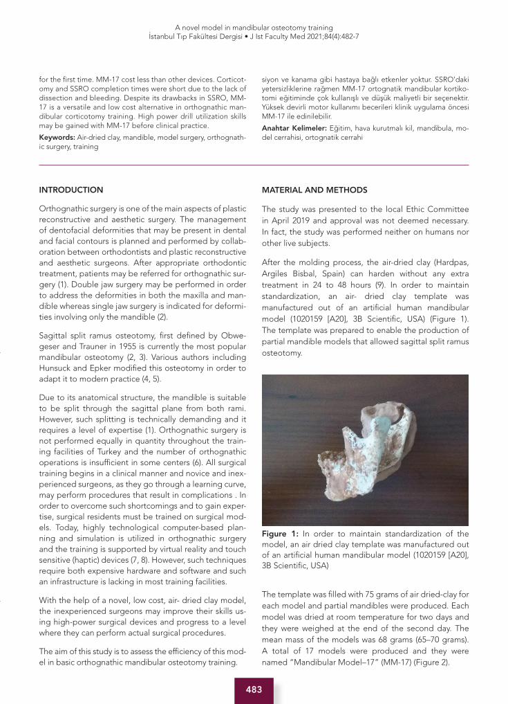
483
A novel model in mandibular osteotomy trainingİstanbul Tıp Fakültesi Dergisi • J Ist Faculty Med 2021;84(4):482-7
RESEARCH / ARAŞTIRMADOI: 10.26650/IUITFD.2021.836789
İst Tıp Fak Derg 2021 / J Ist Faculty Med 2021
A NOVEL TRAINING ALTERNATIVE IN ORTHOGNATHIC MANDIBULAR OSTEOTOMY: AIR DRIED CLAY MODEL
ORTOGNATİK MANDİBULA OSTEOTOMİSİNDE YENİ BİR EĞİTİM SEÇENEĞİ: HAVA KURUTMALI KİL MODELİ
Erol KOZANOĞLU1 , Bora Edim AKALIN1 , Hayri Ömer BERKÖZ1 , Soner KARAALİ2 , Nermin MAMMADOVA1 , Erman AK3 , Ahmet Faruk YÜCEL4 , Ufuk EMEKLİ1
1Istanbul University, Istanbul Faculty of Medicine, Department of Plastic, Reconstructive and Aesthetic Surgery, Istanbul, Turkey 2Erzincan Binali Yildirim University, Mengücek Gazi Education And Research Hospital, Department of Plastic, Reconstructive and Aesthetic Surgery, Erzincan, Turkey 3Istanbul Basaksehir Cam and Sakura City Hospital, Department of Plastic, Reconstructive and Aesthetic Surgery, Istanbul, Turkey4University of Health Science, Kanuni Sultan Süleyman Research and Training Hospital, Department of Plastic, Reconstructive and Aesthetic Surgery, Istanbul, Turkey
ORCID IDs of the authors: E.K. 0000-0003-1192-9520; B.E.A. 0000-0002-5654-2082; H.Ö.B. 0000-0001-8063-9995; S.K. 0000-0001-9496-5513; N.M. 0000-0002-5168-5658; E.A. 0000-0002-0203-1842; A.F.Y. 0000-0002-1461-5378; U.E. 0000-0001-9097-5124
Cite this article as: Kozanoglu E, Akalin BE, Berkoz HO, Karaali S, Mammadova N, Ak E, et al. A novel training alternative in orthognathic mandibular osteotomy: air dried clay model. J Ist Faculty Med 2021;84(4):482-7. doi: 10.26650/IUITFD.2021.836789
INTRODUCTION
Orthognathic surgery is one of the main aspects of plastic reconstructive and aesthetic surgery. The management of dentofacial deformities that may be present in dental and facial contours is planned and performed by collab-oration between orthodontists and plastic reconstructive and aesthetic surgeons. After appropriate orthodontic treatment, patients may be referred for orthognathic sur-gery (1). Double jaw surgery may be performed in order to address the deformities in both the maxilla and man-dible whereas single jaw surgery is indicated for deformi-ties involving only the mandible (2).
Sagittal split ramus osteotomy, first defined by Obwe-geser and Trauner in 1955 is currently the most popular mandibular osteotomy (2, 3). Various authors including Hunsuck and Epker modified this osteotomy in order to adapt it to modern practice (4, 5).
Due to its anatomical structure, the mandible is suitable to be split through the sagittal plane from both rami. However, such splitting is technically demanding and it requires a level of expertise (1). Orthognathic surgery is not performed equally in quantity throughout the train-ing facilities of Turkey and the number of orthognathic operations is insufficient in some centers (6). All surgical training begins in a clinical manner and novice and inex-perienced surgeons, as they go through a learning curve, may perform procedures that result in complications . In order to overcome such shortcomings and to gain exper-tise, surgical residents must be trained on surgical mod-els. Today, highly technological computer-based plan-ning and simulation is utilized in orthognathic surgery and the training is supported by virtual reality and touch sensitive (haptic) devices (7, 8). However, such techniques require both expensive hardware and software and such an infrastructure is lacking in most training facilities.
With the help of a novel, low cost, air- dried clay model, the inexperienced surgeons may improve their skills us-ing high-power surgical devices and progress to a level where they can perform actual surgical procedures.
The aim of this study is to assess the efficiency of this mod-el in basic orthognathic mandibular osteotomy training.
MATERIAL AND METHODS
The study was presented to the local Ethic Committee in April 2019 and approval was not deemed necessary. In fact, the study was performed neither on humans nor other live subjects.
After the molding process, the air-dried clay (Hardpas, Argiles Bisbal, Spain) can harden without any extra treatment in 24 to 48 hours (9). In order to maintain standardization, an air- dried clay template was manufactured out of an artificial human mandibular model (1020159 [A20], 3B Scientific, USA) (Figure 1). The template was prepared to enable the production of partial mandible models that allowed sagittal split ramus osteotomy.
The template was filled with 75 grams of air dried-clay for each model and partial mandibles were produced. Each model was dried at room temperature for two days and they were weighed at the end of the second day. The mean mass of the models was 68 grams (65–70 grams). A total of 17 models were produced and they were named “Mandibular Model–17” (MM-17) (Figure 2).
for the first time. MM-17 cost less than other devices. Corticot-omy and SSRO completion times were short due to the lack of dissection and bleeding. Despite its drawbacks in SSRO, MM-17 is a versatile and low cost alternative in orthognathic man-dibular corticotomy training. High power drill utilization skills may be gained with MM-17 before clinical practice.
Keywords: Air-dried clay, mandible, model surgery, orthognath-ic surgery, training
siyon ve kanama gibi hastaya bağlı etkenler yoktur. SSRO’daki yetersizliklerine rağmen MM-17 ortognatik mandibular kortiko-tomi eğitiminde çok kullanışlı ve düşük maliyetli bir seçenektir. Yüksek devirli motor kullanımı becerileri klinik uygulama öncesi MM-17 ile edinilebilir.
Anahtar Kelimeler: Eğitim, hava kurutmalı kil, mandibula, mo-del cerrahisi, ortognatik cerrahi
Figure 1: In order to maintain standardization of the model, an air dried clay template was manufactured out of an artificial human mandibular model (1020159 [A20], 3B Scientific, USA)

484
A novel model in mandibular osteotomy trainingİstanbul Tıp Fakültesi Dergisi • J Ist Faculty Med 2021;84(4):482-7
Four plastic reconstructive and aesthetic surgeons with at least three years’ experience in orthognathic surgery were invited to participate in the study by post . In order to demonstrate the surgical procedure, the first author performed the model surgery while the four surgeons observed. Demonstrative corticotomy and sagittal split ramus osteotomy were performed and the instructions were given to the surgeons. Each participant was given four MM-17’s, a high-power drill and a cutting handpiece and they performed model surgeries individually. The participants were asked to operate on four models in or-der to increase the reliability of the study. The following data were recorded for each model: Corticotomy and SSRO completion time, MM-17 fracture type and time during corticotomy and SSRO, resistance similarity value of MM-17 cortex and medulla with native mandible, man-dibular representation value of MM-17, and training com-patibility value of MM-17. Resistance similarity and repre-sentation values were subjective measurements. A score of “0” represented no similarity and “10” total similarity and representation between MM-17 and native human mandible. Training compatibility value was another sub-jective measurement with a similar scale. Zero was total
incompatibility whereas ten was total compatibility with orthognathic mandibular osteotomy training. The mean values were calculated and the results were compared. Corticotomies of the entire cortex without fractures were accepted as successful and complete bipartite sagittal split ramus osteotomies without fractures were accepted as successful.
RESULTS
The human mandibular anatomic model (1020159 [A20], 3B Scientific, USA), the high-power drill and the cutting handpiece belong to our institution and they were not included in the study costs. The only expense of the study was the air-dried clay (Hardpas, Argiles Bisbal, Spain) that was used for the production of both the template and the model. After the production of two templates and seven-teen MM-17’s, the cost of a single MM-17 was calculated as 0.6 American Dollars.
The results are listed in Table 1. All corticotomies were completed successfully (16/16) and no fractures were observed during the corticotomies (Figure 3). Mean cor-ticotomy completion time was 127.75 seconds (110–150 seconds). Mean cortical resistance similarity value was 8.75 (8–10). Only three sagittal split ramus osteotomies were completed successfully (3/16) (19%) (Figure 4). The
Table 1: The mean values and results obtained from each surgeon after the model surgeries
CCT (seconds) CSV SSROCT (seconds) SSROSV SSRO failure RV TCV
Surgeon 1 110 8 401 5 3 5 9
Surgeon 2 150 10 0 5 4 4 7
Surgeon 3 116 8 259 6 3 7 10
Surgeon 4 131 9 205 4 3 5 7
Mean 126,75 8,75 288 5 13 failures (81%) 5,25 8,25
3 success (19%)
CCT: Corticotomy completion time; CSV: Corticotomy similarity value; SSROCT: Sagittal split ramus osteotomy completion time; SSROSV: Sagittal split ramus osteotomy similarity value; SSRO failure: Sagittal split ramus osteotomy failure; RV: Representation value of MM-17; TCV: Training compatibility value of MM-17.
Figure 2: “Mandibular Model-17” (MM-17). The osteotomy lines were marked on the medial, lateral and upper margin-al cortices on each model with a surgical marking pen.
Figure 3: A successful MM-17 corticotomy.

485
A novel model in mandibular osteotomy trainingİstanbul Tıp Fakültesi Dergisi • J Ist Faculty Med 2021;84(4):482-7
outer cortex was affected in sixty percent of the fractures and the inner cortex was affected in forty percent of the fractures. Three successful SSRO’s were completed in a mean time of 288 seconds (205-401 seconds). Mean med-ullary resistance similarity value was 5 (4–6). Mean man-dibular representation value of MM-17 was 5.25 (4–7). Mean value of compatibility with orthognathic mandibu-lar osteotomy training was 8.25 (7–10).
No technical problems were observed in either the high- power drills or the handpieces (Figure 5).
DISCUSSION
Structural deformities of the maxilla and the mandible may cause dental misalignment, malocclusion and alter-ations in facial appearance. Such deformities may be cor-rected with orthognathic surgery, ameliorating both form and function (2).
In sagittal split ramus osteotomy, the osteotomy of the medial cortex of the ramus is performed above the lin-gula and the lateral corticotomy is performed between the first and the second molar teeth. Finally, the upper margin of the ramus is corticotomized in order to connect the medial and the lateral corticotomy lines. After the
corticotomies, a full-thickness osteotomy is performed in order to split the ramus into condylar and alveolar frag-ments. At the end of bilateral sagittal split ramus oste-otomies, two condylar and one alveolar fragments are formed and the alveolar fragment may be positioned in three planes according to the orthodontic plan (1-5).
Sagittal split ramus osteotomy (SSRO) is prone to com-plications including unfavorable fractures, unfavorable splits, inferior alveolar and facial nerve injuries and in-ternal maxillary artery injuries (10). The avoidance and management of these complications require technical experience in SSRO.
Although there are numerous plastic reconstructive and aesthetic surgery training institutions in Turkey, orthog-nathic surgery training is still limited and there are vast differences between centers (6). The main reasons for this limitation are requirements of coordination with or-thodontics clinics and of specialized surgical instruments (2). In modern surgical training of the residents, maximal patient safety and minimal complication rates are basic principles (11). However, due to the aforementioned lim-itations, most residents can not have sufficient orthog-nathic surgical training in line with these principles (6). In order to maintain patient safety with minimal complica-tions, orthognathic surgical models may be utilized.
Residents of all surgical fields may gain relevant skills with surgical models and they may support their theoretical knowledge before clinical practice. Palter et al. compared training results with low cost artificial models for fascial repair and the residents who had practiced with the models had a better surgical ability and understanding of the surgical procedure in the operating room (11). Easily reproducible and low cost models such as MM-17 may yield similar results in orthognathic surgery training. Plastic Reconstructive and Aesthetic surgery residents may gain relevant skills with MM-17 and continue clinical practice with more safety and fewer complications.
Cadavers and various artificial materials are regarded as the gold standard in all surgical training models (8). However, they may not be feasible for every training institution due to cost and availability. Three-dimen-sional virtual and solid models can be manufactured using thin sliced maxillofacial computed tomographs and model surgery can be practiced on such models (7). Currently, virtual reality is a popular alternative in ortho-dontic planning and orthognathic surgery training and it may be supported by touch-sensitive haptic simulators (12). With such simulators, the resident may visualize the relevant anatomic area with three-dimensional, real time detail through virtual reality and hand-held or wearable haptic devices may reflect the alterations in the target tissue (8, 13, 14). All such surgical models require spe-cialized computer software, three-dimensional printers
Figure 4: A successful MM-17 sagittal split ramus os-teotomy.
Figure 5: A high power drill that was utilized in the model surgery.

486
A novel model in mandibular osteotomy trainingİstanbul Tıp Fakültesi Dergisi • J Ist Faculty Med 2021;84(4):482-7
and haptic receptors with very high costs. In contrast to these current and expensive models, the MM-17 cost less than an American dollar. Also, real time instrumental expe-rience and skills may be gained during the model surgery with MM-17.
Various materials may be utilized in the production of sagittal split ramus osteotomy (SSRO) artificial models. Baccarin et al. compared three different materials in a model study that evaluated osseous fixation after SSRO: plastic, polyamide and polyurethane (15). None of the materials were biomechanically equivalent to the human mandible in aspects of elasticity, texture and load bearing capacity. All models were found to be relevant only for the simulatory training and for the preparation for clinical practice (15). The ideal material has not been found for the production of mandibular models. The air-dried clay of MM-17, which was utilized for the first time, needs to be evaluated objectively with biomechanical studies.
The literature has a limited number of studies on surgical models produced with air-dried clay. Kazum et al. designed a bone drilling model for orthopaedics surgery residents (16). In this design, the air-dried clay layer was attached to the posterior cortex of an artificial bone and the residents were asked to drill holes through the artificial bone. After the drilling process, the air dried-clay layer was checked for evidence of overdrilling through the posterior cortex and into this clay layer. Kazum et al. designed this study with respect to the similarity between the resistance and texture of the artificial bone cortex and the air-dried clay (16). In accordance with this study, successful corticotomies were performed on each MM-17 and the participants of the study evaluated the MM-17 cortex with a mean similarity value of 8.75 out of 10. According to the similar results of these two studies, air- dried clay represented the bone cortex effectively.
The corticotomy and sagittal split ramus osteotomy completion times were shorter than live surgery. These differences were due to the lack of soft tissue dissection, retraction, bleeding and assistance during the model surgery. Bleeding and the control of bleeding are major factors that prolong operative times. The SSRO success of MM-17 was 19% and numerous osteotomy failures, including outer cortical fractures, were seen during the model surgery. The main reason for these failures was the absence of osseous medulla in the uniformly constituted cortex MM-17 models . Therefore, the participants did not report the same procedural success for SSRO as the corticotomy and the SSRO similarity value was 5 out of 10. Also, the mandibular representation value of MM-17, measured at 5.25 out of 10, was lower because of the absence of osseous medulla. This important detail is a limitation of this surgical model. Also, the ramus of the model is slightly thicker than normal mandible. Due to
this limitation, the model may be regarded as a simple tool for beginners. New designs may be required for completing the learning curve of inexperienced surgeons.
Mandibular corticotomies, which are performed with specialized instruments such as high-power drills, are the first steps of a successful orthognathic surgery. Experi-ence and skills with such instruments should be gained before operating on real patients. Four experienced or-thognathic surgeons evaluated the training compatibility value of MM-17, reporting a mean of 8.25 points out of 10. Thus, the learning curve of corticotomy with special-ized instruments may be completed on MM-17 models.
Not every resident has the opportunity to train in an or-thognathic surgery performing clinic, and clinical practice may not be possible for every surgeon. MM-17 may ad-dress the basic needs of a novice orthognathic surgery resident with its low cost and versatility in mandibular corticotomy.
Peer Review: Externally peer-reviewed.
Author Contributions: Conception/Design of Study- E.K., B.E.A., H.Ö.B., S.K., N.M., E.A., A.F.Y., U.E.; Data Acquisition- E.K., B.E.A., H.Ö.B., S.K., N.M., E.A., A.F.Y., U.E.; Data Analysis/Interpretation- E.K., B.E.A., H.Ö.B., S.K., N.M., E.A., A.F.Y., U.E.; Drafting Manuscript- E.K., B.E.A., H.Ö.B., S.K., N.M., E.A., A.F.Y., U.E.; Critical Revision of Manuscript- E.K., B.E.A., H.Ö.B., S.K., N.M., E.A., A.F.Y., U.E.; Final Approval and Accountability- E.K., B.E.A., H.Ö.B., S.K., N.M., E.A., A.F.Y., U.E.
Conflict of Interest: Authors declared no conflict of interest.
Financial Disclosure: Authors declared no financial support.
Hakem Değerlendirmesi: Dış bağımsız.
Yazar Katkıları: Çalışma Konsepti/Tasarım- E.K., B.E.A., H.Ö.B., S.K., N.M., E.A., A.F.Y., U.E.; Veri Toplama- E.K., B.E.A., H.Ö.B., S.K., N.M., E.A., A.F.Y., U.E.; Veri Analizi/Yorumlama- E.K., B.E.A., H.Ö.B., S.K., N.M., E.A., A.F.Y., U.E.; Yazı Taslağı- E.K., B.E.A., H.Ö.B., S.K., N.M., E.A., A.F.Y., U.E.; İçeriğin Eleştirel İnceleme-si- E.K., B.E.A., H.Ö.B., S.K., N.M., E.A., A.F.Y., U.E.; Son Onay ve Sorumluluk- E.K., B.E.A., H.Ö.B., S.K., N.M., E.A., A.F.Y., U.E.
Çıkar Çatışması: Yazarlar çıkar çatışması beyan etmemişlerdir.
Finansal Destek: Yazarlar finansal destek beyan etmemişlerdir.
REFERENCES
1. Reyneke JP, Ferretti C. The bilateral sagittal split mandibular ramus osteotomy. Atlas of the oral and maxillofacial surgery clinics of North America 2016;24(1):27-36. [CrossRef]
2. Reyneke J. In essential in orthognathic surgery [Chapter 4]. Chicago: Quintessence 2003:171-5.

487
A novel model in mandibular osteotomy trainingİstanbul Tıp Fakültesi Dergisi • J Ist Faculty Med 2021;84(4):482-7
3. Obwegeser H. Zur Operationstechnik bei der progenie und anderer unterkieferanomalien. Dtsch, Z Mund Kieferheilk 1955;23:1-26.
4. Hunsuck E. A modified intraoral sagittal splitting technique for correction of mandibular prognathism. J Oral Surg 1968;26:249-52.
5. Epker BN. Modifications in the sagittal osteotomy of the mandible. J Oral Surg 1977;35(2):157-9.
6. Yuce E, Komerik N. Oral maxillofacial training: opinions of trainees and specialists in Turkey. Journal of Contemporary Medi cal Education 2015;3(1):25-30. [CrossRef]
7. Lutz JC, Hostettler A, Agnus V, Nicolau S, George D, Soler L, et al. A new software suite in orthognathic surgery: patient specific modeling, simulation and navigation. Surgical innovation 2019;26(1):5-20. [CrossRef]
8. Chen X, Hu J. A review of haptic simulator for oral and maxillofacial surgery based on virtual reality. Expert Review of Medical Devices 2018;15(6):435-44. [CrossRef]
9. Website AB. [cited 2019 14.05]. Available from: http://argilesbisbal.com/en/portfolio/hardpast/
10. Mehra P, Castro V, Freitas RZ, Wolford LM. Complications of the mandibular sagittal split ramus osteotomy associated with the presence or absence of third molars. Journal of oral and maxillofacial surgery 2001;59(8):854-8. [CrossRef]
11. Palter VN, Grantcharov T, Harvey A, MacRae HM. Ex vivo technical skills training transfers to the operating room and enhances cognitive learning: a randomized controlled trial. Annals of surgery 2011;253(5):886-9. [CrossRef]
12. Ureturk EU, Apaydin A. Does fixation method affect temporomandibular joints after mandibular advancement? Journal of Cranio-Maxillofacial Surgery 2018;46(6):923-31. [CrossRef]
13. Sohmura T, Hojo H, Nakajima M, Wakabayashi K, Nagao M, Iida S, et al. Prototype of simulation of orthognathic surgery using a virtual reality haptic device. International journal of oral and maxillofacial surgery 2004;33(8):740-50. [CrossRef]
14. Maliha SG, Diaz-Siso JR, Plana NM, Torroni A, Flores RL. Haptic, physical, and web-based simulators: Are they underused in maxillofacial surgery training? Journal of Oral and Maxillofacial Surgery 2018;76(11):2424 e1-. e11. [CrossRef]
15. Baccarin L, Casarin RCV, Lopes-da-Silva J, Passeri L. Analysis of mandibular test specimens used to assess a bone fixation system. Craniomaxillofacial trauma&reconstruction 2015;8(3):171-8. [CrossRef]
16. Kazum E, Dolkart O, Rosenthal Y, Sherman H, Amar E, Salai M, et al. A simple and low-cost drilling simulator for training plunging distance among orthopedic surgery residents. Journal of surgical education 2019;76(1):281-5. [CrossRef]

488
Pseudophakic retinal detachment treatmentİstanbul Tıp Fakültesi Dergisi • J Ist Faculty Med 2021;84(4):488-94
Corresponding author/İletişim kurulacak yazar: [email protected]
Submitted/Başvuru: 07.12.2020 • Revision Requested/Revizyon Talebi: 30.01.2021 •Last Revision Received/Son Revizyon: 23.02.2021 • Accepted/Kabul: 23.02.2021 • Published Online/Online Yayın: 27.08.2021
RESEARCH / ARAŞTIRMADOI: 10.26650/IUITFD.2021.836991
İst Tıp Fak Derg 2021 / J Ist Faculty Med 2021
EVALUATION OF OUTCOMES OF PRIMARY PSEUDOPHAKIC RETINAL DETACHMENT SURGERY
PRİMER PSÖDOFAKİK RETİNA DEKOLMANI CERRAHİSİ SONUÇLARININ DEĞERLENDİRİLMESİ
Zeynep YILMAZABDURRAHMANOĞLU1* , Kemal Turgay ÖZBİLEN2* , Mehmet Selim KOCABORA3 , Osman ÇEKİÇ4
1Bagcilar Training and Research Hospital, Ophthalmology Clinic, Istanbul, Turkey2Istanbul University, Istanbul Faculty of Medicine, Department of Ophthalmology, Istanbul, Turkey 3Istanbul Medipol University, Faculty of Medicine, Department of Ophthalmology Istanbul, Turkey 4Marmara University, Faculty of Medicine, Department of Ophthalmology Istanbul, Turkey
ORCID IDs of the authors: Z.Y. 0000-0002-6075-8513; K.T.Ö. 0000-0002-0234-3803; M.S.K. 0000-0001-5335-3860;O.Ç. 0000-0003-0911-8649
Cite this article as: Yilmazabdurrahmanoglu Z, Ozbilen, KT, Kocabora MS, Cekic O. Evaluation of outcomes of primary pseudophakic retinal detachment surgery. J Ist Faculty Med 2021;84(4):488-94. doi: 10.26650/IUITFD.2021.836991
ABSTRACTObjective: To evaluate the results of pars plana vitrectomy (PPV) alone and combined with circumferential scleral buckling (CSB) surgeries for aphakic or pseudophakic rhegmatogenous retinal detachment (RRD).Materials and Methods: Thirty-seven eyes of 37 patients who underwent PPV (20 and 23 Gauge) alone or PPV combined with CSB due to pseudophakic or aphakic primary RRD were included in the study. Postoperative anatomical success (AS) and functional success (FS) were evaluated. The AS was defined as a completely flattened retina without any subretinal fluids after the removal of the silicone oil tamponade, if used. The FS was defined as two or more decimal improvements in the logMAR equivalent of Snellen visual acuity.Results: The mean age of the patients was 62.43±11.40 (32-80) years, 21 (56.8%) patients were male, and 16 (43.2%) were fe-male. The mean follow-up time was 21.35±16.86 (6-84) months. PPV combined with CSB were performed in 23 patients. AS was found to be 86.5% (32/37), FS was 49.9% (17/37). No statistically significant difference was observed in both AS and FS between the groups according to preoperative PVR presence (AS-p=0.61, FS-p=0.14), preoperative macular involvement (AS-p=0.98, FS-p=0.36), whether PPV combined with CSB (AS-p=0.97, FS-p=0.29), and the type of tamponade (p>0.05 in all).Conclusion: PPV with or without CSB is safe and effective in cases with primary pseudophakic retinal detachment and achieves good AS without being affected by the presence of PVR or macular in-volvement. However, functional success may not always follow.Keywords: Pseudophakic retinal detachment, pars plana vitrec-tomy, circumferential scleral buckling
ÖZET
Amaç: Afakik veya psödofakik regmatojen retina dekolmanı (RRD) için tek başına pars plana vitrektomi (PPV) ve çevresel sk-leral çökertme (ÇSÇ) ile kombine ameliyatların sonuçlarını de-ğerlendirmek.
Gereç ve Yöntem: Psödofakik veya afakik primer RRD’ye nede-niyle tek başına veya ÇSÇ ile kombine PPV (20 ve 23 Gauge) uy-gulanan 37 hastanın 37 gözü çalışmaya dahil edildi. Postoperatif anatomik başarı (AB) ve fonksiyonel başarı (FB) değerlendirildi. AB, eğer kullanılmışsa silikon yağı tamponadının çıkarılmasından sonra hiç subretinal sıvının olmadığı tamamen yatışmış retina ola-rak tanımlandı. Ayrıca FB, Snellen görme keskinliğinin logMAR eş-değerinde iki veya daha fazla ondalık artış olarak tanımlandı.
Bulgular: Hastaların ortalama yaşı 62,43±11,40 (32-80) yıl, 21’i (%56,8) erkek, 16’sı (%43,2) kadındı. Ortalama takip süresi 21,35±16,86 (6-84) aydı. 23 hastaya ÇSÇ ile kombine PPV uygu-landı. AB %86,5 (32/37), FB %49,9 (17/37) olarak bulundu. Hem AB ile hem de FB ile; preoperatif PVR varlığı (AB-p=0,61, FB-p=0,14), preoperartif maküla tutulumu (AB-p=0,98, FB-p=0,36), PPV’nin ÇSÇ ile kombine olup olmaması (AB-p=0,97, FB-p=0,29), ve kulla-nılan tamponad tipi (tümü p>0,05) gibi durumlara göre oluşturu-lan gruplar arasında istatistiksel olarak anlamlı bir fark gözlenmedi.
Sonuç: Primer psödofakik retina dekolmanı olan olgularda PPV yalnız veya ÇSÇ ile kombine olarak uygulandığında, maküla tu-tulumundan veya PVR varlığından etkilenmeden iyi AB ulaşmak için etkili ve güvenlidir. Bununla birlikte, fonksiyonel başarı her zaman eşlik etmeyebilir.
Anahtar Kelimeler: Psödofakik retina dekolmanı, pars plana vit-rektomi, çevresel skleral çökertme
Content of this journal is licensed under a Creative Commons Attribution-NonCommercial 4.0 International License.
*Both authors contributed equally

489
Pseudophakic retinal detachment treatmentİstanbul Tıp Fakültesi Dergisi • J Ist Faculty Med 2021;84(4):488-94
RESEARCH / ARAŞTIRMADOI: 10.26650/IUITFD.2021.836991
İst Tıp Fak Derg 2021 / J Ist Faculty Med 2021
EVALUATION OF OUTCOMES OF PRIMARY PSEUDOPHAKIC RETINAL DETACHMENT SURGERY
PRİMER PSÖDOFAKİK RETİNA DEKOLMANI CERRAHİSİ SONUÇLARININ DEĞERLENDİRİLMESİ
Zeynep YILMAZABDURRAHMANOĞLU1* , Kemal Turgay ÖZBİLEN2* , Mehmet Selim KOCABORA3 , Osman ÇEKİÇ4
1Bagcilar Training and Research Hospital, Ophthalmology Clinic, Istanbul, Turkey2Istanbul University, Istanbul Faculty of Medicine, Department of Ophthalmology, Istanbul, Turkey 3Istanbul Medipol University, Faculty of Medicine, Department of Ophthalmology Istanbul, Turkey 4Marmara University, Faculty of Medicine, Department of Ophthalmology Istanbul, Turkey
ORCID IDs of the authors: Z.Y. 0000-0002-6075-8513; K.T.Ö. 0000-0002-0234-3803; M.S.K. 0000-0001-5335-3860;O.Ç. 0000-0003-0911-8649
Cite this article as: Yilmazabdurrahmanoglu Z, Ozbilen, KT, Kocabora MS, Cekic O. Evaluation of outcomes of primary pseudophakic retinal detachment surgery. J Ist Faculty Med 2021;84(4):488-94. doi: 10.26650/IUITFD.2021.836991
INTRODUCTION
Retinal detachment (RD) is a rare but severe complica-tion of cataract surgery and, if left untreated, results in vision loss of the affected eye. Some researchers have pointed out that about 50% of RD develops within one year of cataract extraction surgery (1, 2). The inci-dence of RD after intracapsular cataract extraction sur-gery (ICCE) is 0.98-3.6%, 0.33-1.7% after extracapsular cataract extraction surgery (ECCE), and 1-1.17% after phacoemulsification. This incidence is eight times more than the normal population (3, 4). With the increase in the number of cataract surgeries performed in the last 20 years, the incidence of aphakic and pseudophakic rheg-matogenous RD (RRD) has also increased. Although it is known that with current phacoemulsification surgery, complications during and after surgery are reduced, ret-inal detachment is a complication that remains relevant, as it significantly reduces the patient’s visual expecta-tions. The number of people undergoing cataract sur-gery accounted for about 3% of the overall population, while 40% of patients with RRD had a history of previous cataract surgery. It is believed that removing the natu-ral lens accelerates vitreous liquefaction, which causes premature posterior vitreous detachment and increases the risk of RD (2-4). The condition of the lens capsule also determines the vitreous liquefaction amounts. It is known that posterior lens capsule perforation during surgery and neodymium yttrium-aluminum-garnet (Nd-YAG) laser capsulotomy significantly increases RD inci-dence (5). Some potential risk factors are influential in RD development after cataract surgery. These factors were indicated as preoperative predisposing factors (myopia, lattice degeneration), intraoperative complica-tions (posterior capsular rupture with or without vitre-ous loss), and postoperative factors (i.e., capsulotomy, vitreous hemorrhage, trauma) that are unrelated to the surgical procedure.
Evaluating fundus’ appearance when examining pseu-dophakic RD patients may be tricky with opacification, reflections, and weak mydriasis. Circumferential scleral buckling (CSB), pneumatic retinopexy, primary pars plana vitrectomy (PPV) alone or with CSB are surgical options used to treat pseudophakic RD (6-8). The use of internal tamponades with PPV increases the success of this sur-gery (9). Although anatomical success rates are high after vitreoretinal surgeries, good visual outcomes are not al-ways achieved as expected (10).
This study examined the clinical findings of primary RD in patients who developed RD after cataract extraction and underwent vitreoretinal surgeries in our clinic. Risk factors, surgery techniques and its results, and prognosis were evaluated.
MATERIALS AND METHODS
Approval was obtained from the Ethics Committee of Bezm-i Alem University Faculty of Medicine for this ret-rospective study. Informed consent forms were obtained from all patients in the study. The study was carried out in accordance with the tenets of the Helsinki Declaration.
Thirty-seven eyes of 37 patients who had been diag-nosed with primary pseudophakic or aphakic RRD and had undergone PPV [20 or 23 Gauge (G)] with or without CSB between 2004 and 2011 were recruited to the study. This study evaluated demographic data, history of sys-temic and eye diseases, presence of myopia, time frame from cataract surgery to RD development, preoperative best-corrected visual acuity (BCVA), intraocular pressure (IOP), intraocular lens (IOL) condition, lens capsule in-tegrity, and the presence of Nd-YAG laser capsulotomy, the condition of the retinal tears and detachment, the presence of macular involvement and proliferative vitre-oretinopathy (PVR) were determined. Perioperative data [i.e., performing CSB and relaxing retinotomy-retinec-tomy, type of intraocular tamponade (IOT), complica-tions], and postoperative data [BCVA, IOP, recurrence, anatomical success (AS), functional success (FS), and complications].
Exclusion criteriaPatients who underwent surgery due to recurrent detach-ment, underwent only scleral procedures without PPV, or were followed-up for less than six months were excluded.
Surgical techniqueStandard PPV was performed, as under the wide-angle imaging systems, 20 G or 23 G (with trocar, transconjunc-tival) conventional three PPV ports were created. In the presence of advanced stage PVR, inferior retinal tears, or multiple tears in different quadrants, CSB was performed in the same session. ACCURUS ® Surgical System (Alcon, Fort Worth, Texas, USA) was used for vitrectomy. Standard surgical procedures were performed. Triamcinolone was used for visualizing posterior hyaloid and membranes. The vitreous base was shaved with indentation. Retinoto-my/retinectomy was made when necessary. Sclerotomies were closed with 7.0 polyglactin sutures. As IOT, either 1000 cst silicone oil, 13% C3F8 gas, 18% SF6 or pure air were used to fill the entire vitreous cavity. If CSB was to be performed in the same session, limbal 360° peritomy was performed on the conjunctiva. A number 240 band was passed under the rectus muscles, and was sutured 13 mm away from the limbus with 5.0 ethibond in 4 quad-rants. The tightening of the band was done after PPV. Conjunctiva was closed with 8.0 polyglactin sutures. After the surgery, rest in the appropriate position was recom-mended. In postoperative care, Tobramycin eye drops (four times a day for ten days), prednisolone acetate 1% drops (six times a day, tapered after one week and dis-

490
Pseudophakic retinal detachment treatmentİstanbul Tıp Fakültesi Dergisi • J Ist Faculty Med 2021;84(4):488-94
continued at the end of the one month) were prescribed. Antiglaucomatous drops were used if needed.
A detailed ophthalmological examination on the postop-erative first day, first week, first, third, sixth months, and the following every six months were performed on all pa-tients. BCVA values were measured with Snellen charts and converted into logMAR. The complications evaluated were corneal edema or opacity, hypotony (IOP ≤5 mmHg), elevated IOP (≥22 mmHg), fibrin formation, hyphemia, or vitreous hemorrhages. The surgeries performed be-tween the study groups were compared in terms of AS, FS, BCVA, and complications. The AS was defined as a complete flattened retina without any SRF after removing the silicone oil (if used) in the final visit. Cases with two or more decimal improvements in the logMAR equivalent of Snellen BCVA were considered as FS.
The SPSS (Version 21, IBM Corp. Armonk, NY: USA) soft-ware statistic program was used for statistical analysis. The Wilcoxon Signed Ranks Test was used to compare descriptive statistics and the parameters that did not show normal distribution in the comparison of quantita-tive data. Fisher’s Exact test and Chi-Square test was used to compare qualitative data. Pearson correlation analysis was used in correlation analyses. Statistically, significance was accepted as p<0.05.
RESULTS
Of the 37 patients included in the study, almost all had reduced vision at various levels at the time of admission. Photopsy-entopsy history was present in 6 (16.2%) cases, and 6 (16.2%) cases had a history of suspected head or ocular trauma.
High IOP was detected in 2 (5.4%) patients in preopera-tive examination. Five of the patients (13.5%) had a his-tory of high myopia. Laser photocoagulation had been
performed on two patients (5.4%) for retinal tears before the surgery. In 9 (24.3%) patients, there was a posterior lens capsule rupture and anterior vitrectomy history. A Nd-YAG laser capsulotomy history was also in 1 (2.7%) three months before RD. Three patients (8.1%) who had anterior chamber I OL had a peripheral iridectomy. De-mographic data and characteristic distributions of pa-tients a re summarized in Table 1.
Preoperative PVR was present in 10 (27.0%) patients, and PPV with CSB was performed on seven of these PVR cas-es. Posterior vitreous detachment (PVD) in 11 (29.7%), prominent inflammatory reaction in 7 (18.9%), and vit-reous hemorrhage (VH) were present in 2 (5.4%) cases. Single retinal tears were detected in 23 patients and mul-tiple retinal tears in 14 patients. The characteristics of ret-inal tears and RD are summarized in Table 2.
Table 1: Demographic and characteristic data of the patients
Variables Results
Male/Female- n (%) 21 (56.8%)/16 (43.2%)
Mean age- (year) 62.43±11.40 (32-80)
Mean follow-up (months) 21.35±16.86 (6-84)
Duration after cataract surgery (months) 15.54±17.24 (3-72)
PPV alone/PPV with CSB- n (%) 23 (62.2%)/14 (37.8%)
PPV 20G/PPV 23G- n (%) 19 (51.4%)/18 (48.6%)
Type of cataract surgery (Phaco vs ECCE)- n (%) 36 (97.3%)/1 (2.7%)
Preoperative lens status (pseudophakic/aphakic)- n (%) 36 (97.3%)/1 (2.7%)
IOL localization (bag/sulcus/AC)- n (%) 28 (75.7%)/5 (13.5%)/3 (8.1%)
PPV: Pars plana vitrectomy, G: Gauge, Phaco: Phacoemulsification, ECCE: Extracapsular cataract extraction, IOL: Intraocular lens,AC: Anterior chamber
Table 2: Retinal tears and retinal detachment features
n (%)
Numbers of retinal tear
Multiple (3.3±1.1)* 14 (37.8%)
Single 23 (62.2%)
Localization of retinal tears
Superior quadrants 9 (24.3%)
Inferior quadrants 8 (21.6%)
Both superior & inferior quadrants 20 (54.1%)
Involvement of retinal detachment
Superior quadrants 9 (24.3%)
Inferior quadrants 8 (21.6%)
Both superior & inferior quadrants 20 (54.1%)
*: The average of only the multiple retinal tears patients

491
Pseudophakic retinal detachment treatmentİstanbul Tıp Fakültesi Dergisi • J Ist Faculty Med 2021;84(4):488-94
In 75.6% of the patients, the final visit’s BCVA values im-proved compared to the postoperative BCVA values. Sig-nificantly, the mean BCVA at all of the postoperative vis-its was better compared to the preoperative mean. The BCVA data before and after the surgery are summarized in Table 3.
The mean preoperative IOP was 13.04±4.11(6-30) mmHg. Although, in the postoperative first week, the mean IOP increased to 14.58±5.32 mm Hg, this increase was insig-nificant (p>0.05). However, in cases using an intraocular gas tamponade, a significant increase in IOP was ob-served. In the postoperative first month, the mean IOP was 17.04±7.94 mmHg, and in the sixth month, it was 15.78±5.12 mmHg; both were significantly high com-pared to the preoperative mean (both p<0.05). A high IOP was present in 7 (18.9%) patients during the postop-erative period. Cyclophotocoagulation was performed in a patient whose IOP was not controlled despite all medical treatments. IOP control was achieved in all pa-tients after the sixth month. The final visit mean IOP was 13.62±4.97 mmHg, with no significant difference be-tween the preoperative mean (p>0.05).
In postoperative first day examinations, corneal epithe-lial defects were observed in four patients (10.8%), and corneal edema in 3 (8.1%) patients. All epithelial defects healed within the first week; even so, epithelial irregular-ity was prolonged until the third month in two patients. Prolonged corneal edema was observed in two patients but disappeared at the first-month examination.
Anterior chamber IOL was removed in 2 (5.4%) patients with anterior PVR whose vitreous base could not be shaved and visualized adequately. Relaxing retinotomy/retinecto-my was performed in six of 10 PVR (+) cases. Drainage ret-inotomy was performed on three cases (8.1%). Two (5.4%) patients developed a limited amount of suprachoroidal hemorrhage as intraoperative complications.
Silicone oil extraction was performed in the postoper-ative period, through the anterior chamber from the
aphakic eye and the pars plana in the pseudophakic eyes. Silicone oil was not removed due to the lack of postoperative light projection in one patient. During the follow-up, 4 (15.4%) of the 26 patients who underwent silicone oil removal developed recurrent RD (one pa-tient on the first day, one in the 1st month, and two in the third month). Of the 11 patients treated by giving C3F8, SF6, or air, 1 (9.1%) patient given pure air developed re-current detachment during the early follow-up. AS was achieved in 32 eyes (86.5%), and FS was achieved in 17 eyes (45.9%). The distribution of intraocular tamponade used is summarized in Table 4. Anatomical and functional success distribution is summarized in Table 5.
No statistically significant difference was observed in both AS and FS between the groups according to preop-erative macular involvement (p=0.98, p=0.36 respective-ly), preoperative PVR presence (p=0.61, p=0.14 respec-tively), and whether PPV combined with CSB (p=0.97, p=0.24 respectively). There was no statistical significance in both AS and FS between the types of the IOT used (p>0.05 on both).
DISCUSSION
The risk of RD occurrence increases after cataract sur-gery. RD incidence is reported as 0.6%-1.7% in the first year after cataract surgery (11-13). Rowe et al. reported that the risk of retinal detachment was six times higher in those who underwent cataract surgery (14). Citirik et al. also stated that cataract surgery is the most common risk
Table 4: Distribution of intraocular tamponade used intraoperatively
Tamponade n %
C3F8 4 10.8%
Air 1 2.7%
SF6 6 16.2%
Silicone oil 26 70.3%
Table 3: Change of mean BCVAs over time
BCVA (logMAR) Mean±SD (min-max) p*
Preop 2.41±1.01 (0.15-3.10)
postop 1st week 1.85±0.83 (0.40-3.10) 0.035*
postop 1st months 1.55±0.94 (0.30-3.10) 0.001**
postop 3rd months 1.42±0.76 (0.22-3.10) 0.001**
postop 6th months 1.38±0.77 (0.22-3.10) 0.001**
postop final examination 1.21±0.85 (0.0-3.10) 0.001**
*Wilcoxon Signed Ranks test, between preoperative mean values and postoperative mean values, preop: preoperative, postop: postopera-tive, **: p<0.001, BCVA: Best corrected visual acuity
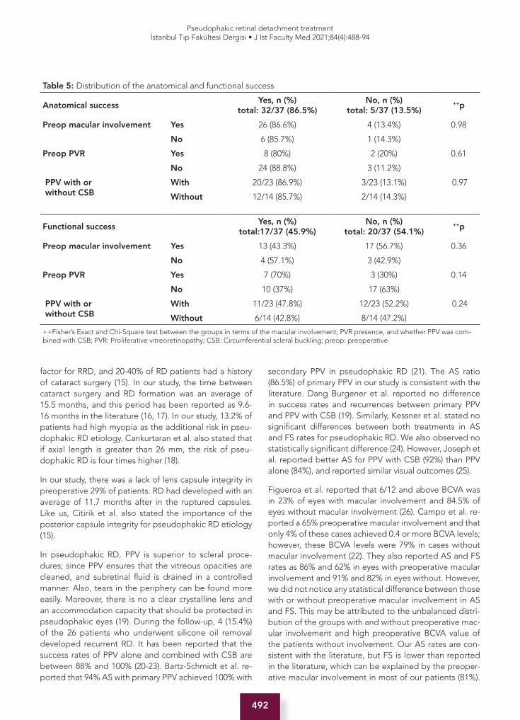
492
Pseudophakic retinal detachment treatmentİstanbul Tıp Fakültesi Dergisi • J Ist Faculty Med 2021;84(4):488-94
factor for RRD, and 20-40% of RD patients had a history of cataract surgery (15). In our study, the time between cataract surgery and RD formation was an average of 15.5 months, and this period has been reported as 9.6-16 months in the literature (16, 17). In our study, 13.2% of patients had high myopia as the additional risk in pseu-dophakic RD etiology. Cankurtaran et al. also stated that if axial length is greater than 26 mm, the risk of pseu-dophakic RD is four times higher (18).
In our study, there was a lack of lens capsule integrity in preoperative 29% of patients. RD had developed with an average of 11.7 months after in the ruptured capsules. Like us, Citirik et al. also stated the importance of the posterior capsule integrity for pseudophakic RD etiology (15).
In pseudophakic RD, PPV is superior to scleral proce-dures; since PPV ensures that the vitreous opacities are cleaned, and subretinal fluid is drained in a controlled manner. Also, tears in the periphery can be found more easily. Moreover, there is no a clear crystalline lens and an accommodation capacity that should be protected in pseudophakic eyes (19). During the follow-up, 4 (15.4%) of the 26 patients who underwent silicone oil removal developed recurrent RD. It has been reported that the success rates of PPV alone and combined with CSB are between 88% and 100% (20-23). Bartz-Schmidt et al. re-ported that 94% AS with primary PPV achieved 100% with
secondary PPV in pseudophakic RD (21). The AS ratio (86.5%) of primary PPV in our study is consistent with the literature. Dang Burgener et al. reported no difference in success rates and recurrences between primary PPV and PPV with CSB (19). Similarly, Kessner et al. stated no significant differences between both treatments in AS and FS rates for pseudophakic RD. We also observed no statistically significant difference (24). However, Joseph et al. reported better AS for PPV with CSB (92%) than PPV alone (84%), and reported similar visual outcomes (25).
Figueroa et al. reported that 6/12 and above BCVA was in 23% of eyes with macular involvement and 84.5% of eyes without macular involvement (26). Campo et al. re-ported a 65% preoperative macular involvement and that only 4% of these cases achieved 0.4 or more BCVA levels; however, these BCVA levels were 79% in cases without macular involvement (22). They also reported AS and FS rates as 86% and 62% in eyes with preoperative macular involvement and 91% and 82% in eyes without. However, we did not notice any statistical difference between those with or without preoperative macular involvement in AS and FS. This may be attributed to the unbalanced distri-bution of the groups with and without preoperative mac-ular involvement and high preoperative BCVA value of the patients without involvement. Our AS rates are con-sistent with the literature, but FS is lower than reported in the literature, which can be explained by the preoper-ative macular involvement in most of our patients (81%).
Table 5: Distribution of the anatomical and functional success
Anatomical success Yes, n (%)
total: 32/37 (86.5%)No, n (%)
total: 5/37 (13.5%)++p
Preop macular involvement Yes 26 (86.6%) 4 (13.4%) 0.98
No 6 (85.7%) 1 (14.3%)
Preop PVR Yes 8 (80%) 2 (20%) 0.61
No 24 (88.8%) 3 (11.2%)
PPV with or without CSB
With 20/23 (86.9%) 3/23 (13.1%) 0.97
Without 12/14 (85.7%) 2/14 (14.3%)
Functional success Yes, n (%)
total:17/37 (45.9%)No, n (%)
total: 20/37 (54.1%)++p
Preop macular involvement Yes 13 (43.3%) 17 (56.7%) 0.36
No 4 (57.1%) 3 (42.9%)
Preop PVR Yes 7 (70%) 3 (30%) 0.14
No 10 (37%) 17 (63%)
PPV with or without CSB
With 11/23 (47.8%) 12/23 (52.2%) 0.24
Without 6/14 (42.8%) 8/14 (47.2%)
++Fisher’s Exact and Chi-Square test between the groups in terms of the macular involvement, PVR presence, and whether PPV was com-bined with CSB; PVR: Proliferative vitreoretinopathy; CSB: Circumferential scleral buckling; preop: preoperative
493
Pseudophakic retinal detachment treatmentİstanbul Tıp Fakültesi Dergisi • J Ist Faculty Med 2021;84(4):488-94
However, in the present study, 75.6% of the patients, visu-al acuity improved compared to preoperative BCVA, but only 45.9% achieved FS criteria.
Dang Burgener et al. reported that the most critical fac-tor affecting the surgical outcome is PVR (19). Çakır et al. reported a negative correlation between PVR grade and improvement of BCVA (27). Nevertheless, we observed no statistically significant difference between the PVR groups. This result may be explained as follows: PPV technique provides better shaving of the vitreous base in aphakic or pseudophakic eyes, better visualization of the peripheral retina, cleaning the membranes and bands, and flatten-ing the retina by performing retinectomy/retinotomy more comfortably. Also, in advanced stage PVR, performing ad-ditional CSB and using a silicone oil tamponade provide better contact of retinal and retinal pigment epithelium and may improve the surgery results.
Temporary IOP elevation after PPV is a common com-plication, but it is rarely permanent. The increased IOP was reported 17.9%-48% after PPV with or without CSB in pseudophakic RD treatment (23, 28, 29). However, we ob-served an elevated IOP in 18.4% of patients, which is con-sistent with the literature. Angle-closure glaucoma after CSB may occur due to a forward rotation and congestion of the ciliary body, and elevated IOP could be controlled with medical treatment and/or laser iridotomy. Intraocu-lar gas tamponades may cause secondary angle-closure glaucoma by expansion. Pupil blocks may occur due to silicone oil tamponade in aphakic eyes, or synechia in pseudophakic eyes. Also, the emulsified silicone passing into the anterior chamber (AC) or inflammatory cells in AC may give rise to obstruction of trabeculae, and forma-tion of glaucoma (30).
This study’s weaknesses are its retrospective design, low number of cases, imbalance in the distribution of pre-operative subgroups (i.e., presence of PVR, involvement of macula), used IOT diversity, and a relatively short fol-low-up period. However, its strength is that it gives an idea about PPV surgery with or without CSB in treating pseudophakic RD. Prospective, randomized controlled trials with large case series and extended follow-ups will provide more accurate data.
CONCLUSION
According to the present study, PPV with or without CSB is effective and safe in primary pseudophakic RD cases. These treatments provide good (86%) anatomical success without being affected by preoperative PVR presence or macular involvement or the kind of endotamponade used. However, functional success may not always follow. The making-decision to surgical strategy (i.e., combining with CSB or the choice of IOT) should be made according to the eye’s condition and RD’s severity.
Ethics Committee Approval: This study was approved by the Ethics Committee of Bezm-i Alem University Faculty of Medicine (Date: 14.10.2009, No:10/12).
Informed Consent: Written consent was obtained from the par-ticipants.
Peer Review: Externally peer-reviewed.
Author Contributions: Conception/Design of Study- O.Ç., K.T.Ö.; Data Acquisition- Z.Y.; Data Analysis/Interpretation- K.T.Ö., M.S.K.; Drafting Manuscript- Z.Y., K.T.Ö.; Critical Revision of Manuscript- K.T.Ö., M.S.K., O.Ç.; Final Approval and Accoun-tability- Z.Y., M.S.K., O.Ç., K.T.Ö.*Both 1st and 2nd authors (Zeynep Abdurrahmanoğlu and Kemal Turgay Özbilen) contributed equally.
Conflict of Interest: Authors declared no conflict of interest.
Financial Disclosure: Authors declared no financial support.
Etik Komite Onayı: Bu çalışma için etik komite onayı Bezm-i Alem Üniversitesi Tıp Fakültesi Etik Kurulu’ndan alınmıştır (Tarih: 14.10.2009, No:10/12).
Bilgilendirilmiş Onam: Katılımcılardan bilgilendirilmiş onam alınmıştır.
Hakem Değerlendirmesi: Dış bağımsız.
Yazar Katkıları: Çalışma Konsepti/Tasarım- O.Ç., K.T.Ö.; Veri Toplama- Z.Y.; Veri Analizi/Yorumlama- K.T.Ö., M.S.K.; Yazı Tasla-ğı- Z.Y., K.T.Ö.; İçeriğin Eleştirel İncelemesi- K.T.Ö., M.S.K., O.Ç.; Son Onay ve Sorumluluk- Z.Y., M.S.K., O.Ç., K.T.Ö.*Birinci ve ikinci yazar (Zeynep Abdurrahmanoğlu ve Kemal Tur-gay Özbilen) makaleye eşit katkıda bulunmuşlardır.
Çıkar Çatışması: Yazarlar çıkar çatışması beyan etmemişlerdir.
Finansal Destek: Yazarlar finansal destek beyan etmemişlerdir.
REFERENCES
1. Hagler WS. Pseudophakic retinal detachment. Trans Am Ophthalmol Soc 1982;80:45-63.
2. Wilkinson CP. Pseudophakic retinal detachments. Retina 1985;5(1):1-4. [CrossRef]
3. Boberg-Ans G, Henning V, Villumsen J, la Cour M. Long Term incidence of rhegmatogenous retinal detachment and survival in a defined population undergoing standardized phacoemulsification surgery. Acta Ophthalmol Scand 2006;84(5):613-8. [CrossRef]
4. Russell M, Gaskin B, Russell D, Polkinghorne PJ. Pseudophakic retinal detachment after phacoemulsification cataract surgery: Ten-year retrospective review. J Cataract Refract Surg 2006;32(3):442-5. [CrossRef]
5. Javitt JC, Tielsch JM, Canner JK, Kolb MM, Sommer A, Steinberg EP. National outcomes of cataract extraction. Increased risk of retinal complications associated with Nd:YAG laser capsulotomy. The Cataract Patient Outcomes Research Team. Ophthalmology 1992;99(10):1487-97; discussion 97-8. [CrossRef]
494
Pseudophakic retinal detachment treatmentİstanbul Tıp Fakültesi Dergisi • J Ist Faculty Med 2021;84(4):488-94
6. American Academy of Ophthalmology, The repair of rhegmatogenous retinal detachments. Ophthalmic Procedure Assessment. Ophthalmology 1996;103(8):1313-24. [CrossRef]
7. Wilkinson CP. Visual results following scleral buckling for retinal detachments sparing the macula. Retina 1981;1(2):113-6. [CrossRef]
8. Burton TC. Recovery of visual acuity after retinal detachment involving the macula. Trans Am Ophthalmol Soc 1982;80:475-97.
9. Erkam N. Vitreoretinal cerrahide göz içi tampon maddeler. Medical Network Ophthalmology 1994(2):140-4.
10. Lois N, Wong D. Pseudophakic retinal detachment. Surv Ophthalmol 2003;48(5):467-87. [CrossRef]
11. Coonan P, Fung WE, Webster RG, Jr., Allen AW, Jr., Abbott RL. The incidence of retinal detachment following extracapsular cataract extraction. A ten-year study. Ophthalmology 1985;92(8):1096-101. [CrossRef]
12. Kraff MC, Sanders DR. Incidence of retinal detachment following posterior chamber intraocular lens surgery. J Cataract Refract Surg 1990;16(4):477-80. [CrossRef]
13. Smith PW, Stark WJ, Maumenee AE, Enger CL, Michels RG, Glaser BM, et al. Retinal detachment after extracapsular cataract extraction with posterior chamber intraocular lens. Ophthalmology 1987;94(5):495-504. [CrossRef]
14. Rowe JA, Erie JC, Baratz KH, Hodge DO, Gray DT, Butterfield L, et al. Retinal detachment in Olmsted County, Minnesota, 1976 through 1995. Ophthalmology 1999;106(1):154-9. [CrossRef]
15. Citirik M. Pseudophakic retinal detachment; risk factors, pathogenesis, clinic and management. Current Retinal Journal 2020;4(3):153-9. [CrossRef]
16. Whang US JK, Park JM. Clinical evaluation of pseudophakic retinal detachment. J Korean Ophthalmol Soc 2001;42(7):991-6.
17. Ho PC, Tolentino FI. Pseudophakic retinal detachment. Surgical success rate with various types of IOLs. Ophthalmology 1984;91(7):847-52. [CrossRef]
18. Cankurtaran V ÖS, Khaleqi Z. Evaluation of risk factors causing retinal detachment after cataract surgery. MN Oftalmoloji 2020;27(1):39-43.
19. Dang Burgener NP, Petropoulos IK, Stangos AN, Pournaras CJ. Recurrence after primary vitrectomy for pseudophakic retinal detachment. J Fr Ophtalmol 2006;29(10):1149-55. [CrossRef]
20. Li X, Beijing Rhegmatogenous Retinal Detachment Study G. Incidence and epidemiological characteristics of rhegmatogenous retinal detachment in Beijing, China. Ophthalmology 2003;110(12):2413-7. [CrossRef]
21. Bartz-Schmidt KU, Kirchhof B, Heimann K. Primary vitrectomy for pseudophakic retinal detachment. Br J Ophthalmol 1996;80(4):346-9. [CrossRef]
22. Campo RV, Sipperley JO, Sneed SR, Park DW, Dugel PU, Jacobsen J, et al. Pars plana vitrectomy without scleral buckle for pseudophakic retinal detachments. Ophthalmology 1999;106(9):1811-5; discussion 6. [CrossRef]
23. Devenyi RG, de Carvalho Nakamura H. Combined scleral buckle and pars plana vitrectomy as a primary procedure for pseudophakic retinal detachments. Ophthalmic Surg Lasers 1999;30(8):615-8. [CrossRef]
24. Kessner R, Barak A. Pseudophakic rhegmatogenous retinal detachment: combined pars plana vitrectomy and scleral buckle versus pars plana vitrectomy alone. Graefes Arch Clin Exp Ophthalmol 2016;254(11):2183-9. [CrossRef]
25. Joseph DP, Ryan EH, Ryan CM, Forbes NJK, Wagley S, Yonekawa Y, et al. Primary Retinal Detachment Outcomes Study: Pseudophakic Retinal Detachment Outcomes: Primary Retinal Detachment Outcomes Study Report Number 3. Ophthalmology 2020;127(11):1507-14. [CrossRef]
26. Figueroa MS, Lopez-Caballero C, Contreras I. Anatomical and functional outcomes of vitrectomy for the treatment of pseudophakic rhegmatogenous retinal detachment. Arch Soc Esp Oftalmol 2010;85(2):59-63. [CrossRef]
27. Çakır M, Çekiç O, Bayraktar Ş, Yılmaz ÖF. Surgical results of combined scleral encircling band and pars plana vitrectomy in pseudophakic retinal detachment. Retina-Vitreus 2007;15(4):249-52.
28. Dardenne MU, Gerten GJ, Kokkas K, Kermani O. Retrospective study of retinal detachment following neodymium:YAG laser posterior capsulotomy. J Cataract Refract Surg 1989;15(6):676-80. [CrossRef]
29. Ross WH. Pseudophakic retinal detachment. Can J Ophthalmol 1984;19(3):119-21.
30. Gedde SJ. Management of glaucoma after retinal detachment surgery. Curr Opin Ophthalmol 2002;13(2):103-9. [CrossRef]

495
First branch of ophthalmic arteryİstanbul Tıp Fakültesi Dergisi • J Ist Faculty Med 2021;84(4):495-501
Corresponding author/İletişim kurulacak yazar: [email protected]
Submitted/Başvuru: 19.04.2021 • Accepted/Kabul: 26.05.2021 • Published Online/Online Yayın: 06.09.2021
RESEARCH / ARAŞTIRMADOI: 10.26650/IUITFD.2021.920797
İst Tıp Fak Derg 2021 / J Ist Faculty Med 2021
FIRST BRANCH OF OPHTHALMIC ARTERY AND ITS CLINICAL IMPORTANCE
ARTERIA OPHTHALMICA’NIN İLK DALI VE KLİNİK ÖNEMİ
Özcan GAYRETLİ1 , Ayşin KALE1 , Osman COŞKUN1 , Adnan ÖZTÜRK1 , Bülent BAYRAKTAR2
1Istanbul University, Istanbul Faculty of Medicine, Department of Anatomy, Istanbul, Turkey2Istanbul University, Istanbul Faculty of Medicine, Department of Sports Medicine, Istanbul, Turkey
ORCID IDs of the authors: Ö.G. 0000-0001-7958-3170; A.K. 0000-0002-2305-420X; O.C. 0000-0002-0337-4927; A.Ö. 0000-0002-5819-0543; B.B. 0000-0001-8102-4896
Cite this article as: Gayretli O, Kale A, Coskun O, Ozturk A, Bayraktar B. First branch of ophthalmic artery and its clinical importance. J Ist Faculty Med 2021;84(4):495-501. doi: 10.26650/IUITFD.2021.920797
ABSTRACT
Objective: The ophthalmic artery and its branches have one of the most complex anatomy and embryology of all arteries. Although many studies have been done on its branches, different references have given different conclusions about the first branch of it. Therefore, it was aimed to investigate the first branch of the ophthalmic artery in this study.
Materials and Method: Forty-one orbits of the cadavers, which belonged to the Department of Anatomy of Istanbul Faculty of Medicine, were examined.
Results: It was observed that the first branch was the central retinal artery in 23 orbits (56%), the posterior ciliary arteries in 14 orbits (34%), the lacrimal artery in three orbits (7%) and the supraorbital artery in one orbit (3%).
Conclusion: It was observed that the first branch of the ophthalmic artery was the central retinal artery or posterior ciliary arteries substantially. Both arteries are essential for vision and are very important to be protected in surgeries involving this area.
Keywords: Ophthalmic artery, central retinal artery, posterior ciliary arteries, lacrimal artery
ÖZET
Amaç: Arteria ophthalmica ve dalları en karmaşık anatomiye ve embriyolojiye sahip arterlerden biridir. Bu arterin dallarıyla ilgili birçok çalışma yapılmış olsa da, ilk dalı hakkında farklı kaynaklar-da farklı sonuçlar bulunmaktadır. Bu nedenle bu çalışmada arte-ria ophthalmica’nın ilk dalının araştırılması amaçlandı.
Gereç ve Yöntem: İstanbul Tıp Fakültesi Anatomi Anabilim Da-lı’nda bulunan kadavralara ait olan 41 orbita incelendi.
Bulgular: Yirmi üç orbitada (%56) a. centralis retinae’nin, 14 or-bitada (%34) aa. ciliares posteriores’in, üç orbitada (%7) a. lac-rimalis’in ve bir orbitada (%3) a. supraorbitalis’in ilk dal olduğu gözlendi.
Sonuç: Arteria ophthalmica’nın ilk dalının yüksek oranda a. cent-ralis retinae ya da aa. ciliares posteriores olduğu görüldü. Her iki arter de görme için temeldir ve bu bölgeyi ilgilendiren cerrahi-lerde bu arterlerin korunması oldukça önemlidir.
Anahtar Kelimeler: Arteria ophthalmica, arteria centralis reti-nae, arteriae ciliares posteriores, arteria lacrimalis
Content of this journal is licensed under a Creative Commons Attribution-NonCommercial 4.0 International License.
INTRODUCTION
The ophthalmic artery (OA) is the first intracranial branch of the internal carotid artery (ICA) (1). After originating from the ICA, it takes a short course in the cranium and then passes through the optic canal and enters the or-bit. Here it gives branches that supply the eyeball, optic nerve (ON) and periophthalmic tissues such as eyelids, lacrimal gland and extraocular muscles.
Embryologically, the OA develops from three different arterial sources. Situations such as non-fusion of these arteries or persistence of the arteries that need to be re-gressed cause variations in the origin or branching of the OA, and these have been widely stated in the literature. These variations have been reported to pose a poten-tial risk in craniotomy or endovascular treatment, such as obliteration of the middle meningeal artery, dural arte-

496
First branch of ophthalmic arteryİstanbul Tıp Fakültesi Dergisi • J Ist Faculty Med 2021;84(4):495-501
riovenous shunt, tumor or epistaxis (2, 3). Therefore, the importance of OA anatomy has increased during the last decades for ophthalmologists, neurosurgeons and inter-ventional neuroradiologists (4).
Branches of the OA are the central retinal artery (CRA), posterior ciliary arteries (PCA), lacrimal artery (LA), mus-cular branches, anterior and posterior ethmoidal arteries, palpebral arteries, supraorbital artery (SA) and its termi-nal branches, the dorsal nasal artery and frontal artery. It has been stated that CRA is the first branch of the OA with a frequency varying from 67% to 77.5% (1, 5). In cases where the CRA is the second or third branch, the prob-able first branch has been reported as the PCA (5). The CRA and PCA supply the retina and choroid respectively and therefore have been noted as critical in vision (1). For these reasons, it was aimed to investigate the morpholo-gy and morphometry of the OA and its first branch.
MATERIALS AND METHOD
The first branch of the OA was evaluated on 41 cadavers. All specimens were obtained from the Department of Anatomy of Istanbul Faculty of Medicine. All dissections were performed on cadavers used in undergraduate and post-graduate medical training. All cadavers were pre-served with a formalin-ethanol-glycerin-phenol solution and kept in cold storage (5-8°C) after embalming.
The dissection process was carried out by preserving the integrity of the orbit and its internal structures, starting from the intracranial opening of the optic canal. When silicone injection was completed, the orbital roof and the bony structure of the optic canal were removed with precision and the orbit was reached (Figure 1). The OA
and its branches and the ON were separated from other intraorbital structures by making delicate dissections, and were followed and recorded for each orbit until the OA gave its end branches (Figure 2). A total of 41 ophthal-mic arteries were included in the present study of which 19 were bilateral and three were one-sided. The reason why those three arteries were studied unilaterally was that those three cadavers could not be evaluated bilaterally during dissection. The branches of the OA were evalu-ated after the colored latex silicone injection was made.
Ethical committee approval (Date: 16.04.2021, No: 172684) was obtained for our study.
RESULTS
In this study, the first branch was observed to be originat-ing from the OA during its course in the orbital cavity. It was determined that the most frequent first branch of the OA was the CRA with a rate of 56% (n=23) (Figure 3). In total, 12 of the 23 CRA’s were in the left orbit and 11 were in the right orbit. The CRA emerged as the first branch bilaterally in nine cadavers (totally 18), consequently the remaining five CRA were determined unilaterally in five cadavers. The second most frequent first branch was the PCA, observed in 14 orbits (34%) (Figure 4). Of these 14 arteries, eight were bilaterally (in four cadavers) and six were unilaterally traced as the first branch in dissected specimens. In those six cadavers, which had the PCA as the first branch of the OA unilaterally, on the contralater-al side, the SA was the first branch in one case, the first branch could not be evaluated in one case and the CRA was the first branch in the remaining orbit (n=3). The third most common first branch was the LA with a rate of 7%
Figure 1: Exposition of orbita after removal of orbital wallL: left, R: right
Figure 2: The dissection procedure of branches of oph-thalmic artery1: internal carotid artery, 2: ophthalmic artery, 3: optic nerve, L: left, R: right
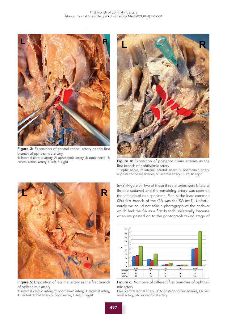
497
First branch of ophthalmic arteryİstanbul Tıp Fakültesi Dergisi • J Ist Faculty Med 2021;84(4):495-501
(n=3) (Figure 5). Two of these three arteries were bilateral (in one cadaver) and the remaining artery was seen on the left side of one specimen. Finally, the least common (3%) first branch of the OA was the SA (n=1). Unfortu-nately we could not take a photograph of the cadaver which had the SA as a first branch unilaterally because when we passed on to the photograph taking stage of
Figure 3: Exposition of central retinal artery as the first branch of ophthalmic artery1: internal carotid artery, 2: ophthalmic artery, 3: optic nerve, 4: central retinal artery, L: left, R: right Figure 4: Exposition of posterior ciliary arteries as the
first branch of ophthalmic artery1: optic nerve, 2: internal carotid artery, 3: ophthalmic artery, 4: posterior ciliary arteries, 5: lacrimal artery, L: left, R: right
Figure 5: Exposition of lacrimal artery as the first branch of ophthalmic artery1: internal carotid artery, 2: ophthalmic artery, 3: lacrimal artery, 4: central retinal artery, 5: optic nerve, L: left, R: right
Figure 6: Numbers of different first branches of ophthal-mic arteryCRA: central retinal artery, PCA: posterior ciliary arteries, LA: lac-rimal artery, SA: supraorbital artery
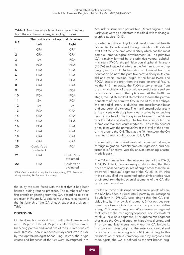
498
First branch of ophthalmic arteryİstanbul Tıp Fakültesi Dergisi • J Ist Faculty Med 2021;84(4):495-501
the study, we were faced with the fact that it had been harmed during routine practices. The numbers of each first branch originating from the OA, according to sides, are given in Figure 6. Additionally, our results concerning the first branch of the OA of each cadaver are given in Table 1.
DISCUSSION
Orbital dissection was first described by the German anat-omist Meyer in 1887 (6). Meyer revealed the anatomical branching pattern and variations of the OA in a series of over 20 cases. Then, in a 3-series study conducted in 1962 by the ophthalmologist Sohan Sing Hayreh, the origin, course and branches of the OA were investigated (7-9).
Around the same time period, Kuru, Moret, Vignaud, and Lasjaunias were also initiators in this field with their angio-graphic studies (10-13).
Knowledge of the embryological development of the OA is essential to understand its origin variations. It is stated that the OA is the craniofacial artery which has the most complex embryological development (4). The primitive OA is mainly formed by the primitive ventral ophthal-mic artery (PVOA), the primitive dorsal ophthalmic artery (PDOA) and stapedial artery. In the 4-6 mm (crown–rump length) embryo, PDOA formation is observed from the bifurcation point of the primitive carotid artery in its cau-dal and cranial division (origin of the future PCA). The PDOA enters the orbit from the superior orbital fissure. At the 7-12 mm stage, the PVOA artery emerges from the cranial division of the primitive carotid artery and en-ters the orbit through the optic canal. At the 16-18 mm stage, the PVOA and PDOA combine to form the perma-nent stem of the primitive OA. In the 18-40 mm embryo, the stapedial artery is divided into maxillomandibular and supraorbital divisions. The maxillomandibular artery anastomoses with the pharyngeal arteries by extending beyond the head from the spinous foramen. The SA en-ters the orbit and divides into two branches called the ethmoidonasal and lacrimal arteries. The ethmoidonasal artery joins with the primitive OA at the level of the arteri-al ring around the ON. Thus, at the 40 mm stage, the OA reaches its adult configuration (1, 3, 4, 13).
This model explains most cases of the variant OA origin through migration, partial/complete regression, and per-sistence of primitive vessels, and/or remaining anasto-motic loops (1).
The OA originates from the intradural part of the ICA (1, 4, 14, 15). In fact, there are many studies stating that they have not observed any source of origin other than the in-tracranial (intradural) segment of the ICA (5, 16-19). Also in this study, all of the examined ophthalmic arteries have originated from the intracranial segments of the ICA: dis-tal to cavernous sinus.
For the purpose of description and clinical points of view, the ICA has been divided into 7 parts by neurosurgeon Bouthillerin in 1996 (20). According to this, the ICA is di-vided into its 1st or cervical segment, 2nd or petrous seg-ment that gives origin to the caroticotympanic and vidian artery, 3rd or lacerum segment, 4th or cavernous segment that provides the meningohypophyseal and inferolateral trunk, 5th or clinoid segment, 6th or ophthalmic segment that gives the OA and superior hypophyseal artery, and 7th or communicating segment where the ICA, before its final division, gives origin to the anterior choroidal and posterior communicating artery (20). According to this classification, which is commonly used by surgeons and radiologists, the OA is defined as the first branch orig-
Table 1: Numbers of each first branches originating from the ophthalmic artery, according to sides
NoThe first branch of ophthalmic artery
Left Right
1 CRA CRA
2 CRA CRA
3 LA PCA
4 PCA PCA
5 CRA CRA
6 CRA CRA
7 PCA PCA
8 CRA CRA
9 PCA PCA
10 PCA PCA
11 SA PCA
12 LA LA
13 PCA CRA
14 CRA CRA
15 CRA PCA
16 PCA CRA
17 CRA CRA
18 CRA CRA
19 CRA CRA
20 Couldn’t beevaluated
PCA
21 CRA Couldn’t beevaluated
22 CRA Couldn’t beevaluated
CRA: Central retinal artery, LA: Lacrimal artery, PCA: Posterior ciliary arteries, SA: Supraorbital artery

499
First branch of ophthalmic arteryİstanbul Tıp Fakültesi Dergisi • J Ist Faculty Med 2021;84(4):495-501
inating from the C6 segment of the ICA. Consequently the OA is also defined as the first intracranial or intra-dural branch of the ICA (1). Later, in 2009, Lehecka et al. divided the C4 segment into 3 portions as ophthalmic, communicating and choroid (21).
As the most common origin variation of the OA report-ed in the literature, it originates from the MMA (1, 3, 15). The MMA origin of the OA could be explained by: the absence of anastomosis between the supraorbital branch and the OA, or the persistence of the proximal stem of the supraorbital branch of the stapedial artery with re-gression of the primitive OA (4).
It has been reported that an OA originating from the cav-ernous part of the ICA is a rare variation and its incidence is estimated at 0.4% (22). In addition, Dilenge, Fiore, Par-lato, Islak and Indo have also defined cases with the OA of cavernous (4th segment) origin (23-27). Toma reported that the OA arising from the cavernous segment of the ICA was defined as the second most common variation and added that this artery entered the orbit from the su-perior orbital fissure (3). Lasjaunias explained this varia-tion by the embryological regression in the PVOA instead of the PDOA. In other words, this anatomical variation is explained by the embryological persistent PDOA (13).
In addition, origin of the OA from the clinoidal (5th seg-ment) or communicating portions (7th segment) of the ICA has been recorded (3, 14, 28, 29).
The double origin of the OA is a very rare variant; its in-cidence is estimated to be around 0.2%, and only a few studies have been reported (4, 13, 22, 30, 31). Lasjaunias explained this variation by the lack of an anastomotic ring around the ON between the PDOA and the PVOA, or persistence of both of these arteries (1, 4, 13).
The rare origin variation of the OA from the MCA has been observed in the absence of the ICA (15, 32-34).
There have been reports of the ophthalmic artery orig-inating from the ACA (27,35-37). This variation corre-sponds to the persistence of the PVOA (1).
It has been reported that it is encountered in the pres-ence of ICA agenesis or hypoplasia (38-40).
OA originating from the basilar artery is an extreme vari-ation. It has been stated that current embryological the-ories do not provide a satisfactory explanation for this formation (41-43).
OA originating from this artery has been reported in only 1 case (44).
After its origin, the OA follows a short intracranial course until it pierces the dura mater and reaches the optic ca-
nal. The course of the OA between its origin and optic canal is named as the intracranial course. This distance was calculated by Hayreh and Dass as 0.5–0.95 mm (8). Although the intracranial course of the OA is short, the knowledge of the anatomy of this course is of utmost surgical importance as surgical intervention for OA aneu-rysms takes place at that area (1).
After the intracranial course, the OA enters the optic ca-nal together with the ON. This portion is defined as in-tracanalicular course.
Afterwards the OA exits the optic canal and enters the orbit. This is the intraorbital part of the OA. The intraor-bital course of the OA divides into three segments. In the first segment, the OA exits the optic canal and proceeds parallel to the ON. The second segment courses medial-ly passing above (83%) or below (17%) the ON (1). Finally, the third segment is separated to its branches medially to the ON, and terminates at the superomedial angle of the orbital opening (3, 4, 9).
Hayreh, who studied the OA branches in the orbits of 59 human cadavers, reported that localization and order of origin of these branches are not identical on either side of the same person (9). It is therefore stated that the branch-ing patterns of the OA are very complex and unique.
Two different types of branching have been defined as the OA passes above or below the ophthalmic nerve (9, 15). According to this classification, if the OA crosses over the ON, the first branch is the medial posterior ciliary ar-teries (MPCA) in common with the CRA; if the OA cross-es under the ON, the first branch is the lateral posterior ciliary arteries (LPCA). However, when the literature was examined, it was observed that there were branching patterns that did not fit this classification. Each most fre-quent first branch of the OA determined in our study is discussed separately.
The CRA is the most frequent first branch of the OA. It supplies blood to the retina, therefore it is critical for vision (1). It is one of the smallest branches of the OA and is a terminal artery. Due to its average diameter of 0.36 mm, the CRA is highly susceptible to occlusion (45). The CRA is commonly the first location where ischemic or embolic events make us notice the fact that there is serious vascular disease and high risk for an upcoming stroke. A central retinal artery embolus may produce a transient blindness in the affected eye, called amauro-sis fugax, which lasts for several minutes but less than an hour (transient ischemic attack) (46). The CRA occlu-sion causes ischemia and results in infarction of the retina and as a result, sudden loss of vision with an incidence of 1–8/100,000 people occurs (1). Therefore, damage of the CRA usually results in blindness (18).

500
First branch of ophthalmic arteryİstanbul Tıp Fakültesi Dergisi • J Ist Faculty Med 2021;84(4):495-501
In the study by Tsutsumi et al. the CRA was found to be the first branch of the OA in 67%, the second branch in 28% and the third branch in 5% of the orbits they exam-ined (5). In the present study, the CRA was observed as the first branch in 56% of cases.
The PCA are branches of the OA, ranging in number from 1 to 5, that supply the choroid and outer layer of the ret-ina. It has been reported that in 80% of cases there are 2 or 3 PCA (1). The PCA are named as medial or later-al according to their location. Ciliary arteries supply the middle vascular tunic. Moreover, they take part in blood supply of the retina; if a detached retina occurs, this com-ponent of blood supply may be disrupted (46).
According to Hayreh, the first branch is either the CRA in common with the MPCA or only the LPCA (9, 15). In our study, the PCA were observed as the first branch with a frequency of 34%, as the second most frequent first branch of the OA.
The LA supplies the lacrimal gland, lateral rectus mus-cle, lateral part of the eyelids and gives off meningeal branches to dura mater (4). The LA is often referred to as the third or fifth branch of the OA (9). When the literature was reviewed, no case was observed in which the LA was reported to be the first branch of the OA. Therefore, this paper is the first one, in which the LA is reported to be the first branch of the OA (7% cases, n=3).
One of the limitations of our study is that we could not take a photograph of the cadaver which had the SA as a first branch. Moreover, we could not describe the practi-cal locations for determining the first branch of the OA of the related region for surgeons.
In conclusion, the anatomy of the OA is quite complicat-ed because it originates from three embryological sourc-es, has a dual intracranial and extracranial course, and both itself and its branches are small in size. In addition, the location and proximity of structures such as the ON, and the critical importance of the CRA and PCA, which are usually the first branches of the OA, cause difficulties in surgical operations in this area. Knowledge of the anat-omy and possible variations of the OA and its branches is essential for neurosurgeons and neuroradiologists while approaching pathologies such as retinoblastoma and CRA occlusion (5, 45).
Ethics Committee Approval: This study was approved by the Clinical Research Ethical Committee of the Istanbul University, Istanbul Faculty of Medicine (Date: 16.04.2021, No: 172684).
Peer Review: Externally peer-reviewed.
Author Contributions: Conception/Design of Study- Ö.G.; Data Acquisition- A.K.; Data Analysis/Interpretation- Ö.G.; Draf-
ting Manuscript- Ö.G.; Critical Revision of Manuscript- O.C.; Final Approval and Accountability- Ö.G., A.K., O.C., A.Ö., B.B.
Conflict of Interest: Authors declared no conflict of interest.
Financial Disclosure: Authors declared no financial support.
Etik Komite Onayı: Bu çalışma için etik komite onayı İstanbul Üniversitesi, İstanbul Tıp Fakültesi Klinik Araştırmalar Etik Kuru-lu’ndan alınmıştır (Tarih: 16.04.2021, No: 172684).
Hakem Değerlendirmesi: Dış bağımsız.
Yazar Katkıları: Çalışma Konsepti/Tasarım- Ö.G.; Veri Toplama- A.K.; Veri Analizi/Yorumlama- Ö.G.; Yazı Taslağı- Ö.G.; İçeriğin Eleştirel İncelemesi- O.C.; Son Onay ve Sorumluluk- Ö.G., A.K., O.C., A.Ö., B.B.
Çıkar Çatışması: Yazarlar çıkar çatışması beyan etmemişlerdir.
Finansal Destek: Yazarlar finansal destek beyan etmemişlerdir.
REFERENCES
1. Michalinos A, Zogana S, Kotsiomitis E, Mazarakis A, Troupis T. Anatomy of the ophthalmic artery: a review concerning its modern surgical and clinical applications. Anat Res Int 2015;2015:591961. [CrossRef]
2. Hayashi N, Kubo M, Tsuboi Y, Nishimura S, Nishijima M, Ahmed Abdel-Aal M, et al. Impact of anomalous origin of the ophthalmic artery from the middle meningeal artery on selection of surgical approach to skull base meningioma. Surg Neurol 2007;68(5):568-72. [CrossRef]
3. Toma N. Anatomy of the ophthalmic artery: embryological consideration. Neurol Med Chir (Tokyo) 2016;56(10):585-91. [CrossRef]
4. Bonasia S, Bojanowski M, Robert T. Embryology and anatomical variations of the ophthalmic artery. Neuroradiology 2020;62(2):139-52. [CrossRef]
5. Tsutsumi S, Rhoton AL Jr. Microsurgical anatomy of the central retinal artery. Neurosurgery 2006;59(4):870-9. [CrossRef]
6. Meyer F. Zur anatomie der Orbitalarteien. Morph Jahr 1887;12:414-58.
7. Hayreh SS, Dass R. The ophthalmic artery: I. Origin and intra-cranial and intra-canalicular course. Br J Ophthalmol 1962;46(2):65-98. [CrossRef]
8. Hayreh SS, Dass R. The ophthalmic artery: II. Intra-orbital course. Br J Ophthalmol 1962;46(3):165-85. [CrossRef]
9. Hayreh SS. The ophthalmic artery: III. Branches. Br J Ophthalmol 1962;46(4):212-47. [CrossRef]
10. Kuru Y. Meningeal branches of the ophthalmic artery. Acta Radiol Diagn (Stockh) 1967;6(3):241-51. [CrossRef]
11. Moret J, Lasjaunias P, Théron J, Merland JJ. The middle meningeal artery. Its contribution to the vascularisation of the orbit. J Neuroradiol 1977;4(2):225-48.
12. Vignaud J, Hasso AN, Lasjaunias P, Clay C. Orbital vascular anatomy and embryology. Radiology 1974;111(3):617-26. [CrossRef]
13. Lasjaunias P, Brismar J, Moret J, Théron J. Recurrent cavernous branches of the ophthalmic artery. Acta Radiol Diagn (Stockh) 1978;19(4):553-60. [CrossRef]
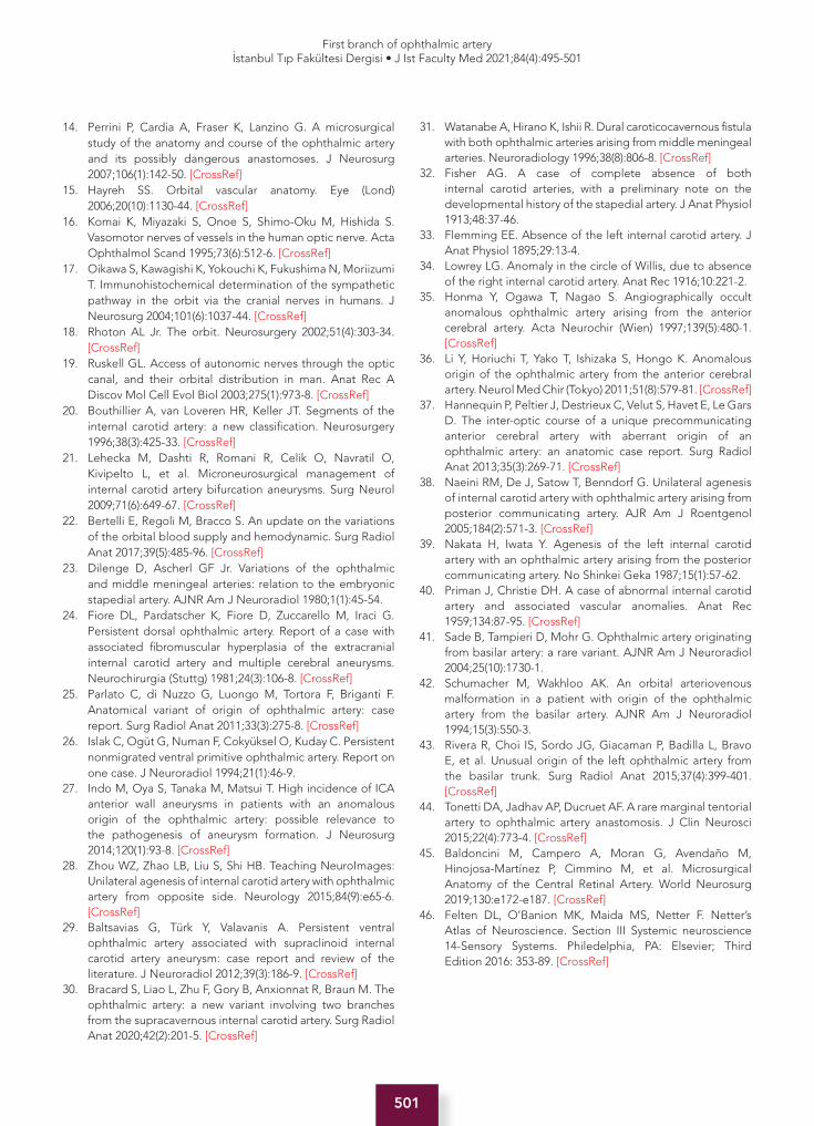
501
First branch of ophthalmic arteryİstanbul Tıp Fakültesi Dergisi • J Ist Faculty Med 2021;84(4):495-501
14. Perrini P, Cardia A, Fraser K, Lanzino G. A microsurgical study of the anatomy and course of the ophthalmic artery and its possibly dangerous anastomoses. J Neurosurg 2007;106(1):142-50. [CrossRef]
15. Hayreh SS. Orbital vascular anatomy. Eye (Lond) 2006;20(10):1130-44. [CrossRef]
16. Komai K, Miyazaki S, Onoe S, Shimo-Oku M, Hishida S. Vasomotor nerves of vessels in the human optic nerve. Acta Ophthalmol Scand 1995;73(6):512-6. [CrossRef]
17. Oikawa S, Kawagishi K, Yokouchi K, Fukushima N, Moriizumi T. Immunohistochemical determination of the sympathetic pathway in the orbit via the cranial nerves in humans. J Neurosurg 2004;101(6):1037-44. [CrossRef]
18. Rhoton AL Jr. The orbit. Neurosurgery 2002;51(4):303-34. [CrossRef]
19. Ruskell GL. Access of autonomic nerves through the optic canal, and their orbital distribution in man. Anat Rec A Discov Mol Cell Evol Biol 2003;275(1):973-8. [CrossRef]
20. Bouthillier A, van Loveren HR, Keller JT. Segments of the internal carotid artery: a new classification. Neurosurgery 1996;38(3):425-33. [CrossRef]
21. Lehecka M, Dashti R, Romani R, Celik O, Navratil O, Kivipelto L, et al. Microneurosurgical management of internal carotid artery bifurcation aneurysms. Surg Neurol 2009;71(6):649-67. [CrossRef]
22. Bertelli E, Regoli M, Bracco S. An update on the variations of the orbital blood supply and hemodynamic. Surg Radiol Anat 2017;39(5):485-96. [CrossRef]
23. Dilenge D, Ascherl GF Jr. Variations of the ophthalmic and middle meningeal arteries: relation to the embryonic stapedial artery. AJNR Am J Neuroradiol 1980;1(1):45-54.
24. Fiore DL, Pardatscher K, Fiore D, Zuccarello M, Iraci G. Persistent dorsal ophthalmic artery. Report of a case with associated fibromuscular hyperplasia of the extracranial internal carotid artery and multiple cerebral aneurysms. Neurochirurgia (Stuttg) 1981;24(3):106-8. [CrossRef]
25. Parlato C, di Nuzzo G, Luongo M, Tortora F, Briganti F. Anatomical variant of origin of ophthalmic artery: case report. Surg Radiol Anat 2011;33(3):275-8. [CrossRef]
26. Islak C, Ogüt G, Numan F, Cokyüksel O, Kuday C. Persistent nonmigrated ventral primitive ophthalmic artery. Report on one case. J Neuroradiol 1994;21(1):46-9.
27. Indo M, Oya S, Tanaka M, Matsui T. High incidence of ICA anterior wall aneurysms in patients with an anomalous origin of the ophthalmic artery: possible relevance to the pathogenesis of aneurysm formation. J Neurosurg 2014;120(1):93-8. [CrossRef]
28. Zhou WZ, Zhao LB, Liu S, Shi HB. Teaching NeuroImages: Unilateral agenesis of internal carotid artery with ophthalmic artery from opposite side. Neurology 2015;84(9):e65-6. [CrossRef]
29. Baltsavias G, Türk Y, Valavanis A. Persistent ventral ophthalmic artery associated with supraclinoid internal carotid artery aneurysm: case report and review of the literature. J Neuroradiol 2012;39(3):186-9. [CrossRef]
30. Bracard S, Liao L, Zhu F, Gory B, Anxionnat R, Braun M. The ophthalmic artery: a new variant involving two branches from the supracavernous internal carotid artery. Surg Radiol Anat 2020;42(2):201-5. [CrossRef]
31. Watanabe A, Hirano K, Ishii R. Dural caroticocavernous fistula with both ophthalmic arteries arising from middle meningeal arteries. Neuroradiology 1996;38(8):806-8. [CrossRef]
32. Fisher AG. A case of complete absence of both internal carotid arteries, with a preliminary note on the developmental history of the stapedial artery. J Anat Physiol 1913;48:37-46.
33. Flemming EE. Absence of the left internal carotid artery. J Anat Physiol 1895;29:13-4.
34. Lowrey LG. Anomaly in the circle of Willis, due to absence of the right internal carotid artery. Anat Rec 1916;10:221-2.
35. Honma Y, Ogawa T, Nagao S. Angiographically occult anomalous ophthalmic artery arising from the anterior cerebral artery. Acta Neurochir (Wien) 1997;139(5):480-1. [CrossRef]
36. Li Y, Horiuchi T, Yako T, Ishizaka S, Hongo K. Anomalous origin of the ophthalmic artery from the anterior cerebral artery. Neurol Med Chir (Tokyo) 2011;51(8):579-81. [CrossRef]
37. Hannequin P, Peltier J, Destrieux C, Velut S, Havet E, Le Gars D. The inter-optic course of a unique precommunicating anterior cerebral artery with aberrant origin of an ophthalmic artery: an anatomic case report. Surg Radiol Anat 2013;35(3):269-71. [CrossRef]
38. Naeini RM, De J, Satow T, Benndorf G. Unilateral agenesis of internal carotid artery with ophthalmic artery arising from posterior communicating artery. AJR Am J Roentgenol 2005;184(2):571-3. [CrossRef]
39. Nakata H, Iwata Y. Agenesis of the left internal carotid artery with an ophthalmic artery arising from the posterior communicating artery. No Shinkei Geka 1987;15(1):57-62.
40. Priman J, Christie DH. A case of abnormal internal carotid artery and associated vascular anomalies. Anat Rec 1959;134:87-95. [CrossRef]
41. Sade B, Tampieri D, Mohr G. Ophthalmic artery originating from basilar artery: a rare variant. AJNR Am J Neuroradiol 2004;25(10):1730-1.
42. Schumacher M, Wakhloo AK. An orbital arteriovenous malformation in a patient with origin of the ophthalmic artery from the basilar artery. AJNR Am J Neuroradiol 1994;15(3):550-3.
43. Rivera R, Choi IS, Sordo JG, Giacaman P, Badilla L, Bravo E, et al. Unusual origin of the left ophthalmic artery from the basilar trunk. Surg Radiol Anat 2015;37(4):399-401. [CrossRef]
44. Tonetti DA, Jadhav AP, Ducruet AF. A rare marginal tentorial artery to ophthalmic artery anastomosis. J Clin Neurosci 2015;22(4):773-4. [CrossRef]
45. Baldoncini M, Campero A, Moran G, Avendaño M, Hinojosa-Martínez P, Cimmino M, et al. Microsurgical Anatomy of the Central Retinal Artery. World Neurosurg 2019;130:e172-e187. [CrossRef]
46. Felten DL, O’Banion MK, Maida MS, Netter F. Netter’s Atlas of Neuroscience. Section III Systemic neuroscience 14-Sensory Systems. Philedelphia, PA: Elsevier; Third Edition 2016: 353-89. [CrossRef]

502
Swallowing in partial laryngectomyİstanbul Tıp Fakültesi Dergisi • J Ist Faculty Med 2021;84(4):502-7
* This manuscript was presented in 36th Turkish National Otorhinolaryngology and Head & Neck Surgery Congress.
Corresponding author/İletişim kurulacak yazar: [email protected]
Submitted/Başvuru: 14.12.2020 • Revision Requested/Revizyon Talebi: 06.01.2021 •Last Revision Received/Son Revizyon: 26.04.2021 • Accepted/Kabul: 06.05.2021 • Published Online/Online Yayın: 20.09.2021
RESEARCH / ARAŞTIRMADOI: 10.26650/IUITFD.2021.839817
İst Tıp Fak Derg 2021 / J Ist Faculty Med 2021
COMPARISON OF SWALLOWING IN DIFFERENT TYPES OF PARTIAL LARYNGECTOMIES
FARKLI PARSİYEL LARENJEKTOMİ TEKNİKLERİNDE YUTMANIN KARŞILAŞTIRILMASI
Zeynep ERDOĞAN ÇETİN1 , Sibel EYİGÖR2 , Kerem ÖZTÜRK1 , Göksel TURHAL1 , Serdar AKYILDIZ1 , Atilla YAVUZER1
1Ege University, School of Medicine, Department of Otolaryngology, Izmir, Turkey2Ege University, School of Medicine, Department of Physical Therapy and Rehabilitation, Izmir, Turkey
ORCID IDs of the authors: Z.E.Ç. 0000-0003-1213-4033; S.E. 0000-0002-9781-2712; K.Ö. 0000-0003-2754-4166; G.T. 0000-0003-0020-1921; S.A. 0000-0001-9539-3916; A.Y. 0000-0002-8446-8721
Cite this article as: Cetin ZE, Eyigor S, Ozturk K, Turhal G, Akyildiz S, Yavuzer A. Comparison of swallowing in different types of partial laryngectomies. J Ist Faculty Med 2021;84(4):502-7. doi: 10.26650/IUITFD.2021.839817
ABSTRACT
Objective: We designed this study to assess and compare the effects of different partial laryngectomy (PL) techniques on swal-lowing.
Material and Methods: Ten patients had laryngofissure with cordectomy, ten had frontal anterior laryngectomy with epi-glottic reconstruction (FAL), ten had frontolateral laryngectomy (FLL), ten had cricohyoidopexy (CHP), ten had cricohyoidoepi-glottopexy (CHEP), and ten had supraglotic laryngectomy. Swal-lowing was assessed with flexible endoscopy.
Results: Mild or moderate dysphagia for solid foods was discov-ered significantly more often in CHP patients compared to FLL and FAL (p<0.05) patients. Dysphagia discoveries for semi-solid and liquid food didn’t significantly differ among PL’s (p>0.05). Compared to other PLs, the penetration-aspiration test with 10 ml of water was distinctly lower in cordectomy and FLL patients (p<0.05).
Conclusion: Penetration and aspiration with 10 ml of water was marked lower in cordectomy and FLL patients matched to other PL patients. With studies involving more patients, it will be pos-sible to increase the evidence value of our results.
Keywords: Partial laryngectomy, fiberoptic endoscopic evalua-tion of swallowing (FEES), deglutition disorders, dysphagia
ÖZET
Amaç: Bu çalışma, farklı parsiyel larenjektomi (PL) tekniklerinin yutma üzerindeki etkilerini değerlendirmek ve karşılaştırmak amacıyla tasarlandı.
Gereç ve Yöntemler: On hastada kordektomi ile laringofissür, onunda epiglotik rekonstrüksiyon (FAL) ile frontal anterior laren-jektomi, on hastada frontolateral larenjektomi (FLL), on hastada krikohiyoidopeksi (CHP), on hastada krikohiyoidoepiglottopeksi (CHEP) ve diğer onunda supraglotik larenjektomi operasyonu yapılmıştı. Yutma fleksible endoskopi ile değerlendirildi.
Bulgular: Katı yiyecekler için hafif veya orta derecede disfaji, CHP hastalarında FLL ve FAL hastalarına kıyasla anlamlı olarak daha sık olduğu bulundu (p<0,05). Yarı katı ve sıvı gıdalar için disfaji araştırmasında, PL’ler arasında önemli ölçüde farklılık gösterilmedi (p>0,05). Diğer PL’lere kıyasla, 10 ml su ile penet-rasyon-aspirasyon testi, kordektomi ve FLL hastalarında belirgin şekilde daha düşüktü (p<0,05).
Sonuç: Kordektomi ve FLL hastalarında diğer PL hastalarına göre 10 ml su ile penetrasyon ve aspirasyon testi, daha düşüktü. Daha fazla hastayı içeren çalışmalar sayesinde vermiş olduğumuz so-nuçların kanıt değerinin artırılması mümkün olacaktır.
Anahtar Kelimeler: Parsiyel larenjektomi, fiberoptik endoskopik yutma değerlendirmesi (FEYD), yutma bozuklukları, disfaji
Content of this journal is licensed under a Creative Commons Attribution-NonCommercial 4.0 International License.
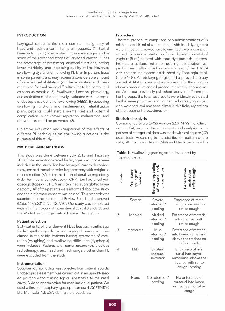
503
Swallowing in partial laryngectomyİstanbul Tıp Fakültesi Dergisi • J Ist Faculty Med 2021;84(4):502-7
RESEARCH / ARAŞTIRMADOI: 10.26650/IUITFD.2021.839817
İst Tıp Fak Derg 2021 / J Ist Faculty Med 2021
COMPARISON OF SWALLOWING IN DIFFERENT TYPES OF PARTIAL LARYNGECTOMIES
FARKLI PARSİYEL LARENJEKTOMİ TEKNİKLERİNDE YUTMANIN KARŞILAŞTIRILMASI
Zeynep ERDOĞAN ÇETİN1 , Sibel EYİGÖR2 , Kerem ÖZTÜRK1 , Göksel TURHAL1 , Serdar AKYILDIZ1 , Atilla YAVUZER1
1Ege University, School of Medicine, Department of Otolaryngology, Izmir, Turkey2Ege University, School of Medicine, Department of Physical Therapy and Rehabilitation, Izmir, Turkey
ORCID IDs of the authors: Z.E.Ç. 0000-0003-1213-4033; S.E. 0000-0002-9781-2712; K.Ö. 0000-0003-2754-4166; G.T. 0000-0003-0020-1921; S.A. 0000-0001-9539-3916; A.Y. 0000-0002-8446-8721
Cite this article as: Cetin ZE, Eyigor S, Ozturk K, Turhal G, Akyildiz S, Yavuzer A. Comparison of swallowing in different types of partial laryngectomies. J Ist Faculty Med 2021;84(4):502-7. doi: 10.26650/IUITFD.2021.839817
INTRODUCTION
Laryngeal cancer is the most common malignancy of head and neck cancer in terms of frequency (1). Partial laryngectomy (PL) is indicated in the early stages and in some of the advanced stages of laryngeal cancer. PL has the advantage of preserving laryngeal functions, having lower morbidity, and increasing quality of life. However, swallowing dysfunction following PL is an important issue in some patients and may require a considerable amount of care and rehabilitation (2). The evaluation and treat-ment plan for swallowing difficulties has to be completed as soon as possible (3). Swallowing function, physiology, and aspiration can be effectively evaluated with fiberoptic endoscopic evaluation of swallowing (FEES). By assessing swallowing functions and implementing rehabilitation plans, patients could start a normal diet and potential complications such chronic aspiration, malnutrition, and dehydration could be prevented (3).
Objective evaluation and comparison of the effects of different PL techniques on swallowing functions is the purpose of this study.
MATERIAL AND METHODS
This study was done between July 2012 and February 2013. Sixty patients operated for laryngeal carcinoma were included in the study. Ten had laryngofissure with cordec-tomy, ten had frontal anterior laryngectomy with epiglottic reconstruction (FAL), ten had frontolateral laryngectomy (FLL), ten had cricohyoidopexy (CHP), ten had cricohyoi-doepiglottopexy (CHEP) and ten had supraglottic laryn-gectomy. All of the patients were informed about the study and their informed consent was gained. This research was submitted to the Institutional Review Board and approved (Date: 14.09.2012, No: 12-7/80). Our study was completed within the framework of international ethical standards and the World Health Organization Helsinki Declaration.
Patient selectionSixty patients, who underwent PL at least six months ago for histopathologically proven laryngeal cancer, were in-cluded in the study. Patients having symptoms of aspi-ration (coughing) and swallowing difficulties (dysphagia) were included. Patients with tumor recurrence, previous radiotherapy, and head and neck surgery other than PL were excluded from the study.
InstrumentationSociodemographic data was collected from patient records. Endoscopic assessment was carried out in an upright-seat-ed position without using topical anesthesia to the nasal cavity. A video was recorded for each individual patient. We used a flexible nasopharyngoscope camera (KAY PENTAX Ltd, Montvale, NJ, USA) during the procedures.
ProcedureThe test procedure comprised two administrations of 3 ml, 5 ml, and 10 ml of water stained with food dye (green) via an injector. Likewise, swallowing tests were complet-ed with two administrations of one dessert spoonful of yoghurt (5 ml) colored with food dye and fish crackers. Premature spillage, retention-pooling, penetration, as-piration and reflex coughing were scored (from 1 to 5) with the scoring system established by Topaloglu et al. (Table 1) (4). An otolaryngologist and a physical therapy and rehabilitation specialist were present for the duration of each procedure and all procedures were video-record-ed. As in our previously published study in different pa-tient groups, the total test results were blindly evaluated by the same physician and unchanged otolaryngologist, who were focused and specialized in this field, regardless of the treatment procedures (5).
Statistical analysisComputer software (SPSS version 22.0, SPSS Inc. Chica-go, IL, USA) was conducted for statistical analysis. Com-parison of categorical data was made with chi-square (X2) exact tests. According to the distribution pattern of the data, Wilcoxon and Mann-Whitney U tests were used in
Table 1: Swallowing grading scale developed by Topaloglu et al.
Po
ints
Pre
mat
ure
sp
illag
e o
f
mat
eria
l
Ret
ensi
on/
po
olin
g
of
mat
eria
l and
/ o
r se
cret
ion
Pen
etra
tio
n-
asp
irat
ion
refl
ex c
oug
h
1 Severe Severe retention/pooling
Enterance of mate-rial into trachea; no
reflex cough
2 Marked Marked retention/pooling
Enterance of material into trachea; with
reflex cough
3 Moderate Mild retention/pooling
Enterance of material into larynx; remaining above the trachea no
reflex cough
4 Mild Coatingresidue/ secretion
Enterance of ma-terial into larynx;
remaining above the trachea with reflex
cough forming
5 None No retention/pooling
No enterance of material into larynx or trachea; no reflex
cough

504
Swallowing in partial laryngectomyİstanbul Tıp Fakültesi Dergisi • J Ist Faculty Med 2021;84(4):502-7
the analysis of nonparametric variables, and indepen-dent, and paired sample t-tests were used in the analysis of parametric variables. Determining the distribution pat-tern of the data was provided by the Shapiro-Wilk test. The distribution of the groups in our study was non-para-metric. Considering the distribution of the data, Pearson or Spearman correlation analysis was used. Data was ex-pressed as “median, interquartile range (IQR)”. A p value of <0.05 was considered statistically significant.
RESULTS
Sixty patients treated with PL were involved in the study. Five (8.3%) of the patients were female and 55 (91.7%) were male, with a mean age of 59.87±7.93 years (range 33-79 years).
Forty-four (73.3%) patients reported coughing, two (3.3%) reported coughing and dysphagia, and 14 (23.3%) reported only dysphagia. The type of PL present in the patient and their symptoms were not statistically correlat-ed (p>0.05). The presence and frequency of the subjec-tive symptoms (difficulty in bolus control, need to clear throat, food gets stuck, choking) are presented in Table 2. Subjective symptoms and the type of PL were not sta-tistically correlated (p>0.05).
Dysphagia for solid, semi-solid, and liquid food was eval-uated and the data (according to the types of PL) is pre-sented in Table 3. Mild or moderate dysphagia for solid foods was significantly more prevalent in CHP patients compared to FLL and FAL (p<0.05) patients. Dysphagia for semi-solid and liquid food did not significantly differ among different PL’s (p>0.05).
Premature spillage, residue-secretion, and penetra-tion-aspiration scores were evaluated with fiberoptic en-doscopy. Scores for different types of PL are presented in Table 4. Penetration-aspiration with 10 ml water was meaningfully lower in cordectomy and FLL patients com-pared to supraglottic laryngectomy, CHP and CHEP pa-tients (p<0.05). Other scores had no significant difference among the groups (Table 4) (p>0.05).
DISCUSSION
There are many reports assessing the swallowing func-tion in a certain type of PL (6-12). While some research-ers evaluated swallowing function with N/G tube remov-al time or gastrostomy tube removal rates in previous reports, others used quality of life measures (9, 12-15). Videofluoroscopy is also a widely used technique for the evaluation of swallowing (7, 8, 12, 16). However, a study,
Table 2: Operation types and subjective complaints
ComplaintOperation type
Supraglottic laryngectomy
Cordectomy CHP CHEP FLL FAL p
Cough N (%) 7 (70%) 8 (80%) 8 (80%) 8 (80%) 7 (70%) 6 (60%) p>0.05
Cough, dysphagia
N (%) 0 (0%) 0 (0%) 2 (20%) 0 (0%) 0 (0%) 0 (0%)
Dysphagia N (%) 3 (30%) 2 (20%) 0 (0%) 2 (20%) 3 (30%) 4 (40%)
Difficulty in bolus control
Not present (N;%)
9 (90%) 9 (90%) 9 (90%) 8 (80%) 10 (100%) 10 (100%) p>0.05
Present (N;%)
1 (10%) 1 (10%) 1 (10%) 2 (20%) 0 (0%) 0 (0%)
Need to clear throat
Not present (N;%)
4 (40%) 7 (70%) 4 (40%) 8 (80%) 8 (80%) 7 (70%) p>0.05
Present (N;%)
6 (60%) 3 (30%) 6 (60%) 2 (20%) 2 (20%) 3 (30%)
Sensation of a lump in the throat
Not present (N;%)
6 (60%) 7 (70%) 5 (50%) 7 (70%) 6 (60%) 5 (50%) p>0.05
Present (N;%)
4 (40%) 3 (30%) 5 (50%) 3 (30%) 4 (40%) 5 (50%)
Sense of choking
Not present (N;%)
7 (70%) 8 (80%) 6 (60%) 9 (90%) 10 (100%) 10 (100%) p>0.05
Present (N;%)
3 (30%) 2 (20%) 4 (40%) 1 (10%) 0 (0%) 0 (%0)
CHP: Cricohyoidopexy; CHEP: Cricohyoidoepiglottopexy; FLL: Frontolateral laryngectomy; FAL: Frontal anterior laryngectomy
505
Swallowing in partial laryngectomyİstanbul Tıp Fakültesi Dergisi • J Ist Faculty Med 2021;84(4):502-7
that compared swallowing functions in different types of PL with FEES, is missing. A study conducted by Ali-candri–Ciufelli et al. compared swallowing functions of supraglottic laryngectomy and supracricoid partial lar-yngectomy (SCPL) patients with FEES (12). Other studies usually evaluated swallowing in only one type of PL or compared supracricoid laryngectomy with total laryngec-tomy (12,16). Alicandri-Ciufelli et al. found no statistically significant difference between supraglottic laryngecto-my and supracricoid laryngectomy patients regarding swallowing functions evaluated with FEES (12). They also evaluated cases with both preserved arytenoids, radio-therapy, different ages, and a different time interval after surgery, and it was concluded that only radiotherapy had a significant negative effect on supracricoid laryngecto-my patients with FEES (4). Premature spillage, residue-se-
Table 3: Dysphagia table
Operation typeSupraglottic
laryngectomyCordectomy CHP CHEP FLL FAL Total
Dysphagia to solid food
Not present n 8 7 5 7 9 9 45
% 80 70 50 70 90 90 75
Mild or moderate n 2 3 2 3 1 1 12
% 20 30 20 30 10 10 20
Severe n 0 0 3 0 0 0 3
% 0 0 30 0 0 0 5
Total n 10 10 10 10 10 10 60
% 100 100 100 100 100 100 100
Dysphagia to semisolid food
Not present n 6 8 6 8 10 9 47
% 60 80 60 80 100 90 78.3
Mild or moderate n 4 2 4 2 0 1 13
% 40 20 40 20 0 10 21.7
Severe n 0 0 0 0 0 0 0
% 0 0 0 0 0 0 0
Total n 10 10 10 10 10 10 60
% 100 100 100 100 100 100 100
Dysphagia to liquid food
Not present n 5 10 8 4 8 8 43
% 50 100 80 40 80 80 71.7
Mild or moderate n 4 0 2 6 2 2 16
% 40 0 20 60 20 20 26.7
Severe n 1 0 0 0 0 0 1
% 10 0 0 0 0 0 1.7
Total n 10 10 10 10 10 10 60
% 100 100 100 100 100 100 100
CHP: Cricohyoidopexy; CHEP: Cricohyoidoepiglottopexy; FLL: Frontolateral laryngectomy; FAL: Frontal anterior laryngectomy
Table 4: P values regarding different laryngectomy types and swallowing scores
Premature
spillageResidue, secretion
Penetration, aspiration,
reflex cough
p value p value p value
3 ml water .539 .294 .097
5 ml water .490 .471 .163
10 ml water .305 .614 .024
5 ml yoghurt
.385 .303 .083
Fish cracker
.540 .314 .066

506
Swallowing in partial laryngectomyİstanbul Tıp Fakültesi Dergisi • J Ist Faculty Med 2021;84(4):502-7
cretion, and penetration-aspiration scores were higher in patients with both arytenoids preserved and in patients that did not receive radiotherapy, but this difference was not statistically significant (4). Because patients with a his-tory of radiotherapy were excluded and both arytenoids were preserved in all patients, these were not evaluated in this study.
Another important step in preserving the swallowing function is to preserve the superior laryngeal nerve. If this nerve is damaged, the cricopharyngeal sphincter and the cough reflex will be negatively affected and the patient won’t be able to recognize aspiration (3, 17, 18). This may hamper subsequent swallowing rehabilitation and may extend adaptation time. All effort should be taken to preserve both superior laryngeal nerves in laryngeal conservation surgery.
Zacharek et al. evaluated the swallowing function of 10 supracricoid laryngectomy patients with FEES and modified barium swallow studies (19). They reported swallowing difficulties in all patients. Supraglottic sensory loss secondary to unilateral or bilateral damaged superior laryngeal nerve, changed base of tongue/vallecular anatomy after extraction of epiglottis, physiologic insufficiency of the neoglottal valve, or a combination of these three mechanisms were the proposed mechanisms for swallowing difficulties (15, 18). All of the patients in this study tolerated oral food intake and were decanulated. None of the patients developed aspiration pneumonia. These findings show that all patients should have a sufficient cough reflex to protect their lungs from aspiration pneumonia and an active tracheopulmonary mucociliary clearance system. In addition, these studies emphasize the importance of adequate respiratory function in patients undergoing SCPL. In a study evaluating the swallowing function of 116 SCPL patients, 45 patients that had aspiration in videofluoroscopic study were assessed with high-resolution computed tomography and no statistically noteworthy difference was established among these patients and control groups regarding the radiographic images (19).
This is the first research to compare swallowing functions of different types of PL with FEES. A limited number of patients and inadequate randomization are the limita-tions of this study. Objective evaluation of swallowing in PL patients provides valuable data and feedback for both the doctor and the patient.
CONCLUSION
In conclusion, penetration and aspiration with 10 ml of water was meaningfully lower in cordectomy and FLL patients compared to supraglottic laryngectomy, CHP, and CHEP patients. This study is important because it is the first study, which evaluated swallowing objectively in patients
who underwent six different PL. The most important result of this study, regardless of which PL technique is applied, is that aspiration problems are not caused, other than high-volume water ingestion, after the 6th postoperative month in patients, who have preserved both arytenoids and have not applied radiotherapy. We think that we can reduce the concern of surgeons with this objective study about swallowing that will occur as a result of the PL technique preference. Further studies with larger patient groups are warranted to obtain more reliable results.
Informed Consent: Written consent was obtained from the par-ticipants.
Ethics Committee Approval: This study was approved by the Clinical Research Ethical Committee of the Ege University Scho-ol of Medicine (Date: 14.09.2012, No: 12-7/80).
Peer Review: Externally peer-reviewed.
Author Contributions: Conception/Design of Study- A.K., K.A.D., G.Ü.; Data Acquisition- Z.E.Ç., G.T.; Data Analysis/Interp-retation- Z.E.Ç.; Drafting Manuscript- Z.E.Ç., S.E., K.Ö.; Critical Revision of Manuscript- Z.E.Ç., S.E., A.Y.; Final Approval and Ac-countability- Z.E.Ç., S.E., K.Ö., G.T., S.A., A.Y.
Conflict of Interest: Authors declared no conflict of interest.
Financial Disclosure: Authors declared no financial support.
Bilgilendirilmiş Onam: Katılımcılardan bilgilendirilmiş onam alınmıştır.
Etik Komite Onayı: Bu çalışma için etik komite onayı Ege Üni-versitesi Tıp Fakültesi Klinik Araştırmalar Etik Kurulu’ndan alın-mıştır (Tarih: 14.09.2012, No: 12-7/80).
Hakem Değerlendirmesi: Dış bağımsız.
Yazar Katkıları: Çalışma Konsepti/Tasarım- A.K., K.A.D., G.Ü.; Veri Toplama- Z.E.Ç., G.T.; Veri Analizi/Yorumlama- Z.E.Ç.; Yazı Taslağı- Z.E.Ç., S.E., K.Ö.; İçeriğin Eleştirel İncelemesi- Z.E.Ç., S.E., A.Y.; Son Onay ve Sorumluluk- Z.E.Ç., S.E., K.Ö., G.T., S.A., A.Y.
Çıkar Çatışması: Yazarlar çıkar çatışması beyan etmemişlerdir.
Finansal Destek: Yazarlar finansal destek beyan etmemişlerdir.
REFERENCES
1. Jemal A, Siegel R, Ward E, Murray T, Xu J, Thun MJ. Cancer statistics, 2007. CA Cancer J Clin 2007;57(1):43-66. [CrossRef]
2. Yücetürk AV, Günhan K. Supracricoid laryngectomy: oncological and functional outcome. Kulak Burun Bogaz Ihtis Der 2004;13(3-4):57-61.
3. Longeman JA. Mechanisms of normal and abnormal swallowing cummings Otolaryngology Head & Neck Surgery Mosby Elsevier. 5th Edition ed. PW F, editor. Philadelphia PA2010. [CrossRef]

507
Swallowing in partial laryngectomyİstanbul Tıp Fakültesi Dergisi • J Ist Faculty Med 2021;84(4):502-7
4. Topaloglu I, Köprücü G, Bal M. Analysis of swallowing function after supracricoid laryngectomy with cricohyoidopexy. Otolaryngol Head Neck Surg 2012;146(3):412-8. [CrossRef]
5. Cetin ZE, Eyigor S, Ozturk K, Akagunduz O, Turhal G, Kirazli T, et al. Evaluation of the effect of various radiotherapy modalities on swallowing function in patients with nasopharyngeal cancer. Ear Nose Throat J 2019;98(9):566-70. [CrossRef]
6. Rademaker AW, Logemann JA, Pauloski BR, Bowman JB, Lazarus CL, Sisson GA, et al. Recovery of postoperative swallowing in patients undergoing partial laryngectomy. Head Neck 1993;15(4):325-34. [CrossRef]
7. Woisard V, Puech M, Yardeni E, Serrano E, Pessey JJ. Deglutition after supracricoid laryngectomy: compensatory mechanisms and sequelae. Dysphagia 1996;11(4):265-9.[CrossRef]
8. Schweinfurth JM, Silver SM. Patterns of swallowing after supraglottic laryngectomy. Laryngoscope 2000;110(8):1266-70. [CrossRef]
9. Adamopoulos G, Yiotakis J, Stavroulaki P, Manolopoulos L. Modified supracricoid partial laryngectomy with cricohyoidopexy: series report and analysis of results. Otolaryngology Head Neck Surg 2000;123(3):288-93. [CrossRef]
10. Bron L, Pasche P, Brossard E, Monnier P, Schweizer V. Functional analysis after supracricoid partial laryngectomy with cricohyoidoepiglottopexy. Laryngoscope 2002;112(7 Pt 1):1289-93. [CrossRef]
11. Lima RA, Freitas EQ, Kligerman J, Dias FL, Barbosa MM, Sa GM, et al. Supracricoid laryngectomy with CHEP: functional results and outcome. Otolaryngology Head Neck Surg 2001;124(3):258-60. [CrossRef]
12. Alicandri-Ciufelli M, Piccinini A, Grammatica A, Chiesi A, Bergamini G, Luppi MP, et al. Voice and swallowing after partial laryngectomy: factors influencing outcome. Head Neck 2013;35(2):214-9. [CrossRef]
13. Bron L, Brossard E, Monnier P, Pasche P. Supracricoid partial laryngectomy with cricohyoidoepiglottopexy and cricohyoidopexy for glottic and supraglottic carcinomas. Laryngoscope 2000;110(4):627-34. [CrossRef]
14. de Vincentiis M, Minni A, Gallo A, Di Nardo A. Supracricoid partial laryngectomies: oncologic and functional results. Head Neck 1998;20(6):504-9. [CrossRef]
15. Zacharek MA, Pasha R, Meleca RJ, Dworkin JP, Stachler RJ, Jacobs JR, et al. Functional outcomes after supracricoid laryngectomy. Laryngoscope 2001;111(9):1558-64. [CrossRef]
16. Dworkin JP, Meleca RJ, Zacharek MA, Stachler RJ, Pasha R, Abkarian GG, et al. Voice and deglutition functions after the supracricoid and total laryngectomy procedures for advanced stage laryngeal carcinoma. Otolaryngology Head Neck Surg 2003;129(4):311-20. [CrossRef]
17. Longeman J. Evaluation and treatment of swallowing disorders, Austin, TX, Pro-Ed,. 2nd ed1998.
18. Tucker HM. Deglutition following partial laryngectomy. In: Silver CE, ed. Laryngeal Cancer. Thieme,. New York1991.
19. Simonelli M, Ruoppolo G, de Vincentiis M, Di Mario M, Calcagno P, Vitiello C, et al. Swallowing ability and chronic aspiration after supracricoid partial laryngectomy. Otolaryngology Head Neck Surg 2010;142(6):873-8. [CrossRef]

508
Pronating radius osteotomy in BPBPİstanbul Tıp Fakültesi Dergisi • J Ist Faculty Med 2021;84(4):508-13
Corresponding author/İletişim kurulacak yazar: [email protected]
Submitted/Başvuru: 26.05.2021 • Revision Requested/Revizyon Talebi: 07.06.2021 •Last Revision Received/Son Revizyon: 09.06.2021• Accepted/Kabul: 14.06.2021 • Published Online/Online Yayın: 09.09.2021
RESEARCH / ARAŞTIRMADOI: 10.26650/IUITFD.2021.942881
İst Tıp Fak Derg 2021 / J Ist Faculty Med 2021
THE PRONATING RADIUS OSTEOTOMY FOR CORRECTING THE SUPINATION DEFORMITY IN BRACHIAL PLEXUS BIRTH PALSY
DOĞUMSAL BRAKİYAL PLEKSUS PARALİZİSİNDE SUPİNASYON DEFORMİTESİNİ DÜZELTMEK İÇİN RADİUS ROTASYON OSTEOTOMİSİ UYGULAMASI
Hayri Ömer BERKÖZ1 , Bora Edim AKALIN1 , Erol KOZANOĞLU1 , Safiye ÖZKAN1 , Atakan AYDIN1
1Istanbul University, Istanbul Faculty of Medicine, Department of Plastic, Reconstructive and Aesthetic Surgery, Istanbul, Turkey
ORCID IDs of the authors: H.Ö.B. 0000-0001-8063-9995; B.E.A. 0000-0002-5654-2082; E.K. 0000-0003-1192-9520;S.Ö. 0000-0001-8051-1502; A.A. 0000-0002-3568-3513
Cite this article as: Berkoz HO, Akalin BE, Kozanoglu E, Ozkan S, Aydin A. The pronating radius osteotomy for correcting the supination deformity in brachial plexus birth palsy. J Ist Faculty Med 2021;84(4):508-13. doi: 10.26650/IUITFD.2021.942881
ABSTRACT
Objective: Severe supination deformity may be seen in brachial plexus birth palsy (BPBP). The aim of this study was to determine the efficacy of pronating radius osteotomy in the management of this deformity.
Material and Methods: BPBP patients with severe supination deformity were included in this study and they were operated between November 2003 and December 2015, by the same operative team. Pronating radius osteotomy was performed and internal fixation was maintained either by Kirschner wires or se-mitubular plates. In some patients, tendon transfers were perfor-med during the same operation for the restoration of shoulder and thumb abduction and wrist extension.
Results: Forty one patients had supination deformities caused by BPBP. The mean age was 9.2 years (4-22). The mean follow-up was 5 years (1-7). The mean active pronation was -60° before the operation, and the passive one was -10°. The mean active pro-nation of the patients was 9° after the operation, and the passive one was 45°. The mean active supination of the patients was 75° before the operation, and the passive one was 85°. The mean active supination of the patients was 45° after the operation, and the passive one was 65°. One malunion was detected at the second year after the operation (1/41). Three patients had low pronation degrees during the follow-up (3/41).
Conclusion: Satisfactory postural and functional improvement can be achieved with the use of pronating radius osteotomy for patients with severe supination contractures.
Keywords: Brachial plexus birth palsy, contracture, pronating radius osteotomy, supination deformity, tendon transfer
ÖZET
Amaç: Doğumsal brakiyal pleksus paralizisi (DBPP) şiddetli su-pinasyon deformitesi ile seyredebilir. Bu çalışmanın amacı, bu deformitenin tedavisinde radius pronasyon osteotomisinin et-kinliğini saptamaktır.
Gereç ve Yöntemler: Şiddetli supinasyon deformiteleri olan DBPP hastaları bu çalışmaya eklenmiştir ve bu hastalar Kasım 2003 ila Aralık 2015 tarihleri arasında, aynı ekip tarafından ameli-yat edilmiştir. Radius pronasyon osteotomisi yapılmıştır ve ke-miksel sabitleme Kirschner telleri veya semitübüler plaklar ile sağlanmıştır. Bazı hastalarda, omuz ve başparmak abdüksiyonu ve el bileği ekstansiyonunun sağlanması için aynı ameliyatta ten-don transferleri de yapılmıştır.
Bulgular: Kırk bir hastada DBPP’ye bağlı supinasyon deformi-teleri saptanmıştır. Hastaların ortalama yaşı 9,2 yıldır (4-22 yıl). Hastaların ortalama takip süresi 5 yıldır (1-7 yıl). Ortalama ameli-yat öncesi etkin pronasyon -60° iken pasif değer -10°’dir. Ortala-ma ameliyat sonrası etkin pronasyon 9° iken pasif değer 45°’dir. Ortalama ameliyat öncesi etkin supinasyon 75° iken pasif değer 85°’dir. Ortalama ameliyat sonrası etkin supinasyon 45° iken pasif değer 65°’dir. Bir hastada ameliyat sonrası ikinci yılda malünyon saptanmıştır (1/41). Üç hastada ameliyat sonrası dönemde yeter-siz pronasyon elde edilmiştir (3/41).
Sonuç: Şiddetli supinasyon kontraktürü olan hastalarda, radius pronasyon osteotomisi ile tatmin edici bir görünüm ve işlevsel düzelme elde edilebilir.
Anahtar Kelimeler: Doğumsal brakiyal pleksus paralizisi, kon-traktür, radius pronasyon osteotomisi, supinasyon deformitesi, tendon transferi
Content of this journal is licensed under a Creative Commons Attribution-NonCommercial 4.0 International License.
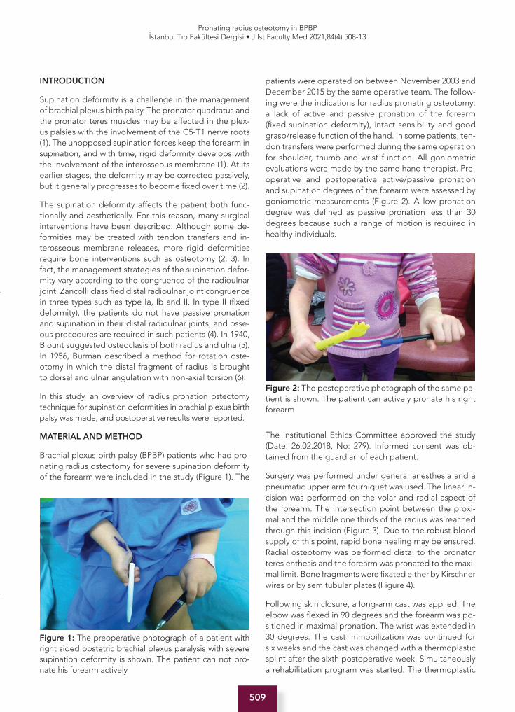
509
Pronating radius osteotomy in BPBPİstanbul Tıp Fakültesi Dergisi • J Ist Faculty Med 2021;84(4):508-13
RESEARCH / ARAŞTIRMADOI: 10.26650/IUITFD.2021.942881
İst Tıp Fak Derg 2021 / J Ist Faculty Med 2021
THE PRONATING RADIUS OSTEOTOMY FOR CORRECTING THE SUPINATION DEFORMITY IN BRACHIAL PLEXUS BIRTH PALSY
DOĞUMSAL BRAKİYAL PLEKSUS PARALİZİSİNDE SUPİNASYON DEFORMİTESİNİ DÜZELTMEK İÇİN RADİUS ROTASYON OSTEOTOMİSİ UYGULAMASI
Hayri Ömer BERKÖZ1 , Bora Edim AKALIN1 , Erol KOZANOĞLU1 , Safiye ÖZKAN1 , Atakan AYDIN1
1Istanbul University, Istanbul Faculty of Medicine, Department of Plastic, Reconstructive and Aesthetic Surgery, Istanbul, Turkey
ORCID IDs of the authors: H.Ö.B. 0000-0001-8063-9995; B.E.A. 0000-0002-5654-2082; E.K. 0000-0003-1192-9520;S.Ö. 0000-0001-8051-1502; A.A. 0000-0002-3568-3513
Cite this article as: Berkoz HO, Akalin BE, Kozanoglu E, Ozkan S, Aydin A. The pronating radius osteotomy for correcting the supination deformity in brachial plexus birth palsy. J Ist Faculty Med 2021;84(4):508-13. doi: 10.26650/IUITFD.2021.942881
INTRODUCTION
Supination deformity is a challenge in the management of brachial plexus birth palsy. The pronator quadratus and the pronator teres muscles may be affected in the plex-us palsies with the involvement of the C5-T1 nerve roots (1). The unopposed supination forces keep the forearm in supination, and with time, rigid deformity develops with the involvement of the interosseous membrane (1). At its earlier stages, the deformity may be corrected passively, but it generally progresses to become fixed over time (2).
The supination deformity affects the patient both func-tionally and aesthetically. For this reason, many surgical interventions have been described. Although some de-formities may be treated with tendon transfers and in-terosseous membrane releases, more rigid deformities require bone interventions such as osteotomy (2, 3). In fact, the management strategies of the supination defor-mity vary according to the congruence of the radioulnar joint. Zancolli classified distal radioulnar joint congruence in three types such as type Ia, Ib and II. In type II (fixed deformity), the patients do not have passive pronation and supination in their distal radioulnar joints, and osse-ous procedures are required in such patients (4). In 1940, Blount suggested osteoclasis of both radius and ulna (5). In 1956, Burman described a method for rotation oste-otomy in which the distal fragment of radius is brought to dorsal and ulnar angulation with non-axial torsion (6).
In this study, an overview of radius pronation osteotomy technique for supination deformities in brachial plexus birth palsy was made, and postoperative results were reported.
MATERIAL AND METHOD
Brachial plexus birth palsy (BPBP) patients who had pro-nating radius osteotomy for severe supination deformity of the forearm were included in the study (Figure 1). The
patients were operated on between November 2003 and December 2015 by the same operative team. The follow-ing were the indications for radius pronating osteotomy: a lack of active and passive pronation of the forearm (fixed supination deformity), intact sensibility and good grasp/release function of the hand. In some patients, ten-don transfers were performed during the same operation for shoulder, thumb and wrist function. All goniometric evaluations were made by the same hand therapist. Pre-operative and postoperative active/passive pronation and supination degrees of the forearm were assessed by goniometric measurements (Figure 2). A low pronation degree was defined as passive pronation less than 30 degrees because such a range of motion is required in healthy individuals.
The Institutional Ethics Committee approved the study (Date: 26.02.2018, No: 279). Informed consent was ob-tained from the guardian of each patient.
Surgery was performed under general anesthesia and a pneumatic upper arm tourniquet was used. The linear in-cision was performed on the volar and radial aspect of the forearm. The intersection point between the proxi-mal and the middle one thirds of the radius was reached through this incision (Figure 3). Due to the robust blood supply of this point, rapid bone healing may be ensured. Radial osteotomy was performed distal to the pronator teres enthesis and the forearm was pronated to the maxi-mal limit. Bone fragments were fixated either by Kirschner wires or by semitubular plates (Figure 4).
Following skin closure, a long-arm cast was applied. The elbow was flexed in 90 degrees and the forearm was po-sitioned in maximal pronation. The wrist was extended in 30 degrees. The cast immobilization was continued for six weeks and the cast was changed with a thermoplastic splint after the sixth postoperative week. Simultaneously a rehabilitation program was started. The thermoplastic
Figure 1: The preoperative photograph of a patient with right sided obstetric brachial plexus paralysis with severe supination deformity is shown. The patient can not pro-nate his forearm actively
Figure 2: The postoperative photograph of the same pa-tient is shown. The patient can actively pronate his right forearm

510
Pronating radius osteotomy in BPBPİstanbul Tıp Fakültesi Dergisi • J Ist Faculty Med 2021;84(4):508-13
splint was used until the direct graphs revealed evidence of bone healing. Kirschner wires were usually removed at the postoperative second month according to X-ray fol-low-ups. Plate and screws were removed after 1-2 years if the patients had complaints such as prominence of the plate under the skin.
RESULTS
Forty one patients had supination deformities caused by BPBP. The mean age was 9.2 years (4-22). Twenty three patients were female whereas 18 patients were male. The mean follow-up was 5 years (1-7). Shoulder, thumb and wrist functions were augmented with tendon transfers in nineteen patients (19/41). There were no patients who underwent tendon transfers for forearm rotation before rotation osteotomy.
The angle 0 was defined as neutral. The negative values stood for supination whereas the positive values stood for pronation (Figure 5). The mean active pronation was - 60° (range - 80° to - 45°) before the operation, and the passive one was - 10° (range - 30° to 5°) (Figure 6). The
Figure 4: The postoperative radiograph of two patients are shown. (a) Kirschner wire (b) Sem-itubular plate
Figure 3: The operative photograph demonstrates our incision of preference (a) and the area of osteotomy on the radius (b)
Figure 5: The angle 0 was defined as neutral. The nega-tive values stood for supination whereas the positive val-ues stood for pronation

511
Pronating radius osteotomy in BPBPİstanbul Tıp Fakültesi Dergisi • J Ist Faculty Med 2021;84(4):508-13
mean postoperative active pronation was 9° (range - 10° to 20°), and the passive one was 45° (range 25° to 60°) (Figure 7). The mean active supination was 75° (range 35° to 85°) before the operation, and the passive one was 85° (range 45° to 90°) (Figure 8). The mean active supination was 45° (range 20° to 60°) after the operation, and the
passive supination was 65° (range 40° to 80°) (Figure 9). On the other hand, three patients had low pronation de-grees (3/41).
One malunion was detected at the second post-opera-tive year (1/41). This patient was treated with open reduc-tion and internal fixation. One patient had a non-union following radius osteotomy due to insufficient plate fixa-tion and the patient was treated by changing the semitu-bular plate with a strong locking compression plate. The osteosynthesis was performed with plates and screws in 30 patients and removal surgery was performed on 12 patients.
DISCUSSION
Tendon transfers, soft tissue releases and corrective oste-otomies may be performed for supination deformities (1-5). The Zancolli procedure is one of the tendon transfers that enhance pronation. After incising the cubital area, the soft tissue contractures are released and the biceps insertion is detached from the ulnar aspect of the radius. The tendon is passed over the radial aspect of the radius to be inserted to the radial side of the radial head (2). This transfer turns the supinatory effect of the biceps muscle to a pronatory one. Although this technique is widely ac-cepted, it was not useful for this study because all the pa-tients had fixed supination deformities refractory to soft tissue reconstruction.
In 2004, Ozkan et al. suggested a new technique of ac-tive pronation maintenance (3). The rigid supination was overcome with interosseous membrane section and the brachioradialis tendon was transferred from the dorsal forearm to the volar side of the distal radius (3). Howev-er, the deformity was too rigid in this study’s population and such soft tissue procedures were not enough to re-store the pronation. Thus, tendon transfers, interosseous membrane releases and tendon lengthening procedures
Figure 6: The mean pronation values (active and passive) are presented for the preoperative period
Figure 7: The mean pronation values (active and passive) are presented for the postoperative period
Figure 8: The mean supination values (active and pas-sive) are presented for the preoperative period
Figure 9: The mean supination values (active and pas-sive) are presented for the postoperative period

512
Pronating radius osteotomy in BPBPİstanbul Tıp Fakültesi Dergisi • J Ist Faculty Med 2021;84(4):508-13
were performed in conjunction with the rotational osteot-omy in order to optimize the results of this study.
The rotational osteotomy is an important part of the recon-structive armamentarium and the benefits of the rotational osteotomies were accepted by the reconstructive commu-nity. Zaoussis performed radius rotation osteotomies on six patients with forearm supination deformities caused by BPBP (7). Zaoussis designed the radius rotation osteoto-mies according to malunion correction principles and his technique was different than Blount osteoclasis technique that preferred a closed approach (5, 7). Zaoussis performed open radius osteotomy distal to the radial tuberosity; how-ever, in this study, osteotomies were performed just distal to the enthesis of pronator teres. Also, some authors com-bined the distal radial osteotomies with proximal humeral osteotomies in severe cases. Goddard et al. performed hu-merus rotational osteotomies in 10 BPBP patients and their findings supported the need for osteotomy procedures in severely disabled patients who were not good candidates for only soft tissue procedures (8).
Metsaars et al. compared the results of osteotomy with biceps rerouting procedure and they evaluated their re-sults according to the severity of the contractures. Both techniques increased the pronation and the gain was pro-portionate to the severity of the deformity. The patients with more severe supination deformities benefited more from the rotational osteotomy. Although the recurrence rate was 20-40% in the osteotomy group and hardware complications were observed by Metsaars et al., such a high recurrence and complication rate were not seen in this study (9).
Bahm et al. operated on 40 patients with severe supina-tion deformities and 23 of their patients were operated on either with Blount osteoclasis or open radial osteot-omy (10). At a mean follow up of four years, their final postoperative pronation was 17 degrees. However, they had five recurrences in the osteotomy group. According to Bahm et al., age at the time of operation was signifi-cant because it affected the compliance of the patients with physiotherapy (10). In 2004, Allende et al. published their results on 66 patients with supination deformities caused by BPBP. Forty four of their patients were operat-ed on with rotation osteotomies whereas 22 of them only had soft tissue procedures. In a mean follow up of 64.3 months, they obtained a postoperative passive pronation of 92 degrees. They had nine recurrences, two delayed unions and one malunion in the osteotomy group (11). In this study, the mean postoperative active pronation was 9 degrees and the mean postoperative passive pronation was 45 degrees. Also, low pronation degrees and recur-rence were seen in three patients and malunion was seen in only one patient. The results of this study were compa-rable with recent literature.
CONCLUSION
Interosseous membrane releases and tendon transfers are preferred treatments in flexible supination deformi-ties. Rotational osteotomies may be preferred in fixed deformities, and the complication rate is relatively low with this technique.
Ethics Committee Approval: This study was approved by the Clinical Research Ethical Committee of the Istanbul University, Istanbul Faculty of Medicine (Date: 26.02.2018, No: 279).
Informed Consent: Written consent was obtained from the par-ticipants.
Peer Review: Externally peer-reviewed.
Author Contributions: Conception/Design of Study- H.Ö.B., B.E.A., E.K., S.Ö., A.A.; Data Acquisition- H.Ö.B., B.E.A., E.K., S.Ö., A.A.; Data Analysis/Interpretation- H.Ö.B., B.E.A., E.K., S.Ö., A.A.; Drafting Manuscript- H.Ö.B., B.E.A., E.K., S.Ö., A.A.; Critical Revision of Manuscript- H.Ö.B., B.E.A., E.K., S.Ö., A.A.; Final Approval and Accountability- H.Ö.B., B.E.A., E.K., S.Ö., A.A.
Conflict of Interest: Authors declared no conflict of interest.
Financial Disclosure: Authors declared no financial support.
Etik Komite Onayı: Bu çalışma için etik komite onayı İstanbul Üniversitesi, İstanbul Tıp Fakültesi Klinik Araştırmalar Etik Kuru-lu’ndan alınmıştır (Tarih: 26.02.2018, No: 279).
Bilgilendirilmiş Onam: Katılımcılardan bilgilendirilmiş onam alınmıştır.
Hakem Değerlendirmesi: Dış bağımsız.
Yazar Katkıları: Çalışma Konsepti/Tasarım- H.Ö.B., B.E.A., E.K., S.Ö., A.A.; Veri Toplama- H.Ö.B., B.E.A., E.K., S.Ö., A.A.; Veri Analizi/Yorumlama- H.Ö.B., B.E.A., E.K., S.Ö., A.A.; Yazı Tasla-ğı- H.Ö.B., B.E.A., E.K., S.Ö., A.A.; İçeriğin Eleştirel İncelemesi- H.Ö.B., B.E.A., E.K., S.Ö., A.A.; Son Onay ve Sorumluluk- H.Ö.B., B.E.A., E.K., S.Ö., A.A.
Çıkar Çatışması: Yazarlar çıkar çatışması beyan etmemişlerdir.
Finansal Destek: Yazarlar finansal destek beyan etmemişlerdir.
REFERENCES
1. Aitken J. Deformity of the elbow joint as a sequel to Erb’s obstetrical paralysis. J Bone Joint Surg Br 1952;34(3):352-65. [CrossRef]
2. Zancolli EA. Paralytic supination contracture of the forearm. J Bone Joint Surg Am 1967;49(7):1275-84. [CrossRef]
3. Ozkan T, Aydin A, Ozer K, Ozturk K, Durmaz H, Ozkan S. A surgical technique for pediatric forearm pronation: brachioradialis rerouting with interosseous membrane release. J Hand Surg Am 2004;29(1):22-7. [CrossRef]

513
Pronating radius osteotomy in BPBPİstanbul Tıp Fakültesi Dergisi • J Ist Faculty Med 2021;84(4):508-13
4. Zancolli EA. Palliative surgery: pronosupination in obstetrical palsy. In: Brachial plexus injuries. Martin Dunitz, London; 2001. p. 275-91.
5. Blount W. Osteoclasis for supination deformities in children. J Bone Joint Surg Am 1940;22(2):300-14.
6. Burman M. Paralytic supination contracture of the forearm. J Bone Joint Surg Am 1956;38(2):303-12. [CrossRef]
7. Zaoussis A. Osteotomy of the proximal end of the radius for paralytic supination deformity in children. J Bone Joint Surg Br 1963;45(3):523-7. [CrossRef]
8. Goddard N, Fixsen J. Rotation osteotomy of the humerus for birth injuries of the brachial plexus. J Bone Joint Surg Br 1984;66(2):257-9. [CrossRef]
9. Metsaars W, Nagels J, Pijls B, Langenhoff J, Nelissen R. Treatment of supination deformity for obstetric brachial plexus injury: a systematic review and meta-analysis. J Hand Surg Am 2014;39(10):1948-58. e2. [CrossRef]
10. Bahm J, Gilbert A. Surgical correction of supination deformity in children with obstetric brachial plexus palsy. J Hand Surg Am 2002;27(1):20-3.
11. Allende CA, Gilbert A. Forearm supination deformity after obstetric paralysis. Clin Orthop Relat Res 2004;426:206-11. [CrossRef]

514
PFN vs DHS for stable intetrochanteric femur fracturesİstanbul Tıp Fakültesi Dergisi • J Ist Faculty Med 2021;84(4):514-20
Corresponding author/İletişim kurulacak yazar: [email protected]
Submitted/Başvuru: 07.07.2021 • Revision Requested/Revizyon Talebi: 31.07.2021 •Last Revision Received/Son Revizyon: 04.08.2021 • Accepted/Kabul: 06.08.2021 • Published Online/Online Yayın: 24.09.2021
RESEARCH - ARAŞTIRMADOI: 10.26650/IUITFD.2021.964078
İst Tıp Fak Derg 2021 / J Ist Faculty Med 2021
PROXIMAL FEMORAL NAILING VERSUS DYNAMIC HIP SCREW IN MANAGEMENT OF STABLE INTERTROCHANTERIC FEMUR FRACTURES: A COMPARISON OF CLINICAL AND RADIOLOGICAL OUTCOMES
STABİL İNTERTROKANTERİK FEMUR KIRIKLARININ TEDAVİSİNDE PROKSİMAL FEMORAL ÇİVİ İLE DİNAMİK KALÇA VİDASI: KLİNİK VE RADYOLOJİK SONUÇLARIN KARŞILAŞTIRILMASI
Tuna PEHLİVANOĞLU1,2 , Serkan BAYRAM3 , Mehmet DEMİREL3 , Mehmet CHODZA3 , Emre KOCAZEYBEK3 , Ahmet SALDUZ3 , Turgut AKGÜL3 , Önder YAZICIOĞLU3
1Emsey Hospital, Department of Orthopaedics and Traumatology, Istanbul, Turkey2Yeni Yüzyıl University, Faculty of Health Sciences, Istanbul, Turkey 3Istanbul University, Istanbul Faculty of Medicine, Department of Orthopaedics and Traumatology, Istanbul, Turkey
ORCID IDs of the authors: T.P. 0000-0001-8886-7538; S.B. 0000-0001-7651-1200; M.D. 0000-0003-1131-7719; M.C. 0000-0002-6996-6597; E.K. 0000-0001-7029-5076; A.S. 0000-0001-9448-6416; T.A. 0000-0002-0704-3797; Ö.Y. 0000-0002-9292-3480
Cite this article as: Pehlivanoglu T, Bayram S, Demirel M, Chodza M, Kocazeybek E, Salduz A, et al. Proximal femoral nailing versus dynamic hip screw in management of stable intertrochanteric femur fractures: a comparison of clinical and radiological outcomes. J Ist Faculty Med 2021;84(4):514-20. doi: 10.26650/IUITFD.2021.964078
Content of this journal is licensed under a Creative Commons Attribution-NonCommercial 4.0 International License.
ABSTRACT
Objective: The aim of this study was to compare the cost-effec-tivity, clinical and radiological outcomes of the proximal femoral nail (PFN) and Dynamic hip screw (DHS) in the management of stable intertrochanteric femur fractures (SIFFs).
Material and Methods: Patients who underwent surgical treatment for a SIFF in our department were retrospectively identified and then divided into two groups according to the treatment modality: Group 1, 57 patients (36 female, 21 male; mean age = 74.5±9.9 years) treated with PFN and Group 2, 65 patients (34 female, 31 male; 72±10.2 years) treated with DHS. Primary outcome measures were: estimated blood loss (EBL), total operating time (TOT), duration of hospital stay (DHS), rate of postoperative complication, rate of mortality, and treatment cost. Radiographic assessment included anteroposterior/lateral tip-apex distance (TAD) and amount of limb length discrepancy (LLD).
Results: No significant differences were observed in demo-graphic characteristics between the two treatment groups (p>0.05). The mean follow-up was 44.2±31 months in group 1 and 53.7±38 months in group 2 (p=0.077). The mean TOT and
ÖZET
Amaç: Bu çalışmanın amacı, stabil intertrokanterik femur kırıkla-rının (SIFF) tedavisinde proksimal femur çivisi (PFN) ve dinamik kalça vidasının (DHS) maliyet-etkililik, klinik ve radyolojik sonuç-larını karşılaştırmaktı.
Gereç ve Yöntemler: SIFF nedeniyle cerrahi tedavi uygulanan hastalar geriye dönük olarak belirlendi ve tedavi şekline göre iki gruba ayrıldı: Grup 1, 57 hasta (36 kadın, 21 erkek; ortalama yaş = 74,5±9,9 yıl) PFN ile tedavi edilen ve Grup 2, DHS ile tedavi edilen 65 hasta (34 kadın, 31 erkek; 72±10,2 yıl). Birincil sonuç ölçütleri; tahmini kan kaybı (EBL), toplam ameliyat süresi (TOT), hastanede kalış süresi (DHS), ameliyat sonrası komplikasyon oranı, ölüm oranı ve tedavi maliyeti olarak yapıldı. Radyografik değerlendirme, ön-arka/yan uç-apeks mesafesini (TAD) ve uzuv uzunluk uyumsuzluğu miktarını (LLD) karşılaştırıldı.
Bulgular: İki tedavi grubu arasında demografik özelliklerde an-lamlı bir farklılık gözlenmedi (p>0,05). Ortalama takip süresi grup 1’de 44,2±31 ay ve grup 2’de 53,7±38 ay idi (p=0,077). Ortalama TOT ve EBL, grup PFN’de grup DHS’ye göre anlamlı olarak daha kısaydı (p<0,001 ve p=0,03). Ortalama hastanede kalış süresi, postoperatif komplikasyon oranı, mortalite oranı ve tedavi mali-
515
PFN vs DHS for stable intetrochanteric femur fracturesİstanbul Tıp Fakültesi Dergisi • J Ist Faculty Med 2021;84(4):514-20
RESEARCH - ARAŞTIRMADOI: 10.26650/IUITFD.2021.964078
İst Tıp Fak Derg 2021 / J Ist Faculty Med 2021
PROXIMAL FEMORAL NAILING VERSUS DYNAMIC HIP SCREW IN MANAGEMENT OF STABLE INTERTROCHANTERIC FEMUR FRACTURES: A COMPARISON OF CLINICAL AND RADIOLOGICAL OUTCOMES
STABİL İNTERTROKANTERİK FEMUR KIRIKLARININ TEDAVİSİNDE PROKSİMAL FEMORAL ÇİVİ İLE DİNAMİK KALÇA VİDASI: KLİNİK VE RADYOLOJİK SONUÇLARIN KARŞILAŞTIRILMASI
Tuna PEHLİVANOĞLU1,2 , Serkan BAYRAM3 , Mehmet DEMİREL3 , Mehmet CHODZA3 , Emre KOCAZEYBEK3 , Ahmet SALDUZ3 , Turgut AKGÜL3 , Önder YAZICIOĞLU3
1Emsey Hospital, Department of Orthopaedics and Traumatology, Istanbul, Turkey2Yeni Yüzyıl University, Faculty of Health Sciences, Istanbul, Turkey 3Istanbul University, Istanbul Faculty of Medicine, Department of Orthopaedics and Traumatology, Istanbul, Turkey
ORCID IDs of the authors: T.P. 0000-0001-8886-7538; S.B. 0000-0001-7651-1200; M.D. 0000-0003-1131-7719; M.C. 0000-0002-6996-6597; E.K. 0000-0001-7029-5076; A.S. 0000-0001-9448-6416; T.A. 0000-0002-0704-3797; Ö.Y. 0000-0002-9292-3480
Cite this article as: Pehlivanoglu T, Bayram S, Demirel M, Chodza M, Kocazeybek E, Salduz A, et al. Proximal femoral nailing versus dynamic hip screw in management of stable intertrochanteric femur fractures: a comparison of clinical and radiological outcomes. J Ist Faculty Med 2021;84(4):514-20. doi: 10.26650/IUITFD.2021.964078
INTRODUCTION
Over the past decades, hip fractures have emerged as a major health problem worldwide confronted by or-thopedic surgeons as a consequence of the enhanced longevity of the population and increased incidence of osteoporosis (1). Intertrochanteric hip fractures account for nearly half the hip fractures in elderly patients and lead to obviously diminished life expectancy as well as dramatic impairment in social, economic, and health cir-cumstances (2).
Over the past 30 years, the dynamic hip screw (DHS) has become the implant of choice for the effective treatment of stable intertrochanteric femoral fractures (IFFs) (Arbe-itsgemeinschaft für Osteosynthesefragen/Orthopaedic Trauma Association (AO/OTA) 31-A1.1–A1.2), with favor-able clinical and radiographic outcomes (3, 4). Nonethe-less, proximal femoral intramedullary nails (PFNs) have been more widely used for such fractures in recent years, despite the lack of strong evidence to support their supe-riority over the DHS (3, 4).
Current literature illustrates the pros and cons of each treatment modality (1-4). However, to the best of our knowledge, evidences from the existing literature to de-termine the more advantageous implant to be used in the treatment for stable IFFs are controversial.
Therefore, the primary aim of this study was to compare the efficacy of DHS versus PFN for the management of stable IFFs based on the several clinical and radiographic outcome measures.
MATERIALS AND METHODS
Data collection and setting of the studyThis retrospective comparative study was conducted on patients who underwent surgical treatment with either PFN or DHS for the treatment of IFFs between 2005 and 2013 at the department of Orthopedics and Traumatolo-
gy in a single tertiary referral center. Inclusion criteria for the study were (4):
1. a diagnosis of unilateral stable IFF (AO/OTA 31– A1),
2. treated with either DHS or PFN3. an age above 50 years old4. a good cognitive function5. adequate medical and radiographic records.
Exclusion criteria were: 1. a diagnosis of a pathological fracture2. history of malignancy-metabolic disease or che
mo-/radiotherapy3. history of polytrauma4. a fracture secondary to high energy trauma (mo
tor vehicle accident, fall from a distant height gun-shot injury)
5. bilateral fractures6. presented with an ASA score IV or higher. 7. Based on the above eligibility criteria, 66 patients
were excluded from the study.
Participants After obtaining institutional review board approval (Date: 04.04.2021, No: 117), a total of 188 patients were assessed retrospectively according to the above eligibility criteria. Af-ter excluding 66 patients, the remaining 122 patients meet-ing the eligibility criteria were enrolled in the study (Figure 1). All the patients include in the study were then divided into two groups according to the implemented treatment mo-dality: Group 1 (PFN) including 57 patients treated with PFN and group 2 (DHS) including 65 patients treated with DHS.
Surgical techniqueAll patients were hospitalized from the emergency room and were managed with low molecular weight heparin and thromboembolic-deterrent stockings for prophylax-is of deep venous thrombosis. Then, all stable IFFs were treated operatively at the earliest opportunity, by three
EBL were significantly shorter in group PFN than in group DHS (p<0.001 and p=0.03). No significant difference was observed in the mean duration of hospital stay, rate of postoperative com-plication, rate of mortality, and treatment cost between the two treatment groups (p>0.05). The post-operative complication rate was 9.5% in group PFN and 8.3% in group DHS, with no sig-nificant difference (p=0.83). There were significant differences in neither TAD nor LLD between the two treatment groups (p=0.69 and p=0.87, respectively).
Conclusion: The two treatment modalities seem to have similar effect to maintain stability for patients with stable IFFs. However, less EBL and shorter operation time can be expected from PFN compared to DHS in such patients.
Keywords: Intertrochanteric fracture, dynamic hip screw, proxi-mal femoral nailing, cost-effectivity
yeti açısından iki grup arasında anlamlı fark gözlenmedi (p>0.05). Ameliyat sonrası komplikasyon oranı grup PFN’de %9,5 ve grup DHS’de %8,3 idi ve anlamlı bir fark yoktu (p=0,83). İki tedavi gru-bu arasında ne TAD ne de LLD’de anlamlı farklılıklar yoktu (sıra-sıyla p=0,69 ve p=0,87).
Sonuç: İki tedavi modalitesi, stabil IFF’leri olan hastalarda stabili-teyi sürdürmek için benzer etkiye sahip görünmektedir. Ancak bu tür hastalarda DHS’ye kıyasla PFN’den daha az EBL ve daha kısa operasyon süresi beklenebilir.
Anahtar Kelimeler: İntertrokanterik kırık, dinamik kalça vidası, proksimal femoral çivileme, maliyet etkinliği

516
PFN vs DHS for stable intetrochanteric femur fracturesİstanbul Tıp Fakültesi Dergisi • J Ist Faculty Med 2021;84(4):514-20
experienced orthopedic trauma surgeons either execut-ing the operation or in attendance.
The type of implant used was decided based on the sur-geon’s preference and experience. After patients were placed supine on an orthopedic traction table, fractures were reduced by traction and internal rotation with the injured limb in a slightly adducted or neutral position to allow an introduction to the greater trochanter. The reduction was then checked, and implants were applied under image intensifier.
In group DHS, a DHS with a four-hole and 135 degrees plate (Dynamic Hip System screw/blade; Synthes GmbH, Basel, Switzerland) was inserted without any additional anti-rotational screw. In group PFN, proximal femoral nail anti-rotation (PFNA) (Synthes GmbH, Oberdorf, Switzer-land) was performed (Figure 2, 3). In all patients, efforts had been paid to ideally place the tip of the screw within the subchondral bone of the femoral head with a com-bined tip-apex distance measuring less than 25 mm on anteroposterior and lateral radiographs.
Postoperative management and follow-up periodPatients were mobilized with partial weight bearing as tol-erated at the second day after surgery, which was increased gradually according to the stability of the fracture and prefer-
ence of the operating surgeon in PFN group. Patients were mobilized at the second day after surgery with no weight bearing until the bony union was confirmed radiographically in DHS group. All patients were recalled for regular follow-up examinations at the second, 6th, 12th, 24th week and once a year. During each follow-up visit, patients’ functional status was assessed. To prevent any bias while comparing the total costs of two groups, implant costs were excluded.
Outcome measures- Clinical assessment The invasiveness of each operation was assessed based on the data including total operating time (TOT), estimat-
Figure 3: Pre- and post-operative AP X-ray of an 81-year-old patient with stable intertrochanteric femur fracture that was treated with DHS
Figure 2: Pre- and post-operative AP X-ray of a 78-year-old patient with stable intertrochanteric femur fracture that was treated with PFN
Figure 1: Flowchart of the study population, together with the inclusion and exclusion criteria

517
PFN vs DHS for stable intetrochanteric femur fracturesİstanbul Tıp Fakültesi Dergisi • J Ist Faculty Med 2021;84(4):514-20
ed blood loss (EBL), duration of hospital stays (DHS), rate of postoperative complications, rate of mortality, and to-tal hospital cost. All the relevant data were documented from our institution medical records including plain radi-ography, operative notes, information on demographic characteristics, discharge reports, and progress notes. EBL was calculated using the Mercuriali’s formula (5). To prevent any bias while comparing the total costs of two groups, implant costs were excluded.
- Radiographic assessment The position of the implants was examined measuring the tip-apex distance (TAD) on latest postoperative fol-low-up radiographs of the patients including anteropos-terior (AP) pelvis and lateral hip views as per the method of Baumgaertner et al. (6). Also, femoral neck-shaft angle was measured on the AP hip radiographs.
Statistical analysisFor the statistical analysis, SPSS software (Version 22.0; SPSS Inc, Chicago, IL, USA) was used. Normality of distri-bution was tested using the Shapiro-Wilk test. Student’s t-test was used to compare quantitative data, normally distributed variables of descriptive statistics (mean, stan-dard deviation, median, frequency, rate, minimum and maximum). Pearson chi-square test, Fisher-Freeman-Hal-ton test, and Fisher’s exact test were used to compare qualitative variables. A p-value less than 0.05 was consid-ered as statistically significant.
RESULTS
Baseline characteristics No significant differences were observed in demograph-ic characteristics between the two treatment groups (p>0.05). Group PFN consisted of 36 females and 21 males, and there were 34 females and 31 males in group DHS (p=0.227). The mean age was 74.7±10 years in group PFN and 72±9.9 years in group DHS (p=0.166). The mean follow-up was 44.2±31 months in group PFN and 53.7±38 months in group DHS (p=0.077) (Table 1).
Clinical outcomes The mean TOT was significantly shorter in group PFN (90.3±19 minutes) than in group DHS (107.1±26 minutes) (p<0.001). The mean EBL was significantly less in group PFN (361±79 ml) than in group DHS (535±90 ml) (p=0.03). No significant difference was observed in the mean du-ration of hospital stay between the two treatment groups (6.62±1.8 days for group PFN vs 7.5±3 days for group DHS [p=0.165] (Table 2).
The post-operative complication rate was 9.5% in group PFN and 8.3% in group DHS, with no significant differ-ence (p=0.83). Two patients developed pulmonary em-bolus, and three patients superficial wound side prob-lems in group PFN. Two patients developed pulmonary
embolism and three patient superficial wound side prob-lem in group DHS. The mortality rate was 73.6% (42 pa-tients) in group PFN and 67.6% (44 patients) (p=0.469).
The mean total hospital cost 1546.86±444.2 USD in group PFN and 1508.59±444.21 USD in group DHS. No signif-icant difference was observed in terms of hospital costs between both groups (p=0.828).
Radiographic outcomes The mean femoral-neck-shaft angle was 132.5º (range= 121º-139º) in group PFN and 133.6º (range=127º-138º) in group DHS (p=0.83). The mean AP/lateral tip apex dis-tance was, respectively, 10.3 mm (range=4.2-13.1) and 9.9 mm (range=6.1-12.7) in group PFN as well as 11.8 mm (range=8.3-15.2) and 10.4 mm (range=6.5-14.2) in group DHS. There were significant differences in neither AP nor lateral tip apex distances between the two treatment groups (p=0.69 and p=0.87, respectively). The mean postoperative LLD was 0.78 mm (range=0-1.4) in group PFN and 0.8 mm (range=0-1.6) in group DHS, with no sig-nificant difference (p=0.74) (Table 2).
DISCUSSION
Over the last two decades, there has been a great con-troversy regarding the optimum treatment of stable IFFs PFN and DHS have been compared each other with re-spect to the clinical outcomes, biomechanical strength, rates of failure, rates of implant-related (blade cut- out, peri-prosthetic fracture) and patient-related (morbidi-ty-mortality) complications, as well as the technical dif-ficulties (7, 8). Current literature illustrates the pros and cons of each treatment modality. However, to the best of our knowledge, evidences from the existing literature to determine the more advantageous implant to be used in the treatment for stable IFFs are controversial. Data ob-tained from the current study have found no significant differences in clinical and radiological outcomes except EBL and TOT which were higher in group DHS.
According to a large Cochrane review, DHS was regarded as the gold standard for the treatment of stable IFFs be-cause of the lower rates of complications and reoperations compared to PFN (8). These results were also supported by many other studies (8-10, 11). In the present study, no statistically significant difference was found with regard to complication rates between the two groups (p=0.83).
Many studies underlined that there was no significant differ-ence between PFN and DHS with regard to bleeding which was another important intra- and post-operative concern (7, 10). EBL was found higher in group DHS compared to PFN detected with a low statistical significance (p=0.048). We believe that this small difference may have possibly oc-curred because of the technical principles of conventional DHS application. As shown in the literature, performing the
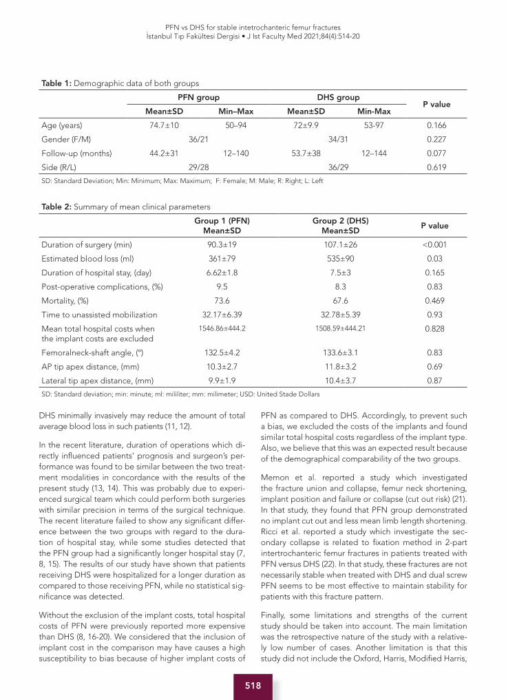
518
PFN vs DHS for stable intetrochanteric femur fracturesİstanbul Tıp Fakültesi Dergisi • J Ist Faculty Med 2021;84(4):514-20
DHS minimally invasively may reduce the amount of total average blood loss in such patients (11, 12).
In the recent literature, duration of operations which di-rectly influenced patients’ prognosis and surgeon’s per-formance was found to be similar between the two treat-ment modalities in concordance with the results of the present study (13, 14). This was probably due to experi-enced surgical team which could perform both surgeries with similar precision in terms of the surgical technique. The recent literature failed to show any significant differ-ence between the two groups with regard to the dura-tion of hospital stay, while some studies detected that the PFN group had a significantly longer hospital stay (7, 8, 15). The results of our study have shown that patients receiving DHS were hospitalized for a longer duration as compared to those receiving PFN, while no statistical sig-nificance was detected.
Without the exclusion of the implant costs, total hospital costs of PFN were previously reported more expensive than DHS (8, 16-20). We considered that the inclusion of implant cost in the comparison may have causes a high susceptibility to bias because of higher implant costs of
PFN as compared to DHS. Accordingly, to prevent such a bias, we excluded the costs of the implants and found similar total hospital costs regardless of the implant type. Also, we believe that this was an expected result because of the demographical comparability of the two groups.
Memon et al. reported a study which investigated the fracture union and collapse, femur neck shortening, implant position and failure or collapse (cut out risk) (21). In that study, they found that PFN group demonstrated no implant cut out and less mean limb length shortening. Ricci et al. reported a study which investigate the sec-ondary collapse is related to fixation method in 2-part intertrochanteric femur fractures in patients treated with PFN versus DHS (22). In that study, these fractures are not necessarily stable when treated with DHS and dual screw PFN seems to be most effective to maintain stability for patients with this fracture pattern.
Finally, some limitations and strengths of the current study should be taken into account. The main limitation was the retrospective nature of the study with a relative-ly low number of cases. Another limitation is that this study did not include the Oxford, Harris, Modified Harris,
Table 1: Demographic data of both groups
PFN group DHS groupP value
Mean±SD Min–Max Mean±SD Min-Max
Age (years) 74.7±10 50–94 72±9.9 53-97 0.166
Gender (F/M) 36/21 34/31 0.227
Follow-up (months) 44.2±31 12–140 53.7±38 12–144 0.077
Side (R/L) 29/28 36/29 0.619
SD: Standard Deviation; Min: Minimum; Max: Maximum; F: Female; M: Male; R: Right; L: Left
Table 2: Summary of mean clinical parameters
Group 1 (PFN)Mean±SD
Group 2 (DHS)Mean±SD
P value
Duration of surgery (min) 90.3±19 107.1±26 <0.001
Estimated blood loss (ml) 361±79 535±90 0.03
Duration of hospital stay, (day) 6.62±1.8 7.5±3 0.165
Post-operative complications, (%) 9.5 8.3 0.83
Mortality, (%) 73.6 67.6 0.469
Time to unassisted mobilization 32.17±6.39 32.78±5.39 0.93
Mean total hospital costs when the implant costs are excluded
1546.86±444.2 1508.59±444.21 0.828
Femoralneck-shaft angle, (º) 132.5±4.2 133.6±3.1 0.83
AP tip apex distance, (mm) 10.3±2.7 11.8±3.2 0.69
Lateral tip apex distance, (mm) 9.9±1.9 10.4±3.7 0.87
SD: Standard deviation; min: minute; ml: mililiter; mm: milimeter; USD: United Stade Dollars

519
PFN vs DHS for stable intetrochanteric femur fracturesİstanbul Tıp Fakültesi Dergisi • J Ist Faculty Med 2021;84(4):514-20
HOOS, WOMAC scores to perform a functional evalua-tion. Bone quality of patients were not investigated which effect the bone union and radiological parameters. Last-ly, other factors affecting mortality, such as chronic ste-roid use, sarcopenia, and cancer were not investigated. The main strength of this study was to provide a solution for the controversy in the literature in terms of the ideal management strategy of sIFFs by underlining that DHS was a more cost-effective method of treatment. However, it should also be highlighted that unstable IFFs were di-agnosed more frequently in older patients as compared to stable IFFs, while intramedullary nailing was consid-ered as the gold standard for the management of unsta-ble IFFs for its biomechanical properties (17-20). To con-fer a better understanding about the optimal treatment of stable IFFs, large prospective randomized studies are needed.
The present study concluded that, for stable IFFs, fixation with dynamic hip screw versus proximal femoral nail had no significant difference with regard to duration of hospi-tal stays, rates of post-operative complications and mor-tality and time until unassisted mobilization, while both groups were noted to have similar radiographic results. Within 40 years following surgery, the two treatment modalities have similar effective to maintain stability for patients with this fracture pattern and complication rate (19-22). However, PFN have lower duration of operation and blood loss than DHS.
Informed Consent: Written consent was obtained from the par-ticipants.
Ethics Committee Approval: This study was approved by the Ethical Committee of the Istanbul University Istanbul Faculty of Medicine (Date: 04.04.2021, No: 117).
Peer Review: Externally peer-reviewed.
Author Contributions: Conception/Design of Study- T.P., S.B., M.C., M.D.; Data Acquisition- S.B., M.C., E.K.; Data Analysis/Interpretation- T.A., A.S., S.B., M.D.; Drafting Manuscript- T.P., S.B., M.D., T.A.; Critical Revision of Manuscript- Ö.Y., T.A., A.S.; Final Approval and Accountability- T.P., S.B., M.D., M.C., E.K., A.S., T.A., Ö.Y.
Conflict of Interest: Authors declared no conflict of interest.
Financial Disclosure: Authors declared no financial support.
Bilgilendirilmiş Onam: Katılımcılardan bilgilendirilmiş onam alınmıştır.
Etik Komite Onayı: Bu çalışma için etik komite onayı İstanbul Üniversitesi, İstanbul Tıp Fakültesi Etik Kurulu’ndan alınmıştır (Ta-rih: 04.04.2021, No: 117).
Hakem Değerlendirmesi: Dış bağımsız.
Yazar Katkıları: Çalışma Konsepti/Tasarım- T.P., S.B., M.C., M.D.; Veri Toplama- S.B., M.C., E.K.; Veri Analizi/Yorumlama- T.A., A.S., S.B., M.D.; Yazı Taslağı- T.P., S.B., M.D., T.A.; İçeriğin Eleştirel İncelemesi- Ö.Y., T.A., A.S.; Son Onay ve Sorumluluk- T.P., S.B., M.D., M.C., E.K., A.S., T.A., Ö.Y.
Çıkar Çatışması: Yazarlar çıkar çatışması beyan etmemişlerdir.
Finansal Destek: Yazarlar finansal destek beyan etmemişlerdir.
REFERENCES
1. Veronese N, Maggi S. Epidemiology and social costs of hip fracture. Injury 2018;49(8):1458-60. [CrossRef]
2. Mundi S, Pindiprolu B, Simunovic N, Bhandari M. Similar mortality rates in hip fracture patients over the past 31 years: A systematic review of RCTs. Acta Orthop 2014;85(1):54-9. [CrossRef]
3. Marsh JL, Slongo TF, Agel J, Broderick JS, Creevey W, DeCoster TA, et al. Fracture and dislocation classification compendium - 2007: Orthopaedic Trauma Association classification, database and outcomes committee. J Orthop Trauma 2007;21(10 Suppl):S1-133. [CrossRef]
4. Barton TM, Gleeson R, Topliss C, Greenwood R, Harries WJ, Chesser TJ. A comparison of the long gamma nail with the sliding hip screw for the treatment of AO/OTA 31-A2 fractures of the proximal part of the femur: a prospective randomized trial. J Bone Joint Surg Am 2010;92(4):792-8. [CrossRef]
5. Irisson E, Hémon Y, Pauly V, Parratte S, Argenson JN, Kerbaul F. Tranexamic acid reduces blood loss and financial cost in primary total hip and knee replacement surgery. Orthop Traumatol Surg Res 2012;98(5):477-83. [CrossRef]
6. Baumgaertner MR CS, Lindskog DM, Keggi JM. The value of the tip apex distance in predicting failure of fixation of peritrochanteric fractures of the hip. J Bone Joint Surg (Am) 1995;77:1058–64. [CrossRef]
7. Matre K, Vinje T, Havelin LI, Gjertsen JE, Furnes O, Espehaug B, et al. TRIGEN INTERTAN intramedullary nail versus sliding hip screw: a prospective, randomized multicenter study on pain, function, and complications in 684 patients with an intertrochanteric or subtrochanteric fracture and one year of follow-up. J Bone Joint Surg Am 2013;95(3):200-8. [CrossRef]
8. Parker MJ, Handoll HH. Gamma and other cephalocondylic intramedullary nails versus extramedullary implants for extracapsular hip fractures in adults. Cochrane Database Syst Rev 2008;(3):CD000093. [CrossRef]
9. Aune AK, Ekeland A, Odegaard B, Grøgaard B, Alho A. Gamma nail vs compression screw for trochanteric femoral fractures. 15 reoperations in a prospective, randomized study of 378 patients. Acta orthopaedica Scandinavica 1994;65(2):127-30. [CrossRef]
10. Hardy DC, Descamps PY, Krallis P, Fabeck L, Smets P, Bertens CL, et al. Use of an intramedullary hip-screw compared with a compression hip-screw with a plate for intertrochanteric femoral fractures. A prospective, randomized study of one hundred patients. J Bone Joint Surg Am 1998;80(5):618-30. [CrossRef]

520
PFN vs DHS for stable intetrochanteric femur fracturesİstanbul Tıp Fakültesi Dergisi • J Ist Faculty Med 2021;84(4):514-20
11. Herrera A, Domingo LJ, Calvo A, Martínez A, Cuenca J. A comparative study of trochanteric fractures treated with the Gamma nail or the proximal femoral nail. Int Orthop 2002;26(6):365-9. [CrossRef]
12. Abreu ELd, Sena CB, Rodrigues Filho SAS. Effectiveness of treatment of transtrochanteric fractures with Dynamic Hip Screws using minimally invasive access. Rev Bras Ortop 2016;51(2):138-42. [CrossRef]
13. Mahmood A, Kalra M, Patralekh M. Comparison between conventional and minimally invasive dynamic hip screws for fixation of intertrochanteric fractures of the femur. ISRN Orthop 2013;2013:484289 [CrossRef]
14. Saudan M, Lübbeke A, Sadowski C, Riand N, Stern R, Hoffmeyer P. Pertrochanteric fractures: is there an advantage to an intramedullary nail?: a randomized, prospective study of 206 patients comparing the dynamic hip screw and proximal femoral nail. J Orthop Trauma 2002;16(6):386-93. [CrossRef]
15. Aros B, Tosteson AN, Gottlieb DJ, Koval KJ. Is a sliding hip screw or im nail the preferred implant for intertrochanteric fracture fixation? Clin Orthop Relat Res 2008;466(11):2827-32. [CrossRef]
16. Aktselis I, Kokoroghiannis C, Fragkomichalos E, Koundis G, Deligeorgis A, et al. Prospective randomised controlled trial of an intramedullary nail versus a sliding hip screw for intertrochanteric fractures of the femur. Int Orthop 2014;38(1):155-61. [CrossRef]
17. Kumar R, Singh R, Singh B. Comparative prospective study of proximal femoral nail and dynamic hip screw in treatment of intertrochanteric fracture femur. J Clin Orthop Trauma. 2012;3(1):28-36. [CrossRef]
18. Taeger G, Schmid C, Zettl R, Schweiberer L, Nast-Kolb D. Stable and unstable pertrochanteric femoral fractures. Differentiated indications for the dynamic hip screw. Unfallchirurg 2000;103(9):741-8. [CrossRef]
19. Lavini F, Renzi-Brivio L, Aulisa R, Cherubino F, Di Seglio PL, Galante N, et al. The treatment of stable and unstable proximal femoral fractures with a new trochanteric nail: results of a multicentre study with the Veronail. Strategies Trauma Limb Reconstr 2008;3(1):15-22. [CrossRef]
20. Z Zhang S, Zhang K, Jia Y, Yu B, Feng W. InterTan nail versus Proximal Femoral Nail Antirotation-Asia in the treatment of unstable trochanteric fractures. Orthopedics 2013;36(3):182-e292. [CrossRef]
21. Memon K, Siddiqui AM, Khan ZA, Zahoor A. Dynamic hip screw fixation vs. proximal femur nail for unstable per-trochanteric fractures: a comparative analysis of outcomes and complications. J Ayub Med Coll Abbottabad. 2021;33(1):34-38.
22. Ricci MJ, McAndrew CM, Miller AN, Kamath G, Ricci WM. Are two-part intertrochanteric femur fractures stable and does stability depend on fixation method? J Orthop Trauma 2019;33(9):428-431. [CrossRef]

521
Neuroendocrine carcinoma of the breastİstanbul Tıp Fakültesi Dergisi • J Ist Faculty Med 2021;84(4):521-5
Corresponding author/İletişim kurulacak yazar: [email protected]
Submitted/Başvuru: 24.11.2020 • Revision Requested/Revizyon Talebi: 30.03.2021 •Last Revision Received/Son Revizyon: 06.04.2021 • Accepted/Kabul: 06.08.2021 • Published Online/Online Yayın: 17.09.2021
RESEARCH - ARAŞTIRMADOI: 10.26650/IUITFD.2021.830495
İst Tıp Fak Derg 2021 / J Ist Faculty Med 2021
NEUROENDOCRINE CARCINOMA OF THE BREAST: 18 CASES WITH LONG-TERM FOLLOW-UP
MEMENİN NÖROENDOKRİN KARSİNOMU: 18 HASTA VE UZUN DÖNEM SONUÇLARI
Enver ÖZKURT1 , Mehmet İLHAN2 , Ömer Cenk CÜCÜK3 , Mustafa TÜKENMEZ2 , Neslihan CABİOĞLU2 , Mahmut MÜSLÜMANOĞLU2
1Doğa Hospital, Department of General Surgery, Istanbul, Turkey2Istanbul University, Istanbul Faculty of Medicine, Department of General Surgery, Istanbul, Turkey3Erciyes University, Faculty of Medicine, Department of Surgery, Surgical Oncology, Kayseri, Turkey
ORCID IDs of the authors: E.Ö. 0000-0003-2597-3119; M.İ. 0000-0002-5473-3483; Ö.C.C. 0000-0002-8274-5940; M.T. 0000-0002-9403-568X; N.Ç. 0000-0002-0989-7411; M.M. 0000-0003-0279-5671
Cite this article as: Ozkurt E, Ilhan M, Cucuk OC, Tukenmez M, Cabioglu N, Muslumanoglu M. Neuroendocrine carcinoma of the breast: 18 cases with long-term follow-up. J Ist Faculty Med 2021;84(4):521-5. doi: 10.26650/IUITFD.2021.830495
Content of this journal is licensed under a Creative Commons Attribution-NonCommercial 4.0 International License.
ABSTRACT
Objective: Primary neuroendocrine carcinoma of the breast (NECB) is a rare distinct type of breast carcinoma. There is limited data about the optimal management, treatment, and prognosis. Therefore, we analyzed the clinicopathological features, manage-ment and the clinical outcome of this rare breast carcinoma.
Material and Methods: Patients diagnosed as NECB between July 2008 and January 2018 were included in the study. Medical records were retrospectively reviewed.
Results: A total of 4,896 breast cancer patients were reviewed and 18 NECB (0.4% of all cases) were extracted. The median age was 61.5 (30-82). Thirteen cases (72.2%) underwent breast conserving surgery. Eight patients had axillary lymph dissection. All of the cases were pathological T1 and T2. Only one patient was pathological stage 3. Median tumor size was 20.5mm (10-45). Only two cases presented with small cell subtype, the rest were well-differentiated. Hormone receptor was positive and HER2/neu was negative for all cases. Of the 15 patients with known Ki-67, three had high expressions (≥20%). No local or distant disease recurrences and death related with NECB were detected at a median follow-up period of 101 months (33-148).
Conclusion: NECB is more likely to be hormone receptor positive and HER2/neu negative as luminal A or B subtype. An excellent clinical outcome is remarkable despite a substantial number of pa-tients with axillary lymph node positivity specifically for well-differ-entiated subtype. Less invasive treatment options should be kept in mind.
Keywords: Neuroendocrine carcinoma, breast neoplasm, prognosis
ÖZET
Amaç: Nöroendokrin meme karsinomu (NMK) nadir görülen ve özellikli bir meme tümörüdür. Bu alt tipin tedavisi ve prognozu ile ilgili bilgiler sınırlıdır. Çalışmamızda, bu nadir görülen tümö-rün klinikopatolojik özelliklerini, tedavisini ve klinik sonuçlarını inceledik.
Gereç ve Yöntemler: Temmuz 2008 ve Ocak 2018 tarihleri ara-sında NMK tanısı alan hastaların verileri retrospektif olarak ince-lendi.
Bulgular: Toplam kayıtlı 4896 meme kanseri hastasının 18’i NMK idi (toplam vakaların %0,4’ü). Ortanca yaş 61,5 (30-82) olarak bu-lundu. On üç hastaya (%72,2) meme koruyucu cerrahi uygulandı. Sekiz hastaya ise aksiller lenf nodu diseksiyonu yapıldı. Sadece bir hasta patolojik evre 3 iken tüm hastalar patolojik olarak T1 ve T2 idi. Ortanca tümör boyutu 20,5 mm (10-45) olarak bulundu. Sadece iki hasta küçük hücreli alt tipi iken 16 hasta iyi-diferansiye alt tipindeydi. Tüm hastalar hormon reseptör pozitif ve HER2/neu negatifti. Ki-67 değeri bilinen 15 hastadan 3 tanesinde yük-sek Ki-67 değeri (>%20) mevcuttu. Ortanca 101 (33-148) aylık ta-kip süresinde lokal-bölgesel veya uzak rekürrens görülmedi. On sekiz hastada hastalığa bağlı ölüm görülmedi.
Sonuç: NMK genellikle hormon reseptör pozitif ve HER2/neu negatif olacak şekilde luminal A veya B olarak tespit edilmek-tedir. Aksilla pozitif hastalar olsa da özellikle iyi-diferansiye alt grupta sağ kalımlar çok iyidir. Bu hastalarda daha az girişimsel tedavi seçenekleri göz önünde bulundurulmalıdır.
Anahtar Kelimeler: Nöroendokrinkarsinom, meme neoplazmı, prognoz

522
Neuroendocrine carcinoma of the breastİstanbul Tıp Fakültesi Dergisi • J Ist Faculty Med 2021;84(4):521-5
INTRODUCTION
Primary neuroendocrine carcinomas of the breast (NECB) are rare neoplasms of the breast with similar morpholog-ical features to neuroendocrine tumors of the gastroin-testinal tract and lung (1). The World Health Organization (WHO) described neuroendocrine marker expression in at least 50% of the total cell population of NECB. Ex-pression of neuroendocrine markers, specifically synap-tophysin and chromogranin A are generally present (2). According to the WHO classification, NECB has three subtypes based on the morphology as well-differentiat-ed neuroendocrine tumors, small cell neuroendocrine carcinomas, and invasive carcinomas of the breast with neuroendocrine differentiation (3).
NECB is the disease of the sixth and seventh decade of females who are generally postmenopausal (4). Although the prevalence was reported between 2 and 5% in the WHO 2003 classification, recent studies demonstrated that it is between 0.1 and 0.5% (4-6).
NECB is associated with a more aggressive behavior, higher propensity for local recurrence, and poorer prog-nosis than ductal carcinoma (7). The optimal treatment for primary NECB is poorly known because of limited re-ports. These tumors can be misdiagnosed due to the lack of distinguishing features on presentation and imaging. It is important to recognize these tumors and distinguish them from other poorly differentiated tumors of the breast and metastatic small cell carcinomas from the lung in order to avoid diagnostic errors that could have ther-apeutic and prognostic implications for these patients. In this study, we demonstrated the clinicopathological characteristics, management and clinical outcomes of this rare breast carcinoma.
MATERIALS AND METHODS
Patients with a diagnosis of NECB between July 2008 and January 2018 were retrospectively evaluated. De-mographic features, clinicopathological factors, treat-ment modalities, and outcomes of patients were record-ed. Each patient was carefully managed in order to rule out breast metastasis of neuroendocrine carcinomas from other organs such as the gastrointestinal tract or lung. All patients were managed as breast carcinoma not otherwise specified (NOS) in concordance with interna-tional guidelines.
Up-to-date WHO criteria for NECB definitions were used. Tumors were stained by immunohistochemistry for neuro-endocrine markers, synaptophysin and/or chromogranin-A along with estrogen receptor (ER) and progesterone receptor (PR), HER2/neu expression, and Ki-67 levels. Adjuvant treat-ment (chemotherapy, radiotherapy and endocrine therapy) was decided as in invasive ductal carcinoma of the breast.
Categorical and continuous variables were summarized using descriptive statistics (e.g. median, range, frequen-cy, and percentage). Kaplan-Meier method was used for survival analysis. All statistical analyses were performed using the SPSS program (Version 22.0, SPSS Inc., Chica-go, IL, USA).
RESULTS
Between July 2008 and January 2018, 4,896 breast cancer patients were treated in our clinic and 18 cases were iden-tified that fulfilled the 2003 WHO criteria for NECB. The incidence of NECB was 0.4% and all cases were female. The median age was 61.5 (30-82). All cases underwent radiologic evaluation by mammogram, ultrasound, and magnetic resonance imaging (MRI) if needed. If axillary lymph nodes were clinically positive, positron emission tomography and computed tomography (PET/CT) were also performed.
Most of the cases (75%) underwent breast conserving surgery. Axillary lymph node dissection (ALND) was per-formed for eight patients. All patients had sentinel lymph node biopsy, except one patient that had clinically stage II nodal disease. She underwent direct ALND. The medi-an tumor size was 20.5 mm (10-45). Pathological stage II nodal disease was also present in one patient who was mentioned before. Of the 18 cases, 16 were classified as well-differentiated and two were small cell neuroendo-crine carcinoma according to the WHO criteria. All cas-es were hormone receptor positive and HER2/neu neg-ative. Only two patients that demonstrated with small cell neuroendocrine carcinoma had higher levels of Ki-67 (60% and 80%). Synaptophysin and chromogranin-A ex-pression were present in all cases. Details of clinical and pathologic features are presented in Table 1.
The median follow-up time was 101 months (33-148). Ad-juvant treatment was administered as for the same prin-ciples for invasive ductal carcinoma of the breast. Seven-teen patients had endocrine therapy with tamoxifen in premenopausal and anastrazole in postmenopausal pa-tients. The only patient who did not received endocrine therapy was an 82-year old patient with co-morbidities. There was no mortality associated with NECB. One pa-tient died of cardiac events. No local recurrences or dis-tant metastases were detected (Table 2).
DISCUSSION
Primary NECB is a rare tumor. On specimens, if more than 50% of the cells express any of neuroendocrine markers (chromogranin-A, chromogranin-B, neuron specific eno-lase, and synaptophysin), it can be described as prima-ry NECB (1). This criteria distinguishes NECB from other breast carcinomas that show only neuroendocrine mor-phological features or focal (i.e.,<50%) neuroendocrine

523
Neuroendocrine carcinoma of the breastİstanbul Tıp Fakültesi Dergisi • J Ist Faculty Med 2021;84(4):521-5
differentiation. The rate of NECB was 0.4% in our series, that is similar to previously published series (0.1-0.5%) (4-6).
NECB is detected as especially round spiculated masses on mammogram (5). They generally present homoge-neous echogenicity with some posterior acoustic en-hancement on sonography. It morphologically seems like triple negative breast tumors. However, these findings are not specific for NECB. As diagnosis and dif-ferential diagnosis is made by pathologic evaluation with neuroendocrine markers, metastasis from a pri-mary neuroendocrine tumor other than breast should be excluded, and a core biopsy is recommended (8). Almost two thirds of NECB are associated with ductal carcinoma in-situ that distinguishes these masses from metastases (9). We performed core needle biopsy for all cases. On advanced stages, PET/CT can be used for sys-temic evaluation. We performed PET/CT for five clinical axilla positive patients and there were no distant metas-tasis. The surgical treatment strategy is generally simi-lar with the management of ductal carcinoma. As many
Table 1: Demographic and pathologic features of cases (n=18)
Factors Category n %
Median age 61.5 (30-82)
<60 8 44.4
≥60 10 55.6
AJCC cT stage I 7 38.9
II 10 55.6
III 1 5.6
AJCC cN stage 0 13 72.2
I 4 22.2
II 1 5.6
Operation type BCS 13 72.2
Mastectomy 5 27.8
Axillary surgery SLNB 10 55.6
ALND 1 5.6
SLNB + ALND 7 38.9
Median tumor diameter (mm)
20.5 (10-45)
AJCC pT stage I 9 50
II 9 50
AJCC pN stage 0 11 61.1
I 6 33.3
II 1 5.6
AJCC p stage I-A 6 33.3
II-A 8 44.4
II-B 3 16.7
III-A 1 5.6
ER status Positive 18 100
Negative 0 0
PR status Positive 17 94.4
Negative 1 5.6
HER2/neu status Positive 0 0
Negative 18 100
Ki-67 (n=15) Positive (≥20%) 3 20
Negative (<20%) 12 80
Factors Category n %
MBR grade I 0 0
II 15 83.3
III 3 16.7
Table 1: Continue
Factors Category n %
WHO classification Well-differen-tiated 16 88.9
Small cell 2 11.1
Adjuvant treatment
Chemotherapy
Yes 9 50
No 9 50
Radiotherapy
Yes 11 61.1
No 7 38.9
Endocrine therapy
Yes 17 94.4
No 1 5.6
AJCC: American Joint Committee on Cancer, BCS: Breast con-serving surgery, SLNB: Sentinel lymph node biopsy,ALND: Axillary lymph node dissection, ER: Estrogen receptor, PR: Progesterone receptor, HER2/neu: Human epidermal growth factor receptor-2, MBR: Modified Bloom-Richardson, WHO: World Health Organization
Table 2: 5-year and 10-year survival features of the patients
Median follow-up (month) 101 (33-148)
5-year disease free survival 100%
5-year overall survival 94.1%
10-year disease free survival 100%
10-year overall survival 94.1%

524
Neuroendocrine carcinoma of the breastİstanbul Tıp Fakültesi Dergisi • J Ist Faculty Med 2021;84(4):521-5
patients are early stage, specifically T1 and T2 cases, we performed breast conserving surgery for 13 (72%) pa-tients. Although only seven patients had pathological axillary lymph node positivity, we performed ALND on eight patients. One patient whose final pathologic nodal evaluation was negative reported as positive in intra-operative pathologic evaluation of the sentinel lymph node. So that ALND was decided upon.
The WHO has classified NECB into three groups as well-differentiated neuroendocrine tumors, small cell neuroendocrine carcinomas, and invasive carcinomas of the breast with neuroendocrine differentiation (3). Six-teen patients in our cohort were well-differentiated and two were small cell. Most of the primary NECB express ER up to 90-100% and PR up to 80-90% (7). This level of hormone receptor positivity is significant for survival. HER2/neu overexpression in NECB is very rare so that most studies in literature classified NECB as luminal A or sometimes luminal B type (10-12). In large series by Bo-gine et al. and Lavigne et al. they reported that almost 50% of cases are luminal A and 50% are luminal B (11, 12). In our series, only three cases of the 15 with a known Ki-67 status were luminal B (high Ki-67 ≥20%). All the cases were hormone receptor positive and there were no HER2/neu positivity.
As there is a lack of information due to the low inci-dence of NECB cases, chemotherapy regimens are not standardized. Nevertheless, general recommendation is treating it similarly to the treatment standards for duc-tal neoplasms (13). If chemotherapy is administered, the first line treatment choice is anthracycline and taxane based regimens (8). Almost all of the cases reported in the literature are luminal type so the adjuvant endocrine therapy is also a standard of care for most of the cas-es (13). All patients, except an 82-year old female with co-morbidities, received endocrine therapy in our series. Radiotherapy also must be taken into consideration for patients with breast conserving surgery and for advanced stage patients in the light of international guidelines. We administered adjuvant radiotherapy to 11 of our patients.
There are different survival reports for the outcome and prognosis of NECB patients. Some of them demonstrate worse survival and few of them demonstrate better sur-vival. Most of the studies provide worse survival rates when compared to invasive ductal carcinomas of the same stage. Wang J et al. reviewed the surveillance, ep-idemiology, and end results (SEER) database for NECB. They indicated that the overall survival and disease spe-cific survival were significantly worse in NECB (n=142) compared with invasive mammary carcinoma, not other-wise specified at the same stage (4). Another study by Zhang et al. also presented that NECB have a higher rate of local recurrence and lower rate of overall survival (14).
On the other hand, recent studies express that poorer local control and survival outcomes are associated with small cell subtype of the NECB, not for the well-differ-entiated subtype (12, 15). These results are more reason-able as we know that morphology is the key for survival of the patients. In our series, we reported excellent survival outcomes with a median follow-up time of 101 months. There was only one death and it was associated with a cardiovascular event. The 5-year and 10-year overall sur-vival rates were both 94.1%. There was no locoregional or distant recurrence during the follow-up time (Disease free survival and disease specific survival were 100%). This is probably because 16 of the 18 patients in our cohort were well-differentiated subtype. Even so, two patients with small cell subtype had 99 (AJCC stage 2A) and 62 (AJCC stage 3A) months of follow-up time with no lo-coregional or distant recurrences. In the light of the re-sults of these recent studies and our study, the need for chemotherapy for well-differentiated NECB should be questioned. Additionally, we can even consider omitting surgical axillary lymph node staging for patients with ear-ly stage and favorable tumor biology as demonstrated by Özkurt et al. for tubular breast cancers (16).
However, we should always keep in mind that small cell subtype is aggressive and can metastasize even after several years from initial diagnosis so that long-term close follow-up is recommended.
In conclusion, NECB is a different subtype and rare vari-ant of breast carcinoma. Our results suggest that NECB is more likely to be ER/PR positive and HER2/neu negative as luminal A or B subtype. An excellent clinical outcome is remarkable despite a substantial number of patients with axillary lymph node positivity specifically for well-dif-ferentiated subtype. This favorable prognosis might be due to good tumor biology profile associated with hor-mone receptor positivity with a substantial benefit from hormone therapy and other adjuvant therapies. Finally, we must be aware of the small cell subtype of NECB and follow-up closely for a long time as it can present with advanced stage disease and distant metastasis.
Acknowledgements: This manuscript was checked for compli-ance with English grammar by Dr. Sami Pinarbasi, who is a native English speaker from the United Kingdom and working as Asso-ciate Lecturer in the Department of History, Politics and Philo-sophy, Manchester Metropolitan University.
Ethics Committee Approval: Ethics committee approval was not received due to the retrospective nature of the study.
Peer Review: Externally peer-reviewed.
Author Contributions: Conception/Design of Study- E.Ö., M.T., N.C., M.M.; Data Acquisition- E.Ö., M.İ., Ö.C.C.; Data Analysis/Interpretation- E.Ö.; Drafting Manuscript- E.Ö., M.İ., Ö.C.C.;

525
Neuroendocrine carcinoma of the breastİstanbul Tıp Fakültesi Dergisi • J Ist Faculty Med 2021;84(4):521-5
Critical Revision of Manuscript- M.M., M.T., N.C.; Final Approval and Accountability- E.Ö., M.İ., Ö.C.C., M.T., N.C., M.M.
Conflict of Interest: Authors declared no conflict of interest.
Financial Disclosure: Authors declared no financial support.
Teşekkür: Yazının İngilizce gramer denetimi anadili İngilizce olan, Manchester Metropolitan Üniversitesi Tarih, Siyaset ve Fel-sefe Bölümü Öğretim Üyesi Dr. Enver Sami Pınarbaşı tarafından yapılmıştır.
Etik Komite Onayı: Retrospektif çalışma olduğundan etik komi-te onayı alınmamıştır
Hakem Değerlendirmesi: Dış bağımsız.
Yazar Katkıları: Çalışma Konsepti/Tasarım- E.Ö., M.T., N.C., M.M.; Veri Toplama- E.Ö., M.İ., Ö.C.C.; Veri Analizi/Yorumlama- E.Ö.; Yazı Taslağı- E.Ö., M.İ., Ö.C.C.; İçeriğin Eleştirel İnceleme-si- M.M., M.T., N.C.; Son Onay ve Sorumluluk- E.Ö., M.İ., Ö.C.C., M.T., N.C., M.M.
Çıkar Çatışması: Yazarlar çıkar çatışması beyan etmemişlerdir.
Finansal Destek: Yazarlar finansal destek beyan etmemişlerdir.
REFERENCES
1. Sapino A, Bussolati G. Is detection of endocrine cells in breast adenocarcinoma of diagnostic and clinical significance? Histopathology 2002;40(3):211-4. [CrossRef]
2. Righi L, Sapino A, Marchio C, Papotti M, Bussolati G. Neuroendocrine differentiation in breast cancer: Established facts and unresolved problems. Semin Diagn Pathol 2010;27(1):69-76. [CrossRef]
3. Tavassoli FA, Devilee P. Tumours of the breast. In: World Health Organization classification of tumors, pathology and genetics of tumors of the breast and female genital organs. Lyon: IARC Press; 2003, pp. 32-4.
4. Wang J, Wei B, Albarracin CT, Hu J, Abraham SC, Wu Y. Invasive neuroendocrine carcinoma of the breast: a population-based study from the surveillance, epidemiology and end results (SEER) database. BMC Cancer 2014;14:147-56. [CrossRef]
5. Gunhan-Bilgen I, Zekioglu O, Ustun EE, Memis A, Erhan Y. Neuroendocrine differentiated breast carcinoma: Imaging features correlated with clinical and histopathological findings. Eur Radiol 2003;13(4):788-93. [CrossRef]
6. Lopez-Bonet E, Alonso-Ruano M, Barraza G, Vazquez-Martin A, Bernadoo L, Menendez JA. Solid neuroendocrine breast carcinomas: Incidence, clinico-pathological features and immunohistochemical profiling. Oncol Rep 2008;20(6):1369-74.
7. Wei B, Ding T, Xing Y, Wei W, Tian Z, Tang F, et al. Invasive neuroendocrine carcinoma of the breast: a distinctive subtype of aggressive mammary carcinoma. Cancer 2010;116(19):4463-73. [CrossRef]
8. Trevisi E, La Salvia A, Daniele L, Brizzi MP, De Rosa G, Scagliotti GV, et al. Neuroendocrine breast carcinoma: a rare but challenging entity. Med Oncol 2020;37(8):70. [CrossRef]
9. Rosen LE, Gattuso P. Neuroendocrine Tumors of the Breast. Arch Pathol Lab Med 2017;141(11):1577-81. [CrossRef]
10. Weigelt B, Horlings HM, Kreike B, Hayes MM, Hauptmann M, Wessels LF, et al. Refinement of breast cancer classification by molecular characterization of histological special types. J Pathol 2008;216(2):141-50. [CrossRef]
11. Bogina G, Munari E, Brunelli M, Bortesi L, Marconi M, Sommaggio M, et al. Neuroendocrine differentiation in breast carcinoma: clinicopathological features and outcome. Histopathology 2016;68(3):422-32. [CrossRef]
12. Lavigne M, Menet E, Tille JC, Lae M, Fuhrmann L, Bonneau C, et al. Comprehensive clinical and molecular analyses of neuroendocrine carcinomas of the breast. Mod Pathol 2018;31(1):68-82. [CrossRef]
13. Özdirik B, Kayser A, Ullrich A, Savic LJ, Reiss M, Tacke F, et al. Primary Neuroendocrine Neoplasms of the Breast: Case Series and Literature Review. Cancers (Basel) 2020;12(3):733. [CrossRef]
14. Zhang Y, Chen Z, Bao Y, Du Z, Li Q, Zhao Y, Tang F. Invasive neuroendocrine carcinoma of the breast: a prognostic research of 107 Chinese patients. Neoplasma 2013;60(2):215-22. [CrossRef]
15. Cloyd JM, Yang RL, Allison KH, Norton JA, Hernandez-Boussard T, Wapnir IL. Impact of histological subtype on long-term outcomes of neuroendocrine carcinoma of the breast. Breast Cancer Res Treat 2014;148(3):637-44. [CrossRef]
16. Özkurt E, Wong S, Rhei E, Golshan M, Brock J, Barbie TU. Omission of surgical axillary lymph node staging in patients with tubular breast cancer. Ann Surg Oncol 2021;28(5):2589-98. [CrossRef]

526
68GaPSMA-11 radiopharmaceuticalİstanbul Tıp Fakültesi Dergisi • J Ist Faculty Med 2021;84(4):526-30
Corresponding author/İletişim kurulacak yazar: [email protected]
Submitted/Başvuru: 01.02.2021 • Revision Requested/Revizyon Talebi: 27.05.2021 •Last Revision Received/Son Revizyon: 28.05.2021 • Accepted/Kabul: 28.05.2021 • Published Online/Online Yayın: 22.09.2021
RESEARCH - ARAŞTIRMADOI: 10.26650/IUITFD.2021.872398
İst Tıp Fak Derg 2021 / J Ist Faculty Med 2021
EVALUATION OF 68Ge/68Ga GENERATOR PRE-ELUTION EFFICIENCY ON METALLIC IMPURITIES IN THE COMPOSITION OF 68GaPSMA-11 RADIOPHARMACEUTICAL IN NUCLEAR MEDICINE PET CHEMISTRY
NÜKLEER TIP PET KİMYASINDAKİ 68GAPSMA-11 RADYOFARMASÖTİK BİLEŞİMİNDEKİ METALİK KİRLİLİKLER ÜZERİNDE 68Ge/68Ga JENERATÖRÜNÜN ÖN ELÜSYON ETKİNLİĞİNİN DEĞERLENDİRİLMESİ
Ayşe UĞUR1 , Doğangün YÜKSEL1
1Pamukkale University, Education and Research Hospital, Department of Nuclear Medicine, Denizli, Turkey
ORCID IDs of the authors: A.U. 0000-0003-0913-6943; D.Y. 0000-0003-0983-2834
Cite this article as: Ugur A, Yuksel D. Evaluation of 68Ge/68Ga generator pre-elution efficiency on metallic impurities in the compostion of 68GaPSMA-11 radiopharmaceutical in nuclear medicine pet chemistry. J Ist Faculty Med 2021;84(4):526-30. doi: 10.26650/IUITFD.2021.872398
Content of this journal is licensed under a Creative Commons Attribution-NonCommercial 4.0 International License.
ABSTRACT
Objective: The presence of metallic impurities can pose a se-rious concern due to their interaction with receptor-specific biomolecules that require the highest possible specific activity, especially in radioactive labeling. There are studies in which Zn and other toxic metal pollution increases if the generator is not used for a long time. In this study the effect of pre-elution on chemical and radiochemical impurities in the synthesis product was investigated.
Material and Methods: In the study, 68GaPSMA-11 labeling was performed with the generator eluate that was not used for one month (sample 1). A blank elution was taken 24 hours after using the generator (sample 2). In the next 24 hours, the second 68GaPSMA-11 labeling was performed (sample 3). Six hours after this labeling, another 68GaPSMA-11 synthesis was performed (sample 4). Eluates were analyzed by Inductively Coupled Plasma Mass Spectrometry (ICP-MS).
Results: Results was reported as a table in μg/ml. 68Ge levels determined at 0.018 ppm, 0.012 ppm and 0.009 ppm levels in the labeled products were determined below the pharmacopoeia limits. Zn(II) impurities were found as 0.625 ppm in the labeling performed without generator pre-elution. In generator pre-eluted labelings; Zn(II) impurities were determined as 0.133 ppm and 0.108 ppm.
Conclusion: Pre-elution of the generator prior to synthesis does not seem to be a chemical requirement other than zinc
ÖZET
Amaç: Metalik safsızlıkların varlığı, özellikle radyoaktif etiketle-mede mümkün olan en yüksek spesifik aktiviteyi gerektiren re-septöre özgü biyomoleküllerle etkileşimleri nedeniyle ciddi bir endişe oluşturabilir. Jeneratörün uzun süre kullanılmaması duru-munda Zn ve diğer toksik metal kirliliğinin arttığı çalışmalar da bulunmaktadır. Bu çalışmada ön elüsyonun sentez ürünündeki kimyasal ve radyokimyasal safsızlıklar üzerindeki etkisi araştırıl-mıştır.
Gereç ve Yöntemler: Çalışmada, 68GaPSMA-11 işaretlemesi, bir ay kullanılmayan jeneratör eluatı (örnek 1) ile yapıldı. Jeneratör kullanıldıktan 24 saat sonra boş bir elüsyon alındı (örnek 2). 24 saat içinde, ikinci 68GaPSMA-11 işaretlemesi gerçekleştirildi (örnek 3). Bu etiketlemeden 6 saat sonra başka bir 68GaPSMA-11 sentezi gerçekleştirildi (numune 4). Elüatlar, Endüktif Olarak Birleştirilmiş Plazma Kütle Spektrometresi (ICP-MS) ile analiz edildi.
Bulgular: Sonuçlar μg/ml cinsinden tablo olarak rapor edildi. İşaretli ürünlerde 0,018 ppm, 0,012 ppm ve 0,009 ppm seviyele-rinde belirlenen 68Ge seviyeleri farmakope limitlerinin altında be-lirlendi. Jeneratör ön elüsyonu yapılmadan yapılan işaretlemede Zn(II) kirliliği 0,625 ppm bulundu. Önceden yıkanmış jeneratör eluatı ile işaretlemelerde; Zn(II) kirliliği 0,133 ppm ve 0,108 ppm olarak belirlenmiştir.
Sonuç: Jeneratörün sentezden önce ön elüsyonu, çinko kirliliği dışında kimyasal bir gereklilik gibi görünmemektedir.

527
68GaPSMA-11 radiopharmaceuticalİstanbul Tıp Fakültesi Dergisi • J Ist Faculty Med 2021;84(4):526-30
RESEARCH - ARAŞTIRMADOI: 10.26650/IUITFD.2021.872398
İst Tıp Fak Derg 2021 / J Ist Faculty Med 2021
EVALUATION OF 68Ge/68Ga GENERATOR PRE-ELUTION EFFICIENCY ON METALLIC IMPURITIES IN THE COMPOSITION OF 68GaPSMA-11 RADIOPHARMACEUTICAL IN NUCLEAR MEDICINE PET CHEMISTRY
NÜKLEER TIP PET KİMYASINDAKİ 68GAPSMA-11 RADYOFARMASÖTİK BİLEŞİMİNDEKİ METALİK KİRLİLİKLER ÜZERİNDE 68Ge/68Ga JENERATÖRÜNÜN ÖN ELÜSYON ETKİNLİĞİNİN DEĞERLENDİRİLMESİ
Ayşe UĞUR1 , Doğangün YÜKSEL1
1Pamukkale University, Education and Research Hospital, Department of Nuclear Medicine, Denizli, Turkey
ORCID IDs of the authors: A.U. 0000-0003-0913-6943; D.Y. 0000-0003-0983-2834
Cite this article as: Ugur A, Yuksel D. Evaluation of 68Ge/68Ga generator pre-elution efficiency on metallic impurities in the compostion of 68GaPSMA-11 radiopharmaceutical in nuclear medicine pet chemistry. J Ist Faculty Med 2021;84(4):526-30. doi: 10.26650/IUITFD.2021.872398
INTRODUCTION
Germanium is a rare element; it represents 10-11% of the earth’s crust. There are four different stable isotopes of germanium; 70Ge (20,55%), 72Ge (27.37%), 73Ge (7,67%), 74Ge (36,74%). 68Ge can be produced with the reactions 75As(d,α2n) 68Ge(79) or 69Ga(p,2n) 68Ge (80). However, re-cently a way to produce carrier-free germanium by reac-tion 66Zn (a, 2n) 68Ge has been obtained by liquid-liquid extraction with CCl4 and Xylene (1).
The use of 68Ge/68Ga generator systems that do not re-quire cyclotron has been a source of motivation for the evolution of 68Ga-radiopharmacy (2-4). Gallium-68 (68Ga3+, β+=89%, Eβ+ max=1.90 MeV) is usually obtained through the electron capture decay of germanium-68), absorbed on an appropriate solid phase (i.e. generator produced). The half-life and other physical properties of both mother and daughter nuclei make 68Ge/68Ga generators suitable for clinical application in cyclotron-free imaging facilities. The daughter exists in secular equilibrium with the 68Ga (t1/2=68.1 min), the mother 68Ge (t1/2=271 days) (5).
In the 68Ge/68Ga generator system, 68Ge is loaded into column material based on mineral oxides or organic extracts, and 68Ga can be eluted using acidic media that vary depending on the generator material (6). Injection of 68Ga3+ in the ionic form results in its very rapid association to native transferrin. The well stable binding requires that 68Ga3+ radiopharmaceutical preparations be free from metallic impurities to ensure that the background blood radioactivity is as low as possible. With the increase in the age of the generator and the increase in the number of elations performed, the 68Ge value may increase in addition to the regular activity. Metal impurities from the generator may not just be radionuclides. Toxic metals coming from the column material are also among the pollutants which can compete with gallium in the complexation reaction. Zn formation occurs with the decay of 68Ga. It is known that the accumulation of Zn(II) in the generator column is constantly increasing. During equilibrium, when the maximum amount of 68Ga accumulates, the amount of Zn(II) exceeds 10 times of 68Ga. This excess is discarded when the generator is eluted and the 68Ga/Zn(II) ratio is still above after ~2 half-life. Therefore, there are studies showing that regular elution before synthesis
reduces the Zn(II) concentration 4-5 times (1, 3). The presence of non-radioactive metals such as Pb, As, Ni, which are considered metallic impurities in the 68Ge/68Ga generator eluate, is known. The presence of these metallic impurities could cause serious concern, particularly due to its interaction in radioactive labeling with receptor-specific biomolecules that require the highest possible specific activity (7). Therefore, secondary purification prior to labeling is an important step in radiopharmaceuticals labeled the used 68Ga for clinical applications (8-10). Different methods used for these processes are based on anion exchange chromatography, cation exchange chromatography, or a combination thereof (11). The intended use of the cation exchange resin is to chemically distinguish between 68Ga(III) and 68Ge(IV). The aim is to quantitatively separate 68Ga on the cartridge from 68Ge completely passed through the cation exchange resin.
Radiolabeled PSMAs are the most important targets for imaging diagnosis and targeted radionuclide therapy of PC and its metastases because of their rapid and effec-tive localization in tumors (12, 13). In particular, gallium, as a radionuclide, is used to label PSMAs to obtain suitable imaging ligands for PET/CT imaging. It shows higher tu-mor uptake and provides more acceptable background clarity. In this context, between 68Ga-PSMA11, 68Ga-PS-MA617, and 68Ga-PSMA I&T, 68Ga-PSMA I&T as therapeu-tic agents are widely used for PET/CT imaging (14-16).
In addition to these disadvantages, studies on the long shelf life of the generator, the radiological stability of the column material, metal cationic impurities, the sterility of the eluate, and the long-life 68Ge waste management are continuing. The development of stable materials addressing the aforementioned issues is an inevitable process that requires continuous improvement efforts by researchers.
In this study; 68GaPSMA-11 synthesis was performed with-out pre-elution of the generator, which was not used for one month. With the same generator, 68GaPSMA-11 syn-thesis was carried out by pre-elution at different day and time intervals. A comparison of the metal impurities in the composition of the 68GaPSMA-11 radiopharmaceuti-cals was made at the present work.
pollution. 68GaPSMA-11 can be pre-eluted for high labeling efficiency, but it is thought that pre-elution is not significant in terms of chemical and radiochemical contamination.
Keywords: Radiopharmaceutical product, 68GaPSMA-11, metal-lic impurities, 68Ge/68Ga generator
68GaPSMA-11, yüksek işaretleme etkinliği için önceden ayrıştırılabilir, ancak ön elüsyonun kimyasal ve radyokimyasal kontaminasyon açısından önemli olmadığı düşünülmektedir.
Anahtar Kelimeler: Radyofarmasötik ürün, 68GaPSMA-11, meta-lik safsızlık, 68Ge/68Ga jeneratör

528
68GaPSMA-11 radiopharmaceuticalİstanbul Tıp Fakültesi Dergisi • J Ist Faculty Med 2021;84(4):526-30
MATERIALS AND METHOD
ChemicalsAll reagents to be used for synthesis and quality control were purchased from Merck in high purity pharmaceuti-cal grade. Kit equipment for the synthesis of 68Ga pep-tides using cationic purification were obtained from ABX D-01454 Radeberg (Germany). The kit contains chem-icals, hardware, and the cassette required for radiosyn-thesis of 68Ga peptides the Scintomics GRP synthesizer using cationic purification. The kit components are cas-sette, PSH+ cartridge, 5 M sodium chloride solution, eth-anol, ethanol/water(1/1), phosphate buffered saline, 1.5 M HEPES buffer solution, and water for injections. The cassette exhibits are disposable and therefore made for single use. Reference PSMA-11 peptide was purchased from ABX D-01454 Radeberg (Germany) and stored at -20°C. Dilutions of PSMA11 were prepared with Far-mako brand sterile water (1:1). Hydrochloric acid (0.1 N ultra-pure HCl) and 1.5 M HEPES (2-[4-(2-hydroxyethyl) piperazin-1-yl] ethane sulfonic acid) buffer solutions were obtained from ABX D-01454 Radeberg (Germany).
InstrumentsGaCl3 eluates were obtained from PARS Isotope-GalluGEN (Iran) 68Ge/68Ga generator with HCl solution in Scintomics GmbH GRP module 4V synthesis module.
68Ge/68Ga Generator Certificate Properties; Column material SnO2, TiO2; HCl eluate concentration 0,1 N; Elu-tion volume 7 ml; Generator age 6 month old, 120th elu-tion; Chemical Impurity Zn<10 μg/GBq, Fe<10 μg/GBq.
Labelling of PSMA 11 with 68GaCl3 elution in automat-ed synthesis moduleCation exchange cartridge (PSH+, no-preconditioned) was used to remove trace metals in GaCl3 solution eluted from 68Ge/68Ga generator. GaCl3 eluted from the PSH+ cartridge with 5.0 M NaCl was added to the reaction vial containing 25 µg peptide dissolved in HEPES buffer. The mixture was then heated for 15 minutes at 90ºC for labeling of the peptide with 68Ga(III). Solution of 68GaPSMA-11 in the reaction vial was passed through the C18 ion exchange cartridge to remove the unbound free 68Ga ions. The retained 68GaPSMA-11 was eluted from the C18 ion exchange cartridge with 2.0 ml ethanol/water (1/1). The product was passed through the 0.22 μm filter syringe and collected in the final vial.
Sample analysisIn the study, 68GaPSMA-11 labeling was performed with the generator eluate that was not used for two months (sample 1). A blank elution was taken 24 hours after using the generator (sample 2). In the next 24 hours, the second 68GaPSMA-11 labeling was done (sample 3). Six hours after this labeling, another 68GaPSMA-11 synthesis was performed (sample 4).
Qualitative and quantitative analyzes of metal contents in 68GaPSMA-11 eluate was made at the μg/ml(ppm) level and reported with the ICP-MS device located in the Ad-vanced Technology Application and Research Center of the university. Some parameters of the device used are shown in Tables 1. With the ICP-MS analysis method; Ger-manium (Ge), Manganese (Mn), Zinc (Zn), Tin (Sn), Cobalt (Co), Nickel (Ni), Lead (Pb), Aluminum (Al) metal presence was examined. ICP-MS standard solutions were obtained from Perkin Elmer. Certified levels of standard solutions Ge: 999 μg.ml-1±5 μg/mL. Mn: 1002 μg.ml-1±5 μg/mL, Zn: 998 μg.ml-1±5 μg/mL, Sn: 1002 μg.ml-1±5 μg/mL, Co: 999 μg.ml-1±5 μg/mL, Pb: 999 μg.ml-1±5 μg/mL, Al: 1002 μg.ml-1±5 μg/mL and Ge, Mn, Zn, Sn, Co, Pb and Al were used as internal standards for ICP-MS analysis. Analyzed metals; during extraction of nat-Ga to 68Ge in cyclotron; column matrices in the 68Ge/68Ga generator and environ-mental factors have been selected for consideration. The results were recorded in the table format of the device and reported in excel format in the order of µg/ml.
RESULTS
The purification method of 68Ga eluate from the generator has been standardized using an automated system. The Ga3+ form of Ga is easily converted into other forms for use as a radiopharmaceutical. 68Ga-gallate in HEPES medium was complexed by heating to convert to 68GaPSMA-11. Therefore, no additional quality control was required for method validation. The automated synthesis was performed within 32 min. The pH of the final products was determined to be in between 6 and 7. The radiochemical yield of 68GaPSMA-11 was > 99% by RP-HPLC. All samples were analyzed for metallic contamination by ICP-MS. The results are shown in Table 2.
Non-radioactive metal ions such as Zn(II) (produced by the decay of 68Ga), Sn(IV) (produced from the SnO2 col-umn-based 68Ge/68Ga generator) and Mn, Fe(III) general
Table 1: The operating conditions of the ICP-MS device
The operating conditions
Rf Powers 1300 W
Gas flow rate 1.5 ml/min
Plasma gas flow 15 ml/min
Auxiliary gas flow 0.2 ml/min
Nebulizer gas flow 0.65 ml/min
Sample flow rate 1.5 ml/min
Flush time 20 sec
Read time 3 s

529
68GaPSMA-11 radiopharmaceuticalİstanbul Tıp Fakültesi Dergisi • J Ist Faculty Med 2021;84(4):526-30
chemical impurity for labeling of radiopharmaceutical precursors it represents the metal ions that can compete with Ga3+. Other chemical non-radioactive impurities arise in the production of 68Ga; Nb, Ni or Cu.
68Ge radioactive impurity was detected 0.325 ppm in the generator elution. It separated 68Ge of the PSH+ cartridge and the liquid were sent to waste. 68Ge levels determined at 0.018 ppm, 0.012 ppm, and 0.009 ppm levels in the labeled products were determined below the pharmacopoeia limits. And there was no significant difference between labeled products with elutions of pre-eluted generator and non-pre-eluted generator. Cobalt impurity occurs during the extraction of 68Ge from the cyclotron. Contamination detected during generator elution is within limits and there is no detection in marked products. Since Zn (II) is a metal that competes with 68Ga, it is known that it can cause the reaction efficiency to decrease. Zn (II) contamination was found as 0.625 ppm in the labeling performed without generator pre-elution. In generator pre-eluted labeling; it was determined as 0.133 ppm and 0.108 ppm. No significant difference was found between the Zn impurities detected in the labeling product. Aluminum impurity occurs at column material in 68Ge/68Ga generator and during 68Ge extraction from cyclotron. Aluminum impurity occurs at column material in 68Ge/68Ga generator and during 68Ge extraction from cyclotron. It appears that the PSH+ cartridge does not purify this contamination. When the study data were examined, lead impurity was determined that although it was in generator elution, it did not switch to radiopharmaceutical. Tin metal pollution caused by the column material of the generator was detected at very low values. Manganese pollution was determined at values less than <0.001 ppm.
DISSUCUSION
Fifty percent of the 68Ga activity in the generator column is produced within a half-life. The generator can be de-composed every 3.5 hours to provide almost full (90%)
radioactivity. Thus, the generator perfectly allows three separate elutions per day.
In this study, three different labeling processes were successfully performed in the automatic synthesis module with 99% efficiency. The chemical and radiochemical impurities examined that may pass into product compositions were compared with the limit values of European Pharmacopoeia monograph and IAEA Safety Standards. Chemical separations with PSH+ cartridge have been reported to reduce the amount of impurities to ppm levels.
The limit value of 68Ge fraction in a 68Ga solution used in radiopharmaceuticals labeling is stated as 0.001% in the European Pharmacopoeia monograph (4). Zn(II) (2 ppm), Nb (<7 ppm) and Cu, Pb, Co, Cr, Cd, Ni, Fe, Mn and Al (all <1 ppm) (4). According to the European Pharmacopoe-ia, the total radionuclide impurity limit in the [68Ga] GaCl3 solution for radiolabeling cannot exceed 0.1% [17]. Lead is in the heavy metals class and is a highly toxic metal [18]. In terms of human health, this metal should not be in the ra-diopharmaceutical composition. The van der Waals radius of the Ga3+ (62 pm) ion is very similar to Mn+3 (64 pm), so they compete with 68Ga in labeling, reducing the labeling efficiency of the generator.
It is thought that pre-elution of the generator before labeling increases the labeling efficiency when labeling PSMA-11 with 68Ga. However, when the analysis results of the labeled products were examined, no significant difference was observed in chemical and radiochemical impurities between the product labeled with pre-elution generator elution and the labeled product with non-elu-tion generator elution.
CONCLUSION
The stability and robustness of generator performance is important for product quality, patient safety, and to pro-cess traceability. There are known methods for obtaining the final product (labeled radiopharmaceutical) with the
Table 2: ICP-MS analysis of 68GaPSMA-11 radiopharmaceuticals and generator eluate
Metal pollution
68GaPSMA-11 labeling(without pre-eluted, μg/ml)
Pre-elute of the generator (μg/ml)
68GaPSMA-11 labeling(pre-eluted, μg/ml)
68GaPSMA-11 labeling (After 6 hours, μg/ml)
Ge 0.018 0.325 0.012 0.009
Co ND 0.001 ND ND
Zn 0.625 1.382 0.133 0.108
Al 0.012 0.708 0.015 0.010
Pb <0.001 0.008 <0.001 <0.001
Sn 0.004 0.003 0.001 0.001
Mn <0.001 <0.001 <0.001 <0.001
Ge: Germanium; Co: Cobalt; Zn: Zinc; Al: Aluminum; Pb: Lead; Sn: Tin; Mn: Manganese

530
68GaPSMA-11 radiopharmaceuticalİstanbul Tıp Fakültesi Dergisi • J Ist Faculty Med 2021;84(4):526-30
least possible metal contamination. All of these methods cannot completely prevent the presence of metal cation impurities and 68Ge radioactive impurity. In radiopharma-ceutical production, these sources of contamination are considered a GMP issue controlled by process validation.
In the reported study, it was examined whether these met-al impurities were contaminated in the product profile. In addition, whether generator pre-elution is important for contamination was evaluated. Pre-elution of the generator prior to synthesis does not seem to be a chemical require-ment other than zinc pollution. Pre-elution can be made for 68GaPSMA-11 in high yield, but no difference was ob-served in pre-elution in chemical and radiochemical terms.
Peer Review: Externally peer-reviewed.
Author Contributions: Conception/Design of Study- D.Y., A.U.; Data Acquisition- D.Y., A.U.; Data Analysis/Interpretation- D.Y., A.U.; Drafting Manuscript- A.U.; Critical Revision of Manuscript- D.Y., A.U.; Final Approval and Accountability- D.Y., A.U.
Çıkar Çatışması: Yazarlar çıkar çatışması beyan etmemişlerdir.
Financial Disclosure: Authors declared no financial support.
Hakem Değerlendirmesi: Dış bağımsız.
Yazar Katkıları: Çalışma Konsepti/Tasarım- D.Y., A.U.; Veri Top-lama- D.Y., A.U.; Veri Analizi/Yorumlama- D.Y., A.U.; Yazı Tasla-ğı- A.U.; İçeriğin Eleştirel İncelemesi- D.Y., A.U.; Son Onay ve Sorumluluk- D.Y., A.U.
Conflict of Interest: Authors declared no conflict of interest.
Finansal Destek: Yazarlar finansal destek beyan etmemişlerdir.
REFERENCES
1. Greene MW, Tucker W. An improved gallium-68 cow. Appl Radiat Isot 1961;12(1-2):62-3. [CrossRef]
2. Roesch F. Maturation of a Key Resource–The Germanium-68/Gallium-68 Generator: Development and New Insights. Curr Radiopharm 2012;5(3):202-11. [CrossRef]
3. Velikyan I, Antoni G, Sörensen J, Estrada S. Organ biodistribution of Germenium-68 in rest in the oresence and absence of [(68)Ga] Ga-DOTA-TOC for the extrapolation to the human organ and wholebody radration dosimetry. Am J Nucl Med Mol Imaging 2013;3(2):154-65.
4. Velikyan, I. 68Ga-Based radiopharmaceuticals: production and application relationship. Molecules 2015;20(7):12913-43. [CrossRef]
5. Uğur, A. Yaylalı, O. Yüksel, D. Examination of metallic impurities of 68Ge/68Ga generators used for radioactive labeling of peptides in clinical PET applications. Nucl Med Commun 2021;42(1):81-5. [CrossRef]
6. Vyas CK, Lee JY, Hur MG, Yang SD, Kong YB, Lee EJ, et al. Chitosan-TiO2 composite: A potential 68Ge/68Ga generator column material. Appl Radiat Isot 2019;149:206-13. [CrossRef]
7. Rosech F. Past, present and future of 68Ge/68Ga generators, Appl Radiat Isot 2013;76:24-30. [CrossRef]
8. Chakraborty U, George CM, Lyndaker AM, Alani E. A. Delicate balance between repair and replication factors regulates recombination between divergent dna sequences in saccharomyces cerevisiae. Genetics 2016;202(2):525-40. [CrossRef]
9. Asti M, De Pietri G, Fraternali A, Grassi E, Sghedoni R, Fioroni F, et al. Validation of 68Ge/68Ga generator processing by chemical purification for routine clinical application of 68Ga-DOTATOC, Nucl Med Biol 2008;35(6):721-24. [CrossRef]
10. Mueller WM, Blackledge JP, Libowitz GG. Metal Hydrides, Academic Press, New York 1968, p. 542-46.
11. Zhernosekov KP, Filosofov DV, Baum RP, Aschoff P, Bihl H, Razbash AA, et al. Processing of generator-produced 68Ga for medical application. J Nucl Med 2007;48(10):1741-8. [CrossRef]
12. Breeman WA, de Jong M, de Blois E, Bernard BF. Konijnenberg M, Krenning EP Radiolabelling DOTA-peptides with 68Ga. Eur J Nucl Med Mol Imaging 2005;32(4):478-85. [CrossRef]
13. Chatalic KLS, Heskamp S, Konijnenberg M, Molkenboer-Kuenen JDM, Franssen GM, Clahsen-van Groningen MC, et al. Towards personalized treatment of prostate cancer: PSMA I&T, a promising prostate-specific membrane, antigen-targeted theranostic agent, Theranostics, 2016;6(6): 849-61. [CrossRef]
14. Rahbar K, Afshar-Oromieh A, Jadvar H, Ahmadzadehfar H. PSMA Theranostics: Current status and future directions, Mol Imaging 2018;17:1536012118776068. [CrossRef]
15. Prasad V, Steffen IG, Diederichs G, Makowski MR, Wust P, Brenner W. Biodistribution of [(68)Ga]PSMA-HBED-CC in patients with prostate cancer: Characterization of uptake in normal organs and tumour lesions. Mol Imaging Biol 2016;18(3):428-36. [CrossRef]
16. Elri T, Aras M, Salihoglu YS, Erdemir RU, Çabuk M. A potential pitfall in the use of 68Ga-PSMA PET/CT: Anthracosis. Rev Esp Med Nucl Imagen Mol 2017;36(1):65-6. [CrossRef]
17. Vamadevan S, Shetty D, Le K, Chuong B, Mansberg R, Loh H. Prostate-Specific Membrane Antigen(PSMA) Avid Pancreatic Neuroendocrine Tumor Clin Nucl Med 2016;41(10):804-6. [CrossRef]
18. European Pharmacopoeia Commission, Council of Europe. European 7 pharmacopoeia. 8th ed. Strasbourg: European Directorate for the Quality of Medicines & Health Care 2013.
19. Abernethy DR , Destefano AJ , Cecil TL, Zaidi K , Williams RL. Metal impurities in food and drugs. Pharm Res 2010;27(5):750-5. [CrossRef]

531
MDR1 C3435T polymorphism and colorectal cancerİstanbul Tıp Fakültesi Dergisi • J Ist Faculty Med 2021;84(4):531-6
Corresponding author/İletişim kurulacak yazar: [email protected]
Submitted/Başvuru: 30.12.2020 • Revision Requested/Revizyon Talebi: 19.05.2021 •Last Revision Received/Son Revizyon: 20.05.2021 • Accepted/Kabul: 27.05.2021 • Published Online/Online Yayın: 29.09.2021
RESEARCH / ARAŞTIRMADOI: 10.26650/IUITFD.2021.849990
İst Tıp Fak Derg 2021 / J Ist Faculty Med 2021
MDR1 C3435T POLYMORPHISM: A PRELIMINARY STUDY ON ITS RELATIONSHIP WITH THE RISK OF COLORECTAL CANCER
MDR1 C3435T POLİMORFİZMİ: KOLOREKTAL KANSER RİSKİ İLE İLİŞKİSİ ÜZERİNE BİR ÖN ÇALIŞMA
Gülçin ÖZKARA1 , İlhan YAYLIM1 , Soykan ARIKAN2 , Turgay İŞBİR3 , Hülya YILMAZ AYDOĞAN1
1Istanbul University, Aziz Sancar Institute of Experimental Medicine, Department of Molecular Medicine, Istanbul, Turkey2Istanbul Education and Research Hospital, General Surgery Clinics, Istanbul, Turkey3Yeditepe University, Faculty of Medicine, Department of Medical Biology and Genetics, Istanbul, Turkey
ORCID IDs of the authors: G.Ö. 0000-0002-4383-6890; İ.Y. 0000-0003-2615-0202; S.A. 0000-0001-7132-6161; T.İ. 0000-0002-7350-6032; H.Y.A. 0000-0002-8837-6664
Cite this article as: Ozkara G, Yaylim I, Arikan S, Isbir T, Yilmaz Aydogan H. MDR1 C3435T polymorphism: a preliminary study on its relationship with the risk of colorectal cancer. J Ist Faculty Med 2021;84(4):531-6. doi: 10.26650/IUITFD.2021.849990
ABSTRACT
Objective: Colorectal carcinoma (CRC) is the third most frequent cancer in the world and a heterogenious disease which aroze from one or a combination of different genetic mechanisms. The multiple drug resistance-1 (MDR1) gene which encodes P-glyco-protein (P-gp) plays a part in the bioavailability of drugs and cell toxicity. In the current study, we aimed to investigate the pos-sible relation between the MDR1 gene C3435T polymorphism and CRC risk in the Turkish population.
Material and Methods: Forty three patients (26 men, 17 women) with CRC and 48 healthy controls (34 men, 14 women) were includ-ed in the study. The MDR1 C3435T genotypes were determined by the Restriction Fragment Length Polymorphism (RFLP) method.
Results: Statistical significance was obtained in terms of geno-type distributions of MDR1 C3435T genotypes between study groups. The frequencies of MDR1 C3435T CC, TT and CT geno-types in the CRC patient group were found as 16.3%, 32.6% and 51.2%, respectively. The frequency of the homozygous TT geno-type was found as 32.6% in CRC patients while it was identified as 14.6% in controls (p=0.04; OR=2.82, 95% CI: 1.01-7.87). When the patients were compared with the healthy population, TT genotype carriers of MDR1 C3435T polymorphism were found at 2.8-fold (p<0.05 ) increased risk for the development of CRC.
Conclusion: Our results show that the MDR1 C3435T polymor-phism might be one of the genetic risk factors for CRC develop-ment in the Turkish population.
Keywords: MDR1, C3435T, polymorphism, colorectal cancer, Turkish population
ÖZET
Amaç: Bir veya çoklu genetik mekanizmaların kombinas-yonuyla oluşan heterojen bir hastalık olan kolorektal kanser (KRK) dünyada en sık görülen üçüncü kanserdir. P-glikopro-teini (P-gp) kodlayan çoklu ilaç direnci geni-1 (Multiple drug resistance-1 (MDR1)) ilaçların biyoyararlanımı ve hücre toksisi-tesinde rol oynar. Bu çalışmada, Türk popülasyonunda MDR1 geni C3435T polimorfizmi ve KRK riski arasındaki olası ilişkiyi araştırmayı amaçladık.
Gereç ve Yöntemler: Çalışmamıza KRK hastası (26 erkek, 17 ka-dın) 43 kişi ve 48 sağlıklı kontrol (34 erkek, 14 kadın) dahil edil-miştir. MDR1 C3435T genotipleri Restriksiyon Fragman Uzunluk Polimorfizmi RFUP) yöntemiyle belirlenmiştir.
Bulgular: MDR1 C3435T genotiplerinin dağılımı açısından ça-lışma grupları arasında istatistiksel anlamlılık elde edilmiştir. KRK hasta grubunda MDR1 C3435T CC, TT ve CT genotiple-rinin sıklığı sırasıyla %16,3, %32,6 ve %51,2 olarak bulunmuş-tur. Homozigot TT genotipi frekansı KRK hastalarında %32,6 bulunurken kontrollerde %14,6 (p=0,04; OR=2,82, 95% CI: 1,01-7,87) olarak tespit edilmiştir. Hastalar ve sağlıklı kontrol-ler karşılaştırıldığında MDR1 C3435T polimorfizmi TT genotipi taşıyıcıları KRK gelişimi için 2,8 kat (p<0,05) artmış risk altında bulunmuştur.
Sonuç: Sonuçlarımız MDR1 C3435T polimorfizminin Türk popü-lasyonunda KRK gelişimi için genetik risk faktörlerinden biri ola-bileceğini göstermektedir.
Anahtar Kelimeler: MDR1, C3435T, polimorfizm, kolorektal kan-ser, Türk popülasyonu
Content of this journal is licensed under a Creative Commons Attribution-NonCommercial 4.0 International License.

532
MDR1 C3435T polymorphism and colorectal cancerİstanbul Tıp Fakültesi Dergisi • J Ist Faculty Med 2021;84(4):531-6
INTRODUCTION
Colorectal carcinoma (CRC) is the third most frequent cancer in the world which is responsible for 9.2% of death from cancer in both sexes according to Globocan 2018 data (1). Approximately 90% of the CRC cases are spo-radic, while less than 10% of them have genetic predis-position (2). CRC is a heterogenious disease which arose from one or a combination of different genetic mecha-nisms, such as chromosomal instability (CIN), CpG island methylator phenotype (CIMP), or microsatellite instability (MSI) leading to malign transformation. Several genes are found in association with the risk of CRC development in numerous studies (2, 3).
Multidrug resistance (MDR) proteins are part of the ATP-binding cassette (ABC) transporter superfamily and characterized as three different forms: atypical MDR, classical MDR and non-Pgp MDR (4). Many investigations have been performed regarding their function in trans-porting cytotoxic drugs out of the cell and their potential role as a target in cancer therapy (5).
The MDR1/ABCB1 gene which is known as the “Classi-cal” MDR phenotype, is located on chromosome 7p21.12 and is highly polymorphic. It is first described in cancer cells and encodes P-glycoprotein (P-gp) which is a trans-membrane protein with 170 kd molecular weight (6, 7). P-gp is principally expressed in the excretory organs like the kidney, liver, and intestines and promotes the excre-tion of drugs and xenobiotics into the bile and urine, thereby limiting intestinal drug absorption (8-10). More-over, P-gp, as a gate-keeper of the blood-brain barrier, prevents the delivery of several drugs into the central nervous system (9).
Several single nucleotide polymorphisms (SNP) have been investigated within the MDR1 gene up to now. Most of them are found as intronic or silent, while some of them have been shown to be related to the changes in expression of the P-gp which affect tissue concentration of P-gp substrates (11-13).
A silent polymorphism in the middle of exon 26 of the MDR1 gene (C3435T) rs1045642 was found to be in relation with different types of human cancers such as breast, kidney, and liver (14-17). Reduced levels of the intestinal P-gp in TT genotype carriers was compared to subjects with CC genotype in the study of Hoffmey-er et al. (11). Contrary to several studies which demon-strated the relation between the 3435TT genotype and decreased levels of P-gp expression, Nakamura et al.
found that the CC genotype was associated with de-creased P-gp expression levels (11-13). Ethnic differenc-es in allelic frequencies of the C3435T polymorphism were also observed (18-20).
The aim of the current study was to investigate the pos-sible association between MDR1 C3435T SNP and CRC development in the Turkish population.
MATERIALS AND METHODS
Patient selection Forty-three CRC patients (26 men, 17 women) diagnosed by radiologic and endoscopic methods, and surgical findings together with a pathologic examination were included in the study. Collection of blood samples were performed before cancer therapy. Patients who applied for non-neoplastic diseases such as trauma or inguinal hernia to general surgery and orthopedia clinics in the same hospital were included in the control group (34 men, 24 women). Ethical approval of the study was obtained from the Ethical Committee of the Istanbul Education and Research Hospital (Date: 23.06.2017, No: 1015). All procedures involving human participants were in accordance with the ethical standards of the institutional committee and the Declaration of Helsinki. Written informed consent was received from all participants prior to collecting their biological samples.
DNA extraction Blood samples were collected into EDTA containing tubes, and the DNA was isolated from peripheral blood according to a salting out procedure (21).
GenotypingGenotypes of the MDR1 C3435T polymorphism were identified with a restriction fragment length poly-morphism (RFLP) (22). The following primer sets were used for polymerase chain reaction (PCR); P1 (5’-ACTCTTGTTTTCAGCTGCTTG-3’) and P2 (5’-AGAGACTTACATTAGGCAGTGACTC-3’). PCR was carried out using 200 ng of DNA, 200 µmol/l of dATP, dCTP, dGDP and TTP (MBI Fermantas, Vilnius, Lithuania), 250 ng of primers; 1.5 mmol/l of magnesium chloride, and 2U of Taq DNA polymerase (MBI Fermantas, Vilni-us, Lithuania) in a total volume of 100 µl at GeneAmp PCR Systems 9700 thermal cycler (Perkin Elmer). Cycling conditions of PCR were as follows: 94°C (2 min) (initial denaturation) followed by 35 cycles at 94°C (30 s) (de-naturation), at 56°C (30 s) (annealing), and at 72°C (30 s) (extension). The terminal elongation step was applied at 72°C (7 min). The C3435T genotypes were determined by digestion of a PCR product with DpnII (MboI) restriction enzyme at 37°C (4 h). Agarose gel with a density of 3% was used for seperation of DNA fragments.
Statistical analysisSPSS (version 20.0) was used for statistical analyses. A chi-square test (X2) was performed for the comparison of genotype and allele distributions in study groups. ‘Gene counting methods’ were performed for the calculation of allele frequencies. Biochemical features of the study

533
MDR1 C3435T polymorphism and colorectal cancerİstanbul Tıp Fakültesi Dergisi • J Ist Faculty Med 2021;84(4):531-6
groups were compared by the Mann Whitney U test. A p-value of less than 0.05 was considered for statistical significance.
A sample size calculation was performed using the ‘‘PS Power and Sample Size Calculation’’ package program, with inputs of p0 (probability of exposure in controls) and p1 (probability of exposure in cases) from the Ensemble genome browser. The type I error probability (α) was used as 0.05. The statistical power of the relationship between SRBI rs5888, rs4238 MDR1 C3435T variation and colorectal cancer risk was obtained as 52.7% in the study.
RESULTS
Demographic and biochemical features of the study groups are given in Table 1. Mean age of the control subjects and CRC patients were 57.31±9.99 years and 54.55±13.44 years, respectively. Sex and age frequen-cies were similar in both groups. Alanine transaminase (ALT) (p<0.001), alkaline phosphatase (ALP), and blood urea nitrogen (BUN) (p<0.001) values were found high-er in CRC group as compared to the control. The MDR1 C3435T genotype and allele frequencies of the study groups are presented in Table 2. The genotype distri-butions were found to be significantly different between CRC patients and the control group. The frequencies of the CC, TT and CT genotypes of MDR1 C3435T among CRC patients were 16.3%, 32.6% and 51.2%, respective-ly. The homozygous TT genotype was identified in 32.6% of CRC patients while it was found in only 14.6% in the control group (p=0.04; OR=2.82, 95% CI:1.01-7.87). The heterozygous genotype (CT) was observed in 51.2% of CRC patients while it was detected in 68.8% among the control group.
DISCUSSION
The product of the MDR1 gene, P-gp, which is an inte-gral membrane protein, actively transports relevant drugs
from the inside to the outside of the cell, thereby playing a role in preventing the accumulation of toxic and carcino-genic substances inside the cell (9, 10, 23, 24). It also limits success of cancer therapy by removing chemotherapeu-tic drugs from tumor cells (25). In several cancer studies, P-gp over-expression has been related to the poor out-come of chemotheraphy in breast cancer, acute myeloid leukaemia, and childhood tumors (10, 16, 26). Altered P-gp expression and function due to genetic polymorphisms as well as physiological and environmental factors have been identified in several studies (8, 26, 27).
The C3435T is a ‘‘silent’’ polymorphism which has no ef-fect on the amino acid sequence of the MDR1 gene and is not expected to have a direct effect on P-gp expres-sion (18, 28). The C3435T polymorphism has been found to have different effects on gene expression and protein formation in previous studies. This synonymous poly-morphism could show its effect by involving in different mechanisms such as mRNA splicing, protein folding, and modification of translation efficiency (11-13, 28, 29).
Table 1: Demographic and clinical features of the study groups
Control (n=48) CRC patients (n=43) p value
Age (year) 57.31±1.44 54.55±2.87 0.621
Sex (female/male, n) 14/34 17/26 0.297
Smoking (%) 34.5 33.3 0.933
ALT (U/L) 32.50±9.31 44.70 ±2.97 0.001
AST (U/L) 23.67±1.57 26.10±4.93 0.409
ALP (U/L) 79.50±9.12 102.11±4.55 0.039
Creatinine (mg/dL) 0.78±0.02 0.92±0.07 0.616
BUN (mg/dL) 0.88±0.02 1.13±0.28 0.001
The results are shown as mean ±SEM (standard error). Mann Whitney U test was performed for the comparison between groups. n: number of individuals, CRC: Colorectal Cancer, ALT: Alanine transaminase, AST: Aspartate transaminase, ALP: alkaline phosphatase, BUN: Blood urea nitrogen
Table 2: Genotype and allele frequencies of MDR1 C3435T in the study groups
GroupControl (n=48)
CRC patients (n=43)
Genotypes
CC 16.7 % (8) 16.3% (7)
TT 14.6% (7) 32.6% (14)*
CT 68.8% (33) 51.2% (22)
Alleles
C 51.04% (49) 41.86% (36)
T 48.95% (47) 58.13% (50)
n: number of individuals, CRC: Colorectal Cancer; *, p=0.04; OR=2.82 95% CI:1.01-7.87

534
MDR1 C3435T polymorphism and colorectal cancerİstanbul Tıp Fakültesi Dergisi • J Ist Faculty Med 2021;84(4):531-6
In the study of Hoffmeyer et al, it was firstly showed that the MDR1 C3435T polymorphism was associated with MDR1 expression in the human duodenum. The study indicated that 3435TT carriers have decreased expression and P-gp function (11). Similarly, in the study of Eichelbaum et al. lower P-gp expression and function was found in MDR1 3435 TT genotype carriers (28). Some studies demonstrated the relation between the 3435TT genotype and decreased levels of P-gp expression, while Nakamura et al. found a positive correlation between CC genotype and decreased P-gp expression (11-13, 28).
There are several studies investigating the relationship between the MDR1 C3435T polymorphism and CRC risk (8, 12, 31, 32). Kurzawski et al. first reported that carriers of the MDR1 3435TT genotype in the Polish population have a 2.7-fold increased risk for colon cancer develop-ment (12). Similarly, Ambudkar et al. showed that the MDR1 3435TT genotype is associated with an increased risk of developing CRC (8). The increased risk has been attributed to the formation of functional defects in the barrier of epithelial cells as a result of downregulation of P-gp and thus exposure of patients to higher damaging toxin levels (8). Humeny et al. studied the C3435T poly-morphism in tumor and normal tissues of CRC patients, and reported no change in the genotypic frequency of polymorphism between tumor and normal samples during colorectal tumorigenesis (30).
Zhao et al. performed a meta-analysis study including 5,485 cases and 5,854 controls and found no significant associations between the ABC1/ MDR1 C3435T polymorphism and CRC susceptibility by obtaining similar results for both Caucasian and Asian populations (31). However, in the the meta-analysis study of Jin et al. including 4818 individuals, T allele carriers of the MDR1 C3435T polymorphism had significant lower risk for CRC development in the Asian population according to both homozygous comparison (TT vs CC) and recessive model (TT vs TC+CC). (32). In the study of Ozhan et al., although no significant association was observed between the C3435T polymorphism and CRC risk in the Turkish population, the ABCB1 haplotype C1236-G2677-T3435 was found significantly more frequently in CRC patients as compared to the control group (p=0.0004, OR=11.96, 95% CI =2.59–55.32) in the haplotype-based analysis (33). The effect of SNPs on CRC development differs according to ethnicity in various studies in the literature. As an example, the MDM2 SNP309 and x-ray repair cross-complementing group 1 (XRCC1) Arg399Gln SNPs were found associated with the risk of CRC in the Asian population, while the same results were not found in European populations (34, 35). Additionally, in the study of Wang et al., the frequency of the MDR1 3435TT allele among healthy individuals was found to be higher in Asians and Caucasians ((27.8%, 49.4% respectively) (36).
In the present study, the distrubution of the C3435T alleles in CRC patients was significantly different from healthy subjects. According to the results of this study, a 2.8-fold increased risk was found for CRC development in patients who have the TT genotype of MDR1 C3435T polymorphism. This finding may support the hypothesis that the 3435TT genotype plays a negative role in the expression of P-gp which is involved in the defense mechanism against detrimental compounds and carcinogens. However, the frequency of genotypes in the present study were found to be different from previous studies (19-21, 31, 36). Half of the individuals were heterozygous carriers of the variant, while more than 32% were homozygous carriers of the variant in the patient group. Also, this control group showed a different distribution of genotypes as compared with other studies. The difference might be due to the different number of subjects and/or different ethnic populations as well as the heterogenous nature of the disease and the effect of environmental factors.
The major limitation of the study includes small sized study groups. In order to shed more light on the association of the MDR1 C3435T polymorphism with CRC risk, this work would be better extended with a larger study group that includes the results of this polymorphism on the expres-sion levels of the MDR1 gene and protein levels.
To conclude, this is a preliminary study to demonstrate whether the MDR1 C3435T polymorphism has an ef-fect in CRC pathogenesis in the the Turkish population. Based on these findings, we propose that the MDR1 gene C3435T polymorphism had a possible effect in the development of CRC in the Turkish population.
Ethics Committee Approval: This study was approved by the Clinical Research Ethical Committee of the Istanbul Education and Research Hospital (Date: 23.06.2017, No: 1015).
Informed Consent: Written consent was obtained from the par-ticipants.
Peer Review: Externally peer-reviewed.
Author Contributions: Conception/Design of Study- H.Y.A., İ.Y.; Data Acquisition- S.A., G.Ö., İ.Y.; Data Analysis/Interpreta-tion- H.Y.A., T.İ.; Drafting Manuscript- G.Ö., İ.Y., S.A., T.İ., H.Y.A.; Critical Revision of Manuscript- H.Y.A., T.İ.; Final Approval and Accountability- G.Ö., İ.Y., S.A., T.İ., H.Y.A.
Conflict of Interest: Authors declared no conflict of interest.
Financial Disclosure: Authors declared no financial support.
Etik Komite Onayı: Bu çalışma için etik komite onayı İstanbul Eğitim ve Araştırma Hastanesi Klinik Araştırmalar Etik Kurulu’n-dan alınmıştır (Tarih: 23.06.2017, No: 1015).

535
MDR1 C3435T polymorphism and colorectal cancerİstanbul Tıp Fakültesi Dergisi • J Ist Faculty Med 2021;84(4):531-6
Bilgilendirilmiş Onam: Katılımcılardan bilgilendirilmiş onam alınmıştır.
Hakem Değerlendirmesi: Dış bağımsız.
Yazar Katkıları: Çalışma Konsepti/Tasarım- H.Y.A., İ.Y.; Veri Top-lama S.A., G.Ö., İ.Y.; Veri Analizi/Yorumlama- H.Y.A., T.İ.; Yazı Taslağı- G.Ö., İ.Y., S.A., T.İ., H.Y.A.; İçeriğin Eleştirel İncelemesi- H.Y.A., T.İ.; Son Onay ve Sorumluluk- G.Ö., İ.Y., S.A., T.İ., H.Y.A.
Çıkar Çatışması: Yazarlar çıkar çatışması beyan etmemişlerdir.
Finansal Destek: Yazarlar finansal destek beyan etmemişlerdir.
REFERENCES
1. Bray F, Ferlay J, Soerjomataram I, Siegel RL, Torre LA, Jemal A. Global cancer statistics 2018:GLOBOCAN estimates of incidence and mortality worldwide for 36 cancers in 185 countries. CA: A Cancer Journal for Clinicians 2018;68(6):394-424. [CrossRef]
2. Bogaert J, Prenen H. Molecular genetics of colorectal cancer. Ann Gastroenterol 2014;27(1):9-14.
3. Tariq K, Ghias K. Colorectal cancer carcinogenesis: a review of mechanisms. Cancer Biol Med 2016;13(1):120-35. [CrossRef]
4. Nooter K, Stoter G. Molecular mechanisms of multidrug resistance in cancer chemotherapy. Pathol Res Pract 1996;192(7):768-80. [CrossRef]
5. Cole SP, Bhardwaj G, Gerlach JH, Mackie JE, Grant CE, Almquist KC, et al. Overexpression of a transporter gene in a multidrug-resistant human lung cancer cell line. Science 1992;258(5088):1650-4. [CrossRef]
6. Gottesman MM, Fojo T, Bates SE. Multidrug resistance in cancer: role of ATP-dependent transporters. Nat Rev Cancer 2002;2(1):48-58. [CrossRef]
7. Jordan A, Scholz R. Selection and characterization of heat-resistant variants of multidrug-resistant human gastric carcinoma cell lines. Exp Oncol 2005;27(1):18-23.
8. Ambudkar SV, Kimchi-Sarfaty C, Sauna ZE, Gottesman MM. P-glycoprotein: from genomics to mechanism. Oncogene 2003;22(47):7468-85. [CrossRef]
9. Schinkel AH. The physiological function of drug-transporting P-glycoproteins. Semin Cancer Biol 1997;8(3):161-70. [CrossRef]
10. Lockhart AC, Tirona RG, Kim RB. Pharmacogenetics of ATP-binding cassette transporters in cancer and chemotherapy. Mol Cancer Ther 2003;2(7):685-98.
11. Hoffmeyer S, Burk O, von Richter O, Arnold HP, Brockmoller J, Johne A. Functional polymorphisms of the human multidrug-resistance gene: multiple sequence variations and correlation of one allele with P-glycoprotein expression and activity in vivo. Proc Natl Acad Sci U S A 2000;97(7):3473-8. [CrossRef]
12. Kurzawski M, Drozdzik M, Suchy J, Kurzawski G, Bialecka M, Gornik W, et al. Polymorphism in the P-glycoprotein drug transporter MDR1 gene in colon cancer patients. Eur J Clin Pharmacol 2005;61(5-6):389-94. [CrossRef]
13. Nakamura T, Sakaeda T, Horinouchi M, Tamura T, Aoyama N, Shirakawa T, et al. Effect of the mutation (C3435T) at exon 26 of the MDR1 gene on expression level of MDR1 messenger ribonucleic acid in duodenal enterocytes of healthy Japanese subjects. Clin Pharmacol Ther 2002;71(4):297-303. [CrossRef]
14. Turgut S, Yaren A, Kursunluoglu R, Turgut G. MDR1 C3435T polymorphism in patients with breast cancer. Arch Med Res 2007;38(5):539-44. [CrossRef]
15. Taheri M, Mahjoubi F, Omranipour R. Effect of MDR1 polymorphism on multidrug resistance expression in breast cancer patients. Genet Mol Res 2010;9(1):34-40. [CrossRef]
16. Siegsmund M, Brinkmann U, Scháffeler E, Weirich G, Schwab M, Eichelbaum M, et al. Association of the P-glycoprotein transporter MDR1 (C3435T) polymorphism with the susceptibility to renal epithelial tumors. J Am Soc Nephrol 2002;13(7):1847-54. [CrossRef]
17. Baldissera VD, de Mattos AA, Corral GP, de Araujo FB, Marroni CA, de Mello Brandao AB, et al. Evaluation of the C3435T polymorphisim in the MDR1 gene in patients with hepatocellular carsinoma. Ann Hepatol 2012;11(6):899-906. [CrossRef]
18. Ozawa S, Soyama A, Saeki M, Fukushima-Uesaka H, Itoda M, Koyano S, et al. Ethnic differences in genetic polymorphisms of CYP2D6, CYP2C19, CYP3As and MDR1/ABCB1.Drug Metab Pharmacokinet 2004;19(2):83-95. [CrossRef]
19. Cascorbi I, Gerloff Th, Johne A, Meisel CH, Hoffmeyer S, Schwab M. Frequency of single nucleotide polymorphisms in the P-glycoprotein drug transporter MDR1 gene in white subjects. Clin Pharmacol Ther 2001;69(3):169-74. [CrossRef]
20. Tan EK, Drozdzik M, Bialecka M, Honczarenko K, Klodowska-Duda G, Teo YY, et al. Analysis of MDR1 haplotypes in Parkinson’s disease in a white population. Neurosci Lett 2004;372(3):240-4. [CrossRef]
21. Miller SA, Dykes DD, Polesky HS. Simples salting out procedure for extracting DNA from human nucleated cells. Nucleic Acid Res 1988;16(3):1215. [CrossRef]
22. Sills GJ, Mohanraj R, Butler E, McCrindle S, Collier L, Wilson EA, et al. Lack of association between the C3435T polymorphism in the human multidrug resistance (MDR1) gene and response to antiepileptic drug treatment. Epilepsia 2005;46(5):643-7. [CrossRef]
23. Ho GT, Moodie FM, Satsangi J. Multidrug resistance 1 gene (P-glycoprotein 170):an important determinant in gastrointestinal disease. Gut 2003;52(5):759-66. [CrossRef]
24. Tanigawara Y. Role of P-glycoprotein in drug disposition. Ther Drug Monit 2000;22(1):137-40. [CrossRef]
25. Jamroziak K, Młynarski W, Balcerczak E, Mistygacz M, Trelinska J, Mirowski M, et al. Functional C3435T polymorphism of MDR1 gene:an impact on genetic susceptibility and clinical outcome of childhood acute lymphoblastic leukemia. Eur J Haematol 2004;72(5):314-21. [CrossRef]
26. Illmer T, Schuler US, Thiede C, Schwarz UI, Kim RB, Gotthard S, et al. MDR1 gene polymorphisms affect therapy outcome in acute myeloid leukemia patients. Cancer Res 2002;62(17):4955-62.
27. Drescher S, Schaeffeler E, Hitzl M, Hofmann U, Schwab M, Brinkmann U, et al. MDR1 gene polymorphisms and disposition of the P-glycoprotein substrate fexofenadine. Br J Clin Pharmacol 2002;53(5):526-34. [CrossRef]
28. Eichelbaum M, Fromm MF, Schwab M. Clinical aspects of the MDR1 (ABCB1) gene polymorphism. Ther Drug Monit 2004;26(2):180-5. [CrossRef]
http://www.ncbi.nlm.nih.gov/entrez/query.fcgi?db=pubmed&cmd=Search&term=%22Bhardwaj+G%22%5BAuthor%5D
536
MDR1 C3435T polymorphism and colorectal cancerİstanbul Tıp Fakültesi Dergisi • J Ist Faculty Med 2021;84(4):531-6
29. Jeleń AM, Sałagacka A, Żebrowska MK, Mirowski M, Talarowska M, Gałecki P, et al. The influence of C3435T polymorphism of the ABCB1 gene on genetic susceptibility to depression and treatment response in Polish population Preliminary Report. Int J Med Sci 2015;12(12):974-9. [CrossRef]
30. Humeny A, Rodel F, Rodel C, Sauer R, Fuzesi L, Becker C. MDR1 single nucleotide polymorphism C3435T in normal colorectal tissue and colorectal carcinomas detected by MALDI-TOF mass spectrometry. Anticancer Res 2003;23(3B):2735-40.
31. Zhao L, Li K, Li W, Yang Z. Association between the C3435T polymorphism of ABCB1/MDR1 gene (rs1045642) and colorectal cancer susceptibility. Tumor Biol 2013;34 (3):1949-57. [CrossRef]
32. Jin SS, Song WJ. Association between MDR1 C3435T polymorphism and colorectal cancer risk: A meta-analysis. Medicine (Baltimore) 2017;96(51):e9428. [CrossRef]
33. Özhan G, Kara M, Sari FM, Yanar HT, Ercan G, Alpertunga B. Associations between the functional polymorphisms in the ABCB1 transporter gene and colorectal cancer risk: a case-control study in Turkish population. Toxicology Mechanisms and Methods 2013;23(4):235-9. [CrossRef]
34. Fu Q, Zhang G, Chen H, Zheng Y, Cheng J. Current evidence on the relationship between SNP309 polymorphism in the MDM2 gene and colorectal cancer risk. Tumour Biol 2013;34(6):3721-9. [CrossRef]
35. Zeng FR, Ling Y, Yang J, Tian XC, Yang X, Luo RC. X-ray repair cross-complementing group 1 Arg399Gln gene polymorphism and susceptibility to colorectal cancer:a meta-analysis. Tumour Biol 2013;34(1):555-63. [CrossRef]
36. Wang J, Wang B, Bi J, Li K, Di J. MDR1 gene C3435T polymorphism and cancer risk: a meta-analysis of 34 case-control studies. J Cancer Res Clin Oncol 2012;138(6):979-89. [CrossRef]

537
Hearing in children with Behçet’s disease İstanbul Tıp Fakültesi Dergisi • J Ist Faculty Med 2021;84(4):537-42
Corresponding author/İletişim kurulacak yazar: [email protected]
Submitted/Başvuru: 08.12.2020 • Revision Requested/Revizyon Talebi: 08.03.2021 •Last Revision Received/Son Revizyon: 16.03.2021 • Accepted/Kabul: 08.04.2021 • Published Online/Online Yayın: 13.09.2021
RESEARCH / ARAŞTIRMADOI: 10.26650/IUITFD.2021.837691
İst Tıp Fak Derg 2021 / J Ist Faculty Med 2021
HEARING STATUS OF CHILDREN WITH BEHÇET’S DISEASE: A PROSPECTIVE PRELIMINARY STUDY
BEHÇET HASTALIĞI OLAN ÇOCUKLARIN İŞİTME DURUMU: PROSPEKTİF BİR ÖN ÇALIŞMA
Nuray AKTAY AYAZ1 , Gonca KESKİNDEMİRCİ2 , Zehra ÇINAR3 , Özgür YİĞİT3 , Çiğdem KALAYCIK ERTUGAY3 , Mustafa ÇAKAN1 , Şerife Gül KARADAĞ1 , Esat ALKAYA3
1Istanbul University, Istanbul Faculty of Medicine, Department of Pediatrics, Division Pediatric Rhomatology, İstanbul, Turkey2Istanbul University, Istanbul Faculty of Medicine, Department of Pediatrics, Division Social Pediatrics, Istanbul, Turkey3Health Science University Istanbul Education and Research Hospital, Otolaryngology Unit, Istanbul, Turkey
ORCID IDs of the authors: N.A.A. 0000-0003-3594-7387; G.K. 0000-0003-1797-2802; Z.Ç. 0000-0002-5027-4750;Ö.Y. 0000-0003-1731-3233; Ç.K.E. 0000-0003-1802-2024; M.Ç. 0000-0002-1034-6406; Ş.G.K. 0000-0002-3232-0055; E.A. 0000-0002-9547-4289
Cite this article as: Aktay Ayaz N, Keskindemirci G, Cinar Z, Yigit O, Kalaycik Ertugay C, Cakan M, et al. Hearing status of children with Behçet’s disease: a prospective preliminary study. J Ist Faculty Med 2021;84(4):537-42. doi: 10.26650/IUITFD.2021.837691
ABSTRACT
Objective: We aimed to evaluate the hearing status of children with Behçet’s Disease (BD).
Materials and Method: It is a prospective, cross-sectional, cont-rolled study. Pure-tone audiometry including high-frequencies and distortion product otoacoustic emission (DPOAE) were per-formed in 15 children with BD and 13 healthy controls.
Results: Although both groups had normal hearing levels, the pure tone average of the study group was higher than the he-althy controls. Hearing thresholds at 500 Hz and 4000 Hz were statistically significantly higher in children with BD. There was a significant difference in DPOAE levels at 1000 Hz and 4000 Hz. Children with BD had lower levels at these frequencies.
Conclusion: In spite of the statistical differences in pure tone audiometry between the two groups, the delta was about 5 dB, which might not have clinical importance. However, this is the first study in children, and further studies are required.
Keywords: Hearing evaluation, Behçet’s disease, otoacoustic emission, children
ÖZET
Amaç: Behçet Hastalığı (BH) olan çocukların işitme durumunu araştırmayı amaçladık.
Gereç ve Yöntem: Bu çalışma prospektif, kesitsel, kontrollü bir çalışmadır. On beş BH olan ve 13 sağlıklı çocuğa yüksek frekansları içeren saf ses odyometrisi ve distorsiyon ürünü otoakustik emisyon (DPOAE) uygulanmıştır.
Bulgular: Her iki grup da normal işitme seviyelerine sahip ol-masına rağmen, çalışma grubunun saf ses ortalaması sağlıklı kontrollerden daha yüksekti. 500 Hz ve 4000 Hz’de işitme eşikleri BH’li çocuklarda istatistiksel olarak anlamlı derecede daha yük-sekti. 1000 Hz ve 4000 Hz’de DPOAE seviyelerinde önemli fark vardı. BH olan çocuklar bu frekanslarda daha düşük seviyelere sahipti.
Sonuç: İki grup arasındaki saf ses odyometrisindeki istatistiksel farklılıklara rağmen, delta yaklaşık 5 dB idi ve bunun klinik önemi olmayabilir. Ancak bu çocuklarda yapılan ilk çalışmadır ve daha fazla çalışmaya ihtiyaç vardır.
Anahtar Kelimeler: işitme değerlendirmesi, Behçet hastalığı, otoakustik emisyon, çocuklar
Content of this journal is licensed under a Creative Commons Attribution-NonCommercial 4.0 International License.

538
Hearing in children with Behçet’s diseaseİstanbul Tıp Fakültesi Dergisi • J Ist Faculty Med 2021;84(4):537-42
INTRODUCTION
Behçet’s disease (BD) is a chronic multisystemic vasculi-tis and defined by Hulusi Behcet in 1937 (1). The onset of BD is usually in the 2nd decade of life, but although rare, manifestations may start during childhood (2). Although the clinical features may trace each other, the diagnosis of pediatric BD is a challenge. There is limited data about the course of BD in children. The main clinical features are mucocutaneous and ophthalmological lesions. However, vascular, pulmonary, gastrointestinal, musculoskeletal, and neurological involvement might also be noted during the course of the disease (3). Since it is a heterogenous disease with diverse organ involvement, audiological impact may participate the disease course. The incidence of hearing impairment was reported at between 12 to 80% in several adult studies (4-7). However, there is only one case report presenting a 15-year-old boy with BD and audiovestibular symptoms in the literature (8). According to our knowledge, there are no studies showing the status of hearing thres-holds in children with BD who do not have audiovestibular symptoms. In this preliminary study, the objective was to evaluate the hearing functions of children with BD by using high frequency pure tone audiometry (PTA) and distortion product otoacoustic emmisions (DPOAE) and to compare these results with healthy controls.
METHODS
Ethical considerationsThe current study protocol proceeded in accordance with the ethical standards in the Declaration of Helsinki 1964 and were approved by the local ethical committee (Date/No: 2017/1066). The informed consent of the par-ents and/or children was obtained.
ParticipantsIn this prospective, cross-sectional, controlled study, 15 consecutive children diagnosed with Behçet’s disease at a tertiary pediatric rheumatology center and 13 healthy controls were enrolled into the study. The diagnosis of BD was done according to the classification criteria of the International Study Group for BD (9).
Main outcome measures Both patient and control groups were evaluated for hearing functions at the otorhinolaryngology unit of the Health Science University Istanbul Training and Research Hospital. The medical records of the patients were re-viewed, and socio-demographic characteristics and clin-ical and laboratory findings were noted. The diagnosis age of all patients was under 18 years, and all were under colchicine treatment. HLA-B51 positivity was checked in all patients.
A clear past history regarding the ear infection, usage of ototoxic medication, trauma, and any other accompany-
ing systemic disease that may affect hearing thresholds was taken from all participants. Additionally, a detailed clinical and audiological evaluation was performed by an otorhinolaryngologist. A high-frequency PTA and DPOAE were performed at the otorhinolaryngology de-partment in the Health Science University Istanbul Train-ing and Research Hospital. These tests were performed in a soundproof room. We performed a high frequency PTA using an AC40 Diagnostic Audiometer (Interacous-tic Company, Denmark). The measured frequencies of routine PTA include 250, 500, 1000, 2000, and 4000 Hz, and high frequency PTA include 8000, 10000, 12500, and 16000 Hz. All these frequencies were evaluated in each patient. We calculated the PTA average by using hearing thresholds between 500 and 2000 Hz. The hearing level lower than 25 dB in each of these frequencies was de-fined as normal hearing. Otoacoustic emission detects the outer hair cell function via detection of the reaction generated by the cochlea across to sound signals. It can be used to differentiate the hearing loss type and pre-dict cochlear sensitivity by early detection of the cochlear damage. DPOAEs were measured at frequencies of 1000, 1400, 2000, 2800, and 4000 Hz using Otodynamics ILO- 288 Echoport equipment (Otodynamics Ltd., Hateld, UK). The ratio of a signal to the noise floor was defined as signal-to-noise ratio (SNR). The high SNR ratio indicates a high number of transmitted signals. This condition supports the DPOAE reliability. Also, the high SNR was shown for high DPOAE reliability.
Statistical analysis The statistical analysis was performed using the SPSS22.0 program. In the evaluation of the data, a descriptive sta-tistical method was used for mean, standard deviation, median, lowest, highest, frequency, and ratio values. The distribution of the variables was measured by the Kolm-ogorov Simirnov test. The Mann-Whitney U test was used in the analysis of quantitative independent data, and a Chi-square test was used in the analysis of qualitative in-dependent data.
RESULTS
Fifteen children (30 ears) with BD (4 female and 11 male) and 13 healthy controls (26 ears) (5 female and 8 male) were enrolled in this study. The mean age of the children with BD and control group were 15.60±1.92 years and 13.92±2.56 years, respectively. The age and sex distribu-tions were similar in both groups. The mean age of the onset of the disease was 10.9±3.01 years, and the mean age at diagnosis was 12.3±2.7 years. The mean duration of the disease was 4.6±2.6 years. All patients were under colchicine therapy. Nine (%60) of the patients had HLA B51 positivity. The clinical characteristics and demograph-ic data of the patients are shown in Table 1. None of the children with BD had neurologic involvement. One of

539
Hearing in children with Behçet’s disease İstanbul Tıp Fakültesi Dergisi • J Ist Faculty Med 2021;84(4):537-42
the patients was using azathioprine, one was using aza-thioprine and prednisolone, and another was using aza-thioprine and warfarin sodium (because of deep venous thrombosis in the lower extremity) at the time of the study.
Audiologic dataThe mean PTA values were in the normal hearing range in both groups whereas the patient group had higher val-ues which were statistically significant (p=0.028). Hearing thresholds at 250 Hz, 1000 Hz, 2000 Hz, 8000 Hz, 10000 Hz, 12500 Hz, and 16000 Hz were in the normal range in both groups. Although within normal levels, hearing
thresholds at 500 Hz and 4000 Hz were statistically sig-nificantly higher in children with BD, (p=0.037 and 0.031 respectively) (Table 2). DPOAE values at frequencies of 1400 Hz, 2000 Hz, and 2800 Hz were similar between the patient and control groups. However, patients had sta-tistically significantly lower levels at 1000 and 4000 Hz in DPOAE than the healthy controls (p=0.040 and 0.028 respectively) (Table 3). SNR evaluations in both groups were similar.
DISCUSSION
In this preliminary study, we evaluated the hearing status of children with BD by using both objective and subjec-tive audiological tests to detect early cochlear involve-ment. We found higher thresholds at 500 and 4000 Hz in PTA, despite being normal levels in both patient and control group, and also lower levels at the frequencies of 1000 and 4000 Hz in the DPOAE test in children with BD. Although these differences were statistically signifi-cant, the delta of 5 dB reported in this study as “statisti-cally significant” and the lower levels at 2 frequencies of DPOAE testing might be meaningless in clinical practice. This is because the standards of most experts are not outside the range of test-retest variability; for instance, the test-retest reliability of an audiogram is 5 dB.
BD is a chronic disease with multiple organ-specific symp-toms. It is most commonly seen in the Mediterranean re-gion, the Middle East, and the Far East (the Silk Road). The prevalence was reported at 42/10000 in a study conducted in Istanbul, the largest cosmopolitan city in
Table 1: Demographic and clinical features of children with Behçet’s disease
Number of patients
%
Consanguinity 6 40
Family history of Behçet’s disease 4 26.6
Oral aphthosis and ulcers 15 100
Genital ulcers 10 66.6
Ocular lesions 5 33.3
Vascular lesions 2 13.3
Skin lesions 12 80
Pathergy 7 46.6
Arthritis 2 13.3
Tablo 2: Pure Tone Average values including high frequencies of control group and Behçet’s disease (BD)
BD Control group
Mean (±SD)/n Median Mean (±SD) Median p
Age 15.6±1.92 16.00 13.9±2.56 14.00 0.094m
Sex Girl n=4 (26.7%) n= 8 (61.5%) 0.063x2
Boy n=11 (73.3%) n= 5 (38.5%)
PTA 9.17 ±2.86 9.00 6.88±3.18 6.00 0.028m
250 Hz 12.17±3.26 12.50 11.77±5.79 14.00 0.098m
500 Hz 11.50±3.25 10.00 8.92±3.12 10.00 0.037m
1000 Hz 8.67±3.39 7.50 7.69±2.75 9.00 0.425m
2000 Hz 7.33±3.59 5.00 6.00±5.87 5.00 0.148m
4000 Hz 9.83±5.04 10.00 5.73±6.44 6.00 0.031m
8000 Hz 5.33±4.62 5.00 7.88±6.99 7.00 0.429m
10000 Hz 3.83±6.19 2.50 4.31±5.37 5.00 0.674m
12500 Hz 2.83±8.01 2.50 5.62±6.42 2.50 0.226m
16000 Hz 4.70±7.33 5.00 4.96±5.23 2.50 0.907m
m: Mann whitney u test x2: chi square test

540
Hearing in children with Behçet’s diseaseİstanbul Tıp Fakültesi Dergisi • J Ist Faculty Med 2021;84(4):537-42
Turkey (10). The disease often begins early in childhood but may not be diagnosed for many years (11, 12). There are numerous studies comparing adult and pediatric pa-tients with BD regarding demographics, clinical features, and course of the disease (13, 14). Several studies have evaluated the hearing impairment in adults with BD, but there is no reported data concerning the audio-vestibu-lar involvement in pediatric patients with BD. Because of this deficiency in past literature, we evaluated the hearing function in children with BD in the present study which is the first study about this issue.
The reports evaluating the inner ear involvement in adults with BD are contradictory. Soylu et al. observed hearing loss in 20 of 72 adults with BD at frequencies of 0.25, 0.5, 2, and 4 kHz while Sonbay et al. reported 23% of sensori-neural hearing loss (4, 5). They pointed out that the most significant loss was at the 4 and 8 kHz frequencies. The authors usually used a standard PTA between the frequen-cies of 250 and 8000 Hz in order to evaluate audio-ves-tibular involvement, and some of them determined a down slope at high frequencies (15). According to these data, recent studies investigated higher frequencies up to 16000 Hz and suggested that the first affected hearing thresholds are usually higher frequencies. Bakhshaee et al. performed a high frequency audiometry (up to 12000 Hz) in 27 patients with BD and showed that 60% of their cases had hearing loss at high frequencies (16). Süslü et al. investigated frequencies up to 16000 Hz, finding higher hearing thresholds at frequencies of 250, 1000, 2000, 4000, and 8000 Hz in patients with BD in comparison to normal subjects although all subjects had normal hearing levels at these frequencies (<20 dB) (7). Based on these reports, we also measured high frequencies (measured 9 consecu-tive frequencies between 250 and 16000 Hz) by a detailed PTA. Similar with Süslü et al., all of our patients had nor-mal hearing levels at all frequencies, but inconsistent with
this study, we observed statistically significant differences at frequencies of 500 and 4000 Hz between the patient group and the healthy controls. Moreover, although the hearing levels were in normal limits, we found higher mean PTA thresholds in children with BD. We did not determine hearing impairment in contrast to the previous adult stud-ies. Furthermore, our findings do not support the theory that hearing loss begins firstly at high frequencies in BD. The reason for this may be due to the younger age, shorter duration of disease in our patients in comparison to adult studies, and also the small number of patients in the pres-ent study. The inconsistency our results with previous stud-ies showing higher ratios of hearing loss can be explained by the low ages of the patients in the present study. Sev-eral reports detecting a relationship between hearing loss and age support this hypothesis (5, 15).
There are many studies measuring the auditory status of patients with BD by using PTA whereas some authors have used DPOAEs in auditory evaluation. Sonbay et al. reported significantly lower responses of DPOAEs at all frequencies in BD patients in comparison to healthy sub-jects (4). Dağlı et al. also found similar results and noted that the DPOAE findings were not correlated with the disease course (17). In the present study, we found that DPOAE responses in only frequencies of 1000 Hz and 4000 Hz were lower in children with BD in comparison to the controls. However, this difference between two groups did not reach statistical significance in the oth-er measured frequencies, and the reason might be the small number of patients of the study group. We know that a normal DPOAE indicates the normal functioning of outer hair cells of cochlea. Based on this fact, our findings may be a sign of weaker outer hair cell motility in pedi-atric patients with BD. However, the number of patients in the present study was small, and therefore, further studies with larger groups should be performed to verify
Table 3: DPOAE and SNR levels of control group and Behçet’s disease
Behçet’s disease Control group
Mean Median Mean Median p
DP1000 Hz 2.58±6. 97 3.45 7.58±5.93 8.90 0.040
DP1400 Hz 6.14±8.61 6.70 7.7±7.84 7.15 0.764
DP2000 Hz 2.60±9.44 4.10 5.60±6.70 5.30 0.661
DP2800 Hz -1.14±11.21 0.90 1.18±12.44 3.50 0.300
DP4000 Hz -0.55±12.16 1.40 8.62±5.25 7.30 0.028
SNR1000 Hz 4.59±9.16 5.60 8.90±7.54 8.00 0.112
SNR1400 Hz 10.75±10.49 15.00 13.16±8.69 9.80 0.908
SNR2000Hz 9.32±11.72 11.40 10.50±10.05 11.05 0.908
SNR2800 Hz 2.32±10.90 10.35 7.03±13.27 9.70 0.836
SNR4000 Hz 8.38±12.58 10.35 14.88±7.54 11.25 0.345

541
Hearing in children with Behçet’s disease İstanbul Tıp Fakültesi Dergisi • J Ist Faculty Med 2021;84(4):537-42
this theory. According to the study of Süslü et al., it was demonstrated that the outer hair cells of patients with BD had less physiological motility (7). This condition was attributed to SNR values that were much lower in com-parison to the control group in patients with BD. They un-derlined that decreased DPOAE responses with normal hearing thresholds in BD could be suggested as one of the early signs of subclinical cochlear involvement. Kemal et al. compared patients with BD and healthy subjects in terms of transient evoked otoacoustic emission (TEOAE) values and revealed that the difference in SNR between the two groups was significant at 4 kHz (6). They suggest-ed that it is an indicator of cochlear involvement with re-producibility parameters together. In our study, there was no statistically significant difference between two groups in terms of SNR 1000 Hz, SNR 1400 Hz, SNR 2000 Hz, SNR 2800 Hz, and SNR 4000 Hz values (p>0.05).
Sensorineural hearing loss in autoimmune diseases is thought to be originated from vasculitic mechanisms. In different autoimmune diseases, different regions of co-chlea were observed to be affected at varying degrees. Hearing loss in BD is also associated with vasculature due to perivascular immunocytic infiltration (7). Three of our patients were using azathioprine and/or warfarin sodium and/or prednisolone. In the literature, there is no data showing the effect of these drugs on hearing functions. All patients with BD in this study were under colchicine treat-ment. Keskindemirci et al. evaluated the hearing function of children with Familial Mediterranean Fever (FMF) that were using colchicine at cumulative doses of 1.5±14 mg and found no hearing loss (18). Therefore, weaker outer hair cell motility may not be related to these medications.
We measured the hearing levels by both a detailed PTA in-cluding high frequencies and DPOAE. Although we found higher mean PTA values and higher thresholds at specifically two frequencies, this difference might not be clinically signif-icant. As vasculitic mechanisms are the major etiopathogen-esis of BD, the involvement of cochlear blood vessels may induce cochlear pathology and progress to hearing loss.
The small number and normal hearing levels of children with BD are the most significant limitations of our study. Another limitation is that we did not make a speech dis-crimination test, and therefore, we could not use a stan-dardized reporting format while evaluating our data. This study may be accepted as a pilot study showing the ne-cessity of evaluation of the cochlear function in children with BD at a larger cohort.
CONCLUSION
We found a statistically significant difference in the audi-ologic tests of the diseased group in comparison to the control group. However, all subjects had normal limits of hearing levels and the ~5 dB difference may not be clinically
important. The data of our study may be available as a base-line hearing status of children with BD. Therefore, this is the preliminary study on this issue, and future studies with larger groups should be performed in order to verify our results.
MAIN POINTS
- In this prospective, cross-sectional, controlled study the hearing status of 15 children with Behçet’s Disease and 13 healthy controls were evaluated.
- Although both groups had normal hearing levels, pure tone average of the study group was statistically sig-nificantly higher than the control group. However the ~5 dB difference may not be clinically important.
- This is the preliminary study about this issue, and fu-ture multi-center studies with larger subjects should be performed.
Ethics Committee Approval: This study was approved from lo-cal ethics committee (Date/No: 2017/1066).
Informed Consent: Written consent was obtained from the par-ticipants.
Peer Review: Externally peer-reviewed.
Author Contributions: Conception/Design of Study- N.A.A., G.K., Z.Ç., Ö.Y., Ç.K.E., M.Ç., Ş.G.K., E.A.; Data Acquisition- N.A.A., G.K., Z.Ç., Ö.Y., Ç.K.E., M.Ç., Ş.G.K., E.A.; Data Analy-sis/Interpretation- N.A.A., G.K., Z.Ç., Ö.Y., Ç.K.E., M.Ç., Ş.G.K., E.A.; Drafting Manuscript- N.A.A., G.K., Z.Ç., Ö.Y., Ç.K.E., M.Ç., Ş.G.K., E.A.; Critical Revision of Manuscript- N.A.A., G.K., Z.Ç., Ö.Y., Ç.K.E., M.Ç., Ş.G.K., E.A.; Final Approval and Accountabili-ty- N.A.A., G.K., Z.Ç., Ö.Y., Ç.K.E., M.Ç., Ş.G.K., E.A.
Conflict of Interest: Authors declared no conflict of interest.
Financial Disclosure: Authors declared no financial support.
Etik Komite Onayı: Bu çalışma için etik komite onayı yerel etik komiteden alınmıştır (Date/No: 2017/1066).
Bilgilendirilmiş Onam: Katılımcılardan bilgilendirilmiş onam alınmıştır.
Hakem Değerlendirmesi: Dış bağımsız.
Yazar Katkıları: Çalışma Konsepti/Tasarım- N.A.A., G.K., Z.Ç., Ö.Y., Ç.K.E., M.Ç., Ş.G.K., E.A.; Veri Toplama- N.A.A., G.K., Z.Ç., Ö.Y., Ç.K.E., M.Ç., Ş.G.K., E.A.; Veri Analizi/Yorumlama- N.A.A., G.K., Z.Ç., Ö.Y., Ç.K.E., M.Ç., Ş.G.K., E.A.; Yazı Taslağı- N.A.A., G.K., Z.Ç., Ö.Y., Ç.K.E., M.Ç., Ş.G.K., E.A.; İçeriğin Eleştirel İncele-mesi- N.A.A., G.K., Z.Ç., Ö.Y., Ç.K.E., M.Ç., Ş.G.K., E.A.; Son Onay ve Sorumluluk- N.A.A., G.K., Z.Ç., Ö.Y., Ç.K.E., M.Ç., Ş.G.K., E.A.
Çıkar Çatışması: Yazarlar çıkar çatışması beyan etmemişlerdir.
Finansal Destek: Yazarlar finansal destek beyan etmemişlerdir.

542
Hearing in children with Behçet’s diseaseİstanbul Tıp Fakültesi Dergisi • J Ist Faculty Med 2021;84(4):537-42
REFERENCES
1. Behcet H. Uber rezideivierende, aphtose, durch ein virüs veursachte Geschure am Mund, Am Auge und an den Genitalien. Dermatol Wachenschr 1937;105:152-7.
2. Karincaoglu Y, Borlu M, Toker SC, Akman A, Onder M, Gunasti S, et al. Demographic and clinical properties of juvenile-onset Behçet’s disease: A controlled multicenter study. J Am Acad Dermatol 2008;58(4):579-84. [CrossRef]
3. Sungur GK, Hazirolan D, Yalvac I, Ozer PA, Yuksel D, Vural ET, et al. Clinical and demographic evaluation of Behcet disease among different paediatric age groups. Br J Ophthalmol 2009;93(1):83-7. [CrossRef]
4. Sonbay ND, Saka C, Tatlican S, Vuralkan E, Aygener N, Eren C, et al. Audiological evaluation in patients with Behçet’s disease. J Laryngol Otol 2014;128(8):694-7. [CrossRef]
5. Soylu L, Aydoğan B, Soylu M, Ozsahinoğlu C. Hearing loss in Behçet’s disease. Ann Otol Rhinol Laryngol 1995;104(11):864-7. [CrossRef]
6. Kemal O, Anadolu Y, Boyvat A, Tatarağası A. Behçet disease as a cause of hearing loss: A prospective, placebo-controlled study of 29 patients. Ear Nose Throat J 2013;92(3):112-20. [CrossRef]
7. Süslü AE, Polat M, Köybaşi S, Biçer YO, Funda YO, Parlak AH. Inner ear involvement in Behçet’s disease. Auris Nasus Larynx 2010;37(3):286-90. [CrossRef]
8. Marsili M, Marzetti V, Lucantoni M, Lapergola G, Guttarno M, Chiarelli F, et al. Autoimmune sensorineural hearing loss as presenting manifestation of paediatric Behçet disease responding to adalimumab: a case report. Ital J Pediatr 2016;42(1):81. [CrossRef]
9. International Study Group for Behcet’s Disease. Criteria for diagnosis of Behcet’s disease. Lancet 1990;335(8697):1078-80. [CrossRef]
10. Azizlerli G, Köse AA, Sarica R,Gül A, Tutkun IT, Kulaç M, et al. Prevalence of Behçet’s disease in Istanbul, Turkey. Int J Dermatol 2003;42(10):803-6. [CrossRef]
11. Gurler A, Boyvat A, Tursen U. Clinical manifestations of Behcet’s disease: an analysis of 2147 patients. Yonsei Med J 1997;38(6):423-7. [CrossRef]
12. Krause I, Uziel Y, Guedj D, Mukamel M, Harel L, Molad Y, et al. Childhood Behcet’s disease: clinical features and comparison with adult-onset disease. Rheumatology (Oxford) 1999;38(5):457-62. [CrossRef]
13. Sarica R, Azizlerli G, Köse A, Dişçi R, Ovül C, Kural Z. Juvenile Behcet’s disease among 1784 Turkish Behcet’s patients. Int J Dermatol 1996;35(2):109-11. [CrossRef]
14. Uziel Y, Brik R, Padeh S, Barash J, Mukamel M, Harel L, et al. Juvenile Behcet’s disease in Israel: the pediatric rheumatology study group of Israel. Clin Exp Rheumatol 1998;16(4):502-5.
15. AK E, Harputluoglu U, Oghan F, Baykal B. Behçet’s disease and hearing loss. Auris Nasus Larynx 2004;31(1):29-33. [CrossRef]
16. Bakhshaee M, Ghasemi MM, Hatef MR, Talebmehr M, Shakeri MT. Hearing loss in Behçet syndrome. Otolaryngol Head Neck Surg 2007;137(3):439-42. [CrossRef]
17. Dagli M, Eryilmaz A, Tanrikulu S, Aydın A, Gönül M, Ulker G, et al. Evaluation of cochlear involvement by distortion product otoacoustic emission in Behçet’s disease. Auris Nasus Larynx 2008;35(3):333-7. [CrossRef]
18. Keskindemirci G, Ayaz NA, Batıoğlu-Karaaltın A, Dönmez Z, Özgür Y, Aydoğan G, et al. Cochlear functions in children with familial Mediterranean fever: any role of the severity of the disease. Int J Pediatr Otorhinolaryngol 2015;79(9):1566-70. [CrossRef]

543
Diabetes and technological applicationsİstanbul Tıp Fakültesi Dergisi • J Ist Faculty Med 2021;84(4):543-51
Corresponding author/İletişim kurulacak yazar: [email protected]
Submitted/Başvuru: 28.01.2021 • Revision Requested/Revizyon Talebi: 15.02.2021 •Last Revision Received/Son Revizyon: 10.05.2021 • Accepted/Kabul: 14.05.2021 • Published Online/Online Yayın: 29.09.2021
RESEARCH - ARAŞTIRMADOI: 10.26650/IUITFD.2021.869948
İst Tıp Fak Derg 2021 / J Ist Faculty Med 2021
THE EFFECT OF SMARTPHONE APPS AND TECHNOLOGY COMPATIBILITY ON DIABETES CONTROL IN DIABETIC PATIENTS USING INSULIN
İNSÜLİN KULLANAN DİYABET HASTALARINDA AKILLI TELEFON UYGULAMALARI VE TEKNOLOJİYE UYUMUN DİYABET KONTROLÜ ÜZERİNE ETKİSİ
Mustafa KAHRAMAN1 , Ramazan ÇAKMAK2 , İlhan SATMAN3,4 , Kubilay KARŞIDAĞ3 , Şükrü ÖZTÜRK5 , Meryem Merve ÖREN6 , Mehmet Akif KARAN7
1Istanbul University, Istanbul Faculty of Medicine, Department of Physiology, Istanbul, Turkey2Başakşehir Çam and Sakura City Hospital, Endocrinology and Metabolism Diseases, Istanbul, Turkey3Istanbul University, Istanbul Faculty of Medicine, Department of Internal Medicine, Department of Endocrine and Metabolic Diseases, Istanbul, Turkey4TUSEB Turkey Institute of Public Health and Chronic Diseases, Istanbul, Turkey5Istanbul University, Istanbul Faculty of Medicine, Department of Internal Medicine, Division of Medical Genetics, Istanbul, Turkey6Istanbul University, Istanbul Faculty of Medicine, Public Health Department, Istanbul, Turkey7Istanbul University, Istanbul Faculty of Medicine, Department of Internal Medicine, Department of Geriatrics, Istanbul, Turkey
ORCID IDs of the authors: M.K. 0000-0002-5775-5928; R.Ç. 0000-0003-3815-7444; İ.S. 0000-0001-8613-1797; K.K. 0000-0002-9332-1262; Ş.Ö. 0000-0002-8809-7462; M.M.Ö. 0000-0002-3383-7830; M.A.K. 0000-0002-9080-404X
Cite this article as: Kahraman M, Cakmak R, Satman I, Karsidag K, Ozturk S, Oren MM, et al. The effect of smartphone apps and technology compatibility on diabetes control in diabetic patients using insulin. J Ist Faculty Med 2021;84(4):543-51. doi: 10.26650/IUITFD.2021.869948
Content of this journal is licensed under a Creative Commons Attribution-NonCommercial 4.0 International License.
ABSTRACT
Objective: This study was done to determine the need for tech-nological systems in diabetes patients using insulin in Turkey, and to investigate the relationship between diabetes control and their smartphone apps.
Material and Methods: This descriptive cross-sectional type of study was carried out with 506 diabetic patients using insulin who were followed up at the Diabetes Outpatient Clinics within the Department of Internal Medicine, Istanbul University Istan-bul Medical Faculty, between March and September 2017. The data were obtained with a face-to-face interview of the physician using the data collection form.
Results: The mean age of the participants was 54.23±15.23 (18-89) years. The investigation of distribution in accordance with age showed that 71 patients (14%) were in the 18-34 age group, 302 (59.7%) in the 35-64 age group, and 133 (26.3%) were aged 65 years and above. The rate of those with Type 1 diabetes was 22.9% (n=116) and the rate of those with Type 2 diabetes was 77.1% (n=390). Four-hundred and sixty-four (92.5%)
ÖZET
Amaç: Türkiye’de insulin kullanan diyabet hastalarında tekno-lojik sistemlere duyulan ihtiyacın belirlenmesi ve akıllı telefon uygulamalarının diyabet kontrolü ile ilişkisinin araştırılması amaçlanmıştır.
Gereç ve Yöntemler: Tanımlayıcı kesitsel tipte tasarlanan ça-lışma Mart ve Eylül 2017 tarihleri arasında Istanbul Üniversitesi, İstanbul Tıp Fakültesi, İç Hastalıkları Bölümü bünyesindeki Diya-bet Polikliniklerinde takip edilen ve insulin kullanmakta olan 506 diyabet hastası ile gerçekleştirildi. Veriler, hazırlanan veri topla-ma formu yardımı ile görevli doktor tarafından yüz yüze görüşme yöntemi kullanılarak elde edildi.
Bulgular: Katılımcıların yaş ortalaması 54,23±15,23 (18 ile 89) yıl idi. Yaş kategorilerine göre dağılım incelendiğinde; 71’i (%14) 18-34, 302’si (%59,7) 35-64 yaş aralığında, 133’ü (%26,3) ise 65 yaş ve üstü grupta bulunmaktaydı. Tip 1 diyabet olanların oranı %22,9 (n=116), tip 2 diyabet olanlar ise %77,1 (n=390) idi. Has-taların 468’i (%92,5) diyabet hastalarının yaşam kalitesini artıra-cak ülkemize özgü bir hasta takip sisteminin faydalı olacağını
544
Diabetes and technological applicationsİstanbul Tıp Fakültesi Dergisi • J Ist Faculty Med 2021;84(4):543-51
INTRODUCTION
Diabetes is a metabolic disease that emerges with abso-lute or relative insulin deficiency or inadequate efficiency of insulin characterized by an increase in the blood glu-cose level which requires continous follow-up because of its chronic nature and the complications that can ensue (1).
Tha data of the World Health Organisation (WHO) published in 2014 showed that the number of diabetic patients was more than 422 million worldwide. The es-timates of WHO showed that approximately 3.4 million of diabetes patients died of hyperglycemia each year (2). The recently published 9th Diabetes Atlas by the Interna-tional Diabetes Federation reported that the prevalance of type 2 Diabetes has been increasing, and 79% of di-abetes patients have been living in low-middle income countries. The same publication reported that approxi-mately 700 million individuals diagnosed with diabetes are expected to reach the ages of 20-79 years in 2045 (3).
The TURDEP-II study performed in 2010 in Turkey reported that the prevalance of diabetes increased to 13.7%, and the number of patients diagnosed with diabetes was estimated to be 6.5 million (4). Performing the same ratio in the population at present showed that the number of diabetic patients can exceed 8.5 million (5).
The cost of chronic diseases was investigated in the report named “Chronic disease: an economic perspective” prepared with the cooperation of the World Health Organisation and The Oxford Health Alliance. The health spending per individual was calculated as $9,206 and higher in the mid-high income group of developed countries, between $2,976 - $9,205 in the low-mid income group countries, and $2,975 and below in low income
group countries in the report in accordance with the data of the World Bank (6).
Diabetes is one of the biggest problems increasing healthcare costs, causing a significant burden for the global world and all countries. The cost of diabetes to the world economy in 2015 has a mean of approximately 1.32 trillion American Dollars (7). The total health expen-ditures for diabetes was 23.8% for the age group of 20-79 years in Turkey. In 2018, the data in Turkey showed that the annual cost of diabetes and its complications was 40 billion Turkish liras (3).
The use of mobile technological devices in the follow up and treatment of chronic diseases, especially in the follow up and treatment of diabetes has become more popular. The use of telemedicine practices and mobile applications can overcome geographical burdens, pro-vide close follow-up, and create an opportunity for a pa-tient to give feedback about their diabetes management (8). In addition, health applications offered for the use of diabetic patients provide management of insulin need and automatic feedback on blood glucose levels. Mobile applications with more features were reported to be used by an increasing number of individuals, enabling better self-management of their diabetes management (8).This study was designed to investigate the use of smart phone applications developed for use in Turkey of insulin dependent diabetes patients for diabetes control.
MATERIALS AND METHODS
The research design The study designed as a descriptive cross-sectional type was conducted with 506 diabetic patients who used insu-lin, and were followed up in Istanbul University Istanbul
of the patients stated that a patient follow-up system specific to our country would be useful in increasing the quality of life for diabetic patients. Three-hundred and twenty-one (63.4%) of the patients stated they have been using applications on - technological and telecommunication devices, and 185 patients (36.6%) stated they did not use the applications. In the model that examined the factors affecting the use of smartphone applications (Nagelkerke R2=44.6%, sensitivity=82.9% specificity=69.2%), it showed that higher education students’ rate of using smartphone applications was 14.7 times higher than those with lower education; and those with an income level between 2 and 4 times above minimum wage were found to be 2.5 times higher than those with an income level of or below minimum wage.
Conclusion: It is anticipated that a system specific to our country will be needed for diabetic patients using insulin and that educated young patients with middle income will be highly interested in this system.
Keywords: Insulin-dependent diabetes mellitus, technological innovations, applications
belirtti. Hastaların 321’i (%63,4) teknolojik iletişim ve haberleş-me cihazlarındaki uygulamaları kullandığını, 185’i (%36,6) ise kullanmadığını belirtti. Akıllı telefon uygulaması kullanımını et-kileyen faktörlerin incelendiği modelde (Nagelkerke R2=%44,6, duyarlılık=%82,9 özgüllük=%69,2) yüksek eğitim görenlerin akıllı telefon uygulaması kullanma oranı, eğitimi olmayanlara göre 14,7 kat; gelir seviyesi asgari ücretin 2 ile 4 katı arasında olanlar da ise gelir seviyesi asgari ücret ve altında olanlara göre 2,5 kat daha yüksek bulundu.
Sonuç: İnsülin kullanan diyabet hastaları için ülkemize özgü bir sisteme ihtiyaç olduğu ve bu sisteme genç eğitimli ve gelir sevi-yesi orta yüksek olan hastaların ilgilerinin yüksek olacağı öngö-rülmektedir.
Anahtar Kelimeler: İnsülin bağımlı diyabet, teknolojik yenilikler, uygulamalar

545
Diabetes and technological applicationsİstanbul Tıp Fakültesi Dergisi • J Ist Faculty Med 2021;84(4):543-51
Faculty of Medicine Department of Internal Medicine from March 2017-September 2017.
The participants were informed about the study, and their informed consents were obtained. The data were obtained with a face-to-face interview method by the as-sociated physician with the prepared data form. The use of applications by patients in smart phone or similar de-vices (tablet, smart watch, and smart wrist band, etc.) was taken as the basis for technology use criteria.
The ethics board approval was granted from Istanbul Uni-versity Clinical Research Ethics Board with (Date: 10.03.2017, No: 268). The study was reported in appropriate with the STROBE guide.
The environment of the research The Department of Internal Medicine in Istanbul Faculty of Medicine is a unit which serves more than a half million peo-ple annualy with 25 outpatient clinics with a 226 bed capacity.
Participants The study was conducted with diabetic patients who used insulin, and were followed up in the diabetes outpatient clinic of Istanbul University, Istanbul Faculty of Medicine Department of Internal Medicine from March 2017-Sep-tember 2017. The systematic sampling method was used in selection of the participants. Accordingly, patients with five or over registrations were included in the study.
Inclusion criteria
1. Having received insulin treatment in the last six months
2. Older than 17 years
Exclusion criteria
1. Pregnant2. Paralysis3. Unable to speak Turkish at a level to understand
questions, and to respond
Variables The primary result criterion of the study was identified as glycolysated hemoglobin (HbA1c). In addition, age, sex, height, weight, diseases, education and income data, received treatments, compliance to treatment, the ability to count carbonhydrates, severe hypoglycemia experiencing conditions, the reasons for experiencing hypoglycemia and hyperglycemia symptoms, physical activity conditions, and technological device use condi-tions of the participants were noted on the prepared data forms. The body mass index (BMI: Body weight/Height2) was calculated. The glycolysated hemoglobin (HbA1c), fasting blood glucose (FBG), lipid profile (HDL, and LDL chloresterol, triglyceride), creatinine, and microalbumin-uria levels, glomerulary filtration rate (GFR), and systolic/
diastolic blood pressures (BP) calculated in the last three months were evaluated from the patient files. The cutting points for evaluating the urinary albumin excretion were taken as normo 30 mg/g, micro between 30-299 mg/g, and macroalbuminuria ≥300 mg/g.
Diabetes associated complications (retinopathy, neurop-athy, and nephropathy) were investigated from the pa-tient files as appropriate with current standard descrip-tions. The patients were asked whether they omitted insulin doses, whether they forgot and reinjected their injections for evaluating the compliance of diabetic pa-tients to treatment protocols, and for investigating new technological methods for enabling patients to adhere to blood glucose regulations. Smart phone application use was checked to see whether individuals used the e-pulse, blood glucose follow up or other health practices on their phones during any period of the disease.
Sample size The sample measuring of the research was performed using the G*Power 3.1.9.7 program. Because the main variable of the study was numerical, the model which can compare the means between two independent groups based on the t test assumptions was selected. A difference of 0.6 unit for HbA1c between the groups, and taking the standard deviations as 2.0 showed the effect width as 0.3. Accordingly, 484 individuals were required to obtain a strength of 95% in the analysis conducted with the assumption of the effect width as 0.30 (small-medium), and the α error as 0.05.
Statistical analysis The data were analysed using the Statistical Package for the Social Sciences 21.0 software (SPSS Inc., Chicago, IL, USA). The continous data in the descriptive statistics were given with mean±standard deviation (SD), and categori-cal data were given with number, and percentage values. The Kolmogorov-Smirnov analysis was used to evaluate the appropriateness of the normal distribution for con-tinous data in the statistical comparison of the data. The t test was used for parametric data in the presence of two independent groups in the comparison, the Mann-Whit-ney U test for nonparametric data, and the Chi square test was used in the comparison of categorical data.
The multivariate logistic regression analysis was performed using the independent variables detected to be effective on technology use in univariate analyses for evaluating the effects of these independent variables on one another, and for detecting which of these technology use behaviors were mostly effective. The model with the highests descriptive feature among these created models was used. Hosmer-Lemeshow test was used for model compliance. P value smaller than 0.05 was accepted adequate for the statistical significance.

546
Diabetes and technological applicationsİstanbul Tıp Fakültesi Dergisi • J Ist Faculty Med 2021;84(4):543-51
RESULTS
Descriptive results The mean age of the participants was 54.23±15.23 (18 to 89 years) years. The distribution in accordance with the age categories showed that 71(14%) were between 18-34 years old, 302 (59.7%) were between 35-64 years old, and 133 (26.3%) were aged 65 years and over. The rates of patients diagnosed with type 1 diabetes (T1DM) was 22.9% (n:116), and the rates of patients with type 2 (T2DM) diabetes was 77.1% (n:390). Three-hundred and twenty-one of the patients (63.4 %) were reported to have used applications on technological communication and telecommunication devices, however 185 patients (36.6%) reported not to have used these devices. Ninety-nine percent of participants (n:467) were reported to have communicated with their physician following up their diabetes by presenting to the outpatient clinic. Four-hundred and sixty-eight of the patients (92.5%) reported that a patient follow up system to increase the quality of life for diabetes patients specific to Turkey would be beneficial.
Paired comparisons The age, BMIs, microalbumin, creatinine, triglyceride, and systolic blood pressures of patients who used smart phone applications were found lower when compared with nonusers, however their GFRs were found higher as compared with the nonusers. There was no statistically significant difference detected in the evaluation of the HbA1c levels in the last three months (Table 1).
Although the rate of type 1 DM was higher in patients who used smart phone applications as compared with patient nonusers, the prevalence in detecting complications was lower. However, no statistically significant difference was detected in the comparison of sex between the groups (Figure 1) (Table 2).
The conducted analyses revealed that the patients who used smart phone apps were more educated, with higher incomes, who performed more exercises, and were de-tected to have less complications (Table 2).
Multivariant analyses The logistic regression model developed to estimate the smart phone application users was detected to have the Nagelkerke R2 value of 44.6%, sensitivity of 82.9%, and specificity of 69.2% (Table 3).
The model investigating the factors affecting the use of smart phone applications showed that the smart phone application use ratios of patients with higher education were 14.7% higher compared with the ratios in patients with less education; and was 2.5% higher in patients with an income level between 2-4% above minimum wage as compared with the ratios of patients with minimum wage income or less (Table 3).
DISCUSSION
Key findings Four-hundred and sixty-eight of the patients (92.5%) in-dicated that a patient follow up system specific to our
Table 1: The comparison of the laboratory results in accordance with the smart phone application use of patients
The use of smart phone application
No Yes
Mean SD Mean SD Z/t p
Age (year) 63 10 49 15 10.148 <0.001
BMI (kg/m2) 30.65 6.22 28.73 6.66 3.275 0.001
DM period (year) 18 10 14 9 4.509 <0.001
FBG (mg/dL) 181 82 192 90 1.414 0.157
HbA1c (%) 9.4 2.4 8.9 2.0 1.467 0.142
Microalbuminuria (mg/day) 181.4 486.6 119.0 375.9 3.290 0.001
Creatinine (mg/dL) 1.1 0.9 0.9 0.8 3.381 0.001
GFR (%) 80.64 37.86 110.98 39.39 4.305 <0.001
SBP (mmHg) 131 18 126 18 2.860 0.004
DBP (mmHg) 75 12 77 11 1.798 0.072
Triglyceride (mg/dL) 188 119 176 156 2.435 0.015
HDL Chloresterol (mg/dL) 46 15 49 18 0.919 0.358
LDL Chloresterol (mg/dL) 120 38 113 41 1.815* 0.070
SD: Standard deviation, Z: Mann-Whitney U test value, t: t test value in independent groups, BMI: body mass index, DM: Diabetes mellitus, GFR: Glomerulary filtration rate, SBP: Systolic blood pressure, DBP: Diastolic blood pressure

547
Diabetes and technological applicationsİstanbul Tıp Fakültesi Dergisi • J Ist Faculty Med 2021;84(4):543-51
country would be beneficial to increase the quality of life of diabetic patients. Of the participants 63.4% used smart phone applications. The patient group using smart phone applications were younger, and their BMIs and systolic BP levels were lower. T1DM patients more fre-quently used smart phone applications as compared with the levels in T2DM patients. The frequency of diabetic complications were lower in these patients, however this difference disappeared in the model which used the age as the confounding factor. The rate of smart phone appli-cation use of patients with higher education was found to be 14.7 fold higher compared with the rate in patients with less than primary education; and the rate in patients with income level between 2 to 4 fold of minimum wage was found to be 2.5 fold higher when compared with the rate in patients with an income level of minimum wage and below minimum wage.
LimitationsThe patient selection method which minimises the effect of the confounding factors such as age, education status, and diabetes process was not used, therefore this was a factor limiting the study. One another limitation was that the smart applications used by patients could not be de-tailed.
InterpretationsThe diabetes prevalance for all age groups was estimated as 2.8% worldwide in 2000, and the estimates for the year 2030 are 4.4% (9). Type 2 diabetic patients consist the majority of the patients. The USA Center for Disease Control and Prevention report showed that the mean ratios for T1DM, and T2DM in all diagnosed cases were 5.8%, and 90.9%, and the remaining 3.3% group consisted of other diabetes types (10). The reason for the detection of the prevalance of T1DM (22.9%) as higher than the literature in our study group, and less detection of T2DM (77.1%) and other diabetes types was suggested to be due to performing the research with patients who had used insulin.
Various technological devices used by diabetic patients in their daily life were included in the study. The self efficacy of patients who received special messages through a mobile phone significantly improved (11). The motivational effect on the users of a smart phone app developed for diabetic patients was investigated in a study. The software based system which was designed for recording the data of the glucose measurements, pedometer, food habits, and enabling feedback was found successful (12). A similar system was also tested on younger adults. Participants accepted that the use of glucometer was easy, and the system was useful in treatment of diabetes (13). In addition, a meta-analysis investigating 22 studies reported strong evidence that mobile phone directing caused statistically significant improvement in glysemic control, and self efficacy in diabetes care (14). In another meta-analysis, the technological applications were reported to be useful in improving symptom management (15).
The ratio of the smart phone application use of patients was found lower (63.4%) in our study. In addition, 92.5% reported that the patient follow-up system compatible with the smart phone applications specific to our country would be useful. However, none of our participants were detected to have used medical devices associated with smart phones. These results indicated that there was a need for enhancements in motivating the participation of patients to the diabetes treatment, and for development of technological systems.
The comparison of the type 1, and type 2 Diabetes patient groups showed that the patients in T1DM group were younger compatible with the literature (16). This age change contributed to the significant difference in the use of smart phone applications possibly in favor of T1DM patients. In addition, the exercising frequency of the app users might have affected their being more advantageous for microalbuminuria, systolic blood pressure, creatinine, and triglyceride. However, no significant difference was detected for the frequency
Figure 1: Study flow chart

548
Diabetes and technological applicationsİstanbul Tıp Fakültesi Dergisi • J Ist Faculty Med 2021;84(4):543-51
Table 2: The comparison of the characteristics of the participants in accordance with the smart phone app use
The use of smart phone app
No Yes
n % n % χ2 p
Sex Male 53 28.6 104 32.4 0.771 0.380
Female 132 71.4 217 67.6
DM type Type 1 9 4.9 107 33.3 53.831 <0.001
Type 2 176 95.1 214 66.7
DM complication yes 171 92.4 235 73.2 27.351 <0.001
No 14 7.6 86 26.8
The use of glucometer at home
Yes 180 97.8 319 99.4 2.396 0.122
No 4 2.2 2 0.6
Carrying glucometer with them
Yes-always 36 19.6 112 34.9 20.260 <0.001
Yes-frequently 12 6.5 30 9.3
Yes-sometimes 34 18.5 62 19.3
No-never 102 55.4 117 36.4
Aware of e-pulse Yes 5 2.7 80 24.9 41.456 <0.001
No 180 97.3 241 75.1
Exercise Yes 72 38.9 163 50.8 6.637 <0.010
No 113 61.1 158 49.2
Aware of CH count Yes 16 8.6 92 28.7 27.996 <0.001
No 169 91.4 229 71.3
Performing CH counting
Yes 14 56.0 76 73.8 3.049 0.081
No 11 44.0 27 26.2
Albuminuria Normal 33 45.2 88 63.7 6.739 0.034
Micro 30 41.1 38 27.5
Macro 10 13.7 12 8.6
Age group 18-34 years 3 1.6 68 21.2 72.170 <0.001
35-64 years 99 53.5 203 63.2
65 years and above 83 44.9 50 15.6
Educational status Less than primary education
49 26.5 14 4.4 77.822 <0.001
Primary and secondary education
127 68.6 218 67.9
Higher education 9 4.9 89 27.7
Income level Minimum wage and less than min. wage
49 26.5 47 14.6 33.556 <0.001
Between minimum wage and 2 fold
105 56.8 144 44.9
2- 4 fold of the min. wage 25 13.5 93 29.0
Higher than 4 fold of minimum wage
6 3.2 37 11.5
χ2: Chi-square test value, DM: Diabetes mellitus, CH: Carbonhydrate

549
Diabetes and technological applicationsİstanbul Tıp Fakültesi Dergisi • J Ist Faculty Med 2021;84(4):543-51
of the variables of HbA1c, fasting blood glucose, and diastolic blood pressure between patients who used or who did not use the applications. The applications were reported to have an effective component for helping to control HbA1c in type 2 diabetes patients (17). The contradicting of the result in our study with this outcome might be due to having patients who used insulin.
The key to success is providing the use of accurate technological devices, preparing education appropriate for the use of technological devices, and having regular communication with the patient for treatment protocol regulations (18). The results of our study revealed that the application types used by patients were inadequate for directly and significantly contributing in the control of diabetes. The development of technological devices specific to disease (smart insulin pen, noninvasive glucose sensor, insulin pump, various applications, etc) was evaluated to be beneficial for the regulation of diabetes treatment in insulin user patients. The reminder messages, and communication were suggested to be effective for motivating the patient.
Researchers in a study reported that the mean diabetes time was 13.5 years in T2DM patients, and the macro-vascular complications in older patients were more common. However, microvascular complications were detected more frequently in patients who were diag-nosed with diabetes at a younger age (19). Similarly, the microvascular complication risk in T1DM patients was re-ported to have increased in a mean of 14 years (20). The
results in our study were evaluated as compatible with the literature. The complication detection frequency in younger individuals who used smart phone applications was found lower.
Our study showed that smart phone applications were more frequently used by younger, more educated pa-tients with better economic welfare. In addition, the par-ticipants who used more technology were the patients who were more aware of the electronic health register system (e pulse) used in the country. This was compati-ble with the data in the literature (21, 22). However, the interactions between the investigated variables must be searched with other studies where the confounding ef-fects were minimised.
As Gallagher et al. reported, the possibility of the use of a mobile technology of individuals aged below 56 years was 4 fold higher than the individuals aged over 69 years, and the use of technology owing to the health reasons was 3 fold higher. In addition, researchers reported in the same study that the possibility of using mobile technology high school graduate participants was 2 fold higher than the possibility of individuals who did not graduate from high school, and the possibility of mobile technology use due to health reasons was 5 fold higher (23). Compatible with the previous studies, our study results show an increased 14.7 fold in higher education patients as compared with individuals who did not receive higher education.
Table 3: Logistic regression computerised outcome
B Wald p.Exp (B)Lower
95% CI Upper
Age -0.068 34.226 <0.001 0.935 0.914 0.956
Education status (Reference category: Less than primary education)
28.459 <0.001
Education status (secondary education) 1.241 12.721 <0.001 3.458 1.749 6.838
Education status (Higher education) 2.688 28.184 <0.001 14.709 5.452 39.687
Income (Reference category: Minimum wage and less)
8.812 0.032
Income (Between Minimum wage and its 2 fold)
0.193 0.418 0.518 1.213 0.676 2.174
Income (2-4 fold of minimum wage) 0.948 6.643 0.010 2.580 1.255 5.306
Income (Higher than 4 fold of minimum wage)
0.850 2.015 0.156 2.341 0.723 7.573
Aware of e-pulse (1) -1.654 10.569 0.001 0.191 0.071 0.519
DM type (Type 1 DM) -0.692 2.403 0.121 0.501 0.209 1.201
Consonant 5.540 59.609 <0.001 254.565
CI: Confidence interval
550
Diabetes and technological applicationsİstanbul Tıp Fakültesi Dergisi • J Ist Faculty Med 2021;84(4):543-51
CONCLUSION
The frequency of diabetes has been increasing, the treatment costs and the burden in the health system has increased owing to complications emerging after poor follow-up of the disease. In addition, the quality of life, and also the life expectancy of patients have been shortened owing to the complications. Therefore, the development of innovative approaches for patients are important.
In addition, access to the health system, and contact with a diabetes team may be difficult in some pandemia and disaster conditions such as the example of the new coronavirus disease (COVID-19) pandemic caused by the new coronavirus SARS-COV-2. The distant follow-up of patients by physicians with mobile technological appli-cations, the integration of patient measurements, and treatments with the laboratory and outpatient clinic re-cordings provide an innovative approach for improving diabetes treatment in such conditions.
The use of high level mobile technology must be enabled for diabetes treatment, and preventing complications, must be combined with the appropriate health educa-tion, and the patients must be motivated. Considering that our study center is a tertiary care referral institution, the lack of the use of technology of patients is striking. There is an important path to pursue for the integration of the ready for use blood glucose measurement devices and other technological devices in the management of diabetes treatment.
We suggest that there is a need for a system specific to our country for diabetic patients who used insulin, and the interest to this system will be higher in younger edu-cated patients with mid to higher income level.
Ethics Committee Approval: This study was approved by the Clinical Research Ethical Committee of the Istanbul University Istanbul Faculty of Medicine (Date: 10.03.2017, No: 268).
Informed Consent: Written consent was obtained from the par-ticipants.
Peer Review: Externally peer-reviewed.
Author Contributions: Conception/Design of Study- M.K., R.Ç., K.K., İ.S., Ş.Ö., M.A.K.; Data Acquisition- M.K.; Data Analysis/Interpretation- M.K., R.Ç., K.K., İ.S., Ş.Ö., M.A.K.; Drafting Ma-nuscript- M.K., R.Ç., İ.S.; Critical Revision of Manuscript- K.K., İ.S., Ş.Ö.; Final Approval and Accountability- M.K., R.Ç., İ.S., K.K., Ş.Ö., M.M.Ö., M.A.K.
Conflict of Interest: Authors declared no conflict of interest.
Financial Disclosure: The present study is a preliminary study of the health technology project named the ‘Syncronised diabetes monitoring system’ which was funded a 209.000 $ by the Scienti-fic and Technological Research Council of Turkey.
Etik Komite Onayı: Bu çalışma için etik komite onayı İstanbul Üniversitesi İstanbul Tıp Fakültesi Klinik Araştırmalar Etik Kuru-lu’ndan alınmıştır (Tarih: 10.03.2017, No: 268).
Bilgilendirilmiş Onam: Katılımcılardan bilgilendirilmiş onam alınmıştır.
Hakem Değerlendirmesi: Dış bağımsız.
Yazar Katkıları: Çalışma Konsepti/Tasarım- M.K., R.Ç., K.K., İ.S., Ş.Ö., M.A.K.; Veri Toplama- M.K.; Veri Analizi/Yorumlama- M.K., R.Ç., K.K., İ.S., Ş.Ö., M.A.K.; Yazı Taslağı- M.K., R.Ç., İ.S.; İçeriğin Eleştirel İncelemesi- K.K., İ.S., Ş.Ö.; Son Onay ve Sorumluluk- M.K., R.Ç., İ.S., K.K., Ş.Ö., M.M.Ö., M.A.K.
Çıkar Çatışması: Yazarlar çıkar çatışması beyan etmemişlerdir.
Finansal Destek: Bu çalışma TÜBİTAK tarafından 209.000 $ des-tek alan “Senkronize Diyabet Takibi Sistemi” adlı sağlık teknolo-jisi projesinin ön çalışmasıdır.
REFERENCES
1. TEMD Diyabet Çalışma Grubu. Diabetes Mellitus ve Komplikasyonlarının Tanı, Tedavi ve İzlem Kılavuzu-2019, retrieved December 19, 2020, from http://temd.org.tr/admin/uploads/tbl_kilavuz/20190819095854-2019tbl_kilavuzb48da47363.pdf
2. World Health Organization. Diabetes, retrieved August 19, 2020, from https://www.who.int/health-topics/diabetes
3. International Diabetes Federation. Diabetes Atlas Ninth Edition 2019. IDF Publ. Brusselles, 2019: pp: 10-41, retrieved November 20, 2020, from https://diabetesatlas.org/upload/resources/material/20200302_133351_IDFATLAS9e-final-web.pdf
4. Satman I, Omer B, Tutuncu Y, Kalaca S, Gedik S, Dinccag N, et al. Twelve-year trends in the prevalence and risk factors of diabetes and prediabetes in Turkish adults. Eur J Epidemiol 2013;28(2):169-80. [CrossRef]
5. TUİK. Adrese Dayalı Nüfus Kayıt Sistemi 2018 verileri, retrieved November 22, 2020, from https://tuikweb.tuik.gov.tr/PreTablo.do?alt_id=1059
6. Suhrcke M, Nugent RA, Stuckler D, Rocco L. Chronic disease: an economic perspective. London: Oxford Health Alliance. November 2006, retrieved November 22, 2020, from https://www.who.int/management/programme/ncd/Chronic-disease-an-economic-perspective.pdf
7. Bommer C, Sagalova V, Heesemann E, Manne-Goehler J, Atun R, Barnighausen T, et al. Global economic burden of diabetes in adults: projections from 2015 to 2030. Diabetes care, 2018;41(5):963-70. [CrossRef]
8. Brzan PP, Rotman E, Pajnkihar M, Klanjek P. Mobile applications for control and self management of diabetes: a systematic review. J Med Syst 2016;40(9):210. [CrossRef]
9. Wild S, Roglic G, Green A, Sicree R, King H. Global prevalence of diabetes: estimates for the year 2000 and projections for 2030. Diabetes care 2004;27(5):1047-53. [CrossRef]
10. Bullard KM, Cowie CC, Lessem SE, Saydah SH, Menke A, Geiss SS, et al. Prevalence of diagnosed diabetes in adults by diabetes type—United States, 2016. MMWR Morb Mortal Wkly Rep 2018;67(12):359. [CrossRef]

551
Diabetes and technological applicationsİstanbul Tıp Fakültesi Dergisi • J Ist Faculty Med 2021;84(4):543-51
11. Faridi Z, Liberti L, Shuval K, Northrup V, Ali A, Katz DL. Evaluating the impact of mobile telephone technology on type 2 diabetic patients’ self-management: the NICHE pilot study. J Eval Clin Pract 2008;14(3):465-9. [CrossRef]
12. Arsand E, Tatara N, Ostengen G, Hartvigsen G. Mobile phone-based self-management tools for type 2 diabetes: the few touch application. J Diabetes Sci Technol 2010;4(2):328-36. [CrossRef]
13. Carroll AE, DG Marrero and SM Downs. The HealthPia GlucoPack™ diabetes phone: a usability study. Diabetes Technol Ther 2007;9(2):158-64. [CrossRef]
14. Liang X, Wang Q, Yang X, Cao J, Chen J, Mo X, et al. Effect of mobile phone intervention for diabetes on glycaemic control: a meta-analysis. Diabet Med 2011;28(4):455-63. [CrossRef]
15. Whitehead L, Seaton P. The effectiveness of self-management mobile phone and tablet apps in long-term condition management: a systematic review. J Med Internet Res 2016;18(5):e97. [CrossRef]
16. Holt RI, Cockram C, Flyvbjerg A, Goldstein BJ. Textbook of diabetes. Fifth Edition. John Wiley and Sons. 2017. [CrossRef]
17. Hou C, Carter B, Hewitt J, Francisa T, Mayor S. Do mobile phone applications improve glycemic control (HbA1c) in the self-management of diabetes? A systematic review, meta-analysis, and GRADE of 14 randomized trials. Diabetes care, 2016;39(11):2089-95. [CrossRef]
18. Franklin V. Influences on technology use and efficacy in type 1 diabetes. J Diabetes Sci Technol 2016;10(3):647-55. [CrossRef]
19. Nanayakkara N, Ranasinha S, Gadowski A, Heritier S, Flack JR, Wischer N, et al. Age, age at diagnosis and diabetes duration are all associated with vascular complications in type 2 diabetes. J Diabetes Complications 2018;32(3):279-90. [CrossRef]
20. Tönnies T, Stahl-Pehe A, Baechle C, Castillo K, Kuss O, Yossa R, et al. Risk of microvascular complications and macrovascular risk factors in early-onset type 1 diabetes after at least 10 years duration: an analysis of three population-based cross-sectional surveys in Germany between 2009 and 2016. Int J Endocrinol 2018;2018:7806980. [CrossRef]
21. Guneş G. Colaklar H. Innovative technologies in the management of electronic medical records: e-pulse. European Association for Health Information and Libraries Congress. 6-11 June, Sevilla, Spain. 2016.
22. Kahraman M, Karan MA, Nalcaci M. Rational drug use habits of patients with chronic diseases: A cross-sectional examination focusing on the use of technological devices. Int J Clin Pract 2021:e14222. [CrossRef]
23. Gallagher R, Roach K, Sadler L, Glinatsis H, Belshaw J, Kirkness A, et al. Mobile technology use across age groups in patients eligible for cardiac rehabilitation: survey study. JMIR Mhealth Uhealth 2017;5(10):e161. [CrossRef]

552
Factors affecting dietary quality of adolescents with type 1 diabetes İstanbul Tıp Fakültesi Dergisi • J Ist Faculty Med 2021;84(4):552-8
Corresponding author/İletişim kurulacak yazar: [email protected]
Submitted/Başvuru: 10.11.2020 • Revision Requested/Revizyon Talebi: 29.12.2020 •Last Revision Received/Son Revizyon: 01.01.2021 • Accepted/Kabul: 14.01.2021 • Published Online/Online Yayın: 29.09.2021
RESEARCH / ARAŞTIRMADOI: 10.26650/IUITFD.2021.823857
İst Tıp Fak Derg 2021 / J Ist Faculty Med 2021
A CROSS-SECTIONAL STUDY ON FACTORS AFFECTING DIETARY QUALITY OF ADOLESCENTS WITH TYPE 1 DIABETES
TİP 1 DİYABETLİ ADÖLESANLARIN DİYET KALİTESİNİ ETKİLEYEN FAKTÖRLER ÜZERİNE KESİTSEL BİR ARAŞTIRMA
Gülsüm ŞAHİN BODUR1 , Alev KESER1 , Zeynep ŞIKLAR2 , Merih BERBEROĞLU2
1Ankara University, Department of Nutrition and Dietetics, Ankara, Turkey2Ankara University, Faculty of Medicine, Department of Pediatric Endocrine, Ankara, Turkey
ORCID IDs of the authors: G.Ş.B. 0000-0001-7450-0428; A.K. 0000-0003-2620-6747; Z.Ş. 0000-0003-0921-2694; M.B. 0000-0003-3102-0242
Cite this article as: Sahin Bodur G, Keser A, Siklar Z, Berberoglu M. A cross-sectional study on factors affecting dietary quality of adolescents with type 1 diabetes. J Ist Faculty Med 2021;84(4):552-8. doi: 10.26650/IUITFD.2021.823857
ABSTRACT
Objective: The diet quality of adolescents with type 1 diabetes is shaped by some individual factors. These include age, gender, sociodemographic characteristics, lifestyle habits, and adaptation to diabetes treatment. This study aims to investigate the factors affecting the diet quality of adolescents with type 1 diabetes.
Material and Methods: The sample in this study consisted of adolescents with type 1 diabetes who were followed in the Department of Pediatric Endocrinology of the Faculty of Medicine at the University of Ankara between July 2017-January 2018. The research data was collected using the face-to-face interview technique with a questionnaire. The physical activity levels of the individuals were determined using a ‘24-hour physical activity level detection form (short)’. Three-day food consumption records were taken and evaluated via BeBiS. The Healthy Eating Index-2010 was used to determine diet quality.
Results: The study was conducted with a total of 110 adolescents (M:51.8%; F:48.2%) with type 1 diabetes in the 10-19 age range (mean age:14.0±2.40 years). Only 15.5% of all individuals have good diet quality. In a linear regression model formed by the vari-ables of exercise status, physical activity type, and PAL value of individuals, a positive significant relationship was found between exercise status and diet quality (χ2 (1,n=110)=1.392, p<0.05).
Conclusion: As a result, it was found that the majority of indi-viduals needed to improve their diet quality and that exercise affected the diet quality of type 1 diabetic adolescents. In ad-dition, exercise levels, which have an important role in both di-abetes management and improvement of diet quality, should be increased.
Keywords: Type 1 diabetes, adolescents, diet quality, exercise
ÖZET
Amaç: Tip 1 diyabetli adölesanların diyet kalitesi bazı bireysel faktörlere göre değişebilmektedir. Bunlar yaş, cinsiyet, sosyode-mografik özellikler, yaşam tarzı alışkanlıkları ve diyabet tedavisi-ne uyumu içerir. Bu çalışmanın amacı, tip 1 diyabetli adölesanla-rın diyet kalitesini etkileyen faktörleri incelemektir.
Gereç ve Yöntemler: Bu çalışmanın örneklemini, Temmuz 2017-Ocak 2018 tarihleri arasında Ankara Üniversitesi Tıp Fakültesi Çocuk Endokrinolojisi Anabilim Dalı’nda takip edilen tip 1 diyabetli adolesanlar oluşturmuştur. Araştırma verileri yüz yüze görüşme tekniği kullanılarak toplanmıştır. Bireylerin fiziksel aktivite düzeyleri ‘24 saatlik fiziksel aktivite düzeyi tespit formu (kısa) ‘ile belirlenmiştir. Üç günlük besin tüketim kayıtları alınmış ve BeBis ile değerlendirilmiştir. Diyet kalitesini belirlemek için Sağlıklı Yeme İndeksi-2010 kullanılmıştır.
Bulgular: Çalışma, 10-19 yaş aralığında (ortalama: 14,0±2,40 yıl) 57’si erkek (%51,8), 53’ü kadın (%48,2) olmak üzere toplam 110 tip 1 diyabetli adolesan ile gerçekleştirilmiştir. Tüm bireylerin sadece %15,5’i iyi diyet kalitesine sahipti. Diyet kalitesinin cin-siyete göre dağılımı istatistiksel olarak anlamlı değildi. (p>0,05). Bireylerin egzersiz durumu, fiziksel aktivite türü ve PAL değeri değişkenlerinden oluşan doğrusal regresyon modelinde egzer-siz durumu ile diyet kalitesi arasında pozitif yönde anlamlı bir ilişki bulunmuştur (χ2 (1,n=110)=1,392, p<0,05).
Sonuç: Sonuç olarak, bu çalışmada, katılımcıların çoğunun di-yet kalitelerini iyileştirmesi gerektiği ve egzersizin tip 1 diyabetli adölesanların diyet kalitesini etkilediği saptanmıştır. Buna ek ola-rak, hem diyabet yönetiminde hem de diyet kalitesinin iyileştiril-mesinde önemli rolü olan egzersiz düzeylerinin artırılması gerek-tiği sonucuna varılmıştır.
Anahtar Kelimeler: Tip 1 diyabet, adolesanlar, diyet kalitesi, egzersiz
Content of this journal is licensed under a Creative Commons Attribution-NonCommercial 4.0 International License.
553
Factors affecting dietary quality of adolescents with type 1 diabetes İstanbul Tıp Fakültesi Dergisi • J Ist Faculty Med 2021;84(4):552-8
RESEARCH / ARAŞTIRMADOI: 10.26650/IUITFD.2021.823857
İst Tıp Fak Derg 2021 / J Ist Faculty Med 2021
A CROSS-SECTIONAL STUDY ON FACTORS AFFECTING DIETARY QUALITY OF ADOLESCENTS WITH TYPE 1 DIABETES
TİP 1 DİYABETLİ ADÖLESANLARIN DİYET KALİTESİNİ ETKİLEYEN FAKTÖRLER ÜZERİNE KESİTSEL BİR ARAŞTIRMA
Gülsüm ŞAHİN BODUR1 , Alev KESER1 , Zeynep ŞIKLAR2 , Merih BERBEROĞLU2
1Ankara University, Department of Nutrition and Dietetics, Ankara, Turkey2Ankara University, Faculty of Medicine, Department of Pediatric Endocrine, Ankara, Turkey
ORCID IDs of the authors: G.Ş.B. 0000-0001-7450-0428; A.K. 0000-0003-2620-6747; Z.Ş. 0000-0003-0921-2694; M.B. 0000-0003-3102-0242
Cite this article as: Sahin Bodur G, Keser A, Siklar Z, Berberoglu M. A cross-sectional study on factors affecting dietary quality of adolescents with type 1 diabetes. J Ist Faculty Med 2021;84(4):552-8. doi: 10.26650/IUITFD.2021.823857
INTRODUCTION
One of the basic elements necessary for metabolic control in adolescents with type 1 diabetes is medical nutrition therapy (1). The main goals of medical nutrition therapy for children and adolescents with diabetes are to achieve glycemic control through accessible and applicable meal plans, to minimize acute and chronic complications, and to maintain normal growth and development (2).
Diet quality, one of the components of medical nutritional treatment of adolescents with type 1 diabetes, has an important role in the management of diabetes (3). Good diet quality is defined as hygienic, nutritious, balanced, diverse, satisfying the needs of the individual, and supporting growth and development (4). Although healthy eating plays a crucial role in the management of type 1 diabetes, research shows that adolescents with type 1 diabetes have poor diet quality (4-6). The diet quality of adolescents with type 1 diabetes is shaped by some individual factors. These include age, gender, sociodemographic characteristics, lifestyle habits, and adaptation to diabetes treatment (7). According to a study conducted with individuals with type 1 diabetes, young girls (19-21 years) have higher diet quality compared to boys and younger age groups (13-15 years) (8). According to another study, parents’ educational levels and diet quality of adolescents with type 1 diabetes have a positive correlation (9). According to a study by Granado-Casas et al., diet quality and physical activity levels of individuals with type 1 diabetes are associated (10). Because of the limited number of studies in which the factors affecting the diet quality of adolescents with type 1 diabetes are evaluated, this study aims to investigate the factors affecting the diet quality of adolescents with type 1 diabetes.
MATERIALS AND METHODS
SubjectsThe sample of this study consisted of adolescents with type 1 diabetes aged 10-19 years who were followed up in the Department of Pediatric Endocrinology of the Faculty of Medicine at the University of Ankara between July 2017 and January 2018. The population of the study consisted of adolescents who attended the hospital and provided inclusion criteria during the given time. The purposive sampling method was used for sampling. Individuals who were diagnosed with type 1 diabetes at least one year ago, have a daily insulin dose of >0.5 units/kg, and receive intensive insulin therapy or have used an insulin pump for at least three months were included in the study. Those who were diagnosed with celiac and hyperlipidemia, and have been on an insulin pump for less than three months and use premixed insulin, and who were <10 years old and >19 years old were excluded from the study.
Data collection The research data was collected using the face-to-face interview technique with a questionnaire. Sociodemo-graphic characteristics of individuals and some data relat-ed to diabetes were obtained through the questionnaire. The physical activity levels of the individuals involved in the study were determined using the ‘24-hour physical activity level detection form (short)’. Using this form, the physical activity level (PAL) values of the individuals were calculated and classified as light/mild, moderate, and vigorous (11). All participants were asked whether hypo-glycemia, defined as fasting blood glucose <70 mg/dL, occurred in the last one month. Food consumption status of the participants was determined by 3-day food con-sumption records. Dietary intakes were evaluated via the Nutrition Information System for Turkey. The Healthy Eat-ing Index (HEI)-2010 was used to determine how much adolescents comply with dietary guidelines and dietary recommendations in the food pyramid and to measure diet quality (12). The amounts consumed were evaluat-ed on the basis of nutrients in grams per 1000 kcal. Diet quality of individuals was defined according to the total HEI score: ≤50 is “poor diet quality,” 51-80 is ‘’needs improvement in diet quality,’’ and 81-100 is “good diet quality” (13).
Statistic analysisThe analysis of the data obtained from the survey was conducted with the SPSS statistical package program. Descriptive statistics were shown as mean±standard deviation for the variables with a normal distribution, as median and the interquartile range values for the variables with non-normal variables, and the number and percentages (%) of the cases for the nominal variables. The statistically significant difference between qualitative variables, if the assumptions of normal distribution are provided, was determined with the help of the Mann Whitney U-Test. The relationship between two categorical variables was analyzed by the Chi-Square Test. To reveal whether there was a statistically significant relationship between two quantitative variables, the Pearson Correlation Coefficient was used when at least one of the variables provided a normal distribution assumption, but Spearman’s Correlation Coefficient was used when the above-mentioned assumption was not met. Logistic regression analysis was performed to determine the factors affecting diet quality. The confidence interval was accepted as 95.0% in all statistical tests and evaluated at p<0.05 significance level.
RESULTS
The study was conducted with a total of 110 adolescents with Type 1 diabetes in the 10-19 age range, 57 of whom were male (51.8%) and 53 of whom were female (48.2%). Of the participants 46.4% were in high school and 5.5%

554
Factors affecting dietary quality of adolescents with type 1 diabetes İstanbul Tıp Fakültesi Dergisi • J Ist Faculty Med 2021;84(4):552-8
were undergraduates. Parents were generally primary/secondary education graduates, with 14.5% of mothers and 21.8% of fathers having a bachelor’s degree (Table 1).
When the data related to diabetes were examined, the mean age of the diabetes was statistically significant between groups (4.6±3.32, p<0.05). 98.2% of the par-ticipants received diabetes education. 41.3% of them met with a dietitian and 39.0% with a nurse. 60.2% of the trainings received were individual training. 47.2% of the participants counted carbohydrates. In the last month, the rate of individuals who had hypoglycemia was 65.5%. 60.0% of them experienced it 1-3 times and 10.0% expe-rienced it 10 times or more (Table 2).
It was found that 84.5% of all participants needed to improve their diet quality. When this was evaluated by gender, it was determined that 86.0% and 83.0% of boys and girls, respectively, needed to improve diet quality. The ratio of those with good diet quality in boys and girls was 14.0% and 17.0%, respectively. Only 15.5% of all individuals had good diet quality. Distribution of diet quality by gender is not statistically significant (p>0.05) (Table 3).
There was only a weak and negative correlation between diet quality score and age in the general sample (p<0.05) (Table 4).
In the model formed by the variables of exercise status, physical activity type, and PAL value of individuals, a positive significant relationship was found between exercise status and diet quality ((1,n=110)=1.392, p<0.05) (Table 5).
DISCUSSION
The adolescent period is a term in which daily energy and nutrient requirements increase with growth and development, and the attitudes and behaviors gained, including nutrition, affect the individual in the short and long term (14). Since type 1 diabetes is a lifelong disease, acquiring healthy eating habits is extremely important in terms of achieving metabolic control. As a result of our study, it was found that there was a weak and negative correlation between the age and diet quality score of adolescents with type 1 diabetes. This shows that it is more important to provide motivation for treatment rather than age for people with diabetes. In the literature some studies suggest a positive relationship between age and diet quality, and some studies suggest the exact
Table 1: Distribution of individuals’ education, parents’ education by gender and mean ages
Male (n=57) Female (n=53) Total (n=110) χ2pan % n % n %
Education status
Primary education 9 15.8 2 3.7 11 10.04.070.13
Secondary education 22 38.6 20 38.0 42 38.1
High school 25 43.8 26 49.0 51 46.4
Undergraduate 1 1.8 5 9.3 6 5.5
Mother’s education status
Primary/secondary education 28 49.1 26 49.1 54 49.11.820.61
High school 22 38.6 17 32.0 39 35.5
Undergraduate 7 12.3 9 17.0 16 14.5
Graduate - - 1 1.9 1 0.9
Father’s education status
Primary/secondary education 21 36.8 20 37.7 41 37.31.840.60
High school 21 36.8 14 26.4 35 31.8
Undergraduate 11 19.4 13 24.6 24 21.8
Graduate 4 7.0 6 11.3 10 9.1
Age (years)
x̄±SDMedianMin-Max
14.6±2.1413.0
10.0-18.0
13.5±2.5315.0
10.0-19.0
14.0±2.4014.0
10.0-19.0
1141.000.02u*
aChi-Square Test u Mann Whitney U Test *p<0.05 x̄: Arithmetic mean; SD: Standard deviation

555
Factors affecting dietary quality of adolescents with type 1 diabetes İstanbul Tıp Fakültesi Dergisi • J Ist Faculty Med 2021;84(4):552-8
Table 2: Distribution of individuals’ diabetes-related data by gender
Male (n=57) Female (n=53) Total (n=110)up
Diabetes diagnosis age (years)
x̄±SDMedianMin-Max
9.5±3.6310.0
3.0-15.0
9.1±3.5510.0
1.0-15.0
9.3±3.5810.0
1.0-15.0
1431.000.63b
Diabetes age (years)
x̄±SDMedianMin-Max
4.0±3.183.0
1.0-13.0
5.2±3.385.0
1.0-14.5
4.6±3.324.0
1.0-14.5
1165.000.03b*
n % n % n %χ2pa
Nutrition education
Did not take - - 2 3.8 2 1.8 2.190.13Took** 57 100.0 51 96.2 108 98.2
Dietician 35 41.2 36 41.4 71 41.32.700.60
Nurse 34 40.0 33 37.9 67 39.0
Doctor 16 18.8 18 20.7 34 19.7
Type of training **
Group training 27 39.7 26 40.0 53 39.8 0.310.85Individual training 41 60.3 39 60.0 80 60.2
Carbohydrate counting
Counting 23 40.4 29 54.7 52 47.22.680.26
Not counting 24 42.1 15 28.3 39 35.5
Sometimes counting 10 17.5 9 17.0 19 17.3
Hypoglycemia in the last 1 month
Did not happen 19 33.4 19 33.0 38 34.5 0.070.78Happened** 38 66.6 34 67.0 72 65.5
1-3 times 21 55.0 22 64.9 43 60.01.680.64
4-6 times 8 21.0 4 11.7 12 17.0
7-9 times 6 16.0 4 11.7 10 13.0
10 times and higher 3 8.0 4 11.7 7 10.0aChi-Square Test bMann Whitney U Test *p<0.05 x̄: Arithmetic mean; SD: Standard deviation; **whose answered yes.
Table 3: Diet quality score and evaluation of individuals by gender
Diet quality Male (n=57) Female (n=53) Total (n=110)
n % n % n %
Needs improvement 49 86.0 44 83.0 93 84.5 χ2=0.18p=0.66aGood 8 14.0 9 17.0 17 15.5
x̄±SDMedianMin-Max
68.95±10.2320.00
42.74-90.03
70.61±9.0369.95
47.28-92.11
69.75±9.6669.68
42.74-92.11
u=1358.00p=0.36b
aChi-Square Test b Mann Whitney U Test, x̄: Arithmetic mean; SD: Standard deviation

556
Factors affecting dietary quality of adolescents with type 1 diabetes İstanbul Tıp Fakültesi Dergisi • J Ist Faculty Med 2021;84(4):552-8
opposite (8, 15-17). In a study, it was determined that the education given to adolescents with type 1 diabetes at an early age increases the motivation for treatment and can be an effective factor in achieving glycemic control and preventing complications (15). According to another study, the age of children and adolescents between the ages of 4-16 increase their adaptation to diet at
earlier ages. According to another study, children and adolescents between the ages of 4-16 years increase their ability to adapt to diets at earlier ages (16). According to Kleiser et al., it was found that healthy eating scores decreased in adolescence compared to childhood (17). In conclusion, it is thought that it will be easier to adapt to nutritional therapy by giving responsibility not only to
Table 4: Correlation of diet quality score of individuals with some parameters by gender
Some parametersMale (n=57) Female (n=53) Total (n=110)
rpa
rpa
rpa
Age (years) -0.2370.075
-0.1700.224
-0.1910.046*
Diabetes age (years) -0.1870.164
0.2040.142
0.0200.838
Diabetes diagnosis age (years) -0.0210.876
-0.2540.067
-0.1250.195
a Spearman Correlation Test *p<0.05
Table 5: Determination of variables which can be effective in classification of individuals in terms of diet quality by logistic regression analysis
Variables β Wald S.S p Odds ratioCoefficient interval for 95% odds ratio
Min Max
Sociodemographic variables
Age 0.137 0.393 1 0.531 1.147 0.747 1.760
Gender 0.341 0.374 1 0.541 1.407 0.471 4.203
Education status -1.242 1.916 1 0.166 0.289 0.050 1.676
Mother’s education status -0.269 0.323 1 0.570 0.764 0.303 1.930
Father’s education status -0.390 1.161 1 0.281 0.677 0.333 1.376
Constant 0.821 0.149 1 0.699 2.273
Variables about diabetes
Diabetes diagnosis age -0.038 0.100 1 0.752 0.963 0.763 1.216
Diabetes age -0.105 0.632 1 0.427 0.900 0.694 1.167
Frequency of hypoglycemia -0.337 3.516 1 0.061 0.714 0.502 1.015
Carbohydrate counting status 0.605 2.664 1 0.103 1.831 0.886 3.787
Presence of diabetes in the family 0.758 1.605 1 0.205 2.134 0.661 6.896
Constant -40.378 0.000 1 0.999 0.000
Variables about exercise
PAL value 0.568 0.078 1 0.780 1.765 0.033 94.287
Exercise status 1.392 5.421 1 0.020* 4.024 1.246 12.992
Type of physical activity 0.264 0.060 1 0.806 1.302 0.158 10.739
Constant -3.511 0.908 1 0.341 0.030
* p<0.05, PAL: Physical activity level
557
Factors affecting dietary quality of adolescents with type 1 diabetes İstanbul Tıp Fakültesi Dergisi • J Ist Faculty Med 2021;84(4):552-8
parents but also to children, and enabling earlier self-care of diabetes in adolescents with type 1 diabetes.
The delivery of diabetes and nutritional education by an experienced pediatric diabetes team to adolescents with type 1 diabetes is crucial in terms of achieving glycemic control and improving the prognosis of diabetes (18). In this study, it was found that the most of the participants received nutrition education and almost half of them received it from a dietician. The reason why the majority of adolescents need improvement in their diet quality may be related to their not having met with a dietician. This is because the dietician, while ensuring the optimal body weight of the diabetic, also improves the compliance of the diabetic with the individual meal plan. Carbohydrate counting applications can determine the appropriate insulin dose for the carbohydrate taken and prevent acute and chronic complications. In addition, the adherence of adolescents to treatment is monitored during frequent follow-up and the meal plan is updated when necessary (2). In a study conducted by Karagüzel et al., the diabetes team, consisting of two dieticians, provided diabetes education to adolescents with type 1 diabetes who were taken to diabetes camp for seven days. HbA1c levels of the participants were significantly lower in the 6th and 12th months compared to the pre-camp levels (19). These results show that it is important to provide nutritional education at certain intervals as of the time of diagnosis in order to improve the diet quality of adolescents with diabetes.
In our study, the educational status of the adolescent and his family did not affect the diet quality score. The reason for this is the homogeneous distribution of diabetic subjects in terms of diet quality. Similarly, according to the study by Lipsky et al., educational level does not affect diet quality (20). However, in another study, adolescents whose parents were educated at university and above had higher diet quality (21). According to a similar study, higher education level is associated with better diabetes self-care, and this plays an important role in improving diet quality and thus in achieving glycemic control (22). In addition, parents with a high level of education are thought to be efficent in understanding the treatment of type 1 diabetes and helping the adolescent to make it a lifestyle. Parents mostly play an active role in the treatment of type 1 diabetes, especially in childhood and early adolescence. In this context, high educational level of the parents is very important for the prognosis of treatment.
Exercise, the third major treatment component for type 1 diabetes after nutrition and insulin, is a specific form of physical activity planned to improve physical health. Exercise was established to play an important role in pro-viding glycemic control, to decrease the risk of cardiovas-cular disease and to be protective against obesity (18). Exercise therapy and medical nutrition therapy should
not be considered separately. For example, exercise may cause hypoglycemia or even coma if enough carbohy-drates are not taken (23). In our study, logistic regression analysis found that the exercise status of individuals had a statistically significant effect on diet quality. According-ly, exercise status increases good diet quality by 4.024 times. In a study by Storey et al., a significant severe rela-tionship was found between poor diet quality of individ-uals with insufficient physical activity level (24). In another study, similar results were obtained and good diet quality was associated with moderate to vigorous physical ac-tivity (20). In An’s study, inadequate physical activity and poor diet quality were associated with obesity (25). This suggests that physically active individuals pay more at-tention to their diet and regular exercise facilitates adher-ence to diabetes treatment.
As a result, in this study, it was found that the majority of individuals needed to improve their diet quality, and ex-ercise affected the diet quality of type 1 diabetic adoles-cents. Therefore, healthy food choices and diet quality of adolescents with type 1 diabetes should be improved. In this context, it is important to include dieticians, who are involved in the regulation of medical nutrition therapy, in the diabetes treatment team. Treatment should continue with regular, effective, and frequent trainings. In addition, exercise levels, which have an important role in both dia-betes management and the improvement of diet quality, should be increased.
Ethics Committee Approval: Thist study was approved by the Ankara University Clinical Research Ethics Committee (Date: 22.05.2017, No: 10-526). A ‘Research Permit’ dated 12.04.2018 and numbered 15255985-302.01.08[774.99]-E.10299 was obtai-ned from Ankara University Faculty of Medicine Cebeci Hospital Pediatric Endocrine Polyclinic.
Informed Consent: Written consent was obtained from the par-ticipants.
Peer Review: Externally peer-reviewed.
Author Contributions: Conception/Design of Study- G.Ş.B., A.K.; Data Acquisition- G.Ş.B., Z.Ş., M.B.; Data Analysis/Interpre-tation- G.Ş.B.; Drafting Manuscript- G.Ş.B., A.K.; Critical Revision of Manuscript- G.Ş.B., A.K.; Final Approval and Accountability- G.Ş.B., A.K., Z.Ş., M.B.
Conflict of Interest: Authors declared no conflict of interest.
Financial Disclosure: Authors declared no financial support.
Etik Komite Onayı: Bu çalışma için etik komite onayı Ankara Üniversitesi Klinik Araştırmalar Etik Kurulu’ndan alınmıştır (Tarih: 22.05.2017, No: 10-526). Ankara Üniversitesi Tıp Fakültesi Cebe-ci Hastanesi Çocuk Endokrin Polikliniği’nden 12.04.2018 tarihli 15255985-302.01.08[774.99]-E.10299 sayılı ‘Araştırma İzni’ alınmıştır.

558
Factors affecting dietary quality of adolescents with type 1 diabetes İstanbul Tıp Fakültesi Dergisi • J Ist Faculty Med 2021;84(4):552-8
Bilgilendirilmiş Onam: Katılımcılardan bilgilendirilmiş onam alınmıştır.
Hakem Değerlendirmesi: Dış bağımsız.
Yazar Katkıları: Çalışma Konsepti/Tasarım- G.Ş.B., A.K.; Veri Toplama G.Ş.B., Z.Ş., M.B.; Veri Analizi/Yorumlama- G.Ş.B.; Yazı Taslağı- G.Ş.B., A.K.; İçeriğin Eleştirel İncelemesi- G.Ş.B., A.K.; Son Onay ve Sorumluluk- G.Ş.B., A.K., Z.Ş., M.B.
Çıkar Çatışması: Yazarlar çıkar çatışması beyan etmemişlerdir.
Finansal Destek: Yazarlar finansal destek beyan etmemişlerdir.
REFERENCES
1. American Diabetes Association/ADA. Nutrition recommendations and interventions for diabetes. Diabetes Care 2018;31(1):61-78.
2. Smart CE, Annan F, Higgins LA, Jelleryd E, Lopez M, Acerini CL. ISPAD Clinical Practice Consensus Guidelines 2018: Nutritional management in children and adolescents with diabetes. Pediatr Diabetes 2018;19(Suppl 27):136-54. [CrossRef]
3. Tse J, Nansel TR, Haynie DL, Mehta SN, Laffel LM. Disordered eating behaviors are associated with poorer diet quality in adolescents with type 1 diabetes. J Acad Nutr Diet 2012;112(11):1810-4. [CrossRef]
4. Guerrerro M, Pérez-Rodríguez F. Diet Quality Indices for Nutrition Assessment: Types and Applications, Functional Food, 2017. Available from: URL: https://www.intechopen.com/books/functional-food-improve-health-through-adequate-food/diet-quality-indices-for-nutrition-assessment-types-and-applications [CrossRef]
5. Lipsky L, Nansel T, Haynie D, Mehta S, Laffel L. Associations of food preferences and household food availability with dietary intake and quality in youth with type 1 diabetes. Appetite 2012;59(2):218-23. [CrossRef]
6. Nansel TR, Lipsky LM, Liu A, Laffel LM, Mehta SN. Contextual factors are associated with diet quality in youth with type 1 diabetes mellitus. J Acad Nutr Diet 2014;114(8):1223-9. [CrossRef]
7. Story M, Kaphingst KM, Robinson-O’Brien R, Glanz K. Creating healthy food and eating environments: policy and environmental approaches. Annu Rev Public Health 2008;29:253-72. [CrossRef]
8. Winpenny E, Greenslade S, Corder K, van Sluijs E. Diet Quality through Adolescence and Early Adulthood: Cross-Sectional Associations of the Dietary Approaches to Stop Hypertension Diet Index and Component Food Groups with Age. Nutrients 2018;10(11):1585. [CrossRef]
9. Antonogeorgos G, Panagiotakos DB, Grigoropoulou D, Papadimitriou A, Anthracopoulos M, Nicolaidou P, et al. The mediating effect of parents’ educational status on the association between adherence to the Mediterranean diet and childhood obesity: the PANACEA study. Int J Public Health 2013;58(3):401-8. [CrossRef]
10. Granado-Casas M, Alcubierre N, Martín M, Real J, Ramírez-Morros AM, Cuadrado M, et al. Improved adherence to Mediterranean Diet in adults with type 1 diabetes mellitus. Eur J Nutr 2019;58(6):2271-9. [CrossRef]
11. World Health Organization/WHO. Human energy requirements. Rome: Food and Nutrition Tech. Rep. Ser., 2004. Available from: URL: http://www.fao.org/3/y5686e/y5686e00.htm
12. National Cancer Enstitute/NCE. Developing the healthy eating index. Available from: URL: https://epi.grants.cancer.gov/SYİ/developing.html.
13. Guenther PM, Kirkpatrick SI, Reedy J, Krebs-Smith SM, Buckman DW, Dodd KW, et al. The Healthy Eating Index-2010 is a valid and reliable measure of diet quality according to the 2010 Dietary Guidelines for Americans. J Nutr 2014;144(3):399-407. [CrossRef]
14. World Health Organization/WHO. Adolescent health and development. Available from: URL: http://www.searo.who.int/entity/child_adolescent/topics/adolescent_health/en/.
15. Milenković T, Smokovski I, Bozhinovska N, Rahelić D, Jovanovska Misevska S, Bitovska Mileva I, et al. Effects of Structured Diabetes Education Program on diabetes knowledge and metabolic control in insulin-treated diabetes patients from Republic of Macedonia. Endocrinol Metab 2017;3(1):4-13. [CrossRef]
16. Golley RK, Hendrie GA, McNaughton SA. Scores on the dietary guideline index for children and adolescents are associated with nutrient intake and socio-economic position but not adiposity. J Nutr 2011;141(7):1340-7. [CrossRef]
17. Kleiser C, Mensink GB, Scheidt-Nave C, Kurth BM. HuSKY: a healthy nutrition score based on food intake of children and adolescents in Germany. Br J Nutr 2009;102(4):610-8. [CrossRef]
18. American Diabetes Association/ADA. 4. Lifestyle Management: Standards of Medical Care in Diabetes-2018. Diabetes Care 2018;41(Suppl 1):S38-S50. [CrossRef]
19. Karaguzel G, Bircan I, Erisir S, Bundak R. Metabolic control and educational status in children with type 1 diabetes: effects of a summer camp and intensive insulin treatment. Acta Diabetol 2005;42(4):156-61. [CrossRef]
20. Lipsky LM, Nansel TR, Haynie DL, Liu D, Li K, Pratt CA, et al. Diet quality of US adolescents during the transition to adulthood: changes and predictors. Am J Clin Nutr 2017;105(6):1424-32. [CrossRef]
21. Berge JM, MacLehose RF, Larson N, Laska M, Neumark-Sztainer D. Family Food Preparation and Its Effects on Adolescent Dietary Quality and Eating Patterns. J Adolesc Health 2016;59(5):530-6. [CrossRef]
22. Gagliardino JJ, Chantelot JM, Domenger C, Ramachandran A, Kaddaha G, Mbanya JC, et al. Impact of diabetes education and self-management on the quality of care for people with type 1 diabetes mellitus in the Middle East (the International Diabetes Mellitus Practices Study, IDMPS). Diabetes Res Clin Pract 2019;147:29-36. [CrossRef]
23. Mohammed J, Deda L, Clarson CL, Stein RI, Cuerden MS, Mahmud FH. Assessment of habitual physical activity in adolescents with type 1 diabetes. Can J Diabetes 2014;38(4):250-5. [CrossRef]
24. Storey KE, Forbes LE, Fraser SN, Spence JC, Plotnikoff RC, Raine KD, et al. Diet quality, nutrition and physical activity among adolescents: the Web-SPAN (Web-Survey of Physical Activity and Nutrition) project. Public Health Nutr 2009;12(11):2009-17. [CrossRef]
25. An R. Diet quality and physical activity in relation to childhood obesity. Int J Adolesc Med Health 2017;29(2). [CrossRef]

559
Fixed drug eruption in elderly patientsİstanbul Tıp Fakültesi Dergisi • J Ist Faculty Med 2021;84(4):559-67
Corresponding author/İletişim kurulacak yazar: [email protected]
Submitted/Başvuru: 08.05.2021 • Revision Requested/Revizyon Talebi: 18.05.2021 •Last Revision Received/Son Revizyon: 04.06.2021 • Accepted/Kabul: 07.06.2021 • Published Online/Online Yayın: 24.09.2021
RESEARCH / ARAŞTIRMADOI: 10.26650/IUITFD.2021.934957
İst Tıp Fak Derg 2021 / J Ist Faculty Med 2021
EPIDEMIOLOGICAL AND CLINICAL CHARACTERISTICS, CAUSATIVE DRUGS, AND DIAGNOSTIC CHALLENGES OF FIXED DRUG ERUPTION IN ELDERLY PATIENTS: IS BULLOUS TYPE A MORE COMMON CLINICAL PHENOTYPE?
YAŞLI HASTALARDA FİKS İLAÇ ERÜPSİYONUNUN EPİDEMİYOLOJİK VE KLİNİK ÖZELLİKLERİ, ETKEN İLAÇLAR VE TANISAL ZORLUKLAR: BÜLLÜ TİP DAHA SIK GÖRÜLEN BİR KLİNİK FENOTİP MİDİR?
Goncagül BABUNA KOBANER1 , Esen ÖZKAYA2
1Istanbul University, Istanbul Faculty of Medicine, Department of Dermatology and Venereology, İstanbul, Turkey
ORCID IDs of the authors: G.B.K. 0000-0002-0985-5027; E.Ö. 0000-0002-9585-9509
Cite this article as: Babuna Kobaner G, Ozkaya E. Epidemiological and clinical characteristics, causative drugs, and diagnostic challenges of fixed drug eruption in elderly patients: is bullous type a more common clinical phenotype? J Ist Faculty Med 2021;84(4):559-67. doi: 10.26650/IUITFD.2021.934957
ABSTRACT
Objective: The present study aimed to evaluate the epidemi-ological and clinical features, causative drugs, and diagnostic challenges of Fixed drug eruption (FDE) in elderly patients.
Material and Methods: In this single-center, retrospective, cross-sectional study, we evaluated data of all consecutive adult patients (aged >18 years) with an established diagnosis of FDE between 1996-2018 in our tertiary referral center in Turkey. El-derly patients who were aged ≥60 years at the time of diagnosis were allocated to the study.
Results: Among 182 adult FDE patients, 14 (7.7%) patients (male/female=1/1.8) were in the elderly group (mean age=66.1±6.9 years). Fix drug eruption was induced by intermittently taken drugs in all patients, 50% of whom had polypharmacy (concur-rent use of ≥5 medications). Trimethoprim-sulfamethoxazole (42.9%) and non-steroidal anti-inflammatory drugs (35.7%) were the main causative drugs, while ornidazole was a remarkable novel FDE inducer since 2011. Fix drug eruption was mostly lo-cated on the trunk and extremities. Mucosal involvement was less frequent. Ten (71.4%) patients had bullous FDE (BFDE). There were no statistically significant differences between the gender and mean ages of the patients with and without BFDE.
Conclusion: Our long-term experience demonstrated that FDE may also affect elderly patients. Most of the patients had BFDE raising the question of whether the bullous type is a more com-mon clinical phenotype in these patients. Among the important
ÖZET
Amaç: Bu çalışmanın amacı, yaşlı hastalarda fiks ilaç erüpsiyonu-nun (FİE) epidemiyolojik ve klinik özelliklerinin, etken ilaçlarının ve tanısal zorluklarının değerlendirilmesidir.
Gereç ve Yöntemler: Tek merkezli, retrospektif kesitsel çalışma-mızda, Türkiye’de üçüncü basamak bir referans merkezi Alerji kliniğinde, 1996-2018 yılları arasında FİE tanısı konulmuş olan erişkin hastaların (>18 yaş) dosyaları incelenmiştir. FİE tanısı ko-nulduğu sırada 60 yaş ve üzerinde olan yaşlı hastalar çalışmaya dahil edilmişlerdir.
Bulgular: Toplam 182 erişkin FİE hastası arasından, 14 (%7,7) hasta (erkek/kadın=1/1,8) yaşlı grubundaydı (ortalama yaş=66,1±6,9). Fiks ilaç Erüpsiyonu, hastaların tümünde aralıklı olarak kullanılan ilaçlara bağlı gelişmişti. Bu hastaların %50’ sinde polifarmasi (eş zamanlı ≥5 ilaç kullanımı) mevcuttu. Fiks ilaç Erüpsiyonunun en sık etkenleri, trimetoprim-sülfametoksazol (%42,9) ve non-steroidal antienflamatuvar ilaçlar (%35,7) olmakla birlikte 2011 yılından itibaren ornidazol de yeni ve dikkat çekici bir Fiks ilaç Erüpsiyonu etkeni olarak karşımıza çıkmıştı. Fiks ilaç Erüpsiyonu lezyonları sıklıkla gövde ve ekstremitelerde yerleşmekteydi. Mukozal tutulum daha nadirdi. On (%71,4) hastada büllü FİE (BFİE) saptandı. Büllü FİE olan ve olmayan hastalar arasında cinsiyet ve ortalama yaş açısından istatistiksel olarak anlamlı farklılıklar saptanmadı.
Sonuç: Uzun-dönem klinik tecrübemiz ışığında, FİE yaşlı kişiler-de de görülebilen ve hastaların büyük bir çoğunluğunda büllü
Content of this journal is licensed under a Creative Commons Attribution-NonCommercial 4.0 International License.

560
Fixed drug eruption in elderly patientsİstanbul Tıp Fakültesi Dergisi • J Ist Faculty Med 2021;84(4):559-67
INTRODUCTION
Population ageing is a global phenomenon driven by decreasing fertility rates and remarkable improvements in life expectancy (1, 2). According to The United Nations, the share of the world’s population over 60 years is growing rapidly and is expected to reach up to 21.2% by 2050 (3). This ongoing demographic transformation poses special challenges for clinicians worldwide.
With advancing age, medication use is steadily rising in the elderly, highlighting the need for further research on adverse cutaneous drug reactions (ACDRs) which are often underreported in this age group (4). Compared with young adults, older people are at an increased risk for developing ACDRs, due to the high prevalence of multimorbidity, polypharmacy, and age-related changes in pharmacokinetics and pharmacodynamics (4-6). Moreover, evaluation of elderly patients with ACDRs may be challenging, especially of those with cognitive impairment, frailty, and immunosenescence. Most of the ACDRs are not life-threatening and spontaneously resolve after the discontinuation of the responsible agent. However, delayed diagnosis may result in recurrences which constitute an important source of morbidity, further decreasing the quality of life in elderly individuals (6).
Fixed drug eruption (FDE) is one of the most common drug eruptions in Turkey (7). It is a T-cell mediated (type IVc) hypersensitivity reaction characterized by the recurrence of skin and/or mucosal lesions at the same sites, following each exposure to the offending drug (7, 8). Its clinical spectrum is quite broad, ranging from typical solitary or multiple, sharply defined, erythematous or violaceous plaques to various atypical variants and even generalized bullous lesions (7, 8). The inducers of FDE show significant geographical and temporal variations depending on the most frequently used drugs in a given time period (9). Fix drug eruption (FDE) may occur throughout life; however, aside from a few anecdotal case reports, there is lack of data regarding its characteristics in the older population (10-12).
The present study aimed to investigate the epidemiological and clinical features, and culprit drugs of FDE in the elderly, with a special focus on bullous FDE (BFDE), which is a rare, but challenging FDE variant clinically mimicking severe and potentially life-threatening drug reactions, such as Stevens-Johnson syndrome and toxic epidermal necrolysis, and autoimmune bullous diseases, such as pemphigus vulgaris and bullous pemphigoid.
MATERIAL AND METHODS
In this single-center, retrospective, cross-sectional study, we evaluated the data of all the consecutive adult patients (aged >18 years) with FDE between 1996 and 2018 in our Allergy unit, a tertiary referral center in Turkey. Exclusion criteria were pediatric patients (aged ≤18 years) and pa-tients in whom oral/topical provocation tests could not be performed, or failed to identify the causative drugs.
Among a total of 182 adult patients with an established diagnosis of FDE, only those who were ≥60 years of age at the time of diagnosis were allocated to the study. In all patients, FDE was diagnosed upon the results of oral/topical provocation tests which were performed during the remission phase of FDE, after a verbal or written in-formed consent had been obtained.
For the oral provocation test, a 1/8 dose of a certain drug was initiated, gradually increasing to 1/4, 1/2, and one full dose at 12 to 24 hour intervals. A positive test result was characterized by flaring up of the old FDE sites showing erythema and/or edema and accompa-nying itching and/or burning that started within 10-30 minutes up to a few hours after taking the test doses of the suspected drug (7). On the other hand, topical prov-ocation tests were performed by applying the suspect-ed drugs at concentrations of 1-10% in white petrolatum to the old FDE sites as occlusive or open patch testing, as described before (7). The development of erythema with/without induration in an old FDE lesion within 24 hours and persistence for at least 6 hours was consid-ered a positive test result (7).
diagnostic challenges of FDE in this age group, e.g., polyphar-macy, multimorbidity, recall problems, cognitive disorders, frail-ty, and immunosenescence, BFDE should also be kept in mind as it may clinically mimic Stevens-Johnson syndrome/toxic epi-dermal necrolysis, and autoimmune bullous diseases.
Keywords: Drug eruptions, aged, vesiculobullous skin diseases, trimethoprim-sulfamethoxazole drug combination, non-steroi-dal anti-inflammatory agents, ornidazole
morfolojinin (BFİE) eşlik ettiği bir ilaç döküntüsü olarak karşımıza çıkmıştır. Bu durum, BFİE’nin bu yaş grubunda sık görülen bir klinik fenotip mi olduğu sorusunu akla getirmiştir. BFİE’nin kli-nik olarak Stevens-Johnson sendromu/toksik epidermal nekroliz veya otoimmün büllü hastalıklar ile ayırıcı tanıya girmesi tanıda zorluklara yol açabilmektedir. Fiks ilaç Erüpsiyonu şüphesi olan yaşlı hastalarda polifarmasi, multimorbidite, hafıza problemleri, kognitif bozukluklar, kırılganlık ve immün sistemin yaşlanması (immunosenescence) gibi tanıyı güçleştirebilecek durumlar ara-sında BFİE de akla gelmelidir.
Anahtar Kelimeler: İlaç erüpsiyonları, yaşlı, büllü deri hastalıkla-rı, trimetoprim-sülfametoksazol, non-steroidal antienflamatuvar ilaçlar, ornidazol

561
Fixed drug eruption in elderly patientsİstanbul Tıp Fakültesi Dergisi • J Ist Faculty Med 2021;84(4):559-67
The files of the patients were analyzed regarding demo-graphic, clinical, histopathological features, and caus-ative drugs, and for patients with trimethoprim-sulfame-thoxazole-induced FDE, additionally for the presence of genetic markers, i.e., HLA-A30 B13 Cw6 haplotype, and HLA-B55 antigen (split of B22). A lymphocytotoxicity as-say was performed for Class I HLA typing (13).
Generalized FDE was defined as the presence of wide-spread FDE lesions involving ≥10% body surface area on ≥3 of 6 different anatomic sites, including the head and neck, anterior trunk, posterior trunk, upper extremities, lower extremities, and genitalia (14).
This study was approved by the local ethics committee (Date: 18.03.2021, No: 139758), and conducted in accordance with the ethical standards of the Declaration of Helsinki.
Statistical analysis Statistical analysis was performed using SPSS (Version 22, IBM). Quantitative data were expressed as means±standard deviation (SD) and qualitative data as number (n) and
percentage (%). Means were compared using Student’s t-test (for normal distributions) or Mann-Whitney U test (for non-normal distributions), while frequencies were compared using the chi-square or Fisher’s exact test. A 2-tailed P-value <0.05 was accepted for statistical significance.
RESULTS
Of 182 adult patients with FDE, 14 (7.7%) patients (male/female ratio 1/1.8) were in the elderly group. The mean age of the patients was 66.1±6.9 years. Two (14.3%) pa-tients had a history of allergic rhinoconjunctivitis, while none of the patients had a family history of FDE. The characteristic features of 14 elderly patients with FDE are presented in Table 1.
The duration of the disease ranged from 1 month to 240 months, with a median of 18 months. In 12 (85.7%) patients, the disease duration was ≥1 year at the time of diagnosis. The number of attacks ranged from 1 (n=1) to 10 (n=2), with a me-dian of 3. Seven (50%) patients reported an increase in the number and/or size of FDE lesions with subsequent attacks.
Table 1: Characteristic features of 14 elderly patients with an established diagnosis of fixed drug eruption between 1996 and 2018
Cas
e no
(yea
r o
f d
iag
nosi
s)
Ag
e (y
ears
)
Gen
der
Ato
py
Cau
sati
ved
rug
Num
ber
of
lesi
ons
Typ
ical
p
laq
ues
Bul
lae
Muc
osa
l in
volv
emen
t
1 (1996) 66 Male No Trimethoprim-sul-famethoxazole
2-10 Yes Yes No
2 (1996) 60 Male No Trimethoprim-sul-famethoxazole
Solitary Yes No No
3 (1997) 62 Female No Naproxen 2-10 Yes Yes Oral
4 (1997) 60 Female No Trimethoprim-sul-famethoxazole
Solitary Yes No No
5 (1999) 67 Female AR Trimethoprim-sul-famethoxazole
>10 Yes Yes No
6 (1999) 60 Male No Trimethoprim-sul-famethoxazole
Solitary Yes Yes Genital
7 (2004) 65 Female No Dipyrone >10 Yes Yes No
8 (2005) 60 Female No Piroxicam 2-10 Yes Yes Oral
9 (2008) 68 Male No Trimethoprim-sul-famethoxazole
2-10 Yes Yes Genital
10 (2008) 60 Male AR Indomethacin Generalized Yes No No
11 (2011) 77 Female No Ornidazole Generalized Purpuric Yes No
12 (2016) 72 Female No Naproxen >10 Yes Yes No
13 (2017) 67 Female No Dimenhydrinate >10 Yes Yes Genital
14 (2018) 82 Female No Ornidazole Generalized Yes No No
AR: Allergic rhinoconjunctivitis

562
Fixed drug eruption in elderly patientsİstanbul Tıp Fakültesi Dergisi • J Ist Faculty Med 2021;84(4):559-67
Before referral to our clinic, FDE lesions of seven patients had been clinically diagnosed as drug eruption of an un-known nature (n=2), eczema (n=1), eosinophilic cellulitis (n=1), Sweet syndrome (n=1), pemphigus and erythema multiforme (n=1), and mycosis fungoides (n=1).
In all patients, the diagnosis of FDE was based upon the results of oral provocation tests which revealed a reacti-vation of old lesions between 30 minutes and 12 hours, following the intake of the responsible drug. Six patients also underwent topical provocation testing which was positive in only one of them. Histopathological exam-ination was performed in eight patients and showed findings consistent with FDE, including an interface der-matitis characterized by dyskeratosis, basal cell vacuolar degeneration, melanophages in the upper dermis, and perivascular mononuclear inflammatory infiltration.
The main cause of FDE in elderly patients was trimethoprim sulfamethoxazole (n=6) which was used for the treatment of infections (urinary tract infection, bronchitis, and tooth infection) or postoperative
infection prophylaxis. The second most common cause was nonsteroidal anti-inflammatory drugs (NSAIDs) comprising naproxen (n=2), dipyrone (n=1), piroxicam (n=1), and indomethacin (n=1), mainly administered for headache and arthralgia. Other causes included ornidazole (n=2) and dimenhydrinate (n=1), which were used to treat urinary tract infection and diverticulitis, and motion sickness, respectively. Between 1996 and 2003, the main culprit drug was trimethoprim-sulfamethoxazole (5/6=83.3%) which was superseded by NSAIDs (4/8=50%) between 2004 and 2018.
More than half (n=4/6; 66.7%) of the patients with tri-methoprim-sulfamethoxazole-induced FDE were males, while more than 3/4 (n=4/5; 80.0%) of the patients with NSAIDs-induced FDE were females. However, these dif-ferences showed no statistical significance.
Three patients had solitary lesions located on the cheek, foot, and penis, respectively, induced by trimetho-prim-sulfamethoxazole. On the other hand, three pa-tients had generalized FDE caused by ornidazole (n=2) and indomethacin (n=1).
FDE presented with typical sharply defined, erythema-tous to violaceous plaque lesions in all patients (Figure 1 and Figure 2), except for one patient who had purpu-
Figure 1: Typical plaque lesions of fixed drug eruption on the trunk
Figure 2: Typical plaque lesions of fixed drug eruption on the upper extremities
Figure 3: Purpuric fixed drug eruption on the left upper thigh

563
Fixed drug eruption in elderly patientsİstanbul Tıp Fakültesi Dergisi • J Ist Faculty Med 2021;84(4):559-67
ric plaques (Figure 3). All patients suffered from pruritus which was accompanied by a burning sensation in 12 pa-tients. In nearly all patients (n=13), FDE lesions healed with postlesional hyperpigmentation.
Bullous skin lesions overlying plaques (Figure 4) were ob-served in 10 (71.4%) patients. Bullae were present since the first attack (n=8), or developed during the subse-quent attacks (n=2). The main causative drugs were tri-methoprim-sulfamethoxazole (n=4) and NSAIDs (n=4), followed by dimenhydrinate (n=1) and ornidazole (n=1). Generalized bullae were seen in only one patient with
ornidazole-induced FDE. Two patients with BFDE had concurrent bullous/erosive oral mucosal lesions. There were no statistically significant differences between the patients with and without BFDE with respect to the mean age, gender, and presence of atopy (Table 2).
Nine patients (64.3%) had skin lesions only, while 4 (28.6%) patients had coexistent skin and mucosal lesions, and one patient (7.1%) had mucosal lesions only. Localization and causative drugs of FDE are presented in Table 3.
With regard to skin involvement (n=13), the most com-monly involved sites were the trunk and extremities (n=7) with a significant involvement of hands (n=7) and feet (n=4). Some patients showed specific patterns of skin involvement, such as finger/toe webs due to trimetho-prim-sulfamethoxazole (n=3), naproxen (n=1), and dipy-rone (n=1), vermillion border of lips due to trimetho-prim-sulfamethoxazole (n=1) and naproxen (n=1), nasal philtrum due to dipyrone (n=1), eyelid due to trimetho-prim-sulfamethoxazole (n=1), and inner eye canthus due to dipyrone (n=1).
Genital FDE was observed in three patients. Two patients had penile involvement due to trimethoprim-sulfamethoxazole, whereas one patient had vulvar involvement due to dimenhydrinate. Penile involvement was the sole presentation of FDE in one patient. On the other hand, oral mucosal FDE was observed in two female patients with multiple bullous/erosive lesions. One patient had buccal mucosa involvement due to naproxen, while the other patient had buccal mucosa, hard palate, and tongue involvement due to piroxicam.
With regard to the results of Class I HLA typing, none of the patients with trimethoprim-sulfamethoxazole-in-duced FDE were positive for HLA-A30 B13 Cw6 haplo-type, or HLA-B55 antigen.
DISCUSSION
To date, there have been no studies specifically focusing on FDE in the elderly population. Thus, our 23-year clini-cal experience presented herein may contribute to shed light on the characteristic features of FDE in this special age group with unique challenges and needs.
Figure 4: Bullous fixed drug eruption on the left upper thigh
Table 2: Characteristics of elderly patients with and without bullous fixed drug eruption
Characteristics of the patientsType of fixed drug eruption
Bullous n=10
Non-bullous n=4
p-value
Age (years), mean±SD 66.4±5.3 65.5±11 0.311*
Male/female, n (%) 3/7 (30/70) 2/2 (50/50) 0.580**
Atopy, n (%) 1 (10) 1 (25) 0.505**
SD: Standard deviation, *Mann-Whitney U test, **Fisher’s exact test
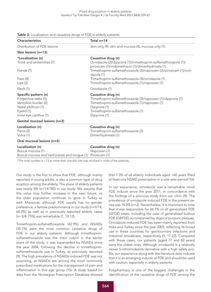
564
Fixed drug eruption in elderly patientsİstanbul Tıp Fakültesi Dergisi • J Ist Faculty Med 2021;84(4):559-67
Our study is the first to show that FDE, although mainly reported in young adults, is also a common type of drug eruption among the elderly. The share of elderly patients was nearly 8% (n=14/182) in our study. We assume that this value may further increase in the near future, as the older population continues to grow in Turkey as well. Moreover, although FDE usually has no gender preference, a female predominance in our study (n=9/14; 64.3%) as well as in previously reported elderly cases (n=3/4; 75%) was remarkable (7, 10-12).
Trimethoprim-sulfamethoxazole (42.9%) and NSAIDs (35.7%) were the most common causative drugs of FDE in our elderly patients. Although trimethoprim-sulfamethoxazole was the main culprit in the earlier years of the study, it was superseded by NSAIDs since the year 2004, following the decline in trimethoprim-sulfamethoxazole use in Turkey, as previously reported (9). The high prevalence of NSAIDs-induced FDE was not surprising, as NSAIDs are among the most commonly prescribed medications for the management of pain and inflammation in this age group (15). A study based on data from the Norwegian Prescription Database showed
that 7.3% of all elderly individuals aged >60 years filled at least one NSAID prescription in a one-year period (16).
In our experience, ornidazole was a remarkable novel FDE inducer since the year 2011, in concordance with the findings of a previous study from our clinic (9). The prevalence of ornidazole-induced FDE in the present se-ries was 14.3% (n=2). Nevertheless, it is important to note that it was responsible for 66.7% of all generalized FDE (GFDE) cases, including the case of generalized bullous FDE (GBFDE) accompanied by atypical purpuric plaques. Ornidazole-induced FDE has been mainly reported from India and Turkey since the year 2005, reflecting its broad use in these countries for genitourinary infections and intestinal amoebiasis, respectively (9, 17-22). Compared with these cases, our patients (aged 77 and 82 years) were the oldest ones. Although ornidazole is a relatively newer 5-nitroimidazole derivative with a high safety pro-file, our experience along with the literature data indicate that it is an emerging inducer of FDE and should be used with caution, especially in elderly patients.
Polypharmacy is one of the biggest challenges in the identification of the causative drugs of FDE among the
Table 3: Localization and causative drugs of FDE in elderly patients
Characteristics Total n=14
Distribution of FDE lesions Skin only (9), skin and mucosa (4), mucosa only (1)
Skin lesions (n=13)
*Localization (n)Trunk and extremities (7)
Hands (7)
Feet (4) Lips (2)
Causative drug (n)Ornidazole (2)/dipyrone (1)/trimethoprim-sulfamethoxazole (1)/piroxicam (1)/indomethacin (1)/dimenhydrinate (1)Trimethoprim-sulfamethoxazole (3)/naproxen (2)/piroxicam (1)/orni-dazole (1)Trimethoprim-sulfamethoxazole (3)/ornidazole (1)Trimethoprim-sulfamethoxazole (1)/naproxen (1)
Neck (1) Ornidazole (1)
Specific pattern (n) Finger/toe webs (5)Vermillon border (2)Nasal philtrum (1)Eyelid (1) Inner eye canthus (1)
Causative drug (n)Trimethoprim-sulfamethoxazole (3)/naproxen (1)/dipyrone (1) Trimethoprim-sulfamethoxazole (1)/naproxen (1)Dipyrone (1) Trimethoprim-sulfamethoxazole (1)Dipyrone (1)
Genital mucosal lesions (n=3)
Localization (n)Penis (2)Vulva (1)
Causative drug (n)Trimethoprim-sulfamethoxazole (2)Dimenhydrinate (1)
Oral mucosal lesions (n=2)
Localization (n)Buccal mucosa (1)Buccal mucosa and hard palate and tongue (1)
Causative drug (n)Naproxen (1)Piroxicam (1)
*The total number is >13 as more than one skin site was involved in most of the patients.

565
Fixed drug eruption in elderly patientsİstanbul Tıp Fakültesi Dergisi • J Ist Faculty Med 2021;84(4):559-67
elderly. Nevertheless, its definition is still a matter of debate. According to the World Health Organization (2019), polypharmacy is often defined as the routine use of five or more medications, including over-the-counter, prescription, and/or traditional and complementary medicines (23). A recent comprehensive review demonstrated that the prevalence of polypharmacy in older adults ranged from 4% to over 96.5% across various studies, according to the definition, age group, healthcare setting, and region (24). Yet, all studies included in that review showed an increasing prevalence of polypharmacy in older adults over time (24). In the present study, on the other hand, half of the patients (n=7) were using ≥5 medications and 21.4% of the patients (n=3) even ≥10 medications (including both the regularly used drugs for comorbidities and intermittently used drugs for infections, pain management, etc.). Interestingly, in all patients, FDE was induced by intermittently taken antibiotics, NSAIDs, or dimenhydrinate. We believe that this is an important finding which may guide clinicians when conducting a medication review. Intermittent use of prescribed or over-the-counter medications should always be questioned in elderly patients with suspected FDE.
In the present study, FDE lesions were mostly located on the trunk and extremities (50%) with a frequent acral (hands and/or feet) involvement. On the other hand, in-volvement of genital (21.4%) and oral mucosa (14.3%) was relatively low when compared with the results of previous studies from our clinic on FDE patients including younger adults (25, 26). All patients were suffering from disturbing pruritus, accompanied by a burning sensation in most of them. Xerosis and other age-related changes in skin could also have contributed to severe pruritus in these patients (27). Most patients had a disease duration of ≥1 year at the time of diagnosis, and experienced recurrent attacks which induced an increase in the number and/or size of FDE lesions in half of the patients. Taken together, these findings highlight the importance of early diagno-sis of FDE to prevent further increase in disease severity, and to preserve the quality of life in elderly patients.
As a striking finding, most patients in this study had BFDE. Bullae had been present since the first attack in the ma-jority, and developed during the subsequent attacks in two patients. BFDE is a rare and severe type of FDE that clinically presents with localized or generalized blisters or erosions overlying plaque lesions on the skin (28). The frequency of BFDE ranged from 29.6% to 36.6% across various studies including heterogeneous age groups from Tunisia, India, and Korea, whereas it was 50.8% in a nationwide retrospective multicentric study from France (28-32). Compared with these literature data, our study presents the highest frequency (71.4%) of BFDE, rais-ing the question of whether the bullous type is a more common clinical subtype among the elderly. Further pro-
spective large studies on elderly patients with FDE are warranted to confirm this potential association between BFDE and older age.
Diagnosing FDE is usually straightforward due to its unique clinical properties. Site-specific recurrence of the lesions is the diagnostic hallmark of FDE. However, bul-lous type, particularly GBFDE, may pose a big diagnostic challenge in elderly patients as it may mimic severe and potentially life-threatening drug reactions, such as Ste-vens-Johnson syndrome and toxic epidermal necrolysis, and autoimmune bullous diseases, such as pemphigus vulgaris and bullous pemphigoid (28, 33). To date, four el-derly cases with FDE have been reported in literature (10-12). They presented almost exclusively with GBFDE, ex-cept a 75-year old male with multiple plaque lesions due to ivermectin (10). GBFDE was induced by the influenza vaccine in a 67-year-old female, whereas metamizole was the causative drug in two female patients, aged 83 and 91 years, respectively (11, 12). These data along with our findings highlight that FDE may present with bullous or generalized bullous lesions even in the late elderly. Thus, awareness of this clinical heterogeneity of FDE remains crucial in preventing misdiagnosis which may lead to re-current attacks, irrelevant investigations, and overtreat-ments in this fragile age group.
Evaluation of elderly patients with FDE has special difficulties. First of all, obtaining a comprehensive medication history may be challenging in this age group, due to the relatively high prevalence of self-medication (including over-to-counter or herbal medications), polypharmacy, recall problems, and cognitive disorders, ranging from mild impairment to severe dementia (4-6). In certain circumstances, it may be helpful to ask the patients to bring in all the medications they regularly or intermittently take, and review the labels (5). Moreover, the use of drug provocation tests has some challenges in the elderly. Although topical provocation testing offers a safer alternative method than systemic provocation, it is also a less reliable diagnostic tool due to false-negative results (7). As elderly people have an age-related decline in immune functions, termed immunosenescence, caution should be exerted when interpreting the negative test results in these patients (4, 30). On the other hand, overpenetration of the drugs through the skin due to age-related skin atrophy may induce irritant and false-positive test reactions (4). Systemic (oral) drug provocation testing is the most reliable method of diagnosing FDE and establishing the causative drugs (7). Nevertheless, according to the European Network on Drug Allergy (ENDA) guidelines, it should not be performed in patients with comorbidities such as cardiac, hepatic, renal, or other diseases, where exposure might provoke a situation that is beyond medical control (35). This is particularly important for elderly patients with multicomorbidity and frailty which develops as a consequence of an age-related
566
Fixed drug eruption in elderly patientsİstanbul Tıp Fakültesi Dergisi • J Ist Faculty Med 2021;84(4):559-67
decline in many physiological systems (36). Frailty refers to an enhanced vulnerability to poor resolution of homeostasis after a stressor event, which increases the risk of negative health-related outcomes (36, 37). Therefore, oral drug provocation tests should be performed with special caution in these patients, following a careful systemic evaluation and assessment of the risk-benefit ratio (4). In our long-term clinical experience, on the other hand, oral provocation tests were performed in all patients aged between 60 and 82 years, without any complications. Importantly, topical provocation testing was not able to identify the causative drug in a majority of patients, who were then diagnosed according to the positive results of oral provocation tests with the same drugs. Our findings indicated that, when performed carefully in selected patients by experienced clinicians, systemic provocation testing was a safe method in establishing the causative drugs of FDE in this age group.
The management of elderly patients with FDE requires a multidisciplinary approach. Dermatologists and clinicians from other specialties should collaborate to prevent further attacks and develop alternative treatment schedules which do not include the implicated drugs. Moreover, both patients and their caregivers should be informed and counseled on avoidance of the culprit medications to increase adherence.
The main limitation of this study was its retrospective nature.
CONCLUSION
The present study is the first to investigate the character-istic features of FDE in elderly patients, to the best of our knowledge. Our findings highlight that FDE, although mainly reported in young adults, might also affect the el-derly. FDE was induced by intermittently taken drugs in all patients, half of whom had a high burden of polypharmacy. Trimethoprim-sulfamethoxazole and NSAIDs were the most common causative drugs, while ornidazole was a remark-able novel FDE inducer since the year 2011. As a striking finding, a great majority of patients had BFDE raising the question of whether the bullous type is a more common clinical phenotype in this age group. BFDE, particularly when generalized, may pose a big diagnostic challenge as it may clinically mimic severe and potentially life-threaten-ing drug reactions, such as Stevens-Johnson syndrome and toxic epidermal necrolysis, and autoimmune bullous diseas-es, such as pemphigus vulgaris and bullous pemphigoid.
Evaluation of elderly patients with FDE has unique chal-lenges such as polypharmacy, multimorbidity, recall problems, cognitive disorders, frailty, and immunosenes-cence. Therefore, awareness of the clinical heterogene-ity, causative drugs, and diagnostic challenges of FDE remains crucial in preventing delayed diagnosis or mis-diagnosis which may lead to recurrent attacks, irrelevant investigations, overtreatments, and further impairment of quality of life in this fragile patient population.
Ethics Committee Approval: This study was approved by the Ethical Committee of the Istanbul University, Istanbul Faculty of Medicine (Date: 18.03.2021, No: 139758).
Informed Consent: Written or verbal consent was obtained from the participants.
Peer Review: Externally peer-reviewed.
Author Contributions: Conception/Design of Study- G.B.K., E.Ö.; Data Acquisition- G.B.K., E.Ö.; Data Analysis/Interpretati-on- G.B.K., E.Ö.; Drafting Manuscript- G.B.K., E.Ö.; Critical Revi-sion of Manuscript- G.B.K., E.Ö.; Final Approval and Accounta-bility- G.B.K., E.Ö.
Conflict of Interest: Authors declared no conflict of interest.
Financial Disclosure: Authors declared no financial support.
Etik Komite Onayı: Bu çalışma için etik komite onayı İstanbul Üniversitesi, İstanbul Tıp Fakültesi Etik Kurulu’ndan alınmıştır (Tarih: 18.03.2021, No: 139758).
Bilgilendirilmiş Onam: Katılımcılardan bilgilendirilmiş onam alınmıştır.
Hakem Değerlendirmesi: Dış bağımsız.
Yazar Katkıları: Çalışma Konsepti/Tasarım- G.B.K., E.Ö.; Veri Toplama- G.B.K., E.Ö.; Veri Analizi/Yorumlama- G.B.K., E.Ö.; Yazı Taslağı- G.B.K., E.Ö.; İçeriğin Eleştirel İncelemesi- G.B.K., E.Ö.; Son Onay ve Sorumluluk- G.B.K., E.Ö.
Çıkar Çatışması: Yazarlar çıkar çatışması beyan etmemişlerdir.
Finansal Destek: Yazarlar finansal destek beyan etmemişlerdir.
REFERENCES
1. World Health Organization (2011). Global Health and Aging. Available from: URL: https://www.who.int/ageing/publications/global_health.pdf
2. United Nations, Department of Economic and Social Affairs, Population Division (2019). World Population Ageing 2019: Highlights. (ST/ESA/SER.A/430). Available from: URL: https://www.un.org/en/development/desa/population/publications/pdf/ageing/WorldPopulationAgeing2019-Highlights.pdf
3. United Nations, Department of Economic and Social Affairs, Population Division (2013). World Population Ageing 2013. (ST/ESA/SER.A/348.). Available from: URL: https://www.un.org/en/development/desa/population/publications/pdf/ageing/WorldPopulationAgeing2013.pdf
4. Heng YK, Lim YL. Cutaneous adverse drug reactions in the elderly. Curr Opin Allergy Clin Immunol 2015;15(4):300-7. [CrossRef]
5. Davies EA, O’Mahony MS. Adverse drug reactions in special populations - the elderly. Br J Clin Pharmacol 2015;80(4):796-807. [CrossRef]
6. Carneiro SC, Azevedo-e-Silva MC, Ramos-e-Silva M. Drug eruptions in the elderly. Clin Dermatol 2011;29(1):43-8. [CrossRef]

567
Fixed drug eruption in elderly patientsİstanbul Tıp Fakültesi Dergisi • J Ist Faculty Med 2021;84(4):559-67
7. Özkaya E. Fixed drug eruption: state of the art. J Dtsch Dermatol Ges 2008;6(3):181-8. [CrossRef]
8. Patel S, John AM, Handler MZ, Schwartz RA. Fixed drug eruptions: an update, emphasizing the potentially lethal generalized bullous fixed drug eruption. Am J Clin Dermatol 2020;21(3):393-9. [CrossRef]
9. Özkaya E. Changing trends in inducer drugs of fixed drug eruption: a 20-year cross-sectional study from Turkey. J Dtsch Dermatol Ges 2018;16(4):474-6. [CrossRef]
10. Ngwasiri CA, Abanda MH, Aminde LN. Ivermectin-induced fixed drug eruption in an elderly Cameroonian: a case report. J Med Case Rep 2018;12(1):254. [CrossRef]
11. Byrd RC, Mournighan KJ, Baca-Atlas M, Helton MR, Sun NZ, Siegel MB. Generalized bullous fixed-drug eruption secondary to the influenza vaccine. JAAD Case Rep 2018;4(9):953-5. [CrossRef]
12. Paulmann M, Mockenhaupt M. Unfreiwillige Reexposition: Generalisiertes bullöses fixes Arzneiexanthem bei 2 älteren Patientinnen [Unintended rechallenge: Generalized bullous fixed drug eruption in two elderly women]. Hautarzt 2017;68(1):59-63. [CrossRef]
13. Ozkaya-Bayazit E, Akar U. Fixed drug eruption induced by trimethoprim-sulfamethoxazole: evidence for a link to HLA-A30 B13 Cw6 haplotype. J Am Acad Dermatol 2001;45(5):712-7. [CrossRef]
14. Cho YT, Lin JW, Chen YC, Chang CY, Hsiao CH, Chung WH, et al. Generalized bullous fixed drug eruption is distinct from Stevens-Johnson syndrome/toxic epidermal necrolysis by immunohistopathological features. J Am Acad Dermatol 2014;70(3):539-48. [CrossRef]
15. Wongrakpanich S, Wongrakpanich A, Melhado K, Rangaswami J. A comprehensive review of non-steroidal anti-inflammatory drug use in the elderly. Aging Dis 2018;9(1):143-150. [CrossRef]
16. Vandraas KF, Spigset O, Mahic M, Slørdal L. Non-steroidal anti-inflammatory drugs: use and co-treatment with potentially interacting medications in the elderly. Eur J Clin Pharmacol 2010;66(8):823-9. [CrossRef]
17. Gupta S, Jain VK, Aggarwal K, Gupta S, Mahendra A. Fixed drug eruption caused by ornidazole. Contact Dermatitis 2005;53(5):300-1. [CrossRef]
18. Gupta S, Mahendra A, Gupta S, Kaur S. Multiple fixed drug eruption caused by ornidazole. Dermatitis 2010;21(6):330-3. [CrossRef]
19. Sanmukhani J, Shah V, Baxi S, Tripathi C. Fixed drug eruption with ornidazole having cross-sensitivity to secnidazole but not to other nitro-imidazole compounds: a case report. Br J Clin Pharmacol 2010;69(6):703-4. [CrossRef]
20. Marya CM, Sharma G, Parashar VP, Dahiya V. Mucosal fixed drug eruption in a patient treated with ornidazole. J Dermatol Case Rep 2012;6(1):21-4. [CrossRef]
21. Gupta R. Fixed drug eruption due to ornidazole. Indian J Dermatol 2014;59(6):635. [CrossRef]
22. Emre S, Ahsen H, Aktaş A. Ornidazole-induced fixed drug reaction on sole: case report and review of the literature. Cutan Ocul Toxicol 2017;36(3):294-6. [CrossRef]
23. World Health Organization (2019). Medication Safety in Polypharmacy. (WHO/UHC/SDS/2019.11). Available from: URL: https://apps.who.int/iris/bitstream/handle/10665/325454/WHO-UHC-SDS-2019.11-eng.pdf?ua=1
24. Pazan F, Wehling M. Polypharmacy in older adults: a narrative review of definitions, epidemiology and consequences. Eur Geriatr Med 2021 Mar 10. [CrossRef]
25. Ozkaya-Bayazit E. Specific site involvement in fixed drug eruption. J Am Acad Dermatol 2003;49(6):1003-7. [CrossRef]
26. Özkaya E. Oral mucosal fixed drug eruption: characteristics and differential diagnosis. J Am Acad Dermatol 2013;69(2):e51-8. [CrossRef]
27. Fourzali KM, Yosipovitch G. Management of itch in the elderly: a review. Dermatol Ther (Heidelb) 2019;9(4):639-53. [CrossRef]
28. Zaouak A, Ben Salem F, Ben Jannet S, Hammami H, Fenniche S. Bullous fixed drug eruption: A potential diagnostic pitfall: a study of 18 cases. Therapie 2019;74(5):527-30. [CrossRef]
29. Ben Fadhel N, Chaabane A, Ammar H, Ben Romdhane H, Soua Y, Chadli Z, et al. Clinical features, culprit drugs, and allergology workup in 41 cases of fixed drug eruption. Contact Dermatitis 2019;81(5):336-40. [CrossRef]
30. Pai VV, Kikkeri NN, Athanikar SB, Shukla P, Bhandari P, Rai V. Retrospective analysis of fixed drug eruptions among patients attending a tertiary care center in Southern India. Indian J Dermatol Venereol Leprol 2014;80(2):194. [CrossRef]
31. Jung JW, Cho SH, Kim KH, Min KU, Kang HR. Clinical features of fixed drug eruption at a tertiary hospital in Korea. Allergy Asthma Immunol Res 2014;6(5):415-20. [CrossRef]
32. Brahimi N, Routier E, Raison-Peyron N, Tronquoy AF, Pouget-Jasson C, Amarger S, et al. A three-year-analysis of fixed drug eruptions in hospital settings in France. Eur J Dermatol 2010;20(4):461-4. [CrossRef]
33. Cho YT, Lin JW, Chen YC, Chang CY, Hsiao CH, Chung WH, et al. Generalized bullous fixed drug eruption is distinct from Stevens-Johnson syndrome/toxic epidermal necrolysis by immunohistopathological features. J Am Acad Dermatol 2014;70(3):539-48. [CrossRef]
34. Weiskopf D, Weinberger B, Grubeck-Loebenstein B. The aging of the immune system. Transpl Int 2009;22(11):1041-50. [CrossRef]
35. Aberer W, Bircher A, Romano A, Blanca M, Campi P, Fernandez J, et al. European Network for Drug Allergy (ENDA); EAACI interest group on drug hypersensitivity. Drug provocation testing in the diagnosis of drug hypersensitivity reactions: general considerations. Allergy 2003;58(9):854-63. [CrossRef]
36. Clegg A, Young J, Iliffe S, Rikkert MO, Rockwood K. Frailty in elderly people. Lancet 2013;381(9868):752-62. [CrossRef]
37. Cesari M, Calvani R, Marzetti E. Frailty in older persons. Clin Geriatr Med 2017;33(3):293-303. [CrossRef]

568
Serum homocysteine level and IgA nephropathy in childrenİstanbul Tıp Fakültesi Dergisi • J Ist Faculty Med 2021;84(4):568-73
Corresponding author/İletişim kurulacak yazar: [email protected]
Submitted/Başvuru: 29.12.2020 • Revision Requested/Revizyon Talebi: 07.05.2021 •Last Revision Received/Son Revizyon: 05.07.2021 • Accepted/Kabul: 06.07.2021 • Published Online/Online Yayın: 29.09.2021
RESEARCH / ARAŞTIRMADOI: 10.26650/IUITFD.2021.847530
İst Tıp Fak Derg 2021 / J Ist Faculty Med 2021
A POSSIBLE RELATIONSHIP BETWEEN SERUM HOMOCYSTEINE LEVEL AND IgA NEPHROPATHY IN CHILDREN
ÇOCUKLARDA SERUM HOMOSİSTEİN DÜZEYİ İLE IgA NEFROPATİSİ ARASINDAKİ OLASI İLİŞKİ
Cemile PEHLİVANOĞLU1 , Zeynep YÜRÜK YILDIRIM1 , Alev YILMAZ1 , Asuman GEDİKBAŞI2 , Nurinisa KARAGÖZ3 , Nurver AKINCI4 , Aysel KIYAK5 , Gül ÖZÇELİK4 , Yasemin ÖZLÜK6 , Işın KILIÇASLAN6 , Ayşe Ayşim ÖZAĞARI7 , Bağdagül YAVAŞ AKSU2 , Sevinç EMRE1
1Istanbul University, Istanbul Faculty of Medicine, Department of Pediatric Nephrology, Istanbul, Turkey2Istanbul University, Institute of Child Health, Department of Pediatric Basic Sciences, Istanbul, Turkey3Istanbul University, Istanbul Faculty of Medicine, Department of Child Health and Diseases, Istanbul, Turkey4Şişli Hamidiye Etfal Training and Research Hospital, Department of Pediatric Nephrology, Istanbul, Turkey5Kanuni Sultan Süleyman Research and Training Hospital, Department of Pediatric Nephrology, Istanbul, Turkey6Istanbul University, Istanbul Faculty of Medicine, Department of Pathology, Istanbul, Turkey7Şişli Hamidiye Etfal Training and Research Hospital, Department of Pathology, Istanbul, Turkey
ORCID IDs of the authors: C.P. 0000-0002-6949-8922; Z.Y.Y. 0000-0003-2891-2231; A.Y. 0000-0003-1743-1491; A.G. 0000-0001-7121-6077; N.K. 0000-0002-4575-4100; N.A. 0000-0002-8355-4214; A.K. 0000-0003-1073-000X; G.Ö. 0000-0001-9394-2977; Y.Ö. 0000-0002-7191-0488; I.K. 0000-0002-4206-9941; A.A.Ö. 0000-0003-2343-1236; B.Y.A. 0000-0003-3274-8024; S.E. 0000-0002-9708-9480
Cite this article as: Pehlivanoglu C, Yuruk Yildirim Z, Yilmaz A, Gedikbasi A, Karagoz N, Akinci N, et al. A possible relationship between serum homocysteine level and IgA nephropathy in children. J Ist Faculty Med 2021;84(4):568-73. doi: 10.26650/IUITFD.2021.847530
ABSTRACT
Objective: The evidences from experimental and epidemio-logical studies suggests that elevated serum homocysteine lev-els may lead to renal injury and may be a significant risk factor for the development of chronic kidney disease. The aim of this study was to investigate a possible relationship between serum homocysteine level and crescent formation in children with IgA nephropathy and Henoch-Schonlein purpura nephritis.
Material and Methods: A total of 31 patients diagnosed as bi-opsy proven IgA nephropathy and Henoch-Schonlein purpura nephritis and idiopathic crescentic glomerulonephritis in three Pediatric Nephrology centers within the last five years and 25 healthy controls were enrolled in the study.
Results: Homocysteine levels of patients were higher than the upper limit of normal value and also higher than the controls (p=0.0001). There was no significant difference between the pa-tients with or without crescent formation regarding homocyste-ine levels (p>0.05). Presence or severity of proteinuria was not related to homocysteine levels (p>0.05).
Conclusion: Serum homocysteine levels are elevated in patients with IgA nephropathy and Henoch-Schonlein purpura nephritis.
ÖZET
Amaç: Deneysel ve epidemiyolojik çalışmalar, yüksek serum homosistein düzeylerinin renal hasara yol açabileceğini ve kronik böbrek hastalığı gelişimi için önemli bir risk faktörü olabileceğini göstermektedir. Çalışmamızın amacı IgA nefropatisi ve Henoch-Schönlein purpura nefriti olan hastalarda serum homosistein düzeyleri ile kresent oluşumu arasında bir ilişki olup olmadığını belirlemektir.
Gereç ve Yöntemler: Üç pediatrik nefroloji merkezinde son beş yılda biyopsi ile IgA nefropatisi, Henoch-Schönlein purpura nef-riti ve idiopatik kresentik glomerulonefrit tanısı alan 31 hasta ve 25 sağlıklı kontrol çalışmaya alındı.
Bulgular: Hastaların homosistein düzeyleri normal değerin üst sınırından ve kontrollerden de daha yüksekti (p=0,0001). Biyop-side kresent görülen ve görülmeyen hastalar arasında da homo-sistein düzeyleri açısından anlamlı fark yoktu (p>0,05). Proteinüri varlığı ve şiddeti ile homosistein düzeyleri arasında ilişki saptan-madı (p>0,05).
Sonuç: Serum homosistein düzeyleri IgA nefropatisi ve Henoch-Schonlein purpura nefriti olan hastalarda yüksek bulunmuştur. Sonuçlarımız, bu hasta grubunda yüksek serum homosistein
Content of this journal is licensed under a Creative Commons Attribution-NonCommercial 4.0 International License.

569
Serum homocysteine level and IgA nephropathy in childrenİstanbul Tıp Fakültesi Dergisi • J Ist Faculty Med 2021;84(4):568-73
RESEARCH / ARAŞTIRMADOI: 10.26650/IUITFD.2021.847530
İst Tıp Fak Derg 2021 / J Ist Faculty Med 2021
A POSSIBLE RELATIONSHIP BETWEEN SERUM HOMOCYSTEINE LEVEL AND IgA NEPHROPATHY IN CHILDREN
ÇOCUKLARDA SERUM HOMOSİSTEİN DÜZEYİ İLE IgA NEFROPATİSİ ARASINDAKİ OLASI İLİŞKİ
Cemile PEHLİVANOĞLU1 , Zeynep YÜRÜK YILDIRIM1 , Alev YILMAZ1 , Asuman GEDİKBAŞI2 , Nurinisa KARAGÖZ3 , Nurver AKINCI4 , Aysel KIYAK5 , Gül ÖZÇELİK4 , Yasemin ÖZLÜK6 , Işın KILIÇASLAN6 , Ayşe Ayşim ÖZAĞARI7 , Bağdagül YAVAŞ AKSU2 , Sevinç EMRE1
1Istanbul University, Istanbul Faculty of Medicine, Department of Pediatric Nephrology, Istanbul, Turkey2Istanbul University, Institute of Child Health, Department of Pediatric Basic Sciences, Istanbul, Turkey3Istanbul University, Istanbul Faculty of Medicine, Department of Child Health and Diseases, Istanbul, Turkey4Şişli Hamidiye Etfal Training and Research Hospital, Department of Pediatric Nephrology, Istanbul, Turkey5Kanuni Sultan Süleyman Research and Training Hospital, Department of Pediatric Nephrology, Istanbul, Turkey6Istanbul University, Istanbul Faculty of Medicine, Department of Pathology, Istanbul, Turkey7Şişli Hamidiye Etfal Training and Research Hospital, Department of Pathology, Istanbul, Turkey
ORCID IDs of the authors: C.P. 0000-0002-6949-8922; Z.Y.Y. 0000-0003-2891-2231; A.Y. 0000-0003-1743-1491; A.G. 0000-0001-7121-6077; N.K. 0000-0002-4575-4100; N.A. 0000-0002-8355-4214; A.K. 0000-0003-1073-000X; G.Ö. 0000-0001-9394-2977; Y.Ö. 0000-0002-7191-0488; I.K. 0000-0002-4206-9941; A.A.Ö. 0000-0003-2343-1236; B.Y.A. 0000-0003-3274-8024; S.E. 0000-0002-9708-9480
Cite this article as: Pehlivanoglu C, Yuruk Yildirim Z, Yilmaz A, Gedikbasi A, Karagoz N, Akinci N, et al. A possible relationship between serum homocysteine level and IgA nephropathy in children. J Ist Faculty Med 2021;84(4):568-73. doi: 10.26650/IUITFD.2021.847530
INTRODUCTION
IgA nephropathy (IgAN), one of the most common glomerulonephritis in adults and children, may progress to end-stage renal disease (ESRD) in 20-50% of patients within 20 years (1-3). Although IgAN usually has a slow progression course, acute deterioration in renal function due to crescent formation may occur during the course of the disease (3, 4). It has been reported that crescent formation in IgAN may influence renal outcomes and aggressive immunosuppressive treatment may be required if the number of crescents involves >50% of the glomeruli in a renal biopsy (5). The Kidney Disease Improving Global Outcomes (KDIGO) guidelines recommend that Henoch-Schonlein Purpura Nephritis (HSPN) should be treated the same as IgAN because it has the same histopathologic features as IgAN in kidney biopsies (5). Crescent formation in glomeruli begins with the emergence of ruptures of glomerular capillary walls (6). These ruptures permit coagulation factors and cells including monocytes and lymphocytes to enter the Bowman’s space, which results in crescent formation (6). There is some evidence to suggest that elevated concentration of serum homocysteine (Hcy) leads to endothelial cell injury, which results in stimulation of the coagulation system and resistance of anticoagulation activity on the endothelial surface (7, 8). Moreover, it has been reported that hyperhomocysteinemia causes glomerular and tubulointerstitial damage in several ways, such as increasing oxidative stress, stimulating monocyte chemoattractant protein-1 (MCP-1) expression, and nuclear factor kappa B activation (NF-B) (9-14).
We observed hyperhomocysteinemia in two patients with IgAN and crescent formation in their renal biopsy. Using observations from these two patients, we hypothesized that crescent formation may be related to hyperhomocysteinemia in IgAN. The aim of the study was to investigate whether a relationship exists between serum Hcy level and crescent formation in children with IgAN and HSPN.
MATERIALS AND METHODS
Thirty-one patients with renal biopsy-proven IgAN (n=17), HSPN (n=12), and idiopathic crescentic glomer-ulonephritis (ICGN; n=2) in three pediatric nephrology centers were enrolled in the study. The control group was consisted of 25 age matched healthy children without medical history of any chronic disease or renal disorder.
This study was approved by the Ethical Committee of Istanbul University Istanbul Faculty of Medicine (Date: 27.05.2013, No: 640). Informed consent was obtained from the parents of all participants. A physical examina-tion was performed, and the patients’ medical history and clinical findings were recorded at the time of the sampling.
Baseline investigations including serum urea, creatinine, electrolytes, lipids, total protein, albumin, Hcy, urinalysis and 24-hour urinary protein were performed in the pa-tient group. Moreover, serum levels of vitamin B12 and folic acid, thyroid function tests, methylenetetrahydro-folate reductase (MTHFR) 677 and MTHFR 1298 genetic mutation analysis were also performed because hyperho-mocysteinemia may be caused by deficiency of folic acid and vitamin B12, hypothyroidism or decreased enzyme activity of MTHFR due to genetic mutations. A physical examination was performed and serum urea, creatinine, Hcy, B12 and urinalysis were performed in the control group.
IgAN, HSPN, and ICGN were diagnosed based on clini-cal, laboratory and immunehistopathologic findings. Me-sangial hypercellularity with predominant IgA deposition in the mesangium and/or capillary wall was described as IgAN in patients without systemic involvement. The MEST score was evaluated as mesangial hypercellulari-ty (M0, M1), endocapillary hypercellularity (E0, E1), seg-mental glomerulosclerosis (S0, S1) and tubular atrophy/interstitial fibrosis (T0, T1), in accordance with the Oxford classification (4). Presence or absence of crescents was re-corded. Henoch-Schonlein Purpura was diagnosed using the EULAR/PReS consensus criteria (15). Extracapillary proliferation of cells in the Bowman’s space with more than two cell layers was described as cellular crescent (16). Idiopathic crescents in more than 50% of glomeruli on renal biopsies were described as ICGN.
Hypertension was defined as average clinic measured systolic and/or diastolic blood pressure ≥95th percentile on the basis of age, sex and height percentiles (17).
The mean estimated glomerular filtration rate (eGFR) was calculated according to the Schwartz formula (18). Hematuria was defined as five or more red blood cells per high power field in a urine specimen (19). Proteinuria was determined as a positive dipstick reading of ≥1+, urinary excretion ≥4 mg/m2/per hour in urine over a period of 24 hours (19).
Our results suggest that elevated serum homocysteine levels may be related to segmental glomerulosclerosis in these patient groups.
Keywords: IgA nephropathy, homocysteine, crescent
düzeylerinin segmental glomerüloskleroz ile ilişkili olabileceğini düşündürmektedir.
Anahtar Kelimeler: IgA nefropatisi, homosistein, kresent
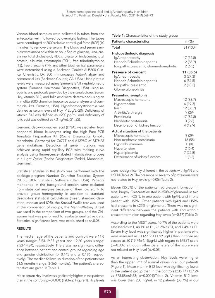
570
Serum homocysteine level and IgA nephropathy in childrenİstanbul Tıp Fakültesi Dergisi • J Ist Faculty Med 2021;84(4):568-73
Venous blood samples were collected in tubes from the antecubital vein, followed by overnight fasting. The tubes were centrifuged at 2000 relative centrifugal force (RCF) (10 minutes) to remove the serum. The blood and serum sam-ples were analyzed within an hour. Serum glucose, urea, cre-atinine, total cholesterol, HDL cholesterol, triglyceride, total protein, albumin, thyrotropin (TSH), free triiodothyronine (T3), free thyroxine (T4), and other biochemical parameters were determined using a Beckman Coulter AU5800 Clin-ical Chemistry, Dxl 800 Immunoassay Auto-Analyzer and commercial kits (Beckman Coulter, CA, USA). Urine protein levels were measured using Siemens BNII nephelometric system (Siemens Healthcare Diagnostics, USA) using re-agents and protocols provided by the manufacturer. Serum Hcy, vitamin B12, and folic acid were determined using an Immulite 2000 chemiluminescence auto-analyzer and com-mercial kits (Siemens, USA). Hyperhomocysteinemia was defined as serum levels of Hcy >12µg/L (20). Deficiency of vitamin B12 was defined as <200 pg/mL and deficiency of folic acid was defined as <3 ng/mL (21, 22).
Genomic deoxyribonucleic acid (DNA), was isolated from peripheral blood leukocytes using the High Pure PCR Template Preparation Kit (Roche Diagnostics GmbH, Mannheim, Germany) for C677T and A1298C of MTHFR gene mutations. Detection of gene mutations was achieved using rapid capillary PCR with melting curve analysis using fluorescence-labeled hybridization probes in a Light Cycler (Roche Diagnostics GmbH, Mannheim, Germany).
Statistical analysis in this study was performed with the package program Number Cruncher Statistical System (NCSS) 2007 Statistical Software (Utah, USA). Patients mentioned in the background section were excluded from statistical analyses because of their low eGFR to provide group homogeneity. In addition to standard descriptive statistical calculations (mean, standard devi-ation, median and IQR), the Kruskal-Wallis test was used in the comparison of groups, the Mann-Whitney U test was used in the comparison of two groups, and the Chi-square test was performed to evaluate qualitative data. Statistical significance level was established at p<0.05.
RESULTS
The median age of the patients and controls were 11.6 years (range: 3.53-19.37 years) and 12.60 years (range: 9.53-14.84), respectively. There was no significant differ-ence between patient and control groups regarding age and gender distribution (p=0.145 and p=0.186; respec-tively). The median follow-up duration of the patients was 61.5 months (range: 3-365 months). The patient’s charac-teristics are given in Table 1.
Mean serum Hcy level was significantly higher in the patients than in the controls (p=0.0001) (Table 2, Figure 1). Hcy levels
were not significantly different in the patients with IgAN and HSPN (Table 2). The presence or severity of proteinuria were not related to Hcy levels (p>0.05) (Table 2).
Eleven (35.5%) of the patients had crescent formation in renal biopsy. Crescents existed in ≥50% of glomeruli in two patients with ICGN, in one patient with IgAN, and in one patient with HSPN. Other patients with IgAN and HSPN had crescents in ≤25% of glomeruli. There was no signif-icant difference between the patients with and without crescent formation regarding Hcy levels (p=0.17) (Table 2).
According to the MEST score, 40.7% of the patients were assessed as M1, 48.1% as E1, 22.2% as S1, and 7.4% as T1. Serum Hcy level was significantly higher in patients who were assessed as S1 (29.36±11.99 µg/L) than in those as-sessed as S0 (19.74±4.15µg/L) with regard to MEST score (p=0.009) although other parameters of the score were not related to Hcy level (p>0.05).
As an interesting observation, Hcy levels were higher than the upper limit of normal values in all our patients (Figure 1). Mean vitamin B12 level was significantly lower in the patient group than in the controls (238.77±127.24 vs 378.88±69.63; p=0.0001)(Table 2). Vitamin B12 level was lower than 200 ng/mL in 12 patients (38.7%) in our
Table 1: Characteristics of the study group
Patients characteristics n (%)
Total 31 (100)
Histopathologic diagnosis IgA nephropathy Henoch-Schonlein nephritis Idiopathic crescentic glomerulonephritis
17 (54.8) 12 (38.7) 2 (6.5)
Presence of crescentIgA nephropathyHenoch-Schonlein nephritisIdiopathic crescentic Glomerulonephritis
11 (35.5) 3 (27.3) 6 (54.5) 2 (18.2)
Presenting symptoms Macroscopic hematuriaHypertensionRashArthritis/arthralgiaProteinuriaNephrotic proteinuriaDeterioration of kidney function
12 (38.7)6 (19.3)12 (38.7)
9 (29)17 (54.8)3 (9.6)4 (12.9)
Actual situation of the patients Microscopic hematuriaNon-nephrotic proteinuriaHypoalbuminemiaHypertensionHyperlipidemiaDeterioration of kidney functions
9 (29)18 (58)0 (0)
2 (6.4)7 (22.5) 1 (3.2)
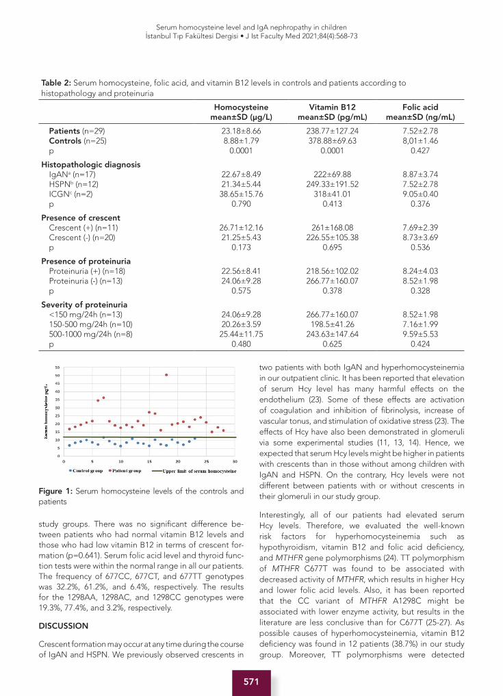
571
Serum homocysteine level and IgA nephropathy in childrenİstanbul Tıp Fakültesi Dergisi • J Ist Faculty Med 2021;84(4):568-73
study groups. There was no significant difference be-tween patients who had normal vitamin B12 levels and those who had low vitamin B12 in terms of crescent for-mation (p=0.641). Serum folic acid level and thyroid func-tion tests were within the normal range in all our patients. The frequency of 677CC, 677CT, and 677TT genotypes was 32.2%, 61.2%, and 6.4%, respectively. The results for the 1298AA, 1298AC, and 1298CC genotypes were 19.3%, 77.4%, and 3.2%, respectively.
DISCUSSION
Crescent formation may occur at any time during the course of IgAN and HSPN. We previously observed crescents in
two patients with both IgAN and hyperhomocysteinemia in our outpatient clinic. It has been reported that elevation of serum Hcy level has many harmful effects on the endothelium (23). Some of these effects are activation of coagulation and inhibition of fibrinolysis, increase of vascular tonus, and stimulation of oxidative stress (23). The effects of Hcy have also been demonstrated in glomeruli via some experimental studies (11, 13, 14). Hence, we expected that serum Hcy levels might be higher in patients with crescents than in those without among children with IgAN and HSPN. On the contrary, Hcy levels were not different between patients with or without crescents in their glomeruli in our study group.
Interestingly, all of our patients had elevated serum Hcy levels. Therefore, we evaluated the well-known risk factors for hyperhomocysteinemia such as hypothyroidism, vitamin B12 and folic acid deficiency, and MTHFR gene polymorphisms (24). TT polymorphism of MTHFR C677T was found to be associated with decreased activity of MTHFR, which results in higher Hcy and lower folic acid levels. Also, it has been reported that the CC variant of MTHFR A1298C might be associated with lower enzyme activity, but results in the literature are less conclusive than for C677T (25-27). As possible causes of hyperhomocysteinemia, vitamin B12 deficiency was found in 12 patients (38.7%) in our study group. Moreover, TT polymorphisms were detected
Table 2: Serum homocysteine, folic acid, and vitamin B12 levels in controls and patients according to histopathology and proteinuria
Homocysteine mean±SD (µg/L)
Vitamin B12 mean±SD (pg/mL)
Folic acid mean±SD (ng/mL)
Patients (n=29) Controls (n=25)p
23.18±8.668.88±1.79
0.0001
238.77±127.24378.88±69.63
0.0001
7.52±2.788,01±1.46
0.427
Histopathologic diagnosis IgANa (n=17) HSPNb (n=12) ICGNc (n=2) p
22.67±8.4921.34±5.4438.65±15.76
0.790
222±69.88249.33±191.52
318±41.010.413
8.87±3.747.52±2.789.05±0.40
0.376
Presence of crescentCrescent (+) (n=11)Crescent (-) (n=20)p
26.71±12.1621.25±5.43
0.173
261±168.08226.55±105.38
0.695
7.69±2.398.73±3.69
0.536
Presence of proteinuriaProteinuria (+) (n=18)Proteinuria (-) (n=13)p
22.56±8.4124.06±9.28
0.575
218.56±102.02266.77±160.07
0.378
8.24±4.038.52±1.98
0.328
Severity of proteinuria<150 mg/24h (n=13)150-500 mg/24h (n=10) 500-1000 mg/24h (n=8) p
24.06±9.2820.26±3.5925.44±11.75
0.480
266.77±160.07198.5±41.26
243.63±147.640.625
8.52±1.987.16±1.999.59±5.53
0.424
Figure 1: Serum homocysteine levels of the controls and patients
572
Serum homocysteine level and IgA nephropathy in childrenİstanbul Tıp Fakültesi Dergisi • J Ist Faculty Med 2021;84(4):568-73
in two patients and CC polymorphism in one patient. However, serum Hcy levels were also found elevated in the remaining 16 patients who did not have features that may predispose to hyperhomocysteinemia. We speculate that hyperhomocysteinemia may be related to IgAN itself because there was no other reason for hyperhomocysteinemia in these patients.
Endothelial cells have an important role in the maintenance of vascular structure integrity in glomeruli (28, 29). It has been considered that endothelial cells are related to the progression of IgAN. Kusano T et al. evaluated the number of glomerular endothelial cells in patients with IgAN by examining the number of nuclei of CD34+ glomerular endothelial cells in renal biopsies (6). The authors demonstrated that the loss of CD34+ endothelial cells was associated with glomerular necrosis, segmental and global sclerosis in IgAN (6). Also, there were fewer CD34+ endothelial cells found in glomeruli with chronic lesions than in normal glomeruli in the biopsy samples (6). Moreover, hyperhomocysteinemia may aggravate endothelial damage and sclerotic modification in glomeruli (11, 13, 30). According to Oxford Classifications, segmental glomerulosclerosis was related to the poor prognosis in IgAN (4). Hcy levels were significantly higher in patients with segmental sclerosis in their kidney biopsies in our study group, which suggests that hyperhomocysteinemia may be related to sclerosis and chronic changes in glomeruli and consequently poor prognosis in IgAN.
Proteinuria is another important factor that is known to im-pact the prognosis of patients with IgAN (31). Thus, the relationship between the serum Hcy levels and proteinuria was evaluated in our study. According to our results, there were no differences between the presence or absence of proteinuria and between the different levels of proteinuria in patients with IgAN in terms of serum Hcy levels. In our previous study, we evaluated whether there was Hcy eleva-tion in idiopathic nephrotic syndrome in children. The se-rum Hcy levels were not higher in nephrotic children than in the control group (32). Kong et al. demonstrated that hyperhomocysteinemia was an independent risk factor of decreased eGFR in the elderly population, although there was no relationship between proteinuria and elevated se-rum Hcy levels in their study (33). On the contrary, Shuwei et al. reported that hyperhomocsyteinemia was associated with lower eGFR and also initial proteinuria in patients with IgAN (34). Although these studies had different results about the relationship between serum Hcy levels and pro-teinuria, they concluded that hyperhomocysteinemia was related to decreasing in eGFR and poor prognosis.
Our study had some limitations. One of these limitations was that our patients were not in the acute phase of the illness. The mean duration between kidney biopsy and blood sampling for the study was 35 months. We
believe that further studies in larger groups of patients in the acute phase of the illness may reveal a relationship between these pathologic prognostic factors. The second limitation was our relatively small sample size.
In conclusion, we demonstrated that serum Hcy levels are elevated in patients with IgAN and HSPN. Our results suggest that elevated serum Hcy levels may be related to segmental glomerulosclerosis in these patient groups.
Ethics Committee Approval: This study was approved by the Clinical Research Ethical Committee of the Istanbul University, Istanbul Faculty of Medicine (Date: 27.05.2013, No: 640).
Informed Consent: Written consent was obtained from the par-ticipants.
Peer Review: Externally peer-reviewed.
Author Contributions: Conception/Design of Study- Z.Y.Y., A.Y., A.G., Y.Ö., S.E.; Data Acquisition- C.P., N.A., N.K.; Data Analysis/Interpretation- B.Y.A., Y.Ö., A.A.Ö., I.K.; Drafting Ma-nuscript- C.P., N.A., N.K., A.K., B.Y.A., A.Ö., G.Ö.; Critical Revisi-on of Manuscript- A.Y., Z.Y.Y., S.E., I.K., Y.Ö., A.G.; Final Approval and Accountability- C.P., Z.Y.Y., A.Y., A.G., N.K., M.K., A.Y., G.Ö., Y.Ö., I.K., A.A.Ö., B.Y.A., S.E.
Conflict of Interest: Authors declared no conflict of interest.
Financial Disclosure: Authors declared no financial support.
Etik Komite Onayı: Bu çalışma için etik komite onayı İstanbul Üniversitesi, İstanbul Tıp Fakültesi Klinik Araştırmalar Etik Kuru-lu’ndan alınmıştır (Tarih: 27.05.2013, No: 640).
Bilgilendirilmiş Onam: Katılımcılardan bilgilendirilmiş onam alınmıştır.
Hakem Değerlendirmesi: Dış bağımsız.
Yazar Katkıları: Çalışma Konsepti/Tasarım- Z.Y.Y., A.Y., A.G., Y.Ö., S.E.; Veri Toplama- C.P., N.A., N.K.; Veri Analizi/Yorumla-ma- B.Y.A., Y.Ö., A.A.Ö., I.K.; Yazı Taslağı- C.P., N.A., N.K., A.K., B.Y.A., A.Ö., G.Ö.; İçeriğin Eleştirel İncelemesi- A.Y., Z.Y.Y., S.E., I.K., Y.Ö., A.G.; Son Onay ve Sorumluluk- C.P., Z.Y.Y., A.Y., A.G., N.K., M.K., A.Y., G.Ö., Y.Ö., I.K., A.A.Ö., B.Y.A., S.E.
Çıkar Çatışması: Yazarlar çıkar çatışması beyan etmemişlerdir.
Finansal Destek: Yazarlar finansal destek beyan etmemişlerdir.
REFERENCES
1. Nakanishi K, Yoshikawa N. Immunoglobulin A Nephropathy. In: Avner ED, Harmon WE, Niaudet P, Yoshikawa N, Emma F, Goldstein SL,editors. Pediatric Nephrology, 7th ed. Berlin Heidelberg: Springer-Verlag; 22016.pp983-1033.
2. Shima Y, Nakanishi K, Hama T, Mukaiyama H, Togawa H, Hashimura Y, et al. Validity of the Oxford classification of IgA nephropathy in children. Pediatr Nephrol 2012;27(5):783-92. [CrossRef]

573
Serum homocysteine level and IgA nephropathy in childrenİstanbul Tıp Fakültesi Dergisi • J Ist Faculty Med 2021;84(4):568-73
3. Coppo R, Davin JC. The difficulty in considering modifiable pathology risk factors in children with IgA nephropathy: crescents and timing of renal biopsy. Pediatr Nephrol 2014;30(2):189-92. [CrossRef]
4. Roberts IS, Cook HT, Troyanov S, Alpers CE, Amore A, Barratt J, et al. The Oxford classification of IgA nephropathy: pathology definitions, correlations, and reproducibility. Kidney Int 2009;76(5):546-56. [CrossRef]
5. Beck L, Bomback AS, Choi MJ, Holzman LB, Langford C, Mariani LH, et.al. KDOQI US Commentary on the 2012 KDIGO Clinical Practice Guideline for Glomerulonephritis. Am J Kidney Dis 2013;62(3):403-41. [CrossRef]
6. Kusano T, Takano H, Kang D, Nagahama K, Aoki M, Morita M, et al. Endothelial cell injury in acute and chronic glomerular lesions in patients with IgA nephropathy. Human Pathology 2015;49:135-44. [CrossRef]
7. Rodgers GM, Conn MT. Homocysteine, an atherogenic stimulus, reduces protein C activation by arterial and venous endothelial cells. Blood 1990;75(4):895-901. [CrossRef]
8. Rodgers GM, Kane WH. Activation of endogenous factor V by homocysteine-induced vascular endothelial cell activator. J Clin Invest 1986;77(6):1909-16. [CrossRef]
9. Tsai JC, Perrella MA, Yoshizumi M, Hsieh CM, Haber E, Schlegel R, et al. Promotion of vascular smooth muscle cell growth by homocysteine: a link to atherosclerosis. Proc Natl Acad Sci 1994;91(14):6369-73. [CrossRef]
10. Farid FA, Faheem MS, Heshmat NM, Shaheen KY, Saad SS. Study of the homocysteine status in children with chronic renal failure. Am J Nephrol 2004;24(3):289-95. [CrossRef]
11. Kumagai H, Katoh S, Hirosawa K, Kimura M, Hishida A, Ikegaya N. Renal tubulointerstitial injury in weanling rats with hyperhomocysteinemia. Kidney Int 2002;62(4):1219-28. [CrossRef]
12. Kanani PM, Sinkey CA, Browning RL, Allaman M, Knapp HR, Haynes WG. Role of oxidant stress in endothelial dysfunction produced by experimental hyperhomocyst(e)inemia in humans. Circulation 1999;100(11):1161-8. [CrossRef]
13. Li N, Chen YF, Zou AP. Implications of hyperhomocysteinemia in glomerular sclerosis in hypertension. Hypertension 2002;39(2 Pt 2):443-8. [CrossRef]
14. Hwang SY, Woo CW, Au-Yeung KK, Siow YL, Zhu TY, O K. Homocysteine stimulates monocyte chemoattractant protein-1 expression in the kidney via nuclear factor-kappaB activation. Am J Physiol Renal Physiol 2007;294(1):F236-44. [CrossRef]
15. Dillon MJ, Ozen S. A new international classification of childhood vasculitis. Pediatr Nephrol 2006;21(9):1219-22. [CrossRef]
16. Roberts IS. Pathology of IgA nephropathy. Nat Rev Nephrol 2014;10(8):445-54. [CrossRef]
17. Flynn JT, Kaelber DC, Baker-Smith CM, Blowey D, Carroll AE, Daniels SR, et al. Clinical practice guideline for screening and management of high blood pressure in children and adolescents. Pediatrics 2017;140(3): e20171904. [CrossRef]
18. Schwartz GJ, Munoz A, Schneider MF, Mak RH, Kaskel F, Warady BA, et al. New equations to estimate GFR in children with CKD. J Am Soc Nephrol 2009;20(3):629-37.[CrossRef]
19. Yap HK, Lau PY. Hematuria and Proteinuria. In: Geary FD, Schaefer F, editors. Comprehensive Pediatric Nephrology, 1st ed. Philadelphia: Mosby Elseiver; 2008.pp179-93. [CrossRef]
20. Nicholson JF, Pesce MA. Reference ranges for laboratory tests and procedures. In: Behrman RE, Kliegman RM, Jenson HB, editors. Nelson Textbook of Pediatrics, 17th ed. Philadelphia: Saunders; 2004.pp2396-427.
21. Carmel R. Biomarkers of cobalamin (vitamin B-12) status in the epidemiologic setting: a critical overview of context, applications, and performance characteristics of cobalamin, methylmalonic acid, and holotranscobalamin II. Am J Clin Nutr 2011;94(1):348-58. [CrossRef]
22. Glader B. Anemias of Inadequate Production. In: Kliegman RM, Behrman HR, Jenson HB, Stanton BF, editors. Nelson Textbook of Pediatrics, 18th ed. Philadelphia: Saunders; 2007.pp2006-18.
23. Stanger O, Weger M, Renner W, Konetschny R. Vascular dysfunction in hyperhomocyst(e)inemia. Implications for atherothrombotic disease. Clin Chem Lab Med 2001;39(8):725-33. [CrossRef]
24. Refsum H, Ueland PM, Nygard O, Vollset SE. Homocysteine and cardiovascular disease. Annu Rev Med 1998;49:31-62. [CrossRef]
25. Weisberg I, Tran P, Christensen B, Sibani S, Rozen R. A second genetic polymorphism in methylenetetrahydrofolate reductase (MTHFR) associated with decreased enzyme activity. Mol Genet Metab 1998;64(3):169-72. [CrossRef]
26. Friedman G, Goldschmidt N, Friedlander Y, Ben-Yehuda A, Selhub J, Babaey S, et al. A common mutation A1298C in human methylenetetrahydrofolate reductase gene: association with plasma total homocysteine and folate concentrations. J Nutr 1999;129(9):1656-61. [CrossRef]
27. Lievers KJ, Boers GH, Verhoef P, den Heijer M, Kluijtmans LA, van der Put NM, et al. A second common variant in the methylenetetrahydrofolate reductase (MTHFR) gene and its relationship to MTHFR enzyme activity, homocysteine, and cardiovascular disease risk. J Mol Med (Berl) 2001;79(9):522-8. [CrossRef]
28. Carmeliet P, Collen D. Vascular development and disorders: molecular analysis and pathogenic insights. Kidney Int 1998;53(6):1519-49. [CrossRef]
29. Endemann DH, Schiffrin EL. Endothelial dysfunction. J Am Soc Nephrol 2004;15(8):1983-92. [CrossRef]
30. McCully KS. Vascular pathology of homocysteinemia; implications for the pathogenesis of arteriosclerosis. Am J Pathol 1969;56:111-28.
31. Coppo R, D’Amico G. Factors predicting progression of IgA nephropathies. J Nephrol 2005;18(5):503-12.
32. Aksu B, Emre S, Yılmaz A, Yürük ZN, Demirel ÜDM, Erol OB, et al. The relationship between serum asymmetric dimethylarginine levels and cardiovascular risk factors in children with nephrotic syndrome. Med Bull Haseki 2019;57:122-8. [CrossRef]
33. Kong X , Ma X, Zhang C, Su X, Xu D. Hyperhomocysteinemia increases the risk of chronic kidney disease in a Chinese middle-aged and elderly population-based cohort. Int Urol Nephrol 2017;49(4):661-7. [CrossRef]
34. Duan S, Liu S, Sun X, Zheng Y, Liu L, Yao F, et al. Potential association of hyperhomocysteinemia with the progression of IgA nephropathy: a retrospective study. Chin Med J 2014;127(10):1849-52.

574
The use of contraceptive method patterns İstanbul Tıp Fakültesi Dergisi • J Ist Faculty Med 2021;84(4):574-81
* This manuscript has been accepted as a poster presentation in “12. Aile Hekimliği Araştırma Günleri” (April 2018).
Corresponding author/İletişim kurulacak yazar: [email protected]; [email protected]
Submitted/Başvuru: 05.04.2021 • Revision Requested/Revizyon Talebi: 04.05.2021 •Last Revision Received/Son Revizyon: 05.05.2021 • Accepted/Kabul: 09.05.2021 • Published Online/Online Yayın: 24.09.2021
RESEARCH / ARAŞTIRMADOI: 10.26650/IUITFD.2021.908828
İst Tıp Fak Derg 2021 / J Ist Faculty Med 2021
THE USE OF CONTRACEPTIVE METHOD PATTERNS: EVALUATION AT FAMILY HEALTH CENTERS*
KONTRASEPTİF YÖNTEM KULLANIM DURUMU: AİLE SAĞLIĞI MERKEZİNDE DEĞERLENDİRME
Özden GÖKDEMİR1 , Halil PAK2 , Olgu AYGÜN3 , Ülkü BULUT4 , Sabire İlke EKİM YARDIM3 , Gürcan BALIK3 , Seval YAPRAK3 , Nilgün ÖZÇAKAR5
1Izmir University of Economics, Faculty of Medicine, Izmir, Turkey2Izmir University of Economics, Vocational School, Izmir, Turkey3The Ministry of Health, Ankara, Turkey4Aksaray University, Faculty of Medicine, Department of Family Medicine, Aksaray, Turkey5Dokuz Eylul University, Faculty of Medicine, Izmir, Turkey
ORCID IDs of the authors: Ö.G. 0000-0002-0542-5767; H.P. 0000-0002-8248-9158; O.A. 0000-0002-9767-011X; Ü.B. 0000-0003-3011-0924; S.İ.E.Y. 0000-0002-6132-3423; G.B. 0000-0002-7251-812X; S.Y. 0000-0002-1454-1612; N.Ö. 0000-0003-0434-214X
Cite this article as: Gokdemir O, Pak H, Aygun O, Bulut U, Ekim Yardim SI, Balik G, et al. The use of contraceptive method patterns: evaluation at family health centers. J Ist Faculty Med 2021;84(4):574-81. doi: 10.26650/IUITFD.2021.908828
ABSTRACT
Objective: Family planning is one of key responsibilities of family physicians; providing birth control methods, and ensuring its practical application remains important for maternal and child health. The aim of this study is to determine the contraception methods used by the individuals who applied to Family Health Centers (FHC) and to reveal the relationship between family planning methods chosen.
Material and Methods: A descriptive research method was adopted for this study using the follow-up records of the FHCs. One thousand two hundred thirty-two follow-up records in total were accessed between March 2018 and December 2018, and SPSS 21.0 was used for data analysis.
Results: Mean ranks provide evidence that age scores were high-er for the users of tubal ligation, and this group had significantly more children than the users of combined oral contraceptives (p<0.001), condom (p<0.001) and intrauterine device (p=0.043). When all the follow-ups were evaluated, it was seen that the num-ber of people who did not use contraceptive methods was high.
Conclusion: Family planning and sexual education are associ-ated with the availability and sustainability of resources that are crucial for healthcare. It remains important to provide Family Planning counseling to those who do not use any contraceptive method during the follow-up.
Keywords: Family planning, women’s health, birth control, fam-ily medicine
ÖZET
Amaç: Aile planlaması, aile hekimlerinin temel sorumlulukların-dan biridir; doğum kontrol yöntemlerinin sağlanması ve pratik uygulamasının sağlanması anne ve çocuk sağlığı için önemini korumaktadır. Bu çalışmanın amacı, Aile Sağlığı Merkezlerine (ASM) başvuran bireylerin kullandığı kontrasepsiyon yöntemle-rini belirlemek ve seçilen aile planlaması yöntemleri arasındaki ilişkiyi ortaya çıkarmaktır.
Gereç ve Yöntemler: Bu çalışma için ASM’lerin izlem kayıtları kullanılarak tanımlayıcı bir araştırma yöntemi benimsenmiştir. Mart 2018 ve Aralık 2018 tarihleri arasında toplam 1232 takip kaydına erişilmiş ve veri analizi için SPSS 21,0 kullanılmıştır.
Bulgular: Tüp ligasyonu kullananların yaş ortalamaları daha yük-sekti ve bu grup kombine oral kontraseptif (p<0,001), prezervatif (p<0,001) ve intrauterin araç (p=0,043) kullananlardan daha fazla çocuk sahibiydi. Tüm izlemler değerlendirildiğinde kontraseptif yöntem kullanmayanların fazla olduğu görüldü.
Sonuç: Aile planlaması ve cinsel eğitim, sağlık hizmetleri için çok önemli olan kaynakların mevcudiyeti ve sürdürülebilirliği ile ilişkilidir. Herhangi bir kontraseptif yöntem kullanmayanlara iz-lemler sırasında Aile Planlaması danışmanlığı verilmesi önemini korumaktadır.
Anahtar Kelimeler: Aile planlaması, kadın sağlığı, doğum kont-rolü, aile hekimliği
Content of this journal is licensed under a Creative Commons Attribution-NonCommercial 4.0 International License.

575
The use of contraceptive method patterns İstanbul Tıp Fakültesi Dergisi • J Ist Faculty Med 2021;84(4):574-81
RESEARCH / ARAŞTIRMADOI: 10.26650/IUITFD.2021.908828
İst Tıp Fak Derg 2021 / J Ist Faculty Med 2021
THE USE OF CONTRACEPTIVE METHOD PATTERNS: EVALUATION AT FAMILY HEALTH CENTERS*
KONTRASEPTİF YÖNTEM KULLANIM DURUMU: AİLE SAĞLIĞI MERKEZİNDE DEĞERLENDİRME
Özden GÖKDEMİR1 , Halil PAK2 , Olgu AYGÜN3 , Ülkü BULUT4 , Sabire İlke EKİM YARDIM3 , Gürcan BALIK3 , Seval YAPRAK3 , Nilgün ÖZÇAKAR5
1Izmir University of Economics, Faculty of Medicine, Izmir, Turkey2Izmir University of Economics, Vocational School, Izmir, Turkey3The Ministry of Health, Ankara, Turkey4Aksaray University, Faculty of Medicine, Department of Family Medicine, Aksaray, Turkey5Dokuz Eylul University, Faculty of Medicine, Izmir, Turkey
ORCID IDs of the authors: Ö.G. 0000-0002-0542-5767; H.P. 0000-0002-8248-9158; O.A. 0000-0002-9767-011X; Ü.B. 0000-0003-3011-0924; S.İ.E.Y. 0000-0002-6132-3423; G.B. 0000-0002-7251-812X; S.Y. 0000-0002-1454-1612; N.Ö. 0000-0003-0434-214X
Cite this article as: Gokdemir O, Pak H, Aygun O, Bulut U, Ekim Yardim SI, Balik G, et al. The use of contraceptive method patterns: evaluation at family health centers. J Ist Faculty Med 2021;84(4):574-81. doi: 10.26650/IUITFD.2021.908828
INTRODUCTION
Equity should be one of the principles of all nations’ health systems. “Health for all” is a family physician prin-ciple adopted at the Astana Declaration last year. The Astana Declaration recognizes that many people need better access to healthcare, particularly the poor, and states that it is “unacceptable that inequity in health and disparities in health outcomes persist” (1).
The United Nations Third-World Women’s Conference in Nairobi was an important point about “equality, de-velopment and peace” and “employment, health and education” (2). In 1994, the World Health Organization (WHO) prepared the “Mother-baby package” with UNDP, UNICEF, UNFPA, World Bank, and some of the govern-ments and universities. In this way, organizations declared that reducing the number of unwanted pregnancies is one of the key points for making motherhood safe (3). These were some of the milestones for women’s health (4).
Contraception method choices are different all over the world. Biological, psychosocial, and cultural determinants in the regulation of fertility affect the use and selection of contraceptives. Among adolescent and young adults in Finland, the first option for adolescents is condoms, backed-up by emergency contraception; and later hormonal contraceptives in a longer, mutually monogamous relationship. Condoms and hormonal contraception combined can be well recommended for adolescents as dual protection. Long-acting reversible contraception (LARC), including both intrauterine contraception and implants, are safe and highly effective, and thus well suited for adolescents (5). The research among 15-49 year-old women in Nepal has reported that oral contraceptives (OCs) were the most preferred method. The second method was injections, but after a one-year education, the choices changed to injections first and condoms second. Women’s fear of their partners also affected the choice of contraceptive methods. The most striking outcome of this research is the women who feared their partners were more likely to choose female sterilization than condoms. Therefore, more education and reduction of the fear of partners could change contraceptive behaviors (6). Yusuf et al. researched intimate partner violence (IPV) versus knowledge and use of contraception methods in the African region. They reported no significant difference between the victims of IPV compared to non-victims, for not only the level of knowledge, but also the actual usage of contraception. This study also revealed, “If a woman knew or used traditional and folkloric methods, traditional method took priority” (7). In Bangladesh, 15,699 married women were evaluated, and it was reported that rural women used contraceptive methods less than urban women. Religious teachings also affect
the usage of contraceptive methods (8). Another point is that women with higher autonomy have higher rates of contraceptive use (9).
Although contraceptive pills for men are in the process of development, condoms, coitus interruptus (CI), and vasectomy are the methods currently used. In the United States, OCs are the most popular reversible contracep-tive method, while the usage rates of condom and OCs increased due to greater gender equity (10, 11).
The first-year failure rate of OCs in USA ranges from 3% to 27% (10). According to research, for women living in poverty and relying on a partner-dependent method (such as the condom or CI), failure rates are greater (12).
The first family planning initiatives in Turkey were imple-mented in 1965. This program was designed to give ed-ucation for promotion of birth control methods and the family planning (13). “Mother and Childcare and Family Panning Centers” are giving service for couples and fam-ilies while this care is the responsibility of “Family Health Centers”.
In 1974, a sexual health education program for couples was implemented in the rural part of the Ankara region (33 villages situated 20-50 km northwest of Ankara). The structured education had two steps: one to one, and group programs. This initiative was effective, and the education programs were repeated in other primary care health centers (14).
On May 27, 1983, the No. 2827 “Population Planning Code” was implemented, and in the same year in November, new abortion rules were also published in Turkey. Until this date, many women died because of “self-induced abortion”. Due to this regulation, free healthcare services for abortion were provided by the state (15). In 2013, because of the sexual health education programs and services at family healthcare services, and Mother and Childcare and Family Planning Centers, the rates of modern contraceptive meth-ods rose to 47%; until the early 2000s, CI had been the most preferred method among Turkish couples (16).
MATERIALS AND METHODS
A descriptive research method was adopted for this study using the follow-up records of the Family Health Centers (FHC) in Izmir. According to the sample formula, the minimum number of participants to be reached with 80% power and 95% confidence interval was calculated as 384. One thousand two hundred thirty-two follow-up records were accessed in total between March 2018 and December 2018, and SPSS 22.0 was used for data analy-sis. The sample was from different settlements of the city, so that different demographic and health characteristics could be presented (Table 1).

576
The use of contraceptive method patterns İstanbul Tıp Fakültesi Dergisi • J Ist Faculty Med 2021;84(4):574-81
RESULTS
The age of the participants ranged from 15 to 49 (M=31.65, SD=9.366). Of the sample, 137 (54.2%) were single, 100 (39.9%) were married, and 16 (6.3%) were divorced. As displayed in Table 2 and Table 3, due to the non-normal distribution of the tested variables, Kruskal-Wallis H tests were used to compare the mean differences of age, and the numbers of FHC visits, maternities, live births, still-births, children, miscarriages, self-induced abortions, therapeutic abortions and congenital anomalies between the users of CI, combined OCs, condoms, intrauterine de-vices and tubal ligation, and the non-users of contracep-tive methods. Dunn’s pairwise tests as post hoc tests, and Bonferroni corrections were performed for the six-paired groups. Also, a chi-square analysis was run to investigate the relationship between marital status and the use and the non-use of contraceptive methods. However, 61 per-cent of the cells had an expected count less than 5, which violated one of the chi-square test assumptions. There-fore, the chi-square analysis was not reported.
The mean ranks of age scores were 362 for the non-use of contraceptive methods, 460 for the use of combined OCs, 568 for the use of condom, 586 for the use of intrauterine device, 611 for the use of CI and 728 for the use of tubal ligation. Bonferroni corrections showed that there were significant differences between the non-
use of contraceptive methods and the uses of condom, intrauterine device, CI, and tubal ligation. Regarding mean ranks, age scores were lower among those who did not use any contraceptive methods compared to those using the condom (p<0.001), intrauterine device (p<0.001), CI (p<0.001) and tubal ligation (p<0.001). Furthermore, there were statistical differences between the use of tubal ligation and the uses of combined OCs and condom. Mean ranks evidenced that age scores were higher for the users of tubal ligation than the users of combined OCs (p<0.001) and condom (p=0.006).
Family health center visit scores’ mean ranks were 509 for the use of condom, 594 for the use of intrauterine device, 631 for the non-use of contraceptive methods, 684 for the use of tubal ligation, 689 for the use of CI and 742 for the use of combined OCs. Bonferroni corrections and mean ranks demonstrated that FHC visit scores were signifi-cantly lower for the condom users than the users of tubal ligation (p=0.002), CI (p<0.001), combined OCs (p<0.001) and the non-users of contraceptive methods (p<0.001).
The mean ranks of maternity scores were 336 for the non-use of contraceptive methods, 500 for the use of com-bined OCs, 594 for the use of condom, 643 for the use of intrauterine device, 647 for the use of CI and 767 for the use of tubal ligation. According to the Bonferroni correc-tions and mean ranks, maternity scores of the non-users
Table 1: Demographic and health information of the participants
Variable n % M SD Min. Max.
Age 896 31.65 9.366 15 49
Marital status 253
Single 137 54.2
Married 100 39.5
Divorced 16 6.3
Maternity 533 59.5 0 9
Live birth 492 54.9 0 7
Stillbirth 10 27.1 0 2
Miscarriage 148 38.5 0 5
Self-induced abortion 58 31.1 0 4
Therapeutic abortion 6 26.8 0 2
Contraceptive use/non-use 1216
Combined oral contraceptives 56 4.6
Intrauterine device 124 10.2
Condom 228 18.8
Tubal ligation 66 5.4
Coitus interrupts 91 7.5
Non-use of contraceptive methods 651 53.5

577
The use of contraceptive method patterns İstanbul Tıp Fakültesi Dergisi • J Ist Faculty Med 2021;84(4):574-81
were significantly lower than for those using combined OCs (p=0.006), condom (p<0.001), intrauterine device (p<0.001), CI (p<0.001) and tubal ligation (p<0.001). Ma-ternity scores of the users of tubal ligation were even higher for those who used the methods of combined OCs (p<0.001) and condom (p=0.001).
Mean ranks of live births were 326 for the non-use of contraceptive methods, 507 for combined OCs, 608 for condom, 650 for intrauterine device, 664 for CI and 793 for tubal ligation. According to the Bonferroni corrections and mean ranks, live births among the non-users of contraceptive methods were significantly lower
Table 2: Kruskal-Wallis H test ranks
Grouping variable
Co
mb
ined
ora
lco
ntra
cep
tive
s
Intr
aute
rine
d
evic
e
Co
ndo
m
Tub
al li
gat
ion
Co
itus
in
terr
upts
No
use
Age
N 32 78 194 41 42 525
Mean rank 460 586 568 728 611 362
Family health center visits
N 56 126 231 66 91 662
Mean rank 742 594 509 684 689 631
Number of maternities
N 32 78 194 41 42 525
Mean rank 500 643 594 767 647 336
Number of live births
N 32 78 194 41 42 525
Mean rank 507 650 608 793 664 326
Number of children
N 32 78 194 41 42 525
Mean rank 510 650 605 732 661 327
Number of miscarriages
N 32 78 194 41 42 525
Mean rank 476 481 490 508 467 435
Number of self-induced abortions
N 32 78 194 41 42 525
Mean rank 455 484 454 526 469 447
Number of congenital anomalies
N 32 78 194 41 42 525
Mean rank 467 377 386 458 357 502
Number of stillbirths
N 32 78 194 41 42 525
Mean rank 452 463 456 452 462 456
Number of therapeutic abortions
N 32 78 194 41 42 525
Mean rank 454 454 458 454 454 457
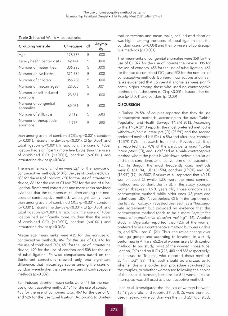
578
The use of contraceptive method patterns İstanbul Tıp Fakültesi Dergisi • J Ist Faculty Med 2021;84(4):574-81
than among users of combined OCs (p=0.001), condom (p<0.001), intrauterine device (p<0.001), CI (p<0.001) and tubal ligation (p<0.001). In addition, the users of tubal ligation had significantly more live births than the users of combined OCs (p<0.001), condom (p<0.001) and intrauterine device (p=0.043).
The mean ranks of children were 327 for the non-use of contraceptive methods, 510 for the use of combined OCs, 605 for the use of condom, 650 for the use of intrauterine device, 661 for the use of CI and 792 for the use of tubal ligation. Bonferroni corrections and mean ranks provided evidence that the numbers of children among the non-users of contraceptive methods were significantly lower than among users of combined OCs (p=0.001), condom (p<0.001), intrauterine device (p<0.001), CI (p<0.001) and tubal ligation (p<0.001). In addition, the users of tubal ligation had significantly more children than the users of combined OCs (p<0.001), condom (p<0.001) and intrauterine device (p=0.043).
Miscarriage mean ranks were 435 for the non-use of contraceptive methods, 467 for the use of CI, 476 for the use of combined OCs, 481 for the use of intrauterine device, 490 for the use of condom and 508 for the use of tubal ligation. Pairwise comparisons based on the Bonferroni corrections showed only one significant difference, that miscarriage scores among the users of condom were higher than the non-users of contraceptive methods (p=0.002).
Self-induced abortion mean ranks were 448 for the non-use of contraceptive method, 454 for the use of condom, 455 for the use of combined OCs, 469 for the use of CI and 526 for the use tubal ligation. According to Bonfer-
roni corrections and mean ranks, self-induced abortion was higher among the users of tubal ligation than the condom users (p=0.004) and the non-users of contracep-tive methods (p<0.001).
The mean ranks of congenital anomalies were 358 for the use of CI, 377 for the use of intrauterine device, 386 for the use of condom, 458 for the use of tubal ligation, 467 for the use of combined OCs, and 502 for the non-use of contraceptive methods. Bonferroni corrections and mean ranks evidenced that congenital anomalies were signifi-cantly higher among those who used no contraceptive methods than the users of CI (p<0.001), intrauterine de-vice (p<0.001) and condom (p<0.001).
DISCUSSION
In Turkey, 26.5% of couples reported that they do use contraceptive methods, according to the data Turkish Population and Health Surveys (TNSA) 2013. According to the TNSA 2013 reports, the most preferred method is withdrawal/coitus interrupts (CI) (25.5%) and the second preferred method is IUDs (16.8%) and after that, condom (15.8%) (17). In research from India, Kovavisarach E et al. reported that 70% of the participants used “coitus interruptus” (CI), and is defined as a male contraceptive method where the penis is withdrawn before ejaculation and is not considered an effective form of contraception (18). In Bingöl, the most frequently used methods were CI (23.1%), IUD (21.5%), condom (19.8%) and OC (13.9%) (19). In 2007, Bozkurt et al. reported that 40.7% women used CI (while IUDs were the most common method, and condom, the third). In this study, younger women (between 17-30 years old) chose condom as a contraceptive method, while older ones (45 years and older) used IUDs. Nevertheless, CI is in the top three of the list (20). Kulczycki revealed this result as a “husband-wife agreement” but provided no evidence that this contraceptive method tends to be a more “egalitarian mode of reproductive decision making” (16). Another study in Diyarbakır reported that 42% of the women preferred to use a contraceptive method but were unable to, and 57% used Cl (21). Thus, the ratios change over the age groups and according to location. In a study performed in Ankara, 65.2% of women use a birth control method. In our study, most of the women chose tubal ligation, OCs and /or IUDs (728, 480 and 586 respectively), in contrast to Tountas, who reported these methods as “limited” (22). This result should be analyzed as to whether this is a co-decision procedure structured by the couples, or whether women are following the choice of their sexual partners, because for 611 women, coitus interruptus was still used as a contraceptive method.
Ilhan et al. investigated the choices of women between 15-49 years old, and reported that IUDs were the most used method, while condom was the third (23). Our study
Table 3: Kruskal-Wallis H test statistics
Grouping variable Chi-square dfAsymp.
sig.
Age 178.737 5 .000
Family health center visits 42.444 5 .000
Number of maternities 306.225 5 .000
Number of live births 371.782 5 .000
Number of children 365.738 5 .000
Number of miscarriages 22.005 5 .001
Number of self-induced abortions
23.531 5 .000
Number of congenital anomalies
69.071 5 .000
Number of stillbirths 3.112 5 .683
Number of therapeutic abortions
1.773 5 .880

579
The use of contraceptive method patterns İstanbul Tıp Fakültesi Dergisi • J Ist Faculty Med 2021;84(4):574-81
revealed the same results, with IUDs as the one of the top three methods. Oral contraceptives, condoms, and IUDs are available for free from FHCs. These results are associated with availability and sustainability, which are crucial for healthcare.
Those using the tubal ligation method were older than those using OCs. This could be because the older women had reached the planned number of children, unlike the younger ones (significantly, the maternity scores of the users of tubal ligation were even higher for those who used combined OCs methods (p<0.001) and condom (p=0.001).
In our study, age scores were significantly lower for those who did not use contraceptive methods. One of the main reasons for this result could be the desire to have a baby, or not being in a sexual relationship. Tountas et al. from Greece reported that adolescent participants must be considered more carefully (22). Both Tountas and our study revealed that the great majority of women followed their sexual partners’ choices, particularly for condom or CI. In the study of Kokanalı et al. coitus interruptus, may have been the most chosen because it was the first choice of adolescents that have undergone voluntary termination. After the contraception methods education, they chose neither the rhythm method nor coitus interruptus (24). Sufficient and well-structured counseling about contraception is still the optimal option. Sexual education is needed not only for legally married women, but also for single women and for the most vulnerable, such as adolescents. Well-structured sexual education focused on sexual abuse, contraceptive methods and prevention of sexually transmitted diseases are needed in public health, and should consider the features of the community, and changes in families/sexual partners caused by migration, COVID-19, etc.
Self-induced abortion seems to be an ongoing problem affecting women and babies’ health and lives. Further research is needed to find the root reasons. Family planning is also a socio-economic issue (13). Gumus et al. reported that pregnant women who describe a negative body image also described negative relations with their husbands. This finding was significant among low-income families, although in this group, 80.7% of women reported planning their pregnancy (25). Ending an unwanted pregnancy has also been subject to legislation. Not only the women’s but also the “legal” husband’s approval is needed (15). Care for the baby and the mother is an important responsibility of the state.
Some studies reported that the “educational status” could affect the “practiced contraceptive method” (4, 26, 27). For example, Doğru et al. has reported that for female university graduates, 69.2% use modern methods (28). Being employed could also affect the method. In 2013, The Turkish Demographic and Health Survey
(TDHS) revealed that employed women preferred modern contraceptive methods over traditional (17). In various societies, the decision to choose the contraceptive method is made by the husband and/or mother-in-law, rather than the woman (4). This traditional situation has negative effects: if the woman needs to get the consent/approval of the decision-makers, the method is more likely to be traditional, or she could be forced into a method not suitable for her (29). In other words, pregnant women may not be able to choose their delivery method. “For decision making”, the family physicians and nurses/midwives should be involved as well as the couples (30).
In our study, the results concerning miscarriage and condom usage are interesting: the only significant difference was that miscarriage scores were higher among the users of condom than the non-users of contraceptive methods (p=0.002). This raises a new research question; why miscarriages are more correlated with the usage of condoms than without any methods. Another research question is whether the couples are using this method properly. Self-induced abortion was significantly higher among the users of tubal ligation than the condom users (p=0.004), and the non-users of contraceptive methods (p<0.001). This shows that women and/or their physicians found an irreversible solution for this problem by choosing tubal ligation.
No couples used vasectomy as a contraceptive method, while tubal ligation was one of the most used. All over the world, vasectomy as a contraceptive method is less chosen than tubal ligation (31).
CONCLUSION
Every minute, 380 women worldwide become pregnant, and of these, half of them face unwanted or unplanned pregnancies, and suffer complications, have miscarriages, and dies. Differential failure risk due to ethnicity and socioeconomic factors could underlie this phenomenon. The Astana declaration recommends “health for all”, and the need to reduce risks through family planning, improved sexual health counseling and education about contraception. Well-structured education programs are needed, not only for individuals and the general population, but also for healthcare workers.
The sociodemographic structure has changed due to the increased refugee population, migration, and globaliza-tion; therefore, sexual education of adolescents should be considered from this perspective.
All people have the right to access quality reproductive health services. Providing counseling on the contracep-tive methods is among the duties of family physicians. To achieve this goal, primary care needs to take a pa-tient-centered, people-oriented holistic approach with continuity of care.
580
The use of contraceptive method patterns İstanbul Tıp Fakültesi Dergisi • J Ist Faculty Med 2021;84(4):574-81
One of the social determinants of health is the rate of the deaths of mother and newborn. Planned pregnancy with well-structured care is essential to achieve this goal. Appropriate contraceptive methods are also important points to women’s health. As the family physicians are the gatekeepers, the discipline also includes “compre-hensiveness, prevention, treatment and rehabilitation, and bio-psycho-social dimensions”. Nevertheless, the care of family physicians in the follow-up of fertile women remains as the key-point.
StrengthsOne of the strengths of the study was that the data were collected from primary care records. For this reason, the analysis of the data we obtained revealed the knowledge and attitudes of people who should receive services in the area of birth control methods. Based on these results, it provides foresight to provide the necessary information on this subject. In this regard, it has been stated which ef-forts should be made to reach the desired level of health.
LimitationsIt is a limitation that the study is conducted only on patients’ records who apply to a primary care facility. For this reason, the situation of those who do not apply to any health institution is unknown. For this, community-based household studies should be conducted. Although family physicians need to know all the characteristics of the registered population, today it is not possible to record these data in a healthy way since the number of registered patients per physician is high. Also, it is difficult for every physician to devote time to special topics and counseling.
It is important to find out to what effect women have ac-cess to methods that can be applied in terms of family planning. In addition, revealing women’s thoughts on family planning through qualitative research can also make an important contribution. The lack of these quali-tative data in our study is one of the limitations.
Ethics Committee Approval: This study was approved by the Ethical Committee of the Dokuz Eylul University (Date: 21.06.2018, No: 15-37).
Informed Consent: Written consent was obtained from the par-ticipants.
Peer Review: Externally peer-reviewed.
Author Contributions: Conception/Design of Study- Ö.G., H.P., O.A., Ü.B., S.Y.; Data Acquisition- O.A., Ü.B., G.B., S.İ.E.Y.; Data Analysis/Interpretation- Ö.G., H.P., G.B., S.İ.E.Y.; Drafting Ma-nuscript- Ö.G., H.P., G.B.; Critical Revision of Manuscript- Ö.G., N.Ö.; Final Approval and Accountability- Ö.G., H.P., O.A., Ü.B., S.İ.E.Y., G.B., S.Y., N.Ö.
Conflict of Interest: Authors declared no conflict of interest.
Financial Disclosure: Authors declared no financial support.
Acknowledgemet: The researchers would like to thank Simon Mumford for proofreading of this study.
Etik Komite Onayı: Bu çalışma için etik komite onayı Dokuz Ey-lül Üniversitesi Etik Kurulu’ndan alınmıştır (Tarih: 21.06.2018, No: 15-37).
Bilgilendirilmiş Onam: Katılımcılardan bilgilendirilmiş onam alınmıştır.
Hakem Değerlendirmesi: Dış bağımsız.
Yazar Katkıları: Çalışma Konsepti/Tasarım- Ö.G., H.P., O.A., Ü.B., S.Y.; Veri Toplama- O.A., Ü.B., G.B., S.İ.E.Y.; Veri Analizi/Yo-rumlama- Ö.G., H.P., G.B., S.İ.E.Y.; Yazı Taslağı- Ö.G., H.P., G.B.; İçeriğin Eleştirel İncelemesi- G Ö.G., N.Ö.; Son Onay ve Sorum-luluk- Ö.G., H.P., O.A., Ü.B., S.İ.E.Y., G.B., S.Y., N.Ö.
Çıkar Çatışması: Yazarlar çıkar çatışması beyan etmemişlerdir.
Finansal Destek: Yazarlar finansal destek beyan etmemişlerdir.
Teşekkür: Araştırmacılar, bu çalışmanın redaksiyonunu yaptığı için Simon Mumford’a teşekkür ederler.
REFERENCES
1. Walraven G. The 2018 Astana Declaration on Primary Health Care, is it useful? J Glob Health 2019;9(1):010313. [CrossRef]
2. Eroğlu K, Koç G. Dünden bugüne sağlık mevzuatında kadın sağlığı kapsamında ana çocuk sağlığı hemşirelik hizmetleri. Anadolu Hemşirelik ve Sağlık Bilimleri Dergisi 2012;15(2):136-51.
3. World Health Organization. Maternal Health and Safe Motherhood Programme. (1996). Mother-baby package: implementing safe motherhood in countries:practical guide. World Health Organization.https://apps.who.int/iris/handle/10665/63268
4. Sağsöz N, Bayram M, Kamacı M. Kırıkkale ili ve çevresinde kullanılan kontraseptif yöntemler. J Clin Obstet Gynecol 2000;10(4):266-9.
5. Apter D. Contraception options: Aspects unique to adolescent and young adult. Best Pract Res Clin Obstet Gynaecol 2018;48:115-27. [CrossRef]
6. Yamamoto Y, Matsumoto K. Choice of contraceptive methods by women’s status: Evidence from large-scale microdata in Nepal. Sex Reprod Healthc 2017;14:48-54. [CrossRef]
7. Yusuf RA, Dongarwar D, Yusuf ZI, Salihu HM. Association between Intimate Partner Violence, Knowledge and Use of Contraception in Africa: Comparative Analysis across Five African Regions. Int J MCH AIDS 2020;9(1):42-52. [CrossRef]
8. Islam MK, Haque MR, Hema PS. Regional variations of contraceptive use in Bangladesh: A disaggregate analysis by place of residence. PLoS One 2020;15(3):e0230143. [CrossRef]
9. Hameed W, Azmat SK, Ali M, Sheikh MI, Abbas G, Temmerman M, et al. Women’s empowerment and contraceptive use: the role of independent versus couples’ decision-making, from a lower middle income country perspective. PloS one 2014;9(8):e104633. [CrossRef]

581
The use of contraceptive method patterns İstanbul Tıp Fakültesi Dergisi • J Ist Faculty Med 2021;84(4):574-81
10. Smith JD, Oakley D. Why do women miss oral contraceptive pills? An analysis of women’s self-described reasons for missed pills. J Midwifery Womens Health 2005;50(5):380-5. [CrossRef]
11. Gomes Alves P, Petersen I, Stevenson F. Searching for information on the risks of Combined Hormonal Contraceptives on the internet: A Qualitative Study across six European Countries. J Med Internet Res 2019;21(3):e10810. [CrossRef]
12. Mansour D, Inki P, Gemzell-Danielsson K. Efficacy of contraceptive methods: a review of the literature. Eur J Contracept Reprod Health Care 2010;15(1):4-16. [CrossRef]
13. Karaoğlan D, Saraçoğlu DŞ. Women’s socioeconomic status and choice of birth control method: an investigation for the case of Turkey. J Biosoc Sci 2021;3(1):137-56. [CrossRef]
14. Fisek NH, Sumbuloglu K. The effects of husband and wife education on family planning in rural Turkey. Stud Fam Plann 1978;9(10/11):280-5. [CrossRef]
15. Rahim Tahliyesi ve Sterilizasyon Hizmetlerinin Yürütülmesi ve Denetlenmesine İlişkin Tüzük. Bakanlar Kurulu Kararı. 14/11/1983, No: 83/7395 Ankara.
16. Kulczycki A. Husband-wife agreement, power relations and contraceptive use in Turkey. Int Fam Plan Perspect 2008;34(3):127-37. [CrossRef]
17. Hacettepe University Institute of Population Studies, 2013 Turkey Demographic and Health Survey http://www.hips.hacettepe.edu.tr/tnsa2013/rapor/TDHS_2013_main.report.pdf Publication No: IPS-HU.14.02
18. Kovavisarach E, Saringcarnan P. Coitus interruptus in female patients seeking services at Obstetrics and Gynecology Department in Rajavithi Hospital. J Med Assoc Thai 2010;93(12):1356-9.
19. Kaya H, Tatlı H, Açık Y, Deveci SE. Bingöl ili uydukent sağlık ocağı bölgesindeki 15–49 yaş kadınların aile planlaması yöntemi kullanım düzeyinin belirlenmesi. Fırat Üniversitesi Sağlık Bilimleri Dergisi 2008;22(4):185-91.
20. Bozkurt N, Özkan S, Onan A, Korucuoğlu Ü, Aygün R, Himmetoğlu Ö. Distribution of contraceptive use in a Turkish population. Eur J Obstet Gynecol Reprod Biol 2007;131(1):52-6. [CrossRef]
21. Çakmak A, Ertem M, Karazeybek H. Diyarbakır Çocuk Hastanesine yatırılan çocukların annelerinin sağlık hizmetlerine erişimi. Turkiye Klinikleri Journal of Pediatrics 2007;16(2):82-9.
22. Tountas Y, Dimitrakaki C, Antoniou A, Boulamatsis D, Creatsas G. Attitudes and behavior towards contraception among Greek women during reproductive age: a country-wide survey. Eur J Obstet Gynecol Reprod Biol 2004;116(2):190-5. [CrossRef]
23. İlhan M, Yıldırım A, Maral I. Contraceptive methods used by women between 15-49 years old and the cause of not using contraceptive methods in the semiurban and urban areas in Ankara. J Clin Obstet Gynecol 2002;12(1):66-72.
24. Kokanalı D, Kuntay Kokanalı M, Ayhan S, Cengaver N, Özakşit G, Engin-Üstün Y. Contraceptive choices of adolescents before and after the voluntary termination of pregnancy. J Obstet Gynaecol 2019;39(6):822-6. [CrossRef]
25. Gumus AB, Çevik N, Hataf Hyusni S, Biçen Ş, Keskin G, Tuna Malak A. Gebelikte benlik saygısı ve beden imajı ile ilişkili özellikler. Anatolian Journal of Clinical Investigation 2011;5(1):7-14.
26. Bajwa SK, Bajwa SJS, Ghai GK, Singh K, Singh N. Knowledge, attitudes, beliefs, and perception of the north Indian population toward adoption of contraceptive practices. Asia Pac J Public Health 2012;24(6):1002-12. [CrossRef]
27. Fisher W, Boroditsky R, Morris B. The 2002 Canadian contraception study: part I. J Obstet Gynaecol Can 2004;26(6):580-90. [CrossRef]
28. Dogru HY, Oktay G, İşgüder ÇK, Özsoy AZ, Çakmak B, Delibaş İB, et al. Yaş gruplarına göre kadınların aile planlamasına bakışları ve seçtikleri yöntemlerin değerlendirilmesi: Tersiyer tek merkez deneyimi. Dicle Tıp Dergisi 2016;43(3):413-8.
29. Pekkurnaz D. Employment status and contraceptive choices of women with young children in Turkey. Feminist Economics 2020;26(1):98-120. [CrossRef]
30. Meriç M, Ergün G, Pola G, Yaycı E, Dal Yılmaz Ü. Women’s experience of cesarean section: A qualitative study. Cyprus J Med Sci 2019;4(3):183-8. [CrossRef]
31. Goldstein M. Surgical management of male infertility and other scrotal disorders. In Campbell’s Urology, 8th edition. Walsh PC, Retik AB, Vaughan ED, Wein AJ, eds. Philadelphia W.B. Saunders; 2002. p 1573-4.

582
Application of test batteries in security guardsİstanbul Tıp Fakültesi Dergisi • J Ist Faculty Med 2021;84(4):582-9
Corresponding author/İletişim kurulacak yazar: [email protected]
Submitted/Başvuru: 19.01.2021 • Revision Requested/Revizyon Talebi: 08.03.2021 •Last Revision Received/Son Revizyon: 08.03.2021 • Accepted/Kabul: 08.03.2021 • Published Online/Online Yayın: 24.09.2021
RESEARCH - ARAŞTIRMADOI: 10.26650/IUITFD.2021.864086
İst Tıp Fak Derg 2021 / J Ist Faculty Med 2021
AN EVALUATION OF CONTINUOUS, SELECTIVE ATTENTION, REASONING AND COLLISION TIME PREDICTION SKILLS IN SECURITY GUARDS
GÜVENLİK GÖREVLİLERİNDE SÜREKLİ, SEÇİCİ DİKKAT, MUHAKEME VE ÇARPIŞMA ZAMANI TAHMİN BECERİLERİNİN DEĞERLENDİRİLMESİ
Halim İŞSEVER1 , Elif EZİRMİK1 , Nefise ŞEKER1 , Zeynep Betül SAĞLAM1 , Gözde ÖZTAN2 , Fatma CANATAR1
1Istanbul University, Istanbul Faculty of Medicine, Public Health Department, Istanbul, Turkey 2Istanbul University, Istanbul Faculty of Medicine, Medical Biology, Istanbul, Turkey
ORCID IDs of the authors: H.İ. 0000-0002-5435-706X; E.İ. 0000-0001-6828-4378; N.Ş. 0000-0003-4722-3087; Z.B.S. 0000-0001-9538-2969; G.Ö. 0000-0002-2970-1834; F.C. 0000-0001-6614-3862
Cite this article as: Issever H, Ezirmik E, Seker N, Saglam ZB, Oztan G, Canatar F. An evaluation of continuous, selective attention, reasoning and collision time prediction skills in security guards. J Ist Faculty Med 2021;84(4):582-9. doi: 10.26650/IUITFD.2021.864086
ABSTRACT
Objective: This study aims at investigating the success of the continuous and selective attention, reasoning and collision prediction time test batteries which were among the tests used in the psychotechnical evaluation of security guards in meeting standard norms.
Materials and Methods: One hundred twenty-three security guards (95 male, 28 female) working as security guards at the university and agreeing to participate in the study were included in the study. Each participant was administered the Continuous Attention Test, Selective Attention Test (SEDT), Reasoning Test (MT), Collision Time Test on the computer-aided system, respectively, and the scores obtained were recorded on the computer-aided system. Licensed evaluation system software developed by ALG Psikoteknik (https://www.algpsikoteknik.com) was used as the test system. Norm values were created from the data collected from 100% Turkish society. Norm values are values for drivers in the computer-aided system.
Results: From the test batteries applied, 99 participants (80.5%) in the continuous attention test, 117 participants (95.1%) in the selective attention test, 111 participants (90.2%) in the reasoning test and 73 participants (59.3%) in the collision time prediction test were successful. In three of the 4 test batteries applied, over 80% success was achieved and almost 60% success was provid-ed in the collision time prediction test.
ÖZET
Amaç: Bu çalışmada güvenlik görevlilerinde, psikoteknik de-ğerlendirmede kullanılan testlerden sürekli ve seçici dikkat, mu-hakeme ve çarpışma zaman tahmin test bataryalarının standart normları karşılamada olan başarılarının araştırılması amaçlanmış-tır.
Gereç ve Yöntemler: Üniversitede güvenlik görevlisi olarak ça-lışan, çalışmaya katılmayı kabul eden 123 güvenlik görevlisi (95 erkek, 28 kadın) çalışma kapsamına dahil edildi. Her bir katılım-cıya sırası ile bilgisayar destekli sistem üzerinde Sürekli Dikkat Testi (SÜDT), Seçici Dikkat Testi (SEDT), Muhakeme Testi (MT), Çarpışma Zamanı Testi uygulanarak alınan puanlar bilgisayar destekli sistem üzerinde kayıt edildi. Test sistemi olarak ALG psikoteknik (https://www.algpsikoteknik.com) tarafından gelişti-rilen lisanslı değerlendirme sistem yazılımı kullanıldı. Bilgisayar destekli sistemde norm değerleri, yüzde yüz Türk toplumundan toplanan verilerden oluşturulmuştur. Norm değerler sürücülere yönelik değerlerdir.
Bulgular: Uygulanan test bataryalarından; Sürekli dikkat testinde 99 katılımcı (%80,5), Seçici dikkat testinde 117 katılımcı (%95,1), Muhakeme testinde 111 katılımcı (%90,2), Çarpışma zamanı tah-min testinde ise 73 katılımcı (%59,3) başarılı olmuştur. Uygulanan 4 test bataryasının üç tanesinde; %80’in üzerinde başarı, çarpış-ma zamanı tahmin testinde ise %60’a yakın bir başarı elde edil-miştir.
Content of this journal is licensed under a Creative Commons Attribution-NonCommercial 4.0 International License.

583
Application of test batteries in security guardsİstanbul Tıp Fakültesi Dergisi • J Ist Faculty Med 2021;84(4):582-9
RESEARCH - ARAŞTIRMADOI: 10.26650/IUITFD.2021.864086
İst Tıp Fak Derg 2021 / J Ist Faculty Med 2021
AN EVALUATION OF CONTINUOUS, SELECTIVE ATTENTION, REASONING AND COLLISION TIME PREDICTION SKILLS IN SECURITY GUARDS
GÜVENLİK GÖREVLİLERİNDE SÜREKLİ, SEÇİCİ DİKKAT, MUHAKEME VE ÇARPIŞMA ZAMANI TAHMİN BECERİLERİNİN DEĞERLENDİRİLMESİ
Halim İŞSEVER1 , Elif EZİRMİK1 , Nefise ŞEKER1 , Zeynep Betül SAĞLAM1 , Gözde ÖZTAN2 , Fatma CANATAR1
1Istanbul University, Istanbul Faculty of Medicine, Public Health Department, Istanbul, Turkey 2Istanbul University, Istanbul Faculty of Medicine, Medical Biology, Istanbul, Turkey
ORCID IDs of the authors: H.İ. 0000-0002-5435-706X; E.İ. 0000-0001-6828-4378; N.Ş. 0000-0003-4722-3087; Z.B.S. 0000-0001-9538-2969; G.Ö. 0000-0002-2970-1834; F.C. 0000-0001-6614-3862
Cite this article as: Issever H, Ezirmik E, Seker N, Saglam ZB, Oztan G, Canatar F. An evaluation of continuous, selective attention, reasoning and collision time prediction skills in security guards. J Ist Faculty Med 2021;84(4):582-9. doi: 10.26650/IUITFD.2021.864086
INTRODUCTION
Private security has been one of the sectors that has developed significantly since the concept of the public service began to change in Turkey. The sector consists of many sub-sectors that provide services to meet various types of security needs of the society (1). Today the need for security is increasing in parallel with the increase in the quality and quantity of the assets that individuals possess. If properly used, the private security guards will effectively meet individual security needs, provide savings on security services, reduce the burden of overall security and lay the groundwork for more convenient and efficient work in their areas. The fragmentary and variable structure of modern society which is associated with the perception of danger and threat increases the need for security and makes police and other private security services more important (2).
However, security guards are exposed to violent attacks by citizens during their duties (3). Moreover, employees in these professions cannot withdraw from their working environment, even if they are under serious threat because they are called upon for help, and they also have a great responsibility to maintain law and order in society. Therefore, the problem-solving skills and personality traits of security guards become important when it comes to intervening in an incident (4, 5). The term “Problem” is defined as the obstacles that an individual faces at any stage to achieve the intended goal. The problem, which is expressed as an obstacle faced by the individual, has certain characteristics which cause uncertainty in the human mind, which must be solved and which bother that individual (6). The effective power available to overcome a wide range of issues from everyday life to traumatic incidents is defined as problem solving. The problem solving stage begins with cognitive processes aimed at a goal. Problem-solving skills mediate individuals’ ability to adapt effectively to the environment in which they live. While some problems have correct answers or clear solutions, others do not. In this case, interdisciplinary knowledge and multifaceted thinking are important in solving problems (7). Different approaches to problem solving have been defined here according to the characteristics of the problem and the person interested in the problem.
The traditional approach involves identifying the problem, analysing its causes, identifying alternative
solutions, evaluating and applying solutions, and determining whether the problem has been solved (8). In addition to the solution method, the continuous and selective attention, reasoning and reaction time of the security guard against the incidents also occupy an important place. Timely intervention in events will prevent the growth of negative consequences. If organizations make wrong choices in recruitment, there will be undesirable consequences including waste of resources for the organization and unhappiness at work for the staff. Choosing the right staff for the right job will make a positive contribution to the organization in terms of ensuring job satisfaction, using creativity and being successful (9). This study aimed to investigate the achievements of security guards in meeting standard norms by using continuous and selective attention, reasoning and collision time estimation tests batteries which are among the psychotechnical evaluation tests.
MATERIALS AND METHODS
One hundred twenty-three security guards (95 male, 28 female) working as security guards at the university and agreeing to participate in the study were included in the study. The success rate of the test of the participants was accepted as 0.60 and the minimum sample size (Type I error 0.05, Type II error 0.20, 0.80 power) was calculated as 110 people with a 20% error margin. Tests, which were applied to the participants by means of a computer-aided system installed in a quiet room, which was closed to external stimuli as shown by the university security supervisor, were explained in detail by Public Health Medical Specialty assistants and implemented after preliminary tests were carried out. After practitioners were trained by a specialist organization and entitled to receive a certificate of participation, the implementation phase was conducted. After answering 13 questions with regard to demographic features, each participant was administered the Continuous Attention Test, Selective Attention Test (SEDT), Reasoning Test (MT), Collision Time Test on the computer-aided system, respectively, and the scores obtained were recorded on the computer-aided system. Licensed evaluation system software developed by ALG Psikoteknik (https://www.algpsikoteknik.com) was used as the test system. Norm values were created from the data collected from 100% Turkish society. There are 12 test batteries in the system that measure mental and psychomotor skills. Four of the 12 test batteries were
Conclusion: Creating norm values specific to security guards and applying psychotechnical evaluation test batteries are ben-eficial in evaluating psychological situations.
Keywords: Security guards, psychotechnical evaluation, test batteries
Sonuç: Güvenlik görevlilerine özgü norm değerlerinin oluştu-rulması ve psikoteknik değerlendirme test bataryalarının uygu-lanması psikolojik durumların değerlendirilmesinde fayda sağla-maktadır.
Anahtar Kelimeler: Güvenlik görevlileri, psikoteknik değerlen-dirme, test bataryaları
584
Application of test batteries in security guardsİstanbul Tıp Fakültesi Dergisi • J Ist Faculty Med 2021;84(4):582-9
implemented with computer support that did not require a driver system. Ethical approval for the study was obtained from İstanbul University; İstanbul Faculty of Medicine; Clinical Research Ethics Committee (Date 19.08.2019, No: 1026). In statistical analysis, student’s t-test and chi-square test were applied in independent groups. Statistical significance was accepted as p<0.05 and two-way.
TESTS CONDUCTED
CONTINUOUS ATTENTION TEST
Definition of the testFor this test, a specific traffic sign was selected as the target figure, and distractors were created that are quite similar to this sign. The target figure and the distractors were arranged in a matrix of 11x17. There are 50 target figures in this matrix consisting of a total of 186 distractors. The task of the participants is to quickly find and mark the same target shape at the top of the Matrix among the shapes in the Matrix. Attention in general means that among the many sets of internal and external stimulants, the critical one is selected for further analysis. In the working environment, they have to pay attention to many stimulants coming from different modalities such as visual or audio, to perceive them and to continue their safe behavior (10).
Purpose of the testThis test was developed to measure the ability of drivers to quickly and accurately identify and select a specific stimulant that is critical at the moment in the routine and monotonous flow of information so that they can demonstrate the necessary cognitive and motor skills in a traffic environment. It was also implemented to test the ability of security guards to quickly and accurately select a specific stimulant.
Overall Test Score Index: It is calculated by the formula [(Total Number of Correct Answers)/((Total Number of Skipped Responses) + (Total Number of Correct Answers) + (Total Number of Incorrect Answers) x100].
This index is a general score that shows the participant’s level of continuous attention. The raw scores obtained by the participants from the test are compared with the norm values and evaluated to determine whether they received above the norm score.
SELECTIVE ATTENTION TEST (SEDT)
Definition of the testEach question in this test consists of 6 traffic signs. Three of these signs are located on the upper row of a 3x3 Matrix, and 3 of them are located on the left side. Two of the traffic signs on the top and left are identical. The Test contains a total of 60 matrices. The participant must quickly find identical shapes, each located on the left and top of the Matrix, and show the intersection of these shapes by marking them on the screen with the index finger or a pen.
Purpose of the test The traffic environment is a task that involves the selective attention of the driver in many cases so that he/she can demonstrate safe driving skills. For this reason, this test is aimed at evaluating the driver’s ability to selectively pay attention to constantly changing environmental and road conditions so that they can demonstrate the necessary cognitive and motor skills in a traffic environment. In order to measure the selective attention of the driver in the test, an attempt is made to measure the ability to select similar ones from different stimulants and ignore others.
Correct Answer Percentage (Overall Index): [(Total Number of Correct Answers/Total Number of Questions) x100]
This index is a general score indicating the participant’s selective attention level. The raw scores obtained by the participants from the test are compared with the norm values and evaluated to determine whether they received above the norm score.
REASONING TEST (MT)
Definition of the testThis test includes 50 multiple choice question items. The questions are sorted from easy to difficult. In each question, the participant is first asked to find the logical relationship between the two figures. Then, the participant must find and mark in which of the options another form of this relationship is presented. Each question has 4 options, and only one of them is the correct answer. The person has an answer time of 3 minutes for each question. The test is automatically terminated when the participant incorrectly answers or skips 2 questions in a row.
a) Colour: The change in the relationship between the two figures is based on color. In other words, the color of the first figure changes, resulting in the second figure.
b) Form: The change in the relationship between the two figures is based on the form. In other words, the form of the first figure changes, resulting in the second figure.
c) Quantity: The change in the relationship between the two figures is based on numbers. The second figure differs from the first figure in number or quantity.
d) Rotation: The change in the relationship between the two figures is based on rotation. The first figure or a part of the figure is rotated in various degrees, resulting in the second figure. When a participant gives an incorrect answer or skips a question twice in a row, the test automatically ends without moving on to other questions.

585
Application of test batteries in security guardsİstanbul Tıp Fakültesi Dergisi • J Ist Faculty Med 2021;84(4):582-9
Purpose of the test The main purpose of this test is to measure one’s gen-eral reasoning ability using nonverbal geometric shapes, regardless of culture. In the test, the participant’s task is to find and determine the relationship between the two figures and then find the correct answer by determining the figure containing the same rule among the figures presented in four options. The main purpose of the test is to measure the reasoning ability through matching, regardless of culture and knowledge of mathematics or arithmetic.
Correct Answer Percentage (Overall Index): [(Total Number of Correct Answers/Total Number of Questions) x100]
This index is a general score indicating the participant’s selective attention level. The raw scores obtained by the participant from the test are compared with the norm values and evaluated to determine whether they received above the norm score (10).
COLLISION TIME TEST
Purpose of the testThe participant is asked to estimate both the collision time and the collision location of the objects.
Test featuresThe moment (duration) at which objects actually collide with each other is considered the center, and the interval from the moment the balls disappear on the screen until the moment when a 77-pixel time was added to the moment when they collide is determined as the response area. Pursuant to the end of this area, the interval from the point of collision (different for each item) until the moment after 1786 ms. is considered to be the late response area. If no response is given, it is passed to the next question when the balls arrive at the end of the late response area, and this question is considered to be skipped. The moment of collision, which is the center of the response area, is rated as “100” and the score decreases at a certain rate as you go from the center to the ends.
The extreme points of the response area and the late response area are “0” points. When calculating the location score, the sum of the distances of the point that the driver responds to for each question, in pixels, to the actual collision point, is divided by the number of questions that the driver responds to. Giving the response after 1056 pixels, which is the screen limit, causes the response to be considered skipped. After the driver specifies each prediction by tapping the screen, the time and location specified by the driver is automatically calculated. According to where this time and place coincide in the designated response area, the driver is given two separate scores for both time and location.
Security guards are asked to predict both the collision time and the collision location of objects oscillating in two directions on the computer screen.
Evaluation of the resultsThe following measurements are taken regarding the results of the test. The Collision Time Prediction Score (Overall Index) is the collision location distance score (Overall Index), the number of late responses and the number of skipped responses. The Collision Time Prediction Score (Overall Index) is a general score that indicates the time of collision. By comparing the raw scores of the participant from the test with the norm values, it is evaluated whether they get above the norm score (10).
FINDINGS
A total of 123 security guards were included in the study, 95 men (77.2%) and 28 women (22.8%) who worked as security guards at the university and agreed to participate in the study. Distribution of age, working year and total working year by gender is given in Table 1. The marital status, working style and educational status of the individuals covered by the study are given in Table 2. According to the distribution of marital status, 34 were single (27.6%), 89 were married (72.4%), 107 (87%) were working in shifts, and the majority 105 (85.4%) were high school graduates.
Table 1: Distribution of age, year of work and total years of work of individuals covered within the scope of the study by gender
Gender AgeWorking
yearTotal
working year
Male
Mean 35.24 9.28 13.76
S. Deviation 4.71 3.57 5.70
Median 35 10 14
Minimum 24 2 3
Maximum 49 16 33
Female
Mean 36.04 10.87 13.16
S. Deviation 5.35 3.02 4.73
Median 34.5 10 13.25
Minimum 28 3.5 3.5
Maximum 48 16 26
Total Mean 35.42 9.648 13.63
S. Deviation 4.8 3.51 5.48
Median 35 10 14
Minimum 24 2 3
Maximum 49 16 33
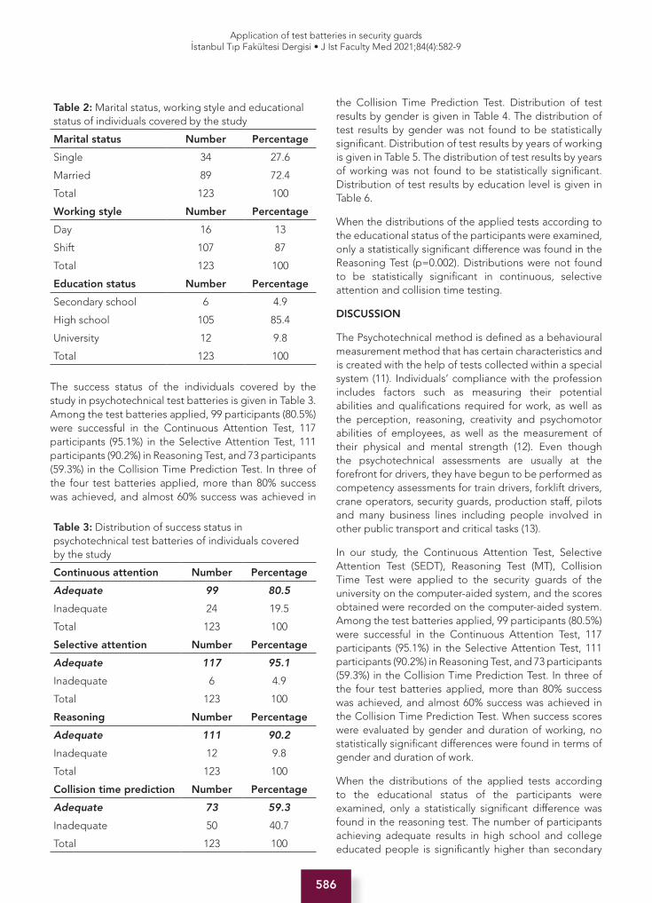
586
Application of test batteries in security guardsİstanbul Tıp Fakültesi Dergisi • J Ist Faculty Med 2021;84(4):582-9
The success status of the individuals covered by the study in psychotechnical test batteries is given in Table 3. Among the test batteries applied, 99 participants (80.5%) were successful in the Continuous Attention Test, 117 participants (95.1%) in the Selective Attention Test, 111 participants (90.2%) in Reasoning Test, and 73 participants (59.3%) in the Collision Time Prediction Test. In three of the four test batteries applied, more than 80% success was achieved, and almost 60% success was achieved in
the Collision Time Prediction Test. Distribution of test results by gender is given in Table 4. The distribution of test results by gender was not found to be statistically significant. Distribution of test results by years of working is given in Table 5. The distribution of test results by years of working was not found to be statistically significant. Distribution of test results by education level is given in Table 6.
When the distributions of the applied tests according to the educational status of the participants were examined, only a statistically significant difference was found in the Reasoning Test (p=0.002). Distributions were not found to be statistically significant in continuous, selective attention and collision time testing.
DISCUSSION
The Psychotechnical method is defined as a behavioural measurement method that has certain characteristics and is created with the help of tests collected within a special system (11). Individuals’ compliance with the profession includes factors such as measuring their potential abilities and qualifications required for work, as well as the perception, reasoning, creativity and psychomotor abilities of employees, as well as the measurement of their physical and mental strength (12). Even though the psychotechnical assessments are usually at the forefront for drivers, they have begun to be performed as competency assessments for train drivers, forklift drivers, crane operators, security guards, production staff, pilots and many business lines including people involved in other public transport and critical tasks (13).
In our study, the Continuous Attention Test, Selective Attention Test (SEDT), Reasoning Test (MT), Collision Time Test were applied to the security guards of the university on the computer-aided system, and the scores obtained were recorded on the computer-aided system. Among the test batteries applied, 99 participants (80.5%) were successful in the Continuous Attention Test, 117 participants (95.1%) in the Selective Attention Test, 111 participants (90.2%) in Reasoning Test, and 73 participants (59.3%) in the Collision Time Prediction Test. In three of the four test batteries applied, more than 80% success was achieved, and almost 60% success was achieved in the Collision Time Prediction Test. When success scores were evaluated by gender and duration of working, no statistically significant differences were found in terms of gender and duration of work.
When the distributions of the applied tests according to the educational status of the participants were examined, only a statistically significant difference was found in the reasoning test. The number of participants achieving adequate results in high school and college educated people is significantly higher than secondary
Table 2: Marital status, working style and educational status of individuals covered by the study
Marital status Number Percentage
Single 34 27.6
Married 89 72.4
Total 123 100
Working style Number Percentage
Day 16 13
Shift 107 87
Total 123 100
Education status Number Percentage
Secondary school 6 4.9
High school 105 85.4
University 12 9.8
Total 123 100
Table 3: Distribution of success status in psychotechnical test batteries of individuals covered by the study
Continuous attention Number Percentage
Adequate 99 80.5
Inadequate 24 19.5
Total 123 100
Selective attention Number Percentage
Adequate 117 95.1
Inadequate 6 4.9
Total 123 100
Reasoning Number Percentage
Adequate 111 90.2
Inadequate 12 9.8
Total 123 100
Collision time prediction Number Percentage
Adequate 73 59.3
Inadequate 50 40.7
Total 123 100
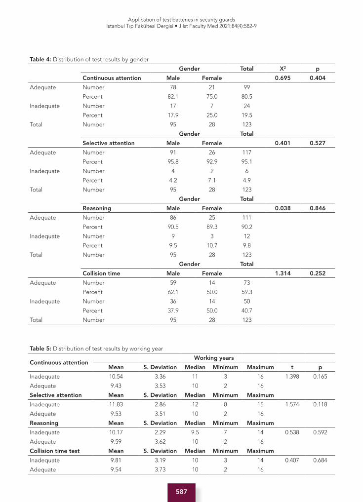
587
Application of test batteries in security guardsİstanbul Tıp Fakültesi Dergisi • J Ist Faculty Med 2021;84(4):582-9
Table 4: Distribution of test results by gender
Gender Total X2 p
Continuous attention Male Female 0.695 0.404
Adequate Number 78 21 99
Percent 82.1 75.0 80.5
Inadequate Number 17 7 24
Percent 17.9 25.0 19.5
Total Number 95 28 123
Gender Total
Selective attention Male Female 0.401 0.527
Adequate Number 91 26 117
Percent 95.8 92.9 95.1
Inadequate Number 4 2 6
Percent 4.2 7.1 4.9
Total Number 95 28 123
Gender Total
Reasoning Male Female 0.038 0.846
Adequate Number 86 25 111
Percent 90.5 89.3 90.2
Inadequate Number 9 3 12
Percent 9.5 10.7 9.8
Total Number 95 28 123
Gender Total
Collision time Male Female 1.314 0.252
Adequate Number 59 14 73
Percent 62.1 50.0 59.3
Inadequate Number 36 14 50
Percent 37.9 50.0 40.7
Total Number 95 28 123
Table 5: Distribution of test results by working year
Continuous attentionWorking years
Mean S. Deviation Median Minimum Maximum t p
Inadequate 10.54 3.36 11 3 16 1.398 0.165
Adequate 9.43 3.53 10 2 16
Selective attention Mean S. Deviation Median Minimum Maximum
Inadequate 11.83 2.86 12 8 15 1.574 0.118
Adequate 9.53 3.51 10 2 16
Reasoning Mean S. Deviation Median Minimum Maximum
Inadequate 10.17 2.29 9.5 7 14 0.538 0.592
Adequate 9.59 3.62 10 2 16
Collision time test Mean S. Deviation Median Minimum Maximum
Inadequate 9.81 3.19 10 3 14 0.407 0.684
Adequate 9.54 3.73 10 2 16

588
Application of test batteries in security guardsİstanbul Tıp Fakültesi Dergisi • J Ist Faculty Med 2021;84(4):582-9
school graduates. Since there were no previous studies on this subject, it was not possible to compare it with similar study results. The importance of personnel selection systems, which ensure that the right personnel are reached and the appropriate persons are brought into the institution, is better understood every day. The basis of personnel selection systems that will allow the selection of the right personnel is to recognize and define the work correctly (14). Difficulties and hardship in social life are especially defined as problems. Problems usually consist of uncertainties, situations where accuracy and authenticity cannot be achieved and troubles and relationships with difficulties (15). In their study on private security guards, which is a dangerous and risky profession, Balli et al. suggested that the “extrovert” personality trait explains the perception of private security guards (in management positions) as leaders (16). In professions where interpersonal relationships are intense, placing a person suitable for work will keep possible problems at a minimal level.
Limitations of the researchThe fact that the study was carried out only with security guards in a university and the compulsory use of some of the psychotechnical evaluations can be considered as limitations.
The results of this study showed that security guards were sufficiently successful in the attention, reasoning, and collision time prediction tests which were set as standard. In addition to assessing their psychological status when necessary, we believe that it would be useful to conduct a psychotechnical assessment prior to hiring those who work in critical tasks with intensive public interaction and communication (17).
Ethics Committee Approval: This study was approved by the Clinical Research Ethical Committee of the Istanbul University, Istanbul Faculty of Medicine (Date: 19.08.2019 No: 1026).
Informed Consent: Written consent was obtained from the participants.
Peer Review: Externally peer-reviewed.
Author Contributions: Conception/Design of Study- H.İ., E.E., G.Ö.; Data Acquisition- H.İ., E.E., N.Ş, Z.B.S., F.C.; Data Analysis/Interpretation- E.H.İ., G.Ö.; Drafting Manuscript- H.İ., G.Ö., F.C.; Critical Revision of Manuscript- H.İ., E.E., N.Ş., Z.B.S., G.Ö., F.C.; Final Approval and Accountability- H.İ., E.E., N.Ş., Z.B.S., G.Ö., F.C.
Conflict of Interest: Authors declared no conflict of interest.
Financial Disclosure: Authors declared no financial support.
Etik Komite Onayı: Bu çalışma için etik komite onayı İstanbul Üniversitesi, İstanbul Tıp Fakültesi Klinik Araştırmalar Etik Kuru-lu’ndan alınmıştır (Tarih: 19.08.2019 No: 1026).
Bilgilendirilmiş Onam: Katılımcılardan bilgilendirilmiş onam alınmıştır.
Hakem Değerlendirmesi: Dış bağımsız.
Yazar Katkıları: Çalışma Konsepti/Tasarım- H.İ., E.E., G.Ö.; Veri Toplama- H.İ., E.E., N.Ş, Z.B.S., F.C.; Veri Analizi/Yorumlama- H.İ., G.Ö.; Yazı Taslağı- H.İ., G.Ö., F.C.; İçeriğin Eleştirel İncelemesi- H.İ., E.E., N.Ş., Z.B.S., G.Ö., F.C.; Son Onay ve Sorumluluk- H.İ., E.E., N.Ş., Z.B.S., G.Ö., F.C.
Çıkar Çatışması: Yazarlar çıkar çatışması beyan etmemişlerdir.
Finansal Destek: Yazarlar finansal destek beyan etmemişlerdir.
Table 6: Distribution of test results by education Level
Education level
Continuous attention Sec. School High School University Total X2 p
Adequate Number 5 87 7 99 4.15 0.125
Percent. 83.3 82.8 58.3 80.5
Selective attention
Adequate Number 6 99 12 117 1.08 0.582
Percent. 100.0 94.3 100.0 95.1
Reasoning test
Adequate Number 3 96 12 111 12.5 0.002
Percent. 50.0 91.4 100.0 90.2
Collision time test
Adequate Number 3 65 5 73 2.05 0.358
Percent. 50.0 61.9 41.7 59.3

589
Application of test batteries in security guardsİstanbul Tıp Fakültesi Dergisi • J Ist Faculty Med 2021;84(4):582-9
REFERENCES
1. Aras H. Özel güvenlik görevlilerinde kurumsal uzmanlaşma: bir model önerisi. Güvenlik Bilimleri Dergisi 2018;7(1):131-58. [CrossRef]
2. Akbaş F, Dursun G, Ürün E. Özel güvenlik mesleğinin unvan sorunu ve gelişimi. Avrasya Sosyal ve Ekonomi Araştırmaları Dergisi (ASEAD) 2018;5(6):230-40.
3. Leino TM, Selin R, Summala H. Virtanen M. Violence and psychological distress among police officers and security guards. Occup Med 2011;61(6):400-6. [CrossRef]
4. Beech B, Leather P. Workplace violence in the health care sector: a review of staff training and integration of training evaluation models. Aggression Violent Behav 2006;11(1):27-43. [CrossRef]
5. Akca N, Yılmaz A, Işık O. Sağlık çalışanlarına uygulanan şiddet: özel bir tıp merkezi örneği. Ankara Sağlık Hizmetleri Dergisi 2014;13(1):1-12. [CrossRef]
6. Oğuz V, Akyol KA. Problem çözme becerisi ölçeği (PÇBÖ) geçerlik ve güvenirlik çalışması. Çukurova Üniversitesi Eğitim Fakültesi Dergisi 2015;44(1):105-22. [CrossRef]
7. Soyer MK, Bilgin A. Problem solving skill perceptions of university students according to various variables. International Conference on New Trends in Education and Their Implications; 2010; November 11-13; Antalya, Türkiye: 2010.p.307-314.
8. Ekici DI, Balım AG. Ortaokul öğrencileri için problem çözme becerilerine yönelik algı ölçeği: geçerlilik ve güvenirlik çalışması. Yüzüncü Yıl Üniversitesi Eğitim Fakültesi Dergisi 2013;10(1):67-86.
9. Telman N, Türetgen İÖ, editors. Element selection. Istanbul: Epsilon Publishing; 2004.
10. ALG Psychotechnical Evaluation Tests. Available from: URL: https://www.algpsikoteknik.com/testler.html.
11. Akyıldız H, Kayalar M. İşletmelerin ruhsal tasarımında psikoteknik yöntemin transaksiyonel analiz ile boyutlandırılması. Uludağ Üniversitesi İktisadi ve İdari Bilimler Fakültesi Dergisi 2003;22(2):75-92.
12. Spor NY. Psikoteknik ve kullanım alanları. Mesleki Sağlık ve Güvenlik Dergisi(MSG) 2001; 2(5):13-6.
13. Tütüncü Ö, Tarlan D, Mamyrkulov N. Seyahat acentalarında çalışanların işe alma sürecini algılamaları ve İzmir ili örneği, Dokuz Eylül Üniversitesi Sosyal Bilimler Enstitüsü Dergisi 2003;5(1):113-40.
14. Gürer A. Psikoteknik yöntemin personel seçiminde uygulanması: Kit’ler üzerine bir alan araştırması. Uluslararası Sosyal Araştırmalar Dergisi 2017;10(51):1007-1020. [CrossRef]
15. Bagnall RJ. Lifelong education: the institutionalisation of an illiberal and regressive ideology? Educational Philosophy and Theory 1990; 22(1):1-7. [CrossRef]
16. Balli E. The effects of personality traits on being perceived as a leader: an empirical study in a private security officer sample. Anadolu University Journal of Social Sciences 2013;13(3):85-94.
17. İssever H, Onen L, Sabuncu HH, Altunkaynak O. Personality characteristics, psychological symptoms and anxiety levels of drivers in charge of urban transportation in Istanbul. Occupational Medicine 2002;52(6);297-303. [CrossRef]
590
COVID-19 and thyroid İstanbul Tıp Fakültesi Dergisi • J Ist Faculty Med 2021;84(4):590-4
Corresponding author/İletişim kurulacak yazar: [email protected]
Submitted/Başvuru: 19.05.2021 • Revision Requested/Revizyon Talebi: 27.06.2021 •Last Revision Received/Son Revizyon: 27.06.2021 • Accepted/Kabul: 28.06.2021 • Published Online/Online Yayın: 24.09.2021
REVIEW - DERLEMEDOI: 10.26650/IUITFD.2021.939684
İst Tıp Fak Derg 2021 / J Ist Faculty Med 2021
SARS-COV-2 INFECTION AND THYROID DISEASES
SARS-COV-2 ENFEKSİYONU VE TİROİD HASTALIKLARI
Özlem ÇELİK1
1Acibadem University, Acibadem Medical School, Department of Internal Medicine, Division of Endocrinology and Metabolism, Istanbul, Turkey
ORCID IDs of the authors: Ö.Ç. 0000-0002-7254-0757
Cite this article as: Celik O. Sars-CoV-2 infection and thyroid diseases. J Ist Faculty Med 2021;84(4):590-4. doi: 10.26650/IUITFD.2021.939684
ABSTRACT
The severe acute respiratory syndrome-coronavirus-2 (SARS-CoV-2) virus, was identified as the cause of a pandemic of respiratory illness in Wuhan, China one year ago. The Coronavirus disease 2019 (COVID-19), may cause mild disease with nonspecific signs and symptoms such as fever, cough, myalgia, and fatigue, or severe pneumonia with respiratory failure and sepsis. However, endocrinological manifestations are yet to be established, in patients with COVID-19. The effect of COVID-19 on thyroid function is unknown at this time. Evidence support that patients with COVID-19 who are followed up in intensive care units may develop thyroid dysfunction as a non-thyroidal illness syndrome. Until now, twenty-two cases with subacute thyroiditis and five cases with Graves’ Diseases potentially associated with SARS-CoV-2 infection have been reported in literature. Physicians should be aware of possible relationships between thyroid dysfunction and COVID-19. This study aimed to review thyroid dysfunction in patients with COVID-19, and to overview thyroid diseases that are probably related to COVID-19.
Keywords: COVID-19, SARS-CoV-2, thyroid, non-thyroidal illness syndrome, subacute thyroiditis, Graves’ disease
ÖZET
Şiddetli akut solunum sendromu-koronavirüs-2 (SARS-CoV-2) virüsü, bir yıl önce Çin’in Wuhan kentinde bir solunum yolu has-talığı salgınının nedeni olarak tanımlandı. Coronavirus hastalığı 2019 (COVID-19), ateş, öksürük, miyalji ve yorgunluk gibi spesifik olmayan bulgu ve semptomlarla hafif hastalığa veya solunum yetmezliği ve sepsisle birlikte şiddetli pnömoniye neden olabilir. Bununla birlikte, COVID-19 hastalarında endokrinolojik belirtiler henüz tanımlanmamıştır. Şu anda, COVID-19’un tiroid fonksiyo-nu üzerinde bir etkisinin olup olmadığı belirsizdir. Kanıtlar yoğun bakım ünitelerinde takip edilen COVID-19 hastalarının hasta ötiroid sendrom olarak tiroid disfonksiyonu geliştirebileceklerini desteklemektedir. Şimdiye kadar, literatürde potansiyel olarak SARS-CoV-2 enfeksiyonu ile ilişkili yirmi iki subakut tiroidit vakası ve beş Graves’ hastalığı vakası bildirilmiştir. Hekimler, tiroid dis-fonksiyonu ile COVID-19 arasındaki olası ilişkilerin farkında olma-lıdır. Bu derlemede, COVID-19 hastalarında tiroid disfonksiyonu ve muhtemelen COVID-19 ile ilişkili tiroid hastalıklarının gözden geçirilmesi amaçlanmıştır.
Anahtar Kelimeler: COVID-19, SARS-CoV-2, tiroid, non-tiroidal hastalık sendromu, subakut tiroidit, Graves’ hastalığı
Content of this journal is licensed under a Creative Commons Attribution-NonCommercial 4.0 International License.
INTRODUCTION
Coronavirus disease 2019 (COVID-19), which is caused by the severe acute respiratory syndrome-coronavirus-2 (SARS-CoV-2) virus, has already become a pandemic just a few months after it was first reported in China (1). The virus penetrates the body via upper respiratory mucous membranes and then spreads to the lungs. The majori-ty of COVID-19 patients suffer from a mild to moderate illness (fever, cough, myalgia, fatigue) or viral pneumo-nia after an incubation period of 2–14 days (median five
days). However, some patients experience serious dis-ease characterized by respiratory failure, acute respirato-ry distress (ARDS), sepsis, myocarditis, and acute kidney injury (2). However, endocrine disorders have yet to be explicitly identified in patients with COVID-19. Research-ers have looked into the possibility of thyroid dysfunction among the various extra-pulmonary manifestations. In this review, thyroid dysfunction in patients with COVID-19 and an of overview thyroid diseases probably related to COVID-19 were explored.

591
COVID-19 and thyroid İstanbul Tıp Fakültesi Dergisi • J Ist Faculty Med 2021;84(4):590-4
REVIEW - DERLEMEDOI: 10.26650/IUITFD.2021.939684
İst Tıp Fak Derg 2021 / J Ist Faculty Med 2021
SARS-COV-2 INFECTION AND THYROID DISEASES
SARS-COV-2 ENFEKSİYONU VE TİROİD HASTALIKLARI
Özlem ÇELİK1
1Acibadem University, Acibadem Medical School, Department of Internal Medicine, Division of Endocrinology and Metabolism, Istanbul, Turkey
ORCID IDs of the authors: Ö.Ç. 0000-0002-7254-0757
Cite this article as: Celik O. Sars-CoV-2 infection and thyroid diseases. J Ist Faculty Med 2021;84(4):590-4. doi: 10.26650/IUITFD.2021.939684
SARS-CoV-2 and thyroid glandThe receptor-binding domain of SARS-CoV-2 uses host angiotensin-converting enzyme 2 (ACE2) for a fusion of viral and host cell membranes. The thyroid, pancreas, intestine, testis, ovary, adrenal glands, and pituitary are among the endocrine organs that express ACE2 (2, 3). Recently, intestine, testis, kidneys, heart, thyroid, and adipose tissue were shown to have the greatest levels of ACE2 expression. Only in males, ACE2 expression levels demonstrated strong positive associations with CD8+ T cell enrichment and interferon reaction markers in the thyroid gland (4).
In 2002, it was observed that the severe acute respiratory syndrome (SARS) epidemic caused some abnormalities in the thyroid function. Although SARS-CoV was isolat-ed in endocrine organs including parathyroid, pituitary, pancreas, adrenal gland, it could not be detected in the thyroid, testis, ovary and uterus (5). Wei L et al. showed that thyroid glands in autopsies of five SARS cases were extensively affected by the disease with substantial injury to the follicular epithelial cells and the para-follicular cells (6). It was characterized by destruction of the follicular ep-ithelium and desquamation of the epithelial cells into the follicular lumen.
They revealed presence of apoptosis by the TUNEL assay but no inflammatory infiltrate or features of cellular ne-crosis (6). However, Yao et al. evaluated pathological al-terations in individuals who died of COVID-19 using mini-mally invasive autopsies from several organs. The thyroid follicular morphology was normal, although lymphocytic infiltration in the interstitium was seen (7). SARS-CoV-2 was not discovered in the thyroid gland by tissue immu-nohistochemistry or PCR (8, 9). By using direct molecular analysis of surgical samples of thyroid tissue, Rotondi et al. demonstrated that the ACE-2 receptor mRNA is ex-pressed in thyroid follicular cells, making them a possible target for SARS-CoV-2 invasion (10).
COVID-19 and thyroid dysfunction including non-thy-roidal illness syndromeEvidence supports that patients with COVID-19 who are followed up in an intensive care unit may develop thyroid dysfunction as a non-thyroidal illness syndrome (NTIS). Despite the fact that the pathways causing the NTIS are complex, circulating cytokines are thought to be the primary mediators because of their various effects on the hypothalamic-pituitary-thyroid (HPT) axis, circulating thyroid hormone-binding proteins, and thyroid hormone peripheral metabolism (11). Recently, Lania et al. evaluated thyroid function tests and serum interleukin-6 (IL-6) values in 287 patients hospitalized for COVID-19 in non-intensive care units. In the regression analysis, thyrotoxicosis was detected in 58 patients (20.2%) (overt in 31 cases), and thyrotoxicosis was found
to be significantly associated with higher IL-6. They concluded that COVID-19 may be attributed to a greater risk of thyrotoxicosis in a relationship with systemic immune activation caused by the SARS-CoV-2 infection (12). Chen et al. reported low tyroid sitimulating hormon (TSH) and total triiodothyronine (T3) levels during the course of their COVID-19 infection due to NTIS or the direct pituitary effect of the virus (13).
Müller et al. compared patients admitted to a high intensity of care unit (HICU) in 2020 because of COVID-19 (HICU-20 group), with those admitted to HICU (non-COVID-19) in 2019 (HICU-19 group) and patients with COVID-19 who were admitted to the low intensity of care units (LICU-20 group) (14). Thirteen (15%) of 85 patients in the HICU-20 group had thyrotoxicosis (defined as TSH 0.28 mIU/L and/or free thyroxine (FT4)>21.9 pmol/L), compared to one (1%) of 78 patients in the HICU-19 group (p=0.002) and one (2%) of 41 patients in the LICU-20 group (p=0.025). The number of males was higher in the HICU-20 group (nine [64%] men and five [36%] women; p=0.017), and they had higher C-reactive protein (CRP) levels and free thyroxine concentrations. After 55 days of follow-up, euthyroid status was maintained in 75% of patients (6/8) with 25% (2/8) of them developing hypothyroidism. They concluded their finding as a combination of thyrotoxicosis (possibly due to subacute thyroiditis) and NTIS (14). However, Kohoo et al. followed up 456 patients from 3 different London Hospitals with a clinical suspicion of COVID-19 (15). The majority of patients (86.6%) presenting with COVID-19 were euthyroid, with none presenting with overt thyrotoxicosis after excluding the potential interference of cortisol on TSH. Of the participants in 5.1% had subclinical hypothyroidism, 0.6% had overt hypothyroidism. Secondary hypothyroidism was suspected in eight patients (2.4%). TSH and FT4 levels were mildly reduced, which was consistent with an NTIS. Lui et al. showed that 13.1% had thyroid dysfunction among 191 patients with mild to moderate COVID-19. Ten patients had isolated low FT3, with normal TSH and FT4 levels, indicating a possible NTIS. Ten patients had isolated low TSH, indicating subclinical thyrotoxicosis related to thyroiditis, albeit autoimmunity was likely a factor in two of them. Another patient’s subclinical hypothyroidism was almost certainly caused by autoimmune thyroiditis (16). Recently, abnormal thyroid function tests were observed in 62 patients (16.9%) in another study from this group. None of the patients showed overt thyrotoxicosis or hypothyroidism. NTIS was found in twenty-seven patients (7.4%). Five of the patients had pre-existing autoimmune thyroid conditions (17). Gao et al. evaluated thyroid function tests in patients with mild COVID-19, survivors, and non-survivors from COVID-19 (18). TSH and FT3 levels, but not FT4 levels, were significantly lower in patients with severe COVID-19 than those in patients with mild COVID-19.

592
COVID-19 and thyroid İstanbul Tıp Fakültesi Dergisi • J Ist Faculty Med 2021;84(4):590-4
Patients with severe COVID-19 had lower FT3 levels, which projected all-cause mortality. Also, Schwarz et al. reported that patients with a low FT3 (in the lowest tertile of FT3 values) had a significantly higher disease severity and increased mortality (40% mortality rate) compared with patients with a higher FT3 (5% mortality rate in the higher tertiles) (19). Campi et al. found that suppressed TSH levels were observed in 39% of patients at admission or during hospitalization and were related with low FT3 in half of the cases. They hypothesized that COVID-19 causes a combined effect in the hypothalamic-pituitary level and peripheral organs due to cytokine release (20).
In conclusion, numerous studies in COVID-19 patients re-veal that the NTIS is the most often observed alteration in thyroid diseases.
Subacute thyroiditis SAT is an inflammatory disease of the thyroid associated with painful thyroid enlargement. Anterior neck pain radiating to the jaw and ear, malaise, fatigue, myalgia, and arthralgia are typical symptoms of SAT. A mild to moderate fever is often seen, sometimes occasionally exceeding 40°C, rising especially at night. SAT clinic may reach its peak within 3 to 4 days and disappears within a week, but usually, the onset extends over 1 to 2 weeks and persists for 3 to 6 weeks (21). Clinic presentation of SAT and COVID-19 may be similar to each other in many aspects. SAT incidence is four times higher in women than in men and SAT is more frequent between ages 40-50 years (22). Several studies showed that susceptibility to the disease and recurrence risk are associated with human leukocyte antigens (HLA) mainly HLA-Bw35, but also HLAB67, HLA-B15/62, and HLA-Drw8 (23, 24). Previous viral infection (around 2–6 weeks earlier) caused by viruses including Coxsackie virus, Epstein-Barr virus, adenoviruses, influenza viruses, mumps, measles, primary human immunodeficiency virus infection is thought to be a trigger factor (25). SAT is defined by elevated erythrocyte sedimentation rate (ESR) and CRP level, typical ultrasound findings including inhomogeneous hypo-echogenic texture with diminished vascularity and laboratory markers of thyrotoxicosis. Symptomatic treatment includes nonsteroidal anti-inflammatory drugs (NSAIDs), glucocorticoids (21).
First, Brancatella et al. presented an 18-year-old wom-an with SAT diagnosis which occurred after 2 weeks of SARS-CoV-2 infection (26). Prednisone (25 mg/day as the starting dose) was given to the patient, thyroid function and inflammatory markers of the patient normalized in 40 days. Ippolito et al. reported a 69-year-old woman with COVID-19 during the recovery phase following back surgery. Previously, she had a non-toxic nodular goiter and she diagnosed with thyrotoxicosis during COVID-19. They considered SAT because the patient responded to
steroids, not methimazole (27). Asfuroglu et al. reported a 41-year-old woman with SAT and they warned physi-cians should be aware of screening SAT patients for COVID-19 (28). Ruggeri et al. described a 43-year-old woman who developed SAT with thyrotoxicosis six weeks after SARS-COV-2 infection. Oral prednisone (25 mg/day as the starting dose) was given to the patient and pro-gressive remission of symptoms and signs and euthyroid status was provided after four weeks (29). Brancatella et al. described an additional four patients with SAT after COVID-19 (30). Until now, 22 cases of SAT potentially as-sociated with SARS-CoV-2 infection have been reported in literature (31-39). In a recent review, SAT was found more frequent in women than in men (18 women/4 men), patients with a mean age of 39±11 years during or after (21±11 days) an episode of COVID-19. These patients have mild symptoms and signs including fever, myalgia, asthenia, palpitations, weight loss, and anterior neck pain or asymptomatic. Most patients were treated with β-blockers, aspirin, glucocorticoids (prednisone 25–40 mg) gradually discontinued over an average of 3 or 4 weeks. Despite a short follow-up (35±12 days), euthyroid status was achieved after a short duration of subclinical hypothyroidism in most patients (40).
Graves’ diseaseGraves’ disease (GD) is an autoimmune disorder caused by stimulating thyroid autoantibodies that results in thy-roxine overproduction leading to hyperthyroidism. The etiology of GD is not clear. It has been suggested that different environmental conditions (i.e. infections, smok-ing, stress, radiation, medications, iodine, etc.) can trig-ger GD especially in genetically vulnerable individuals
The significance of stress in the development of hyper-thyroidism in GD patients is still debated. In cross-sec-tional studies, stressful life events (SLE) have been shown to be more common in the months before the develop-ment of GD (47). Vita et al. evaluated the relationship of SLE with the onset and outcome of GD. Patients with SLE experienced at least one exacerbation or relapse prior to each exacerbation or relapse. The patients who experi-enced more exacerbation or relapse lived more SLE than the patients with remission (48). Previously, we showed that the number and impact of negative SLE in GD pa-tients were higher when compared to healthy controls according to Life Experience Survey (49). Recently we have recommended methimazole and beta-blocker com-bination for initial therapy and considered dietary chang-es and RAI treatment unadvisable during the COVID-19 pandemic (50).
CONCLUSION
In patients who were severely affected during the course of COVID-19, changes in thyroid function may relate to NTIS but there may be a relation with a specific thyroid

593
COVID-19 and thyroid İstanbul Tıp Fakültesi Dergisi • J Ist Faculty Med 2021;84(4):590-4
disease after COVID-19. Thyroid dysfunction could be observed during and after COVID-19 and, therefore, it is expected that some new-onset or recurrent thyroid dys-functions could be attributed to a recent SARS-CoV-2 in-fection. Physicians should be aware of possible relation-ships between thyroid dysfunction and COVID-19, which should be researched by prospective studies.
Peer Review: Externally peer-reviewed.
Conflict of Interest: Authors declared no conflict of interest.
Financial Disclosure: Authors declared no financial support.
Hakem Değerlendirmesi: Dış bağımsız.
Çıkar Çatışması: Yazarlar çıkar çatışması beyan etmemişlerdir.
Finansal Destek: Yazarlar finansal destek beyan etmemişlerdir.
REFERENCES
1. Guan WJ, Ni ZY, Hu Y, Liang WH, Ou CQ, He JX, et al. Clinical characteristics of coronavirus disease 2019 in China. N Engl J Med 2020;382(18):1708-20. [CrossRef]
2. Pal R, Banerjee M. COVID-19 and the endocrine system: exploring the unexplored. J Endocrinol Invest 2020;43(7):1027-31. [CrossRef]
3. Liu F, Long X, Zhang B, Zhang W, Chen X, Zhang Z. ACE2 expression in pancreas may cause pancreatic damage after SARS-CoV-2 infection. Clin Gastroenterol Hepatol 2020;18(9):2128-2130.e2. [CrossRef]
4. Li MY, Li L, Zhang Y, Wang XS. Expression of the SARS-CoV-2 cell receptor gene ACE2 in a wide variety of human tissues. Infect Dis Poverty 2020;9(1):45. [CrossRef]
5. Ding Y, He L, Zhang Q, Huang Z, Che X, Hou J, et al. Organ distribution of severe acute respiratory syndrome (SARS) associated coronavirus (SARS-CoV) in SARS patients: Implications for pathogenesis and virus transmission pathways. J Pathol 2004;203(2):622-30. [CrossRef]
6. Wei L, Sun S, Xu CH, Zhang J, Xu Y, Zhu H et al. Pathology of the thyroid in severe acute respiratory syndrome. Hum Pathol 2007;38(1):95-102. [CrossRef]
7. Yao XH, Li TY, He ZC, Ping YF, Liu HW, Yu SC, et al. Histopathological study of new coronavirus pneumonia (COVID-19) in three patients. Zhonghua Bing Li Xue Za Zhi 2020;849(5):411-7.
8. Barton LM, Duval EJ, Stroberg E, Ghosh S, Mukhopadhyay S. COVID-19 autopsies, Oklahoma, USA. Am J Clin Pathol 2020;153(6):725-33. [CrossRef]
9. Bradley BT, Maioli H, Johnston R, Chaudhry I, Fink SL, Xu H, et al. Histopathology and ultrastructural findings of fatal COVID-19 infections in Washington State: a case series. Lancet 2020;396(10247):320-32. [CrossRef]
10. Rotondi M, Coperchini F, Ricci G, Denegri M, Croce L, Ngnitejeu ST, et al. Detection of SARS-COV-2 receptor ACE-2 mRNA in thyroid cells: a clue for COVID-19-related subacute thyroiditis. J Endocrinol Invest 2021;44(5):1085-90. [CrossRef]
11. Croce L, Gangemi D, Ancona G, Liboà F, Bendotti G, Minelli L, et al. The cytokine storm and thyroid hormone changes in COVID-19. J Endocrinol Invest 2021;44(5):891-904. [CrossRef]
12. Lania A, Sandri MT, Cellini M, Mirani M, Lavezzi E, Mazziotti G. Thyrotoxicosis in patients with COVID-19: THE THYRCOV STUDY. Eur J Endocrinol 2020;183(4):381-7. [CrossRef]
13. Chen M, Zhou W, Xu W. Thyroid function analysis in 50 patients with COVID-19: A retrospective study. Thyroid 2021;31(1):8-11. [CrossRef]
14. Muller I, Cannavaro D, Dazzi D, Covelli D, Mantovani G, Muscatello A, et al. SARS-CoV-2-related atypical thyroiditis. Lancet Diabetes Endocrinol 2020;8(9):739-41. [CrossRef]
15. Khoo B, Tan T, Clarke SA, Mills EG, Patel B, Modi M, et al. Thyroid function before, during and after Covid-19. J Clin Endocrinol Metab 2021; 23:106(2): e803-11. [CrossRef]
16. Lui DTW, Lee CH, Chow WS, Lee ACH, Tam AR, Fong CHY, et al. Thyroid dysfunction in relation to immune profile, disease status and outcome in 191 patients with Covid-19. J Clin Endocrinol Metab 2021;23:106(2):e926-35. [CrossRef]
17. Lui DTW, Lee CH, Chow WS, Lee ACH, Tam AR, Fong CHY et al. Role of non-thyroidal illness syndrome in predicting adverse outcomes in COVID-19 patients predominantly of mild-to-moderate severity. Clin Endocrinol (Oxf) 2021;95(3):469-77. [CrossRef]
18. Gao W, Guo W, Guo Y, Shi M, Dong G, Wang G, et al. Thyroid hormone concentrations in severely or critically ill patients with Covid-19. J Endocrinol Invest 2021; 44(5):1031-40. [CrossRef]
19. Schwarz Y, Percik R, Oberman B, Yaffe D, Zimlichman E, Tirosh A. Sick euthyroid syndrome on presentation of patients with COVID-19: A potential marker for disease severity. Endocr Pract 2021;27(2):101-9. [CrossRef]
20. Campi I, Bulgarelli I, Dubini A, Perego GB, Tortorici E, Torlasco C, et al. The spectrum of thyroid function tests during hospitalization for SARS COV-2 infection. Eur J Endocrinol 2021;184(5):699-709. [CrossRef]
21. Samuels MH. Subacute, silent, and postpartum thyroiditis. Med Clin North Am 2012;96:223-33. [CrossRef]
22. Alfadda AA, Sallam RM, Elawad GE, Aldhukair H, Alyahya MM. Subacute thyroiditis: clinical presentation and long term outcome. Int J Endocrinol 2014;2014:794943. [CrossRef]
23. Erdem N, Erdogan M, Ozbek M, Karadeniz M, Cetinkalp S, Ozgen AG, et al. Demographic and clinical features of patients with subacute thyroiditis: results of 169 patients from a single university center in Turkey. J Endocrinol Invest 2007;30(7):546-50. [CrossRef]
24. Stasiak M, Tymoniuk B, Stasiak B, Lewiński A. The risk of recurrence of subacute thyroiditis is HLA-Dependent. Int J Mol Sci 2019;20(5):1089. [CrossRef]
25. Desaillud R, Hober D. Virus and thyroiditis: an update. Virol J 2009;6:5. [CrossRef]
26. Brancatella A, Ricci D, Viola N, Sgrò D, Santini F, Latrofa F. Subacute thyroiditis after Sars-COV-2 infection. J Clin Endocrinol Metab 2020;105(7):dgaa276. [CrossRef]
27. Ippolito S, Dentali F, Tanda ML. SARS-CoV-2: a potential trigger for subacute thyroiditis? Insights from a case report. J Endocrinol Invest 2020;43(8):1171-2. [CrossRef]
28. Asfuroglu Kalkan E, Ates I. A case of subacute thyroiditis associated with Covid-19 infection J Endocrinol Invest 2020;43(8):1173-4. [CrossRef]

594
COVID-19 and thyroid İstanbul Tıp Fakültesi Dergisi • J Ist Faculty Med 2021;84(4):590-4
29. Ruggeri RM, Campennì A, Siracusa M, Frazzetto G, Gullo D. Subacute thyroiditis in a patient infected with SARS-COV-2: an endocrine complication linked to the COVID-19 pandemic. Hormones (Athens) 2021;20(1):219-21. [CrossRef]
30. Brancatella A, Ricci D, Cappellani D, Viola N, Sgrò D, Santini F, Latrofa F. Is subacute thyroiditis an underestimated manifestation of SARS-CoV-2 infection? Insights From a Case Series. J Clin Endocrinol Metab 2020;105(10):dgaa537. [CrossRef]
31. Mattar SAM, Koh SJQ, Rama Chandran S, Cherng BPZ. Subacute thyroiditis associated with COVID-19. BMJ Case Rep 2020;13(8):e237336. [CrossRef]
32. Khatri A, Charlap E, Kim A. Subacute thyroiditis from COVID-19 infection: A case report and review of literature. Eur Thyroid J 2021;9(6):324-8. [CrossRef]
33. Campos-Barrera E, Alvarez-Cisneros T, Davalos-Fuentes M. Subacute thyroiditis associated with COVID-19. 2020 Sep 28; 2020:8891539. [CrossRef]
34. Ruano R, Zorzano-Martinez M, Campos A, Rius F, Hernández M. Subacute thyroiditis might be a complication triggered by SARS-CoV-2. Endocrinol Diabetes Nutr (Engl Ed). 2020 13;S2530-0164(20)30206-8. [CrossRef]
35. Chong WH, Shkolnik B, Saha B, Beegle S. Subacute thyroiditis in the setting of Coronavirus Disease 2019. Am J Med Sci 2021 ;361(3):400-2. [CrossRef]
36. San Juan MDJ, Florencio MQV, Joven MH. Subacute thyroiditis in a patient with coronavirus disease 2019. AACE Clin Case Rep 2020;6(6):e361-4. [CrossRef]
37. Álvarez Martín MC, Del Peso Gilsanz C, Hernández López A. Subacute De Quervain thyroiditis after SARS-CoV-2 infection. Endocrinol Diabetes Nutr 2020;S2530-0164(20)30244-5. [CrossRef]
38. Chakraborty U, Ghosh S, Chandra A, Ray AK. Subacute thyroiditis as a presenting manifestation of COVID-19: a report of an exceedingly rare clinical entity. BMJ Case Rep 2020;13(12):e239953. [CrossRef]
39. Sohrabpour S, Heidari F, Karimi E, Ansari R, Tajdini A, Heidari F. Subacute thyroiditis in COVID-19 patients. Eur Thyroid J 2021;9(6):321-3. [CrossRef]
40. Caron P. Thyroiditis and SARS-CoV-2 pandemic: a review. Endocrine 2021;72(2):326-31. [CrossRef]
41. Antonelli A, Ferrari SM, Ragusa F, Elia G, Paparo SR, Ruffilli I, et al. Graves’ Disease: Epidemiology, genetic and environmental risk factors and viruses. Best Pract Res Clin Endocrinol Metab 2020;34(1):101387. [CrossRef]
42. Valtonen VV, Ruutu P, Varis K, Ranki M, Malkamäki M, Mäkelä PH. Serological evidence for the role of bacterial infections in the pathogenesis of thyroid diseases. Acta Med Scand 1986;219(1):105-11. [CrossRef]
43. Davies TF. Infection and autoimmune thyroid disease. J Clin Endocrinol Metab 2008;93(3):674-6. [CrossRef]
44. Mateu-Salat M, Urgell E, Chico A. SARS-COV-2 as a trigger for autoimmune disease: report of two cases of Graves’ disease after COVID-19. J Endocrinol Invest 2020;43(10):1527-8. [CrossRef]
45. Jiménez-Blanco S, Pla-Peris B, Marazuela M. COVID-19: a cause of recurrent Graves’ hyperthyroidism? J Endocrinol Invest 2021;44(2):387-8. [CrossRef]
46. Pastor S, Molina Á Sr, De Celis E. Thyrotoxic crisis and COVID-19 infection: an extraordinary case and literature review. Cureus 2020;12(11):e11305. [CrossRef]
47. Dayan CM. Stressful life events and Graves’ disease revisited. Clin Endocrinol (Oxf) 2001;55(1):13-4. [CrossRef]
48. Vita R, Lapa D, Trimarchi F, Benvenga S. Stress triggers the onset and the recurrences of hyperthyroidism in patients with Graves’ disease. Endocrine 2015;48(1):254-63. [CrossRef]
49. Topcu CB, Celik O, Tasan E. Effect of stressful life events on the initiation of Graves’ disease. Int J Psychiatry Clin Pract 2012;16(4):307-11. [CrossRef]
50. Agcaoglu O, Sezer A, Makay O, Erdogan MF, Bayram F, Guldiken S, et al. Management of endocrine surgical disorders during COVID-19 pandemic: expert opinion for non-surgical options. Updates Surg 2021:1-11. [CrossRef]

595
SLE mimicking COVID-19 pneumoniaİstanbul Tıp Fakültesi Dergisi • J Ist Faculty Med 2021;84(4):595-8
*This case report was presented as an electronic poster at 50th Anniversary National Congress of Turkish Respiratory Society in October 2020.
Corresponding author/İletişim kurulacak yazar: [email protected]
Submitted/Başvuru: 11.04.2021 • Revision Requested/Revizyon Talebi: 26.07.2021 •Last Revision Received/Son Revizyon: 26.07.2021 • Accepted/Kabul: 30.07.2021 • Published Online/Online Yayın: 17.09.2021
CASE REPORT - OLGU SUNUMUDOI: 10.26650/IUITFD.2021.912861
İst Tıp Fak Derg 2021 / J Ist Faculty Med 2021
A CASE WITH SYSTEMIC LUPUS ERITHEMATOSUS MIMICKING COVID-19 PNEUMONIA*
COVID-19 PNÖMONİSİNİ TAKLİT EDEN SİSTEMİK LUPUS ERİTHEMATOSUS VAKASI
Ece ŞAHİNOĞLU1 , Serap ARGUN BARIŞ1 , Sevtap DOĞAN2 , Aynur KARADENİZLİ3 , İlknur BAŞYİĞİT1
1Kocaeli University, Faculty of Medicine, Department of Pulmonary Disease, Umuttepe, Kocaeli, Turkey²Kocaeli University, Faculty of Medicine, Department of Radiology, Umuttepe, Kocaeli, Turkey³Kocaeli University, Faculty of Medicine, Department of Microbiology, Umuttepe, Kocaeli, Turkey
ORCID IDs of the authors: E.Ş. 0000-0002-3422-6433; S.A.B. 0000-0002-4429-9441; S.D. 0000-0002-5862-6730; A.K. 0000-0002-8267-5284; İ.B. 0000-0001-7706-9311
Cite this article as: Sahinoglu E, Argun Baris S, Dogan S, Karadenizli A, Basyigit I. A case with systemic lupus erithematosus mimicking COVID-19 pneumonia. J Ist Faculty Med 2021;84(4):595-8. doi: 10.26650/IUITFD.2021.912861
ABSTRACT
Coronavirus-19 disease (COVID-19) is a worldwide health emergency which has a high mortality ratio. Diagnosis requires a positive quantitative real-time polymerase chain reaction (qRT-PCR) test however there are radiological findings strongly suggest the diagnosis of COVID-19. Here we reported a 63-year-old woman presented with cough and dyspnea and medical history of lung cancer and systemic lupus erythematosus (SLE). Chest computed tomography demonstrated widespread ground glass opacities in both lung fields that have been reported to be compatible with COVID-19 pneumonia. qRT-PCR test was negative for twice and radiological regression after hydroxychloroquine, azithromycin and piperacillin-tazobactam was not significant. Considering lung involvement of SLE methylprednisolone was initiated, symptoms and radiological findings improved. The underlying diseases may mimic the COVID-19 infection or the signs and symptoms of the disease may be seen together with COVID-19.
Keywords: COVID-19, pneumonia, systemic lupus erythematosus, differential diagnosis
ÖZET
Koronavirus-19 hastalığı (COVID-19), dünya genelinde yüksek mortalite oranına sahip sağlık acil durumudur. Radyolojik bulgu-ların COVID-19 ile uyumlu olmasının yanında, kantitatif gerçek zamanlı polimeraz zincir reaksiyonu (qRT-PCR) testinin pozitifliği tanı için gereklidir. Bu çalışmada, 63 yaşında akciğer kanseri ve sistemik lupus eritematozus (SLE) öyküsü olan, öksürük ve nefes darlığı ile başvuran bir kadın hasta sunuldu. Toraks tomografisin-de; COVID-19 pnömonisi ile uyumlu olan her iki akciğer alanla-rında da yaygın buzlu cam dansiteleri saptandı. İki kez bakılan qRT-PCR testi negatif olarak raporlandı. Hidroksiklorokin, azit-romisin ve piperasilin-tazobaktam tedavisi sonrası anlamlı bir radyolojik düzelme görülemedi. SLE’nin akciğer tutulumu ola-bileceği düşünülerek metilprednizolon başlanan hastanın semp-tomlarında ve radyolojik bulgularında iyileşme izlendi. Altta yatan hastalıklar COVID-19 enfeksiyonunu taklit edebilir veya hastalığın belirti ve bulguları COVID-19 ile birlikte görülebilir.
Anahtar Kelimeler: COVID-19, pnömoni, sistemik lupus erite-matozus, ayırıcı tanı
Content of this journal is licensed under a Creative Commons Attribution-NonCommercial 4.0 International License.

596
SLE mimicking COVID-19 pneumoniaİstanbul Tıp Fakültesi Dergisi • J Ist Faculty Med 2021;84(4):595-8
INTRODUCTION
Coronavirus-19 disease (COVID-19) was detected in Wu-han, China in December 2019 (1). Severe acute respira-tory syndrome coronavirus-2 (SARS-Cov-2) which is an RNA virus is responsible for the condition (2). COVID-19 pneumonia particularly affects the lower lobes of the lung. Ground glass opacities (GGO) involving multiple lobes of the lung can be generally detected (3). The aim of this case report is to emphasize the importance of dif-ferential diagnosis in patients with underlying disease in pandemic era so as not to miss the lung involvement of the primary disease.
CASE
A 63-year-old woman was admitted to our inpatient clin-ic for cough and shortness of breath. She had a medical history of systemic lupus erythematosus for 11 years and left upper lobectomy for squamous cell lung cancer seven months ago. She was given carboplatin and paclitaxel chemotherapy regimen and had ongoing radiotherapy for bone metastasis on the second rib. She had a 40 pack year history of smoking. On admission, her oxygen satu-ration on room air was measured as 89 percent by pulse oximeter. Arterial blood sample was obtained and there was neither acidosis nor hypercapnia. It showed that par-tial pressure of oxygen (PaO2) was 54 mmHg which was consistent with hypoxemia and hypoxemic respiratory failure. Through administration of the high flow nasal cannula oxygen, oxygen saturation hardly reached 95 percent.
Chest X-ray showed reticulonodular opacities in both lung fields, elevated left hemidiaphragm due to left upper lo-bectomy (Figure 1). Previous radiological images of the patient demonstrated that there was mild lung involve-
ment from lupus. Chest computed tomography (chest CT) was performed which revealed widespread ground glass opacities in both lung fields that have been reported to be consistent with COVID-19 pneumonia due to the pan-demic (Figure 2a, 2b). Blood work showed normal white blood cell count and low lymphocyte count (0,3x10³/mL). The level of d-dimer was higher than 20 microgram/mL. Erythrocyte sedimentation rate (ESR) was slightly elevat-ed. Lactate dehydrogenase enzyme level was extreme-ly high at 923 U/L. C-reactive protein level was 81 mg/dl (laboratory upper limit is lower than 5 mg/dl).
Both laboratory findings and radiological evidence were compatible with COVID-19 pneumonia. Combined naso-pharyngeal and oropharyngeal swab sample was taken to evaluate COVID-19 by qRT-PCR on 2 different occa-sions-subsequently on admission and after 24 hours. However, at the beginning of the pandemic, we could
Figure 2b: Both diffuse and patchy ground glass opacities
Figure 2a: Contrast enhanced tomography: diffuse ground glass opacities in both lung fields
Figure 1: Chest radiography; reticulonodular opacities in both lung fields, elevated left hemidiaphragm
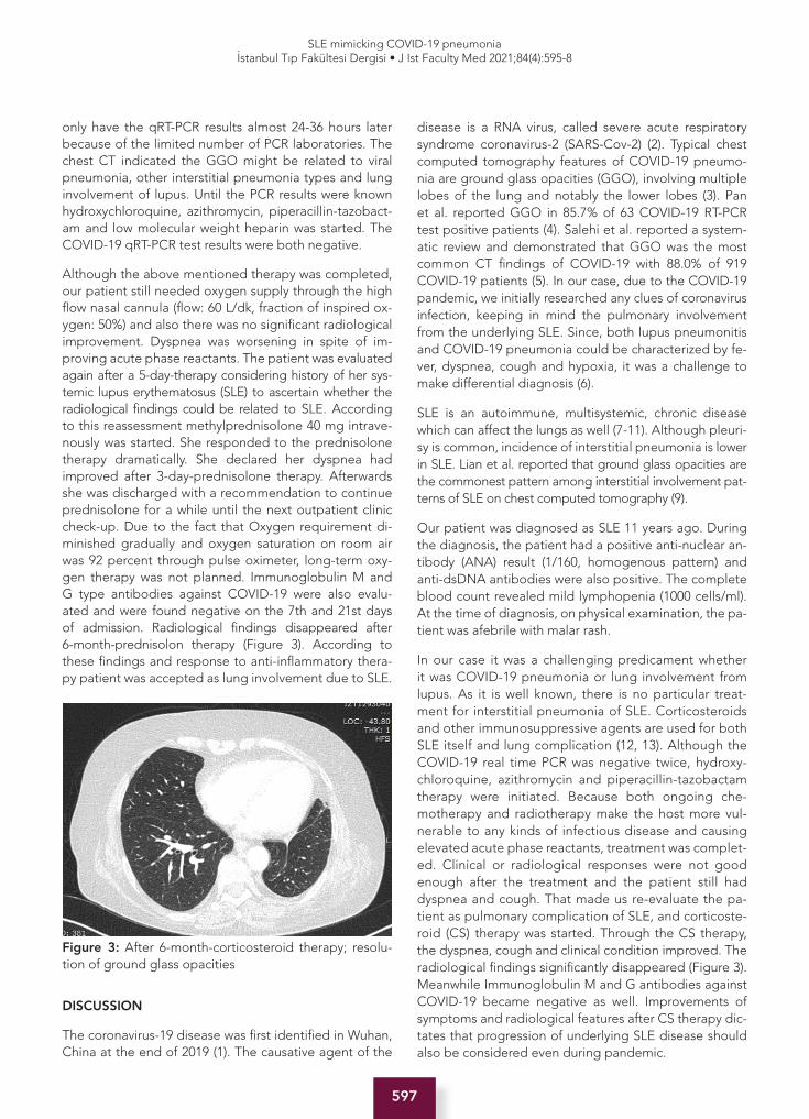
597
SLE mimicking COVID-19 pneumoniaİstanbul Tıp Fakültesi Dergisi • J Ist Faculty Med 2021;84(4):595-8
only have the qRT-PCR results almost 24-36 hours later because of the limited number of PCR laboratories. The chest CT indicated the GGO might be related to viral pneumonia, other interstitial pneumonia types and lung involvement of lupus. Until the PCR results were known hydroxychloroquine, azithromycin, piperacillin-tazobact-am and low molecular weight heparin was started. The COVID-19 qRT-PCR test results were both negative.
Although the above mentioned therapy was completed, our patient still needed oxygen supply through the high flow nasal cannula (flow: 60 L/dk, fraction of inspired ox-ygen: 50%) and also there was no significant radiological improvement. Dyspnea was worsening in spite of im-proving acute phase reactants. The patient was evaluated again after a 5-day-therapy considering history of her sys-temic lupus erythematosus (SLE) to ascertain whether the radiological findings could be related to SLE. According to this reassessment methylprednisolone 40 mg intrave-nously was started. She responded to the prednisolone therapy dramatically. She declared her dyspnea had improved after 3-day-prednisolone therapy. Afterwards she was discharged with a recommendation to continue prednisolone for a while until the next outpatient clinic check-up. Due to the fact that Oxygen requirement di-minished gradually and oxygen saturation on room air was 92 percent through pulse oximeter, long-term oxy-gen therapy was not planned. Immunoglobulin M and G type antibodies against COVID-19 were also evalu-ated and were found negative on the 7th and 21st days of admission. Radiological findings disappeared after 6-month-prednisolon therapy (Figure 3). According to these findings and response to anti-inflammatory thera-py patient was accepted as lung involvement due to SLE.
DISCUSSION
The coronavirus-19 disease was first identified in Wuhan, China at the end of 2019 (1). The causative agent of the
disease is a RNA virus, called severe acute respiratory syndrome coronavirus-2 (SARS-Cov-2) (2). Typical chest computed tomography features of COVID-19 pneumo-nia are ground glass opacities (GGO), involving multiple lobes of the lung and notably the lower lobes (3). Pan et al. reported GGO in 85.7% of 63 COVID-19 RT-PCR test positive patients (4). Salehi et al. reported a system-atic review and demonstrated that GGO was the most common CT findings of COVID-19 with 88.0% of 919 COVID-19 patients (5). In our case, due to the COVID-19 pandemic, we initially researched any clues of coronavirus infection, keeping in mind the pulmonary involvement from the underlying SLE. Since, both lupus pneumonitis and COVID-19 pneumonia could be characterized by fe-ver, dyspnea, cough and hypoxia, it was a challenge to make differential diagnosis (6).
SLE is an autoimmune, multisystemic, chronic disease which can affect the lungs as well (7-11). Although pleuri-sy is common, incidence of interstitial pneumonia is lower in SLE. Lian et al. reported that ground glass opacities are the commonest pattern among interstitial involvement pat-terns of SLE on chest computed tomography (9).
Our patient was diagnosed as SLE 11 years ago. During the diagnosis, the patient had a positive anti-nuclear an-tibody (ANA) result (1/160, homogenous pattern) and anti-dsDNA antibodies were also positive. The complete blood count revealed mild lymphopenia (1000 cells/ml). At the time of diagnosis, on physical examination, the pa-tient was afebrile with malar rash.
In our case it was a challenging predicament whether it was COVID-19 pneumonia or lung involvement from lupus. As it is well known, there is no particular treat-ment for interstitial pneumonia of SLE. Corticosteroids and other immunosuppressive agents are used for both SLE itself and lung complication (12, 13). Although the COVID-19 real time PCR was negative twice, hydroxy-chloroquine, azithromycin and piperacillin-tazobactam therapy were initiated. Because both ongoing che-motherapy and radiotherapy make the host more vul-nerable to any kinds of infectious disease and causing elevated acute phase reactants, treatment was complet-ed. Clinical or radiological responses were not good enough after the treatment and the patient still had dyspnea and cough. That made us re-evaluate the pa-tient as pulmonary complication of SLE, and corticoste-roid (CS) therapy was started. Through the CS therapy, the dyspnea, cough and clinical condition improved. The radiological findings significantly disappeared (Figure 3). Meanwhile Immunoglobulin M and G antibodies against COVID-19 became negative as well. Improvements of symptoms and radiological features after CS therapy dic-tates that progression of underlying SLE disease should also be considered even during pandemic.
Figure 3: After 6-month-corticosteroid therapy; resolu-tion of ground glass opacities

598
SLE mimicking COVID-19 pneumoniaİstanbul Tıp Fakültesi Dergisi • J Ist Faculty Med 2021;84(4):595-8
Differential diagnosis of COVID-19 and other diseases is essential. Chen et al. reported a pulmonary contusion case having trauma history and GGO on chest CT re-sembling COVID-19 pneumonia that was spontaneous-ly alleviated (14). It should be noted that COVID-19 may concomitantly superimpose on an underlying condition. Bekci et al. reported a 7-year-old boy with a history of trauma having both COVID-19 pneumonia with positive qRT-PCR and pulmonary contusion (15).
The aim of this case is to emphasize the differential diag-nosis of COVID-19 pneumonia. In the pandemic period; it is reasonable to consider the diagnosis of COVID-19 in all patients whose clinical and/or radiological findings sug-gested disease. However, alternative diagnoses should also be kept in mind especially in patients with underlying diseases. Additionally, it should not be forgotten that COVID-19 and other infectious or non-infectious disease with pulmonary involvement may coexist.
CONCLUSION
Ground glass opacities on chest CT is a significant dilem-ma in the COVID-19 era. Real time PCR and laboratory findings can be helpful for differential diagnosis. It should be kept in mind that pulmonary involvement of any sys-temic condition, infectious diseases other than COVID-19 and trauma may mimic radiological findings of COVID-19 or may co-exist with COVID-19.
Informed Consent: Written consent was obtained from the par-ticipants.
Peer Review: Externally peer-reviewed.
Author Contributions: Conception/Design of Study- E.Ş., İ.B., S.A.B.; Data Acquisition- E.Ş., S.D.; Data Analysis/Interpretation- A.K., İ.B.; Drafting Manuscript- E.Ş., A.K.; Critical Revision of Manuscript- İ.B., S.D., S.A.B.; Approval and Accountability- S.A.B., S.D., A.K., İ.B.
Conflict of Interest: Authors declared no conflict of interest.
Financial Disclosure: Authors declared no financial support.
Bilgilendirilmiş Onam: Katılımcılardan bilgilendirilmiş onam alınmıştır.
Hakem Değerlendirmesi: Dış bağımsız.
Yazar Katkıları: Çalışma Konsepti/Tasarım- E.Ş., İ.B., S.A.B.; Veri Toplama- E.Ş., S.D.; Veri Analizi/Yorumlama- A.K., İ.B; Yazı Tasla-ğı- E.Ş., A.K.; İçeriğin Eleştirel İncelemesi- İ.B.,S.D., S.A.B.; Son Onay ve Sorumluluk- S.A.B.,S.D., A.K., İ.B.
Çıkar Çatışması: Yazarlar çıkar çatışması beyan etmemişlerdir.
Finansal Destek: Yazarlar finansal destek beyan etmemişlerdir.
REFERENCES
1. Lu H, Stratton CW, Tang YW. Outbreak of pneumonia of unknown etiology in Wuhan, China: The mystery and the miracle. J Med Virol 2020;92(4):401-2. [CrossRef]
2. Coronaviridae Study Group of the International Committee on Taxonomy of Viruses. The species Severe acute respiratory syndrome-related coronavirus: classifying 2019-nCoV and naming it SARS-CoV-2. Nat Microbiol 2020;5(4):536-44. [CrossRef]
3. Wang Y, Dong C, Hu Y, Li C, Ren Q, Zhang X, et al. Temporal changes of CT findings in 90 patients with COVID-19 pneumonia: a longitudinal study. Radiology 2020;296(2):E55-E64. [CrossRef]
4. Pan Y, Guan H, Zhou S, Wang Y, Li Q, Zhu T, et al. Initial CT findings and temporal changes in patients with the novel coronavirus pneumonia (2019-nCoV): a study of 63 patients in Wuhan, China. Eur Radiol 2020;30(6):3306-9. [CrossRef]
5. Salehi S, Abedi A, Balakrishnan S, Gholamrezanezhad A. Coronavirus disease 2019 (COVID-19): A systematic review of imaging findings in 919 patients. AJR Am J Roentgenol 2020;215(1):87-93. [CrossRef]
6. Keane MP, Lynch JP. III. Pleuropulmonary manifestations of systemic lupus erythematosus. Thorax 2000;55(02):159-66. [CrossRef]
7. Mathai SC, Danoff SK. Management of interstitial lung disease associated with connective tissue disease. BMJ 2016;352:h6819. [CrossRef]
8. Pego-Reigosa JM, Medeiros DA, Isenberg DA. Respiratory manifestations of systemic lupus erythematosus: old and new concepts. Best Pract Res Clin Rheumatol 2009;23(4):469-80. [CrossRef]
9. Lian F, Zhou J, Wang Y, Cui W, Chen D, Li H, et al. Clinical features and independent predictors of interstitial lung disease in systemic lupus erythematosus. Int J Clin Exp Med 2016;9(2):4233-42
10. Fidler L, Keen KJ, Touma Z, Mittoo S. Impact of pulmonary disease on patient-reported outcomes and patient performed functional testing in systemic lupus erythematosus. Lupus 2016.25:1004-11. [CrossRef]
11. Fenlon HM, Doran M, Sant SM, Breatnach E. High-resolution chest CT in systemic lupus erythematosus. AJR Am J Roentgenol 1996;166:301-7. [CrossRef]
12. Bertsias G, Ioannidis JP, Boletis J, Bombardieri S, Cervera R, Dostal C, et al. EULAR recommendations for the management of systemic lupus erythematosus. Report of a Task Force of the EULAR Standing Committee for International Clinical Studies Including Therapeutics. Ann Rheum Dis 2008;67:195-205. [CrossRef]
13. van Vollenhoven RF, Mosca M, Bertsias G, Isenberg D, Kuhn A, Lerstrøm K, et al. Treat-to-target in systemic lupus erythematosus: recommendations from an international task force. Ann Rheum Dis 2014;73:958-67. [CrossRef]
14. Chen LR, Chen ZX, Liu YC, Peng L, Zhang Y, Xu Q, et al. Pulmonary contusion mimicking COVID-19: A case report. World J Clin Cases 2020;8(8):1554-60. [CrossRef]
15. Bekci T, Aslan S, Cakir IM. COVID- 19 pneumonia misdiagnosed as pulmonary contusion in a child. Br J Hosp Med 2020;81(5):1. [CrossRef]
599
Neonatal diabetes insipidus and holoprosencephalyİstanbul Tıp Fakültesi Dergisi • J Ist Faculty Med 2021;84(4):599-602
Corresponding author/İletişim kurulacak yazar: [email protected]
Submitted/Başvuru: 07.10.2020 • Revision Requested/Revizyon Talebi: 17.12.2020 •Last Revision Received/Son Revizyon: 23.12.2020 • Accepted/Kabul: 28.12.2020 • Published Online/Online Yayın: 17.09.2021
CASE REPORT / OLGU SUNUMUDOI: 10.26650/IUITFD.2021.807168
İst Tıp Fak Derg 2021 / J Ist Faculty Med 2021
EARLY-ONSET CENTRAL DIABETES INSIPIDUS IN A NEWBORN WITH HOLOPROSENCEPHALY
HOLOPROZENSEFALİ TANILI BİR YENİDOĞANDA ERKEN BAŞLANGIÇLI SANTRAL DİABETES İNSİPİDUS
Mustafa Törehan ASLAN1 , Zeynep İNCE1 , Asuman ÇOBAN1
1Istanbul University Istanbul Faculty of Medicine, Department of Pediatrics, Division of Neonatology, Istanbul, Turkey
ORCID IDs of the authors: M.T.A. 0000-0002-3966-4635; Z.İ. 0000-0002-7304-099X; A.Ç. 0000-0001-6573-242X
Cite this article as: Aslan MT, Ince Z, Coban A. Early-onset central diabetes insipidus in a newborn with holoprosencephaly. J Ist Faculty Med 2021;84(4):599-602. doi: 10.26650/IUITFD.2021.807168
ABSTRACT
Holoprosencephaly is a complex brain malformation caused by the inability of the prosencephalon to divide to form the cerebral hemispheres. Central diabetes insipidus (CDI), as a result of a defect in vasopressin release, may be seen due to the abnormal hypothalamic infundibular region. CDI developing secondary to holoprosencephaly in the early neonatal period has rarely been reported in the literature. A case of early-onset CDI with holoprosencephaly and 13q deletion is presented.
Keywords: Holoprosencephaly, central diabetes insipidus, newborn, desmopressin
ÖZET
Holoprozensefali, prozensefalonun serebral hemisferleri oluş-turmak için bölünmesi sırasındaki yetersizlikten kaynaklanan kompleks bir beyin malformasyonudur. Anormal hipotalamik infundibular bölge nedeniyle vazopressin salınımındaki kusura bağlı santral diabetes insipidus (SDİ) görülebilmektedir. Erken neonatal dönemde holoprozensefaliye ikincil gelişen SDİ vakası enderdir. Holoprozensefali ve 13q delesyonu olan erken başlan-gıçlı bir SDİ vakası sunulmuştur.
Anahtar Kelimeler: Holoprozensefali, santral diabetes insipi-dus, yenidoğan,
Content of this journal is licensed under a Creative Commons Attribution-NonCommercial 4.0 International License.
INTRODUCTION
Holoprosencephaly (HPE) is a complex brain malforma-tion caused by the failure of the prosencephalon to divide between the 35th and 42nd days of embryonic life. Depend-ing on the degree of septation deficiency, it can occur in three forms as alobar, semilobar and lobar. The incidence of alobar HPE has been reported to be 0.6-1.9 per 10,000 live births. Holoprosencephaly can either be isolated or accompany various syndromes. The prognosis depends on the severity of the brain and facial deformities along with the presence of related anomalies. Alobar HPE car-ries the worst prognosis among other prosencephalon di-vision anomalies. Central diabetes insipidus (CDI) may be an accompanying feature due to a defect in vasopressin release as a result of abnormalities in the hypothalamic infundibular region (1, 2).
CASE PRESENTATION
A baby boy with a birth weight of 3,000 grams (AGA) was born to a 34-year-old G6P4A2 mother at 393/7 weeks of ges-tation. Antenatal ultrasonography (USG) showed that the fetus had HPE and corpus callosum agenesis. After birth the baby was admitted to the neonatal intensive care unit for further investigation and follow-up. The history of the mother revealed two previous pregnancies, one resulting in miscarriage at the 8th week and the other live birth with HPE who died at the age of 5.5 years. No genetic analysis had been performed for either the child who died with holoprosencephaly or the two miscarriages. The parents were first degree cousins. Microcephaly, slanted palpe-bral fissures, hypotelorism, clinodactyly in the fifth finger of the left hand and mild hypertonicity were detected on physical examination (Figure 1).
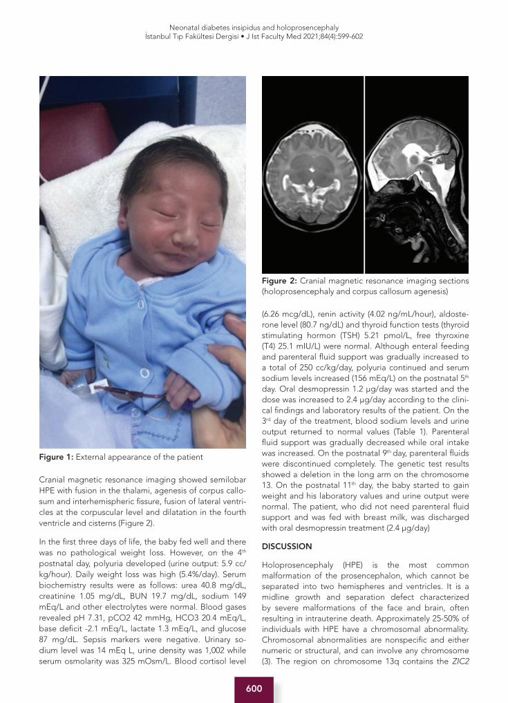
600
Neonatal diabetes insipidus and holoprosencephalyİstanbul Tıp Fakültesi Dergisi • J Ist Faculty Med 2021;84(4):599-602
Cranial magnetic resonance imaging showed semilobar HPE with fusion in the thalami, agenesis of corpus callo-sum and interhemispheric fissure, fusion of lateral ventri-cles at the corpuscular level and dilatation in the fourth ventricle and cisterns (Figure 2).
In the first three days of life, the baby fed well and there was no pathological weight loss. However, on the 4th postnatal day, polyuria developed (urine output: 5.9 cc/kg/hour). Daily weight loss was high (5.4%/day). Serum biochemistry results were as follows: urea 40.8 mg/dL, creatinine 1.05 mg/dL, BUN 19.7 mg/dL, sodium 149 mEq/L and other electrolytes were normal. Blood gases revealed pH 7.31, pCO2 42 mmHg, HCO3 20.4 mEq/L, base deficit -2.1 mEq/L, lactate 1.3 mEq/L, and glucose 87 mg/dL. Sepsis markers were negative. Urinary so-dium level was 14 mEq L, urine density was 1,002 while serum osmolarity was 325 mOsm/L. Blood cortisol level
(6.26 mcg/dL), renin activity (4.02 ng/mL/hour), aldoste-rone level (80.7 ng/dL) and thyroid function tests (thyroid stimulating hormon (TSH) 5.21 pmol/L, free thyroxine (T4) 25.1 mIU/L) were normal. Although enteral feeding and parenteral fluid support was gradually increased to a total of 250 cc/kg/day, polyuria continued and serum sodium levels increased (156 mEq/L) on the postnatal 5th day. Oral desmopressin 1.2 μg/day was started and the dose was increased to 2.4 μg/day according to the clini-cal findings and laboratory results of the patient. On the 3rd day of the treatment, blood sodium levels and urine output returned to normal values (Table 1). Parenteral fluid support was gradually decreased while oral intake was increased. On the postnatal 9th day, parenteral fluids were discontinued completely. The genetic test results showed a deletion in the long arm on the chromosome 13. On the postnatal 11th day, the baby started to gain weight and his laboratory values and urine output were normal. The patient, who did not need parenteral fluid support and was fed with breast milk, was discharged with oral desmopressin treatment (2.4 μg/day)
DISCUSSION
Holoprosencephaly (HPE) is the most common malformation of the prosencephalon, which cannot be separated into two hemispheres and ventricles. It is a midline growth and separation defect characterized by severe malformations of the face and brain, often resulting in intrauterine death. Approximately 25-50% of individuals with HPE have a chromosomal abnormality. Chromosomal abnormalities are nonspecific and either numeric or structural, and can involve any chromosome (3). The region on chromosome 13q contains the ZIC2
Figure 1: External appearance of the patient
Figure 2: Cranial magnetic resonance imaging sections (holoprosencephaly and corpus callosum agenesis)

601
Neonatal diabetes insipidus and holoprosencephalyİstanbul Tıp Fakültesi Dergisi • J Ist Faculty Med 2021;84(4):599-602
gene. The ZIC2 transcription factor is one of the most commonly mutated genes in HPE probands (4). The genetic test results of our case showed a deletion in the long arm on the chromosome 13. However as genetic analysis had not been performed for either the child who died with holoprosencephaly or the two miscarriages, nor the parents, we could not comment on the inheritance model of HPE seen in this index case.
Central diabetes insipidus (CDI) is a disease characterized by hypernatremia, hypostenuria and polyuria that develops due to a deficiency of antidiuretic hormone (ADH) secretion from the posterior pituitary. CDI in the neonatal period generally develops as a complication of intrauterine and perinatal diseases (5). Asphyxia, severe infections, intracranial hemorrhage, trauma, hereditary mutations of the gene encoding arginine vasopressin and central nervous system malformations, may be the etiologic factors underlying neonatal CDI. In some rare cases no etiology can be found (6, 7). In a study conducted with 12 babies diagnosed with CDI during a ten-year period, it was reported that 10 babies had central nervous system malformations whereas only one patient had semilobar holoprosencephaly and partial corpus callosum agenesis similar to our patient (8). In another study including 23 babies diagnosed with CDI between
the ages of 1 day and 9 months, holoprosencephaly was found in 5 babies (9). Other studies reported perinatal asphyxia, intraventricular bleeding and idiopathic cases (10, 13). Considering the age at diagnosis of CDI in the literature, it was reported that CDI developed in the early neonatal period in two infants in one study and at the postnatal 90th day in another study (8, 9). In another case report, CDI developed on the postnatal 30th day in a baby diagnosed with holoprosencephaly (14). The age of the development of CDI was also in the late neonatal period in some other reports (11, 12). In our patient, intracranial hemorrhage, neonatal sepsis and meningitis were excluded, and CDI was accepted as secondary to HPE. Looking at the relationship between CDI and other endocrinopathies, no additional endocrinopathy was reported in some cases (9, 15), while anterior pituitary insufficiency accompanied HPE in other cases (8). In our patient, thyroid function tests (TSH, fT4) and blood cortisol levels were normal excluding additional endocrinopathies. A standard treatment for neonatal CDI has not been defined in the literature and the management of these cases is difficult. It has been reported that intranasal desmopressin treatment causes hypernatremia and wide fluctuations in antidiuretic effects due to irregular nasal absorption in babies while oral desmopressin treatment has more positive results
Table 1: Follow-up during hospitalization
Days of life 1 2 3 4 5 6 7 8 9 10 11
Total fluid(mL/kg/day)
Bre
astf
eed
ing
Bre
astf
eed
ing
Bre
astf
eed
ing
150
(Bre
astf
eed
ing
and
p
aren
tera
l flu
id)
200
(Bre
astf
eed
ing
and
p
aren
tera
l flu
id)
250
(Bre
astf
eed
ing
and
p
aren
tera
l flu
id)
200
(Bre
astf
eed
ing
and
p
aren
tera
l flu
id)
180
(Bre
astf
eed
ing
and
p
aren
tera
l flu
id)
Bre
astf
eed
ing
Bre
astf
eed
ing
Bre
astf
eed
ing
Daily weightdifference (%)
-1.2 -2.5 -3.1 -5.4 -2.1 -1.5 +3.6 +3.4 +2.9 +1.4 +0.9
Serum sodium(mEq/L)
- - 146 149 156 151 144 142 140 138 139
Serum osmolarity (mOsm/L)
- - - 325 332 304 297 289 278 280 -
Diuresis(cc/kg/hour)
- - - 5.9 5.4 4.1 2.3 2.0 2.5 2.1 2.2
Urine osmolarity (mOsm/L)
- - - 79 75 108 197 264 262 256 -
Urine density - - - 1002 1002 1003 1009 1011 1011 1010 1011
Spot urine sodium (mEq/L)
- - - 14 10 19 17 15 18 16 -
Oral desmopressin dose (μg/day)
- - - - 1.2 2.4 2.4 2.4 2.4 2.4 2.4

602
Neonatal diabetes insipidus and holoprosencephalyİstanbul Tıp Fakültesi Dergisi • J Ist Faculty Med 2021;84(4):599-602
than the intranasal route (11, 16, 17). Oral desmopressin treatment was preferred in our patient whose normal sodium levels were achieved with appropriate feeding and fluid support as needed.
In conclusion, although HPE is an uncommon condition, it should be kept in mind that hypernatremic dehydra-tion may develop due to CDI in these patients, even in the early neonatal period. There is no predictive factor for the development of this condition to date, and close follow-up and management should be done accordingly.
Informed Consent: Written consent was obtained from the par-ticipants.
Peer Review: Externally peer-reviewed.
Author Contributions: Conception/Design of Study- M.T.A., A.Ç., Z.İ.; Data Acquisition- M.T.A.; Data Analysis/Interpretation- M.T.A., A.Ç., Z.İ.; Drafting Manuscript- M.T.A., A.Ç., Z.İ.; Critical Revision of Manuscript- Z.İ.,A.Ç.; Approval and Accountability- M.T.A., A.Ç., Z.İ.
Conflict of Interest: Authors declared no conflict of interest.
Financial Disclosure: Authors declared no financial support.
Bilgilendirilmiş Onam: Katılımcılardan bilgilendirilmiş onam alınmıştır.
Hakem Değerlendirmesi: Dış bağımsız.
Yazar Katkıları: Çalışma Konsepti/Tasarım- M.T.A., A.Ç., Z.İ.; Veri Toplama- M.T.A.; Veri Analizi/Yorumlama- M.T.A., A.Ç., Z.İ; Yazı Taslağı- M.T.A., A.Ç., Z.İ.; İçeriğin Eleştirel İncelemesi- Z.İ., A.Ç.; Son Onay ve Sorumluluk- M.T.A., A.Ç., Z.İ.
Çıkar Çatışması: Yazarlar çıkar çatışması beyan etmemişlerdir.
Finansal Destek: Yazarlar finansal destek beyan etmemişlerdir.
REFERENCES
1. Dubourg C, Bendavid C, Pasquier L, Henry C, Odent S, David V. Holoprosencephaly. Orphanet J Rare Dis 2007;2:8. [CrossRef]
2. Makaryus AN, McFarlane SI. Diabetes insipidus: Diagnosis and treatment of a complex disease, review. Clevel Clin J Med 2006;73(1):65-71. [CrossRef]
3. Dubourg C, Kim A, Watrin E, de Tayrac M, Odent S, David V, et al. Recent advances in understanding inheritance of holoprosencephaly. Am J Med Genet 2018;178(2):258-69. [CrossRef]
4. Barratt KS, Arkell RM. ZIC2 in Holoprosencephaly. Adv Exp Med Biol 2018;1046:269-99. [CrossRef]
5. Yang YH, Lin JJ, Hsia SH, Wu CT, Wang HS, Hung PC, et al. Central diabetes insipidus in children with acute brain insult. Pediatr Neurol 2011;45(6):377-80. [CrossRef]
6. Ferlin ML, Sales DS, Celini FP, Martinelli Junior CE. Central diabetes insipidus: alert for dehydration in very low birth weight infants during the neonatal period. A case report. Sao Paulo Med J 2015;133(1):60-3. [CrossRef]
7. Qureshi S, Galiveeti S, Bichet DG, Roth J. Diabetes insipidus: celebrating a century of vasopressin therapy. Endocrinology 2014;155(12):4605-21. [CrossRef]
8. Djermane A, Elmaleh M, Simon D, Poidvin A, Carel JC, Léger J. Central diabetes insipidus in infancy with or without hypothalamic adipsic hypernatremia syndrome: Early identification and outcome. J Clin Endocrinol Metab 2016;101(2):635-43. [CrossRef]
9. Kasim N, Bagga B, Diaz-Thomas A. Intracranial pathologies associated with central diabetes insipidus in infants. J Pediatr Endocrinol Metab 2018;31:951-8. [CrossRef]
10. Ueda H, Numoto S, Kakita H, Takeshita S, Muto D, Goto T, et al. Neonatal central diabetes insipidus caused by severe perinatal asphyxia. Pediatr Ther 2016; 6(1): 278. [CrossRef]
11. Atasay B, Berberoğlu M, Günlemez A, Evliyaoğlu O, Adiyaman P, Unal S, et al. Management of central diabetes insipidus with oral desmopressin in a premature neonate. J Pediatr Endocrinol Metab 2004;17(2):227-230. [CrossRef]
12. Yarber B, Wood B. Early diagnosis and treatment of diabetes insipidus in a newborn infant: a case study. Neonatal Netw 1992;11:17-20. pmid:1287448.
13. Quetin F, Garnier H, Brauner R, Vodovar M, Magny JF. [Persistent central diabetes insipidus in a very low birth weight infant] [Article in French]. Arch Pediatr 2007;14(11): 1321-3. [CrossRef]
14. Raisingani M, Gopi RP, Shah B. Use of chlorothiazide in the management of central diabetes insipidus in early infancy. Case Rep Pediatr 2017;2407028. [CrossRef]
15. Molnar Z, Sotiridou E, Dixon H, Ogilvy-Stuart A. Transient diabetes insipidus in a very-low-birthweight preterm infant with intraventricular haemorrhage. Acta Paediatr 2012;101(9):389-90. [CrossRef]
16. Fjellestad-Paulsen A, Paulsen O, d’Agay-Abensour L, Lundin S, Czemichow P. Central diabetes insipidus: Oral treatment with dDAVP. Regul Pept 1993;45(1-2):303-7. [CrossRef]
17. Stick SM, Betts PR. Oral desmopressin in neonatal diabetes insipidus. Arch Dis Child 1987;62(11):1177-8. [CrossRef]
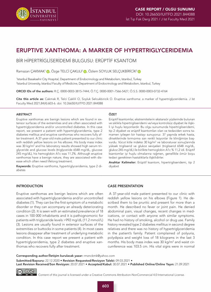
603
Eruptive xanthoma: a marker of hypertriglyceridemiaİstanbul Tıp Fakültesi Dergisi • J Ist Faculty Med 2021;84(4):603-6
Corresponding author/İletişim kurulacak yazar: [email protected]
Submitted/Başvuru: 22.12.2020 • Revision Requested/Revizyon Talebi: 09.03.2021 •Last Revision Received/Son Revizyon: 20.07.2021 • Accepted/Kabul: 30.07.2021 • Published Online/Online Yayın: 21.09.2021
CASE REPORT / OLGU SUNUMUDOI: 10.26650/IUITFD.2021.844088
İst Tıp Fak Derg 2021 / J Ist Faculty Med 2021
ERUPTIVE XANTHOMA: A MARKER OF HYPERTRIGLYCERIDEMIA
BİR HİPERTRİGLİSERİDEMİ BULGUSU: ERÜPTİF KSANTOM
Ramazan ÇAKMAK1 , Özge TELCİ ÇAKLILI2 , Özlem SOYLUK SELÇUKBİRİCİK2
1Istanbul Basaksehir City Hospital, Department of Endocrinology and Metabolism, Istanbul, Turkey2Istanbul University, Istanbul Faculty of Medicine, Department of Endocrinology and Metabolism, Istanbul, Turkey
ORCID IDs of the authors: R.Ç. 0000-0003-3815-7444; Ö.T.Ç. 0000-0001-7566-5427; Ö.S.S. 0000-0003-0732-4764
Cite this article as: Cakmak R, Telci Caklili O, Soyluk Selcukbiricik O. Eruptive xanthoma: a marker of hypertriglyceridemia. J Ist Faculty Med 2021;84(4):603-6. doi: 10.26650/IUITFD.2021.844088
ABSTRACT
Eruptive xanthomas are benign lesions which are found in ex-tensor surfaces of the extremities and are often associated with hypertriglyceridemia and/or uncontrolled diabetes. In this case report, we present a patient with hypertriglyceridemia, type 2 diabetes mellitus and eruptive xanthomas who recovers fully af-ter treatment. A 37-year-old male patient presented to our clinic with reddish yellow lesions on his elbows. His body mass index was 30 kg/m2 and his laboratory results showed high serum tri-glyceride and glucose levels (triglyceride 6548 mg/dL, glucose 245 mg/dL), his hemoglobin A1c was 11.2%. Although eruptive xanthomas have a benign nature, they are associated with dis-ease which often need lifelong treatment.
Keywords: Eruptive xanthoma, hypertriglyceridemia, type 2 di-abetes
ÖZET
Erüptif ksantomlar, ekstremitelerin ekstansör yüzlerinde bulunan ve sıklıkla hipertrigliseridemi ve/veya kontrolsüz diyabet ile ilişki-li iyi huylu lezyonlardır. Bu olgu sunumunda hipertrigliseridemi, tip 2 diyabet ve erüptif ksantomları olan ve tedaviden sonra ta-mamen iyileşen bir hastayı sunuyoruz. 37 yaşında erkek hasta, dirseklerinde kırmızımsı sarı renkli lezyonlar ile kliniğimize baş-vurdu. Vücut kitle indeksi 30 kg/m2 ve laboratuvar sonuçlarında yüksek trigliserid ve glukoz seviyeleri (trigliserid 6548 mg/dL, glukoz 245 mg/dL) ile birlikte hemoglobin A1c % 11,2 idi. Erüptif ksantomlar iyi huylu olmalarına rağmen, genellikle ömür boyu tedavi gerektiren hastalıklarla ilişkilidirler.
Anahtar Kelimeler: Erüptif ksantom, hipertrigliseridemi, tip 2 diyabet
Content of this journal is licensed under a Creative Commons Attribution-NonCommercial 4.0 International License.
INTRODUCTION
Eruptive xanthomas are benign lesions which are often associated with hypertriglyceridemia and/or uncontrolled diabetes (1). They can be the first symptom of a metabolic disorder or they can accompany an already deteriorating condition (2). It is seen with an estimated prevalence of 18 cases in 100 000 inhabitants and it is pathognomonic for patients with triglyceride levels >992 mg/dL (11.2 mmol/L) (3). Lesions are usually found in extensor surfaces of the extremities or buttocks in some patients (4). In most cases lesions disappear after treatment of underlying metabolic condition. In this case report we present a patient with hypertriglyceridemia, type 2 diabetes and eruptive xan-thomas who recovers fully after treatment.
CASE PRESENTATION
A 37-year-old male patient presented to our clinic with reddish yellow lesions on his elbows (Figure 1). He de-scribed them to be pruritic and present for more than a month. He described no fever or joint pain. He denied abdominal pain, visual changes, recent changes in med-ications, or contact with anyone with similar symptoms. He had no history of smoking, alcohol or drug use. Family history revealed type 2 diabetes mellitus in second degree relatives and there was no history of hypertriglyceridemia in the patient’s family. Patient complained of polyuria, polydipsia and weight loss of 18 kilograms in the last 3 months. His body mass index was 30 kg/m2 and waist cir-cumference was 103.5 cm. His vital signs were in normal

604
Eruptive xanthoma: a marker of hypertriglyceridemiaİstanbul Tıp Fakültesi Dergisi • J Ist Faculty Med 2021;84(4):603-6
range. There were multiple 2-5 mm sized eruptive xan-thomas on both elbows and there was a palmar xantho-ma on fourth finger of right hand along with the skinfold line (Figure 2). Punch biopsy revealed keratinized multi-layered squamous epithelium and underneath it there were foamy macrophages below dermis compatible with eruptive xanthoma (Figure 3). Laboratory results showed high triglyceride and glucose levels (triglyceride 6548 mg/dL, total cholesterol 842 mg/dL, LDL-cholesterol 261 mg/dL, HDL-cholesterol 38 mg/dL, glucose 245 mg/dL), his hemoglobin A1c was 11.2%. Other than these parameters his laboratory work up was normal (Table 1). Patient was started on hypocaloric diet, metformin 2x1000 mg, insulin glargine 1x14 units, fenofibrate 267 mg 1x1 and omega-3 2.5 grams/day. After one month his triglyceride levels re-sumed to normal values and his xanthomas faded. The patient provided informed consent for publication.
DISCUSSION
There are five types of xanthomas; eruptive xanthomas, tuberous/tendinous xanthoma, flat xanthomas, verrucous xanthomas, and xanthelasma (5). Eruptive xanthomas are skin lesions which are a manifestation of hypertriglyceridemia and with keratinized multilayered squamous epithelium and foamy macrophages under the dermis in their pathology. Differential diagnosis of eruptive xanthomas includes Langerhans cell histiocytosis, disseminated granuloma annulare, non-Langerhans cell histiocytosis (xanthoma disseminatum, the micronodular form of juvenile xanthogranuloma), and generalized eruptive histiocytoma.
On the other hand, tendon xanthomas are closely as-sociated with familial hypercholesterolemia and coro-nary heart disease (6). Xanthelasma are small, soft and yellowish lesions on the eyelids and are often bilateral. They can present in case of hyperlipidemia however in some cases there are no lipid disorders (7). Nevertheless Christoffersen et al. has reported an association between xanthelasma and coronary heart disease independent of conventional cardiac risk factors (8).
Figure 2: Palmar xanthoma
Figure 3: Pathology image
Figure 1: Eruptive xanthoma
Table 1: Laboratory findings
Laboratory parameters Results Reference values
Fasting Blood Glucose (mg/dL) 245 70-100
Hba1c (%) 11.8 4.5-5.7
Fasting Insulin (µIU/mL) 16.6 0-25
Total cholesterol (mg/dL) 842 0-200
Triglyceride (mg/dL) 6548 0-150
HDL-cholesterol (mg/dL) 38 40-60
LDL-cholesterol (mg/dL) 261 0-100
Creatinine (mg/dL) 0.9 0.7-1.2
Uric acid (mg/dL) 5 2.4-5.7
Sodium (mEq/L) 138 135-145
Potassium (mEq/L) 4.16 3.5-5.3
Amilase (U/L) 42 28-110
Lipase (U/L) 37 13-60
Aspartate aminotransferase (U/L) 22 10-35
Alanine aminotransferase (U/L) 35 0-35
Gamma glutamyl transferase (U/L) 51 15-85
Alkaline Phosphatase (U/L) 84 30-105
Thyroid-stimulating hormone (µIU/mL) 1.6 0.27-4.2
Free thyroxine (ng/dL) 1.12 0.93-1.7
605
Eruptive xanthoma: a marker of hypertriglyceridemiaİstanbul Tıp Fakültesi Dergisi • J Ist Faculty Med 2021;84(4):603-6
Detection of eruptive xanthomas should lead to an early investigation of triglyceride, glucose, thyroid stimulating hormone and creatinine levels because they can be the telltale lesion of diabetes, hypothyroidism or chronic kid-ney disease. Because hypertriglyceridemia is associated with acute pancreatitis in some cases these lesions can help physicians diagnose the precise cause of abdomi-nal pain in emergency units (9). Our patient did not have a secondary disease that could be the underlying cause of hyperlipidemia other than type 2 diabetes mellitus. He didn’t have a family history of hyperlipidemia. Pa-tient had high triglyceride and low HDL cholesterol lev-els. Although it seemed his lipid profile was compatible with type 1 hyperlipidemia of Frederickson classification because he also had high LDL levels, it was hard to dis-tinguish from type 4 and type 5 hyperlipoproteinemia (10). His VLDL level couldn’t be calculated due to high triglyceride levels and unlike his LDL, his VLDL was not measured directly. However, because he also had type 2 diabetes in an early age, it is plausible to think an early atherosclerotic disease associated with type 4 or type 5 hyperlipoproteinemia can also be present in this patient. Type 2 diabetes and high LDL are known atherosclerotic risk factors whereas the impact of hypertriglyceridemia on coronary heart disease has not been laid as clear as other classic risk factors. Lee et al. has suggested Apo B/A1 ratio to identify cardiovascular risk more definitely in a patient with type 1 diabetes and hypertriglyceridemia (7). To the best of our knowledge, unlike tendon xanthomas, there are no reports of an association between eruptive xanthomas and cardiovascular disease to this date.
Studies have shown deleterious effects of non-esteri-fied (free) fatty acids (NEFAs) on beta cell function (11). They induce nitric oxide mediated apoptosis and cause loss of beta cell function (12). Therefore, hyperlipidemia should be avoided in these patients and they should be ordered a strict diet to preserve beta cell function and to decrease fat deposit in visceral organs. Treatment of hyperlipidemia starts with diet and physical activity. Phar-macological treatment is added often in moderate and severe cases of hypertriglyceridemia. These treatments include fenofibrates, omega-3 fatty acids, niacin, insulin in patients with diabetes and triglyceride apheresis or plasmapheresis in patients with severe hypertriglyceri-demia (13, 14). Volanesorsen is a new drug developed by antisense technology with promising results in patients with familial chylomicronemia syndrome (15).
CONCLUSION
In conclusion, although eruptive xanthomas have a be-nign nature, they are associated with disease s which of-ten need lifelong treatment. Therefore, every physician should be aware of the appearance and disease burden of eruptive xanthomas.
Informed Consent: Written consent was obtained from the par-ticipants.
Peer Review: Externally peer-reviewed.
Author Contributions: Conception/Design of Study- R.Ç., Ö.T.Ç.; Data Acquisition- R.Ç.; Data Analysis/Interpretation- Ö.S.S.; Drafting Manuscript- R.Ç., Ö.T.Ç.; Critical Revision of Manuscript- Ö.S.S.; Approval and Accountability- R.Ç., Ö.T.Ç., Ö.S.S.
Conflict of Interest: Authors declared no conflict of interest.
Financial Disclosure: Authors declared no financial support.
Bilgilendirilmiş Onam: Katılımcılardan bilgilendirilmiş onam alınmıştır.
Hakem Değerlendirmesi: Dış bağımsız.
Yazar Katkıları: Çalışma Konsepti/Tasarım- R.Ç., Ö.T.Ç.; Veri Toplama- R.Ç.; Veri Analizi/Yorumlama- Ö.S.S; Yazı Taslağı- R.Ç., Ö.T.Ç.; İçeriğin Eleştirel İncelemesi- Ö.S.S.; Son Onay ve Sorum-luluk- R.Ç., Ö.T.Ç., Ö.S.S.
Çıkar Çatışması: Yazarlar çıkar çatışması beyan etmemişlerdir.
Finansal Destek: Yazarlar finansal destek beyan etmemişlerdir.
REFERENCES
1. Hsueh YC, Chou CL, Lee TI. Diabetic dyslipidemia with eruptive xanthoma. Cleve Clin J Med 2019;86(9):575. [CrossRef]
2. Kashif M, Kumar H, Misbahuddin K. An unusual presentation of eruptive xanthoma A case report and literature review. Medicine (Baltimore) 2016;95(37):e4866. [CrossRef]
3. Zak A, Zeman M, Slaby A, Vecka M. Xanthomas: clinical and pathophysiological relations. Biomed Pap Med Fac Univ Palacky Olomouc Czech Repub 2014;158(2):181-8. [CrossRef]
4. Kala J, Mostow EN. Eruptive xanthoma. N Engl J Med 2012;366(9):835. [CrossRef]
5. Streit E, Helmbold P. 65-year-old man with yellow-orange papules on both forearms. Eruptive xanthomas. Hautarzt 2009;60(10):834-7. [CrossRef]
6. Mangili LC, Miname MH, Silva PRS, Bittencourt MS, Rocha VZ, Mangili OC, et al. Achilles tendon xanthomas are associated withthe presence and burden of subclinical coronary atherosclerosis inheterozygous familial hypercholesterolemia: A pilot study. Atherosclerosis 2017;263:393-7. [CrossRef]
7. Lee SY, Sheth CA. Eruptive xanthoma associated with severe hypertriglyceridemia and poorly controlled type 1 diabetes mellitus. J Community Hosp Intern Med Perspect 2019;9(4):344-6. [CrossRef]
8. Christoffersen M, Frikke-Schmidt R, Schnohr P, Jensen GB, Nordestgaard BG, Tybjærg-Hansen A. Xanthelasmata, arcus corneae, and ischaemic vascular disease and death in general population: prospective cohort study. BMJ 2011;343:d5497. [CrossRef]

606
Eruptive xanthoma: a marker of hypertriglyceridemiaİstanbul Tıp Fakültesi Dergisi • J Ist Faculty Med 2021;84(4):603-6
9. Duzayak S, Sayiner ZA, Erkılıç S, Inaloz HS. Acute pancreatitis with eruptive xanthoma. BMJ Case Reports 2017;2017:bcr2017221543. [CrossRef]
10. Fredrickson DS. An international classification of hyperlipidemias and hyperlipoproteinemias. Ann Intern Med 1971;75(3):471-2. [CrossRef]
11. Cnop M. Fatty acids and glucolipotoxicity in the pathogenesis of Type 2 diabetes. Biochem Soc Trans 2008;36(Pt 3):348-52. [CrossRef]
12. Shimabukuro M, Ohneda M, Lee Y. Unger, RH. Role of nitric oxide in obesity induced β-cell disease. J Clin Invest 1997;100(2):290-5. [CrossRef]
13. Alagözlü H, Cindoruk M, Karakan T, Ünal S. Heparin and insulin in the treatment of hypertriglyceridemia-induced severe acute pancreatitis. Dig Dis Sci 2006;51(5):931-3. [CrossRef]
14. Ewald N, Kloer HU. Treatment options for severe hypertriglyceridemia (SHTG): the role of apheresis. Clin Res Cardiol Suppl 2012;7(Suppl 1):31-5. [CrossRef]
15. Witztum JL, Gaudet D, Freedman SD, Alexander VJ, Digenio A, Williams KR, et al. Volanesorsen and triglyceride levels in familial chylomicronemia syndrome. N Engl J Med 2019;381(6):531-42. [CrossRef]