Oxidative Stress and Mitochondrial Functions in the Intestinal Caco-2/15 Cell Line
10
Oxidative Stress and Mitochondrial Functions in the Intestinal Caco-2/15 Cell Line Rame Taha 1 , Ernest Seidman 2,3 , Genevieve Mailhot 1 , Franc ¸ois Boudreau 3 , Fernand-Pierre Gendron 3 , Jean-Franc ¸ois Beaulieu 3 , Daniel Me ´ nard 3 , Edgard Delvin 4 , Devendra Amre 5 , Emile Levy 1,3 * 1 Department of Nutrition, Research Center, CHU-Sainte-Justine, Universite ´ de Montre ´al, Montreal, Canada, 2 Research Institute, McGill University, Montreal, Canada, 3 Canadian Institutes for Health Research Team on the Digestive Epithelium, Department of Anatomy and Cellular Biology, Faculty of Medicine and Health Sciences, Universite ´ de Sherbrooke, Sherbrooke, Canada, 4 Department of Biochemistry, Research Center, CHU-Sainte-Justine, Universite ´ de Montre ´al, Montreal, Canada, 5 Department of Pediatrics, Research Center, CHU-Sainte-Justine, Universite ´ de Montre ´ al, Montreal, Canada Abstract Background: Although mitochondrial dysfunction and oxidative stress are central mechanisms in various pathological conditions, they have not been extensively studied in the gastrointestinal tract, which is known to be constantly exposed to luminal oxidants from ingested foods. Key among these is the simultaneous consumption of iron salts and ascorbic acid, which can cause oxidative damage to biomolecules. Methodology/Principal Findings: The objective of the present work was to evaluate how iron-ascorbate (FE/ASC)-mediated lipid peroxidation affects mitochondrion functioning in Caco-2/15 cells. Our results show that treatment of Caco-2/15 cells with FE/ASC (0.2 mM/2 mM) (1) increased malondialdehyde levels assessed by HPLC; (2) reduced ATP production noted by luminescence assay; (3) provoked dysregulation of mitochondrial calcium homeostasis as evidenced by confocal fluorescence microscopy; (4) upregulated the protein expression of cytochrome C and apoptotic inducing factor, indicating exaggerated apoptosis; (5) affected mitochondrial respiratory chain complexes I, II, III and IV; (6) elicited mtDNA lesions as illustrated by the raised levels of 8-OHdG; (7) lowered DNA glycosylase, one of the first lines of defense against 8-OHdG mutagenicity; and (8) altered the gene expression and protein mass of mitochondrial transcription factors (mtTFA, mtTFB1, mtTFB2) without any effects on RNA Polymerase. The presence of the powerful antioxidant BHT (50 mM) prevented the occurrence of oxidative stress and most of the mitochondrial abnormalities. Conclusions/Significance: Collectively, our findings indicate that acute exposure of Caco-2/15 cells to FE/ASC-catalyzed peroxidation produces harmful effects on mitochondrial functions and DNA integrity, which are abrogated by the powerful exogenous BHT antioxidant. Functional derangements of mitochondria may have implications in oxidative stress-related disorders such as inflammatory bowel diseases. Citation: Taha R, Seidman E, Mailhot G, Boudreau F, Gendron F-P, et al. (2010) Oxidative Stress and Mitochondrial Functions in the Intestinal Caco-2/15 Cell Line. PLoS ONE 5(7): e11817. doi:10.1371/journal.pone.0011817 Editor: Immo A. Hansen, New Mexico State University, United States of America Received June 7, 2010; Accepted July 2, 2010; Published July 27, 2010 Copyright: ß 2010 Taha et al. This is an open-access article distributed under the terms of the Creative Commons Attribution License, which permits unrestricted use, distribution, and reproduction in any medium, provided the original author and source are credited. Funding: This study was supported by a Canadian Institutes of Health Research team grant (CTP-82942), the J.A. DeSeve Research Chair in Nutrition (EL) and the Fonds de la Recherche en Sante du Quebec(RT). The funders had no role in study design, data collection and analysis, decision to publish, or preparation of the manuscript. Competing Interests: The authors have declared that no competing interests exist. * E-mail: [email protected] Introduction Reactive Oxygen Species (ROS) are by-products of normal aerobic metabolism and are now considered to be important signaling molecules that play a role in gene expression, cell growth and survival as well as oxygen sensing in various cell types [1,2]. The generation of ROS by a cascade of reactions is efficiently blocked by various endogenous antioxidants to overcome their potentially injurious actions [2,3]. However, excessive formation of ROS leads to lasting oxidative stress, characterized by an imbalance between oxidant-producing systems and antioxidant defense mechanisms, which can trigger cell damage by oxidizing macromolecular structures (lipids, proteins and DNA) and modifying their biological functions that ultimately causes cell death [4]. Thus, depending on their cell concentrations, ROS can act as either beneficial or harmful biological agents. The gastrointestinal tract is frequently exposed to noxious stimuli that may cause oxidative stress and injury. In fact, oxygen free radicals are generated both in the lumen and in the intestinal mucosa. Intraluminal pro-oxidants from ingested nutrients, such as alcohol, cholesterol oxides or iron salts and ascorbic acid, frequently consumed together in multiple-vitamin preparations or ingested foods, can build a pro-oxidant milieu [5–7]. Moreover, local microbes or infections, ischemia/reperfusion, gastric acid production and non- steroidal anti-inflammatory drugs may promote the formation of reactive radicals [8–10]. In addition, the influx of leukocytes, neutrophils and monocytes (associated with inflammation) can produce further ROS via respiratory burst enzymes as well as those involved in prostaglandin and leukotriene metabolism [11]. Clearly, significant oxidative stress has been said to be always associated with mucosal erosions and a causative role in a variety of gastrointestinal diseases such as Crohn’s disease and ulcerative colitis [12–14]. PLoS ONE | www.plosone.org 1 July 2010 | Volume 5 | Issue 7 | e11817
Transcript of Oxidative Stress and Mitochondrial Functions in the Intestinal Caco-2/15 Cell Line

Oxidative Stress and Mitochondrial Functions in theIntestinal Caco-2/15 Cell LineRame Taha1, Ernest Seidman2,3, Genevieve Mailhot1, Francois Boudreau3, Fernand-Pierre Gendron3,
Jean-Francois Beaulieu3, Daniel Menard3, Edgard Delvin4, Devendra Amre5, Emile Levy1,3*
1 Department of Nutrition, Research Center, CHU-Sainte-Justine, Universite de Montreal, Montreal, Canada, 2 Research Institute, McGill University, Montreal, Canada,
3 Canadian Institutes for Health Research Team on the Digestive Epithelium, Department of Anatomy and Cellular Biology, Faculty of Medicine and Health Sciences,
Universite de Sherbrooke, Sherbrooke, Canada, 4 Department of Biochemistry, Research Center, CHU-Sainte-Justine, Universite de Montreal, Montreal, Canada,
5 Department of Pediatrics, Research Center, CHU-Sainte-Justine, Universite de Montreal, Montreal, Canada
Abstract
Background: Although mitochondrial dysfunction and oxidative stress are central mechanisms in various pathologicalconditions, they have not been extensively studied in the gastrointestinal tract, which is known to be constantly exposed toluminal oxidants from ingested foods. Key among these is the simultaneous consumption of iron salts and ascorbic acid,which can cause oxidative damage to biomolecules.
Methodology/Principal Findings: The objective of the present work was to evaluate how iron-ascorbate (FE/ASC)-mediatedlipid peroxidation affects mitochondrion functioning in Caco-2/15 cells. Our results show that treatment of Caco-2/15 cellswith FE/ASC (0.2 mM/2 mM) (1) increased malondialdehyde levels assessed by HPLC; (2) reduced ATP production noted byluminescence assay; (3) provoked dysregulation of mitochondrial calcium homeostasis as evidenced by confocalfluorescence microscopy; (4) upregulated the protein expression of cytochrome C and apoptotic inducing factor, indicatingexaggerated apoptosis; (5) affected mitochondrial respiratory chain complexes I, II, III and IV; (6) elicited mtDNA lesions asillustrated by the raised levels of 8-OHdG; (7) lowered DNA glycosylase, one of the first lines of defense against 8-OHdGmutagenicity; and (8) altered the gene expression and protein mass of mitochondrial transcription factors (mtTFA, mtTFB1,mtTFB2) without any effects on RNA Polymerase. The presence of the powerful antioxidant BHT (50 mM) prevented theoccurrence of oxidative stress and most of the mitochondrial abnormalities.
Conclusions/Significance: Collectively, our findings indicate that acute exposure of Caco-2/15 cells to FE/ASC-catalyzedperoxidation produces harmful effects on mitochondrial functions and DNA integrity, which are abrogated by the powerfulexogenous BHT antioxidant. Functional derangements of mitochondria may have implications in oxidative stress-relateddisorders such as inflammatory bowel diseases.
Citation: Taha R, Seidman E, Mailhot G, Boudreau F, Gendron F-P, et al. (2010) Oxidative Stress and Mitochondrial Functions in the Intestinal Caco-2/15 CellLine. PLoS ONE 5(7): e11817. doi:10.1371/journal.pone.0011817
Editor: Immo A. Hansen, New Mexico State University, United States of America
Received June 7, 2010; Accepted July 2, 2010; Published July 27, 2010
Copyright: � 2010 Taha et al. This is an open-access article distributed under the terms of the Creative Commons Attribution License, which permitsunrestricted use, distribution, and reproduction in any medium, provided the original author and source are credited.
Funding: This study was supported by a Canadian Institutes of Health Research team grant (CTP-82942), the J.A. DeSeve Research Chair in Nutrition (EL) and theFonds de la Recherche en Sante du Quebec(RT). The funders had no role in study design, data collection and analysis, decision to publish, or preparation of themanuscript.
Competing Interests: The authors have declared that no competing interests exist.
* E-mail: [email protected]
Introduction
Reactive Oxygen Species (ROS) are by-products of normal
aerobic metabolism and are now considered to be important
signaling molecules that play a role in gene expression, cell growth
and survival as well as oxygen sensing in various cell types [1,2].
The generation of ROS by a cascade of reactions is efficiently
blocked by various endogenous antioxidants to overcome their
potentially injurious actions [2,3]. However, excessive formation of
ROS leads to lasting oxidative stress, characterized by an
imbalance between oxidant-producing systems and antioxidant
defense mechanisms, which can trigger cell damage by oxidizing
macromolecular structures (lipids, proteins and DNA) and
modifying their biological functions that ultimately causes cell
death [4]. Thus, depending on their cell concentrations, ROS can
act as either beneficial or harmful biological agents.
The gastrointestinal tract is frequently exposed to noxious stimuli
that may cause oxidative stress and injury. In fact, oxygen free
radicals are generated both in the lumen and in the intestinal mucosa.
Intraluminal pro-oxidants from ingested nutrients, such as alcohol,
cholesterol oxides or iron salts and ascorbic acid, frequently
consumed together in multiple-vitamin preparations or ingested
foods, can build a pro-oxidant milieu [5–7]. Moreover, local microbes
or infections, ischemia/reperfusion, gastric acid production and non-
steroidal anti-inflammatory drugs may promote the formation of
reactive radicals [8–10]. In addition, the influx of leukocytes,
neutrophils and monocytes (associated with inflammation) can
produce further ROS via respiratory burst enzymes as well as those
involved in prostaglandin and leukotriene metabolism [11]. Clearly,
significant oxidative stress has been said to be always associated with
mucosal erosions and a causative role in a variety of gastrointestinal
diseases such as Crohn’s disease and ulcerative colitis [12–14].
PLoS ONE | www.plosone.org 1 July 2010 | Volume 5 | Issue 7 | e11817

Despite the frequent occurrence of oxidative stress in the
gastrointestinal tract and its involvement in the initiation and
propagation of the chronic inflammatory response in chronic
bowel diseases [15], little is known about mitochondrion
response even though this special organelle is both a major
source of oxidants and a target for their damaging effects [16].
We have hypothesized that oxidative stress may affect various
mitochondrial functions, including ATP production, calcium
(Ca2+) homeostasis, cellular redox state regulation, apoptosis, as
well as mtDNA integrity [17,18]. Therefore, the specific aim of
the present study was to characterize the interplay between
oxidative stress and mitochondrial dysfunction in the Caco-2/15
cell line using the iron-ascorbate (FE/ASC) oxygen radical-
generating system, which participates in lipid peroxidation in
inflammatory bowel diseases (IBD) and represents a powerful
tool in our hands for the initiation of highly reactive hydroxyl
radicals and for the down-regulation of endogenous antioxidants
[19–26].
Materials and Methods
Caco-2/15 Cell CulturesThe colon carcinoma cell line, Caco-2/15 (ATCC, Rockville,
MD), was cultured at subconfluent stages in MEM (GIBCO-
BRL, Grand Island, NY) containing 1% penicillin-streptomycin
and 1% MEM non-essential amino acids (GIBCO-BRL) and
supplemented with 10% decomplemented fetal bovine serum
(FBS) (Flow, McLean, VA) as described previously [27]. Briefly,
Caco-2/15 cells (passage 20-30) were maintained in T-75-cm2
flasks (Corning Glass Works, Corning, NY). Cultures cells were
split (1:6) when they reached 90% confluence by use of 0.05%
trypsin-0.5 mM EDTA (GIBCO-BRL). For individual experi-
ments, cells were plated at a density of 16106 cells/well on
24.5 mm polyester Transwell filter inserts with 0.4-mm pores
(Coster, Cambridge, MA) in MEM supplemented with 5% FBS.
Cells were cultured for 21 days post confluence, at which the
Caco-2/15 cells are highly differentiated and appropriate for lipid
synthesis and metabolism. The medium was refreshed every
second day. To determine the implication of oxidative stress per se
in alterations in mitochondrial functions, Caco-2/15 cells were
incubated with FE/ASC (0.2 mM/2 mM) for 6 h alone and/or
with the antioxidant butylated hydoxytoluene (BHT) (2,6-di-t-
butyl-p-cresol, Sigma, St-Louis, MA) (50 mM). Caco-2/15 cells
were divided into four groups: control (without any addition),
oxidative (FE/ASC), antioxidant (BHT), oxidative and antioxi-
dant (FE/ASC + BHT).
Lipid PeroxidationCaco-2/15 cells were cultured in the presence or absence of
(0.2 mM/2 mM) FE/ASC added to the medium. Incubation
periods were terminated with 50 mM BHT to measure mal-
ondialdehyde (MDA). The level of MDA formed during the
oxidative reaction was determined by HPLC, as previously
described [19]. Briefly, proteins were first precipitated with a
10% sodium tungstate (Na2WO4) (Aldrich, Milwaukee, WI)
solution. The protein-free supernatants were then reacted with an
equivalent volume of 0.5% (wt/vol) thiobarbituric acid solution
(TBA; Sigma) at 90uC for 60 min. After cooling to room
temperature, the pink chromogene [(TBA) 2-MDA] was
extracted with 1-butanol and dried over a stream of nitrogen at
37uC. The dry extract was then resuspended in a potassium
dehydrogen phosphate (KH2PO4)/methanol mobile phase
(70; 30, pH 7.0) before MDA determination by HPLC with
fluorescence detection.
Assessment of Intracellular ATPIntracellular ATP was measured by luciferase driven biolumi-
nescence using ATP Bioluminescence Assay Kit from (Calbio-
chem, EMD Chemicals, Inc. Gibbstown, NJ) as reported
previously [28]. Values were then normalized further with regard
to the protein content of the respective sample. All Caco-2/15
culture cells were performed in duplicate.
Calcium Measurements by ConfocalFor mitochondrial Ca2+ monitoring, Caco-2/15 cells were
trypsinized, transferred from cell culture flasks to 8-well chamber
slides (Lab-TekTM Nunc, Rochester, NY) at a density of 2,56104
cells in 500 ml of cell culture medium. After a period of three days,
cells were serum-starved and incubated with FE/ASC and/or
BHT as described above. Cells were rinsed twice in serum-free
culture medium and loaded with a mixture of 5 mM Rhod-2/AM
(Molecular Probes, Eugene, OR), a fluorescent probe specific for
mitochondrial Ca2+, with 0,01% pluronic acid for 30 min at 37uCas described previously [29–31]. Medium was removed, replaced
with dye-free culture medium and incubated for an additional 60
minutes at 37uC. Thereafter, 1 ml of the fluorescent mitochondria-
specific dye MitoTrackerTM (green fluorescence, Molecular
Probes) was added to each well at the last 30 min of incubation.
Cells were visualized using an inverted laser-scanning confocal
microscope equipped with a 406 objective (LSM 510, Zeiss).
Excitation wavelength was 488 nm and fluorescence emission was
recorded at 543 nm (for Rhod-2) and 516 nm (for MitoTrack-
erTM). Six to eight fluorescence images were randomly chosen in
selected microscopic fields. Fluorescence intensity was quantified
using the Image J software (http://rsb.info.nih.gov/ij).
Mitochondrial PreparationsMitochondria were isolated using standard differential centri-
fugation techniques [32]. Briefly, Caco-2/15 cells were treated
with (0.2 mM/2 mM) FE/ASC and/or (50 mM) BHT for 6 h at
37uC. Cells were homogenized with a glass pestle Dounce
homogenizer in a buffer containing 210 mM mannitol, 70 mM
sucrose, 1 mM EGTA, 0.5% fatty acid-free bovine serum
albumin, and 5 mM HEPES, pH 7.2. The homogenate was
centrifuged at 1000 x g for 10 min at 4uC. The supernatant was
then collected and centrifuged at 10000 x g for 10 min to obtain
the pellets containing mitochondria. The pellets were used
immediately or stored at 280uC. The protein contents of
mitochondrial suspension were determined by the Bradford assay
(BioRad, Mississauga, ON) with BSA as a standard.
Evaluation of 8-hydroxy -2-deoxyguanosineOxidative DNA damage in whole Caco-2/15 cells, nuclei and
mitochondria was evaluated by assessing 8-hydroxy -2-deoxygua-
nosine (8-OHdG) with high-sensitivity competitive ELISA assays
performed with a commercial kit from Genox Corporation
(Baltimore, USA). Briefly, 8-OHdG antibody plus sample DNA
were added to a 96-well plate percolated with 8-OHdG and
incubated overnight at 4uC. After the plate was washed,
horseradish peroxidase–conjugated secondary antibody was added
for 1 h at room temperature. After washing, 3,39,5,59-tetramethyl-
benzidine was added and incubated for 15 min at room
temperature in the dark. The reaction was terminated by the
addition of phosphoric acid, and absorbance was measured at
450 nm. All assays were performed in duplicate. Negative controls
and 8-OHdG standards (0.125–10 ng/mL) were included in the
assay. The average concentration of 8-OHdG was calculated for
each sample based on the standard curve.
Mitochondrial Dysfunction
PLoS ONE | www.plosone.org 2 July 2010 | Volume 5 | Issue 7 | e11817
Mitochondrial Enzyme AssaysThe activities of respiratory chain complexes were assayed as
previously described in detail [32–34]. Briefly, 20–30 mg of
mitochondrial protein were used for each complex every 30 sec
for 5 min. The activity of complex I (NADH: ubiquinone
oxidoreductase) was measured by monitoring the reduction of
decylubiquinone. Complex II (succinate:ubiquinone oxidoreduc-
tase) activity was examined by monitoring the reduction of
dichloroindophenol when coupled to complex II-catalyzed
reduction of decylubiquinone. Complex III (ubiquinol:ferricyto-
chrome C oxidoreductase) activity was assayed using oxidized
cytochrome C. The activity of complex IV (cytochrome C oxidase)
was determined by oxidation of reduced cytochrome C. Enzyme
activities were expressed in nanomoles of substrate used per
minute per milligram of protein. Enzyme assays for all complexes
were performed in duplicate in mitochondrial fraction of Caco-2/
15 cell line. Complex I, II, III, IV chemicals were purchased from
Sigma Chemical, St Louis, MO.
Western BlotsTo assess the protein mass of mitochondrial transcription factors
(mt TF): mtTFA, mtTFB1, mtTFB2 and POLRMT, as well as 8-
oxoG-DNA glycosylase (OGG1), apoptosis-inducing factor (AIF)
and cytochrome C, Caco-2/15 cells were homogenized and
adequately prepared for Western blotting as described previously
[22,23,27,35–39]. The Bradford assay (Bio-Rad) was used to
estimate protein concentration. Proteins were denatured in sample
buffer containing SDS and b-mercaptoethanol, separated on a 4–
20% gradient SDS-PAGE and electroblotted onto nitrocellulose
membranes. Nonspecific binding sites of the membranes were
blocked with defatted milk proteins followed by the addition of
primary antibodies directed against the different proteins. The
relative amount of primary antibody was detected with species-
specific horseradish peroxidase-conjugated secondary antibody.
Even though identical protein amounts of tissue homogenates
were applied, the b-actin protein was used to confirm equal
loading on SDS-PAGE (results not shown). Blots were developed
and the mass of proteins was quantitated using an HP Scanjet
scanner equipped with a transparency adapter and software.
Rabbit polyclonal mtTFA Ab was obtained from Santa Cruz
Biotechnology Santa Cruz, CA; rabbit polyclonal POLRMT from
Abcam, Cambridge, MA; and mouse polyclonal mtTFB1 and
mtTFB2 Ab, rabbit polyclonal OGG1 Ab, rabbit polyclonal AIF
Ab, and mouse monoclonal cytochrome C Ab from Novus
Biologicals, Inc.
RT-PCRExperiments for mRNA quantification as well as for GAPDH
(as a housekeeping gene) were performed in Caco2/15 cells using
the UNO II thermocycler (Biometra) as reported previously
[35,40]. Approximately 30–40 cycles of amplification were used at
95uC for 30 s, 58uC for 30 s, and 72uC for 30 s. Amplicons were
visualized on standard ethidium bromide-stained agarose gels.
Under these experimental conditions related to RT-PCR, the
cycles for mtTFA, mtTFB1, mtTFB2, POLRMT, OGG1 and
GAPDH were 31, 31,35,31,31, and 30, respectively corresponding
to the linear portion of the exponential phase. Fold induction and
quantification were determined with the software UN-SCAN-IT
gel 6.1.
Primers UsedGAPDH (F- AGAAGGCTGGGGCTCATT/R-GGGCCAT-
CCACAGTCTTCT)
H-OGG1 (F-GGGGATTCACAAGGTGAAGA/R-GTAAG-
CTGGCTTGCATCACA)
POLRMT (F-CATCACCTACACCCACAACG/R-GTGCA-
CAGAGACGAAGGTCA)
H-mtTFB2 (F-GTCGCTTTTGCATTTTAGGG/R-GCT-
GTCCAAGGAACTGCTTC)
h-mtTFB1 (F-CTCCTGGACTTGAGGCTGAC/R-TTCT-
CAGTTTCCCAGGTGCT)
h-mtTFA (F-GGGTTCCAGTTGTGATTGCT/R-TGGA-
CAACTTGCCAAGACAG)
Statistical AnalysesStatistical analyses of data were performed with Prism 4.03
software (GraphPad Software). All values were expressed as the
mean 6 SEM. The data were evaluated by ANOVA, where
appropriate, and the differences between the means were assessed
using the Bonferroni’s multiple comparison test. A p-value of less
than 0.05 was considered to be significant.
Results
MDA Generation after Iron-Ascorbate ExposureBefore evaluating the role of oxidative stress on mitochondrial
function, we evaluated the effectiveness of FE/ASC in initiating
lipid peroxidation after incubation with Caco-2/15 cells. At the
end of a 6-h culture period, the degree of lipid peroxidation was
determined by measuring MDA in cells. As illustrated in Figure 1,
FE/ASC induced a significant increase in MDA levels above
baseline values compared with control cells. The concentration of
MDA was 4-fold higher in cells supplemented with FE/ASC
compared with untreated cells. Pre-incubation with the strong
antioxidant BHT markedly suppressed the production of MDA,
providing direct evidence for the ability of the FE/ASC system to
provoke profound lipid peroxidation.
Effect of FE/ASC on Cellular ATP ContentThe main function of the mitochondrion is the production of
energy in the form of ATP via oxidative phosphorylation and
oxygen consumption. We therefore assessed the amount of ATP
levels in Caco-2/15 cells exposed to the FE/ASC oxygen radical-
generating system. As noted in Figure 2, the administration of FE/
ASC led to a four-fold reduction compared with untreated cells.
Moreover, pre-incubation with BHT at a concentration of
0.5 mM resulted in a trend of ATP normalization.
Figure 1. Malondialdehyde (MDA) concentrations in Caco-2/15cells challenged with iron/ascorbate and/or BHT. At 21 days ofdifferentiation, cells were exposed to (0.2 mM/2 mM) FE/ASC, (50 mM)BHT or both for 6 h at 37uC. Oxidative stress was assessed by measuringMDA as an index of lipid peroxidation. Values are means 6 SEM forthree independent experiments. *P,0.05.doi:10.1371/journal.pone.0011817.g001
Mitochondrial Dysfunction
PLoS ONE | www.plosone.org 3 July 2010 | Volume 5 | Issue 7 | e11817

Oxidative Phosphorylation ActivitySince mitochondrial oxidative phosphorylation (OXPHOS) is
fundamental to all aspects of cell life under aerobic conditions, we
evaluated its activity during oxidative stress. The enzymatic
activity related to complexes I, II, III, and IV was performed on
mitochondrial fraction prepared from Caco-2/15 cells. Our
findings documented a significant decrease in the specific activities
of complex I, II, III and IV following FE/ASC treatment
(Figure 3). Pre-incubation with BHT abrogated the decline in
the OXPHOS enzymatic activities.
Changes in Mitochondrial Calcium Induced by Iron-Ascorbate in Caco-2/15 Cells
We next tested whether FE/ASC caused a change in
mitochondrial Ca2+ in Caco-2/15 cells using the positively
charged and cell permeant Ca2+ indicator, Rhod-2/AM, which
accumulates predominantly in the negatively charged matrix of
the mitochondria. The dye MitotrackerTM was used to confirm the
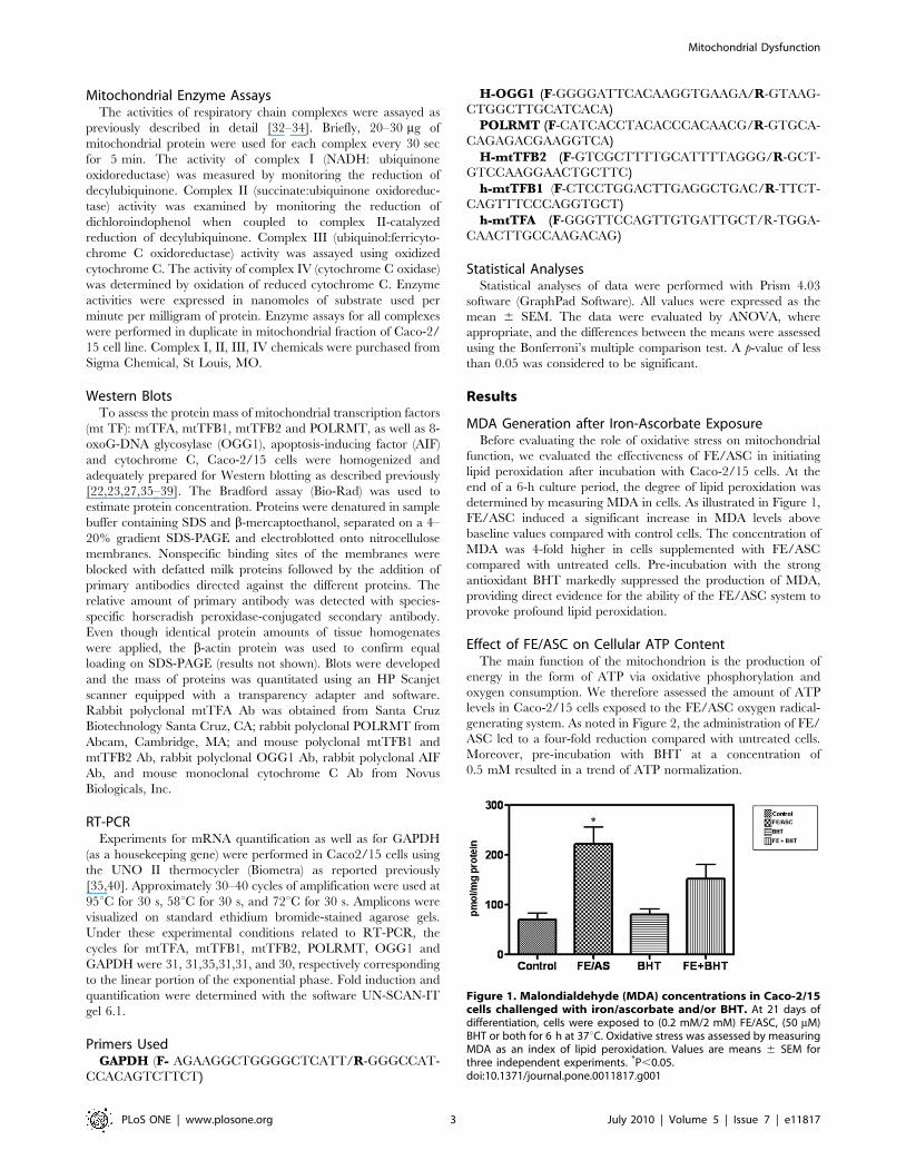
mitochondrial localization of Rhod-2. As presented in Figure 4D,
FE/ASC treatment of Caco-2/15 cells induced an increase in
Rhod-2 fluorescence that appears predominantly located in the
Figure 2. ATP Levels in Caco-2/15 cells exposed to iron/ascorbate in the presence or absence of BHT. Caco-2/15 cellswere grown on 96-well plates and, after 21 days post confluence, theywere treated with (0.2 mM/2 mM) FE/ASC and/or (50 mM) BHT for 6 h at37uC. ATP levels were measured with a bioluminescence assay andcorrected for intracellular protein concentrations. Values are expressedas ng of ATP per mg of cellular protein and represent the means 6 SEMfor three independent experiments. *P,0.001.doi:10.1371/journal.pone.0011817.g002
Figure 3. Effect of iron/ascorbate and/or BHT treatment on enzymatic activities of mitochondrial respiratory chain complexes inCaco-2/15 cells. Enzyme activities of mitochondrial respiratory chain complexes I, II, III, IV were measured by spectrophotometric assays inmitochondrial samples in Caco-2/15 cells treated with (0.2 mM/2 mM) FE/ASC and/or (50 mM) BHT for 6 h at 37uC. Enzyme activities are expressed asnmol/min/mg protein. Each value represents the mean 6 SEM for 3 separate experiments performed in duplicate. *P,0.05 vs. controls.doi:10.1371/journal.pone.0011817.g003
Mitochondrial Dysfunction
PLoS ONE | www.plosone.org 4 July 2010 | Volume 5 | Issue 7 | e11817
mitochondria as demonstrated by the yellow spots of strong
intensity found in the merged image (Figure 4F). In contrast, the
distribution pattern of colocalized Rhod-2 and MitotrackerTM
observed in control cells revealed spots of less intensity
characterized by a more diffuse distribution (Figure 4E). Quan-
tification of Rhod-2 fluorescence intensity is shown in Figure 5.
Cells exhibited an increase in Rhod-2 fluorescence after FE/ASC
treatment whereas pre-incubation with BHT restored fluorescence
intensity to control level.
AIF and Cytochrome C Protein ExpressionAIF is normally located in the inter-membrane space of
mitochondria and is involved in initiating a caspase-independent
pathway of apoptosis by causing DNA fragmentation and
chromatin condensation. Furthermore, when cell death is
triggered by an apoptotic stimulus, cytochrome C is released
into the cytosol, and contributes to the caspase-dependent
pathway of apoptosis. Western blot analysis revealed a marked
(P,0.001) increase in the level of AIF and cytochrome C protein
mass in Caco-2/15 cells following FE/ASC compared with
controls (Figures 6). Pre-incubation with BHT before the addition
of FE/ASC prevented the rise in AIF and cytochrome C protein
mass.
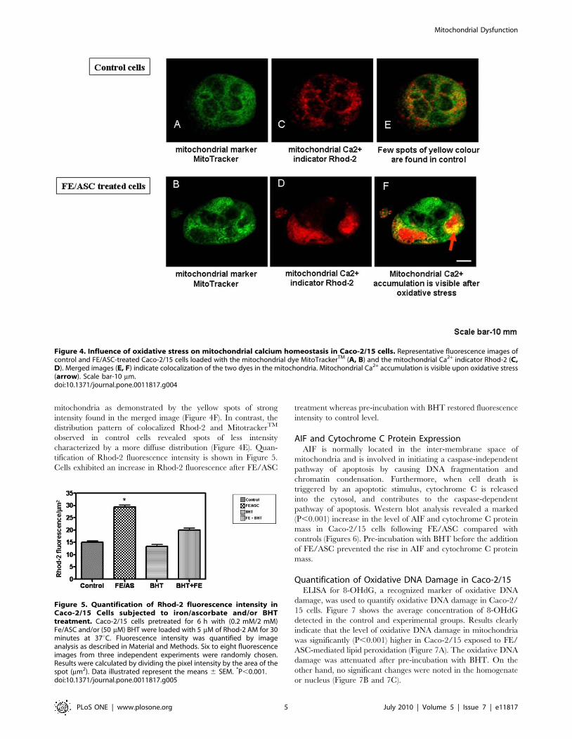
Quantification of Oxidative DNA Damage in Caco-2/15ELISA for 8-OHdG, a recognized marker of oxidative DNA
damage, was used to quantify oxidative DNA damage in Caco-2/
15 cells. Figure 7 shows the average concentration of 8-OHdG
detected in the control and experimental groups. Results clearly
indicate that the level of oxidative DNA damage in mitochondria
was significantly (P,0.001) higher in Caco-2/15 exposed to FE/
ASC-mediated lipid peroxidation (Figure 7A). The oxidative DNA
damage was attenuated after pre-incubation with BHT. On the
other hand, no significant changes were noted in the homogenate
or nucleus (Figure 7B and 7C).
Figure 4. Influence of oxidative stress on mitochondrial calcium homeostasis in Caco-2/15 cells. Representative fluorescence images ofcontrol and FE/ASC-treated Caco-2/15 cells loaded with the mitochondrial dye MitoTrackerTM (A, B) and the mitochondrial Ca2+ indicator Rhod-2 (C,D). Merged images (E, F) indicate colocalization of the two dyes in the mitochondria. Mitochondrial Ca2+ accumulation is visible upon oxidative stress(arrow). Scale bar-10 mm.doi:10.1371/journal.pone.0011817.g004
Figure 5. Quantification of Rhod-2 fluorescence intensity inCaco-2/15 Cells subjected to iron/ascorbate and/or BHTtreatment. Caco-2/15 cells pretreated for 6 h with (0.2 mM/2 mM)Fe/ASC and/or (50 mM) BHT were loaded with 5 mM of Rhod-2 AM for 30minutes at 37uC. Fluorescence intensity was quantified by imageanalysis as described in Material and Methods. Six to eight fluorescenceimages from three independent experiments were randomly chosen.Results were calculated by dividing the pixel intensity by the area of thespot (mm2). Data illustrated represent the means 6 SEM. *P,0.001.doi:10.1371/journal.pone.0011817.g005
Mitochondrial Dysfunction
PLoS ONE | www.plosone.org 5 July 2010 | Volume 5 | Issue 7 | e11817

OGG1 Repair Enzyme LevelIn mitochondria, the base excision repair pathway is primarily
responsible for removing 8-OHdG from DNA [41]. In humans, 8-
oxodG is repaired by 8-oxoguanine DNA glycosylase (OGG1), an
enzyme that recognizes and hydrolyzes the aberrant base from the
DNA backbone. We, therefore, examined its gene expression and
protein mass in Caco-2/15 cells. As well illustrated in Figure 8,
treatment with FE/ASC resulted in a significant (P,0.001)
reduction of OGG1 mRNA and protein mass compared with
controls. However, pre-incubation of Caco-2/15 cells with BHT
prevented the decline in OGG1 expression.
Mitochondrial Transcription FactorsHuman mitochondrial transcription requires bacteriophage-
related RNA polymerase, POLRMT, mtDNA-binding protein, h-
mtTFA/TFAM, and two transcription factors/rRNA methyl-
transferases, h-mtTFB1 and h-mtTFB2. These crucial proteins
define mitochondrial biogenesis and gene expression that together
likely fine-tune mitochondrial functions. Given the deleterious
effects of FE/ASC, it was mandatory to explore how oxidative
stress modulates the core protein components required for
mitochondrial transcription. PCR and Western Blot analyses
showed a significant (P,0.01) increase in mtTFA, mtTFB1 and
mtTFB2 gene expression (Figure 9A) and protein mass (Figure 9B)
without any changes in POLRMT in Caco-2/15 cells treated with
FE/ASC compared with controls. Pre-incubation with BHT
attenuated the modifications of those transcription factors.
Discussion
The Caco-2/15 cell line has been used to examine a variety of
intestinal functions. This intestinal model exhibits many of the
features of small intestinal epithelial cells. We employed the FE/
ASC oxygen radical-generating system to determine how oxidative
stress modulates mitochondrial DNA integrity and function in
Caco-2/15 cells [20]. Our results show for the first time that FE/
ASC can induce lipid peroxidation accompanied by ATP
depletion, mitochondrial transport chain complex inhibition,
mitochondrial Ca2+ overload, cell apoptosis, mitochondrial DNA
lesions and mitochondrial transcription factors alterations.
Iron is the most abundant transition metal in mammalian cells
and is essential for the physiological function of multiple proteins
[42]. However, excess or non-protein-bound (labile) iron can be
detrimental because it can initiate oxygen radical formation and
promote ROS [43]. Therefore, iron may cause oxidative damage
to biological macromolecules and alter the intracellular redox
environment, thereby affecting redox-sensitive cell signaling
pathways and transcription factors [44,45]. Although the mech-
anisms underlying the cytotoxicity of iron in different organs are
not fully delineated, many reports have pointed to the participa-
tion of iron-mediated peroxidation in numerous pathological
states, including atherosclerosis [46,47], cancer [48,49], ischemia-
reperfusion injury [50], IBD [51], and conditions of iron overload
[52]. Several laboratories [19–21,23–26,52–54] have shown the
ability of iron to initiate strong lipid peroxidation, whereas
ascorbic acid can amplify the oxidative potential of iron by
promoting metal ion-induced lipid peroxidation. The data
presented here clearly indicate that the FE/ASC system
functioned as a producer of lipid peroxidation and, at the same
time, altered the DNA integrity and the function of mitochondria.
It is noteworthy that the iron dose used in the current study is
comparable with normal iron concentration in the gut [11]. The
deteriorations resulting from the exposure of Caco-2/15 cells to
FE/ASC are probably attributable to oxidative stress, because the
addition of the BHT antioxidant simultaneously prevented the
occurrence of lipid peroxidation and improved the cellular
Figure 6. Cytochrome C and AIF expression levels in Caco-2/15 Cells treated with iron/ascorbate and/or BHT. Caco-2/15 cells wereincubated with (0.2 mM/2 mM) FE/ASC and/or (50 mM) BHT for 6 h at 37uC. Gene and protein expression were determined by RT-PCR and Westernblotting, respectively. Values are expressed as means 6 SEM for three independent experiments. *P,0.001.doi:10.1371/journal.pone.0011817.g006
Mitochondrial Dysfunction
PLoS ONE | www.plosone.org 6 July 2010 | Volume 5 | Issue 7 | e11817
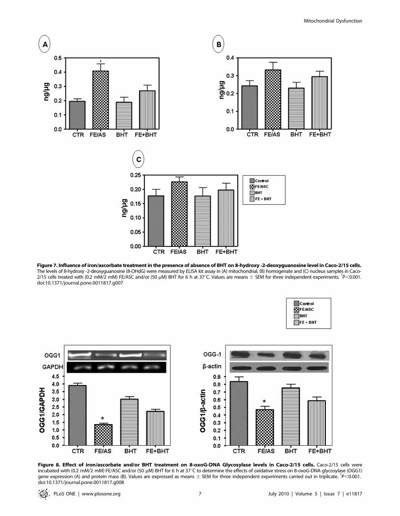
Figure 7. Influence of iron/ascorbate treatment in the presence of absence of BHT on 8-hydroxy -2-deoxyguanosine level in Caco-2/15 cells.The levels of 8-hydroxy -2-deoxyguanosine (8-OHdG) were measured by ELISA kit assay in (A) mitochondrial, (B) homogenate and (C) nucleus samples in Caco-2/15 cells treated with (0.2 mM/2 mM) FE/ASC and/or (50 mM) BHT for 6 h at 37uC. Values are means 6 SEM for three independent experiments. *P,0.001.doi:10.1371/journal.pone.0011817.g007
Figure 8. Effect of iron/ascorbate and/or BHT treatment on 8-oxoG-DNA Glycosylase levels in Caco-2/15 cells. Caco-2/15 cells wereincubated with (0.2 mM/2 mM) FE/ASC and/or (50 mM) BHT for 6 h at 37uC to determine the effects of oxidative stress on 8-oxoG-DNA glycosylase (OGG1)gene expression (A) and protein mass (B). Values are expressed as means 6 SEM for three independent experiments carried out in triplicate. *P,0.001.doi:10.1371/journal.pone.0011817.g008
Mitochondrial Dysfunction
PLoS ONE | www.plosone.org 7 July 2010 | Volume 5 | Issue 7 | e11817

processes of mitochondrial integrity and functions. BHT was
selected as an antioxidant because it represents a powerful agent
inhibiting iron-mediated oxidative stress and does not have any
toxic effects on Caco-2/15 cell culture [21].
Previous reports observed that an accumulation of peroxidation
products in mitochondria leads to a decrease in ATP production
and compromises the maintenance of cellular homeostasis [55]. In
this study, incubation of Caco-2/15 cells with FE/ASC induced a
marked decrease in ATP levels. Our data are consistent with
previous investigations showing that ATP decreased in the HT-29
intestinal cell-line after oxidative injury by hydrogen peroxide
[56]. The fall in ATP synthesis is probably related to the low
mitochondrial metabolic activity resulting from the FE/ASC-
mediated lipid peroxidation. In fact, electron movement through
complexes I, II, III, and IV enables movement of hydrogen ions
across the inner membrane into the inter-membrane space
creating an electrochemical gradient, which is harnessed into
ATP production by ATP synthase in complex V. We reasonably
propose that mitochondrial damage from ROS may lead to a
degradation in the efficiency of the mitochondrial respiratory
chain enzymes and hence a decline in ATP production. The
impairment of mitochondrial complex I, II, III and IV activity
noted in our experiments may be attributable to ROS-induced
cardiolipin damage that has recently been reported in ischemia/
reperfusion rat heart, which ultimately led to a decrease in
oxidative phosphorylation [57,58]. The phospholipid cardiolipin is
found almost exclusively in the inner mitochondrial membrane
where it promotes the optimal function of numerous enzymes
involved in mitochondrial energy metabolism. Finally, inactivation
of mitochondrial electron transport chain enzymes and/or ATP-
synthase may account for the ATP depletion triggered by the
administration of FE/ASC to Caco-2/15 cells.
On top of its ATP generation ability, mitochondria also play a
part in modulating the amplitude and spatiotemporal organization
of Ca2+ signals through rapidly accumulating and releasing Ca2+
[59,60]. Indeed, intracellular Ca2+ plays a key role in cellular
metabolism. However, excessive mitochondrial Ca2+ overload can
trigger ROS overproduction, mitochondrial membrane depolar-
ization and ATP production inhibition, all hallmark events of
mitochondrial dysfunction clearly observed in the present work.
Additionally, these defective processes may eventually lead to
apoptosis [59,60], which was also documented in our studies. In
particular, mitochondrial Ca2+ overload can favor cardiolipin
peroxidation, thereby affecting mitochondrial permeability tran-
sition, inducing AIF and cytochrome C release, and culminating in
mitochondrial dysfunction and apoptosis [61,62]. Therefore, tools
Figure 9. Effect of iron/ascorbate and/or BHT treatment on gene and protein expression of mitochondrial transcription factors inCaco-2/15 cells. Effects of (0.2 mM/2 mM) FE/ASC, (50 mM) BHT or both for 6 h at 37uC on gene expression (A) and protein mass (B) of mtTFA,mtTB1, mtTB2, POLRMT. A GAPDH cDNA probe was used as a control for RNA loading; b-actin was used as loading control protein. Data originatedfrom three independent experiments. Values are expressed as means 6 SEM. *P,0.01.doi:10.1371/journal.pone.0011817.g009
Mitochondrial Dysfunction
PLoS ONE | www.plosone.org 8 July 2010 | Volume 5 | Issue 7 | e11817

capable of minimizing mitochondrial Ca2+ overload would
decrease mitochondrial ROS accumulation and improve mito-
chondrial energy production, which may impact on mitochondri-
al-oxidative mediated diseases.
The core human mitochondrial transcription machinery
comprises a single subunit bacteriophage-related RNA polymer-
ase (POLRMT), mtTFA, and two transcriptional co-activator
proteins, h-mtTFB1 and h-mtTFB2. Both factors seem to interact
directly with POLRMT forming a heterodimer that, in addition
to mtTFA, is required for the accurate initiation on both H1 and
L promoters [63]. The main function of mtTFA is the
maintenance of mtDNA replication and transcription during
mitochondria biogenesis [64]. In our study, we observed that
mtTFA, mtTFB1, mtTFB2 transcriptional level and protein mass
were augmented in the presence of Fe/ASC with no marked
difference for POLRMT. Currently, we do not know whether the
upregulation of mtTFA, mtTFB1, and mtTFB2 in our experi-
ments represent a compensatory mechanism in response to
oxidative stress-related reduction in energy metabolism such as
defective electron transport chain, incomplete mitochondrion
biogenesis or accelerated apoptosis. Accordingly, mtTFA was
found upregulated in response to lipopolysaccharide-induced
oxidative damage to mitochondria, presumably to enhance
mtDNA levels and OXPHOS activity [65]. Furthermore, over-
expression of human TFB2M in HeLa cells induced an increase
in TFB1M mRNA levels and protein expression [66], suggesting
the existence of a retrograde signaling pathway from mitochon-
dria to the nucleus, which precisely regulates the expression of
these related factors. Further investigation is needed to examine
these important aspects.
In the present study, FE/ASC raised 8-OHdG that represents
one of the most frequently generated oxidative base lesions within
DNA, owing to guanine, the lowest redox potential among the
nucleic acid bases formed in pathological conditions [67].
Similarly, the double immunofluorescence technique revealed
that oxidative DNA damage is induced in colon epithelial cells of
the IBD mouse model [68]. Furthermore, nuclei were not affected
by FE/ASC-mediated oxidative stress, which confirms that
mtDNA is more vulnerable than nuclear DNA to oxidative
damage given that it is situated much closer to the site of ROS
generation and that mitochondria lack protective histones and far
fewer mechanisms that prevent reduced base excision repair
activity than DNA from nuclei [69]. Our findings confirm that
oxidative DNA damage is one of the most common threats to
mitochondrial genome stability.
OGG1 is the DNA repair enzyme that recognizes and excises 8-
oxodG [70]. The present study shows that incubation of Caco-2/
15 with FE/ASC resulted in a marked decrease in OGG1
transcript level and protein mass. Deficiency in DNA repair
enzyme OGG1 has likely important functional consequences,
compromising the ability of cells to repair DNA. Therefore,
intestinal epithelial cells are as sensitive to lipid peroxidation as
other cell types, including kidney cortex cells that accumulate 8-
OHdG mainly in the mtDNA and to a lesser extent in nuclear
DNA under diabetic conditions [71]. We believe that mtDNA
damage is linked to the numerous abnormal processes noted in our
study, including ATP generation, Ca2+ homeostasis and release of
signals for cell death.
In summary, the FE/ASC system in Caco-2/15 appeared to be
very effective in promoting lipid peroxidation and, at the same
time, altering the mitochondrial function. This mitochondrial
dysfunction is probably related to oxidative stress, because the
addition of antioxidants prevented the occurrence of lipid
peroxidation and improved the mitochondrial function in terms
of ATP production, Ca2+ homeostasis and apoptotic protein
expression. The pattern of our results using the Caco-2/15 cell line
may prove useful in elucidating the molecular mechanisms
implicated in IBD. Overall, our data suggest that oxidative-
mitochondrial dysfunction is not mediated by a single mechanism,
but that it may instead be a consequence of multiple vicious circles
organized within a complex functional network.
Acknowledgments
The authors thank Mrs. Schohraya Spahis for her technical assistance.
Author Contributions
Conceived and designed the experiments: RT EL. Performed the
experiments: RT EGS GM FPG JFB EL. Analyzed the data: RT JFB
EL. Contributed reagents/materials/analysis tools: RT FB DM ED DA
EL. Wrote the paper: RT EL.
References
1. Brown DI, Griendling KK (2009) Nox proteins in signal transduction. FreeRadic Biol Med 47: 1239–1253.
2. Gillespie MN, Pastukh V, Ruchko MV (2009) Oxidative DNA modifications in
hypoxic signaling. Ann N Y Acad Sci 1177: 140–150.
3. Haddad JJ (2002) Antioxidant and prooxidant mechanisms in the regulation ofredox(y)-sensitive transcription factors. Cell Signal 14: 879–897.
4. Andersen JK (2004) Oxidative stress in neurodegeneration: cause or conse-
quence? Nat Med 10 Suppl. pp S18–S25.
5. Young IS, Woodside JV (2001) Antioxidants in health and disease. J Clin Pathol
54: 176–186.
6. Parks DA (1989) Oxygen radicals: mediators of gastrointestinal pathophysiology.
Gut 30: 293–298.
7. Mazalli MR, Bragagnolo N (2009) Increase of cholesterol oxidation and
decrease of PUFA as a result of thermal processing and storage in eggs enrichedwith n-3 fatty acids. J Agric Food Chem 57: 5028–5034.
8. Sanchez S, Martin MJ, Ortiz P, Motilva V, Alarcon dlL (2002) Effects of
dipyrone on inflammatory infiltration and oxidative metabolism in gastricmucosa: comparison with acetaminophen and diclofenac. Dig Dis Sci 47:
1389–1398.
9. Parks DA, Williams TK, Beckman JS (1988) Conversion of xanthinedehydrogenase to oxidase in ischemic rat intestine: a reevaluation. Am J Physiol
254: G768–G774.
10. Biswas K, Bandyopadhyay U, Chattopadhyay I, Varadaraj A, Ali E, et al. (2003)A novel antioxidant and antiapoptotic role of omeprazole to block gastric ulcer
through scavenging of hydroxyl radical. J Biol Chem 278: 10993–11001.
11. Babbs CF (1992) Oxygen radicals in ulcerative colitis. Free Radic Biol Med 13:
169–181.
12. Kruidenier L, Verspaget HW (2002) Review article: oxidative stress as a
pathogenic factor in inflammatory bowel disease–radicals or ridiculous?. AlimentPharmacol Ther 16: 1997–2015.
13. Pravda J (2005) Radical induction theory of ulcerative colitis. World J Gastroenterol
11: 2371–2384.
14. Rezaie A, Parker RD, Abdollahi M (2007) Oxidative stress and pathogenesis of
inflammatory bowel disease: an epiphenomenon or the cause?. Dig Dis Sci 52:
2015–2021.
15. Nishikawa M, Oshitani N, Matsumoto T, Nishigami T, Arakawa T, et al. (2005)Accumulation of mitochondrial DNA mutation with colorectal carcinogenesis in
ulcerative colitis. Br J Cancer 93: 331–337.
16. Anders MW, Robotham JL, Sheu SS (2006) Mitochondria: new drug targets for
oxidative stress-induced diseases. Expert Opin Drug Metab Toxicol 2: 71–79.
17. Pessayre D (2007) Role of mitochondria in non-alcoholic fatty liver disease.
J Gastroenterol Hepatol 22 Suppl 1: S20–S27.
18. Kwong JQ, Beal MF, Manfredi G (2006) The role of mitochondria in inherited
neurodegenerative diseases. J Neurochem 97: 1659–1675.
19. Bernotti S, Seidman E, Sinnett D, Brunet S, Dionne S, et al. (2003)Inflammatory reaction without endogenous antioxidant response in Caco-2
cells exposed to iron/ascorbate-mediated lipid peroxidation. Am J Physiol
Gastrointest Liver Physiol 285: G898–G906.
20. Brunet S, Thibault L, Lepage G, Seidman EG, Dube N, et al. (2000) Modulation
of endoplasmic reticulum-bound cholesterol regulatory enzymes by iron/ascorbate-mediated lipid peroxidation. Free Radic Biol Med 28: 46–54.
21. Courtois F, Delvin E, Ledoux M, Seidman E, Lavoie JC, et al. (2002) The
antioxidant BHT normalizes some oxidative effects of iron + ascorbate on lipid
metabolism in Caco-2 cells. J Nutr 132: 1289–1292.
Mitochondrial Dysfunction
PLoS ONE | www.plosone.org 9 July 2010 | Volume 5 | Issue 7 | e11817

22. Levy E, Trudel K, Bendayan M, Seidman E, Delvin E, et al. (2007) Biological
role, protein expression, subcellular localization, and oxidative stress response ofparaoxonase 2 in the intestine of humans and rats. Am J Physiol Gastrointest
Liver Physiol 293: G1252–G1261.
23. Marcil V, Delvin E, Sane AT, Tremblay A, Levy E (2006) Oxidative stressinfluences cholesterol efflux in THP-1 macrophages: role of ATP-binding
cassette A1 and nuclear factors. Cardiovasc Res 72: 473–482.
24. Trudel K, Sinnett D, James RW, Delvin E, Amre D, et al. (2005) Iron-ascorbicacid-induced oxidant stress and its quenching by paraoxonase 1 in HDL and the
liver: comparison between humans and rats. J Cell Biochem 96: 404–411.
25. Courtois F, Seidman EG, Delvin E, Asselin C, Bernotti S, et al. (2003)
Membrane peroxidation by lipopolysaccharide and iron-ascorbate adversely
affects Caco-2 cell function: beneficial role of butyric acid. Am J Clin Nutr 77:744–750.
26. Courtois F, Suc I, Garofalo C, Ledoux M, Seidman E, et al. (2000) Iron-ascorbate alters the efficiency of Caco-2 cells to assemble and secrete
lipoproteins. Am J Physiol Gastrointest Liver Physiol 279: G12–G19.
27. Mailhot G, Ravid Z, Barchi S, Moreau A, Rabasa-Lhoret R, et al. (2009) CFTRknockdown stimulates lipid synthesis and transport in intestinal Caco-2/15 cells.
Am J Physiol Gastrointest Liver Physiol 297: G1239–G1249.
28. Drew B, Leeuwenburgh C (2003) Method for measuring ATP production inisolated mitochondria: ATP production in brain and liver mitochondria of
Fischer-344 rats with age and caloric restriction. Am J Physiol Regul IntegrComp Physiol 285: R1259–R1267.
29. Okuda T, Kadotsuji K, Takayama C, Hanada K, Mukaizawa F, et al. (2006)
Involvement of intracellular Ca2+ dynamics in cytoprotective action by aminoacids and cytotoxicity by sodium laurate, an absorption enhancer. J Pharm Sci
95: 2256–2265.
30. Abramov AY, Duchen MR (2003) Actions of ionomycin, 4-BrA23187 and anovel electrogenic Ca2+ ionophore on mitochondria in intact cells. Cell Calcium
33: 101–112.
31. Wang R, Miura T, Harada N, Kametani R, Shibuya M, et al. (2006) Pleiotropic
effects of the beta-adrenoceptor blocker carvedilol on calcium regulation during
oxidative stress-induced apoptosis in cardiomyocytes. J Pharmacol Exp Ther318: 45–52.
32. Trounce IA, Kim YL, Jun AS, Wallace DC (1996) Assessment of mitochondrialoxidative phosphorylation in patient muscle biopsies, lymphoblasts, and
transmitochondrial cell lines. Methods Enzymol 264: 484–509.
33. Janssen AJ, Trijbels FJ, Sengers RC, Smeitink JA, van den Heuvel LP, et al.(2007) Spectrophotometric assay for complex I of the respiratory chain in tissue
samples and cultured fibroblasts. Clin Chem 53: 729–734.
34. Krahenbuhl S, Talos C, Wiesmann U, Hoppel CL (1994) Development andevaluation of a spectrophotometric assay for complex III in isolated
mitochondria, tissues and fibroblasts from rats and humans. Clin Chim Acta230: 177–187.
35. Sane AT, Sinnett D, Delvin E, Bendayan M, Marcil V, et al. (2006) Localization
and role of NPC1L1 in cholesterol absorption in human intestine. J Lipid Res47: 2112–2120.
36. Leblond F, Seidah NG, Precourt LP, Delvin E, Dominguez M, et al. (2009)Regulation of the proprotein convertase subtilisin/kexin type 9 in intestinal
epithelial cells. Am J Physiol Gastrointest Liver Physiol 296: G805–G815.
37. Ravid Z, Bendayan M, Delvin E, Sane AT, Elchebly M, et al. (2008)Modulation of intestinal cholesterol absorption by high glucose levels: impact on
cholesterol transporters, regulatory enzymes, and transcription factors.Am J Physiol Gastrointest Liver Physiol 295: G873–G885.
38. Mailhot G, Rabasa-Lhoret R, Moreau A, Berthiaume Y, Levy E (2010) CFTR
depletion results in changes in fatty acid composition and promotes lipogenesisin intestinal Caco 2/15 cells. PLoS One 5: e10446.
39. Cammisotto PG, Bendayan M, Sane A, Dominguez M, Garofalo C, et al. (2010)
Receptor-Mediated Transcytosis of Leptin through Human Intestinal Cells InVitro. Int J Cell Biol 2010: 928169.
40. Montoudis A, Seidman E, Boudreau F, Beaulieu JF, Menard D, et al. (2008)Intestinal fatty acid binding protein regulates mitochondrion beta-oxidation and
cholesterol uptake. J Lipid Res 49: 961–972.
41. Bohr VA, Stevnsner T, de Souza-Pinto NC (2002) Mitochondrial DNA repair ofoxidative damage in mammalian cells. Gene 286: 127–134.
42. Papanikolaou G, Pantopoulos K (2005) Iron metabolism and toxicity. Toxicol
Appl Pharmacol 202: 199–211.
43. Ozment CP, Turi JL (2009) Iron overload following red blood cell transfusion
and its impact on disease severity. Biochim Biophys Acta 1790: 694–701.
44. Welch KD, Davis TZ, Van Eden ME, Aust SD (2002) Deleterious iron-mediated oxidation of biomolecules. Free Radic Biol Med 32: 577–583.
45. Flohe L, Brigelius-Flohe R, Saliou C, Traber MG, Packer L (1997) Redoxregulation of NF-kappa B activation. Free Radic Biol Med 22: 1115–1126.
46. Zhang WJ, Wei H, Frei B (2010) The iron chelator, desferrioxamine, reduces
inflammation and atherosclerotic lesion development in experimental mice. ExpBiol Med (Maywood) 235: 633–641.
47. Ahluwalia N, Genoux A, Ferrieres J, Perret B, Carayol M, et al. (2010) Iron
status is associated with carotid atherosclerotic plaques in middle-aged adults.J Nutr 140: 812–816.
48. Stevens RG, Graubard BI, Micozzi MS, Neriishi K, Blumberg BS (1994)Moderate elevation of body iron level and increased risk of cancer occurrence
and death. Int J Cancer 56: 364–369.
49. Herrinton LJ, Friedman GD, Baer D, Selby JV (1995) Transferrin saturationand risk of cancer. Am J Epidemiol 142: 692–698.
50. Ferrari R, Alfieri O, Curello S, Ceconi C, Cargnoni A, et al. (1990) Occurrenceof oxidative stress during reperfusion of the human heart. Circulation 81:
201–211.51. Gasche C, Lomer MC, Cavill I, Weiss G (2004) Iron, anaemia, and
inflammatory bowel diseases. Gut 53: 1190–1197.
52. Levy E, Brunet S, Alvarez F, Seidman E, Bouchard G, et al. (2007) Abnormalhepatobiliary and circulating lipid metabolism in the Long-Evans Cinnamon rat
model of Wilson’s disease. Life Sci 80: 1472–1483.53. Jourd’Heuil D, Vaananen P, Meddings JB (1993) Lipid peroxidation of the
brush-border membrane: membrane physical properties and glucose transport.
Am J Physiol 264: G1009–G1015.54. Levy E, Rizwan Y, Thibault L, Lepage G, Brunet S, et al. (2000) Altered lipid
profile, lipoprotein composition, and oxidant and antioxidant status in pediatricCrohn disease. Am J Clin Nutr 71: 807–815.
55. Chance B, Sies H, Boveris A (1979) Hydroperoxide metabolism in mammalianorgans. Physiol Rev 59: 527–605.
56. Masaki N, Kyle ME, Serroni A, Farber JL (1989) Mitochondrial damage as a
mechanism of cell injury in the killing of cultured hepatocytes by tert-butylhydroperoxide. Arch Biochem Biophys 270: 672–680.
57. Petrosillo G, Ruggiero FM, Di VN, Paradies G (2003) Decreased complex IIIactivity in mitochondria isolated from rat heart subjected to ischemia and
reperfusion: role of reactive oxygen species and cardiolipin. FASEB J 17:
714–716.58. Fry M, Green DE (1981) Cardiolipin requirement for electron transfer in
complex I and III of the mitochondrial respiratory chain. J Biol Chem 256:1874–1880.
59. Szalai G, Krishnamurthy R, Hajnoczky G (1999) Apoptosis driven by IP(3)-linked mitochondrial calcium signals. EMBO J 18: 6349–6361.
60. Deng X, Yin F, Lu X, Cai B, Yin W (2006) The apoptotic effect of brucine from
the seed of Strychnos nux-vomica on human hepatoma cells is mediated via Bcl-2 and Ca2+ involved mitochondrial pathway. Toxicol Sci 91: 59–69.
61. Pinton P, Giorgi C, Siviero R, Zecchini E, Rizzuto R (2008) Calcium andapoptosis: ER-mitochondria Ca2+ transfer in the control of apoptosis. Oncogene
27: 6407–6418.
62. Paradies G, Petrosillo G, Paradies V, Ruggiero FM (2009) Role of cardiolipinperoxidation and Ca2+ in mitochondrial dysfunction and disease. Cell Calcium
45: 643–650.63. Asin-Cayuela J, Gustafsson CM (2007) Mitochondrial transcription and its
regulation in mammalian cells. Trends Biochem Sci 32: 111–117.64. Virbasius JV, Scarpulla RC (1994) Activation of the human mitochondrial
transcription factor A gene by nuclear respiratory factors: a potential regulatory
link between nuclear and mitochondrial gene expression in organelle biogenesis.Proc Natl Acad Sci U S A 91: 1309–1313.
65. Suliman HB, Carraway MS, Welty-Wolf KE, Whorton AR, Piantadosi CA(2003) Lipopolysaccharide stimulates mitochondrial biogenesis via activation of
nuclear respiratory factor-1. J Biol Chem 278: 41510–41518.
66. Cotney J, Wang Z, Shadel GS (2007) Relative abundance of the humanmitochondrial transcription system and distinct roles for h-mtTFB1 and h-
mtTFB2 in mitochondrial biogenesis and gene expression. Nucleic Acids Res 35:4042–4054.
67. Rachek LI, Musiyenko SI, LeDoux SP, Wilson GL (2007) Palmitate induced
mitochondrial deoxyribonucleic acid damage and apoptosis in l6 rat skeletalmuscle cells. Endocrinology 148: 293–299.
68. Ding X, Hiraku Y, Ma N, Kato T, Saito K, et al. (2005) Inducible nitric oxidesynthase-dependent DNA damage in mouse model of inflammatory bowel
disease. Cancer Sci 96: 157–163.69. Kanki T, Nakayama H, Sasaki N, Takio K, Alam TI, et al. (2004) Mitochondrial
nucleoid and transcription factor A. Ann N Y Acad Sci 1011: 61–68.
70. Liao J, Seril DN, Lu GG, Zhang M, Toyokuni S, et al. (2008) Increasedsusceptibility of chronic ulcerative colitis-induced carcinoma development in
DNA repair enzyme Ogg1 deficient mice. Mol Carcinog 47: 638–646.71. Kakimoto M, Inoguchi T, Sonta T, Yu HY, Imamura M, et al. (2002)
Accumulation of 8-hydroxy-29-deoxyguanosine and mitochondrial DNA
deletion in kidney of diabetic rats. Diabetes 51: 1588–1595.
Mitochondrial Dysfunction
PLoS ONE | www.plosone.org 10 July 2010 | Volume 5 | Issue 7 | e11817