Real-time detection of pneumothorax using electrical impedance tomography

CLINICAL STUDY
Outcomes of an Algorithmic Approach to
Management of Pneumothorax Complicating
Thermal Ablation of Pulmonary Neoplasms
Nour-Eldin Nour-Eldin, MD, MSc, EDiR, Nagy N.N. Naguib, MSc,Ahmed M. Tawfik, MSc, Karen Koitka, MBBCh, Ahmed S. Saeed, MD, and
Thomas J. Vogl, MD
ABSTRACT
Purpose: To investigate the outcomes of an algorithm for treatment of pneumothorax in association with radiofrequency (RF) andmicrowave (MW) ablation of pulmonary neoplasms.
Materials and Methods: This retrospective study included data from 248 ablation sessions for lung tumors in 164 patients (92 men;mean age, 59.7 y � 9.8): 200 RF ablations (80.6%) and 48 MW ablations (19.4%). Pneumothorax was classified as mild, moderate, orsevere. Twelve patients developed mild pneumothorax and were observed for further complications, and 33 developed moderate orsevere pneumothorax and were managed with percutaneous aspiration of the pneumothorax. The decision to abort or continue ablationwas determined based on clinical response to percutaneous aspiration, clinical distress, and feasibility of applying the applicator withinthe lesion.
Results: Incidence of pneumothorax was 18.1% (45 of 248 sessions), with four (8.9%) occurrences during MW ablation and 41(91.1%) during RF ablation. Pneumothoraces were mild in 12 sessions (26.7%), moderate in 27 (60%), and severe in six (13.3%).Complete evacuation of the pneumothorax was achieved in 25 of 33 sessions (75.8%). Intercostal tube drainage was indicated in eightsessions (24.2%), including six severe and two moderate pneumothoraces. Pneumothorax evolved immediately after thoracic puncturein 10 patients. Ablation therapy was aborted in two sessions in which severe pneumothorax occurred, and an intercostal chest tubewas inserted.
Conclusions: Mild pneumothorax can be managed by close observation without interruption of ablation therapy. Manual evacuationwas an effective strategy for management of moderate pneumothorax and allowed for adequate positioning of the electrode, but didnot suffice for severe and progressive pneumothorax, which required placement of an intercostal chest tube.
ABBREVIATIONS
MW � microwave, RF � radiofrequency
atcaao2rm
anctm
Thermal ablation may be used to treat a variety of thoracicmalignancies, including primary lung cancer, recurrent pri-mary lung cancer, metastatic disease, chest wall masses,
From the Diagnostic and Interventional Radiology Institute (N.E.N.E.,N.N.N.N., A.M.T., T.J.V.), Johann Wolfgang Goethe University Hospital, The-odor-Stern-Kai 7, Frankfurt, Hessen 60590, Germany; Department of InternalMedicine (K.K.), Toowoomba Base Hospital, Toowoomba, Australia; and Di-agnostic and Interventional Radiology Department (N.E.N.E, A.S.S.), CairoUniversity Hospital, Cairo, Egypt. Received January 19, 2009; final revisionreceived May 24, 2011; accepted May 31, 2011. Address correspondenceto N.E.N.E.; E-mail: [email protected]
None of the authors have identified a conflict of interest.
© SIR, 2011
J Vasc Interv Radiol 2011; 22:1279–1286
lDOI: 10.1016/j.jvir.2011.05.014
nd painful, bony thoracic metastases (1–14). As the use ofhis modality becomes more widespread, it is vital that asso-iated complications be considered and evidence-based man-gement protocols established for situations in which theyrise. Pneumothorax is the most frequent serious complicationf thoracic thermal ablation, with a reported mean incidence of8% (1–12). There is a need for evidence-based knowledgeegarding the factors associated with pneumothorax develop-ent and appropriate management protocols.
Most scientific research has focused on therapy efficacynd complications of thermal ablation, but there are currentlyo protocols to guide the management of these associatedomplications. The purpose of the present study was to inves-igate the outcomes of an algorithm for the treatment of pneu-othorax occurring in association with thermal ablation of
ung neoplasms.

abwfp(ltlluNtt
tlpt
AApcuwtdbmadptltci1CRp1EtbtdsBpnaosppatt
C
SPamPwpo
1280 � Algorithmic Approach to Ablation-related Pneumothorax Management Nour-Eldin et al � JVIR
MATERIALS AND METHODS
Approval to carry out the study was obtained from theinstitutional review board. Informed consent was obtainedfrom all patients (including approval for ablation therapy,management of emergent complications, including pneu-mothorax, and the anonymous use of data for researchwork). A retrospective analysis of a prospectively collecteddatabase of lung ablations with a predetermined pneumo-thorax management protocol was performed. The studyinvolved 248 ablation sessions for lung tumors carried outin the time period between March 2004 and August 2008.Of the 248 sessions, 20 were for primary lesions and 228for metastatic lesions in 164 patients (92 men, 72 women;mean age, 59.7 y � 9.8 [SD]). A total of 200 cases wereblated by radiofrequency (RF) energy and 48 were ablatedy microwave (MW) energy. In all cases, the malignanciesere deemed medically inoperable or the patient had re-
used surgical intervention. Tumors were pathologicallyroven and were classified as primary lung neoplasmsnon–small-lung cancer) in 20 patients and as metastaticung neoplasms in 228 patients with no evidence of ex-rapulmonary metastases. Patients were excluded from ab-ation therapy if there were more than five lesions, if anyesion had a diameter greater than 5 cm, or if there wasncorrectable coagulopathy indicated by an Internationalormalized Ratio greater than 1.8 or platelet count lower
han 75,000. The ablation protocol at the authors’ institu-ion was designed to ablate one lesion per session.
Rationale and Study ProtocolRadiologic evaluation of preprocedural, intraprocedural,and postprocedural images was consensually carried out bytwo senior radiologists. The study analysis focused on thecircumstances of pneumothorax management rather than onthe risk factors involved in pneumothorax development.Emphasis was placed on the onset of pneumothorax beforeor after insertion of the applicator (ie, electrode for RFablation or antenna for MW ablation) within the lesion,pneumothorax severity (mild, moderate, or severe), man-agement pathway selection, and monitoring of response totreatment.
Preablation AssessmentThe thermal ablation treatment plan for an individual wasdevised by the treating interventional radiologist in consul-tation with the patient and the referring physician. Self-referred patients were assessed by a multidisciplinary teamthat included representatives from thoracic surgery, medicaloncology, and pulmonary disease departments. A compre-hensive clinical history and physical examination was doc-umented, in addition to a review of recent imaging studiesand a discussion of the indications, risk, and benefits of theprocedure. Preprocedural laboratory investigations, includ-ing complete blood count and coagulation profile, werecompleted.
Prophylactic antibiotics were not routinely adminis- o
ered. The ablation parameters (including applicator type,ength, and number), in addition to the position of theatient and site of puncture, were planned according toumor size and anatomic location.
blation Techniquell lung ablations were performed with the use of com-uted tomographic (CT) fluoroscopic guidance with 5-mmollimation (SOMATOM 4; Siemens, Erlangen, Germany)nder aseptic conditions by two interventional radiologistsith more than 8 and 15 years of experience in interven-
ional radiology, respectively. Ablation was performed un-er conscious sedation with fentanyl citrate (1 �g/kg ofody weight) and midazolam hydrochloride (0.010–0.035g/kg). Continuous electrocardiography, pulse oximetry,
nd blood pressure monitoring was carried out for theuration of the procedure. Oxygen was administered toatients via a nasal cannula at a rate of 3–5 nL/min duringhe ablation. The patient was positioned depending on theocation of the lesion to ensure the shortest ablation path inhe position most tolerable for the patient. RF ablation wasarried out with the use of (i) a CelonProSurge bipolarnternally cooled applicator (20–40-mm electrode length,5-gauge shaft [1.8 mm], and 100–250-mm shaft length;elon, Teltow, Germany) in 148 ablation sessions or (ii) aITA Starburst XL device (14-gauge/6.4-F, nine arrayslus active trocar tip, five thermocouples, shaft length of 10,5, and 25 cm; RITA, Manchester, Georgia) in 52 sessions.ach lesion was ablated with the use of one applicator, with
he exception of four cases in which two applicators (Celonipolar electrodes) were applied simultaneously to ablatehe lesion. Microwave ablation was performed with Covi-ien antennas (shaft length of 12, 17, or 22 cm, radiatingection of 3.7 cm) and Covidien MW generators (Covidien,oulder, Colorado) in 48 sessions. Applicators were ap-lied through a single pleural puncture without guidingeedles. The target location was monitored by CT duringblation, and the applicator was manipulated to ensureptimal positioning within the lesion. Periodic CT fluoro-copic scanning was performed to assess the applicatorosition and the progress of associated complications. Torevent seeding of malignant cells in the needle tract duringpplicator removal and to induce local hemostasis of theract, needle tract coagulation was routinely performed athe end of the procedure.
T Classification of Pneumothorax
everityneumothorax was classified based on axial CT imagesccording to the largest distance of retraction of the pul-onary surface estimated after scanning of the entire chest.neumothorax was classified as mild if it was associatedith lung surface retraction of less than 2 cm from theleural surface, moderate if it was associated with retractionf 2–4 cm, and severe if it was associated with retraction
f more than 4 cm, increased progressively on two con-
P
s
tot
pltwaope
horax
Volume 22 � Number 9 � September � 2011 1281
secutive radiologic control images within the first 2hours after the procedure, was associated with mediasti-nal shift, or was accompanied by respiratory or circula-tory distress (Fig 1) (15).
Pneumothorax Management ProtocolThe proposed protocol for pneumothorax managementwas devised according to available evidence. Variousstudies have described the management of pneumothoraxcomplicating a variety of thoracic interventions, partic-ularly lung biopsy and thoracic ablation therapy (1,3–10,15,16). This knowledge was combined with our insti-tutional technical experience in the field of thoracicintervention to formulate the algorithm for the manage-ment of pneumothorax complicating pulmonary ablation
Figure 1. Protocol algorithm for the management of pneumot
therapy (Figs 1, 2). r
neumothorax detected during ablation ses-
ion. Patients in whom pneumothorax developed afterhoracic puncture with the applicator but before completionf the ablation procedure constituted one group. The pro-ocol of management was as follows.
In the case of mild pneumothorax not interfering withroper positioning of the applicator within the lesion, ab-ation therapy was continued under close observation (par-icularly of the vital signs) and pneumothorax progressionas monitored by limited fluoroscopic CT scanning. If
dequate positioning was not possible because of slippagef the retracting lung, manual evacuation of the air from theleural sac was carried out, followed by reapplication of thelectrode on complete expansion of the retracted lung.
In the case of pneumothorax causing lung surface
arising during the ablation procedure.
etraction of more than 2 cm from the pleural surface and

rsc
C
DIisisftpitswetaes(a
EMwpgvsctssiatdapate
PAep(ap
1282 � Algorithmic Approach to Ablation-related Pneumothorax Management Nour-Eldin et al � JVIR
progressively increasing with time during the ablation pro-cedure, pneumothorax was manually evacuated with anintercostal catheter (5- or 10-F). If the air progressivelydecreased without reaccumulation, ablation therapy wascontinued. If the pneumothorax was refractory to manualevacuation or the patient developed respiratory distress, theprocedure was immediately terminated and an intercostalchest tube was inserted.
Pneumothorax accumulating after ablation ther-
apy. Mild pneumothorax that accumulated after ablationtherapy was managed by observation of clinical status andvital signs. Chest CT examinations were carried out at 1hour and 8 hours after the procedure. Patients were deemedsafe for discharge if they remained asymptomatic and therewas no increase in pneumothorax size. A 24-hour postpro-cedure follow-up CT scan was mandatory to exclude de-layed development of pneumothorax (Fig 2).
Moderate and severe pneumothorax were primarilytreated by immediate manual evacuation with use of anintercostal catheter (5- or 10-F). On complete successfulevacuation of the air, management was continued as for amild, stable pneumothorax.
Failure of manual air evacuation or a progressive in-crease in patient respiratory distress necessitated insertionof a large-bore (16–22-F) intercostal chest tube for drain-
Figure 2. Protocol algorithm for the management of pneumo-thorax developing after the ablation procedure.
age. The patient was then followed up regularly to ensure l
esolution of the pneumothorax and improvement of patientymptoms. Persistent pneumothorax despite intercostalhest tube placement was managed with pleurodesis.
atheter Application and Manual Air
rainage of Pneumothoraxn the cases that required manual evacuation of air, anntercostal catheter (5- or 10-F) was inserted. The anatomicites for thoracic puncture drainage were the second or thirdntercostal space in the midclavicular line or the fifth orixth intercostal space in the anterior axillary line. Toacilitate optimal air drainage, the chosen site was closest tohe area of maximal pulmonary retraction. Infrequently, aosterolateral approach was required for patients who weren prone position during ablation and developed a pneumo-horax. This allowed for continuation of ablation in cases ofuccessful pneumothorax evacuation. The draining catheteras inserted in a cephalad direction under CT guidance to
nsure adequate positioning over the lung surface (ie, notrapped between lung fissures) and allow free drainage ofir. When adequate position had been achieved, the externalnd was connected to a three-way stopcock and a 50-mLyringe. The valve of the stopcock was alternately openedto allow suction of air by the syringe) and then closed (tollow air to be expelled outside sequentially).
ndpoint of Manual Evacuationanual evacuation was continued, and limited CT scansere taken every 2–3 minutes to assess the course ofneumothorax. Successful drainage was indicated by a pro-ressive reduction of accumulated air in the pleural sac asisualized on CT images, an increase in resistance duringuction of air, and the absence of air reaccumulation onessation of suction. The evacuation was terminated whenhere had been complete evacuation of air from the pleuralac. Manual evacuation of pneumothorax was defined to beuccessful when there was complete evacuation of air fromnside the pleural sac and an absence of reaccumulation ofir. Failure of manual evacuation was considered whenhere was (i) progressive increase of pneumothorax sizeespite manual evacuation, (ii) an absence of air reductionfter 10–15 minutes of continuous manual suction, or (iii)ersistent clinical distress. These events suggest a persistentir leak, and indicated placement of an intercostal chestube for adequate drainage. The volume of aspirated air wasstimated by measuring the number of syringes aspirated.
ostprocedural Positioningfter the ablation procedure, the patient was positioned to
nsure that the aspiration/ablation puncture sites were de-endent in an attempt to reduce reaccumulation of air15,16). This position favors development of dependenttelectasis and close apposition of the visceral and parietalleural surfaces, providing a physical barrier to the further
eakage of air (15,16).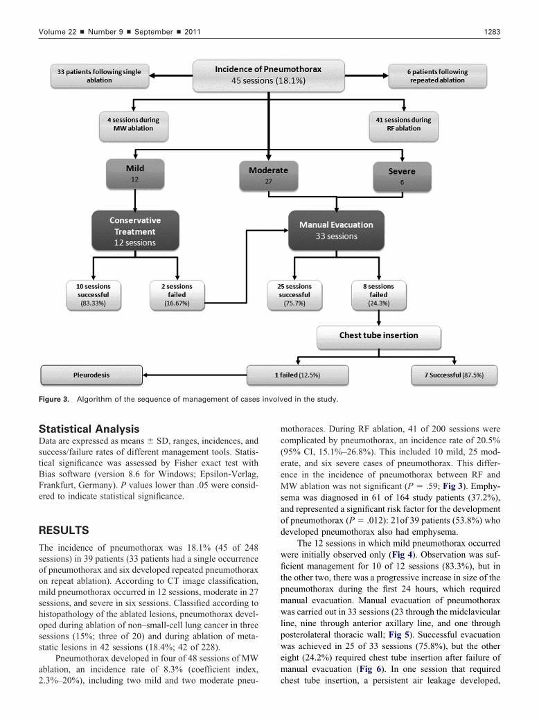
mc(eeMsaod
wfitpmwlpwem
involv
Volume 22 � Number 9 � September � 2011 1283
Statistical AnalysisData are expressed as means � SD, ranges, incidences, andsuccess/failure rates of different management tools. Statis-tical significance was assessed by Fisher exact test withBias software (version 8.6 for Windows; Epsilon-Verlag,Frankfurt, Germany). P values lower than .05 were consid-ered to indicate statistical significance.
RESULTS
The incidence of pneumothorax was 18.1% (45 of 248sessions) in 39 patients (33 patients had a single occurrenceof pneumothorax and six developed repeated pneumothoraxon repeat ablation). According to CT image classification,mild pneumothorax occurred in 12 sessions, moderate in 27sessions, and severe in six sessions. Classified according tohistopathology of the ablated lesions, pneumothorax devel-oped during ablation of non–small-cell lung cancer in threesessions (15%; three of 20) and during ablation of meta-static lesions in 42 sessions (18.4%; 42 of 228).
Pneumothorax developed in four of 48 sessions of MWablation, an incidence rate of 8.3% (coefficient index,
Figure 3. Algorithm of the sequence of management of cases
2.3%–20%), including two mild and two moderate pneu- c
othoraces. During RF ablation, 41 of 200 sessions wereomplicated by pneumothorax, an incidence rate of 20.5%95% CI, 15.1%–26.8%). This included 10 mild, 25 mod-rate, and six severe cases of pneumothorax. This differ-nce in the incidence of pneumothorax between RF andW ablation was not significant (P � .59; Fig 3). Emphy-
ema was diagnosed in 61 of 164 study patients (37.2%),nd represented a significant risk factor for the developmentf pneumothorax (P � .012): 21of 39 patients (53.8%) whoeveloped pneumothorax also had emphysema.
The 12 sessions in which mild pneumothorax occurredere initially observed only (Fig 4). Observation was suf-cient management for 10 of 12 sessions (83.3%), but in
he other two, there was a progressive increase in size of theneumothorax during the first 24 hours, which requiredanual evacuation. Manual evacuation of pneumothoraxas carried out in 33 sessions (23 through the midclavicular
ine, nine through anterior axillary line, and one throughosterolateral thoracic wall; Fig 5). Successful evacuationas achieved in 25 of 33 sessions (75.8%), but the other
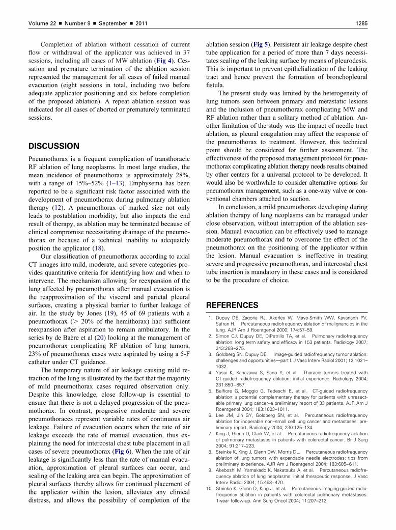
ight (24.2%) required chest tube insertion after failure ofanual evacuation (Fig 6). In one session that required
ed in the study.
hest tube insertion, a persistent air leakage developed,

wwptsmbewapmoeswpqitlchest tube was placed (arrow).
Ablation continued without interruption of the ablation session, and p
evacuation shows near-complete expansion of the lung.
1284 � Algorithmic Approach to Ablation-related Pneumothorax Management Nour-Eldin et al � JVIR
hich necessitated pleurodesis. Of the eight sessions inhich manual evacuation failed, six were cases of severeneumothorax and two were cases of moderate pneumo-horax. The mean volume of air evacuated manually inuccessful sessions was 480 mL � 50.8 (range, 250–950L). Durations of hospital stay were 24 h for cases managed
y close observation, 24–48 h for cases managed by manualvacuation of pneumothorax, and as long as 7 days for patientsho underwent chest tube insertion. Persistent air leakage
fter 7 days despite chest tube drainage was an indication forleurodesis (17). Chronologically, pneumothorax evolved im-ediately after thoracic puncture in 10 sessions. Development
f pneumothorax did not interfere with adequate applicator (ie,lectrode or antenna) insertion in six sessions, and in twoessions (with moderate pneumothorax), manual evacuationas sufficient to allow positioning of the applicator. Severeneumothorax developed in another two sessions, which re-uired cessation of ablation therapy and intercostal chest tubensertion. In contrast, in the remaining 35 sessions, pneumo-horax developed after insertion of the applicator within the
a 52-year-old woman. (a) During RF ablation (in prone position),e with electrode positioning (white arrow) or produce clinicalwithout cessation or premature termination of the session. (b)
crease of pneumothorax during the procedure (black arrow).neumothorax was managed by close observation.
6-year-old man. (a) RF ablation was complicated by moderatetal catheter (white arrow). (b) Axial CT image after manual
Figure 6. Image of severe right-sided pneumothorax and sur-gical emphysema complicating RF ablation of a right basalpulmonary metastasis from hepatocellular carcinoma in a 51-year-old man. After failure of manual evacuation, an intercostal
Figure 4. Images of pulmonary metastases from breast carcinoma inmild pneumothorax developed (black arrowhead) but did not interferdistress during ablation. Pneumothorax was managed by observationNote the RF electrode within the lesion (white arrow) and the mild in
Figure 5. Images of lung metastasis from colorectal carcinoma in a 4pneumothorax, which was evacuated manually by a 10-F intercos
esion or after the completion of the ablation procedure.
attTtfi
laRoatpembwpv
acsmptstt
R
1
Volume 22 � Number 9 � September � 2011 1285
Completion of ablation without cessation of currentflow or withdrawal of the applicator was achieved in 37sessions, including all cases of MW ablation (Fig 4). Ces-sation and premature termination of the ablation sessionrepresented the management for all cases of failed manualevacuation (eight sessions in total, including two beforeadequate applicator positioning and six before completionof the proposed ablation). A repeat ablation session wasindicated for all cases of aborted or prematurely terminatedsessions.
DISCUSSION
Pneumothorax is a frequent complication of transthoracicRF ablation of lung neoplasms. In most large studies, themean incidence of pneumothorax is approximately 28%,with a range of 15%–52% (1–13). Emphysema has beenreported to be a significant risk factor associated with thedevelopment of pneumothorax during pulmonary ablationtherapy (12). A pneumothorax of marked size not onlyleads to postablation morbidity, but also impacts the endresult of therapy, as ablation may be terminated because ofclinical compromise necessitating drainage of the pneumo-thorax or because of a technical inability to adequatelyposition the applicator (18).
Our classification of pneumothorax according to axialCT images into mild, moderate, and severe categories pro-vides quantitative criteria for identifying how and when tointervene. The mechanism allowing for reexpansion of thelung affected by pneumothorax after manual evacuation isthe reapproximation of the visceral and parietal pleuralsurfaces, creating a physical barrier to further leakage ofair. In the study by Jones (19), 45 of 69 patients with apneumothorax (� 20% of the hemithorax) had sufficientreexpansion after aspiration to remain ambulatory. In theseries by de Baère et al (20) looking at the management ofpneumothorax complicating RF ablation of lung tumors,23% of pneumothorax cases were aspirated by using a 5-Fcatheter under CT guidance.
The temporary nature of air leakage causing mild re-traction of the lung is illustrated by the fact that the majorityof mild pneumothorax cases required observation only.Despite this knowledge, close follow-up is essential toensure that there is not a delayed progression of the pneu-mothorax. In contrast, progressive moderate and severepneumothoraces represent variable rates of continuous airleakage. Failure of evacuation occurs when the rate of airleakage exceeds the rate of manual evacuation, thus ex-plaining the need for intercostal chest tube placement in allcases of severe pneumothorax (Fig 6). When the rate of airleakage is significantly less than the rate of manual evacu-ation, approximation of pleural surfaces can occur, andsealing of the leaking area can begin. The approximation ofpleural surfaces thereby allows for continued placement ofthe applicator within the lesion, alleviates any clinical
distress, and allows the possibility of completion of theblation session (Fig 5). Persistent air leakage despite chestube application for a period of more than 7 days necessi-ates sealing of the leaking surface by means of pleurodesis.his is important to prevent epithelialization of the leaking
ract and hence prevent the formation of bronchopleuralstula.
The present study was limited by the heterogeneity ofung tumors seen between primary and metastatic lesionsnd the inclusion of pneumothorax complicating MW andF ablation rather than a solitary method of ablation. An-ther limitation of the study was the impact of needle tractblation, as pleural coagulation may affect the response ofhe pneumothorax to treatment. However, this technicaloint should be considered for further assessment. Theffectiveness of the proposed management protocol for pneu-othorax complicating ablation therapy needs results obtained
y other centers for a universal protocol to be developed. Itould also be worthwhile to consider alternative options forneumothorax management, such as a one-way valve or con-entional chambers attached to suction.
In conclusion, a mild pneumothorax developing duringblation therapy of lung neoplasms can be managed underlose observation, without interruption of the ablation ses-ion. Manual evacuation can be effectively used to manageoderate pneumothorax and to overcome the effect of the
neumothorax on the positioning of the applicator withinhe lesion. Manual evacuation is ineffective in treatingevere and progressive pneumothorax, and intercostal chestube insertion is mandatory in these cases and is consideredo be the procedure of choice.
EFERENCES
1. Dupuy DE, Zagoria RJ, Akerley W, Mayo-Smith WW, Kavanagh PV,Safran H. Percutaneous radiofrequency ablation of malignancies in thelung. AJR Am J Roentgenol 2000; 174:57–59.
2. Simon CJ, Dupuy DE, DiPetrillo TA, et al. Pulmonary radiofrequencyablation: long term safety and efficacy in 153 patients. Radiology 2007;243:268–275.
3. Goldberg SN, Dupuy DE. Image-guided radiofrequency tumor ablation:challenges and opportunities—part I. J Vasc Interv Radiol 2001; 12,1021–1032.
4. Yasui K, Kanazawa S, Sano Y, et al. Thoracic tumors treated withCT-guided radiofrequency ablation: initial experience. Radiology 2004;231:850–857.
5. Belfiore G, Moggio G, Tedeschi E, et al. CT-guided radiofrequencyablation: a potential complementary therapy for patients with unresect-able primary lung cancer--a preliminary report of 33 patients. AJR Am JRoentgenol 2004; 183:1003–1011.
6. Lee JM, Jin GY, Goldberg SN, et al. Percutaneous radiofrequencyablation for inoperable non–small cell lung cancer and metastases: pre-liminary report. Radiology 2004; 230:125–134.
7. King J, Glenn D, Clark W, et al. Percutaneous radiofrequency ablationof pulmonary metastases in patients with colorectal cancer. Br J Surg2004; 91:217–223.
8. Steinke K, King J, Glenn DW, Morris DL. Percutaneous radiofrequencyablation of lung tumors with expandable needle electrodes: tips frompreliminary experience. AJR Am J Roentgenol 2004; 183:605–611.
9. Akeboshi M, Yamakado K, Nakatsuka A, et al. Percutaneous radiofre-quency ablation of lung neoplasms: initial therapeutic response. J VascInterv Radiol 2004; 15:463–470.
0. Steinke K, Glenn D, King J, et al. Percutaneous imaging-guided radio-
frequency ablation in patients with colorectal pulmonary metastases:1-year follow-up. Ann Surg Oncol 2004; 11:207–212.
1
1
1
1
2
1286 � Algorithmic Approach to Ablation-related Pneumothorax Management Nour-Eldin et al � JVIR
11. vanSonnenberg E, Shankar S, Morrison PR, et al. Radiofrequency ablationof thoracic lesions: part 2, initial clinical experience--technical and multidisci-plinary considerations in 30 patients. AJR Am J Roentgenol 2005; 184:381–390.
12. Yamagami T, Kato T, Hirota T, Yoshimatsu R, Matsumoto T, Nishimura T.Pneumothorax as a complication of percutaneous radiofrequency abla-tion for lung neoplasms. J Vasc Interv Radiol 2006; 17:1625–1629.
13. McTaggart RA, Dupuy DE. Thermal ablation of lung tumors. Tech VascInterv Radiol 2007; 10:102–113.
14. Goldberg SN, Grassi CJ, Cardella JF, et al. Image-guided tumor abla-tion: standardization of terminology and reporting criteria. J Vasc IntervRadiol 2005; 16:765–778.
15. Yeow KM, Su IH, Pan KT, et al. Risk factors of pneumothorax andbleeding: multivariate analysis of 660 CT-guided coaxial cutting needle
lung biopsies. Chest 2004; 126:748–754.6. Moore EH, Shepard JA, McLoud TC, Templeton PA, Kosiuk JP.Positional precautions in needle aspiration lung biopsy. Radiology 1990;175:733–735.
7. Rinaldi S, Felton T, Bentley A. Blood pleurodesis for the medical man-agement of pneumothorax. Thorax 2009; 64:258–260.
8. Nour-Eldin NE, Naguib NN, Saeed AS, et al. Risk factors involved in thedevelopment of pneumothorax during radiofrequency ablation of lungneoplasms. AJR Am J Roentgenol 2009; 193:43–48.
9. Jones JJ. A place for aspiration in the treatment of spontaneous pneu-mothorax. Thorax 1985; 40:66–67.
0. de Baère T, Palussière J, Aupérin A, et al. Midterm local efficacy andsurvival after radiofrequency ablation of lung tumors with minimum
follow-up of 1 year: prospective evaluation. Radiology 2006; 240:587–596.Copyright © 2022 FDOKUMEN