
Cytotoxic 2-benzylidene-6-(nitrobenzylidene)cyclohexanones which display substantially greater...
15
Cytotoxic 2-benzylidene-6-(nitrobenzylidene)cyclohexanones which display substantially greater toxicity for neoplasms than non-malignant cells Umashankar Da a , Alireza Doroudi a,† , H. Inci Gul a,‡ , Hari N. Pati a,§ , Masami Kawase b , Hiroshi Sakagami c , Qing Chu c , James P. Stables d , and Jonathan R. Dimmock a,* a Drug Design and Discovery Research Group, College of Pharmacy and Nutrition, University of Saskatchewan, 110 Science Place, Saskatoon, Saskatchewan, Canada S7N 5C9 b Faculty of Pharmaceutical Sciences, Matsuyama University, 4-2 Bunkyo-cho, Maysuyama, Ehime 790-8578, Japan c Division of Pharmacology, Department of Diagnostic and Therapeutic Sciences, Meikai University School of Dentistry, Saitama 350-0238, Japan d National Institute of Neurological Disorders and Stroke, 6001 Executive Boulevard, Rockville, MD 20852, USA Abstract Various 2-benzylidene-6-(nitrobenzylidene)cyclohexanones were prepared as candidate cytotoxins in which the nitro group was located in the ortho, meta and para positions leading to series 1–3, respectively. The CC 50 values towards human HSC-2 and HSC-4 oral squamous cell carcinomas as well as human HL-60 promyelocytic leukemic cells are in the low micromolar range in general. On the other hand, most of the compounds afforded clear evidence of being far less toxic towards human HGF gingival fibroblasts, HPC pulp cells and HPLF periodontal ligament fibroblasts which are non-malignant cells. Selectivity index (SI) figures were generated which are the ratios of the average CC 50 values towards normal cells and the CC 50 figure towards a malignant cell line. Huge SI values were obtained for many of the compounds. In particular 1c, 2f, 3c and 3g which have average SI values of >76, >38, 124 and 341, respectively, are clearly lead molecules affording direction for amplification of this area of study. A lead compound 1c caused internucleosomal DNA fragmentation and activation of caspase-3 in HL-60 cells but not in HSC-2 carcinomas. In a short-term toxicity study, doses up to and including 300 mg/kg of the majority of the compounds prepared in this study did not cause any mortalities to mice. Some guidelines for development of these tumor-selective cytotoxins are presented. Keywords Unsaturated ketones; Selective cytotoxicity; Structure1–activity relationships; Murine toxicity © 2010 Elsevier Ltd. All rights reserved. * Corresponding author. Tel.: +1 306 966 6331; fax: +1 966 6377. [email protected] (J.R. Dimmock). † Present address: School of Pharmacy, Ahwaz Jondishapur University of Medical Sciences, Ahwaz, Khouzestan, Iran. ‡ Present address: Department of Pharmaceutical Chemistry, Faculty of Pharmacy, Ataturk University, 25240 Erzurum, Turkey. § Present address: Advinus Therapeutics Pvt. Ltd, Peenya Industrial Area, Bangalore 560 058, India. PubMed Central CANADA Author Manuscript / Manuscrit d'auteur Bioorg Med Chem. Author manuscript; available in PMC 2012 March 23. Published in final edited form as: Bioorg Med Chem. 2010 March 15; 18(6): 2219–2224. doi:10.1016/j.bmc.2010.01.069. PMC Canada Author Manuscript PMC Canada Author Manuscript PMC Canada Author Manuscript
-
Upload
uin-alauddin -
Category
Documents
-
view
0 -
download
0
Transcript of Cytotoxic 2-benzylidene-6-(nitrobenzylidene)cyclohexanones which display substantially greater...
Cytotoxic 2-benzylidene-6-(nitrobenzylidene)cyclohexanoneswhich display substantially greater toxicity for neoplasms thannon-malignant cells
Umashankar Daa, Alireza Doroudia,†, H. Inci Gula,‡, Hari N. Patia,§, Masami Kawaseb,Hiroshi Sakagamic, Qing Chuc, James P. Stablesd, and Jonathan R. Dimmocka,*
aDrug Design and Discovery Research Group, College of Pharmacy and Nutrition, University ofSaskatchewan, 110 Science Place, Saskatoon, Saskatchewan, Canada S7N 5C9bFaculty of Pharmaceutical Sciences, Matsuyama University, 4-2 Bunkyo-cho, Maysuyama,Ehime 790-8578, JapancDivision of Pharmacology, Department of Diagnostic and Therapeutic Sciences, MeikaiUniversity School of Dentistry, Saitama 350-0238, JapandNational Institute of Neurological Disorders and Stroke, 6001 Executive Boulevard, Rockville,MD 20852, USA
AbstractVarious 2-benzylidene-6-(nitrobenzylidene)cyclohexanones were prepared as candidate cytotoxinsin which the nitro group was located in the ortho, meta and para positions leading to series 1–3,respectively. The CC50 values towards human HSC-2 and HSC-4 oral squamous cell carcinomasas well as human HL-60 promyelocytic leukemic cells are in the low micromolar range in general.On the other hand, most of the compounds afforded clear evidence of being far less toxic towardshuman HGF gingival fibroblasts, HPC pulp cells and HPLF periodontal ligament fibroblastswhich are non-malignant cells. Selectivity index (SI) figures were generated which are the ratiosof the average CC50 values towards normal cells and the CC50 figure towards a malignant cellline. Huge SI values were obtained for many of the compounds. In particular 1c, 2f, 3c and 3gwhich have average SI values of >76, >38, 124 and 341, respectively, are clearly lead moleculesaffording direction for amplification of this area of study. A lead compound 1c causedinternucleosomal DNA fragmentation and activation of caspase-3 in HL-60 cells but not in HSC-2carcinomas. In a short-term toxicity study, doses up to and including 300 mg/kg of the majority ofthe compounds prepared in this study did not cause any mortalities to mice. Some guidelines fordevelopment of these tumor-selective cytotoxins are presented.
KeywordsUnsaturated ketones; Selective cytotoxicity; Structure1–activity relationships; Murine toxicity
© 2010 Elsevier Ltd. All rights reserved.*Corresponding author. Tel.: +1 306 966 6331; fax: +1 966 6377. [email protected] (J.R. Dimmock).†Present address: School of Pharmacy, Ahwaz Jondishapur University of Medical Sciences, Ahwaz, Khouzestan, Iran.‡Present address: Department of Pharmaceutical Chemistry, Faculty of Pharmacy, Ataturk University, 25240 Erzurum, Turkey.§Present address: Advinus Therapeutics Pvt. Ltd, Peenya Industrial Area, Bangalore 560 058, India.
PubMed Central CANADAAuthor Manuscript / Manuscrit d'auteurBioorg Med Chem. Author manuscript; available in PMC 2012 March 23.
Published in final edited form as:Bioorg Med Chem. 2010 March 15; 18(6): 2219–2224. doi:10.1016/j.bmc.2010.01.069.
PMC
Canada Author M
anuscriptPM
C C
anada Author Manuscript
PMC
Canada Author M
anuscript
1. IntroductionThe major emphasis of this laboratory is the design, syntheses and bioevaluations ofcandidate antineoplastic agents. These compounds are principally conjugated unsaturatedketones which is a class of compounds known to have an affinity for thiols but possess farless propensity to interact with amines.1,2 Hence these enones may exert their cytotoxicproperties without interfering with nucleic acids and hence be bereft of the genotoxic sideeffects of various anticancer drugs.3 Various in vitro studies revealed that conjugated enoneslower thiol concentrations of malignant cells and this effect may be prevented by theaddition of various thiols such as glutathione to the media.4,5
Previously a single enone pharmacophore was employed in the design of different series ofcompounds.6 An aryl ring with different substituents was normally attached to the α,β-unsaturated keto group so that the polarity of the olefinic atoms varies which may influenceantineoplastic potencies.7 In addition, the use of different aryl substituents affects thehydrophobic and steric properties of the molecules. However recently several series ofcompounds have been prepared which contain the 1,5-diaryl-3- oxo-1,4-pentadienyl group(AR–CH=CHCOCH=CHAR) which is referred to hereafter as the dienone moiety. Thereason for preparing this pharmacophore is that a number of tumours are more sensitive tochemical insult than some normal cells.8,9 Hence an initial reaction with cellular mercaptansfollowed by a second thiol alkylation may lead to greater toxicity in malignant cells thantowards normal tissues. In other words, compounds which are capable of successive attacksin cells may display lethal effects preferentially in neoplasms. This theory is known assequential cytotoxicity and has been described in detail10 while investigations based on thishypothesis have been undertaken.11,12
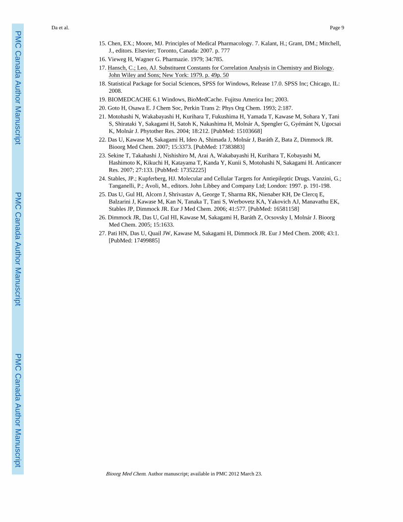
Several years ago, the decision was made to initiate a project in which compounds weredesigned so that olefinic carbon atoms which interact with thiols bore divergent electroniccharges. In this way, a difference between the atomic charges of the olefinic carbon atomsexists13 which will enhance successive interactions with cellular thiols. In other words, onewould predict that an initial reaction would take place at the more electropositive carbonatom followed by thiol alkylation on the remaining olefinic carbon. The design of thecompounds is indicated in Figure 1A. Phase I of this project involved a preliminaryinvestigation as to whether the compounds possessed cytotoxic properties. This work hasbeen completed and the biodata reveal that the compounds inhibit the growth of human Molt4/C8 and CEM T-lymphocytes and murine L1210 cells.13 On the basis of these results;Phase II of this study was initiated which addresses three issues. First, the crucial issues ofwhether the compounds exert preferential cytotoxicity to malignant cells compared tonormal cells. Second, the mode of action of a lead compound to discern the way in whichcytotoxicity is mediated. Third, if they are well tolerated in mammals or associated with anyundesired toxicity. This report indicates the results of the Phase II evaluations.
2. ResultsThe compounds in series 1–4 which comprise the basis of this study are indicated in Figure1B. The syntheses of 1–4 have been described previously13 where as 5a–c are newcompounds. In general, condensation between a nitrobenzaldehyde and cyclohexanone ledto the corresponding 2-(nitrobenzylidene)cyclohexanone which on reaction with a variety ofaromatic aldehydes gave the desired products in series 1–3. Reaction of cyclohexanone and2-nitrobenzaldehyde afforded the related aldol which on dehydration led to 4a. Conversionof cyclohexanone into the corresponding enamine followed by reaction with 3-nitrobenzaldehyde produced 4b. The olefinic double bonds in all of these compoundsassumed the E configuration and the electron densities on the CB atoms are invariably lower
Da et al. Page 2
Bioorg Med Chem. Author manuscript; available in PMC 2012 March 23.
PMC
Canada Author M
anuscriptPM
C C
anada Author Manuscript
PMC
Canada Author M
anuscript
than the CA atoms in series 1–3. In view of the biodata obtained from series 1–4 vide infra,5a–c were prepared by condensing 2-(4-nitrobenzylidene)-cyclohexanone with various arylaldehydes (see Fig. 2).
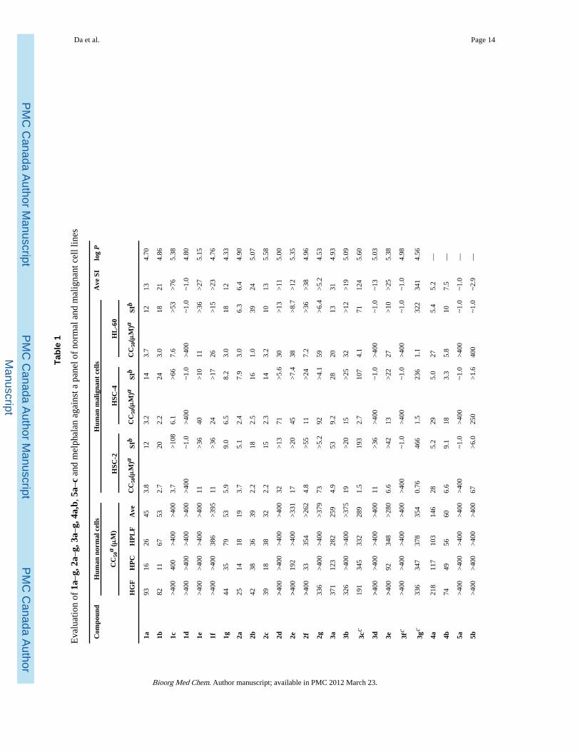
All of the compounds in series 1–5 were evaluated against three human non-malignant cells,namely HGF gingival fibroblasts, HPC pulp cells and HPLF periodontal ligament fibroblastsand three malignant cells, namely HSC-2 and HSC-4 squamous cell carcinomas as well asHL-60 promyelocytic leukemia cells. These results are presented in Table 1. Mode of actionstudies revealed that a lead compound 1c caused internucleosomal DNA fragmentation (Fig.3) and activation of caspase-3 in HL-60 cells but not in HSC-2 carcinomas (Fig. 4). Doses of30, 100 and 300 mg/kg of 1a–c,f,g, 2a,b,d–g, 3a–g, 4a,b and 5a–c were injectedintraperitoneally into mice and the animals were examined for mortalities and neurotoxicityat the end of 0.5 and 4 h.
3. DiscussionThe first issue to be resolved is whether the compounds in series 1–3 are well tolerated byHGF, HPC and HPLF normal cells. The percentages of the CC50 values of 1a–g, 2a–g and3a–g which are in excess of 100 μM are 57 (12 out of 31), 52 (11 out of 21) and 95 (20 outof 21), respectively, indicating that the compounds in series 3 have lower unwanted toxicproperties towards normal cells than the analogs in series 1 and 2. Overall 68% (43 out of63) of the CC50 figures of 1–3 are greater than 100 μM and for the remaining compoundsthe average CC50 value is 42 μM. In general, therefore, these normal cells are not adverselyaffected by the dienones 1–3.
The biodata of the sensitivity of the human HSC-2, HSC-4 and HL-60 neoplastic cell linesto the compounds in series 1–3 are presented in Table 1 which reveal that, in general, thesecompounds are cytotoxic to oral HSC-2 and HSC-4 tumours as well as HL-60 cells (chosenas a positive control of apoptosis since this biochemical effect is readily induced in this cellline by many chemotherapeutic agents). There are three outliers, namely 1d, 3d and 3fwhich have CC50 values in excess of 400 μM in all three bioassays with the exception of 3dtowards HSC-2 cells. The CC50 figures of all of the other compounds in series 1–3 arebelow 100 μM and, in fact, 48% and 59% of these CC50 values are less than 5 and 10 μM,respectively.
In order to ascertain if the electronic, hydrophobic or steric properties of the groups in ring Binfluence cytotoxic potencies, linear (l) and semilogarithmic (sl) plots were made betweenthe CC50 values of 1a–c,e–g in the HSC-2 assay and the Hammett sigma, Hansch pi andmolecular refractivity (MR) figures of the aryl substituents. In addition, logarithmic plotswere constructed using MR constants. The experiment was repeated using the biodata in theHSC-4 test and then the results using HL-60 cells. A similar situation was undertaken with2a–g and finally with 3a–c,e,g. The following correlations (p <0.05) and trends towardssignificance (p <0.1) were noted. A negative correlation was found between the CC50figures of 1a–c,e,g towards HSC-2 cells and the σ values. The MR constants of the arylsubstituents in series 2 correlated positively with the CC50 values obtained in the HSC-2,HSC-4 and HL-60 bioassays. On the other hand, a negative correlation was noted betweenthe MR constants of 3a–c,e,g and the potencies towards HL-60 cells and there was anegative trend towards significance in the case of HSC-4 cells. No other correlations ortrends to significance were noted. Thus in developing these compounds, increasingpotencies may result from inserting groups into aryl ring B with large positive σ values inseries 1, small substituents (low MR values) in series 2 and large groups (high MRconstants) in series 3. Since no correlations were noted with π values, lipophilicity appearsto be unimportant in reference to the cytotoxic potencies in series 1–3. This conclusion was
Da et al. Page 3
Bioorg Med Chem. Author manuscript; available in PMC 2012 March 23.
PMC
Canada Author M
anuscriptPM
C C
anada Author Manuscript
PMC
Canada Author M
anuscript
reinforced when linear, semilogarithmic and logarithmic plots were made between the log Pvalues of 1a–g, 2a–g and then 3a–g (which are presented in Table 1) and the CC50 values ineach series against HSC-2 cells, then HSC-4 neoplasms and finally HL-60 cells. Nocorrelations were found and only a trend towards a negative correlation between the CC50figures of 2a–g in the HSC-2 screen and the log P values was noted (p <0.1).
In a further attempt to discern correlations between the electronic, hydrophobic and stericproperties of the aryl substituents and cytotoxic potencies, multi-linear regression (MLR)analyses were undertaken. A data set was obtained for 21 compounds viz 1a–g, 2a–g and3a–g and four physiochemical descriptors (σ, π, MR and log P). When the ring B is di or tri-substituted, the sum of individual parameters (Σσm,p, Σπ, ΣMR) was used. The resultsindicate that there were no statistical correlations between the cytotoxicity against HSC-2and HSC-4 cells and the four physicochemical descriptors. However, a modest correlationwas noted against the HL-60 cell line (Eq. 1). Efforts were made to improve this result byexcluding one or more descriptors from the regression analysis which led to a lowering ofthe statistical quality observed in Eq. 1.
(1)
n = 21, r = 0.741, radj = 0.631, s = 0.61, F = 3.65, p = 0.023.
In this equation, n represents the number of data points, r is the correlation coefficient, radj isthe adjusted r, s is the standard deviation of the regression equation, and the F value isrelated to the F-statistic analysis (Fisher test).
The cytotoxicity of the compounds in series 1–5 is considered to be mediated principally byreactions at the olefinic carbon atoms with cellular thiols. In order to gain an understandingof the effect of different aryl substituents on the electron densities at the olefinic carbonatoms, the 13C NMR spectra of the representative enones 1a–c,e–g were determined (1d wasomitted due to the absence of specific CC50 values). The 13C NMR absorptions at CA (asdesignated in Fig. 1A) are in the range of 132.53–133.11 while at CB, the relevant figuresare 136.62–138.17 (the precise values of individual compounds are listed in theExperimental Section). These data reveal that the CB carbon atoms are more electrondeficient than the CA atoms and are the loci of the initial interactions with thiols. Linear andlogarithmic plots were constructed between the 13C chemical shifts of the CB atoms theCC50 values of 1a–c,e–g in the HSC-2, HSC-4 and HL-60 bioassays. No correlations ortrends to significance were noted although the logarithmic plot between the CC50 figures inthe HSC-2 assay provided a p value of 0.125 (a positive correlation). One may conclude thatthe spectroscopic data reveal a marked disparity in the electron densities on the olefiniccarbon atoms which did not correlate significantly with cytotoxic potencies.
The discernment of whether the compounds in series 1–3 display greater toxicity toneoplasms than normal cells was achieved as follows. In patients afflicted with cancer, themalignant cells are often surrounded by a number of different normal tissues. Hence in orderto simulate clinical conditions, a comparison was made between the cytotoxicity of acompound to a malignant cell line compared to the average CC50 value towards threenormal cell lines. The figure generated is referred to as the selectivity index (SI) value of thecompound and these data are listed in Table 1. A SI figure of 10 was arbitrarily chosen as anindicator of marked selectivity. The results of the outliers 1d, 3d and 3f were removed fromconsideration and for the other compounds in series 1–3, this criterion was observed in 80%of the SI data. Hence the 2-benzylidene-6-(nitrobenzylidene) cyclohexanones are a novelcluster of cytotoxins with a substantial predilection for greater toxicity towards neoplasms
Da et al. Page 4
Bioorg Med Chem. Author manuscript; available in PMC 2012 March 23.
PMC
Canada Author M
anuscriptPM
C C
anada Author Manuscript
PMC
Canada Author M
anuscript
than normal cells. In particular, the compounds demonstrating the highest selective toxicityto malignant cells (average SI values in parentheses) are 1c(>76), 2f (>38), 3c (124) and 3g(341), all of which clearly serve as lead molecules. A comparison was made between 4a and4b and the analogs in series 1 and 2, respectively, in terms of their potencies towardsHSC-2, HSC-4 and HL-60 malignant cells and SI values. With the exception of the outlier1d, in general the CC50 figures of 4a are higher than obtained for series 1 while the SIvalues are invariably lower. The enone 4b is less potent than the analogs in series 2 inapproximately half of the comparisons made while the SI figures are generally lower thanwas observed for 2a–g. Thus the addition of an arylidene group to anitrobenzylidenecyclohexanone leads, in general, to increases in both potencies to malignantcells as well as SI values.
Conjugated enones alkylate cellular thiols, vide supra, and comparisons were made betweenthe potencies and selective toxicity of a clinically used alkylating agent melphalan and thecompounds in series 1–3. In the case of melphalan, the CC50 figures are higher and the SIvalues lower in 68% and 65%, respectively, of the comparisons made. This observationreinforces the decision to develop these compounds further.
An important issue to resolve which will affect the way in which the project may beamplified is whether the positions of the three methoxy groups in 3g control cytotoxicpotencies and SI values. Hence three structural isomers of 3g were prepared namely 5a–cand their bioevaluation is presented in Table 1. These compounds are less toxic to humannormal cells than 3g. However apart from the lethal effects of 5c towards HL-60 cells,significant potency among 5a–c towards the neoplasms is lacking. One ortho methoxy groupis present in 5a and 5b while two are present in 5c and hence the possibility exists that thesegroups will increase the torsion angles (θB) between aryl ring B and the adjacent olefinicgroup. However models reveal that the θB angles of 5a–c are 54.5°, −54.8° and 74.8°,respectively, compared to a figure of −51.9 for 3g.11 The different substituents in ring Bhave little effect on the θA values (the torsion angles between ring A and the attachedolefinic group) which for 3g and 5a–c are 51.2,11 −48.0, 51.1 and −48.1, respectively. Hencethe interplanar angles θA and θB appear to contribute little to the disparity in cytotoxicitybetween 3g and the closely related structural analogs 5a–c. However while the π and MRvalues of 3g are the same as 5a–c, there are huge differences in the Σσvalues of 3g (−0.03)on one hand and 5a (−0.37), 5b (−0.37) and 5c (−0.71) on the other hand which likelycontributes significantly to the different potencies. In addition, similar electronic factorslikely account for the low cytotoxicity of 3d (σp = −0.83) and 3f (σp = −0.27).
A mode of action study was undertaken using a representative compound 1c. Thiscompound was chosen based on its cytototic potency and especially its large SI values.Incubation of 1c with HL-60 cells induced the internucleosomal DNA fragmentation (Fig. 3)and concentration-dependent activation of caspase-3 (Fig. 4) by 1c, suggesting theoccurrence of apoptosis via the mitochondrial pathway. On the other hand, there is noevidence from the data provided in Figs. 3 and 4 that 1c causes apoptosis in HSC-2carcinomas. Hence the lethality of 1c to this cell line is mediated by interference with otherbiochemical mechanisms. Thus the modes of action are dependent on the cell line underconsideration which in turn gives rise to the enormous SI values displayed by many of thesenovel cytotoxins.
The final segment of this study involved the determination of the tolerability of the majorityof the compounds in series 1–5 in mice. This investigation is important since manyanticancer agents are highly toxic to mammals, for example, the LD50 of melphalan in miceis 4.0 mg/kg14 and neurotoxicity is a side effect of certain anticancer drugs such asoxaliplatin and etoposide.15 Doses of 30, 100 and 300 mg/kg of 1a–c,f,g, 2a,b,d–g, 3a–g,
Da et al. Page 5
Bioorg Med Chem. Author manuscript; available in PMC 2012 March 23.
PMC
Canada Author M
anuscriptPM
C C
anada Author Manuscript
PMC
Canada Author M
anuscript
4a,b and 5a–c were injected intraperitoneally into mice and the animals examined formortalities and neurotoxicity after 0.5 and 4 h. None of the compounds caused any deaths tothe animals. Mice receiving doses of 1a,b,f,g, 2e,g, 3a,c,f and 5a,c did not display anyneurotoxic symptoms. One of these dienones, namely 3c, was identified earlier as a leadcompound since it has an average SI value of 124 and the absence of overt mammaliantoxicity enhances its status for analog development. Some neurotoxicity was observed in theremaining compounds, specific details of which are given in the Experimental section.Marginal neurotoxicity was noted in less than half of the animals examined in the case of2a,b,d,f, 3b,d,g, 4a,b and 5b after 0.5 but not 4 h. Neurological deficit occurred in half ormore mice receiving 300 mg/kg of 1c after 4 h and both 2a and 3e when observed after 0.5h. The conclusion to be drawn from this short-term toxicity evaluation of the majority of thecompounds prepared in this study is that these dienones are well tolerated in mice.
4. ConclusionsSeries 1–3 are novel tumor-specific cytotoxins many of which have huge SI values. Inparticular 1c, 2f, 3c and 3g are clearly lead molecules. In some instances correlations werenoted between various physicochemical parameters and cytotoxic potencies. Thus in series1, the IC50 values diminished as the magnitude of the Σ value increased. On the other hand,the overall electron-releasing effect of the substituents in ring B of 5a–c likely contributedsignificantly to their lack of marked antineoplastic potencies. Cytotoxicity is favoured by theplacement of small groups in ring B in series 2 and large substituents in series 3. A leadcompound 1c caused apoptosis in HL-60 cells but not in HSC-2 carcinomas. In general thecompounds are well tolerated in mice.
5. Experimental5.1. Synthesis of compounds
Melting points are recorded in degrees Celsius on a Gallenkamp apparatus and areuncorrected. 1H and 13C NMR spectra were recorded on a Bruker AMX 500 FT instrumentwhile elemental analyses were obtained using an Elementer analyzer.
5.1.1. Synthesis of the compounds in series 1–5—The methodology for thepreparation of the nitrobenzylidenecyclohexanones in series 1–4 has been describedpreviously.13 The general procedure for the synthesis of 5a–c is as follows. Dry hydrogenchloride was passed into a mixture of 2-(4-nitrobenzylidene) cyclohexanone (0.005 mol),which was prepared by a literature procedure,16 the appropriate trimethoxybenzaldehyde(0.005 mol), chloroform(5 mL) and diethyl ether (40 mL). The reaction mixture was stirredat room temperature for 6–8 h and the resultant solid was collected and crystallized fromchloroform/ether (1:5).
5.1.1.1. 6-(4-Nitrobenzylidene)-2-(2,3,4-trimethoxybenzylidene) cyclohexanone (5a):Mp 176 °C; yield 72%. 1H NMR (CDCl3): δ 1.82 (p, 2H, CH2), 2.90 (m, 4H, 2 × CH2), 3.90(s, 3H, OCH3), 3.92 (s, 3H, OCH3), 3.93 (s, 3H, OCH3), 6.71 (d, 1H, Ar-H, J = 8.73 Hz),7.11 (d, 1H, Ar-H, J = 8.69 Hz), 7.59 (d, 2H, Ar-H, J = 8.67 Hz), 7.79 (s, 1H, =CH),7.98 (s,1H, =CH), 8.27 (d, 2H, Ar-H, J = 8.73 Hz). Anal. Calcd for C23H23NO6: C, 67.47; H, 5.66;N, 3.42. Found: C, 67.40; H, 5.43; N, 3.43.
5.1.1.2. 6-(4-Nitrobenzylidene)-2-(2,4,5-trimethoxybenzylidene) cyclohexanone (5b):Mp 163 °C; yield 64%. 1H NMR (CDCl3): δ 1.84 (p, 2H, CH2), 2.92 (m, 4H, 2 × CH2), 3.89(s, 3H, OCH3), 3.90 (s, 3H, OCH3), 3.97 (s, 3H, OCH3), 6.57 (s, 1H, Ar-H), 6.96 (s, 1H, Ar-H), 7.60 (d, 2H, Ar-H, J = 8.60 Hz), 7.79 (s, 1H, =CH), 8.08 (s, 1H, =CH), 8.28 (d, 2H, Ar-
Da et al. Page 6
Bioorg Med Chem. Author manuscript; available in PMC 2012 March 23.
PMC
Canada Author M
anuscriptPM
C C
anada Author Manuscript
PMC
Canada Author M
anuscript
H, J = 8.68 Hz). Anal. Calcd for C23H23NO6: C, 67.47; H, 5.66; N, 3.42. Found: C, 67.33;H, 5.66; N, 3.33.
5.1.1.3. 6-(4-Nitrobenzylidene)-2-(2,4,6-trimethoxybenzylidene) cyclohexanone (5c): Mp159 °C; yield 58%. 1H NMR (CDCl3): δ 1.75 (p, 2H, CH2), 2.51 (t, 2H, CH2), 2.89 (t, 2H,CH2), 3.83 (s, 6H, 2 × OCH3), 3.86 (s, 3H, OCH3), 6.17 (s, 2H, Ar-H), 7.58 (d, 2H, Ar-H, J= 8.66 Hz), 7.73 (s, 1H, =CH), 7.77 (s, 1H, =CH), 8.26 (d, 2H, Ar-H, J = 8.75 Hz). Anal.Calcd for C23H23NO6: C, 67.47; H, 5.66; N, 3.42. Found: C, 67.32; H, 5.56; N, 3.42.
5.2. Structure–activity relationshipsThe constants used for various atoms and groups in series 1–3 (σ, π and MR values aregiven in parentheses) were taken from a reference source17 and are as follows: hydrogen(0.00, 0.00, 1.03), 4-fluoro (0.06, 0.14, 0.92), 4-chloro (0.23, 0.71, 6.03), 4-dimethylamino(−0.83, 0.18, 15.55), 4-methyl (−0.17, 0.56, 5.65), 4-methoxy (−0.27, −0.02, 7.87) and3,4,5-trimethoxy (−0.03, −0.06, 23.61). The MR value of the hydrogen atom is 1.03 and not0.00. Hence in the case of the monosubstituted compounds in series 1–3 viz. b–f, 2.06 (2 ×1.03) was added to the MR figure of the substituent and the MR value of the unsubstitutedcompound is 3.09 (3 × 1.03). The linear, semilogarithmic and logarithmic plots wereconstructed using a statistical software package.18 The following correlations (p <0.05) werenoted [nature of the plots viz. linear (l), semilogarithmic (sl) or logarithmic (log)/cell lineunder consideration, in parentheses]: 1a–c,e,g versus σ (l, sl/HSC-2 cells), 2a–g versus MR(l, sl, log/HSC-2, HSC-4, HL-60 cells) and 3a–c,e,g versus MR [sl, log/HL-60 cells]. Atrend to significance (p <0.1) was noted as follows namely 3a–c,e,g versus MR (sl, log/HSC-4 cells) and 2a– g versus log P (sl/HSC-2). The MLR analyses used a commercialsoftware package.18
5.3. 13C NMR determinationsConcentrations of 40 μM of 1a–c,e–g in deuterated chloroform were prepared and the 13CNMR spectra determined at room temperature. The chemical shifts of the olefinic carbonatoms were assigned with the help of HMQC experiments. The chemical shifts of thesecompounds at carbon atoms CA and CB, respectively, are as follows: 1a: 133.00, 138.06; 1b:133.10, 136.88; 1c: 133.11, 136.62; 1e: 132.75, 137.93; 1f 132.53, 138.17; 1g: 132.99,137.89, respectively.
5.4. Molecular modelingModels of the compounds in series 5 were built using a BIOMEDCACHE program.19 Theconformational analysis was undertaken using the CONFLEX procedure20 which generateslow energy conformers. This program incorporates down-stream, reservoir-filling, cornerflap, edge flip and stepwise rotation as well as a pre-check. The structures generated areoptimized using a modified MM2 molecular mechanics program. The heat of formation ofvarious conformations was calculated using the PM3 Hamiltonian approach in MOPAC2002 in order to obtain the global minimum energy as the true optimized geometry.
5.5. Bioevaluations5.5.1. Cytotoxicity assays—The examination of the compounds in series 1–5 towardsnormal HGF, HPC and HPLF cells as well as HSC-2, HSC-4 and HL-60 neoplasms wasaccomplished using a literature procedure21 which has been summarized recently.22 In brief,the cells were incubated with different concentrations of the compounds for 24 h and afterthe appropriate workup, the CC50 values were determined from dose–response curves.
Da et al. Page 7
Bioorg Med Chem. Author manuscript; available in PMC 2012 March 23.
PMC
Canada Author M
anuscriptPM
C C
anada Author Manuscript
PMC
Canada Author M
anuscript
5.5.2. Mode of action study—The methodology for evaluating 1c for the induction ofinternucleosomal DNA fragmentation and caspase-3 activation has been describedpreviously.23
5.5.3. Murine toxicity study—The evaluation of 1a–c,f,g, 2a,b,d–g, 3a–g,4a,b and 5a–cfor mortalities and neurotoxicity in mice was undertaken by the National Institute ofNeurological Disorders, and Stroke, USA according to their protocols.24 No deaths wereobserved. The evaluation of 3c,f,g in this screen has been recorded previously25 whichrevealed that 3c,f demonstrated no neurotoxicity while a dose of 100 mg/kg of 3g causedneurological deficit in 2/8 animals when observed after 0.5 h. Neurotoxicity was observedafter administration of the following compounds (dose in mg/kg, number of animalsdemonstrating neurological deficit/number of treated mice in parentheses viz. 2a (100, 2/8;300, 2/4), 2b (100, 1/8), 2d (100, 1/8; 300,1/4), 2f (30,1/4), 3b (100, 1/8), 3d (100, 2/8); 3e(300,3/4), 4a (300,1/4), 4b (300, 1/4) and 5b (100, 1/8; 300, 1/4). After 4 h, neurotoxicitywas observed in one of two mice who received a dose of 300 mg/kg of 1c. The animals werehoused, fed and handled using the protocols described in the ‘Guide for the Care and Use ofLaboratory Animals’ which is published by the National Research Council.
AcknowledgmentsThe authors thank the Canadian Institutes of Health Research for an operating grant to J. R. Dimmock and theMinistry of Education, Science, Sports and Culture of Japan for a Grant-in-Aid (No. 19592156) to H. Sakagami.The National Institute of Neurological Disorders and Stroke, USA undertook the in vivo experimentation with micewhich is recorded with gratitude. Our appreciation is extended to the Iranian Ministry of Health and MedicalEducation who provided financial support for A. Doroudi while H. I. Gul was supported by a visiting scholar grant(NATO-B2) distributed by the Scientific and Technical Research Council of Turkey (TUBITAK).
References and notes1. Pati HN, Das U, Sharma RK, Dimmock JR. Mini-Rev Med Chem. 2007; 7:131. [PubMed:
17305587]
2. Dimmock JR, Raghavan SK, Logan BM, Bigam GE. Eur J Med Chem. 1983; 18:248.
3. Okey, AB.; Harper, PA. Principles of Medical Pharmacology. 7. Kalant, H.; Grant, DM.; Mitchell,J., editors. Elsevier; Toronto, Canada: 2007. p. 902
4. Kamiya D, Uchihata Y, Ichikawa E, Kato K, Umezawa K. Bioorg Med Chem. 2005; 15:1111.
5. Samudio I, Konopleva M, Hail N Jr, Shi YX, McQueen T, Hsu T, Evans R, Honda T, Gubble GW,Sporn M, Gilbert HF, Sabe S, Andreeff M. J Biol Chem. 2005; 280:36273. [PubMed: 16118208]
6. Dimmock JR, Taylor WG. J Pharm Sci. 1975; 64:241.
7. Dimmock JR, Smith LM, Smith PJ. Can J Chem. 1980; 58:984.
8. Siemann DW, Beyers KL. Br J Cancer. 1993; 68:1071. [PubMed: 8260357]
9. del Olmo M, Alouso-Varona A, Castro B, Calle Y, Bilbao P, Palomares T. Melanoma Res. 2000;10:103. [PubMed: 10803710]
10. Dimmock JR, Sidhu KK, Chen M, Reid RS, Allen TM, Kao GY, Truitt GA. Eur J Med Chem.1993; 28:313.
11. Dimmock JR, Padmanilayam MP, Zello GA, Quail JW, Oloo EO, Prisciak JS, Kraatz HB,Cherkasov A, Lee JS, Allen TM, Santos CL, Manavathu EK, De Clercq E, Balzarini J, Stables JP.Eur J Med Chem. 2002; 37:813. [PubMed: 12446039]
12. Dimmock JR, Kandepu NM, Nazarali AJ, Motaganahalli NL, Kowalchuk TP, Pugazhenthi U,Prisciak JS, Quail JW, Allen TM, LeClerc R, Santos CL, De Clercq E, Balzarini J. J Med Chem.2000; 43:3933. [PubMed: 11052798]
13. Das U, Doroudi A, Das S, Bandy B, Balzarini J, De Clercq E, Dimmock JR. Bioorg Med Chem.2008; 16:6261. [PubMed: 18450457]
14. Quinn ER, Milne GWA. Fundam Appl Toxicol. 1986; 6:270. [PubMed: 3699317]
Da et al. Page 8
Bioorg Med Chem. Author manuscript; available in PMC 2012 March 23.
PMC
Canada Author M
anuscriptPM
C C
anada Author Manuscript
PMC
Canada Author M
anuscript
15. Chen, EX.; Moore, MJ. Principles of Medical Pharmacology. 7. Kalant, H.; Grant, DM.; Mitchell,J., editors. Elsevier; Toronto, Canada: 2007. p. 777
16. Vieweg H, Wagner G. Pharmazie. 1979; 34:785.
17. Hansch, C.; Leo, AJ. Substituent Constants for Correlation Analysis in Chemistry and Biology.John Wiley and Sons; New York: 1979. p. 49p. 50
18. Statistical Package for Social Sciences, SPSS for Windows, Release 17.0. SPSS Inc; Chicago, IL:2008.
19. BIOMEDCACHE 6.1 Windows, BioMedCache. Fujitsu America Inc; 2003.
20. Goto H, Osawa E. J Chem Soc, Perkin Trans 2: Phys Org Chem. 1993; 2:187.
21. Motohashi N, Wakabayashi H, Kurihara T, Fukushima H, Yamada T, Kawase M, Sohara Y, TaniS, Shirataki Y, Sakagami H, Satoh K, Nakashima H, Molnár A, Spengler G, Gyémánt N, UgocsaiK, Molnár J. Phytother Res. 2004; 18:212. [PubMed: 15103668]
22. Das U, Kawase M, Sakagami H, Ideo A, Shimada J, Molnár J, Baráth Z, Bata Z, Dimmock JR.Bioorg Med Chem. 2007; 15:3373. [PubMed: 17383883]
23. Sekine T, Takahashi J, Nishishiro M, Arai A, Wakabayashi H, Kurihara T, Kobayashi M,Hashimoto K, Kikuchi H, Katayama T, Kanda Y, Kunii S, Motohashi N, Sakagami H. AnticancerRes. 2007; 27:133. [PubMed: 17352225]
24. Stables, JP.; Kupferberg, HJ. Molecular and Cellular Targets for Antiepileptic Drugs. Vanzini, G.;Tanganelli, P.; Avoli, M., editors. John Libbey and Company Ltd; London: 1997. p. 191-198.
25. Das U, Gul HI, Alcorn J, Shrivastav A, George T, Sharma RK, Nienaber KH, De Clercq E,Balzarini J, Kawase M, Kan N, Tanaka T, Tani S, Werbovetz KA, Yakovich AJ, Manavathu EK,Stables JP, Dimmock JR. Eur J Med Chem. 2006; 41:577. [PubMed: 16581158]
26. Dimmock JR, Das U, Gul HI, Kawase M, Sakagami H, Baráth Z, Ocsovsky I, Molnár J. BioorgMed Chem. 2005; 15:1633.
27. Pati HN, Das U, Quail JW, Kawase M, Sakagami H, Dimmock JR. Eur J Med Chem. 2008; 43:1.[PubMed: 17499885]
Da et al. Page 9
Bioorg Med Chem. Author manuscript; available in PMC 2012 March 23.
PMC
Canada Author M
anuscriptPM
C C
anada Author Manuscript
PMC
Canada Author M
anuscript
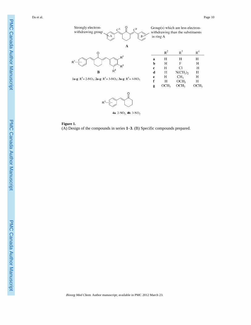
Figure 1.(A) Design of the compounds in series 1–3. (B) Specific compounds prepared.
Da et al. Page 10
Bioorg Med Chem. Author manuscript; available in PMC 2012 March 23.
PMC
Canada Author M
anuscriptPM
C C
anada Author Manuscript
PMC
Canada Author M
anuscript
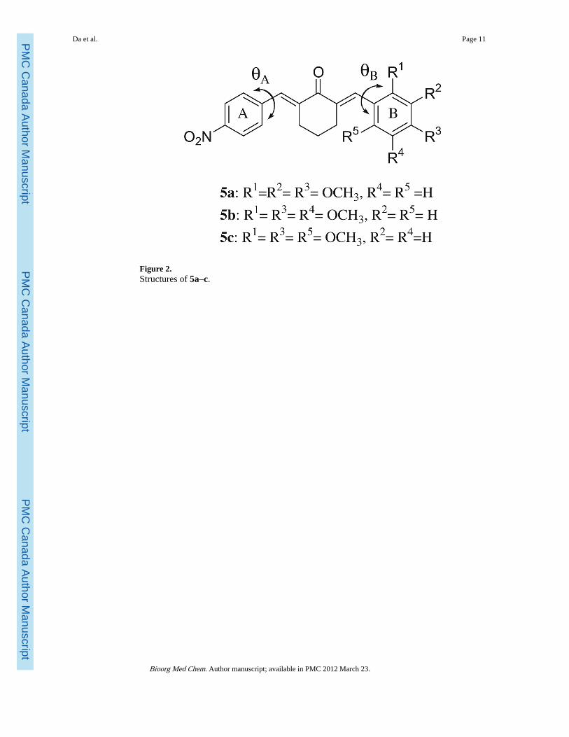
Figure 2.Structures of 5a–c.
Da et al. Page 11
Bioorg Med Chem. Author manuscript; available in PMC 2012 March 23.
PMC
Canada Author M
anuscriptPM
C C
anada Author Manuscript
PMC
Canada Author M
anuscript
Figure 3.Evaluation of 1c on the induction of internucleosomal DNA fragmentation in HSC-2 andHL-60 cells after 6 h of incubation. M is the molecular weight marker of DNA.
Da et al. Page 12
Bioorg Med Chem. Author manuscript; available in PMC 2012 March 23.
PMC
Canada Author M
anuscriptPM
C C
anada Author Manuscript
PMC
Canada Author M
anuscript
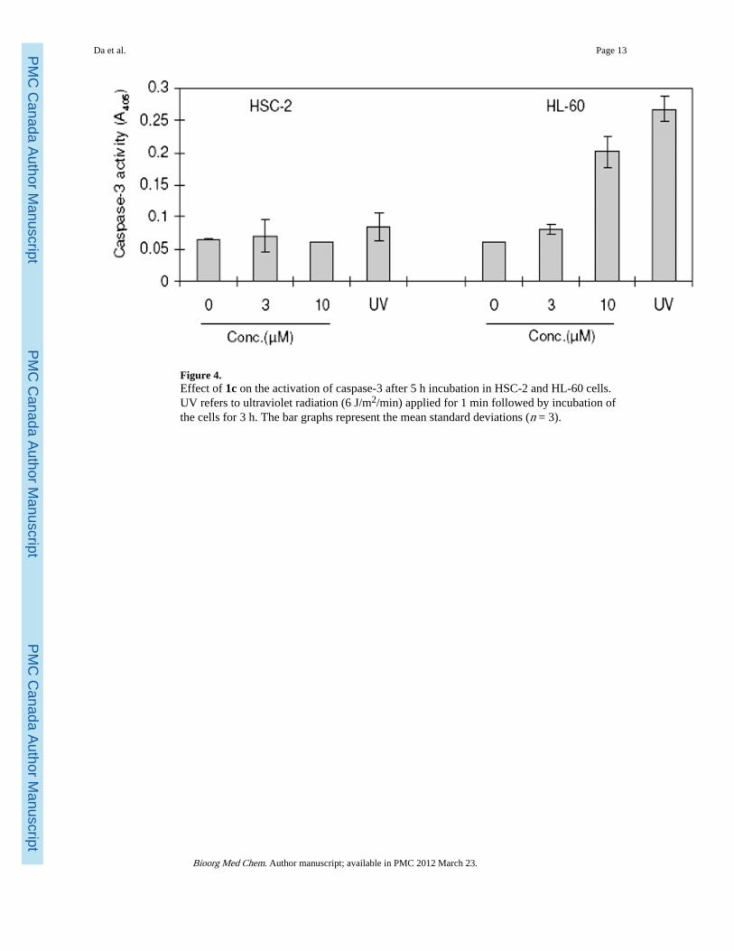
Figure 4.Effect of 1c on the activation of caspase-3 after 5 h incubation in HSC-2 and HL-60 cells.UV refers to ultraviolet radiation (6 J/m2/min) applied for 1 min followed by incubation ofthe cells for 3 h. The bar graphs represent the mean standard deviations (n = 3).
Da et al. Page 13
Bioorg Med Chem. Author manuscript; available in PMC 2012 March 23.
PMC
Canada Author M
anuscriptPM
C C
anada Author Manuscript
PMC
Canada Author M
anuscript
PMC
Canada Author M
anuscriptPM
C C
anada Author Manuscript
PMC
Canada Author
Manuscript
Da et al. Page 14
Tabl
e 1
Eva
luat
ion
of 1
a–g,
2a–
g, 3
a–g,
4a,
b, 5
a–c
and
mel
phal
an a
gain
st a
pan
el o
f no
rmal
and
mal
igna
nt c
ell l
ines
Com
poun
dH
uman
nor
mal
cel
lsH
uman
mal
igna
nt c
ells
Ave
SI
log
P
CC
50a
(μM
)H
SC-2
HSC
-4H
L-6
0
HG
FH
PC
HP
LF
Ave
CC
50(μ
M)a
SIb
CC
50(μ
M)a
SIb
CC
50(μ
M)a
SIb
1a93
1626
453.
812
3.2
143.
712
134.
70
1b82
1167
532.
720
2.2
243.
018
214.
86
1c>
400
400
>40
0>
400
3.7
>10
86.
1>
667.
6>
53>
765.
38
1d>
400
>40
0>
400
>40
0>
400
~1.0
>40
0~1
.0>
400
~1.0
~1.0
4.80
1e>
400
>40
0>
400
>40
011
>36
40>
1011
>36
>27
5.15
1f>
400
>40
038
6>
395
11>
3624
>17
26>
15>
234.
76
1g44
3579
535.
99.
06.
58.
23.
018
124.
33
2a25
1418
193.
75.
12.
47.
93.
06.
36.
44.
90
2b42
3836
392.
218
2.5
161.
039
245.
07
2c39
1838
322.
215
2.3
143.
210
135.
58
2d>4
00>
400
>40
0>
400
32>
1371
>5.
630
>13
>11
5.00
2e>4
0019
2>
400
>33
117
>20
45>
7.4
38>
8.7
>12
5.35
2f>4
0033
354
>26
24.
8>
5511
>24
7.2
>36
>38
4.96
2g33
6>
400
>40
0>
379
73>
5.2
92>
4.1
59>
6.4
>5.
24.
53
3a37
112
328
225
94.
953
9.2
2820
1331
4.93
3b32
6>
400
>40
0>
375
19>
2015
>25
32>
12>
195.
09
3cc
191
345
332
289
1.5
193
2.7
107
4.1
7112
45.
60
3d>4
00>
400
>40
0>
400
11>
36>
400
~1.0
>40
0~1
.0~1
35.
03
3e>4
0092
348
>28
06.
6>
4213
>22
27>
10>
255.
38
3fc
>40
0>
400
>40
0>
400
>40
0~1
.0>
400
~1.0
>40
0~1
.0~1
.04.
98
3gc
336
347
378
354
0.76
466
1.5
236
1.1
322
341
4.56
4a21
811
710
314
628
5.2
295.
027
5.4
5.2
—
4b74
4956
606.
69.
118
3.3
5.8
107.
5—
5a>
400
>40
0>
400
>40
0>
400
~1.0
>40
0~1
.0>
400
~1.0
~1.0
—
5b>
400
>40
0>
400
>40
067
>6.
025
0>
1.6
400
~1.0
~2.9
—
Bioorg Med Chem. Author manuscript; available in PMC 2012 March 23.
PMC
Canada Author M
anuscriptPM
C C
anada Author Manuscript
PMC
Canada Author
Manuscript
Da et al. Page 15
Com
poun
dH
uman
nor
mal
cel
lsH
uman
mal
igna
nt c
ells
Ave
SI
log
P
CC
50a
(μM
)H
SC-2
HSC
-4H
L-6
0
HG
FH
PC
HP
LF
Ave
CC
50(μ
M)a
SIb
CC
50(μ
M)a
SIb
CC
50(μ
M)a
SIb
5c>
400
>40
0>
400
>40
0>
400
~1.0
>40
0~1
.011
>36
~13
—
Mel
phal
and
>20
0>
200
>20
0>
200
35>
5.7
81>
2.5
6.0
>33
>14
—
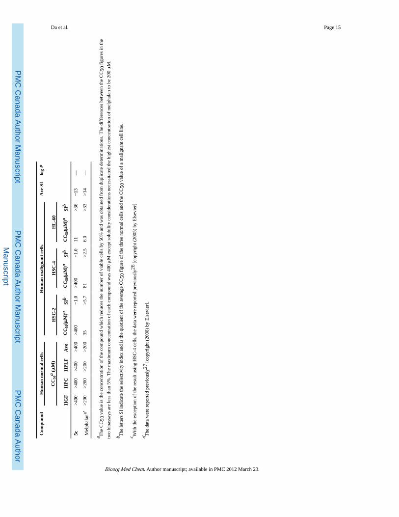
a The
CC
50 v
alue
is th
e co
ncen
trat
ion
of th
e co
mpo
und
whi
ch r
educ
es th
e nu
mbe
r of
via
ble
cells
by
50%
and
was
obt
aine
d fr
om d
uplic
ate
dete
rmin
atio
ns. T
he d
iffe
renc
es b
etw
een
the
CC
50 f
igur
es in
the
two
bioa
ssay
s ar
e le
ss th
an 5
%. T
he m
axim
um c
once
ntra
tion
of e
ach
com
poun
d w
as 4
00 μ
M e
xcep
t sol
ubili
ty c
onsi
dera
tions
nec
essi
tate
d th
e hi
ghes
t con
cent
ratio
n of
mel
phal
an to
be
200 μ
M.
b The
lette
rs S
I in
dica
te th
e se
lect
ivity
inde
x an
d is
the
quot
ient
of
the
aver
age
CC
50 f
igur
e of
the
thre
e no
rmal
cel
ls a
nd th
e C
C50
val
ue o
f a
mal
igna
nt c
ell l
ine.
c With
the
exce
ptio
n of
the
resu
lt us
ing
HSC
-4 c
ells
, the
dat
a w
ere
repo
rted
pre
viou
sly2
6 [c
opyr
ight
(20
05)
by E
lsev
ier]
.
d The
dat
a w
ere
repo
rted
pre
viou
sly2
7 [c
opyr
ight
(20
08)
by E
lsev
ier]
.
Bioorg Med Chem. Author manuscript; available in PMC 2012 March 23.


![The cytotoxic properties and preferential toxicity to tumour cells displayed by some 2,4- bis(benzylidene)-8-methyl-8-azabicyclo[3.2.1] octan-3-ones and 3,5- bis(benzylidene)-1-methyl-4-piperidones](https://static.fdokumen.com/doc/165x107/63160f7c511772fe4510a640/the-cytotoxic-properties-and-preferential-toxicity-to-tumour-cells-displayed-by.jpg)


![Accuracy of distinguishing between dysembryoplastic neuroepithelial tumors and other epileptogenic brain neoplasms with [11C]methionine PET](https://static.fdokumen.com/doc/165x107/63360da5cd4bf2402c0b568c/accuracy-of-distinguishing-between-dysembryoplastic-neuroepithelial-tumors-and-other.jpg)

