outcome analysis for closed intraarticular displaced calcaneal ...
111
OUTCOME ANALYSIS FOR CLOSED INTRAARTICULAR DISPLACED CALCANEAL FRACTURES WITH LOCKING PLATE Dissertation submitted to THE TAMILNADU DR.M.G.R MEDICAL UNIVERSITY In partial fulfilment of the regulations for The award of the degree of ORTHOPAEDICS M.S. BRANCH - II THANJAVUR MEDICAL COLLEGE, THANJAVUR - 613 004. THE TAMILNADU DR.MGR MEDICAL UNIVERSITY CHENNAI - 600 032. APRIL -2016
-
Upload
khangminh22 -
Category
Documents
-
view
2 -
download
0
Transcript of outcome analysis for closed intraarticular displaced calcaneal ...
OUTCOME ANALYSIS FOR CLOSED INTRAARTICULAR
DISPLACED CALCANEAL FRACTURES WITH LOCKING PLATE
Dissertation submitted to
THE TAMILNADU DR.M.G.R MEDICAL UNIVERSITY
In partial fulfilment of the regulations for
The award of the degree of
ORTHOPAEDICS
M.S. BRANCH - II
THANJAVUR MEDICAL COLLEGE,
THANJAVUR - 613 004.
THE TAMILNADU DR.MGR MEDICAL UNIVERSITY
CHENNAI - 600 032.
APRIL -2016
CERTIFICATE BY THE GUIDE
This is to certify that this dissertation titled “OUTCOME ANALYSIS FOR
CLOSED INTRAARTICULAR DISPLACED CALCANEAL FRACTURES
WITH LOCKING PLATE” is the bonafide original work of Dr.E.SIVA, in the
partial fulfillment of the requirements for M.S Orthopaedics (Branch II)
Examination of The Tamil Nadu Dr.M.G.R Medical University to be held in
APRIL 2016. The period of study is from 2013 -2015.
Prof.Dr.S.Kumaravel,M.S.Ortho.,D.Ortho.,Ph.D.,
Guide,
Professor of Orthopaedics and Traumatology,
Department of Orthopaedics and Traumatology,
Thanjavur Medical College, Thanjavur
CERTIFICATE
This is to certify that this dissertation titled “OUTCOME ANALYSIS FOR
CLOSED INTRAARTICULAR DISPLACED CALCANEAL FRACTURES
WITH LOCKING PLATE” is the bonafide original work of Dr.E.SIVA, under
the guidance and supervision of Prof.Dr.S.Kumaravel, M.S.Ortho., D.Ortho.,Ph.D.,
(Professor, Department of Orthopaedic Surgery) Thanjavur Government Medical
college Hospital, Thanjavur .
Prof.Dr.M.GulamMohideen,M.S.Ortho.,D.Ortho.,
Professor and Head,
Department of Orthopaedics,
Thanjavur Medical College,
Thanjavur.
Prof. Dr.Singaravelu, M.D.,D.C.H., Dean,
Thanjavur Medical College,
Thanjavur.
DECLARATION
I, Dr.E.SIVA solemnly declare that this dissertation “OUTCOME ANALYSIS
FOR CLOSED INTRAARTICULAR DISPLACED CALCANEAL FRACTURES
WITH LOCKING PLATE” is a bonafide work done by me at Government
Thanjavur Medical College And Hospital between 2013–2016,under the guidance
and supervision of Prof.Dr.S.Kumaravel, M.S.Ortho., D.Ortho.,Ph.D., Department of
Orthopaedic Surgery.
This dissertation is submitted to The Tamil Nadu Dr.M.G.R Medical
University towards partial fulfilment of regulation for the award of M.S Degree
(Branch II) on Orthopaedic Surgery.
Place: Thanjavur Dr.E.SIVA
Date: Post Graduate
MS – Orthopaedics
Thanjavur Medical College
Thanjavur

ACKNOWLEDGEMENT
At the outset, I would like to thank Prof. Singaravelu,M.D.,DCH.,Dean,
Thanjavur Medical College, for having permitted me to conduct the study and use the hospital
resources in the study.
I express my gratitude to Prof. M.Gulam Mohideen, M.S.Ortho.,D.Ortho.,
Professor and Head, Department of Orthopaedics And Traumatology, Thanjavur Medical
College and Hospital, for his inspiration, advice and guidance in making this work complete.
I am indebted to my chief Prof.Dr.A.Bharathy, M.S.Ortho.,D.Ortho., for his
untiring help and guidance during the study.
I am extremely thankful to my Prof.Dr.S.Kumaravel, M.S.Ortho.,D.Ortho.,Ph.D.,
for guiding me and and for the prompt help rendered whenever approached.
I sincerely acknowledge my beloved Assistant Professors Dr.D.Thirumalaipandiyan,
Dr.G.A.Rajmohan,Dr.A.Sivasenthil,Dr.M.C.Chinnadurai, Dr.Senthilkumar.K, Dr.C.Balaji for
their constant help, advice and guidance rendered to me in preparing this dissertation.
I am grateful to my fellow post graduates, juniors and interns who helped me in all possible
ways in this study.
My sincere thanks to our operative room personnel and staff members of Department of
Anaesthesiology and Radiology for their help in the study. My sincere thanks to all my patients
who co-operated with me for this study.
I wish to thank God Almighty for giving me the health and strength to complete this
study. Last but not the least I would thank my family and my relatives for making me what I am
today.


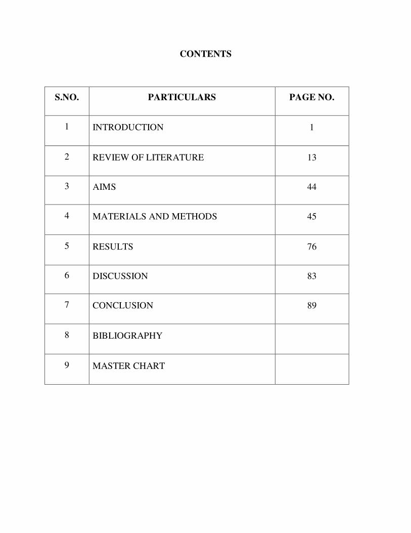
CONTENTS
S.NO. PARTICULARS PAGE NO.
1 INTRODUCTION 1
2 REVIEW OF LITERATURE 13
3 AIMS 44
4 MATERIALS AND METHODS 45
5 RESULTS 76
6 DISCUSSION 83
7 CONCLUSION 89
8 BIBLIOGRAPHY
9 MASTER CHART
CONTENTS
S.NO. PARTICULARS PAGE NO.
1 INTRODUCTION 1
2 REVIEW OF LITERATURE 13
3 AIMS 44
4 MATERIALS AND METHODS 45
5 RESULTS 76
6 DISCUSSION 83
7 CONCLUSION 89
8 BIBLIOGRAPHY
9 MASTER CHART
1
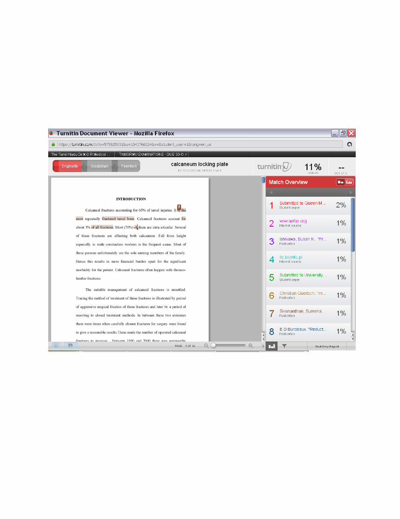
INTRODUCTION
Calcaneal fractures accounting for 65% of tarsal injuries. It is the
most repeatedly fractured tarsal bone. Calcaneal fractures account for
about 3% of all fractures. Most (70%) of them are intra articular. Several
of these fractures are affecting both calcaneum .Fall from height
especially in male constuction workers is the frequent cause. Most of
these persons unfortunately are the sole earning members of the family.
Hence this results in more financial burden apart for the significant
morbidity for the patient. Calcaneal fractures often happen with thoraco-
lumbar fractures.
The suitable management of calcaneal fractures is unsettled.
Tracing the method of treatment of these fractures is illustrated by period
of aggressive surgical fixation of these fractures and later by a period of
resorting to closed treatment methods. In between these two extremes
there were times when carefully chosen fractures for surgery were found
to give a reasonable results.These made the number of operated calcaneal
fractures to increase. Between 1990 and 2000 there was noteworthy
development in the management of calcaneal fractures an exemplified by
the gross decrease in complication rates connected with the existing
intervention of these potentially disturbing injuries.
2
SURGICAL ANATOMY
Calcaneum as a bone forms a base or vertical support for body
weight. It is the biggest of all tarsal bones with many articulations. It also
has many ligament and tendon attachments. It also functions as a lever
arm powered by gastro-soleus. It also supports and conserves the length
of lateral column of the foot.
Calcaneum has a thin cortical shell which encloses a mass of
cancellous bone that remodels with various stresses applied to it. So it has
been described being ‘Egg like’ i.e. hard on the outside and soft on the
inside.
The anatomy of the calcaneum on its lateral aspect is particularly
vital as mainly this lateral surface area is exposed during the most
common surgical approach used for fracture fixation. Tuberosity is the
most posterior aspect of the calcaneum; distal to the tuberosity, is the
body of calcaneum.
In the plantar aspect of calcaneum, a small process in the slightly
lateral portion is called the lateral process of tuberosity gives origin to
muscles and attachment to plantar fascia.
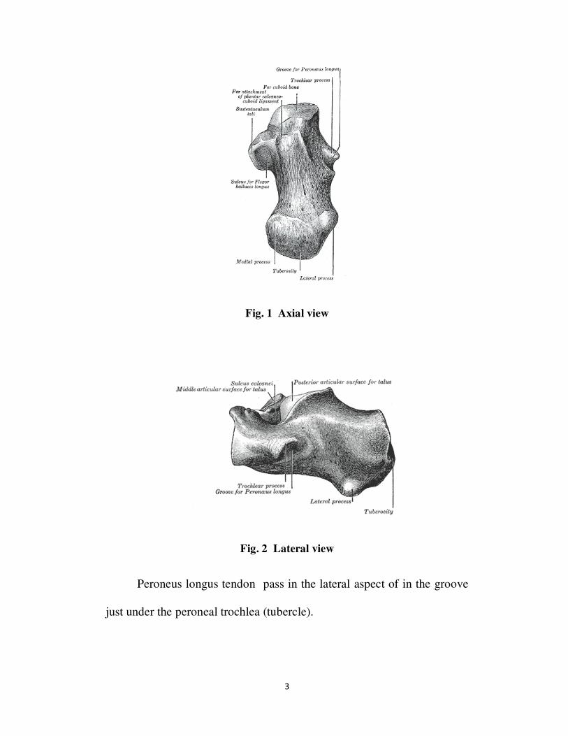
3
Fig. 1 Axial view
Fig. 2 Lateral view
Peroneus longus tendon pass in the lateral aspect of in the groove
just under the peroneal trochlea (tubercle).
4
The lateral margin of posterior facet is observable in the middle
part of the calcaneum on the lateral side. This is important in open
reduction and internal fixation of intra articular fractures of calcaneum as
the more lateral portion of the posterior facet usually has to be
reconstructed and fixed with screws.
Distally on the lateral side, the articular surface of the
calcaneocuboid joint is found.
The superior surface of the calcaneum has the three articular facets
in the anterior half.
The largest facet is the posterior facet and is convex.
The middle facet which is slightly concave is situated on the
sustentaculum tali. This facet frequently continues anteriorly as the
anterior facet, also slightly concave.
The inter-osseous sulcus (calcaneal groove) lies between the
middle and posterior facets. It opens broadly laterally and forms with the
talar sulcus, the sinus tarsi.
These anterior middle and posterior calcaneal facets articulate with
anterior middle and posterior talar facets to form the complex subtalar
joint.
5
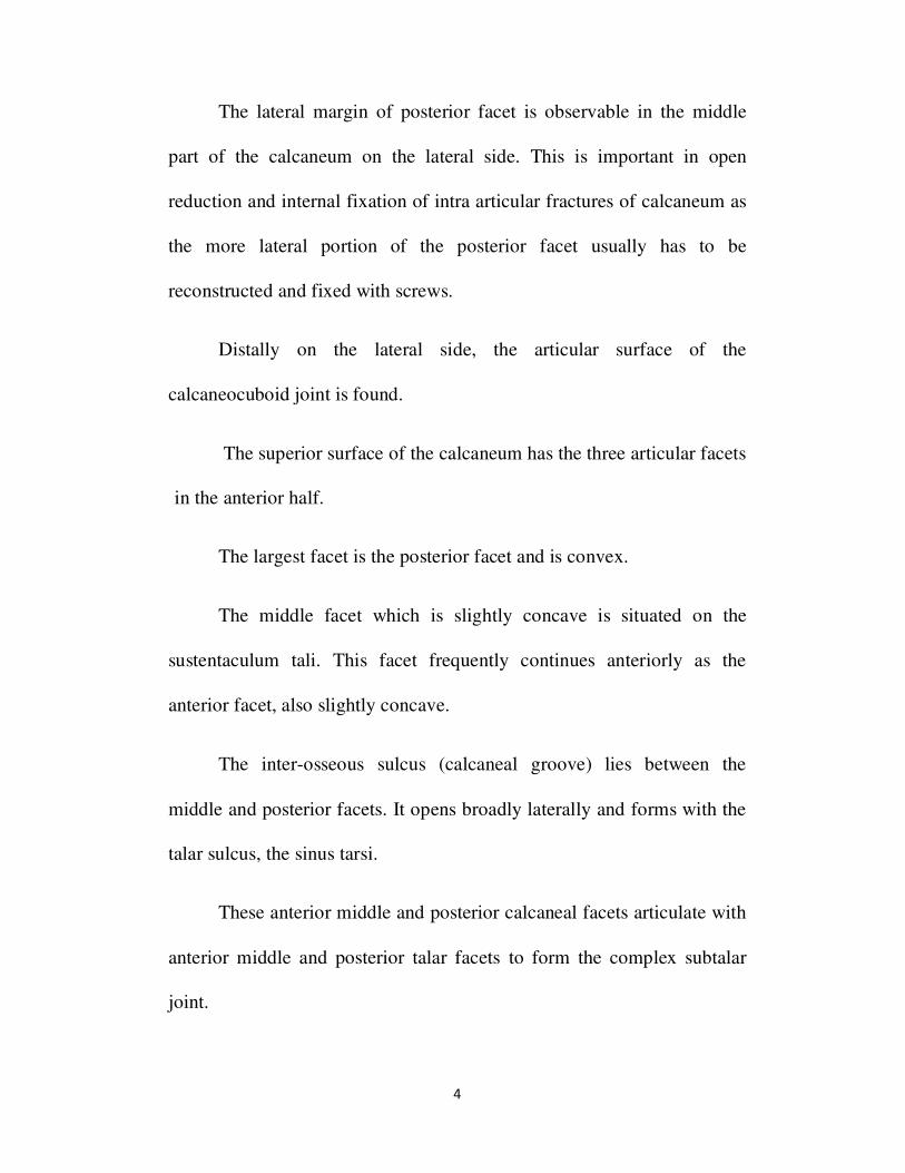
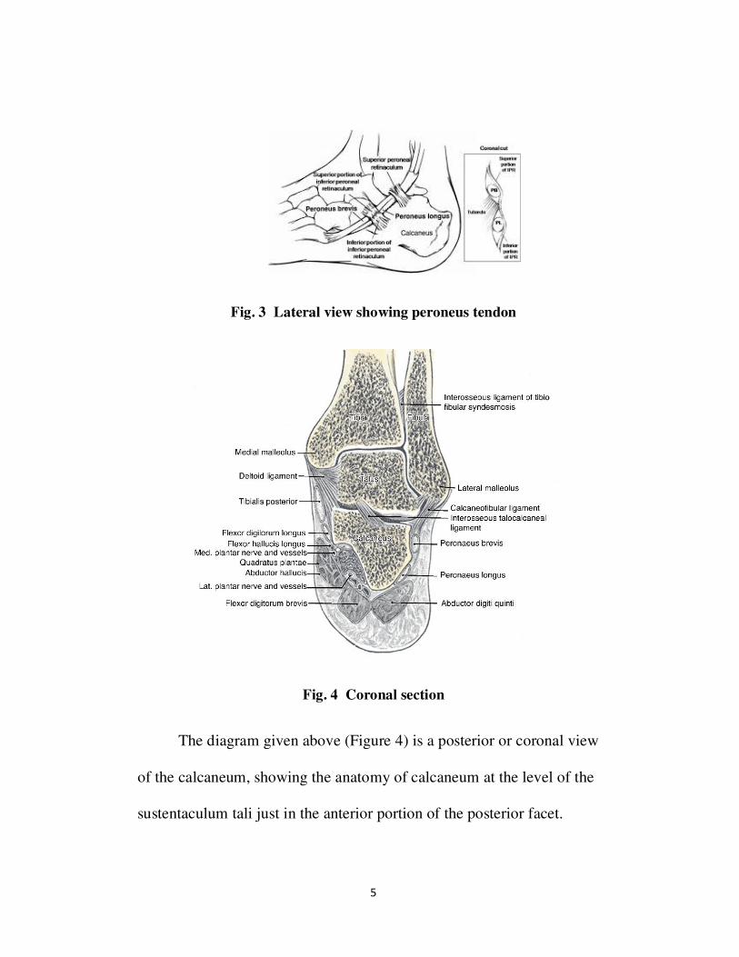
Fig. 3 Lateral view showing peroneus tendon
Fig. 4 Coronal section
The diagram given above (Figure 4) is a posterior or coronal view
of the calcaneum, showing the anatomy of calcaneum at the level of the
sustentaculum tali just in the anterior portion of the posterior facet.
6
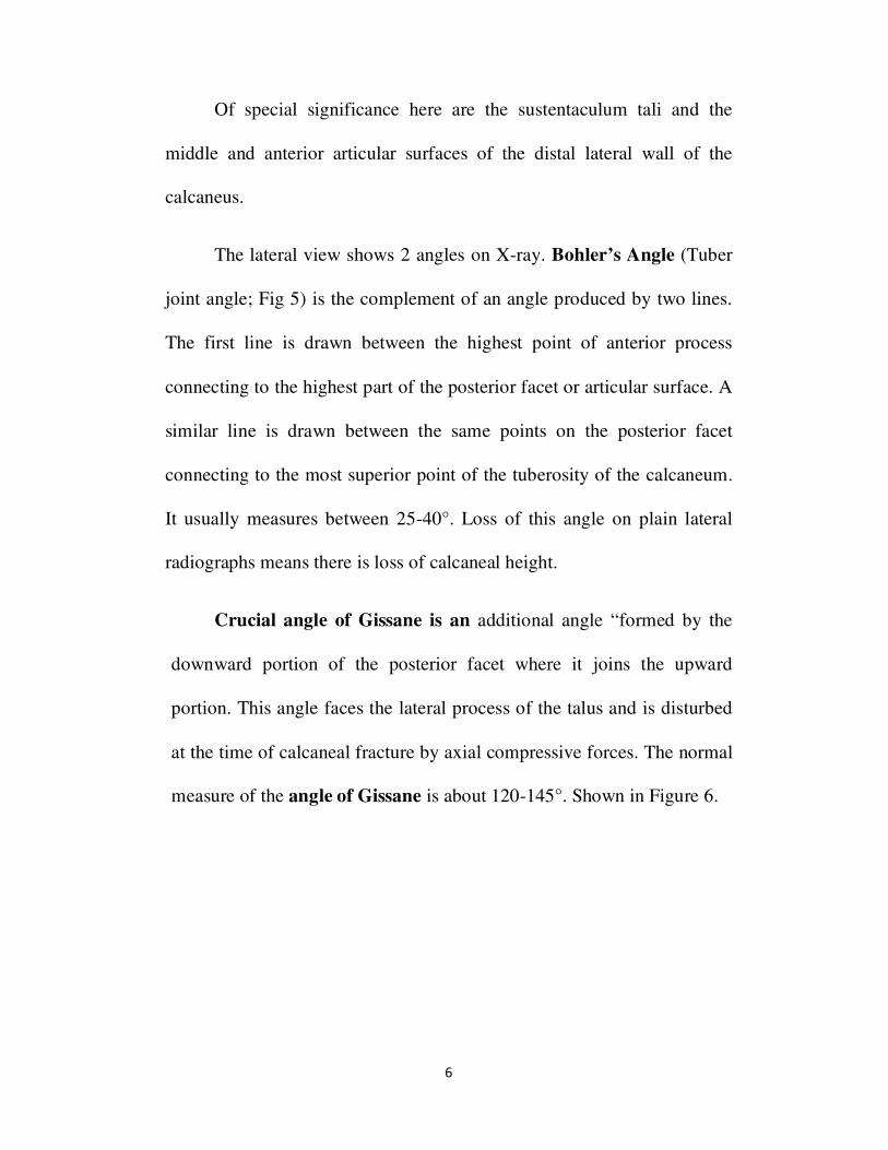
Of special significance here are the sustentaculum tali and the
middle and anterior articular surfaces of the distal lateral wall of the
calcaneus.
The lateral view shows 2 angles on X-ray. Bohler’s Angle (Tuber
joint angle; Fig 5) is the complement of an angle produced by two lines.
The first line is drawn between the highest point of anterior process
connecting to the highest part of the posterior facet or articular surface. A
similar line is drawn between the same points on the posterior facet
connecting to the most superior point of the tuberosity of the calcaneum.
It usually measures between 25-40°. Loss of this angle on plain lateral
radiographs means there is loss of calcaneal height.
Crucial angle of Gissane is an additional angle “formed by the
downward portion of the posterior facet where it joins the upward
portion. This angle faces the lateral process of the talus and is disturbed
at the time of calcaneal fracture by axial compressive forces. The normal
measure of the angle of Gissane is about 120-145°. Shown in Figure 6.

7
Fig. 5 Radiological Lateral view showing Bohler’s Angle
Fig. 6 Radiological Lateral view showing Gissane’s Angle
An extra proximal coronal view of the calcaneus showing the
posterior facet is an important view (CT cut). This demonstrate the
quantity of comminution and displacement of the posterior facet with
intra articular fracture of calcaneum.

8
There are few unique anatomical aspects of calcaneum.
1. To permit passage of the tendons and neurovascular structures into the
foot, the calcaneum is concaved out on the medial side
2. Thus the centre of calcaneal tuberosity to be a little lateral to the center
of talus.
3. If a force is applied vertically to the talus, with the calcaneal tuberosity
fixed to the ground, then shear stress take place all the way through the
body of the calcaneum.
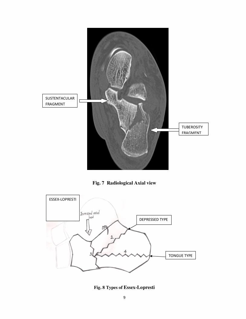
The calcaneum essentially breaks along this stress line, forming two
main fracture fragments,(Figure 7)
1. medially -the sustentacular fragment and
2. laterally- the tuberosity fragment
9
Fig. 7 Radiological Axial view
Fig. 8 Types of Essex-Lopresti
SUSTENTACULAR
FRAGMENT
TUBEROSITY
FRAGMENT
DEPRESSED TYPE
TONGUE TYPE
ESSEX-LOPRESTI

10
Transmission of this downward energy of the talus occurs, with the
calcaneal tuberosity fixed on the ground. Thus the talus and the
sustentacular fragment move inferomedially, and so calcaneal tuberosity
is becomes more lateral and elevated. The talus powers its way and
pushes the lateral part of the posterior facet into the cancellous bone of
the tuberosity fragment.
Viewed from above, the talus and sustentacular fragment shift
inferiorly and posteriorly. This leaves the tuberosity fragment in a lateral
and anterior position. The anterior aspect of the tuberosity fragment
rotates laterally, and the lateral tuberosity cortex is comminuted.
Based on the length of the supero-lateral fragments with a small
part of the articular surface of the posterior facet, makes the difference
between the types.
The two main regular types of calcaneal fractures are
(1) Joint depression type. Here the fragment is short; it extends only for a
short distance behind the posterior facet.
(2) Tongue type. Here the fragment is long, and it extends to the posterior
aspect of the calcaneal tuberosity.
11
These fracture lines are according to Essex-Lopresti. The fracture
lines cross the postero-lateral part of the posterior facet in each.
The main means of fracture reduction in medial incision is the
sustentacular spike. One must be aware of this. In this sustentacular
fragment, there is a spike which is a thin, sharp structure on the medial
side, consisting of mainly the medial cortex. This is very thin posteriorly
and distally but almost immediately becomes turns thicker in the upper
part.
This spike helps in identifying the sustentacular fragment. On table
only after identifying this fragment, the other fracture fragments can be
matched and aligned with it. This will restore the near original structure
of the calcaneus. The reason for the primary fracture line (Essex
Lopresetti) is driving down of the sharp taloid spur into the calcaneum
especially in the everted position of sub-talar joint. In the inverted
position of sub-talar joint, the tuberosity comes more directly below the
talus; this causes a reduction in the shear stress.
Anatomy of calcaneum the mechanism of fracture
Mechanism and cause of the injury
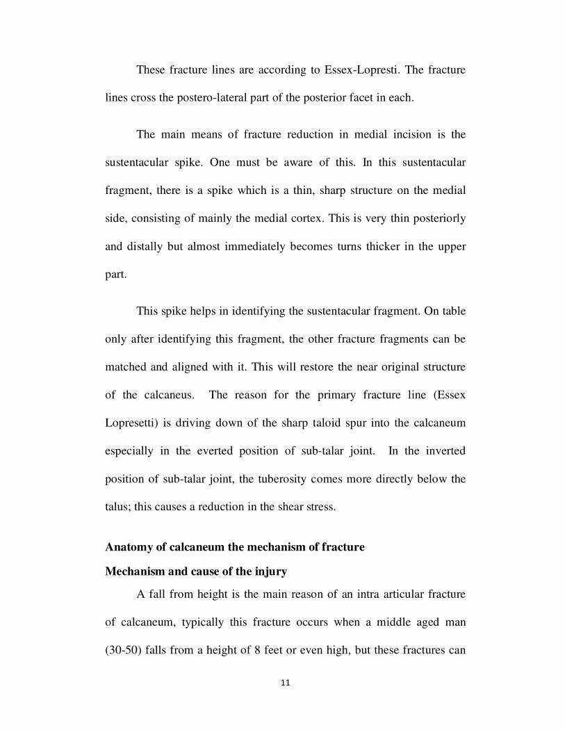
A fall from height is the main reason of an intra articular fracture
of calcaneum, typically this fracture occurs when a middle aged man
(30-50) falls from a height of 8 feet or even high, but these fractures can
12
happen with falls from lesser heights, like in a domestic setting like a fall
from a chair, particularly in the elderly citizen with osteoporotic bones.
Rarely landing on the heel in motor vehicle accidents, floor board or
brake pedal injuries also can cause intra articular calcaneal fractures.
Fig 9,10 Mechanism of Fracture

13
REVIEW OF LITERATURE
Obviously originally in of calcaneal fractures, the treatment
depended on the diagnostic modalities that were available at that period
of time. Because of total absence of diagnostic tools from time immortal
to the nineteenth century, naturally the only treatment approach is only
careful neglect (ie conservative management) . This is because the special
views and advanced CT scan of the calcaneum are needed to make the
decision regarding reduction of intraarticular aspect of the calcaneum.
Cotton and Wilson recommended (1908) that one should not do an
open reduction of a calcaneal fracture at all. The famous quote by
McLaughlin compared open reduction and fixation of a calcaneal fracture
to “nailing of a custard pie to the wall”.
Later in 1931 Bohler supported open reduction but still the main
reasons for the dominance of non operative treatment were due to the the
practical problems not only connected specifically with operative
treatment of calcaneum but also to other general problems like ineffective
anesthesia, deficiency of good quality intraoperative radiography like
fluoroscopy and the type of antibiotics at that period. Also the poor
understanding of the principles of internal fixation leads to more
complications.
14
Conn in 1935 found poor results with the standard treatment
methods, recommended primary triple arthrodesis, in a delayed manner.
In this method he achieved exceptional results.
Later in 1943 Gallie, supported sub-talar arthrodesis as ultimate
treatment but only after union of the fracture.
Discontented with either conservative or late surgical management
of the calcaneal fractures, Palmer tried operative treatment of acute
displaced intra articular calcaneal fractures using a standard lateral
Kocher’s approach. He reconstructed the joint by elevating the fracture
fragment with bone graft and published his work in 1948. Later 1952,
Essex Lopresti reported similar findings.
Not all surgeons were contented with the results of open reduction
and fixation, Dick and Harris began started using Gallie’s technique of
subtalar arthrodesis for malunited fractures of calcaneum as the treatment
of choice, even for acute calcaneal fractures. They showed excellent
results with patients returning to work early. Following this many
surgeons performed sub-talar arthrodesis for acute calcaneal fracture.
Even after all these in a long term follow up Lindsay and Daver,
concluded that sub-talar arthrodesis was not only unnecessarly but also
resulted in problems. They concluded that best results were got only with
15
conservative treatment of patients. As a result the operative treatment of
acute calcaneal fractures once again went into disrepute. So only later,
between 1960s and70s most workers advocacted of conservative
management.
In the last 20 years, because of improved anesthesia, introduction
of antibiotics principles of internal fixation and preoperative imaging CT
and intra operative imaging intensifier have permitted surgeons to employ
operative fixation for many intraarticular fracture, obtaining good results.
If the personality of fracture is not carefully studied and mode of
fixation are not carefully selected and the basic principles of open
reduction and internal fixation adhered to by the surgeon then results
cannot be expected. Even if these newer techniques promise good results
in displaced intra articular calcaneal fractures, experience with these for
most intra articular fractures that treatment remains challenging.
Maintenance of Bohler's angle is necessary for satisfactory results
along with maintenance of articular congruence of posterior facet of
calcaneum and crucial angle of Gissane. Open reduction and internal
fixation with locking compressive plate in displaced intraarticular fracture
calcaneum shows good outcome. Results are more favourable in less
comminuted as compared to more comminuted.(23)
16
Controversies also exit with regards to primary bone grafting to
prevent collapse. A series of cases have been reported with no significant
collapse, even without using bone graft for calcaneal fixation, showing no
specific benefit with use of the bone graft to prevent collapse(20-22)
However, Leung et al(21)
, Thordarson et al(22)
, and Schildhauer et
al(23)
recommended use of bone graft or cement to increase stability and
compressive strength of fixation and rapid rehabilitation. Zhongguo et
all(22)
study reports bone graft in the surgical treatment of calcaneal
fractures carries no advantage.
17
EVOLUTION OF IMPLANT USE AND RATIONALE FOR LCP
The management of fractures of calcaneus has undergone a sea of
change from the initial use of supervised neglect to the use of
complicated closed reduction devices .Today there are complex implants ,
used for internal fixation needs better understanding of complex fracture
anatomy and the maneuvers required to reduce the various fracture
fragments. They also needs good knowledge about trauma mechanisms,
better delineation of vascular zones and their relationship to skin incision,
better stability with newer implants and bolder surgical interventions,
whether by open or percutaneous means.
The consensus of the issue of the best implant for a particular type
of calcaneal fracture is not arrived. Also there is no definite proof about
the ideal modality of fixation. This is again due to availability of
numerous implants, different methods of reduction, and many surgical
approaches to the fractured calcaneum.
A clear and targeted understanding of the goals aimed at after
calcaneal fracture fixation is vital and a prerequisite for implant use. The
ideal achievement in any intraaricular fracture are better anatomical
articular reconstruction, maintenance of the articular surface,

18
reconstitution of Bohler’s and Gissane angle, less invasived faster surgery
, minimal wound complications and early mobilization of the ankle.
Modified surgical techniques (appropriate use of the extensile
lateral approach) and minimally invasive techniques when indicated, and
better fixation devices for stability.
A clear and better understanding of the hindfoot biomechanics has
also helps to fix the extra articular fractures and the tongue type of
fractures with modifications of the technique described by Essex
loperesti, Gissane, and the other authors.
Despite the contrary evidence favouring both modalities, today
there is a gradual shift in the management of calcaneal fractures from
conservative to surgical treatment. This is due to better understanding and
clarity of patterns of fractures by radiological techniques like
Computerised tomography .There is still confusion in the aspects of open
reduction an internal fixation, and many difficulties persists.
One main reason for failure of surgical intervention is
inappropriate implant or incision choice. Complications manifest in the
form of inadequate fixation and inadequate 3 dimensionl reconstruction
and wound related complications.

19
Implant decision is a complex issue .This is because the complex
anatomy of the calcaneum, an odd shape of the bone which breaks like an
egg and it is difficult to maintain various articular surfaces and
tuberosities in position till healing process is completed. Its odd shape is
the reason behind difficulty in application of implants. Regaining the
Bohler’s and Gissane angle and reconstruction of posterior facet are all
the complex task. It is always accompanied with some soft tissue
compromise
Last three decades, new calcaneal implants have evolved due to
better knowledge on biomechanics, metallurgy and implant designs.
Newer implants allow early mobilization. Use of these implants depends
on surgeons expertise, patient expectations and cost issues, also there
should be consideration to the local factors like skin conditions or
wounds, swelling, blister etc
20
IMPLANTS
SCREW FIXATION OF CALCANEAL FRACTURE
Avulsion Extaarticular fractures are comparatively easily stabilized
and fixed with 1 or 2 lag screws can be applied percutaneosly with
minimal surgical trauma.
WIRE FIXATION OF CALCANEAL FRACTURE
For complex calcaneal fractures K wires are inadequate. While in
undisplaced avulsion fractures or undisplaced body fractures, if carefully
applied Kwires give percutaneous stabilization, though early mobilisation
cannot be achieved. K wires are ideal for temporory stabilisation. But
now these are supplemented with external fixation as a support to
maintain the fracture reduction of different fragments.
EXTERNAL FIXATORS OF CALCANEAL FRACTURE
These are handy in open fracture, in calcaneal fractures also , they
can be used as a temporary devices till the open wound heals or to
maintain the fracture geometry till the fracture healing, in addition to K
wires. In comminuted fractures which appear as a bag of bone the ideal
management is by image guided percutaneous multiple K wires fixation
and supported with these external fixators.
21
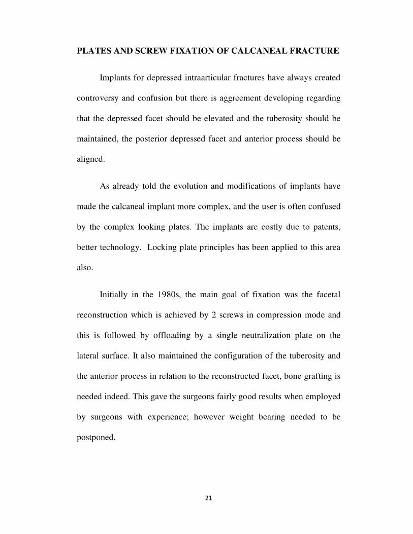
PLATES AND SCREW FIXATION OF CALCANEAL FRACTURE
Implants for depressed intraarticular fractures have always created
controversy and confusion but there is aggreement developing regarding
that the depressed facet should be elevated and the tuberosity should be
maintained, the posterior depressed facet and anterior process should be
aligned.
As already told the evolution and modifications of implants have
made the calcaneal implant more complex, and the user is often confused
by the complex looking plates. The implants are costly due to patents,
better technology. Locking plate principles has been applied to this area
also.
Initially in the 1980s, the main goal of fixation was the facetal
reconstruction which is achieved by 2 screws in compression mode and
this is followed by offloading by a single neutralization plate on the
lateral surface. It also maintained the configuration of the tuberosity and
the anterior process in relation to the reconstructed facet, bone grafting is
needed indeed. This gave the surgeons fairly good results when employed
by surgeons with experience; however weight bearing needed to be
postponed.
22
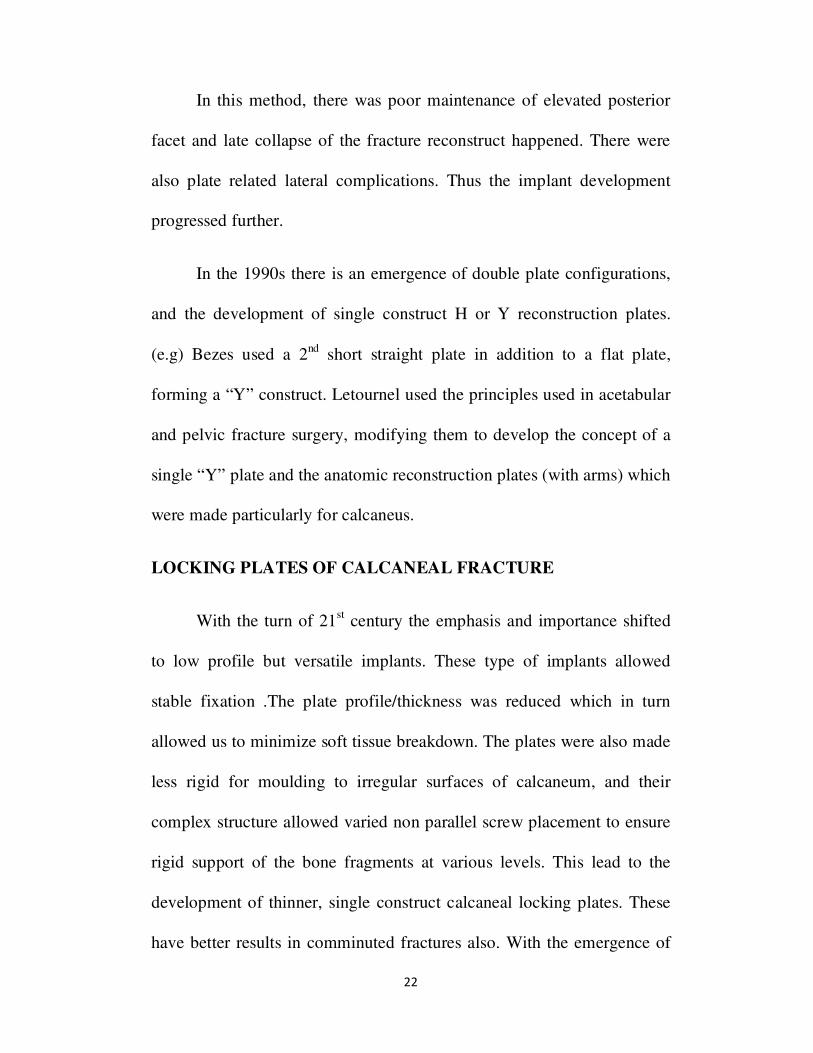
In this method, there was poor maintenance of elevated posterior
facet and late collapse of the fracture reconstruct happened. There were
also plate related lateral complications. Thus the implant development
progressed further.
In the 1990s there is an emergence of double plate configurations,
and the development of single construct H or Y reconstruction plates.
(e.g) Bezes used a 2nd
short straight plate in addition to a flat plate,
forming a “Y” construct. Letournel used the principles used in acetabular
and pelvic fracture surgery, modifying them to develop the concept of a
single “Y” plate and the anatomic reconstruction plates (with arms) which
were made particularly for calcaneus.
LOCKING PLATES OF CALCANEAL FRACTURE
With the turn of 21st century the emphasis and importance shifted
to low profile but versatile implants. These type of implants allowed
stable fixation .The plate profile/thickness was reduced which in turn
allowed us to minimize soft tissue breakdown. The plates were also made
less rigid for moulding to irregular surfaces of calcaneum, and their
complex structure allowed varied non parallel screw placement to ensure
rigid support of the bone fragments at various levels. This lead to the
development of thinner, single construct calcaneal locking plates. These
have better results in comminuted fractures also. With the emergence of
23
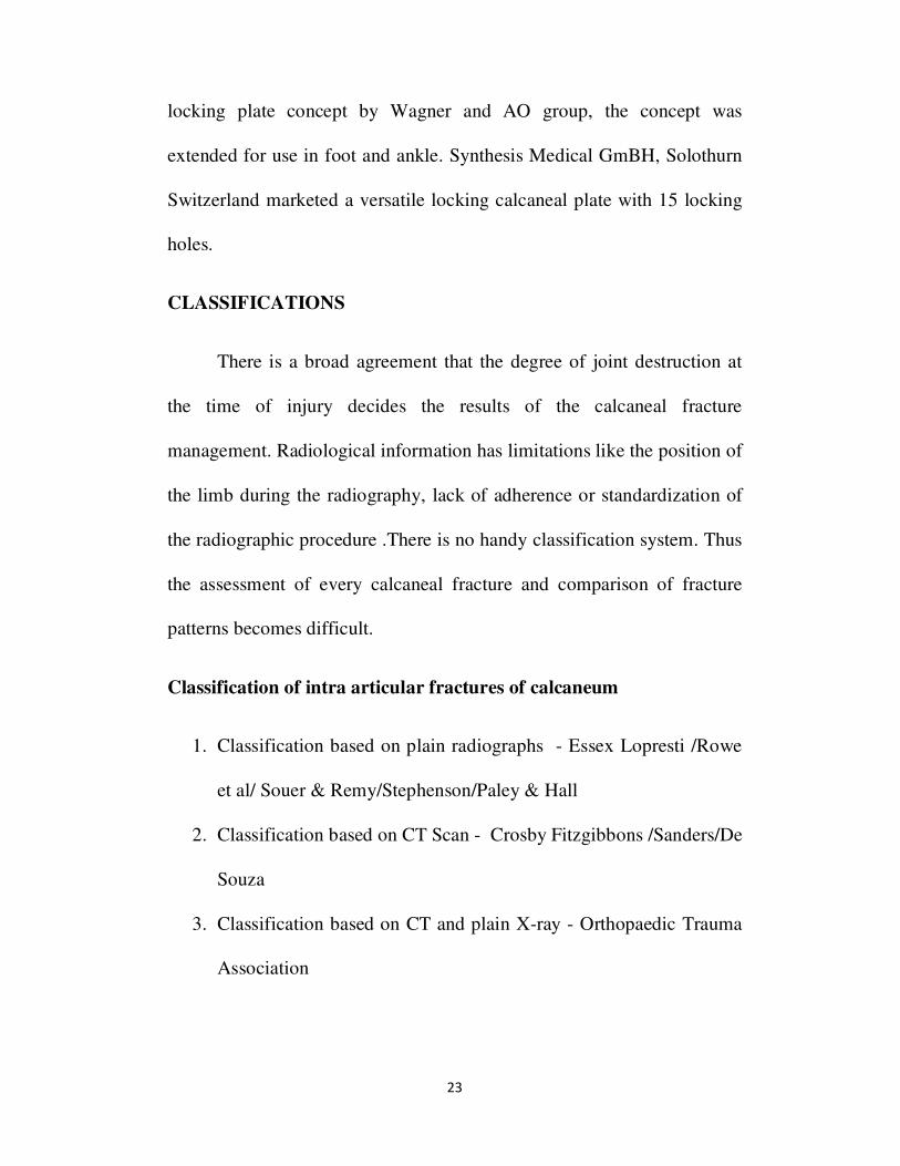
locking plate concept by Wagner and AO group, the concept was
extended for use in foot and ankle. Synthesis Medical GmBH, Solothurn
Switzerland marketed a versatile locking calcaneal plate with 15 locking
holes.
CLASSIFICATIONS
There is a broad agreement that the degree of joint destruction at
the time of injury decides the results of the calcaneal fracture
management. Radiological information has limitations like the position of
the limb during the radiography, lack of adherence or standardization of
the radiographic procedure .There is no handy classification system. Thus
the assessment of every calcaneal fracture and comparison of fracture
patterns becomes difficult.
Classification of intra articular fractures of calcaneum
1. Classification based on plain radiographs - Essex Lopresti /Rowe
et al/ Souer & Remy/Stephenson/Paley & Hall
2. Classification based on CT Scan - Crosby Fitzgibbons /Sanders/De
Souza
3. Classification based on CT and plain X-ray - Orthopaedic Trauma
Association
24
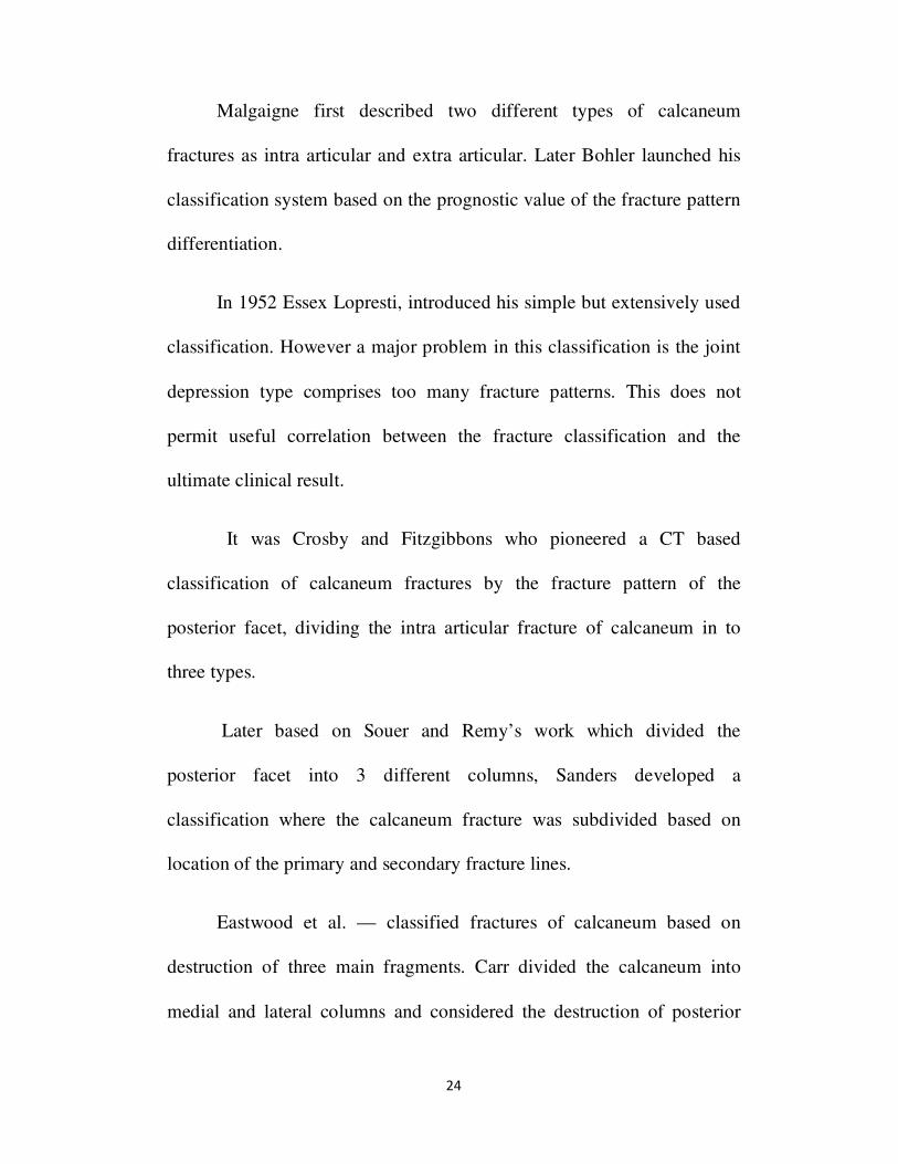
Malgaigne first described two different types of calcaneum
fractures as intra articular and extra articular. Later Bohler launched his
classification system based on the prognostic value of the fracture pattern
differentiation.
In 1952 Essex Lopresti, introduced his simple but extensively used
classification. However a major problem in this classification is the joint
depression type comprises too many fracture patterns. This does not
permit useful correlation between the fracture classification and the
ultimate clinical result.
It was Crosby and Fitzgibbons who pioneered a CT based
classification of calcaneum fractures by the fracture pattern of the
posterior facet, dividing the intra articular fracture of calcaneum in to
three types.
Later based on Souer and Remy’s work which divided the
posterior facet into 3 different columns, Sanders developed a
classification where the calcaneum fracture was subdivided based on
location of the primary and secondary fracture lines.
Eastwood et al. — classified fractures of calcaneum based on
destruction of three main fragments. Carr divided the calcaneum into
medial and lateral columns and considered the destruction of posterior
25
facet and calcaneo-cuboid joint. Levin and Nunley considered soft tissue
problems and found six groups.
Zwipp classified calcaneum into 5 main fragments and 3 joints.
This considered the number of destroyed fragment and joints and the
degree of soft tissue damage.
As already elaborated, for clinical use, Essex Lopresetti
classification appear simplest but it is inadequate and cannot offer a
outline for forming surgical strategies or for calculating the long term
result. Sanders’s classification is simple, comprehensive and has the
advantage of allowing prognostication of results for various fracture types
of calcaneum. Yet another classification called Zwipp classification,
describe the typically complex pattern of calcaneum fractures.
The common classification followed is Sanders, which is based on
the CT coronal image of the posterior facet which is more descriptive and
complex.
26
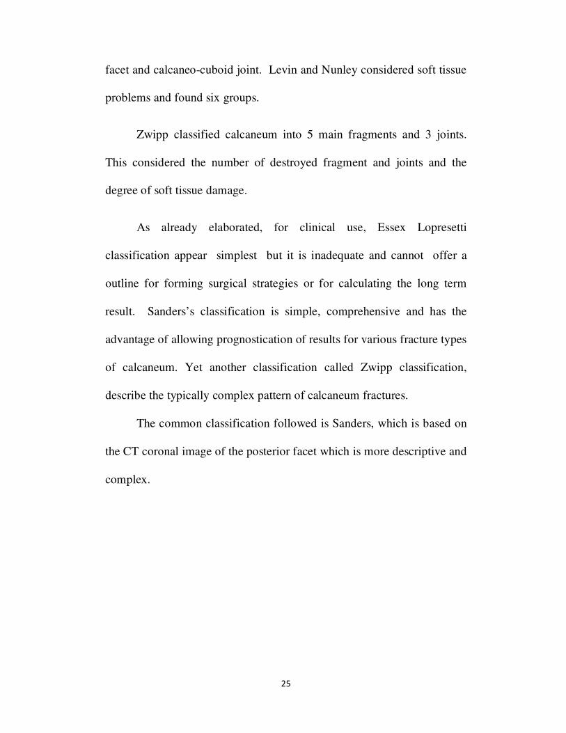
SANDER’S CLASSIFICATION
Type 1 All nondisplaced articular fractures (less than 2mm)
Type 11 Two-part fractures of the posterior facet
Type 11A,11B,11C Based on location of primary fracture line
Type 111
Three-part fractures usually featuring a centrally
depressed fragment
Types 111AB,111AC,111BC Based on location of primary fracture line
Type 1V Four part articular fracture
Fig.11 Coronal view
27
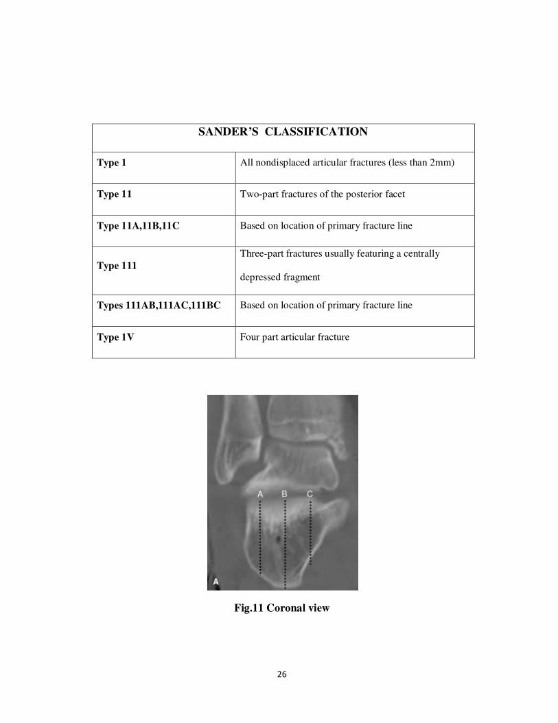
Type—1 Intra articular fracture which can have as many as three
fractures lines and four fragments but they are only minimally displaced
or undisplaced.
Fig.12 Coronal view – Type 1
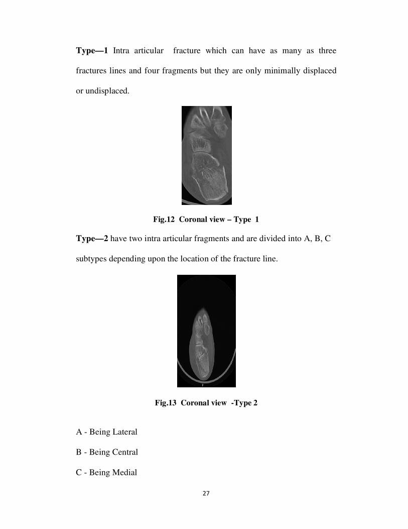
Type—2 have two intra articular fragments and are divided into A, B, C
subtypes depending upon the location of the fracture line.
Fig.13 Coronal view -Type 2
A - Being Lateral
B - Being Central
C - Being Medial
28
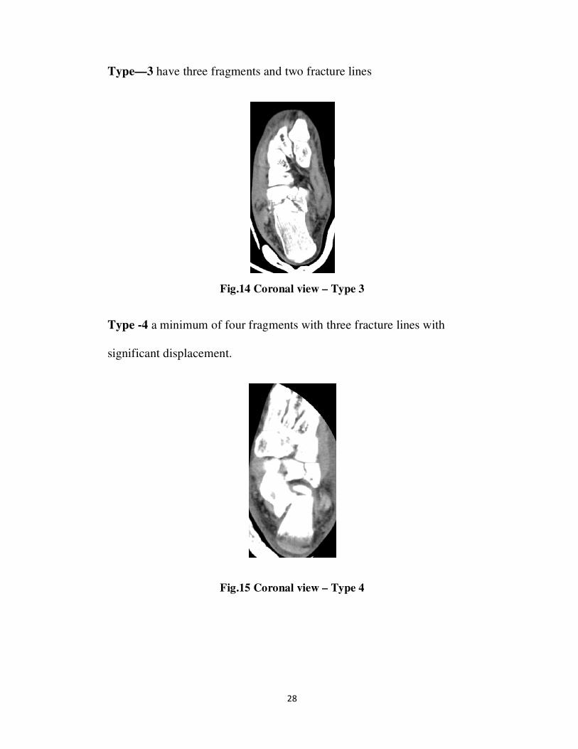
Type—3 have three fragments and two fracture lines
Fig.14 Coronal view – Type 3
Type -4 a minimum of four fragments with three fracture lines with
significant displacement.
Fig.15 Coronal view – Type 4
29
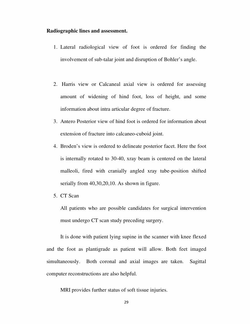
Radiographic lines and assessment.
1. Lateral radiological view of foot is ordered for finding the
involvement of sub-talar joint and disruption of Bohler’s angle.
2. Harris view or Calcaneal axial view is ordered for assessing
amount of widening of hind foot, loss of height, and some
information about intra articular degree of fracture.
3. Antero Posterior view of hind foot is ordered for information about
extension of fracture into calcaneo-cuboid joint.
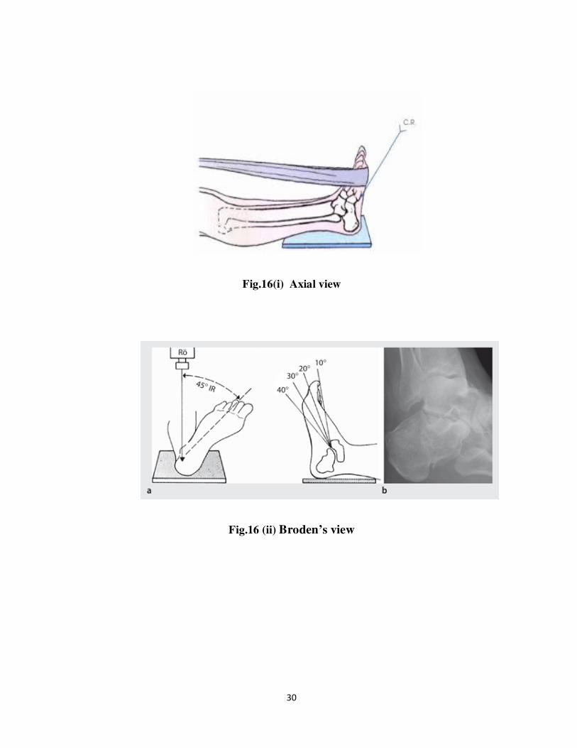
4. Broden’s view is ordered to delineate posterior facet. Here the foot
is internally rotated to 30-40, xray beam is centered on the lateral
malleoli, fired with cranially angled xray tube-position shifted
serially from 40,30,20,10. As shown in figure.
5. CT Scan
All patients who are possible candidates for surgical intervention
must undergo CT scan study preceding surgery.
It is done with patient lying supine in the scanner with knee flexed
and the foot as plantigrade as patient will allow. Both feet imaged
simultaneously. Both coronal and axial images are taken. Sagittal
computer reconstructions are also helpful.
MRI provides further status of soft tissue injuries.
30
Fig.16(i) Axial view
Fig.16 (ii) Broden’s view

31
TREATMENT
The choice of treatment is as follows
1. Closed treatment
a) Accept position, no reduction and early motion.
b) Closed reduction, short term immobilization, reasonably early motion
2. Semi Open technique
a) Essex Lopresti close reduction by manipulation of the fragment with
percutaneous pin and fixation.
Fig.17 Essex-Lopresti Reduction Technique
b) Percutaneous techniques which are recently popular.
c) Limited open reduction and external fixation technique.
ESSEX-LOPRESTI REDUCTION TECHNIQUE
32
3. Open Surgical Technique
a) Open reduction and internal fixation with a lateral extensile incision.
b) Open reduction and internal fixation with a medial approach.
c) Combined medial and lateral exposure using open reduction and
internal fixation.
d) Primary arthrodesis.
Closed treatment
It consists of “RICE” Rest, Ice application, Compression, Elevation of
limb and NSAIDS. It is accomplished in two ways.
a) One has to accept the fracture as it presented to the surgeon without
making an attempt to reduce, with short term immobilization, non
weight bearing for 6-8 weeks followed by gradual early motion.
b) By external pressure fracture is manipulated manually or with
tongs for reduction and immobilization done, later early
physiotherapy for range of motion exercises are advised. Weight
bearing is allowed after 8 weeks. Manipulating the fracture can be
done by Omoto technique.
33
Semi Open Techniques are easy for the surgeon with low surgical risk
to the patient than open techniques. But correct patient selection (i.e)
only tongue type fracture patterns and determination on anatomic
reduction of the joint surfaces can be expected to cause acceptable and
good results.
a) Essex Loprestti and King’s technique.
b) Surgical technique of Tornetta,
Open surgical technique. The indications
a) Type II and III Sanders with displacement more than two
millimeters in the setting of soft tissue conditions that have no
increased risk of complications and a patient — who can comply
with post operative care and advise.
b) Type IV Sanders usually treated by primary subtalar fusion.
They are classified into following
a) Lateral approach
(i) Benirschke and Sangeorzan
ii) Sanders
34
The approach was described by Benirschke and Sangeorzan and
popularized by Sanders. The advantage of this approach is that the
reduction and fixation of the posterior facet can be done directly.
Limited approaches are
Palmer approach, Sinus tarsi approach, Small lateral approach,
Extensile sinus tarsi approach, Geel and Flemister approach.
b) Medial Approach
i) McReynolds
ii) Burdeaux
This is based on the principle of restoring the medial wall of the
calcaneal which can be done adequately only from the medial side. An
accurate reduction produces stability, restores length and height and
partially restores a width. The joint or tongue type fragment is reduced to
restore the articular surface of the posterior facet.
c) Combined Medial and Lateral Approach
(i) Stephenson
ii) Johnson and Gebhardt
35
Stephenson pioneered a combined medial and lateral approach with
rigid internal fixation with screws and staples, followed by early range of
motion post operatively. Good results, with the good quality of fixation
are achieved but there is limited visualization of the subtalar joint in this
approach.
d) Early Primary subtalar fusion for those patients with severely
comminuted intra articular fracture is advocated e.g. Sander’s
advised primary arthrodesis in his type IV fractures.
The order of importance is:
a) Reduction and fixation of the posterior facet (reconstruction
of the posterior facet platform).
b) Correction for loss of height and increased width.
c) Reduction and fixation of fracture of the calcaneo-cuboid
and anterior and middle facet joints.
36
COMPLICATIONS OF INTRA ARTICULAR CALCANEAL
FRACTURE
It can be divided into
1) Immediate complications
Fracture blisters, swelling, and Compartment syndrome
2) Late Complications
Malunion, Arthritis, calcaneo —fibular abutment, heel pad problems
3) Complications with non operative treatment
Arthritis with stiffness and pain
4) Complications with operative treatment complications
Infection, Wound dehiscence, iatrogenic nerve injury
Fracture blisters and swelling
Acute calcaneal fracture accompanies significant soft tissue
swelling. Fracture blisters may occur anywhere over the foot usually
within 24-48 hours after injury and have clear fluid or blood. If there are
extensive blisters then surgery is contra indicated. If incision is done
through these blisters then wound infection is possible, so initial swelling
37
must be reduced by elevation. By pinching the skin of the heel a wrinkle
must appear this is called the “wrinkle” test. It should be done before any
surgical treatment.
2) An another common complication of surgical treatment is wound
infection. It may be (i) superficial (in 10-27% of all cases) (ii) Deep (1.3-
2.5% of all cases). Safety measures in calcaneal surgery
Timing of surgery, methods to decrease swelling and meticulous surgical
technique especially the lateral approach with sharp dissection to raise
full thickness flaps from skin to periosteum, use of no-retraction
technique by K wires, using Allgower stitch (atraumatic skin closure
technique), and suture removal after 3 weeks are recommended.
Post operative wound dehiscence usually begins at angle of incision and
has been called ‘apical’ wound necroses. Flap edge necrosis can happen
when the incision extends to the edges or watershed areas or the lateral
heel, which is an area that receives blood supply from posterior peroneal
artery. Superficial or deep wound dehiscence can happen as late as four
weeks postoperatively. The risk factors are single layered closure, high
BMI, lag of time between injury and surgery, smoking, diabetes mellitus.

38
Compartment syndrome.
This is caused by bleeding from cancellous bone fragments
crushing high energy injury coupled with anatomic soft tissue constraint
by the plantar aponeurosis. The calcaneal compartment, continuous with
the deep posterior compartment of the leg has been described to be the
compartment at risk after calcaneal fractures, incidence is 10%.
There is persistent pain, which is out of proportion to injury with
severe swelling. There may be toe flexor weakness and stretch pain on
passive extension of toes.
Fig.18 showing blisters over the ankle
There may also be associated plantar hyperesthesia apart from
fracture blisters and plantar ecchymosis e present. Most reliable physical
finding is tense swelling of the foot. Compartment pressure should be
measured over calcaneal, medial, lateral, superficial and interosseus
compartment of involved foot.
39
If the compartment pressure reaches 30 mmHg (or) with is under 10-80
mmHg of diastolic blood pressure, then it is the time to do a faciotomy.
NERVE INJURY
Acute neurologic injury most commonly occurs. e.g iatrogenically
in the lateral approach, Sural nerve involved and in the medial approach -
calcaneal branch of posterior tibial nerve is involved. Injury to both
medial and lateral plantar nerve can happen when screws or wires are
inserted from the lateral approach especially anteroinferior aspect of the
posterior facet.
Nerve Entrapment also can happen later due to soft tissue scarring
or bony malunion or exostosis formation causing the impingement. This
is usually from conservative treatment. The medial plantar, lateral plantar
and calcaneal branch of tibial nerve medially may be involved and cause
pain. Sometimes the sural nerve laterally also may be involved. When
examined, Tinel’s sign may elicit over the area of the involved nerve.
This pain around the distribution of the nerve, may be apparent both at
rest and while standing. Selective nerve blocks with anesthetics also may
help to diagnose nerve involvement.
40
IMPINGEMENT OF TENDON AND BONE
Tendon impingement and calcaneofibular impingement can occur by
a) Fracture spikes protruding through the tendons.
b) Dislocation of the tendons from their anatomic groves
c) Entrapment of tendons between fracture fragments
d) Impingement of tendons between malunited bony fragments.
Peroneal tendinitis can be caused by implant irritation when a
lateral approach is used.
Pain over the lateral aspect of the heel is the most common site of
persistent pain after calcaneal fracture. This should be differentiated from
secondary pain due to
a) Pure peroneal tendinitis
b) Calcaneofibular abutment and
c) Subtalar arthritis or
d) Combination of the above three.
41
Buckling or giving way when walking also may suggest peroneal
tendon dysfunction ‘ To distinguish between pure peroneal tendinitis and
calcaneofibular abutment ,confirm localization of pain along the course of
the peroneal tendon and eliciting pain with passive dorsiflexion and
resistance to evertion of the hind-foot.
Diagnostic peroneal synoviogram by injecting radiographic dye or
local anesthetic or both to demonstrate stenosis or narrowing along the
involved tendon sheath and induce pain relief.
Heel pad pain and heel exostosis
Heel pad pain is the second most common site of pain after a
calcaneal fracture. It is due to injury to the heel pad close to calcaneum
during the time of injury. Diagnosis is done by the presence of significant
heel pain, over to the area of soft tissue and heel pad under the bone,
tenderness on the side to side palpation or thumping over the heel pad.
There will be thinning and increased mobility of the heelpad. Thus the
heel pad will be softer and less firm compared to the uninjured site. Bony
calcaneal spurs, heel exostosis develop from the undersurface of
calcaneum in patients with injury to the plantar cortex of calcaneum after
injury. It is due to proliferative bony changes at the origin of the plantar
fascia.
42
Malunion occur commonly in conservative treatment but may occur in
inadequately or improperly done reduction in surgeries. It results in
a) Widened heel syndrome
b) Pain and instability secondary to tendon impingement
c) Post traumatic arthritis of subtalar or calcaneocuboid joint
d) Hind-foot malalignment and altered gait secondary
e) Nerve impingement.
Pain can be due to varus malunion on lateral aspect of foot valgus
malunion on lateral sub-talar area.
When examined there may be a) Callosities and sores over lateral aspect
of foot b) Widened heel c) Abnormal shoe wear
Arthritis may affect - subtalar or calcaneocuboid joint .Subtalar
incongruity or penetration of implants into the sub-talar joint may cause
late arthritis. There is significant unloading of the posterior facet with as
little as 2 mm of articular surface depression, supporting the concept of
articular surface reduction as aim of treatment in operative treatment of
calcaneal fracture.
43
Even in an anatomically reduced fracture, arthritis can occur due to
cartilage injury that is caused by initial trauma. Thus it is also the severity
of initial injury that determines the ultimate outcome and not the accuracy
of articular surface reduction as obtained in one study.
Placement of implants within the articular surface may occur after
operative treatment and implant exit is a must before weight bearing or
range of movements. If left the patient might develop pain on weight
bearing, aggravated by valgus or varus stressing of subtalar joint but with
no significant tenderness on the lateral aspect of the heel.
44
AIMS
1. To analysis the radiological and functional outcome of closed
displaced intraarticular fractures
2. To determine whether it is beneficial in maintaining restoration of
Bohler’s and Gissane angles, calcaneal height and anatomical
articular reconstruction.
3. To determine the correlation exists between the timing of surgery
and the occurrence of complications.

45
MATERIALS AND METHODS
This study was conducted in Government Thanjavur Medical
College Hospital from 2013 to 2015. Patients with displaced intra
articular fractures of calcaneum were selected and treated with open
reduction and internal fixation with locking compression plate. Most
(number) of these cases resulted from fall from height. Few (number) had
history of Road Traffic Accident. The cases presented with swelling and
pain of the heel and inability to walk.
All the patients were evaluated with X-ray of the calcnaeum —
axial, lateral and AP views along with CT scan.
The patients for whom open reduction and internal fixation was
planned were treated by limb elevation, ice application and crepe bandage
to reduce the edema.
Injection tetanus toxoid was given to all patients. Except for a few
(number) due to gross edema most of the patients were operated within
10-14 days of injury. All the patients were given pre operative antibiotics.
In this study, we followed the Sander’s classification to
classify the fractures.
46
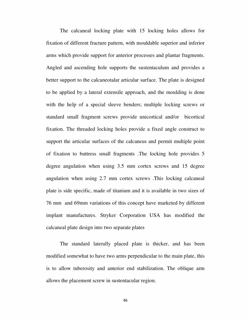
The calcaneal locking plate with 15 locking holes allows for
fixation of different fracture pattern, with mouldable superior and inferior
arms which provide support for anterior processes and plantar fragments.
Angled and ascending hole supports the sustentaculum and provides a
better support to the calcaneotalar articular surface. The plate is designed
to be applied by a lateral extensile approach, and the moulding is done
with the help of a special sleeve benders; multiple locking screws or
standard small fragment screws provide unicortical and/or bicortical
fixation. The threaded locking holes provide a fixed angle construct to
support the articular surfaces of the calcaneus and permit multiple point
of fixation to buttress small fragments .The locking hole provides 5
degree angulation when using 3.5 mm cortex screws and 15 degree
angulation when using 2.7 mm cortex screws .This locking calcaneal
plate is side specific, made of titanium and it is available in two sizes of
76 mm and 69mm variations of this concept have marketed by different
implant manufactures. Stryker Corporation USA has modified the
calcaneal plate design into two separate plates
The standard laterally placed plate is thicker, and has been
modified somewhat to have two arms perpendicular to the main plate, this
is to allow tuberosity and anterior end stabilization. The oblique arm
allows the placement screw in sustentacular region.
47
LOCKING PLATE AND SCREWS
Fig. 19, 20 Locking Plate and Screw
Locking thread
SELF TAPPINGTIP
ARM/TAB
POSTERIOR ANTERIOR

48
Fig. 21 Locking Plate superimposed over calcaneum bone outline
The mesh plate design provides surgeons with an extremely low
profile plates (1mm thick). Their advantage is that they can be easily
contoured, and also they has many screw placement options .
Fig 22 Various calcaneum locking plates
49
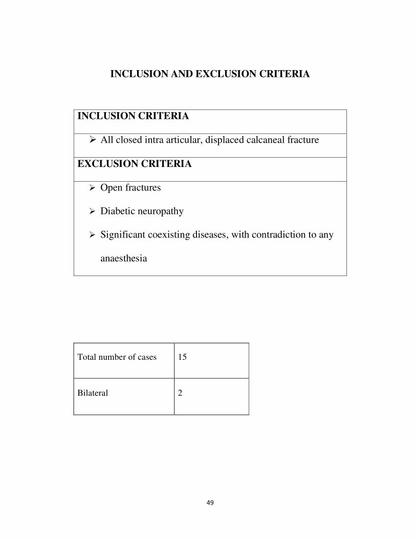
INCLUSION AND EXCLUSION CRITERIA
INCLUSION CRITERIA
� All closed intra articular, displaced calcaneal fracture
EXCLUSION CRITERIA
� Open fractures
� Diabetic neuropathy
� Significant coexisting diseases, with contradiction to any
anaesthesia
Total number of cases 15
Bilateral 2

50
Sex No’s
Male 15
Female Nil
Mode of injury No’s
Fall from height 13
RTA 2
ASSOCIATED INJURIES
Thoracolumbar fractures 1
Fracture tibia 1
ADDITIONAL PROCEDURES
Tibia nailing 1
Sander’s Type in CT scan No. of Cases
II
III
6
9

51
SURGICAL TECHNIQUE FOR OPEN REDUCTION AND
INTERNAL FIXATION
Patient in lateral position, under spinal anaesthesia in 14 cases and
General anaesthesia in one patient with spine injury, pneumatic
tourniquet was applied after intra venous prophylatic antibiotics.
Through lateral extensile incision (Benirschke and Sangeorzan) .Sural
Nerve explored and protected within retracted flap. Sub-periosteal
dissection of anterior skin flap with K wire retraction. Peroneal tendon
sheath dislocated over fibula.
Fig. 23(a,b). Extensile lateral approach and flap retraction with K-wires
One K-wire is passed in fibula, talar neck and cuboid each
(Fig.23b). As shown in above figure. The lateral wall of calcaneum is
excised, the body and the tuberosity of calcaneum is mobilisd. Correction
52
of varus, loss of height and increased width is done. This arrangement is
temporarily stabilized (tuberosity to medial fragment) with K wires. This
is followed by reconstruction and fixation of intra articular fragment. This
is again temporarily stabilized with a K wires. Then insertion of 2 parallel
partially threaded small fragment cancellous screws is done. Reduction
of anterior and middle facets and calcaneocuboid joint is done if
necessary. Application of locking compression calcaneal plate, locking
screws is used for plate fixation. Intra operative radiographic evaluation
with image intensifier with lateral, axial and Antero posterior view. 4 -0
Prolene® “corner stitch” 2-0 Vicryl
®subcutaneous layer closure done
with minimal sutures• Skin is closed with staples or Ethilon®.
Sterile
dressing followed by well padded short below knee slab is applied.

53
Fig 24. Steps in reduction of Tuberosity and Facet
STEPS IN REDUCTION OF TUBEROSITY AND FACET
54
POST OPERATIVE MANAGEMENT
Limb elevation, intravenous antibiotics for five days. Smoking is
avoided atleast sutures are removed. Closed suction drainage is kept for
24 — 48 hours. Short leg splint is removed at fifth postoperative day. If
the flap shows uncomplicated healing and the wound is healthy, early
active range of motion is begun at that time. At second Postoperative
week, active range of motion of the ankle joint is started and gentle
passive movement of subtalar joint done. Suture removal at 3 weeks.
Weight bearing is allowed after 12 weeks, till that protection can be
provided with removable posterior splint. The plate removal is necessary
if the patient shows symptoms that too after one year.

55
Illustrated cases
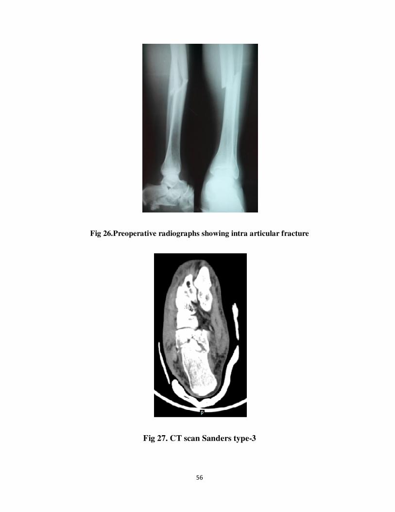
CASE NO.1
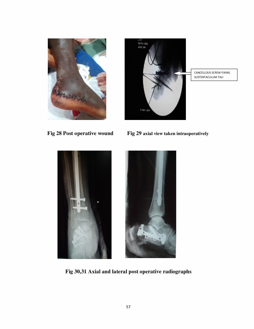
56 years male, Mr.JR, a manual labourer presented with an history of
RTA, sustained right calcaneal and both bone leg fracture in the same
leg, there was swelling and pain in the heel and leg (Fig.25,26). There
was no other spinal or pelvic injury or history of loss of consciousness.
His CT scan revealed Sander’s type3 pattern (Fig.27), the patient was
operated on 13th
day after trauma (Fig.29). Open reduction and internal
fixation of calcaneum was done (Fig.30,31). Postoperatively patient
developed mild infection of the surgical wound, which was settled in two
weeks (Fig.28). According to AOFAS patient had a fair result with 68
score, for his tibia fracture closed nailing was done prior to calcaneal
fixation. He returned to his work in four months (Fig 32).
Fig. 25 Preoperative skin condition(upper arrow showing site of tibia #,lower arrow
showing site of calcaneal #)
56
Fig 26.Preoperative radiographs showing intra articular fracture
Fig 27. CT scan Sanders type-3
57
Fig 28 Post operative wound Fig 29 axial view taken intraoperatively
Fig 30,31 Axial and lateral post operative radiographs
CANCELLOUS SCREW FIXING
SUSTENTACULUM TALI

58
Fig 32 Functional outcome
Fig 33, 34 Radiological Outcome
59
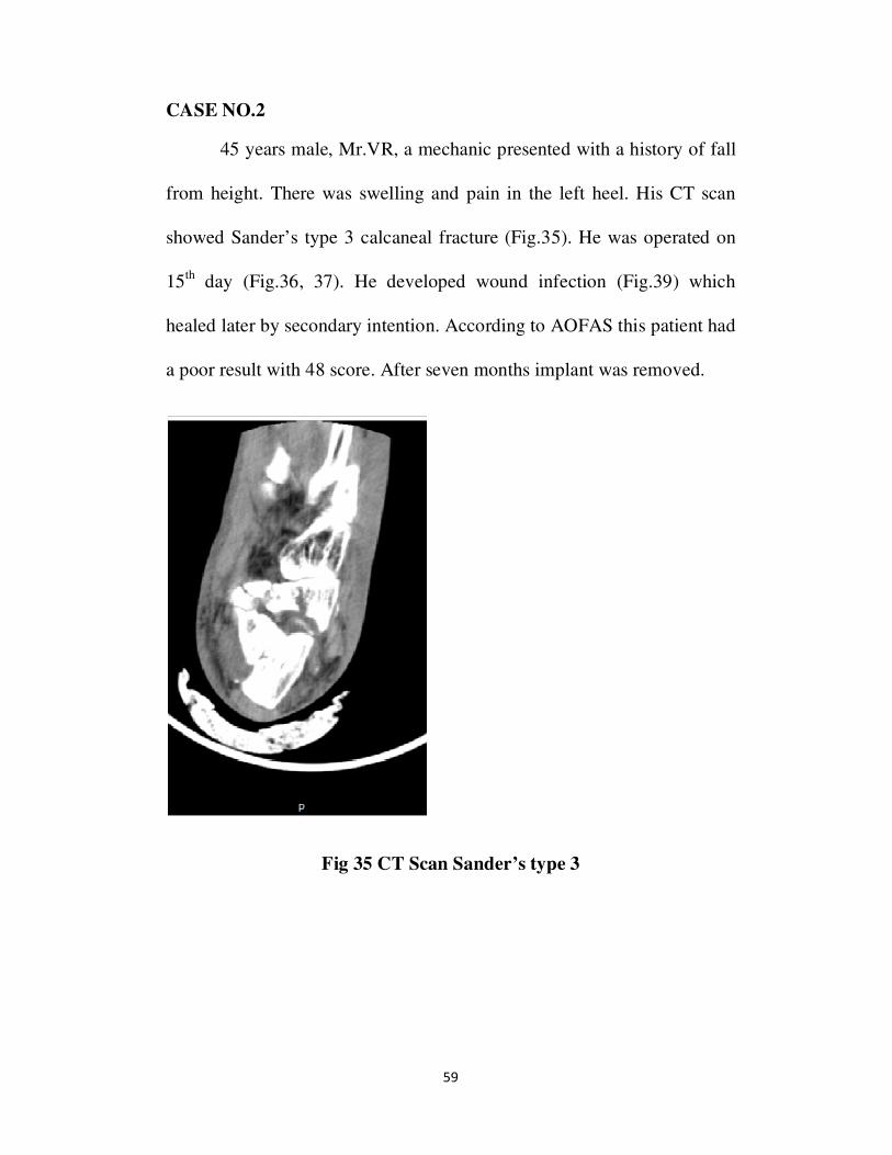
CASE NO.2
45 years male, Mr.VR, a mechanic presented with a history of fall
from height. There was swelling and pain in the left heel. His CT scan
showed Sander’s type 3 calcaneal fracture (Fig.35). He was operated on
15th
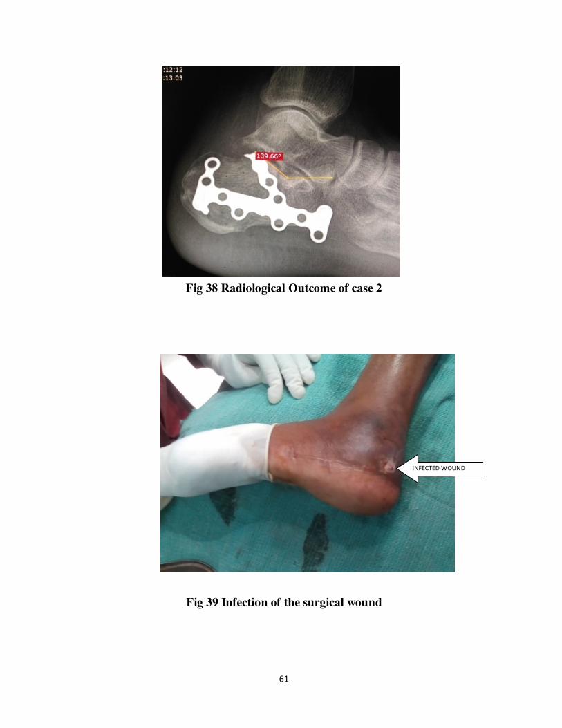
day (Fig.36, 37). He developed wound infection (Fig.39) which
healed later by secondary intention. According to AOFAS this patient had
a poor result with 48 score. After seven months implant was removed.
Fig 35 CT Scan Sander’s type 3
60
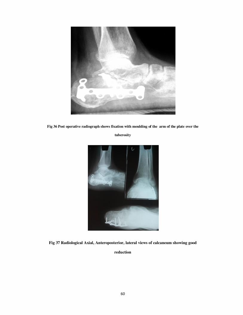
Fig 36 Post operative radiograph shows fixation with moulding of the arm of the plate over the
tuberosity
Fig 37 Radiological Axial, Anteroposterior, lateral views of calcaneum showing good
reduction
61
Fig 38 Radiological Outcome of case 2
Fig 39 Infection of the surgical wound
INFECTED WOUND

62
CASE NO.3
Mr.VP, 32 years old male, a farmer presented with swelling and
pain in the heel after an motor vehicle accident, his Xray showed (Fig.41)
calcaneal fracture and his CT scan showed (Fig.42) Sander’s type 3. He
had wound in the medial side of foot (Fig.40). He was operated on 7th
day
after trauma. An Open reduction and internal fixation of calcaneum
(Fig.43, 44) was done, Post operatively his wound healed well. According
to AOFAS he had a good result with 93 score.
Fig 40 Preoperative skin condition
Fig. 41 Preop x- ray Fig. 42 CT Sander’s type 3
63
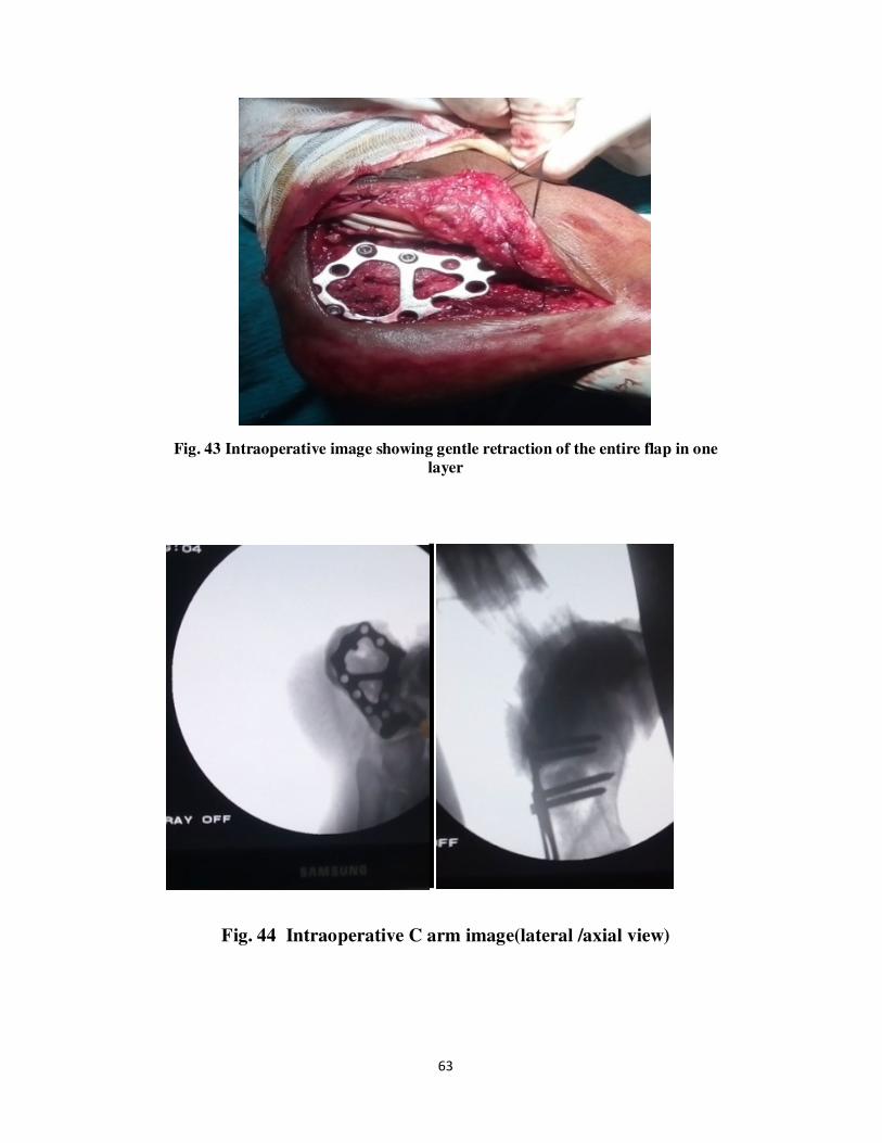
Fig. 43 Intraoperative image showing gentle retraction of the entire flap in one
layer
Fig. 44 Intraoperative C arm image(lateral /axial view)
64
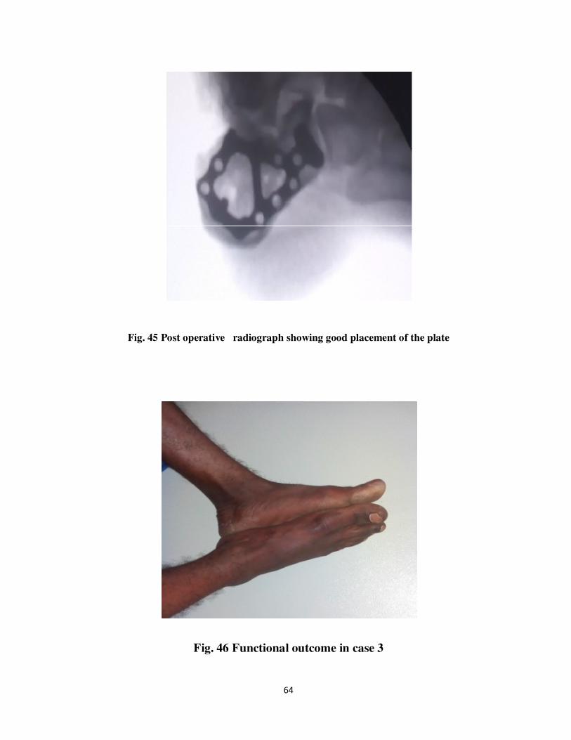
Fig. 45 Post operative radiograph showing good placement of the plate
Fig. 46 Functional outcome in case 3

65
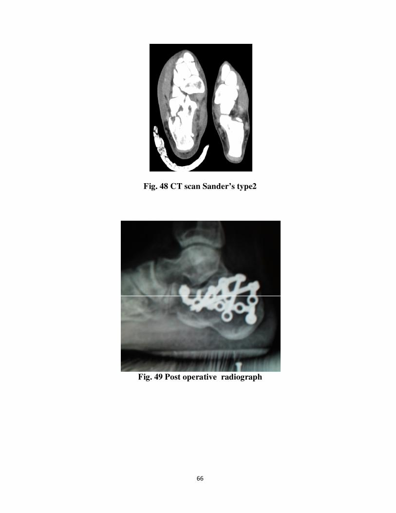
CASE NO.4
Mr.D, 27 years old male, a labourer presented with after a fall
from height and his radiograph (Fig.47) showed a fracture of calcaneum.
His CT scan showed (Fig.48) Sander’s type 2. He was operated on 8th
day
after trauma, (postoperative radiograph is seen in figure 49) Postoperative
period was uneventful and the wound healed well (Fig.50). According to
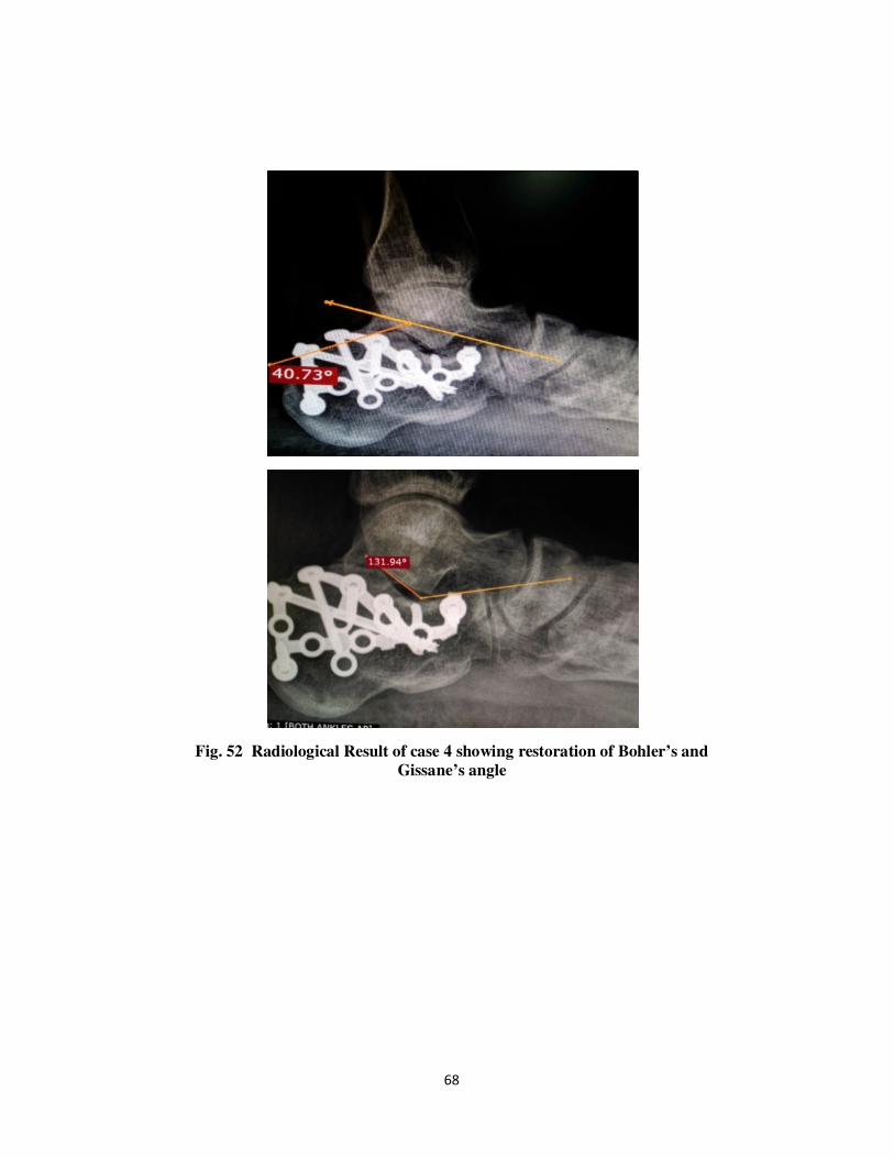
AOFAS he showed a good result with 81 score (Fig.51). The radiological
reconstruction is seen in figure 52.
Fig. 47 Preoperative radiographs
66
Fig. 48 CT scan Sander’s type2
Fig. 49 Post operative radiograph

67
Fig. 50 Post operative wound showing good healing
Fig. 51 Functional outcome of case 4
68
Fig. 52 Radiological Result of case 4 showing restoration of Bohler’s and
Gissane’s angle
69
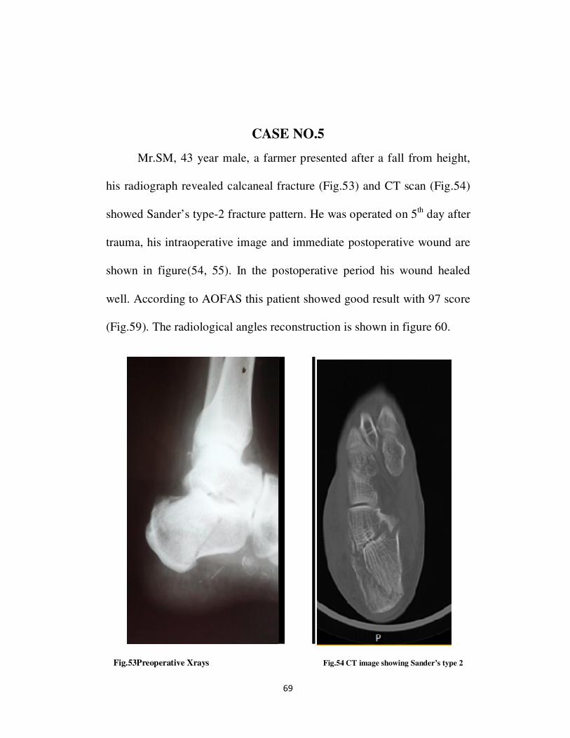
CASE NO.5
Mr.SM, 43 year male, a farmer presented after a fall from height,
his radiograph revealed calcaneal fracture (Fig.53) and CT scan (Fig.54)
showed Sander’s type-2 fracture pattern. He was operated on 5th day after
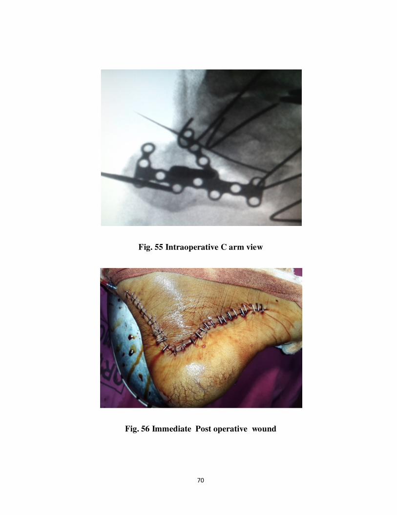
trauma, his intraoperative image and immediate postoperative wound are
shown in figure(54, 55). In the postoperative period his wound healed
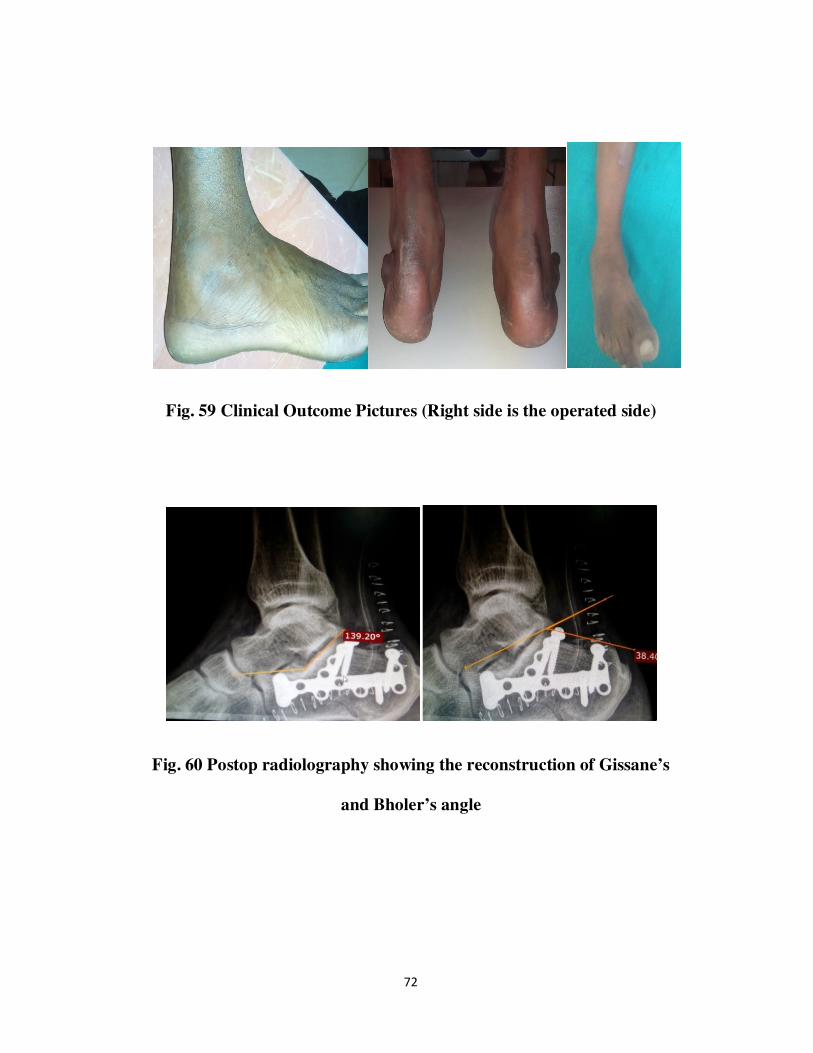
well. According to AOFAS this patient showed good result with 97 score
(Fig.59). The radiological angles reconstruction is shown in figure 60.
Fig.53Preoperative Xrays Fig.54 CT image showing Sander’s type 2
70
Fig. 55 Intraoperative C arm view
Fig. 56 Immediate Post operative wound
71
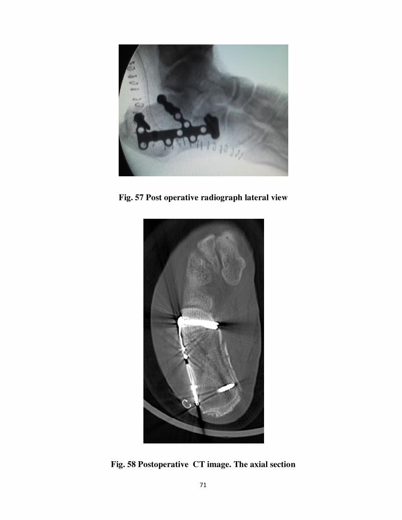
Fig. 57 Post operative radiograph lateral view
Fig. 58 Postoperative CT image. The axial section
72
Fig. 59 Clinical Outcome Pictures (Right side is the operated side)
Fig. 60 Postop radiolography showing the reconstruction of Gissane’s
and Bholer’s angle
73
CASE NO.6
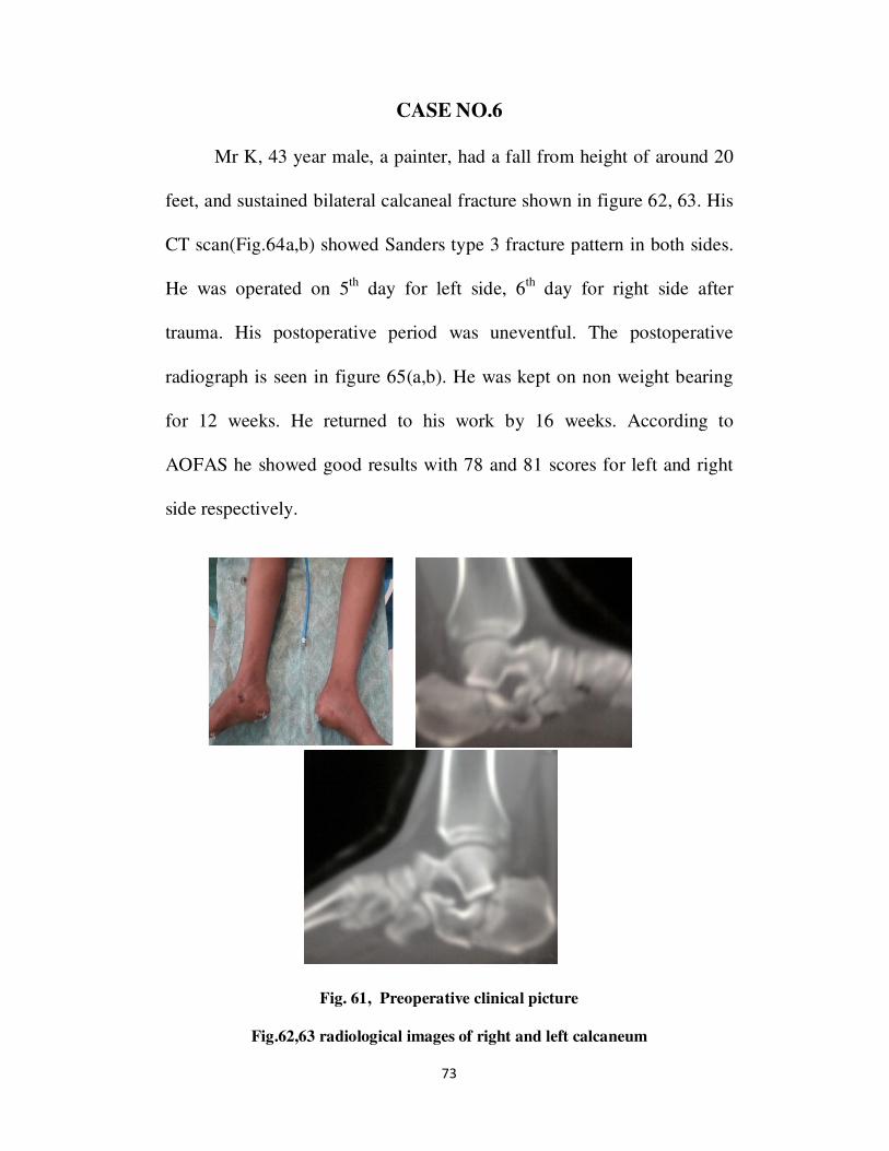
Mr K, 43 year male, a painter, had a fall from height of around 20
feet, and sustained bilateral calcaneal fracture shown in figure 62, 63. His
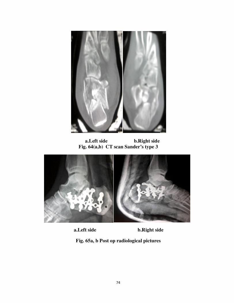
CT scan(Fig.64a,b) showed Sanders type 3 fracture pattern in both sides.
He was operated on 5th
day for left side, 6th
day for right side after
trauma. His postoperative period was uneventful. The postoperative
radiograph is seen in figure 65(a,b). He was kept on non weight bearing
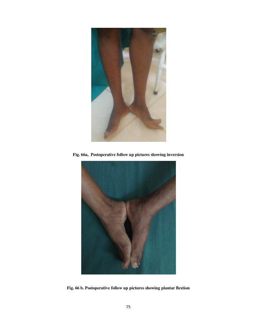
for 12 weeks. He returned to his work by 16 weeks. According to
AOFAS he showed good results with 78 and 81 scores for left and right
side respectively.
Fig. 61, Preoperative clinical picture
Fig.62,63 radiological images of right and left calcaneum
74
a.Left side b.Right side
Fig. 64(a,b) CT scan Sander’s type 3
a.Left side b.Right side
Fig. 65a, b Post op radiological pictures
75
Fig. 66a, Postoperative follow up pictures showing inversion
Fig. 66 b. Postoperative follow up pictures showing plantar flextion
76
RESULTS
Bony union occurred in all patients. Two patients had superficial
wound infection which settled with appropriate antibiotics. Two patients
had superficial wound dehiscence among that one healed by secondary
intention, other patient went for implant removal after radiological
evidence of fracture union.
None of the patients had compartment syndrome, heel pad
problems, peroneal tendinitis and a reflex sympathetic dystrophy
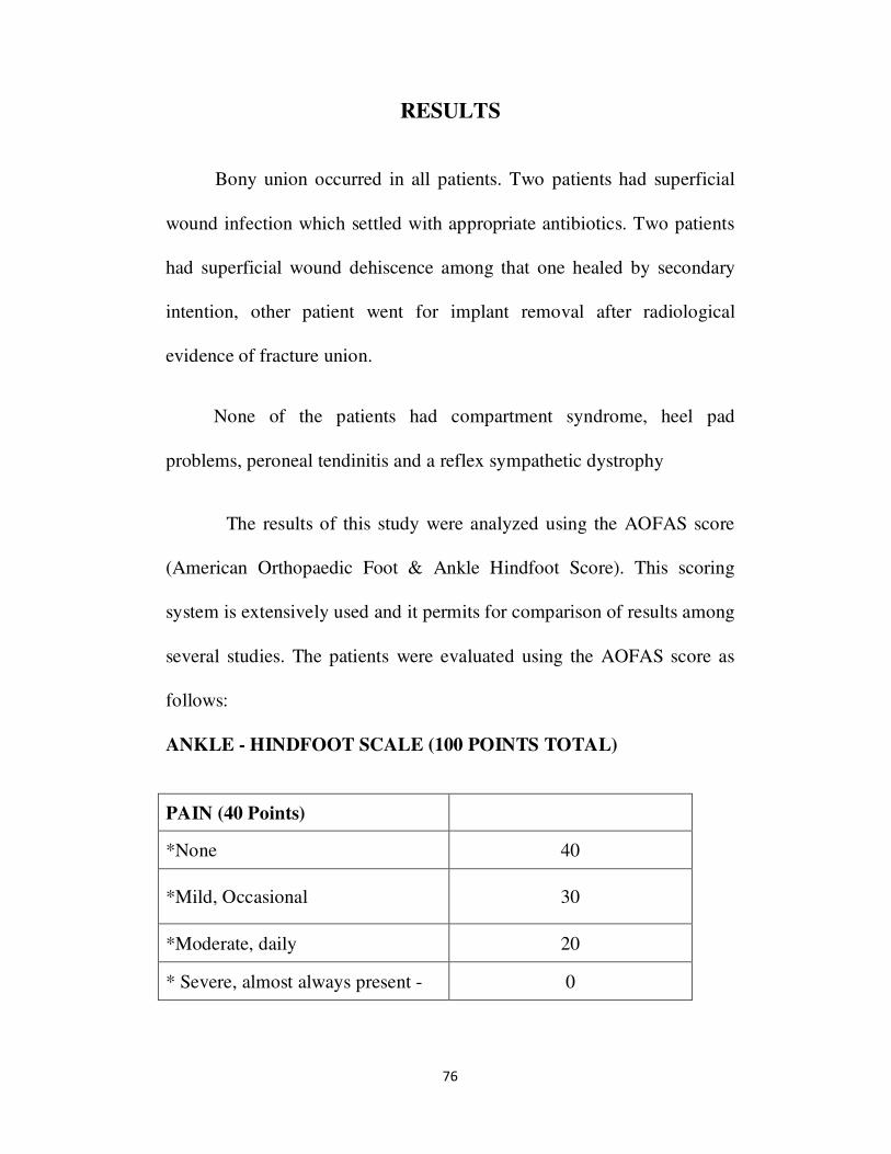
The results of this study were analyzed using the AOFAS score
(American Orthopaedic Foot & Ankle Hindfoot Score). This scoring
system is extensively used and it permits for comparison of results among
several studies. The patients were evaluated using the AOFAS score as
follows:
ANKLE - HINDFOOT SCALE (100 POINTS TOTAL)
PAIN (40 Points)
*None 40
*Mild, Occasional 30
*Moderate, daily 20
* Severe, almost always present - 0

77
Function (50 Points) support requirement Activity limitations,
No limitations, no support 10
No limitation of daily activities, limitation of recreational
activities, no support 7
Limited daily and recreational activities, cane - 4
Severe limitation of daily and recreational activities, walker,
crutches, wheelechair, brace – 0
Maximum walking distance, blocks
Greater than 6 blocks 5
4—6 blocks 4
1—3 blocks 2
Less than l block 0
Walking surfaces
No difficulty on any surface 5
Some difficulty on uneven terrain, stairs inclines, ladders 3
Severe difficulty on uneven terrain, stairs, inclines, ladders 0
Gait abnormality
None or slight 8
Obvious 4
Marked 0

78
Sagittal motion (flexion plus extension)
Normal or mild restriction (30o or more) 8
Moderate restriction (15° - 29o) 4
Severe restriction (less than 15°) 0
Hind foot motion (inversion plus eversion)
Normal or mild restriction (75% - 100% normal) 6
Moderate restriction (25% - 74% normal) 3
Marked restriction (less than 25% normal) 0
Ankle — hindfoot stability (anteroposterior, varus — valgus)
Stable - 8
Definitely unstable – 0 0
Alignment (10 points)
Good, plantigrade foot, ankle-hindfoot well aligned - 15
Fair, plantigrade foot, some degree of ankle-hindfoot
malalignment observed no symptoms
8
* Poor, non-plantigrade foot, severe malalignment, symptoms
0
* Good : >75
* Fair 50—74
* Poor <50

79
In our study the results were as follows:
Good 10
Fair 4
Poor 1
80
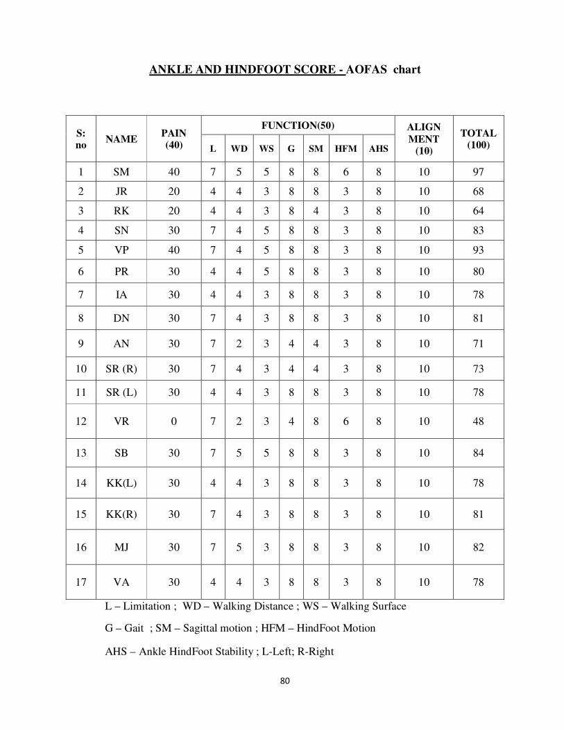
ANKLE AND HINDFOOT SCORE - AOFAS chart
L – Limitation ; WD – Walking Distance ; WS – Walking Surface
G – Gait ; SM – Sagittal motion ; HFM – HindFoot Motion
AHS – Ankle HindFoot Stability ; L-Left; R-Right
S: no
NAME PAIN (40)
FUNCTION(50) ALIGN
MENT (10)
TOTAL (100) L WD WS G SM HFM AHS
1 SM 40 7 5 5 8 8 6 8 10 97
2 JR 20 4 4 3 8 8 3 8 10 68
3 RK 20 4 4 3 8 4 3 8 10 64
4 SN 30 7 4 5 8 8 3 8 10 83
5 VP 40 7 4 5 8 8 3 8 10 93
6 PR 30 4 4 5 8 8 3 8 10 80
7 IA 30 4 4 3 8 8 3 8 10 78
8 DN 30 7 4 3 8 8 3 8 10 81
9 AN 30 7 2 3 4 4 3 8 10 71
10 SR (R) 30 7 4 3 4 4 3 8 10 73
11 SR (L) 30 4 4 3 8 8 3 8 10 78
12 VR 0 7 2 3 4 8 6 8 10 48
13 SB 30 7 5 5 8 8 3 8 10 84
14 KK(L) 30 4 4 3 8 8 3 8 10 78
15 KK(R) 30 7 4 3 8 8 3 8 10 81
16 MJ 30 7 5 3 8 8 3 8 10 82
17 VA 30 4 4 3 8 8 3 8 10 78
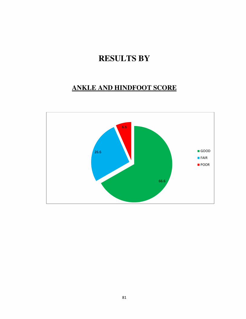
81
RESULTS BY
ANKLE AND HINDFOOT SCORE
66.6
26.6
6.6
GOOD
FAIR
POOR

82
COMPLICATIONS
In our study we encountered the following complications
Superficial wound infection - 2
Minor wound dehiscence - 2
Inadequate reduction - 2 (Arthritis in one)
Sural nerve hypoaesthesia – 2
The results of the surgical procedure analyzed radiographically and
clinically at 4 months post operatively and periodically afterwards. In the
initial cases which we operated, we had complications like superficial
wound infection, wound dehiscence and inadequate anatomical reduction.
These complications developed due to delay in operative treatment. Later
cases had no skin complications.
The poor results were due to prolonged operative time in reducing the
fractures which were taken up late for surgery. However, bony Union was
achieved in all cases.
83
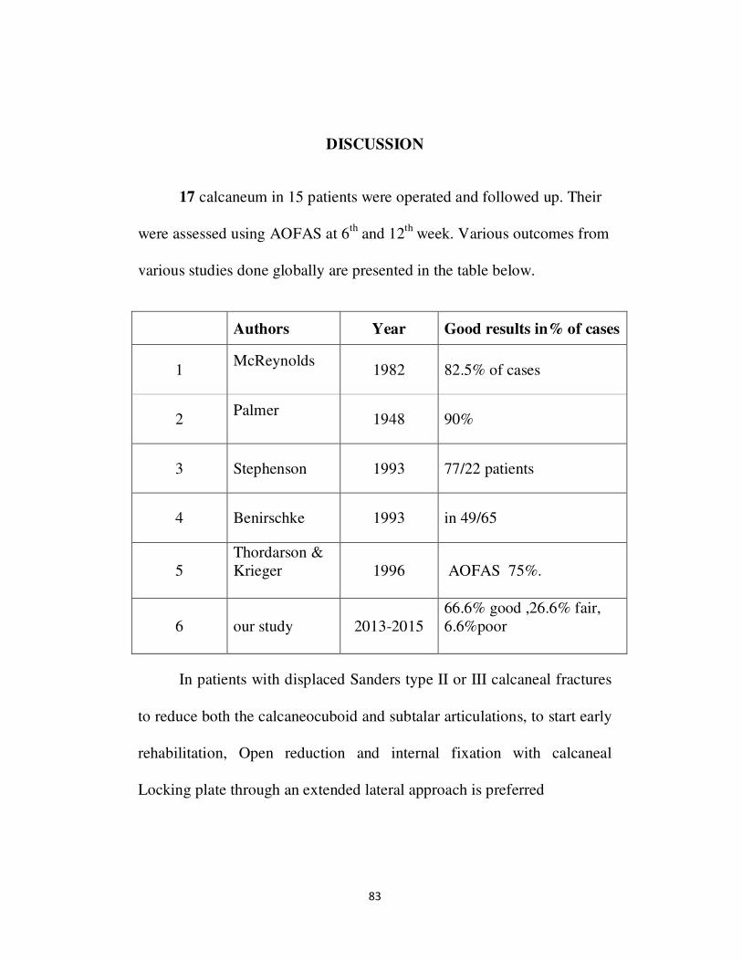
DISCUSSION
17 calcaneum in 15 patients were operated and followed up. Their
were assessed using AOFAS at 6th
and 12th week. Various outcomes from
various studies done globally are presented in the table below.
Authors Year Good results in% of cases
1 McReynolds
1982 82.5% of cases
2 Palmer
1948 90%
3 Stephenson 1993 77/22 patients
4 Benirschke 1993 in 49/65
5
Thordarson &
Krieger
1996 AOFAS 75%.
6 our study 2013-2015
66.6% good ,26.6% fair,
6.6%poor
In patients with displaced Sanders type II or III calcaneal fractures
to reduce both the calcaneocuboid and subtalar articulations, to start early
rehabilitation, Open reduction and internal fixation with calcaneal
Locking plate through an extended lateral approach is preferred
84
Due to the risk of early complications, a clear idea about
indications and contraindications and the timing of surgery are important.
Pre and postoperative CT scans are essential. Fractures presenting with
compartment syndrome are indicated for urgent fasciotomy and plating
should be delayed.
In patients with open calcaneal fractures and multiple trauma, a
temporary stabilization with an external fixator medially can be done
first, and then converted to a second stage open reduction and internal
fixation procedure.
Sustentaculum tali screw fixation has provide the advantages
of high stability, less postoperative pain, strong fixed strength, rapid
functional recovery in treating Sanders type II and III calcaneal
fractures(21).
The poor results in our patient were in the patient taken later 2nd
week.
The calcaneus locking plates has come closer to address nearly all
the problem of calcaneal fracture fixation. It has specific proven merits
with lower profile, better hold in bone, and versatile screw placement,
reducing the need for graft or bone substitutes and allowing for earlier
weight bearing. This documented by the study of Rak et al(36)
, who
compared results of 42 non locking plates with 34 locking plates and
found lesser complications and better results with the former.
85
A study by Hyer et al-2010(37)
, further showed that there
was no significant loss of calcaneal height, stability or joint reduction
after early weight bearing of calcaneal fractures fixed with locked plates.
These outcomes were found to be due to the inherent stability of the
locking plate construct.
Variations of the plate construct have also directed to the
evolution of the so called “wave plate” by Tornier Medical®
, Saint Ismier
Cedex France. This plate’s design allows it to get accommodated in a less
invasive incision. This plate is the form of a wave and can be inserted
percutaneously by a less invasive incision. The anatomic contour of the
plate was created after detailed CT study of over 30 different calcaneal
anatomical patterns; the plate has a non locking apex hole, which
provides optimal lag reduction of the sustentaculum tali. Each extra hole
allows a non locking or locking screw option. This plate has specific
reduction instrumentation for tuberosity manipulation during the fracture
reduction.
Thus at present the locking plates for comminuted calcaneus
fractures seems to be the best choice(38)
. But surgical expertise and cost of
the plate are two main limiting factors. They should be considered and
analyzed along with the benefits of the procedure because implant
affordability and availability of technical expertise is often a problem or
86
challenge in developing countries. It is very important to consider that
calcaneal fracture surgery using complex implants has a steep learning
curve and requires more professional expertise.
87
FUTURE DIRECTIONS AND INNOVATIONS
In future the focus should be on developing refined percutaneous,
minimally invasive techniques. New plates like polyaxial locking plates
can be useful. Multidirectional screw locking with non parallel is
possible. The plate itself does not possess a thread, but a lip, and the
screw with extra thread in the head cuts and thread into the plate at an
angle determined by the surgeon. Due to increasing thread diameter, the
screw locks in this position. The plate can be moulded as the plate and
screw is made of titanium of different hardness grade. Since the plate is
softer than screws and a special screw driver is needed to tighten the
screws and ensure that they cut a thread into the lip of the plate.
BIOABSORBABLE IMPLANTS AND SCREWS
Evolution of bioabsorbable implant has made many professionals
to apply them in selected calcaneal fractures. The problems of the
metallic implants are high infection rate, irritability of plate and later need
for implant removal make the option of bioabsorbable implants
theoretically attractive, Zang and colleagues(39)
have used bioscrews and
prospectively compared them with plates in 97 randomized patients over
a two year period.
88
They found acceptable results at a followup of an average 23
months. Bioabsorbable implants may not be strong enough to withstand
the stress of these displaced calcaneal fractures and their indications in
complex calcaneal fractures are hence limited now. Min and colleagues(40)
have used bioabsorbable pins for calcaneal fracture; however they need
long follow up and assessment in calcaneal fractures.
89
CONCLUSION
In displaced intra-articular fractures of calcaneum osteosynthesis
by open reduction and internal fixation with locking plate using extensile
lateral approach after adequate preoperative planning gave early
functional recovery with acceptable results. Careful consideration of the
surgical technique is a must.
The above method not only restored anatomical height, width of
calcaneum, but also its Bohler's and Gissiane's angles. This allows early
mobilization.
The timing of the surgery is a vital determinant for the treatment
outcome and determined by subsidence of edema and appearance of
wrinkle sign. Those cases which were taken up for fixation early within
10 days had good results than those which were operated later. These
patients had superficial wound infections and minor wound dehiscence.
If for other reasons operation is done after three weeks, it causes
not only soft tissue healing problems and high infection rate but also
intraoperative difficulty in fracture reduction, as the fracture has started
consolidation. Hence it is better to delay surgery till soft tissue heals and
during this presurgical period patients should be managed by
splintingwith proper padding, limb elevation.
90
To conclude intra articular calcaneal fractures are complex
fractures which are difficult to stabilize and manage. The reason behind
the improved results with open reduction and internal fixation in our
series may be due to less traumatic techniques and stronger but malleable
implants. Also locking plates for calcaneum decrease the need for bone
graft, allow early weight bearing and it provides rigidity especially in
osteoporotic cancellous bone. High cost and steep learning curve are the
present limitations.
Ý󣌄C îèõ™ Ý󣌄C îèõ™ Ý󣌄C îèõ™ Ý󣌄C îèõ™ 
î…¬ê Üó² ñ¼ˆ¶õñ¬ù‚° °F裙(Calcaneum) ⽋¹ ºP¾
ãŸð†´ õ¼‹ ݇ ñŸÁ‹ ªð‡ Þ¼ ð£Lù¼‚°‹ î计 (Locking plate)
àîM ªè£‡´ à¬ì‰¶ Þ¼ˆî¬ô êK ªêŒ»‹ ñ °Pˆ¶ å¼ Ý󣌄C
ï¬ìªðŸÁ õ¼A¡ø¶.
Þ‰î Ý󣌄CJ™ cƒèÀ‹ ðƒ«èŸè M¼‹¹A«ø£‹. Þ‰î Ý󣌄CJ™
°Fè£L¡ ⽋¹ ºP¾ ãŸð†´ õ¼‹ ݇ ñŸÁ‹ ªð‡ Þ¼ ð£Lù¼‚°‹
î计 àîM ªè£‡´ êK ªêŒò àœ«÷£‹. Þîù£™ îƒè÷¶ ºP¾ ãŸð†ì
裙 º¿¬ñò£è Ãì õ£ŒŠ¹‡´ â¡ð¬î ªîKMˆ¶ ªè£œA«ø£‹.
º®¾è¬÷ Ü™ô¶ 輈¶è¬÷ ªõOJ´‹«ð£«î£ Ü™ô¶
Ý󣌄CJ¡ «ð£«î£ îƒè÷¶ ªðò¬ó«ò£ Ü™ô¶ ܬìò£÷ƒè¬÷«ò£
ªõOJìñ£†«ì£‹ â¡ð¬î»‹ ªîKMˆ¶‚ ªè£œA«ø£‹.
Þ‰î Ý󣌄CJ™ ðƒ«èŸð¶ îƒèÀ¬ìò M¼ŠðˆF¡ «ðK™ 
Þ¼‚Aø¶. «ñ½‹ cƒèœ ≫ïóº‹ Þ‰î Ý󣌄CJL¼‰¶ H¡ õ£ƒèô£‹
â¡ð¬î»‹ ªîKMˆ¶‚ ªè£œA«ø£‹.
Þ‰î ðK«ê£î¬ùJ¡ º®¾è¬÷ Ý󣌄CJ¡«ð£¶ Ü™ô¶
Ý󣌄CJ¡ º®M¡«ð£¶ îƒèÀ‚° ÜPM‚èŠð´‹ â¡ð¬î»‹
ªîgMˆ¶‚ ªè£œA«ø£‹.
Ý󣌄Cò£÷˜ ¬èªò£Šð‹ ðƒ«èŸð£÷˜ ¬èªò£Šð‹
PROFORMA
OUTCOME ANALYSIS FOR CLOSED INTRAARTICULAR DISPLACED
CALCANEAL FRACTURES WITH LOCKING PLATE Name : Patient ID :
Age :
Sex :
Mode of injury :
H/O loss of consciousness :
H/O chest pain :
H/O abdominal pain :
H/O back pain :
H/O weakness:
H/O loss of senation :
H/O bowel, bladder disturbances:
Past H/O : h/o CAD , CVA , HTN,DM
Personal H/O : h/o smoking , alcohol
General examination :
Vitals: PR,BP
Systemic examination :
� CVS
� RS
� ABDOMEN
LOCAL EXAMINATION OF ANKLE/HINDFOOT
LOCAL EXAMINATION OF SPINE/PELVIS/OTHER LIMBS
INVESTIGATIONS :
CBC

BT,CT
RBS
RFT
CXR
ECG
Xrays
Ankle AP/Lateral/
Calcaneum-Axial view
Pelvis with both hip-AP
Dorsolumbar spine-AP/Lat
CT calcaneum
PROCEDURE DETAILS
POSTOPERATIVE WOUND STATUS:
DATE OF DISCHARGE:
POST OPERATIVE FOLLOWUP
1st visit- 14
TH Postoperative day
Suture removal
2nd
visit-6th
week
Xray-ankle AP/Lateral/Calcaneum axial view
Physiotheraphy
3rd
visit-12th
week
Physiotheraphy
4th
visit-16th
week
AOFAS SCORE
BIBLIOGRAPHY
1. Rockwood & Green’s fractures in Adults. Fifth Edition.
2. Campbell’s Operative Orthopaedics Ninth Edition, Edited by Terry Canale.
3. Displaced intra articular fractures of calcaneum by Roy Sanders. JBJS 82-A,
No.2, Feb. 2000.
4. Management of calcaneal fractures in Adultus by Therman et al. CORR
No.353, pp.107-124, August, 1998.
5. Biomechanics of calcaneal fractures. CORR NO.388, pp.218-224, July, 2001
6. Reduction of displaced intraarticular fracture of calcaneum by B.D. Burdeaux,
Corr, No.177, July, August, 1983.
7. Surgical treatment of calcaneal fractures by Risk Hammesfahr OCNA Vol.20,
No.4, October, 1989,
8. Complications of intraarticular calcaneal fractures by EDWARD. V.A. et al.
CORR No.391, pp.7-16, October, 2001.
9. Wound healing risk factors after ORIF of calcaneal fractures by FRANKLIN
SHULER et al. OCNA, Vol.32, No.1, January, 2001.
10. Kinner BJ, Best R, Falk K, Thon KP Is there a reliable outcome
measurement for displaced intra-articular calcaneal fractures. J. Trauma 2002
Dec: 53(6) : 1904- 101.
11. Asik, M. Sen C. Surgical management of intraarticular fractures of the
calcaneus. Arch. Orthop. Trauma. Surg. 2002, Jul: 122(6) : 354-9.
12. Barei, DP, Bellabarba C, Sangeorzan BJ, Benirschke 5K. Fractures of the
calcaneus. Orthop. Clin. North Am. 2002 Jan 33(1) : 263-85. p
13. Harvey EJ, Grujic L, Early JS, Benirschke 5K, Sangeorzan BJ. Morbidity
associated with ORIF of intra-articular calcaneus fractures using a lateral
approach. Foot Ankle Tnt. 2001 Nov. 22(11) 868-73.
14. Lim EV, Leung JP. Complications of intraarticular calcaneal fractures. Clin,
Orthop. 2001 Oct. (391) : 7-16.
15. Burdeaux BD Jr. Historical and current treatment of calcaneal fractures. J.
Bone Joint Surg. Am. 2001 . Sep. 83-A(9): 1438-40.
16. Randle JA, Kreder JH, Stephen D, Williams J, Jaglal S. Hu R. Should
calcaneal fractures be treated surgically? A meta — analysis. Clin, Orthop.
2000 Aug: (377) : 217- 27.
17. Ebraheim NA, Elgafy H, Sabry, FF, Tao S. Calcaneus fractures with
subluxation of the posterior facet. A surgical indication. Clin. Orthop. 2000
Aug: (377) : 210- 6.
18. O’Farrell DA, O’Byrne JM, McCabe JP, et al. Fractures of the os calcis:
improved results with internal fixation. Injury1993;24(4):263-5.
19. Pendse A, Daveshwar RN, Bhatt J, et al. Outcome after open reduction
internal fixation of intra-articular fracture of calcaneum without use of bone
graft. Indian J Orthop 2006;40(2):111-4.
20. Longino D, Buckley RE. Bone graft in operative treatment of displaced
intraarticular calcaneus fractures. Is it helpful? J Orthop Trauma
2001;15(4):280-6.

21. Leung KS, Chan WS, Shen WY, et al. Operative treatment of intraarticular
fractures of the os calcis— the role of rigid internal fixation and primary bone
grafting: preliminary results. J Orthop Trauma 1989;3(3):232-40.
22. Thordarson DB, Hedman TP, Yetkinler DN, et al. Superior compressive
strength of a calcaneal fracture construct augmented with remodelable
cancellous bone cement. J Bone Joint SurgAm 1999;81(2):239-46.
23. Schildhauer TA, Bauer TW, Josten C, et al. Open reduction and
augmentation of internal fixation with an injectable skeletal cement for the
treatment of complex calcaneal fractures. J Orthop Trauma 2001;14(5):309-17
24. Zhongguo Gu Shang. 2015 Jan;28(1):315. [Sustentaculum tali screw fixation
for the treatment of Sanders type II andIII calcaneal fractures]. [Article in
Chinese]
25. Zhongguo Gu Shang. 2011 Apr;24(4):3057.[A study of 22 displaced
intraarticular calcaneal fractures using locking plates with and without bone
graft]
26.J Clin Orthop Trauma. 2015 Sep;6(3):14752.doi: 10.1016/j.jcot.2015.05.006.
Epub 2015 Jun 6. Evaluation of functional outcome and complications of
locking calcaneum plate for fracture calcaneum. Locking plate Carr JB
.Surgical treatment of Intraarticular calcaneal fractures .Review of small
incision approaches .J Orthop Trauma
27.Dewall M,Henderson CE ,McKinley TO ,Phelps T ,Dolan L ,Marsh J L;
percutanoeus reduction and fixation of displaced intra articular calcaneus
fracture 2010
28. Schepers T ,Vogels LM ,Schipper IB ,et al .Percutaneous reduction and
fixation of displaced intraarticular calcaneal fractures.Oper Orthop Traumatol
Femino JE, vaseenon T ,Levin DA
29.Illert T, Rammelt S,Drewers T ,et al.Stability of locking and non locking
plates in an osteoporotic calcaneal fracture model. Foot angle int .2011;
32(3):307-13
30. Redfern Dj,Oliverira ML,Campbell JT, et al.A biomechanicalcomparison of
locking nd non locking plates for the fixation of calcaneal fractures . Foot
Ankle Int.mar 2006 -27(3) 196-201
31.Blake MH,Owen JR, Sanford TS,et al.Biomechanical evaluation of a locking
and non locking reconstruction plate in an osteoporotic calcaneal fracture
model. Foot Ankle Int.2011 ;32 (4)432 -6
32.Shen C ,Shen Y,Dai LZ,et al,locking compression treatment for intra articular
calcaneal fractures.Zhongguo Gu Shang .2010;23(3).225-7
33.Stoffel K ,Booth G ,Rohrl SM, et al A comparison of conventional versus
locking plates in intra articular calcaneal fractures.A biomechanical study in
human cedavers.Clin Biomech 2000 ;22(1);100-5
34.Wagner M General principles for the clinical use of the LCP. Injury. 2003;34
suppl2:B31-42
35.Wagner M, Frenk A,Frigg R. New concepts of bone fracture treatment and
the locking compression plate.Surg technol int 2004 ;12;271-7
36.Rak V ,Ira D,Masek M.Operative treatment of intra articular calcaneal
fractures with calcaneal plates and its complications.Indian J orthop.July 2009
;43(3)271-80
37.Hyer CF Atway S Berlet GC ,Lee TH .Early weight bearing of calcaneal
fractures fixated with locked plates .A radiographic review.Foot Ankle spec
.Dec 2010;3(6);320-3
38.wang H, Zhang Q,Duan D,et al.The use of calcaneal anatomic plate in
arthroscopically-assisted open reduction and internal fixation of intra-articular
calcaneal fractures.J Huazhong Univ Sci Technology Med
Sci.2006;26(3):319-21
39.Kankare J.Operative treatment of displaced intra-articular fractures of the
calcaneus using absorbable internal fixation.A prospective study of twenty-
five fractures.J Orthop Trauma.1998;12(6):413-9
40.Min W,Munro M,Sanders R.Stabilization of displaced articular fragments in
calcaneal fractures using bioabsorbable pin fixation.A technique guide.J
Orthop trauma.2010;24(12):770-4.
41. Johnson EE, Gebhardt JS. Surgical management of calcaneal fractures using
bilateral incisions and minimal internal fixation. Clin Orthop Relat Res
1993;(290):117-24.
42. Stephenson JR. Surgical treatment of displaced intraarticular fractures of the
calcaneus. Acombined lateral and medial approach.Clin Orthop Relat Res
1993;(290):68-75.
43. Burdeaux BD Jr. Reduction of calcaneal fractures by the McReynolds
medial approach technique and its experimental basis. Clin Orthop Relat Res
1983;(177):87-103.
44. Burdeaux BD Jr. The medial approach for calcaneal fractures. Clin Orthop
Relat Res 1993;(290):96-107.
45. Palmer I. The mechanism and treatment of fractures of the calcaneus. J Bone
Joint Surg. 1948;30A:2-8
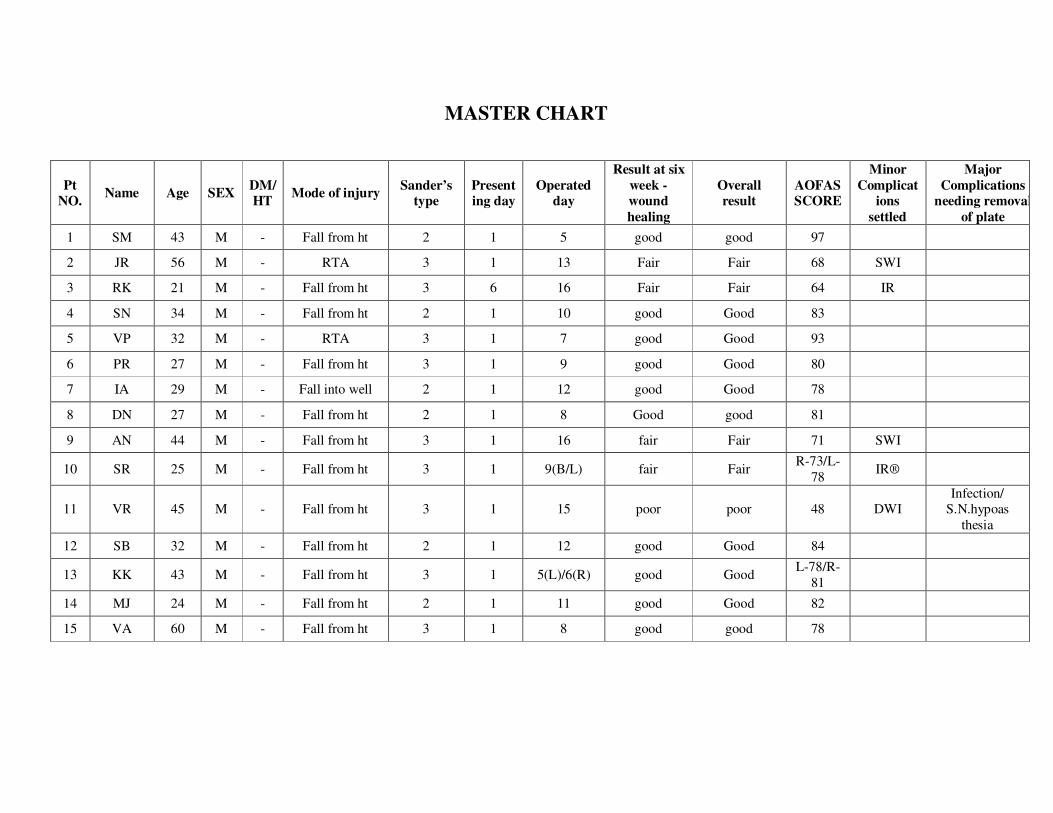
MASTER CHART
Pt
NO. Name Age SEX
DM/
HT Mode of injury
Sander’s
type
Present
ing day
Operated
day
Result at six
week -
wound
healing
Overall
result
AOFAS
SCORE
Minor
Complicat
ions
settled
Major
Complications
needing removal
of plate
1 SM 43 M - Fall from ht 2 1 5 good good 97
2 JR 56 M - RTA 3 1 13 Fair Fair 68 SWI
3 RK 21 M - Fall from ht 3 6 16 Fair Fair 64 IR
4 SN 34 M - Fall from ht 2 1 10 good Good 83
5 VP 32 M - RTA 3 1 7 good Good 93
6 PR 27 M - Fall from ht 3 1 9 good Good 80
7 IA 29 M - Fall into well 2 1 12 good Good 78
8 DN 27 M - Fall from ht 2 1 8 Good good 81
9 AN 44 M - Fall from ht 3 1 16 fair Fair 71 SWI
10 SR 25 M - Fall from ht 3 1 9(B/L) fair Fair R-73/L-
78 IR®
11 VR 45 M - Fall from ht 3 1 15 poor poor 48 DWI
Infection/
S.N.hypoas
thesia
12 SB 32 M - Fall from ht 2 1 12 good Good 84
13 KK 43 M - Fall from ht 3 1 5(L)/6(R) good Good L-78/R-
81
14 MJ 24 M - Fall from ht 2 1 11 good Good 82
15 VA 60 M - Fall from ht 3 1 8 good good 78
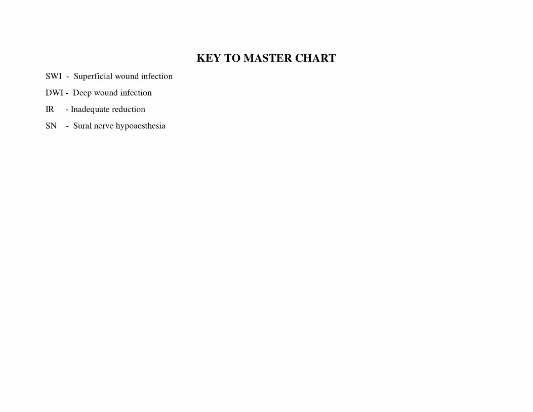
KEY TO MASTER CHART
SWI - Superficial wound infection
DWI - Deep wound infection
IR - Inadequate reduction
SN - Sural nerve hypoaesthesia