Madagascar corals reveal a multidecadal signature of rainfall and river runoff since 1708
ORIGINAL PAPER
Onset of symbiosis and distribution patterns of symbioticdinoflagellates in the larvae of scleractinian corals
Saki Harii Æ Naoko Yasuda ÆMauricio Rodriguez-Lanetty ÆTakahiro Irie Æ Michio Hidaka
Received: 9 October 2008 / Accepted: 11 February 2009 / Published online: 5 March 2009
� Springer-Verlag 2009
Abstract The establishment of symbiosis in early
developmental stages is important for reef-building corals
because of the need for photosynthetically derived nutri-
tion. Corals spawn eggs and sperm, or brood planula larvae
and shed them into the water. Some coral eggs or planulae
directly inherit symbiotic dinoflagellates (Symbiodinium
spp.) from their parents, while others acquire them at each
generation. In most species examined to date, the larvae
without dinoflagellates (aposymbiotic larvae) can acquire
symbionts during the larval stage, but little is known
regarding the timing and detailed process of the onset of
symbiosis. We examined larval uptake of symbiotic dino-
flagellates in nine species of scleractinian corals, the onset
of symbiosis through the early larval stages, and the dis-
tribution pattern of symbionts within the larval host, while
living and with histology, of two acroporid corals under
laboratory conditions. The larvae acquired symbiotic
dinoflagellates during the planktonic phase in all corals
examined which included Acropora digitifera, A. florida,
A. intermedia, A. tenuis, Isopora palifera, Favia pallida,
F. lizardensis, Pseudosiderastrea tayamai, and Ctenactis
echinata. The larvae of A. digitifera and A. tenuis first
acquired symbionts 6 and 5 days after fertilization,
respectively. In A. digitifera larvae, this coincided with the
formation of an oral pore and coelenteron. The number of
symbiotic dinoflagellates increased over the experimental
periods in both species. To test the hypothesis that nutrients
promotes symbiotic uptake, the number of incorporated
dinoflagellates was compared in the presence and absence
of homogenized Artemia sp. A likelihood ratio test
assuming a log-linear model indicated that Artemia sp. had
a significantly positive effect on symbiont acquisition.
These results suggest that the acquisition of symbiotic
dinoflagellates during larval stages is in common with
many coral species, and that the development of both a
mouth and coelenteron play important roles in symbiont
acquisition.
Introduction
Mutualistic endosymbioses between two organisms are
widespread in coral reefs. Marine invertebrates such as
foraminiferans, giant clams, and cnidarians (sea anemones,
jellyfish and corals) harbor unicellular symbiotic dino-
flagellates called zooxanthellae (Symbiodinium spp.).
Symbiotic dinoflagellates contribute to host nutrition by
translocating as much as 95% of their photosynthate to the
host, while the symbionts receive inorganic carbon and
other nutrients in return (Muscatine 1990).
Communicated by M. Kuhl.
S. Harii (&) � N. Yasuda
Graduate School of Engineering and Science,
University of the Ryukyus, 1 Senbaru,
Nishihara, Okinawa 903-0213, Japan
e-mail: [email protected]
M. Rodriguez-Lanetty
Biology Department, The University of Louisiana at Lafayette,
Lafayette, LA 70504, USA
T. Irie
Sesoko Station, Tropical Biosphere Research Center,
University of the Ryukyus, 3422 Sesoko,
Motobu, Okinawa 905-0227, Japan
M. Hidaka
Department of Chemistry, Biology and Marine Science,
Faculty of Science, University of the Ryukyus, 1 Senbaru,
Nishihara, Okinawa 903-0213, Japan
123
Mar Biol (2009) 156:1203–1212
DOI 10.1007/s00227-009-1162-9
Acquisition of symbiotic dinoflagellates occurs either
vertically, where symbionts are transmitted directly from
parent to offspring, or horizontally, where the offspring
acquire symbionts from a new environment in each gen-
eration (Douglas 1998). Vertical transmission ensures that
the host offspring is provided with a complement of sym-
bionts; these vertically transferred symbionts are believed
to be genetically identical in offspring and parent colonies.
There are several clades of Symbiodinium spp. (Baker
2003; Coffroth and Santos 2005) and these are thought to
differ in their environmental tolerances (e.g., to high light,
Rowan 2004). Accordingly, vertical transmitters, which
have a reduced genetic spectrum of Symbiodinium spp.,
may limit their ability to adapt to environmental changes
(Weis et al. 2001). Horizontal transmission provides hosts
with the opportunity to acquire symbionts that are more
suitable for their local environments. However, the disad-
vantage of horizontal transmission is that the host may fail
to acquire symbionts (Weis et al. 2001; Schwarz et al.
2002). Thus, the initial acquisition of symbionts is an
important event for corals that have horizontal acquisition.
Establishment of the cnidarian symbiosis varies inter-
specifically among host developmental stages. Symbiosis
may begin in the embryonic developmental stages
(Scyphozoa: Linuche unguiculata; Montgomery and
Kremer 1995), planula stages (Anthozoa: Anthopleura
elegantissima; Schwarz et al. 2002; Weis et al. 2002), or
polyp stages (Scyphozoa: Cassiopea xamachana; Colley
and Trench 1983; Sachs and Wilcox 2006). Scleractinian
corals spawn eggs and sperm or brood planula larvae
(Harrison and Wallace 1990). Planula larvae float for
several to tens of days before settling (Babcock and
Heyward 1986; Harrison and Wallace 1990). Once these
larvae settle, they metamorphose into polyps and grow into
coral colonies. Most scleractinian corals spawn gametes
that lack symbiotic dinoflagellates and fertilized eggs
develop into aposymbiotic planula larvae (Fadlallah 1983;
Babcock and Heyward 1986; Harrison and Wallace 1990).
Acquisition of symbionts usually occurs at larval or juve-
nile stages. The endodermal cells of the primary polyp
phagocytose symbionts (Hirose et al. 2008a).
Aposymbiotic larvae of corals can acquire symbiotic
dinoflagellates from their environment under experimental
conditions (Schwarz et al. 1999; van Oppen 2001; Weis
et al. 2001; Rodriguez-Lanetty et al. 2004; Baird et al.
2006; Rodriguez-Lanetty et al. 2006). The acquisition of
symbionts by the larvae of the coral Fungia scutaria occurs
4–5 days after fertilization when the oral pore forms
(Schwarz et al. 1999). Although most corals are horizontal
transmitters, the acquisition of symbionts during larval
stages has been observed only in Acropora millepora (van
Oppen 2001), A. muricata (Baird et al. 2006), F. scutaria
(Schwarz et al. 1999; Weis et al. 2001; Rodriguez-Lanetty
et al. 2004, 2006), Favia chinensis and Goniastrea aspera
(Nozawa and Harrison 2005). Acroporid larvae begin
taking up symbionts 6 days after fertilization (Baird et al.
2006); however, Hirose et al. (2008a) suggested that the
initial acquisition of symbionts occurs only after meta-
morphosis, that is, in the initial polyp stage in A. nobilis
(= A. intermedia, Wallace 1999) and A. microphthalma.
Thus, we know the larvae can acquire symbionts, but we do
not know exactly when or how this process occurs. In
addition, little is known regarding the relationship between
establishment of symbiosis and larval developmental
stages.
In the present study, we first confirmed that symbiotic
dinoflagellates are acquired during the planktonic stage in
nine scleractinian coral species in five genera: Acropora
digitifera, A. florida, A. intermedia, A. tenuis, Isopora
palifera, Favia pallida, F. lizardensis, Pseudosiderastrea
tayamai, and Ctenactis echinata. We experimentally
examined the timing of initial symbiosis in acroporid lar-
vae to determine when and how larvae acquire symbionts.
In addition, to examine whether symbiont acquisition is
enhanced by nutrients, we examined the effect of Artemia
homogenate on the number of symbionts incorporated by
laboratory-reared acroporid larvae.
Materials and methods
Collection and maintenance of coral larvae
The colonies of the nine coral species, Acropora digitifera,
A. florida, A. intermedia, A. tenuis, Isopora palifera, Favia
pallida, F. lizardensis, Pseudosiderastrea tayamai and
Ctenactis echinata, were collected in Okinawa, Japan,
during the spawning season in 2007 and 2008. Isopora
palifera is a hermaphroditic brooder (Kojis 1986) and
C. echinata is a gonochoric broadcast spawner (Loya and
Sakai 2008), while the others are hermaphroditic broadcast
spawners (Richmond and Hunter 1990; Hayashibara et al.
1993; Kitamura et al. 2007). All species examined are
common in the Indo-Pacific region and acquire their
symbionts horizontally. The spawning corals were col-
lected and maintained in a running seawater aquarium prior
to spawning. Favia pallida was collected in May 2007
at the Akajima Marine Science Laboratory. Acropora
digitifera and A. florida were collected in May 2007,
C. echinata in July 2007, and F. lizardensis and P. tayamai
in June 2008 at the Sesoko Station of the Tropical Bio-
sphere Research Center at the University of the Ryukyus.
Acropora tenuis was collected in May 2008 at the Ishigaki
Tropical Station, Seikai National Fisheries Research
1204 Mar Biol (2009) 156:1203–1212
123

Institute of the Fisheries Research Agency. The egg–sperm
bundles of A. intermedia were provided by the Okinawa
Churaumi Aquarium in June 2007. After spawning, egg–
sperm bundles were mixed for fertilization. The fertilized
eggs of each species were washed twice in filtered
(0.22 lm) seawater (FSW) and kept in 2-L plastic con-
tainers. All planula larvae were transferred to the Sesoko
Station for the experiments. The colonies of I. palifera
were collected and maintained at the Sesoko Station until
they released larvae in June 2007. All planula larvae were
transferred to new containers filled with fresh FSW daily.
Preparation of symbiont isolates
Symbiodinium spp. were isolated from coral colonies using
a WaterPik (Doltz EW1250, Panasonic, Japan) to remove
the coral tissue. Tissue suspensions were homogenized and
filtered through nylon mesh (180 lm) and then concen-
trated using a centrifuge at 2,400 rpm for 4 min. The algal
pellets were re-suspended in FSW, filtered (40 lm), and
then rinsed three times using a centrifuge at 1,000–
1,500 rpm for 4 min. The number of algal cells from 4
subsamples was counted using a hemacytometer in order to
know the density of algae. The average number of cells
was used. Symbionts were immediately isolated prior to
their use in each experiment. Cultured Symbiodinium spp.
isolated from Tridacna sp. were provided by Okinawa
Prefectural Fisheries and Ocean Research Center and used
to infect the F. lizardensis larvae (see below).
Acquisition of symbionts by the larvae of different
coral species
The larvae of each species were infected with symbiotic
dinoflagellates by one of the two ways. The first infec-
tion method was for the preliminary experiment. One to
four larvae of A. intermedia, A. florida, I. palifera, or
F. pallida were incubated in a 40 ll drop of seawater
containing isolated symbiotic dinoflagellates (approxi-
mately 2.0 9 106 cells ml-1) on a tissue culture dish for
12 h. Approximately 10 drops were made for each spe-
cies. All larvae in the drops were mixed and washed
twice using FSW and moved to a new dish filled with
2 ml FSW. In the second infection method, 10 larvae of
F. lizardensis, P. tayamai, and C. echinata were trans-
ferred to tissue culture dishes containing 2 ml of FSW
with the symbiotic dinoflagellates (same concentration as
before) and a few drops of homogenized Artemia sp.
Four replicates were established. The larvae in each dish
were washed twice using FSW after 6 h and moved to a
new dish containing 2 ml of fresh FSW. For both
methods, the larvae in the final containers were main-
tained for 12 h. The number of larvae that acquired
symbionts and the number of incorporated algal cells
were counted carefully under either a light or fluores-
cence microscope. Either homologous (algae isolated
from the same species) or heterologous (algae isolated
from different species) algae were used for both methods
(Table 1).
Table 1 Acquisition of symbiotic dinoflagellates in the larvae of nine coral species; brooding species (*)
Species Days after
spawning
Type of
symbionts
Infection time
(hours)
Infection
(±SE)a (%)
Average number
of algal cells per
infected larva (±SE)a
Acropora digitiferab 6 Homologous 6 60.0 ± 5.8 (4) 24.9 ± 6.2 (4)
A. floridac 15 Coral (A. tenuis) 12 89.7 ± 4.7 (4) 22.8 ± 5.3 (4)
A. intermediac 4 Homologous 12 0 (30) 0 (30)
5 Homologous 12 60.0 (40) 24.8 ± 6.5 (40)
11 Homologous 12 93.3 (30) 111.2 ± 19.9 (30)
A. tenuisb 5 Homologous 6 20.0 ± 5.8 (4) 3.4 ± 0.8 (4)
Isopora paliferac,* 2 Coral (A. intermedia) 12 63.2 (19) 100.9 ± 30.1 (19)
Favia pallidac 6 Coral (A. intermedia) 12 73.3 (30) 62.1 ± 12.4 (30)
F. lizardensisb 6 Tridacna sp. 6 100 (4) 58.4 ± 8.8 (4)
Psudosiderastrea tayamaib 5 Coral (A. tenuis) 6 26.1 ± 6.0 (4) 3.1 ± 1.2 (4)
12 Coral (A. tenuis) 6 63.8 ± 3.2 (4) 47.2 ± 18.8 (4)
Ctenactis echinatab 9 Coral (A. digitifera) 6 50.0 ± 11.8 (4) 55.8 ± 6.9 (4)
a The number in the brackets represents either the number of larvae examined (method 1) or replication (method 2). See also Figs. 2 and 3 for
Acropora digitifera and A. tenuisb,c Refer to the two different infection methods (see Methods ‘‘acquisition of symbionts by the larvae of different coral species’’)
Mar Biol (2009) 156:1203–1212 1205
123
Timing of the onset of symbiosis in Acropora digitifera
and A. tenuis
Thirty larvae each of A. digitifera and A. tenuis were pre-
pared using the second method described above except 10
larvae of A. digitifera were used on 20 days after fertil-
ization because of limited number of the larvae. Acropora
digitifera was examined on 3–8, 10, and 20 days after
fertilization, and A. tenuis was examined on 3–8 and
10 days after fertilization. Four replicates were established
for each acquisition time. The number of acquired algal
cells was counted in ten individuals from each dish under
either a light or fluorescence microscope with the exception
of day 20 for A. digitifera when only five larvae were
observed in each dish. The remaining A. digitifera larvae in
the dishes were fixed for histological observations (see
below).
Histological observations
Eggs and larvae of A. digitifera were fixed using either
10% formaldehyde in FSW for 10 h for paraffin embedding
or 2.5% glutaraldehyde in 0.2 M phosphate buffer (pH 7.4)
containing 3% NaCl for 2 h for resin embedding. Speci-
mens for the paraffin sections were moved to 70% ethanol
and kept for more than 12 h. They were then dehydrated in
a graded series of ethanol and benzene and were embedded
with paraffin. The blocks were serially sectioned at 5 lm
and mounted on slides, which were stained with Delafield’s
hematoxylin and eosin and examined under a light micro-
scope. Specimens for the resin embedding were rinsed with
0.2 M phosphate buffer (pH 7.4) overnight and then post-
fixed using 1% osmium tetroxide in the same buffer for
2 h. The specimens were then dehydrated in a graded series
of ethyl alcohol and embedded in a Quetol 651 resin
mixture. For light microscopic observation, 0.5 lm speci-
men sections were stained with 1% azur II in 1% borax.
Larval morphology on histological sections was interpreted
referring to Hayashibara et al. (1997), Ball et al. (2002),
and Okubo and Motokawa (2007).
Effect of homogenized Artemia sp. on acquisition
of symbionts
To examine the effects of homogenized Artemia sp. on
symbiont acquisition in coral larvae, five 14-day-old A.
digitifera larvae were exposed to an algal suspension using
the second type of infection described above, but with or
without homogenized Artemia sp. (50 ll) for 6 and 12 h.
Artemia sp. are known to stimulate a feeding response in
the coral larvae Fungia scutaria (Schwarz et al. 1999).
Four replicates (i.e., four tissue culture dishes) were
established. After 6 and 12 h, algal counts were performed
as described above.
Statistical analysis of the ratio of symbiotic larvae
Since the larvae in a dish scramble for the finite number
of symbiotic dinoflagellates, the number of algal cells
acquired by each larva may not be statistically indepen-
dent. To avoid pseudoreplication, we analyzed the number
of the larvae that acquired symbiont(s), considering the
wells rather than the larvae as the experimental unit.
Our interest was in the time course of increase in the
proportion of symbiotic larvae, defined as the number of
larvae with at least one algal cell divided by the total
number of larvae examined in a dish. We assumed that the
proportion follows a logit-normal distribution and analyzed
the ratio by fitting a generalized linear model. We did not
fit the model to transformed data because recent studies
have criticized this method as inexact (e.g., Kasuya 2004;
Wilson 2007).
For each A. digitifera and A. tenuis, the ratio of sym-
biotic larvae, y, was related to the number of days since
fertilization, x, by the equation
logit yð Þ ¼ aþ bx; ð1Þ
where a and b are constants that are estimated. Setting
y = 0.5 and solving Eq. (1) for x provides the timing at
which half of the larvae acquire symbiotic dinoflagellates
as
x0:5 ¼ �a=b: ð2Þ
Based on the observed data for x and y, the maximum
estimates of a and b were calculated by maximizing the log-
likelihood of the logit-normal distribution. Sample sizes
were 24 (four dishes over 6 days) for both species; data
from day 20 were removed from the analysis in A. digitifera
because of their negative effect on the goodness-of-fit of the
model, and thus on the estimation accuracy.
Statistical analysis of the average number of algal cells
The average number of algal cells was calculated for all
larvae examined in a dish, and was analyzed as the
experimental unit. We used generalized linear models with
lognormal distribution because this score is non-negative
and the variance appears to co-vary with the mean.
In the first analysis, we examined whether the average
number of symbiotic algae increased as time advanced
after fertilization. For each A. digitifera and A. tenuis, we
fit the relationship between the average algal number per
larva (y) and the number of days after fertilization, x, to the
equation
1206 Mar Biol (2009) 156:1203–1212
123
ln yð Þ ¼ aþ bx; ð3Þ
where a and b are constants to be estimated. The maximum
log-likelihood was calculated based on the observed data
and the null hypothesis that the dependent variable is
independent of time (i.e., b = 0) was tested using likeli-
hood ratio tests (see Pawitan 2001). Sample sizes were 24
(4 dishes 9 6 days) for both species.
A similar generalized linear model was applied to the
data with regard to the effect of Artemia homogenate on
symbiont acquisition by A. digitifera. In this analysis,
independent variables were assumed to be nominal (i.e.,
presence/absence of Artemia sp. and dichotomous timing
of observation; see Fig. 4) instead of the covariate x in the
above analysis. Accordingly, the natural logarithm of
average algal number was related to a two-way crossed
ANOVA model with Artemia supply, timing of observation
and their interaction as explanatory variables. Significant
testing was conducted based on the likelihood ratio of the
full model to null models constructed by removing each of
the main effects and their interaction from the full model.
Results
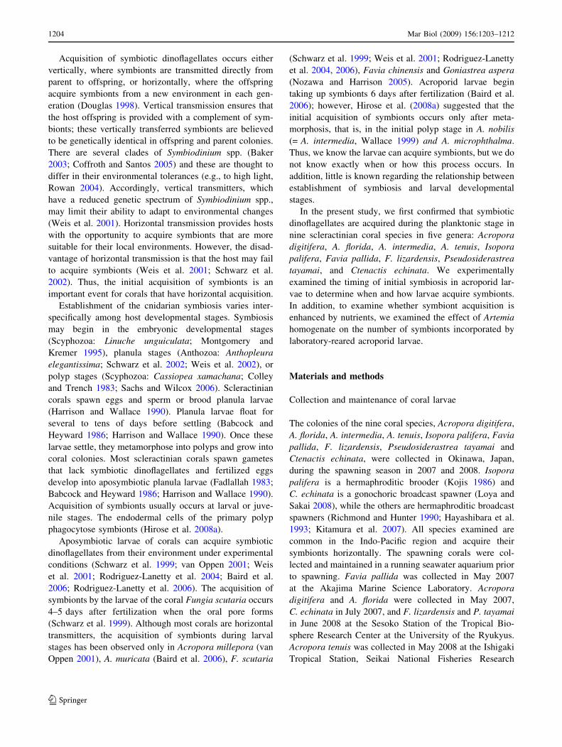
Eggs and larvae of all corals examined did not initially
contain Symbiodinium spp. when they were released. All
of the larvae acquired symbiotic dinoflagellates under
laboratory conditions (Table 1; Fig. 1). The larvae that
acquired symbionts were active swimmer generating par-
ticle movement from the aboral to the oral side using their
cilia. The larvae of Acropora intermedia began to acquire
symbionts 5 days after fertilization, however, the larvae of
the brooder Isoopora palifera acquired symbionts 2 days
after release (Table 1).
Symbionts were first observed in the larvae of A. digi-
tifera 6 days after fertilization, and thereafter more than
60% of larvae had symbionts (Fig. 2a). The logit normal
model estimated the day that half of the larvae contained
symbionts as x0.5 = 5.7. A likelihood ratio test indicated
that the average number of symbiotic algae per larva
significantly increased with time (Fig. 2b; n = 24, v2 =
19.1877, df = 1, P \ 0.0001). Similar patterns were
observed in A. tenuis, but the day that the larvae first
acquired symbionts was 1 day earlier than that of A. digi-
tifera (Fig. 2c), and the maximum likelihood estimate
of x0.5 was 6.1. The average number of symbiotic algae
per larvae also significantly increased with time after
fertilization in A. tenuis (Fig. 2d; n = 24, v2 = 20.1136,
df = 1, P \ 0.0001).
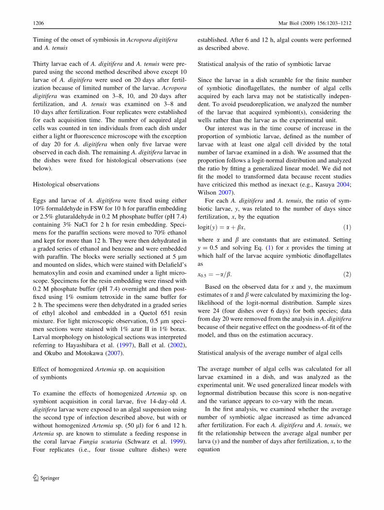
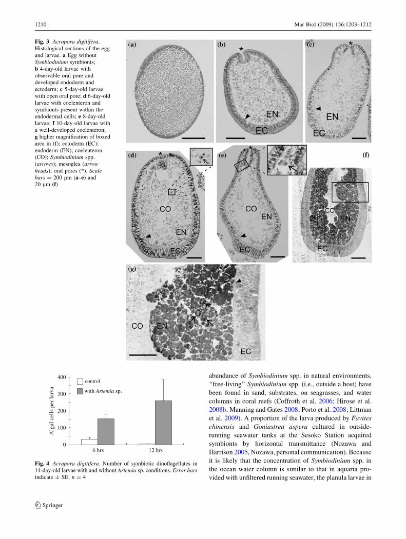
The egg and early stage of larva of A. digitifera did
not contain symbionts (Fig. 3a–c). In a 4-day-old larva,
the epidermal layer (ectoderm) on the oral side was
invaginated in A. digitifera (Fig. 3b). The oral pore was
observed but the coelenteron (gastric cavity) was not
developed in a 5-day-old larva. Symbiotic dinoflagellates
were not detected in the larvae at this time (Fig. 3c). On the
other hand, a few symbionts were found in the endodermal
tissue of the larvae 6 days after fertilization (Fig. 3d). At
this stage, the coelenteron had developed and connected to
the oral pore. In 8- and 10-day-old larvae, the coelenteron
was well developed with wide space (Fig. 3e, f). Several
symbionts occurred in the endodermal tissue around the
oral pore and the coelenteron of the larvae (Fig. 3e, f).
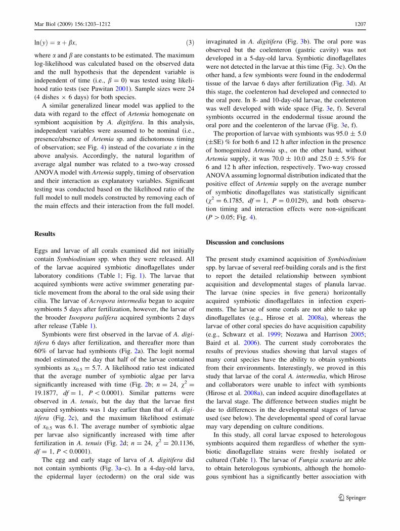
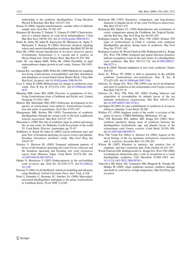
The proportion of larvae with symbionts was 95.0 ± 5.0
(±SE) % for both 6 and 12 h after infection in the presence
of homogenized Artemia sp., on the other hand, without
Artemia supply, it was 70.0 ± 10.0 and 25.0 ± 5.5% for
6 and 12 h after infection, respectively. Two-way crossed
ANOVA assuming lognormal distribution indicated that the
positive effect of Artemia supply on the average number
of symbiotic dinoflagellates was statistically significant
(v2 = 6.1785, df = 1, P = 0.0129), and both observa-
tion timing and interaction effects were non-significant
(P [ 0.05; Fig. 4).
Discussion and conclusions
The present study examined acquisition of Symbiodinium
spp. by larvae of several reef-building corals and is the first
to report the detailed relationship between symbiont
acquisition and developmental stages of planula larvae.
The larvae (nine species in five genera) horizontally
acquired symbiotic dinoflagellates in infection experi-
ments. The larvae of some corals are not able to take up
dinoflagellates (e.g., Hirose et al. 2008a), whereas the
larvae of other coral species do have acquisition capability
(e.g., Schwarz et al. 1999; Nozawa and Harrison 2005;
Baird et al. 2006). The current study corroborates the
results of previous studies showing that larval stages of
many coral species have the ability to obtain symbionts
from their environments. Interestingly, we proved in this
study that larvae of the coral A. intermedia, which Hirose
and collaborators were unable to infect with symbionts
(Hirose et al. 2008a), can indeed acquire dinoflagellates at
the larval stage. The difference between studies might be
due to differences in the developmental stages of larvae
used (see below). The developmental speed of coral larvae
may vary depending on culture conditions.
In this study, all coral larvae exposed to heterologous
symbionts acquired them regardless of whether the sym-
biotic dinoflagellate strains were freshly isolated or
cultured (Table 1). The larvae of Fungia scutaria are able
to obtain heterologous symbionts, although the homolo-
gous symbiont has a significantly better association with
Mar Biol (2009) 156:1203–1212 1207
123

the host larvae than do heterologous symbionts (Weis et al.
2001; Rodriguez-Lanetty et al. 2004, 2006). The early
polyp stage of soft corals and scleractinian corals tend to
acquire heterologous symbionts, in contrast to the adults
(Coffroth et al. 2001; Little et al. 2004; Gomez-Cabrera
et al. 2008). It is likely that larvae or initial polyps of corals
do not have strict symbiont specificity and may lack
mechanisms for symbionts identity recognition.
The timing of symbiont acquisition varies by coral
species. The onset of symbiosis in the larvae of A. digi-
tifera, A. intermedia, and A. tenuis occurred 5–6 days after
fertilization (Fig. 2; Table 1), which is later than in Fungia
scutaria, whose larvae begin to acquire symbionts from
3 days after fertilization (Schwarz et al. 1999). In the
brooding coral Isopora palifera, the larvae acquired sym-
bionts 2 days after release (Table 1). The differences
Fig. 1 a–h Acropora tenuis,A. digitifera, Favia pallida,F. lizardensis,Pseudosiderastrea tayamai and
Ctenactis echinata.Microphotographs of the larvae
with or without Symbiodiniumspp. a Ten-day-old larvae of
A. tenuis with acquired
symbionts; b higher
magnification of the box area in
a; c–f larvae of A. digitifera(c, 7-day-old), F. pallida(d, 6-day-old), F. lizardensis(e, 6-day-old), and P. tayamai(f, 12-day-old) under a
fluorescence microscope.
Symbiodinium spp. in the larvae
are shown as red particles.
g Three-day-old larvae of
C. echinata without symbionts,
and h 9-day-old larvae of
C. echinata with acquired
symbionts (circles); ectoderm
(EC); endoderm (EN);
Symbiodinium spp. (S); lipids
(LI); oral pore (*).
Scale bars = 200 lm (a, c–g),
100 lm (h) and 50 lm (b)
1208 Mar Biol (2009) 156:1203–1212
123

among the coral species in the timing of larval symbiont
acquisition may be related to differences in the develop-
mental stages at which they were exposed to symbionts. In
A. digitifera larvae, the oral pore developed by day 4, and
the coelenteron was formed on day 5, when symbiont
acquisition occurred. Okubo and Motokawa (2007) repor-
ted that larvae of five acroporid corals, including
A. digitifera, had formed the coelenteron and mesenterial
filaments by the ninth day after fertilization. The larvae of
F. scutaria acquired symbionts following oral pore and
coelenteron development (Schwarz et al. 1999). Isopora
palifera larvae, which have a well-developed coelenteron
when released (Kojis 1986), acquired symbionts 2 days
after release. In contrast, the coelenteron is not formed
16 days after fertilization in A. intermedia and A. micro-
phthalma, and larvae of these species do not take up
symbionts during the period before the coelenteron appears
(Hirose et al. 2008a). Together, these findings suggest that
the onset of symbiosis in coral larvae occurs after oral pore
formation and subsequent coelenteron development. The
pore allows symbionts entry into the coelenteron. However,
in some cases, acquisition of symbionts may occur through
the epithelial cells of the blastoderm (Marlow and Mar-
tindale 2007). The differences in larval developmental
stages among coral species may explain the differences in
the timing of symbiont acquisition by larvae.
Our study demonstrated experimentally that the number
of algal cells acquired per day by coral larvae increased
over time (Fig. 2b, d). This may be related to the continued
development over time of gastrodermal cells lining the
coelenteron. Hirose et al. (2008a) reported that the cilia of
the gastrodermal cells are elongated and sometimes trap
symbionts in the coelenteron of acroporid coral primary
polyps. Similarly, larvae of the soft coral Xenia umbellate
also have cilia in the coelenteron (Benayahu et al. 1988).
With the passage of time after fertilization, the larval
coelenteron may become enlarged and lined with well-
developed gastrodermal cells. This enlargement and the
current produced by well-developed, ciliated gastrodermal
cells may enhance acquisition of symbionts by late devel-
opmental stage larvae. Once symbionts are retained in the
coelenteron, the hosts (larvae) recognize and phagocytose
them. Some lectins in the octocoral Sinularia lochmodes
have been proposed as mediators of interactions between
host and symbionts (Jimbo et al. 2000; Koike et al. 2004).
Lectin/glycan interactions have recently been implicated as
components of the inter-partner signalling mechanisms of
symbiosis in the coral Fungia scutaria larvae (Wood-
Charlson et al. 2006). It is likely that well-developed larvae
have mechanisms to recognize symbionts for subsequent
phagocytose.
The larvae of A. digitifera acquired more symbionts in
the presence of Artemia homogenate than in its absence.
This is consistent with the findings that larvae of F. scu-
taria under laboratory conditions acquire symbionts though
ingestion while feeding (Schwarz et al. 1999; Rodriguez-
Lanetty et al. 2004, 2006). Larvae of the sea anemone
Anthopleura elegantissima also ingest dinoflagellates while
feeding (Schwarz et al. 2002). Once an oral pore and
coelenteron have developed, the larvae may obtain nutri-
ents via ingestion. These observations suggest that the
Artemia homogenate is effective in stimulating the estab-
lishment of planula larva symbiosis.
We demonstrated that larvae of many coral species have
the potential to acquire symbionts during the planktonic
phase. Although little is known about the distribution and
0
20
40
60
80
100
n.d.
Days after fertilization
(d) A. tenuis
A. tenuis
0
20
40
60
80
100
n.d.
(c)
n.d.0
20
40
60
80
100
120
140A
lgal
cel
ls p
er la
rva
Days after fertilization
(b) A. digitiferaPe
rcen
tage
(%
)
0
20
40
60
80
100
4 5 6 7 8 9 10 20 4 5 6 7 8 9 10
4 5 6 7 8 9 10 20 4 5 6 7 8 9 10
n.d.
A. digitifera(a)Fig. 2 Acropora digitifera and
A. tenuis. Temporal changes in
the percentage of larvae with
symbionts (a and c) and in
the number of symbiotic
dinoflagellates in the larvae
(b and d). Error barsindicate ± SE, n = 4
Mar Biol (2009) 156:1203–1212 1209
123
abundance of Symbiodinium spp. in natural environments,
‘‘free-living’’ Symbiodinium spp. (i.e., outside a host) have
been found in sand, substrates, on seagrasses, and water
columns in coral reefs (Coffroth et al. 2006; Hirose et al.
2008b; Manning and Gates 2008; Porto et al. 2008; Littman
et al. 2009). A proportion of the larva produced by Favites
chinensis and Goniastrea aspera cultured in outside-
running seawater tanks at the Sesoko Station acquired
symbionts by horizontal transmittance (Nozawa and
Harrison 2005, Nozawa, personal communication). Because
it is likely that the concentration of Symbiodinium spp. in
the ocean water column is similar to that in aquaria pro-
vided with unfiltered running seawater, the planula larvae in
Fig. 3 Acropora digitifera.
Histological sections of the egg
and larvae. a Egg without
Symbiodinium symbionts;
b 4-day-old larvae with
observable oral pore and
developed endoderm and
ectoderm; c 5-day-old larvae
with open oral pore; d 6-day-old
larvae with coelenteron and
symbionts present within the
endodermal cells; e 8-day-old
larvae; f 10-day-old larvae with
a well-developed coelenteron;
g higher magnification of boxed
area in (f); ectoderm (EC);
endoderm (EN); coelenteron
(CO); Symbiodinium spp.
(arrows); mesoglea (arrowheads); oral pores (*). Scalebars = 200 lm (a–e) and
20 lm (f)
with Artemia sp.
control
6 hrs 12 hrs
100
200
300
400
0
Alg
al c
ells
per
larv
a
Fig. 4 Acropora digitifera. Number of symbiotic dinoflagellates in
14-day-old larvae with and without Artemia sp. conditions. Error barsindicate ± SE, n = 4
1210 Mar Biol (2009) 156:1203–1212
123
seawater tanks may have had an opportunity to acquire
symbionts under ‘‘natural’’ conditions.
Symbiotic larvae have the advantage of obtaining energy
from symbiotic dinoflagellate cells (Richmond 1987; van
Oppen 2001; Harii et al. 2002), though they are also
exposed to elevated risk of oxidative damage under stressful
conditions (Yakovleva et al. 2009). Symbiotic larvae of
Pocillopora damicornis, which is a brooder and vertical
transmitter, have a long larval dispersal period ([100 days),
which may be related to the additional energy provided by
the photosynthetic algal cells (Richmond 1987; Harii et al.
2002). If aposymbiotic larvae do not find a suitable place to
settle during the competent period (i.e., 4 days after fertil-
ization; Babcock and Heyward 1986; Harii et al. 2007), they
may extend their planktonic life (Nishikawa et al. 2003;
Harii et al. 2007; Graham et al. 2008). During this time the
larvae have the potential to acquire symbiotic dinoflagel-
lates, which may increase larval survivorship and/or
settlement-competency periods and thus play an important
role in larval dispersal time (van Oppen 2001). On the other
hand, Yakovleva et al. (2009) reported that symbiotic larvae
suffer a high level of oxidative cellular damage under high
temperature, resulting in lower survivorship of symbiotic
larvae in comparison with aposymbiotic larvae. They sug-
gest that symbionts are a costly burden for larval stages
under stressful environmental conditions. Determination of
when and how larvae acquire symbionts in the natural
environment is key in understanding advantages and dis-
advantages of coral–algal relationship.
Acknowledgments We thank the staff of the Sesoko Station of the
Tropical Biosphere Research Center, University of the Ryukyus, for
providing research facilities. We also thank Kenji Iwao, Masaya
Morita, Akira Iguchi, Yossi Loya, Takeshi Hayashibara, Naoko
Isomura, Hironobu Fukami, Nami Okubo, Makoto Kitamura, Kenji
Iwai and staff of Okinawa Churaumi Aquarium for providing sam-
ples. We are also grateful to Mamiko Hirose for valuable suggestions
regarding histological observations and the reviewers who helped to
improve this manuscript. This research was funded by the 21st
Century Center of Excellence (COE) program of the University of the
Ryukyus, a Ministry of Education, Culture, Sports, Science and
Technology Grant-in-Aid for Young Scientists (B) No. 20770017
(SH), and the Australian Research Council Centre of Excellence for
Coral Reef Studies at the University of Queensland.
References
Babcock RC, Heyward AJ (1986) Larval development of certain
gamete-spawning scleractinian corals. Coral Reefs 5:111–116.
doi:10.1007/BF00298178
Baird AH, Gilmour JP, Kamiki TM, Nonaka M, Pratchett MS,
Yamamoto HH, Yamasaki H (2006) Temperature tolerance of
symbiotic and non-symbiotic coral larvae. Proc 10th Int Coral
Reef Symp 1:38–42
Baker AC (2003) Flexibility and specificity in coral-algal symbiosis:
diversity, ecology, and biogeography of Symbiodinium. Annu
Rev Ecol Evol Syst 34:661–689
Ball EE, Hayward DC, Reece-Hoyes JS, Hislop NR, Samuel G, Saint
R, Harrison PL, Miller DJ (2002) Coral development: from
classical embryology to molecular control. Int J Dev Biol
46:671–678
Benayahu Y, Achituv Y, Berner T (1988) Embryogenesis and
acquisition of algal symbionts by planulae of Xenia umbellata(Octocorallia: Alcyonacea). Mar Biol (Berl) 100:93–101. doi:
10.1007/BF00392959
Coffroth MA, Santos SR (2005) Genetic diversity of symbiotic
dinoflagellates in the genus Symbiodinium. Protist 156:19–34.
doi:10.1016/j.protis.2005.02.004
Coffroth MA, Santos SR, Goulet TL (2001) Early ontogenetic
expression of specificity in a cnidarian-algal symbiosis. Mar
Ecol Prog Ser 222:85–96. doi:10.3354/meps222085
Coffroth MA, Lewis CF, Santos SR, Weaver JL (2006) Environmen-
tal populations of symbiotic dinoflagellates in the genus
Symbiodinium can initiate symbioses with reef cnidarians. Curr
Biol 16:R985–R987. doi:10.1016/j.cub.2006.10.049
Colley NJ, Trench RK (1983) Selectivity in phagocytosis and
persistence of symbiotic algae by the scyphistoma stage of the
jellyfish Cassiopeia xamachana. Proc R Soc Lond B Biol Sci
219:61–82
Douglas AE (1998) Host benefit and the evolution of specialization in
symbiosis. Heredity 81:599–603. doi:10.1038/sj.hdy.6884550
Fadlallah YH (1983) Sexual reproduction, development and larval
biology in scleractinian corals. Coral Reefs 2:129–150. doi:
10.1007/BF00336720
Gomez-Cabrera MC, Ortiz JC, Loh WKW, Ward S, Hoegh-Guldberg
O (2008) Acquisition of symbiotic dinoflagellates (Symbiodini-um) by juveniles of the coral Acropora longicyathus. Coral Reefs
27:219–226. doi:10.1007/s00338-007-0315-x
Graham EM, Baird AH, Connolly SR (2008) Survival dynamics of
scleractinian coral larvae and implications for dispersal. Coral
Reefs 27:529–539. doi:10.1007/s00338-008-0361-z
Harii S, Kayanne H, Takigawa H, Hayashibara T, Yamamoto M
(2002) Larval survivorship, competency periods and settlement
of two brooding corals, Heliopora coerulea and Pocilloporadamicornis. Mar Biol 141:39–46. doi:10.1007/s00227-002-
0812-y
Harii S, Nadaoka K, Yamamoto M, Iwao K (2007) Temporal changes
in settlement, lipid content and lipid composition of larvae of the
spawning hermatypic coral Acropora tenuis. Mar Ecol Prog Ser
346:89–96. doi:10.3354/meps07114
Harrison PL, Wallace CC (1990) Reproduction, dispersal and
recruitment of scleractinian corals. In: Dubinsky Z (ed) Ecosys-
tems of the world 25. Coral reefs. Elsevier, Amsterdam, pp 133–
207
Hayashibara T, Shimoike K, Kimura T, Hosaka S, Heyward A,
Harrison P, Kudo K, Omori M (1993) Patterns of Coral
Spawning at Akajima Island, Okinawa, Japan. Mar Ecol Prog
Ser 101:253–262
Hayashibara T, Ohike S, Kakinuma Y (1997) Embryonic and larval
development and planula metamorphosis of four gamete-spawn-
ing Acropora (Anthozoa, Scleractinia). Proc 8th Int Coral Reef
Symp 2:1231–1236
Hirose M, Yamamoto H, Nonaka M (2008a) Metamorphosis and
acquisition of symbiotic algae in planula larvae and primary
polyps of Acropora spp. Coral Reefs 27:247–254. doi:
10.1007/s00338-007-0330-y
Hirose M, Reimer JD, Hidaka M, Suda S (2008b) Phylogenetic
analyses of potentially free-living Symbiodinium spp. isolated
from coral reef sand in Okinawa, Japan. Mar Biol 155:105–112.
doi:10.1007/s00227-008-1011-2
Jimbo M, Yanohara T, Koike K, Koike K, Sakai R, Muramoto K,Kamiya H (2000) The D-galactose-binding lectin of the octo-
coral Sinularia lochmodes: characterization and possible
Mar Biol (2009) 156:1203–1212 1211
123
relationship to the symbiotic dinoflagellates. Comp Biochem
Physiol B Biochem Mol Biol 125:227–236
Kasuya E (2004) Angular transformation—another effect of different
sample sizes. Ecol Res 19:165–167
Kitamura M, Koyama T, Nakano Y, Uemura D (2007) Characteriza-
tion of a natural inducer of coral larval metamorphosis. J Exp
Mar Biol Ecol 340:96–102. doi:10.1016/j.jembe.2006.08.012
Koike K, Jimbo M, Sakai R, Kaeriyama M, Muramoto K, Ogata T,
Maruyama T, Kamiya H (2004) Octocoral chemical signaling
selects and controls dinoflagellate symbionts. Biol Bull 207:80–86
Kojis BL (1986) Sexual reproduction in Acropora (Isopora) species
(Coelenterata: Scleractinia). I. A. cuneata and A. palifera on
Heron Island reef, Great Barrier Reef. Mar Biol 91:291–309
Little AF, van Oppen MJH, Willis BL (2004) Flexibility in algal
endosymbioses shapes growth in reef corals. Science 304:1492–
1494
Littman RA, van Oppen MJH, Willis BL (2009) Methods for sampling
free-living Symbiodinium (zooxanthellae) and their distribution
and abundance at Lizard Island (Great Barrier Reef). J Exp Mar
Biol Ecol. (in press). doi 10.1016/j.jembe.2008.06.034
Loya Y, Sakai K (2008) Bidirectional sex change in mushroom stony
corals. Proc R Soc B 275:2335–2343. doi:10.1098/rspb.2008.
0675
Manning MM, Gates RD (2008) Diversity in populations of free-
living Symbiodinium from a Caribbean and Pacific reef. Limnol
Oceanogr 53:1853–1861
Marlow HQ, Martindale MQ (2007) Embryonic development in two
species of scleractinian coral embryos: Symbiodinium localiza-
tion and mode of gastrulation. Evol Dev 9:355–367
Montgomery MK, Kremer PM (1995) Transmission of symbiotic
dinoflagellates through the sexual cycle of the host scyphozoan
Linuche unguiculata. Mar Biol 124:147–155
Muscatine L (1990) The role of symbiotic algae in carbon and energy
flux in reef corals. In: Dubinsky Z (ed) Ecosystems of the world
25. Coral Reefs. Elsevier, Amsterdam, pp 75–87
Nishikawa A, Katoh M, Sakai K (2003) Larval settlement rates and
gene flow of broadcast-spawning (Acropora tenuis) and planula-
brooding (Stylophora pistillata) corals. Mar Ecol Prog Ser
256:87–97
Nozawa Y, Harrison PL (2005) Temporal settlement patterns of
larvae of the broadcast spawning reef coral Favites chinensis and
the broadcast spawning and brooding reef coral Goniastreaaspera from Okinawa, Japan. Coral Reefs 24:274–282. doi:
10.1007/s00338-005-0476-4
Okubo N, Motokawa T (2007) Ernbryogenesis in the reef-building
coral Acropora spp. Zool Sci 24:1169–1175. doi:10.2108/zsj.
24.1169
Pawitan Y (2001) In all likelihood: statistical modelling and inference
using likelihood. Oxford University Press, New York, p 528
Porto I, Granados C, Restrepo JC, Sanchez JA (2008) Macroalgal-
associated dinoflagellates belonging to the genus Symbiodiniumin Caribbean Reefs. PLoS ONE 3:e2160
Richmond RH (1987) Energetics, competence, and long-distance
dispersal of planula larvae of the coral Pocillopora damicornis.
Mar Biol 93:527–533
Richmond RH, Hunter CL (1990) Reproduction and recruitment of
corals: comparisons among the Caribbean, the Tropical Pacific,
and the Red Sea. Mar Ecol Prog Ser 60:185–203
Rodriguez-Lanetty M, Krupp DA, Weis VM (2004) Distinct ITS
types of Symbiodinium in Clade C correlate with cnidarian/
dinoflagellate specificity during onset of symbiosis. Mar Ecol
Prog Ser 275:97–102
Rodriguez-Lanetty M, Wood-Charlson EM, Hollingsworth LL, Krupp
DA, Weis VM (2006) Temporal and spatial infection dynamics
indicate recognition events in the early hours of a dinoflagellate/
coral symbiosis. Mar Biol 149:713–719. doi:10.1007/s00227-
006-0272-x
Rowan R (2004) Thermal adaptation in reef coral symbionts. Nature
430:742
Sachs JL, Wilcox TP (2006) A shift to parasitism in the jellyfish
symbiont Symbiodinium microadriaticum. Proc R Soc B
273:425–429. doi:10.1098/rspb.2005.3346
Schwarz JA, Krupp DA, Weis VM (1999) Late larval development
and onset of symbiosis in the scleractinian coral Fungia scutaria.
Biol Bull 196:70–79
Schwarz JA, Weis VM, Potts DC (2002) Feeding behavior and
acquisition of zooxanthellae by planula larvae of the sea
anemone Anthopleura elegantissima. Mar Biol 140:471–478.
doi:10.1007/s00227-001-0736-y
van Oppen M (2001) In vitro establishment of symbiosis in Acroporamillepora planulae. Coral Reefs 20:200
Wallace CC (1999) Staghorn corals of the world—a revision of the
genus Acropora. CSIRO Publishing, Melbourne, 421 pp
Weis VM, Reynolds WS, deBoer MD, Krupp DA (2001) Host-
symbiont specificity during onset of symbiosis between the
dinoflagellates Symbodinium spp. and planula larvae of the
scleractinian coral Fungia scutaria. Coral Reefs 20:301–308.
doi:10.1007/s003380100179
Weis VM, Verde EA, Pribyl A, Schwarz JA (2002) Aspects of the
larval biology of the sea anemones Anthopleura elegantissimaand A. artemisia. Invertebr Biol 121:190–201
Wilson JB (2007) Priorities in statistics, the sensitive feet of
elephants, and don’t transform data. Folia Geobot 42:161–167
Wood-Charlson EM, Hollingsworth LL, Krupp DA, Weis VM (2006)
Lectin/glycan interactions play a role in recognition in a coral/
dinoflagellate symbiosis. Cell Microbiol 8:1985–1993. doi:
10.1111/j.1462-5822.2006.00765.x
Yakovleva IM, Baird AH, Yamamoto HH, Bhagooli R, Nonaka M,
Hidaka M (2009) Algal symbionts increase oxidative damage
and death in coral larvae at high temperature. Mar Ecol Prog Ser
(in press)
1212 Mar Biol (2009) 156:1203–1212
123
Copyright © 2022 FDOKUMEN