Characterization of black band disease in Red Sea stony corals
Photophysiological stress in scleractinian corals in
response to short-term sedimentation
Eva Philippa, Katharina Fabriciusb,*
aAlfred Wegener Institute for Polar and Marine Research, Columbusstr., 27568 Bremerhaven, GermanybAustralian Institute of Marine Science, PMB No. 3, Townsville MC, Queensland 4810, Australia
Received 15 August 2001; received in revised form 3 September 2002; accepted 28 October 2002
Abstract
Effects of short-term sedimentation on common coastal coral species were investigated in
laboratory and field experiments on the Great Barrier Reef (GBR) using pulse-amplitude modulated
(PAM) chlorophyll fluorometry. In the laboratory, changes in maximal quantum yields of
photosystem II (Fv/Fm) in Montipora peltiformis were examined in response to the amount of
sedimentation (79–234 mg cm� 2) and duration of exposure (0–36 h). In control colonies, Fv/Fm
ranged from 0.67 to 0.71, and did not show any temporal trend, while maximum yields of sediment-
covered fragments declined steadily and reached levels below 0.1 in most colonies after 36 h
coverage. Maximal quantum yield in M. peltiformis declined linearly in relation to both the amount
of sediment deposited per unit surface area and the duration of exposure. Zooxanthellae densities and
chlorophyll concentrations per unit area of sediment-treated corals decreased in the same manner,
however, their responses were not quite as strong as the changes in Fv/Fm. Within the ranges
measured, sedimentation stress of colonies exposed to large amounts of sediment for short periods of
time was similar to that exposed to low amounts of sediments for prolonged periods of time.
Colonies were recovered from short-term, or low-level, sedimentation within < 36 h, whereas long-
term exposure, or high levels of sedimentation, killed exposed colony parts. Field experiments
comparing susceptibilities of common coastal coral species towards sedimentation showed
significant reductions in effective quantum yields (DF/FmV) in 9 out of 12 common coastal species
after 22 h of exposure. Three out of twelve investigated species were not affected by the
experimental application of sediments (Galaxea fascicularis, Fungia crassa, and Pectinia lactuca).
Our results suggest that anthropogenic sediment deposition can negatively affect the photosynthetic
activity of zooxanthellae and thus the viability of corals. However, the results also showed the ability
of corals to compartmentalise sedimentation stress, as the photosynthetic activity only from tissues
0022-0981/02/$ - see front matter. Crown Copyright D 2002 Published by Elsevier Science B.V. All rights
reserved.
PII: S0022 -0981 (02 )00495 -1
* Corresponding author. Tel.: +61-7-4753-4444; fax: +61-7-4772-5852.
E-mail address: [email protected] (K. Fabricius).
www.elsevier.com/locate/jembe
Journal of Experimental Marine Biology and Ecology
287 (2003) 57–78

directly underneath the sediment declined, whereas that of adjacent clean tissues did not change
measurably.
Crown Copyright D 2002 Published by Elsevier Science B.V. All rights reserved.
Keywords: Photophysiological stress; Scleractinian corals; Short-term sedimentation
1. Introduction
There is a significant natural variation in sedimentation on coral reefs, and tolerance to
sedimentation varies widely among corals. Natural processes, such as resuspension from
the sea floor and sediment imports from rivers, determine concentrations of suspended
solids in the water column. However, excessive sedimentation constitutes one of the biggest
sources of reef degradation from human activities (Rogers, 1990). The main sources of
sedimentation due to human activities are dredging or drilling at sea and runoff from river
catchments degraded by upstream deforestation, removal of riparian vegetation and
mangroves, and agriculture in wetlands and floodplains (Guilcher, 1985; Cortes and Risk,
1985; Hubbard, 1986; van Katwijk et al., 1993; Brodie, 1995). If wave energy is low (e.g.,
during calm weather, on leeward sides, or at greater depths), the suspended sediment settles
out of the water column, and is deposited on the sea floor and upon sea floor-inhabiting
organisms.
The effects of sedimentation on corals and their symbiotic algae have been investigated
in a number of studies around the world (reviewed in Rogers, 1990). Some species are able
to clean off deposits efficiently and show no damage due to sedimentation (Abdel-Salam
and Porter, 1988; Stafford-Smith, 1993; Riegl, 1995; Wesseling et al., 1999). In other coral
species, sedimentation depresses rates of photosynthesis and enhances respiration and
mucus production (Riegl and Branch, 1995; Yentsch et al., 2002). With increasing
sedimentation, growth rates decline, the corals loose their zooxanthellae, and the under-
lying tissue dies (Bak, 1978; Lasker, 1980; Rogers, 1983; Peters and Pilson, 1985).
Sedimentation also negatively affects rates of survival and settlement of coral larvae
(Babcock and Davies, 1991; Gilmour, 1999). In the longer term, high sedimentation
regimes can influence coral cover as well as the species composition in communities due to
differences in sediment tolerances between species (Dodge and Vaisnys, 1977; Cortes and
Risk, 1985).
In this study, we used the noninvasive pulse-amplitude modulated chlorophyll fluor-
ometry (PAM) to quantify the build-up and recovery from photosynthetic stress in corals
exposed to short-term (12–36 h) periods of high levels of sedimentation. A submersible
PAM-instrument (Diving-PAM, Walz, Germany) enabled us to conduct measurements both
in the laboratory and in the field. This instrument has previously been used on corals for in
situ studies of reactions of symbiotic zooxanthellae to diurnal changes in light intensity or
different water depth (Beer and Ilan, 1998; Ralph et al., 1999), and to detect stress caused
by cyanide, heavy metals or temperature changes (Warner et al., 1996; Jones et al., 1999,
2000; Prange and Dennison, 2000).
The aim of this study was, firstly, to investigate the short-term stress reaction of the
coral Montipora peltiformis to sediment exposure and its potential to recover from such
E. Philipp, K. Fabricius / J. Exp. Mar. Biol. Ecol. 287 (2003) 57–7858

stress under controlled laboratory conditions. M. peltiformis is a foliose coral species,
which occurs in high abundances on some turbid nearshore reefs of the central Great
Barrier Reef (GBR). In particular, the effects of exposure to (a) different amounts of
sediment for (b) different times on photosynthetic activity, zooxanthellae count, and
chlorophyll concentration, were investigated. Secondly, the susceptibility of a range of
coral species to sediment stress was assessed in the field. Responses were compared
among reefs as well as among species, and it was tested whether the photosynthetic
stress is confined to the coral surface directly underneath the sediment. The results of
this investigation may help to understand the damage of corals caused by short-term
exposure to high levels of sedimentation and potential shifts in community structure in
relation to prolonged or repeated enhanced levels of sedimentation in coastal reefs of
the Great Barrier Reef.
2. Materials and methods
2.1. Laboratory experiments
2.1.1. Experimental setup
The effects of sediments on corals were studied in the outdoor flow-through aquarium
system of the Australian Institute of Marine Science (AIMS) in January and February
2001. Fragments of the foliose coral M. peltiformis were sampled from 3 to 5 m water
depth at Magnetic Island, a turbid nearshore fringing reef where this species occurs in
high abundance (Fig. 1). Corals were transported to AIMS within 2 h of collection.
Large coral pieces were split into fragments with a mean area of 33 cm2 (F 8 SD) and
left in a tank on plastic racks for 3 days to recover. The oval 1000-l tank had a
continuous inflow of natural seawater at rate of 600 l h� 1. A circular laminar flow of
about 3–4 cm s� 1 was created by a system of water and air hoses. The water
temperature was around 29 jC, equivalent to that of the ambient sea. Corals were
exposed to natural sunlight reduced by a 70% shade cloth to simulate natural conditions
at f6 m water depth. Fine, muddy sediment was collected from 3 m water depth
behind the breakwater wall of the AIMS jetty. It was sieved through a 3-mm mesh to
remove coarse material, and aerated for two days. The sediment contained 2.37%
carbon, 0.13% nitrogen, and 459 Ag/g phosphorous. When the experiments commenced,
half of the coral fragments were temporarily removed from the tank and put into a
holding tank of the same size, flow, and light exposure. After the incoming water was
turned off in both tanks, the sediment was stirred and distributed as evenly as possible in
the water column of the experimental tank. When the sediment had settled after 6 h, the
inflow of seawater was turned on again, and the control corals were moved back from
the holding tank into the experimental tank, and evenly distributed between the now
sediment-loaded corals.
2.1.2. PAM chlorophyll fluorometry
Photosynthetic activity was determined by measuring variable chlorophyll fluores-
cence of photosystem II (PS II), with a pulse-amplitude modulated chlorophyll
E. Philipp, K. Fabricius / J. Exp. Mar. Biol. Ecol. 287 (2003) 57–78 59

fluorometer (Diving-PAM; Schreiber et al., 1986). In dark acclimated corals, maximal
quantum yield was calculated as the ratio of variable to maximum fluorescence (Fv/Fm).
For dark adaptation, coral fragments were kept in black boxes with running seawater for
30 min, while the sediment remained on the sediment-treated fragments. At the end of
the dark period, the sediment was washed off each fragment and collected in separate
vials for dry weight determination. Chlorophyll fluorescence was measured with the
fiberoptics of the PAM chlorophyll fluorometer at 3-mm distance to the coral surface. F0
was measured by applying a pulsed measuring beam ( < 1 Amol quanta m� 2 s� 1),
followed by a saturation pulse of white light to record Fm (>1000 Amol quanta
m� 2 s� 1). Fifteen measurements, evenly distributed over the surface, were made on
each fragment.
2.1.3. Calculation of sediment load
The sediment retrieved from each fragment of M. peltiformis was dried in an
oven at 60 jC until constant weight. The surface area of each of the foliose
Fig. 1. Map of the Great Barrier Reef showing the positions of AIMS, Magnetic Island, and the four reefs of the
field experiments.
E. Philipp, K. Fabricius / J. Exp. Mar. Biol. Ecol. 287 (2003) 57–7860

fragments was determined by tracing its outline onto paper, cutting and weighing
the piece of paper, and using a calibration curve to convert paper weight to sur-
face area. The amount of sediment retrieved from each coral was then normalised
to their surface area. It averaged 151F 37 mg DW cm� 2 (range: 79–234 mg
cm� 2).
2.1.4. Determination of chlorophyll concentrations and zooxanthellae density
Zooxanthellae densities and chlorophyll (chl a and chl c2) concentrations were
analysed on subsamples of the frozen corals. The tissue was stripped from the skeleton
with an airbrush using 60–100 ml filtered seawater. The water–tissue solution was
homogenised in a glass homogeniser, and five 10 ml subsamples were taken. To
determine chlorophyll concentrations, three of the subsamples were centrifuged at
1500� g for 3 min. The supernatant was rejected and the tissue redissolved in 2 ml
100% acetone, sonicated for 1 min, and the chlorophyll extracted overnight in darkness
at 4 jC. The next day, the samples were again centrifuged at 1500� g for 3 min, and
the supernatant was transferred into vials and frozen at � 20 jC. A second extraction
was performed on the remaining tissue pellet, which was redissolved in 2 ml 100%
acetone, and processed as described before. Both extracts were measured in a
spectrophotometer at 630 and 663 nm, and chl a and chl c2 concentrations were
calculated after Jeffrey and Humphrey (1975). Zooxanthellae densities in two subsam-
ples of the homogenised water–tissue solution were counted under the microscope using
a haemocytometer slide (eight replicates) and counts were normalised to coral surface
area.
2.1.4.1. Experiment 1.1: effects of amount and duration of sedimentation. After 0, 12,
18, 24, and 36 h, sediment-loaded and control fragments were removed from the
experimental tank and carefully transferred into a darkened flow-through 10-l tank for
Fv/Fm measurements. Two to five replicate fragments were analysed for each treatment
and time. Subsequently, half of the fragments were frozen for analyses of chlorophyll
and zooxanthellae densities (see above); the rest were moved back into the experimental
tank to monitor their recovery from the treatment.
2.1.4.2. Experiment 1.2: effects of duration of exposure on recovery. Recovery from
sedimentation was monitored for up to 7 days by repeated measurements of maximum
PS II quantum yields (Fv/Fm) of M. peltiformis fragments, which had been exposed to
sediment for 0, 12, 18, and 24 h.
2.1.4.3. Experiment 1.3: effects of sedimentation on the adjacent tissue. To determine
whether sediment stress in one area also affected the surrounding tissues, sediment was
applied to about 50% of the surface of four colonies of M. peltiformis, while the
remaining surface was left clean. After 48 h exposure, maximum quantum yields of the
clean surfaces away from the sediment, as well as those along the edge of the sediment
patch, were measured and compared with those of the colonies before sediment
application. After sediment removal, Fv/Fm of the cleaned tissues was determined. This
experiment was later repeated in the field on four colonies each of Echinopora lamellosa
E. Philipp, K. Fabricius / J. Exp. Mar. Biol. Ecol. 287 (2003) 57–78 61

and massive Porites. These colonies were exposed to sediments for 22 h before effective
quantum yield measurements.
2.2. Field experiments
Field experiments were carried out on the windward sides of reefs at 6–8 m depth
in February 2001. The reefs fringed four inshore islands located at 5–8 km off the
coast. Two islands were in the northern Great Barrier Reef (Hay Island: 13j40VS,143j42VE, and Wilkie Island: 13j47VS, 143j38VE), and two were in the Central GBR
(Green Island: 16j46VS, 146j00VE, and Normandy Island: 17j12VS, 146j04VE)(Fig. 1). Sediment was freshly collected from 15 m depth at the study site in the
beginning of the experiment. This provided a finer sediment fraction than found at
the depth of the experimental corals. Sediment nutrient concentrations are listed in
Table 1. The amount of sediment loaded onto the corals could not easily be
quantified, but we aimed to apply amounts similar to those added in the laboratory
experiments (about 200 mg cm� 2).
In the field experiments, advantage was taken of the fact that responses were
confined to directly exposed tissues (see results to Experiment 1.3): we added the
sediment to one portion of the investigated colonies and used other parts of the same
colonies as controls (without sediment load; Fig. 2). This allowed us to eliminate
potential between colony variability in effective quantum yields (DF/FmV ) caused by
small-scale environmental heterogeneity (depth, flow exposure, shading by neighbours,
etc.).
2.2.1. Experiment 2.1: comparison of sedimentation effects among reefs
Replicate colonies of six common nearshore species (Pachyseris speciosa, massive
Porites, E. lamellosa, Turbinaria peltata, Fungia crassa, Galaxea fascicularis) were
measured on several of the four islands to compare responses to sediment application
among different locations. After tagging the corals, effective quantum yields of the tissue
were recorded with the Diving-PAM (about 30 measurements per colony). DF/FmV were
measured across the whole colony surface at time zero because the exact position of the
sediment deposit, and its edge, could not be fully controlled. One part of the colony
surface was then covered with a thin layer of the freshly collected sediment. After 22 h,
the effective quantum yield of the tissue in the area free of sediment was measured as
control, then the sediment was removed from the remaining colony and DF/FmV of the
cleaned tissue was recorded.
Table 1
Content of carbon (C), nitrogen (N) and phosphorous (P) in sediments of different reefs
C (%) N (%) P (Ag/g)
Hay Island 10.33 0.03 320
Wilkie Island 9.89 0.02 864
Normandy Island 9.68 0.05 291
Green Island 11.08 0.05 286
E. Philipp, K. Fabricius / J. Exp. Mar. Biol. Ecol. 287 (2003) 57–7862

2.2.2. Experiment 2.2: comparison of sedimentation stress among coral species
Colonies of 12 common nearshore species belonging to a range of families (Table 5)
were tagged by scuba divers and treated as in Experiment 2.1.
2.3. Statistical methods
2.3.1. Experiment 1.1: effects of sedimentation dose and duration of exposure on yields
A two-way analysis of variance was used to compare the changes in Fv/Fm of
sediment exposed and control colonies over time in the laboratory experiments. Time (0,
12, 18, 24, and 36 h) and treatment (control versus sediment) were used as factors. The
Fv/Fm data were heteroscedastic, and were stabilised by expressing them as proportion
of maximum Fv/Fm (0.70) and arc sine square root transformed. The heteroscedascity
was severe so the transformation was applied twice.
The amount of sediment recovered from each fragment varied, and linear models were
used to explain Fv/Fm as a function of the amount of sediment added (henceforth ‘‘dose’’;
0–234 mg cm� 2) and duration of exposure (‘‘time’’; 12, 18, 24, and 36 h).
Linear regression was also used to determine the relationship between chlorophyll
concentrations and zooxanthellae counts. The change over time in the ratio between
Fv/Fm, and both chlorophyll concentration and zooxanthellae counts in sediment-
exposed corals, was modelled as a linear regression model.
2.3.2. Experiment 1.3: effects of sedimentation on the adjacent tissue
Changes in Fv/Fm and DF/FmV , in response to sedimentation, were investigated using
analysis of variance. The factors in the model were species (M. peltiformis, E. lamellosa,
Fig. 2. Sediment-induced tissue discolouration in a massive Porites from Green Island, Great Barrier Reef. The
dark circle marks an area on the colony surface that was covered with sediment for 22 h. The white circle shows a
control patch that was not covered with sediment.
E. Philipp, K. Fabricius / J. Exp. Mar. Biol. Ecol. 287 (2003) 57–78 63
and massive Porites), colonies nested within species, and treatments. The four treatments
were (a) all tissues before sediment application, (b) non-sediment tissue after 22 h of
sediment application, (c) tissue along the edge (about 5 mm away) of the sediment patch
after 22 h, and (d) tissue directly under the sediment after sediment removal. Species
and treatment were treated as fixed effects, and colonies nested in species were random.
Single degree of freedom contrasts were used to compare Fv/Fm and DF/FmV values of
the three types of tissues measured at the end of the experiment, with values of the
control tissue at time zero. Data were expressed as proportion of maximum Fv/Fm or
DF/FmV , and double arc sine square root transformed, as in Experiment 1.1, to stabilise
error distributions.
2.3.3. Experiment 2.1: comparison of sedimentation effects among reefs
An analysis of variance was used to test for differences in effective quantum yields of
P. speciosa among reefs. The factors in the model were reef (Normandy, Green, and Hay
Islands), colonies nested within reef, and treatments. The treatments were (a) all tissues
before sediment application, (b) non-sediment tissue after 22 h of sediment application,
and (c) tissue directly under the sediment after sediment removal. The analysis and data
transformation were as for Experiment 1.3.
2.3.4. Experiment 2.2: comparison of sedimentation stress among coral species
An analysis of variance was used to test for differences in responses to sedimentation
among 12 species in the field. The factors in the model were species, colonies nested
within species, and treatments. The treatments were (a) control tissues before sediment
application, (b) non-sediment tissue after 22 h of sediment application, and (c) tissue
directly under the sediment after sediment removal. The analysis and data transformation
were as for Experiment 1.3.
All analyses were done with S-Plus statistical software, Version 2000 (Statistical
Sciences, 1999).
3. Results
3.1. Laboratory experiments
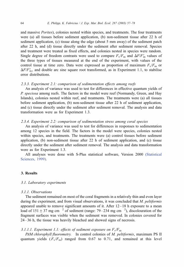
3.1.1. Observations
The sediment remained on most of the coral fragments in a relatively thin and even layer
during the experiment, and from visual observations, it was concluded that M. peltiformis
appeared unable to remove significant amounts of it. After 12–18 h exposure to a mean
load of 151F 37 mg cm� 2 of sediment (range: 79–234 mg cm� 2), discolouration of the
fragment surfaces was visible when the sediment was removed. In colonies covered for
24–36 h, the tissue was heavily bleached and showed signs of necrosis.
3.1.1.1. Experiment 1.1: effects of sediment exposure on Fv/Fm
PAM chlorophyll fluorometry. In control colonies of M. peltiformis, maximum PS II
quantum yields (Fv/Fm) ranged from 0.67 to 0.71, and remained at this level
E. Philipp, K. Fabricius / J. Exp. Mar. Biol. Ecol. 287 (2003) 57–7864

throughout the experiment. In fragments covered with sediment, Fv/Fm declined
consistently and reached levels below 0.1 in most colonies after 36 h coverage (Fig.
3A). Both exposure time and treatment (sedimentation versus control) significantly
influenced the photosynthetic activity (Table 2).
Chlorophyll concentrations and zooxanthellae density. Sediment-covered fragments
showed a continuous decline both in numbers of zooxanthellae and chlorophyll con-
centration per unit surface area with time after the onset of the experiment (Fig. 3B,C).
The chlorophyll concentration in control corals ranged from 4.0 to 8.6 Ag cm� 2 colony
surface area, whereas those exposed to sediment for 36 h contained 1.3–2.5 Ag cm� 2.
Zooxanthellae densities of controls were 2.6–4.1�106 cells cm� 2. In contrast, densities
Table 2
Analysis of variance for photosynthetic yields ( Fv/Fm) in M. peltiformis in response to treatment (control versus
sediment) and duration of exposure (time)
df F Pr(F)
Treatment 1 455.8 < 0.0001
Time 4 58.23 < 0.0001
Treatment:Time 4 80.40 < 0.0001
Residuals 60
Sums of squares are sequential.
Fig. 3. Effects of short-term sedimentation on the photophysiology of M. peltiformis. White circles: control
corals without sediment; black circles: corals covered with 151F 37 mg cm� 2 sediment. Data are meansF SE.
(A) Fv/Fm (backtransformed), measured by PAM chlorophyll fluorometry (N= 4–11 colony fragments per
treatment, 15 measurements per fragment). (B) Zooxanthellae density (N= 4–7 fragments per treatment, two
subsamples with eight zooxanthellae counts each per subsample and fragment). (C) Chlorophyll concentration
per unit surface area (N= 4–7 fragments per treatment, three replicates per fragment).
E. Philipp, K. Fabricius / J. Exp. Mar. Biol. Ecol. 287 (2003) 57–78 65

fell to 1.5–1.9� 106 cells cm� 2 after 36 h sediment exposure. Chlorophyll concen-
trations were strongly, linearly related to zooxanthellae densities in the tissues of M.
peltiformis (R2 = 0.82, F(3,43) = 56.6, P < 0.0001), thus, the chlorophyll content per
zooxanthellae did not change in response to the treatments. The ratio of yields and
zooxanthella numbers of sediment-exposed corals declined monotonously with time
(R2 = 0.44, F(1,19) = 14.88, P= 0.001), and the same pattern was found for the ratio of
yields and chlorophyll, which also decreased monotonously within 36 h (R2 = 0.42,
F(1,19) = 13.86, P= 0.0015). After 12 h exposure, yields were 82% of control values,
whereas the number of zooxanthellae was still 94%. After 18 h, maximum PS II quantum
yields were reduced to only 61% of controls, and the number of zooxanthellae was
around 81% of control values. Fv/Fm reached baseline levels when the tissue still
contained about 25% of initial zooxanthellae and chlorophyll concentrations. In the
control corals, no significant change in chlorophyll concentration and zooxanthellae
density was observed during the experimental run.
Dose–time relationship. The amount of sediment (dose) retrieved from the colonies
varied from 79 to 234 mg cm� 2 (mean = 151 mg cm� 2). A series of linear models were
used to further explore the relationship between Fv/Fm and dose and time. The initial
model treated time as a factor and dose as numeric, and comprised different slopes and
intercepts for each level of time. The slopes of this model were significantly different
(F(3,61) = 47.7, P < 0.0001), but the intercepts were not (F(4,61) = 1.49, P= 0.246). A
common intercept model was fitted and the four slopes of the time groups increased
linearly (Fig. 4A). This suggested that the decline of Fv/Fm could be explained as a
linear function of dose and dose� time, and a test of this model against the four slopes
model confirmed this (F(2,65) = 0.62, P= 0.542). Finally, dose was dropped from the
model (F(1,66) = 2.62, P= 0.111), thus, the model simplified to decreasing Fv/Fm being a
linear function of dose� time (henceforth ‘‘exposure’’; Fig. 4B). The same model (linear
change with only exposure in the model) was best for explaining the numbers of
zooxanthellae and chlorophyll concentrations (not shown). For all three models, the
response depended only on the product of dose and time, and thus, for example, 12 h
exposure to 200 mg cm� 2 was equivalent to 24 h of 100 mg cm� 2. The photo-
physiological stress in corals exposed to high levels of sedimentation for a short time
period is therefore similar to that of corals exposed to low levels of sedimentation for
extended periods of time.
3.1.1.2. Experiment 1.2: effects of exposure on recovery. All fragments of M. peltifor-
mis, which were exposed to sediments for 12 and 18 h, recovered to normal Fv/Fm ratios
within 24–36 h (Fig. 5A,B). After 24 h of exposure, one coral fragment loaded with
107 mg cm� 2 sediment fully recovered to initial values within 48 h. Two other
fragments, loaded for 24 h with 109 and 138 mg cm� 2, respectively, showed further
declines in Fv/Fm after the sediment was washed off, and died (Fig. 5C). Coral
fragments covered with sediment for 36 h did not recover from the treatment.
3.1.1.3. Experiment 1.3: effects on tissues adjacent to a sediment deposit. In the
laboratory experiments, about 50% of the surfaces of four M. peltiformis were covered
by sediment for 48 h, the other surfaces remained sediment-free. Effects were confined
E. Philipp, K. Fabricius / J. Exp. Mar. Biol. Ecol. 287 (2003) 57–7866

Fig. 4. Photosynthetic yields in M. peltiformis in response to sedimentation. (A) Fv/Fm (backtransformed) as a
function of the amount of sediment deposited and duration of sedimentation application (12 h: black squares; 18 h:
white squares; 24 h: black triangles; 36 h: white triangles). (B) Fv/Fm (backtransformed) as a function of sediment
exposure (duration� dose). The solid line represents the mean; the dashed lines represent upper and lower 90%
confidence intervals.
E. Philipp, K. Fabricius / J. Exp. Mar. Biol. Ecol. 287 (2003) 57–78 67

to the tissue directly exposed to sedimentation, while tissue about 5 mm away from a
sediment patch showed no difference in Fv/Fm or colouration to control fragments at
time zero and after 48 h (Fig. 6). The same result was obtained in light-adapted E.
lamellosa and massive Porites (Fig. 6) after 22 h of exposure in the field. In all
Fig. 5. Recovery of Fv/Fm in M. peltiformis after 12 (A), 18 (B), and 24 h (C) sediment treatment, measured by
PAM chlorophyll fluorometer. White circles: Fv/Fm in control corals (means of 4–11 fragments, each represented
by about 15 measurements). Black circles: Fv/Fm of individual sediment-treated fragments (each represented by
about 15 measurements). The dashed vertical line indicates the sediment removal and the beginning of the
postburial time. Error bars are omitted for clarity.
E. Philipp, K. Fabricius / J. Exp. Mar. Biol. Ecol. 287 (2003) 57–7868

species, effective quantum yields (DF/FmV ) were indistinguishable between tissues
adjacent to a sediment patch at the end of the experiment, tissues away from the
sediment at the end of the experiment, and tissues prior to the sediment application
(Fig. 6, Table 3).
3.2. Field experiments
3.2.1. Observations
Similar to the laboratory experiments, some of the corals, e.g., Porites massive, E.
lamellosa, or Merulina scabricula, were heavily bleached and showed signs of necrosis.
E. lamellosa and Montipora crassituberculata developed particularly thick and sticky
mucus underneath the sediment, which made it difficult for the divers to fan off the
sediment.
3.2.1.1. Experiment 2.1: comparison of sedimentation effects among reefs. The photo-
physiological stress response to sedimentation was similar among colonies of P.
speciosa and did not vary among three reefs despite different levels of sedimentation
and turbidity (Normandy Island being the most turbid and Green Island the clearest
Fig. 6. Effects of sediment exposure on DF/FmV in tissues of E. lamellosa, Porites massive, and Fv/Fm in M.
peltiformis. Data are backtransformed mean DF/FmV or Fv/FmF SE (three to four colonies per species).
Abbreviations, C0: tissues before sediment exposure (white bars); C22 or 48: control areas at the end of the
experiment after 22 or 48 h (dense-structured), E22 or 48: tissue along the boundary of a sediment patch (light-
structured); and S22 or 48: tissue that was sediment-covered for 22 or 48 h (black). About 30 DF/FmV or Fv/Fm
readings were taken from each colony and treatment.
E. Philipp, K. Fabricius / J. Exp. Mar. Biol. Ecol. 287 (2003) 57–78 69

reef, K. Fabricius, unpublished data). Twenty-two hours of sedimentation had
significant effects on effective quantum yields (interaction treatment and time), but
both baseline DF/FmV and the reduction in DF/FmV after sediment exposure neither
differed among reefs nor among colonies (Table 4, Fig. 7). Similarly, neither
differences among reefs nor colonies were found in other coral species (massive
Porites, E. lamellosa, T. peltata, F. crassa, G. fascicularis; data not shown).
Table 4
Comparison of photosynthetic yields (DF/FmV ) in colonies of P. speciosa growing on three reefs of the Great
Barrier Reef (Normandy Island, Green Island, Hay Island; see Fig. 7)
df F Pr( F)
Reef 2 0.465 0.658
Colonies/reef 4 4.23 0.395
Treatments 2 134 < 0.0001
Reef:Treatments 4 2.47 0.129
Residuals 8
Table 3
Effects of sedimentation on the photosynthetic yields of coral tissues
(A) df F Pr( F)
Species 2 4.986 0.0349
Colonies/species 9 4.5751 0.0426
Treatment 3 251.6 < 0.0001
Species:Treatment 6 9.111 < 0.0001
Residuals 25
(B) Montipora peltiformis Echinopora lamellosa Massive Porites
(a) Cend Mean diffF SE � 0.079F 0.047 0.086F 0.041 � 0.0026F 0.041
T value � 1.661 2.094 � 0.0639
P 0.109 0.047 0.950
(b) Eend Mean diffF S.E. � 0.050F 0.046 0.011F 0.041 � 0.017F 0.041
T value � 1.084 0.2659 � 0.425
P 0.289 0.793 0.674
(c) Send Mean diffF S.E. � 0.78F 0.046 � 0.393F 0.041 � 0.477F 0.041
T value � 16.99 � 9.594 � 11.66
P < 0.0001 < 0.0001 < 0.0001
(A) Analysis of variance testing for differences in yields between four treatments (underneath the sediment patch
at the end of the experiment, along the edges of the sediment patches at the end of the experiment, controls before
sediment application, and controls at the end of the experiment away from the sediment patch), and in three
species of corals (M. peltiformis, E. lamellosa, and massive Porites). (B) Single-contrast analysis of variances
comparing control yields at the beginning of the experiment with yields at the end of the experiment of (a) tissue
that remained free of sediment (Cend), (b) tissues not exposed to sediment along the edge of the sediment patch
(Eend), and (c) tissue covered by sediment (Send).
E. Philipp, K. Fabricius / J. Exp. Mar. Biol. Ecol. 287 (2003) 57–7870

3.2.1.2. Experiment 2.2: comparison of sedimentation stress among coral species. Sedi-
ment was added to 12 common coastal species of scleractinian corals at 6–8 m depth on
coastal reefs (Table 5). In these species, DF/FmV ranged from 0.63 to 0.73 at the beginning
of the experiment and did not differ from those of the controls at the end of the experiment
(Fig. 8).
Most corals were unable to remove the sediment within the 22 h of experimental
run. Nine of the species showed minor to massive photophysiological stress responses
to sedimentation, with DF/FmV being significantly reduced in the sediment-covered
areas (Fig. 8, Table 6). DF/FmV reduction was greatest in E. lamellosa, P. speciosa,
massive Porites, Turbinaria reniformis, and M. crassituberculata. In these species,
Table 5
List of coral species investigated and reefs on the Great Barrier Reef where field measurements were conducted
Species Reef Species Reef
Echinopora lamellosa N, W Merulina scabricula H
Fungia crassa N, G Pachyseris speciosa N, G, W
Galaxea fascicularis N, G Pectinia lactuca H
Montipora crassituberculata H massive Porites N, W, H
Montipora danae W Turbinaria peltata W, H
Montipora tuberculosa G Turbinaria reniformis N
N=Normandy Island, G=Green Island, W=Wilkie Island, H =Hay Island.
Fig. 7. Effects of 22 h sedimentation on the effective yield of P. speciosa at different reefs (see Table 5).
Abbreviations and symbols, C0: tissues before sediment exposure (white bars); C22: control areas at the end of the
experiment after 22 h (structured); and S22: underneath the sediment (black). Data are backtransformed
meansF SE of four to six colonies per reef. About 30 DF/FmV readings were taken from each colony and
treatment. N =Normandy Island, G =Green Island, H=Hay Island.
E. Philipp, K. Fabricius / J. Exp. Mar. Biol. Ecol. 287 (2003) 57–78 71

DF/FmV values on sediment-loaded areas were only about 50% of control areas. Three
species showed no differences in DF/FmV between the sediment-treated and control
areas. These were G. fascicularis and F. crassa, which were both able to clean off
the sediment, and Pectinia lactuca, which inflated the underlying tissues. The effects
of sedimentation in three species of Montipora (the genus used in the laboratory
experiments) were by no means extreme compared with other species.
Table 6
Results of an analysis of variance comparing changes in photosynthetic yields (DF/FmV) in response to
sedimentation among 12 coral species in the field
df F Pr( F)
Species 11 25.8917 < 0.0001
Colonies/Species 46 15.311 < 0.0001
Treatment 2 294.1645 < 0.0001
Species:Treatment 22 11.6751 < 0.0001
Residuals 92
DF/FmV in tissues not exposed to sediment (control areas before and after 22 h of sediment application, and the
sediment area before sediment application) varied greatly from yields in the sediment-exposed tissues. DF/FmVwere not significantly different in controls before and after 22 h of sediment exposure in any of the 12 species,
however DF/FmV varied significantly among colonies within some of the species, and among species.
Fig. 8. Effects of 22 h sediment loading on the effective yields of 12 common inshore hard corals species,
measured by PAM chlorophyll fluorometry in situ. The species names are listed in Table 5. Data are
backtransformed mean DF/F mVF SE (N= 2–11 colonies per species) of: tissues before sediment exposure (white
bars); tissues not exposed to sediments after 22 h at the end of the experiment (structured); and areas directly
underneath the sediment (black). A mean of 30 measurements was taken from each colony and treatment. Species
marked with asterisks showed significant changes in DF/F mV due to sedimentation.
E. Philipp, K. Fabricius / J. Exp. Mar. Biol. Ecol. 287 (2003) 57–7872

4. Discussion
Short-term exposure to sedimentation under laboratory conditions severely affected
the quantum yield of photosystem II, chlorophyll a and c2 concentrations, and
zooxanthellae densities in M. peltiformis. The corals responded initially with discolou-
ration. Severe bleaching and tissue necrosis occurred within 24–36 h of sediment
exposure, indicating that such sedimentation inflicted irreversible damage to both the
zooxanthellae and the animal. Comparisons with 12 other coral species in the field
showed that the responses of M. peltiformis were typical for corals inhabiting turbid
coastal reefs.
Large differences among species in their ability to reject and survive from deposition
of fine and coarse sediments have been described in previous studies (Lasker, 1980;
Abdel-Salam and Porter, 1988; Rogers, 1983; Stafford-Smith and Ormond, 1992;
Wesseling et al., 1999). Prolonged and severe sedimentation can lead to a shift in the
species composition of reef communities (Dodge and Vaisnys, 1977). Bleaching and
tissue necrosis caused by sediment loads of 30–4000 mg cm� 2 for 2–40 days have
been reported for scleractinian and alcyonacean corals (Bak and Elgershuizen, 1976;
Rogers, 1983; Hodgson, 1990; Riegl, 1995; Wesseling et al., 1999). Our field measure-
ments showed that the changes in photosynthetic yields in response to fine, muddy
sediments differed also among common coastal corals. Particularly sensitive were foliose
corals or corals with relatively small polyps, such as E. lamellosa, Montipora spp., and
massive Porites. Sediment was firmly lodged on concave or flat coral surfaces, and the
polyps appeared unable to remove or shift the sediment. In many of these species, DF/
FmV was significantly reduced after less than 24 h of sediment exposure. In contrast,
corals with bigger calyces or a more open skeletal structure, like F. crassa, G.
fascicularis, or P. lactuca, were not affected by sedimentation. These corals were either
able to clean off the sediment (F. crassa, G. fascicularis) or inflate the underlying tissue,
probably to increase a gas exchange through the uncovered parts (P. lactuca).
The specific causes of the severe decline in Fv/Fm in corals exposed to sedimentation
remain unknown to date. Fv/Fm naturally varies by a few percent with light availability,
declining from 0.62 to 0.57 after 4 h darkness, and increasing again with exposure to
light or oxygen-enriched water (Jones and Hoegh-Guldberg, 2001), confirming that the
availability of light and oxygen are major factors influencing the photosynthetic activity.
However, sedimentation has significant energetic implications for corals, as it reduces
photo- and heterotrophic energy acquisition, and increases energy expenditure. A
sediment cover of 100 mg cm� 2 reduces the available light by 75% (Riegl and Branch,
1995), depressing gross and net photosynthesis as well as oxygen evolution (Shashar et
al., 1993; Kuhl et al., 1995; Riegl and Branch, 1995; Gardella and Edmunds, 1999).
Sediment that interferes with the prey-capturing polyps also compromises the corals’
ability to heterotrophic energy gain. Furthermore, the energy required to produce mucus
in corals, normally f35% of the daily net photosynthesis, increases to 65% at a
sediment load of 200 mg cm� 2 (Riegl and Branch, 1995). Consequently, the energy
budget of a coral may reach the compensation point or even a deficit (Riegl and Branch,
1995; Yentsch et al., 2002). The negative energetic consequences of long-term exposure
to sedimentation will be particularly severe for corals growing in deeper water where
E. Philipp, K. Fabricius / J. Exp. Mar. Biol. Ecol. 287 (2003) 57–78 73

photosynthetic energy gain is low and sediment deposition is high (Anthony and
Fabricius, 2000).
The copious amounts of mucus, which were produced underneath the sediment by
some corals, have often been interpreted as a cleaning mechanism. However, E.
lamellosa and M. crassituberculata, which had particularly thick and sticky mucus,
were among the most severely sediment-affected species. Our observations therefore
support previous suggestions that mucus trapping of small particles on the coral surface
contributes to anoxia and thus the death of underlying tissues (Bak and Elgershuizen,
1976; Stafford-Smith and Ormond, 1992). Sediment-covered corals treated with tetracy-
cline suffer less from bleaching and tissue necrosis than sediment-covered corals not
supplied with this antibiotic, which showed necrosis after 2 days and died after 7 days
(Hodgson, 1990). This suggests that the expulsion of zooxanthellae and tissue necrosis
may not be related to light reduction, whereas tetracycline-sensitive microorganisms and
their metabolic products in the sediment-mucus layer may significantly contribute to it.
Our study showed that the photosynthetic activity in M. peltiformis decreased linearly
with both the amount of sedimentation and the time it remained on the tissues, which
indicates that any threshold value for sedimentation tolerance has to incorporate both
amount and time. The threshold level of M. peltiformis for recovery from sedimentation
was around 24 h� 0.11 gc 2.5 g h cm� 2. Thus, that within the exposure range tested,
low levels of sedimentation for extended periods of time altered the photophysiology of
these corals as much as exposure to high amounts of sediment for short periods of time.
For example, half the amount of sedimentation for twice the time had the same
photophysiological effect as twice the amount of sediment for half the time. The linear
effect of time and dose also indicated that planned sediment-suspending human activities
such as dredging would cause least damage if they were to be conducted during
conditions that promote resuspension (rather than deposition) of sediment from coral
surfaces, e.g., if calm weather periods with low waves and weak tidal currents are
avoided.
M. peltiformis was able to recover to normal Fv/Fm values within 24–36 h after the
removal of the sediment, if the duration was short ( < 24 h) or if doses were low.
Recovery was possible even when yields had temporarily dropped to 60%, and
zooxanthellae counts to 80% of initial numbers. Yields generally declined more rapidly
than the number of zooxanthellae, and reached a baseline level when about 25% of
zooxanthellae were still present. The linear correlation between chlorophyll concen-
trations and zooxanthellae densities demonstrated that chlorophyll contents per zoox-
anthellae remained unaltered, thus, reductions in zooxanthellae numbers, probably due to
the expulsion of zooxanthellae, were responsible for the drop in tissue chlorophyll
concentrations. Bleaching of corals (the loss of zooxanthellae from the host tissue) is
known as a stress response to a range of environmental conditions such as high
temperature, cyanide exposure, low salinity, or sedimentation (Bak, 1978; Thompson,
1980; Neudecker, 1981; Dallmeyer et al., 1982; Rogers, 1990; Jokiel and Coles, 1990).
In a study of cyanide and heat-stressed corals, which showed a fast recovery of quantum
yields, but a reduction in zooxanthellae densities, Jones et al. (1999) argued that a
selective expulsion of damaged zooxanthellae (with lower yield) might give the
impression of recovery by increasing the proportion of healthy algae (with higher
E. Philipp, K. Fabricius / J. Exp. Mar. Biol. Ecol. 287 (2003) 57–7874

yields) in the remnant algal population. This finding could explain the fast recovery of
Fv/Fm in M. peltiformis in the present study, however, further research is necessary to
understand the mechanisms leading to decreases in zooxanthellae yields and to death or
fast recovery in coral tissue.
Several reef around the world, including the fringing reefs of Magnetic Island where the
experimental corals were collected, experience sedimentation rates in excess of 200 mg
cm� 2 day� 1 for periods of days (Maragos, 1972; Mapstone et al., 1989; Cortes and Risk,
1984; Riegl, 1995). The sediment exposure threshold of 2.5 g h� 1 cm� 2 forM. peltiformis
would be reached if a full day’s deposition of sediment remained on the surface for a
further 12 h. Such exposure is feasible in particular on leeward reefs and at deeper depths
where resuspension is low. And indeed, few corals grow on the leeward sides of Magnetic
Island and similar reefs, and coral cover is often < 10% below 8 m depth on their
windward sides (K. Fabricius, personal observations).
On offshore reefs of the Great Barrier Reef, corals are not commonly exposed to natural
sedimentation levels as high as those applied by us experimentally (151F 37 mg cm� 1).
However, much higher sedimentation rates occur in areas of human activities (Rogers,
1990), and increasing sedimentation, resulting from anthropogenic sources, is of serious
concern for coastal coral reefs worldwide (Wilkinson, 2000; Yentsch et al., 2002; Burke et
al., 2002). Dredging and drilling suspend high levels of sediments, which can settle locally
on the benthos (Bak, 1978). Sediment is resuspended by bottom trawl fishery (Riemann
and Hoffman, 1991), and their potential contribution to the degradation of some inshore
reefs has so far been little studied. However, of greatest concern are the terrigenous
sediments eroding from cleared land (Burke et al., 2002). River plumes pass over large
proportions of coastal coral reefs of the central Great Barrier Reef, carrying typically 5–50
mg l� 1 of fine, organically, and nutrient-enriched suspended sediments, with maximum
concentrations of up to 300 mg l� 1 close to river mouths (Devlin et al., 2001). Effects of
the freshly imported muddy sediments on benthic organisms can be more detrimental than
that of old nutrient-poorer resuspended sea floor sediment, possibly due to enhanced
organic and nutrient contents that promote microbial growth, stickiness, and flocculation
(Fabricius and Wolanski, 2000; Fabricius et al., in press).
Bleached patches caused by natural sedimentation were observed on a small
proportion of corals during our study on the turbid inshore reefs of the GBR.
Sediment-covered patches were particularly noticeable on colonies with concave surfaces
such as Montipora spp. or E. lamellosa, and in grooves of massive Porites. Underneath
the sediment, we found either bleached tissue or, more commonly, dead patches,
whereas the surrounding tissues appeared to be healthy. Another commonly observed
apparent outcome of sedimentation was colony fragmentation in foliose corals, when the
central concave colony area was covered by sediment and dead, whereas the more
peripheral parts survived.
Our experiments demonstrated the ability of corals to compartmentalise sedimentation
stress, as yields only from tissues directly underneath the sediment suffered, whereas those
from adjacent clean tissues did not change measurably. Such compartmentalisation may be
an important survival mechanism for corals growing in turbid environments: even if
proportions of a colony are killed by sedimentation, the remaining colony surfaces remain
physiologically unaffected and continue to contribute to the coral community.
E. Philipp, K. Fabricius / J. Exp. Mar. Biol. Ecol. 287 (2003) 57–78 75

Acknowledgements
The study was funded by the Commonwealth of Australia Cooperative Research
Centres Program through the Cooperative Research Centre for the Great Barrier Reef
World Heritage Area. It was supported by the Australian Institute of Marine Science and
the Deutsche Akademische Austauschdienst (German Academic Exchange Organisation,
DAAD). Many thanks to Glenn De’ath for his help with the statistical analyses. Thanks
also to Emre Turak for helping to identify the hard coral species. We greatly appreciate
comments and suggestions on the manuscript by David Barnes, Jon Brodie, and Kai
Bischof. [SS]
References
Abdel-Salam, H.A, Porter, J.W., 1988. Physiological effects of sediment rejection on photosynthesis and respi-
ration in three Caribbean reef corals. Proc. 6th Int. Coral Reef Symp., Townsville, vol. 2, pp. 285–292.
Anthony, K.R.N., Fabricius, K.E., 2000. Shifting roles of heterotrophy and autotrophy in coral energetics under
varying turbidity. J. Exp. Mar. Biol. Ecol. 252, 221–253.
Babcock, R., Davies, P., 1991. Effects of sedimentation on settlement of Acropora millepora. Coral Reefs 9,
205–208.
Bak, R.P.M., 1978. Lethal and sublethal effects of dredging on reef corals. Mar. Pollut. Bull. 9, 14–17.
Bak, R.P.M., Elgershuizen, J.H.B.W., 1976. Patterns of oil-sediment rejection in corals. Mar. Biol. 37, 105–113.
Beer, S., Ilan, M., 1998. In situ measurements of photosynthetic irradiance responses of two Red Sea sponges
growing under dim light conditions. Mar. Biol. 131, 613–617.
Brodie, J., 1995. Terrestrial runoff and the Great Barrier Reef. Australas. Sci. 16, 23–25.
Burke, L., Selig, E., Spalding, M., 2002. Reefs at Risk in Southeast Asia. World Resources Institute, Cambridge.
72 pp.
Cortes, N.J., Risk, M.J., 1984. The coral reef of the Cahuita National Park, Costa Rica. Revista De Biologia
Tropical. San Jose Rev. Biol. Trop. 32, 109–121.
Cortes, N.J., Risk, M.J., 1985. A reef under siltation stress: Cahuita, Costa Rica. Bull. Mar. Sci. 36, 339–356.
Dallmeyer, D.G., Porter, J.W., Smith, G.J., 1982. Effects of particulate peat on the behaviour and physiology of
the Jamaican reef-building coral Montastrea annularis. Mar. Biol. 68, 229–233.
Devlin, M., Waterhouse, J., Taylor, J., Brodie, J., 2001. Flood plumes in the Great Barrier Reef: spatial and
temporal patterns in composition and distribution. Great Barrier Reef Marine Park Authority Report, Towns-
ville.
Dodge, R.E., Vaisnys, J.R., 1977. Coral populations and growth patterns: responses to sedimentation and
turbidity associated with dredging. J. Mar. Res. 35, 715–730.
Fabricius, K.E., Wolanski, E., 2000. Rapid smothering of coral reef organisms by muddy marine snow. Estuar.
Coast. Shelf Sci. 50, 120–150.
Fabricius, K.E., Wild, C., Wolanski, E., Adele, D., in press. Effects of transparent exopolymer particles (TEP) and
muddy terrigenous sediments on the survival of hard coral recruits. Estuar. Coast. Shelf Sci. 57 (5).
Gardella, D.J., Edmunds, P.J., 1999. The oxygen microenvironment adjacent to the tissue of scleractinian
Dichocoenia stokesii and its effects on symbiont metabolism. Mar. Biol. 135, 289–295.
Gilmour, J., 1999. Experimental investigation into the effects of suspended sediment on fertilisation, larval
survival and settlement in a scleractinian coral. Mar. Biol. 135, 451–462.
Guilcher, A., 1985. Nature and human changes of sedimentation in lagoons behind Barrier Reefs in the humid
tropics. Proc. 5th Int. Coral Reef Congr., Tahiti, vol. 4, pp. 207–212.
Hodgson, G., 1990. Tetracycline reduces sedimentation damage to corals. Mar. Biol. 104, 493–496.
Hubbard, D.K., 1986. Sedimentation as a control of reef development: St. Croix, U.S.V.I. Coral Reefs 5,
117–125.
E. Philipp, K. Fabricius / J. Exp. Mar. Biol. Ecol. 287 (2003) 57–7876

Jeffrey, S.W., Humphrey, G.F., 1975. New spectrophotometric equitations for determining chlorophylls a, b, c1and c2 in higher plants, algae and natural phytoplankton. Biochem. Physiol. Pflanzen 167, 191–194.
Jokiel, P.L., Coles, S.L., 1990. Response of Hawaiian and other Indo Pacific reef corals to elevated temperature.
Corals Reefs 8, 155–162.
Jones, R.J., Hoegh-Guldberg, O., 2001. Diurnal changes in the photochemical efficiency of the symbiotic
dinoflagellates (Dinophyceae) of corals: photoprotection, photoinactivation and the relationship to coral
bleaching. Plant Cell Environ. 24, 89–99.
Jones, R.J., Kildea, T., Hoegh-Guldberg, O., 1999. PAM Chlorophyll fluometry: a new in situ technique for stress
assessment in scleratinian corals, used to examine the effects of cyanide from cyanide fishing. Mar. Pollut.
Bull. 38 (10), 864–874.
Jones, R.J., Ward, S., Amri, A.Y., Hoegh-Guldberg, O., 2000. Changes in quantum efficiency of Photosystem II
of symbiotic dinoflagellates of corals after heat stress, and of bleached corals sampled after the 1998 Great
Barrier Reef mass bleaching event. Mar. Freshw. Res. 51, 63–71.
Kuehl, M., Cohen, Y., Dalsgaard, T., Joergensen, B.B., Revsbech, N.P., 1995. Microenvironment and photosyn-
thesis of zooxanthellae in scleractinian corals studied with microsensors for O sub(2), pH and light. Mar. Ecol.
Prog. Ser. 117 (1–3), 159–172.
Lasker, H.R., 1980. Sediment rejection by reef corals: the roles of behaviour and morphology in Montastrea
cavernosa (Linnaeus). J. Exp. Mar. Biol. Ecol. 47, 77–87.
Mapstone, B.D., Choat, J.H., Cumming, R.L., Oxley, W.G., 1989. The fringing reefs of Magnetic Island: benthic
biota and sedimentation. A baseline study. A report to the Great Barrier Reef Marine Park Authority, Towns-
ville, 88 pp.
Maragos, J.E., 1972. A study of the ecology of Hawaiian reef corals. PhD dissertation, University of Hawaii,
Honolulu.
Neudecker, S., 1981. Growth and survival of scleractinian corals exposed to thermal effluents at Guam. 4th Int.
Coral Reef Symp., Manila, Philippines, pp. 173–180.
Peters, E.C., Pilson, M.E.Q., 1985. A comparative study of the effects of sedimentation on symbiotic and
asymbiotic colonies of the coral Astrangia danae (Milne Edwards and Haime, 1849). J. Exp. Mar. Biol.
Ecol. 92, 215–230.
Prange, J.A., Dennison, W.C., 2000. Physiological responses of five Seagrass species to trace metals. Mar. Pollut.
Bull. 41, 327–336.
Ralph, P.J., Gademann, R., Larkum, A.W.D., Schreiber, U., 1999. In situ underwater measurements of photo-
synthetic activity of coral zooxanthellae and other reef-dwelling dinoflagellate endosymbionts. Mar. Ecol.
Prog. Ser. 180, 139–147.
Riegl, B., 1995. Effects of sand deposition on scleratinian and alcyonacean corals. Mar. Biol. 121, 517–526.
Riegl, B., Branch, G.M., 1995. Effects of sediment on the energy budgets of four scleractinian (Bourne 1900) and
five alcyonacean (Lamouroux 1816) corals. J. Exp. Mar. Biol. Ecol. 186, 259–275.
Riemann, B., Hoffman, E., 1991. Ecological consequences of dredging and bottom trawling in the Limfjord,
Denmark. Mar. Ecol. Prog. Ser. 69, 171–178.
Rogers, C.S., 1983. Sublethal and lethal effects of sediment applied to common Caribbean reef corals in the field.
Mar. Pollut. Bull. 14, 378–382.
Rogers, C.S., 1990. Responses of coral reefs and reef organisms to sedimentation. Mar. Ecol. Prog. Ser. 62,
185–202.
Schreiber, U., Schliwa, U., Bilger, W., 1986. Continuous recording of photochemical and non-photochemical
chlorophyll fluorescence quenching with a new type of modulation fluorometer. Photosynth. Res. 10,
51–62.
Shashar, N., Cohen, Y., Loya, Y., 1993. Extreme diel fluctuations of oxygen in diffusive boundary layers
surrounding stony corals. Biol. Bull. Mar. Biol. Lab., Woods Hole, vol. 185, pp. 455–461.
Stafford-Smith, M.G., 1993. Sediment-rejection efficiency of 22 species of Australian scleractinian corals. Mar.
Biol. 115, 229–243.
Stafford-Smith, M.G., Ormond, R.F.G., 1992. Sediment-rejection mechanisms of 42 species of Australian scler-
actinian corals. Aust. J. Mar. Freshw. Res. 43, 683–705.
Statistical Sciences, 1999. S-PLUS, Version 2000 for Windows Statistical Sciences, a division of Mathsoft,
Seattle, USA.
E. Philipp, K. Fabricius / J. Exp. Mar. Biol. Ecol. 287 (2003) 57–78 77

Thompson Jr., J.H., 1980. Responses of selected scleratinian corals to drilling fluids used in the marine environ-
ment. PhD Dissertation. Texas A & M University, College Station, U.S.G.S. Rep.
van Katwijk, M.M., Meier, N.F., van Loon, R., van Hove, E.M., Giesen, W.B.J.T., van der Velde, G., den Hartog,
C., 1993. Sabaki River sediment load and coral stress: correlation between sediments and condition of the
Malindi–Watamu reefs in Kenya (Indian Ocean). Mar. Biol. 117, 675–683.
Warner, M.E., Fitt, K.W., Schmidt, G.W., 1996. The effects of elevated temperature on the photosynthetic
efficiency of zooxanthellae in hospite from four different species of reef coral: a novel approach. Plant Cell
Environ. 19, 291–299.
Wesseling, I., Uychiaoco, A.J., Alino, P.M., Aurin, T., Vermaat, J.E., 1999. Damage and recovery of four
Philippine corals from short-term sediment burial. Mar. Ecol., Prog. Ser. 176, 11–15.
Wilkinson, C. (Ed.), 2000. Status of Coral Reefs of the World: 2000. Australian Institute of Marine Science,
Townsville, Australia. 364 pp.
Yentsch, C.S., Yentsch, C.M., Cullen, J.J., Lapointe, B., Phinney, D.A., Yentsch, S.W., 2002. Sunlight and water
transparency: cornerstones in coral research. J. Exp. Mar. Biol. Ecol. 268, 171–183.
E. Philipp, K. Fabricius / J. Exp. Mar. Biol. Ecol. 287 (2003) 57–7878
Copyright © 2022 FDOKUMEN