Characterization of black band disease in Red Sea stony corals
12
Characterization of black band disease in Red Sea stony corals Orit Barneah, 1 Eitan Ben-Dov, 1,2 Esti Kramarsky-Winter 3,4 and Ariel Kushmaro 1 * 1 Department of Biotechnology Engineering, Ben-Gurion University of the Negev, PO Box 653, Be’er-Sheva, 84105, Israel. 2 Achva Academic College MP Shikmim, 79800, Israel. 3 Department of Zoology, George S. Wise Faculty of Life Sciences, Tel Aviv University, Ramat Aviv, Tel Aviv, 69978, Israel. 4 Pacific Biomedical Research Center University of Hawaii at Manoa Honolulu, HI 96822, USA. Summary Microbial communities associated with black band disease (BBD) in massive stony corals from the Northern Red Sea (Eilat) were examined for the first time using molecular tools and microscopy. A high microbial diversity was revealed in the affected tissue in comparison with the healthy area of the same colony. Microscopy revealed the penetration of cyanobacteria into the coral mesoglea and adjacent tissues. Cyanobacterial sequences from Red Sea BBD-affected corals formed a cluster with sequences previously identified from black band and red band diseased corals from the Indo-Pacific and Caribbean. In addition, 11 sequences belonging to the genus Vibrio were retrieved. This group was previously documented as pathogenic to corals. Sulfate- reducing bacteria, a group known to be associated with BBD and produce toxic sulfide, were studied using specific primers for the amplification of the dissimilatory sulfite reductase gene (dsrA). This tech- nique facilitated and improved the resolution of the study of diversity of this group. All the sequences obtained were closely related to sequences of the genus Desulfovibrio and 46% showed high homology to Desulfovibrio desulfuricans. The complex nature of BBD and the lack of success in isolating a single causative agent suggest that BBD may be considered a polymicrobial disease. Introduction Black band disease (BBD) is a widespread coral disease, which infects mainly the massive-framework-building corals (Frias-Lopez et al., 2004). It was first described on reefs of Belize, the Florida Keys and Bermuda (Antonius, 1973; Garrett and Ducklow, 1975). In the following decades it has been also reported to occur in the Indo- Pacific and Red Sea (Antonius, 1985; Al-Moghrabi, 2001) and in the Great Barrier Reef (Dinsdale, 2002; Willis et al., 2004), indicating that this disease has a global distribution. A seasonal pattern of the disease has been reported with higher incidences coinciding with elevated water tem- peratures (Kuta and Richardson, 2002). Symptoms of the disease are well described and include the black band (5–30 mm wide) that moves across the surface of the coral colony causing tissue lysis leading to either partial or complete death of the colony (Viehman et al., 2006). Although this disease affects a relatively low proportion of the susceptible coral community (approximately 1%), its persistence in the environment and rate of progress across colonies (3 mm-1 cm day -1 ) make it an important threat to the reef community (Antonius, 1981; Edmunds, 1991; Carlton and Richardson, 1995; Kuta and Richard- son, 1996). Black band disease was reported from the Red Sea by Antonius (1988). The disease was encountered on six different locations in the vicinity of Jeddah port, Saudi Arabia and in Al Wajh, which is located further north. The severity of the disease was ranked from rare to moderate and its presence was found to be positively correlated to seawater pollution and temperature. Since the above- mentioned study, only limited data on the occurrence of BBD in this area have been available. In 1996 an unusual BBD outbreak was reported in the northern tip of the Gulf of Aqaba (Al-Moghrabi, 2001). A quantitative survey in this area showed that the number of infected colonies at the Aqaba industrial zone was 10 times higher that at a pro- tected site nearby (Al-Moghrabi, 2001). Despite this, to date, the microbial community associated with BBD in the Red Sea corals has not been studied. Previous studies showed that similar to other stratified microbial communities (e.g. biofilms and microbial mats), the BBD microbial mat oxygen dynamics was dominated by the photosynthetic activity of the cyanobacterium (for Received 23 January, 2007; accepted 16 March, 2007. *For correspondence. E-mail [email protected]; Tel. (+972) 8 6479024; Fax (+972) 8 6472983. Environmental Microbiology (2007) 9(8), 1995–2006 doi:10.1111/j.1462-2920.2007.01315.x © 2007 The Authors Journal compilation © 2007 Society for Applied Microbiology and Blackwell Publishing Ltd
-
Upload
independent -
Category
Documents
-
view
1 -
download
0
Transcript of Characterization of black band disease in Red Sea stony corals
Characterization of black band disease in Red Seastony corals
Orit Barneah,1 Eitan Ben-Dov,1,2
Esti Kramarsky-Winter3,4 and Ariel Kushmaro1*1Department of Biotechnology Engineering, Ben-GurionUniversity of the Negev, PO Box 653, Be’er-Sheva,84105, Israel.2Achva Academic College MP Shikmim, 79800, Israel.3Department of Zoology, George S. Wise Faculty of LifeSciences, Tel Aviv University, Ramat Aviv, Tel Aviv,69978, Israel.4Pacific Biomedical Research Center University ofHawaii at Manoa Honolulu, HI 96822, USA.
Summary
Microbial communities associated with black banddisease (BBD) in massive stony corals from theNorthern Red Sea (Eilat) were examined for the firsttime using molecular tools and microscopy. A highmicrobial diversity was revealed in the affected tissuein comparison with the healthy area of the samecolony. Microscopy revealed the penetration ofcyanobacteria into the coral mesoglea and adjacenttissues. Cyanobacterial sequences from Red SeaBBD-affected corals formed a cluster with sequencespreviously identified from black band and red banddiseased corals from the Indo-Pacific and Caribbean.In addition, 11 sequences belonging to the genusVibrio were retrieved. This group was previouslydocumented as pathogenic to corals. Sulfate-reducing bacteria, a group known to be associatedwith BBD and produce toxic sulfide, were studiedusing specific primers for the amplification of thedissimilatory sulfite reductase gene (dsrA). This tech-nique facilitated and improved the resolution of thestudy of diversity of this group. All the sequencesobtained were closely related to sequences of thegenus Desulfovibrio and 46% showed high homologyto Desulfovibrio desulfuricans. The complex nature ofBBD and the lack of success in isolating a singlecausative agent suggest that BBD may be considereda polymicrobial disease.
Introduction
Black band disease (BBD) is a widespread coral disease,which infects mainly the massive-framework-buildingcorals (Frias-Lopez et al., 2004). It was first described onreefs of Belize, the Florida Keys and Bermuda (Antonius,1973; Garrett and Ducklow, 1975). In the followingdecades it has been also reported to occur in the Indo-Pacific and Red Sea (Antonius, 1985; Al-Moghrabi, 2001)and in the Great Barrier Reef (Dinsdale, 2002; Willis et al.,2004), indicating that this disease has a globaldistribution.
A seasonal pattern of the disease has been reportedwith higher incidences coinciding with elevated water tem-peratures (Kuta and Richardson, 2002). Symptoms of thedisease are well described and include the black band(5–30 mm wide) that moves across the surface of thecoral colony causing tissue lysis leading to either partial orcomplete death of the colony (Viehman et al., 2006).Although this disease affects a relatively low proportion ofthe susceptible coral community (approximately 1%), itspersistence in the environment and rate of progressacross colonies (3 mm-1 cm day-1) make it an importantthreat to the reef community (Antonius, 1981; Edmunds,1991; Carlton and Richardson, 1995; Kuta and Richard-son, 1996).
Black band disease was reported from the Red Sea byAntonius (1988). The disease was encountered on sixdifferent locations in the vicinity of Jeddah port, SaudiArabia and in Al Wajh, which is located further north. Theseverity of the disease was ranked from rare to moderateand its presence was found to be positively correlated toseawater pollution and temperature. Since the above-mentioned study, only limited data on the occurrence ofBBD in this area have been available. In 1996 an unusualBBD outbreak was reported in the northern tip of the Gulfof Aqaba (Al-Moghrabi, 2001). A quantitative survey in thisarea showed that the number of infected colonies at theAqaba industrial zone was 10 times higher that at a pro-tected site nearby (Al-Moghrabi, 2001). Despite this, todate, the microbial community associated with BBD in theRed Sea corals has not been studied.
Previous studies showed that similar to other stratifiedmicrobial communities (e.g. biofilms and microbial mats),the BBD microbial mat oxygen dynamics was dominatedby the photosynthetic activity of the cyanobacterium (for
Received 23 January, 2007; accepted 16 March, 2007. *Forcorrespondence. E-mail [email protected]; Tel. (+972) 8 6479024;Fax (+972) 8 6472983.
Environmental Microbiology (2007) 9(8), 1995–2006 doi:10.1111/j.1462-2920.2007.01315.x
© 2007 The AuthorsJournal compilation © 2007 Society for Applied Microbiology and Blackwell Publishing Ltd

review see Carlton and Richardson, 1995). It was foundthat changes in light intensity caused changes in O2 con-centration, in turn causing an opposite trend in H2S con-centration. This resulted in alterations between oxic andanoxic state inside the BBD mat (Carlton and Richardson,1995). In spite of the similarities between BBD and othermicrobial mats, BBD is unique in that the < 1-mm-thickbiofilm migrates horizontally across a living animal sub-stratum that is subsequently killed by an anoxic, sulfide-rich microenvironment, created under the band (Carltonand Richardson, 1995). The deadly sulfide is presumablygenerated by sulfate-reducing bacteria (SRB) (Richard-son, 2004). To date only limited information is availableregarding this group involvement in the BBD.
Black band disease is believed to be caused by aconsortium of microorganisms (Antonius, 1981; Richard-son et al., 1997; Richardson, 1998; Dinsdale, 2002), andmay not have a primary pathogen (Richardson, 2004).The recent progress in molecular techniques has enabledmolecular characterization of the bacterial communityassociated with BBD (Cooney et al., 2002; Frias-Lopezet al., 2002; 2004; Sekar et al., 2006). It was found thatthe BBD microbial community is dominated, in terms ofbiomass, by filamentous cyanobacteria (Rützler andSantavy, 1983; Bythell et al., 2002; Cooney et al., 2002;Frias-Lopez et al., 2003; Richardson, 2004), numerousheterotrophic bacteria (Garrett and Ducklow, 1975;Cooney et al., 2002), sulfide-oxidizing bacteria (Ducklowand Mitchell, 1979; Viehman and Richardson, 2002), SRB(Chet and Mitchell, 1975; Garrett and Ducklow, 1975;Cooney et al., 2002; Frias-Lopez et al., 2002; Viehmanet al., 2006) and marine fungi (Ramos-Flores, 1983). Thisdiversity pattern differed according to geographic region,host coral species and methodology used (Cooney et al.,2002; Frias-Lopez et al., 2004; Sekar et al., 2006). Inspite of the fact that this disease was already identified inthe 1970s there is still a wide gap of knowledge pertainingto its etiology.
The aim of the present study therefore is to characterizethe composition and diversity of the bacteria present inthe BBD bacterial mat and in adjacent affected tissues ofcorals from Eilat (Red Sea) using a diverse set of molecu-lar techniques. To do so we ascertained general bacterialdiversity, using traditional 16S rRNA gene primers andinosine-modified 16S primers. In addition, specific primersfor the amplification of the dissimilatory sulfite reductasegene (dsrA) were used in order to determine the diversityof SRB associated with the BBD mat.
Results
Black band disease in Eilat (Red Sea) was found to affectmainly a number of massive Faviid coral genera includingFavia, Favites, Goniastrea and Platygyra (Fig. 1). Micro-
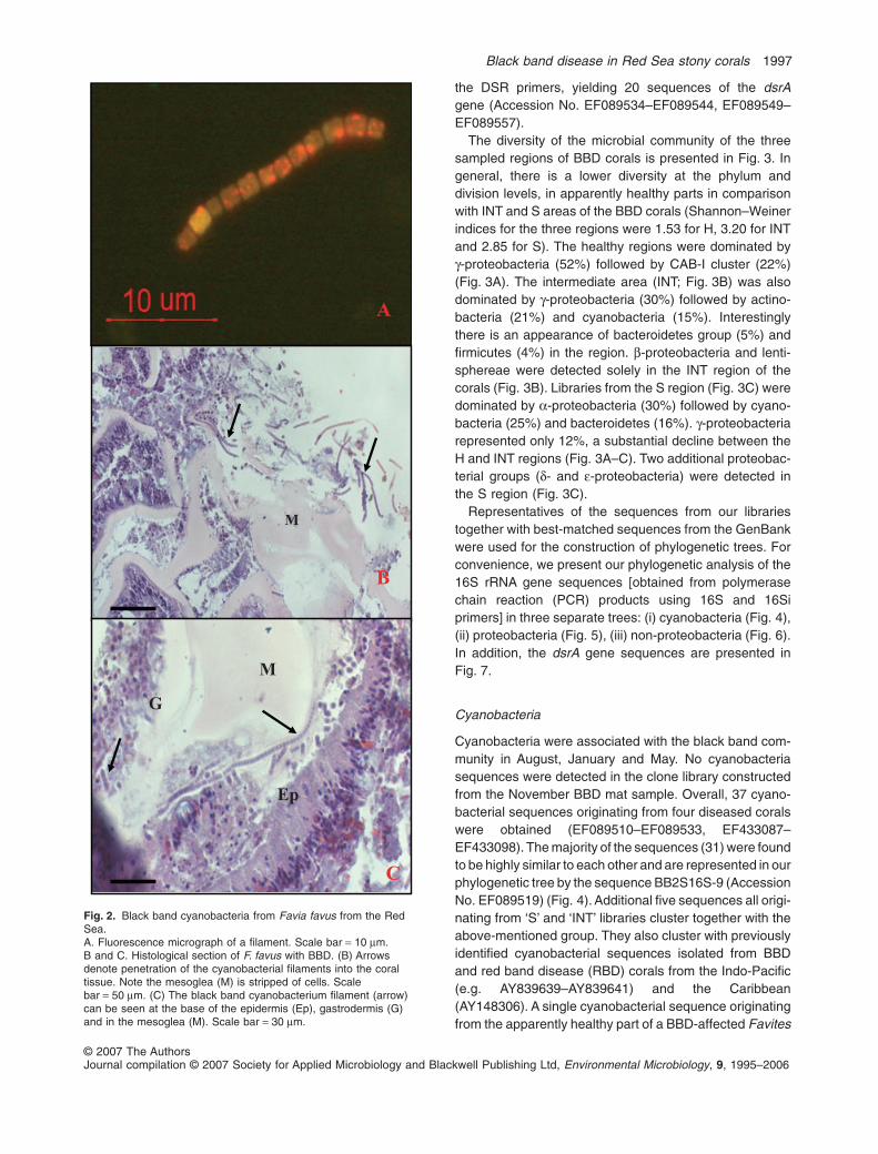
scopic observations of BBD mat samples from a Faviafavus included cyanobacteria filaments (Fig. 2A) thatexhibit typical fluorescence of the photosynthetic pigments(orange) with spectrophotometric absorption maximum at565 nm. This indicated the presence of the light-harvestingcyanobacterial pigment phycoerythrin. Histological sec-tions of the black band and underlying tissues (Fig. 2B andC) revealed an area of necrosis. The area juxtaposed to theband included intact fragments of mesoglea devoid ofadjacent cellular layers. Dense populations of filamentousmicroorganisms morphologically identified as cyanobacte-ria were found in the degraded tissue. At the interfacebetween the band and apparently healthy tissue, cyano-bacteria were found penetrating the coral tissue, in themesoglea or at the point of attachment of the tissues to themesoglea. Cells in this area also were necrotic and manypyknotic nuclei are visible (Fig. 2B and C).
We analysed the black band community and surfacemucus layer from five different diseased Faviid corals,which were sampled at different seasons (see Table 1).During this study a total of 14 16S rRNA gene librarieswere constructed (see Table 1) using universal and modi-fied 16S primers, yielding 215 sequences (AccessionNo. EF089403–EF089533, EF433087–EF433174). Eightlibraries of the 16S rRNA gene of the black band mat (S),three from the coral surface mucus layer adjacent to theband (INT) and three from the apparently healthy surfacemucus layer (H) were obtained using universal and modi-fied 16S primers. Three additional libraries were obtainedfrom INT and S areas of the corals (see Table 1) using
Fig. 1. Black band disease on Favia sp. from the Gulf of Eilat. Asthe band moves across the coral it completely degrades coraltissue and leaves behind exposed coral skeleton. Geometric formsindicate the position of different samples taken: (�) healthy region(H), (�) interface region (INT), (�) sick region (S).
1996 O. Barneah, E. Ben-Dov, E. Kramarsky-Winter and A. Kushmaro
© 2007 The AuthorsJournal compilation © 2007 Society for Applied Microbiology and Blackwell Publishing Ltd, Environmental Microbiology, 9, 1995–2006
the DSR primers, yielding 20 sequences of the dsrAgene (Accession No. EF089534–EF089544, EF089549–EF089557).
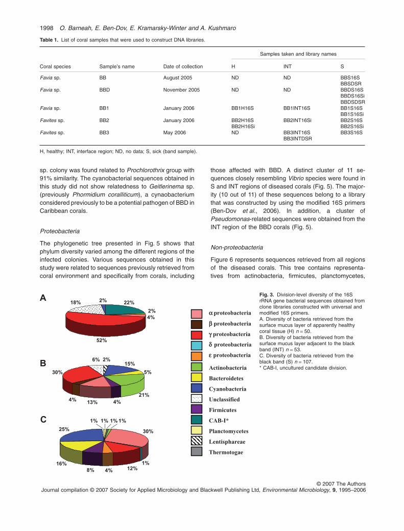
The diversity of the microbial community of the threesampled regions of BBD corals is presented in Fig. 3. Ingeneral, there is a lower diversity at the phylum anddivision levels, in apparently healthy parts in comparisonwith INT and S areas of the BBD corals (Shannon–Weinerindices for the three regions were 1.53 for H, 3.20 for INTand 2.85 for S). The healthy regions were dominated byg-proteobacteria (52%) followed by CAB-I cluster (22%)(Fig. 3A). The intermediate area (INT; Fig. 3B) was alsodominated by g-proteobacteria (30%) followed by actino-bacteria (21%) and cyanobacteria (15%). Interestinglythere is an appearance of bacteroidetes group (5%) andfirmicutes (4%) in the region. b-proteobacteria and lenti-sphereae were detected solely in the INT region of thecorals (Fig. 3B). Libraries from the S region (Fig. 3C) weredominated by a-proteobacteria (30%) followed by cyano-bacteria (25%) and bacteroidetes (16%). g-proteobacteriarepresented only 12%, a substantial decline between theH and INT regions (Fig. 3A–C). Two additional proteobac-terial groups (d- and e-proteobacteria) were detected inthe S region (Fig. 3C).
Representatives of the sequences from our librariestogether with best-matched sequences from the GenBankwere used for the construction of phylogenetic trees. Forconvenience, we present our phylogenetic analysis of the16S rRNA gene sequences [obtained from polymerasechain reaction (PCR) products using 16S and 16Siprimers] in three separate trees: (i) cyanobacteria (Fig. 4),(ii) proteobacteria (Fig. 5), (iii) non-proteobacteria (Fig. 6).In addition, the dsrA gene sequences are presented inFig. 7.
Cyanobacteria
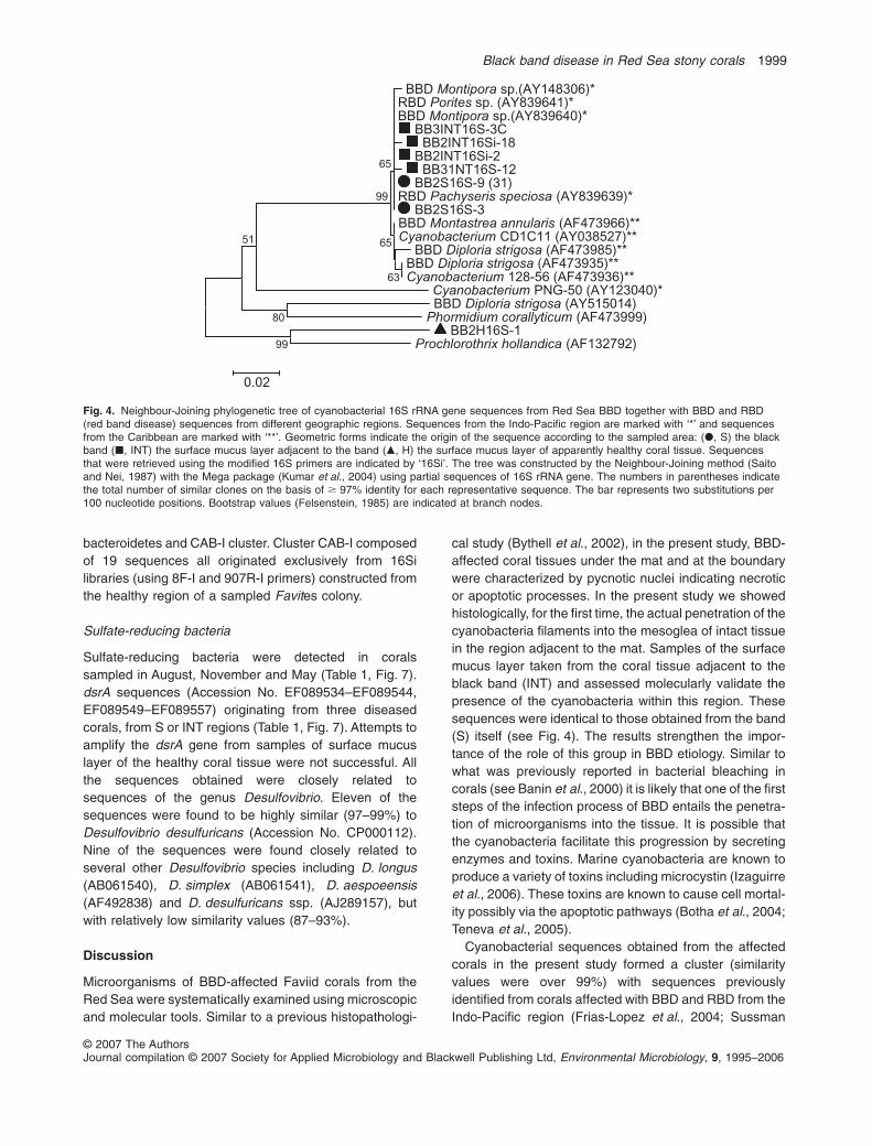
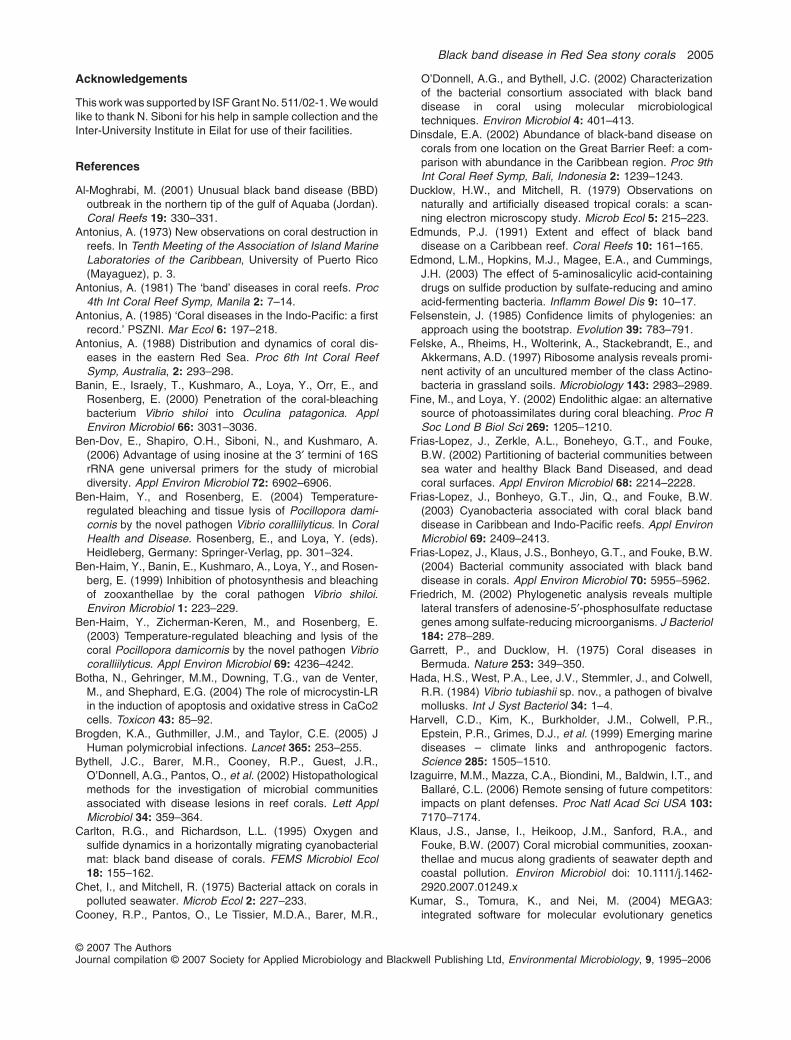
Cyanobacteria were associated with the black band com-munity in August, January and May. No cyanobacteriasequences were detected in the clone library constructedfrom the November BBD mat sample. Overall, 37 cyano-bacterial sequences originating from four diseased coralswere obtained (EF089510–EF089533, EF433087–EF433098). The majority of the sequences (31) were foundto be highly similar to each other and are represented in ourphylogenetic tree by the sequence BB2S16S-9 (AccessionNo. EF089519) (Fig. 4). Additional five sequences all origi-nating from ‘S’ and ‘INT’ libraries cluster together with theabove-mentioned group. They also cluster with previouslyidentified cyanobacterial sequences isolated from BBDand red band disease (RBD) corals from the Indo-Pacific(e.g. AY839639–AY839641) and the Caribbean(AY148306). A single cyanobacterial sequence originatingfrom the apparently healthy part of a BBD-affected Favites
B
C
Ep
G
M
M
Fig. 2. Black band cyanobacteria from Favia favus from the RedSea.A. Fluorescence micrograph of a filament. Scale bar = 10 mm.B and C. Histological section of F. favus with BBD. (B) Arrowsdenote penetration of the cyanobacterial filaments into the coraltissue. Note the mesoglea (M) is stripped of cells. Scalebar = 50 mm. (C) The black band cyanobacterium filament (arrow)can be seen at the base of the epidermis (Ep), gastrodermis (G)and in the mesoglea (M). Scale bar = 30 mm.
Black band disease in Red Sea stony corals 1997
© 2007 The AuthorsJournal compilation © 2007 Society for Applied Microbiology and Blackwell Publishing Ltd, Environmental Microbiology, 9, 1995–2006
sp. colony was found related to Prochlorothrix group with91% similarity. The cyanobacterial sequences obtained inthis study did not show relatedness to Geitlerinema sp.(previously Phormidium coralliticum), a cynaobacteriumconsidered previously to be a potential pathogen of BBD inCaribbean corals.
Proteobacteria
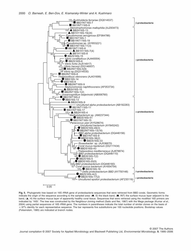
The phylogenetic tree presented in Fig. 5 shows thatphylum diversity varied among the different regions of theinfected colonies. Various sequences obtained in thisstudy were related to sequences previously retrieved fromcoral environment and specifically from corals, including
those affected with BBD. A distinct cluster of 11 se-quences closely resembling Vibrio species were found inS and INT regions of diseased corals (Fig. 5). The major-ity (10 out of 11) of these sequences belong to a librarythat was constructed by using the modified 16S primers(Ben-Dov et al., 2006). In addition, a cluster ofPseudomonas-related sequences were obtained from theINT region of the BBD corals (Fig. 5).
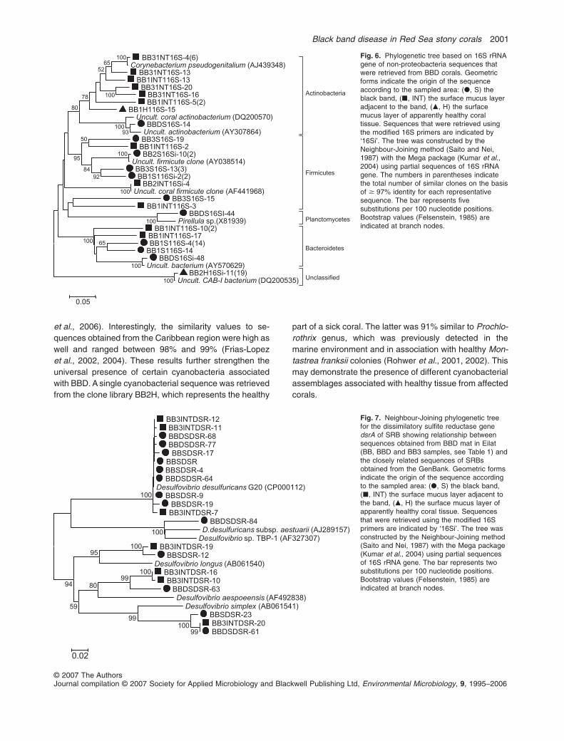
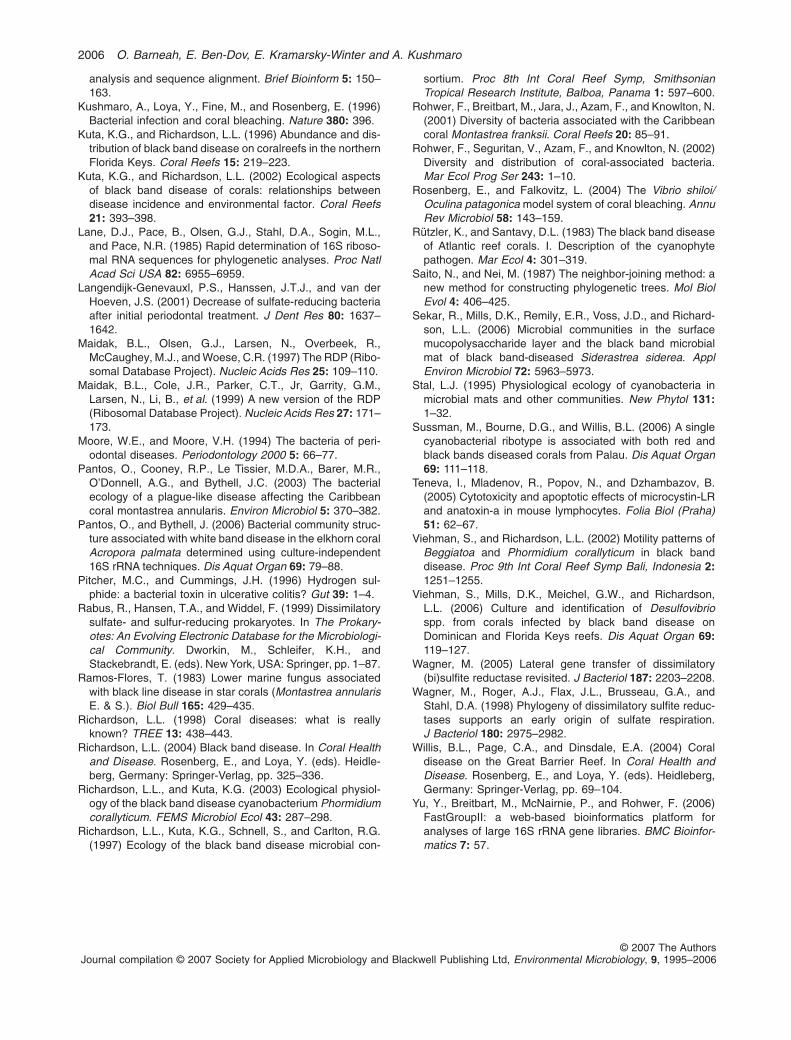
Non-proteobacteria
Figure 6 represents sequences retrieved from all regionsof the diseased corals. This tree contains representa-tives from actinobacteria, firmicutes, planctomycetes,
Table 1. List of coral samples that were used to construct DNA libraries.
Coral species Sample’s name Date of collection
Samples taken and library names
H INT S
Favia sp. BB August 2005 ND ND BBS16SBBSDSR
Favia sp. BBD November 2005 ND ND BBDS16SBBDS16SiBBDSDSR
Favia sp. BB1 January 2006 BB1H16S BB1INT16S BB1S16SBB1S16Si
Favites sp. BB2 January 2006 BB2H16SBB2H16Si
BB2INT16Si BB2S16SBB2S16Si
Favites sp. BB3 May 2006 ND BB3INT16SBB3INTDSR
BB3S16S
H, healthy; INT, interface region; ND, no data; S, sick (band sample).
Fig. 3. Division-level diversity of the 16SrRNA gene bacterial sequences obtained fromclone libraries constructed with universal andmodified 16S primers.A. Diversity of bacteria retrieved from thesurface mucus layer of apparently healthycoral tissue (H) n = 50.B. Diversity of bacteria retrieved from thesurface mucus layer adjacent to the blackband (INT) n = 53.C. Diversity of bacteria retrieved from theblack band (S) n = 107.* CAB-I, uncultured candidate division.
2%15%
5%
21%4%13%4%
30%
6%
a proteobacteria
b proteobacteria
g proteobacteria
d proteobacteria
e proteobacteria
Actinobacteria
Bacteroidetes
Cyanobacteria
Unclassified
Firmicutes
CAB-I*
Planctomycetes
Lentisphareae
Thermotogae
1% 1% 1%
30%
1%12%4%8%
16%
25%
1%C
A
B
22%
2%4%
52%
18% 2%
1998 O. Barneah, E. Ben-Dov, E. Kramarsky-Winter and A. Kushmaro
© 2007 The AuthorsJournal compilation © 2007 Society for Applied Microbiology and Blackwell Publishing Ltd, Environmental Microbiology, 9, 1995–2006
bacteroidetes and CAB-I cluster. Cluster CAB-I composedof 19 sequences all originated exclusively from 16Silibraries (using 8F-I and 907R-I primers) constructed fromthe healthy region of a sampled Favites colony.
Sulfate-reducing bacteria
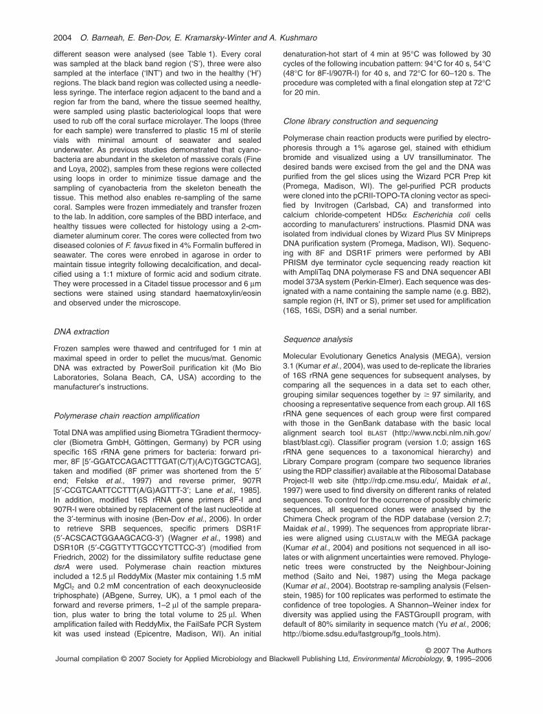
Sulfate-reducing bacteria were detected in coralssampled in August, November and May (Table 1, Fig. 7).dsrA sequences (Accession No. EF089534–EF089544,EF089549–EF089557) originating from three diseasedcorals, from S or INT regions (Table 1, Fig. 7). Attempts toamplify the dsrA gene from samples of surface mucuslayer of the healthy coral tissue were not successful. Allthe sequences obtained were closely related tosequences of the genus Desulfovibrio. Eleven of thesequences were found to be highly similar (97–99%) toDesulfovibrio desulfuricans (Accession No. CP000112).Nine of the sequences were found closely related toseveral other Desulfovibrio species including D. longus(AB061540), D. simplex (AB061541), D. aespoeensis(AF492838) and D. desulfuricans ssp. (AJ289157), butwith relatively low similarity values (87–93%).
Discussion
Microorganisms of BBD-affected Faviid corals from theRed Sea were systematically examined using microscopicand molecular tools. Similar to a previous histopathologi-
cal study (Bythell et al., 2002), in the present study, BBD-affected coral tissues under the mat and at the boundarywere characterized by pycnotic nuclei indicating necroticor apoptotic processes. In the present study we showedhistologically, for the first time, the actual penetration of thecyanobacteria filaments into the mesoglea of intact tissuein the region adjacent to the mat. Samples of the surfacemucus layer taken from the coral tissue adjacent to theblack band (INT) and assessed molecularly validate thepresence of the cyanobacteria within this region. Thesesequences were identical to those obtained from the band(S) itself (see Fig. 4). The results strengthen the impor-tance of the role of this group in BBD etiology. Similar towhat was previously reported in bacterial bleaching incorals (see Banin et al., 2000) it is likely that one of the firststeps of the infection process of BBD entails the penetra-tion of microorganisms into the tissue. It is possible thatthe cyanobacteria facilitate this progression by secretingenzymes and toxins. Marine cyanobacteria are known toproduce a variety of toxins including microcystin (Izaguirreet al., 2006). These toxins are known to cause cell mortal-ity possibly via the apoptotic pathways (Botha et al., 2004;Teneva et al., 2005).
Cyanobacterial sequences obtained from the affectedcorals in the present study formed a cluster (similarityvalues were over 99%) with sequences previouslyidentified from corals affected with BBD and RBD from theIndo-Pacific region (Frias-Lopez et al., 2004; Sussman
BBD Montipora sp.(AY148306)*RBD Porites sp. (AY839641)*BBD Montipora sp.(AY839640)*
BB3INT16S-3CBB2INT16Si-18
BB2INT16Si-2BB31NT16S-12
BB2S16S-9 (31)RBD Pachyseris speciosa (AY839639)*
BB2S16S-3BBD Montastrea annularis (AF473966)**Cyanobacterium CD1C11 (AY038527)**
BBD Diploria strigosa (AF473985)**BBD Diploria strigosa (AF473935)**Cyanobacterium 128-56 (AF473936)**
Cyanobacterium PNG-50 (AY123040)*BBD Diploria strigosa (AY515014)
Phormidium corallyticum (AF473999)BB2H16S-1
Prochlorothrix hollandica (AF132792)
63
65
65
99
80
51
99
0.02
Fig. 4. Neighbour-Joining phylogenetic tree of cyanobacterial 16S rRNA gene sequences from Red Sea BBD together with BBD and RBD(red band disease) sequences from different geographic regions. Sequences from the Indo-Pacific region are marked with ‘*’ and sequencesfrom the Caribbean are marked with ‘**’. Geometric forms indicate the origin of the sequence according to the sampled area: (�, S) the blackband (�, INT) the surface mucus layer adjacent to the band (�, H) the surface mucus layer of apparently healthy coral tissue. Sequencesthat were retrieved using the modified 16S primers are indicated by ‘16Si’. The tree was constructed by the Neighbour-Joining method (Saitoand Nei, 1987) with the Mega package (Kumar et al., 2004) using partial sequences of 16S rRNA gene. The numbers in parentheses indicatethe total number of similar clones on the basis of � 97% identity for each representative sequence. The bar represents two substitutions per100 nucleotide positions. Bootstrap values (Felsenstein, 1985) are indicated at branch nodes.
Black band disease in Red Sea stony corals 1999
© 2007 The AuthorsJournal compilation © 2007 Society for Applied Microbiology and Blackwell Publishing Ltd, Environmental Microbiology, 9, 1995–2006
Burkholderia ferrariae (DQ514537)BB31NT16S-7
BB1INT116S-9Stenotrophomonas maltophilia (AJ293473)
BB2H16S-13BB1H116S-19(24)
Pseudomonas aeruginosa (EF064786)BB31NT16S-1
BB1INT116S-19Pseudomonas sp. (AY653221)
BB31NT16S-11(3)BB1INT116S-8
BB1H116S-7(4)BB1H116S-8
BB2S16Si-15Vibrio corallilyticus (AJ440004)
BB2S16Si-6Vibrio fortis (AJ514917)Vibrio harveyi (DQ146937)
BB2INT16Si-5(9)Vibrio sp.(DQ314530)
BB2INT16Si-6Thalassolituus oleivorans (AJ431699)
BBS16S-14BBS16S-8
BB2INT16SI-9Neptunomonas naphthovorans (AF053734)
BB2H16S-15BB2INT16Si-1(2)
Oceanospirillum beijerinckii (AB006760)BB31NT16S-2
BBS16S-13BB2H16Si-9
Uncultured alpha proteobacterium (AB192283)BB1INT116S-11
BB31NT16S-17BB2H16S-4
Methylobacterium sp. (AM237344)BBDS16Si-51
BB31NT16S-15Paracoccus yeei (AY528674)
Uncultured bacterium (AY845242)BBDS16Si-49(3)
BB2INT16Si-13(16)BBD alpha proteobacterium (DQ446156)
BB31NT16S-9BBDS16Si-38(2)
BBDS16Si-33Roseobacter sp. (AJ536670)Coral mucus bacterium (DQ117434)
BBDS16Si-43Thalassobius mediterraneus (AJ878874)BBD proteobacterium (DQ446110)
BB3S16S-7(2)BB2S16S-5
BBDS16S-20(5)BBD proteobacterium (DQ446160)Coral mucus bacterium (AY654754)
BB3S16S-18Delta proteobacterium BBD (AY750148)
BB2S16Si-4(2)BB2S16Si-17(2)
Uncultured epsilon proteobacterium (AF235116)
100
62100
100
100
68100
62100
56100
100
100100
100
96
96
76
5692
99
65
50
99
99
80
98
97
97
95
93
91
85
70
80
77
65
63
58
65
99100
0.05
β-proteobacteria
γ-proteobacteria
α-proteobacteria
δ-proteobacteria
ε-proteobacteria
Fig. 5. Phylogenetic tree based on 16S rRNA gene of proteobacteria sequences that were retrieved from BBD corals. Geometric formsindicate the origin of the sequence according to the sampled area: (�, S) the black band, (�, INT) the surface mucus layer adjacent to theband, (�, H) the surface mucus layer of apparently healthy coral tissue. Sequences that were retrieved using the modified 16S primers areindicated by ‘16Si’. The tree was constructed by the Neighbour-Joining method (Saito and Nei, 1987) with the Mega package (Kumar et al.,2004) using partial sequences of 16S rRNA gene. The numbers in parentheses indicate the total number of similar clones on the basis of� 97% identity for each representative sequence. The bar represents five substitutions per 100 nucleotide positions. Bootstrap values(Felsenstein, 1985) are indicated at branch nodes.
2000 O. Barneah, E. Ben-Dov, E. Kramarsky-Winter and A. Kushmaro
© 2007 The AuthorsJournal compilation © 2007 Society for Applied Microbiology and Blackwell Publishing Ltd, Environmental Microbiology, 9, 1995–2006
et al., 2006). Interestingly, the similarity values to se-quences obtained from the Caribbean region were high aswell and ranged between 98% and 99% (Frias-Lopezet al., 2002, 2004). These results further strengthen theuniversal presence of certain cyanobacteria associatedwith BBD. A single cyanobacterial sequence was retrievedfrom the clone library BB2H, which represents the healthy
part of a sick coral. The latter was 91% similar to Prochlo-rothrix genus, which was previously detected in themarine environment and in association with healthy Mon-tastrea franksii colonies (Rohwer et al., 2001, 2002). Thismay demonstrate the presence of different cyanobacterialassemblages associated with healthy tissue from affectedcorals.
Fig. 6. Phylogenetic tree based on 16S rRNAgene of non-proteobacteria sequences thatwere retrieved from BBD corals. Geometricforms indicate the origin of the sequenceaccording to the sampled area: (�, S) theblack band, (�, INT) the surface mucus layeradjacent to the band, (�, H) the surfacemucus layer of apparently healthy coraltissue. Sequences that were retrieved usingthe modified 16S primers are indicated by‘16Si’. The tree was constructed by theNeighbour-Joining method (Saito and Nei,1987) with the Mega package (Kumar et al.,2004) using partial sequences of 16S rRNAgene. The numbers in parentheses indicatethe total number of similar clones on the basisof � 97% identity for each representativesequence. The bar represents fivesubstitutions per 100 nucleotide positions.Bootstrap values (Felsenstein, 1985) areindicated at branch nodes.
BB31NT16S-4(6)Corynebacterium pseudogenitalium (AJ439348)
BB31NT16S-13BB1INT116S-13
BB31NT16S-20BB31NT16S-16BB1INT116S-5(2)
BB1H116S-15Uncult. coral actinobacterium (DQ200570)
BBDS16S-14Uncult. actinobacterium (AY307864)BB3S16S-19
BB1INT116S-2BB2S16Si-10(2)
Uncult. firmicute clone (AY038514)BB3S16S-13(3)BB1S116Si-2(2)
BB2INT16Si-4Uncult. coral firmicute clone (AF441968)
BB3S16S-15BB1INT116S-3
BBDS16SI-44Pirellula sp.(X81939)
BB1INT116S-10(2)BB1INT116S-17BB1S116S-4(14)
BB1S116S-14BBDS16Si-48
Uncult. bacterium (AY570629)BB2H16Si-11(19)
Uncult. CAB-I bacterium (DQ200535)
100
100
100
65100
100
100
100
100
9284
50
95
6552
78
80
93100
0.05
Actinobacteria
Firmicutes
Planctomycetes
Bacteroidetes
Unclassified
Fig. 7. Neighbour-Joining phylogenetic treefor the dissimilatory sulfite reductase genedsrA of SRB showing relationship betweensequences obtained from BBD mat in Eilat(BB, BBD and BB3 samples, see Table 1) andthe closely related sequences of SRBsobtained from the GenBank. Geometric formsindicate the origin of the sequence accordingto the sampled area: (�, S) the black band,(�, INT) the surface mucus layer adjacent tothe band, (�, H) the surface mucus layer ofapparently healthy coral tissue. Sequencesthat were retrieved using the modified 16Sprimers are indicated by ‘16Si’. The tree wasconstructed by the Neighbour-Joining method(Saito and Nei, 1987) with the Mega package(Kumar et al., 2004) using partial sequencesof 16S rRNA gene. The bar represents twosubstitutions per 100 nucleotide positions.Bootstrap values (Felsenstein, 1985) areindicated at branch nodes.
BB3INTDSR-12BB3INTDSR-11BBDSDSR-68BBDSDSR-77BBSDSR-17
BBSDSRBBSDSR-4BBDSDSR-64
Desulfovibrio desulfuricans G20 (CP000112)BBSDSR-9
BBSDSR-19BB3INTDSR-7
BBDSDSR-84D.desulfuricans subsp. aestuarii (AJ289157)
Desulfovibrio sp. TBP-1 (AF327307)BB3INTDSR-19
BBSDSR-12Desulfovibrio longus (AB061540)
BB3INTDSR-16BB3INTDSR-10
BBDSDSR-63Desulfovibrio aespoeensis (AF492838)
Desulfovibrio simplex (AB061541)BBSDSR-23BB3INTDSR-20BBDSDSR-6199
100
100
100
100
99
99
95
80
59
94
100
0.02
Black band disease in Red Sea stony corals 2001
© 2007 The AuthorsJournal compilation © 2007 Society for Applied Microbiology and Blackwell Publishing Ltd, Environmental Microbiology, 9, 1995–2006

In the present study comparison of microbial communi-ties was carried out within different areas of diseasedcolonies. Results showed that similar to previous studieson BBD (see Cooney et al., 2002; Frias-Lopez et al.,2002: Sekar et al., 2006) a high microbial diversity wasrevealed in the affected tissue (INT and S) in comparisonwith the healthy (H) area of the same colony (Fig. 3). Astudy by Pantos and colleagues (2003) on plague-likedisease suggested that the microbial community of unaf-fected corals differ from those of the healthy area ofaffected corals. This phenomenon should be furtherinvestigated for BBD and other coral diseases.
Polymerase chain reaction-based techniques using tra-ditional universal 16S rRNA gene primers are limited inthe ability to reveal true diversity of microbial communities(Ben-Dov et al., 2006). This is particularly true whenstudying coral disease-associated microbial diversity. In arecent study, Pantos and Bythell (2006) stated that pref-erential carbon utilization methods suggest the presenceof Vibrio species in white band disease-affected corals,yet this genus was not detected by molecular PCR-basedtechniques using traditional 16S primers. The authors(Pantos and Bythell, 2006) specifically bring up a possiblelimitation of the 16S primer set. The use of the modifiedprimer set 8F-I/907R-I (where the 3′-terminus of bothprimers were substituted by inosine) has been provenuseful as a tool to overcome the shortcomings of universal16S rRNA gene primers (8F and 907R) (Ben-Dov et al.,2006). Using this technique we were able to retrievesequences belonging to a number of genera, in particularsequences belonging to the genus Vibrio, a group thatwas under-represented in BBD clone libraries analysed todate. In the present study the vibrionic sequencesretrieved from the S and INT libraries were closely relatedto known Vibrio species (Fig. 5). This genus includesspecies that were previously documented as pathogensof corals and other marine organisms (Kushmaro et al.,1996; Harvell et al., 1999; Ben-Haim et al., 2003). Forexample, Vibrio sequences found in the present studyclustered together with the coral pathogen Vibrio coralli-lyticus (Accession No. AJ440004), a bacterium known tocause rapid tissue lysis in the stony coral Pocilloporadamicornis by metalloproteinase at elevated tempera-tures (Ben-Haim et al., 2003; Rosenberg and Falkovitz,2004). An additional clone BB2INT16Si-7 (Accession No.EF089442) was similar (99%) to an algicide producingVibrio sp. (Accession No. AB180389). A similar capabilitywas reported for another Vibrio species, Vibrio shiloi. Thelatter, which is the causative agent of bacterial bleachingin the coral Occulina patagonica, is known to produce aproline-rich peptide that inhibits photosynthesis and a pro-tease that lyses zooxanthellae (Ben-Haim et al., 1999;Banin et al., 2000; Rosenberg and Falkovitz, 2004). As inseveral cases, virulence of Vibrio is associated with pro-
tease activity (e.g. Hada et al., 1984; Ben-Haim et al.,1999; Banin et al., 2000; Ben-Haim and Rosenberg,2004; Rosenberg and Falkovitz, 2004) it is possible thatthis group of organisms may play a key role in the etiologyof BBD. The observed characteristic lysis of coral tissue inthe disease interface may therefore be associated withproteolytic enzyme activity.
In our clone libraries, bacterial sequences were derivedfrom a wide taxonomic range, with the majority of clonesclosely related to bacteria previously described frommarine environments and some previously reported asgeneral coral-associated bacteria. A distinct cluster ofsequences originating form the surface mucus layer of thehealthy coral region and obtained solely with the modified16S primers showed high similarity (99%) to CAB-I bacte-ria (Accession No. DQ200535, DQ200526, DQ200473),which were associated with the shallow water colonies ofthe coral Montastrea annularis (Klaus et al., 2007). Amongthe sequences originating from the black band and theinterface regions, several (BB2S16Si-10, BB2S16Si-12Accession No. EF089454 and EF089455 respectively)showed high homology (99%) to uncultured Firmicuteclones CD4C3 (Accession No. AY038514), CD12A1(Accession No. AF441968) and clone 34-36 (AccessionNo. AF473969) previously detected from BBD-infectedcorals (Cooney et al., 2002; Frias-Lopez et al., 2002).Four sequences belonging to the clone library BB3INT16Swere highly homologous (99%) to bacteria belonging tothe genus Corynebacterium (Accession No. AY494655,AJ439348) known to contain pathogenic species.
Previous studies dealing with the BBD consortium haveaddressed the role of SRB along with the other key players(Cooney et al., 2002; Frias-Lopez et al., 2002; 2004;Sekar et al., 2006; Viehman et al., 2006). Bacteria belong-ing to the genus Desulfovibrio have been identified in theBBD mat and on denuded coral skeleton behind the bandin corals from different geographic locations (Cooneyet al., 2002; Frias-Lopez et al., 2002; 2004; Viehmanet al., 2006). A recent study by Viehman and colleagues(2006) focused on cultured Desulfovibrio spp. from BBDcorals in the Dominican and Florida Keys reefs. In thatstudy, 16S rRNA gene sequences of SRBs selectivelycultured from six BBD bands on four coral species werecompared. Desulfovibrio bacteria are known to reducesulfate to sulfide and are known to be associated withdisease processes (Pitcher and Cummings, 1996). Inter-estingly, in our library the one sequence (BB3S16S-18Accession No. EF089473, Fig. 5) detected using 16SrRNA gene primer was similar by 98% to the culturedDesulfovibrio spp. (Accession No. AY750148 with highhomology to Desulfovibrio sp. TBP-1, Fig. 7) isolated fromBBD of Siderastrea siderea from the Caribbean (seeViehman et al., 2006). Eight out of 10 cultures sequencedby Viehman and colleagues (2006) were highly homolo-
2002 O. Barneah, E. Ben-Dov, E. Kramarsky-Winter and A. Kushmaro
© 2007 The AuthorsJournal compilation © 2007 Society for Applied Microbiology and Blackwell Publishing Ltd, Environmental Microbiology, 9, 1995–2006
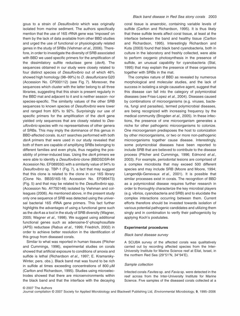
gous to a strain of Desulfovibrio which was originallyisolated from marine sediment. The authors specificallymention that the use of 16S rRNA gene was ‘imposed’ onthem by the lack of data available from other BBD studiesand urged the use of functional or physiologically relatedgenes in the study of SRBs (Viehman et al., 2006). There-fore, in order to investigate the diversity of SRB associatedwith BBD we used specific primers for the amplification ofthe dissimilatory sulfite reductase gene (dsrA). Thesequences obtained in our study were closely related tofour distinct species of Desulfovibrio out of which 46%showed high homology (98–99%) to D. desulfuricans G20(Accession No. CP000112) (see Fig. 7). Moreover, thesequences which cluster with the latter belong to all threelibraries, suggesting that this strain is present regularly inthe BBD mat and adjacent to it and is neither season- norspecies-specific. The similarity values of the other SRBsequences to known species of Desulfovibrio were lowerand ranged from 83% to 92%. Surprisingly our use ofspecific primers for the amplification of the dsrA geneyielded only sequences that are closely related to Des-ulfovibrio species with no representatives of other generaof SRBs. This may imply the dominance of this genus inBBD-affected corals. BLAST searches performed with bothdsrA primers that were used in our study revealed thatboth of them are capable of amplifying SRBs belonging todifferent families and even phyla, thus negating the pos-sibility of primer-induced bias. Using the dsrA primers wewere able to identify a Desulfovibrio clone (BBDSDSR-84Accession No. EF089550) with a similarity value of 94% toDesulfovibrio sp. TBP-1 (Fig. 7), a fact that may suggestthat this clone is related to the clone in our 16S library(Clone No. BB3S16S-18; Accession No. EF089473)(Fig. 5) and that may be related to the Desulfovibrio spp.(Accession No. AY750148) isolated by Viehman and col-leagues (2006). As mentioned above, in the present studyonly one sequence of SRB was detected using the univer-sal bacterial 16S rRNA gene primers. This fact furtherhighlights the advantages of using a functional gene suchas the dsrA as a tool in the study of SRB diversity (Wagner,2005; Wagner et al., 1998). We suggest using additionalfunctional genes such as adenosine-5′-phosphosulfate(APS) reductase (Rabus et al., 1999; Friedrich, 2002) inorder to achieve better resolution in the identification ofthis group from diseased corals.
Similar to what was reported in human tissues (Pitcherand Cummings, 1996), experimental studies on coralsshowed that artificial exposure to conditions of anoxia andsulfide is lethal (Richardson et al., 1997; E. Kramarsky-Winter, pers. obs.). Black band mat was found to be richin sulfide at times exceeding concentrations of 800 mM(Carlton and Richardson, 1995). Studies using microelec-trodes showed that there are microenvironments withinthe black band and that the interface with the decaying
coral tissue is anaerobic, containing variable levels ofsulfide (Carlton and Richardson, 1995). It is thus likelythat these sulfide levels affect coral tissue, at least at theinterface between the band and healthy tissue (Carltonand Richardson, 1995). Interestingly Richardson andKuta (2003) found that black band cyanobacteria, both inculture in the laboratory and freshly collected, were ableto perform oxygenic photosynthesis in the presence ofsulfide, an unusual capability for cyanobacteria (Stal,1995) that may explain the presence of these organismstogether with SRBs in the mat.
The complex nature of BBD as revealed by numerousmorphological and molecular studies, and the lack ofsuccess in isolating a single causative agent, suggest thatthis disease can fall into the category of polymicrobialdiseases (see Frias-Lopez et al., 2004). Diseases causedby combinations of microorganisms (e.g. viruses, bacte-ria, fungi and parasites), termed polymicrobial diseases,are being recognized with increasing frequency by themedical community (Brogden et al., 2005). In these infec-tions, the presence of one microorganism generates aniche for other pathogenic microorganisms to colonize.One microorganism predisposes the host to colonizationby other microorganisms, or two or more non-pathogenicmicroorganisms together cause disease. Interestingly,some polymicrobial diseases have been reported toinclude SRB that are believed to contribute to the diseaseprocess (Pitcher and Cummings, 1996; Edmond et al.,2003). For example, periodontal lesions are comprised ofa complex microbiota that may exceed 500 differentspecies and may include SRB (Moore and Moore, 1994;Langendijk-Genevaux et al., 2001). It is possible thatsimilar processes exist in corals. The recognition of BBDas a polymicrobial disease requires further research inorder to thoroughly characterize the key microbial players(e.g. vibrios, cyanobacteria and SRB) and to elucidate thecomplex interactions occurring between them. Currentefforts therefore should be invested towards isolation ofvarious potential pathogenic candidates and utilizing themsingly and in combination to verify their pathogenicity byapplying Koch’s postulates.
Experimental procedures
Black band disease survey
A SCUBA survey of the affected corals was qualitativelycarried out by recording affected species from the Inter-University Institute for Marine Science reef at Eilat, Israel, inthe northern Red Sea (29°51′N, 34°94′E).
Sample collection
Infected corals Favites sp. and Favia sp. were detected in thereef across from the Inter-University Institute for MarineScience. Five samples of the diseased corals collected at a
Black band disease in Red Sea stony corals 2003
© 2007 The AuthorsJournal compilation © 2007 Society for Applied Microbiology and Blackwell Publishing Ltd, Environmental Microbiology, 9, 1995–2006
different season were analysed (see Table 1). Every coralwas sampled at the black band region (‘S’), three were alsosampled at the interface (‘INT’) and two in the healthy (‘H’)regions. The black band region was collected using a needle-less syringe. The interface region adjacent to the band and aregion far from the band, where the tissue seemed healthy,were sampled using plastic bacteriological loops that wereused to rub off the coral surface microlayer. The loops (threefor each sample) were transferred to plastic 15 ml of sterilevials with minimal amount of seawater and sealedunderwater. As previous studies demonstrated that cyano-bacteria are abundant in the skeleton of massive corals (Fineand Loya, 2002), samples from these regions were collectedusing loops in order to minimize tissue damage and thesampling of cyanobacteria from the skeleton beneath thetissue. This method also enables re-sampling of the samecoral. Samples were frozen immediately and transfer frozento the lab. In addition, core samples of the BBD interface, andhealthy tissues were collected for histology using a 2-cm-diameter aluminum corer. The cores were collected from twodiseased colonies of F. favus fixed in 4% Formalin buffered inseawater. The cores were enrobed in agarose in order tomaintain tissue integrity following decalcification, and decal-cified using a 1:1 mixture of formic acid and sodium citrate.They were processed in a Citadel tissue processor and 6 mmsections were stained using standard haematoxylin/eosinand observed under the microscope.
DNA extraction
Frozen samples were thawed and centrifuged for 1 min atmaximal speed in order to pellet the mucus/mat. GenomicDNA was extracted by PowerSoil purification kit (Mo BioLaboratories, Solana Beach, CA, USA) according to themanufacturer’s instructions.
Polymerase chain reaction amplification
Total DNA was amplified using Biometra TGradient thermocy-cler (Biometra GmbH, Göttingen, Germany) by PCR usingspecific 16S rRNA gene primers for bacteria: forward pri-mer, 8F [5′-GGATCCAGACTTTGAT(C/T)(A/C)TGGCTCAG],taken and modified (8F primer was shortened from the 5′end; Felske et al., 1997) and reverse primer, 907R[5′-CCGTCAATTCCTTT(A/G)AGTTT-3′; Lane et al., 1985].In addition, modified 16S rRNA gene primers 8F-I and907R-I were obtained by replacement of the last nucleotide atthe 3′-terminus with inosine (Ben-Dov et al., 2006). In orderto retrieve SRB sequences, specific primers DSR1F(5′-ACSCACTGGAAGCACG-3′) (Wagner et al., 1998) andDSR10R (5′-CGGTTYTTGCCYTCTTCC-3′) (modified fromFriedrich, 2002) for the dissimilatory sulfite reductase genedsrA were used. Polymerase chain reaction mixturesincluded a 12.5 ml ReddyMix (Master mix containing 1.5 mMMgCl2 and 0.2 mM concentration of each deoxynucleosidetriphosphate) (ABgene, Surrey, UK), a 1 pmol each of theforward and reverse primers, 1–2 ml of the sample prepara-tion, plus water to bring the total volume to 25 ml. Whenamplification failed with ReddyMix, the FailSafe PCR Systemkit was used instead (Epicentre, Madison, WI). An initial
denaturation-hot start of 4 min at 95°C was followed by 30cycles of the following incubation pattern: 94°C for 40 s, 54°C(48°C for 8F-I/907R-I) for 40 s, and 72°C for 60–120 s. Theprocedure was completed with a final elongation step at 72°Cfor 20 min.
Clone library construction and sequencing
Polymerase chain reaction products were purified by electro-phoresis through a 1% agarose gel, stained with ethidiumbromide and visualized using a UV transilluminator. Thedesired bands were excised from the gel and the DNA waspurified from the gel slices using the Wizard PCR Prep kit(Promega, Madison, WI). The gel-purified PCR productswere cloned into the pCRII-TOPO-TA cloning vector as speci-fied by Invitrogen (Carlsbad, CA) and transformed intocalcium chloride-competent HD5a Escherichia coli cellsaccording to manufacturers’ instructions. Plasmid DNA wasisolated from individual clones by Wizard Plus SV MiniprepsDNA purification system (Promega, Madison, WI). Sequenc-ing with 8F and DSR1F primers were performed by ABIPRISM dye terminator cycle sequencing ready reaction kitwith AmpliTaq DNA polymerase FS and DNA sequencer ABImodel 373A system (Perkin-Elmer). Each sequence was des-ignated with a name containing the sample name (e.g. BB2),sample region (H, INT or S), primer set used for amplification(16S, 16Si, DSR) and a serial number.
Sequence analysis
Molecular Evolutionary Genetics Analysis (MEGA), version3.1 (Kumar et al., 2004), was used to de-replicate the librariesof 16S rRNA gene sequences for subsequent analyses, bycomparing all the sequences in a data set to each other,grouping similar sequences together by � 97 similarity, andchoosing a representative sequence from each group. All 16SrRNA gene sequences of each group were first comparedwith those in the GenBank database with the basic localalignment search tool BLAST (http://www.ncbi.nlm.nih.gov/blast/blast.cgi). Classifier program (version 1.0; assign 16SrRNA gene sequences to a taxonomical hierarchy) andLibrary Compare program (compare two sequence librariesusing the RDP classifier) available at the Ribosomal DatabaseProject-II web site (http://rdp.cme.msu.edu/, Maidak et al.,1997) were used to find diversity on different ranks of relatedsequences. To control for the occurrence of possibly chimericsequences, all sequenced clones were analysed by theChimera Check program of the RDP database (version 2.7;Maidak et al., 1999). The sequences from appropriate librar-ies were aligned using CLUSTALW with the MEGA package(Kumar et al., 2004) and positions not sequenced in all iso-lates or with alignment uncertainties were removed. Phyloge-netic trees were constructed by the Neighbour-Joiningmethod (Saito and Nei, 1987) using the Mega package(Kumar et al., 2004). Bootstrap re-sampling analysis (Felsen-stein, 1985) for 100 replicates was performed to estimate theconfidence of tree topologies. A Shannon–Weiner index fordiversity was applied using the FASTGroupII program, withdefault of 80% similarity in sequence match (Yu et al., 2006;http://biome.sdsu.edu/fastgroup/fg_tools.htm).
2004 O. Barneah, E. Ben-Dov, E. Kramarsky-Winter and A. Kushmaro
© 2007 The AuthorsJournal compilation © 2007 Society for Applied Microbiology and Blackwell Publishing Ltd, Environmental Microbiology, 9, 1995–2006
Acknowledgements
This work was supported by ISF Grant No. 511/02-1. We wouldlike to thank N. Siboni for his help in sample collection and theInter-University Institute in Eilat for use of their facilities.
References
Al-Moghrabi, M. (2001) Unusual black band disease (BBD)outbreak in the northern tip of the gulf of Aquaba (Jordan).Coral Reefs 19: 330–331.
Antonius, A. (1973) New observations on coral destruction inreefs. In Tenth Meeting of the Association of Island MarineLaboratories of the Caribbean, University of Puerto Rico(Mayaguez), p. 3.
Antonius, A. (1981) The ‘band’ diseases in coral reefs. Proc4th Int Coral Reef Symp, Manila 2: 7–14.
Antonius, A. (1985) ‘Coral diseases in the Indo-Pacific: a firstrecord.’ PSZNI. Mar Ecol 6: 197–218.
Antonius, A. (1988) Distribution and dynamics of coral dis-eases in the eastern Red Sea. Proc 6th Int Coral ReefSymp, Australia, 2: 293–298.
Banin, E., Israely, T., Kushmaro, A., Loya, Y., Orr, E., andRosenberg, E. (2000) Penetration of the coral-bleachingbacterium Vibrio shiloi into Oculina patagonica. ApplEnviron Microbiol 66: 3031–3036.
Ben-Dov, E., Shapiro, O.H., Siboni, N., and Kushmaro, A.(2006) Advantage of using inosine at the 3′ termini of 16SrRNA gene universal primers for the study of microbialdiversity. Appl Environ Microbiol 72: 6902–6906.
Ben-Haim, Y., and Rosenberg, E. (2004) Temperature-regulated bleaching and tissue lysis of Pocillopora dami-cornis by the novel pathogen Vibrio coralliilyticus. In CoralHealth and Disease. Rosenberg, E., and Loya, Y. (eds).Heidleberg, Germany: Springer-Verlag, pp. 301–324.
Ben-Haim, Y., Banin, E., Kushmaro, A., Loya, Y., and Rosen-berg, E. (1999) Inhibition of photosynthesis and bleachingof zooxanthellae by the coral pathogen Vibrio shiloi.Environ Microbiol 1: 223–229.
Ben-Haim, Y., Zicherman-Keren, M., and Rosenberg, E.(2003) Temperature-regulated bleaching and lysis of thecoral Pocillopora damicornis by the novel pathogen Vibriocoralliilyticus. Appl Environ Microbiol 69: 4236–4242.
Botha, N., Gehringer, M.M., Downing, T.G., van de Venter,M., and Shephard, E.G. (2004) The role of microcystin-LRin the induction of apoptosis and oxidative stress in CaCo2cells. Toxicon 43: 85–92.
Brogden, K.A., Guthmiller, J.M., and Taylor, C.E. (2005) JHuman polymicrobial infections. Lancet 365: 253–255.
Bythell, J.C., Barer, M.R., Cooney, R.P., Guest, J.R.,O’Donnell, A.G., Pantos, O., et al. (2002) Histopathologicalmethods for the investigation of microbial communitiesassociated with disease lesions in reef corals. Lett ApplMicrobiol 34: 359–364.
Carlton, R.G., and Richardson, L.L. (1995) Oxygen andsulfide dynamics in a horizontally migrating cyanobacterialmat: black band disease of corals. FEMS Microbiol Ecol18: 155–162.
Chet, I., and Mitchell, R. (1975) Bacterial attack on corals inpolluted seawater. Microb Ecol 2: 227–233.
Cooney, R.P., Pantos, O., Le Tissier, M.D.A., Barer, M.R.,
O’Donnell, A.G., and Bythell, J.C. (2002) Characterizationof the bacterial consortium associated with black banddisease in coral using molecular microbiologicaltechniques. Environ Microbiol 4: 401–413.
Dinsdale, E.A. (2002) Abundance of black-band disease oncorals from one location on the Great Barrier Reef: a com-parison with abundance in the Caribbean region. Proc 9thInt Coral Reef Symp, Bali, Indonesia 2: 1239–1243.
Ducklow, H.W., and Mitchell, R. (1979) Observations onnaturally and artificially diseased tropical corals: a scan-ning electron microscopy study. Microb Ecol 5: 215–223.
Edmunds, P.J. (1991) Extent and effect of black banddisease on a Caribbean reef. Coral Reefs 10: 161–165.
Edmond, L.M., Hopkins, M.J., Magee, E.A., and Cummings,J.H. (2003) The effect of 5-aminosalicylic acid-containingdrugs on sulfide production by sulfate-reducing and aminoacid-fermenting bacteria. Inflamm Bowel Dis 9: 10–17.
Felsenstein, J. (1985) Confidence limits of phylogenies: anapproach using the bootstrap. Evolution 39: 783–791.
Felske, A., Rheims, H., Wolterink, A., Stackebrandt, E., andAkkermans, A.D. (1997) Ribosome analysis reveals promi-nent activity of an uncultured member of the class Actino-bacteria in grassland soils. Microbiology 143: 2983–2989.
Fine, M., and Loya, Y. (2002) Endolithic algae: an alternativesource of photoassimilates during coral bleaching. Proc RSoc Lond B Biol Sci 269: 1205–1210.
Frias-Lopez, J., Zerkle, A.L., Boneheyo, G.T., and Fouke,B.W. (2002) Partitioning of bacterial communities betweensea water and healthy Black Band Diseased, and deadcoral surfaces. Appl Environ Microbiol 68: 2214–2228.
Frias-Lopez, J., Bonheyo, G.T., Jin, Q., and Fouke, B.W.(2003) Cyanobacteria associated with coral black banddisease in Caribbean and Indo-Pacific reefs. Appl EnvironMicrobiol 69: 2409–2413.
Frias-Lopez, J., Klaus, J.S., Bonheyo, G.T., and Fouke, B.W.(2004) Bacterial community associated with black banddisease in corals. Appl Environ Microbiol 70: 5955–5962.
Friedrich, M. (2002) Phylogenetic analysis reveals multiplelateral transfers of adenosine-5′-phosphosulfate reductasegenes among sulfate-reducing microorganisms. J Bacteriol184: 278–289.
Garrett, P., and Ducklow, H. (1975) Coral diseases inBermuda. Nature 253: 349–350.
Hada, H.S., West, P.A., Lee, J.V., Stemmler, J., and Colwell,R.R. (1984) Vibrio tubiashii sp. nov., a pathogen of bivalvemollusks. Int J Syst Bacteriol 34: 1–4.
Harvell, C.D., Kim, K., Burkholder, J.M., Colwell, P.R.,Epstein, P.R., Grimes, D.J., et al. (1999) Emerging marinediseases – climate links and anthropogenic factors.Science 285: 1505–1510.
Izaguirre, M.M., Mazza, C.A., Biondini, M., Baldwin, I.T., andBallaré, C.L. (2006) Remote sensing of future competitors:impacts on plant defenses. Proc Natl Acad Sci USA 103:7170–7174.
Klaus, J.S., Janse, I., Heikoop, J.M., Sanford, R.A., andFouke, B.W. (2007) Coral microbial communities, zooxan-thellae and mucus along gradients of seawater depth andcoastal pollution. Environ Microbiol doi: 10.1111/j.1462-2920.2007.01249.x
Kumar, S., Tomura, K., and Nei, M. (2004) MEGA3:integrated software for molecular evolutionary genetics
Black band disease in Red Sea stony corals 2005
© 2007 The AuthorsJournal compilation © 2007 Society for Applied Microbiology and Blackwell Publishing Ltd, Environmental Microbiology, 9, 1995–2006
analysis and sequence alignment. Brief Bioinform 5: 150–163.
Kushmaro, A., Loya, Y., Fine, M., and Rosenberg, E. (1996)Bacterial infection and coral bleaching. Nature 380: 396.
Kuta, K.G., and Richardson, L.L. (1996) Abundance and dis-tribution of black band disease on coralreefs in the northernFlorida Keys. Coral Reefs 15: 219–223.
Kuta, K.G., and Richardson, L.L. (2002) Ecological aspectsof black band disease of corals: relationships betweendisease incidence and environmental factor. Coral Reefs21: 393–398.
Lane, D.J., Pace, B., Olsen, G.J., Stahl, D.A., Sogin, M.L.,and Pace, N.R. (1985) Rapid determination of 16S riboso-mal RNA sequences for phylogenetic analyses. Proc NatlAcad Sci USA 82: 6955–6959.
Langendijk-Genevauxl, P.S., Hanssen, J.T.J., and van derHoeven, J.S. (2001) Decrease of sulfate-reducing bacteriaafter initial periodontal treatment. J Dent Res 80: 1637–1642.
Maidak, B.L., Olsen, G.J., Larsen, N., Overbeek, R.,McCaughey, M.J., and Woese, C.R. (1997) The RDP (Ribo-somal Database Project). Nucleic Acids Res 25: 109–110.
Maidak, B.L., Cole, J.R., Parker, C.T., Jr, Garrity, G.M.,Larsen, N., Li, B., et al. (1999) A new version of the RDP(Ribosomal Database Project). Nucleic Acids Res 27: 171–173.
Moore, W.E., and Moore, V.H. (1994) The bacteria of peri-odontal diseases. Periodontology 2000 5: 66–77.
Pantos, O., Cooney, R.P., Le Tissier, M.D.A., Barer, M.R.,O’Donnell, A.G., and Bythell, J.C. (2003) The bacterialecology of a plague-like disease affecting the Caribbeancoral montastrea annularis. Environ Microbiol 5: 370–382.
Pantos, O., and Bythell, J. (2006) Bacterial community struc-ture associated with white band disease in the elkhorn coralAcropora palmata determined using culture-independent16S rRNA techniques. Dis Aquat Organ 69: 79–88.
Pitcher, M.C., and Cummings, J.H. (1996) Hydrogen sul-phide: a bacterial toxin in ulcerative colitis? Gut 39: 1–4.
Rabus, R., Hansen, T.A., and Widdel, F. (1999) Dissimilatorysulfate- and sulfur-reducing prokaryotes. In The Prokary-otes: An Evolving Electronic Database for the Microbiologi-cal Community. Dworkin, M., Schleifer, K.H., andStackebrandt, E. (eds). New York, USA: Springer, pp. 1–87.
Ramos-Flores, T. (1983) Lower marine fungus associatedwith black line disease in star corals (Montastrea annularisE. & S.). Biol Bull 165: 429–435.
Richardson, L.L. (1998) Coral diseases: what is reallyknown? TREE 13: 438–443.
Richardson, L.L. (2004) Black band disease. In Coral Healthand Disease. Rosenberg, E., and Loya, Y. (eds). Heidle-berg, Germany: Springer-Verlag, pp. 325–336.
Richardson, L.L., and Kuta, K.G. (2003) Ecological physiol-ogy of the black band disease cyanobacterium Phormidiumcorallyticum. FEMS Microbiol Ecol 43: 287–298.
Richardson, L.L., Kuta, K.G., Schnell, S., and Carlton, R.G.(1997) Ecology of the black band disease microbial con-
sortium. Proc 8th Int Coral Reef Symp, SmithsonianTropical Research Institute, Balboa, Panama 1: 597–600.
Rohwer, F., Breitbart, M., Jara, J., Azam, F., and Knowlton, N.(2001) Diversity of bacteria associated with the Caribbeancoral Montastrea franksii. Coral Reefs 20: 85–91.
Rohwer, F., Seguritan, V., Azam, F., and Knowlton, N. (2002)Diversity and distribution of coral-associated bacteria.Mar Ecol Prog Ser 243: 1–10.
Rosenberg, E., and Falkovitz, L. (2004) The Vibrio shiloi/Oculina patagonica model system of coral bleaching. AnnuRev Microbiol 58: 143–159.
Rützler, K., and Santavy, D.L. (1983) The black band diseaseof Atlantic reef corals. I. Description of the cyanophytepathogen. Mar Ecol 4: 301–319.
Saito, N., and Nei, M. (1987) The neighbor-joining method: anew method for constructing phylogenetic trees. Mol BiolEvol 4: 406–425.
Sekar, R., Mills, D.K., Remily, E.R., Voss, J.D., and Richard-son, L.L. (2006) Microbial communities in the surfacemucopolysaccharide layer and the black band microbialmat of black band-diseased Siderastrea siderea. ApplEnviron Microbiol 72: 5963–5973.
Stal, L.J. (1995) Physiological ecology of cyanobacteria inmicrobial mats and other communities. New Phytol 131:1–32.
Sussman, M., Bourne, D.G., and Willis, B.L. (2006) A singlecyanobacterial ribotype is associated with both red andblack bands diseased corals from Palau. Dis Aquat Organ69: 111–118.
Teneva, I., Mladenov, R., Popov, N., and Dzhambazov, B.(2005) Cytotoxicity and apoptotic effects of microcystin-LRand anatoxin-a in mouse lymphocytes. Folia Biol (Praha)51: 62–67.
Viehman, S., and Richardson, L.L. (2002) Motility patterns ofBeggiatoa and Phormidium corallyticum in black banddisease. Proc 9th Int Coral Reef Symp Bali, Indonesia 2:1251–1255.
Viehman, S., Mills, D.K., Meichel, G.W., and Richardson,L.L. (2006) Culture and identification of Desulfovibriospp. from corals infected by black band disease onDominican and Florida Keys reefs. Dis Aquat Organ 69:119–127.
Wagner, M. (2005) Lateral gene transfer of dissimilatory(bi)sulfite reductase revisited. J Bacteriol 187: 2203–2208.
Wagner, M., Roger, A.J., Flax, J.L., Brusseau, G.A., andStahl, D.A. (1998) Phylogeny of dissimilatory sulfite reduc-tases supports an early origin of sulfate respiration.J Bacteriol 180: 2975–2982.
Willis, B.L., Page, C.A., and Dinsdale, E.A. (2004) Coraldisease on the Great Barrier Reef. In Coral Health andDisease. Rosenberg, E., and Loya, Y. (eds). Heidleberg,Germany: Springer-Verlag, pp. 69–104.
Yu, Y., Breitbart, M., McNairnie, P., and Rohwer, F. (2006)FastGroupII: a web-based bioinformatics platform foranalyses of large 16S rRNA gene libraries. BMC Bioinfor-matics 7: 57.
2006 O. Barneah, E. Ben-Dov, E. Kramarsky-Winter and A. Kushmaro
© 2007 The AuthorsJournal compilation © 2007 Society for Applied Microbiology and Blackwell Publishing Ltd, Environmental Microbiology, 9, 1995–2006