Diseases affect cold-water corals too: Eunicella verrucosa (Cnidaria: Gorgonacea) necrosis in SW...
11
DISEASES OF AQUATIC ORGANISMS Dis Aquat Org Vol. 76: 87–97, 2007 Published June 29 INTRODUCTION Study of disease in warm-water corals is a major research area because of its growing incidence in tropical and Mediterranean waters with widespread concerns over the deleterious knock-on effects on bio- diversity, tourism and coastal defence (Harvell et al. 2002, Bruno et al. 2003, Linares et al. 2005, Ritchie 2006). However, there have been no previous studies on the diseases of cold-water corals, despite the pre- valence and known importance of corals in the functional ecology of temperate and deep-sea en- vironments (Guinotte et al. 2006, Roberts et al. 2006, Hall-Spencer et al. 2007). Although reports of warm-water coral diseases have been on the increase, disease aetiology has only been established in a small number of cases (Rosenberg & Ben Haim 2002, Munn 2004). One of the first coral diseases for which a clear microbial aetiology was confirmed is aspergillosis, which has caused localised outbreaks of disease in Caribbean populations of Gor- gonia ventalina and G. flabellum since 1984, with mass mortalities recorded in 1994 (Kim & Harvell 2004). The disease is characterised by thickened tissue and purple areas of necrosis caused by Aspergillus sydowii, a fun- gus found commonly in decaying matter in terrestrial environments (Nagelkerken et al. 1997, Geiser et al. 1998). It has been proposed that A. sydowii may be deposited in the Caribbean marine environment via dust storms from deserts in Africa (Garrison et al. 2003). The causes of diseases in other warm-water gor- gonians are less well-known; the cyanobacterium Scy- tonema sp. has been associated with a disease in Caribbean Briareum asbestinum gorgonians during a bleaching event associated with high sea-surface tem- peratures, although it was not possible to determine if © Inter-Research 2007 · www.int-res.com *Email: [email protected] Diseases affect cold-water corals too: Eunicella verrucosa (Cnidaria: Gorgonacea) necrosis in SW England Jason M. Hall-Spencer*, James Pike, Colin B. Munn Marine Institute, University of Plymouth, Plymouth PL4 8AA, UK ABSTRACT: The first recorded incidence of cold-water coral disease was noted in Eunicella verru- cosa, a coral on the international ‘red list’ of threatened species, at a marine protected area in SW England in 2002. Video surveys of 634 separate colonies at 13 sites revealed that disease outbreaks were widespread in SW England from 2003 to 2006. Coenchyme became necrotic in diseased speci- mens, leading to tissue sloughing and exposing skeletal gorgonin to settlement by fouling organisms. Sites where necrosis was found had significantly higher incidences of fouling. No fungi were isolated from diseased or healthy tissue, but significantly higher concentrations of bacteria occurred in dis- eased specimens. Of 21 distinct bacteria isolated from diseased tissues, 19 were Vibrionaceae, 15 were strains of Vibrio splendidus and 2 others closely matched Vibrio tasmaniensis. Vibrios isolated from E. verrucosa did not induce disease at 15°C, but, at 20°C, controls remained healthy and test gorgonians became diseased, regardless of whether vibrios were isolated from diseased or healthy colonies. Bacteria associated with diseased tissue produced proteolytic and cytolytic enzymes that damaged E. verrucosa tissue and may be responsible for the necrosis observed. Monitoring at the site where the disease was first noted showed new gorgonian recruitment from 2003 to 2006; some indi- viduals had died and become completely overgrown, whereas others had continued to grow around a dead central area. KEY WORDS: Cold-water coral · Disease · Gorgonian · NE Atlantic Resale or republication not permitted without written consent of the publisher OPEN PEN ACCESS CCESS
Transcript of Diseases affect cold-water corals too: Eunicella verrucosa (Cnidaria: Gorgonacea) necrosis in SW...

DISEASES OF AQUATIC ORGANISMSDis Aquat Org
Vol. 76: 87–97, 2007 Published June 29
INTRODUCTION
Study of disease in warm-water corals is a majorresearch area because of its growing incidence intropical and Mediterranean waters with widespreadconcerns over the deleterious knock-on effects on bio-diversity, tourism and coastal defence (Harvell et al.2002, Bruno et al. 2003, Linares et al. 2005, Ritchie2006). However, there have been no previous studieson the diseases of cold-water corals, despite the pre-valence and known importance of corals in thefunctional ecology of temperate and deep-sea en-vironments (Guinotte et al. 2006, Roberts et al. 2006,Hall-Spencer et al. 2007).
Although reports of warm-water coral diseases havebeen on the increase, disease aetiology has only beenestablished in a small number of cases (Rosenberg &Ben Haim 2002, Munn 2004). One of the first coral
diseases for which a clear microbial aetiology wasconfirmed is aspergillosis, which has caused localisedoutbreaks of disease in Caribbean populations of Gor-gonia ventalina and G. flabellum since 1984, with massmortalities recorded in 1994 (Kim & Harvell 2004). Thedisease is characterised by thickened tissue and purpleareas of necrosis caused by Aspergillus sydowii, a fun-gus found commonly in decaying matter in terrestrialenvironments (Nagelkerken et al. 1997, Geiser et al.1998). It has been proposed that A. sydowii may bedeposited in the Caribbean marine environment viadust storms from deserts in Africa (Garrison et al.2003). The causes of diseases in other warm-water gor-gonians are less well-known; the cyanobacterium Scy-tonema sp. has been associated with a disease inCaribbean Briareum asbestinum gorgonians during ableaching event associated with high sea-surface tem-peratures, although it was not possible to determine if
© Inter-Research 2007 · www.int-res.com*Email: [email protected]
Diseases affect cold-water corals too: Eunicellaverrucosa (Cnidaria: Gorgonacea) necrosis in
SW England
Jason M. Hall-Spencer*, James Pike, Colin B. Munn
Marine Institute, University of Plymouth, Plymouth PL4 8AA, UK
ABSTRACT: The first recorded incidence of cold-water coral disease was noted in Eunicella verru-cosa, a coral on the international ‘red list’ of threatened species, at a marine protected area in SWEngland in 2002. Video surveys of 634 separate colonies at 13 sites revealed that disease outbreakswere widespread in SW England from 2003 to 2006. Coenchyme became necrotic in diseased speci-mens, leading to tissue sloughing and exposing skeletal gorgonin to settlement by fouling organisms.Sites where necrosis was found had significantly higher incidences of fouling. No fungi were isolatedfrom diseased or healthy tissue, but significantly higher concentrations of bacteria occurred in dis-eased specimens. Of 21 distinct bacteria isolated from diseased tissues, 19 were Vibrionaceae, 15were strains of Vibrio splendidus and 2 others closely matched Vibrio tasmaniensis. Vibrios isolatedfrom E. verrucosa did not induce disease at 15°C, but, at 20°C, controls remained healthy and testgorgonians became diseased, regardless of whether vibrios were isolated from diseased or healthycolonies. Bacteria associated with diseased tissue produced proteolytic and cytolytic enzymes thatdamaged E. verrucosa tissue and may be responsible for the necrosis observed. Monitoring at the sitewhere the disease was first noted showed new gorgonian recruitment from 2003 to 2006; some indi-viduals had died and become completely overgrown, whereas others had continued to grow arounda dead central area.
KEY WORDS: Cold-water coral · Disease · Gorgonian · NE Atlantic
Resale or republication not permitted without written consent of the publisher
OPENPEN ACCESSCCESS
Dis Aquat Org 76: 87–97, 2007
this cyanobacterium was the primary causative agent(Harvell et al. 2001). In the Mediterranean, Cerrano etal. (2000) reported extensive gorgonian mortality dur-ing unusually warm water conditions in 1999, whichMartin et al. (2002) linked to infection by Vibrio spp.bacteria at elevated temperatures.
Against this background of a growing understandingof warm-water coral diseases, the present paperreports in situ and laboratory observations of a diseaseoutbreak in cold-water populations of Eunicella verru-cosa following preliminary reports of the disease at awell-monitored site at Lundy, a Marine Protected Areain SW England (Hiscock 2003). This gorgonian has alimited distribution around the British Isles and washistorically collected for souvenirs so it is now one ofthe few marine species fully protected under theWildlife and Countryside Act, 1981, and listed in theUK Biodiversity Action Plan. It is also listed as vulner-able on the International Union for Conservation ofNature and Natural Resources ‘red list’ of threatenedspecies (IUCN 2006). The 3 main aims of the presentstudy were to: (1) evaluate whether an outbreak in2002 was an isolated incident, (2) to examine thenature of the disease and (3) to investigate the hypoth-esis that pathogenic microbes were responsible.
MATERIALS AND METHODS
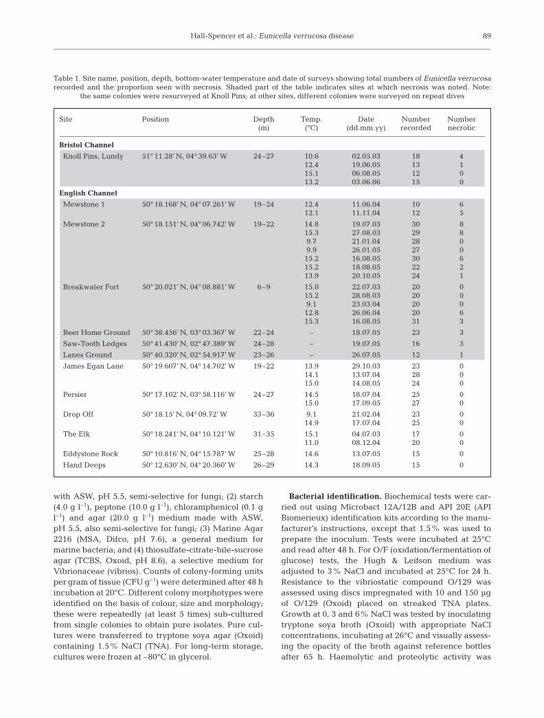
Field observations. Destructive sampling was keptto a minimum because of the protected status of Euni-cella verrucosa. Surveys of disease incidence wereundertaken using a Sony PC-110 digital video cameraheld in a Sea & Sea VXPC110 underwater housingduring SCUBA dives along depth-stratified belt tran-sects at sites known to support dense populations ofthe gorgonians based on interviews with local divers.Twelve sites were surveyed between 1 and 7 times inthe English Channel, and 1 site was surveyed in theLundy No Take Zone, Bristol Channel, from 2003 to2006, allowing analysis of the incidence of disease in634 separate gorgonian colonies (Table 1).
The camera was used to record all gorgonians en-countered in a 25 min period along belt transectswithin depth contours of ±1.5 m. Still pictures weretaken from the video showing perpendicular photo-quadrats of each gorgonian next to a ruler for scale.The photographs were analysed to record prevalenceof necrosis, and the degree of fouling by epibionts wasrecorded on a 3-rank scale (0 = no overgrowth, 1 =<10% of gorgonian overgrown, 2 = >10% of gorgonianovergrown) to test the hypothesis that fouling organ-isms were more prevalent on gorgonians at sites wherenecrosis was noted. Repeat surveys of the same gor-gonians were made at Knoll Pins, Lundy, using obvious
features of the submerged cliff and boulder field to re-locate individuals, whereas different areas were sam-pled during repeat surveys at the other sites to ensurethat the same individuals were not surveyed twice.
On 27 August 2003, divers collected the first 21 gor-gonians encountered from a patch of specimens in var-ious states of disease and overgrowth to identify theirassociated macrobiota at 19 to 22 m below chart datumon Mewstone Ledges (50° 18.15’ N, 4° 06.74’ W) inaccordance with a licence granted by English Nature.Each gorgonian was encased in a labelled plastic bagbefore using garden secateurs to cut through the stemand tying the bag closed. Samples were preserved in4% seawater formalin and rinsed over a 0.2 mm sieveto retain larger epibiota. Attached species wereremoved under a dissecting microscope, identifyingthe various species present.
Histology. Clippings of 100 mm length were takenfrom the extremities of Eunicella verrucosa coloniescollected at Mewstone Ledges on 18 August 2005.Each clipping was placed in zip-sealed plastic bagswith seawater and returned to the laboratory in a coolbox for processing within 3 h of collection. Sampleswere taken from healthy colonies and from the regionwhere exposed skeleton met wasted tissue on diseasedcolonies; then, they were fixed in 2.5% gluteraldehydein 10 µm filtered and autoclaved artificial seawater(ASW, Instant Ocean) for 48 h. For SEM (scanningelectron microscopy), samples were rinsed in ASW andthen in distilled water (15 min each); they were dehy-drated in an ethanol series, critical point dried(Emitech K550) and then frozen in liquid nitrogenbefore freeze-fracturing with a razor blade. Scleriteswere disassociated from the tissue by macerating insodium hypochlorite, repeatedly rinsing in water andfinally rinsing in 95% ethanol. Samples were mountedon stubs, gold sputter coated (Emitech K850) andobserved in a JEOL 5600LV SEM. For light micro-scopy, clippings were decalcified in 10% EDTAadjusted to pH 7.0 with NaOH for 24 h and dehydratedin graded alcohols, cleared in xylene and embedded inparaffin wax. Sections (4 to 6 µm) were cut using arotary microtome after softening the gorgonin using aproprietary depilatory cream (Veet). Sections werestained using haematoxylin and eosin protocols (Bucke1989) and examined using a Leica DM1RB.
Microbiology. Clippings of 20 mm length wererinsed repeatedly in sterile ASW, weighed and thenground with a sterile mortar and pestle with 1.0 ml ster-ile ASW to remove soft tissues. The homogenate wasremoved, and the procedure was repeated 3 times toproduce 3.0 ml of homogenate prior to reweighing theclippings. Duplicate 0.1 ml samples of 10-fold serial di-lutions in ASW of each homogenate were spread-plated onto: (1) malt extract agar (MEX, Oxoid) made
88
Hall-Spencer et al.: Eunicella verrucosa disease
with ASW, pH 5.5, semi-selective for fungi; (2) starch(4.0 g l–1), peptone (10.0 g l–1), chloramphenicol (0.1 gl–1) and agar (20.0 g l–1) medium made with ASW,pH 5.5, also semi-selective for fungi; (3) Marine Agar2216 (MSA, Difco, pH 7.6), a general medium formarine bacteria; and (4) thiosulfate-citrate-bile-sucroseagar (TCBS, Oxoid, pH 8.6), a selective medium forVibrionaceae (vibrios). Counts of colony-forming unitsper gram of tissue (CFU g–1) were determined after 48 hincubation at 20°C. Different colony morphotypes wereidentified on the basis of colour, size and morphology;these were repeatedly (at least 5 times) sub-culturedfrom single colonies to obtain pure isolates. Pure cul-tures were transferred to tryptone soya agar (Oxoid)containing 1.5% NaCl (TNA). For long-term storage,cultures were frozen at –80°C in glycerol.
Bacterial identification. Biochemical tests were car-ried out using Microbact 12A/12B and API 20E (APIBiomerieux) identification kits according to the manu-facturer’s instructions, except that 1.5% was used toprepare the inoculum. Tests were incubated at 25°Cand read after 48 h. For O/F (oxidation/fermentation ofglucose) tests, the Hugh & Leifson medium wasadjusted to 3% NaCl and incubated at 25°C for 24 h.Resistance to the vibriostatic compound O/129 wasassessed using discs impregnated with 10 and 150 µgof O/129 (Oxoid) placed on streaked TNA plates.Growth at 0, 3 and 6% NaCl was tested by inoculatingtryptone soya broth (Oxoid) with appropriate NaClconcentrations, incubating at 26°C and visually assess-ing the opacity of the broth against reference bottlesafter 65 h. Haemolytic and proteolytic activity was
89
Site Position Depth Temp. Date Number Number(m) (°C) (dd.mm.yy) recorded necrotic
Bristol Channel
Knoll Pins, Lundy 51° 11.28’ N, 04° 39.63’ W 24–27 10.6 02.05.03 18 412.4 19.06.05 13 115.1 06.08.05 12 013.2 03.06.06 15 0
English Channel
Mewstone 1 50° 18.168’ N, 04° 07.261’ W 19–24 12.4 11.06.04 10 612.1 11.11.04 12 5
Mewstone 2 50° 18.151’ N, 04° 06.742’ W 19–22 14.8 19.07.03 30 815.3 27.08.03 29 89.7 21.01.04 28 09.9 26.01.05 27 015.2 16.08.05 30 615.2 18.08.05 22 213.9 20.10.05 24 1
Breakwater Fort 50° 20.021’ N, 04° 08.881’ W 6–9 15.0 22.07.03 20 015.2 28.08.03 20 09.1 23.03.04 20 012.8 26.06.04 20 615.3 16.08.05 31 3
Beer Home Ground 50° 38.456’ N, 03° 03.367’ W 22–24 – 18.07.05 23 3
Saw-Tooth Ledges 50° 41.430’ N, 02° 47.389’ W 24–28 – 19.07.05 16 3
Lanes Ground 50° 40.320’ N, 02° 54.917’ W 23–26 – 26.07.05 12 1
James Egan Lane 50° 19.607’ N, 04° 14.702’ W 19–22 13.9 29.10.03 23 014.1 13.07.04 28 015.0 14.08.05 24 0
Persier 50° 17.102’ N, 03° 58.116’ W 24–27 14.5 18.07.04 25 015.0 17.09.05 27 0
Drop Off 50° 18.15’ N, 04° 09.72’ W 33–36 9.1 21.02.04 23 014.9 17.07.04 25 0
The Elk 50° 18.241’ N, 04° 10.121’ W 31–35 15.1 04.07.03 17 011.0 08.12.04 20 0
Eddystone Rock 50° 10.816’ N, 04° 15.787’ W 25–28 14.6 13.07.05 15 0
Hand Deeps 50° 12.630’ N, 04° 20.360’ W 26–29 14.3 18.09.05 15 0
Table 1. Site name, position, depth, bottom-water temperature and date of surveys showing total numbers of Eunicella verrucosarecorded and the proportion seen with necrosis. Shaded part of the table indicates sites at which necrosis was noted. Note:
the same colonies were resurveyed at Knoll Pins; at other sites, different colonies were surveyed on repeat dives

Dis Aquat Org 76: 87–97, 2007
assessed by spot inoculation on TNA plates containing5% horse blood or 1% skim milk, respectively.
DNA was extracted from overnight cultures using aPromega Wizard Genomic DNA purification kit.Amplification was carried out in an MWG thermalcycler using Promega PCR Master Mix (25 µl reactionvolume) with 10 pmol of universal bacterial primers27F and 1492R (Lane 1991); the amplification protocolwas: 1 cycle at 95°C for 5 min; 30 cycles at 95°C for1 min, 54°C for 1 min and 72°C for 2 min; and a finalcycle of 95°C for 1 min, 54°C for 1 min and 72°C for10 min. PCR products were purified using a PromegaWizard SV Gel and PCR Clean-Up System andchecked for purity on a 1% agarose gel. Sequencingwas carried out by MWG-Biotech (Ebersberg) witheither Primer 27F or 1492R. Sequences were alignedand matched using the online tools from the RibosomalDatabase Project (http://rdp.cme.msu.edu/, accessed28 June 2006). Phylogenetic trees were drawn usingthe Phylip interface and the maximum-likelihoodmethod. Confirmation of identity of 2 strains by ampli-fied fragment length polymorphism (AFLP) was car-ried out by Fabiano Thompson using methods hedescribed in Thompson et al. (2003).
Infection challenge. Eunicella verrucosa clippings(50 mm) were attached to small sterile pebbles usingepoxy putty (The Milliput Company) and placed indi-vidually in 1 l plastic containers filled with aeratedsterile ASW and kept without feeding. Clippings wereinoculated by rubbing with cultures taken from agarplates (TNA, 24 h) using sterile cotton swabs, and pro-portions of clippings affected by necrosis wererecorded daily. In a preliminary experiment, clippingswere maintained at 15 or 20°C for 15 d after inocula-tion with mixed vibrios: either CC071, CC074 andCC090 (n = 5) isolated from diseased E. verrucosacolonies or CC075 and CC082 (n = 5) isolated from
healthy E. verrucosa colonies. In a follow-up experi-ment, clippings were maintained at 20°C for 19 d andinoculated with the individual vibrio strains CC071(n = 12) or CC075 (n = 12), whereas controls (n = 12)were sham-inoculated with swabs dipped in ASW.
Effect of bacterial extracellular products (ECP). ECPfrom Strain CC071 was prepared by growth on cello-phane overlays on TNA + 0.5% skim milk plates (250 ×250 mm) for 24 h at 22°C. Cells plus ECP were removedwith 15.0 ml ice-cold phosphate-buffered saline (PBS,3.3% NaCl) and centrifuged at 3500 × g for 30 min;supernatants were filtered with a 0.22 µm membranefilter. Protease activity of ECP preparations was as-sayed using the azocasein assay method of Montero &Austin (1999). The sample used had a protease activityof 100 U (1 U = increase of absorbance at 540 nm of 0.01U after 20 min). The specific activity was 1300 U mg–1
protein (protein assay using a Sigma rapid proteinquantification kit). Haemolytic activity was assessed byfilling 0.7 mm wells in 5% horse blood–TNA plates.Clippings (20 mm) of Eunicella verrucosa were treatedby immersion for 24 h at 22°C in 0.5 ml of a preparationof ECP from Strain CC071, or in PBS (3.3% NaCl). Re-lease of pigment from the tissue was measured spec-trophotometrically at 400 nm in the supernatant aftercentrifugation at 16 000 × g (10 min).
RESULTS
Field observations
Video transects allowed subsequent analyses of 634gorgonian colonies around SW England from 2003 to2006, of which 9% had tissue necrosis (Table 1).Affected colonies had patches of necrotic tissue thatwere soft and white (Fig. 1A) and could easily be
90
Fig. 1. Eunicella verrucosa. At 21 m depth at Knoll Pins, Lundy, on 16 May 2003: (A) early onset of coenchyme necrosis (arrow)and (B) post-necrotic exposure of gorgonin skeleton (arrow) with fouling community of hydroids, barnacles Solidobalanus fallax
and bryozoans Cellaria sp. Scale bars = 40 mm

Hall-Spencer et al.: Eunicella verrucosa disease
rubbed off by hand to expose an underlying black gor-gonin skeleton. Healthy gorgonians and unaffectedparts of colonies with necrotic tissue damage had atough, typically orange-pink coenchyme, which wasfirmly attached to the skeleton. Gorgonian necrosiswas recorded at 7 out of 13 sites surveyed between7 May 2003 and 2 June 2006 on both the north andsouth coasts of SW England. The disease was notedfrom May to October at temperatures of 12.1 to 15.3°Cand depths of 6 to 28 m, but was not found at depths of>30 m or on the 99 colonies examined in winter(Table 1). Repeated observations made of diseasedcolonies at Knoll Pins, Lundy, revealed a progression,whereby necrotised tissue was sloughed allowingepibionts to colonise exposed skeleton (Fig. 1B) withremaining tissue taking on a thin, wasted appearancewith a grey hue. By June 2006, 5 of the 18 coloniesfilmed in 2003 were dead and completely covered inepibionts, whereas the remainder were still surviving,exhibiting a dead central region overgrown by epi-bionts with continued colony growth at the extremities.Two new colonies (4 to 6 cm in height) had settled inthis area and were devoid of necrosis and visibleepibionts.
Organisms associated with the living coenchyme ofgorgonians (as opposed to fouling organisms attachedto skeletal gorgonin) comprised drift algae Dictyotadichotoma, nudibranchs Tritonia nilsodhneri), dogfishScyliorhinus canicula eggs and squid Loligo sp. eggsand were found at sites where gorgonians werehealthy as well as diseased. Most (74%) of the 431 gor-gonians recorded at sites where necrosis had beenfound were overgrown by a range of fouling organisms(Fig. 2) attached to dead skeletal material. Those sam-pled on 27 August 2003 at Mewstone Ledges at 19 to
22 m depth comprised sponges (Leucosolenia com-plicata, Scypha compressa, Sycon ciliatum), hydroids(Aglaophenia pluma, Eudendrium rameum, Haleciumspp., Lafoea dumosa, Obelia dichotoma, O. geniculata,Sertularella spp.), soft coral (Alcyonium digitatum),bryozoans (Aeta sp., Alcyonidium sp., Bugula plum-osa, Cellepora hyalina, Crisia denticulata, Disporellahispida, Scrupocellaria sp., Turbicellepora avicularis),barnacles (Solidobalanus fallax) and sea squirts(Didemnidae). Results of a 1-way ANOVA showed sig-nificant differences in the proportion of seafans withno fouling (F1,30 = 48.19, p < 0.001), <10% fouling (F1,30
= 15.94, p < 0.001) and >10% fouling (F1,30 = 36.07, p <0.001) between sites with no necrosis observed (n = 11)and sites with necrosis observed (n = 21) (Fig. 2). Pro-portional data was angular transformed, tested andfound to have equal variances. Whilst significantlymore gorgonians were fouled at sites where necrosishad been noted, some unfouled colonies were found atthese sites and were not visibly affected by disease.
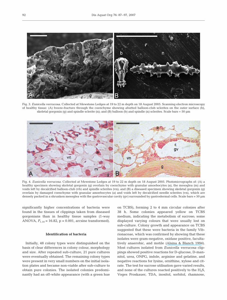
Ultrastructure of healthy and diseased tissue
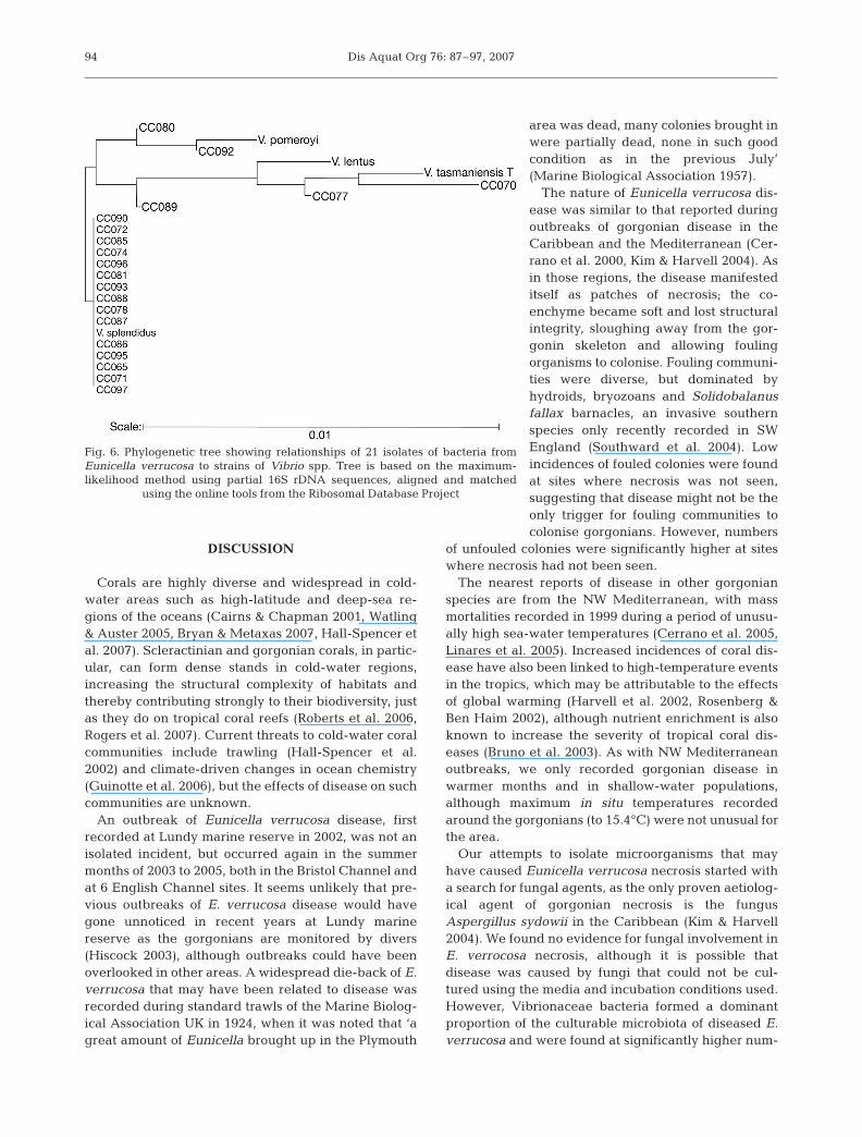
The coenchyme from healthy Eunicella verrucosacolonies had a thin outer layer consisting of tightlyabutted balloon-club sclerites forming a tough rindoverlying high concentrations of granular amoebo-cytes. Beneath this was a thick inner layer contain-ing the mesoglea and granular amoebocytes, bothsupported structurally by spindle sclerites (Fig. 3).Necrotic coenchyme was fragile and structurally dis-rupted, with the balloon-club sclerites loosely packedor absent and granular amoebocytes occurring withinthe shrunken mesoglea of the inner coenchyme(Fig. 4). This shrinking of the mesoglea substantiallyreduced the spacing between spindle sclerites (Fig. 4),giving a grey, wasted appearance to diseased speci-mens as the black gorgonin skeleton becomes visiblethrough the shrunken coenchyme.
Microbes cultured from healthy and diseased tissue
No fungi were isolated from any of the Eunicella ver-rucosa tissue samples tested, despite using culturemedia that were selective for fungi. On MSA media,however, far more bacteria were cultured from dis-eased than from healthy tissue (Fig. 5). This differencewas not statistically significant, due to wide variationin bacterial numbers found between replicate tissuesamples (1-way ANOVA, F1,18 = 3.43, p = 0.081, datawere arcsine transformed to achieve homogeneity ofvariance in the presence of zero counts). However,using TCBS media (which is semi-selective for vibrios),
91
Fig. 2. Eunicella verrucosa. Proportion (+SE) of gorgoniansrecorded with no fouling, light fouling and heavy fouling atsites where necrosis was noted (shaded, number of dives = 20)compared with sites where no necrosis was noted (unshaded,number of dives = 11) between 6 and 36 m depth off SWEngland between 2003 and 2006. Significant differences
(p < 0.05) between means indicated by asterisks
Dis Aquat Org 76: 87–97, 2007
significantly higher concentrations of bacteria werefound in the tissues of clippings taken from diseasedgorgonians than in healthy tissue samples (1-wayANOVA, F1,18 = 16.82, p = 0.001, arcsine transformed).
Identification of bacteria
Initially, 48 colony types were distinguished on thebasis of clear differences in colony colour, morphologyand size. After repeated sub-culture, 21 pure cultureswere eventually obtained. The remaining colony typeswere present in very small numbers on the initial isola-tion plates and became non-viable after sub-culture toobtain pure colonies. The isolated colonies predomi-nantly had an off-white appearance (with a green hue
on TCBS), forming 2 to 4 mm circular colonies after38 h. Some colonies appeared yellow on TCBSmedium, indicating the metabolism of sucrose; somedisplayed varying colours that were usually lost onsub-culture. Colony growth and appearance on TCBSsuggested that these were bacteria in the family Vib-rionaceae, which was confirmed by showing that theseisolates were gram-negative, oxidase positive, faculta-tively anaerobic, and motile (Alsina & Blanch 1994).Most cultures isolated from Eunicella verrucosa clip-pings showed positive reactions for D-glucose, D-man-nitol, urea, ONPG, indole, arginine and gelatine, andnegative reactions for lysine, ornithine, xylose and cit-rate. The test for sucrose utilisation gave varied results,and none of the cultures reacted positively to the H2S,Voges Proskauer, TDA, inositol, sorbitol, rhamnose,
92
Fig. 3. Eunicella verrucosa. Collected at Mewstone Ledges at 19 to 22 m depth on 18 August 2005. Scanning electron microscopyof healthy tissue: (A) freeze-fracture through the coenchyme showing abutted balloon-club sclerites on the outer surface (b),
skeletal gorgonin (g) and spindle sclerite (n); and (B) balloon (b) and spindle (n) sclerites. Scale bars = 50 µm
Fig. 4. Eunicella verrucosa. Collected at Mewstone Ledges at 19 to 22 m depth on 18 August 2005. Photomicrographs of: (A) ahealthy specimen showing skeletal gorgonin (g) overlain by coenchyme with granular amoebocytes (a), the mesoglea (m) andvoids left by decalcified balloon-club (vb) and spindle sclerites (vn); and (B) a diseased specimen showing skeletal gorgonin (g)overlain by damaged coenchyme with granular amoebocytes (a) and voids left by decalcified needle sclerites (vn), which aredensely packed in a shrunken mesoglea with the gastrovascular cavity (gv) surrounded by gastrodermal cells. Scale bars = 50 µm

Hall-Spencer et al.: Eunicella verrucosa disease
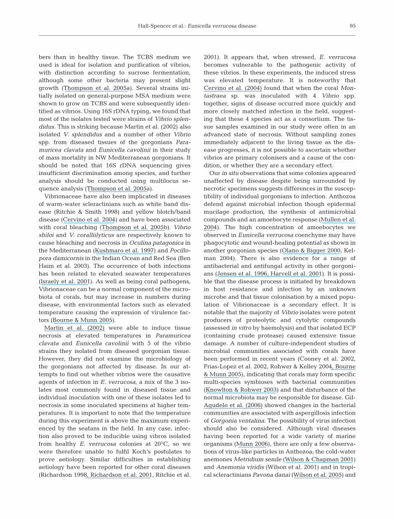
salicin, lactose, or arabinose tests. Tests for growth atvarying NaCl concentrations, resistance to O/129 andproduction of caseinase and haemolysin(s) provedmore effective in discriminating between cultures. Useof keys for environmental Vibrio species (Alsina &Blanch 1994) with Probabilistic Identification of Bacte-ria (PIB) software (Bryant 2004) did not yield reliableidentifications, although several isolates had profilescharacteristic of the V. splendidus group. Clusteranalysis (unweighted pair group method using arith-metic averages) showed phenotypic differentiationbetween isolates from diseased and healthy E. verru-cosa, and a group consisting of 13 strains was consid-ered to include the best candidates for pathogenicactivity, as the strains were present only in diseasedtissue. Of 21 sequenced isolates, 15 showed 100% sim-ilarity of partial 16S rDNA gene sequences to V. splen-didus (Fig. 6). Some of the isolates that give differentbiochemical test results were nevertheless identifiedas part of the large group of V. splendidus strains. Twostrains (CC070 and CC077) showed a close match to V.tasmaniensis, which was also identified by AFLP fin-gerprinting in samples obtained in 2004 from Lundy.
Infection challenge
Eunicella verrucosa clippings maintained at 15°Cdid not develop tissue necrosis regardless with whichmixtures of vibrios they were inoculated. After 15 d at20°C necrosis was evident in 60% of samples inocu-lated with a mixture of vibrios originating from dis-eased colonies (CC071, CC074 and CC090) and in80% of samples inoculated with vibrios isolated fromhealthy specimens (CC075 and CC082). Necrosisoccurred after 4 d in 40% of clippings inoculated withCC071, CC074 and CC090; all the remaining clippingsthat became necrotic did so in 11 to 13 d.
After 19 d at 20°C no control clippings (n = 12)became necrotic, whereas those inoculated with singlevibrio strains, CC071 (n = 12) and CC075 (n = 12),became necrotic in 67 and 58% of the cases, respec-tively. A chi-squared goodness-of-fit test showed a sig-nificant difference in the occurrence of necrosisbetween the controls, those inoculated with CC071and those inoculated with CC075 (χ2 = 13.029, df = 2,p < 0.01). There was no significant difference in theincidence of necrosis between clippings inoculatedwith the vibrio strains CC071 and CC075 (χ2 = 0.177,df = 1, p > 0.05), nor in the proportion of tissue affectedby necrosis (CC071: 65.8 ± 12.6%; CC075: 48.3 ±12.4%; mean ± SE; 1-way ANOVA, F1,20 = 2.26, p =0.148, data angular transformed prior to statistical test-ing). Clippings inoculated with single vibrio strains didnot become necrotic until the Day 9 after inoculation. A1-way ANOVA showed no significant difference in thetiming of the onset of necrosis between clippings inoc-ulated with either CC071 or CC075 (12.7 ± 1.0 and11.8 ± 0.4 d, respectively; mean ± SE; F1,13 = 0.38, p =0.55). In these experiments, the progression of necrosiswas similar to that observed in the field, i.e. co-enchyme tissue lost colour, became fragile andsloughed from the skeleton. This process began at dis-creet points on the clippings and from these points pro-gressed outwards in both directions.
Effect of bacterial ECP
There was a visible colour and opacity differencebetween ECP preparations and controls followingimmersion of Eunicella verrucosa clippings for 24 h.ECP induced shrivelling of the tissue, and the damagewas evident through the release of pink-orange pig-ment, which was quantified by spectrophotometricmeasurement at 400 nm. A 1-way ANOVA showedthat the ECP caused a significantly higher release ofpigment than the control (F1,18 = 37.39, p < 0.001;exposed: 0.27 ± 0.09 abs (absorption units); control0.05 ± 0.09 abs; mean ± SD).
93
Fig. 5. Numbers of bacteria (+SE) cultured on Marine Agar2216 (MSA) and thiosulfate-citrate-bile-sucrose agar (TCBS),from healthy (n = 10) and diseased (n = 10) tissue clippingsfrom Eunicella verrucosa collected at Mewstone Ledges at19 to 22 m depth on 18 August 2005. Significant difference
(p < 0.01) between means indicated by an asterisk
Dis Aquat Org 76: 87–97, 2007
DISCUSSION
Corals are highly diverse and widespread in cold-water areas such as high-latitude and deep-sea re-gions of the oceans (Cairns & Chapman 2001, Watling& Auster 2005, Bryan & Metaxas 2007, Hall-Spencer etal. 2007). Scleractinian and gorgonian corals, in partic-ular, can form dense stands in cold-water regions,increasing the structural complexity of habitats andthereby contributing strongly to their biodiversity, justas they do on tropical coral reefs (Roberts et al. 2006,Rogers et al. 2007). Current threats to cold-water coralcommunities include trawling (Hall-Spencer et al.2002) and climate-driven changes in ocean chemistry(Guinotte et al. 2006), but the effects of disease on suchcommunities are unknown.
An outbreak of Eunicella verrucosa disease, firstrecorded at Lundy marine reserve in 2002, was not anisolated incident, but occurred again in the summermonths of 2003 to 2005, both in the Bristol Channel andat 6 English Channel sites. It seems unlikely that pre-vious outbreaks of E. verrucosa disease would havegone unnoticed in recent years at Lundy marinereserve as the gorgonians are monitored by divers(Hiscock 2003), although outbreaks could have beenoverlooked in other areas. A widespread die-back of E.verrucosa that may have been related to disease wasrecorded during standard trawls of the Marine Biolog-ical Association UK in 1924, when it was noted that ‘agreat amount of Eunicella brought up in the Plymouth
area was dead, many colonies brought inwere partially dead, none in such goodcondition as in the previous July’(Marine Biological Association 1957).
The nature of Eunicella verrucosa dis-ease was similar to that reported duringoutbreaks of gorgonian disease in theCaribbean and the Mediterranean (Cer-rano et al. 2000, Kim & Harvell 2004). Asin those regions, the disease manifesteditself as patches of necrosis; the co-enchyme became soft and lost structuralintegrity, sloughing away from the gor-gonin skeleton and allowing foulingorganisms to colonise. Fouling communi-ties were diverse, but dominated byhydroids, bryozoans and Solidobalanusfallax barnacles, an invasive southernspecies only recently recorded in SWEngland (Southward et al. 2004). Lowincidences of fouled colonies were foundat sites where necrosis was not seen,suggesting that disease might not be theonly trigger for fouling communities tocolonise gorgonians. However, numbers
of unfouled colonies were significantly higher at siteswhere necrosis had not been seen.
The nearest reports of disease in other gorgonianspecies are from the NW Mediterranean, with massmortalities recorded in 1999 during a period of unusu-ally high sea-water temperatures (Cerrano et al. 2005,Linares et al. 2005). Increased incidences of coral dis-ease have also been linked to high-temperature eventsin the tropics, which may be attributable to the effectsof global warming (Harvell et al. 2002, Rosenberg &Ben Haim 2002), although nutrient enrichment is alsoknown to increase the severity of tropical coral dis-eases (Bruno et al. 2003). As with NW Mediterraneanoutbreaks, we only recorded gorgonian disease inwarmer months and in shallow-water populations,although maximum in situ temperatures recordedaround the gorgonians (to 15.4°C) were not unusual forthe area.
Our attempts to isolate microorganisms that mayhave caused Eunicella verrucosa necrosis started witha search for fungal agents, as the only proven aetiolog-ical agent of gorgonian necrosis is the fungusAspergillus sydowii in the Caribbean (Kim & Harvell2004). We found no evidence for fungal involvement inE. verrocosa necrosis, although it is possible thatdisease was caused by fungi that could not be cul-tured using the media and incubation conditions used.However, Vibrionaceae bacteria formed a dominantproportion of the culturable microbiota of diseased E.verrucosa and were found at significantly higher num-
94
Fig. 6. Phylogenetic tree showing relationships of 21 isolates of bacteria fromEunicella verrucosa to strains of Vibrio spp. Tree is based on the maximum-likelihood method using partial 16S rDNA sequences, aligned and matched
using the online tools from the Ribosomal Database Project
Hall-Spencer et al.: Eunicella verrucosa disease
bers than in healthy tissue. The TCBS medium weused is ideal for isolation and purification of vibrios,with distinction according to sucrose fermentation,although some other bacteria may present slightgrowth (Thompson et al. 2005a). Several strains ini-tially isolated on general-purpose MSA medium wereshown to grow on TCBS and were subsequently iden-tified as vibrios. Using 16S rDNA typing, we found thatmost of the isolates tested were strains of Vibrio splen-didus. This is striking because Martin et al. (2002) alsoisolated V. splendidus and a number of other Vibriospp. from diseased tissues of the gorgonians Para-muricea clavata and Eunicella cavolinii in their studyof mass mortality in NW Mediterranean gorgonians. Itshould be noted that 16S rDNA sequencing givesinsufficient discrimination among species, and furtheranalysis should be conducted using multilocus se-quence analysis (Thompson et al. 2005a).
Vibrionaceae have also been implicated in diseasesof warm-water scleractinians such as white band dis-ease (Ritchie & Smith 1998) and yellow blotch/banddisease (Cervino et al. 2004) and have been associatedwith coral bleaching (Thompson et al. 2005b). Vibrioshiloi and V. coralliilyticus are respectively known tocause bleaching and necrosis in Oculina patagonica inthe Mediterranean (Kushmaro et al. 1997) and Pocillo-pora damicornis in the Indian Ocean and Red Sea (BenHaim et al. 2003). The occurrence of both infectionshas been related to elevated seawater temperatures(Israely et al. 2001). As well as being coral pathogens,Vibrionaceae can be a normal component of the micro-biota of corals, but may increase in numbers duringdisease, with environmental factors such as elevatedtemperature causing the expression of virulence fac-tors (Bourne & Munn 2005).
Martin et al. (2002) were able to induce tissuenecrosis at elevated temperatures in Paramuriceaclavata and Eunicella cavolinii with 5 of the vibriostrains they isolated from diseased gorgonian tissue.However, they did not examine the microbiology ofthe gorgonians not affected by disease. In our at-tempts to find out whether vibrios were the causativeagents of infection in E. verrucosa, a mix of the 3 iso-lates most commonly found in diseased tissue andindividual inoculation with one of these isolates led tonecrosis in some inoculated specimens at higher tem-peratures. It is important to note that the temperatureduring this experiment is above the maximum experi-enced by the seafans in the field. In any case, infec-tion also proved to be inducible using vibros isolatedfrom healthy E. verrucosa colonies at 20°C, so wewere therefore unable to fulfil Koch’s postulates toprove aetiology. Similar difficulties in establishingaetiology have been reported for other coral diseases(Richardson 1998, Richardson et al. 2001, Ritchie et al.
2001). It appears that, when stressed, E. verrucosabecomes vulnerable to the pathogenic activity ofthese vibrios. In these experiments, the induced stresswas elevated temperature. It is noteworthy thatCervino et al. (2004) found that when the coral Mon-tastraea sp. was inoculated with 4 Vibrio spp.together, signs of disease occurred more quickly andmore closely matched infection in the field, suggest-ing that these 4 species act as a consortium. The tis-sue samples examined in our study were often in anadvanced state of necrosis. Without sampling zonesimmediately adjacent to the living tissue as the dis-ease progresses, it is not possible to ascertain whethervibrios are primary colonisers and a cause of the con-dition, or whether they are a secondary effect.
Our in situ observations that some colonies appearedunaffected by disease despite being surrounded bynecrotic specimens suggests differences in the suscep-tibility of individual gorgonians to infection. Anthozoadefend against microbial infection though epidermalmucilage production, the synthesis of antimicrobialcompounds and an amoebocyte response (Mullen et al.2004). The high concentration of amoebocytes weobserved in Eunicella verrucosa coenchyme may havephagocytotic and wound-healing potential as shown inanother gorgonian species (Olano & Bigger 2000, Kel-man 2004). There is also evidence for a range ofantibacterial and antifungal activity in other gorgoni-ans (Jensen et al. 1996, Harvell et al. 2001). It is possi-ble that the disease process is initiated by breakdownin host resistance and infection by an unknownmicrobe and that tissue colonisation by a mixed popu-lation of Vibrionaceae is a secondary effect. It isnotable that the majority of Vibrio isolates were potentproducers of proteolytic and cytolytic compounds(assessed in vitro by haemolysis) and that isolated ECP(containing crude protease) caused extensive tissuedamage. A number of culture-independent studies ofmicrobial communities associated with corals havebeen performed in recent years (Cooney et al. 2002,Frias-Lopez et al. 2002, Rohwer & Kelley 2004, Bourne& Munn 2005), indicating that corals may form specificmulti-species symbioses with bacterial communities(Knowlton & Rohwer 2003) and that disturbance of thenormal microbiota may be responsible for disease. Gil-Agudelo et al. (2006) showed changes in the bacterialcommunities are associated with aspergillosis infectionof Gorgonia ventalina. The possibility of virus infectionshould also be considered. Although viral diseaseshaving been reported for a wide variety of marineorganisms (Munn 2006), there are only a few observa-tions of virus-like particles in Anthozoa; the cold-wateranemones Metridium senile (Wilson & Chapman 2001)and Anemonia viridis (Wilson et al. 2001) and in tropi-cal scleractinians Pavona danai (Wilson et al. 2005) and
95
Dis Aquat Org 76: 87–97, 2007
Montastraea spp. suffering yellow band/blotch disease(Cervino et al. 2004).
The disease outbreaks recorded in SW England haveso far not been as devastating to gorgonian popula-tions as the mass mortality events recorded in theCaribbean in 1994 and in the NW Mediterranean in1999. Eunicella verrucosa recruitment was noted inareas affected by disease, despite outbreaks beingrecorded each summer in 2003 to 2005, and many ofthe colonies affected in SW England showed regenera-tion around damaged parts of affected colonies. Therewere also numerous sites in the region that showed noevidence of disease outbreak. However, both Linareset al. (2005) and Cerrano et al. (2005) noted that sub-lethal effects of gorgonian disease can have long-termeffects on regional benthic ecology. Effects can belong-lasting because gorgonian corals are typicallyslow-growing, high-biomass organisms that contributeto the 3-dimensional complexity of the benthic habitat.Clearly, our first observations of localised disease out-breaks in E. verrucosa indicate that coral diseases maybe widespread in cold-water regions of the planet andthat vigilance is required to assess their causes andeffects.
Acknowledgements. Thanks to K. Hiscock for alerting us tothe disease outbreak, E. Peters for histological advice, F.Thompson for bacterial identifications, S. Lindsey-Leake forepibiont identifications, L. Dallas for help during a Society ofGeneral Microbiology studentship and the Coxside divingsupport staff at the University of Plymouth. We acknowledgeresearch grants to J.H.-S. from English Nature, the EsmeeFairbairn Foundation and the Royal Society.
LITERATURE CITED
Alsina M, Blanch AR (1994) Improvement and update of a setof keys for biochemical identification of Vibrio species.J Appl Bacteriol 77:719–721
Ben Haim Y, Zicherman-Keren M, Rosenberg E (2003) Tem-perature-regulated bleaching and lysis of the coral Pocil-lopora damicornis by the novel pathogen Vibrio coralli-ilyticus. Appl Environ Microbiol 69:4236–4242
Bourne DG, Munn CB (2005) Diversity of bacteria associatedwith the coral Pocillopora damicornis from the Great Bar-rier Reef. Environ Microbiol 7:1162–1174
Bruno JF, Petes LE, Harvell CD, Hettinger A (2003) Nutrientenrichment can increase the severity of coral diseases.Ecol Lett 6:1056–1061
Bryan TL, Metaxas A (2007) Predicting suitable habitat fordeep-water gorgonian corals on the Atlantic and PacificContinental Margins of North America. Mar Ecol Prog Ser330:113–126
Bryant TN (2004) Letter to the editor. J Appl Microbiol 97:1326–1327
Bucke D (1989) Histology. In: Austin B, Austin DA (eds) Meth-ods for the microbiological examination of fish and shell-fish. Ellis-Horwood, Chichester, p 69–97
Cairns SD, Chapman RE (2001) Biogeographic affinities of theNorth Atlantic deep-water Scleractinia. In: Willison JHW,
Hall J, Gass SE, Kenchington ELR, Butler M, Doherty P(eds) Proceedings of the 1st international symposium ondeep-sea corals. Ecology Action Centre, Halifax, p 30–57
Cerrano C, Bavestrello G, Bianchi CN, Cattaneo-Vietti R and8 others (2000) A catastrophic mass-mortality episode ofgorgonians and other organisms in the Ligurian Sea(northwestern Mediterranean), summer 1999. Ecol Lett 3:284–293
Cerrano C, Arillo A, Azzini F, Calcinai B and 5 others (2005)Gorgonian population recovery after a mass mortalityevent. Aquat Conserv Mar Freshw Ecosyst 15:147–157
Cervino JM, Hayes RL, Polson SW, Polson SC, Goreau TJ,Martinez RJ, Smith GW (2004) Relationship of Vibrio spe-cies infection and elevated temperatures to yellowblotch/band disease in Caribbean corals. Appl EnvironMicrobiol 70:6855–6864
Cooney RP, Pantos O, Le Tissier MDA, Barer MR, O’DonnellAG, Bythell JC (2002) Characterisation of the bacterialconsortium associated with black band disease in coralusing molecular microbiological techniques. EnvironMicrobiol 4:401–413
Frias-Lopez J, Zerkle AL, Bonheyo GT, Fouke BW (2002) Par-titioning of bacterial communities between seawater andhealthy, black band diseased, and dead coral surfaces.Appl Environ Microbiol 68:2214–2228
Garrison VH, Shinn EA, Foreman WT, Griffin DW and 6 oth-ers (2003) African and Asian dust: from desert soils to coralreefs. Bioscience 53:469–480
Geiser DM, Taylor JW, Ritchie KB, Smith GW (1998) Cause ofsea fan death in the West Indies. Nature 394:137–138
Gil-Agudelo DL, Myers C, Smith GW, Kim K (2006) Changesin the microbial communities associated with Gorgoniaventalina during aspergillosis infection. Dis Aquat Org 69:89–94
Guinotte JM, Orr J, Cairns S, Freiwald A, Morgan L, GeorgeR (2006) Will human-induced changes in seawater chem-istry alter the distribution of deep-sea scleractinian corals?Front Ecol Environ 4:141–146
Hall-Spencer J, Allain V, Fossa JH (2002) Trawling damage toNortheast Atlantic ancient coral reefs. Proc R Soc Lond B269:507–511
Hall-Spencer J, Rogers A, Davies J, Foggo A (2007) Deep-seacoral distribution on seamounts, oceanic islands and conti-nental slopes in the NE Atlantic. Bull Mar Sci 80: in press
Harvell CD, Mitchell CE, Ward JR, Altizer S, Dobson AP, Ost-feld RS, Samuel MD (2002) Climate warming and diseaserisks for terrestrial and marine biota. Science 296:2158–2162
Harvell D, Kim K, Quirolo C, Weir J, Smith G (2001) Coralbleaching and disease: contributors to 1998 mass mortalityin Briareum asbestinum (Octocorallia, Gorgonacea).Hydrobiologia 460:97–104
Hiscock K (2003) Where have all the corals gone? Mar Con-serv 6:8–9
Israely T, Banin E, Rosenberg E (2001) Growth, differentiationand death of Vibrio shiloi in coral tissue as a function ofseawater temperature. Aquat Microb Ecol 24:1–8
IUCN (International Union for Conservation of Nature andNatural Resources) (2006) IUCN red list of threatened spe-cies. Available from www.iucnredlist.org (accessed on 28June 2006)
Jensen PR, Harvell CD, Wirtz K, Fenical W (1996) Antimicro-bial activity of extracts of Caribbean gorgonian corals.Mar Biol 125:411–419
Kelman D (2004) Antimicrobial activity of sponges and corals.In: Rosenberg E, Loya Y (eds) Coral health and disease.Springer, Heidelberg, p 244–263
96
Hall-Spencer et al.: Eunicella verrucosa disease
Kim K, Harvell CD (2004) The rise and fall of a six-yearcoral–fungal epizootic. Am Nat 164:52–63
Knowlton N, Rohwer F (2003) Multispecies microbial mutu-alisms on coral reefs: the host as a habitat. Am Nat 162:51–62
Kushmaro A, Rosenberg E, Fine M, Loya Y (1997) Bleachingof the coral Oculina patagonica by Vibrio AK-1. Mar EcolProg Ser 147:159–165
Lane DJ (1991) 16S/23S rRNA sequencing. John Wiley &Sons, Chichester
Linares C, Coma R, Diaz D, Zabala M, Hereu B, Dantart L(2005) Immediate and delayed effects of a mass mortalityevent on gorgonian population dynamics and benthiccommunity structure in the NW Mediterranean Sea. MarEcol Prog Ser 305:127–137
Marine Biological Association (1957) Plymouth MarineFauna, 3rd edn. Plymouth
Martin Y, Bonnefort JL, Chancerelle L (2002) Gorgoniansmass mortality during the 1999 late summer in FrenchMediterranean coastal waters: the bacterial hypothesis.Water Res 36:779–782
Montero AB, Austin B (1999) Characterization of extracellularproducts from an isolate of Vibrio harveyi recovered fromdiseased post-larval Penaeus vannamei (Bonne). J FishDis 22:377–386
Mullen KM, Peters EC, Harvell CD (2004) Coral resistance todisease. In: Rosenberg E, Loya Y (eds) Coral health anddisease. Springer, Heidelberg, p 377–399
Munn CB (2004) Marine microbiology: ecology and applica-tions. Garland Science/BIOS Scientific Publishers, Abing-don and New York
Munn CB (2006) Viruses as pathogens of marine organisms —from bacteria to whales. J Mar Biol Assoc UK 86:453–467
Nagelkerken I, Buchan K, Smith GW, Bonair K and 10 others(1997) Widespread disease in Caribbean sea fans: II. Pat-terns of infection and tissue loss. Mar Ecol Prog Ser 160:255–263
Olano CT, Bigger CH (2000) Phagocytic activities of the gor-gonian coral Swiftia exserta. J Invertebr Pathol 76:176–184
Richardson LL (1998) Coral diseases: What is really known?Trends Ecol Evol 13:438–443
Richardson LL, Smith GW, Ritchie KB, Carlton RG (2001) Inte-grating microbiological, microsensor, molecular, andphysiologic techniques in the study of coral disease patho-genesis. Hydrobiologia 460:71–89
Ritchie KB (2006) Regulation of microbial populations by coralsurface mucus and mucus-associated bacteria. Mar EcolProg Ser 322:1–14
Ritchie KB, Smith GW (1998) Type II white-band disease. RevBiol Trop 46:199–203
Ritchie KB, Polson SW, Smith GW (2001) Microbial diseasecausation in marine invertebrates: problems, practices,and future prospects. Hydrobiologia 460:131–139
Roberts JM, Wheeler AJ, Freiwald A (2006) Reefs of the deep:the biology and geology of cold-water coral ecosystems.Science 312:543–547
Rogers AD, Baco A, Griffiths, H, Hall-Spencer JM (2007)Corals on seamounts. In: Pitcher TJ, Hart PJB, Morato T,Santos R, Clark M (eds) Seamounts: ecology fisheries andconservation. Blackwell Fisheries and Aquatic ResourcesSeries, Blackwell Scientific, Oxford, p 141–169
Rohwer F, Kelley S (2004) Culture-independent analyses ofcoral-associated microbes. In: Rosenberg E, Loya Y (eds)Coral health and disease. Springer-Verlag, New York,p 264–277
Rosenberg E, Ben Haim Y (2002) Microbial diseases of coraland global warming. Environ Microbiol 4:318–326
Southward AJ, Hiscock K, Kerckhof F, Moyse J, Elfimov AS(2004) Habitat and distribution of the warm-water barna-cle Solidobalanus fallax (Crustacea: Cirripedia). J MarBiol Assoc UK 84:1169–1177
Thompson FL, Thompson CC, Swings J (2003) Vibrio tasman-iensis sp. nov., isolated from Atlantic salmon (Salmo salarL.). Syst Appl Microbiol 26:65–69
Thompson FL, Gevers D, Thompson CC, Dawyndt P, NaserS, Hoste B, Munn CB, Swings J (2005a) Phylogenyand molecular identification of vibrios based on multi-locus sequence analysis. Appl Environ Microbiol 77:5107–5115
Thompson FL, Thompson CC, Naser S, Hoste B, Vande-meulebroecke K, Munn C, Bourne D, Swings J (2005b)Photobacterium rosenbergii sp. nov. and Enterovibriocoralii sp. nov., vibrios associated with coral bleaching. IntJ Syst Evol Microbiol 55:913–917
Watling L, Auster PJ (2005) Distribution of deep-sea Alcy-onacea off the northeast coast of the United States. In:Freiwald A, Roberts JM (eds) Cold-water corals andecosystems. Springer-Verlag, Heidelberg, p 279–296
Wilson WH, Chapman DM (2001) Observation of virus-likeparticles in thin sections of the plumose anemone, Metrid-ium senile. J Mar Biol Assoc UK 81:879–880
Wilson WH, Francis I, Ryan K, Davy SK (2001) Temperatureinduction of viruses in symbiotic dinoflagellates. AquatMicrob Ecol 25:99–102
Wilson WH, Dale AL, Davy JE, Davy SK (2005) An enemywithin? Observations of virus-like particles in reef corals.Coral Reefs 24:145–148
97
Editorial responsibility: Richard Cawthorn,Charlottetown, Prince Edward Island, Canada
Submitted: July 18, 2006; Accepted: March 14, 2007Proofs received from author(s): June 25, 2007