dms and dmsp production by marine dinoflagellates - Archimer
305
DMS AND DMSP PRODUCTION BY MARINE DINOFLAGELLATES Amandine Caruana A thesis submitted to the School of Environmental Sciences, at the University of East Anglia, for the degree of Doctor of Philosophy, September 2010 © This copy of the thesis has been supplied on condition that anyone who consults it is understood to recognise that its copyright rests with the author and that no quotation from the thesis, nor any information derived therefrom, may be published without the author's prior, written consent
-
Upload
khangminh22 -
Category
Documents
-
view
5 -
download
0
Transcript of dms and dmsp production by marine dinoflagellates - Archimer

DMS AND DMSP PRODUCTION
BY MARINE DINOFLAGELLATES
Amandine Caruana
A thesis submitted to the School of Environmental Sciences, at the
University of East Anglia, for the degree of Doctor of Philosophy,
September 2010
© This copy of the thesis has been supplied on condition that anyone who consults it is
understood to recognise that its copyright rests with the author and that no quotation
from the thesis, nor any information derived therefrom, may be published without the
author's prior, written consent

2
ABSTRACT
Dimethylsulphoniopropionate (DMSP) is the biogenic precursor of the climate-
cooling gas dimethylsulphide (DMS). DMSP is produced by certain phytoplankton
groups including dinoflagellates. DMSP is a multifunctional compound potentially
acting as an osmolyte, cryoprotectant, antioxidant, methyl donor, grazer deterrent and
overflow exudate but its primary biological role and the variation in DMSP content in
dinoflagellates remain unclear. As dinoflagellates can be sensitive to agitation, the
methods for DMSP measurements in cultures were assessed and the measurement of
total DMSP as a surrogate for intracellular DMSP was retained for most experiments. I
collected all the published DMSP data in dinoflagellates and measured DMSP in 9
species. The obtained dataset proposed a summary of DMSP concentrations in
dinoflagellates which spread over 6 orders of magnitude with an average of 242 mM
(n=61) and include the widest and highest DMSP concentrations reported in
phytoplankton. The DMSP content was analysed in relation with various biological
criteria that determine the wide diversity of the dinoflagellate group. Bioluminescent
species and species holding haptophyte-like plastids have significantly lower DMSP
concentrations whereas higher concentrations were found in Mediterranean species. The
role of DMSP was further investigated in the heterotrophic dinoflagellate
Crypthecodinium cohnii by testing the effect of abiotic parameters on its DMSP content.
DMSP acted as an osmolyte with a short-term response to hyperosmotic shock and
long-term response to hypoosmotic shock. Concentrations also increased with growth as
a result of glucose depletion suggesting that DMSP might replace glucose-derived
osmolytes. The DMSP adjustment of cultures transferred into glucose-depleted or
repleted media appeared to be exceptionally fast (<1 min). Nitrogen limitation also
increased DMSP concentrations, possibly due to DMSP acting as an overflow
metabolite for excess sulphur or as an antioxidant under starvation stress. Overall the
results strongly support the contribution of the dinoflagellate group to the DMSP
production.

3
RÉSUMÉ (French version of the abstract)
Le diméthylsulfoniopropionate (DMSP) est le précurseur biogène du
diméthylsulfure (DMS), gaz à effet de refroidissement sur le climat. Le DMSP est
produit par certains groupes de phytoplancton dont les dinoflagellés. Le DMSP peut
avoir de multiples fonctions telles que: osmolyte, cryoprotecteur, antioxydant, donneur
de méthyle, molécule de défense contre les brouteurs et exsudat de surcharges
métaboliques en conditions de croissance déséquilibrée. Cependant, le rôle biologique
majeur du DMSP ainsi que la variabilité du contenu en DMSP chez les dinoflagellés
demeure à ce jour incertain. Comme les dinoflagellés peuvent être sensibles à
l’agitation, des méthodes de mesure du DMSP dans les cultures ont été évaluées et la
mesure du DMSP total en tant qu’estimateur du DMSP intracellulaire a été retenue pour
la majeure partie des expériences. J’ai regroupé toutes les valeurs publiées de
concentration en DMSP mesurées chez les dinoflagellés et ajouté mes propres mesures
effectuées au laboratoire sur 9 espèces. La base de données obtenue propose une
synthèse des valeurs de concentration en DMSP exprimées par volume cellulaire qui
s’étalent sur 6 ordres de grandeur avec une moyenne de 242 mM (n=61) et qui incluent
les valeurs les plus extrêmes et les plus élevées obtenues parmi tous les groupes de
phytoplancton. Le contenu en DMSP a été analysé en fonction de plusieurs critères
biologiques à l’origine de la grande diversité des dinoflagellés. Les espèces
bioluminescentes et les espèces qui contiennent des plastes d’haptophyte ont des
concentrations en DMSP significativement plus faibles alors que des concentrations
plus fortes sont observées dans les espèces méditerranéennes. Le rôle biologique du
DMSP a été plus particulièrement étudié pour le dinoflagellé hétérotrophe
Crypthecodinium cohnii. Le DMSP agit en tant qu’osmolyte avec une réponse à court
terme suite a un choc hyperosmotique et à long terme après un choc hypoosmotique. De
plus, la concentration en DMSP augmentait avec la croissance de la culture en réponse à
une carence en glucose, laquelle suggérait que le DMSP pourrait remplacer d’autres
osmolytes dérivés du glucose. La réponse d’ajustement du DMSP pour une culture
transférée dans un milieu carencé ou supplémenté en glucose s’effectuait de manière
exceptionnellement rapide (<1 min). La limitation en azote a également affecté la
concentration en DMSP chez C. cohnii en augmentant sa concentration, soit agissant

4
potentiellement en tant que métabolite de surcharge lors d’excès de sulfates, soit en tant
qu’antioxydant en conditions de stress nutritif. Dans l’ensemble, ces résultats
confirment l’importance de la contribution des dinoflagellés dans la production de
DMSP.

5
LIST OF CONTENTS
List of contents..................................................................................................................5 List of figures ....................................................................................................................9 List of tables....................................................................................................................17 List of abbreviations........................................................................................................20 Dedication .......................................................................................................................21 Acknowledgements.........................................................................................................22 Chapter 1 Introduction ....................................................................................................23
1.1. Dimethylsulphide - DMS.................................................................................... 23 1.1.1. DMS and the sulphur cycle ...........................................................................23 1.1.2. Atmospheric chemistry .................................................................................27 1.1.3. Impact on climate..........................................................................................28 1.1.4. Distribution ...................................................................................................30
1.2. Dimethylsulphoniopropionate - DMSP...............................................................33 1.2.1. Precursor of DMS .........................................................................................33 1.2.2. Biosynthesis pathways of DMSP..................................................................34 1.2.3. Biological functions of DMSP......................................................................37 1.2.4. Fate of oceanic DMSP and DMS..................................................................40
1.3. Dinoflagellates .................................................................................................... 43 1.3.1. Generalities ...................................................................................................43 1.3.2. Classification.................................................................................................44 1.3.3. Morphology...................................................................................................45 1.3.4. Anatomy........................................................................................................47 1.3.5. Plastid evolution in plants and dinoflagellates..............................................51 1.3.6. Reproduction.................................................................................................54 1.3.7. Distribution ...................................................................................................55 1.3.8. Ecology .........................................................................................................56 1.3.9. Toxins and diseases.......................................................................................56 1.3.10. Palaeogeological interest.............................................................................59 1.3.11. Food and pharmaceutical industries............................................................59 1.3.12. Challenge of culturing dinoflagellates ........................................................61 1.3.13. DMSP and DLA in dinoflagellates .............................................................62
1.4. The major objectives of this study ...................................................................... 63 Chapter 2 General methodology .....................................................................................65
2.1. Culturing techniques for dinoflagellates ............................................................. 65 2.1.1. Algal culturing conditions.............................................................................65 2.1.2. Medium preparation......................................................................................68
2.1.2.1. f/2 medium .............................................................................................68 2.1.2.2. L1 medium .............................................................................................70 2.1.2.3. Provasoli medium ..................................................................................70
6
2.1.2.4. f/2 + NPM medium ................................................................................71 2.1.3. DAPI staining................................................................................................72 2.1.4. Handling and sampling techniques for dinoflagellate cultures.....................74
2.2. Analytical instrumentation and techniques ......................................................... 75 2.2.1. Particle counter for cell density and cell volume measurements ..................75 2.2.2. Gas Chromatography for DMS and DMSP measurements...........................78
2.2.2.1. Purge-and-trap method...........................................................................80 2.2.2.2. Headspace method for DMSP................................................................84 2.2.2.3. Headspace method for in vitro DMSP lyase activities...........................85
2.2.3. Fluorometer and in vitro determination of chlorophyll a by acetone extraction and acidification method........................................................................88 2.2.4. Elemental analyser and CHN measurement..................................................91 2.2.5. Uncertainty analyses .....................................................................................93
Chapter 3 Testing and optimising the methodology for DMSP and DMS measurement in dinoflagellate cultures .................................................................................................94
3.1. Introduction......................................................................................................... 95 3.2. Heterocapsa triquetra as a laboratory model ..................................................... 97 3.3. Statistical analyses .............................................................................................. 98 3.4. Biological variation of DMSP concentrations .................................................... 99
3.4.1. Variation of DMSP concentration over the photoperiod ..............................99 3.4.2. Variation of DMSP concentration over growth stages ...............................100
3.5. Effect of the methods of sample preparation on DMSP and DMS concentrations 103
3.5.1. Optimisation of the DMSPp measurement ..................................................105 3.5.1.1. DMSPp measurement of standards.......................................................105 3.5.1.2. DMSPp measurement of culture samples.............................................105
3.5.1.2.1. Effect of the sample volume when filtering by means of a hand vacuum pump on DMSPp concentrations .....................................................106 3.5.1.2.2. Comparison of filtration using an electrical pump and a hand vacuum pump................................................................................................108 3.5.1.2.3. Comparison of filtration with a syringe pump using various speeds of filtration ....................................................................................................109 3.5.1.2.4. Comparison of filtration by gravity with other filtration methods113 3.5.1.2.5. Conclusion on the effect of the filtration methods on DMSPp measurement .................................................................................................114
3.5.2. Adaptation of the headspace method for DMSPT measurement.................117 3.5.2.1. DMSPT measurement for standards .....................................................117 3.5.2.2. DMSPT measurement for culture samples ...........................................119
3.5.3. Optimisation of DMS measurement ...........................................................121 3.5.3.1. DMS measurement of standards ..........................................................121 3.5.3.2. DMS measurements of culture samples...............................................123
3.6. Conclusion ........................................................................................................ 129 Chapter 4 DMSP content and DLA in batch cultures of nine dinoflagellate species ...131
4.1. Introduction....................................................................................................... 132 4.2. Materials and Methods...................................................................................... 134
4.2.1. Dinoflagellate cultures ................................................................................134 4.2.2. Culture conditions .......................................................................................134 4.2.3. Experimental approach................................................................................137

7
4.2.4. Data analysis ...............................................................................................138 4.3. Results............................................................................................................... 139 4.4. Discussion ......................................................................................................... 148
4.4.1. C and N content...........................................................................................148 4.4.2. DMSP and DMSP lyase activity.................................................................149 4.4.3. Input for models ..........................................................................................156
4.5. Conclusion ........................................................................................................ 160 Chapter 5 A re-appraisal of the variability in DMSP content and DLA in dinoflagellates.......................................................................................................................................162
5.1. Introduction....................................................................................................... 163 5.2. Dinoflagellates associated with DMSP production in the field ........................ 164 5.3. The variability in DMSP content in dinoflagellates.......................................... 167 5.4. Intra-specific variability in DMSP content of dinoflagellates .......................... 177 5.5. Comparison with other taxonomic groups ........................................................ 181 5.6. Biological criteria as a source of DMSP variability.......................................... 183
5.6.1. Toxicity .......................................................................................................183 5.6.2. Bioluminescence .........................................................................................186 5.6.3. Plastid types ................................................................................................187 5.6.4. Thecate and athecate species.......................................................................191 5.6.5. Taxonomic orders and phylogeny...............................................................192 5.6.6. Oceanic provinces .......................................................................................199
5.7. DMSP lyase activities in dinoflagellates .......................................................... 203 5.8. Conclusions....................................................................................................... 206
Chapter 6 Effects of growth phases, salinity and nutrient variation on the DMSP content of the heterotrophic dinoflagellate Crypthecodinium cohnii- evidence for an osmotic role of DMSP ................................................................................................................209
6.1. Introduction....................................................................................................... 210 6.2. Material and methods........................................................................................ 212
6.2.1. Culture conditions .......................................................................................212 6.2.2. Salinity treatments.......................................................................................213 6.2.3. Nutrient supplementation treatments ..........................................................213 6.2.4. Carbon availability treatment......................................................................214 6.2.5. Time-response of DMSP adjustment ..........................................................214 6.2.6. DMSP and DMS production .......................................................................215
6.3. Results............................................................................................................... 216 6.3.1. Growth, DMSP and DMS concentrations in batch cultures .......................216 6.3.2. Effect of salinity treatments on growth and DMSP content .......................219 6.3.3. Influence of nutrients on growth and DMSP content..................................222 6.3.4. Influence of the C availability on the DMSP content .................................224 6.3.5. Time-response of DMSP content to the external glucose concentration ....226
6.4. Discussion ......................................................................................................... 228 6.4.1. Effect of salinity on C. cohnii batch cultures – evidence for an osmotic role of DMSP ...............................................................................................................228 6.4.2. Influence of nutrients on C. cohnii batch cultures – antagonism with glucose...............................................................................................................................230 6.4.3. Time-response of DMSP adjustment ..........................................................232
6.5. Conclusion ........................................................................................................ 234

8
Chapter 7 Effects of darkness and nitrogen availability on growth and DMSP content of the heterotrophic dinoflagellate Crypthecodinium cohnii.............................................236
7.1. Introduction....................................................................................................... 237 7.2. Materials and Methods...................................................................................... 239
7.2.1. Light and dark treatment .............................................................................239 7.2.2. Nitrogen limitation treatment......................................................................239 7.2.3. Measurements and analyses ........................................................................240
7.3. Results............................................................................................................... 242 7.3.1. Effect of light on growth and DMSP content..............................................242 7.3.2. Effect of nitrogen availability on growth and DMSP content ....................244
7.4. Discussion ......................................................................................................... 247 7.4.1. Effect of light on growth and DMSP content..............................................247 7.4.2. Effect of nitrogen availability on growth and DMSP content ....................249
7.5. Conclusion ........................................................................................................ 251 Chapter 8 Synthesis: progress and perspectives............................................................252
8.1. DMSP and DMS measurement in dinoflagellate cultures ................................ 252 8.2. The contribution of the dinoflagellate group to the DMSP and DMS oceanic pool 255 8.3. The biological role of DMSP and DLA in heterotrophic dinoflagellates ......... 258 8.4. Further issue- DMSP synthesis and its subcellular localisation ....................... 260
Appendix I List of species.............................................................................................262 Appendix II Bioluminescent and non-bioluminescent species .....................................265 References .....................................................................................................................267

9
LIST OF FIGURES
Figure 1.1 Structural formula of DMS. (Left) 2D and (right) 3D representations (Wikipedia contributors, 2010). .....................................................................24
Figure 1.2 A schematic of the global biogeochemical sulphur cycle. S was initially present in sedimentary rocks, weathered and dissolved in the ocean as SO4
2-. Marine organisms assimilate S into sulphur-containing amino-acids, incorporate sulphur into a range of organic cell components and release it as DMS, COS, and CS2. Other S compounds enter the cycle from land-based natural and anthropogenic sources. All these compounds are oxidised in the atmosphere and return to the earth surface by wet or dry deposition. Anaerobic bacteria mineralise the organic sulphur compounds. ...................26
Figure 1.3 Schematic of the atmospheric oxidation pathways of DMS in the marine boundary layer. From von Glasow and Crutzen 2004. Irregular shapes represent clouds or particles that can uptake sulphur compounds. ................28
Figure 1.4 Scheme for the “CLAW hypothesis” which describes a potential feedback control of phytoplankton on climate through DMS emissions. .....................29
Figure 1.5 The structural formula for DMSP..................................................................33 Figure 1.6 Cleavage reaction of DMSP in DMS and acrylate by DMSP lyases. ...........33 Figure 1.7 Biosynthesis pathway of DMSP in marine algae. (Left) Theoretical pathway
in the heterotrophic dinoflagellate Crypthecodinium cohnii, (Right) Established pathway in the green macroalgae Enteromorpha intestinalis.....35
Figure 1.8 Central role of the plastid in sulphur metabolism in a schematic algal cell represented by the outer black box. ...............................................................36
Figure 1.9 Schematic to show the evolution and cycling of DMSP and DMS in the oceanic and atmospheric environments (from Stefels et al. 2007). In the oceanic compartment, transformation processes are performed by biological groups: green for phytoplankton, blue for zooplankton and red for bacteria. The abiotic factors salinity, light temperature and nutrients affect algal physiology, growth and DMSP content. CCN: cloud condensation nuclei; MSA: methylsulphonic acid; SO4
2-: sulphate; DMSO dimethylsulphoxide; DOM: dissolved organic matter; MeSH: methanethiol; H2S: hydrogen sulphide; MMPA: 3-methiolpropionate; MPA: 3-mercaptopropionate.........41
Figure 1.10 Examples of dinoflagellate cell shapes. Scanning electron micrographs of (A) Lingulodinium polyedrum (B) Ceratium tripos (C) Dinophysis caudata (Asma Benouna, Plankton*Net Data Provider). Optical microscopy photographs of (D) Prorocentrum micans (E) Perdiniella catenella (Regina Hansen and Susan Busch, Plankton*Net Data Provider) and (F) Ceratocorys horrida (Alexandra, Plankton*Net Data Provider)........................................46
Figure 1.11 Cell morphologies (A) desmokont, (B) dinokont (Smithsonian Institution, 2010). .............................................................................................................47
Figure 1.12 Detailed morphology of dinokont cells. (MacRae, 2010). ..........................47 Figure 1.13 Plastid evolution and diversity in the dinoflagellate group. Illustration
modified from Keeling (2004). ......................................................................52 Figure 2.1 Example of batch cultures of algae. (A) Several cultures of Crypthecodinium
cohnii inside an incubator. (B) Erlenmeyer flask capped with a cotton bung. This culture of the heterotrophic C. cohnii displays a pale colour due to the

10
lack of chlorophyll. (C) Example of 3 replicate cultures of the photosynthetic species Heterocapsa triquetra in stationary phase.........................................67
Figure 2.2 DAPI structure (http:// sigmaaldrich.com) ....................................................72 Figure 2.3 Comparison of axenic (A and B) and non-axenic (C and D) cultures of
dinoflagellates stained with DAPI and observed with an epifluorescent microscope. Axenic culture of (A) Crypthecodinium cohnii and (B) Heterocapsa triquetra observed at the 100-fold magnification objective. Samples were taken at the end of the experiments in order to check the axenicity. Only algal cells fluoresce, absence of bacteria is proved by no fluorescing background. Bacterial contamination of Crypthecodinium cohnii culture (C) At 20-fold magnification objective, the fluorescent algal cells appear superimposed on a cloudy background. (D) At 100-fold magnification bacteria appear as elongated sticks. ...............................................................73
Figure 2.4 Example of cell density result obtained with the particle counter for the measurement of Heterocapsa triquetra culture. The reading of the cell density is taken between a lower and upper limit of particle diameter on both sides of the peak. (here, 54919 cell ml-1). ......................................................76
Figure 2.5 Comparison of cell density results obtained using the particle counter (●) and the haemocytometer (○). Four dilutions of Heterocapsa triquetra culture sample were determined (25, 50, 75 and 100 % culture). R2 is the correlation coefficient associated with the linear regression of the culture dilution and the cell density measured with the particle counter. ............................................77
Figure 2.6 Linearity response of cell density measurements performed with the particle counter using different dilutions (20, 50 and 100 %) of 3 samples of a dense Crypthecodinium cohnii culture. For the 3 samples, the linear regression of the culture dilution and the cell density measured using the particle counter gave a correlation coefficient R2=0.99...........................................................77
Figure 2.7 Example of GC chromatogram for a DMSP sample. The peak has a Retention Time (RT) of 2.05 min and the peak area is automatically integrated by the software (delimited by the red line and arrows).................79
Figure 2.8 Diagram of the purge-and-trap system. It is composed of (a) sample injection port, (b) glass purge tube, (c) glass wool-containing water trap, (d) counter flow Perma Pure gas drier, (e) manual six-port sample stream switching valve, (f) in-house cold trap consisting of: (g) Dewar flask with liquid nitrogen, (h) heating resistor and (i) temperature probe, (j) trap temperature controller, (k) flow meter, (l) GC. Two nitrogen inputs are coming in the system: N2 1 is the purging gas that carries DMS along the system; N2 2 is the drier flow of the Perma Pure gas drier. The rates of these two flows are checked by the flow meter. ............................................................................80
Figure 2.9 Example of calibration curve obtained with the purge and trap method. (SQRT PA = square root of the peak area) for duplicate DMS standards ranging from 0.01 to 1.47 nmoles. The linear regression curve is shown with its correlation coefficient R2...........................................................................82
Figure 2.10 S-S-PT system: Syringe pump - Sample loop - Purge and trap system used for sample preparation (filtration and injection into purge tube) before DMS measurement. .................................................................................................83
Figure 2.11 Example of calibration obtained for headspace method of DMSP measurement by gas chromatography. (SQRT PA= square root of the peak area)................................................................................................................85

11
Figure 2.12 Example of DMS production measured in Lingulodinium polyedrum extract samples (L. poly) and in tris buffer (Buffer). A and B are duplicate samples of the algal cell extract. The dotted line shows the addition of DMS at time 0.........................................................................................................................86
Figure 2.13 Example of calibration curve obtained by gas chromatography for DMSP lyase measurements for duplicate DMSP standars ranging from 0.1 to 30 µmol L-1. The linear regression curve is shown with its correlation coefficient R2. (SQRT PA= square root of the peak area) ...............................................87
Figure 2.14 Chemical structure of chlorophyll a. ...........................................................88 Figure 2.15 Example of linear response of the fluorometer for fluorescence
measurements of non-acidified and acidified Chl a standards. The average ratio U/A is used in the calculation of sample Chl a concentrations. ............90
Figure 2.16 Schematic diagram of the CHN elemental analyser (CE440, Exeter Analytical)......................................................................................................92
Figure 3.1 Illustration of the DMSPT fractions in a phytoplankton culture. ...................95 Figure 3.2 Heterocapsa triquetra cells in exponential growth phase observed under the
microscope at several magnification objectives 40-fold and 100-fold and phase contrast; the average cell length was 22 µm according to the particle counter measurement. ....................................................................................97
Figure 3.3 Effect of 14 h light exposure on DMSPp concentration per cell volume (in mM equivalent to mmol Lcell
-1) in a Heterocapsa triquetra culture. Averages are derived from 3 replicate samples and range bars show minimum and maximum values. The dashed line is the linear regression of these 2 parameters and the correlation coefficient R2 is shown. ................................99
Figure 3.4 Growth and DMSPp content of Heterocapsa triquetra over 12 days. (A) Total cell volume, cell density and DMSPp concentrations in culture. (B) DMSPp concentrations per cell volume and per cell. Cell density and total cell volume were measured from 1 culture aliquot sampled once every 3 days. DMSPp values are averages derived from 3 replicate samples; range bars show minimum and maximum values. The dashed line separates the exponential growth phase from the onset of the stationary growth phase based on the growth rate reduction. .............................................................101
Figure 3.5 Summary of the techniques applied to the measurement of DMSPT and its fractions. GC indicates the use of gas chromatography. DMSPT is measured on the liquid culture sample, DMSPp is analysed from the filter which has collected the cells and DMSPd and DMS are measured in the filtrate.........103
Figure 3.6 Concentrations per cell volume of DMSPT and its fractions DMSPp, DMSPd and DMS measured in a sample of Heterocapsa triquetra culture in exponential phase. Results are presented on 2 panels because of the broad span of the data (A) DMSPT, DMSPp, DMSPd. (B) DMSPd and DMS. Data are average values from 3 replicate samples and range bars show minimum and maximum values....................................................................................104
Figure 3.7 Comparison between DMSPp standards without filter (DMSPp 1) and containing a filter (DMSPp 2). (A) Calibration curves for the 2 sets of standards, both curves are superimposed. (B) Least-squares regression obtained for these 2 sets of standards. SQRT PA means the square root of the peak area obtained on the chromatogram.....................................................105
Figure 3.8 Effect of the volume of filtered culture on DMSPp measurement in Heterocapsa triquetra culture (A) DMSPp concentration in the culture, (B) per cell, (C) per cell volume (CV). The column height represents the average

12
(n=3 except n=2 for the filtered volume of 2 ml) and range bars display the minimum and maximum values. The dashed line shows the least -squares regression of the DMSPp concentrations to the volume of sample filtered and the correlation coefficient R2 is shown. .......................................................106
Figure 3.9 Comparison of DMSPp concentrations per cell volume obtained using 2 different filtration methods (A) hand vacuum pump and (B) an electrical pump at a similar pressure for a sample volume of 2 ml of Heterocapsa triquetra culture. Averages (n=3) are shown by the column height and error bars show minimum and maximum values..................................................108
Figure 3.10 Effect of delivery speeds (from 0.5 to 7 ml min-1) of the syringe pump (grey symbols) compared with the use of the hand vacuum pump (black symbol on the Y axis) on the DMSPp values. The volume of Heterocapsa triquetra culture sample was 2 ml. Syringes of 2.5 ml volume were used with the syringe pump. Shown are average values with range of data (n=3). ...........109
Figure 3.11 Effect of 8 delivery speeds of the syringe pump on the DMSPp values using the 10 ml syringes and 2 ml culture sample of Heterocapsa triquetra culture at at 3×10⁴ cell ml-1. Shown are average values with ranges of data (n=3 except n=2 for 2.5 and 5 ml min-1)...............................................................110
Figure 3.12 Effect of 4 delivery speeds of the syringe pump on DMSPp per cell volume for 2 ml of a culture at a cell density of 10.5×10⁴ cell ml-1. Shown are average values with ranges of data (n=3). .................................................................110
Figure 3.13 Effect of 4 delivery speeds of the syringe pump on the DMSPp concentrations per cell volume for 5 ml of culture at a cell density of 3.5×10⁴ cell ml-1.........................................................................................................111
Figure 3.14 Effect of 3 filtration systems on DMSPp concentrations per cell volume of culture samples at low and high cell densities (4.6×10⁴
cell ml-1 and 25×104 cell ml-1 respectively). (A) Filtration by gravity, (B) syringe pump at 7 ml min-1, (C) hand vacuum pump. ....................................................................113
Figure 3.15 Comparison of standards prepared with NaOH diluted in distilled water (DW) and prepared with one third of seawater (SW) to simulate the culture sample preparation. SQRT PA means the square root of the peak area obtained on the chromatogram. (A) Calibration curves for standards prepared with DW and SW, both curves are superimposed. (B) Regression of the 2 sets of standards (DW, SW)................................................................................118
Figure 3.16 Comparison of standards prepared by putting a drop of DMSP solution under the cap of the vial containing the NaOH solution and closing the vial (DMSP 1) and standards prepared by addition of 1 ml of DMSP solution in 2 ml of 0.75 M NaOH similarly to the culture sample (DMSP 2). The square root of the peak area (SQRT PA) is the equivalent DMSP value read on the chromatogram. .............................................................................................119
Figure 3.17 DMSPT and DMSPp concentrations in Heterocapsa triquetra culture over time. (A) DMSP per litre of culture. (B) DMSP per cell volume. Shown are average values and range bars give minimum and maximum values (n=3, except n=2 for DMSPT data at t=0 and t=12). When not visible, range bars are smaller than symbol size. .......................................................................120
Figure 3.18 Purge efficiency of the purge and trap system containing a total liquid volume of 5 ml that includes 1 ml of DMSP standard solution (16.24 ng S ml-
1) and monitored during 45 minutes. The symbol (○) indicates the extracted DMS expressed as a cumulative percentage of the final DMS peak areas at 45

13
minutes and the symbol (□) shows the square root of the peak area read on the chromatogram.........................................................................................121
Figure 3.19 Comparison of the purge efficiency using distilled water (DW) and using seawater (SW) in the standard preparation. (A) Cumulative square root of the peak areas (SQRT PA) obtained after 6 periods of purging. (B) The symbols (○●) indicate the extracted DMS expressed as a cumulative percentage of the final DMS amount at 15 minutes and the symbols (□■) show the square root of the peak area read on the chromatogram. ................................................122
Figure 3.20 Effect of resting time of the filtrate, volume of the filtrate and swirl of the flask on DMS measurement from Heterocapsa triquetra culture samples. For each filtrate (18, 20 and 38 ml) 3 aliquots of 2 ml were injected in the purge tube and analysed at about 15 minute intervals. Only the first filtrate (20 ml) was obtained from a non-swirled culture. ....................................................125
Figure 3.21 Regression of DMS values (SQRT PA for square root of the peak area obtained from the chromatogram) and the duration of filtration. ................128
Figure 4.1 Growth curves for 9 dinoflagellate species. The data are presented in terms of cell density in cell ml-1 (●) and cell volume in µm3 ml-1(○). The average value for 3 replicate cultures is given with range bars to show the minimum and maximum values. Exponential trendlines are shown for the cell density curves with correlation coefficients to support how the data points follow an exponential increase. The last data points may be excluded from the trendline when its incorporation reduces the correlation coefficient due to the ending of exponential phase. Samples were taken for analysis during the exponential phase as indicated by the rectangles and the growth rate (µ in day-1) and cell volume (Vol) for this sampling point are indicated. Species are ordered from left to right in increasing growth rate order. ................................................140
Figure 4.2 (A) Least-squares regression of log10-transformed cell carbon (○) and cell nitrogen (□) to cell volume for the 8 autotrophic dinoflagellates. Data for the heterotroph C. cohnii are shown by the black symbols (●■) and inclusion or exclusion of these leads to the regression equations shown in Table 4.3. (B) Visualisation of the same data without log10 transformation. ......................142
Figure 4.3 Least squares regression of (A) DMSP per cell to cell volume (B) log10
transformed data...........................................................................................145 Figure 4.4 Box and whisker plots showing the spread of the data (error bar), the 1st and
3rd quartile range (box) and the median value (horizontal bar) obtained for each parameter measured for 9 dinoflagellate species. The symbol (○) denotes excluded outlier data referring to Lingulodinium polyedrum. (A) Cellular contents of C, N, Chl a, DMSP, expressed in pg cell-1 and the volume of the cell in µm3. (B) Concentrations of C, N, Chl a, DMSP per cell volume in pg µm-3 and the C:N ratio. ..........................................................147
Figure 4.5 Box and whisker plots showing the spread of the data (error bar), the 1st and 3rd quartile range (box) and the median value (horizontal bar) obtained for (A) DMSP and (B) DMSP lyase activity (DLA) measured for 9 dinoflagellate species. The symbol (○) denotes excluded outlier data referring to Scrippsiella trochoidea. DMSP is expressed per cell volume (CV) in mM, per cell in pmol cell-1, per carbon in mmol mol-1, per nitrogen in mmol mol-1 and per Chl a in mmol g-1. DLA is expressed per cell in fmol cell-1 h-1 and per total CV in mmol Lcell
-1 h-1...........................................................................147 Figure 5.1 DMSP concentrations per cell in dinoflagellate species ranged in decreasing
order. Several biological criteria are indicated on the graph as shown in the

14
figure legend. Heterotrophic species have a remnant plastid or no plastid and all but one of the species are of marine origin. Due to space limitations the clone number is not indicated but different strains are designated by a number (1 - 6)............................................................................................................172
Figure 5.2 DMSP concentrations per cell volume in dinoflagellates ranged in decreasing order. Several biological criteria are illustrated on the graph as shown in the figure legend. Heterotrophic species have a remnant plastid or no plastid. Due to space limitations the clone number is not indicated but different strains are designated by a number (1 - 4). ..................................................173
Figure 5.3 Box and whisker plots to show the spread of the DMSP concentration data for dinoflagellate strains (error bar), the upper and lower quartile range (box) and the median value (horizontal bar). (A) DMSP per cell volume (B) DMSP per cell data. The symbols denote excluded outlier data (○ between 1.5 and 3 box length from the upper and lower edge of the box) and extreme cases (� > 3 box length from the upper or lower edge of the box). ...........................175
Figure 5.4 Intra-specific variability in DMSP measurements (A) per cell volume and (B) per cell. .........................................................................................................178
Figure 5.5 Box and whisker plots showing the distribution of DMSP concentration (A) per cell volume for toxic (n=15) and non toxic (n=46) strains and (B) per cell for toxic (n=12) and non-toxic (n=54) strains. A general description for box and whisker plots is given in Figure 5.3. .....................................................185
Figure 5.6 Box and whisker plots to show the distribution of DMSP concentration (A) per cell volume for bioluminescent (B., n=13) and non-bioluminescent strains (non-B., n=48) and (B) per cell for bioluminescent (n=10) and non-bioluminescent strains (n=56). A general description for box and whisker plots is given in Figure 5.3...........................................................................186
Figure 5.7 Box and whisker plots to show the distribution of DMSP concentration depending on plastid types (P: Peridinin plastid; H: Haptophyte-like plastid, C: Cryptomonad-like plastid, D: diatom-like plastid, R: Remnant or absent plastid) and (A) expressed per cell volume (n=50, 4, 1, 1, 5 from left to right boxes) and (B) per cell (n=55, 4, 1, 1, 5 from left to right boxes). For cryptomonad- and diatom-like plastids only one value is available and in each case this is represented by a horizontal line without box. A general description for box and whisker plots is given in Figure 5.3. ......................189
Figure 5.8 Box and whisker plots showing the distribution of DMSP concentration per cell volume for thecate (n=40) and athecate (n= 18) strains. Three species are not defined (unknown category). A general description for box and whisker plots is given in Figure 5.3...........................................................................191
Figure 5.9 Box and whisker plots showing the distribution of DMSP concentration (A) per cell volume (n= 1, 18, 16, 15, 6, 2, 2, 1 from left to right boxes) and (B) per cell (n= 1, 15, 14, 14, 5, 2, 14, 1 from left to right boxes) sorted by taxonomic orders. Taxonomic orders are ranged in alphabetic order. The Dinophysiales and Thoracosphaerales are represented by only one value illustrated by a horizontal line without box. A general description for box and whisker plots is given in Figure 5.3. ............................................................194
Figure 5.10 Comparison of DMSP content among dinoflagellate species and their phylogenetic relationships. Tree of small subunit rDNA sequences from 37 alveolates including 35 dinoflagellates, inferred with Bayesian posterior probabilities using an evolutionary model GTR+Γ+COV. All bootstrap values are shown (0.57 - 1.00). Cold and warm colours illustrate the range in

15
DMSP concentration. Peridinium gatunense contains 0.036 pmol cell-1 of DMSP but its range in terms of DMSP per cell volume is unknown. .........197
Figure 5.11 Division of the ocean in 57 biogeochemical provinces by Longhurst (2007).......................................................................................................................200
Figure 5.12 Box and whisker plots to show the distribution of DMSP concentration (A) per cell volume (n=3, 16, 2, 2, 10, 6, 2, 1, 2, 2, 1, 14 from left to right boxes) and (B) per cell (n=4, 13, 2, 1, 1, 9, 5, 5, 2, 1, 1, 2, 2, 1, 16 from left to right boxes) depending on oceanic provinces. Provinces are ranged from West to East from CARB to CCAL based on the representation of Longhurst (2007) and APLR is positioned at the end as it means the Austral Polar front. Some provinces are represented by only one value which is indicated by a horizontal line without box. A general description for box and whisker plots is given in Figure 5.3....................................................................................201
Figure 6.1 Growth and DMSP/DMS parameters in batch cultures of Crypthecodinium cohnii. (A) Cell density and total cell volume (CV). (B) Growth rate and volume per cell. (C) DMSPT, DMSPp and DMS concentrations in the culture. (D) DMSPT per cell and per CV. (E) Production of DMSPT and DMS in culture. Shaded areas indicate the stationary phase of growth based on measurements of total CV. Shown are average values with range of data (n=3, except in panel C for DMSPp concentrations where the asterisk indicates n=2). When no range bars are visible, the range of data was smaller than the symbol size. Baseline of panels B and E show the zero growth and zero production respectively. .......................................................................217
Figure 6.2 Effect of salinity on growth parameters and DMSPT per CV in batch cultures of Crypthecodinium cohnii. For ease of comparison, data are divided into “hypoosmotic shock” (left column, salinities of 9, 20 and 31) and “hyperosmotic shock” (right column, salinities of 31, 45, and 58). (A and B) Total CV. (C and D) Volume per cell. (E and F) DMSPT per CV. Shaded areas show the first 12 hours of treatment. Shown are average values with range bars of data (n=3, except in panels E and F for DMSPT per CV at salinity 31 where the asterisk indicates n=2). When no range bars are visible, the range of data is smaller than the symbol size.........................................220
Figure 6.3 Summary of the effect of salinity on DMSPT concentrations per cell volume (CV). ............................................................................................................221
Figure 6.4 Effect of nutrient additions on growth parameters and DMSPT per CV in batch cultures of Crypthecodinium cohnii. (A to E) Total CV. (F to J) Volume per cell. (K to O) DMSPT per CV. Different nutrient additions are presented on each column. Control 0 has no addition, Control 1receive all nutrients and other treatments receive less nutrients than Control 1. Time 0 indicate time of nutrient addition and shading indicates exponential growth before nutrient addition. Shown are average values with range of data (n=3). When no range bars are visible, the range of data is smaller than the symbol size................................................................................................................223
Figure 6.5 Effect of external organic C concentrations on DMSP per cell volume (CV). Cultures were transferred in medium containing 5, 10, 20, 50 and 100 % C. Time 0h illustrates the first sampling performed a few minutes after the inoculation in new medium. Shown are average values with range of data (n=3, except at 5 % C where the asterisk indicates n=2). When no range bars are visible, the range of data is smaller than the symbol size. .....................224

16
Figure 6.6 Effect of external organic C concentrations on volume per cell. Cultures were transferred in medium containing 5, 10, 20, 50 and 100 % C at time 0 h. The first sampling was performed a few minutes after the inoculation into new medium. (A) Treatments with 5 and 10 % were significantly different from the control at 100 % C. (B) Treatments with 20 and 50 % were not significantly different from the control at 100 % C. Shown are average values with range of data (n=3, except at 5 % C where the asterisk indicates n=2). When no range bars are visible, the range of data is smaller than the symbol size................................................................................................................225
Figure 6.7 Time-response of DMSPT per CV to (A) D-glucose (Glc) limitation, (B) D-Glc limitation compensated with L-Glc in batch cultures of Crypthecodinium cohnii and time-response of DMSPT per L of culture to (C) D-Glc limitation, (D) D-Glc limitation compensated with L-Glc The first data point indicates DMSP concentration in stock culture before the introduction in new medium. Shown are average values with range of data (n=3). When no range bars are visible, the range of data is smaller than the symbol size. ...........................226
Figure 7.1 Effect of light exposure and darkness on growth and DMSP parameters in batch cultures of Crypthecodinium cohnii. (A) Cell density. (B) Total cell volume. (C) DMSPT in culture. (D) DMSPT per CV. Shown are average values with range of data (n=3). When no range bars are visible, the range of data was smaller than the symbol size. The growth is divided into 3 phases and the grey-shaded area is the stationary phase. ........................................242
Figure 7.2 Effect of nitrogen availability on growth and DMSP parameters in batch cultures of Crypthecodinium cohnii. (A) Cell density. (B) Total cell volume (CV). (C) DMSP per CV. Each colour shows a different nitrogen treatment. Shown are average values with range of data (n=3). When no range bars are visible, the range of data was smaller than the symbol size.........................245
Figure 7.3 Regression of DMSP production and total cell volume (CV) production from 30 to 78 h of growth over nitrogen concentration (expressed as % of concentration in original recipe). Each colour visualises a different nitrogen treatment as shown in the legend. Each data point shows the average of 3 culture replicates. When no range bars are visible, the range of data was smaller than the symbol size. .......................................................................245
Figure 7.4 Effect of nitrogen addition (indicated by vertical dotted lines) in nitrogen limited medium on growth (A and B) and DMSP content (C and D). Comparison of cultures grown in normal recipe medium (100 % N, black line), N-limited medium (10 %, orange dashed line), N-limited medium (10 % N + addition of 100 % N, orange dotted line). Shown are average values with range of data (n=3). When no range bars are visible, the range of data was smaller than the symbol size. ................................................................246

17
LIST OF TABLES
Table 1.1 Global sulphur emissions and sources (Bates et al., 1992; Malin, 1996). Anthropogenic emissions include biomass burning. Natural emissions include biogenic (marine and land) and volcanic sources. .........................................25
Table 1.2 Taxonomic Orders of the Class of Dinophyceae as described in Algaebase website (http://www.algaebase.org/ consulted on the 29/06/2010, Guiry and Guiri, 2009) ....................................................................................................45
Table 1.3 The pigment composition for dinoflagellates of different plastid types. The typical carotenoids for each group are shown in bold. ..................................49
Table 1.4 Anatomic features of different plastid types among dinoflagellate species....50 Table 1.5 Examples of species with non peridinin plastid types. ...................................53 Table 2.1 Dinoflagellate species investigated. Details include their full names,
synonyms, strain codes, origin, toxicity and axenicity of the culture, and the medium, light intensity (LI in µmol photons m-2 s-1) and temperature (T °C) used in this study............................................................................................66
Table 2.2 Components of f/2 medium without silicate. The proportions are listed for the stock solution preparation, the volume of the solution used and the final concentrations obtained in the medium (dH2O means distilled water)..........69
Table 2.3 Trace metals of L1 medium ............................................................................70 Table 2.4 Organic solution of f/2 + NPM. This solution was added as 100 ml in 900 ml
of f/2 medium.................................................................................................71 Table 2.5 Calculation of uncertainties (σx and px) for the measurement of cell density
and total cell volume (CV) based on 5 replicate measurements of a H triquetra sample. ........................................................................................78
Table 2.6 GC and flow settings used for the purge and trap system...............................82 Table 2.7 Description of the GC settings associated with the headspace method ..........84 Table 3.1 Probability (P) of significant difference between DMSPp per cell values
obtained for 2 volumes of filtration obtained with the non-parametric Mann-Whitney U test. P≤ 0.05 indicates significantly different DMSPp values....107
Table 3.2 Comparison of DMSPp concentrations and RSD obtained between filtration using the syringe pump and the hand vacuum pump for a low cell density culture of Heterocapsa triquetra..................................................................115
Table 3.3 DMS measurement in standards and Heterocapsa triquetra samples using the S-S-PT system (Syringe pump-Sample loop-Purge and trap). Average of the square root of the peak area is shown as the result of the measurement and the relative standard deviation (RSD) represents the analytical uncertainty of the method for (n) measurements.................................................................124
Table 3.4 Tests of filtrate stability, agitation effect and sample volume on DMS measurement in H. triquetra sample. Average of the square root of the peak area is shown as the result of the measurement and the relative standard deviation (RSD) represents the analytical uncertainty of the method for (n) measurements...............................................................................................125
Table 3.5 DMS measurements performed on two 20 ml samples of H. triquetra, one sample filtered in 3 parts by stop-start filtering the culture and 1 sample filtered entirely in one go. ............................................................................126
Table 3.6 DMS measurements performed after gravity filtration of 2 ml of H. triquetra sample. Three replicate samples were taken without swirling the flasks and 3
18
others were taken after swirling the flask. The duration of filtration is shown for each replicate. .........................................................................................127
Table 4.1 A summary of the dinoflagellate species investigated. Names, origin, toxicity, axenicity and culture conditions (LI is light intensity in µmol photons m-2 s-1 and T is temperature in °C) are described. Three species are athecate (A. carterae, P. glacialis and K. veneficum), others are thecate. K. foliaceum contains diatom-like plastids, K. veneficum has haptophyte-like plastids and others have peridinin plastids except the heterotroph C. cohnii which is thought to have a remnant plastid. ...............................................................136
Table 4.2 Growth rate, mean cell volume, C, N and Chl a concentrations measured in 9 dinoflagellate species. The C:N ratio was calculated with C and N expressed per litre of culture. Averages (± relative standard deviation) obtained from 9 culture samples are given. Crypthecodinium cohnii is a heterotrophic species so Chl a measurements were not possible and the average values for the phototrophs exclude this species..................................................................141
Table 4.3 Regression analyses for log10-transformed carbon and nitrogen to cell volume data. For each analysis, the intercept (a) and the slope (b) with their 95% confidence intervals (CI) of the resulting equation [ Log10 C (pg cell-1) =a + (b × Log10 Vol (µm3))] are listed with the square of the correlation coefficient (r2), the number of data points (n) and the probability (P) associated with the regression. ....................................................................................................142
Table 4.4 DMSP data for 9 dinoflagellate species. DMSPT is expressed per total cell volume (CV), per cell, per carbon, per nitrogen and per chlorophyll a. (C)DMSP:C indicates the DMSP-carbon to C ratio and (S)DMSP:N indicates the DMSP-sulphur to N ratio. Values are ranged in decreasing order of DMSPT per cell volume. Averages (± relative standard deviation) obtained from 9 culture samples are given. Phototrophs include all species except the heterotroph Crypthecodinium cohnii. ..........................................................144
Table 4.5 In vitro DMSP lyase activity (DLA) for 9 dinoflagellate species. DMS production is expressed per litre of culture, per cell and per total cell volume (CV). Averages (± relative standard deviation) obtained from 3 replicate cultures are listed. BDL means Below Detection Limit. .............................146
Table 4.6 Comparison of DMSP and DMSP lyase activity (DLA) results from this study and published data. Data are given for DMSP per cell volume (CV), DMSP per cell, DLA per cell and DLA per CV. DMSP values expressed in pg cell-1 were converted to mol cell-1 units using a DMSP molecular weight of 134.2 g. (ND) indicates that DLA was not detected. (NT) indicates that DLA was not tested. .....................................................................................................151
Table 5.1 Dinoflagellate species associated with DMSP concentrations in the field. Species that were dominant in the bloom or that showed a correlation with DMSP concentrations are listed in the 1st column; other species that were less abundant but were still a potential DMSP source are listed in the 3rd column. The field area and the reference of the publication are detailed. .................166
Table 5.2 DMSP contents for dinoflagellates arranged in decreasing order of DMSP concentration per cell volume (CV). DMSP concentrations per cell are also shown. For some species the concentration is only available in 1 of the 2 units. The data were collected from the literature as shown. References are given in the table footer. The abbreviation ND indicates that DMSP was not detected. Additional details for the species such as clone designations and synonyms, presence of theca, oceanic provinces of origin and taxonomic

19
orders are provided. DMSP values expressed in pg cell -1 were transformed to mol cell-1 using a DMSP molecular weight of 134.2 g mol-1.......................168
Table 5.3 Groups of dinoflagellate species sorted according to level of DMSP content. These levels give indication of the potential role of DMSP as an osmolyte.......................................................................................................................176
Table 5.4 List of the standard deviation (SD) and the relative standard deviation (RSD) calculated from the database presented in table 5.2. The number of data available for each species is given (n)..........................................................179
Table 5.5 Plastid types harboured by dinoflagellate species listed in this study. .........188 Table 5.6 : Taxonomic Orders of the Class of Dinophyceae as described in Algaebase.
The orders marked with an asterisk are represented in the dataset. Incertae sedis means of uncertain taxonomic position...............................................193
Table 5.7 List of the strains analysed for the phylogenetic tree and their Genbank accession number from the Nucleotide database of NCBI. Strains are ranged in alphabetic order........................................................................................196
Table 5.8 DMSP lyase activities (DLA) measured in dinoflagellate cultures ranged in decreasing order per cell volume. Strain X has no identification number. All DLA values result from in vitro measurements, except data from source (3) which are in vivo measurements. NA indicates not available data. ND indicates that DLA was not detected............................................................204
Table 6.1 NPM organics. Concentrations shown are per litre of final medium............212 Table 6.2 List of nutrient supplementations. The f/2 enrichment comprises sodium
nitrate, sodium phosphate, trace metals and vitamins (Guillard and Ryther, 1962; Guillard, 1975). Glc means glucose...................................................214
Table 6.3 Probabilities obtained with Mann-Whitney U tests for comparison of growth rate, volume per cell and DMSPT per CV between salinity treatments (9, 20, 45, 58) and the control (salinity of 31) over a period of time after treatment. Asterisks show significant difference between the treatment and the control.......................................................................................................................219
Table 6.4 Probabilities (P) obtained with Mann-Whitney U tests for comparison of DMSPT per CV and volume per cell between treatment with low C concentration (5, 10, 20, and 50 %) and control (100 %). Asterisks show significant difference between the treatment and the control. .....................225
Table 7.1 Organic sources of nitrogen in original f/2+NPM recipe equivalent to 100% N...................................................................................................................240
Table 7.2 Statistical probabilities obtained with Mann-Whitney U test for comparison of growth and DMSP parameters between cultures exposed to light and in darkness over a period of growth (GPh = Growth phase). Asterisks show significant differences. .................................................................................243

20
LIST OF ABBREVIATIONS
AZP Azaspiracid shellfish poisoning
CAMR Central american coastal province
CLAW Hypothesis proposed by Charlson, Lovelock, Andreae and Warren.
CCMP Center for culture of marine phytoplankton, Boothbay Harbor USA
CCN Cloud condensation nuclei
CFP Ciguatera fish poisoning
Chl Chlorophyll
CV Cell volume
DHA Docosahexanoic acid
DLA DMSP lyase activity
DMS Dimethylsulphide
DMSHB 4-dimethylsulphonio-2-hydroxybutyrate
DMSO Dimethylsulphoxide
DMSP Dimethylsulphoniopropionate
DMSPd Dissolved dimethylsulphoniopropionate
DMSPp Particulate dimethylsulphoniopropionate
DMSPT Total dimethylsulphoniopropionate (DMSPp + DMSPd +DMS)
DSP Diarrehic shellfish poisoning
FPD Flame photometric detector
GC Gas chromatography
HAB Harmful algal bloom
MMPA 3-methiolpropionate
MSA Methyl sulfonic acid
MSIA Methylsulphinic acid
NSP Neurolytic shellfish poisoning
PFT Plankton functional types
PSP Paralytic shellfish poisoning
PUFA Polyunsaturated fatty acids
PTFE Polytetrafluoroethylene
ROS Reactive oxygen species
RSD Relative standard deviation
RT Retention time
SD Standard deviation
SQRT PA Square root of the peak area
S-S-PT system System including the succession of a syringe pump, sample loop and purge and trap equipment.
TCD Thermal conductivity detector

21
To my mother Maryvonne,
Nature is a universal and reliable teacher
for the one who observes it.
La nature est un professeur universel et sûr
pour celui qui l’observe.
Carlo Goldoni 1707-1793

22
Acknowledgements
I would like to thank my main supervisor Dr Gill Malin and my co-supervisors
Dr Michael Steinke and Dr Sue Turner for giving me the chance to investigate this
topic, develop my ideas and create my experiments but also for being enthusiastic and
curious towards my results. I am also very grateful for their encouragement and support
in presenting my results in several conferences and in attending workshops which are
real motivation and reward for scientific research effort.
I would like to thank the post-doc and technicians who taught me how to use and
maintain the instruments of the laboratory and who were always there to answer a few
more questions: Dr Tom Bell, Dr Dan Franklin, Gareth Lee and Rob Utting.
I thank Prof Jane Lewis (School of Life Sciences, University of Westminster)
and Dr Carol Robinson (School of Environmental Sciences, UEA) for reading and
examining this thesis.
Furthermore, I am also very grateful to Dr Ros Boar who gave me the chance to
do some teaching in her practical course of aquatic ecology over 3 successive years and
who demonstrates the same fervour and passion in teaching this course each year.
Thanks to all friends at UEA who have accompanied me along these years in
England and shared with me their culture: Lee Gumm, Matthew Jones, Elisabeth Jones,
Greg Colbourn, James Clark and Marie-Fanny Racault.
Finally I am very grateful to my mother, my partner, my brother and sister and
friends for supporting me during this marathon. And particularly thanks to my mother
who funded all my university studies and again partially funded this work by giving me
a house and food when I had no more grant and allowing to complete my studies.

Chapter 1 Introduction
23
Chapter 1 Introduction
This thesis is concerned with the production of dimethylsulphoniopropionate
(DMSP) and dimethylsulphide (DMS) by dinoflagellates. This fascinating group of
phytoplankton is considered to be highly significant for the production of DMS in
marine systems, but the database behind this is scant and dispersed. In this introductory
chapter, I will start by discussing the relevance of DMS for the sulphur cycle, the
chemical reactions that determine the fate of DMS in the atmosphere, the impact of this
atmospheric DMS on climate and the current data on the global distribution of DMS.
Then, I will consider the biological origin of DMS, the precursor DMSP, its potential
biological functions, its fate in the marine environment including conversion to DMS.
Subsequently I will introduce the reader to dinoflagellates and review their main
characteristics including aspects that have attracted research interest. Finally, I will
present the objectives of this study.
1.1. Dimethylsulphide - DMS
1.1.1. DMS and the sulphur cycle
DMS is a biogenic sulphur gas present in aquatic systems, able to volatilise to
the atmosphere and contributing to the sulphurous smell of the sea and sometimes
products of the sea (chemical structure detailed in Figure 1.1). It is distinct from the
smell of stagnant waters in ponds and lagoons which is often attributable to hydrogen
sulphide (H2S). As reported by Paul Haas, Prof. Challenger was the first to propose
methyl sulphide as the odoriferous compound released by seaweeds and Haas identified

Chapter 1 Introduction
24
this compound in Polysiphonia fastigiata, a red marine macroalga, in 1935 (Haas,
1935). This unattractive smell may also be released from shellfish and fish, causing a
symptom known as blackberry feed, reducing fish quality and resulting in financial loss
(Levasseur et al., 1994).
Figure 1.1 Structural formula of DMS. (Left) 2D and (right) 3D representations (Wikipedia contributors, 2010).
Interest in DMS has grown rapidly since it was suggested (Lovelock et al., 1972)
that this gas was responsible for the main transfer of reduced sulphur from the ocean to
the atmosphere. Many other authors have since provided further evidence for this role of
DMS (Cline and Bates, ; Nguyen et al., 1978; Barnard et al., 1982; Andreae and
Raemdonck, 1983; Nguyen et al., 1983). Previously it had been assumed that H2S
balanced the global sulphur budget (Kellogg et al., 1972).
DMS is the major volatile sulphur compound in the ocean. It is more abundant
than the other volatile sulphur compounds which are carbonyl sulphide (usually
abbreviated COS), carbon disulphide (CS2) and H2S. Based on flux estimations and
modelling studies, 24 % (Bates et al., 1992) to 30 % (Simo, 2001) of the global sulphur
emission to the atmosphere (detailed in Table 1.1) results from natural inputs including
marine algae and seaweeds, land vegetation and volcanoes. As DMS is estimated to
represent 21 % (Simo, 2001) of the global sulphur flux, it is the major natural sulphur
input. This DMS flux represents a significant input in the global sulphur cycle (detailed
in Figure 1.2) of 15 ×1012 to 33 ×1012 g S yr-1 (Kettle and Andreae, 2000) and accounts
for more than 38 % of the global sulphate burden of the atmosphere (Simo, 2001). For
comparison anthropogenic emissions were estimated to generate a flux between 55 and
68 ×1012 g S yr-1 in 2000 (Stern, 2005).

Chapter 1 Introduction
25
Table 1.1 Global sulphur emissions and sources (Bates et al., 1992; Malin, 1996). Anthropogenic emissions include biomass burning. Natural emissions include biogenic (marine and land) and volcanic sources.
G moles S yr-1 Anthropogenic Natural Marine Land Volcanic
% % % % %
Northern
hemisphere 2655 84.3 15.7 7.5 0.3 7.9
Southern
hemisphere 638 42.5 57.5 43.9 0.6 13
Global 3293 76.2 23.8 14.6 0.3 8.9
Sulphur is a vital element required by all organisms. It is found in the amino
acids methionine, cysteine and homocysteine and all the polypeptides, proteins and
enzymes that contain them (Giordano et al., 2005). Cysteine and methionine are the
precursors of all biological sulphur compounds and cysteine, which contains a reactive
sulphydryl group (-SH), forms disulphide bridges that are critical for the tri-dimensional
structure of proteins. Sulphur is also a constituent of a variety of compounds including
vitamins (B1, B7), polysaccharides, sulpholipids, the antioxidant compound glutathione
and its derivatives, various hormones (steroids), the bile constituent taurine and some
bacterial transfer-ribonucleic acid (Jocelyn, 1972). Sulphur is present in cofactors such
as coenzyme A and iron-sulphur clusters. Iron-sulphur clusters are essential cofactors
acting as electron donors or acceptors in many biological reactions including
photosystem I and mitochondrial respiratory complexes, acting as enzyme catalyses and
as a sensor of intra- and extra-cellular conditions for regulation of gene expression (Lill,
2009). Sulphur is also a constituent of many secondary compounds involved in plant
defence such as glucosinolates, alliins and thiopenes (Dahl et al., 2006). In some algae,
most cellular sulphur may be accumulated in the metabolite
dimethylsulphoniopropionate (Matrai and Keller, 1994).

Chapter 1 Introduction
26
Figure 1.2 A schematic of the global biogeochemical sulphur cycle. S was initially present in sedimentary rocks, weathered and dissolved in the ocean as SO4
2-. Marine organisms assimilate S into sulphur-containing amino-acids, incorporate sulphur into a range of organic cell components and release it as DMS, COS, and CS2. Other S compounds enter the cycle from land-based natural and anthropogenic sources. All these compounds are oxidised in the atmosphere and return to the earth surface by wet or dry deposition. Anaerobic bacteria mineralise the organic sulphur compounds.

Chapter 1 Introduction
27
1.1.2. Atmospheric chemistry
Following emission to the troposphere, DMS has a short residence time of 20 to
28 h because its rapid oxidation is promoted principally by hydroxyl radicals (OH)
during the day and nitrate radicals (NO3) at night (Koga and Tanaka, 1993). OH is also
called “the detergent of the troposphere” as this major oxidiser makes many trace
components water-soluble and they are subsequently rained out of the atmosphere
(Comes, 1994). At night, and especially in polluted areas, NO3 becomes the dominant
oxidiser and acts similarly to OH (Winer et al., 1984).
DMS is oxidized by OH, NO3, BrO and Cl to form, via two main pathways,
sulphate (SO42-) and methylsulphonic acid (MSA) as shown in Figure 1.3. DMS may
react with OH or BrO to add an O atom (addition pathway) and form
dimethylsulphoxide (DMSO) which produces dimethylsulphone (DMSO2),
methylsulphinic acid (MSIA) and MSA. All these products may be taken up by particles
especially if clouds are present, limiting the occurrence of the whole pathway. MSIA
may also lead to the production of sulphur dioxide (SO2) and join the other pathway.
DMS may react with OH, NO3 or Cl to abstract an H atom (abstraction pathway) and
forming after successive reactions, SO2 and sulphuric acid (H2SO4). Only sulphuric acid
leads to the formation of new aerosol particles which increase the number of cloud
condensation nuclei (CCN); the other particulate sulphur products condense on and
enlarge existing particles that increase the size and the hygroscopicity of CCN.
Depending on which pathway is favoured by the conditions of temperature and cloud
presence, an increase in CCN number would increase the albedo whereas an increase in
CCN size would reduce the albedo (von Glasow and Crutzen, 2004).

Chapter 1 Introduction
28
Figure 1.3 Schematic of the atmospheric oxidation pathways of DMS in the marine boundary layer. From von Glasow and Crutzen 2004. Irregular shapes represent clouds or particles that can uptake sulphur compounds.
1.1.3. Impact on climate
The oxidation of atmospheric DMS leads to the formation of sulphate aerosols
(SO42-) which influence climate. Sulphate aerosol particles alter the Earth’s radiation
budget by absorbing and refracting solar radiation (Shaw, 1983). Moreover sulphate
aerosols can act as cloud condensation nuclei (CCN) for water droplet aggregation and
contribute to the formation of cloud over remote oceans and these reflect additional
solar radiation back into space (Easter and Hobbs, 1974; Charlson et al., 1987). These
two processes directly and indirectly increase the planetary albedo and consequently
decrease light irradiance and temperature on the Earth’s surface. In addition, DMS-
derived products such as MSA and H2SO4 contribute to the acidification of rain water.
For instance, estimates based on measurements at Amsterdam Island in the Southern
Indian Ocean suggested that DMS oxidation products are responsible for 40 % of the
rain acidity (Nguyen et al., 1992).

Chapter 1 Introduction
29
The biological origin of DMS gives the organisms involved in its production a
potential role in climate regulation. This theory was first introduced by Lovelock (1972)
whilst describing the “Gaïa hypothesis” in which biosphere interacts with and alters its
environment. Then Shaw (1983) emphasised this idea by invoking the sulphur cycle as
a mean whereby organisms can influence atmosphere. Subsequently, the “CLAW
hypothesis” proposed by Charlson, Lovelock, Andreae and Warren (1987) pointed out
DMS as the biogenic sulphur gas able to influence climate. This famous hypothesis
goes even further by postulating a feedback control of climate on organisms. The
CLAW loop (illustrated in Figure 1.4) includes (1) a positive effect of solar radiation on
temperature and marine phytoplankton growth, (2) phytoplankton produce DMS which
is emitted to the atmosphere leading to formation of sulphate aerosols and clouds
reducing the solar radiation, (3) this final negative effect would reduce the temperature
and lead to the decrease in phytoplankton growth.
Figure 1.4 Scheme for the “CLAW hypothesis” which describes a potential feedback control of phytoplankton on climate through DMS emissions.
More than twenty years after the publication of the “CLAW hypothesis”,
research to date has confirmed some connections within the CLAW loop but only at a
regional level (Ayers and Cainey, 2007). Many studies have established a connection
between oceanic phytoplankton and DMS emissions though this linkage is generally
complex being dependant on phytoplankton physiology and the food web (Archer,
DMS
+ -
Phytoplankton
CCN
Sulphate aerosols
Cloud albedo
Marine food web

Chapter 1 Introduction
30
2007). There is evidence that oxidation products of DMS regulate the formation of
cloud condensation nuclei over the ocean (Raes, 1995; Cainey and Harvey, 2002) and
several models seem to support the negative feedback of cooling climate through
increased DMS emissions (Bopp et al., 2003; Gunson et al., 2006; Cropp et al., 2007).
The elucidation of the CLAW hypothesis has been further complicated by results
suggesting that volatile iodinated compounds that are also produced biogenically also
lead to aerosols and CCN production (Whitehead et al., 2010).
The current warming effect of the climate (I.P.C.C., 2007) on DMS emissions is
unclear. According to the “CLAW hypothesis” the temperature increase would enhance
phytoplankton growth and DMS emissions would potentially counteract the warming
effect. This theory is supported by the results of the model of Vallina et al. (2007)
which showed DMS increase in conditions of global warming. However, the warming
climate might also generate a positive feedback with less algal biomass due to
intensified water stratification and consequently less DMS emissions (Lovelock, 1997).
1.1.4. Distribution
DMS is found in the atmosphere and marine and freshwater systems. In the
oceans DMS is ubiquitous and concentrations are highly variable in spatial and seasonal
terms. Coastal waters with high levels of biological activity tend to have higher DMS
concentrations than open ocean waters, and surface waters hold more DMS than deeper
waters. Mapped sea surface DMS measurements from numerous cruises displays the
highest concentration values in coastal North Atlantic, North Pacific and Southern
Oceans including Ross Sea, Drake Passage and the South Indian Ocean (Kettle et al.,
1999; NOAA-PMEL, 2004). For instance, DMS values can reach 94 nM in the North
Atlantic (Malin et al., 1993), 290 nM in the Southern Ocean (Gibson et al., 1990) and
similar concentration have been observed in Maizuru Bay in Japan Sea (Uchida et al.,
1992a). Exceptional DMS concentrations of 18665 nM have been observed in coral
mucus (Broadbent and Jones, 2004). Lowest coverage of the map is found in the Indian
Chapter 1 Introduction
31
and South Pacific Oceans. High DMS concentrations also occur in upwelling areas
(North Africa, Peru, Angola and the Equatorial Pacific Ocean).
In seawater, the global vertical pattern of DMS is characterised by a maximum
concentration at or just below the sea-surface and a sharp decrease with depth through
the euphotic zone (Andreae and Barnard, 1984). Below the light penetration limit (<1%
of sea surface light) very low DMS concentrations can be measured. In the Atlantic
Ocean below 250 m a range of 0.02 to 0.31 nM (0.8 - 10 ng S L-1) was measured by
Andreae and Barnard in 1980 and 1981 whereas higher values from 1.8 to 8 nM (58 -
257 ng S L-1, Andreae and Barnard, 1984) were found in surface waters. Due to its
biological origin, the vertical distribution of DMS tends to follow that of primary
production. DMS is significantly correlated to chlorophyll a (Chl a) in its vertical
pattern but Chl a maximum is often not clearly associated with DMS maximum. Sea
surface DMS concentrations show a diurnal variation in which DMS increases in the
day probably due to the diurnal behaviour of phytoplankton (Andreae and Barnard,
1984).
Seawater DMS concentrations fluctuate following a seasonal cycle at mid and
high latitudes, with summer maximum and winter minimum in both the northern and
the southern hemispheres (Turner et al., 1988; Leck et al., 1990; Kettle et al., 1999).
Both hemispheres display a similar DMS annual cycle with a six-month interval. For
example, Turner et al. (1988) measured DMS in seawater around the United Kingdom
and found a mean concentration of 6.9 nM (220 ng S L-1) in the summer and 0.1 nM
(4 ng S L-1) in the winter. Seasonal cycles also appeared to occur in Antarctic waters
with higher DMS concentrations in the summer season (Turner et al., 1995).
The seasonal variation in DMS concentrations generally follows the seasonal
phytoplankton bloom but it remains difficult to establish correlation between Chl a and
DMS field measurements (Kettle et al., 1999). On one hand, this correlation occurs
during bloom events, for example: Uchida et al. (1992a) obtained good correlation
between DMS and Chl a concentrations in a dinoflagellate bloom in Japanese waters
(r>0.9); Smith and DiTullio (1995) found a significant correlation in diatom-dominated
Antarctic waters and similar observations have resulted from many field studies
(Locarnini et al., 1998; Walker et al., 2000; Yang et al., 2005; Yang et al., 2006). On the

Chapter 1 Introduction
32
other hand, many other field studies are lacking this correlation, for example: Holligan
et al (1987) found no correlation in surface waters of the English Channel and similar
results were reported by Uchida et al. (1992b), Watanabe et al. (1995), Townsend and
Keller (1996), Locarnini et al. (1998) and Uher et al. (2000). These divergent
observations may result from the indirect relationship between DMS concentration and
phytoplankton biomass (Watanabe et al., 1995) due to the effect of phytoplankton
speciation and physiology on DMS production (Andreae and Raemdonck, 1983). For
example a good correlation may be obtained where the phytoplankton is dominated by a
strong DMSP producer but not where there is a mixed phytoplankton population. Also,
cruises that cover many different hydrographic regions tend to obtain low correlation.

Chapter 1 Introduction
33
1.2. Dimethylsulphoniopropionate - DMSP
In this section I will discuss how the DMS is produced starting with the
biosynthesis of DMSP in algae, the functional reasons for its production and the
transformation of DMSP to DMS in the ocean.
1.2.1. Precursor of DMS
(CH3)2S+CH2CH2COO-
Figure 1.5 The structural formula for DMSP
DMS comes from the breakdown of dimethylsulphoniopropionate (DMSP,
previously named dimethyl-β-propiothetin, Figure 1.5) produced by algae and a few
higher plants (Challenger and Simpson, 1948; Challenger, 1951; Cantoni and Anderson,
1956). Polysiphonia fastigiata, a red macroalga, was the first marine alga investigated
as a DMS emitter (Haas, 1935) and DMSP producer (Challenger, 1951). DMSP may be
cleaved by the DMSP lyase enzyme which releases DMS, acrylate and a proton (Figure
1.6). Such isozymes can be found in micro- and macro-algae, bacteria and fungi
(Steinke et al., 1996; Bacic et al., 1998; Yoch, 2002; Todd et al., 2009). Recently a new
DMS-producing enzyme was identified that does not lead to acrylate production. The
inferred pathway involved the modification of DMSP with acyl coenzyme-A (Todd et
al., 2009).
(CH3)2S+CH2CH2COO- → CH3SCH3 + CH2=CHCOO- + H+
DMSP DMS Acrylate Proton
Figure 1.6 Cleavage reaction of DMSP in DMS and acrylate by DMSP lyases.
The ability to synthesise this DMS precursor is restricted to certain groups of
macroalgae and microalgae. In 1989, Keller described a generalised, “rule of thumb”

Chapter 1 Introduction
34
relationship between the DMSP content and the taxonomic position of these DMSP
producers (Keller et al., 1989a). The main producers can be divided into two groups
depending on their pigment content: (1) chlorophyll a and c species including
dinophyceae, prymnesiophyceae, and chromophyte algae, (2) chlorophyll a and b
species only represented by the prasinophytes. Macroalgae exhibit the opposite pattern:
chlorophyll a and b species are the main producers (Malin and Kirst, 1997). This
relationship between DMSP producers and typical pigment groups suggests a possible
link between the plastid type and the ability for DMSP synthesis. Moreover, studies on
the DMSP synthesis pathway have suggested that the chloroplast is involved in DMSP
synthesis in higher plants (Trossat et al., 1996; Stefels, 2000).
DMSP is a zwitterion (ion with both positive and negative charge) that is soluble
at high concentration and unable to permeate through membranes. Thus, it requires a
specific transporter (Groene, 1995; Kiene et al., 1998).
1.2.2. Biosynthesis pathways of DMSP
The synthesis pathway of DMSP has been established for 2 land plants and 1
marine macroalga (Stefels, 2000). The pathway has not been elucidated for microalgae,
although a theoretical pathway was proposed for the heterotrophic dinoflagellate
Crypthecodinium cohnii (Uchida et al., 1996). For the 2 land plants, Wollastonia biflora
(Compositae) and Spartina alterniflora (Gramineae), two different pathways have been
established from methionine. Both include the production of S-methylmethionine in the
cytosol which is then transported to the chloroplast (Trossat et al., 1996) where the
subsequent DMSP synthesis reactions take place.
For marine algae, the DMSP pathway has been determined from experiments
with the green macroalgae Enteromorpha intestinalis (Gage et al., 1997). This pathway
involves the formation of the intermediate 2-oxo acid 4-methylthio-2-oxobutyrate
(MTOB), its reduction to 4-methylthio-2-hydroxybutyrate (MTHB), methylation to
produce 4-dimethylsulphonio-2-hydroxybutyrate (DMSHB), and finally oxidation of
DMSHB to DMSP. DMSHB has also been found in several groups of microalgae: the

Chapter 1 Introduction
35
prasinophyte Tetraselmis, the prymnesiophyte Emiliana huxleyi and the diatom
Melosira nummuloides (Gage et al., 1997). As they are also able to convert DMSHB
into DMSP, they seem to use the same pathway as E. intestinalis. In contrast, the
pathway proposed for the heterotrophic dinoflagellate Crypthecodinium cohnii has
methylthiopropylamine (MTPA) and methylmercaptopropionic acid (MMPPA) as
intermediates (Uchida et al., 1996). The cellular compartmentalisation of DMSP
biosynthesis in marine algae is still unknown. Figure 1.7 illustrates these pathways
which start with the precursor methionine.
Figure 1.7 Biosynthesis pathway of DMSP in marine algae. (Left) Theoretical pathway in the heterotrophic dinoflagellate Crypthecodinium cohnii, (Right) Established pathway in the green macroalgae Enteromorpha intestinalis.
METHIONINE CH3SCH2CH2C(NH3+)HCOO-
2-oxo acid, 4-methylthio-2-oxobutyrate MTOB CH3SCH2CH2COCOO-
transaminase
4-methylthio-2-hydroxybutyrate MTHB CH3SCH2CH2COHCOO-
4-dimethylsulphonio-2-hydroxybutyrate DMSHB (CH3)2S
+CH2CH2COHCOO-
MTOB reductase Highly active in DMSP producers
D-MTHB S-methyl transferase Specific to DMSP synthesis
DMSP (CH3)2S
+CH2CH2COO-
Oxidative decarboxylation
Only in DMSP
producers
Methylthiopropylamine MTPA CH3SCH2CH2CH2NH2
Methylmercaptopropionic acid MMPPA
CH3SCH2CH2COOH
Hypothetical pathway in Crypthecodinium cohnii
(Uchida et al., 1996)
Established pathway in Enteromorpha intestinalis
(Gage et al., 1997; Summers et al., 1998)
Methionine decarboxylase
Loss of amino group and oxidation
Methylation

Chapter 1 Introduction
36
The plastid organelle has a central role in sulphur assimilation and organic
sulphur production (illustrated in Figure 1.8). As observed in the chlorophyte
Chlamydomonas reinhardtii, sulphate is imported into the cell by membrane sulphate
transporters and then transported to the plastids. It is transformed to 5’adenyl sulphate
(APS) and then in turn to sulphide, sulphite and cysteine. Cysteine is the precursor of
methionine and all other cellular sulphur compounds (Giordano et al., 2005). In higher
plants, cysteine is also synthesised in the cystosol and in the mitochondria and
methionine may be synthesised in the plastid and in the cytosol (Wirtz and Droux,
2005). DMSP seems to be synthesised in the chloroplast of higher plants (Trossat et al.,
1996) and whilst no evidence has been reported for algal cells, the possible DMSP
synthesis in the plastid would require the synthesis or the transport of its precursor
methionine into the plastid.
Figure 1.8 Central role of the plastid in sulphur metabolism in a schematic algal cell represented by the outer black box.
Sulphate Protein and
other sulphur compounds
DMSP ? Sulphate Cysteine
PLASTID
CYTOPLASM
Methionine

Chapter 1 Introduction
37
1.2.3. Biological functions of DMSP
DMSP is described as a multifunctional compound with many potential roles but
no universal one. At a cellular scale, it may be an osmolyte, a cryoprotectant, an
antioxidant, an overflow metabolite for sulphur excess, a methyl donor in metabolic
reactions and the precursor of a compound involved in a circadian rhythm. At the
ecosystem scale, it may be used as a defence chemical against grazers and can also be
involved in signalling food location for higher trophic level predators. These functions
are described in more detail below.
DMSP is generally recognised as having a primary function as an osmolyte,
osmoprotectant or osmoregulator, which means a compound used by cells to regulate
osmotic pressure and control turgor in response to salinity fluctuation. This
osmoregulation function has been observed in green macroalgae (Dickson et al., 1980)
and microalgae such as haptophytes (Vairavamurthy et al., 1985) and prasinophytes
(Dickson and Kirst, 1986). DMSP is a tertiary sulphonium compound and it is often
compared to glycine betaine which is a quaternary ammonium compound used as an
osmolyte by some algae including prasinophytes, cyanobacteria, bacteria and terrestrial
plants including salt marsh plants (Dickson and Kirst, 1986; Mulholland and Otte, 2000;
Stefels, 2000; Welsh, 2000). Algal cells may contain a variety of osmolyte compounds
including ions and organic solutes (Kirst, 1989). DMSP can act as an organic osmolyte
for marine algae, but it is not present in all marine algae (Keller et al., 1989a). It is also
found in a few freshwater species including for instance, Peridinium gatunense
(Ginzburg et al., 1998) and Peridinium bipes (Uchida et al., 1992b).
DMSP is additionally considered as a compatible solute that has no enzyme
inhibitor properties when accumulated in the cell or that gives a protective role for
enzyme activity against changes in ionic strength due to salinity or low-temperature
stresses (Brown, 1976; Gröne and Kirst, 1991). With its compatible properties, DMSP
may act as an osmolyte and also as a cryoprotectant. Therefore, the accumulation of
higher DMSP concentrations in polar algae, such as green macroalgae from Antarctica
(Ulothrix implexa, Ulothrix suflaccida, Enteromorpha bulbosa, Acrosiphonia arcta),
when grown at 0 º C and in comparison with algae grown at 10º C supports a potential

Chapter 1 Introduction
38
role of cryoprotectant for DMSP (Karsten et al., 1992). The prymnesiophyte
Phaeocystis pouchetii was associated with high DMSP measurements in the Weddell
Sea, Antarctica (Kirst et al., 1991). A cryoprotectant may protect proteins against
damage from freeze-thawing or may help in maintaining a liquid cytoplasm at
temperatures below 0º C but this has not been proven for DMSP yet (Carpenter and
Crowe, 1988; Kirst et al., 1991).
It has also been suggested that DMSP can act as an antioxidant (Sunda et al.,
2002). All aerobic organisms suffer oxidative stress when the production of reactive
oxygen species (ROS: superoxide radical .O2- , singlet oxygen 1O2, hydrogen peroxide
H2O2 and hydroxyl radical .OH) exceeds the organism’s ability to detoxify them and
repair the damage they cause. The chloroplast is one cellular site of ROS production due
to incomplete reduction of oxygen by cytochromes (Parent et al., 2008). The formation
of ROS can cause severe cellular damage such as DNA mutation, DNA breakage,
enzyme inactivation and membrane destabilization by lipid scavenging (Lesser, 2006).
Cellular defence systems ensure limitation of free radical formation. Antioxidant
enzymes such as superoxide dismutases, catalases and peroxidases catalyse ROS
reduction to stable species such as H2O and O2. Antioxidant compounds such as
ascorbic acid, glutathione, tocopherols and carotenoids are also able to quench ROS.
Additionally, Sunda et al. (2002) showed that DMSP accumulates under a variety of
oxidative stressors such as limitation of Fe, CO2, UV radiation, H2O2 and high
concentrations of copper. They hypothesised that DMSP, DMSO, and more efficiently,
DMS and acrylate are reactive with .OH and also that DMS is able to diffuse through
lipids including photosynthetic membranes to act as an antioxidant. Bucciarelli and
Sunda (2003) also observed an increase in intracellular DMSP concentrations of
nutrient-limited cultures of diatoms that could be due to an oxidative stress induced by
nutrient limitation. Several studies reported an increase in DMSP concentrations in
some Antarctic macro- and microalgae exposed to high light conditions (Karsten et al.,
1990; Karsten et al., 1992; Stefels and van Leeuwe, 1998) which can cause oxidative
stress for photosynthetic organisms (Hideg et al., 2002; Asada, 2006).
DMSP is potentially released as an overflow metabolite for excess energy,
excess sulphate and carbon (Stefels, 2000). DMSP would be involved in the
nitrate/sulphate balance, both required for protein synthesis. In conditions of nitrogen

Chapter 1 Introduction
39
deficiency, excess cysteine (precursor of proteins and methionine) could be converted in
DMSP and excreted as an overflow mechanism. The excretion of DMSP into the
external medium would in theory maintain a constant intracellular level of cysteine and
methionine similar to the excretion of glutathione and hydrogen sulphide in higher
plants (Rennenberg, 1989; Stefels, 2000). Several studies report the accumulation of
DMSP in response to N limitation as expected by this hypothesis (Turner et al., 1988;
Gröne and Kirst, 1992). Stefels (2000) also suggested that DMSP production could be
involved in protein turnover by re-distributing the N released from methionine during
DMSP synthesis to other amino acids. The protein turnover mechanism seems essential
in plants to adapt to changing conditions of the environment (Stefels, 2000).
Two earlier studies (Maw and Duvigneaud, 1948; Ishida and Kadota, 1968)
proposed that DMSP might be used as methyl donor in metabolic reactions. They show
that DMSP may transfer its methyl group to homocysteine and may produce methionine
and methylthiopropionic acid. In that case, DMSP would recycle its precursor
methionine. No other studies have further investigated this potential function.
DMSP is also involved in the circadian rhythm of the photosynthetic
dinoflagellate Lingulodinium polyedrum (previously named Gonyaulax polyedra) in
that it is the precursor of gonyauline, a substance that shortens the circadian clock
period (Nakamura, 1990; Roenneberg et al., 1991; Nakamura et al., 1997). Roenneberg
et al (1991) observed a shortening of the period of the circadian rhythm of
bioluminescence under increased concentrations of gonyauline. Many biological
functions including photosynthesis, cell division, bioluminescence and migration are
dependant on circadian rhythms that are maintained by light sensors and chemicals.
However, to date, gonyauline has only been found in L. polyedrum.
DMSP and its by-products have revealed biochemical properties whereby they
can act as infochemicals in trophic relationships (Steinke et al., 2002b). Acrylate and
DMS have been proposed to act as deterrents against the dinoflagellate Oxyrrhis marina
when it grazes on Emiliania huxleyi (Wolfe et al., 1997) and thus as an algal defence
mechanism. Microzooplankton grazers selectively ingested E. huxleyi strains with low-
DMSP lyase activity suggesting that the concentrations of DMSP by-products played a
role in the interaction. Thirdly, DMS may act as a cue in a tritrophic interaction (Steinke

Chapter 1 Introduction
40
et al., 2002b) whereby it is released by phytoplankton when they are grazed upon by
microzooplankton in order to warn mesozooplankton about its prey location. Increasing
predation on the microzooplankton would decrease the grazing pressure on
phytoplankton and improve fitness of DMS producing strains. At higher trophic levels
DMS may influence the migration behaviour of zooplankton to assist them in locating
their prey and even provide a signal for areas of high biological activity for predators
such as seabirds and marine mammals (Nevitt and Bonadonna, 2005; Bonadonna et al.,
2006). Kowalewsky et al., (2006) showed that harbour seals have extraordinarily high
olfactory sensitivity for DMS that could enable them to identify and move to profitable
feeding grounds.
1.2.4. Fate of oceanic DMSP and DMS
From the biogenic synthesis of DMSP from phytoplankton to DMS emission to
the air, there are many bio-physico-chemical processes that influence the
transformation, assimilation and degradation of these compounds. Figure 1.9 illustrates
these marine DMS source and sink processes and pathways. Moreover, the indirect
connection between DMSP and DMS caused by the complexity of the microbial food
web (phytoplankton, zooplankton, bacteria, fungi and viruses) explains the lack of
correlation between Chl a and DMS data (mentioned in section 1.1.4) and complicates
prediction of DMS emissions.
The DMSP released by algae in the ocean may be enzymatically cleaved to
release DMS or assimilated by marine organisms. Particulate DMSP (DMSPp) is
released by algal cells through exudation or lysis due to senescence, grazing and viral
infection (Yoch, 2002; Stefels et al., 2007). Dissolved DMSP (DMSPd) becomes
available for assimilation by other algal cells or organisms such as bacteria and fungi as
a sulphur and/or carbon source (Bacic et al., 1998; Kiene et al., 1999; Zubkov et al.,
2001; Van Bergeijk et al., 2003; Vila-Costa et al., 2006). It is also ingested by
phytoplankton grazers with a portion usually being eliminated as DMSPd or through
faecal pellet excretion and sedimentation.

Chapter 1 Introduction
41
Figure 1.9 Schematic to show the evolution and cycling of DMSP and DMS in the oceanic and atmospheric environments (from Stefels et al. 2007). In the oceanic compartment, transformation processes are performed by biological groups: green for phytoplankton, blue for zooplankton and red for bacteria. The abiotic factors salinity, light temperature and nutrients affect algal physiology, growth and DMSP content. CCN: cloud condensation nuclei; MSA: methylsulphonic acid; SO4
2-: sulphate; DMSO dimethylsulphoxide; DOM: dissolved organic matter; MeSH: methanethiol; H2S: hydrogen sulphide; MMPA: 3-methiolpropionate; MPA: 3-mercaptopropionate.

Chapter 1 Introduction
42
DMSPd may be enzymatically cleaved to DMS by algal and bacterial lyases and
then emitted to the atmosphere or oxidised to DMSO. DMS is a volatile compound that
can transfer to the atmosphere. However, DMS may be oxidized to DMSO by
photochemical or bacterial oxidation (Lee and de Mora, 1999). Del Valle et al. (2007),
observed a biological DMSO production rate of 0.07-0.33 nM day-1 in the Sargasso Sea.
Thus, this non-volatile compound represents an oceanic sink for DMS. DMSO may also
be produced by microalgae and then released into the environment as dissolved DMSO
(Simo et al., 1998; Hatton and Wilson, 2007). In that case, DMSO could be produced
from intracellular oxidation of DMS or DMSP (Hatton et al., 2005). Reduction of
DMSO to DMS also exists in some aerobic and anaerobic bacteria and some
cyanobacteria (Lee and de Mora, 1999).
The bacterial consumption of DMSP is a DMS source and sink. Various bacteria
consume DMSP: free-living bacteria in waters and in sediments, epiphytes or in the
“phycosphere” that surrounds algal cells, attached to zooplankton or associated with
marine animal guts (Yoch, 2002). The degradation of DMSP is either a cleavage to
DMS and acrylate that both may be subsequently degraded or either sequentially
demethylated to 3-methiolpropionate (MMPA) and 3-mercaptopropionate (3-MPA);
(Taylor and Gilchrist, 1991) or MMPA can be demethiolated to methanethiol (MeSH);
(Kiene and Visscher, 1987; Kiene and Taylor, 1988; Kiene, 1996). Kiene et al (1999)
showed that the whole range of 13 bacterial strains in culture as well as field samples
tested for MeSH utilisation was incorporating 49 to 100% of the added MeSH
suggesting that this DMSP degradation product is a major sulphur source for marine
bacteria. Four genes involved in the bacterial degradation of DMSP have recently been
identified: dmdA encoding for a DMSP demethylase (Howard et al., 2006), dddL
encoding for an enzyme that produce DMS and acrylate from DMSP (Johnston et al.,
2008), dddD which encodes for an enzyme that degrades DMSP by addition of a
coenzyme A releasing DMS and acryloyl CoA (Todd et al., 2007) and dddP encoding
for another DMS-producing enzyme (Todd et al., 2009).

Chapter 1 Introduction
43
1.3. Dinoflagellates
In this section, I present the main features of the dinoflagellate group including
their classification, biology with anatomic and morphologic description, plastid types
and origin, reproduction, distribution, ecology, toxicity, and their scientific applications
in palaeogeology and the food and pharmaceutical industries, DMSP content and DMSP
lyase activities (DLA) and the challenge of culturing dinoflagellates.
1.3.1. Generalities
Dinoflagellates are eukaryotic aquatic organisms of the plankton. They are also
called Dinophyta, from “dinos” in greek meaning a whirling, rotation and referring to
their characteristic movement (section 1.3.3.). They were previously known as
Pyrrhophyta from “pyrrhos” meaning fire due to their bioluminescent properties. The
dinoflagellate group has more than 2000 living species and the fossil records suggest
that there were as many as 4500 species in the past (Gomez, 2005; Taylor et al., 2008).
The dinoflagellates already existed at the Triassic (about 240 million years ago) as
evidenced by relict resting cells with carbonaceous walls. These walls contain a
diagnostic and resistant constituent, dinosporin, which keeps the cell well preserved in
marine sediments and allows their identification (Falkowski et al., 2004). In the ocean
today, dinoflagellates are generally microscopic with a cell size between 20 and 200 µm
(Taylor and Pollingher, 1987) though the diversity of dinoflagellates ranges in cell size
from picoplanktonic species < 3 µm (Lopez-Garcia et al., 2001; Moon-van der Staay et
al., 2001; Lin et al., 2006) to 2 mm size mesoplanktonic cells like Noctiluca sp (Taylor
and Pollingher, 1987). They are mostly unicellular but some rare species are
filamentous (Dinothrix sp., Dinoclonium sp.) or able to form chains (Alexandrium
catenella, A. affine, A. monilatum; Hsia et al., 2006).
Dinoflagellates are generally considered as algae but were previously segregated
into phytoplankton and zooplankton by reason of their mixed trophic behaviour. Indeed,
Chapter 1 Introduction
44
all nutritional modes are represented within dinoflagellates. About 50 % of the group
are autotrophic (using light, CO2 and inorganic nutrients), others are heterotrophic
(using organic nutrients) and some mixotrophic species can be both. The non-
photosynthetic trophic modes are exemplified by species that can be osmotrophs,
grazers, symbionts or parasites. For instance, most common corals are a symbiosis
between a benthic Cnidaria and a dinoflagellate from the genus Symbiodinium
(Goodson et al., 2001). Non-photosynthetic species that are predators have developed
special organelles to capture their prey including the peduncle (for prey attachment),
stomopod (injects lytic substances), a pseudopod extending in a feeding veil or pallium
to envelop and digest the prey, tentacles and food vacuoles (Gaines and Taylor, 1984;
Gaines and Elbrachter, 1987).
1.3.2. Classification
The traditional classification for dinoflagellates was established by Fensome and
co-workers (1993). However, with constant progress in molecular techniques and
phylogenetic methods the classification of dinoflagellates is under continuous revision.
The website algaebase (Guiry and Guiry, 2009) gives updated classification of algae
including dinoflagellates (http://www.algaebase.org/ consulted on the 29/06/2010). In
this classification, the class of Dinophyceae belongs to the empire of Eukaryota, the
kingdom of Protozoa, the subkingdom of Biciliata, the infrakingdom of Alveolata and
the Myzozoa phylum. This class contains 18 orders as detailed in Table 1.2.

Chapter 1 Introduction
45
Table 1.2 Taxonomic Orders of the Class of Dinophyceae as described in Algaebase website (http://www.algaebase.org/ consulted on the 29/06/2010, Guiry and Guiri, 2009)
1.3.3. Morphology
On the basis of external morphology there are 2 groups of dinoflagellates: the
armoured and the naked dinoflagellates. The armoured or thecate dinoflagellates have
polygonal cellulosic plates under the membrane, located either inside a unique or
distributed into several vesicles which form the theca. These thecal plates give a rigid
structure to the cell. The number and disposition of the plates is used for the
morphologically-based classification. The naked or athecate dinoflagellates have some
empty vesicles under the membrane. The outermost layers of the cell composed of the
theca (the plasma membrane and subjacent vesicles, either empty or containing plates)
and microtubules are collectively called the amphiesma.
Diverse cell shapes are seen in the dinoflagellates, they can be round, oval,
polygonal or with elongated expansions like a fork or an anchor as illustrated in Figure
1.10. A motile dinoflagellate bears 2 dissimilar flagella, perpendicularly inserted into
the flagella pore and other non-motile dinoflagellates are non-flagellated, ameboid,
Order Authority Species Actiniscales Sournia 6 Blastodiniales Chatton 16 Coccidiniales 11 Dinamoebales Loeblich III 1 Dinophyceae incertae sedis 14 Dinophysiales Kofoid 334 Gonyaulacales F.J.R. Taylor 392 Gymnodiniales Apstein 565 Lophodiniales J.D. Dodge 14 Noctilucales Haeckel 19 Oxyrrhinales Cavalier-Smith 1 Peridiniales Haeckel 669 Peridiniphycidae incertae sedis
3
Phytodiniales T. Christensen 19 Prorocentrales Lemmermann 75 Pyrocystales Apstein 18 Suessiales R.A. Fensome et al. 16 Thoracosphaerales Tangen 12

Chapter 1 Introduction
46
coccoid, palmelloid, or filamentous (Barsanti and Gualtieri, 2006). For motile
dinoflagellates, the longitudinal flagellum ensures propulsion and the transversal,
helical flagellum allows rotation. The resulting motion is helical but directional.
Dinoflagellates “swim” from 200 to 500 µm s-1 and are the fastest motile algae with the
exception of Mesodinium, a symbiotic association of a ciliate and a cryptophyte (Raven
and Richardson, 1984). Their mobility allows them to perform diel vertical migrations
in relatively calm waters. This confers an advantage compared to non-motile species in
that they might migrate to the water surface during the day in order to efficiently
capture the light and then move down to 5 to 10 meters where nutrients may be more
available at night (Raven and Richardson, 1984).
Figure 1.10 Examples of dinoflagellate cell shapes. Scanning electron micrographs of (A) Lingulodinium polyedrum (B) Ceratium tripos (C) Dinophysis caudata (Asma Benouna, Plankton*Net Data Provider). Optical microscopy photographs of (D) Prorocentrum micans (E) Perdiniella catenella (Regina Hansen and Susan Busch, Plankton*Net Data Provider) and (F) Ceratocorys horrida (Alexandra, Plankton*Net Data Provider).
Two basic cell shapes exist in the dinoflagellates: desmokont cells which are
composed of 2 large plates and 2 dissimilar flagella inserted apically and dinokont cells
with 2 dissimilar flagella inserted ventrally (as illustrated by Figure 1.11). A dinokont
cell is divided into two parts, an epicone and a hypocone separated by a groove named
the cingulum or girdle and can be thecate or athecate (Lee, 1999). This is illustrated on
Figure 1.12.
BA C
DE F
BA C
DE F
polygonal anchor
elongated chain

Chapter 1 Introduction
47
Figure 1.11 Cell morphologies (A) desmokont, (B) dinokont (Smithsonian Institution, 2010).
Figure 1.12 Detailed morphology of dinokont cells. (MacRae, 2010).
1.3.4. Anatomy
The molecular phylogeny of the dinoflagellates strongly confirms their
eukaryotic position (de la Espina et al., 2005) but their atypical nucleus or dinokaryon
(previously the mesocaryon) presents a unique genome organisation with some non-
eukaryotic features. Dinoflagellates are haploid and the genome size is between 1.5 to
225 pg DNA cell-1 (LaJeunesse et al., 2005) which is a huge amount given that
eukaryotes in general have a range of 0.04 to 3 pg DNA cell-1 and up to 40 pg DNA
cell-1 in plants (de la Espina et al., 2005). Even the lowest values seen in the Genus
Symbiodinium (1.5-4.8 pg DNA cell-1) are roughly equivalent to the human genome (3.5
pg cell-1; Gregory et al., 2007; www.genomesize.com) and DNA content is even higher
in Prorocentrum micans and Karenia mikimotoi (LaJeunesse et al., 2005). On the other
hand, the recently discovered picodinoflagellates have the potential to display smaller
genomes (Lin, 2006). Such high levels of DNA can be explained by a redundancy up to
Epitheca
Right valve
Hypotheca

Chapter 1 Introduction
48
60 % of repetitive sequences (de la Espina et al., 2005) and by gene transfers from
endosymbiont nuclei (see section 1.3.5). Dinoflagellate DNA has a high G + C ratio and
exceptional bases such as 5-hydroxymethyluracil. Additionally, dinoflagellates are
unique in the eukaryotic group because they have no DNA-associated histones and
consequently the DNA is not packaged in nucleosomes in the chromosomes. They do
have unusual basic histone-like proteins associated with the DNA, but these are more
similar to bacterial DNA-binding proteins than eukaryotic histones. Dinoflagellate
chromosomes are permanently condensed even during interphase. The number of
chromosomes is highly variable between species from 4 to 8 in parasitic species and
from 20 to 270 in free-living species. However, the mitotic cycle is mostly similar to
eukaryotes except that the nuclear membrane does not split (de la Espina et al., 2005).
Photosynthetic dinoflagellates display a diverse range of 5 types of chloroplasts
or plastids (detailed in section 1.3.5). Peridinin plastids are the most common in existant
dinoflagellates but 4 other types have been identified: the prasinophyte-like plastid,
haptophyte-like plastid, cryptophyte-like, and diatom-like plastids according to the
affiliation of their plastidial endosymbiont. Pigment content differs between them
(Table 1.3): chlorophylls a and c2 occur in all of them except the prasinophyte-like
plastid which contains chlorophyll a and b. Chlorophyll c1 is additionally present in
haptophyte-like and diatom-like plastids. The carotenoid group also differs with each
plastid type containing representative carotenoids (listed in Table 1.3). Diatom-like
plastids are differentiated by astaxanthin whilst fucoxanthin is the major carotenoid.
The exceptional freshwater dinoflagellate Tovellia sanguinea contains a special
carotenoid assemblage of a diatom-like plastid but with more astaxanthin than peridinin
associated with dinoxanthin, diadinoxanthin and β -carotene (Frassanito et al., 2006). In
general, a dinoflagellate species will hold only one kind of chloroplast, but there can be
several of these generally distributed in the periphery of the cell or under the surface, or,
occasionally plastids are aggregated in bands or on one side of the cell. Plastids usually
have an individual or several internal pyrenoids which are centres for CO2 fixation (de
Salas et al., 2008).

Chapter 1 Introduction
49
Table 1.3 The pigment composition for dinoflagellates of different plastid types. The typical carotenoids for each group are shown in bold. Plastid Chlorophyll Carotenoids
Peridinin a, c2 β-carotene, peridinin diadinoxanthin, diathoxanthin,
dinoxanthin (Lee, 1999)
Haptophyte-like a, c1, c2 19’-hexanoyloxyfucoxanthin and or 19’-
butanoyloxyfucoxanthin, fucoxanthin, β,ε-carotene,
β, β -carotene, diatoxanthin, diadinoxanthin, 19-
hexanoyloxyparacentrone 3-acetate, in some species
the diester of gyroxanthin
(Hansen et al., 2000; Bjornland et al., 2003; de Salas
et al., 2005).
Cryptophyte-like a, c2 Alloxanthin (Meyer-Harms and Pollehne, 1998)
Diatom-like a, c1, c2 Fucoxanthin, diadinoxanthin, diatoxanthin, an
unidentified fucoxanthin-like xanthophyll, β -
carotene, γ-carotene and astaxanthin (Withers et al.,
1977).
Prasinophyte-like a, b Prasinoxanthin, β –carotene (Watanabe et al., 1987;
Hackett et al., 2004a)
Dinoflagellate chloroplasts can be surrounded by several layers of membranes.
These provide evidence of endosymbiotic events in that they result from phagocytosis,
but the number of layers can also reduce with evolution’s time. The endosymbiont
components vary between plastid types depending on their evolutionary history (Table
1.4). The peridinin plastid is thought to have a red algal origin (Zhang et al., 1999;
details in section 1.3.5). This plastid type is bounded by 3 membranes and its genetic
information is concentrated on plasmid-like minicircles, each containing 0-3 genes and
a total of genes up to 14 (Zhang et al., 1999; Howe et al., 2008). Koumandou and Howe
(2007) observed in experiments with Amphidinium operculatum that whilst the copy
number of minicircles can show little difference (up to 4-fold) between different
minicircles at the same growth stage, the copy number of minicircles dramatically
increases (1-2 orders of magnitude) during the latter stages of growth. The haptophyte-
like plastid is also delimited by 3 membranes but no endosymbiont nucleus has been
identified. The cryptophyte-like plastid is enclosed by 2 membranes and the nucleus

Chapter 1 Introduction
50
appears also absent. The diatom-like plastid is bordered by only 1 membrane and the
nucleus and several organelles such as mitochondria, ribosomes, and even plastids of
the endosymbiont are present suggesting a more recent endosymbiosis (Schnepf and
Elbrachter, 1988; Hackett et al., 2003; Hackett et al., 2004a).
Table 1.4 Anatomic features of different plastid types among dinoflagellate species.
Plastid type Membranes Nucleus Organelles
Peridinin 3 1-3 minicircle genes
Haptophyte-like 3 absent 3 thylakoids (Steidinger, 1998)
Cryptophyte-like 2 absent
Diatom-like 1 nucleus Mitochondria, ribosomes,
plastids
Prasinophyte-like 2 absent Ribosomes
The dinoflagellate cytoplasm contains other organelles including mitochondria,
Golgi apparatus, endoplasmic reticulum and atypical structures may be found in some
species such as trichocysts, a pusule and an eyespot (Lee, 1999). The pusule is a saclike
element that opens into the flagellar canal and is thought to act as an osmoregulator. The
eyespot, which is responsible for phototaxis, is composed of lipid globules with or
without a plastid remnant structure and occasionally has a more complex structure (Lee,
1999). The trichocysts are rod-shaped organelles assumed to excrete material for
defence (Ukeles and Sweeney, 1969; Lee, 1999; Tillmann and Reckermann, 2002).
Grains of starch and oil can be accumulated as metabolic storage in the cytoplasm
(Dodge and Crawford, 1971; Lee, 1999).

Chapter 1 Introduction
51
1.3.5. Plastid evolution in plants and dinoflagellates
It is clear that different plastid types exist within terrestrial and marine plants.
Given that the plastid is involved in sulphur metabolism and potentially involved in
DMSP synthesis (as described in section 1.2.2) as well as photosynthesis, an organism’s
physiology might be strongly influenced by its plastid type. For these reasons, in this
section I will describe plastid evolution in plants and dinoflagellates.
The insertion of the plastid in the eukaryotic lineage was the result of the
phagocytosis of a prokaryotic alga without digestion and with its subsequent
conservation as an endosymbiont (Keeling, 2004). In general, endosymbiotic events
were infrequent through plant and algal evolution, but dinoflagellates are exceptional in
their ability to gain, lose or replace their plastids (Saldarriaga et al., 2001). Some
dinoflagellates are known to be kleptoplastidic (Lewitus et al., 1999; Gast et al., 2007)
whereby they exhibit an intermediate stage between the endosymbiont and its full
incorporation as a plastid. For this reason, dinoflagellates have attracted research
attention as a key stage for understanding the acquisition of plastids in eukaryote
evolution. The use of new molecular techniques has allowed comparison of the plastid
and nuclear genomes of different organisms and the ongoing readjustment of the
classification of dinoflagellates.
It is now established that the plastids present in the photosynthetic eukaryotes
today came from a first endosymbiotic event between a eukaryote and a
cyanobacterium-like cell (McFadden, 2001; Keeling, 2004). This ancestral alga gave
rise to three further groups depending on their pigment diversification: glaucophytes,
green algae and red algae (Illustrated by Figure 1.13). The glaucophyte group is still
present nowadays (Bhattacharya and Schmidt, 1997). The green algal group,
characterised by chlorophyll a and b, is at the origin of land plants and a secondary
endosymbiosis event led to the green aquatic lineage. Depending on the eukaryotic host,
different clades compose this lineage: euglenoids, chlorarachniophytes and
dinoflagellates with a “green plastid” (Falkowski et al., 2004). The red algal group,
defined with chlorophyll a and c, is at the origin of red macroalga and a second

Chapter 1 Introduction
52
Haptophyte-like plastid
Eukaryote Cyanobacterium
1st endosymbiosis
2nd endosymbiosis
Other endosymbiotic events
Red algaeGreen algae
Peridinin plastidDinoflagellates with :
Prasinophyte-like plastid Diatom-like
plastid
Cryptomonad-like plastid
Glaucophytes
Land plantsRemnant Plastid
Eukaryote
Haptophyte-like plastid
Eukaryote Cyanobacterium
1st endosymbiosis
2nd endosymbiosis
Other endosymbiotic events
Red algaeGreen algae
Peridinin plastidDinoflagellates with :
Prasinophyte-like plastid Diatom-like
plastid
Cryptomonad-like plastid
Glaucophytes
Land plantsRemnant Plastid
Eukaryote
endosymbiosis resulted in the red lineage which includes diatoms, haptophytes,
cryptophytes, dinoflagellates and brown macroalgae. These endosymbiotic events have
ensured the transfer of photosynthetic ability from prokaryotic to eukaryotic organisms,
the spread of autotrophic behaviour and the expansion of aquatic and terrestrial primary
producers.
Focusing on dinoflagellates, the red plastid or peridinin dinoflagellates are the
most common today. A tertiary endosymbiotic event allowed the acquisition of other
types of plastid (illustrated by Figure 1.13). Therefore, for example, the endosymbiosis
of a haptophyte led to a dinoflagellate with a haptophyte-like plastid. The peridinin
plastid from the previous endosymbiosis may be lost as in haptophyte-like plastid
containing dinoflagellates or may be kept as in diatom-like plastid containing
dinoflagellates. The origin of dinoflagellates with green or prasinophyte-like plastid was
proposed by Keeling (2004) to have come from a serial secondary endosymbiosis after
the loss of the red plastid and not from the green lineage.
Figure 1.13 Plastid evolution and diversity in the dinoflagellate group. Illustration modified from Keeling (2004).
The identification of dinoflagellate species with non-peridinin plastids is recent
and only about 20 such species are known - a low number in comparison with the
estimated 1000 photosynthetic species (Table 1.5).

Chapter 1 Introduction
53
Table 1.5 Examples of species with non-peridinin plastid types.
During the endosymbiotic process the endosymbiont undergoes several changes
to become the slave of the host (Keeling, 2004) and host control is enabled by a massive
gene transfer from the endosymbiont nucleus to the host genome (Hackett et al., 2004b).
In most cases, the endosymbiont has undergone some changes as the degeneration or
loss of its nucleus, cytoplasm and some membranes from the second and tertiary
endosymbiosis. Several stages of the endosymbiont conversion to a plastid are found in
the different plastid types of dinoflagellates.
The peridinin plastid genome of dinoflagellates is extremely reduced to
minicircles containing up to 14 genes (Koumandou et al., 2004; Leung and Wong,
2009) whereas plant and algal plastid genomes contain 130 to 200 genes (Zhang et al.,
1999). The minicircle genes encode the core subunits of the photosystems and rRNA,
other plastidic genes have been transferred to the nucleus (Hackett et al., 2004b).
Dinoflagellates have certainly developed a particular machinery of gene-transfer which
explains massive gene flows.
The haptophyte-like plastid dinoflagellates have gained some new haptophyte
nuclear-encoded plastid genes including RuBisCO form I instead of form II which is of
proteobacterial origin and found in peridinin plastids, one photosystem gene encoding
oxygen-evolving enhancer protein 1 (psbO), a new plastid-targeted glyceraldehyde-3-
phosphate dehydrogenase (GAPDH) gene closely related to the haptophytes and unlike
Plastid types Species
Prasinophyte-like plastids Lepidodinium viride, Gymnodinium chlorophorum
Haptophyte-like plastids Karenia brevis, Karenia mikimotoi, Karenia umbella,
Karlodinium micrum (K. veneficum), Karlodinium armiger,
Takayama tasmanica, Takayama helix, Gyrodinium aureolum
(K. mikimotoi), Karlodinium australe.
Diatom-like plastids Durinskia baltica, Durinskia capensis, Galeidinium rugatum,
Kryptoperidinium foliaceum, Peridinium quinquecorne.
Cryptophyte-like plastids
Dinophysis acuminata, Dinophysis acuta, Dinophysis
norvegica, Gymnodinium acidotum

Chapter 1 Introduction
54
the cytosolic form encoded by a GAPDH gene closely related to peridinin plastids
(Takishita et al., 2004; Yoon et al., 2005). However, some of the previous peridinin
genes are missing in the nuclear genome of the haptophyte-like plastid dinoflagellates.
For example a sequence of 48 nuclear encoded genes has not been found (Yoon et al.,
2005).
Dinoflagellates with a diatom-like plastid suggest an endosymbiont that is not
completely “submissive”. This intermediate plastid is surrounded by only a single
membrane and it retains a cytoplasm with a nucleus, ribosomes, mitochondria and
chloroplasts (Schnepf and Elbrachter, 1999; Horiguchi and Takano, 2006). Moreover,
the host still bears a remnant peridinin plastid surrounded by three membranes but
which is deprived of pigments. This is also called the stigma or eyespot (Dodge and
Crawford, 1969).
Some heterotrophic dinoflagellates retain a remnant plastid. For instance, the
osmotrophs Crypthecodinium cohnii and Oxyrrhis marina contains some plastid
targeting N-terminal peptides (Sanchez-Puerta et al., 2007; Slamovits and Keeling,
2008) and the parasite Perkinsus atlanticus bears some remnant plastids with four
membranes (Teles-Grilo et al., 2007).
1.3.6. Reproduction
Asexual and sexual reproduction is used by dinoflagellates. Asexual
reproduction is commonly used for population growth. Haploid vegetative cells divide
by binary fission and mitotic division of the nucleus. Some types of sexual reproduction
may be induced by nutrient deficiency (Figueroa and Bravo, 2005) and starts with the
production of gametes that may be of different sexual types (+/-). The fusion of gametes
leads to the production of diploid hypnozygotes or planozygotes. The hypnozygote is a
resting non-motile stage, morphologically dissimilar to vegetative cells and
planozygotes are motile cells, morphologically similar to vegetative cells.

Chapter 1 Introduction
55
1.3.7. Distribution
Dinoflagellates are globally distributed and have colonised all aquatic
environments. Mainly marine, they generally develop in coastal waters (e.g.
Alexandrium spp, Karlodinium spp. Karenia spp, Gymnodinium spp), in estuaries (e.g.
Heterocapsa triquetra, Alexandrium tamarense, Dinophysis acuminata), in brackish
waters (Durinskia baltica), open oceans (e.g. Gambierdiscus toxicus, Akashiwo
sanguinea in the Sargasso Sea) and in freshwaters (e.g. Gymnodinium fuscum,
Peridiniopsis berolinense, Peridinium bipes, Peridinium gatunense). New dinoflagellate
species are regularly isolated and identified (de Salas et al., 2003; de Salas et al., 2004a;
de Salas et al., 2004b; de Salas et al., 2005; Mason et al., 2007). It is difficult to
determine whether they are emergent species or that new species identification results
from improved scientific techniques and monitoring.
Dinoflagellates proliferate in blooms of up to 105 cells ml-1 (Hall et al., 2008) at
all latitudes and all coasts. They are often called “red tides” because of the deep reddish-
brown colouration of the water due to their plastidial pigments. Several factors affect
the formation and expansion of dinoflagellate blooms. Some dinoflagellate species
show a geographic expansion that may have resulted from vectors such as transport with
organisms for aquaculture, transport in ballast water of ocean vessels, natural migration
with water currents or migratory birds (Hoppenrath et al., 2007). Moreover,
eutrophication and changing climate may favour or influence the occurrence of
dinoflagellate blooms (Livingston, 2007; Mohamed and Mesaad, 2007). The abundance
of some dinoflagellate species has increased with sea surface temperature (Edwards et
al., 2006). In the NW Atlantic Ceratium arcticum formed denser winter populations in
the 1990s compared to the 1960s (Johns et al., 2003). In the Central North Sea Ceratium
fusus has bloomed earlier in the season and reached higher abundance in the period
1981-2002 compared to the period 1958-1980 (Edwards and Richardson, 2004). In the
Mediterranean Sea dinoflagellate abundance increased relative to that of diatoms from
1997 to 2002 (Mercado et al., 2007). Finally, dinoflagellate blooms are more frequently
observed due to an increased effort in water monitoring because of toxin production
(see section 1.3.9).

Chapter 1 Introduction
56
1.3.8. Ecology
Dinoflagellates are a key component of marine ecosystems. Heterotrophic
dinoflagellates constitute 1/3rd of the microzooplankton biomass (Buitenhuis et al., in
press) that actively graze on diatoms and other marine phytoplankton (Sherr and Sherr,
2007), bacterial biomass, marine snow, other dinoflagellates, copepod eggs and early
naupliar stages of zooplankton (Jeong, 1999). In turn they are eaten by other
dinoflagellates (with existence of reciprocal predation and cannibalism), other protists
and copepods (Jeong et al., 2007). Photosynthetic dinoflagellates are also grazed by
microzooplankton such as heterotrophic dinoflagellates and mesozooplankton such as
copepods (Calbet et al., 2003; Johnson et al., 2003). Generally photosynthetic
dinoflagellates are in competition with diatoms for the same niche space including light
and nutrients (Smayda, 2002). Many dinoflagellates tend to prefer calmer waters
(Berdalet, 1992; Barlow et al., 1995) though certain species appear adapted to more
turbulent habitats (Smayda and Reynolds, 2001; Smayda, 2002). Some dinoflagellate
species have several advantages which lead to their dominance such as (1) the ability to
migrate towards the illuminated surface or deeper rich nutrient zone, (2) mixotrophic
feeding that allows them to grow on organic matter (Stoecker, 1999), (3) cyst
production that ensures annual bloom events and also emigration to and colonisation of
new areas (Smayda, 2007) and (4) production of chemical defences including
allelopathic compounds (Tillmann et al., 2008), toxins, bioluminescent compounds and
DMSP that may reduce grazing (Verity and Smetacek, 1996; Steinke et al., 2002b;
Wendler et al., 2002). Dinoflagellate populations may be limited by virus attacks.
Several viruses have been identified and their role in controlling dinoflagellate blooms
is under investigation (Nagasaki et al., 2006)
1.3.9. Toxins and diseases
Toxic species of dinoflagellates represent a minority of dinoflagellate diversity
with less than 60 species out of 2000 living species (Steidinger and Tangen, 1997), but
they can sometimes form Harmful Algal Blooms (HAB). The production of toxins can

Chapter 1 Introduction
57
sometimes have dramatic consequences on the ecosystem, public health and the
economics of seafood products (Van Dolah, 2000; Paul et al., 2007; Wang, 2008).
Dinoflagellates produce a large variety of toxins including neurotoxins and toxins that
affect the digestive system. Toxins that are associated with human poisoning syndromes
are detailed below.
Paralytic Shellfish Poisoning (PSP) is caused by the ingestion of seafood
contaminated with saxitoxins or gonyautoxins which create neurologic and
gastrointestinal disorders. These toxins may block neuronal activity by binding to
voltage-dependant Na channels. Depending on the amount of toxin ingested, syndromes
can range from numbness to drowsiness and through fatal respiratory paralysis.
Causative species are members of the genera Alexandrium, Gymnodinium,
Pyrodinium.(Van Dolah, 2000; Wang, 2008). A gonyautoxin analogue has been
isolated from Alexandrium minutum but its mode of action has not been investigated yet
(Lim et al., 2007).
Neurolytic Shellfish Poisoning (NSP) is induced by assimilation of brevetoxins
produced by Karenia brevis. These toxins selectively bind to voltage-dependant Na
channels altering Na ion fluxes and also trigger neural Ca influx. Symptoms such as
nausea, loss of motor control, muscular ache, and unconsciousness appear after
brevetoxin exposure. Massive fish kills have been caused by these toxins but no human
mortality has been reported (Flewelling et al., 2005; Wang, 2008).
Diarrhetic Shellfish Poisoning (DSP) is created by toxins such as okadaic acid
produced by Prorocentrum lima, Prorocentrum belizeanum and Dinophysis toxins.
These toxins inhibit the activity of protein phosphatases involved in cellular metabolism
and the cell cycle which affects the function of intestinal epithelia inducing
gastrointestinal problems including vomiting and diarrhoea. After chronic exposure
they may act as tumor promotors (Van Dolah, 2000; Wang, 2008; Blunt et al., 2009).
Pectenotoxins are produced by several Dinophysis sp (Wilkins et al., 2006) and
accumulate in the hepatopancreas of shellfish (Espina et al., 2010). These toxins are
associated with DSP toxins and their mode of action is under investigation. The organ

Chapter 1 Introduction
58
target seems to be the liver given that direct intraperitoneal injection in mice causes
liver congestion and bleeding (Espina et al., 2010).
Ciguatera Fish Poisoning (CFP) is an intoxication syndrome of the Ciguatera
disease. It is caused by the maitotoxins which are initially produced by Gambierdiscus
toxicus and then biotransformed and bioaccumulated along the food chain, ending in the
lipidic tissues of fish. Ciguatera symptoms include a series of gastrointestinal,
neurologic and cardiovascular disturbances (numbness, reversal of temperature
perception, tachycardia, hypertension, blurred vision, paralysis). In rare cases it may be
fatal or cause chronic symptoms for months or years. The ciguatoxins bind to Na
channels like brevetoxins but with a higher toxic potency. Ciguatera is a world health
problem for tropical and subtropical areas and is estimated to affect 10000 - 50000
persons per year (Withers, 1982; Durand-Clement, 1987; Lewis, 2006; Friedman et al.,
2008).
Azaspiracid Shellfish Poisoning (AZP) is caused by the assimilation of
azaspiracids produced by the recently identified dinoflagellate Azadinium spinosum
(Tillmann et al., 2009). Symptoms include gastrointestinal and neurologic troubles,
necrosis of organ tissues and after chronic exposure lung tumors may be initiated.
These toxins appear to trigger Ca channels (Wang, 2008).
Estuary-associated syndromes are induced by assimilation of toxins associated
with blooms of the heterotrophic dinoflagellate Pfiesteria and cause headache, skin
lesions and respiratory irritation (Engelhaupt, 2007).
Yessotoxins produced by Protoceratium reticulatum, Lingulodinium polyedrum
and Gonyaulax spinifera have recently been isolated from dinoflagellates (Wang, 2008;
Blunt et al., 2009). They are potent neurotoxins that might interact with Ca and Na
channels. Palytoxins and ovatoxin-a (Ciminiello et al., 2008) have been identified from
the benthic species Ostreopsis spp. Palytoxins may cause human illness and death with
symptoms of ataxia, drowsiness, weakness of limbs (Wang, 2008), respiratory
difficulties, conjunctivitis and skin irritation (Gallitelli et al., 2005).

Chapter 1 Introduction
59
1.3.10. Palaeogeological interest
In common with phytoplanktonic foraminifera, dinoflagellates are used as
tracers for palaeoclimatic and palaeoceanographic studies (de Vernal et al., 1997; de
Vernal et al., 2007). Assemblages of dinoflagellate cysts in sediments are identified and
provide a proxy for conditions of temperature, salinity, ice cover and seasonality that
allow reconstitution of past climatic and oceanographic conditions. Cysts result from
the fusion of gametes and are a dormant stage that sediments and resists degradation
due to a resistant cell wall that contains dinosporin. Not all dinoflagellates are able to
form cysts; 13 to 16 % of living dinoflagellates are estimated to produce cysts (Taylor et
al., 2008). Some cysts may also contain silicate or calcium carbonate and have
contributed to fossil limestone and chalk deposition (Wendler et al., 2002). In addition
to providing information about the environment in the past, calcareous dinoflagellates
also contribute to knowledge of the global calcium cycle and also provide information
of the past environment (Gussone et al., 2010).
1.3.11. Food and pharmaceutical industries
Food interest in Crypthecodinium cohnii
The omega-3 polyunsaturated fatty acids (PUFA) are required by the human
body as a membrane constituent because they cannot be synthesized directly (Richard et
al., 2008). Docosahexanoic acid (DHA, 22:6) is one of the most important ω-3 PUFA
along with eicosapentaenoic acid (EPA, 20:5) and α-linolenic acid (18:3). DHA
accumulates in membranes of several human tissues including reproductive, nervous
and visual tissues and the grey matter of the brain. They are considered essential in
infant brain development and nutrition. DHA is also involved in the
anticholesterolaemic and anti-inflammatory process and reported to contribute to the
prevention and treatment of diseases such as arterioscleriosis, arthritis, thrombosis and
variety of cancers. Food sources of DHA are fish and fish oils which are a limited

Chapter 1 Introduction
60
resource. Microalgae are considered a promising source and particularly non-light
limited heterotrophic species such as Crypthecodinium cohnii (Mendes et al., 2009).
Furthermore, this species possesses high fatty acid content (> 20 %) with predominance
of DHA (30 – 50 %) which makes it easier for DHA purification for food supplements
and infant food manufacture. C. cohnii cultures can be produced profitably in industrial
scale bioreactors with a low price carbon source like carob pulp (Mendes et al., 2007).
Pharmacologically bioactive compounds in dinoflagellates
Because of their chemical diversity, dinoflagellates are a rich source of bioactive
natural compounds with application in pharmacology. The Ciguatera-responsible
maitotoxin produced by Gambierdiscus toxicus causes platelet aggregation (Nakahata et
al., 1999). An extracellular polysaccharide produced by Gymnodinium sp is a potential
anti-cancer agent due to its topoisomerase DNA inhibition activities (Umemura et al.,
2003) and also induces apoptosis of human myeloid leukemia K562 cells. Other
dinoflagellate chemicals have been found with cytotoxic activity (Mayer and Gustafson,
2006): amphidinoli X (Tsuda et al., 2003a), amphidinoli Y (Tsuda et al., 2003b) and the
polyketide lingshuiol (Huang et al., 2004) produced from Amphidinium sp; protoceratin
II-IV is a polyether glycoside from Protoceratium cf. reticulatum (Konishi et al., 2004)
and zooxanthelactone is a fatty acid produced by Symbiodinium sp. (Onodera et al.,
2004).
Several other new molecules of interest have been recently identified (Blunt et
al., 2009) including several iriomoteolides and amphidinolides extracted from some
Amphidinium sp which have a cytotoxic activity to human B lymphocytes. Some
amphidinolactones also extracted from species of Amphidinium have cytotoxic activity
against some leukemia cells and epidermoid carcinoma cells. New zooxanthellamide
and symbiodinolide have been extracted from some Symbiodinium species and have
special cytotoxic and enzyme (cyclooxygenases which are involved in inflammatory
response) inhibition activities respectively. A complex spirolide has been found to be
produced by Alexandrium ostenfeldii.

Chapter 1 Introduction
61
All these new molecules testify to the chemical richness of dinoflagellates and
their unknown potential. These important findings provide promise for the development
of new drugs and research into diseases including cancer.
1.3.12. Challenge of culturing dinoflagellates
Many scientific observations led to the conclusion that some dinoflagellates are
very sensitive to turbulence and they are considered amongst the most fragile algal
groups (Thomas and Gibson, 1990). Attempts to grow dinoflagellates can fail if the
cultures are aerated, bubbled or rotary shaken. In general, this sensitivity can be a
limiting factor in studying dinoflagellates and has inevitably resulted in a selective
approach towards the easily-grown strains. Berdalet and Estrada (2006) summarised the
effect of turbulence on 44 dinoflagellate strains: the growth of 5 strains was increased,
7 strains showed no effect, the growth of 29 strains was reduced and cell death occurred
for 3 strains including Crypthecodinium cohnii.
Water motion may cause physical and physiological damage such as loss of
flagella, cell lysis or growth inhibition by transient arrest of the cell cycle (Yeung and
Wong, 2003; Yeung et al., 2006; Camacho et al., 2007b; Llaveria et al., 2009b). Cells
are even more susceptible to damage during the dark phase when they are in the
division process (Camacho et al., 2007b). Turbulence may also affect more specific
physiological processes including an increase of toxin production (Juhl et al., 2001),
bioluminescence and DMSP cleavage (Wolfe et al., 2002).
On the other hand, the recent commercial interest in C. cohnii cultures for
omega-3 fatty acid production led to the development of fast-growing dense cultures
bubbled with oxygen (de la Jara et al., 2003), in flasks rotated by an orbital shaker
(Lopes Da Silva et al., 2006) and also in stirred bioreactors (Mendes et al., 2007).
Therefore this species seems to be more robust than previously described by Berdalet
and Estrada (2006). Hu et al. (2007) confirmed that C. cohnii ATCC30772 was able to

Chapter 1 Introduction
62
resist a certain amount of hydrodynamic force though with an associated loss of
flagella. The example of C. cohnii suggests that the culture of dinoflagellates can
become feasible with perseverance and the acquisition of more knowledge.
1.3.13. DMSP and DLA in dinoflagellates
Various studies reported the DMSP content of dinoflagellates in cultures and
showed the contribution of dinoflagellates in DMSP and DMS pool in the field (detailed
in Chapter 5). In 2007, Stefels and co-workers recommended the inclusion of the
dinoflagellate group amongst the 6 phytoplankton groups recognised as DMS producers
for inclusion in future global ocean climate models. To date, about 60 species and
strains including 5 heterotrophic species have been analysed for their DMSP content.
DMSP concentrations are widely variable within the group with 6 orders of magnitude
difference observed in DMSP concentrations per cell volume (Chapter 5). Only the
heterotroph Oxyrrhis marina has been reported with no DMSP (Keller et al., 1989a).
The underlying reasons for this wide variation in intracellular DMSP concentration have
not been investigated.
Several field studies reported some potential DLA associated with dinoflagellate
blooms and subsequent laboratory studies began to investigate DLA in dinoflagellate
cultures. Only fifteen dinoflagellate species and strains have been tested and reported
with associated DLA (Chapter 5).

Chapter 1 Introduction
63
1.4. The major objectives of this study
The research work that I conducted for this thesis can be divided into 3 parts
focusing on 3 major objectives: (1) the adaptation of methods for DMSP and DMS
measurement for dinoflagellate cultures (2) the addition of new DMSP and DLA data
for dinoflagellate cultures and the compilation and analysis of all published DMSP and
DLA data (3) the investigation of factors affecting DMSP content in the heterotrophic
dinoflagellate Crypthecodinium cohnii.
Methods for DMS and DMSP measurements have been developed primarily for
field studies and large volumes of sample and require assessment of precision and
optimisation for small volumes of cultures in laboratory studies. Moreover, Kiene and
Slezak (2006) recently criticised some methods of DMSP measurements. They
compared various filtration procedures and concluded that filtration may damage cells
and transfer particulate DMSP to the dissolved fraction. They recommended the use of
gentle filtration and especially gravity filtration when possible. Given that dinoflagellate
cells are known to be sensitive to turbulence and agitation that may break cells, they
may be prone to damage during handling. The first challenge of this study was to adapt
the method of DMSP and DMS measurements specifically to dinoflagellate cultures. I
began by assessing the analytical uncertainty and investigated the factors that may affect
results such as the sample processing time and sample volume.
The dinoflagellates are currently considered as one of the major DMSP-
producing phytoplanktonic groups (Keller et al., 1989a; Stefels et al., 2007) but this is
despite studies being rather sporadic and the large diversity within the group which
encompasses species with high concentrations of DMSP and some that have little or no
DMSP. In addition, the release of DMS from DMSP to the oceanic pool depends in part
on the activity of algal DMSP-lyase enzyme (DLA) and yet very few studies have
reported the presence of such enzymes in dinoflagellates. The compilation of all the
published DMSP and DLA data for dinoflagellates was warranted to summarise and
analyse the available knowledge and identify the main knowledge gaps. I also collected

Chapter 1 Introduction
64
additional data of DMSP and DLA from dinoflagellate cultures to enable a more in-
depth assessment of the DMSP production ability of this phytoplankton group and its
contribution to the DMS pool and sulphur cycle.
Various studies show that DMSP has multiple biological roles. Only a few of
these investigations have involved dinoflagellates and even less heterotrophic
dinoflagellates. Thus, I performed a series of experiments with the model heterotrophic
dinoflagellate Crypthecodinium cohnii to determine how external factors may affect the
DMSP content of this species.
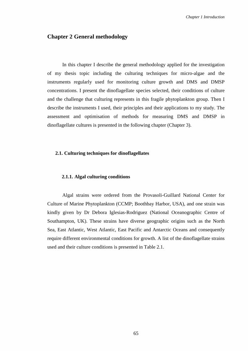
Chapter 1 Introduction
65
Chapter 2 General methodology
In this chapter I describe the general methodology applied for the investigation
of my thesis topic including the culturing techniques for micro-algae and the
instruments regularly used for monitoring culture growth and DMS and DMSP
concentrations. I present the dinoflagellate species selected, their conditions of culture
and the challenge that culturing represents in this fragile phytoplankton group. Then I
describe the instruments I used, their principles and their applications to my study. The
assessment and optimisation of methods for measuring DMS and DMSP in
dinoflagellate cultures is presented in the following chapter (Chapter 3).
2.1. Culturing techniques for dinoflagellates
2.1.1. Algal culturing conditions
Algal strains were ordered from the Provasoli-Guillard National Center for
Culture of Marine Phytoplankton (CCMP; Boothbay Harbor, USA), and one strain was
kindly given by Dr Debora Iglesias-Rodriguez (National Oceanographic Centre of
Southampton, UK). These strains have diverse geographic origins such as the North
Sea, East Atlantic, West Atlantic, East Pacific and Antarctic Oceans and consequently
require different environmental conditions for growth. A list of the dinoflagellate strains
used and their culture conditions is presented in Table 2.1.

Chapter 2 General methodology
66
Table 2.1 Dinoflagellate species investigated. Details include their full names, synonyms, strain codes, origin, toxicity and axenicity of the culture, and the medium, light intensity (LI in µmol photons m-2 s-1) and temperature (T °C) used in this study.
Latin name Synonyms Strain code Collection site Medium LI T °C T Alexandrium minutum Alexandrium ibericum CCMP113 Ria de Vigo, Spain L1 156 15 A Amphidinium carterae CCMP1314 Falmouth, USA f/2 204 22 A Crypthecodinium cohnii Glenodinium cohnii CCMP316 Unknown f/2 + NPM 127 22 A Heterocapsa triquetra Peridinium triquetra CCMP449 St Lawrence Estuary, Canada f/2 120 15 T Karlodinium veneficum Karlodinium micrum CCMP415 Norway, North Sea f/2 122 15
Kryptoperidinium foliaceum Peridinium foliaceum,
Glenodinium foliaceum
CCMP1326 La Jolla, California Bight, USA L1 204 22
PT Lingulodinium polyedrum Gonyaulax polyedra LP2810 San Diego, California, USA f/2 204 22
Polarella glacialis CCMP1138 McMurdo Sound, Antarctica Provasoli 101 4 A Scrippsiella trochoidea Peridinium trochoideum CCMP1599 Falmouth, USA f/2 102 15
A Axenic, T Toxic and PT Potentially toxic

Chapter 2 General methodology
67
The phytoplankton strains were grown in batch culture. Erlenmeyer flasks of
100 ml or 250 ml volume were filled up to one third of the volume with enriched
seawater media (different media are mentioned in Table 2.1) and capped with a cotton-
filled muslin bung covered with a piece of foil. Cultures were set in incubators (MLR-
351 Plant Growth Chamber, Sanyo, Loughborough, UK) under controlled conditions of
light intensity, photoperiod and temperature that ensured algal growth (Figure 2.1). The
photoperiod was 14:10 light:dark cycle with an incubator level 3/5 of maximum output
of light intensity supplying 150 ± 50 µmol photons m-2 s-1. The phytoplankton species
were divided into 2 groups depending on their tolerable range of temperature. The “cold
group” was placed in an incubator at 15 ºC and the “warm group” was placed in another
incubator at 22 ºC. These temperatures fall in the range of temperatures recommended
by the algal supplier but I have not tested if they are optimal.
Figure 2.1 Example of batch cultures of algae. (A) Several cultures of Crypthecodinium cohnii inside an incubator. (B) Erlenmeyer flask capped with a cotton bung. This culture of the heterotrophic C. cohnii displays a pale colour due to the lack of chlorophyll. (C) Example of 3 replicate cultures of the photosynthetic species Heterocapsa triquetra in stationary phase.
Manipulation of algal cultures requires a clean and sterile environment to avoid
contamination by other organisms (bacteria, fungi, other algae). In this regard, all

Chapter 2 General methodology
68
manipulations were conducted inside a laminar flow cabinet (Class II, Walker). This
cabinet and all items going inside were wiped with 70 % ethanol before use. Glassware
and culture media were autoclaved at 120 ºC for 20 minutes. Those cultures that were
axenic were checked regularly for axenicity by microscopic observation of samples
stained with DAPI (see section 2.1.3 for more details).
2.1.2. Medium preparation
All different media were enriched seawater. They were prepared with natural
seawater collected either from the open ocean, during oceanic cruises, or from coastal
waters pumped out from the North Sea by the CEFAS institute (Lowestoft, UK). This
water was stored in the dark at 12 ºC. The water was then filtered through a 0.2 µm pore
size cellulose acetate filter to eliminate potential organisms and particles. According to
the recommendations of Mclachlan (1973) to avoid precipitation in the medium, the
seawater was diluted with distilled water (2.5 %), and one drop of concentrated
hydrochloric acid (HCl) was added before autoclaving. HCl is used to compensate for
the increase of pH due to the loss of CO2 during the heating process (120 ºC during 30
minutes). Also, the nutrients were added aseptically several hours after cooling down.
Several media were made up for the panel of species grown: f/2, L1, Provasoli, f/2 +
NPM. All these medium recipes are based on the CCMP recipes (Wilson, 2009).
2.1.2.1. f/2 medium
The f/2 formulation (Table 2.2) is commonly used for phytoplankton (Guillard
and Ryther, 1962; Guillard, 1975). The denomination f/2 means that concentrations are
divided by 2 compared to the original formula. This formula contains macronutrients
including nitrogen and phosphorus and micronutrients such as a trace metals and
vitamins at concentrations that ensure algal growth. Silicate is a component of f/2 but
was omitted since it is not required for dinoflagellate growth.

Chapter 2 General methodology
69
Table 2.2 Components of f/2 medium without silicate. The proportions are listed for the stock solution preparation, the volume of the solution used and the final concentrations obtained in the medium (dH2O means distilled water).
Compounds Stock solution
g/L dH2O
Quantity in 1 L of medium
Medium molar concentration
M
f/2 Medium
NaNO3 75 1 ml 8.83 × 10⁻⁴ NaH2PO4 H2O 5 1 ml 3.63 × 10⁻⁵ Trace metals solution 1 ml Vitamin solution 1 ml
Trace metals Fe/EDTA 6.25 CuSO4 7H2O 9.8 1 ml 4 × 10⁻⁸ Na2MoO4 2H2O 6.3 1 ml 3 × 10⁻⁸ ZnSO4 7H2O 22 1 ml 8 × 10⁻⁸ CoCl2 6H2O 10 1 ml 5 × 10⁻⁸ MnCl2 4H2O 180 1 ml 9 × 10⁻⁷
Vitamin solution
Vitamin B12 (cyanocobalamin)
1 1 ml 2 × 10⁻¹⁰
Biotin 0.1 10 ml 4 × 10⁻⁹ Thiamine HCl 200 mg 6 × 10⁻⁷
Trace metals include iron, copper, molybdenum, zinc, cobalt and manganese.
These elements are essential for algae by participating in enzymatic and cytochrome
reactions but become toxic if introduced above a trace concentration (Reynolds, 2006).
Vitamins consist of cobalamin (vitamin B12), biotin and thiamine. Some algae from all
lineages (including dinoflagellates) are not able to synthesize some of these vitamins
and are called auxotrophs. Because bacteria may supply these vitamins it was initially
difficult to develop axenic cultures (Croft et al., 2005; Reynolds, 2006).

Chapter 2 General methodology
70
2.1.2.2. L1 medium
L1 medium (Table 2.3) differs from the f/2 formulation by the addition of trace
elements. Selenium, nickel, vanadium and chromium are added to the f formulation.
These metals are metabolised by certain microorganisms and used as enzyme
components (Madigan et al., 2003). Some organisms that have a higher demand of
nutrients should be grown in this richer medium rather than f/2.
Table 2.3 Trace metals of L1 medium
Compounds Stock solution
g/L dH2O
Quantity in 1 L of trace metal solution
Medium molar concentration
M FeCl3 ·6H2O 3.15 g 1.17 × 10⁻⁵ Na2EDTA 2H2O 4.36 g 1.17 × 10⁻⁵ CuSO4 ·5H2O 2.45 0.25 mL 1 × 10⁻⁸ Na2MoO4 ·2H2O 19.9 3 mL 9 × 10⁻⁸ ZnSO4 ·7H2O 22 1 mL 8 × 10⁻⁸ CoCl2 ·6H2O 10 1 mL 5 × 10⁻⁸ MnCl2·4H2O 180 1 mL 9 × 10⁻⁷ H2SeO3 1.3 1 mL 1 × 10⁻⁸ NiSO4 ·6H2O 2.7 1 mL 1 × 10⁻⁸ Na3VO4 1.84 1 mL 1 × 10⁻⁸ K2CrO4 1.94 1 mL 1 × 10⁻⁹
2.1.2.3. Provasoli medium
Provasoli medium is derived from f/2 with addition of 15 ml L-1 of an alkaline
soil extract (Provasoli et al., 1957). Soil extract was prepared with soil collected in the
grounds of the University in an area containing no fertilizer or pesticides. This soil was
diluted with two parts of distilled water and this solution was made alkaline with NaOH
(2 - 3 g L-1) before autoclaving for 2 hours. After cooling down, the mixture was filtered
and the extract was 50–fold diluted with distilled water. The soil extract supplies
organic substances which allows the culture of some algae unable to grow in only
mineral enriched medium such as the Antarctic dinoflagellate Polarella glacialis.
However, the key active compounds remain unknown due to the complexity of the soil
chemistry.

Chapter 2 General methodology
71
2.1.2.4. f/2 + NPM medium
The derivative f/2 + NPM medium (Table 2.4) contains an extra organic solution
necessary for the culture of heterotrophic or mixotrophic organisms (Guillard, 1960). I
used this medium for culturing the heterotrophic dinoflagellate Crypthecodinium cohnii.
f/2 components supply a mineral source of nutrients available for mixotrophs and also
metals and vitamins for mixotrophs and heterotrophs. The organic solution contains an
organic source of carbon and nitrogen. Acetate, succinate and glucose are potential
carbon sources. Peptone, bacto-tryptone (enzymatic digest of casein) and yeast extract
are potential nitrogen sources, supplying peptides and amino-acids.
Table 2.4 Organic solution of f/2 + NPM. This solution was added as 100 ml in 900 ml of f/2 medium.
Compounds Quantity in 1L of organic solution
Sodium acetate 1 g Glucose 6 g (di-) sodium succinate 6H2O 3 g Peptone 4 g Bacto-tryptone 1 g Yeast extract 0.1 g

Chapter 2 General methodology
72
2.1.3. DAPI staining
DAPI staining is a simple method to check algal culture axenicity. DAPI (4',6-
diamidino-2-phenylindole, Figure 2.2) is a fluorochrome commonly used to stain DNA.
This fluorochrome binds to minor grooves of double-stranded DNA and intercalates
between adenosine and thymine bases (Kapuscinski, 1995). When excited with UV light
(360 nm), this stable complex fluoresces by emitting blue light (450 nm). Observation
of stained DNA by fluorescence microscopy gives a clear view of bacterial presence or
absence among algal cells (Porter and Feig, 1980).
Figure 2.2 DAPI structure (http:// sigmaaldrich.com)
To avoid bacterial contamination during the staining process, I used 0.2 µm
filtered reagents and rinsing seawater. Also, I performed all manipulations wearing
protective clothes in a fume cupboard to prevent hand from the carcinogenic properties
of this dye.
The culture sample (1-5 ml) was fixed with a reactive mixture of lugol (3 µl ml-1
of aqueous KI 10 % w/v and iodine 5 % w/v) and formalin (50 µl/ml of 20 % aqueous
formaldehyde with 100 g L-1 hexamine) that keeps cells more intact than lugol only
(Sherr et al., 1993; Sherr and Sherr, 1993). Then, sodium thiosulphate (1 µl/ml of a 3 %
solution stored in the fridge) was added to discolour the iodine colouration that would
reduce the DAPI fluorescence and finally, DAPI (10 µl ml-1 of 1mg ml-1 solution,
Sigma-Aldrich, D9542) was added. The culture sample was left to react with the
chemicals for at least 5 minutes. The stained sample was filtered by means of a water
vacuum pump through 2 filters: a 0.2 µm pore black polycarbonate filter of 25 mm
diameter supported by a 0.45 µm pore nitrate cellulose backing filter. The filters were
then rinsed with sterile seawater. The black filter was collected and deposited on a slide
bearing a drop of immersion oil for adherence. The filter was covered by a second drop
of oil immersion and a cover slip. Slides were examined under the microscope (BX40,
Olympus, Essex, UK) at 100-fold magnification objective with epifluorescence light

Chapter 2 General methodology
73
and a UV light filter. The dinoflagellate cells fluoresce with intense blue colour (Figure
2.3). If bacteria are present, the black filter background appears cloudy at low
magnification (20-fold) and bacteria are visible with elongated blue shapes at high (100-
fold) magnification (illustrated in Figure 2.3).
Figure 2.3 Comparison of axenic (A and B) and non-axenic (C and D) cultures of dinoflagellates stained with DAPI and observed with an epifluorescent microscope. Axenic culture of (A) Crypthecodinium cohnii and (B) Heterocapsa triquetra observed at the 100-fold magnification objective. Samples were taken at the end of the experiments in order to check the axenicity. Only algal cells fluoresce, absence of bacteria is proved by no fluorescing background. Bacterial contamination of Crypthecodinium cohnii culture (C) At 20-fold magnification objective, the fluorescent algal cells appear superimposed on a cloudy background. (D) At 100-fold magnification bacteria appear as elongated sticks.
A B
C D

Chapter 2 General methodology
74
2.1.4. Handling and sampling techniques for dinoflagellate cultures
Dinoflagellates are amongst the most fragile phytoplankton group and are
subject to cell breakage when agitated or manipulated in laboratory culture (detailed in
section 1.3.12). In this study, special attention was given to the culture handling and
sampling techniques to reduce the physiological damage of cells.
To minimise disturbing dinoflagellate physiology, cultures were grown in batch
mode without agitation. Only Crypthecodinium cohnii cultures were agitated once a day
to avoid aggregate formation due to the production of extra-cellular polysaccharides.
Prior to sampling, flasks were manually agitated in order to collect a
homogenous sample, representative of the culture. The flasks were gently swirled 2 to 3
times and sampled straight away. For each flask sampling, 1 culture aliquot was taken
and used for the measurement of several parameters. The sampling of 1 culture aliquot
rather than 1 sample for each parameter measurement reduces the turbulence and stress
applied to the culture flask. Cultures were kept outside of the incubator for the shortest
time possible to minimise the effect of different environmental conditions of light and
temperature. For the polar species Polarella glacialis, the culture was carried between
the incubator and the safety cabinet using a coolbox containing ice packs.

Chapter 2 General methodology
75
2.2. Analytical instrumentation and techniques
This section describes the techniques used to characterise the cultures and the
parameterisation includes the cell counts, cell volume, chlorophyll a, CHN content,
DMS and DMSP content.
2.2.1. Particle counter for cell density and cell volume measurements
Cell density may be measured by counting cells on a haemocytometer placed
under a microscope or using a particle counter that automatically counts a large number
of cells. The use of the particle counter which is less laborious and less time consuming
was favoured. However, I compared the 2 methods to assess the use of the particle
counter for dinoflagellate cells that have a close to spherical shape.
A particle counter (Coulter Multisizer 3, Beckman, High Wycombe, UK),
equipped with a 100 µm aperture tube, was used to monitor the following growth
parameters: cell density (cell ml-1), total cell volume (µLcell L-1 that is equivalent to
106 µm3 ml-1) and volume per cell (µm3).
The Coulter particle counter principle is based on the displacement of an
electrolyte volume by a particle passing between two electrodes. The changing flow
leads to a change in the impedance that is transformed into a pulse proportional to the
particle volume. The measurement of several pulses in a given volume is equivalent to a
measure of the particle concentration in the sample. The particle volume is
automatically converted to particle diameter by calculations set for spherical particles.
The change in impedance cannot convey additional information of shape and colours.
In general, I diluted 1 ml of culture sample in 10 ml of filtered seawater as the
electrolyte. The dilution avoids saturating the electrodes and could be increased for
dense cultures in stationary phase, for instance 0.5 or 0.2 ml of culture sample could be
added. The cell density is the integral of the peak obtained on the computer interface (an

Chapter 2 General methodology
76
example is shown on Figure 2.4). Limits of the peak are taken outside of the peak area
and are kept constant for a species. The total cell volume (CV) was read similarly to the
cell density. The volume of the cell was derived from the reading of the mean cell size
displayed by the statistics of the software then calculating the spherical volume
associated. Indeed, the software displays the mean size value derived from the particle
volume obtained for an assumed spherical particle. The size value is not representative
for dinoflagellate cells which are not exactly round shaped, but the calculation of the
volume gives a correct representative dimension independent of the dinoflagellate
shape.
Figure 2.4 Example of cell density result obtained with the particle counter for the measurement of Heterocapsa triquetra culture. The reading of the cell density is taken between a lower and upper limit of particle diameter on both sides of the peak. (here, 54919 cell ml-1).
To assess the use of the particle counter for dinoflagellate cultures, I have
compared cell density measurements performed with the particle counter and the
counting with a haemocytometer under the microscope (BX40, Olympus, Essex, UK).
Using the haemocytometer, the method was to count 5 haemocytometer cells on 5 slides
and to calculate the average. Four different dilutions of a Heterocapsa triquetra culture
(25, 50, 75, 100 %) were counted with the 2 methods to assess how the particle counter
measurements respond to a range of low to high cell density. The results are shown in
Figure 2.5.

Chapter 2 General methodology
77
0E+00
1E+05
2E+05
3E+05
4E+05
0 20 40 60 80 100
% of culture in dilution
Cel
l den
sity
(cel
l ml
-1)
R2 = 1.00
R2 = 0.96
0.0E+00
5.0E+04
1.0E+05
1.5E+05
2.0E+05
0 25 50 75 100% culture in dilution
Cel
l den
sity
(cel
l ml
-1)
Figure 2.5 Comparison of cell density results obtained using the particle counter (●) and the haemocytometer (○). Four dilutions of Heterocapsa triquetra culture sample were determined (25, 50, 75 and 100 % culture). R2 is the correlation coefficient associated with the linear regression of the culture dilution and the cell density measured with the particle counter.
The use of particle counter and haemocytometer gave very similar values of cell
density. The linear response of the particle counter was better than the haemocytometer.
The use of the particle counter was therefore satisfactory for dinoflagellates with a
similar shape to Heterocapsa triquetra (almost spherical shape, like a diamond) and
allowed fast measurements of total cell volume and volume per cell. All other
dinoflagellate species investigated in this study have a similar shape to H. triquetra,
round to oval shape. Given that many experiments have been performed with
Crypthecodinium cohnii, which also has a similar shape to H. triquetra, I tested
C. cohnii for linearity of response using the particle counter. In Figure 2.6, 3 samples of
a dense C. cohnii culture were prepared at 3 dilutions (100, 50 and 20 %) and show
satisfactory linearity of the measurement with this species.
Figure 2.6 Linearity response of cell density measurements performed with the particle counter using different dilutions (20, 50 and 100 %) of 3 samples of a dense Crypthecodinium cohnii culture. For the 3 samples, the linear regression of the culture dilution and the cell density measured using the particle counter gave a correlation coefficient R2=0.99.

Chapter 2 General methodology
78
To assess the precision of the particle counter, I took 5 measurements of a
H. triquetra culture sample in exponential growth phase and calculated the uncertainties
(Table 2.5, formulas are detailed in section 2.2.5). I obtained a RSD of 2.2 and 2.3 %
for cell density and total cell volume (CV) respectively and a precision px of 2.7 and
3.9 %. These values showed that the uncertainty of the measurement with the particle
counter was low.
Table 2.5 Calculation of uncertainties (σx and px) for the measurement of cell density and total cell volume (CV) based on 5 replicate measurements of a H triquetra sample.
Replicates of measurement Cell density (cell ml-1) Total CV (µL cell L-1)
1 35559 89.3 2 37092 93.0 3 36579 92.1 4 35523 88.8 5 37114 93.3 Average 36373 91.3 σx 789.6 2.10 px 980.2 3.54
2.2.2. Gas Chromatography for DMS and DMSP measurements
Gas chromatography is a technique of analytical chemistry used to separate,
identify and quantify gaseous compounds. A gas chromatograph (Schimadzu GC-2010,
Milton Keynes, UK) was used for DMS and DMSP measurements. It was equipped
with a fused-silica capillary column 30 m × 0.53 mm CP-SIL 5CB (Varian, Oxford,
UK). According to the supplier, this column is appropriate for a large panel of
compounds including sulphur compounds and separates mainly by boiling point.
A flame photometric detector (FPD) was used due to its sensitivity to sulphur
compounds. A mix of hydrogen (60 ml min-1) and air (70 ml min-1) supplied the flame
which burned the sulphur compounds subsequently emitting a light signal. Helium was
used as neutral carrier gas (28 or 35 ml min-1 for DMS and DMSP methods,
respectively) to convey the sample injected through the column.
When DMS is released by the column at a specific time called the retention time
(RT), DMS is perceived by the FPD which emits a signal amplified by photomultiplier
tubes and transmitted to the computer. A peak appears on the computer interface and

Chapter 2 General methodology
79
when this peak is higher than the background noise of the chromatograph, the peak is
automatically integrated by the software. An example of peak obtained is shown on
Figure 2.7. The RT and the shape of the peak (sharp and narrow with maximum
possible height) are dependent on the gas chromatoghraph (GC) settings including
column temperature, gas flow rate and temperature. The GC settings used in this study
are described for each method (purge and trap and headspace analyses) in the following
section 2.2.3.1. In the optimum range of the detector, the response is non-linear but
approximates a square root function. Therefore, the square roots of peak areas (SQRT
PA) are used for the DMS quantification by comparison with a concentration range of
standards. The chromatograph was calibrated using commercially available DMSP that
is converted to DMS by cold hydrolysis (Centre for Analysis, Spectroscopy and
Synthesis, University of Groningen laboratories, The Netherlands). The linear range of
the FPD for sulphur compounds is limited to 3 orders of magnitude.
Figure 2.7 Example of GC chromatogram for a DMSP sample. The peak has a Retention Time (RT) of 2.05 min and the peak area is automatically integrated by the software (delimited by the red line and arrows).
DMSP (not volatile) needs to be converted to DMS prior to measurement and
total DMSP is about 100-fold higher in concentration in cultures than DMS. Thus, there
are considerations of sample preparation, sample size and calibration range. To measure
DMS in cultures, I used a “purge-and-trap” method which involves extraction of almost
all the gas from a liquid sample and cryogenic concentration in a sample loop. For
DMSP measurements in cultures, I performed the conversion to DMS by chemical
cleavage of DMSP to DMS and acrylate with a strong base (NaOH). DMSP was
measured by gas chromatography using the headspace method. The calculation of

Chapter 2 General methodology
80
DMSP concentration was based on the 1:1 ratio of this chemical conversion (equation
1); (Challenger and Simpson, 1948; Cantoni and Anderson, 1956; Ackman et al., 1966).
(CH3)2S+CH2CH2COO- → CH3SCH3 + CH2=CHCOO- + H+ (1)
DMSP DMS Acrylate
These methods of sample preparations are detailed in the following section.
2.2.2.1. Purge-and-trap method
This method was based on that developed by Turner et al., (1990) to allow
measurement of DMS at a low concentration level, initially in seawater field samples.
As indicated in the name, the first step consists of purging a liquid sample with a
purging gas in order to extract, in a second step, DMS that concentrates in a cryogenic
trap before injecting it into the GC. Figure 2.8 illustrates the purge-and-trap system.
Figure 2.8 Diagram of the purge-and-trap system. It is composed of (a) sample injection port, (b) glass purge tube, (c) glass wool-containing water trap, (d) counter flow Perma Pure gas drier, (e) manual six-port sample stream switching valve, (f) in-house cold trap consisting of: (g) Dewar flask with liquid nitrogen, (h) heating resistor and (i) temperature probe, (j) trap temperature controller, (k) flow meter, (l) GC. Two nitrogen inputs are coming in the system: N2 1 is the purging gas that carries DMS along the system; N2 2 is the drier flow of the Perma Pure gas drier. The rates of these two flows are checked by the flow meter.
N2 2
b
c d
e
f
g h
i
j
k
l
N2 1
a
He

Chapter 2 General methodology
81
The procedure was to introduce the filtered culture sample through the injection
port (a) into the purge tube (b) which received the purging gas (nitrogen, oxygen-free
grade, purified through an activated charcoal filter) through a fine glass frit which
generated streams of small bubbles. Using an elongated purge tube increased the path
length for nitrogen bubbles and enhanced the purging efficiency. The top of the purge
tube was connected by 1/8-inch-o.d. PTFE (polytetrafluoroethylene) tubing to a series
of water traps in order to remove water vapour from the gas extract, thus avoiding ice
formation and blockages in the cryogenic trap (f). The first water trap (c) was a glass
wool-containing glass tube followed by a gas drier (d, MD-050, Perma Pure) as a
second trap. The gas drier was composed of a Nafion membrane of 72 inches length
which was permeable to water vapour and not DMS: the water vapour was rapidly
removed by a counterflow of N2 (N2 2, 150 ml min-1). The sample gas extract passed
through the cold trap (f, 25 cm 1/8th inch PTFE tubing wound in a double loop
maintained at – 150 ºC ± 5 ºC) where DMS was accumulated and N2 passed to the flow
meter (k). The cold trap was suspended in the headspace of a Dewar flask containing
liquid nitrogen and the temperature was regulated by a simple feed-back system
consisting of a temperature sensor (i) attached to the trap, a resistor (h) immersed in the
liquid N2, and an electronic control box (j, designed and built at UEA). If the
temperature of the headspace rose above -150 ºC, the resistor generated heat and
increased vapour pressure of the liquid N2 until the headspace achieved the target
temperature. This technique was used to avoid the risk of trapping oxygen at lower
temperature which, if injected into the detector, can change the flame characteristics and
sensitivity. The sample purging lasted for several minutes to allow the accumulation of
DMS in the cold trap. I determined that 10 minutes of purging were required for a 5 ml
filtered sample with a purge flow rate of 60 ml min-1 (detailed in chapter 3, section
3.5.3.1). The cold loop was promptly heated in boiling water while the sample was
flushed by the He carrier gas into the GC (l) by switching a six-port valve (e). The gas
flow settings of the purge and trap system and the GC settings associated with this
method are detailed in Table 2.6.

Chapter 2 General methodology
82
y = 43375x - 1006
R2 = 1.00
0
20000
40000
60000
80000
0.0 0.4 0.8 1.2 1.6S (nmoles)
SQ
RT
PA
Table 2.6 GC and flow settings used for the purge and trap system.
GC components Temperature (ºC)
Gas Flow (ml min -1)
Pressure
Total flow 28.4 Injector 200 Helium Purge flow 3
45.6 kPa
Column 60 Hydrogen 60 Detector 250 Air 70
Purge and Trap components Purging flow Nitrogen 60 Drying flow (Nafion drier) Nitrogen 150
Standards were prepared in 5 ml gas tight glass vials closed with a screw cap
and a Teflon septum. DMSP stocks were prepared by dilution of purchased DMSP in
distilled water to a concentration of 0.8 µg S ml-1 and stored at -20 °C. The stocks were
then defrosted and diluted again with distilled water to solutions of concentrations
ranging from 0.004 to 0.016 µg S ml-1. Several DMSP standards were prepared by
adding DMSP solutions, distilled water up to 4 ml and 1 ml of 10 M NaOH. The
mixture was left overnight in the dark to react. Each standard concentration was
prepared in duplicate and all standards covered a concentration range from 0.002 -
0.294 µmol L-1 that was broader than the span of sample concentrations. The detection
limit was 0.002 µmol L-1 based on the lowest concentration used in my calibration.
Concentrations measured were well above this detection limit. Standards were injected
into the purge tube using a 5 ml disposable syringe.
The method was assessed by checking the linearity of the calibration curve and
by calculating the uncertainty of 5 standard replicates. I obtained excellent linearity
using this method as shown in Figure 2.9 (R2=1). Also I obtained very good precision
with 5 standards replicates (RSD = 3.2 % and px= 3.9 %).
Figure 2.9 Example of calibration curve obtained with the purge and trap method. (SQRT PA = square root of the peak area) for duplicate DMS standards ranging from 0.01 to 1.47 nmoles. The linear regression curve is shown with its correlation coefficient R2.

Chapter 2 General methodology
83
DMS was measured in the aqueous fraction of cultures. A sample preparation
system for small volumes (< 2ml) was built. The S-S-PT system (Syringe pump -
Sample loop - Purge and Trap, Figure 2.10) was used to filter and introduce the culture
sample into the purge and trap system. The aim was to introduce precise and
reproducible sample volumes.
Figure 2.10 S-S-PT system: Syringe pump - Sample loop - Purge and trap system used for sample preparation (filtration and injection into purge tu be) before DMS measurement.
From a culture aliquot, 5 ml of sample was taken up with a 5 ml disposable
syringe fitted with a stopcock and placed on the syringe pump (KDS220, Linton
Instrumentation, Diss, United Kingdom). The pump was set to a filtration speed of
14 ml min-1 (maximum speed available for this syringe volume) as I observed that rapid
filtration minimised the conversion of DMSP to DMS (chapter 3, section 3.5.1.2.3 and
3.5.3.2). Only 4.7 ml of the sample was filtered to avoid stressing the cells as I observed
that a gradient of DMS was generated over the filtration (chapter 3, section 3.5.3.2). The
filtration was through a filter holder (Swinnex, Millipore, Watord, UK) containing a
25 mm diameter glass-fibre filter (Whatman, GF/F). This filter holder was connected to
a sample loop which ensured accurate delivery of small sample volumes. The sample
loop was made out of 1/8th inch of PTFE tubing and was connected to a six-port valve.
N2
Syringe pump
Filter holder with a filter
Sample loop
Purge tube connected to the purge and trap system
Waste sample outlet
6-port valve
N2

Chapter 2 General methodology
84
After loading, the sample was flushed into the purge tube. Loops of different volumes
were used (0.2, 0.5 and 2 ml).
2.2.2.2. Headspace method for DMSP
This method has been developed by Steinke et al., (2000) to measure DMS
production by DMSP lyase and has been adapted in this study for measuring DMSP.
This technique allows measurement of elevated concentrations of DMSP (DMS up to
80 µmol L-1) in a liquid sample made alkaline by NaOH addition. Liquid samples are
equilibrated with a headspace of air and the partition coefficient for DMS is controlled
by temperature.
For total DMSP (DMSPT= particulate + dissolved DMSP + DMS), 1 ml of
culture was directly introduced into a 5 ml vial containing 2 ml of 0.75 M NaOH. The
vial was immediately closed with a screw cap and a Teflon septum, then agitated.
Samples were left in the dark to react (> 12 h) and could be stored at that stage in the
dark for two weeks. Prior to the analyses, samples and standards were put overnight (12
h) on a heating block set at 30 ºC to allow DMS to equilibrate between the liquid and
gas phase. Eighty µl of the headspace was sampled and injected into the gas
chromatograph by means of an autosampler (Multipurpose sampler MPS, Gerstel,
Mülheim an der Ruhr, Germany) composed of an automatic arm equipped with a 100 µl
heated glass syringe. The GC settings applied for this method are described in Table
2.7.
Table 2.7 Description of the GC settings associated with the headspace method
GC components
Temperature (ºC)
Gas Flow (ml min -1)
Pressure
Total flow 34.7 Injector 200 Helium
Purge flow 3
68.5 kPa
Column 120 Detector 250 Hydrogen 60 Air 70
Chapter 2 General methodology
85
y = 529.94x + 69.95
R2 = 1.00
0
10000
20000
30000
40000
50000
0 20 40 60 80
DMSP concentration in vial (µM)
SQ
RT
PA
A concentration range of duplicate DMSP standards was used to calibrate the
GC and calculate the DMSP concentration of the sample. The standards were prepared
as described in Steinke et al. (2000) by adding a drop of DMSP stock solution into the
cap of the vial. The vial, containing 3 ml of 0.5 M NaOH, was closed and agitated. The
samples and the standards had the same headspace volume and final NaOH
concentration. An example of a calibration curve is shown in Figure 2.11.
Figure 2.11 Example of calibration obtained for headspace method of DMSP measurement by gas chromatography. (SQRT PA= square root of the peak area).
The uncertainty of this method was RSD= 1.8 % and px = 2.2 % for standards
(n=5) and RSD= 1.3 % and px = 1.6 % for H. triquetra samples (n=5). When
normalising DMSP to cell number or total CV, the uncertainty was RSD= 2.5 % and
2.6 % respectively and px=1.6 %.
2.2.2.3. Headspace method for in vitro DMSP lyase activities
The method was based on the description from Steinke et al., (2000). One
hundred ml of dinoflagellate culture was sampled in mid to late exponential phase. The
culture sample was filtered through a 2 µm pore size polycarbonate filter of 47 mm
diameter. The speed of filtration was gently improved by means of a hand vacuum
pump (< 15 cm Hg). The filter was folded 3 times and cut into pieces of a suitable size
to fit into a 1.8 ml sterile cryogenic vial (cryotube, Thermo Scientific Nunc) containing
1.7 ml of 300 mM tris buffer in 500 mM NaCl and adjusted to pH 8.2 with concentrated
HCl. The sample was sonicated (sonicator Microson XL2000, Misonix, Farmingdale,

Chapter 2 General methodology
86
USA) at 5 W, 5 pulses of 5 s with 10 s intervals to ensure cell extraction without heating
and enzyme damage. The cryogenic vial containing the enzyme extract was snap-frozen
in liquid nitrogen and stored at – 80 ºC for later analysis. This cold storage after
extraction does not affect the DMSP lyase activity (Steinke et al., 2000).
Prior to measurement of DMSP lyase activity, the enzyme extract was defrosted
and kept cold in ice. Then, 295 µl of enzyme extract was introduced into 2 ml glass
vials closed with a screw cap and a Teflon septum. The vials were incubated at 30 ºC on
a heating block in agreement with the standard temperature recommended to perform an
enzyme assay (International Union of Biochemistry, 1965). Fifteen µl of the vial
headspace was auto-sampled at 10 to 15 minute intervals before and after the addition of
5 µl 1.2 mM DMSP stock solution. Two samples were taken before the addition of
DMSP to obtain the baseline activity. Then, DMSP (previously purged for 30 minutes
to reduce the amount of free DMS) was added by rapidly injecting the DMSP with a
Gilson pipette and immediately closing and agitating the vial. DMS production was then
monitored over time by performing 4 sequential measurements. To determine the
DMSP lyase activity, DMS production was compared between vials containing enzyme
extract and vials containing tris buffer (295 µl), both receiving DMSP addition. An
example of the data obtained is shown in Figure 2.12.
Figure 2.12 Example of DMS production measured in Lingulodinium polyedrum extract samples (L. poly) and in tris buffer (Buffer). A and B are duplicate samples of the algal cell extract. The dotted line shows the addition of DMS at time 0.
y = 0.25x - 0.70
R2 = 1.00
y = 0.07x + 0.21
R2 = 1.00
0
2
4
6
8
10
12
-30 -20 -10 0 10 20 30 40 50
Time (min)
DM
S (µ
M)
Buffer ABuffer BL. poly AL. poly BSeries1

Chapter 2 General methodology
87
y = 692.12x - 85.19
R2 = 1.00
0
5000
10000
15000
20000
25000
0 10 20 30
DMSP concentration in vial (µM)
SQ
RT
PA
DMS production rate was calculated from the slope generated by all the
sampling points after DMSP addition, corrected by a loss rate (+ 6 %) due to the DMS
loss of the pierced septum of the vial and corrected by the DMS production measured in
the buffer vials that represent the DMSP abiotic cleavage as applied by Steinke et al.
(2000). The gas chromatography system was calibrated using duplicate standards
prepared by mixing 300 µl NaOH with a drop of DMSP stock solution in the 2 ml vials
and covering a concentration range of 0.10 - 30 µmol L -1. An example of a calibration
curve is shown in Figure 2.13. The detection limit of the assay was based on the average
DMS production detected in buffers of 7.34 nM h-1.
Figure 2.13 Example of calibration curve obtained by gas chromatography for DMSP lyase measurements for duplicate DMSP standars ranging from 0.1 to 30 µmol L-1. The linear regression curve is shown with its correlation coefficient R2. (SQRT PA= square root of the peak area)

Chapter 2 General methodology
88
2.2.3. Fluorometer and in vitro determination of chlorophyll a by acetone
extraction and acidification method
A fluorometer measures the fluorescence emitted by pigments such as
chlorophyll. When excited with low wavelengths of high energy light (blue light at ~
450 nm) the chlorophyll fluoresces by emitting longer wavelengths of lower energy
light (red light at 680 nm). The fluorescence is generally proportional to the chlorophyll
a (Chl a) concentration, if no other pigment interferes with the signal. The AU-10 Field
Turner Designs fluorometer was equipped with a 10-045 lamp, an excitation filter of
340 - 500 nm and an emission filter > 650 nm.
The method of chlorophyll extraction from cells by acetone and acidification
was applied, according to the methods described by Parsons et al (1984). Phaeophytin is
a natural degradation product of Chl a (i.e. loss of the Mg ion), present in variable
amounts and can interfere with the fluorescence signal of Chl a (structure of Chl a is
shown in Figure 2.14). In order to correct for this background fluorescence, the sample
is acidified, to convert any Chl a to phaeophytin. The equations (4 and 5 detailed later)
used for calculating the Chl a concentrations incorporate correction for the original
phaeophytin contribution.
Figure 2.14 Chemical structure of chlorophyll a.
For the sample preparation, 5 ml of culture were filtered by gravity through a
GF/F filter of 25 mm of diameter on a glass filtration unit. The use of a filter holder
without a fritted glass filter support allowed fast gravity filtration of a couple of

Chapter 2 General methodology
89
minutes. The filter was folded in half, wrapped in foil and snap-frozen in liquid nitrogen
prior to storage in a freezer at – 80 ºC. This storage prevents the degradation by light
and heat without significant chlorophyll loss for up to a month, as recommended by the
Method 445.0 Revision 1.2 from the U.S. Environmental Protection Agency (Arar E. J.
and Collins G. B., 1997)
The following steps of Chl a extraction from cells were performed in a low light
environment to prevent chlorophyll degradation. The filter was introduced into a 20 ml
glass vial, closed with a screw cap, then 10 ml of 90 % acetone (prepared by mixing
analytical grade acetone with distilled water) were added. These steps were repeated for
each filter and the set of vials was covered with foil and placed in a fridge at 4 ºC in the
dark to achieve the chlorophyll extraction during 20 to 24 hours. During this period,
vials were manually agitated several times to improve the extraction. About 9 ml of
extract were poured into a borosilicate tube for fluorescence reading. The fluorescence
was measured before and after acidification with 3 drops of 8 % HCl. The chlorophyll a
concentration was calculated according to equation 2 (Parsons T.R. et al., 1984):
Chlorophyll a in mg/L = FD × (Y/Y-1) (U - A) v / V (2)
Where,
FD = mean (Chla concentration / U) for chlorophyll standards
Y = mean (U / A) for chlorophyll standards
U = fluorescence before acidification
A = fluorescence after acidification
v = volume of extract (ml)
V = volume of culture (ml)
The fluorometer was calibrated with a range of pure Chl a solutions of known
concentrations (0.006 - 0.227 mg L-1) on the day of the sample analyses. These standard
solutions were prepared from a stock solution of 1 mg L-1 of Chl a extracted from
Anacystis nidulans, Cyanophyceae (Sigma-Aldrich, C6144) and diluted in 90 %
acetone. Prior to the standard preparation, the concentration of the stock solution was
checked with a spectrophotometer (Lambda 25, Perkin Elmer) and the average value of

Chapter 2 General methodology
90
y = 352.21x + 1.34
R2 = 1.00
y = 93.46x + 0.13
R2 = 1.00
0
20
40
60
80
100
0.00 0.05 0.10 0.15 0.20 0.25
Chl a concentration (mg L -1)
Flu
ores
cenc
e (fl
uore
scen
ce
units
)
Unacidified (U)Acidified (A)U/A
the results obtained from calculations of the Scor (U.N.E.S.C.O., 1966) and Lorenzen
methods (Lorenzen, 1967), equations 3 and 4, was used.
Scor Chl a = 11.6(U665) – 0.14(U630) – 1.31(U645) (3)
Lorenzen Chl a = 26.7(U665-A665) (4)
Where U is the absorbance of the solution and A is the absorbance after acidification
with 2 drops of 8 % concentrated HCl (which is equivalent to 2.88 % chloride).
An example of fluorometer calibration is illustrated in Figure 2.15. The
fluorometer has a linear response to a range of Chl a standards measured before and
after acidification. The average ratio U/A is used in the calculation as detailed in
equation 2. The fluorescence measurements on dinoflagellate species showed a RSD
that ranged between 14 and 55 % (n=9); (see chapter 4 for more details).
Figure 2.15 Example of linear response of the fluorometer for fluorescence measurements of non-acidified and acidified Chl a standards. The average ratio U/A is used in the calculation of sample Chl a concentrations.

Chapter 2 General methodology
91
2.2.4. Elemental analyser and CHN measurement
The elemental analyser (CE440, Exeter Analytical, Coventry, UK) was used to
determine the C and N composition of a culture sample. I was interested in using the C
and N content as another culture characteristic (Matrai and Keller, 1994; Menden-Deuer
and Lessard, 2000) and to express DMSP concentration per C units. These data are
particularly useful for modelling studies.
The analysis was performed on cells harvested by filtration and dried. In this
regard, 2 ml of culture were filtered through a filter holder (Swinnex, Millipore,
Watord, UK) containing a 13 mm diameter GF/F filter (Whatman, GF/F), previously
combusted at 450 ºC for 4 hours in a muffle furnace. The gentle filtration was
performed using a hand vacuum pump (< 5 cm Hg). The filter was folded in half,
wrapped in foil and rapidly cooled in liquid nitrogen prior to storage at – 80 ºC. The
samples were stored for less than a month before analysing a set of samples. Two days
before analysis, the samples and blank filters were dried at 35 ºC for 48 hours then
placed in nickel capsules, previously combusted at 1000 ºC for 1 h.
Figure 2.16 is a schematic diagram of the components of the instrument. To start
the analyses, the samples were placed into an autosampler where they were purged
continuously with helium which is used as a carrier gas through the analytical system.
The first sample fell into a combustion chamber injected with pure oxygen, inside a
high temperature furnace at 928 ºC. These conditions lead to the combustion of the
sample into elements (C, H, N). The gas mixture was carried through oxidation reagents
that transform C into CO2, H to H2O and N to NxOy. The oxidation reagents also
removed other unwanted elements like phosphorus, halogens and sulphur. Then the gas
sample passed in a copper column which reduced the nitrogen oxides to nitrogen and
eliminated the surplus of oxygen.

Chapter 2 General methodology
92
Figure 2.16 Schematic diagram of the CHN elemental analyser (CE440, Exeter Analytical).
The gas sample was pulsed into the mixing chamber that ensured constant
pressure, temperature and homogeneity of the sample. The gas stream then flowed
through a series of 3 thermal conductivity detectors (TCD). First, a water absorption
trap was connected to a TCD which detected the water content of the gas stream before
and after it passed through the water trap. Similarly, a CO2 trap connected to another
TCD resulted in C detection. Finally N was detected by a TCD which compared the gas
stream containing N and He with another flow of pure He as reference. The detection
signal is proportional to the concentration of components in the sample and this
concentration is calculated from standards. The analyser was adjusted with 10 standards
of 2 mg of acetanilide (C8H9NO) which were measured at the beginning of the run and
at intervals between samples. The sample measurements were blank corrected with
analysis of pre-combusted, blank filters. The RSD (σx) obtained for the C and N
measurements ranged between 7 and 32 % (n=9); (see Chapter 4 for details).
Autosampler
Combustion chamber
TCD for N
He O2
Oxidation reagents
Copper
O2 CO2 H2O NxOy
CuO
C H N
975ºC
519ºC
Mixing chamber
TCD for C CO2 trap
TCD for H H2O trap
CO2 H2O N2
Chapter 2 General methodology
93
2.2.5. Uncertainty analyses
For the analytical methods that were used commonly through my study (particle
counter, purge-and-trap and GC , headspace analyses and GC), I have assessed the
precision of the measurements using 5 replicates of standards or of Heterocapsa
triquetra culture samples in exponential phase. The uncertainty was calculated in two
ways (equation 1): the standard deviation of the sample (σx) also reported as the relative
standard deviation (RSD) and the precision (px) obtained by using the t-statistic
derivation for small numbers (< 30) of samples (equation 5, Beckwith T.G. et al. 1993).
x ± px px = t 0.025, ν (σx/ √n) (5)
Where x is the average of the result, t is the value read out from the student
distribution for a probability of 0.025 or 95% confidence and a degree of freedom ν
equivalent to n – 1 where n is the number of samples.
The uncertainty of a result involving the measurement of 2 parameters was
calculated with the formula of error propagation (equation 6, Lichten 1999). For
instance, the DMSP value (z) expressed per cell volume includes the errors of
measurement for both DMSP (x) and cell volume (y).
z = x / y pz = p(x/y) = √ [(x/y) 2 ((px/x)2 + (py/y)2)] (6)

Chapter 3 Testing and optimising the methodology
94
Chapter 3 Testing and optimising the methodology for DMSP and
DMS measurement in dinoflagellate cultures
Abstract
I have performed a series of experiments on Heterocapsa triquetra batch cultures to
optimise the measurement of DMSP and DMS in dinoflagellate cultures. I found that
DMSP concentrations were increasing during the photoperiod and at the end of the
exponential growth phase. The sampling time for further experiments was, therefore,
established to be at the mid-time of the photoperiod and at mid-exponential phase to
enable the determination of the effect of the experimental treatment. I assessed the mass
balance of measurements of total DMSP (DMSPT) and the sum of its individual
fractions (DMSPp, DMSPd and DMS) and found that measurement of DMSPT gave 25
% more than the sum of the parts. As DMSPp was the major fraction, I investigated the
method of DMSPp measurement with a special focus on the filtration methods. Overall,
the use of a hand vacuum pump, an automated syringe pump and gravity filtration did
not show significant difference in DMSPp concentration. When using the syringe pump,
DMSPp concentrations were increased and the analytical uncertainty reduced by setting
the highest delivery speed. DMSPT measurements showed a better analytical uncertainty
than DMSPp measurements and no significant difference with DMSPp concentrations,
which supports the use of DMSPT as a surrogate of intracellular DMSP. DMS
measurement was also optimised with the use of the syringe pump at high delivery
speed and a sample loop to inject the constant volume of samples. The analytical
uncertainty of culture samples were rather higher than the standards but the study
showed that shortening the filtrations and using a small volume resulted in lower DMS
concentrations.

Chapter 3 Testing and optimising the methodology
95
3.1. Introduction
Prior to investigating scientific questions on DMSP and DMS production by
dinoflagellates, the first issue was to assess and optimise the methods applied for the
measurement of these parameters. Intracellular DMSP is also called particulate DMSP
(DMSPp). The fraction of DMSP released into the culture medium by cell lysis and
exudation is called dissolved DMSP (DMSPd). It can then lead to the DMS formation
by enzymatic cleavage. Some DMS may also potentially be contained by cells: it was
initially thought that the DMSP lyase was segregated from its substrate DMSP in the
cell (Wolfe and Steinke, 1996) but more recently, Sunda et al. (2002) have hypothesised
that this enzyme could produce intracellular DMS. Here, total DMSP (DMSPT) includes
DMSPp, DMSPd and DMS (Figure 3.1) and differs from the notation DMSPt that can be
found in other studies and which excludes DMS.
Figure 3.1 Illustration of the DMSPT fractions in a phytoplankton culture.
The methods used to measure these parameters were tested and optimised for
dinoflagellate cultures. Several sources of variation may influence the result such as the
biology of the organism and sample preparation.
The biology of the organism involves changes in the metabolic processes
throughout the diel cycle and the life time of the organism and the population. The diel
cycle determines a circadian rhythm in photosynthetic dinoflagellates such as
Lingulodinium polyedrum (Roenneberg and Merrow, 2001) with growth occurring
during the light period and cell division at the end of the dark period (Wong and Kwok,
2005). Diel vertical migrations (Raven and Richardson, 1984) are also performed by
DMSPp DMS
DMSPd DMS
DMSPT
enzyme

Chapter 3 Testing and optimising the methodology
96
dinoflagellates. To test whether the diel cycle would affect the DMSP content of the
photosynthetic dinoflagellate Heterocapsa triquetra, DMSPp concentrations were
monitored over 14 h of the photoperiod. Over the life time of a phytoplankton
population, the physiology of the organism may change with ageing and nutrient
limitation. To determine whether the growth stages of the population would affect the
DMSP content of dinoflagellates, DMSPp concentrations were monitored in H. triquetra
cultures over 12 days. By identifying these potential biological variations in DMSP
concentrations, I aimed to determine the sampling conditions that would limit these
variations when comparing different experimental treatments.
Dinoflagellates are fastidious organisms known to be sensitive to agitation and
prone to cell damage (Berdalet and Estrada, 2006). The consequences on the
measurement of compounds such as DMSP and DMS is not well known and need to be
considered. Kiene and Slezak (2006) pointed out that the effect of filtration pressure on
DMSPd release due to cell breakage could lead to overestimation of this parameter. This
finding implies that DMSPp would be lost by the cell and transferred to the dissolved
fraction. Therefore, DMSPp would potentially be underestimated. On the other hand,
mechanical stress such as turbulence may trigger an increase in intracellular DMSP
concentration in dinoflagellates (Llaveria et al., 2009a). Therefore, the filtration process
may affect DMSP measurement by increasing or lowering its result. For these reasons
several filtration systems and protocols were investigated for DMSP and DMS
measurements.
I have conducted a series of experiments on batch cultures of the dinoflagellate
species Heterocapsa triquetra to determine the potential sources of variation and
imprecision in DMSP and DMS measurements, with the aim of optimising method
assay for their measurement.

Chapter 3 Testing and optimising the methodology
97
3.2. Heterocapsa triquetra as a laboratory model
Heterocapsa triquetra is a photosynthetic and bloom-forming dinoflagellate
commonly found in the world in estuaries and coastal waters (Litaker et al., 2002).
H. triquetra is easily cultured and has become one of the most investigated
dinoflagellates in laboratory work, especially in molecular studies (McEwan et al.,
2008). This species is included in a genome sequencing project in Canada. H. triquetra
may be considered as a laboratory model for photosynthetic dinoflagellates due to its
growing ability in laboratory cultures, geographic distribution and the available
knowledge. It is a thecate species with a basic dinokont shape like a diamond (Figure
3.2).
Figure 3.2 Heterocapsa triquetra cells in exponential growth phase observed under the microscope at several magnification objectives 40-fold and 100-fold and phase contrast; the cell length was 22 µm according to microscope measurements.
The following experiments presented in this chapter have been conducted with
H. triquetra as an example of dinoflagellate behaviour and response to the sample
preparation and analytical techniques used for DMS and DMSP measurements.
Heterocapsa triquetra CCMP 449 was kept axenically and was grown in batch cultures
at 15 ºC under 150 ± 50 µmol photons m-2 s-1 with a 14:10 light:dark cycle.
Chapter 3 Testing and optimising the methodology
98
3.3. Statistical analyses
Statistical analyses were conducted to compare and assess the use of several
techniques applied to DMSP and DMS measurements. Given that I observed that DMSP
values are not normally distributed, I used non-parametric tests.
The non-parametric Mann-Whitney U test (SPSS package, version 16.0) was
performed to determine if 2 datasets were statistically different (P<0.05) or similar
(P>0.05). This test was applied to compare the DMSP values obtained with 2 different
methods such as 2 different volumes of culture or 2 methods of filtration. The Mann-
Whitney U test was also performed for comparing 2 methods of standard preparation for
DMSPT measurements and for comparing DMSPp and DMSPT values.
The non-parametric Kruskal-Wallis test (SPSS package, version 16.0) was
conducted for determination of the significant difference between a group of several
datasets and identifying the effect of a variable factor. In this study, this test was applied
for assessing the effect of growth on DMSPp concentrations and also for the comparison
of DMSPp data obtained at various delivery speeds of the syringe pump.
The Spearman test (SPSS package, version 16.0) was performed to establish or
exclude a potential correlation between 2 parameters such as DMSP concentrations and
the duration of the photoperiod.
The analytical uncertainty of a method was represented by the relative standard
deviation (RSD) associated with the average of several replicates. The comparison of
RSD obtained for different methods was applied to assess the method that gives the best
reproducibility (lowest RSD).
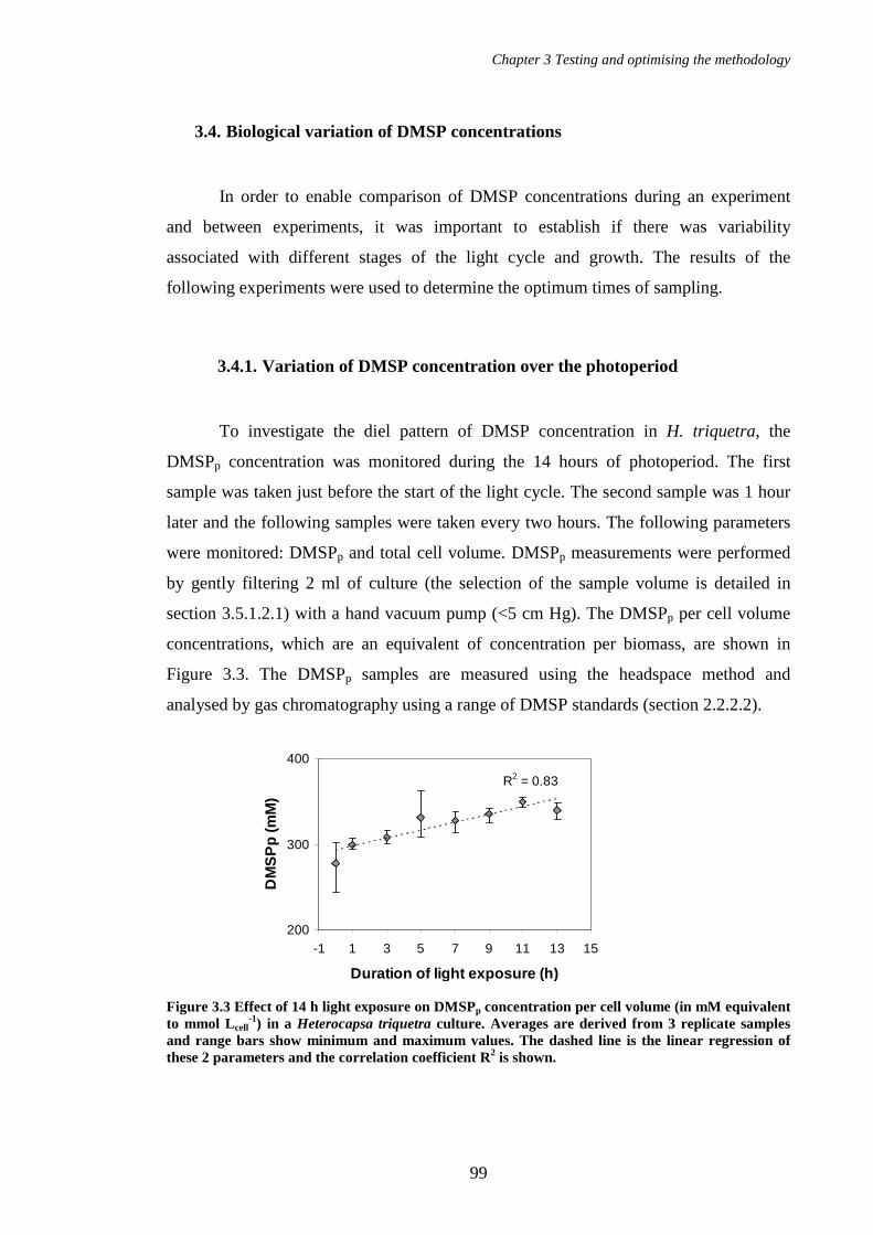
Chapter 3 Testing and optimising the methodology
99
R2 = 0.83
200
300
400
-1 1 3 5 7 9 11 13 15
Duration of light exposure (h)
DM
SP
p (m
M)
3.4. Biological variation of DMSP concentrations
In order to enable comparison of DMSP concentrations during an experiment
and between experiments, it was important to establish if there was variability
associated with different stages of the light cycle and growth. The results of the
following experiments were used to determine the optimum times of sampling.
3.4.1. Variation of DMSP concentration over the photoperiod
To investigate the diel pattern of DMSP concentration in H. triquetra, the
DMSPp concentration was monitored during the 14 hours of photoperiod. The first
sample was taken just before the start of the light cycle. The second sample was 1 hour
later and the following samples were taken every two hours. The following parameters
were monitored: DMSPp and total cell volume. DMSPp measurements were performed
by gently filtering 2 ml of culture (the selection of the sample volume is detailed in
section 3.5.1.2.1) with a hand vacuum pump (<5 cm Hg). The DMSPp per cell volume
concentrations, which are an equivalent of concentration per biomass, are shown in
Figure 3.3. The DMSPp samples are measured using the headspace method and
analysed by gas chromatography using a range of DMSP standards (section 2.2.2.2).
Figure 3.3 Effect of 14 h light exposure on DMSPp concentration per cell volume (in mM equivalent to mmol Lcell
-1) in a Heterocapsa triquetra culture. Averages are derived from 3 replicate samples and range bars show minimum and maximum values. The dashed line is the linear regression of these 2 parameters and the correlation coefficient R2 is shown.

Chapter 3 Testing and optimising the methodology
100
The DMSPp concentration showed an increase (277 to 350 mM at maximum,
27 % increase) with increasing duration of light exposure and these 2 parameters were
significantly correlated (P<0.01, Spearman test). In agreement with previous
observations and hypothesis of Merzouk et al., (2004), the DMSPp concentrations of
photosynthetic dinoflagellates showed a variation over the photoperiod. Potentially
acting as an antioxidant, DMSP could be involved during the light cycle to protect cell
damage from the oxidative photosynthetic products (hydroxyl radicals, singlet oxygen
and other reactive oxygen species; Sunda et al., 2002). However, whilst Merzouk et al.,
(2004) found maximum DMSPp concentrations occurring at midday in field samples, I
observed here, that DMSP seemed to accumulate over the photoperiod. The difference
between field and laboratory observations might be due to the fact that light intensity is
constant in the laboratory incubator whereas in natural conditions the light intensity
reaches its maximum at midday. On the other hand, another photo-protective
compound, UV absorbent mycosporin-like amino acid (MAA) accumulates in culture of
the dinoflagellate Scrippsiella sweeneyae with a maximum concentration in the middle
of the light period (Taira et al., 2004). In cultures of the coccolithophore Emiliania
huxleyi, DMSP concentrations also show a diel cycle with maximum occurring at the
end of the dark period and decreasing over the light period (Bucciarelli et al., 2007).
The diel cycle of DMSP appear to differ among phytoplankton species.
According to these results, experiments which involve, for instance, one sample
per day over several days should have a sampling time that remains constant (at the
same time for each day) to limit any effect of diel variability. Therefore, I established,
for the following experiments, to sample the cultures at the mid-time of the photoperiod,
at 7 hours after the start of the photoperiod.
3.4.2. Variation of DMSP concentration over growth stages
The variation of DMSPp concentrations was also investigated during the growth
of H. triquetra. The culture was sampled every three days, during 12 days. The DMSPp,
total cell volume and cell density were monitored. DMSPp measurements were

Chapter 3 Testing and optimising the methodology
101
performed as previously described. Figure 3.4 shows data for the growth parameters and
DMSP concentrations.
Figure 3.4 Growth and DMSPp content of Heterocapsa triquetra over 12 days. (A) Total cell volume, cell density and DMSPp concentrations in culture. (B) DMSPp concentrations per cell volume and per cell. Cell density and total cell volume were measured from 1 culture aliquot sampled once every 3 days. DMSPp values are averages derived from 3 replicate samples; range bars show minimum and maximum values. The dashed line separates the exponential growth phase from the onset of the stationary growth phase based on the growth rate reduction.
H. triquetra culture grew exponentially during the first 6 days from 1.4×10⁴ to
9.6×10⁴ cell ml-1 (25 to 153 µLcell L-1) then the growth slowed down towards the
stationary phase to reach a maximum cell density of 2.4×10⁵ cell ml-1 (407 µLcell L -1)
after 12 days (Figure 3.4 A). DMSPp concentrations in the culture increased similarly to
the total cell volume and the cell density. The DMSPp per cell volume values appeared
to increase at the end of the exponential phase (Figure 3.4 B). A significant difference
was observed between the DMSP concentration per cell volume at the different
sampling times from 0 to 12 days (P<0.05, Kruskal-Wallis test) and hence, there is a
significant effect of the growth on the DMSPp concentration per cell volume (from 159
to 256 mM so 61 % increase over the growth). The DMSPp per cell seems to slightly
increase from 0 to 6 days during the exponential growth phase and significant difference
0
100
200
300
400
500
0 3 6 9 12
Time (days)
DM
SP
per
cel
l vol
ume
(mM
)
0.0
0.2
0.4
0.6
DM
SP
per
cel
l
(pm
ol c
ell-1
)
DMSPp per cell volume
DMSPp per cell
A
B
1
10
100
1000
DM
SP
in c
ultu
re (µ
M),
cell
dens
ity (×
104 c
ell m
l-1),
tota
l cel
l vol
ume
(µ
Lce
ll L
-1)
Total cell volumeDMSPpCell density

Chapter 3 Testing and optimising the methodology
102
was obtained for the DMSPp concentrations per cell between the exponential growth
phase (0 - 6 days) and the onset of the stationary growth phase (9 - 12 days); (P<0.05,
Mann-Whitney U test).
Additionally, other experiments on the heterotrophic dinoflagellate
Crypthecodinium cohnii have shown some substantial variations of the DMSP
concentrations over the growth stages (presented in Chapter 6). Given that DMSP
concentrations vary over growth stages, I decided to perform the sampling of the culture
at the same growth stage to allow comparison between experiments. I choose the mid-
exponential phase of growth as it is usually used to avoid the effect of nutrient
limitation and ageing population, as for example to compare the DMSP content of
several species.

Chapter 3 Testing and optimising the methodology
103
DMSPT = DMSPp + DMSPd + DMS
DMSPpDMSPd+ DMS
DMSPT
FILTRATION
Headspace analysis - GC Purge and Trap - GC
NaOH
DMS
DMS
NaOH
DMSPT = DMSPp + DMSPd + DMS
DMSPpDMSPd+ DMS
DMSPT
FILTRATION
Headspace analysis - GC Purge and Trap - GC
NaOH
DMS
DMS
NaOH
3.5. Effect of the methods of sample preparation on DMSP and DMS
concentrations
At the beginning of my laboratory work with dinoflagellates, I measured the
total DMSP in cultures (DMSPT) and all the fractions DMSPp, DMSPd and DMS to
assess if there was a mass balance. At that time, DMS measurements were performed on
the filtrate collected from 2 ml of culture passed through a 25 mm diameter glass-fibre
filter (Whatman, GF/F) by using a hand vacuum pump (not exceeding <5 cm Hg) and
directly poured into the purge tube of the purge and trap system. For DMSPd +DMS
measurements, 1 ml of NaOH was added to another filtrate of sample obtained similarly
to the DMS sample and the DMSPd value was calculated by subtraction of the DMS
value. The DMSPp and DMSPT measurements were done as previously described
(section 2.2.2.2 and 3.4.1). Figure 3.5 gives a general overview of the DMSP pool and
fractions and the analytical methods applied for their measurements.
Figure 3.5 Summary of the techniques applied to the measurement of DMSPT and its fractions. GC indicates the use of gas chromatography. DMSPT is measured on the liquid culture sample, DMSPp is analysed from the filter which has collected the cells and DMSPd and DMS are measured in the filtrate.

Chapter 3 Testing and optimising the methodology
104
On 2 successive days, I performed sampling of a culture of Heterocapsa
triquetra in exponential phase. Each day I took 3 samples from this culture and
measured the 4 parameters DMSPT, DMSPp, DMSPd and DMS in each of the 3 samples.
Figure 3.6 shows the data normalised to cell volume.
Figure 3.6 Concentrations per cell volume of DMSPT and its fractions DMSPp, DMSPd and DMS measured in a sample of Heterocapsa triquetra culture in exponential phase. Results are presented on 2 panels because of the broad span of the data (A) DMSPT, DMSPp, DMSPd. (B) DMSPd and DMS. Data are average values from 3 replicate samples and range bars show minimum and maximum values.
According to these results, it appeared that the sum of the fractions was not
equal to but lower than the DMSPT measurements and that a fraction of about 24 to
27 % was missing. Given that DMSPp was 70 - 74% of DMSPT for the H. triquetra
culture in exponential phase whereas DMSPd and DMS were only 2 – 3 % and 0.1 -
0.2 % of DMSPT, respectively, it was more likely that the missing fraction would come
from the DMSPp measurement.
In this regard, I first focussed my attention on testing and improving the DMSPp
measurement and essentially in the filtration process which was the critical step in the
sample preparation that could produce underestimation (Kiene and Slezak, 2006) and
imprecision. Therefore, I investigated several parameters influencing the filtration
pressure such as: the sample volume of culture, the methods of filtration such as the
hand vacuum pump, the syringe pump with a range of delivery speeds and filtration by
gravity.
A B
0
100
200
300
400
1 2Day
DM
SP
(mM
)
DMSP
DMSPp
DMSPd
0
5
10
15
20
25
1 2Day
DM
S a
nd D
MS
Pd (m
M)
DMSPd
DMS
T

Chapter 3 Testing and optimising the methodology
105
3.5.1. Optimisation of the DMSPp measurement
3.5.1.1. DMSPp measurement of standards
Given that culture samples contained a filter I tested if the presence of a filter in
the standards was affecting the DMSPp concentrations. Some duplicate standards were
prepared by adding the DMSP stock solution on the filter, folding it and introducing it
in the vial containing NaOH similar to sample preparation. The comparison of these 2
methods is presented in Figure 3.7.
Figure 3.7 Comparison between DMSPp standards without filter (DMSPp 1) and containing a filter (DMSPp 2). (A) Calibration curves for the 2 sets of standards, both curves are superimposed. (B) Least-squares regression obtained for these 2 sets of standards. SQRT PA means the square root of the peak area obtained on the chromatogram.
The results showed similar values obtained for the calibration curve with the 2
methods tested (P>0.05, Mann-Whitney U test). The filters had therefore no effect on
the DMSP measurement and the use of filters was unnecessary for further DMSPp
standard preparation.
3.5.1.2. DMSPp measurement of culture samples
For preparation of culture samples, the sample was filtered using a hand vacuum
pump not exceeding 5 cm Hg. The filter was folded in 4 and put in the top part of the
y = 0.78x + 359.61
R2 = 1.00
0
4000
8000
12000
0 5000 10000 15000
SQRT PA of standards with filter
SQ
RT
PA
of s
tand
ards
w
ithou
t filt
er
y1 = 0.78x + 359.61
R2 = 1.00
y2 = 0.79x + 90.05
R2 = 1.00
0
4000
8000
12000
0 5 10 15 20
DMSP (µM)
SQ
RT
PA
DMSPp 1
DMSPp 2
B A

Chapter 3 Testing and optimising the methodology
106
vial with no direct contact with NaOH to avoid DMS release in the open vial. The vial
was closed with a gas tight cap, agitated and stored as previously described, for later
analyses.
3.5.1.2.1. Effect of the sample volume when filtering by means of a hand vacuum pump on DMSPp concentrations
To assess the effect of the sample volume I filtered 1 ml, 2 ml, 3 ml, 4 ml and
5 ml of culture using the hand vacuum pump. Each condition was repeated 3 times.
DMSP concentrations obtained for each sample volume are shown in Figure 3.8.
Figure 3.8 Effect of the volume of filtered culture on DMSPp measurement in Heterocapsa triquetra culture (A) DMSPp concentration in the culture, (B) per cell, (C) per cell volume (CV). The column height represents the average (n=3 except n=2 for the filtered volume of 2 ml) and range bars display the minimum and maximum values. The dashed line shows the least -squares regression of the DMSPp concentrations to the volume of sample filtered and the correlation coefficient R2 is shown.
B
C
A
R2 = 0.67
0.0
0.2
0.4
0.6
DM
SP
p pe
r cel
l
(pm
ol c
ell-1
)
R2 = 0.77
0
100
200
300
1 2 3 4 5Filtered volume (ml)
DM
SP
p pe
r CV
(mM
)
R2 = 0.77
0
30
60
90
120
DM
SP
p in
cul
ture
(µM
)

Chapter 3 Testing and optimising the methodology
107
The filtration duration lasted between 1 and 3 minutes, with longer periods for
larger volumes. There was a significant correlation between the DMSPp concentration in
the culture and the volume of the sample filtered (P<0.01, Spearman test). Similar
correlation was obtained for DMSPp normalised per cell and per cell volume. The
DMSPp values decrease with increasing volume of culture filtered. The larger volumes
(4 - 5 ml) showed significant difference with the smallest volume of 1 ml (6 and 8 %
respectively, P=0.05, Mann-Whitney U test) and suggestive difference with a volume of
2 ml (9 and 11 % respectively, P<0.10; Table 3.1).
Table 3.1 Probability (P) of significant difference between DMSPp per cell values obtained for 2 volumes of filtration obtained with the non-parametric Mann-Whitney U test. P≤ 0.05 indicates significantly different DMSPp values.
Volume compared (ml) 1 - 2 2 - 3 2 - 4 2- 5 1 - 4 1 - 5
Probability (P) 0.564 0.248 0.083 0.083 0.05 0.05
The volume of culture filtered had an effect on DMSP concentration. The load
of numerous cells aggregating on the filter may cause blockage and more pressure is
required to allow the filtration. A larger volume seems to reduce the DMSP value
probably due to a DMSP loss (i.e. if cells are damaged, they may release DMSP into the
dissolved fraction) as suggested by Kiene and Slezak (2006). I, therefore, favoured the
use of smaller volumes and selected the volume of 2 ml for the following experiments
in this project rather than 1 ml to allow detectable measurement of low density samples
or potentially low DMSP producing-species.
In this experiment the use of the hand vacuum pump gave a good reproducibility
of DMSPp measurement with a relative standard deviation (RSD) between 0.4 and 5 %
(n=3). However, these values vary within and across experiments (in another
experiment shown previously in Figure 3.3, the RSD spread between 2 and 11 %). I
tested other filtration systems to see how it would affect DMSP concentrations.

Chapter 3 Testing and optimising the methodology
108
0
50
100
150
200
250
300
A B
DM
SP
p (m
M)
3.5.1.2.2. Comparison of filtration using an electrical pump and a hand vacuum pump
The effect of the filtration system was tested by comparing DMSPp
concentrations obtained using the hand vacuum pump and an electrical pump set to a
comparable low pressure (5 cm Hg). For each filtration method, 3 replicates of culture
sample were filtered and analysed for DMSPp concentrations. Results are shown in
Figure 3.9.
Figure 3.9 Comparison of DMSPp concentrations per cell volume obtained using 2 different filtration methods (A) hand vacuum pump and (B) an electrical pump at a similar pressure for a sample volume of 2 ml of Heterocapsa triquetra culture. Averages (n=3) are shown by the column height and error bars show minimum and maximum values.
The DMSP value obtained with the electrical pump was significantly lower than
the value obtained with the hand vacuum pump (5 % difference, P= 0.05, Mann-
Whitney U test). In this experiment the DMSPp measurement gave a RSD of 1.3 %
when using a hand vacuum pump and 2.9 % when using the electrical pump. According
to these results, the hand vacuum pump gave a higher DMSP concentration with better
precision whereas the use of the electrical pump had no benefit.

Chapter 3 Testing and optimising the methodology
109
0
100
200
300
0 2 4 6 8
Speed of filtration (ml min -1)
DM
SP
p (m
M)
3.5.1.2.3. Comparison of filtration with a syringe pump using various speeds of filtration
An automated syringe pump (KDS220, Linton Instrumentation, Diss, United
Kingdom) allows consistent delivery speed and consequently identical filtration
pressure on each culture replicate. I tested 5 delivery speeds of 0.5, 1, 2, 4 and 7 ml min-
1 on 2 ml aliquots of culture using 2.5 ml plastic syringes. The culture sample, in late
exponential growth phase at a cell density of 8.7×10⁴ cell ml-1, was directly sucked up
with the syringe up to 2 ml and placed on the syringe pump. The effect of the syringe
pump on the DMSP values was compared with the use of the hand vacuum pump which
filters at a speed of 1 to 2 ml min-1. The DMSP concentrations obtained are shown in
Figure 3.10.
Figure 3.10 Effect of delivery speeds (from 0.5 to 7 ml min-1) of the syringe pump (grey symbols) compared with the use of the hand vacuum pump (black symbol on the Y axis) on the DMSPp values. The volume of Heterocapsa triquetra culture sample was 2 ml. Syringes of 2.5 ml volume were used with the syringe pump. Shown are average values with range of data (n=3).
Delivery speeds between 0.5 and 7 ml min-1 showed similar DMSPp values
(P>0.05, Kruskal-Wallis test). The variation between replicate samples (RSD 4 – 17 %)
appeared in some cases larger than the variation between the delivery speeds (RSD
13.9 %). Therefore, the use of the syringe pump at delivery speeds between 0.5 and
7 ml min-1 did not reduce the variation between replicate samples. However, a
significantly higher DMSPp value (16 % difference, P=0.05, Mann-Whitney U test) was
obtained using the syringe pump at the highest delivery speed of 7 ml min-1 compared to
the result obtained using the hand vacuum pump and which suggests that less DMSP
may have been lost in the dissolved fraction.

Chapter 3 Testing and optimising the methodology
110
0
50
100
150
200
250
0 5 10 15 20
Delivery speed (ml min -1)
DM
SP
p (m
M)
0
50
100
150
200
250
0 5 10 15 20
Delivery speed (ml min -1)
DM
SP
p (m
M)
To test if the use of higher delivery speeds (> 7 ml min-1) had any effect on
DMSPp values, larger syringe volumes needed to be used since the speed range of the
pump was dependant on the syringe volume. Plastic syringes of 10 ml volume were
used to investigate the delivery speeds between 2.5 and 20 ml min-1. To obtain an
accurate volume in the syringe, the sample of culture in exponential growth phase at
3.0 ×10⁴ cell ml-1 was taken up with a Gilson pipette and injected into the syringe
blocked with a stopcock. The samples were filtered and analysed for DMSPp
concentrations which are shown in Figure 3.11.
Figure 3.11 Effect of 8 delivery speeds of the syringe pump on the DMSPp values using the 10 ml syringes and 2 ml culture sample of Heterocapsa triquetra culture at at 3×10⁴ cell ml-1. Shown are average values with ranges of data (n=3 except n=2 for 2.5 and 5 ml min-1).
Again in this experiment, no significant differences were obtained between the
delivery speeds tested (P>0.05, Kruskal-Wallis test; Figure 3.11) on a culture sample of
Heterocapsa triquetra culture. The RSD obtained was between 6 and 19 % with the
smallest RSD for a delivery speed of 20 ml min-1.
A similar test was repeated for 4 delivery speeds using a higher cell density
culture, in late exponential phase at 10.5×10⁴ cell ml-1, to test how more elevated
biomass would have an effect on the filtration and DMSPp measurement (Figure 3.12).
Figure 3.12 Effect of 4 delivery speeds of the syringe pump on DMSPp per cell volume for 2 ml of a culture at a cell density of 10.5×10⁴ cell ml-1. Shown are average values with ranges of data (n=3).

Chapter 3 Testing and optimising the methodology
111
At a cell density of 10.5×10⁴ cell ml-1, the same variability was obtained for
sample replicates (RSD of 4 - 20 %) with the smallest variability found at 20 ml min-1.
The DMSPp concentration increased with the increase of the delivery speed used for the
filtration (significant correlation, P<0.05, Spearman test).
I compared the DMSPp concentrations obtained for the delivery speeds 5, 10, 15,
20 ml min-1 at low and high cell density of H. triquetra culture (Figure 3.11 and Figure
3.12, respectively). No significant difference was obtained for the delivery speeds of 5,
10 and 15 ml min-1 (P>0.05 Mann-Whitney U tests for each speed), however,
significant difference was observed for the delivery speed of 20 ml min-1 (P<0.05
Mann-Whitney U test). Therefore the DMSPp concentration per cell volume was higher
in H. triquetra culture of 10.5×10⁴ cell ml-1 (194 mM, 11 % higher) than in a culture of
3.0 ×10⁴ cell ml-1 (174 mM) when the samples were filtered at 20 ml min-1. However, it
was only possible to observe this difference if the variation between replicate samples
was minimised as appeared to be the case at 20 ml min-1 of delivery speed.
I tested, in a different way, how elevated biomass would have an effect on the
filtration and DMSPp measurement by increasing the sample volume to 5 ml. The effect
of 4 different delivery speeds on DMSPp values was tested for a 5 ml sample of culture
at a cell density of 3.5×10⁴ cell ml-1. The results are shown in Figure 3.13.
Figure 3.13 Effect of 4 delivery speeds of the syringe pump on the DMSPp concentrations per cell volume for 5 ml of culture at a cell density of 3.5×10⁴ cell ml-1.
Similar concentrations were obtained for 10, 15 and 20 ml min-1 (P>0.05,
Kruskal-Wallis test) and significantly lower DMSP concentrations were obtained at
5 ml min-1 (P<0.01, Mann-Whitney U test). The DMSPp concentration and the delivery
0
50
100
150
200
250
0 5 10 15 20
Delivery speed (ml min -1)
DM
SP
p (m
M)
DM
SP
p (m
mol
Lce
ll-1
)

Chapter 3 Testing and optimising the methodology
112
speed are again positively correlated (P<0.05, Spearman test). The analytical
uncertainty was from 4 to 12 % with the smallest one being at 5 ml min-1.
When comparing all the results obtained for the 4 delivery speeds from 5 to
20 ml min-1 in the 3 previous experiments (Figures 3.11, 3.12 and 3.13), no significant
difference in DMSPp concentrations was obtained between experiment (P>0.05, Mann-
Whitney U test) and no difference appeared between the 4 delivery speeds tested
(P>0.05, Kruskal-Wallis test). However, significant correlation was found between the
DMSPp concentrations per cell volume and the delivery speed of the filtration (P<0.05,
Spearman test).
With the use of the syringe pump, DMSPp concentrations increased with
increase in delivery speed. For a culture at higher cell density (8 - 10 ×104 cell ml-1), the
lowest variability between replicates was obtained at the highest delivery speed of 20 ml
min-1. This delivery speed also allowed the observation of significantly higher DMSPp
concentration in cultures of higher cell density than cultures of lower cell density and
which was hidden by the large variability of replicates when using the hand vacuum
pump. The use of a higher sample volume such as 5 ml did not show any benefit in
comparison with the experiment using a 2 ml sample and supported the idea that low
delivery speeds such as 5 ml min-1 should be avoided given that it resulted in
significantly lower DMSPp concentrations.
In conclusion, higher delivery speeds needed to be favoured when using the
syringe pump for DMSPp measurements since the DMSPp concentrations were
increased and a better precision of the measurement was generally observed (4 - 9 % at
20 ml min-1). Therefore, I decided to set the syringe pump at the maximum available
speed depending on the syringe type which was 14 ml min-1 for a 5 ml syringe and 7 ml
min-1 for a 2.5 ml syringe.

Chapter 3 Testing and optimising the methodology
113
0
100
200
300
A B C
DM
SP
(mM
)
Low cell density
High cell density
3.5.1.2.4.Comparison of filtration by gravity with other filtration methods
Gravity filtration was recommended by Kiene and Slezak (2006) to avoid
potential breakage of cells and DMSP transfer from the particulate to the dissolved
fraction of the culture. Initially, gravity filtration did not work since there was too much
back pressure from the glass frit, supporting the filter. I finally used a glass filter holder
without a glass frit which enabled the filtration by gravity. Other processes of filtration
were performed including filtration using the syringe pump (with a 2.5 ml syringe at
7 ml min-1) and the hand vacuum pump. Two cell densities of culture (4.6×10⁴ cell ml-1
and 25×104 cell ml-1) were tested for this experiment in order to assess H. triquetra
responses to the different filtration methods applied. For each cell density, 3 replicates
of 2 ml culture sample were filtered and analysed. The results of the experiment are
given in Figure 3.14.
Figure 3.14 Effect of 3 filtration systems on DMSPp concentrations per cell volume of culture samples at low and high cell densities (4.6×10⁴
cell ml-1 and 25×104 cell ml-1 respectively). (A) Filtration by gravity, (B) syringe pump at 7 ml min-1, (C) hand vacuum pump.
The syringe pump gave the best reproducibility of the measurements, with the
smallest RSD of 4 - 5 % at the 2 cell densities in comparison with 8 - 9 % and 9 – 10 %
for the gravity filtration method and the use of the hand vacuum pump, respectively.
The 3 filtration methods showed no difference in DMSPp concentrations at high cell
density (P>0.05, Kruskal-Wallis test). At low cell density gravity filtration showed
significantly higher DMSP than when using the syringe pump (P=0.05). However there
was no significant difference between the gravity filtration method and the use of the
hand vacuum pump (P>0.05, Mann-Whitney U test) and between the use of the hand
vacuum pump and the syringe pump (P>0.05, Mann-Whitney U test). Therefore, the
benefit of using the gravity filtration method was not obvious and did not need to be
Chapter 3 Testing and optimising the methodology
114
especially used for DMSPp measurements in dinoflagellates as might have been
expected according to the recommendations of Kiene and Slezak (2006). In this
experiment, the use of the syringe pump did not lead to a higher DMSP concentration
than the use of the vacuum pump in contrast to the result observed in Figure 3.10.
The difference between DMSPp values obtained for low and high cell density
might be due to different physiological conditions. However, the response of
H. triquetra cells to the filtration methods, in terms of analytical uncertainty, is similar
for the 2 different growth stages (i.e. similar RSD at low and high cell densities).
Therefore, the growth stage of the culture had no effect on the precision of DMSPp
measurement but could affect the value of DMSP concentration. On the other hand, the
choice of filtration method affected the precision of the measurement.
3.5.1.2.5. Conclusion on the effect of the filtration methods on DMSPp measurement
Several filtration methods were tested with the aim of selecting the method that
would produce the smallest analytical uncertainty and the highest DMSPp value.
Between the 3 methods, including the hand vacuum pump, the syringe pump and
gravity filtration, no general significant difference was obtained for DMSPp
concentration. The DMSPp measurement was thus not increased by the use of any of the
filtration methods and the analytical uncertainty appeared lower in some cases for the
use of the syringe pump.
As I observed that DMSPp concentration varied over growth stages I compared
the effect of the use of the hand vacuum pump and the syringe pump on the DMSPp
concentrations in culture of similar cell densities (low cell density in exponential phase).
Table 3.2 shows the summary of the results obtained across several experiments
presented previously in this chapter.

Chapter 3 Testing and optimising the methodology
115
Table 3.2 Comparison of DMSPp concentrations and RSD obtained between filtration using the syringe pump and the hand vacuum pump for a low cell density culture of Heterocapsa triquetra. Filtration method Experiment Culture cell
density ×104 cell ml-1
Average DMSPp mM (n=3)
RSD (n=3)
Syringe pump (2.5-20 ml min-1) Fig 3.11 3 160 - 184 6 - 19 Syringe pump 20 ml min-1 Fig 3.11 3 174 6 Syringe pump (5-20 ml min-1) Fig 3.13 3.5 148 - 195 4 - 12 Syringe pump 20 ml min-1 Fig 3.13 3.5 195 9.1 Syringe pump (7 ml min-1) Fig 3.14 4.6 193 4 Hand vacuum pump (growth stages)
Fig 3.4 B 4.2 159 1.2
Hand vacuum pump Fig 3.14 4.6 214 9 SUMMARY Hand vacuum pump 4.2 ; 4.6 159 ; 214 1.2 - 9 Syringe pump 3 - 4.6 148 - 195 4 - 19 Syringe pump 20 ml min-1 3 ; 3.5 174 ; 195 6 ; 9
Both filtration methods produced similar range of DMSPp concentrations: 159 –
214, 148 - 195 mM for the hand vacuum pump and the syringe pump, respectively. The
analytical uncertainty appeared in these cases less important with the hand vacuum
pump (1.2 - 9 %) than the syringe pump (4 – 19 %). The same conclusion is true for all
experiments presented in this chapter (RSD of 0.4 - 11 % for the hand vacuum pump
and 4 - 20 % with the syringe pump). The rather large range of variability has the
potential to hide crucial differences in DMSPp concentrations as for example, over
growth stages. The variation between replicates tended to be reduced by using the
highest delivery speeds available on the syringe pump. At delivery speed of 20 ml min-1
the range of DMSP concentration obtained and the analytical uncertainty were reduced
(174 - 195 mM, 6 - 9 % RSD) and the analytical uncertainty was in the same range
obtained using the hand vacuum pump.
Finally, given that the DMSPp measurement was not obviously successfully
improved by using other filtration methods, I investigated the DMSPT measurement as a
surrogate of the intracellular DMSP. Previous measurements showed that DMSPp was
the major fraction of DMSPT (70 – 74 %, Figure 3.6) and that the analytical uncertainty
for DMSPT measurement was 1.3 % (n=5) for H. triquetra samples of culture in
exponential phase (3×10⁴ cell ml-1; see chapter 2 section 2.2.2.2). Therefore, the
analytical uncertainty for DMSPT measurement was better than the uncertainty obtained
for DMSPp measurement. The absence of a filtration step in the DMSPT measurement
ensured a better replication of samples. Reducing the analytical uncertainty was crucial

Chapter 3 Testing and optimising the methodology
116
to improve the likelihood of determining variation of DMSP in different experimental
conditions. The absence of filtration also reduced the time of sample preparation and
enabled successive sampling at short intervals, that would not be possible for DMSPp
measurements (as performed in chapter 6, section 6.2.5). Moreover, DMSPT represented
the total production of DMSP and derivatives by phytoplankton cells in the culture. The
DMSPT measurement was also used by Keller et al. (Keller, 1988/1989; Keller et al.,
1989a; Keller et al., 1989b) for measuring the DMSP content of 123 phytoplankton
strains and by Yost et al. (2009), who observed no significant difference between
DMSPT and DMSPp concentrations in 5 strains of the dinoflagellate Symbiodinium
microadriaticum in exponential growth phase.

Chapter 3 Testing and optimising the methodology
117
3.5.2. Adaptation of the headspace method for DMSPT measurement
3.5.2.1. DMSPT measurement for standards
The method for measuring DMSPT (DMSPp + DMSPd + DMS) was adapted
from the headspace method similar to the protocol for DMSPp measurement but without
filtering the sample. This method was therefore convenient for DMSPT analyses since
the concentrations fall in a similar range to DMSPp and DLA measurements (Steinke et
al., 2000). As previously described (chapter 2 section 2.2.2.2) the standards for
calibrating the gas chromatograph were made up by introducing 3 ml of 0.5 M NaOH in
a 5 ml glass vial, then adding a drop of DMSP (2 - 15 µl) solution under the cap of the
vial and closing and agitating the vial. DMSPT samples were prepared in order to obtain
the same final NaOH concentration. In this way, 1 ml of culture was rapidly added to
2 ml of 0.75 M NaOH in a 5 ml glass vial and immediately closed with a gas tight cap
to avoid DMS loss. The samples and standards were left to react in the dark for later
analyses.
Similar to the standards, the DMSPT samples were prepared to end up with a
final concentration of 0.5 M NaOH, a total liquid volume of 3 ml and an identical
headspace volume of 2 ml. However, DMSPT samples contained 1 ml of phytoplankton
culture grown in enriched seawater which might alter the pH of the sample and which
contained salts that might also alter the transfer of DMS to the gaseous phase in
comparison with the distilled water used to prepare the standards. Henry’s law constant
(Hc= concentration in the air/ concentration in water) of DMS is higher in seawater than
in distilled water, for the same conditions of temperature (0.069 and 0.056, respectively,
measured by Wong and Wang, 1997), i.e. DMS is less soluble in seawater than in
distilled water.
For testing if the addition of 1 ml of culture grown in seawater was affecting the
DMSP cleavage into DMS or the DMS partitioning in the air and liquid phases, I
prepared a range of standards by adding 1 ml of seawater to 2 ml of 0.75 M NaOH in
the 5 ml glass vial before adding a drop of DMSP solution under the cap. I compared

Chapter 3 Testing and optimising the methodology
118
those standards to the standards made up by introducing 3 ml of 0.5 M NaOH and the
DMSP solution as described above. Each standard was prepared in duplicate. Results
are shown in Figure 3.15.
Figure 3.15 Comparison of standards prepared with NaOH diluted in distilled water (DW) and prepared with one third of seawater (SW) to simulate the culture sample preparation. SQRT PA means the square root of the peak area obtained on the chromatogram. (A) Calibration curves for standards prepared with DW and SW, both curves are superimposed. (B) Regression of the 2 sets of standards (DW, SW).
The standards made up with a third of seawater in the NaOH solution led to
comparable peak areas to those obtained for standards made up with NaOH diluted in
distilled water (P>0.05, Mann-Whitney U test, and R2 of 1.0 for the least squares
regression of peak areas for both standards preparation, Figure 3.15 B). This difference
in the preparation did not alter the chemical cleavage of DMSP by NaOH and did not
affect the DMS transfer to the air phase of the analytical vial. However, a white
precipitate appeared when the seawater was added.
The standards used for calibrating the GC are prepared by adding a drop of
DMSP solution under the cap of the vial, subsequently closed and agitated. This method
avoided mixing the DMSP solution and the NaOH while the vial was open and
prevented any DMS loss. For DMSPT samples, the culture was directly added in the vial
containing NaOH and rapidly closed. To test if the sample preparation for DMSPT
measurement produced a DMS loss I prepared 5 standards by adding 1 ml of DMSP
solution in 2 ml of 0.75 M NaOH (called DMSP 2). I compared these standards with
standards prepared by putting a drop of DMSP solution under the cap and closing the
B y = 572.81x + 400.98
R2 = 1.00
y = 562.59x + 572.61
R2 = 1.00
0
10000
20000
30000
40000
50000
0 20 40 60 80 100
DMSP (µM)
SQ
RT
PA
DWSWLinéaire (SW)
A (sw)
(Dw)
y = 1.02x - 134.27
R2 = 1.00
0
10000
20000
30000
40000
50000
0 10000 20000 30000 40000 50000
SQRT PA of DW standards
SQ
RT
PA
of S
W s
tand
ards

Chapter 3 Testing and optimising the methodology
119
vial containing the NaOH solution (DMSP 1). Each standard was made in duplicate.
Results are given in Figure 3.16.
Figure 3.16 Comparison of standards prepared by putting a drop of DMSP solution under the cap of the vial containing the NaOH solution and closing the vial (DMSP 1) and standards prepared by addition of 1 ml of DMSP solution in 2 ml of 0.75 M NaOH similarly to the culture sample (DMSP 2). The square root of the peak area (SQRT PA) is the equivalent DMSP value read on the chromatogram.
The 2 sets of standards gave similar values of peak areas for the range of DMSP
concentrations prepared (P>0.05, Mann-Whitney U test). Moreover, the least-squares
regression of the peak areas obtained with both methods of standard preparation gave a
correlation coefficient of 0.99 (Figure 3.16 B) which supported the result that these 2
methods (DMSP 1 and 2) gave similar results. Therefore, given that culture samples are
prepared similarly to the standards DMSP 2, no significant DMS loss appeared to occur
when using this method.
The analytical uncertainty of the DMSPT measurements was 1.8 % (n=5) for
standards and 1.3 % (n=5) for H. triquetra samples in exponential growth phase.
3.5.2.2. DMSPT measurement for culture samples
Given that DMSPp concentrations vary over the growth I investigated how
DMSPT and DMSPp both varied over the growth of Heterocapsa triquetra to test if
DMSPT was a good surrogate of intracellular DMSP (Figure 3.17). The culture was
sampled every 3 days, over 12 days. Culture aliquots of 2 ml were filtered using a hand
vacuum pump and analysed for DMSPp and 1 ml aliquots were directly mixed with
y = 1.00x - 220.73
R2 = 0.99
0
4000
8000
12000
0 4000 8000 12000
SQRT PA DMSP 2
SQ
RT
PA
DM
SP
1
A B
y1 = 0.78x1 + 359.61
R2 = 1.00
y2 = 0.78x2 + 130.8
R2 = 1.00
0
4000
8000
12000
0 5 10 15 20
DMSP (µM)
SQ
RT
PA
DMSP 1 DMSP 2Linéaire (DMSP 1 )

Chapter 3 Testing and optimising the methodology
120
NaOH and analysed for DMSPT. Results obtained for the measurements of these 2
parameters are presented in Figure 3.17.
Figure 3.17 DMSPT and DMSPp concentrations in Heterocapsa triquetra culture over time. (A) DMSP per litre of culture. (B) DMSP per cell volume. Shown are average values and range bars give minimum and maximum values (n=3, except n=2 for DMSPT data at t=0 and t=12). When not visible, range bars are smaller than symbol size.
DMSPp and DMSPT concentrations in the culture were not significantly different
(P>0.05, Mann-Whitney U test) and showed similar increases over time. DMSPp and
DMSPT concentrations per cell volume showed the same general trend over time with
an increase between days 3 and 6 and a slight decrease after day 6. No significant
difference was observed between the DMSPp production and the DMSPT production
over time (P>0.05, Mann-Whitney U test; the productions were calculated as the
difference of DMSP concentrations between one data point and the next one over the
period of time). Between 3 and 12 days, DMSPp was 77 to 85 % of the DMSPT fraction.
The RSD obtained for DMSPT measurements (0.6 - 5.2 %) were smaller than those
obtained for DMSPp measurements (1.1 - 16.4 %). With a better analytical uncertainty
and not significantly different values, DMSPT seems to be a good surrogate of the
intracellular DMSP concentration.
0
40
80
120
0 3 6 9 12Time (days)
DM
SP
in c
ultu
re (µ
M)
DMSP pDMSP T
0
100
200
300
400
0 3 6 9 12Time (days)
DM
SP
per
cel
l vol
ume
(mM
)
DMSP pDMSP T
B A
T T

Chapter 3 Testing and optimising the methodology
121
0
20
40
60
80
100
0 10 20 30 40 50
Time (min)
% o
f tot
al p
urge
d D
MS
0
4000
8000
12000
16000
20000
SQ
RT
PA
% of purged DMS
SQRT PA
3.5.3. Optimisation of DMS measurement
3.5.3.1. DMS measurement of standards
The purge and trap method was applied to DMS measurement as it was initially
developed (details in Chapter 2 section 2.2.2.1). The purge efficiency (i.e. the amount of
DMS extracted) depends on the volume of the liquid, the purge flow rate and the time of
purging. The volume was fixed at 5 ml, to fall in the sensitivity range of the GC,
according to preliminary tests. The time required for purging DMS out of the solution
with a purge flow rate of 60 ml min-1 was tested.
A 5 ml solution was introduced in the purge tube by successively adding 1 ml of
DMSP solution, 3 ml of distilled water, 1 ml of 10 M NaOH and then rapidly closing
the purge tube. In order to determine the time necessary for purging, the amount of
DMS purged out was monitored over 45 minutes by stop-start purging the same sample.
The square root of the peak area (SQRT PA) shows the value of the DMS peak obtained
by the GC for a defined period of purging (Figure 3.18). The percentage of extracted
DMS is calculated as the cumulative SQRT PA at tx over the cumulative SQRT PA at
45 minutes of purging.
Figure 3.18 Purge efficiency of the purge and trap system containing a total liquid volume of 5 ml that includes 1 ml of DMSP standard solution (16.24 ng S ml-1) and monitored during 45 minutes. The symbol (○) indicates the extracted DMS expressed as a cumulative percentage of the final DMS peak areas at 45 minutes and the symbol (□) shows the square root of the peak area read on the chromatogram.

Chapter 3 Testing and optimising the methodology
122
The SQRT PA decreased abruptly up to 6 minutes of purging, as the DMS
concentration decreased in the purge tube. The cumulative percentage increased to 89 %
after 4 minutes of purging, 93 % after 6 minutes and 95 % after 10 minutes. I thus,
decided to establish 10 minutes as purging protocol for a 5 ml solution of both standards
and samples.
Given that DMS is less soluble in seawater than distilled water, I tested whether
there was a difference between standards prepared with 3 ml of distilled water and 3 ml
of seawater. Results are given in Figure 3.19.
Figure 3.19 Comparison of the purge efficiency using distilled water (DW) and using seawater (SW) in the standard preparation. (A) Cumulative square root of the peak areas (SQRT PA) obtained after 6 periods of purging. (B) The symbols (○●) indicate the extracted DMS expressed as a cumulative percentage of the final DMS amount at 15 minutes and the symbols (□■) show the square root of the peak area read on the chromatogram.
The use of seawater led to slightly higher DMS values (Figure 3.19 A)
compared to those obtained with distilled water but no significant difference was
observed (P>0.05, Mann-Whitney U test). The seawater may contain a DMS
background that could explain the slightly higher DMS values observed or the purging
0
20
40
60
80
100
0 5 10 15
Time (min)
% o
f tot
al p
urge
d D
MS
0
4000
8000
12000
16000
20000S
QR
T P
A% of total DMS-DW% of total DMS-SWSQRT PA-DWSQRT PA -SW
0
5000
10000
15000
20000
25000
30000
Cum
ulat
ive
SQ
RT
PA
SW
DW
A
B

Chapter 3 Testing and optimising the methodology
123
in seawater may yield more DMS at the beginning. However, at 10 minutes of purging
the cumulative percentage of DMS appeared similar with distilled water or seawater
(Figure 3.19 B). Because distilled water is always available in the laboratory and is less
likely to contain DMS background, I decided to use distilled water in the DMS
standards and in filtered samples to make up the volume to 5 ml.
Given that for DMSPT measurements DMSP is left for 24 hours to react with
NaOH, I decided to do the same with the DMS standards. I prepared DMS standards in
5 ml glass vials closed with a screw cap and a Teflon septa and left them 24 hours to
react in the dark before performing DMS measurements (as described in Chapter 2
section 2.2.2.1). The analytical uncertainty for DMS standards was 3.2 % and was
slightly higher than the analytical uncertainty of DMSPT standards (1.3 %). This
difference is likely to result from the coupling of the purge and trap system and the gas
chromatography for DMS standard analyses whereas only gas chromatography is used
for analyses of DMSPT standards.
3.5.3.2. DMS measurements of culture samples
The sample preparation for DMS measurement in culture samples differs from
that of standards, by the addition of a filtration step and a successive transfer into the
purge tube. Because the syringe pump showed a better analytical uncertainty in several
experiments for filtration of replicate samples in DMSPp measurement, I used this
system for DMS measurements. The syringe, placed on the pump, was connected to a
filter holder which was connected to a sample loop that dispensed the exact volume of
filtrate into the purge and trap system (S-S-PT system: Syringe pump - Sample loop -
Purge and Trap, described in 2.2.2.1). I determined the analytical uncertainty of this
system by performing DMS measurements of replicate standards and H. triquetra
replicate samples. Table 3.3 shows the average and the RSD of the square root of the
peak areas.

Chapter 3 Testing and optimising the methodology
124
Table 3.3 DMS measurement in standards and Heterocapsa triquetra samples using the S-S-PT system (Syringe pump-Sample loop-Purge and trap). Average of the square root of the peak area is shown as the result of the measurement and the relative standard deviation (RSD) represents the analytical uncertainty of the method for (n) measurements.
S-S-PT system Test Average of
SQRT PA
RSD
%
n
Standards (2 ml at 0.73 ng S ml-1) 1948 2 3
Heterocapsa triquetra sample
(2 ml analysed)
1
2
715
874
25
33
3
3
The analytical uncertainty of the S-S-PT system was low for standards (2 %,
n=3), however, the uncertainty for DMS measurement in Heterocapsa triquetra samples
was rather high (25 and 33 %). Several factors might produce this variability: the
culture flask agitation before sampling, the filtration process (e.g. delivery speed of the
pump, duration of filtration, filter, cell density, sample volume) and the degree of
potential DMS production from dissolved DMSP by DMSP lyase activity given that this
enzyme may be produced by H. triquetra (Niki et al., 2000).
To test if there was any production of DMS in the filtrate of culture sample, I
filtered a large volume of culture (20 ml) from a plastic syringe (20 and 60 ml) through
a filter holder (Swinnex, Millipore, Watord, UK) containing a 25 mm diameter glass-
fibre filter (Whatman, GF/F) connected to a glass syringe (20 and 50 ml) using the
syringe pump set at 14 ml min-1. I eliminated the air in the glass syringe and introduced
a 2 ml aliquot of filtrate in the sample loop. The sample was then purged and analysed
for DMS. Then, the measurement was repeated twice more at 15 minute intervals to see
if there was any change in DMS value and potential DMS production in the filtrate over
time. This experiment was performed once with an aliquot of a culture not agitated
before sampling and then repeated with the same culture (18 ml) agitated before each
sampling to see if DMS measurement was affected by the swirling of the flask (i.e. if
the DMS transfer to the air was increased by manual agitation). The experiment was
repeated for a larger volume of culture (38 ml) to determine if the sample volume has an
effect on DMS measurement. Table 3.4 shows the average and the RSD of SQRT PA
obtained and Figure 3.20 shows the DMS values obtained over time.

Chapter 3 Testing and optimising the methodology
125
0
3000
6000
9000
12000
15000
0 15 30
Time (min) between aliquot filtrate analyses
SQ
RT
PA
20 ml / Flask not swirled
18 ml / Flask swirled
38 ml / Flask swirled
Table 3.4 Tests of filtrate stability, agitation effect and sample volume on DMS measurement in H. triquetra sample. Average of the square root of the peak area is shown as the result of the measurement and the relative standard deviation (RSD) represents the analytical uncertainty of the method for (n) measurements.
Figure 3.20 Effect of resting time of the filtrate, volume of the filtrate and swirl of the flask on DMS measurement from Heterocapsa triquetra culture samples. For each filtrate (18, 20 and 38 ml) 3 aliquots of 2 ml were injected in the purge tube and analysed at about 15 minute intervals. Only the first filtrate (20 ml) was obtained from a non-swirled culture.
For a 20 ml or 18 ml filtrate, whether the flask was agitated or not, the RSD of
the DMS measurement was low (2 and 6 %) and no trend appeared over time for the 3
replicates of filtrate aliquot. These results suggest that the DMS concentration remained
stable in the filtrate between the measurements of the 3 aliquots of culture and that no
DMS appeared to be produced after the filtration of the sample. Consequently, the
variability between replicates in DMS measurement could not come from DMS
production in the filtrate. Therefore, the variability in DMS measurement is more likely
to come from the filtration step rather than from post-filtration processes acting on the
filtrate. The averages of DMS values were significantly different (P=0.05, Mann-
Whitney U test) with higher DMS values for swirled flask (5426 for 18 ml filtrate) than
non-swirled flask (4238 for 20 ml filtrate). Therefore the agitation of the flask seemed
to increase (28 %) the DMS value. For a filtrate of double volume (38 ml), larger RSD
Large filtrate kept in a glass syringe.
For each filtrate, 3 aliquots of 2 ml were analysed
Average of
SQRT PA
RSD
%
n
20 ml filtrate (Flask was not swirled before sampling.)
18 ml filtrate. (Usual swirl of the flask).
38 ml filtrate. (Usual swirl of the flask).
4238
5426
10166
2
6
19
3
3
3

Chapter 3 Testing and optimising the methodology
126
of 19 % and double DMS value of SQRT PA (square root of the peak area) was
obtained even if only 2 ml aliquot of filtrate was analysed (P<0.05, Mann-Whitney U
test). In this case, the result implies that the DMS value obtained was affected by the
volume of the filtrate (as found for DMSPp measurement in section 3.5.1.2.1) or the
time of filtration which increased with the sample volume (discussed below).
To test if the filtration process was generating a gradient of DMS production as
cells accumulated on the filter, I stop-started filtering a 20 ml culture sample (in
stationary phase). This provided 3 aliquots of culture sample (2 ml), at different stages
of the filtration, for DMS analysis. For comparison, I measured the DMS content of
another 20 ml sample of the same culture, filtering 17 ml in one go and taking the
ending aliquot of filtration. Table 3.5 shows the SQRT PA obtained for each filtrate.
Table 3.5 DMS measurements performed on two 20 ml samples of H. triquetra, one sample filtered in 3 parts by stop-start filtering the culture and 1 sample filtered entirely in one go.
The SQRT PA obtained for each of the successive filtration aliquots (Filtrate 1,
2 and 3) of the 1st culture sample increased (33 % RSD). This result indicated that DMS
was not homogenous in the filtrate and that the more the cells accumulated on the filter,
the more DMS was released in the filtrate. The filtration of the second culture sample
(17 ml) which was filtered continuously gave a higher value (21981) than all the DMS
values obtained for the 1st sample. Therefore, a gradient of DMS appeared over the
filtration of the sample, i.e. the filtration pressure triggered DMS release from cells.
Consequently, the time that cells are left in the syringe during filtration may also affect
the DMS release. I, hence, decided to perform DMS measurements with only small
sample volumes (4.7 ml when using the syringe pump) and high delivery speed of the
syringe.
SQRT PA RSD % n
1st sample, 20 ml culture sample, 4.7 ml filtered 3 times 13502 33 3
Filtrate 1
Filtrate 2
Filtrate 3
8787
13975
17743
2nd sample, 20 ml culture sample, 17 ml filtered
continuously
21981 1

Chapter 3 Testing and optimising the methodology
127
To investigate the effect of the filtration method on DMS measurement, the
gravity filtration was also tested. Two ml of culture were filtered through a 3.0 µm pore
cellulose nitrate filter of 25 mm of diameter (I noticed that these filters became wet
faster than GF/F filters and could shorten the filtration) on a glass filtration unit. The
filtrate was collected in a 2 ml disposable tube and 1 ml of filtrate was analysed for
DMS measurement. Table 3.6 shows the average of the SQRT PA and RSD for samples
taken without agitating the flask and after agitation.
Table 3.6 DMS measurements performed after gravity filtration of 2 ml of H. triquetra sample. Three replicate samples were taken without swirling the flasks and 3 others were taken after swirling the flask. The duration of filtration is shown for each replicate.
Gravity filtration of 2ml sample.
Analysis of 1 ml filtrate.
Duration of
filtration
SQRT PA RSD
%
n
Without swirling flask
Usual swirl of the flask
04:00
05:15
02:30
Average
04:00
03:10
02:10
Average
4352
5838
1294
3828
3973
2974
1547
2831
61
43
3
3
The analytical uncertainty was not improved by this method (RSD of 43 - 61 %).
The DMS values obtained for non-agitated samples were higher than the values of the
agitated samples (26 % difference) but no significant difference was observed (P>0.05,
Mann-Whitney U test). By contrast with previous observation of a significant increase
of DMS value when the flask was agitated (Table 3.4), in this experiment the agitation
of the flask appeared to reduce the DMS value and did not lead to a significant
difference. The effect of the agitation of the flask on DMS measurement is rather
complex and cannot be predicted with the results obtained here. Nevertheless, the
agitation of the flask should be generally limited for dinoflagellate culture and
sampling.

Chapter 3 Testing and optimising the methodology
128
y = 1512.12x - 1983.76
R2 = 0.97
0
2000
4000
6000
0 2 4 6
Duration of filtration (min)
SQ
RT
PA
of D
MS
However, the duration of filtration for these replicates was recorded (Table 3.6)
and the duration and DMS values were significantly correlated (P<0.01, Spearman test)
and the least-squares regression of these 2 parameters gave a high correlation coefficient
R2=0.97 (Figure 3.21). The DMS values thus, increased with the time of filtration.
Figure 3.21 Regression of DMS values (SQRT PA for square root of the peak area obtained from the chromatogram) and the duration of filtration.
The duration of filtration is the main parameter affecting the variability of the
DMS measurement in the method of gravity filtration. To allow comparable
measurements during experiments, similar volumes of culture samples will be used for
filtration (5 ml). The syringe pump will be set at high speed (14 ml min-1 for 5 ml
syringe) to shorten the filtration step and avoid increasing DMS value. Moreover, the
sample loop ensures injecting the same portion of the filtrate for all replicates.

Chapter 3 Testing and optimising the methodology
129
3.6. Conclusion
Similar to other phytoplankton species such as Emiliania huxleyi, the
intracellular DMSP concentration of photosynthetic dinoflagellates varies with the diel
cycle. In this study, I showed for the first time that the DMSPp concentration varied over
the light cycle in the dinoflagellate species Heterocapsa triquetra grown in batch
cultures and that DMSPp accumulated over the photoperiod. I also determined that the
DMSPp concentration fluctuates over the growth of the batch culture, with an increase at
the end of the exponential growth phase. For comparison within an experiment or
between several experiments, I decided to perform sampling of the cultures at the mid-
time of the photoperiod and at mid-exponential phase to exclude these potential
biological variations.
It is the first time that the accuracy of the methods for DMSP and DMS
measurements was assessed by measuring all the fractions of the DMSP pool (DMSPT,
DMSPp, DMSPd and DMS). For the model species of dinoflagellate, Heterocapsa
triquetra, I found that a fraction of about a quarter of DMSPT was missing. Given that
DMSPp was the major fraction of DMSPT, I investigated the method for DMSPp
measurement by testing several methods of filtration. In one experiment, the use of the
automated syringe pump lead to higher DMSPp concentrations than the hand vacuum
pump but in another experiment, no difference appeared when using the syringe pump,
the hand vacuum pump and the filtration by gravity. However, the method of filtration
using the syringe pump was optimised by using the highest delivery speed available
given that I observed that this speed was positively correlated with the DMSP
concentration obtained and reduced the analytical uncertainty. Moreover this method
was also applied for DMS measurement.
Given that DMSPp measurement was not obviously improved by the use of
different filtration methods, I favoured the use of DMSPT as a surrogate of intracellular
DMSP. Therefore, I assessed the method of DMSPT measurement and determined that
the direct addition of 1ml of phytoplankton culture in 2 ml of 0.75 NaOH was not
affecting the DMSP cleavage or the DMS partitioning in the air and liquid phases in
comparison with the method of standard preparation. Moreover the variation of DMSP

Chapter 3 Testing and optimising the methodology
130
over the growth was similar for DMSPp and DMSPT and the analytical uncertainty was
lower for DMSPT than DMSPp measurements.
I also investigated the method for DMS measurement and obtained an analytical
uncertainty of 3.2 % (n=5) for the method of standard preparation. I used a SSPT
system (syringe pump, sample loop and purge and trap) for optimising the analytical
uncertainty of the culture samples, however, the RSD was much higher (33 %) than for
the standards (2 %) . I found that DMS concentrations can be affected by swirling the
flask and were reduced by shortening the time of filtration and decreasing the sample
volume. Consequently, I decided to limit as much as possible the flask agitation before
the sampling and to use small culture volume (5 ml) and high delivery speed of the
syringe (14 ml min-1) for DMS measurement.

Chapter 4 DMSP content and DLA in batch cultures of nine dinoflagellate species
131
Chapter 4 DMSP content and DLA in batch cultures of nine
dinoflagellate species
Abstract
Dinoflagellates are recognised as one of the major phytoplankton groups producing
DMSP, the precursor of the sulphur trace gas dimethylsulphide (DMS) which has the
potential to cool the climate. In order to increase the DMSP data base for this group I
measured DMSP content, DMSP lyase activity (DLA), carbon, nitrogen and Chl a in 9
dinoflagellate cultures (8 phototrophic and 1 heterotrophic strain). Culture growth rates
ranged from 0.11 to 1.82 day-1 with the highest value being observed for the
heterotroph. Cell volume varied between 454 and 18439 µm3 and C and N content were
proportional to the cell volume. However, DMSP content did not correlate with cell
volume and I observed 2 orders of magnitude variability in DMSP content and detected
DLA in 5 of the 9 species. The dataset includes the first DMSP measurements for a
dinoflagellate from the Antarctic and a species with diatom-like plastids. Lower DMSP
concentrations were found in athecate species and a dinoflagellate that harbours
haptophyte-like plastids. These data should help towards including the dinoflagellates as
a separate group in future global climate models including those that explicitly include
DMSP production.

Chapter 4 DMSP content and DLA in batch cultures of nine dinoflagellate species
132
4.1. Introduction
Dimethylsulphide (DMS) is derived from the degradation of the phytoplankton
metabolite dimethylsulphoniopropionate (DMSP). It is transferred from the ocean to the
atmosphere by sea-to-air exchange and rapidly oxidises to form sulphate aerosols that
can serve as cloud condensation nuclei (CCN). These aerosols and CCN increase the
albedo and reduce the amount of solar radiation reaching the Earth’s surface. These
effects are more substantial in areas remote from anthropogenic influence where
industrial aerosols are dominant. Charlson and co-workers, (1987) proposed the
“CLAW hypothesis”, a potential control feed back by algae on climate through the
sulphur cycle as initially suggested by Shaw (1983). In this conceptual model they
suggested that in an ocean warming scenario algae would reduce solar radiation via
increased DMS emissions, thus counteracting the initial warming. The validity of the
CLAW hypothesis continues to be a topic that attracts considerable debate (e.g. Chapter
1, section 1.1.3).
Modelling DMS production and its effect on climate is a real challenge due to
the complexity of the biological and physical processes involved (Stefels et al., 2007).
The biological processes are initiated with DMSP production by various marine
phytoplankton, the primary issues being which species and groups synthesise DMSP, in
what amounts and under what conditions. Subsequently, DMSP is released to the
environment by cell lysis due to senescence, grazing and viral infection. Much of this
dissolved DMSP is assimilated or catabolised by bacteria without the production of
DMS or exported via sedimentation of grazer faecal pellets. Nonetheless, a portion of
the DMSP is converted to DMS by the action of bacterial and algal enzymes.
Here, we focus on marine dinoflagellates which are recognised as one of the
major DMSP-producing phytoplankton groups (Stefels et al., 2007). Dinoflagellates are
common in aquatic ecosystems, benthic environments and sea-ice. They are a very
diverse group with free-living, symbiotic and parasitic representatives. It is thought that
about 50 % of the 2000 or so living dinoflagellate species are photosynthetic, whilst
others are heterotrophic and some have both nutritional modes. Some of the earliest
DMSP and DMS production studies focussed on cultures of the osmotrophic

Chapter 4 DMSP content and DLA in batch cultures of nine dinoflagellate species
133
dinoflagellate Gyrodinium cohnii which is now known as Crypthecodinium cohnii
(Ishida, 1968). The later work of Keller et al. (1988/1989; 1989a; 1989b) highlighted
the wide variability in DMSP concentration between dinoflagellates. To date about 40
dinoflagellate species have been tested for DMSP content, 9 species have also been
assessed for DMSP lyase activity and data from various field studies suggests that
dinoflagellates may make a significant contribution to marine DMS production (Gibson
et al., 1996; Steinke et al., 2002a; Franklin et al., 2009).
Here, I present DMSP content and DMSP lyase activity data for 9 diverse
dinoflagellate species with associated C, N and Chl a measurements. My overall aim
was to increase the database available for describing and modelling the contribution of
this important phytoplankton group to DMSP and DMS production.

Chapter 4 DMSP content and DLA in batch cultures of nine dinoflagellate species
134
4.2. Materials and Methods
4.2.1. Dinoflagellate cultures
Within the constraints of the range of cultures readily obtainable from culture
collections, I selected 9 diverse dinoflagellate species for this study. Eight cultures were
purchased from the Provasoli-Guillard National Center for Culture of Marine
Phytoplankton (Bigelow Laboratory for Ocean Sciences, Boothbay Harbor, USA) and
Lingulodinium polyedrum LP2810 was kindly provided by Dr Débora Iglesias-
Rodríguez (National Oceanography Centre, Southampton, UK). Among the 9
dinoflagellate species, 4 photosynthetic species (Heterocapsa triquetra CCMP449,
Scrippsiella trochoidea CCMP1599, Amphidinium carterae CCMP1314 and
Lingulodinium polyedrum LP2810) and 1 heterotrophic species (Crypthecodinium
cohnii CCMP316) were chosen because they are relatively common species and allow
for some comparison with published values for DMSP content and DMSP lyase
activity. In addition, I selected Alexandrium minutum (CCMP113) a photosynthetic
species which has been studied only in field samples and a representative from polar
waters Polarella glacialis (CCMP1138). The dinoflagellate group is unique in having
species that harbour a range of different plastid types, so to cover this I also used
Kryptoperidinium foliaceum (CCMP1326) which harbours diatom-like plastids and
Karlodinium veneficum (CCMP4151) with its haptophyte-like plastids. Dinoflagellate
cells are also diverse in their morphology with some cells having an armour of
cellulosic plates or being naked. Among these species, 3 are naked or athecate
(A. carterae, P. glacialis and K. veneficum) and others are armoured or thecate.
4.2.2. Culture conditions
Basic information for the 9 dinoflagellate species and the growth conditions
used are given in Table 4.1. Enriched seawater media were prepared as recommended

Chapter 4 DMSP content and DLA in batch cultures of nine dinoflagellate species
135
by the culture collection or originator. Growth conditions were at or close to their
recommendations and were not optimised for maximum growth. For all experiments,
triplicate batch cultures were grown in incubators (MLR-351 Plant Growth Chamber,
Sanyo, Loughborough, UK) with a light: dark cycle of 14:10 hours. Four of the species
were axenic and axenicity was checked at the end of each experiment by microscopic
observation of samples stained with DAPI (detailed in section 2.1.3).

Chapter 4 DMSP content and DLA in batch cultures of nine dinoflagellate species
136
Table 4.1 A summary of the dinoflagellate species investigated. Names, origin, toxicity, axenicity and culture conditions (LI is light intensity in µmol photons m-2 s-1 and T is temperature in °C) are described. Three species are athecate (A. carterae, P. glacialis and K. veneficum), others are thecate. K. foliaceum contains diatom-like plastids, K. veneficum has haptophyte-like plastids and others have peridinin plastids except the heterotroph C. cohnii which is thought to have a remnant plastid.
Latin name Synonyms Strain code Collection site Medium* LI T °C T Alexandrium minutum Alexandrium ibericum CCMP113 Ria de Vigo, Spain L1 156 15 A Amphidinium carterae CCMP1314 Falmouth, MA, USA f/2 204 22 A Crypthecodinium cohnii Glenodinium cohnii
Gyrodinium cohnii
CCMP316 Unknown f/2 + NPM 127 22
A Heterocapsa triquetra Peridinium triquetra CCMP449 St Lawrence Estuary, Canada f/2 120 15 T Karlodinium veneficum Karlodinium micrum CCMP415 Norway, North Sea f/2 122 15
Kryptoperidinium foliaceum Peridinium foliaceum,
Glenodinium foliaceum
CCMP1326 La Jolla, California Bight,
USA
L1 204 22
PT Lingulodinium polyedrum Gonyaulax polyedra LP2810 San Diego, California, USA f/2 204 22
Polarella glacialis CCMP1138 McMurdo Sound, Antarctica Provasoli 101 4 A Scrippsiella trochoidea Peridinium trochoideum CCMP1599 Falmouth, MA, USA f/2 102 15
* Media were prepared as described in chapter 2 A Axenic, T Toxic and PT Potentially toxic

Chapter 4 DMSP content and DLA in batch cultures of nine dinoflagellate species
137
4.2.3. Experimental approach
Three replicate cultures were grown for each species. Triplicate samples were
taken for DMSP, C, N and Chl a analyses in early to mid-exponential phase based on
cell density increase and in a range of total cell volume between 20 and 60 µLcell L-1
given that cell volume varied substantially between species. Average and relative
standard deviation (RSD) from all samples (n=9) are given for each parameter in the
results. All parameters were measured following the methods described in Chapter 2.
The total DMSP (DMSPT) which includes particulate and dissolved DMSP plus DMS,
was measured as an approximation of intracellular DMSP (Keller et al., 1989a; Yost
and Mitchelmore, 2009).
For in vitro DMSP lyase activity (DLA) measurements, the cultures were
sampled in mid-to late exponential phase at a total cell volume between 40 and
100 µLcell L-1. These higher cell densities were more favourable for detection of enzyme
activity. The culture was concentrated by gently filtering 100 ml through a 2.0 µm
polycarbonate filter and collecting the filter (detailed in section 2.2.2.3.). Culture
extracts were prepared from the filter by sonicating in buffer and duplicate
measurements were performed on each culture extract (detailed in section 2.2.2.3.). The
average and standard deviation of 3 replicate cultures are shown in the results. The
detection limit of the assay was based on the average DMS production detected in
buffers of 7.34 nM h-1.
For monitoring culture growth, cell density (cell ml-1), total cell volume (µLcell
L-1 equivalent to 106 µm3 ml-1) and the mean cell volume (µm3) were monitored daily,
or less frequently depending upon the growth rate of each individual species. The
growth rate was calculated using the formula [µ = (lnN2/lnN1)/t2-t1] where N1 and N2
were the cell densities recorded at times t1 and t2. For the main sampling time where
additional measurements of DMSP were performed, the growth rate was calculated as
an average of two growth rates calculated for the period preceding (t1-t0) and following
(t2-t1) the main sampling time (t1).

Chapter 4 DMSP content and DLA in batch cultures of nine dinoflagellate species
138
4.2.4. Data analysis
The normality of the data was assessed by performing Kolmogorov-Smirnov
and Shapiro-Wilk tests (using SPSS, version 16.0). Some data were normalised by log10
transformation prior to regression analysis. The carbon to volume relationship and
nitrogen to volume relationship were determined by model I regression as described by
Menden-Deuer and Lessard (2000). The least-squares regression analyses were
performed using Microsoft Office Excel, version 2003 and the statistical significance of
regressions were tested by ANOVA. The correlation of two variables was tested with
Pearson tests for normal variables and the non-parametric Spearman test for non-normal
variables (SPSS, version 16.0). The non-parametric Mann-Whitney U test was also
applied for the comparison of two independent groups of non-normal data (SPSS,
version 16.0).

Chapter 4 DMSP content and DLA in batch cultures of nine dinoflagellate species
139
4.3. Results
Growth curves for the nine dinoflagellate species are shown in Figure 4.1.
Growth rates varied between 0.11 and 1.82 day-1 with an average RSD of 8 %. The
average growth rate for phototrophic species was 0.32 day-1 (Table 4.2). The polar
species P. glacialis grew the slowest at 0.11 day-1 and A. carterae was the fastest
growing phototrophic species at 0.72 day-1. The heterotrophic species C. cohnii grew
faster than any of the phototrophic species at 1.82 day-1. These growth rates are
comparable to dinoflagellate growth rates previously reported in the literature (e.g. 0.16
- 1.28 day-1, Tang 1996).
The mean cell volume was extremely variable between species (Table 4.2) with
a 4-fold difference between the smallest (454 µm3 for A. carterae) and the biggest
species (18439 µm3 for L. polyedrum). The 3 athecate species (A. carterae, P. glacialis
and K. veneficum) were the smallest of the group (454-680 µm3). These cell volumes
fall in the range reported for dinoflagellates by Menden-Deuer and Lessard (2000). The
average 6 % RSD for the cell volume measurements I measured with a Coulter
Multisizer 3 particle counter compares favourably with 22-56 % RSD values derived
from microscopy data (Verity et al., 1992; Menden-Deuer and Lessard, 2000).
The dinoflagellate cells contained on average 1233 ± 1801.3 pg C cell-1and 277
± 445.2 pg N cell-1 (Table 4.2). The average concentrations on a cell volume basis were
0.330 ± 0.2515 pg C µm-3 and 0.067 ± 0.0410 pg N µm-3. The RSD of the C and N
measurements per cell and per cell volume were 19 % and 20 % respectively. C. cohnii
contained higher C and N concentrations than photosynthetic species. The C and N
concentrations per cell volume were compared for the thecate and athecate
dinoflagellates, but no significant difference was found (Mann-Whitney U test, P>0.05).
The C: N ratio varied between 3.6 and 6.3 (Table 4.2).

Chapter 4 DMSP content and DLA in batch cultures of nine dinoflagellate species
140
Figure 4.1 Growth curves for 9 dinoflagellate species. The data are presented in terms of cell density in cell ml-1 (●) and cell volume in µm3 ml-1(○). The average value for 3 replicate cultures is given with range bars to show the minimum and maximum values. Exponential trendlines are shown for the cell density curves with correlation coefficients to support how the data points follow an exponential increase. The last data points may be excluded from the trendline when its incorporation reduces the correlation coefficient due to the ending of exponential phase. Samples were taken for analysis during the exponential phase as indicated by the rectangles and the growth rate (µ in day-1) and cell volume (Vol) for this sampling point are indicated. Species are ordered from left to right in increasing growth rate order.
Time (day-1)
×104 c
ell m
l-1
Time (day-1) Time (day-1)
×104 c
ell m
l-1
×104 c
ell m
l-1
µLce
ll L
--1
P. glacialis y = 1.08e0.13x
R2 = 1.00
0
2
4
6
8
0 5 10 15
µ = 0.11 Vol = 658 µm 3
A. minutum
y = 0.69e0.18x
R2 = 0.990
1
2
3
4
0 2 4 6 8 10
µ = 0.20 Vol = 2712 µm 3
K. foliaceum
y = 0.21e0.22x
R2 = 1.000
1
2
3
4
0 5 10 15
0
40
80
120
µ = 0.23 Vol = 3389 µm 3
K. veneficum
y = 1.18e0.25x
R2 = 0.980
4
8
12
0 2 4 6 8 10
µ = 0.24 Vol = 680 µm 3
S. trochoidea
y = 0.15e0.15x
R2 = 0.920
1
2
3
4
0 5 10 15 20
µ = 0.24 Vol = 3339 µm 3
H. triquetra
y = 0.58e0.38x
R2 = 0.990
2
4
6
8
0 2 4 6 8
0
40
80
120µ = 0.34 Vol = 1879 µm 3
L. polyedrum
y = 0.0301e0.4943x
R2 = 0.9970.0
0.2
0.4
0.6
0.8
1.0
0 2 4 6 8
µ = 0.52 Vol = 18439 µm 3
A. carterae
y = 1.18e0.70x
R2 = 0.990
6
12
18
24
0 1 2 3 4 5
µ = 0.72 Vol = 454 µm 3
C. cohnii
y = 0.28e1.38x
R2 = 0.920
2
4
6
8
0.0 0.5 1.0 1.5 2.0
0
40
80
120µ = 1.82 Vol = 1726 µm 3
µLce
ll L
--1
µLce
ll L
--1
1.00

Chapter 4 DMSP content and DLA in batch cultures of nine dinoflagellate species
141
Table 4.2 Growth rate, mean cell volume, C, N and Chl a concentrations measured in 9 dinoflagellate species. The C:N ratio was calculated with C and N expressed per litre of culture. Averages (± relative standard deviation) obtained from 9 culture samples are given. Crypthecodinium cohnii is a heterotrophic species so Chl a measurements were not possible and the average values for the phototrophs exclude this species.
Species Growth rate Volume C N C : N Chl a (day -1) (µm3) (pg cell-1) (pg µm-3) (pg cell-1) (pg µm-3) ratio (fg cell-1) (fg µm-3)
H. triquetra 0.34 (0.11) 1879 (0.03) 344 (0.14) 0.173 (0.13) 55 (0.10) 0.028 (0.09) 6.3 (0.15) 2873 (0.31) 1.45 (0.30) S. trochoidea 0.24 (0.06) 3339 (0.11) 649 (0.23) 0.163 (0.19) 153 (0.31) 0.038 (0.28) 4.3 (0.30) 2424 (0.52) 0.61 (0.50) A. carterae 0.72 (0.02) 454 (0.04) 139 (0.18) 0.301 (0.20) 31 (0.12) 0.066 (0.14) 4.5 (0.19) 2311 (0.16) 4.94 (0.18) A. minutum 0.20 (0.07) 2712 (0.07) 745 (0.11) 0.251 (0.21) 160 (0.11) 0.054 (0.20) 4.7 (0.07) 3309 (0.17) 1.11 (0.24) C. cohnii 1.82 (0.09) 1726 (0.05) 2051 (0.22) 0.987 (0.24) 352 (0.23) 0.169 (0.24) 5.8 (0.15) - - - P. glacialis 0.11 (0.16) 658 (0.11) 199 (0.18) 0.285 (0.21) 37 (0.12) 0.052 (0.17) 5.5 (0.21) 1005 (0.54) 1.43 (0.55) K. foliaceum 0.23 (0.08) 3389 (0.05) 1037 (0.17) 0.260 (0.18) 225 (0.14) 0.057 (0.15) 4.6 (0.17) 14771 (0.14) 3.71 (0.15) L. polyedrum 0.52 (0.08) 18439 (0.04) 5760 (0.32) 0.308 (0.30) 1429 (0.29) 0.076 (0.27) 4.0 (0.25) 57352 (0.37) 3.07 (0.35) K. veneficum 0.24 (0.07) 680 (0.01) 176 (0.18) 0.244 (0.13) 48 (0.27) 0.067 (0.27) 3.6 (0.32) 1879 (0.18) 2.60 (0.18)
Averages
Phototrophs 0.32 (0.62) 3944 (1.52) 1131 (1.68) 0.248 (0.22) 267 (1.78) 0.055 (0.29) 4.7 (0.18) 10740 (1.80) 2.37 (0.63)
All species 0.49 (1.09) 3697 (1.53) 1233 (1.46) 0.330 (0.76) 277 (1.61) 0.067 (0.61) 4.8 (0.18) - - - -

Chapter 4 DMSP content and DLA in batch cultures of nine dinoflagellate species
142
Least-squares regression analyses of log10 transformed C per cell amounts to the
mean cell volume (Figure 4.2) showed a significant relationship (P<0.01, Table 4.3)
between these variables. The correlation was also supported by the Pearson test
(P<0.01). The N per cell values showed similar relationships with cell volume.
Therefore the C and N content may be predicted from the equations listed in Table 4.3.
Figure 4.2 (A) Least-squares regression of log10-transformed cell carbon (○) and cell nitrogen (□) to cell volume for the 8 autotrophic dinoflagellates. Data for the heterotroph C. cohnii are shown by the black symbols (●■) and inclusion or exclusion of these leads to the regression equations shown in Table 4.3. (B) Visualisation of the same data without log10 transformation.
Table 4.3 Regression analyses for log10-transformed carbon and nitrogen to cell volume data. For each analysis, the intercept (a) and the slope (b) with their 95% confidence intervals (CI) of the resulting equation [ Log10 C (pg cell-1) =a + (b × Log10 Vol (µm3))] are listed with the square of the correlation coefficient (r2), the number of data points (n) and the probability (P) associated with the regression.
a 95%CI b 95%CI r 2 n P Log10 C per cell Autotrophs -0.572 1.182 0.997 0.355 0.97 8 <0.01 All species -0.445 2.847 0.981 0.858 0.81 9 <0.001 Log10 N per cell Autotrophs -1.321 1.743 1.023 0.524 0.94 8 <0.001 All species -1.213 2.729 1.009 0.822 0.81 9 <0.001
y = 1.00x - 0.57
R2 = 0.97
y = 1.02x - 1.32
R2 = 0.941
2
3
4
2 3 4 5
Log cell volume (µm 3)
Log
cell
C a
nd N
(pg
cell
-1)
0
2000
4000
6000
8000
0 5000 10000 15000 20000
Cell volume (µm 3)
C a
nd N
con
tent
(pg
cell
-1)
A B

Chapter 4 DMSP content and DLA in batch cultures of nine dinoflagellate species
143
The Chl a content ranged from 1005 fg cell-1 in P. glacialis to 57352 fg cell-1 in
L. polyedrum with an average of 10740 ± 19344.5 fg cell-1. These values are close to the
190 - 23000 fg cell-1 range reported for 35 dinoflagellate species (Tang, 1996).
Expressed on a per cell volume basis Chl a concentrations ranged from 0.61 fg µm-3 in
S. trochoidea to 4.94 pg µm-3 in A. carterae with an average of 2.37 ± 1.481 pg µm-3.
Chlorophyll data showed an RSD between replicates of 30 % which was larger than for
other parameters but of the same order observed in other phytoplankton culture studies
(e.g. Steinke et al., 1998).
I adopted the method of DMSPT used here because I found the model
dinoflagellate Heterocapsa triquetra was quite sensitive to filtration and this technique
gave more consistent data. With this method, the average RSD obtained here for 9
dinoflagellate species was 14 % per cell and 16 % per cell volume on average (Table
4.4) which is lower than the RSD of 52 % given for DMSPT analysis by Yost et al
(2009). DMSPT concentrations per cell volume ranged from 11 mM in K. veneficum to
364 mM in H. triquetra and the average was 174 ± 142.8 mM (Table 4.4). Two groups
of species can be distinguished: (1) A. minutum, A. carterae, S. trochoidea and
H. triquetra had DMSP concentrations between 290 and 364 mM strongly above the
average; (2) K. veneficum, L. polyedrum, K. foliaceum, P. glacialis and C. cohnii had
medium to low DMSP concentrations ranging from 11 to 103 mM. I also found that
thecate species contained significantly higher DMSP concentrations per cell volume
than athecate species (Mann-Whitney U test, P<0.05). Given that the athecate species
had a smaller cell volume than thecate species I tested the correlation between the cell
volume and the DMSP content but no significant correlation was obtained between the
cell volume and DMSP concentration per cell volume (Spearman test, P>0.05). For
correlation between cell volume and DMSP per cell opposite conclusions were obtained
with different statistical tests. These two parameters were significantly correlated
(P<0.05) with a Spearman’s test, not significantly correlated (P>0.05) but suggestively
correlated (P=0.097 <0.10) with Pearson’s test using data normalised by log10
transformation and the least-squares regression was not significant (P>0.05; regression
illustrated in Figure 4.3).

Chapter 4 DMSP content and DLA in batch cultures of nine dinoflagellate species
144
Table 4.4 DMSP data for 9 dinoflagellate species. DMSPT is expressed per total cell volume (CV), per cell, per carbon, per nitrogen and per chlorophyll a. (C)DMSP:C indicates the DMSP-carbon to C ratio and (S)DMSP:N indicates the DMSP-sulphur to N ratio. Values are ranged in decreasing order of DMSPT per cell volume. Averages (± relative standard deviation) obtained from 9 culture samples are given. Phototrophs include all species except the heterotroph Crypthecodinium cohnii.
Species DMSPT:cell DMSPT:C DMSPT:N DMSPT:Chla (C)DMSP:C (S)DMSP:N
DMSPT:CV (mM) (pmol cell-1) (mmol mol-1) (mmol mol-1) (mmol g-1) (%) g g-1
H. triquetra 364 (0.06) 0.719 (0.07) 25.1 (0.13) 184.5 (0.09) 250 (0.30) 12.6 (0.16) 0.422 (0.13) S. trochoidea 326 (0.13) 1.300 (0.19) 24.0 (0.16) 119.3 (0.26) 536 (0.49) 12.0 (0.30) 0.273 (0.37) A. carterae 300 (0.12) 0.140 (0.09) 12.1 (0.18) 63.8 (0.11) 61 (0.16) 6.0 (0.20) 0.146 (0.15) A. minutum 290 (0.20) 0.861 (0.10) 13.9 (0.06) 75.4 (0.05) 260 (0.14) 6.9 (0.15) 0.173 (0.15) C. cohnii 103 (0.24) 0.213 (0.22) 1.2 (0.13) 8.5 (0.14) - - 0.6 (0.31) 0.019 (0.32) P. glacialis 94 (0.12) 0.066 (0.05) 4.0 (0.18) 25.3 (0.12) 66 (0.54) 2.0 (0.19) 0.058 (0.13) K. foliaceum 56 (0.13) 0.223 (0.12) 2.6 (0.15) 13.8 (0.12) 15 (0.12) 1.3 (0.21) 0.032 (0.19) L. polyedrum 23 (0.32) 0.426 (0.33) 0.9 (0.30) 4.2 (0.27) 7 (0.36) 0.4 (0.46) 0.010 (0.44) K. veneficum 11 (0.09) 0.008 (0.09) 0.5 (0.19) 2.3 (0.27) 4 (0.19) 0.3 (0.20) 0.005 (0.28)
Averages Phototrophs 183 (0.82) 0.468 (0.97) 10.4 (0.97) 61.1 (1.05) 150 (1.25) 5.2 (0.97) 0.14 (1.05) All species 174 (0.82) 0.440 (0.99) 9.4 (1.05) 55.2 (1.14) - - 4.7 (1.05) 0.13 (1.14)

Chapter 4 DMSP content and DLA in batch cultures of nine dinoflagellate species
145
DMSPT per cell varied between 0.008 pmol cell-1 in K. veneficum and 1.3 pmol
cell-1 in S. trochoidea with an average of 0.44 ± 0.434 pmol cell-1. Because the cell
volume was highly variable between species, the DMSPT values expressed per cell
volume and per cell did not give the same species order.
Figure 4.3 Least squares regression of (A) DMSP per cell to cell volume (B) log10 transformed data.
The DMSPT per C and per N values followed a similar pattern to DMSPT per
cell volume values because the C and N contents were positively correlated with the cell
volume. The DMSPT per C quotas ranged from 0.5 to 25.1 mmol mol-1 with an average
of 9.4 ± 9.89 mmol mol-1 (Table 4.4). The DMSPT per N values ranged from 2.3 to
184.5 mmol mol-1 with an average of 55.2 ± 62.75 mmol mol-1. Using the DMSPT
values as an approximation of the intracellular DMSP, the C in DMSP would represent
between 0.3 and 12.6 % of the total cell C across this group of dinoflagellates. The
sulphur in DMSP to N ratio (S(DMSP):N, Table 4.4), which is a key parameter for
DMS models, ranged from 0.005 to 0.42 g g-1 with an average of 0.13 ± 0.144 g g-1.
DMSPT: Chl a ranged from 4 mmol g-1 in K. veneficum to 536 mmol g-1 in
S. trochoidea with an average of 150 ± 187.6 mmol g-1. The DMSPT:Chl a ratio showed
a similar species order to the DMSPT per cell volume apart from A. carterae and
S. trochoidea which had high and low Chl a concentration per cell volume respectively.
Five of the 9 dinoflagellate species presented detectable DMSP lyase activity
with DMS production ranging from 15.7 to 1180.5 nM h-1 in culture extracts (Table
4.5). The highest DMSP lyase activity was found in the A. minutum culture (1180.5 nM
h-1). This activity was about twice that observed for the A. carterae culture, 5 and 7-fold
y = 0.82x - 0.35R2 = 0.35
0
1
2
3
4
2 3 4 5
Log cell volume (µm 3)
Log
cell
DM
SP
(fm
ol c
ell
-1)
0.0
0.4
0.8
1.2
1.6
0 5000 10000 15000 20000
cell volume (µm 3)
cell
DM
SP
(fm
ol c
ell
-1)
A B

Chapter 4 DMSP content and DLA in batch cultures of nine dinoflagellate species
146
higher than in C. cohnii and L. polyedrum cultures. DMSP lyase activity was barely
detectable in S. trochoidea (15.7 nM h-1) slightly above the detection limit (7.3 nM h-1)
and no activity was detected with this assay method in H. triquetra, P.glacialis,
K. foliaceum and K. veneficum.
Table 4.5 In vitro DMSP lyase activity (DLA) for 9 dinoflagellate species. DMS production is expressed per litre of culture, per cell and per total cell volume (CV). Averages (± relative standard deviation) obtained from 3 replicate cultures are listed. BDL means Below Detection Limit.
Species DLA DLA: cell DLA: CV (nM h-1) (fmol cell-1 h-1) (mmol Lcell
-1 h-1) H. triquetra BDL BDL BDL S. trochoidea 15.7 (0.30) 0.61 (0.37) 0.15 (0.34) A. carterae 613.0 (0.02) 3.07 (0.08) 6.70 (0.06) A. minutum 1180.5 (0.07) 59.73 (0.07) 22.56 (0.10) C. cohnii 239.8 (0.17) 7.27 (0.27) 2.86 (0.33) P. glacialis BDL BDL BDL K. foliaceum BDL BDL BDL L. polyedrum 164.8 (0.10) 0.80 (0.16) 1.75 (0.14) K. veneficum BDL BDL BDL
Averages Phototrophs 493.5 (1.06) 16.05 (1.81) 7.79 (1.31) All species 442.8 (1.06) 14.30 (1.79) 6.80 (1.34)
The variability among the 9 species obtained for each parameter is summarised
and visualised in the box and whisker plots shown in Figure 4.4 and Figure 4.5. The
C:N ratio showed little variation between species (Figure 4.4). The C and N
concentrations showed little variation per cell volume and variation per cell over 2
orders of magnitude. This difference arises from the variation in cell volume that also
spreads over 2 orders of magnitude. The variation in DMSP concentrations expressed
per cell volume, per cell, per C, N and Chl a extended over 2 orders of magnitude
(Figure 4.5). The DMSP lyase activity showed same order of variability per cell and
slightly less per cell volume.

Chapter 4 DMSP content and DLA in batch cultures of nine dinoflagellate species
147
Figure 4.4 Box and whisker plots showing the spread of the data (error bar), the 1st and 3rd quartile range (box) and the median value (horizontal bar) obtained for each parameter measured for 9 dinoflagellate species. The symbol (○) denotes excluded outlier data referring to Lingulodinium polyedrum. (A) Cellular contents of C, N, Chl a, DMSP, expressed in pg cell-1 and the volume of the cell in µm3. (B) Concentrations of C, N, Chl a, DMSP per cell volume in pg µm-3 and the C:N ratio.
Figure 4.5 Box and whisker plots showing the spread of the data (error bar), the 1st and 3rd quartile range (box) and the median value (horizontal bar) obtained for (A) DMSP and (B) DMSP lyase activity (DLA) measured for 9 dinoflagellate species. The symbol (○) denotes excluded outlier data referring to Scrippsiella trochoidea. DMSP is expressed per cell volume (CV) in mM, per cell in pmol cell-1, per carbon in mmol mol-1, per nitrogen in mmol mol-1 and per Chl a in mmol g-1. DLA is expressed per cell in fmol cell-1 h-1 and per total CV in mmol Lcell
-1 h-1.
A B 100000
10000
1000
100
10
1
10
1
0.1
0.01
0.001
0.0001
B A 1000
100
10
1
0.1
10
1
0.1
100

Chapter 4 DMSP content and DLA in batch cultures of nine dinoflagellate species
148
4.4. Discussion
In order to compare the DMSP levels found in different phytoplankton groups
and with a view to providing values for inclusion in ecosystem and global climate
models, it is necessary to express DMSP data in terms of C, N and Chl a (Stefels et al.,
2007). Menden-Deuer and Lessard (2000) developed an equation to estimate carbon
content from cellular volume using a database of 34 dinoflagellate species. DMSP
values expressed per C often come from estimates derived from such equations and only
rarely from real measurements. DMSP values from field studies are sometimes
expressed in terms of readily available Chl a data. Whilst it is difficult to apportion
Chl a to specific phytoplankton in the field, it is an attractive parameter for modelling
studies as a proxy for phytoplankton biomass and because of the ready availability of
satellite Chl a data. In this study, my approach was to measure these parameters
directly.
4.4.1. C and N content
The C and N contents as well as the C:N ratio (3.6 - 6.3) obtained in my study
are close to the 3.44 - 6.45 range for 20 dinoflagellates published by Menden-Deuer and
Lessard (2000). Verity et al., (1992) obtained a higher range of C:N values from 5.1 to
8.8 for 3 phototrophic dinoflagellates. Meksumpun et al. (1994) obtained a C:N ratio of
6.8 and 7.6 in Alexandrium catenella and Scrippsiella trochoidea respectively. They
also noticed that this ratio varied with growth stage by increasing at the early
exponential phase, then decreasing and increasing again at the stationary phase in
A. catenella and increasing during the exponential phase and decreasing at the
stationary phase in S. trochoidea. These variations might explain some of the variability
between studies. Turning to the basic Redfield ratio which defines the elemental
composition of marine phytoplankton as 106:16:1 for C:N:P, the C:N ratio would be
6.625. I found that the photosynthetic species showed a C:N ratio of 4.7 ± 0.84, slightly
lower than the Redfield ratio.

Chapter 4 DMSP content and DLA in batch cultures of nine dinoflagellate species
149
Whilst C. cohnii showed a higher C concentration per cell volume (0.987 pg µm-
3) than the phototrophic species measured here (average of 0.248 ± 0.0539), its carbon
quota of 2051 pg C cell-1 fits well within the range of 223 - 35349 pg cell-1 published
for other heterotrophic species (Menden-Deuer and Lessard, 2000). The N content of
this species (352 pg cell-1) was also higher compared to that of the photosynthetic
species and higher than values obtained in another study (48.43; 89.09 pg cell-1,
Menden-Deuer and Lessard, 2000) but I am not aware of any further data for
comparison.
The C content measured in L. polyedrum (5760 pg cell-1) was 6 times higher
than the 990 pg cell-1 value reported by Mullin et al. (1966) despite similar cell volumes
of 18439 µm3 and 16800 µm3 respectively. It is not clear whether these differences
reflect the potential for variation between the many strains of this species.
In agreement with the results of Menden-Deuer and Lessard (2000), I observed a
significant positive relationship between cell volume and C and N content. I obtained an
equation to predict the C content from the mean cell volume [Log10 C = -0.445 + (0.981
× Log10 Vol)] which is similar to the equation that they calculated [Log10 C = -0.353 +
(0.864 × Log10 Vol)] on the basis of all previous published values. Moreover in
agreement with their findings, I found no significant difference in C and N
concentrations between thecate and athecate dinoflagellates.
4.4.2. DMSP and DMSP lyase activity
All the dinoflagellates included in this study produced DMSP. However, in
common with a previous study by Keller et al., (1989a) considerable variability was
observed. In contrast with coccolithophores where a significant correlation between cell
volume and DMSP cell quotas has been found (Franklin et al., 2010), no obvious
correlation was found for these 9 dinoflagellate species. The significant correlation
obtained with non parametric test between cell volume and DMSP cell quotas for the 9
dinoflagellate species was not confirmed by the parametric test using log10-transformed
data. However, thecate species contained significantly higher DMSP concentrations

Chapter 4 DMSP content and DLA in batch cultures of nine dinoflagellate species
150
than athecate species. No relationship between the presence or absence of cellulosic
plates and DMSP content has been described previously. The collection of more data is
warranted to clarify these relationships.
H. triquetra, S. trochoidea, A. carterae and A. minutum showed high DMSP
concentrations (290 - 364 mM) that are comparable with or higher than those reported
for the well-studied prymnesiophytes Emiliania huxleyi (50 - 304 mM from for example
Franklin et al., 2010; Harada, 2007; Keller, 1988/1989; Keller et al., 1989a; Steinke et
al., 1998; van Rijssel and Gieskes, 2002; Wolfe et al., 1997) and Phaeocystis sp. (71 -
260 mM from Keller et al., 1989a and Stefels and van Boekel 1993). These
dinoflagellates are common bloom-forming species that may act as strong sources of
DMS in coastal waters especially during bloom events.
I compared the DMSP and DMSP lyase activity data obtained in this study with
data available in the literature and the values are summarised in Table 4.6. In the
following section I consider each of the species investigated in turn.
Heterocapsa triquetra is found in estuaries and coastal waters worldwide
(Tomas, 1997; Litaker et al., 2002) and has developed as a model photosynthetic
dinoflagellate species for laboratory experiments. I obtained similar DMSP content
values for H. triquetra CCMP449 to those for strain NIES (Niki et al., 2000; Table 4.6).
These strains were isolated from different geographic zones (Japan Bay for NIES and
the Canadian St Lawrence Estuary for CCMP449) and it is not clear whether they were
cultured in different temperature and light conditions, though the same medium was
used. Niki et al. (2000) detected substantial DMSP lyase activity in this species
suggesting that it would also directly contribute to the DMS pool in the sea, but I was
unable to detect any DLA in CCMP449.

Chapter 4 DMSP content and DLA in batch cultures of nine dinoflagellate species
151
Table 4.6 Comparison of DMSP and DMSP lyase activity (DLA) results from this study and published data. Data are given for DMSP per cell volume (CV), DMSP per cell, DLA per cell and DLA per CV. DMSP values expressed in pg cell-1 were converted to mol cell-1 units using a DMSP molecular weight of 134.2 g. (ND) indicates that DLA was not detected. (NT) indicates that DLA was not tested.
Species /Strain Data DMSP : CV DMSP : cell DLA: cell DLA : CV source (mM) (pmol cell-1) (fmol cell-1 h-1) (mmol Lcell
-1 h
-1) H. triquetra CCMP449 This study 364 0.719 ND ND NIES-7 Niki et al., (2000) 300 0.6 30 20 S. trochoidea CCMP1599 This study 326 1.3 0.61 0.15 CCAP1134/1 Hatton and Wilson (2007) 169 0.329 NT NT CCMP1131 Keller et al., (1989a) 350 2.861 NT NT NIES-369 Niki et al., (2000) 600 1.3 18 8 A. carterae CCMP1314 This study 300 0.140 3.07 6.70 CCAP1102/1 Hatton and Wilson (2007) 57 0.0147 NT NT CCMP1314 Spiese et al. (2009) 109 - NT NT X Harada (2007) 288 0.0977 4488 13260 CCMP1314 Harada (2007) 326 0.133 1.48 3.61 CCMP1314 Keller (1988/1989) 377 0.144 NT NT A. minutum CCMP113 This study 290 0.861 59.73 22.56 Field sample Jean et al., (2005) 3388 14.2 NT NT C. cohnii CCMP316 This study 103 0.213 7.27 2.86 CCMP316 Keller et al., (1989a) 377 0.341 NT NT L. polyedrum Lp2810 This study 23 0.435 0.80 1.75 CCMP1738 Harada (2007) 13 0.119 102.6 11.52 CCAP1121/2 Hatton and Wilson (2007) 5 0.0545 NT NT
Chapter 4 DMSP content and DLA in batch cultures of nine dinoflagellate species
152
Scrippsiella trochoidea is a common, cosmopolitan neritic and estuarine species
(Tomas, 1997). The DMSP content of S. trochoidea has been measured in several
studies and shows intra-specific variation in the range 169 - 600 mM (Table 4.6). Here,
DMSP in S. trochoidea CCMP1599 (326 mM, grown at 15 º C) is in the middle of the
range previously observed and close to the value reported for CCMP1131 (350 mM)
which was grown at 20º C. I detected DLA of 0.61 fmol cell-1 h-1in S. trochoidea culture
extracts which was lower than the 18 fmol cell-1 h-1 obtained by direct measurement of
in vivo assay on a culture sample of the other strain NIES-369 performed by Niki et al.,
(2000). This difference is likely to result from the use of different analytical methods
but does not exclude a potential intra-specific variability between strains of
S. trochoidea.
Amphidinium carterae is a common species in temperate and tropical waters
(Tomas, 1997). Published values for A. carterae include 3 other studies on CCMP314
and the data suggest intra-specific variation in DMSP content in the range of 57 –
377 mM (Table 4.6). A. carterae CCMP1314 is also a potential DMS producer due to
the presence of DMSP lyase activity. I obtained DLA of 3.07 fmol cell-1 h-1 which is
rather close to the 1.48 fmol cell-1 h-1 value for in vitro activity for the same strain
reported by Harada (2007). DLA values for what we assumed to be a different strain of
A. carterae (strain X, 4488 fmol cell-1 h-1; Harada, 2007) suggests wide intra-specific
variation in DLA.
Alexandrium minutum is a toxic species that can cause paralytic shellfish
poisoning (PSP) after consumption of contaminated seafood. It is globally distributed
due to a recent expansion and blooms have been more frequent in recent decades
(McCauley et al., 2009). Jean et al. (2005) isolated a small number of A. minutum cells
(<80) in a field sample from the Mediterranean Sea and found the DMSP content to be
3388 mM and 14.2 pmol cell-1 (Table 4.6). This is the highest DMSP level reported for
any phytoplankton. Using A. minutum CCMP113 originally isolated on the Atlantic
coast of Spain, I found 10-fold lower DMSP but nevertheless this is a substantial DMSP
concentration. Moreover the DMSP lyase activity (59.73 fmol cell-1 h-1) associated with
this species makes it a potential DMS producer.

Chapter 4 DMSP content and DLA in batch cultures of nine dinoflagellate species
153
Crypthecodinium cohnii is a brackish to marine heterotrophic dinoflagellate that
is usually associated with decomposing temperate and tropical seaweeds (Tomas, 1997;
Mendes et al., 2009). C. cohnii CCMP316 showed a lower DMSP content than previous
results (Table 4.6). However, I have seen considerable variation in DMSP content
during different growth phases and depending on nutrient availability (reported in
Chapter 6 and Chapter 7). C. cohnii is a fast growing species and I took care to monitor
growth and perform analysis during the exponential phase. This might account for the
higher values reported by Keller and co-workers (1989a). The DMSP lyase activity of
C. cohnii was previously detected by Kadota and Ishida (1968) but was not quantified.
In this study, I reported for the first time in vitro DLA measurements in C. cohnii
culture extracts. The heterotrophic species C. cohnii synthesized a medium range
concentration of DMSP and DMSP lyase activity and thus, is a potential DMS producer.
Polarella glacialis is a bipolar species found in Antarctic sea-ice, saline lakes
and Canadian Arctic seawater (Montresor et al., 1999; Montresor et al., 2003; Thomson
et al., 2004; Rengefors et al., 2008). Dinoflagellates have been suggested to be a major
source of DMSP in Antarctic coastal waters (Gibson et al., 1996) and this species was
reported as dominant in the phytoplankton community in Antarctic fast ice (Thomson et
al., 2006). P. glacialis is the first polar dinoflagellate tested for DMSP content. A
medium DMSP concentration was obtained in this species (94 mM and 0.066 pmol cell-
1) and no DMSP lyase activity was detected.
Kryptoperidinium foliaceum is an estuarine species that holds a diatom
endosymbiont (Withers and Haxo, 1975; Chesnick et al., 1997) and the first
dinoflagellate harbouring diatom-like plastids assessed for DMSP content. A medium to
low DMSP concentration was contained by this species (56 mM and 0.223 pmol cell-1)
and no DMSP lyase activity was detected.
Lingulodinium polyedrum is a neritic species present in warm waters from
temperate to tropical regions and is generally considered as a non-toxic dinoflagellate
(Lewis and Hallett, 1997; Tomas, 1997). Sulfonium compounds other than DMSP such
as gonyauline and gonyol have been identified in this species and may contribute to the
total DMSP pool thereby overestimating DMSP concentration (Nakamura et al., 1996).
L. polyedrum LP2810 showed a lower DMSP content (23 mM) than other species but

Chapter 4 DMSP content and DLA in batch cultures of nine dinoflagellate species
154
slightly higher than value obtained for other strains (5 - 13 mM, Table 4.6).
L. polyedrum LP2810 showed some DMSP lyase activity but with lower DMS
production compared with the strain CCMP1738 (Harada, 2007).
Karlodinium veneficum is an ichtyotoxic species that is common in temperate
estuarine waters (Bachvaroff et al., 2009) and a representative of dinoflagellates that
have haptophyte-like plastids. This species displayed a low DMSP content and no DLA
was detected with my assay conditions. A few dinoflagellates with haptophyte-like
plastids have been tested for DMSP and DMSP lyase activity and they also showed low
DMSP content (18 mM in Karenia brevis from Harada, 2007, 0.65 and 0.36 mM in
Karenia mikimotoi from Keller et al., 1989a). Harada (2007) also detected DLA in
Karenia brevis.
DMSP-carbon represented a variable fraction of the total cellular carbon. I found
that in species containing significant amounts of DMSP (H. triquetra, S. trochoidea,
A. carterae, A. minutum), DMSP was a major component of the total cellular carbon (6 -
13 %). These results are in agreement with the calculations of Matrai and Keller (1994)
which showed 12 – 20 % values for 2 dinoflagellates, with the calculations of Stefels et
al. (2007) based on 32 dinoflagellate species (11 %) and with the value of 8 %
calculated by Archer et al., (2002a) from previous published data and used in a model
predicting the DMSP conversion into DMS in a phytoplankton bloom. In species that
produce smaller amounts of DMSP (K. veneficum, L. polyedrum, K. foliaceum
P. glacialis and C. cohnii), DMSP contributed less to the cellular carbon pool (0.3 -
2%). The DMSP to C ratio was also variable between species. I obtained a DMSP:C
average value of 0.0094 mol mol-1 (n=9) that was lower than the 0.022 mol mol-1
calculated by Stefels et al., (2007) from 32 DMSP published values converted to C units
using the Menden-Deuer and Lessard (2000) equation. In general, the range of
dinoflagellate species used here appear to have lower DMSP contents than the 32 values
considered by Stefels et al. (2007) and thus, lower DMSP:C quotas.
The DMSP:Chl a ratio is often quoted in field data but seldom given alongside
culture measurements. The DMSP:Chl a average obtained here was 150 mmol g-1
(Table 4.4) and is provided to allow comparison with field data and provide values for
modellers. It is in agreement with the 111 mmol g-1 calculated by Stefels et al (2007) for

Chapter 4 DMSP content and DLA in batch cultures of nine dinoflagellate species
155
32 dinoflagellate species. The DMSP:Chl a range of 4 to 536 mmol g-1 (Table 4.4) I
obtained in this study was a little broader than the values reported for mixed field
populations (40 - 280 mmol g-1 in Belviso et al., 2000 and Lee et al., 2009). On the
other hand, dinoflagellate cultures have a higher DMSP:Chl a ratio than other
taxonomic groups like diatoms (2 – 97 mmol g-1) and Phaeocystis pouchetii (11 –
134 mmol g-1) measured in their natural environment (field data reviewed by Matrai and
Vernet, 1997). For comparison with laboratory data, dinoflagellate cultures also have a
higher DMSP:Chl a ratio than Emiliania huxleyi cultures (40 and 85 mol mol-1 which is
equivalent to 45 and 95 mmol g-1; Bucciarelli et al., 2007). The higher DMSP:Chl a
ratio of dinoflagellates may result from their lower Chl a content. Tang et al., (1996)
found a 55 % lower Chl a content per C unit than in other taxa (diatoms,
prymnesiophytes and chrysophytes). In this context, methods are being developed to
allow identification of some phytoplankton groups from satellite data (Alvain et al.,
2005; Alvain et al., 2008), so this might allow the prediction of phytoplankton-
associated DMSP concentrations in the field in the future.
The DMSP lyase activity measured in these dinoflagellate cultures varied from
0.61 - 59.73 fmol cell-1 h-1 (Table 4.5), which is comparable with the range observed for
strains of Emiliania huxleyi (0.12 - 750 fmol cell-1 h-1 ; Steinke et al., 1998 ). However,
I measured DLA at pH 8.2 which is close to the natural environment of phytoplankton,
but I did not optimise the pH for maximal enzyme activity as Steinke et al., (1998) did.
pH optima of 6 to 6.5 and 6.0 have been reported for DLA in C. cohnii and
Alexandrium cultures respectively (Kadota and Ishida, 1968; Wolfe et al., 2002). On the
other hand, Yost et al., (2009) found that a pH of 8.0 was best for DLA measurements
with the dinoflagellate Symbiodinium microadriaticum and Harada (2007) obtained
stronger DLA with a pH of 8 to 8.5 for dinoflagellate cultures. Given the comparison of
DLA from different studies may be distorted by the use of different pH conditions, I
recommend the first use of a close to seawater pH for future phytoplankton screening
studies that would represent the activity of external lyases. Little is known on
intracellular activity and sub-cellular localisation of DMSP lyases and dinoflagellate
cells generally have a pH similar to that of the seawater of 8 (Dason and Colman, 2004)
whereas other phytoplankton species may have a more acidic intracellular pH from 5 to
8 (Raven and Smith, 1980; Wheeler and Hellebust, 1981; M. J. Merrett et al., 1996).

Chapter 4 DMSP content and DLA in batch cultures of nine dinoflagellate species
156
Among the 5 species which displayed DMSP lyase activity, 3 were axenic. The
presence of bacteria in A. minutum and K. foliaceum cultures means that I cannot
exclude the potential for bacterial DMSP lyase activity in these cultures. For DLA
measurement in this study, the preparation of the cellular extract started with a filtration
(2.0 µm pore filter) which should exclude most unattached bacteria and reduce the
bacterial population. Attached bacteria represent only a small fraction of the total
bacterial population associated with healthy phytoplankton (Smith et al., 1995) and are
unlikely to be the major source of DMSP in the assay. Moreover, Wolfe et al. (2002)
observed similar DMSP lyase activities in axenic and non-axenic strains of
Alexandrium. Ideally, confirmation of the origin of the enzymatic activity would
necessitate testing axenic cultures of the same strains. However, in the field, DLA will
always be associated with the whole microbial community and not the dinoflagellates
alone.
4.4.3. Input for models
Various scientists have used diagnostic (empirical) models to simulate global
DMS emission based on remote sensing data such as chlorophyll a, mixed layer depth
and solar radiation (Simo and Dachs, 2002; Vallina and Simo, 2007). They have been
successful in realistically predicting the DMS emission in comparison with observed
DMS concentrations and have even reproduced the DMS seasonality (Simo and Dachs,
2002; Halloran et al., 2010) and particular DMS patterns including the DMS summer
paradox of high DMS and low Chl a concentrations in low latitudes (Vallina and Simo,
2007). Moreover a strong positive correlation between the solar radiation dose and the
DMS concentrations in the upper mixed layer at a global scale has been established
(Vallina and Simo, 2007). However, these diagnostic models appear limited in
simulating DMS predictions in areas such as shelf-seas, equatorial and Antarctic regions
(Halloran et al., 2010). This is not surprising given that Chl a and DMS are not directly
connected (see section 1.1.4) and that there is generally a lack of correlation between
these two parameters in field data (Holligan et al., 1987; Watanabe et al., 1995; Uher et
al., 2000).

Chapter 4 DMSP content and DLA in batch cultures of nine dinoflagellate species
157
Prognostic (mechanistic) models have been developed to improve the
description of the underlying ecosystem processes that control DMSP and DMS
production. The approach has generally been to couple existing ecological models
which implement the nutrient-phytoplankton-zooplankton food chain to DMSP and
DMS production. In Gabric et al. (1993; 1999) and Cropp et al. (2007), the
phytoplankton component is described as a whole and the DMSP production is
parameterised by the γ factor (γ = S(DMSP):N). The γ factors of 0.3 (Gabric et al.,
1993) and 0.358 (Cropp et al., 2007) were based on limited data for a few
phytoplankton species or measurements of a specified Arctic phytoplankton population
(γ=0.04 and 0.2, Gabric et al., 1999). For the better prediction of DMSP concentrations
for different phytoplankton populations and areas γ factors are needed for bigger set of
culture and field measurements. Here, I report additional values for this factor (average
γ = 0.13) for the 9 dinoflagellate species studied (Table 4.4). As yet these types of
model do not take the taxonomic variability in phytoplankton cell DMSP content into
account, but this would be necessary to represent the phytoplankton diversity and
succession.
The phytoplankton module of DMS models has progressed by implementing
DMSP quotas for different phytoplankton groups. In 1996, van den Berg and co-
workers succeeded in representing the DMS production and variation in the southern
North Sea by including 6 phytoplankton groups: 3 of which produced DMSP: diatoms,
non-grazed flagellates (Phaeocystsis) and grazed flagellates. The other parameters
needed for gradual improvement of models are the description of the conversion of
DMSP to DMS, exudation rates (Laroche et al., 1999), DMSP lyase activity (van den
Berg et al., 1996), bacterial cleavage and microzooplankton grazing (Archer et al.,
2002a). Then, in 2005 Le Quéré and co-workers proposed a model which
compartmentalises the phytoplankton into 10 Plankton Functional Types (PFT)
including phytoplankton and zooplankton classified according to their biogeochemical
roles (e.g. coccolithophores as calcifiers, diatoms as silicifiers). In this scheme
autotrophic dinoflagellates are placed in the “mixed phytoplankton” PFT group. The
parameterisation of this group is based on a DMSP:C production value of 12 mmol mol-
1 which is the same as that used for the “DMS producers” group. My measurement for 8
phototrophic dinoflagellates gave an average of 10.4 ± 10.06 mmol mol-1 which
supports their estimation. On the other hand, the autotrophic dinoflagellates are

Chapter 4 DMSP content and DLA in batch cultures of nine dinoflagellate species
158
considered in this PFT as inefficient DMS producers lacking DMSP lyase activity,
having no direct impact on the S cycle and no defined biogeochemical role. However, 5
of the 9 species tested here showed substantial DMSP lyase activity. Moreover some
dinoflagellate species may contain DMSP in comparable and higher concentrations than
Phaeocystis which they included in the “DMS producers” group or “Emiliania” which
is not included but recognised as affecting the DMS cycle. In view of my results, I
suggest that dinoflagellates should be described as efficient DMSP and DMS producers.
The phytoplankton groups implemented in models should include a separate
dinoflagellate group or include a defined group of dinoflagellate species (which contain
substantial amounts of DMSP) inside a DMS producer group. Moreover, DMS cleavage
by DMSP lyase could be further implemented for each phytoplankton group. Similar to
mechanistic models that have been developed for toxin production by dinoflagellates
(John and Flynn, 2002), simple models could be developed for DMSP and DMS
production by dinoflagellate species in coastal areas where dinoflagellates periodically
form blooms.
A DMS model has been tested by Vogt et al., (2010) with the implementation of
5 PFT from Le Quéré et al., (2005) including coccolithophores, diatoms, nanoflagellates
(N2-fixers, DMS producers represented mainly by Phaeocystis and mixed
phytoplankton represented by autotrophic dinoflagellates and chrysophytes) micro- and
meso-zooplankton for the prediction of DMSP production. This model was successful
in predicting mean DMS concentrations and seasonality at mid and high latitudes
comparable to observed data. The model-data agreement is reasonably good with
correlations obtained of 0.47 and 0.62 for the Kettle and Andreae (2000) database and
the Global Surface Sea database (http://saga.pmel.noaa.gov/dms/). However, this model
underestimated chlorophyll concentrations in various areas including the Arctic, North
Atlantic and most upwelling regions, and underestimated DMS concentrations in the
Southern Ocean, equatorial Pacific and Northern North Atlantic. These limitations
might be reduced by further developing the biological component of the DMS model
with all the phytoplankton groups described by Stefels et al. (2007) as main DMSP-
containing species. This will be possible by increasing the database of DMSP:C data of
the key phytoplankton groups and the other species-specific data needed for the various
parameters of the model, for example: grazing, cleavage by free DMSP-lyase and
photolysis (as described in Vogt et al., 2010).

Chapter 4 DMSP content and DLA in batch cultures of nine dinoflagellate species
159
Heterotrophic dinoflagellates are thought to represent about 50 % of the living
dinoflagellates i.e. about 1000 species. A few heterotrophs have been shown to contain
DMSP including Crypthecodinium cohnii, Pfiesteria piscicida, Pfiesteria shumwayae
(Miller and Belas, 2004) and Protoperidinium pellucidum (Jean et al., 2005). I found
that the DMSP:C ratio of C. cohnii was substantially lower than for photosynthetic
species due to its higher C content (Table 4.2and Table 4.4). The capacity of DMSP
production in heterotrophic dinoflagellates warrants further investigation with a bigger
set of species because they may be a large source of DMSP and DMS which needs to be
better defined and potentially parameterised in models.

Chapter 4 DMSP content and DLA in batch cultures of nine dinoflagellate species
160
4.5. Conclusion
I have reported here supplementary data for C, N, Chl a, DMSP content and
DMSP lyase activity of 9 dinoflagellate species. I address these data to modellers in
order to implement the dinoflagellate group in future DMS model investigations.
I confirmed that dinoflagellates have a low growth rate as previously described
except in the case of Crypthecodinium cohnii, an osmotrophic species that grows
rapidly on a medium containing glucose as the dominant C source. I also verified the
relationship between cell volume and C and N content proposed for dinoflagellates by
adding new species to the available dataset.
Wide variability in cell volume and Chl a content was evident among the 9
species. DMSP content varied over 2 orders of magnitude with some species containing
similar DMSP concentrations to those observed in the well-studied DMSP-producers
Phaeocystis and Emiliania. Athecate dinoflagellates and species that harbour
haptophyte-like plastids tended to have lower DMSP concentrations than other
dinoflagellate species. Comparison with published data suggests that there is a potential
for intra- and inter-specific variation in DMSP concentrations and DMSP lyase activity.
It is possible that contrasting dinoflagellate species, with for example 12 % or 0.3 % of
total carbon content as DMSP, may not be using this metabolite in the same way.
Further research to elucidate the multifunctional role of this compound for
dinoflagellates and other types of phytoplankton is warranted and might explain some
of the inter-specific variability observed.
Dinoflagellates are a complex group to parameterise in DMSP and DMS models
due to their extreme biological diversity and the large variability in DMSP
concentrations. A compilation of all available data on DMSP content and DMSP lyase
activity in dinoflagellates is necessary to define the contribution of this plankton group
to DMS production and the S cycle more precisely. This database (Chapter 5) is aimed
to implement the dinoflagellates in the phytoplankton ecosystem of climate models. The
dinoflagellate group is generally divided into the 2 autotrophs and heterotrophs sub

Chapter 4 DMSP content and DLA in batch cultures of nine dinoflagellate species
161
groups, but it is not yet clear that division is warranted on their DMSP and DMS
contribution level.
I measured several new dinoflagellate species and the effort should be sustained
to increase our knowledge of the capacity of dinoflagellates to produce DMSP and
DMSP lyase. In this regard, the heterotrophic dinoflagellates deserve particular
attention. More data will lead scientists to a better definition of this group and a more
accurate prediction of DMS emissions. Dinoflagellate populations are being affected by
global warming: as evidenced by an increase in abundance in the Mediterranean and
North Seas (Edwards et al., 2006; Mercado et al., 2007) and earlier seasonal blooming
(Edwards and Richardson, 2004). Moreover, we are currently lacking knowledge on the
future of polar species in the context of melting sea ice in polar areas. Polar species such
as Polarella glacialis may be a DMSP source. The consequences for DMS emissions
and climate need to be addressed. With more species-specific data implemented in
mechanistic models it would be possible to predict the likely consequences of the
proliferation or extinction of one or more species. Finally, given that dinoflagellates are
also more carbon dense than diatoms (Menden-Deuer and Lessard, 2000), their
proliferation could have a valuable positive effect on ocean carbon sequestration.

Chapter 5 A re-appraisal of the variability in DMSP content and DLA in dinoflagellates
162
Chapter 5 A re-appraisal of the variability in DMSP content and DLA
in dinoflagellates
Abstract
Twenty years ago Maureen Keller and co-workers published a seminal study that
identified dinoflagellates as an important marine phytoplankton group with respect to
the production of DMSP. Here, I present an updated synthesis and analysis of all the
DMSP and DLA measurements currently available for dinoflagellates. The dataset
covers 66 species and strains and reveals over 6 orders of magnitude variability in
intracellular DMSP concentration per cell volume and substantial variation in DLA.
Inter-specific variability was explored with reference to biological characteristics.
Neither toxicity nor the presence of a theca appeared to be related to DMSP
concentration. Bioluminescent species produced significantly lower concentrations
(P<0.05) than non-bioluminescent ones. Dinoflagellates with a haptophyte-like plastid
contained lower amounts of DMSP than those with peridinin plastids (P<0.01), whereas
those containing cryptomonad-like plastids tended to have higher DMSP
concentrations. The only clear phylogenetic relationship seen was lower DMSP
concentrations in bioluminescent species of the Pyrocystales. Heterotrophic
dinoflagellates were also considered given their potential importance in the field. They
are the only heterotrophs known to synthesise DMSP and this ability may support the
theory of their photosynthetic origin. However, the heterotrophic species investigated to
date showed wide variability in DMSP content and the species Oxyrrhis marina had no
detectable DMSP. Over the whole data set there was no apparent relationship with the
oceanic province of origin, with the exception of isolates from the Mediterranean
province which had significantly higher DMSP content than those from other areas
(P<0.05). This data compilation and analysis supports the notion that DMSP-containing
dinoflagellates are an important potential source of DMS to the global atmosphere and
highlights current gaps in knowledge.

Chapter 5 A re-appraisal of the variability in DMSP content and DLA in dinoflagellates
163
5.1. Introduction
Various dinoflagellate species were highlighted as dimethylsulphoniopropionate
(DMSP) and dimethylsulphide (DMS) producers in some of the earliest DMSP research
in the 1960’s. Amphidinium carterae was the first dinoflagellate shown to produce DMS
(Ackman et al., 1966). The heterotrophic dinoflagellate Crypthecodinium cohnii
(previously Gyrodinium cohnii) produces substantial amounts of DMSP and was used
by Ishida and co-workers as a model species for a series of laboratory studies (see Ref
Ishida, 1968). They looked at the biochemical properties of the enzyme responsible for
the cleavage of DMSP to DMS; now known as DMSP-lyase. They also investigated the
metabolic pathway of DMSP in this species (reviewed by Uchida et al., 1996). An
extensive DMSP screening of 123 clonal phytoplankton cultures was done by Keller et
al. (Keller, 1988/1989; Keller et al., 1989a; Keller et al., 1989b). On the basis of
analyses on 25 dinoflagellate strains, they concluded that dinoflagellates are significant
DMSP producers. More recently, Yoch (2002) reviewed and emphasised the
contribution of small dinoflagellates (< 10 µm) in DMSP production. Furthermore,
Stefels et al. (2007) recommended that dinoflagellates should be included in the 6
phytoplankton groups recognised as DMS producers for inclusion in future global
climate models.
It is now generally accepted that dinoflagellates are one of the most significant
phytoplankton groups in terms of production of DMSP, the precursor of DMS. However
this generalisation hides the variability apparent in the Keller et al., (1989a) data in that
they found some dinoflagellate species to have very high intracellular DMSP
concentrations whereas others produced little or none. The underlying causes of this
variability are unknown. Moreover, it is fundamental to know which species are strong
DMSP and DMS sources in order to better predict their potential impact on climate,
especially when considering bloom-forming species. In the twenty years since Keller’s
survey, several scientists have measured the DMSP content of dinoflagellate cells in
laboratory experiments and provided evidence of their contribution to DMS and DMSP
production in the field. In this chapter, I review and analyse the available literature in
order to summarise and reconsider the knowledge and progress achieved in this research

Chapter 5 A re-appraisal of the variability in DMSP content and DLA in dinoflagellates
164
field, and to highlight the issues where further investigation is warranted. The initial
approach was therefore to re-assess the diversity in DMSP production ability amongst
dinoflagellate species and strains. Secondly, this diversity was explored against
biological criteria such as ability to produce toxins and bioluminescent compounds,
plastid types, taxonomic orders and phylogenetic links, and oceanic provinces of origin.
Finally, given the apparent multifunctional roles that have been suggested for DMSP
(see Chapter 1, section 1.2.3), I will consider what this diversity could imply in terms of
the functional roles of DMSP for the dinoflagellates that produce it.
5.2. Dinoflagellates associated with DMSP production in the field
The relevance of DMS for the S cycle and climate has led scientists to put a lot
of effort into surveying DMS and DMSP concentrations in the field. Phytoplankton can
be monitored by different analyses e.g. chlorophyll a, detailed pigment composition,
taxonomic and abundance determinations. Dinoflagellates are common to abundant in
most aquatic systems worldwide (Taylor et al., 2008) and the group includes both
photosynthetic and heterotrophic species. In today’s ocean most of the photosynthetic
species contain peridinin and so can be identified as belonging to the dinoflagellate
group by the presence of this typical pigment. However, a smaller sub-set of
dinoflagellate species lack this pigment or contain additional pigments due to having
gained another type of endosymbiont (more details in section 1.3.5).
In some field surveys, DMSP concentrations have been correlated with
dinoflagellate pigments like the carotenoids peridinin and diadinoxanthin. In water
samples from the Caribbean Belize lagoon, Sunda et al. (2005) obtained a positive
correlation based on regression analysis between these pigments and DMSP
concentrations (r2=0.92, P<0.001 with peridinin and r2=0.90, P<0.001 with
diadinoxanthin). Belviso et al. (2001) found a weak correlation with peridinin (r2=0.22,
n=201, not statistically significant) in data for surface water samples from a range of
diverse oceanic regions. Furthermore, since a few dinoflagellate species do not contain
peridinin, surveying for this pigment in bloom areas where there are non-peridinin

Chapter 5 A re-appraisal of the variability in DMSP content and DLA in dinoflagellates
165
species can lead to an underestimate of the dinoflagellate biomass (Belviso et al., 2000)
and thereby misrepresent its correlation with DMSP concentrations.
In other field campaigns, dinoflagellate abundance has been correlated with DMSP
concentration in various marine areas. In the northwest Atlantic, Levasseur et al., (1994)
and Yang et al. (2009) obtained significant correlations (r2=0.45, P≤0.01 and r2=0.60,
P<0.001 respectively). In Mediterranean coastal waters, Jean et al. (2006) found
significant correlation (r2=0.58, P<0.05) as did Gibson et al. (1996) in Antarctic coastal
waters (r=0.875, P≤ 0.01). In the western English channel, Turner et al. (1988) found a
good correlation of one dominant species Gyrodinium aureolum with DMS
concentrations (r=0.79, n=25). In phytoplankton blooms dominated by one or more
dinoflagellate species, DMSP concentrations were associated with the prevailing
dinoflagellate species. So far, 13 dinoflagellate species have been reported (Table 5.1,
1st column). Some other dinoflagellate species have been reported to be present during
bloom events associated with DMS production but their lesser abundance did not
correlate with DMS or DMSP concentrations (Table 5.1, 3rd column). From these
potential DMSP-containing species, 4 species (Prorocentrum micans, Dinophysis
acuminata, Heterocapsa triquetra and Prorocentrum arcuatum) have been confirmed to
contain DMSP by direct measurements (see section 5.3.).
Symbiodinium spp, the dinoflagellate symbionts or zooxanthellae in corals, are a
substantial source of DMSP and DMS in the Great Barrier reef where some of the
highest concentrations (18665 nM) reported for any marine environment have been
found (Broadbent and Jones, 2004). Other dinoflagellate symbionts in giant clams,
jellyfish and anemones also potentially contain DMSP (as shown in in Table 5.2 and
reported by Van Alstyne et al., 2006).

Chapter 5 A re-appraisal of the variability in DMSP content and DLA in dinoflagellates
166
Table 5.1 Dinoflagellate species associated with DMSP concentrations in the field. Species that were dominant in the bloom or that showed a correlation with DMSP concentrations are listed in the 1st column; other species that were less abundant but were still a potential DMSP source are listed in the 3rd column. The field area and the reference of the publication are detailed.
Dominant species associated
with DMSP in the field
Correlations and
comments
Other potential DMSP producers present Field area Reference
Gyrodinium aureolum (now Karenia mikimotoi)
Abundance correlated with DMS concentrations r=0.79, n=25
Western English Channel Turner et al. (1988)
Prorocentrum sp, Gymnodinium sp.
Major DMSP contributors in size fraction >10 µm
Northwestern mediterranean coastal waters
Belviso et al (1990)
Katodinium sp. DMSPT measured in bloom sample from 104 to 314 nmol L-1
Gymnodinium sp, Alexandrium sp, Scrippsiella sp, Dinophysis sp.
Gulf of Maine, northwest Atlantic
Townsend and Keller (1996)
Gyrodinium impudicum Diel cycle in DMSP and cell numbers
Prorocentrum micans, Prorocentrum triestinum, Scrippsiella sp, Amphidinium sp.
Mediterranean Sea Belviso et al (2000)
Prorocentrum minimum Estimated as contributing to up to 35% of the DMSP concentration.
Dinophysis acuminata, Dinophysis norvegica, Diplopsalis spp, Gonyaulax spp.
Northern North Sea Archer et al. (2001) Archer et al., (2002b)
Gyrodinium flagellare Northwest Atlantic Scarratt et al., (2002) Alexandrium tamarense Scrippsiella trochoidea
r2=0.95, P<0.01 r2=0.68, P≤ 0.05
Heterocapsa triquetra St Lawrence Estuary, Canada Merzouk et al., (2004)
Heterocapsa rotundata Protoperidinium ovatum
Most abundant species
Abundance follows the same pattern as DMSP concentrations
Alexandrium tamarense, Dinophysis acuminata, Gymnodinium sp, Scrippsiella trochoidea
St Lawrence Estuary, Canada Michaud et al., (2007)
Alexandrium minutum Correlated at P<0.05 Ceratium furca (r=0.56 P=0.052) Dinophysis acuminata (r=0.55, P=0.055) Prorocentrun arcuatum (r=0.54, P=0.059)
Mediterranean coastal waters. Jean et al., (2005)
Prorocentrum compressum r=0.70, P<0.05 Gymnodinium sp. Mediterranean coastal waters. Jean et al., (2009)

Chapter 5 A re-appraisal of the variability in DMSP content and DLA in dinoflagellates
167
5.3. The variability in DMSP content in dinoflagellates
To investigate the variability in the DMSP content of dinoflagellates I searched
published data and found 17 papers that report DMSP values. The data are listed in
Table 5.2 and include my data from chapter 4. Particulate DMSP (DMSPp) and total
DMSP values (DMSPT = DMSPp + dissolved DMSP + DMS) were taken to be
equivalent values for estimation of DMSP content on the assumption that this is the
main fraction of DMSPT (Keller et al., 1989a; Yost and Mitchelmore, 2009). Keller’s
survey (1988/1989; 1989a; 1989b) remains the richest work done so far and provides 24
of the 61 DMSP concentrations per cell volume values available for dinoflagellate
strains. Nonetheless, other publications have brought some new values for DMSP
content in this phytoplankton group and this review aims to bring all these data together.
Fifty five of these values result from laboratory culture measurements, with only 6
values derived from field samples (Belviso et al., 2000; Jean et al., 2005). Hence, the
data are as they were published except that I converted all the data into the same units
for comparison. DMSP values are plotted in 2 graphs: one is expressed per cell (pmol
cell-1, Figure 5.1), the other per cell volume (mM, equivalent to mmol Lcell-1, Figure
5.2). Since some data are available only per cell or per cell volume, the 2 graphs do not
contain the same number of data points. Figure 5.1 and 5.2 also visualise some of the
biological criteria that are discussed in section 5.6.

Chapter 5 A re-appraisal of the variability in DMSP content and DLA in dinoflagellates
168
Table 5.2 DMSP content for dinoflagellates arranged in decreasing order of DMSP concentration per cell volume (CV). DMSP concentration per cell are also shown. For some species the concentration is only available in 1 of the 2 units. The data were collected from the literature as shown. References are given in the table footer. The abbreviation ND indicates that DMSP was not detected. Additional details for the species such as clone designations and synonyms, presence of theca, oceanic provinces of origin and taxonomic orders are provided. DMSP values expressed in pg cell-1 were transformed to mol cell-1 using a DMSP molecular weight of 134.2 g mol-1.
DMSP concentrations Original species and clone names Synonyms per CV (mM)
per cell (pmol cell-1)
Data source
Thecate/ Athecate
Longhurst oceanic
provinces
Taxonomic orders (Algaebase)
Alexandrium minutum 3388 14.2 1 T MEDI Gonyaulacales Prorocentrum sp. IIB2B1 1082 0.122 2 T Unknown Prorocentrales Gyrodinium impudicum 820 - 3 A MEDI Gymnodiniales Scrippsiella trochoidea NIES-369 600 1.3 4 T KURO Peridiniales Dinophysis acuminata 477 14.7 1 T MEDI Dinophysales Heterocapsa pygmaea GYMNO CCMP1322 451 0.145 2 T CARB Peridiniales Prorocentrum arcuatum 442 13.5 1 T MEDI Prorocentrales Amphidinium carterae AMPHI CCMP1314 377 0.1438 5 A NWCS Gymnodiniales Crypthecodinium cohnii CCOHNII CCMP316 377 0.341 2 T Unknown Gonyaulacales Heterocapsa triquetra CCMP449 364 0.7019 6 T NWCS Peridiniales Scrippsiella trochoidea PERI CCMP1331 350 2.86 2 T Unknown Peridiniales Symbiodinium microadriaticum HIPP CCMP827 345 0.1803 2 T Unknown Suessiales Amphidinium carterae CCMP1314 326 0.133 7 A NWCS Gymnodiniales Scrippsiella trochoidea CCMP1599 326 1.3 6 T NWCS Peridiniales Amphidinium operculatum CCAP1102/6 312 0.299 8 A Unknown Gymnodiniales Heterocapsa triquetra NIES-7 300 0.6 4 T KURO Peridiniales Amphidinium carterae CCMP1314 300 0.14 6 A NWCS Gymnodiniales Alexandrium minutum CCMP113 290 0.861 6 T NAST(E) Gonyaulacales Amphidinium carterae 288 0.0977 7 A Unknown Gymnodiniales Gymodnium nelsoni 280 - 9 A Unknown Gymnodiniales Alexandrium tamarense CCMP115 235 - 10 T NECS Gonyaulacales

Chapter 5 A re-appraisal of the variability in DMSP content and DLA in dinoflagellates
169
DMSP concentrations Original species and clone names Synonyms per CV (mM)
per cell (pmol cell-1)
Data source
Thecate/ Athecate
Longhurst oceanic
provinces
Taxonomic orders (Algaebase)
Alexandrium tamarense CCMP116 205 - 10 T NAST(E) Gonyaulacales Alexandrium tamarense CCMP1771 196 - 10 T NECS Gonyaulacales Thoracosphaera heimii L603 CCMP1071 194 0.198 2 Unknown GEST Thoracosphaerales Cachonina niei CACH Heterocapsa niei 193 0.3204 2 T Unknown Peridiniales Prorocentrum micans M12-11 CCMP691 190 4.418 2 T Unknown Prorocentrales Heterocapsa sp. GT23 CCMP450 190 0.582 2 T NWCS Peridiniales Alexandrium fundyense CCMP1719 183 0 10 T NWCS Peridiniales Scrippsiella trochoidea CCAP1134/1 169 0.329 8 T NECS Peridiniales Prorocentrum minimum CCMP1329 167 0 11 T NWCS Prorocentrales Alexandrium fundyense GT429 CCMP1846 140 1.975 12 T NWCS Gonyaulacales Protoperidinium pellucidum 134 14.7 1 T MEDI Peridiniales Gymnodinium sp. 94GYR 125 0.1788 2 A NWCS Gymnodiniales Prorocemtrum minimum EXUV CCMP1329 111 0.159 5 T NWCS Prorocentrales Amphidinium carterae CCMP1314 109 0 11 A NWCS Gymnodiniales Crypthecodinium cohnii CCMP316 103 0.213 6 T Unknown Gonyaulacales Polarella glacialis CCMP1138 94 0.066 6 A APLR Suessiales Prorocentrum micans SB1 87 0.083 8 T Unknown Prorocentrales Amphidinum carterae CCAP1102/1 57 0.0147 8 A NECS Gymnodiniales Kryptoperidinium foliaceum CCMP1326 56 0.223 6 T CCAL Peridiniales Gonyaulax spinifera LY11363 48 0.155 8 T NECS Gonyaulacales Gymnodinium simplex WT8 Protodinium sp CCMP419 46 0.238 2 A PNEC Gymnodiniales Ceratium furca 38 9.8 1 T MEDI Gonyaulacales Gymnodinium simplex CCAP1117/3 Protodinium sp CCMP 418 35 0.0009 8 A NECS Gymnodiniales Gymnodinium nelsoni GSBL Akashiwo sanguinea 30 1.818 2 A Unknown Gymnodiniales
Table 5.2 continued

Chapter 5 A re-appraisal of the variability in DMSP content and DLA in dinoflagellates
170
DMSP concentrations Original species and clone names Synonyms per CV (mM)
per cell (pmol cell-1)
Data source
Thecate/ Athecate
Longhurst oceanic
provinces
Taxonomic orders (Algaebase)
Lingulodinium polyedrum LP2810 23 0.426 6 T CCAL Gonyaulacales Alexandrium tamarense CCAP1119/1 CCMP115 20 0.083 8 T NECS Gonyaulacales Karenia brevis CCMP2281 18 0.00011 7 A CARB Gymnodiniales Gonyaulax spinifera W1 CCMP409 16 1.08 2 T NWCS Gonyaulacales Lingulodinium polyedrum CCMP1738 13 0.119 7 T CARB Gonyaulacales Karlodinium veneficum CCMP415 Karlodinium micrum 11 0.008 6 A NECS Gymnodiniales Gambierdiscus toxicus GT200A 10 1.192 2 T Unknown Gonyaulacales Lingulodinium polyedrum CCAP1121/2 5.09 0.0545 8 T NECS Gonyaulacales Gonyaulax polyedra GP60e Lingulodinium polyedrum
CCMP405 4.01 0.134 2 T CAMR Gonyaulacales
Dissodinium lunula L823 Pyrocystis lunula CCMP731 1.94 0.864 2 Unknown GEST Pyrocystales Gyrodinium aureolum KT3 Karenia mikimotoi CCMP430 0.65 0.005 2 A Unknown Gymnodiniales Gyrodinium aureolum PLY497A Karenia mikimotoi CCMP429 0.36 0.005 2 A NECS Gymnodiniales Ceratium longipes 090201 0.23 0.0149 12 T NWCS Gonyaulacales Pyrocystis noctiluca CCMP4 CCMP732 0.01 0.045 2 Unknown CAMR Pyrocystales Pfiesteria shumwayae CCMP2089 0.00425 0.003 13 T NWCS Peridiniales Pfiesteria piscicida CCMP1830 0.00344 0.003 13 T NWCS Peridiniales Symbiodinium microadriaticum CCMP1633 - 0.3299 14 T NPTG Suessiales Symbiodinium sp isolated from Tridacna gigas (giant clam)
-
0.310 15 Unknown Suessiales
Symbiodinium sp isolated from Lobophytum compactum (coral)
-
0.285 16 AUSE Suessiales
Symbiodinium microadriaticum CCMP421 - 0.201 14 T NEWZ Suessiales Symbiodinium pilosum isolated from Zoanthus sociatus (zoanthid)
-
0.164 16 AUSE Suessiales
Symbiodinium microadriaticum CCMP828 - 0.123 14 T CARB Suessiales
Table 5.2 continued

Chapter 5 A re-appraisal of the variability in DMSP content and DLA in dinoflagellates
171
DMSP concentrations Original species and clone names Synonyms
per CV (mM)
per cell (pmol cell-1)
Data source
Thecate/ Athecate
Longhurst oceanic
provinces
Taxonomic orders (Algaebase)
Symbiodinium sp A3 isolated from Tridacna squamosa (giant clam)
-
0.116 16 AUSE Suessiales
Symbiodinium microadriaticum CCMP829 - 0.0814 14 T AUSE Suessiales Symbiodinium sp isolated from Montipora verrucosa (coral)
0.0635 17 Unknown Suessiales
Symbiodinium sp isolated from Cassiopeia xamachina (polyp stage of the jellyfish)
-
0.048 16 AUSE Suessiales
Symbiodinium sp isolated from Pocillopora damicornis (coral)
0.048 17 Unknown Suessiales
Peridinium gatunense - 0.036 18 T (Lake) Peridiniales Symbiodinium microadriaticum CCMP830 - 0.0338 14 T NAST(W) Suessiales Oxyrrhis marina ND ND 2 A Unknown Oxyrrhinales
Note: Data are from the following sources: [1] Jean et al. (2005), [2] Keller et al. (1989a), [3] Belviso et al. (2000), [4] Niki et al. (2000), [5] Keller (1988/1989), [6] data from chapter 4, [7] Harada (2007), [8] Hatton and Wilson (2007), [9] Dacey and Wakeham (1986), [10] Wolfe et al. (2002), [11], Spiese et al. (2009), [12] Keller et al. (1989b), [13] Miller and Belas (2004), [14] Yost and Mitchelmore (2009) [15] Jones et al. (1994), [16] Broadbent et al. (2002), [17] Hill et al. (1995), [18] Ginzburg et al. (1998).
Table 5.2 continued

Chapter 5 A re-appraisal of the variability in DMSP content and DLA in dinoflagellates
172
0.00001
0.0001
0.001
0.01
0.1
1
10
100
D. a
cum
inat
aP
. pel
luci
dum
A.m
inut
um 1
P. a
rcua
tum
C. f
urca
P. m
ican
sS
. tro
choi
dea
1A
. fud
yens
e 1
A. s
angu
inea
S. t
roch
oide
a 2
S. t
roch
oide
a 3
G. t
oxic
usG
. spi
nife
ra 1
P. l
unul
aA
.min
utum
2H
. triq
uetr
a 2
H. t
rique
tra
1H
eter
ocap
sa s
pL.
pol
yedr
um 4
C. c
ohni
i 1S
. tro
choi
dea
3S
.mic
road
riatic
um1
H. n
iei
Sym
biod
iniu
m s
p 1
A. o
perc
ulat
umS
ymbi
odin
ium
sp
2G
. sim
plex
1K
. fol
iace
umC
. coh
nii 1
S.m
icro
adria
ticum
2T
. hei
mii
S.m
icro
adria
ticum
3G
ymno
dini
um s
pS
. pilo
sum
P. m
inim
umG
. spi
nife
ra 2
H. p
ygm
aea
A. c
arte
rae
1A
. car
tera
e 1
L. p
olye
drum
3A
. car
tera
e 1
S.m
icro
adria
ticum
4P
roro
cent
rum
sp
L. p
olye
drum
1S
ymbi
odin
ium
sp
3A
. car
tera
e 2
A. t
amar
ense
P. m
ican
sS
.mic
road
riatic
um5
P. g
laci
alis
Sym
biod
iniu
m s
p 4
L. p
olye
drum
2S
ymbi
odin
ium
sp
5S
ymbi
odin
ium
sp
6P
. noc
tiluc
aP
. gat
unen
seS
.mic
road
riatic
um6
C. l
ongi
pes
A.c
arte
rae
3K
. ven
efic
umK
. mik
imot
oi 1
K. m
ikim
otoi
2P
. pis
cici
daP
. sch
umw
ayae
G. s
impl
ex 2
K. b
revi
s
DM
SP
per
cel
l (p
mol
cel
l -1
)
Peridinin plastid
Haptophyte-like plastid
Cryptomonad-like plastid
Diatom-like plastid
Remnant or absent plastid
Toxic
Bioluminescent
Freshwater
Figure 5.1 DMSP concentrations per cell in dinoflagellate species ranged in decreasing order. Several biological criteria are indicated on the graph as shown in the figure legend. Heterotrophic species have a remnant plastid or no plastid and all but one of the species are of marine origin. Due to space limitations the clone number is not indicated but different strains are designated by a number (1 - 6).

Chapter 5 A re-appraisal of the variability in DMSP content and DLA in dinoflagellates
173
0.001
0.01
0.1
1
10
100
1000
10000
A. m
inut
um 1
.
P
roro
cent
rum
sp
G. i
mpu
dicu
m
S. t
roch
oide
a 1
D. a
cum
inat
aH
. pyg
mae
a
P
. arc
uatu
m
A. c
arte
rae
1
C
. coh
nii 1
H
. triq
uetr
a 1
S. t
roch
oide
a 2
S. m
icro
adria
ticum
A
. car
tera
e 1
S. t
roch
oide
a 3
A
. ope
rcul
atum
H. t
rique
tra
2
A. c
arte
rae
1
A. m
inut
um 2
A. c
arte
rae
2G
. nel
soni
A
. tam
aren
se 1
A
. tam
aren
se 3
A
. tam
aren
se 2
T
. hei
mii
H
. nie
i
P. m
ican
s.
Het
eroc
apsa
. sp
A
. fun
dyen
se 2
S.
troc
hoid
ea 4
P. m
inim
um
A. f
udye
nse
1
P. p
ellu
cidu
mG
ymno
dini
um s
p
P. m
inim
um
A. c
arte
rae
1
C
. coh
nii 1
P.g
laci
alis
P
. mic
ans
A
. car
tera
e 3
K. f
olia
ceum
G
.spi
nife
ra 2
G. s
impl
ex 1
C
. fur
caG
. sim
plex
2A
. san
guin
eaL.
poly
edru
m 1
A
. tam
aren
se 1
K. b
revi
sG
. spi
nife
ra 1
L. p
olye
drum
2K
. ven
efic
um
G
. tox
icus
L. p
olye
drum
3L.
pol
yedr
um 4
P
. lun
ula
K
. mik
imot
oi 1
K. m
ikim
otoi
2
C
. lon
gipe
s
P
. noc
tiluc
a
P
. shu
mw
ayae
P. p
isci
da
DM
SP
per
cel
l vol
ume
(mM
)
Peridinin plastid
Haptophyte-like plastid
Cryptomonad-like plastid
Diatom-like plastid
Remnant or absent plastid
Toxic
Bioluminescent
Figure 5.2 DMSP concentrations per cell volume in dinoflagellates ranged in decreasing order. Several biological criteria are illustrated on the graph as shown in the figure legend. Heterotrophic species have a remnant plastid or no plastid. Due to space limitations the clone number is not indicated but different strains are designated by a number (1 - 4).

Chapter 5 A re-appraisal of the variability in DMSP content and DLA in dinoflagellates
174
In all there are 66 values of DMSP concentration per cell describing 48 species
and 63 strains, and 61 values of DMSP concentration per cell volume describing
40 species and 56 strains. There is more than 1 value for some species and strains. For
example, there are data for 4 different L. polyedrum strains and 3 A. carterae strains and
one of these have 4 published values. This may give some indication of the intra-
specific variability in DMSP content observed; this will be discussed in section 5.4.
Overall Figure 5.3 visualises the high variability in DMSP concentrations reported for
dinoflagellates. The whole range of DMSP concentration per cell spreads over 5 orders
of magnitude from a minimum of 0.00011 pmol cell-1 in Karenia brevis to a maximum
of 14.7 pmol cell-1 in Dinophysis acuminata. The mean concentration is 1.409 pmol
cell-1 (± 3.565 as standard deviation) and the median is 0.171 pmol cell-1. DMSP
measurements done in marine invertebrates that harbour zooxanthallae that were not
included in this analysis, show DMSP in the range of 0.021 - 3.831 pmol cell-1 (Van
Alstyne et al., 2006), which easily falls within the overall range presented here. The
range in DMSP concentrations per cell volume covers 6 orders of magnitude from a
minimum of 0.0034 mM in Pfiesteria piscicida to a maximum of 3388 mM in
Alexandrium minutum. The average concentration is 242 mM (± 459 as standard
deviation) and the median is 140 mM. When outlier and extreme values are excluded,
the range of DMSP concentration covers 4 orders of magnitude in both units (Figure
5.3).

Chapter 5 A re-appraisal of the variability in DMSP content and DLA in dinoflagellates
175
Figure 5.3 Box and whisker plots to show the spread of the DMSP concentration data for dinoflagellate strains (error bar), the upper and lower quartile range (box) and the median value (horizontal bar). (A) DMSP per cell volume (B) DMSP per cell data. The symbols denote excluded outlier data (○ between 1.5 and 3 box length from the upper and lower edge of the box) and extreme cases (���� > 3 box length from the upper or lower edge of the box).
The values of DMSP per cell volume provide some insight into the potential
biological role of DMSP as an osmolyte. A unicellular alga living in seawater with a
salinity of 35 containing approximately 0.6 M NaCl and having an osmolarity of
1200 mOsm L-1 should in theory produce intracellular osmolytes to the equivalent
osmolarity of 1200 mOsm L-1. Cells generally have more than one compound with
osmotic properties including ions, carbohydrate derivatives and amino acid derivative
compounds (Kirst, 1989; Welsh, 2000). Kirst (1989) estimated the contribution of
organic osmolytes to be 25 – 60 % or 300 – 700 mOsm L-1 of the osmotic potential of
microalgae characterised by a high cytoplasm to vacuole ratio, as is the case for most
dinoflagellates (Taylor, 1987). According to the range of values listed in Table 5.2 for
dinoflagellates (0.0034 - 3388 mM), I propose the division of the species into 4 groups
according to DMSP concentration (Table 5.3).
10000
1000
100
10
1
0.1
0.01
0.001
10000
1000
100
10
1
0.1
0.01
0.001
100
10
1
0.1
0.01
0.001
0.0001
100
10
1
0.1
0.01
0.001
0.0001
B A

Chapter 5 A re-appraisal of the variability in DMSP content and DLA in dinoflagellates
176
Table 5.3 Groups of dinoflagellate species sorted according to level of DMSP content. These levels give indication of the potential role of DMSP as an osmolyte.
Species containing more than 300 mM of DMSP (groups 1 and 2) are likely to
use DMSP as an osmolyte. A concentration greater than 500 mM (group 1) would make
DMSP the major osmolyte in the cell. Values above 500 mM are rare; only 4 are found
in the dataset. These concentration values result from field samples (Jean et al., 2005),
one value is estimated (Belviso et al., 2000) and all of them originate from studies done
in the Mediterranean Sea. Three of these values are above 700 mM and exceed the
contribution of organic osmolytes estimated by Kirst (1989). Alexandrium minutum
produces 3388 mM of DMSP (Jean et al., 2005) which is far above the osmolarity of the
seawater. However, these extremely high values might imbalance the osmolar pressure
of the cell and might indicate additional roles of DMSP. Some dinoflagellates are very
sensitive to turbulence and shear stress (Camacho et al., 2007a) and it has been shown
that this can cause transient cell cycle arrest within a day, mortality in several days
(Llaveria et al., 2009b) and also increases in DMSP production and content on a time-
scale of days in Alexandrium minutum cells (Llaveria et al., 2009a). During the
sampling process in the field or in the lab, stressed cells may thus increase their DMSP
Groups DMSP per cell volume
Expected role for DMSP
Species
1 > 500 mM Major osmolyte and/or other
roles
Alexandrium minutum, Prorocentrum sp., Gyrodinium impudicum, Scrippsiella trochoidea
2 300-500 mM Osmolyte Dinophysis acuminata, Heterocapsa pygmaea, Prorocentrum arcuatum, Amphidinium carterae, Crypthecodinium cohnii, Heterocapsa triquetra, Scrippsiella trochoidea, Symbiodinium microadriatcum, Amphidinium operculatum.
3 10-300 mM Medium to minor osmolyte
Alexandrium minutum, Gymnodinium nelsoni, Alexandrium tamarense, Thoracosphaera heimii, Heterocapsa niei, Prorocentrum micans, Heterocapsa sp., Alexandrium fundyense, Scrippsiella trochoidea, Prorocentrum minimum, Protoperidinium pellucidum, Gymnodinium sp, Amphidinium carterae, Crypthecodinium cohnii, Gymnodninium simplex, Kryptoperidinium foliaceum, Gonyaulax spinifera, Ceratium furca, Akashiwo sanguinea, Lingulodinium polyedrum, Karenia brevis, Karlodinium veneficum.
4 < 10 mM Unlikely to be a significant
osmolyte
Gambierdiscus toxicus, Lingulodinium polyedrum, Pyrocystis lunula, Karlodonium mikimotoi, Ceratium longipes, Pyrocystis noctiluca, Pfiesteria shumwayae, Pfiesteria piscicida.

Chapter 5 A re-appraisal of the variability in DMSP content and DLA in dinoflagellates
177
concentration. These elevated values may also result from an overestimation of DMSP
due to the potential cleavage of other DMS precursors by the cold alkali hydrolysis
generally used for analysis (Spielmeyer and Pohnert, 2009). Various studies have shown
that algae may contain other compounds with a DMS group
(dimethylsulphoniomethoxybutyrate, dimethylsulphoniomethoxypropanoate isolated
from red algae by Patti et al., 1993, and gonyol and gonyauline found in the
dinoflagellate Lingulodinium polyedrum by Nakamura et al., 1997). Nonetheless, it is
not known whether these compounds release DMS under cold alkali treatment.
A concentration below 300 mM (group 3) does not exclude a contribution of
DMSP to the osmotic balance of the cell but positions it as a medium to minor
osmolyte. Finally, a concentration of less than 10 mM (group 4) is unlikely to support a
significant role as an osmolyte, though its presence might imply some benefits for the
organism. The freshwater Peridinium gatunense isolated from the Lake of Galilee is in
this group. This species is unlikely to use DMSP as an osmolyte in its natural
environment. Nonetheless, it is produced at a concentration that falls in the range seen
in other marine dinoflagellates (0.036 pmol cell-1) and Ginzburg and co-workers (1998)
showed that P. gatunense increases its DMSP content when transferred to a saline
environment. Further investigations on the biogeographic expansion of this species are
warranted to determine if its ability to produce DMSP could be a relic from its evolution
in a saline environment.
5.4. Intra-specific variability in DMSP content of dinof lagellates
This dataset for DMSP content also illustrates the intra-specific variability
because some of the species included have been analysed between 2 and 6 times (Figure
5.4, Table 5.4). These data include measurements on different strains and multiple
analyses on the same strain. The intra-specific variability was assessed by calculating
the relative standard deviation (RSD in %) and varied between 19 and 119 % for DMSP
measurements expressed per cell volume and between 0 and 140 % for DMSP
measurements expressed per cell. Whilst a few species show rather small variability

Chapter 5 A re-appraisal of the variability in DMSP content and DLA in dinoflagellates
178
between studies with 14 - 28 % for the DMSP measurements per cell volume and 0 - 32
% for the DMSP measurements per cell, a more noticeable intra-specific variability is
obtained for most of the species studied several times with a RSD of 41 - 119 % for
measurements of DMSP per cell volume (Figure 5.4 A) and 51 - 140 % for
measurements of DMSP per cell (Figure 5.4 B).
Figure 5.4 Intra-specific variability in DMSP measurements (A) per cell volume and (B) per cell.
0.1
1
10
100
1000
10000
A.
min
utum
S.
troc
hoid
ea
A.
cart
erae
C.
cohn
ii
H.
triq
uetr
a
A
. ta
mar
ense
A
. fu
ndye
nse
P.
min
imum
G.
sim
plex
G.
spin
ifera
L. p
olye
drum
K.
mik
imot
oi
DM
SP
per
cel
l vol
ume
(mM
)
0.0001
0.001
0.01
0.1
1
10
100
A.
min
utum
S.
troc
hoid
ea
G.
spin
ifera
H.
triq
uetr
a
L. p
olye
drum
C.
cohn
ii
G.
sim
plex
Sym
biod
iniu
msp
p
A.
cart
erae
K.
mik
imot
oi
DM
SP
per
cel
l (pm
ol c
ell
-1)
A
B

Chapter 5 A re-appraisal of the variability in DMSP content and DLA in dinoflagellates
179
Table 5.4 List of the standard deviation (SD) and the relative standard deviation (RSD) calculated from the database presented in table 5.2. The number of data available for each species is given (n).
Several values are reported for Symbiodinium spp isolated from diverse marine
invertebrates. These species have not been identified and given that the original species
Symbiodinium microadriaticum that was thought to be the common symbiont is now
considered to include several species (Coffroth and Santos, 2005), I decided to show
also the variability between all 13 values for Symbiodinium spp, which is 29 % (Table
5.4).
One source of intra-specific variability comes from the use of diverse strains
which could imply diverse genotypes and/or phenotypes leading to different capacities
for DMSP synthesis. For instance, the strains NIES369, CCMP1331, CCMP1599 and
CCAP1134/1 of Scrippsiella trochoidea contain DMSP in concentrations of 600, 350,
326, 169 mM respectively, which gives a RSD of 49%. Similarly, 6 strains of
S. microadriaticum show an intra-specific variability of 66% RSD for DMSP content
per cell. However, this is not the only source of variability given that the strain
A. carterae CCMP1314 produce a wide range of DMSP concentrations between 4
studies (109 mM in Spiese et al., 2009, 300 mM in Chapter 4, 326 mM in Harada, 2007
and 377 mM in Keller, 1988/1989). The evolution of the strain maintained in laboratory
for several years may explain a part of the variability obtained for the same strain as it
has been shown that 50 % of the DNA of the diatom Thalassiosira weissflogii diverged
from another isolate in 7 years (von Dassow et al., 2008). Intra-specific variability in
DMSP per cell volume (mM) DMSP per cell (pmol cell-1) Species n Mean SD RSD % n Mean SD RSD%
A. minutum 2 1839 2190.6 119 2 7.531 9.4321 125 S. trochoidea 4 361 178.3 49 4 1.448 1.0476 72 A. carterae 6 243 128.5 53 5 0.106 0.0541 51 C. cohnii 2 240 193.7 81 2 0.277 0.0898 32 H. triquetra 2 332 45.3 14 2 0.660 0.0841 13 A. tamarense 4 164 97.2 59 - - - - A. fundyense 2 161 30.7 19 - - - - P. minimum 2 139 39.6 28 - - - - G. simplex 2 41 7.8 19 2 0.119 0.1677 140 G. spinifera 2 32 22.6 71 2 0.618 0.6541 106 L. polyedrum 4 11 8.8 78 4 0.183 0.1654 90 K. mikimotoi 2 0.5 0.21 41 2 0.005 0 0 S. microadriaticum - - - - 6 0.158 0.1043 66 Symbiodinium spp - - - - 13 0.245 0.0718 29

Chapter 5 A re-appraisal of the variability in DMSP content and DLA in dinoflagellates
180
DMSP has also been observed in other phytoplankton groups. Steinke et al. (1998)
measured the DMSP content in 6 strains of Emiliania huxleyi and obtained a RSD of
39 %.
Intra-specific variability might also result from different environmental
conditions in the field or laboratory culture at the sampling time leading to various
physiological and metabolic responses in the organism or population being studied (e.g.
different culture growth phases, photosynthetic efficiencies, nutrient availability and
uptake). Five strains of S. microadriaticum, originating from 5 different oceanic
provinces and measured successively in the same laboratory show large intra-specific
variability (75 % RSD). On the other hand, variation in Crypthecodinium cohnii
CCMP316 DMSP content during different batch culture growth phases (as shown in
Chapter 6) could immediately explain the variability in the range of 103 to 377 mM
reported in different studies (Table 5.2).
The axenicity of the culture might also affect the DMSP result. Bacteria may
consume dissolved DMSP as a carbon and/or sulphur source leading to a slight decrease
in DMSPT given that the dissolved fraction is a minor fraction of DMSPT in healthy
cultures (Yost and Mitchelmore, 2009). Moreover, the presence of bacteria in the
phytoplankton cultures, as under the natural conditions of phytoplankton cells in the
sea, alters the environmental conditions by using some nutrients, re-mineralising others
and supplying the vitamin B12 required by some algal species (Croft et al., 2005). In
this way the presence of bacteria could affect the physiology of the algae and potentially
the DMSP content. However, contrary to these ideas, Wolfe et al. (2002) measured the
DMSP content of 3 strains of Alexandrium tamarense and obtained a similar value for
the axenic strain CCMP1771 (196 mM) and for the non-axenic strains CCMP115 and
CCMP116 (235 and 205 mM). In this case, the presence of bacteria does not seem to
alter the DMSP content of dinoflagellate cultures substantially.
Whilst the intra-specific variability observed in the dataset presented here could
result from a combination of the potential reasons outlined above, in most cases it is
hard to explain the main cause of the variability. For instance the species Alexandrium
tamarense CCMP115 has been used in 2 studies with a concentration of 235 mM
reported in one of these (Wolfe et al., 2002) and 20 mM in the other (Hatton and

Chapter 5 A re-appraisal of the variability in DMSP content and DLA in dinoflagellates
181
Wilson, 2007). In these studies the cultures were maintained in similar conditions of
temperature (14 - 15ºC), light (70 - 80 µmol photons m-2 s-1) and photoperiod (14:10
L:D cycle), though the medium (F/2 or Kmin+L1) and the growth phases sampled
(exponential phase or early exponential phase) were slightly different. Differences in
sample preparation (e.g. sampling method, sample volume, type of filtration for DMSP
or no filtration for DMSPT measurement) and analytical methods could perhaps also
explain some of the variability. Therefore, a general method for DMSP measurement in
phytoplankton cultures (e.g. use of the DMSPT measurement as in chapter 4, sampling
in exponential phase) should be established to limit the variation attributable to a range
of analytical methods. Finally to be more realistic, the DMSP content of a species
should be described by a range of values which could cover those for different strains
and those for strains in various environmental conditions.
5.5. Comparison with other taxonomic groups
Discussions on DMSP production often emphasise the 2 prymnesiophyte
representatives Emiliania huxleyi and Phaeocystis spp (Liss et al., 1993; Malin and
Steinke, 2004; Stefels et al., 2007). These two species are important DMSP producers
and they have been investigated in-depth due to their capacity to form large-scale
blooms in e.g. the North Atlantic Ocean and the Antarctic Ocean. In consequence, a
range of data has been published from laboratory studies on these 2 species. Focussing
on DMSP content per cell volume, various strains of E. huxleyi have been reported to
contain between 50 and 304 mM (For example Franklin et al., 2010; Harada, 2007;
Keller, 1988/1989; Keller et al., 1989a; Steinke et al., 1998; van Rijssel and Gieskes,
2002; Wolfe et al., 1997) and some Phaeocystis species contain 71 to 260 mM (For
example Keller et al., 1989a; Stefels and van Boekel, 1993). These data provide good
evidence of a wide intra-specific variation for E. huxleyi and inter-specific variation for
Phaeocystis sp. Moreover, the variation in DMSP content may result from different
physiological conditions of the species since Stefels and van Boekel (1993) observed an
increase in DMSP content over the growth from 71 in exponential growth phase to
150 mM in stationary growth phase.

Chapter 5 A re-appraisal of the variability in DMSP content and DLA in dinoflagellates
182
However, a taxonomic comparison using the larger 29 values dataset for DMSP
content for a range of prymnesiophytes from the Keller et al. surveys gives a range of 3
to 413 mM with an average of 155 mM (Keller, 1988/1989; Keller et al., 1989a; Keller
et al., 1989b). Moreover, a recent study performed by Franklin et al., (in press.) on
10 species of coccolithophores (included in the prymnesiophytes) reports DMSP
content in the range 174 mM (Gephyrocapsa oceanica) to 715 mM (Umbilicosphaera
hulburtiana). According to these values the dinoflagellates, where the data for DMSP
concentrations span from 0 to 3388 mM with an average of 242 mM (Table 5.2), could
be described as the taxonomic group that produces the widest range of intracellular
DMSP concentrations (per cell volume) as well as the highest values seen to date.
The comparison between DMSP content data for dinoflagellates and other
phytoplankton types presented here, suggests that it would be worthwhile to do more
monitoring of dinoflagellate blooms and further investigation of individual species with
respect to DMS and DMSP production capacity. The need for such research is
compelling given the potential contribution of DMS to climate cooling and because
dinoflagellate populations appear to be changing in response to the changing climate.
Dinoflagellate abundance has increased recently in some areas including the NW
Atlantic where Ceratium arcticum formed denser winter blooms in the 1990s compared
to the 1960s (Johns et al., 2003); the Central North Sea where Ceratium fusus has
bloomed earlier in the season and reached higher abundance in the period 1981 - 2002
compared to 1958 - 1980 (Edwards and Richardson, 2004); and in the Mediterranean
Sea where the abundance of dinoflagellates relative to diatoms increased between 1997
and 2002 (Mercado et al., 2007).

Chapter 5 A re-appraisal of the variability in DMSP content and DLA in dinoflagellates
183
5.6. Biological criteria as a source of DMSP variability
The 5 to 6 orders of magnitude variability in DMSP content among
dinoflagellate species and strains revealed here could certainly be a reflection of the
huge diversity of this group and its long evolutionary history since the Triassic (-240
million years ago). This phytoplankton group is currently thought to contain more than
2000 benthic, planktonic and symbiotic species distributed ubiquitously in aquatic
habitats across the globe in marine, brackish, freshwater, snow and sea ice
environments. The diversity is also substantial in terms of biogeography, trophic
behaviour (e.g. autotroph, heterotroph, mixotroph), ecological interactions (primary
producers, predators, symbionts, parasites), morphology, plastid types and production of
toxins and bioluminescent compounds (Taylor, 1987; Lee, 1999). Among these
biological criteria, I now consider the available data for toxicity, bioluminescence,
plastid types, taxonomic orders, phylogeny and oceanic provinces in relation to the
variability in DMSP content in dinoflagellates. Given that there is wide variation in cell
size among dinoflagellate species (Chapter 4, section 4.3), more attention is focussed on
DMSP per cell volume data rather than DMSP values per cell.
5.6.1. Toxicity
About 60 of the 2000 dinoflagellate species are able to produce toxins
(Steidinger and Tangen, 1997) and form Harmful Algal Blooms (HAB). These toxins
can accumulate in seafood and impact human health. A variety of toxic molecules,
including saxitoxin, brevetoxin, ciguatoxin, maitotoxin, okadaic acid, yessotoxin,
azaspiracid, have been isolated from dinoflagellates (see details in Chapter 1 section
1.3.9). These toxins have diverse chemical structures, mechanism of actions and cause
syndromes such as paralytic, neurotoxic, diarrheic and azaspiracid shellfish poisoning,
ciguatera disease and estuary associated syndrome (Hackett et al., 2004a). The
ecological role of toxins is not clearly defined but the consequences for marine
ecosystems can be dramatic in cases of massive kills of fish, birds or mammals (Paul et
al., 2007). Generally, toxins are thought to act in chemical defence as grazer deterrents

Chapter 5 A re-appraisal of the variability in DMSP content and DLA in dinoflagellates
184
(Ianora et al., 2006). Allelopathic properties have been shown for karlotoxin which is
produced by Karlodinium veneficum (also called K. micrum) and inhibits the growth of
some other dinoflagellates (Adolf et al., 2006). However, allelopathic activity in most
dinoflagellates has been associated with compounds other than toxins (Ianora et al.,
2006) which are still under research investigation and not yet identified (Tillmann and
John, 2002; Ma et al., 2009).
There is a commonality here between toxins and DMSP because DMSP may
also be involved in predator-prey interactions. DMSP and its by-products acrylate and
DMS may act as grazer deterrents and signal prey location as a chemical cue for higher
trophic levels such as fish, birds and marine mammals (Wolfe et al., 1997; Steinke et
al., 2002b; Strom et al., 2003; Nevitt and Bonadonna, 2005; Bonadonna et al., 2006;
DeBose et al., 2010). No published studies have investigated the relationship between
these two defence metabolites (i.e. the favoured metabolite production in species able to
produce both metabolites). In Figure 5.5 I compare the distribution of DMSP
concentrations for toxic and non-toxic species. L. polyedrum was considered here as a
non-toxic species because no information was available for these particular strains
whereas other strains are reported to be potentially toxic (Bruno et al., 1990; Armstrong
and Kudela, 2006). From the 61 data available for DMSP concentrations per cell
volume Figure 5.5 A), 15 dinoflagellate strains are toxic and produce DMSP across the
whole range of values (0.003 - 3388 mM). The lack of any obvious relationship was
confirmed using the non-parametric Mann-Whitney U test which showed no significant
difference between toxic and non-toxic species in terms of DMSP content (P>0.05,
SPSS, version 16.0).

Chapter 5 A re-appraisal of the variability in DMSP content and DLA in dinoflagellates
185
Figure 5.5 Box and whisker plots showing the distribution of DMSP concentration (A) per cell volume for toxic (n=15) and non toxic (n=46) strains and (B) per cell for toxic (n=12) and non-toxic (n=54) strains. A general description for box and whisker plots is given in Figure 5.3.
Clearly the dataset is limited to just a few toxic species and some of these may
have been growing in conditions that stimulated or inhibited toxin production in some
way. In this regard it is interesting to note that nitrogen may affect toxin and DMSP
biosynthesis in opposite ways. Saxitoxins contain 33 % nitrogen and hence production
depends on nitrogen availability: Anderson and co-workers (1990) observed that
Alexandrium fundyense cultures produced less saxitoxin in low nitrogen medium.
Because DMSP does not contain any nitrogen, it has been hypothesised that synthesis
ought to be favoured over that of nitrogen-containing osmolytes in cases of nitrogen
deficiency (Andreae, 1986) but such experiments have not been done with
dinoflagellates. Whilst only a small fraction of dinoflagellates are toxic, it would be
interesting to assess the relationship between the production of these DMSP and N-
containing toxins across variable nutrient conditions.
10000
1000
100
10
1
0.1
0.01
0.001
10000
1000
100
10
1
0.1
0.01
0.001
100
10
1
0.1
0.01
0.001
0.0001
100
10
1
0.1
0.01
0.001
0.0001
A B

Chapter 5 A re-appraisal of the variability in DMSP content and DLA in dinoflagellates
186
5.6.2. Bioluminescence
Some marine dinoflagellate species emit a light flash under water turbulence
called bioluminescence. This light is a chemical reaction of the enzyme luciferase acting
on its substrate luciferin that occurs in vesicles called scintillons and results in an
emission of photon (Lee, 1999). For most species represented in the dataset, I collected
bioluminescence data from the CCMP website (Wilson, 2009) and personal
communication from scientists. Several species were classified as non-bioluminescent
when no direct data was available and when no record was found on potential
bioluminescence. Over these species, Ceratium furca, Prorocentrum micans SB1 and
Protoperidinium pellucidum are considered here as bioluminescent due to previous
listing as bioluminescent species (Tett, 1971; Poupin et al., 1999), although no specific
data are available for the strains present in the dataset. Bioluminescent species are rather
weakly represented in the dataset: 13 and 10 species in the datasets for DMSP per cell
volume and per cell respectively (as shown in Figure 5.1 and 5.2 and appendix II). They
contain significantly lower DMSP concentrations per cell volume (0.003 - 235 mM,
Figure 5.6) than the non-bioluminescent ones (P<0.05, Mann-Whitney U test).
Bioluminescent species contain a higher range of DMSP concentration per cell (0.045 –
14.7 pmol cell-1) compared to other dinoflagellates.
Figure 5.6 Box and whisker plots to show the distribution of DMSP concentration (A) per cell volume for bioluminescent (B., n=13) and non-bioluminescent strains (non-B., n=48) and (B) per cell for bioluminescent (n=10) and non-bioluminescent strains (n=56). A general description for box and whisker plots is given in Figure 5.3.
10000
1000
100
10
1
0.1
0.01
0.001
10000
1000
100
10
1
0.1
0.01
0.001
10000
1000
100
10
1
0.1
0.01
0.001
100
10
1
0.1
0.01
0.001
0.0001
100
10
1
0.1
0.01
0.001
0.0001
A B
strains strains strains strains

Chapter 5 A re-appraisal of the variability in DMSP content and DLA in dinoflagellates
187
The most intensively studied bioluminescent species such as Pyrocystis spp and
Lingulodinium polyedrum produce very low DMSP concentration per cell volume (0.01
to 23 mM). On the other hand, the non-bioluminescent strain Alexandrium tamarense
CCMP116 produces similar DMSP concentration per cell volume (205 mM) to the
bioluminescent strains CCMP115 and CCMP1171 (235 and 196 mM respectively).
Theories on the benefit gained from bioluminescence by dinoflagellates include
descriptions of a nocturnal grazer deterrent role (Lee, 1999) whereby the light
“frightens” the predator (Buskey et al., 1983) or a signal that indicates the presence of
grazer to higher trophic level organisms (Abrahams and Townsend, 1993). The latter
idea has also been suggested for the lysis by-product of DMSP (Wolfe et al., 1997;
Steinke et al., 2002b). As yet, no research has been done on the potential inter-play
between bioluminescence and DMSP-based grazer deterrence.
5.6.3. Plastid types
Dinoflagellates are a unique phytoplankton group in that they contain
representatives that harbour different plastid types (detailed in chapter 1, section 1.3.5)
On the evolutionary time-scale, dinoflagellates have acquired and replaced plastids
through several endosymbiosis events (Falkowski et al., 2004; Keeling, 2004). So far, 6
plastid types have been identified among dinoflagellate species including the peridinin
plastid, haptophyte-like plastid, diatom-like plastid, cryptomonad-like plastid,
prasinophyte-like plastid and the possible remnant but non-functional plastid as
suggested for the heterotrophic species Crypthecodinium cohnii (Sanchez-Puerta et al.,
2007).
Trossat et al., (1996) have shown that the coastal terrestrial plant Wollastonia
biflora synthesises DMSP in the chloroplast, and whilst there is no direct evidence, it is
generally assumed that the same is true for phytoplankton. Trossat and co-workers
showed that the chloroplast was the site of one of the steps in the DMSP biosynthesis
pathway leading to the DMSP precursor DMSP-aldehyde. They also isolated a
chloroplastidic enzyme that oxidises this precursor and estimated after measurement

Chapter 5 A re-appraisal of the variability in DMSP content and DLA in dinoflagellates
188
that half of the DMSP content is located in chloroplasts (Trossat et al., 1998). Such
findings support the assumption of the chloroplast as the location of part of the DMSP
synthesis pathway, at least in higher plants.
I was interested to see if the variability in the plastids of dinoflagellates
(equivalent to the chloroplast of land plants) may account for some of the wide
variability in DMSP concentrations seen in this group. Among the 6 existing plastid
types in dinoflagellate species, only 5 of them are represented in the dataset of DMSP
values I assembled including the peridinin plastid, haptophyte-like plastid, diatom-like
plastid, cryptomonad-like plastid and the remnant plastid found in heterotrophic species
(Table 5.5). No measurements for DMSP concentration appear to have been carried out
on species harbouring prasinophyte-like plastids and I was unable to obtain an example
to test. I assume here that all the heterotrophic species included in the dataset have a
remnant plastid or no plastid but acknowledge that direct evidence is not available for
all of these species. Nonetheless this is in line with recent findings for a few additional
heterotrophic species that were shown to have a remnant plastid (Sanchez-Puerta et al.,
2007; Slamovits and Keeling, 2008). Dinoflagellates with the 4 functional plastid types
(Figure 5.7) produce DMSP in a smaller concentration range (3 orders of magnitude for
species with peridinin plastids, 2 for species with haptophyte-like plastids, 1 value
available for species with cryptomonad and diatom-like plastids) than the heterotrophic
dinoflagellates whose DMSP concentration spread over 4 to 5 orders of magnitude.
Table 5.5 Plastid types harboured by dinoflagellate species listed in this study.
Plastid types Species
Haptophyte-like Karenia mikimoitoi Karenia brevis Karlodinium veneficum
Cryptomonad-like Dinophysis acuminata
Diatom-like Kryptoperidinium foliaceum
Remnant or absent Prorocentrum pellucidum Crypthecodinium cohnii Pfiesteria piscicida Pfiesteria schumwayae
Peridinin plastid All other species listed in table 5.2 are assumed to have peridinin plastids.

Chapter 5 A re-appraisal of the variability in DMSP content and DLA in dinoflagellates
189
Species with peridinin plastids are well represented in the dataset which is not
surprising as they are the most common plastids found in dinoflagellates in the ocean
today. These 50 values mainly range over 3 orders of magnitude from 10 to 3388 mM
for DMSP per cell volume (with values between 0.01 and 10 mM considered as outlier
and extreme values) and 4 orders of magnitude from 0.0009 to 14.2 pmol cell-1 for
DMSP per cell data (with 1 outlier value excluded).
Figure 5.7 Box and whisker plots to show the distribution of DMSP concentration depending on plastid types (P: Peridinin plastid; H: Haptophyte-like plastid, C: Cryptomonad-like plastid, D: diatom-like plastid, R: Remnant or absent plastid) and (A) expressed per cell volume (n=50, 4, 1, 1, 5 from left to right boxes) and (B) per cell (n=55, 4, 1, 1, 5 from left to right boxes). For cryptomonad- and diatom-like plastids only one value is available and in each case this is represented by a horizontal line without box. A general description for box and whisker plots is given in Figure 5.3.
Only single values are available for the cryptomonad-like and diatom-like
plastid groups and both fall in the range of the peridinin-plastid species. The high value
of 477 mM obtained for Dinophysis acuminata which has cryptomonad-like plastids
comes from the field work study of Jean et al. (2005) mentioned previously. It is
possible to grow this species in culture (Park et al., 2006) so laboratory measurements
should now be possible. Unfortunately no other species containing this plastid type are
available in culture. The single value for the diatom-like plastid species comes from this
thesis (Chapter 4).
Four strains harbouring haptophyte-like plastids (2 strains of Karenia mikimotoi,
1 strain of Karenia brevis, 1 strain of Karlodinium veneficum) produce 0.36 - 17.7 mM
100
10
1
0.1
0.01
0.001
0.0001
100
10
1
0.1
0.01
0.001
0.0001
10000
1000
100
10
1
0.1
0.01
0.001
����
10000
1000
100
10
1
0.1
0.01
0.001
����
A B

Chapter 5 A re-appraisal of the variability in DMSP content and DLA in dinoflagellates
190
and 0.00011 - 0.008 pmol cell-1 DMSP. This is significantly lower than the DMSP
concentration per cell volume of the peridinin plastid containing-dinoflagellates
(P<0.01, Mann-Whitney U test). As mentioned earlier, a lot of research has focussed on
DMSP in the haptophytes E. huxleyi and Phaeocystis sp but in contrast the 4
dinoflagellates with haptophyte-like plastids produce rather low amounts of DMSP. I
can only conclude that the available data do not support the idea that DMSP
concentration in dinoflagellates is related to the plastid type. It seems more likely that
the ability to produce DMSP depends on the conservation or loss of genes through
evolution and potentially the regulation of gene expression.
It has been shown that Karenia brevis blooms have become more frequent and
reached 20-fold higher biomass at the end of the 20th century compared to the 1950s.
This might be due to the increase of nutrients released by human activities (Brand and
Compton, 2007). Cell numbers can reach in excess of 750 000 cell L-1 so whilst
K. brevis has a medium to low DMSP concentration, its capacity to form such dense
blooms makes it an important potential DMSP and DMS source.
It is clear that the ability to produce DMSP in dinoflagellates exists across the
whole range of plastid types and in the absence of functional plastids. Heterotrophic
dinoflagellates have a large variability in DMSP content that spread from 0.003 to
376.9 mM and from 0.003 to 14.7 pmol cell-1. DMSP production by heterotrophic
species does not exclude a plastid origin of the responsible genes. According to the
plastid evolution theory, the acquisition of a new plastid is followed by the transfer of
some plastid genes to the dinoflagellate nucleus (Keeling, 2004). This would allow
heterotrophic species to synthesise DMSP if these genes were initially present in the
functional plastid of a photosynthetic ancestor and transferred to the nucleus. This could
explain why these dinoflagellates are the only heterotrophic organisms capable of
DMSP synthesis. It would also be coherent with the hypothesis of a common
photosynthetic ancestor for dinoflagellates (McFadden, 2001). For instance, the
heterotrophic species Crypthecodinium cohnii is thought to contain a relic plastid
(Sanchez-Puerta et al., 2007) and produces a considerable amount of DMSP (Table 5.2).
It is unlikely that DMSP synthesis occurs in a relic plastid and where plastids are absent
synthesis would require an alternative subcellular location. This might possibly account
for the DMSP metabolic pathway in Crypthecodinium cohnii (Uchida et al., 1996)

Chapter 5 A re-appraisal of the variability in DMSP content and DLA in dinoflagellates
191
10000
1000
100
10
1
0.1
0.01
0.001
��������
10000
1000
100
10
1
0.1
0.01
0.001
��������
which is different to that described for algae (Gage et al., 1997). By contrast, the
heterotrophic Oxyrrhis marina does not produce DMSP (Keller et al., 1989a). It is an
early-branching dinoflagellate with plastid-derived genes (Slamovits and Keeling, 2008)
that may have a cryptic plastid. Genes or their function might have been lost in this
species.
5.6.4. Thecate and athecate species
Some dinoflagellate cells are covered by an external rigid layer of cellulose
plates known as theca or armour, whereas naked dinoflagellates are called athecate or
unarmoured. In the previous chapter (Chapter 4) I found a significant difference in
DMSP content between thecate and athecate species. Here, I tested this difference with
a bigger dataset for 40 thecate species and 18 athecate, plus 3 non-defined species. The
range of DMSP concentrations per cell volume for athecate species is within that for the
thecate species (Figure 5.8). The non-parametric Mann-Whitney U test obtained no
significant correlation (P>0.05). Therefore the difference observed in Chapter 4 could
be coincidental due to the small sample size (as observed and discussed in section 4.3
and 4.4.2).
Figure 5.8 Box and whisker plots showing the distribution of DMSP concentration per cell volume for thecate (n=40) and athecate (n= 18) strains. Three species are not defined (unknown category). A general description for box and whisker plots is given in Figure 5.3.

Chapter 5 A re-appraisal of the variability in DMSP content and DLA in dinoflagellates
192
5.6.5. Taxonomic orders and phylogeny
The diversity of dinoflagellates which results from the long evolutionary history
of this taxa can be represented by phylogenetic trees or by clustering species in
taxonomic orders. Here, I test whether taxonomic orders and phylogeny may account
for a part of the inter-specific variability in DMSP content observed for dinoflagellates.
Given the ongoing progress in phylogenetic studies and molecular techniques,
the classification for dinoflagellates is frequently revised. The updated and publically
accessible classification of the online database “Algaebase” (Guiry and Guiry, 2009)
was used here to identify the taxonomic order of each species of interest. The class
Dinophyceae contains 18 orders (Table 5.6) of which 9 are covered by the dataset
presented here (Table 5.2, Figure 5.9). An older classification for dinoflagellates
published by Fensome and co-workers in 1993 grouped living and fossil dinoflagellates.
However, this classification is less advantageous in that the genera Pfiesteria and
Polarella are missing because they were isolated after it was published. This
“Algaebase classification” differs from the Fensome classification by the presence of
the Pyrocystales order whose representatives were previously placed in the
Gonyaulacales order.

Chapter 5 A re-appraisal of the variability in DMSP content and DLA in dinoflagellates
193
Table 5.6 : Taxonomic Orders of the Class of Dinophyceae as described in Algaebase. The orders marked with an asterisk are represented in the dataset. Incertae sedis means of uncertain taxonomic position.
The Gonyaulacales, which is one of the best represented orders in the database
in terms of number of species belonging to it (Figure 5.9), shows the largest range of
DMSP concentrations per cell volume (0.23 - 3388 mM). However, the order of
Gymnodiniales shows the largest range of DMSP concentrations per cell (0.0001 - 1.8
pmol cell-1). Comparing the medians of DMSP content in most taxonomic orders, a
similar range of DMSP concentrations per cell volume (194 - 346 mM) is observed. The
Pyrocystales produce the lowest median value of DMSP (0.97 mM, 2 values only) and
the Dinophysiales produce the highest median value of DMSP (477 mM, 1 fieldwork
value only). The 2 representatives of the Pyrocystales are bioluminescent species
(section 5.6.2).
Order Authority Species Actiniscales Sournia 6 Blastodiniales Chatton 16 Coccidiniales 11 Dinamoebales Loeblich III 1 Dinophyceae incertae sedi 14 Dinophysiales* Kofoid 334 Gonyaulacales* F.J.R. Taylor 392 Gymnodiniales* Apstein 565 Lophodiniales J.D. Dodge 14 Noctilucales Haeckel 19 Oxyrrhinales* Cavalier-Smith 1 Peridiniales* Haeckel 669 Peridiniphycidae incertae sedis
3
Phytodiniales T. Christensen 19 Prorocentrales* Lemmermann 75 Pyrocystales* Apstein 18 Suessiales* R.A. Fensome et al. 16 Thoracosphaerales* Tangen 12

Chapter 5 A re-appraisal of the variability in DMSP content and DLA in dinoflagellates
194
Figure 5.9 Box and whisker plots showing the distribution of DMSP concentration (A) per cell volume (n= 1, 18, 16, 15, 6, 2, 2, 1 from left to right boxes) and (B) per cell (n= 1, 15, 14, 14, 5, 2, 14, 1 from left to right boxes) sorted by taxonomic orders. Taxonomic orders are ranged in alphabetic order. The Dinophysiales and Thoracosphaerales are represented by only one value illustrated by a horizontal line without box. A general description for box and whisker plots is given in Figure 5.3.
Given these results I can conclude that there is no substantial variability between
most of the taxonomic orders (Gonyaulacales, Gymnodiniales, Peridiniales,
Prororcentrales, Suessiales, Thoracosphaerales). The Pyrocystales and the
Dinophysiales stand out a little more by having lower and higher DMSP contents
10000
1000
100
10
1
0.1
0.01
0.001
��������
10000
1000
100
10
1
0.1
0.01
0.001
��������
100
10
1
0.1
0.01
0.001
0.0001
100
10
1
0.1
0.01
0.001
0.0001
B
A

Chapter 5 A re-appraisal of the variability in DMSP content and DLA in dinoflagellates
195
respectively, though in these cases very few data are available. More measurements
might enable a firm conclusion to be drawn.
Dinoflagellate diversity can be illustrated with phylogenetic trees which show
the evolutionary separation between species, so I built a tree based on the observed
range in DMSP content among species (Table 5.2). The tree was generated from 37
small subunit rDNA sequences available on the Nucleotide database of the National
Center for Biotechnology Information (NCBI,Table 5.7). These sequences are for 37
alveolates including 35 dinoflagellates and 2 species, Perkinsus marinus and
Toxoplasma gondii, which share a common ancestor with dinoflagellates (Fast et al.,
2001; Stelter et al., 2007) are used to root the tree. Sequences of 1703 nucleotides were
aligned using Bioedit software and optimised manually. The tree was constructed using
MrBayes software using the evolutionary model GTR + Γ + COV as described in
Shalchian-Tabrizi et al. (2006). This model was compared with another model (GTR +
invΓ) and the model with the highest harmonic mean was favoured (GTR + Γ + COV).
The phylogenetic tree obtained (Figure 5.10) is colour coded according to
DMSP concentrations. It shows similarities in species positions with trees based on SSU
rDNA obtained in other studies (Saldarriaga et al., 2001; Saldarriaga, 2004; Shalchian-
Tabrizi et al., 2006). For instance Polarella glacialis, Gymnodinium simplex and
Symbiodinium microadriaticum form a cluster as observed by Saldarriaga et al. (2001).
Alexandrium, Pyrocystis, Ceratium and Crypthecodinium cohnii are grouped in the
most recently diverging species as seen in other studies (Saldarriaga et al., 2001;
Saldarriaga, 2004; Murray et al., 2005; Shalchian-Tabrizi et al., 2006). Gymnodinium
impudicum was also obtained as an early diverging species as in Saldarriaga et al.
(2004).

Chapter 5 A re-appraisal of the variability in DMSP content and DLA in dinoflagellates
196
Table 5.7 List of the strains analysed for the phylogenetic tree and their Genbank accession number from the Nucleotide database of NCBI. Strains are ranged in alphabetic order.
Species Strain number Genbank accession number Akashiwo sanguinea CCMP1740 EF492486 Alexandrium fundyense CCMP1719 DQ444290 Alexandrium minutum none U27499 Alexandrium tamarense MUCC99 AF022191 Amphidinium carterae CCMP1314 AF274251 Amphidinium carterae ACWGTNZ CAWD10 AF009217 Amphidinium operculatum CCMP123 EF057406 Ceratium furca none AJ276699 Ceratium longipes CCMP1770 EU927566 Crypthecodinium cohnii ATCC 30336 FJ821501 Dinophysis acuminata none EU130569 Gambierdiscus toxicus HIT91_D8_1 EF202890 Gonyaulax polyedra CCMP1738 EF492507 (Lingulodinium polyedrum) Gonyaulax spinifera CCMP409 AF022155 Gymnodinium galatheanum GE-2 AF272050 (Karlodinium veneficum) Gymnodinium impudicum CCMP1678 DQ785884 Gymnodinium simplex CCMP419 U41086 Heterocapsa niei CCMP447 AF274265 Heterocapsa pygmaea CCMP1322 EF492500 Heterocapsa triquetra MUCC285 AF022198 Karena brevis CCMP 718 AF274259 Karenia mikimotoi CCMP429 FJ587220 Peridinium foliaceum none AF231804 (Kryptoperidinium foliaceum) Peridinium gatunense none DQ166208 Perkinsus marinus P1 AF126013 Pfiesteria piscicida none AF077055 Pfiesteria shumwayae Noga-S, VIMS 1049 AY245694 Polarella glacialis CCMP 2088 EF434275 Prorocentrum micans Isolate 1 AY803739 Prorocentrum minimum CCMP1329 DQ336060 Protoperidinium pellucidum none AY443022 Pyrocystis lunula CCCM 517 AF274274 Pyrocystis noctiluca CCMP732 AF022156 Scrippsiella trochoidea CCCM 602 AF274277 Symbiodinium microadriaticum CCMP830 AY456111 Thoracosphaera heimii CCCM670 AF274278 Toxoplasma gondii ME49 L37415

Chapter 5 A re-appraisal of the variability in DMSP content and DLA in dinoflagellates
197
Figure 5.10 Comparison of DMSP content among dinoflagellate species and their phylogenetic relationships. Tree of small subunit rDNA sequences from 37 alveolates including 35 dinoflagellates, inferred with Bayesian posterior probabilities using an evolutionary model GTR+Γ+COV. All bootstrap values are shown (0.57 - 1.00). Cold and warm colours illustrate the range in DMSP concentration. Peridinium gatunense contains 0.036 pmol cell-1 of DMSP but its range in terms of DMSP per cell volume is unknown.
0.1
1.00
1.00
1.00
0.79
0.70
0.94
0.95
1.000.59
1.00
0.54
0.58
0.55
0.71
1.00
0.820.99
Gymnodinium impudicumToxoplasma gondii
Perkinsus marinus
Heterocapsa triquetraHeterocapsa niei
1.00
Dinophysis acuminataKarlodinium veneficum
Kryptoperidinium foliaceum
Scrippsiella trochoideaThoracosphaera heimii
Karenia mikimotoiKarenia brevis
1.00
Protoperidinium pellucidumPeridinium gatunense
Pfiesteria shumwayae
Pfiesteria piscicida1.00
Prorocentrum micans
Prorocentrum minimum1.00
Heterocapsa pygmaea1.00
Amphidinium carterae CCMP1314Amphidinium carteraeAmphidinium operculatum
0.97
Akashiwo sanguineaPolarella glacialis
Symbiodinium microadriaticumGymnodinium simplex
0.98
Lingulodinium polyedrumCrypthecodinium cohnii
Gonyaulax spiniferaCeratium longipes
Ceratium furca1.00
Pyrocystis lunulaPyrocystis noctiluca
Alexandrium fundyenseAlexandrium tamarense
Alexandrium minutumGambierdiscus toxicus
10 3 - 10 4
10 2 - 10 310 1 - 10 2
10 0 - 10 1
10-1 - 10 0
10-2 - 10-1
10-3 - 10-2
DMSP range in mM per cell volume
10 3 - 10 4
10 2 - 10 310 1 - 10 2
10 0 - 10 1
10-1 - 10 0
10-2 - 10-1
10-3 - 10-2
DMSP range in mM per cell volume
0.1
1.00
1.00
1.00
0.79
0.70
0.94
0.95
1.00
0.59
1.00
0.54
0.58
0.55
0.71
1.00
0.820.99
Gymnodinium impudicumToxoplasma gondii
Perkinsus marinus
Heterocapsa triquetraHeterocapsa niei
1.00
Dinophysis acuminataKarlodinium veneficum
Kryptoperidinium foliaceum
Scrippsiella trochoideaThoracosphaera heimii
Karenia mikimotoiKarenia brevis
1.00
Protoperidinium pellucidumPeridinium gatunense
Pfiesteria shumwayae
Pfiesteria piscicida1.00
Prorocentrum micans
Prorocentrum minimum1.00
Heterocapsa pygmaea1.00
Amphidinium carterae CCMP1314Amphidinium carteraeAmphidinium operculatum
0.97
Akashiwo sanguineaPolarella glacialis
Symbiodinium microadriaticumGymnodinium simplex
0.98
Lingulodinium polyedrumCrypthecodinium cohnii
Gonyaulax spiniferaCeratium longipes
Ceratium furca1.00
Pyrocystis lunulaPyrocystis noctiluca
Alexandrium fundyenseAlexandrium tamarense
Alexandrium minutumGambierdiscus toxicus
10 3 - 10 4
10 2 - 10 310 1 - 10 2
10 0 - 10 1
10-1 - 10 0
10-2 - 10-1
10-3 - 10-2
DMSP range in mM per cell volume
10 3 - 10 4
10 2 - 10 310 1 - 10 2
10 0 - 10 1
10-1 - 10 0
10-2 - 10-1
10-3 - 10-2
DMSP range in mM per cell volume
1000 - 10000 100 - 1000 10 - 100 1 - 10 0.1 - 1 0.01 - 0.1 0.001 - 0.01

Chapter 5 A re-appraisal of the variability in DMSP content and DLA in dinoflagellates
198
The phylogenetic tree shows that some closely related species vary substantially
in DMSP concentrations. For instance Ceratium furca and Ceratium longipes produce
38 mM and 0.2 mM respectively. Similarly, Pyrocystis noctiluca and Pyrocystis lunula
produce 0.01 and 0.65 mM. Lingulodinium polyedrum (photosynthetic species) and
Crypthecodinium cohnii (heterotrophic species) appear as sister groups in some
phylogenetic trees (Saldarriaga et al., 2001; Saldarriaga, 2004) but produce DMSP at
very different concentrations (5, 13, 23 mM for L. polyedrum and 103, 377 mM for
C. cohnii). A lot of variability in DMSP content is also observed between closely-
related species such as Alexandrium, Gambierdiscus, Pyrocystis and Ceratium. The
recently diverging species Alexandrium minutum falls in the highest DMSP
concentration range. By contrast, early diverging species such as Gymnodinium
impudicum, Heterocapsa triquetra and H. niei contain rather high amount of DMSP
which suggests that this character might be ancient and tend to be conserved through
dinoflagellate evolution but with nuances between species. I conclude that DMSP-
producing ability does not show a coherent phylogenetic pattern.

Chapter 5 A re-appraisal of the variability in DMSP content and DLA in dinoflagellates
199
5.6.6. Oceanic provinces
Dinoflagellates are globally distributed in the ocean (Taylor, 2008) and more
abundant in coastal areas. Longhurst (2007) has defined 57 biogeochemical oceanic
provinces based on physical forcing and algal ecology (Figure 5.11). By identifying the
province of origin for each species in the database (Table 5.2), I investigated the
environmental influence on the DMSP synthesis capacity of dinoflagellates. Moreover,
I attempted to assess oceanic regions with DMSP-rich dinoflagellate populations or with
populations of low DMSP content.
With 11 to 14 of the 57 oceanic provinces covered by the DMSP per cell volume
and per cell data respectively (Figure 5.12), only a very small part of the ocean is
illustrated so far in the dinoflagellate species database. Unfortunately, the strain
collection site is not known for 23 % (14 of 61) and 24 % (16 of 66) of the strains for
DMSP concentrations per cell volume and per cell respectively. The missing data
weaken the analyses. Furthermore, there is currently no data for the North Pole, South
Atlantic and Indian Ocean, and only 8 values are available for the southern hemisphere
including the Austral Polar province, the New Zealand Coastal province and the East
Coastal Australian province.

Chapter 5 A re-appraisal of the variability in DMSP content and DLA in dinoflagellates
200
Figure 5.11 Division of the ocean in 57 biogeochemical provinces by Longhurst (2007).

Chapter 5 A re-appraisal of the variability in DMSP content and DLA in dinoflagellates
201
Figure 5.12 Box and whisker plots to show the distribution of DMSP concentration (A) per cell volume (n=3, 16, 2, 2, 10, 6, 2, 1, 2, 2, 1, 14 from left to right boxes) and (B) per cell (n=4, 13, 2, 1, 1, 9, 5, 5, 2, 1, 1, 2, 2, 1, 16 from left to right boxes) depending on oceanic provinces. Provinces are ranged from West to East from CARB to CCAL based on the representation of Longhurst (2007) and APLR is positioned at the end as it means the Austral Polar front. Some provinces are represented by only one value which is indicated by a horizontal line without box. A general description for box and whisker plots is given in Figure 5.3.
Comparing the median of the DMSP concentrations per cell volume within
oceanic provinces, they vary over 2 orders of magnitude when excluding the CAMR
province (2 – 459 mM, Figure 5.12). The species examined from the CAMR province in
the East Pacific were bioluminescent (Pyrocystis noctiluca and Lingulodinium
100
10
1
0.1
0.01
0.001
0.0001
����
100
10
1
0.1
0.01
0.001
0.0001
����
100
10
1
0.1
0.01
0.001
0.0001
����
10000
1000
100
10
1
0.1
0.01
0.001
��������
����
10000
1000
100
10
1
0.1
0.01
0.001
��������
����
10000
1000
100
10
1
0.1
0.01
0.001
��������
����
A
B

Chapter 5 A re-appraisal of the variability in DMSP content and DLA in dinoflagellates
202
polyedrum) and consequently significantly lower DMSP productions per cell volume
(P<0.05, Mann-Whitney U test) were seen (see section 5.6.2). The same variability
separates the median of the DMSP concentrations per cell which vary over more than 2
orders of magnitude (0.054 - 14.2 pmol cell-1). However, large variability may also be
observed inside an oceanic province as is the case of the Northeast Atlantic Shelf
province (NECS) which is represented by values that cover 3 orders of magnitude (0.36
- 235 mM) or in the Central American Coastal province (CAMR) with values from 0.01
- 4.01 mM.
Interestingly, the Mediterranean province (MEDI), which has slightly higher
salinity and temperature than other provinces (Black and MacDougall, 2002) shows
DMSP values that are significantly higher than for other provinces (P<0.05, Mann-
Whitney U test). This is interesting in that DMSP acts as an osmolyte (Dickson et al.,
1980; Vairavamurthy et al., 1985; Dickson and Kirst, 1986). However, the
Mediterranean Sea has a salinity of 38 which is equivalent to 0.65 M NaCl or an
osmolarity of 1300 mOsm L-1 which is barely higher than the average osmolarity of the
ocean (1200 mOsm L-1). Hence, it is unlikely that salinity could explain more than a
small part of the higher DMSP content observed in some Mediterranean species.
Furthermore, all DMSP values for Mediterranean species come from fieldwork studies
(Belviso et al., 2000; Jean et al., 2005) so there is a need for measurements to be done
under controlled laboratory conditions. The Kuroshio Current province (KURO) in the
North West Pacific also shows higher DMSP values (mM) than other provinces with the
presence of Scrippsiella trochoidea and Heterocapsa triquetra. Such species have been
reported with elevated DMSP concentrations (Table 5.2).
Different oceanic provinces provide different environmental conditions (e.g.
water current, temperature, turbidity, nutrients and biota) affecting phytoplankton
distribution, physiology and metabolism, and subsequently DMSP production. This
analysis highlights the scarcity of data for some provinces. A substantial, and probably
unrealistic, effort would be required to increase the number of DMSP measurements for
dinoflagellate species to the point of good global coverage. Improvement in our
understanding of how the environmental conditions affect the DMSP production by this
group should perhaps focus on lab studies in the short-term.

Chapter 5 A re-appraisal of the variability in DMSP content and DLA in dinoflagellates
203
5.7. DMSP lyase activities in dinoflagellates
The contribution of dinoflagellates to DMS production results first from their
capacity to produce DMSP and secondly from their ability to synthesize DMSP lyase
which might transform DMSP into DMS and acrylate before or just after its release
from the cell. Less investigation has been done so far into DMSP lyase activity in
dinoflagellates compared to the DMSP content. Nonetheless, Gibson et al. (1996) have
observed high acrylate concentrations in Antarctic seawater that correlated with a bloom
of dinoflagellates and cryptophytes and might suggest DMSP lyase activity.
Additionally, Steinke et al. (2002a) noted the significant contribution of dinoflagellates
to in vitro DMSP lyase activity levels in a phytoplankton bloom during a North Atlantic
field campaign.
A few laboratory studies have confirmed the ability of dinoflagellates in
producing this enzyme but with a noticeable inter- and intra-specific variability (Table
5.8). In vitro DMSP lyase activity (DLA) varies between species from 0.15 to
13260 mmol Lcell-1 h-1 and from 0.61 to 13.3×10¹⁰ fmol cell-1 h-1. Amphidinium carterae
shows a very large variability in DLA between strains including values from 3.61 to
13260 mmol Lcell-1 h-1. Scrippsiella trochoidea and Lingulodinium polyedrum also show
substantial variability over 1 to 2 orders of magnitude among studies (depending on the
unit per cell or per CV). DLA has also been detected in 5 strains of Symdiodinium
microadriaticum with extremely high DMS production (Yost and Mitchelmore, 2009).
The extremely high per cell DLA values for the coral symbiont S. microadriaticum
could explain why some of the highest DMS concentrations reported in the marine
environment are for mucus rope samples from coral reefs (Broadbent and Jones, 2004).

Chapter 5 A re-appraisal of the variability in DMSP content and DLA in dinoflagellates
204
Table 5.8 DMSP lyase activities (DLA) measured in dinoflagellate cultures ranged in decreasing order per cell volume. Strain X has no identification number. All DLA values result from in vitro measurements, except data from source (3) which are in vivo measurements. NA indicates not available data. ND indicates that DLA was not detected.
Note The data shown originate from the following sources: (1) Harada, 2007, (2) Wolfe et al., 2002 , (3) Niki et al., 2000 , (4) data from Chapter 4, (5) Yost and Mitchelmore, 2009.
Only 2 values result from in vivo measurements (data 3 in table 5.8, Niki et al.
2000) whereas all the other values are from in vitro measurements (data 1, 2, 4, 5 in
Table 5.8, Harada 2007, Wolfe et al. 2002, data from Chapter 4, Yost and Mitchelmore
2009). For in vitro measurements, the pH optimum for enzyme activity differs among
species. For instance, DLA from Crypthecodinium cohnii (Ishida, 1968) and
Alexandrium (Wolfe et al., 2002) were highest at a pH around 6 whereas DMSP lyase
activities from Amphidinium carterae and Karenia brevis were maximal at a more basic
pH of 8 - 8.5 (Harada, 2007). Hence, one source of the variability observed could be the
different pH conditions applied while analysing DLA. Some DLA values come from
Species Strains DLA Data per cell volume
(mmol Lcell-1 h-1)
per cell (fmol cell-1 h-1)
source
Amphidinium carterae X 13260 4488 1 Alexandrium fundyense CCMP1719 1620 NA 2 Alexandrium tamarense CCMP115 438 NA 2 Alexandrium tamarense CCMP116 336 NA 2 Alexandrium tamarense CCMP1771 234 NA 2 Karenia brevis CCMP2281 166.8 924 1 Alexandrium minutum CCMP113 22.56 59.73 4 Heterocapsa triquetra NIES7 20 30 3 Lingulodinium polyedrum CCMP1738 11.52 102.6 1 Scrippsiella trochoidea NIES369 8 18 3 Amphidinium carterae CCMP1314 6.70 3.07 4 Amphidinium carterae CCMP1314 3.61 1.48 1 Crypthecodinium cohnii CCMP316 2.86 7.27 4 Lingulodinium polyedrum LP2810 1.75 0.80 4 Scrippsiella trochoidea CCMP1599 0.15 0.61 4 Symbiodinium microadriaticum CCMP829 NA 13.3×10¹⁰ 5 Symbiodinium microadriaticum CCMP1633 NA 12.9×10¹⁰ 5 Symbiodinium microadriaticum CCMP421 NA 3.86×10¹⁰ 5 Symbiodinium microadriaticum CCMP828 NA 3.46×10¹⁰ 5 Symbiodinium microadriaticum CCMP830 ND ND 5 Heterocapsa triquetra CCMP449 ND ND 4 Polarella glacialis CCMP1138 ND ND 4 Kryptoperidinium foliaceum CCMP1326 ND ND 4 Karlodinium veneficum CCMP415 ND ND 4

Chapter 5 A re-appraisal of the variability in DMSP content and DLA in dinoflagellates
205
measurements performed on axenic cultures and others from non-axenic cultures. In
S. microadriaticum, 1 to 15 % of DLA was from bacterial origin (Yost and
Mitchelmore, 2009). Nonetheless, in their natural environment, dinoflagellates are
always associated with bacteria. Overall the DLA values observed in dinoflagellates
(n=15) which cover 11 orders of magnitude for DLA per cell (n=24) or 4 orders of
magnitude (n=15) when excluding S. microadriaticum data. This range is wider than
that observed for 6 strains of the coccolithophore Emiliania huxleyi which spanned
nearly 3 orders of magnitude from 1.2 to 750 fmol cell-1 h-1 (Steinke et al., 1998).

Chapter 5 A re-appraisal of the variability in DMSP content and DLA in dinoflagellates
206
5.8. Conclusions
The database for about 60 values of DMSP in dinoflagellates assembled here
reveals 5 to 6 orders of magnitude variation per cell and per cell volume respectively
between species. The dinoflagellates produce an average 1.409 pmol cell-1 DMSP or
242 mM per cell volume DMSP. The values (0 - 3388 mM ) are the widest and the
highest DMSP concentrations seen to date in phytoplankton, so the initial assertion that
dinoflagellates can be a significant potential DMSP source in the field is correct. DLA
measurements in dinoflagellates are limited to the data set from a very small number of
studies. And these already show a large inter- and intra-specific variation amongst
dinoflagellate species. I therefore recommend increased monitoring of dinoflagellate
blooms and their evolution with respect to global change, with a special focus on DMSP
and DMS production via DMSP lyase activity.
I examined whether the large inter-specific variability in DMSP concentration
observed could relate to underlying biological characteristics of the different species
and strains. There is no apparent relationship with toxicity, the presence of theca,
phylogenetic relationships or the oceanic province of origin. However, bioluminescent
species appear to produce significantly lower DMSP concentrations than non-
bioluminescent ones and consequently so do the Pyrocystales order and the CAMR
province which are represented in the dataset by bioluminescent species. This result
should motivate further research on the benefit and interaction of DMSP and
bioluminescence production given that they have a potential common role in deterring
grazers. Among the plastid types represented, the species that harbour haptophyte-like
plastids had significantly lower DMSP content whereas species with cryptomonad-like
plastids seemed to have higher DMSP content than the other plastid types. The
identification of the genes involved in DMSP synthesis could help in understanding this
difference. Also, it is unclear whether heterotrophic species could synthesise DMSP in a
remnant plastid or in a different sub-cellular location. However, their unique ability to
synthesise DMSP as heterotrophs gives support to the theory of their photosynthetic
origin. Among other oceanic provinces represented, only the Mediterranean appears to
contain species with higher DMSP content. The intra-specific variability observed for

Chapter 5 A re-appraisal of the variability in DMSP content and DLA in dinoflagellates
207
some species is as large as or larger than in other phytoplankton species such as
Emiliania huxleyi.
The analyses done on the assembled dataset highlight several gaps in
knowledge. Overall, no clear relationship was found between DMSP concentration and
plastid type but the dataset is strongly dominated by species with peridinin plastids
which are the most common type in the oceans. Rather few examples of the other
plastid types are available in culture. An improved analysis would necessitate more data
for non-peridinin plastid types in order to obtain a better analysis on the role of the
plastid type. There is also a considerable bias in the geographic origin of the
dinoflagellates available in culture. Most data relate to species collected in the northern
hemisphere and only about a quarter of the oceanic provinces is covered by the current
dataset. Few data are available so far from the southern hemisphere or the poles,
although new species of dinoflagellates have been identified during the last decade in
South African and South Australian waters and in the Southern Ocean and most of these
have haptophyte-like plastids (de Salas et al., 2003; de Salas et al., 2004a; de Salas et
al., 2004b; de Salas et al., 2005; de Salas et al., 2008). Also dinoflagellates may be a
dominant population of the fast ice protist community as Polarella glacialis was in East
Antarctica in spring 1996 and 1997 (Thomson et al., 2006). There have been rather few
field studies focussing on DMSP concentrations and DMSP lyase activities in
dinoflagellates so our overall understanding of how environmental conditions affect
them is quite poor. Due to toxicity issues, many coastal areas around the world are
monitored for dinoflagellates and recurring dinoflagellate blooms e.g. Karenia brevis
blooms in the Gulf of Mexico but these investigations have never been extended to
DMSP and DMS production.
Most data compiled here are based on measurements done at a single time point
and rather few data indicate how DMSP content varies with growth conditions, age of
culture and physiological conditions (e.g. the variation of abiotic factors such as
salinity, light, nutrient availability, temperature and the presence of competitive species,
predators and viruses). This sulphur metabolite could be variously involved in many
processes such as osmotic balance of the cell, antioxidative mechanisms and prey-
predator interactions (see Chapter 1 section 1.2.3) and hence, is certainly affected by
dinoflagellate physiology. In-depth investigations on the effect of dinoflagellate

Chapter 5 A re-appraisal of the variability in DMSP content and DLA in dinoflagellates
208
physiology on DMSP content and DMSP lyase activity would give us a better insight
into the biological role of DMSP for this phytoplankton group. Nonetheless, from the
dataset compiled here, it seems unlikely that the primary function of DMSP could be
that of an osmolyte where DMSP concentrations are very low (<10 mM). Another
function suggested for DMSP and its by-products as anti-grazing compounds has not
yet been shown for dinoflagellates, though they are well known for the production of
toxins or luciferin (in the case of bioluminescence). Interestingly, Alexandrium
tamarense is able to produce these 3 categories of compounds. Grazing
experimentations with this species might help to clarify the role of these compounds,
especially under N-limited conditions which may affect the saxitoxin and DMSP
production in opposite ways.
This review summarises the available data on DMSP content in dinoflagellates.
This major phytoplankton group is highly diverse in terms of its geographic distribution
and biological characteristics and this is reflected in the wide range of DMSP
concentrations and DMSP lyase activities seen. The data support the overall conclusion
that dinoflagellates are one of the major DMSP-producing phytoplankton groups with
potential to contribute to DMSP production and its flux to the atmosphere. A number of
gaps in knowledge are highlighted that I hope will stimulate further DMSP-related
research on this fascinating protist group.

Chapter 6 Effects of growth phases, salinity and nutrient variation on the DMSP content of C. cohnii
209
Chapter 6 Effects of growth phases, salinity and nutrient variation on
the DMSP content of the heterotrophic dinoflagellate Crypthecodinium
cohnii- evidence for an osmotic role of DMSP
Abstract
Although dimethylsulphoniopropionate (DMSP) is known to be primarily produced by
photosynthetic organisms, some heterotrophic dinoflagellates are able to produce
DMSP in relatively high concentrations. I investigated the biological role of DMSP for
heterotrophic species by testing the physiological response of Crypthecodinium cohnii
on the cellular DMSP concentration over growth stages and under various salinity and
nutrient conditions. DMSP appeared to act as an osmolyte under salinity treatments with
long-term response after hypoosmotic shock and short-term response after hyperosmotic
shock. I also found evidence for a 4-fold increase in DMSP concentration per cell
volume at the end of exponential growth phase in response to glucose depletion in the
medium. I suggested that DMSP might replace glucose-derived osmolytes in glucose-
deprived medium for this species. The adjustment of DMSP content to the external
glucose concentration was performed extremely fast within seconds and likely involves
similar mechanisms of sensing and molecular processes that were recently described in
yeasts. These results suggest that DMSP may be used as an osmolyte not only by
photosynthetic macro and microalgae but also by heterotrophic dinoflagellates and that
such function is regulated by an exceptionally fast response to environmental stimuli.

Chapter 6 Effects of growth phases, salinity and nutrient variation on the DMSP content of C. cohnii
210
6.1. Introduction
Crypthecodinium cohnii is a heterotrophic protist belonging to the large and
diverse dinoflagellate group. This species is distributed in temperate and marine waters
living in marine to brackish habitat and usually found on decaying seaweeds (Tomas,
1997; Mendes et al., 2009). Heterotrophic dinoflagellates represent about half of living
dinoflagellates (Taylor, 1987) and the dinoflagellate group contains 2183 species (Guiry
and Guiry, 2009, http://www.algaebase.org/ consulted on the 29/07/2010).
Heterotrophic dinoflagellates also account for 1/3rd of the microzooplankton biomass
(Buitenhuis et al., in press) and actively graze on diatoms (Sherr and Sherr, 2007),
hence, provide a crucial component of the planktonic foodweb. This species is also of
recent interest for biotechnological applications since it is intensively studied for the
production of the ω-3 fatty acid, docosahexaenoic acid DHA that is known to reduce
cardio-vascular diseases (Mendes et al., 2009).
In laboratory cultures, C. cohnii can be grown as an osmotroph, because it is
able to actively take up dissolved organic matter through membranes. However,
C. cohnii may not be restricted to osmotrophic feeding given that it showed ability to
prey on small flagellates (Taylor, 1987; Ucko et al., 1997). Other phagotrophic
heterotrophic dinoflagellates including Oxyrrhis marina, Noctiluca scintillans and
Gyrodinium lebourae are facultative osmotrophs and some heterotrophic species are
suspected to be obligate osmotrophs, for example, species of the genera Histioneis,
Ornithocercus, Protoperidinium and Triposolenia (Taylor, 1987).
Ishida (1968) was the first to identify the presence of
dimethylsulphoniopropionate (DMSP), the precursor of the climate active gas
dimethylsulphide (DMS) in C. cohnii. The intracellular DMSP content was quantified in
a range from 103 mM (see chapter 4) to 377 mM (Keller et al., 1989a). Additionally,
Kadota and Ishida (1968) identified the presence of an enzyme responsible for DMSP
cleavage to DMS in C. cohnii, that possibly belongs to the group of DMSP lyase
isozymes. I previously quantified the in vitro activity of this enzyme at 2.86 mmol Lcell-1

Chapter 6 Effects of growth phases, salinity and nutrient variation on the DMSP content of C. cohnii
211
h-1 (see chapter 4). Nevertheless, other enzymes for DMS production exist in bacteria
and fungi but these have not been investigated in protists (Todd et al., 2009).
DMSP and DMSP lyase enzymes are both synthesised in many but not all
dinoflagellates (listed in chapter 5). Heterotrophic dinoflagellates are remarkable given
they are the only heterotrophic organisms having this capacity. Moreover, the metabolic
pathway of DMSP in C. cohnii (Uchida et al., 1996) differs from the one established for
macroalgae (Gage et al., 1997); (see Chapter 1 section 1.2.2). The benefit of the
conservation of DMSP synthesis in heterotrophic organisms is unclear. Research on
marine algae suggests several biological roles for DMSP (compatible solute, methyl
donor, anti-oxidant, overflow mechanism, predator deterrent; see Chapter 1 section
1.2.3. for more details) and it is possible that it is “multifunctional”, hence serves
various physiological roles. Its role in heterotrophic dinoflagellates in general and
C. cohnii specifically is unknown but my own measurements and published values of
DMSP concentrations in C. cohnii suggest that it is present at sufficient levels to
involve DMSP in a potential osmolyte role (discussed in Chapter 5, section 5.3).
In the study presented here I investigated the physiological conditions that affect
the DMSP content of Crypthecodinium cohnii with the aim of elucidating the function
of this sulphur metabolite in this heterotrophic dinoflagellate. To achieve this aim, I
quantified DMSP at different culture growth stages and under different salinity and
nutrient conditions. The data provide a first insight into the contribution of DMSP and
DMS from the ecologically important group of heterotrophic dinoflagellates.
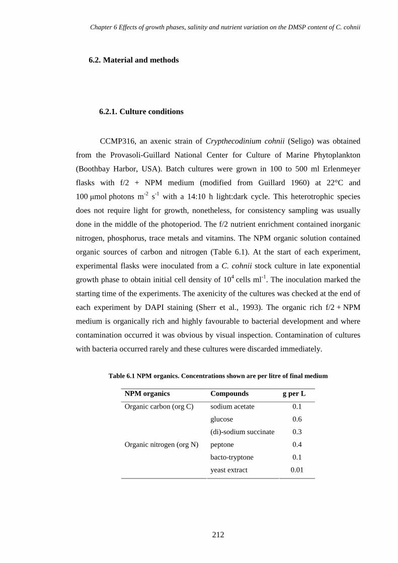
Chapter 6 Effects of growth phases, salinity and nutrient variation on the DMSP content of C. cohnii
212
6.2. Material and methods
6.2.1. Culture conditions
CCMP316, an axenic strain of Crypthecodinium cohnii (Seligo) was obtained
from the Provasoli-Guillard National Center for Culture of Marine Phytoplankton
(Boothbay Harbor, USA). Batch cultures were grown in 100 to 500 ml Erlenmeyer
flasks with f/2 + NPM medium (modified from Guillard 1960) at 22°C and
100 µmol photons m-2 s-1 with a 14:10 h light:dark cycle. This heterotrophic species
does not require light for growth, nonetheless, for consistency sampling was usually
done in the middle of the photoperiod. The f/2 nutrient enrichment contained inorganic
nitrogen, phosphorus, trace metals and vitamins. The NPM organic solution contained
organic sources of carbon and nitrogen (Table 6.1). At the start of each experiment,
experimental flasks were inoculated from a C. cohnii stock culture in late exponential
growth phase to obtain initial cell density of 104 cells ml-1. The inoculation marked the
starting time of the experiments. The axenicity of the cultures was checked at the end of
each experiment by DAPI staining (Sherr et al., 1993). The organic rich f/2 + NPM
medium is organically rich and highly favourable to bacterial development and where
contamination occurred it was obvious by visual inspection. Contamination of cultures
with bacteria occurred rarely and these cultures were discarded immediately.
Table 6.1 NPM organics. Concentrations shown are per litre of final medium
NPM organics Compounds g per L
sodium acetate 0.1
glucose 0.6
Organic carbon (org C)
(di)-sodium succinate 0.3
peptone 0.4
bacto-tryptone 0.1
Organic nitrogen (org N)
yeast extract 0.01

Chapter 6 Effects of growth phases, salinity and nutrient variation on the DMSP content of C. cohnii
213
6.2.2. Salinity treatments
Fifteen flasks (100 ml of volume) containing f/2 + NPM medium (medium
recipe described in section 2.1.2.4) were inoculated with a stock culture to obtain 24 ml
of starting batch cultures at 104 cells ml-1. All flasks were sampled 6 and 24 h after the
inoculation before diluting the cultures with fresh medium at 24h to alter the salinity.
This salinity change marked the start of the experiment (t=0). Four salinity treatments
were tested (9, 20, 45 and 58, measured using the practical salinity scale) and compared
with a control of unchanged salinity (salinity of 31). Each of the 5 conditions was
performed in 3 culture replicates. The dilution was done with 60 ml of f/2 + NPM
medium prepared at various salinities to obtain the final salinities described above.
Medium used for dilutions were prepared as follows: (1) for hypoosmotic shock, filtered
seawater was diluted with distilled water to lower salinities. (2) for hyperosmotic shock,
filtered seawater was heated below its boiling point to evaporate water and increase
salinity. To limit the pH variation, the pH was adjusted with HCl and NaOH solutions
before autoclaving medium. Autoclaved medium was enriched with sterile f/2 + NPM
solutions and added to the cultures. Final pH ranged from 6.8 to 7.4. The cultures were
monitored for up to 78 h after treatment but my investigations focus on the period of
exponential growth during the first 36 h after change of salinity.
6.2.3. Nutrient supplementation treatments
Eighteen flasks (100 ml of volume) containing f/2 + NPM medium were
inoculated with a stock culture to obtain 77 ml of starting batch cultures at 9 -
10×103 cells ml-1. All flasks were sampled at 6, 30, 36 and 48 h. Immediately after the
fourth sampling and as the cultures entered the stationary growth phase, nutrients were
added in 15 flasks. Four treatments of nutrient additions were tested and compared with
a control (0) that received no addition and a control (1) that received addition of all
nutrients as indicated in Table 6.2. I prepared 3 organic stock solutions (NPM = org C +
org N; org C; org N) and for each treatment I added one of these solutions at the same
volume and concentration as the organic solution added in control 1 (6.8 ml). For 3
treatments, the f/2 enrichment was added at the same volume and concentration as in

Chapter 6 Effects of growth phases, salinity and nutrient variation on the DMSP content of C. cohnii
214
control 1 (0.68 ml). Each of the six conditions was performed in 3 culture replicates.
For comparison of growth between all conditions, total cell volume was corrected
according to the dilution applied to the culture that was not applied to the control 0.
Table 6.2 List of nutrient supplementations. The f/2 enrichment comprises sodium nitrate, sodium phosphate, trace metals and vitamins (Guillard and Ryther, 1962; Guillard, 1975). Glc means glucose.
Controls and treatments Nutrients added Control 0 no addition Control 1 = all nutrients f/2 enrichments + org C + org N Control 1 – org N f/2 enrichments + org C Control 1 – org C f/2 enrichments + org N Control 1 – Glc f/2 enrichments + org N +
sodium acetate + sodium succinate
Control 1 – F/2 org C + org N
6.2.4. Carbon availability treatment
Five media of different organic C concentrations were prepared in triplicate in
15 flasks of 100 ml volume. The organic C concentrations were 5, 10, 20, 50 and 100 %
of original recipe (Table1). A stock culture of C. cohnii was grown in normal f/2 +
NPM medium and used as inoculum in the 15 flasks. Batch cultures were sampled after
few minutes, 3, 24, 30, 36 and 54 h to measure DMSPT per cell volume (CV).
6.2.5. Time-response of DMSP adjustment
Three media were prepared with different types and concentrations of organic C:
(1) 100 % D-Glc according to the normal recipe for f/2 + NPM, (2) 5 % D-Glc, (3) 5 %
D-Glc + 95 % L-Glc. Each medium was prepared in triplicate and dispensed in a total of
9 flasks. Each flask was inoculated with a stock culture of C. cohnii and immediately
sampled after 30 seconds for DMSPT per CV measurements and every 30 seconds up to
5 min. At 10 min a last sample was taken for DMSPT per CV and total CV
measurements.

Chapter 6 Effects of growth phases, salinity and nutrient variation on the DMSP content of C. cohnii
215
12
12production DMSPtt
DMSPDMSP
−−
=
12
12production DMStt
DMSDMS
−−
=
6.2.6. DMSP and DMS production
Production of DMS and DMSPT were calculated by subtracting the DMS/P
value from one sample point to the next one and dividing by the period between the two
sampling points (equation 1).
(1)
(2)

Chapter 6 Effects of growth phases, salinity and nutrient variation on the DMSP content of C. cohnii
216
6.3. Results
6.3.1. Growth, DMSP and DMS concentrations in batch cultures
The growth of Crypthecodinium cohnii comprised 3 successive stages, the
relatively short exponential and stationary growth phases and decline phase over a
period of about 174 h (Figure 6.1 A). The exponential phase lasted for approximately
54 h during which total cell density and cell volume increased to reach a maximum of
4.2 × 10⁵ cells ml-1 and 452 µLcell L-1. However, whilst growth rate remained constant
up to 54 h the volume per cell started to decrease earlier and indicated continued cell
division while accumulation of biomass was reduced (Figure 6.1 B). The stationary
phase followed and lasted for about 24 h, though this shows up best in total cell volume
(CV) data (Figure 6.1 A). Between 54 and 78h cell density and total cell volume
remained fairly stable, but growth rate declined and volume per cell reduced by 54 %
from 2100 to 970 µm3. Finally the following decline phase was characterised by a
reduction in cell density and total cell volume and a negative growth rate.
DMSPT and DMSPp concentrations in culture showed similar patterns over the
growth which demonstrated that DMSPp was the major contributor to DMSPT (Figure
6.1 C). Statistical analyses showed no significant difference between DMSPT and
DMSPp measurements (Mann-Whitney U test, P=0.566 > 0.05). DMSPT concentrations
noticeably increased in line with cell growth during the exponential phase and decreased
during the following phases.

Chapter 6 Effects of growth phases, salinity and nutrient variation on the DMSP content of C. cohnii
217
Figure 6.1 Growth and DMSP/DMS parameters in batch cultures of Crypthecodinium cohnii. (A) Cell density and total cell volume (CV). (B) Growth rate and volume per cell. (C) DMSPT, DMSPp and DMS concentrations in the culture. (D) DMSPT per cell and per CV. (E) Production of DMSPT and DMS in culture. Shaded areas indicate the stationary phase of growth based on measurements of total CV. Shown are average values with range of data (n=3, except in panel C for DMSPp concentrations where the asterisk indicates n=2). When no range bars are visible, the range of data was smaller than the symbol size. Baseline of panels B and E show the zero growth and zero production respectively.
-0.04
-0.02
0.00
0.02
0.04
0.06
Gro
wth
rate
(h-1
)
0
500
1000
1500
2000
2500
Vol
ume
per c
ell (µ
m3 )
baselineGrowth rateVolume per cell
1
10
100
Cel
l den
sity
(x10
4 cel
l ml-1
)
10
100
1000
Tot
al C
V (µ
Lce
ll L
-1)
Cell densityTotal CV
-50
0
50
100
0 48 96 144 192Time (h)
DM
SP
T p
rodu
ctio
n in
cultu
re (µ
M d
ay-1
)
-1
0
1
2
DM
S p
rodu
ctio
n in
cu
lture
(µ
M d
ay-1
)
DMSP prod.DMS prod.baseline
T
0.0
0.1
0.2
0.3
0.4
0.5
DM
SP
T p
er c
ell (
pmol
cel
l-1
)
0
100
200
300
400
DM
SP
T p
er C
V (m
mol
Lce
ll-1
)
DMSP per cellDMSP per CVT
T
0
30
60
90
120
DM
SP
in c
ultu
re (µ
M)
0
1
2
3
DM
S in
cul
ture
(µ
M)
DMSP DMSP DMS
pT
*
A
B
C
D
E
Chapter 6 Effects of growth phases, salinity and nutrient variation on the DMSP content of C. cohnii
218
Normalised DMSPT concentrations substantially increased at the end of the
exponential phase and decreased during the following phases (Figure 6.1 D). DMSPT
concentrations per cell volume (CV) increased 4-fold from 61 mM (equivalent to
mmol Lcell-1) at the early exponential phase (6 h) to 255 mM at the beginning of the
stationary phase (54 h) and DMSPT concentrations per cell increased 3-fold from 0.142
to 0.415 pmol cell-1. Then, DMSPT concentrations decreased somewhat during the
stationary phase when normalised to cell volume but showed a sharp decrease when
normalised to cell density. Indeed, cell volume reduction compensated for the decrease
of DMSPT concentration when it was normalised to CV. During the decline phase
DMSPT concentrations slightly increased and decreased again.
DMS concentration in the culture was low during the exponential and stationary
phase (0.023 to 0.229 µM) and increased during the decline phase to a maximum of
2.342 µM (Figure 6.1 C). In mid-exponential phase, DMS concentration was 0.063 µM
and represented 0.4 % of DMSPT (17.6 µM). DMS concentration increased to 0.229 µM
at 54 h similar to the increase in total cell volume. Then, DMS concentration increased
sharply to 2.342 µM during the decline phase accounting at its maximum for 4 % of
DMSPT concentration. Whilst the major DMSP production appeared to occur at the end
of the exponential phase, the major DMS production occurred during the decline phase
when growth rate was negative (Figure 6.1 E and B).

Chapter 6 Effects of growth phases, salinity and nutrient variation on the DMSP content of C. cohnii
219
6.3.2. Effect of salinity treatments on growth and DMSP content
After 24 hours of exponential growth, batch cultures of Crypthecodinium cohnii
were diluted to obtain various salinities in the medium. Medium with a salinity of 31
obtained with the normal recipe of the f/2 + NPM medium recipe was used as a control
for comparisons with other salinity treatments (9, 20, 45 and 58).
The total CV showed a similar pattern for the different salinities tested except
lower values at a salinity of 9 that resulted from lower volume per cell. After dilution
with fresh medium, the cultures recovered to similar exponential growth rates during the
first 12 h (Mann-Whitney U test, P>0.05, Table 6.3) then reduced after 24 h indicating
the onset of the stationary growth phase. Moreover, the growth became more erratic at
salinities of 45 and 58 after 24 hours of treatment (Figure 6.2 A and B).
The volume per cell was significantly affected by the different salinities (Mann-
Whitney U test, P<0.05, Table 6.3). The volume per cell reduced with salinities 9 and
20 from 6 h and 12 h respectively up to the end (Figure 6.2 C and D). The volume per
cell increased at salinities 45 and 58 from 24h to the end of the experiment.
Table 6.3 Probabilities obtained with Mann-Whitney U tests for comparison of growth rate, volume per cell and DMSPT per CV between salinity treatments (9, 20, 45, 58) and the control (salinity of 31) over a period of time after treatment. Asterisks show significant difference between the treatment and the control.
Growth rate Volume per cell DMSPT per CV
6-12h 6-36 h 6-12 h 12-36 h 6-12 h 12-36 h 36-78 h
Salinity 9 0.688 ≤0.001* 0.002* ≤0.001* 1.000 ≤0.001* ≤0.001*
Salinity 20 0.378 0.002* 0.63 ≤0.001* 0.004* 0.002* 0.009*
Salinity 45 0.688 0.023* 0.173 0.023* 0.025* 0.453 0.627
Salinity 58 0.337 0.019* 1.000 0.019* 0.004* 0.583 0.310

Chapter 6 Effects of growth phases, salinity and nutrient variation on the DMSP content of C. cohnii
220
Figure 6.2 Effect of salinity on growth parameters and DMSPT per CV in batch cultures of Crypthecodinium cohnii. For ease of comparison, data are divided into “hypoosmotic shock” (left column, salinities of 9, 20 and 31) and “hyperosmotic shock” (right column, salinities of 31, 45, and 58). (A and B) Total CV. (C and D) Volume per cell. (E and F) DMSPT per CV. Shaded areas show the first 12 hours of treatment. Shown are average values with range bars of data (n=3, except in panels E and F for DMSPT per CV at salinity 31 where the asterisk indicates n=2). When no range bars are visible, the range of data is smaller than the symbol size.
The DMSPT per CV was also significantly affected by the different salinities.
For most salinity treatments, the DMSPT per CV was rapidly adjusted to the external
salinities after 6 hours following the medium transfer (Figure 6.2 E and F). Only the
treatment of salinity 9 showed a delay in the DMSP adjustment after 12 h. Then, whilst
at low salinities the DMSPT per CV remained adjusted during the whole experiment up
to 78 h, at high salinities the DMSPT per CV shortly returns after 12 h to similar
concentrations as in the control. Table 6.3 shows the probabilities obtained for statistical
comparisons between treatments and the control at different periods of the experiment.
From 6 to 12 h following the salinity treatment, the DMSPT per CV was significantly
different from the control at salinities of 20, 45 and 58. From 12 to 78 h, the DMSPT per
Hypoosmotic shock
C
F
Hyperosmotic shock
*
A
E
D
B
0
100
200
-24 -12 0 12 24 36
Time (h)
DM
SP
T p
er C
V (m
M)
0
100
200
300
400
Tot
al C
V (µ
Lce
ll L
-1)
31209
31 4558
-24 -12 0 12 24 36
Time (h)
0
1000
2000
3000
Vol
ume
per c
ell (µ
m3 )

Chapter 6 Effects of growth phases, salinity and nutrient variation on the DMSP content of C. cohnii
221
CV was significantly different from the control at salinities of 9 and 20. Therefore
C. cohnii cells appeared to be capable of short-term acclimation to hyperosmotic shock
by increasing their DMSP to CV ratio but were not able to maintain this metabolic
response for long period of time. However, C. cohnii cells performed short to long-term
acclimation to hypoosmotic shock by decreasing their DMSP to CV ratio and this was a
lasting metabolic adaptation (Figure 6.3 visualises the DMSPT per CV for all salinities
tested at different sampling times).
Therefore, the DMSP content and the volume of the cell were affected
alternately in response to the salinity treatment. At salinity of 9, the volume per cell was
first reduced and then the DMSP per CV decreased whereas at high salinities of 45 and
58, the DMSP per CV first increased and then the volume per cell enlarged.
Figure 6.3 Summary of the effect of salinity on DMSPT concentrations per cell volume (CV). (A) DMSPT per CV expressed as a difference to DMSPT per CV obtained at salinity of 31. (B) DMSPT per CV obtained for 5 salinities tested (9, 20, 31, 45 and 58); shown are average values with range of data (n=3, except at time 0 for salinity of 31 where the asterisk indicates n=2).
-120
-60
0
60
Diff
eren
ce to
DM
SP
T p
er C
V
obta
ined
at s
alin
ity o
f 31
0
50
100
150
200
250
-18 0 6 12 24 30 36Time (h)
DM
SP
T p
er C
V (m
M)
9
20
31
45
58
A
B
*

Chapter 6 Effects of growth phases, salinity and nutrient variation on the DMSP content of C. cohnii
222
6.3.3. Influence of nutrients on growth and DMSP content
The increase in DMSPT per cell and per CV at the end of exponential growth
phase in batch cultures (Figure 6.1 C) was further investigated by supplementing
cultures with the various components of the f/2 + NPM medium (Table 6.1 and Table
6.2) at the onset of the stationary growth phase.
The growth, volume per cell and DMSPT per CV of each supplementation
treatment was compared with that of a control with addition of all nutrients (Control 1)
and a control without nutrient addition (Control 0). All the cultures showed similar
growth and DMSPT per CV patterns until the nutrients were added in late exponential
phase (Figure 6.4, shading). As before, C. cohnii culture reached the stationary phase
after 48 h of growth and over this period DMSPT per CV increased 4-fold (47 to
184 mM, Figure 6.4 A and K). The volume per cell reduced slightly during the
exponential growth phase then reduced more substantially during the following phases
(Figure 6.4 F, “Control 0”).
The addition of all nutrients (Control 1: f/2 enrichment nutrients + org C + org
N) restarted growth, delayed the reduction of the cell volume and a significant decrease
of DMSPT per CV was observed over the next 6 h (from 184 to 100 mM, Figure 6.4 A,
F, K, “Control 1”). Similar results were obtained for the addition of all nutrients – f/2
enrichment (Figure 6.4 B, G, L). For other treatments which lacked some organic
components, the growth rates were lower than after complete nutrient addition (Figure
6.4 C, D, E) and the volume per cell decreased, more substantially in treatment lacking
org C (H, I, J). The addition of all nutrients – org N (Figure 6.4 M) led to similar DMSP
decrease than after complete nutrient addition. However, when all nutrients – org C
(Figure 6.4 N) or all nutrients – Glc (Figure 6.4 O) were added the decline in DMSPT
per CV was eliminated. This suggested that org C and especially glucose was required
to obtain the drop of DMSPT per CV. In other words, the carbon depletion was
responsible for the increase in DMSPT per CV at the end of the exponential phase when
no nutrients were supplemented (Figure 6.4 K, “Control 0”).

Chapter 6 Effects of growth phases, salinity and nutrient variation on the DMSP content of C. cohnii
223
Figure 6.4 Effect of nutrient additions on growth parameters and DMSPT per CV in batch cultures of Crypthecodinium cohnii. (A to E) Total CV. (F to J) Volume per cell. (K to O) DMSPT per CV. Different nutrient additions are presented on each column. Control 0 has no addition, Control 1receive all nutrients and other treatments receive less nutrients than Control 1. Time 0 indicate time of nutrient addition and shading indicates exponential growth before nutrient addition. Shown are average values with range of data (n=3). When no range bars are visible, the range of data is smaller than the symbol size.
0
250
500
750
1000
Tot
al C
V (µ
L cel
l L
-1)
Control 1Control 0
Control 1Control 1 - f/2
0
1000
2000
3000
Vol
ume
per
cell
(µ
m3 )
Control 1Control 0
Control 1Control 1 - f/2
0
100
200
300
-48 -24 0 24 48
DM
SP
T pe
r C
V (
mM
)
Control 1Control 0
-48 -24 0 24 48
Control 1Control 1 - f/2
Control 1Control 1 - org N
Control 1Control 1 - org N
-48 -24 0 24 48
Control 1Control 1 - org N
Control 1Control 1 - org C
Control 1Control 1 - org C
-48 -24 0 24 48
Control 1Control 1 - org C
Control 1Control 1 - Glc
Control 1Control 1 - Glc
-48 -24 0 24 48
Control 1Control 1 - Glc
A C D E
F H I J
K M N O
Time (h) Time (h) Time (h) Time (h) Time (h)
B
G
L

Chapter 6 Effects of growth phases, salinity and nutrient variation on the DMSP content of C. cohnii
224
0
100
200
300
0 12 24 36 48 60
Time (h)
DM
SPT
per
CV
(m
M)
5% C10% C20% C50% C100% C
6.3.4. Influence of the C availability on the DMSP content
The depletion of C causing the increase in DMSPT per CV that I observed in the
nutrient supplementation experiment (Figure 6.4 N and O) was further investigated by
transferring C. cohnii in media with various C concentrations (5, 10, 20, 50 and100 %
of organic carbon). Batch cultures were sampled just after inoculation into new medium
(<5 min) and over the next hours (3, 24, 30, 36, 54 h) to measure the DMSPT per CV.
Reducing the available C in the medium resulted in an increase in the DMSPT
per CV (Figure 6.5). There was a significant effect of the available C concentrations on
the DMSPT content (Kruskal-Wallis, P<0.001). I also checked that all tested C
treatments gave significantly different DMSPT per CV concentrations in comparison to
those of the control with 100 % C (Mann-Whitney U test, P<0.01 detailed in Table 6.4).
The adjustment of DMSP per CV in C. cohnii cells to the external C concentration
appeared to be very fast in a period of minutes or seconds since the DMSPT per CV was
adjusted at the first sampling a few minutes after the transfer.
Figure 6.5 Effect of external organic C concentrations on DMSP per cell volume (CV). Cultures were transferred in medium containing 5, 10, 20, 50 and 100 % C. Time 0h illustrates the first sampling performed a few minutes after the inoculation in new medium. Shown are average values with range of data (n=3, except at 5 % C where the asterisk indicates n=2). When no range bars are visible, the range of data is smaller than the symbol size.
*

Chapter 6 Effects of growth phases, salinity and nutrient variation on the DMSP content of C. cohnii
225
Table 6.4 Probabilities (P) obtained with Mann-Whitney U tests for comparison of DMSPT per CV and volume per cell between treatment with low C concentration (5, 10, 20, and 50 %) and control (100 %). Asterisks show significant difference between the treatment and the control.
C concentration P obtained for
DMSPT per CV
P obtained for
volume per cell
5 % ≤0.001* 0.027*
10 % ≤0.001* 0.019*
20 % ≤0.001* 0.129
50 % 0.002* 0.429
The volume per cell appeared to be also affected by the external C
concentrations (Figure 6.6). At 5 % and 10 % organic C concentration, the volume per
cell was significantly lower than the control (Table 6.4). At 20 and 50 % organic C
concentration, no significant difference was observed with the control. Only very low C
concentrations affected the volume per cell of C. cohnii by reducing it.
Figure 6.6 Effect of external organic C concentrations on volume per cell. Cultures were transferred in medium containing 5, 10, 20, 50 and 100 % C at time 0 h. The first sampling was performed a few minutes after the inoculation into new medium. (A) Treatments with 5 and 10 % were significantly different from the control at 100 % C. (B) Treatments with 20 and 50 % were not significantly different from the control at 100 % C. Shown are average values with range of data (n=3, except at 5 % C where the asterisk indicates n=2). When no range bars are visible, the range of data is smaller than the symbol size.
0 12 24 36 48 60
Time (h)
20% C50% C100% C
0
500
1000
1500
2000
2500
0 12 24 36 48 60
Time (h)
Vol
ume
per c
ell (
µm3 )
5% C10% C100% C
A B
*

Chapter 6 Effects of growth phases, salinity and nutrient variation on the DMSP content of C. cohnii
226
6.3.5. Time-response of DMSP content to the external glucose concentration
In order to test the time required by Crypthecodinium cells to adjust their DMSP
content to the external C concentration of the medium, I monitored the DMSPT
concentrations every 30 seconds during 5 min after the inoculation of C. cohnii in a
medium depleted in glucose (Glc, 5 % of the normal concentration).
Figure 6.7 A shows that DMSPT per CV was adjusted in less than 30 sec to the
external C concentration. On the first sampling point, the DMSPT per CV increased by
100 % (from 72 to 149 mM) in cells inoculated in glucose-depleted medium whereas
the DMSPT per CV decreased by 60% (from 87 to 35 mM) in cells inoculated in replete
medium.
Figure 6.7 Time-response of DMSPT per CV to (A) D-glucose (Glc) limitation, (B) D-Glc limitation compensated with L-Glc in batch cultures of Crypthecodinium cohnii and time-response of DMSPT per L of culture to (C) D-Glc limitation, (D) D-Glc limitation compensated with L-Glc The first data point indicates DMSP concentration in stock culture before the introduction in new medium. Shown are average values with range of data (n=3). When no range bars are visible, the range of data is smaller than the symbol size.
A B
D C
0
40
80
120
160
200
DM
SP
T p
er C
V (m
M) 5% D-Glc
100 % D-Glc95% L-Glc 5% D-Glc
100 % D-Glc
0 2 4 6 8 10Time (min)
95% L-Glu 5% D-Glu
100 % D-Glu
100
1000
10000
100000
0 2 4 6 8 10Time (min)
DM
SP
T in
cul
ture
(nM
) 5% D-Glu100 % D-Glu

Chapter 6 Effects of growth phases, salinity and nutrient variation on the DMSP content of C. cohnii
227
Given that the medium with 5 % glucose differed from the 100 % glucose
medium in osmolar properties, I tested the same glucose limitation treatment with
compensation of 95 % L-Glc. In presence of 95 % L-Glc and 5 % D-Glc, DMSPT per
CV was similar to that of the control 100% D-Glc (Figure 6.7 B). This suggested that L-
Glc was absorbed by cells similarly to D-Glc or that L-Glc potentially balanced the
osmolar pressure of the medium. After 10 min of treatment the mean volume of the cell
was similar for the 3 conditions tested: 1787 µm3 at 100 % D-Glc, 1675 µm3 at 5 % D-
Glc and 1763 µm3 at 5 % D-Glc compensated with 95 % L-Glc (Mann-whitney U tests,
P>0.05 for each treatment compared with the control, n=6 for each treatment).
Measurements of DMSPT in culture not normalised to cell volume, also showed an
obvious difference between the 5 % D-Glc treatment and the other conditions (Figure
6.7 C and D).

Chapter 6 Effects of growth phases, salinity and nutrient variation on the DMSP content of C. cohnii
228
6.4. Discussion
I investigated the growth and DMSP content of Crypthecodinium cohnii in batch
cultures and under different salinity and nutrient conditions in order to identify the
possible physiological roles of DMSP in this species.
DMSPT concentrations were used as an estimation of the intracellular DMSP.
Dinoflagellates are very sensitive to shear stress (as described in sections 1.3.12) so that
measurements of intracellular DMSP are often affected by filtration artefacts (see
section 3.5.1.2). With no significant differences between DMSPT and DMSPp
measurements, DMSPp was the major fraction of DMSPT over the growth. Dissolved
DMSP (DMSPd) and DMS might account for a larger fraction during the decline phase
when the population became senescent. According to my results, maximum DMS
concentrations accounted for 4 % of DMSPT. My own measurements of DMSPd were
highly variable but suggested that DMSPd accounted for 0.5 to 3 % of DMSPT over the
growth (data not shown).
6.4.1. Effect of salinity on C. cohnii batch cultures – evidence for an osmotic
role of DMSP
DMSP is a sulphur metabolite described as a potential compatible solute (Stefels
et al., 2007) and I found here that its concentration was significantly affected by salinity
change and acted certainly as such in C. cohnii. DMSP per CV decreased with low
salinities and increased with high salinities. This species may often be found associated
with decaying seaweeds on beaches, in estuaries or drifting attached on macrophyte
branches (Mendes et al., 2009) thereby affected by large osmotic variations due to tidal
exposure, evaporation and desiccation, and precipitation. The growth of C. cohnii
appeared not to be affected by the salinity change in accordance with the adaptation to
the brackish and marine natural habitat of this species (Tomas, 1997). However, even if
C. cohnii survived at high salinity treatments in our study, this species has not been
reported in natural hyper-saline environments.

Chapter 6 Effects of growth phases, salinity and nutrient variation on the DMSP content of C. cohnii
229
C. cohnii cells respond to external salinity variation by short-term
osmoacclimation to high salinities and long-term osmoacclimation to low salinities. At
low salinities, DMSP per CV decreased and remained adjusted over a relatively long
period of several days. At high salinities, DMSP per CV increased and remained
adjusted over a short period of less than 12 h.
The response of DMSP per CV may differ at low and high salinity treatments as
it has been observed in the diatom Cylindrotheca closterium. The DMSP content of this
diatom increases with salinity up to 44 but does not increase further at higher salinities
but leads to the increase of other osmolytes than DMSP (Van Bergeijk et al., 2003). The
different responses observed in C. cohnii suggest the potential production of other
osmolytes at high salinities.
The volume per cell of C. cohnii was affected by salinity treatments as a
potential result of cell division, biomass increase or sexuality but not of cell turgor.
After a few seconds of treatment, the volume of wall-less cells or the turgor pressure of
walled cells might increase at lower salinity and decrease at higher salinity (Kirst 1989).
C. cohnii cells are covered by thin thecal plates likely to maintain the cell volume.
Moreover the opposite trend of the volume per cell was observed through salinity
treatments showing decrease at lower salinity and increase at higher salinities after
several hours. This volume change was thus, not directly related to the cell osmotic
variation. As growth rates (based on cell number measurements) were similar at
medium and low salinities, the smaller volume at low salinities could result from a
reduced biomass increase at similar cell division rates. At high salinities the volume
increased resulting generally from biomass increase when the growth rate was
unchanged and sometimes from lower growth rates (especially at extremely high
salinity). The cell size variation may also result from change in gametes and zygotes
production. Beam and Himes (1974) observed homothallic sexual reproduction in
several strains of C. cohnii that lead to the production of gametes and zygotes of various
sizes. Zygotes can result from the fusion of 2 or more rarely 3 cells and can generate 4
or 8 cells. The life cycle which influence cell sizes may be affected by external factor
such as oxidative stress that induces the activation of sex genes in the multicellular
green alga Volvox carteri (Nedelcu et al., 2004) or phosphate deficiency that allows

Chapter 6 Effects of growth phases, salinity and nutrient variation on the DMSP content of C. cohnii
230
gamete fusion in the dinoflagellate Lingulodinium polyedrum (Figueroa and Bravo,
2005).
The osmolyte role for DMSP is not generalised to all algae that produce it. There
is evidence that DMSP acts as an osmolyte in some haptophytes (Vairavamurthy et al.,
1985; Stefels, 2000), prasinophytes (Dickson and Kirst, 1986) and green macroalgae
(Dickson et al., 1980; Karsten et al., 1991). In some other green macroalgae species,
DMSP does not respond to salinity variations (Edwards et al., 1987; Van Alstyne et al.,
2003). Some dinoflagellates including Pfiesteria piscicida, Ceratium longipes,
Pyrocystis lunula, Gambierdiscus toxicus contain DMSP in such small amounts that it is
unlikely to act as an osmolyte (as concluded in Chapter 5, section 5.3). Additionally,
DMSP concentration remains stable under salinity changes in some other organisms
such as the terrestrial plant Spartina anglica (van Diggelen et al., 1986). Here, I report
for the first time in detail that DMSP acts as an osmolyte in a heterotrophic
dinoflagellate.
6.4.2. Influence of nutrients on C. cohnii batch cultures – antagonism with
glucose
In normal conditions (f/2 + NPM medium), C. cohnii cultures showed a rapid
growth due to the rich organic nutrition, reaching the stationary phase in about 54 h and
the DMSP per CV increased substantially at the end of the exponential phase. At this
stage, also called the retardation phase, nutrients start to deplete in the culture and affect
the physiological state (Barsanti and Gualtieri, 2006). Hence, I have investigated
whether the nutrient depletion that occurs at the end of the exponential growth phase
could cause the increase in DMSP content.
The nutrient addition experiments in nutrient-depleted batch cultures identified
the carbon source and especially glucose as the limiting nutrient triggering the increase
of DMSP at the end of the exponential growth phase. DMSP production is also
dependant on other nutrients than carbon. In my results the input of organic N and f/2
nutrients led to a greater DMSP production. F/2 nutrients supply trace metals and

Chapter 6 Effects of growth phases, salinity and nutrient variation on the DMSP content of C. cohnii
231
vitamins necessary to enzyme activities involved in metabolic processes and DMSP
synthesis.
Deschamps et al. (2008) observed that starch synthesis and accumulation in
C. cohnii occurs during the early to mid-exponential phase then recedes and stops at the
onset of the stationary phase when glucose was no longer available. Clearly, glucose
assimilation and DMSP production both vary with growth stages and show an
antagonistic behaviour. It is possible that DMSP replaces the glucose-derived osmolytes
when glucose is limited. In many organisms, glucose is metabolised to carbohydrates
that are known to play a role as osmolytes: trehalose in some cyanobacteria, yeasts,
bacteria, actinomycetes (Welsh, 2000) and animals (Westh and Ramlov, 1991); sucrose
in some microalgae, cyanobacteria and phototrophic bacteria; glycerol in some
microalgae and yeasts; glucosylglycerol in cyanobacteria; arabitol and sorbitol in yeasts
(Welsh, 2000); mannitol in microalgae, bacteria, fungi and plants (Iwamoto and
Shiraiwa, 2005). Therefore, in microalgae including cyanobacteria, sucrose, mannitol,
glycerol, glucosylglycerol and trehalose have been reported as glucose-derived
osmolytes. C. cohnii produces a large variety of glucose-derived compounds as it
irregularly excretes polysaccharides containing mainly glucose, galactose and mannose
and also in lesser amounts fucose, uronic acid and xylose (de Swaaf et al., 2001). No
studies have reported the cell content of glucose-derived compounds inside C. cohnii
cells. In our experiments, glucose limitation could lead to the lack of glucose-derived
osmolytes, the utilisation of most intracellular polysaccharides and the replacement by
amino acid-derived osmolytes including DMSP. Similarly some yeasts produce
different organic osmolytes during the different growth phases, for example they
accumulate glycerol during exponential phase then trehalose during stationary phase
when glucose is limiting (Meikle et al., 1988). Indeed, in situations of C limitation for
C. cohnii, DMSP can be synthesised via the pool of existing methionine or through the
degradation of proteins as described by Uchida et al. (1996). This hypothesis moreover
explains the requirement of N organic source to produce DMSP as observed in my
experiment.

Chapter 6 Effects of growth phases, salinity and nutrient variation on the DMSP content of C. cohnii
232
6.4.3. Time-response of DMSP adjustment
The osmotic action of DMSP was described as a rather slow process (Kirst,
1989; Stefels, 2000) but I observed an exceptionally fast DMSP per CV adjustment in
response to glucose limitation. Microalgal DMSP responds slowly to hyper- and hypo-
osmotic shocks over a period of a few hours compared to non-organic osmolytes
including Na+ and K+ ions that change within an hour (Kirst, 1989). However, there is
the exception of the prasinophyte Tetraselmis subcordiformis that adjusts its DMSP
content in less than 1 h to hypoosmotic shock and about 3 h to hyperosmotic shock
(Dickson and Kirst, 1986). In my salinity experiments, the DMSP per CV changed in
less than 6 h in response to most of the hyper- and hypo-osmotic treatments and a
longer period of about 12 h was observed at salinity of 9. These periods were the first
sampling point of our experiment and the exact duration has not been investigated.
However, the DMSP concentration was adjusted within seconds in response to carbon
and glucose limitation in the medium. This rapidity in the DMSP metabolism observed
in C. cohnii might result from the fast general metabolic activity of this species in
laboratory cultures. C. cohnii is an osmotrophic heterotrophic eukaryote grown on
organic source of N and C. It could be compared to yeasts in terms of metabolism and
growing conditions.
The physiological response of C. cohnii may be similar to other unicellular
eukaryotes such as yeasts which are able to sense nutrient changes, especially glucose,
and adjust their metabolism within minutes. Wang et al., (2004) observed that starved
yeasts change the expression of about 60 % of their genes by more than 2-fold increase
or decrease 20 minutes after glucose addition. Zaman et al. (2008) observed similar
response for yeasts which change by 2-fold the expression of 40 % of their genes within
minutes after glucose addition in culture grown on glycerol. This adjustment occurs in a
few minutes and involves complex signalling networks and rapid adjustment of the
metabolism. Such system is not known in C. cohnii and further research is required to
understand how this extremely fast adjustment could occur within seconds.
The fast response of DMSPT per CV within 30 seconds could more likely result
from a physical response of the cell volume change due to a difference in osmolar
pressure of the medium. However, the cell volume was unchanged after 10 minutes of
Chapter 6 Effects of growth phases, salinity and nutrient variation on the DMSP content of C. cohnii
233
treatment at 5 % D-Glc and the measurement of DMSPT per litre of culture also showed
a large difference between the 5 % D-Glc and the 100 % D-Glc conditions. Some
experiments investigating the cell volume change over the 10 minutes period after
introduction in the 5 % D-Glc could determine more precisely a potential effect of the
different osmolarities of the media. Additionally, the time-response of C. cohnii to
DMSP adjustment could be tested by adding glucose in a culture entering the stationary
phase, as I already observed a sharp decrease in DMSP concentrations. Finally, to
support the idea of an extremely rapid DMSP synthesis within the 30 seconds of culture
transfer in 5 % D-Glc medium the investigation of the molecular processes involved in
DMSP synthesis are necessary, as for example the measurement of an enzyme activity
involved in DMSP synthesis in C. cohnii such as the hypothetical methionine
decarboxylase.
In the experiment with 5 % D-Glc compensated with 95 % L-Glc, C. cohnii cells
reacted as if they were incubated in 100 % D-Glc showing similar DMSPT per CV and
DMSPT in culture concentrations. Given that L-Glc cannot be used as a carbon source,
the addition of L-Glc was applied here to complete the osmotic potential of glucose
depleted medium (95 % L-Glc + 5 % D-Glc). These results suggest that the addition of
L-Glc could balance the osmolarity of the medium similarly to the presence of 100 %
D-Glc in the medium and eliminating the requirement of intracellular DMSP production
or the addition of L-Glc could balance the osmolarity of C. cohnii cells by being
absorbed similarly to D- Glc, potentially acting as an osmolyte and replacing DMSP.
The assimilation of L-Glc has been observed in other organisms such as in cells of rat
and hamster, rabbit intestines (Neale and Wiseman, 1968; Bihler et al., 1969; Wood et
al., 1989), house sparrows (Chang et al., 2004) and yeasts (Ritchie et al., 1986). Further
experiments with radio-labelled (14C) L-Glc could determine whether this sugar is
absorbed by C. cohnii cells. In conditions of low D-Glc concentrations completed with
L-Glc, the medium was carbon-limited and the absence of DMSP increase excluded the
effect of starvation stress as a stimulus for DMSP production.
Crypthecodinium cohnii potentially re-metabolised its DMSP stock in conditions
of carbon-replete conditions. In conditions of glucose-depleted medium that was
repleted by glucose addition, DMSPT concentration decreased and I observed that the
DMSPd fraction decreased as well (result not shown) suggesting that DMSP was rather

Chapter 6 Effects of growth phases, salinity and nutrient variation on the DMSP content of C. cohnii
234
used by cells than released to the medium. Kadota and Ishida (1968) observed that
C. cohnii was not able to incorporate an external source of DMSP but more recently,
Salo et al., (2009) found that the heterotroph Oxyrrhis marina was able to incorporate
and assimilate dissolved DMSP by osmotrophy. Such ability should be re-assessed for
C. cohnii cells. Therefore, DMSP appears to be an organic resource not only for bacteria
but potentially for heterotrophic dinoflagellates which are able to assimilate and
metabolise DMSP. Moreover C. cohnii might be able to re-metabolise its intracellular
stock of DMSP. Another example of microalga utilising DMSP has been described by
Kates and Volcani (1996) who presented DMSP as a theoretical intermediate in the
metabolic pathway of the phosphatidylsulfocholine in the diatoms Nitzschia spp.
Further experiments with C.cohnii cultures supplemented with radio-labelled (35S)
DMSP could determine whether DMSP is incorporated by C. cohnii cells in replete
conditions and in glucose-limited conditions.
6.5. Conclusion
I have shown that DMSP acts as an osmolyte for Crypthecodinium cohnii in
various salinity treatments and suggested that other osmolytes might be produced at
high salinity. Moreover, DMSP also seems to be produced as a substitute for glucose-
derived osmolytes in conditions of carbon or glucose depletion. Osmotic regulation for
turgor pressure is a fundamental process for aquatic species. In this regard and
especially for osmotrophic species such as C. cohnii are special cases in that uptake of
carbon compounds will disturb osmolarity.
DMSP might act as an osmolyte in other heterotrophic dinoflagellates as well.
Other heterotrophic dinoflagellates that are known to produce DMSP include
Protoperidinium pellucidum, Pfiesteria schumwayae and Pfiesteria pisciscida and
potentially Protoperidinium ovatum (see Chapter 5). However, some of these other
heterotrophs show different trophic behaviours, including grazing and are likely to use
DMSP in different ways. On the other hand, C. cohnii strains are adapted to their
natural brackish habitat and have developed or maintained strategies such as DMSP

Chapter 6 Effects of growth phases, salinity and nutrient variation on the DMSP content of C. cohnii
235
synthesis to survive large salinity variations; other heterotrophs that would live in
different environments might not have this ability.
Contrary to slow adjustment of organic osmolytes previously observed in
microalgae, the adjustment of DMSP concentration to glucose limitation is
exceptionally fast within seconds. Similar processes have been observed in yeasts
within minutes involving nutrient availability sensing and response by regulating gene
expression. Given that this species is also investigated in biotechnology areas such as ω-
3 fatty acid production, extremely fast molecular processes might be of interest.
I observed that DMSP concentration may suddenly increase but also decrease
depending on the external glucose concentration and in the latter case the fate of DMSP
that could be released or re-metabolised is still unclear. According to our observations
DMSP seemed to be re-metabolised but more investigation would help to confirm this
first observation.
Only a small number of heterotrophic dinoflagellates have been investigated for
their DMSP content in regard to the thousand or so living species that constitute 1/3rd of
the microzooplankton biomass. This significant component of the marine ecosystem is a
potential source of DMSP and DMS and a potential contributor to the global sulphur
cycle. I hope my results are going to encourage further research on the DMS and DMSP
production by heterotrophic dinoflagellates.
Further research is warranted to determine the influence of nutrient and salinity
variations on DMSP content of C. cohnii in conditions close to their natural
environment. In its natural habitat, C. cohnii likely utilises various sugars and organic
molecules and the influence of a natural carbon source might differ from the antagonism
behaviour between glucose and DMSP that I observed. Mesocosm cultures of C. cohnii
could be used to assess the effect of natural disturbance such as precipitations and
natural salinity variation on its DMSP content or laboratory cultures with seawater from
areas where C. cohnii have been identified would represent more realistically the natural
DMSP content of this species and its variation over the growth.

Chapter 7 Effects of darkness and nitrogen availability on growth and DMSP content of C. cohnii
236
Chapter 7 Effects of darkness and nitrogen availability on growth and
DMSP content of the heterotrophic dinoflagellate Crypthecodinium
cohnii
Abstract
Following the description in the previous chapter of an osmolyte function of DMSP in
the heterotrophic dinoflagellate Crypthecodinium cohnii, I have investigated other
potential roles for this secondary metabolite. The possible functions of antioxidant or
overflow metabolite were examined by assessing the effect of light and nitrogen
limitation on growth and DMSP content of C. cohnii. Batch cultures were grown in dark
and light conditions. Although C. cohnii reached significantly lower cell volume under
light exposure (24 % difference in average, P<0.05), the DMSP per cell volume was
similar (89 - 620 mM in the dark and 95 - 788 mM in the light, P>0.05). The presence
of photo-protective carotenoids in C. cohnii is likely to reduce the requirement of
antioxidant and, hence, DMSP may not be utilised as an antioxidant under light
exposure. The nitrogen limitation was applied by reducing the concentration of
nitrogen-containing compounds in the medium. The results showed that during the
exponential growth phase, nitrogen limitation increased the DMSP content (1.54-fold
increase for a 20-fold nitrogen dilution) suggesting a potential role for DMSP as an
antioxidant in response to N-limitation stress or a possible role of overflow metabolite
to remove excess sulphur and recycle amino groups. During the late exponential and
stationary growth phases, the DMSP production in response to glucose limitation (as
observed in the previous chapter) was clearly dependent on nitrogen availability. These
new results highlight the multifunctional properties of DMSP within C. cohnii species
and show a strong relationship between the nitrogen availability and the DMSP content
of heterotrophic dinoflagellates.

Chapter 7 Effects of darkness and nitrogen availability on growth and DMSP content of C. cohnii
237
7.1. Introduction
In the previous chapters, I described how DMSP concentrations vary in
dinoflagellate species to better understand the biological role of DMSP in this important
plankton group. In chapter 6, I focussed my study on the frequently used heterotrophic
model Crypthecodinium cohnii given that whilst heterotrophic dinoflagellates are a
potential source of DMSP, few studies have investigated the role of this compound in
such organisms.
Previously, I showed that C. cohnii utilises DMSP as an osmolyte and as a
potential substitute to glucose-derived osmolytes by investigating the effect of some
abiotic parameters such as salinity and nutrients on the DMSP content of C. cohnii. For
nutrient treatments, I examined, in depth, the effect of glucose limitation, demonstrated
that this results in increased DMSP concentrations and I also noticed the effect of
nitrogen limitation that appeared to restrain the DMSP concentration in conditions of
glucose co-limitation. The effect of nitrogen limitation without glucose limitation on
DMSP concentration in C. cohnii is not known. In other phytoplankton groups, nitrogen
limitation has been reported to increase DMSP concentrations, such as in prasinophytes
(Gröne and Kirst, 1992) and diatoms (Bucciarelli and Sunda, 2003). In these reports,
DMSP was suggested to act as a potential antioxidant in response to nutrient-limitation
stress (Bucciarelli and Sunda, 2003) or as an overflow metabolite during conditions of
unbalanced growth with excess sulphur and lack of nitrogen (Stefels, 2000). In this
chapter, I investigated in more detail the effect of nitrogen limitation on the DMSP
concentration in C. cohnii.
Some other abiotic parameters such as light exposure or dark conditions might
also affect the DMSP concentrations. C. cohnii is able to grow in complete darkness as
a heterotrophic species. In fact, it is growing with even higher growth rate in the dark in
comparison to light. Therefore, light has been suggested to inhibit growth by allocation
of resources towards the production of photo-protective pigments such as carotenoids
(Tuttle and Loeblich, 1973). Several observations suggest that the presence or absence
of light may trigger different metabolic responses in protists: Jakobsen et al. (2004)

Chapter 7 Effects of darkness and nitrogen availability on growth and DMSP content of C. cohnii
238
have shown that heterotrophic species have a circadian cycle of growth and feeding
rates that is maintained by a light cue such as a light irradiance threshold in the case of
the heterotrophic dinoflagellate Oxyrrhis marina. Strom (2001) reported that the
heterotrophic dinoflagellate Noctiluca scintillans shows light-aided digestion that could
result from the potential photo-oxidation of the intracellular organic matter. Moreover
the same study showed that two ciliate species grow with substantially higher rates
when exposed to light than in darkness.
As far as I am aware, the effect of light or darkness on the DMSP content of
heterotrophic dinoflagellates that are exposed to a light-dark cycle in natural conditions
has not been investigated. Important questions that need addressing are: Does growth
inhibition in the presence of light also affect the DMSP content? Does light exposure
trigger the production of antioxidant compounds such as DMSP?
In this chapter, I investigated the effect of the abiotic parameters light exposure
and nitrogen limitation on the DMSP content of C. cohnii batch cultures. The aim was
to identify other potential roles of DMSP in C. cohnii and increase our insight into the
factors that are affecting its concentration.

Chapter 7 Effects of darkness and nitrogen availability on growth and DMSP content of C. cohnii
239
7.2. Materials and Methods
Axenic batch cultures of Crypthecodinium cohnii were grown in f/2 + NPM
medium at 22 ºC in a controlled environment (MLR-351 Plant Growth Chamber,
Sanyo). The treatments tested with the cultures are described below.
7.2.1. Light and dark treatment
A stock culture grown under 100 µmol photons m-2 s-1 with a 14:10 light:dark
cycle in late exponential growth phase was inoculated into 6 Erlenmeyer flasks of
250 ml volume containing 120 ml of medium. Three starting cultures were placed in
three 1L Winchester black plastic containers inside the incubator and three starting
cultures were placed under light exposure that was similar to the light levels
experienced by the stock culture. Growth parameters and total DMSP were monitored
for these 2 treatments once a day over growth phases including the exponential,
stationary and decline phases of growth in 7 days. All cultures were sampled at the
same time that was the mid-time of the photoperiod of cultures in light.
7.2.2. Nitrogen limitation treatment
The compounds that constitute the main sources of organic nitrogen in the
f/2 + NPM medium are peptone, bacto-tryptone and yeast extract. As described in
chapter 6 (section 6.2.1), other f/2 + NPM components include the organic C sources
glucose, sodium acetate and sodium succinate. I investigated N limitation by removing
the main sources of N from the medium (Table 7.1).
Fifteen flasks containing 5 different media in triplicate were inoculated at the
beginning of the experiment with a stock culture in late exponential growth phase. The
media contained various concentrations of organic nitrogen: 5, 10, 20, 50 and 100 % of
the original recipe (Table 7.1). Growth parameters and total DMSP were monitored over

Chapter 7 Effects of darkness and nitrogen availability on growth and DMSP content of C. cohnii
240
the different growth phases to quantify the effect of nitrogen limitation. Three
supplementary flasks containing medium with 10 % organic nitrogen were inoculated
similarly to the others. After 54 h of growth and as these 3 cultures appear to be in
stationary growth phase, the organic nitrogen was added to reach the 100 % of the
original recipe in order to check the effect of nitrogen limitation and supplementation on
growth and DMSP concentrations.
Table 7.1 Organic sources of nitrogen in original f/2+NPM recipe equivalent to 100% N.
7.2.3. Measurements and analyses
Growth parameters including cell density (cells ml-1) and total cell volume
(µLcell L-1) were monitored using a particle counter (Multisizer 3, Beckman).
Total DMSP (DMSPT) measurements were performed as an approximation of
particulate DMSP (DMSPp) and were conducted similarly to the methods described in
Chapter 2, Chapter 4 and Chapter 6.
The production of total cell volume (CV) was calculated as a difference of CV
between one sample point and the previous one over the period of time (equation 1).
The same calculation was performed for the production of DMSP over a period of time
(equation 2).
NPM organic nitrogen Compounds g per L
peptone 0.4
bacto-tryptone 0.1
Organic nitrogen (org N)
yeast extract 0.01
12
12production DMSPtt
DMSPDMSP
−−
=12
12production CVtt
CVCV
−−= (1) (2)

Chapter 7 Effects of darkness and nitrogen availability on growth and DMSP content of C. cohnii
241
Given that data were not normally distributed, non parametric statistical tests
(Mann-Whitney U and Spearman tests) were carried out with a statistical software
package (SPSS, version 16.0). When analysing the effect of a treatment on several
parameters over the growth of the culture, I performed statistical analyses on all data
collected over the growth to consider the global effect of the treatment and I also
performed statistical analyses on parts of the growth to allow observation of a potential
effect of the treatment during specific periods of the growth. For the light experiment, I
divided the total data into 3 growth phases and performed analyses on the whole growth
(6 - 150 h) and on each phase (6 - 54 h, 54 - 102 h, 102 - 154 h).

Chapter 7 Effects of darkness and nitrogen availability on growth and DMSP content of C. cohnii
242
D
B A
C
7.3. Results
7.3.1. Effect of light on growth and DMSP content
Some batch cultures of Crypthecodinium cohnii were grown in complete
darkness and other batch cultures were exposed to light. Cultures were sampled over the
growth phases to investigate the effect of light on growth and DMSP content.
For detailed comparison between treatments, I divided all the data into 3 phases
of growth (Figure 7.1, shading): (1) the exponential phase from 6 to 54 h when the cell
density increased, (2) the stationary phase from 54 to 102 h when cell density appeared
stable (3) the decline phase from 102 to 154 h when the cell density decreased. The
DMSPT in culture followed the same trend as the cell density over growth. The total
CV, however, showed a short plateau at the end of phase 1 as cells were dividing
without accumulating biomass and an increase at the beginning of phase 2 as cells
where not dividing but accumulating biomass.
Figure 7.1 Effect of light exposure and darkness on growth and DMSP parameters in batch cultures of Crypthecodinium cohnii. (A) Cell density. (B) Total cell volume. (C) DMSPT in culture. (D) DMSPT per CV. Shown are average values with range of data (n=3). When no range bars are visible, the range of data was smaller than the symbol size. The growth is divided into 3 phases and the grey-shaded area is the stationary phase.
0
20
40
60
Cel
l den
sity
(× 1
04 c
ell m
l-1)
lightdark
0
200
400
600
Tot
al C
V (µ
Lce
ll L
-1)
0
50
100
150
0 48 96 144
Time (h)
DM
SP
T in
cul
ture
(µM
)
0
250
500
750
1000
0 48 96 144
Time (h)
DM
SP
T p
er C
V (m
M)

Chapter 7 Effects of darkness and nitrogen availability on growth and DMSP content of C. cohnii
243
The growth was significantly affected by light exposure (Figure 7.1 A and B).
Cell density and total CV were significantly different over the growth phases between
the 2 treatments with lower values in the presence of light (Mann-Whitney U test,
P<0.05, Table 7.2). The total CV of cultures exposed to light represents 24 % less on
average than total CV of cultures grown in the dark. Similarly, the light exposure
resulted in an average of 25 % reduction of the cell density in comparison with cultures
in the dark. However, similar cell density was observed between the 2 treatments during
the exponential growth phase and similar total CV was obtained during the stationary
phase (Table 7.2).
The effect of light exposure on DMSP concentrations in the culture was
insignificant and ranged from 2 to 121 µM in the dark and 2 and 107 µM in the light
(Mann-Whitney U test, P>0.05, Table 7.2) over the whole period of the experiment.
However, during the stationary phase only, a significant difference was observed
between dark and light treatment (P<0.05). After normalising DMSP to total CV, the
data ranged from 89 - 620 mM and 95 - 788 mM DMSP per CV in the dark and light
respectively and the difference was insignificant for the whole period of the
experiment(P>0.05). During the decline phase only, a significant difference was
obtained in DMSP per CV between dark and light treatments (P<0.01).
Table 7.2 Statistical probabilities obtained with Mann-Whitney U test for comparison of growth and DMSP parameters between cultures exposed to light and in darkness over a period of growth (GPh = Growth phase). Asterisks show significant differences.
All growth Exponential
GPh
Stationary
GPh
Decline Ph
6 - 150 h 6 - 54 h 54 - 102 h 102 - 150 h
Cell density 0.013* 0.402 0.001* 0.002*
Total CV 0.045* 0.046* 0.171 0.007*
DMSP in culture 0.178 0.402 0.007* 0.07
DMSP per CV 0.054 0.145 0.233 ≤0.001*

Chapter 7 Effects of darkness and nitrogen availability on growth and DMSP content of C. cohnii
244
7.3.2. Effect of nitrogen availability on growth and DMSP content
Given that I previously observed that DMSP concentration in C. cohnii was
partially dependent on nitrogen availability (Chapter 6), I further investigated the
influence of a range of nitrogen concentrations (5, 10, 20, 50, 100 %) on growth and
DMSP content.
The cell density and total cell volume (CV) was affected by nitrogen availability
and the DMSP content showed an opposite response between exponential and stationary
growth phases (Figure 7.2). The maximum cell density and total CV was lower under
nitrogen limitations (Figure 7.2 A and B), as for instance the total CV was 617, 398,
242, 159 and 139 µLcell L-1 in cultures containing 100, 50, 20, 10 and 5 % nitrogen,
respectively. Whilst DMSP per CV showed significant negative correlation with N
concentrations during exponential growth phase (6 - 30 h, Spearman test, P<0.05)
DMSP per CV showed significant positive correlation with N concentrations during
stationary growth phase (48 – 78 h, Spearman test, P<0.01; Figure 7.2 C). Therefore,
during the exponential growth phase, at 6 h after the culture inoculation in nitrogen-
limited media, higher DMSP concentrations were observed compared to the nitrogen-
enriched medium. For instance, DMSP was increased significantly (Kruskal-Wallis test,
P<0.05) 1.54-fold in 5 % N compared to 100 % N medium (106 and 69 mM of DMSP
respectively), 1.46-fold in 10 % N, 1.39-fold in 20 % N and 1.22-fold in 50 % N (101,
96 and 84 mM of DMSP respectively). During the period of late exponential and
stationary phases, the DMSP concentrations were more elevated in cultures with higher
N concentrations. During the same period, between 30 and 78 h, the biomass production
(total CV production) and the DMSP production were both dependent on nitrogen
concentrations (R2= 0.99 and R2=0.93 respectively, Figure 7.3).

Chapter 7 Effects of darkness and nitrogen availability on growth and DMSP content of C. cohnii
245
100%50%20%10%5%
R2 = 0.99
R2 = 0.93
0
20
40
60
0 20 40 60 80 100
% organic N
DM
SP
T p
er C
V p
rodu
ctio
n
30-7
8 h
(mM
day
-1)
0
100
200
300
400
Tot
al C
V p
rodu
ctio
n
30-7
8 h
(µL
cell L
-1 d
ay-1
)
Figure 7.2 Effect of nitrogen availability on growth and DMSP parameters in batch cultures of Crypthecodinium cohnii. (A) Cell density. (B) Total cell volume (CV). (C) DMSP per CV. Each colour shows a different nitrogen treatment. Shown are average values with range of data (n=3). When no range bars are visible, the range of data was smaller than the symbol size.
Figure 7.3 Regression of DMSP production and total cell volume (CV) production from 30 to 78 h of growth over nitrogen concentration (expressed as % of concentration in original recipe). Each colour visualises a different nitrogen treatment as shown in the legend. Each data point shows the average of 3 culture replicates. When no range bars are visible, the range of data was smaller than the symbol size.
C
A
0
20
40
60
Cel
l den
sity
(× 1
04 c
ell m
l-1)
100%50%20%10%5%
50
150
250
350
0 24 48 72 96 120Time (h)
DM
SP
per
CV
(mM
)
0
200
400
600
800
Tot
al C
V (µ
Lce
ll L
-1) B

Chapter 7 Effects of darkness and nitrogen availability on growth and DMSP content of C. cohnii
246
To confirm the effect of nitrogen limitation on growth and DMSP content I
tested the effect of nitrogen supplementation. The N addition was performed after 54 h
of culture in nitrogen-deprived medium (10 %).
Figure 7.4 shows that growth parameters and DMSP concentration increased to
reach similar maximum levels as the control culture with 100 % nitrogen. Nitrogen was
thus, limiting the growth and DMSP production.
Figure 7.4 Effect of nitrogen addition (indicated by vertical dotted lines) in nitrogen limited medium on growth (A and B) and DMSP content (C and D). Comparison of cultures grown in normal recipe medium (100 % N, black line), N-limited medium (10 %, orange dashed line), N-limited medium (10 % N + addition of 100 % N, orange dotted line). Shown are average values with range of data (n=3). When no range bars are visible, the range of data was smaller than the symbol size.
B
C D
A
0
1
10
100
Cel
l den
sity
(×10
4 cel
l ml-1
)
100%10%10% + N
10
100
1000
Tot
al C
V (
µLce
ll L
-1)
50
150
250
0 24 48 72 96 120Time (h)
DM
SP
per
CV
(mM
)
0.1
0.2
0.3
0.4
0.5
0 24 48 72 96 120Time (h)
DM
SP
per
cel
l (pm
ol c
ell
-1)

Chapter 7 Effects of darkness and nitrogen availability on growth and DMSP content of C. cohnii
247
7.4. Discussion
7.4.1. Effect of light on growth and DMSP content
Heterotrophic protists do not require light for growth, however, light interferes
with several physiological functions such as growth, feeding and digestion. For
instance, light cue determines the circadian cycle of cell divisions and feeding rates of
heterotrophic protists (Jakobsen and Strom, 2004). Additionally, the growth rate has
been observed to be higher in ciliates at 0.36 day-1 under 100 µmol photons m-2 s-1 of
fluorescent light compared to almost null growth in the dark (Strom, 2001). On the
other hand, the growth rate is 10-fold lower in the dinoflagellate Crypthecodinium
cohnii under more than 3 W m-2 of fluorescent light compared to cells grown in the dark
(Tuttle and Loeblich, 1975).
Crypthecodinium cohnii is a heterotrophic dinoflagellate that acquires organic
matter by osmotrophy or, more rarely observed, by predation on unicellular red algae
(Ucko et al., 1997). This species probably contains a remnant plastid (Sanchez-Puerta et
al., 2007) but is incapable of photosynthesis. Nevertheless, this heterotroph contains 3
carotenoids (Tuttle and Loeblich, 1973) that protect cells against damage from sunlight
(Withers and Tuttle, 1979). The abundance of these carotenoids is 4-fold lower in cells
that are being kept in the dark compared to cells grown under 73 µmol photons m-2 s-1
of fluorescent light (calculated equivalent to 500 foot candles, reported by Tuttle and
Loeblich 1973) and the higher growth rate observed in darkness could result from the
resources saved by decreasing carotenoid synthesis (Tuttle and Loeblich, 1975). In our
experiments, growth and DMSP content of C. cohnii were also significantly affected by
light exposure. Similar to previous observations of lower growth rate under light
exposure, I observed lower cell density and total cell volume in cultures exposed to light
at 100 µmol photons m-2 s-1.
Carotenoids are vital for C. cohnii cells exposed to light and carotenoid-deficient
mutants show higher mortality rate under light exposure (Withers and Tuttle, 1979).

Chapter 7 Effects of darkness and nitrogen availability on growth and DMSP content of C. cohnii
248
One function of carotenoids in photosynthetic organisms is the photo-protection of
reaction centre complexes against photo-oxidative processes. With polyene chains of
more than 9 double bonds, carotenoids quench the chlorophyll triplet state to avoid the
generation of oxygen singlet or carotenoids may also directly quench oxygen singlet
and other radical species. This quenching process occurs via formation of carotenoid
triplet stage lower in energy than triplet chlorophyll or oxygen singlet and is followed
by excessive energy dissipation as heat towards the return of carotenoid to its normal
state (Vershinin, 1999; Frank and Brudvig, 2004). The photo-protective mechanism of
carotenoids in a non-photosynthetic organism such as C. cohnii is unknown but
carotenoids also have non-photosynthetic functions (Krinsky, 1978). They potentially
act as antioxidant defence via scavenging and elimination of free radicals as described
for some other non-photosynthetic organisms (Mathews, 1966; Krinsky, 1989;
Vershinin and Lukyanova, 1993; Vershinin, 1999) and are likely to protect cells against
reactive oxygen species (ROS) generation (H2O2, singlet O2, radical .OH and
peroxides). Aerobic organisms are subject to ROS generation usually due to the
intracellular metabolism in mitochondria, peroxisomes and enzyme systems and under
particular exposure to UV, chemicals or toxins (Finkel and Holbrook, 2000).Given that
DMSP and its by-products have shown capacities in scavenging ROS, hence similar
function of cell protection against ROS (Sunda et al., 2002), the DMSP concentration
may also be affected by light exposure analogous to carotenoid content. However, my
experiments showed no difference in DMSP per CV in the presence or absence of light.
On the other hand, C. cohnii possesses other active anti-oxidant mechanisms including
iron-containing superoxide dismutases, enzymes that are able to degrade harmful
superoxide radicals (O2.-); (Dufernez et al., 2008). Therefore, carotenoids and
superoxide dismutases appear to protect C. cohnii cells against photo-oxidation
efficiently enough so that the additional antioxidant properties of DMSP may not be
required. Nonetheless, this result does not exclude the potential use of DMSP in
C. cohnii cells in other conditions of oxidative stress such as exposure to solar
ultraviolet radiations or elevated concentrations of heavy metals (Okamoto et al., 2001)
or under nutrient limitation (Bucciarelli and Sunda, 2003).

Chapter 7 Effects of darkness and nitrogen availability on growth and DMSP content of C. cohnii
249
7.4.2. Effect of nitrogen availability on growth and DMSP content
The nutrient experiment of chapter 6 (section 6.3.3) suggested that N-limitation
restrained the DMSP production in C. cohnii and I decided to investigate N-limitation
by removing the N source of the medium in more detail. However, N-containing
compounds (peptone, bacto-tryptone and yeast extract) are complex substrates that may
also supply carbon, sulphur, phosphorus, trace metals, vitamins and other potential
compounds in lesser proportions than nitrogen. Organic carbon is mainly supplied by
glucose, sodium acetate and sodium succinate and is not considered to be limited by
reducing the concentrations of N-containing compounds. Hence, it is likely that
limitation of N-containing compounds will mostly affect N-dependant metabolic
processes.
During the late exponential and stationary growth phases, the N-limitation
resulted in lower DMSP per cell volume (CV) and this effect was confirmed by an
increase of DMSP content in response to N supplementation after a period of limitation.
In agreement with the conclusions of Chapter 6, the glucose depletion in late
exponential and stationary growth phases triggered an increase in DMSP concentration
that is dependent on the available organic source of nitrogen. This may be explained by
the obligate requirement for the DMSP precursor, methionine (Uchida et al., 1996)
which may be obtained directly or could be produced from the organic nitrogen source
in the medium. Similarly, Gröne and Kirst (1992) proposed that the rate of DMSP
synthesis is dependent on methionine availability in the autotroph Tetraselmis
subcordiformis that accumulates more DMSP in methionine-enriched medium.
During the exponential growth phase, glucose was not limiting and the nitrogen
limitation appeared to increase the DMSP per CV. Similar increase in DMSP
concentrations during N-limitation have been reported. Turner et al. (1988) observed
higher DMSP concentrations in field measurements of mixed phytoplankton population
from areas of lower nitrate concentrations and in laboratory measurements of the
coccolithophore Emiliania huxleyi cultured in low nitrate medium. Gröne and Kirst
(1992) found higher DMSP content in T. subcordiformis cells cultured in N-deprived

Chapter 7 Effects of darkness and nitrogen availability on growth and DMSP content of C. cohnii
250
medium. Bucciarelli and Sunda (2003) observed increased DMSP concentrations in N-
limited cultures of Thalassiosira pseudonana and suggested that DMSP acts as a
potential antioxidant in response to nutrient limitation. Similarly, the response of
C. cohnii may also be a result of nutrient limitation that creates oxidative stress. On the
other hand, the production of DMSP in conditions of N-deficiency may support the
“overflow hypothesis” that describes the function of DMSP production and exudation to
eliminate the excess sulphur (Stefels, 2000). Sulphur and nitrogen are both required for
protein synthesis and the lack of nitrogen would result in sulphur excess. The
production of DMSP, as observed, here, by the increase of DMSP concentration per CV
during the exponential growth phase in conditions of N-limitation, may serve to
catabolise excess sulphur and recycle amino groups to satisfy N-dependant metabolic
processes.
Further experiments are necessary to confirm or eliminate these hypotheses: the
“antioxidant hypothesis” could be tested with measurements of ROS production (for
example H2O2, radical .OH, superoxide anion O2.- as described by Degli Esposti 2002)
and checked if their concentration is increased by the N-limitation stress; the “overflow
hypothesis” could be assessed by the measurement of the dissolved fraction of DMSP
(DMSPd) to check if the DMSP exudation is increased by the N-deprivation. However,
if DMSP is re-metabolised instead of excreted no observation of DMSPd would be
possible and in that case, the N:S ratio measurements of the cells could determine
whether the elemental state of the cell is unbalanced.
As previously discussed, the N-limitation treatments may have resulted in
decreased concentrations of other nutrients. However, in conditions of nitrogen
limitation during the exponential growth phase, the increase of DMSP concentrations
showed that sulphur was not limiting the DMSP production. Therefore, the decrease of
nitrogen-containing compounds did not appear to generate a result in sulphur limitation.
Moreover, the DMSP synthesis involves the anabolic machinery of enzymes that
requires metals and vitamins. These elements did not appear to be limited by the lower
levels of nitrogen-containing compounds. Finally, the use of defined organic media for
the culture of C. cohnii such as media used by Tuttle and Loeblich (1975) would allow
experimenting with more precision the limitation of nutrients by removing exactly one
type of nutrient only.

Chapter 7 Effects of darkness and nitrogen availability on growth and DMSP content of C. cohnii
251
7.5. Conclusion
The heterotrophic dinoflagellate Crypthecodinium cohnii is an obligate
heterotroph that is able to grow in complete darkness, however, light interferes with
growth. C. cohnii growth is reduced by light exposure but the DMSP per CV does not
appear to be affected by the presence or absence of light. Therefore under similar
conditions of light exposure where Tuttle and Loeblich (1973) observed that carotenoids
were produced for photo-protection (fluorescent light of 100 and 73 µmol photons m-2 s-
1 here and in Tuttle and Loeblich 1973 respectively), DMSP seemed ineffective as an
antioxidant.
Growth and DMSP content of C. cohnii is, however, affected by the availability
of organic nutrients in the medium. As previously observed, nitrogen was required for
DMSP synthesis during late exponential and stationary growth phases where glucose
depletion enhanced the increase of DMSP content. I confirmed here that DMSP
concentrations were significantly dependent on external organic nitrogen
concentrations.
Nitrogen limitation has a different effect during the exponential phase when
glucose is not a limiting factor. In such conditions, nitrogen limitation enhanced the
increase of DMSP content. The role of DMSP during the exponential growth phase of
C. cohnii needs more investigation to determine whether it is a response to oxidative
stress due to nutrient limitation or whether it is part of an overflow mechanism.
These results point out the multifunctional properties of DMSP within the same
species. In the previous chapter, the role of DMSP as an osmolyte was proposed and
demonstrated by some of the experiments. With the new results presented here, other
potential roles of DMSP as an antioxidant or overflow metabolite were suggested in
C. cohnii cells. Moreover, a strong relationship between the nitrogen availability and the
DMSP content was observed and the novel data represent the first steps in elucidating
the role of DMSP in heterotrophic organisms.

Chapter 8 Synthesis: progress and perspectives
252
Chapter 8 Synthesis: progress and perspectives
In this section, I describe the major issues investigated throughout this thesis.
The main results are summarised and discussed in relation with the progress achieved
and from the perspective of further research that could be developed.
8.1. DMSP and DMS measurement in dinoflagellate cultures
Regarding performing DMSP and DMS measurements on dinoflagellate culture
samples, time was invested in tests to optimise the sampling and handling strategy
before setting up experiments. The analytical uncertainty of the methods was assessed
and the potential sources of variation in DMSP and DMS concentrations were
investigated.
For particulate DMSP (DMSPp) measurement, I examined the variability in
DMSPp concentration over the day and over the growth. Diel cycle in DMSP
concentrations have been observed in other phytoplankton species such as in batch
cultures of the coccolithophore Emiliania huxleyi and suggested for the dinoflagellates
Alexandrium tamarense and Scrippsiella trochoidea in their natural environment
(Merzouk et al., 2004; Bucciarelli et al., 2007). Here, I showed for the first time that
DMSP concentration per cell volume increased over the photoperiod in batch cultures
of the photosynthetic dinoflagellate Heterocapsa triquetra (Chapter 3). During growth
in batch cultures, the physiological conditions of the phytoplankton cells generally
change over time in response to nutrient depletion and cell ageing. I found that DMSP
concentrations increased at the end of the exponential growth phase (Chapter 3). This
variability is critical for deciding upon culture sampling time during an experiment. To
exclude diel variability in DMSP concentration, cultures were sampled at the same time

Chapter 8 Synthesis: progress and perspectives
253
(mid-time) within the photoperiod each day. In addition sampling was done at the same
point of the growth phase, usually mid-exponential phase to avoid the effect of nutrient
limitation.
A comparison of values obtained from measurement of total DMSP (DMSPT)
with the sum of all its measured individual fractions, i.e. DMSPp, DMSPd and DMS,
suggested a missing fraction of 25 % in the additive value. No study has ever reported
the measurements of all fractions in culture or field samples that would fully validate
the different methods used for these 4 parameters. The investigation of the major
fraction DMSPp (72 %) using 3 different filtration methods (hand vacuum pump,
automated syringe pump and filtration by gravity) only showed an increase in DMSPp
concentration in one experiment where it was higher using the automated syringe pump
rather than using the hand vacuum pump (Chapter 3). When using the syringe pump, I
observed that increasing the delivery speed resulted in increasing DMSP concentration
and reducing the analytical uncertainty. Nevertheless, the analytical uncertainty was in a
similar range when using the syringe pump and the hand vacuum pump (Chapter 3).
The filtration step is critical for DMSPp measurement and as Kiene and Slezak
(2006) pointed out it can move DMSP from the particulate to the dissolved fraction. In
dinoflagellate cultures especially, filtration may create even more damage as many
species are known to be very fragile to turbulence. The filtration of a dinoflagellate
sample increases the analytical uncertainty (relative standard deviation or RSD) for
DMSPp measurement: within an experimentt detailed in chapter 3, I obtained 1.1 -
16.4 % RSD for DMSPp and 0.6 - 5.2 % RSD for DMSPT measurement without
filtration. The measurement of DMSPT avoids filtering culture samples and it gives data
with less analytical uncertainty so it was favoured in this study (chapter 3). A similar
technique was also applied by Keller et al (1989a) to determine DMSP concentrations
of 123 phytoplankton species and by Yost and Mitchelmore (2009) who measured the
DMSP content of the dinoflagellate Symbiodinium spp. Therefore, DMSPT is a rather
common representation of DMSP content in phytoplankton. Nonetheless, using the
method I described in detail in Chapter 3 and 4, I obtained the best analytical precision
(6 - 32 % of, RSD, for culture samples n=9 on average in 9 species) given that Yost and
Mitchelmore obtained RSD between 34 and 76 % (n=3 - 4). By eliminating the filtration
step, this method also allowed fast measurements (including sampling and sample

Chapter 8 Synthesis: progress and perspectives
254
preparation) in quick succession that would not have been possible with DMSPp
measurement (time response of C. cohnii DMSP adjustment to glucose limitation,
Chapter 6 section 6.4.3).
DMS measurements were performed on culture samples filtered with the syringe
pump at the highest delivery speed available (14 ml min-1) in agreement with findings
on DMSPp measurements. The filtrate was introduced into the purge tube with a sample
loop device to ensure injection of constant volume. This entire system described in
Chapter 3 was aimed at ensuring satisfactory replication of DMS measurements in
dinoflagellate cultures (Chapter 6, section 6.3.1). This system led to a low analytical
uncertainty for standards (2 %) but larger for culture samples (25-33 %). I observed that
agitation of culture can affect the DMS measurement by either increasing or decreasing
its value. I also found that DMS concentrations were increased by increased time of
filtration and large sample volume. According to these results I performed filtration of
small volume samples (5 ml) using the syringe pump set at the maximum delivery speed
of 14 ml min-1.
DMSPd measurement includes the same steps for sample preparation as DMS
measurement plus the addition of NaOH to the filtrate sample. The variations in DMS
concentration that may result from the handling and the filtration of the culture are
likely to be similar for DMSPd measurement. Some investigations should be done to
optimise DMSPd measurements in dinoflagellate cultures. I have observed large
variation on preliminary DMSPd measurements (Chapter 3) and the analytical
uncertainty could be tested and improved using the syringe pump at high delivery speed
according to the results obtained for DMS and DMSPp. Some tests could also be done
on agitated and non-agitated flasks to determine if the handling of the flask would have
any effect on DMSPd measurement.

Chapter 8 Synthesis: progress and perspectives
255
8.2. The contribution of the dinoflagellate group to the DMSP and DMS
oceanic pool
Several laboratory and field studies have pointed out the occurrence of DMSP in
dinoflagellate cells. The commonly referred studies done by Keller et al., (Keller,
1988/1989; 1989a; 1989b) reported the DMSP content of 25 dinoflagellate strains
amongst 123 phytoplankton species and strains. They concluded that dinoflagellates are
a significant DMSP-producing group.
Twenty years later, Stefels et al. (2007) recommended that the dinoflagellate
group should be inserted as DMS producers in ocean climate models. However, so far
only 1 model has included this group, and in this model dinoflagellates are mixed in
with other nanophytoplankton (Vogt et al., 2010). Le Quéré et al (2005), did incorporate
the dinoflagellate group when describing a 10 Plankton Functional Types (PFT)
representative ecosystem for the use of modellers. The photosynthetic dinoflagellates
were placed in the “mixed phytoplankton group” and, even though described as non
efficient DMS producers, their DMSP production parameterisation was equivalent to
that of the PFT “DMS producers” (12 mmol mol-1 C). By directly measuring DMSP and
carbon content in 8 photosynthetic dinoflagellates, I obtained a DMSP content value
very close to this (10.4 mmol mol-1 C, Chapter 4). The heterotrophic dinoflagellates
were placed in the “proto-zooplankton” PFT with no parameterised DMSP production.
It is now clear that several heterotrophic species synthesise DMSP (listed in Chapter 5)
but currently the extent of this ability amongst the large diversity of the heterotrophic
dinoflagellates is unknown. The limited amount of data available so far and the large
diversity of the dinoflagellates including both photosynthetic and heterotrophic species
makes it difficult for scientists to accurately describe, incorporate or parameterise the
DMSP and DMS contribution of this group.
In this thesis, I provided more data on DMSP content and DMSP lyase activities
(DLA) for dinoflagellates and gathered the existing data to summarise the available
knowledge with the aim of giving a better understanding and description of the DMSP
and DMS contribution of dinoflagellates. I produced DMSP measurements of 9 species
from my laboratory work and gathered together a total of 61 concentration values for

Chapter 8 Synthesis: progress and perspectives
256
DMSP per cell volume from the literature (Chapters 4 and 5). By analysing this dataset
I observed that dinoflagellates contain DMSP in highly variable concentrations that
spread over 6 orders of magnitude difference (per cell volume) between species.
Furthermore, the dataset includes the broadest span of values and the highest values
reported to date in any phytoplankton group. Moreover, I concluded that several factors
appeared to affect the DMSP content of dinoflagellates: species with a haptophyte-like
plastid and bioluminescent species (Pyrocystales order from the Longhurst oceanic
province CAMR in the East Pacific) contained significantly lower concentration of
DMSP whereas species from the Mediterranean province had higher concentration of
DMSP. Additional measurements of species belonging to these subgroups would help to
reinforce or refute these conclusions which are based on the 61 values available per cell
volume. From this database I also noticed that species from the southern hemisphere
and the poles are underrepresented and need more investigation.
The causes underlying the variability in DMSP content of dinoflagellates are
still unclear and might be related to the multifunctional role of this sulphur compound. I
also noted that the osmolyte role of DMSP is unlikely in species that produce very low
DMSP concentrations (Chapter 5).
The DMSP content of dinoflagellates makes them a potential source of DMS.
Some dinoflagellates are able to synthesise DMSP lyase that may cleave DMSP to DMS
and acrylate. I detected in vitro DMSP lyase activity in 5 dinoflagellate species from the
9 species tested in this study (Chapter 4). Given that very few DLA data have been
published for dinoflagellates, this increases the DLA dataset to data for 23 strains and
17 species (Chapter 5). More effort in increasing the number of species assessed for
DLA would be worthwhile as it is now clear that some dinoflagellates can produce
substantial amounts of DMSP and DMSP lyase. The release of DMS from DMSP
occurs in seawater through the action of algal and bacterial enzymes (Steinke et al.,
1996; Yoch, 2002; Todd et al., 2009). The fraction due to algal enzyme activities and
the overall contribution from dinoflagellates may be substantial in areas where
dinoflagellates form blooms (Steinke et al., 2002a). Sampling and measuring DLA in
several dinoflagellate blooms from diverse areas and environments, as for instance in
blooms of Mediterranean species which have high DMSP concentrations or in recurring
dense blooms of Karenia brevis in the Gulf of Mexico, would further assess the

Chapter 8 Synthesis: progress and perspectives
257
potential impact of dinoflagellate DLA on DMS release in the ecosystem. Also, the
bloom period of maximum DLA per cell or per cell volume could be investigated by
performing successive DLA measurements over the initiation, development and
termination of a dinoflagellate bloom. The contribution of the dinoflagellate group in
comparison with other phytoplankton groups able to produce DMSP lyases could be
assessed by performing DLA measurements over phytoplankton successions.
The classification of DMSP-rich species I listed for some dinoflagellate species
in Chapter 5 and especially bloom-forming species would help to identify potential
areas of elevated DMSP concentrations and to estimate the amount of associated DMS
production. Additional measurements of DMSP:Chl a ratio in the lab (Chapter 4) and in
the field open doors towards future estimation of oceanic DMSP concentrations from
Chl a satellite data as it is now possible to describe the dominant assemblages of
phytoplankton taxa by remote sensing using a range of accessory pigments (Alvain et
al., 2008). This tool could be applied to estimate global oceanic DMSP concentrations
given that DMSP measurements are limited to the tracks of scientific cruises.
Nevertheless, an estimation of DMSP:Chl a concentration for the dinoflagellate group
could hardly be realistic due to the broad variation in DMSP content across
dinoflagellate species.
Furthermore, dinoflagellate populations seem to be affected by the changing
climate. Changes in abundance due to increased sea surface temperature have been
observed (Edwards et al., 2006) but consequences of ocean acidification for this group
are not known yet. Future research on the effect of ocean warming and acidification on
dinoflagellate populations and their DMSP content could be critical for predicting the
future DMS emissions and climate interactions and scenarios. Dinoflagellate blooms are
often monitored for toxicity by many countries and some of this monitoring could be
associated with sampling and analysis for DMS and DMSP to substantially increase the
available field database and prospect for DMSP-rich species and oceanic areas.

Chapter 8 Synthesis: progress and perspectives
258
8.3. The biological role of DMSP and DLA in heterotrophic dinoflagellates
Some heterotrophic dinoflagellates are able to synthesise DMSP and species
such as Crypthecodinium cohnii contain DMSP in rather high concentrations (61 –
255 mM). Heterotrophic dinoflagellates are unique amongst free-living heterotrophs in
this respect. This ability might originate from a common photosynthetic ancestor of the
dinoflagellates (McFadden, 2001) and may provide some support to the evolutionary
theory of a photosynthetic ancestor for the dinoflagellate group. Some heterotrophic
dinoflagellates such as C. cohnii and Oxyrrhis marina are thought to harbour a vestigial
plastid because they synthesise plastid-targeted genes and peptides (Sanchez-Puerta et
al., 2007; Slamovits and Keeling, 2008). However, whilst C. cohnii contains DMSP,
none has been found in O. marina. Indeed this species has been proved useful for
studies on the incorporation of DMSP by grazing DMSP-producing prey and by
osmotrophy because it does not contain DMSP (Salo et al., 2009).
The open question is: why have some heterotrophic dinoflagellates conserved
the ability to produce DMSP if, as far as I am aware, no other heterotrophs synthesise
it? I investigated the role of DMSP in C. cohnii and found in common with some
photosynthetic organisms that DMSP acts as an osmolyte (Chapter 6). Heterotrophic
marine organisms are subject to essentially the same osmotic variations experienced by
photosynthetic marine organisms and require the same sort of adaptive mechanisms
including osmolyte synthesis. In other microzooplankton including ciliates,
osmoregulation may be performed by variation of amino acid composition and
abundance (Kaneshiro et al., 1969).
The DMSP concentration in C. cohnii cells is also dependent on nutrient
availability. Nutrient limitation appeared to enhance DMSP accumulation (Chapter 6
and 7). In the case of glucose limitation, DMSP is probably accumulated in replacement
of glucose-derived osmolytes. The determination of the polysaccharide composition in
C. cohnii cells and its variation in glucose-deprived medium could confirm this
hypothesis. Additionally, the analysis of other potential osmolytes such as amino acids
and their derivatives generally including proline and glycine betaine (Welsh, 2000)
could be conducted in C. cohnii cells. In case of nitrogen limitation, DMSP is
accumulated potentially as an overflow mechanism or possibly as an antioxidant in

Chapter 8 Synthesis: progress and perspectives
259
response to nutrient-limitation stress. However, in conditions of D-glucose limitation
compensated with L-glucose, the absence of DMSP increase suggested that if L-glucose
was incorporated but non-assimilated by the cells, the nutrient-limitation stress would
not cause any DMSP increase. To clearly eliminate the hypothesis of nutrient-limitation
stress triggering a DMSP increase, the incorporation of L-glucose by C. cohnii cells has
to be tested by performing experiments with C. cohnii cells grown in medium composed
of radiolabelled L-Glucose as the unique source of glucose. The antioxidant hypothesis
could be tested by measuring the reactive oxygen species in C. cohnii cultures in N-
limited and N-replete conditions which could indicate whether there is an oxidative
stress due to nutrient-limitation. The overflow mechanism hypothesis could be assessed
by determining if there is any exudation of DMSP in N-limited conditions and
measuring the potential fraction of extracellular DMSP concentration in comparison
with N replete conditions.
The production of DMSP lyase has been observed and quantified in C. cohnii
and it is not yet known whether other heterotrophic dinoflagellates are also able to
produce DMSP lyase and, thereby represent a potential source of DMS. DLA
measurements in other heterotrophic dinoflagellates should be done in future research.
The presence of a DMS-producing enzyme in heterotrophic dinoflagellates might
suggest that these organisms could use DMS as a grazer deterrent as has been suggested
for phytoplankton (Wolfe and Steinke, 1996; Wolfe et al., 1997). Grazing experiments
with C. cohnii and other heterotrophic dinoflagellates or other protists could be done to
determine whether this function exists for DMS in these organisms.
C. cohnii is also used in the biotechnology research area for the production of ω-
3-fatty acids (Mendes et al., 2009). I have observed exceptionally fast responses to
external stimuli such as the DMSP synthesis in response to transfer of culture in
glucose-deprived medium and which occurred within a few seconds (Chapter 6). Such
response could be dependent on gene expressions and regulations as it is reported for
yeasts which can change the expression of 40 % of their genes within minutes in
response to nutrient changing conditions. If a similar molecular mechanism could
happen in less than a minute it could lead towards several applications: it could be
utilised by genetic modification toward the production of any key products in the same
way that yeasts are used as laboratory factories for drug production (Szczebara et al.,

Chapter 8 Synthesis: progress and perspectives
260
2003) or the responsible genes could be introduced in plasmids of bacteria to accelerate
any metabolite production. The identification of such genes and mechanisms would also
shed light on the DMSP metabolism in heterotrophic dinoflagellates.
8.4. Further issue- DMSP synthesis and its subcellular localisation
A major issue that remains for the DMSP production by phytoplankton research
field is the DMSP pathway and its subcellular location. Wollastonia biflora seems to
produce DMSP in the chloroplast (Trossat et al., 1996) and the same might happen for
phytoplankton but no evidence has been reported yet. Before starting this study, I had
hoped that investigating the DMSP content of dinoflagellate species that harbour
different plastid types would give an indication on the role of the plastid in DMSP
synthesis. However, more variation in DMSP content was observed between species
holding the same plastid type than between species with different plastid types. This
result was strongly influenced by the fact that species holding peridinin plastids and
heterotrophic dinoflagellates are by far the most common types of species in the ocean
today. Rather few non-peridinin plastid species are currently available in culture and the
species harbouring haptophyte-like plastids showed lower DMSP content than species
with a peridinin plastid. This in itself was surprising given that haptophytes in general
have substantial DMSP content. It seems that the conservation or loss of genes may be
more likely to affect the DMSP content rather than the plastid type.
Several questions remain: Is DMSP produced in the plastids of dinoflagellates
and other microalgae? Is it possible that heterotrophic dinoflagellates synthesise DMSP
in a remnant plastid or is DMSP produced in a different subcellular location? The
purification of plastids in dinoflagellates appears to be a very delicate and difficult
process due to the fact that plastids are fragile and are never isolated completely intact
as shown by the loss of proteins (Wang et al., 2005). However, the separation of
cytoplasmic and plastidic fractions could be a satisfactory preliminary approach towards
determining the major subcellular distribution of DMSP.

Chapter 8 Synthesis: progress and perspectives
261
The DMSP pathway in microalgae is assumed to be similar to the one described
for the green macroalgae Enteromorpha intestinalis (Gage et al., 1997) after detection
of the same key intermediates in a range of microalgal cells. However, this pathway has
not been fully verified and there is scope for further investigation. The identification of
enzymes involved in the DMSP pathway (such as the enzyme that oxidizes the DMSP
precursor, DMSP-aldehyde found by Trossat et al., 1996) could be used for identifying
the subcellular localisation of DMSP biosynthesis according to the presence of an N-
terminal targeting signal or by using an immunolabelling technique such as that used to
locate proteins and enzymes in Snapdragon flowers (Kolosova et al., 2001).
The future identification of the genes involved in DMSP and DMSP lyase
synthesis would represent a landmark for the DMSP research area. If they were
conserved, such genes could be used to rapidly screen a high number of phytoplankton
species and strains. This would lead to the identification of potential DMSP and DMS
producers in cultures and in the field similar to the detection of toxin production genes
that are used for monitoring harmful algal blooms. Molecular methods could allow
detection of the presence of such genes with just a few cells using the PCR-
amplification. The detection of DMSP-producing genes would thus be possible in
species that are difficult to grow in culture and in field samples of low cell density.
Moreover, the generation of mutants deficient in DMSP-production genes could be used
in comparative eco-physiological experiments with normal clones in order to determine
the role and benefit of DMSP. However, recent work has shown that the bacterial genes
involved in DMSP lyase synthesis appear to be rather diverse with 3 genes encoding for
3 different enzymes that catalyse diverse reactions of DMS production from DMSP
(Todd et al., 2007; Johnston et al., 2008; Todd et al., 2009). If the same proves true for
DMSP production and DMSP lyase synthesis genes in phytoplankton this would limit
their application as suggested above. Nevertheless, the investigation on genes involved
in DMSP and DMSP lyase synthesis in dinoflagellate and other phytoplankton cells
would be of great interest.

262
Appendix I List of species
All the species mentioned in the thesis are listed here with their author names and date
when available. Species are divided into Class and listed by alphabetic order.
Class Bacillariophyceae
Cylindrotheca closterium (Ehrenberg) Reimann & Lewin 1964
Class of Dinophyceae
Akashiwo sanguinea (Hirasaka) Hansen & Moestrup 2000
Alexandrium affine (Inoue & Fukuyo) Balech 1985
Alexandrium catenella (Whedon & Kofoid) Balech 1985
Alexandrium minutum Halim 1960
Alexandrium monilatum (Howell) Balech 1985
Alexandrium ostenfeldii (Paulsen) Balech & Tangen 1985
Alexandrium tamarense (Lebour) Balech 1995
Amphidinium carterae Hulburt 1957
Amphidinium operculatum Claparède & Lachmann 1859
Azadinium spinosum Elbrächter & Tillmann 2009
Ceratium arcticum (Ehrenberg) Cleve 1901
Ceratium fusus (Ehrenberg) Dujardin 1841
Ceratium longipes (Bailey) Gran 1902
Ceratium tripos (Müller) Nitzsch 1817
Ceratocorys horrida Stein 1883
Crypthecodinium cohnii Seligo 1952
Dinoclonium sp Pascher 1927
Dinophysis acuminata Claparède & Lachmann 1859
Dinophysis acuta Ehrenberg 1839
Dinophysis caudata Saville-Kent 1881

263
Dinophysis norvegica Claparède & Lachmann 1859
Dinothrix sp Pascher
Dissodinium lunula (Schütt) Pascher
Durinskia baltica (Levander) Carty & Cox
Durinskia capensis Pienaar, Sapiai & Horiguchi 2007
Galeidinium rugatum Tamura & Horiguchi 2005
Gambierdiscus toxicus Adachi & Fukuyo 1979
Gonyaulax spinifera (Claparède & Lachmann) Diesing 1866
Gymnodinium acidotum Nygaard 1949
Gymnodinium chlorophorum Elbrächter & Schnepf 1996
Gymodinium fuscum (Ehrenberg) Stein 1878
Gymodinium nelsoni Martin 1929
Gyrodinium aureolum Hulburt 1957
Gyrodinium impudicum Fraga & Bravo 1995
Heterocapsa pygmaea Lobelich, Schmidt & Sherley 1981
Heterocapsa triquetra (Ehrenberg) Stein 1883
Karenia brevis (Davis) Hansen & Moestrup 2000
Karenia mikimotoi (Miyake & Kominami ex Oda) Hansen & Moestrup 2000
Karenia umbella de Salas, Bolch & Hallegraeff 2004
Karlodinium armiger Bergholtz, Daugbjerg & Moestrup 2006
Karlodinium australe de Salas, Bolch & Hallegraeff 2005
Karlodinium veneficum (Ballantine) Larsen 2000
Lepidodinium viride Watanabe, Suda, Inouye, Sawaguchi & Chihara 1990
Lingulodinium polyedrum (Stein) Dodge 1989
Noctiluca sp Suriray 1836
Ostreopsis sp Schmidt 1901
Oxyrrhis marina Dujardin 1841
Peridiniopsis berolinense (Lemmermann) Bourelly
Peridinium bipes Stein 1883
Peridinium gatunense Nygaard 1925
Peridinium quinquecorne Abé 1927
Peridiniella catenata (Levander) Balech 1977
Perkinsus atlanticus Azevedo 1989
Pfiesteria piscicida Steidinger & Burkholder 1996

264
Pfiesteria shumwayae Glasgow & Burkholder 2001
Prorocentrum arcuatum Issel 1928
Prorocentrum belizeanum Faust 1993
Prorocentrum lima (Ehrenberg) Dodge 1975
Prorocentrum micans Ehrenberg 1833
Prorocentrum sp. Ehrenberg 1834
Protoceratium reticulatum (Claparède & Lachmann) Butschli 1885
Pyrocystis noctiluca Murray ex Haeckel 1890
Pyrodinium sp Plate 1906
Scrippsiella trochoidea (Stein) Balech ex Loeblich 1965
Symbiodinium microadriaticum Freudenthal 1962
Symbiodinium pilosum Trench & Blank 2000
Symbiodinium sp Freudenthal 1962
Takayama helix de Salas, Bolch, Botes & Hallegraeff 2003
Takayama tasmanica Bolch & Hallegraeff 2003
Tovellia sanguinea Moestrup, Hansen, Daugbjerg, Flaim & D’andrea 2006
Class Prymnesiophyceae
Emiliania huxleyi (Lohmann) Hay & Mohler 1967
Phaeocystis sp Lagerheim 1893
Phaeocystis pouchetii (Hariot) Lagerheim 1896

265
Appendix II Bioluminescent and non-bioluminescent species
The dinoflagellate species presented in chapter 5 are listed in the table below
with description of bioluminescence property and the source of this data. CCMP means
that information about bioluminescence was available on the CCMP website (Wilson,
2009).
Species Bioluminescence Data source Alexandrium minutum No No record as bioluminescent
Prorocentrum sp. IIB2B1 No Unknown
Gyrodinium impudicum No No record as bioluminescent
Scrippsiella trochoidea NIES-369 No No record as bioluminescent
Dinophysis acuminata No No record as bioluminescent
Heterocapsa pygmaea CCMP1322 No CCMP, (Wilson, 2009)
Prorocentrum arcuatum No No record as bioluminescent
Amphidinium carterae CCMP1314 No CCMP
Crypthecodinium cohnii CCMP316 No CCMP
Heterocapsa triquetra CCMP449 No CCMP
Scrippsiella trochoidea CCMP1331 No CCMP Symbiodinium microadriaticum CCMP827
No CCMP
Amphidinium carterae CCMP1314 No CCMP
Scrippsiella trochoidea CCMP1599 No CCMP
Amphidinium operculatum CCAP1102/6 No No record as bioluminescent
Heterocapsa triquetra NIES-7 No No record as bioluminescent
Amphidinium carterae CCMP1314 No CCMP
Alexandrium minutum CCMP113 No CCMP
Amphidinium carterae No CCMP
Gymnodinium nelsoni No No record as bioluminescent
Alexandrium tamarense CCM P115 Yes CCMP
Alexandrium tamarense CCMP116 No CCMP
Alexandrium tamarense CCMP1771 Yes CCMP
Thoracosphaera heimii L603 No CCMP
Cachonina niei CACH No No record as bioluminescent
Prorocentrum micans CCMP 691 No CCMP
Heterocapsa sp. CCMP450 No CCMP
Alexandrium fundyense CCMP1719 Yes CCMP
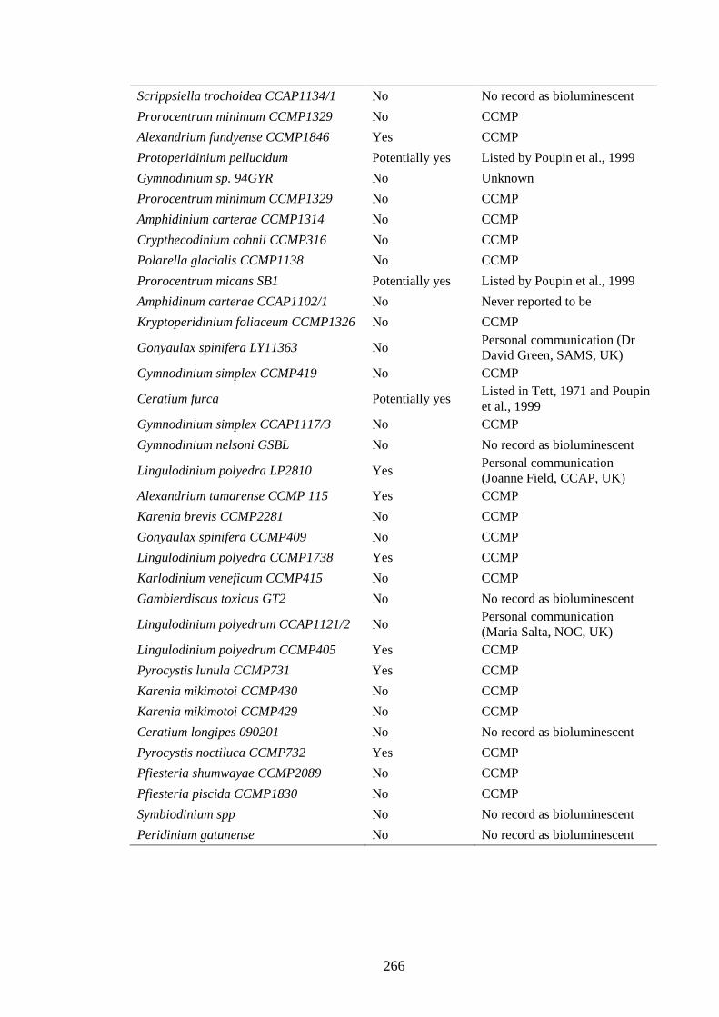
266
Scrippsiella trochoidea CCAP1134/1 No No record as bioluminescent
Prorocentrum minimum CCMP1329 No CCMP
Alexandrium fundyense CCMP1846 Yes CCMP
Protoperidinium pellucidum Potentially yes Listed by Poupin et al., 1999
Gymnodinium sp. 94GYR No Unknown
Prorocentrum minimum CCMP1329 No CCMP
Amphidinium carterae CCMP1314 No CCMP
Crypthecodinium cohnii CCMP316 No CCMP
Polarella glacialis CCMP1138 No CCMP
Prorocentrum micans SB1 Potentially yes Listed by Poupin et al., 1999
Amphidinum carterae CCAP1102/1 No Never reported to be
Kryptoperidinium foliaceum CCMP1326 No CCMP
Gonyaulax spinifera LY11363 No Personal communication (Dr David Green, SAMS, UK)
Gymnodinium simplex CCMP419 No CCMP
Ceratium furca Potentially yes Listed in Tett, 1971 and Poupin et al., 1999
Gymnodinium simplex CCAP1117/3 No CCMP
Gymnodinium nelsoni GSBL No No record as bioluminescent
Lingulodinium polyedra LP2810 Yes Personal communication (Joanne Field, CCAP, UK)
Alexandrium tamarense CCMP 115 Yes CCMP
Karenia brevis CCMP2281 No CCMP
Gonyaulax spinifera CCMP409 No CCMP
Lingulodinium polyedra CCMP1738 Yes CCMP
Karlodinium veneficum CCMP415 No CCMP
Gambierdiscus toxicus GT2 No No record as bioluminescent
Lingulodinium polyedrum CCAP1121/2 No Personal communication (Maria Salta, NOC, UK)
Lingulodinium polyedrum CCMP405 Yes CCMP
Pyrocystis lunula CCMP731 Yes CCMP
Karenia mikimotoi CCMP430 No CCMP
Karenia mikimotoi CCMP429 No CCMP
Ceratium longipes 090201 No No record as bioluminescent
Pyrocystis noctiluca CCMP732 Yes CCMP
Pfiesteria shumwayae CCMP2089 No CCMP
Pfiesteria piscida CCMP1830 No CCMP
Symbiodinium spp No No record as bioluminescent
Peridinium gatunense No No record as bioluminescent

267
References
Abrahams, M.V. and Townsend, L.D. (1993) Bioluminescence in dinoflagellates - a test
of the burglar alarm hypothesis. Ecology 74, 258-260.
Ackman, R.G., Tocher, C.S. and McLachlan, J. (1966) Occurrence of dimethyl-beta-
propiothetin in marine phytoplankton. Journal of the Fisheries Research Board
of Canada 23, 357-364.
Adolf, J.E., Bachvaroff, T.R., Krupatkina, D.N., Nonogaki, H., Brown, P.J.P., Lewitus,
A.J., Harvey, H.R. and Place, A.R. (2006) Species specificity and potential roles
of Karlodinium micrum toxin. African Journal of Marine Science 28, 415-419.
Alvain, S., Moulin, C., Dandonneau, Y. and Breon, F.M. (2005) Remote sensing of
phytoplankton groups in case 1 waters from global SeaWiFS imagery. Deep-Sea
Research Part I-Oceanographic Research Papers 52, 1989-2004.
Alvain, S., Moulin, C., Dandonneau, Y. and Loisel, H. (2008) Seasonal distribution and
succession of dominant phytoplankton groups in the global ocean: A satellite
view. Global Biogeochemical Cycles 22.
Anderson, D.M., Kulis, D.M., Sullivan, J.J., Hall, S. and Lee, C. (1990) Dynamics and
physiology of saxitoxin production by the dinoflagellates Alexandrium spp.
Marine Biology 104, 511-524.
Andreae, M.O. (1986) The ocean as a source of atmospheric sulphur compounds. In: P.
Buat-Menard. (Ed) The role of air–sea exchange in geochemical cycling, New-
York, p. 331–362.
Andreae, M.O. and Barnard, W.R. (1984) The marine chemistry of dimethylsulfide.
Marine Chemistry 14, 267-279.
Andreae, M.O. and Raemdonck, H. (1983) Dimethyl sulfide in the surface ocean and
the marine atmosphere: a global view. Science 221, 744-747.
Arar E. J. and Collins G. B. (1997) Method 445.0 : In vitro determination of chlorophyll
a and pheophytin a in marine and freshwater algae by fluoresence. Revision 1.2.
In. National Exposure Research Laboratory Office of Research and
development, U.S. Environmental Protection Agency Cincinnati, Ohio, Ohio.
Archer, S. (2007) Crucial uncertainties in predicting biological control of DMS
emission. Environmental Chemistry 4, 404-405.

268
Archer, S.D., Gilbert, F.J., Nightingale, P.D., Zubkov, M.V., Taylor, A.H., Smith, G.C.
and Burkill, P.H. (2002a) Transformation of dimethylsulphoniopropionate to
dimethylsulphide during summer in the North Sea with an examination of key
processes via a modelling approach. Deep-Sea Research Part II-Topical Studies
in Oceanography 49, 3067-3101.
Archer, S.D., Smith, G.C., Nightingale, P.D., Widdicombe, C.E., Tarran, G.A., Rees,
A.P. and Burkill, P.H. (2002b) Dynamics of particulate
dimethylsulphoniopropionate during a Lagrangian experiment in the northern
North Sea. Deep-Sea Research Part II-Topical Studies in Oceanography 49,
2979-2999.
Archer, S.D., Widdicombe, C.E., Tarran, G.A., Rees, A.P. and Burkill, P.H. (2001)
Production and turnover of particulate dimethylsulphoniopropionate during a
coccolithophore bloom in the northern North Sea. Aquatic Microbial Ecology
24, 225-241.
Armstrong, M. and Kudela, R. (2006) Evaluation of California isolates of
Lingulodinium polyedrum for the production of yessotoxin. African Journal of
Marine Science 28, 399-401.
Asada, K. (2006) Production and scavenging of reactive oxygen species in chloroplasts
and their functions. Plant Physiology 141, 391-396.
Ayers, G.P. and Cainey, J.M. (2007) The CLAW hypothesis: a review of the major
developments. Environmental Chemistry 4, 366-374.
Bachvaroff, T.R., Adolf, J.E. and Place, A.R. (2009) Strain variation in Karlodinium
veneficum (Dinophyceae): toxin profiles, pigments, and growth characteristics.
Journal of Phycology 45, 137-153.
Bacic, M.K., Newell, S.Y. and Yoch, D.C. (1998) Release of dimethylsulfide from
dimethylsulfoniopropionate by plant-associated salt marsh fungi. Applied and
Environmental Microbiology 64, 1484-1489.
Barlow, R.G., Mantoura, R.F.C., Peinert, R.D., Miller, A.E.J. and Fileman, T.W. (1995)
Distribution, sedimentation and fate of pigment biomarkers following thermal
stratification in the western Alboran Sea. Marine Ecology Progress Series 125,
279-291.
Barnard, W.R., Andreae, M.O., Watkins, W.E., Bingemer, H. and Georgii, H.W. (1982)
The flux of dimethylsulfide from the oceans to the atmosphere. Journal of
Geophysical Research-Oceans and Atmospheres 87, 8787-8793.

269
Barsanti, L. and Gualtieri, P. (2006) Algae: anatomy, biochemistry, and biotechnology.
CRC Press, Taylor & Francis, Boca Raton, London
Bates, T.S., Lamb, B.K., Guenther, A., Dignon, J. and Stoiber, R.E. (1992) Sulfur
emissions to the atmosphere from natural sources. Journal of Atmospheric
Chemistry 14, 315-337.
Beam C.A. and M., H. (1974) Evidence for sexual fusion and recombination in the
dinoflagellate Crypthecodinium (Gyrodinium) cohnii. Nature 250, 435-436.
Beckwith T.G., Marangoni R.D. and Lienhard J. H. (1993) Mechanical measurements.
In. Addison-Wesley Publishing Company, Reading, MA.
Belviso, S., Christaki, U., Vidussi, F., Marty, J.C., Vila, M. and Delgado, M. (2000)
Diel variations of the DMSP-to-chlorophyll a ratio in northwestern
Mediterranean surface waters. Journal of Marine Systems 25, 119-128.
Belviso, S., Claustre, H. and Marty, J.C. (2001) Evaluation of the utility of
chemotaxonomic pigments as a surrogate for particulate DMSP. Limnology and
Oceanography 46, 989-995.
Belviso, S., Kim, S.K., Rassoulzadegan, F., Krajka, B., Nguyen, B.C., Mihalopoulos, N.
and Buatmenard, P. (1990) Production of dimethylsulfonium propionate
(DMSP) and dimethylsulfide (DMS) by a microbial food web. Limnology and
Oceanography 35, 1810-1821.
Berdalet, E. (1992) Effects of turbulence on the marine dinoflagellate Gymnodinium
nelsonii. Journal of Phycology 28, 267-272.
Berdalet, E. and Estrada, M. (2006) Effect of small-scale turbulence on the
physiological functioning of marine microalgae. In: D.V.S. Rao (Ed) Algal
Cultures Analogues of Blooms and Applications, Vol. Vol. 2. Science
Publishers Enfield (NH), USA, Plymouth UK.
Bhattacharya, D. and Schmidt, H.A. (1997) Division Glaucocystophyta. Plant
Systematics and Evolution, 139-148.
Bihler, I., Kim, N.D. and Sawh, P.C. (1969) Active transport of L-glucose and D-xylose
in hamster intestine, in vitro. Canadian Journal of Physiology and Pharmacology
47, 525-532.
Bjornland, T., Haxo, F.T. and Liaaen-Jensen, S. (2003) Carotenoids of the Florida red
tide dinoflagellate Karenia brevis. Biochemical Systematics and Ecology 31,
1147-1162.

270
Black, K.D. and MacDougall, N. (2002) Hydrography of four Mediterranean marine
cage sites. Journal of Applied Ichthyology 18, 129-133.
Blunt, J.W., Copp, B.R., Hu, W.P., Munro, M.H.G., Northcote, P.T. and Prinsep, M.R.
(2009) Marine natural products. Natural Product Reports 26, 170-244.
Bonadonna, F., Caro, S.P., Jouventin, P. and Nevitt, G.A. (2006) Evidence that blue
petrel, Halobaena caerulea, fledglings can detect and orient to dimethyl sulfide.
Journal of Experimental Biology. 209, 2165-2169.
Bopp, L., Aumont, O., Belviso, S. and Monfray, P. (2003) Potential impact of climate
change on marine dimethyl sulfide emissions. Tellus Series B-Chemical and
Physical Meteorology 55, 11-22.
Brand, L.E. and Compton, A. (2007) Long-term increase in Karenia brevis abundance
along the Southwest Florida Coast. Harmful Algae 6, 232-252.
Broadbent, A.D. and Jones, G.B. (2004) DMS and DMSP in mucus ropes, coral mucus,
surface films and sediment pore waters from coral reefs in the Great Barrier
Reef. Marine and Freshwater Research 55, 849-855.
Broadbent, A.D., Jones, G.B. and Jones, R.J. (2002) DMSP in corals and benthic algae
from the Great Barrier Reef. Estuarine Coastal and Shelf Science 55, 547-555.
Brown, A.D. (1976) Microbial water stress. Bacteriological Reviews. 40, 803-846.
Bruno, M., Gucci, P.M.B., Pierdominici, E., Ioppolo, A. and Volterra, L. (1990)
Presence of saxitoxin in toxic extracts from Gonyaulax polyedra. Toxicon 28,
1113-1116.
Bucciarelli, E. and Sunda, W.G. (2003) Influence of CO2, nitrate, phosphate, and
silicate limitation on intracellular dimethylsulfoniopropionate in batch cultures
of the coastal diatom Thalassiosira pseudonana. Limnology and Oceanography
48, 2256-2265.
Bucciarelli, E., Sunda, W.G., Belviso, S. and Sarthou, G. (2007) Effect of the diel cycle
on production of dimethylsulfoniopropionate in batch cultures of Emiliania
huxleyi. Aquatic Microbial Ecology 48, 73-81.
Buitenhuis, E., R. Rivkin, S. Sailley and C. Le Quere. (in press) Global biogeochemical
fluxes through microzooplankton. Global biogeochemical cycles.
Buskey, E., Mills, L. and Swift, E. (1983) The Effects of dinoflagellate bioluminescence
on the swimming behavior of a marine copepod. Limnology and Oceanography
28, 575-579.

271
Cainey, J. and Harvey, M. (2002) Dimethylsulfide, a limited contributor to new particle
formation in the clean marine boundary layer. Geophysical Research Letters 29.
Calbet, A., Vaque, D., Felipe, J., Vila, M., Sala, M.M., Alcaraz, M. and Estrada, M.
(2003) Relative grazing impact of microzooplankton and mesozooplankton on a
bloom of the toxic dinoflagellate Alexandrium minutum. Marine Ecology
Progress Series 259, 303-309.
Camacho, F.G., Rodriguez, J.J.G., Miron, A.S., Garcia, M.C.C., Belarbi, E.H. and
Grima, E.M. (2007a) Determination of shear stress thresholds in toxic
dinoflagellates cultured in shaken flasks - Implications in bioprocess
engineering. Process Biochemistry 42, 1506-1515.
Camacho, F.G., Rodriguez, J.J.G., Miron, A.S., Garcia, M.C.C., Belarbi, E.H. and
Grima, E.M. (2007b) Determination of shear stress thresholds in toxic
dinoflagellates cultured in shaken flasks - Implications in bioprocess
engineering. Process Biochemistry 42, 1506-1515.
Cantoni, G.L. and Anderson, D.G. (1956) Enzymatic cleavage of dimethylpropiothetin
by Polysiphonia lanosa. Journal of Biological Chemistry 222, 171-177.
Carpenter, J.F. and Crowe, J.H. (1988) The mechanism of cryoprotection of proteins by
solutes. Cryobiology 25, 244-255.
Challenger, F. (1951) Biological methylation. Advances in Enzymology and Related
Subjects of Biochemistry 12, 429-491.
Challenger, F. and Simpson, M.I. (1948) Studies on biological methylation .12. a
precursor of the dimethyl sulphide evolved by Polysiphonia fastigiata -
Dimethyl-2-carboxyethylsulphonium hydroxide and its salts. Journal of the
Chemical Society, 1591-1597.
Chang, M.H., Chediack, J.G., Caviedes-Vidal, E. and Karasov, W.H. (2004) L-glucose
absorption in house sparrows (Passer domesticus) is nonmediated. Journal of
Comparative Physiology B: Biochemical, Systemic, and Environmental
Physiology 174, 181-188.
Charlson, R.J., Lovelock, J.E., Andreae, M.O. and Warren, S.G. (1987) Oceanic
phytoplankton, atmospheric sulfur, cloud albedo and climate. Nature 326, 655-
661.
Chesnick, J.M., Kooistra, W., Wellbrock, U. and Medlin, L.K. (1997) Ribosomal RNA
analysis indicates a benthic pennate diatom ancestry for the endosymbionts of

272
the dinoflagellates Peridinium foliaceum and Peridinium balticum
(Pyrrhophyta). Journal of Eukaryotic Microbiology 44, 314-320.
Ciminiello, P., Dell'Aversano, C., Fattorusso, E., Forino, M., Tartaglione, L., Grillo, C.
and Melchiorre, N. (2008) Putative palytoxin and its new analogue, ovatoxin-a,
in Ostreopsis ovata collected along the Ligurian Coasts during the 2006 toxic
outbreak. Journal of the American Society for Mass Spectrometry 19, 111-120.
Cline, J.D. and Bates, T.S. Dimethyl sulfide in the equatorial Pacific Ocean: a natural
source of sulfur to the atmosphere. Geophysical Research Letters 10.
Coffroth, M.A. and Santos, S.R. (2005) Genetic diversity of symbiotic dinoflagellates in
the genus Symbiodinium. Protist 156, 19-34.
Comes, F.J. (1994) Recycling in the Earth's atmosphere: the OH radical - Its importance
for the chemistry of the atmosphere and the determination of its concentration.
Angewandte Chemie International Edition in English 33, 1816-1826.
Croft, M.T., Lawrence, A.D., Raux-Deery, E., Warren, M.J. and Smith, A.G. (2005)
Algae acquire vitamin B-12 through a symbiotic relationship with bacteria.
Nature 438, 90-93.
Cropp, R., Norbury, J. and Braddock, R. (2007) Dimethylsulphide, clouds, and
phytoplankton: Insights from a simple plankton ecosystem feedback model.
Global Biogeochemical Cycles 21.
Dacey, J.W.H. and Wakeham, S.G. (1986) Oceanic dimethylsulfide - Production during
zooplankton grazing on phytoplankton. Science 233, 1314-1316.
Dahl, C., Hell, R., Knaff, D. and T., L. (2006) Introduction to sulfur metabolism in
phototrophic organisms. In: R. Hell, C. Dahl, D.B. Knaff and T. Leustek (Eds),
Sulfur metabolism in phototrophic organisms. Springer, Dordrecht, p. 1-14.
Dason, J.S. and Colman, B. (2004) Inhibition of growth in two dinoflagellates by rapid
changes in external pH. Canadian Journal of Botany-Revue Canadienne De
Botanique 82, 515-520.
de la Espina, S.M.D., Alverca, E., Cuadrado, A. and Franca, S. (2005) Organization of
the genome and gene expression in a nuclear environment lacking histones and
nucleosomes: the amazing dinoflagellates. European Journal of Cell Biology 84,
137-149.
de la Jara, A., Mendoza, H., Martel, A., Molina, C., Nordstron, L., de la Rosa, V. and
Diaz, R. (2003) Flow cytometric determination of lipid content in a marine

273
dinoflagellate, Crypthecodinium cohnii. Journal of Applied Phycology 15, 433-
438.
de Salas, M.E., Bolch, C.J.S. and Hallegraeff, G.M. (2004b) Karenia asterichroma sp
nov (Gymnodiniales, Dinophyceae), a new dinoflagellate species associated
with finfish aquaculture mortalities in Tasmania, Australia. Phycologia 43, 624-
631.
de Salas, M.F., Bolch, C.J.S., Botes, L., Nash, G., Wright, S.W. and Hallegraeff, G.M.
(2003) Takayama gen. nov (Gymnodiniales, Dinophyceae), a new genus of
unarmored dinoflagellates with sigmoid apical grooves, including the
description of two new species. Journal of Phycology 39, 1233-1246.
de Salas, M.F., Bolch, C.J.S. and Hallegraeff, G.M. (2004a) Karenia umbella sp nov
(Gymnodiniales, Dinophyceae), a new potentially ichthyotoxic dinoflagellate
species from Tasmania, Australia. Phycologia 43, 166-175.
de Salas, M.F., Bolch, C.J.S. and Hallegraeff, G.M. (2005) Karlodinium australe sp nov
(Gymnodiniales, Dinophyceae), a new potentially ichthyotoxic unarmoured
dinoflagellate from lagoonal habitats of south-eastern Australia. Phycologia 44,
640-650.
de Salas, M.F., Laza-Martinez, A. and Hallegraeff, G.M. (2008) Novel unarmored
dinoflagellates from the toxigenic family Kareniaceae (Gymnodiniales): Five
new species of Karlodinium and one new Takayama from the Australian sector
of the Southern Ocean. Journal of Phycology 44, 241-257.
de Swaaf, M.E., Grobben, G.J., Eggink, G., de Rijk, T.C., van der Meer, P. and Sijtsma,
L. (2001) Characterisation of extracellular polysaccharides produced by
Crypthecodinium cohnii. Applied Microbiology and Biotechnology 57, 395-400.
de Vernal, A., Rochon, A., Radi, T. and Scott, A.E. (2007) Paleoceanography,
biological proxies - Dinoflagellates. In: Encyclopedia of Quaternary Science.
Elsevier, Oxford, p. 1652-1667.
de Vernal, A., Rochon, A., Turon, J.-L. and Matthiessen, J. (1997) Organic-walled
dinoflagellate cysts: Palynological tracers of sea-surface conditions in middle to
high latitude marine environments. Geobios 30, 905-920.
DeBose, J.L., Nevitt, G.A. and Dittman, A.H. (2010) Rapid communication:
Experimental evidence that juvenile pelagic jacks (Carangidae) respond
behaviorally to DMSP. Journal of Chemical Ecology 36, 326-328.

274
Degli Esposti, M. (2002) Measuring mitochondrial reactive oxygen species. Methods
26, 335-340.
del Valle, D.A., Kieber, D.J. and Kiene, R.P. (2007) Depth-dependent fate of
biologically-consumed dimethylsulfide in the Sargasso Sea. Marine Chemistry
103, 197-208.
Deschamps, P., Guillebeault, D., Devassine, J., Dauvillee, D., Haebel, S., Steup, M.,
Buleon, A., Putaux, J.L., Slomianny, M.C., Colleoni, C., Devin, A., Plancke, C.,
Tomavo, S., Derelle, E., Moreau, H. and Ball, S. (2008) The heterotrophic
dinoflagellate Crypthecodinium cohnii defines a model genetic system to
investigate cytoplasmic starch synthesis. Eukaryotic Cell 7, 872-880.
Dickson, D.M., Jones, R.G.W. and Davenport, J. (1980) Steady-state osmotic
adaptation in Ulva lactuca. Planta 150, 158-165.
Dickson, D.M.J. and Kirst, G.O. (1986) The role of beta-dimethylsulphoniopropionate,
glycine betaine and homarine in the osmoacclimation of Platymonas
subcordiformis. Planta. 167, 536-543.
Dodge, J.D. and Crawford, R.M. (1969) Observations on fine structure of eyespot and
associated organelles in dinoflagellate Glenodinium foliaceum. Journal of Cell
Science 5, 479-493.
Dodge, J.D. and Crawford, R.M. (1971) A fine-structural survey of dinoflagellate
pyrenoids and food-reserves. Botanical Journal of the Linnean Society 64, 105-
115.
Dufernez, F., Derelle, E., Noel, C., Sanciu, G., Mantini, C., Dive, D., Soyer-Gobillard,
M.O., Capron, M., Pierce, R.J., Wintjens, R., Guillebault, D. and Viscogliosi, E.
(2008) Molecular characterization of iron-containing superoxide dismutases in
the heterotrophic dinoflagellate Crypthecodinium cohnii. Protist 159, 223-238.
Durand-Clement, M. (1987) Study of production and toxicity of cultured
Gambierdiscus toxicus. Biological Bulletin 172, 108-121.
Easter, R.C. and Hobbs, P.V. (1974) The formation of sulfates and the enhancement of
cloud condensation nuclei in clouds. Journal of the Atmospheric Sciences 31,
1586-1594.
Edwards, D.M., Reed, R.H., Chudek, J.A., Foster, R. and Stewart, W.D.P. (1987)
Organic solute accumulation in osmotically stressed Enteromorpha intestinalis.
Marine Biology 95, 583-592.

275
Edwards, M., Johns, D.G., Leterme, S.C., Svendsen, E. and Richardson, A.J. (2006)
Regional climate change and harmful algal blooms in the northeast Atlantic.
Limnology and Oceanography 51, 820-829.
Edwards, M. and Richardson, A.J. (2004) Impact of climate change on marine pelagic
phenology and trophic mismatch. Nature 430, 881-884.
Engelhaupt, E. (2007) New Pfiesteria toxin identified. Environmental Science &
Technology 41, 1060-1060.
Espina, B., Louzao, M.C., Ares, I.R., Fonfria, E.S., Vilarino, N., Vieytes, M.R.,
Yasumoto, T. and Botana, L.M. (2010) Impact of the pectenotoxin C-43
oxidation degree on its cytotoxic effect on rat hepatocytes. Chemical Research in
Toxicology 23, 504-515.
Falkowski, P.G., Katz, M.E., Knoll, A.H., Quigg, A., Raven, J.A., Schofield, O. and
Taylor, F.J.R. (2004) The evolution of modern eukaryotic phytoplankton.
Science 305, 354-360.
Fast, N.M., Kissinger, J.C., Roos, D.S. and Keeling, P.J. (2001) Nuclear-encoded,
plastid-targeted genes suggest a single common origin for apicomplexan and
dinoflagellate plastids. Molecular Biology and Evolution 18, 418-426.
Fensome, R.A., Taylor, F.J.R., Norris, G., Sarjeant, W.A.S., Wharton, D.I. and
Williams, G.L. (1993) A classification of living and fossil dinoflagellates.
Micropaleontology Special Publication No 7.
Figueroa, R.I. and Bravo, I. (2005) Sexual reproduction and two different encystment
strategies of Lingulodinium polyedrum (Dinophyceae) in culture. Journal of
Phycology 41, 370-379.
Finkel, T. and Holbrook, N.J. (2000) Oxidants, oxidative stress and the biology of
ageing. Nature 408, 239-247.
Flewelling, L.J., Naar, J.P., Abbott, J.P., Baden, D.G., Barros, N.B., Bossart, G.D.,
Bottein, M.Y.D., Hammond, D.G., Haubold, E.M., Heil, C.A., Henry, M.S.,
Jacocks, H.M., Leighfield, T.A., Pierce, R.H., Pitchford, T.D., Rommel, S.A.,
Scott, P.S., Steidinger, K.A., Truby, E.W., Van Dolah, F.M. and Landsberg, J.H.
(2005) Red tides and marine mammal mortalities. Nature 435, 755-756.
Frank, H.A. and Brudvig, G.W. (2004) Redox functions of carotenoids in
photosynthesis. Biochemistry 43, 8607-8615.
Franklin, D.J., Poulton, A.J., Steinke, M., Young, J., Peeken, I. and Malin, G. (2009)
Dimethylsulphide, DMSP-lyase activity and microplankton community structure

276
inside and outside of the Mauritanian upwelling. Progress in Oceanography 83,
134-142.
Franklin, D.J., Steinke, M., Young, J., Probert, I. and Malin, G. (2010)
Dimethylsulphoniopropionate (DMSP), DMSP-lyase activity (DLA) and
dimethylsulphide (DMS) in 10 species of coccolithophore. Marine Ecology
Progress Series 410, 13-23.
Franklin, J.D., Steinke, M., Young, J., Probert, I. and Malin, G. (in press.)
Dimethylsulphoniopropionate (DMSP), DMSP-lyase activity (DLA) and
dimethylsulphide (DMS) in 10 species of coccolithophore. Marine Ecology
Progress Series.
Frassanito, R., Flaim, G., Mancini, I. and Guella, G. (2006) High production of
unexpected carotenoids in Dinophyceae. Astaxanthin esters from the freshwater
dinoflagellate Tovellia sanguinea. Biochemical Systematics and Ecology 34,
843-853.
Friedman, M.A., Fleming, L.E., Fernandez, M., Bienfang, P., Schrank, K., Dickey, R.,
Bottein, M.Y., Backer, L., Ayyar, R., Weisman, R., Watkins, S., Granade, R.
and Reich, A. (2008) Ciguatera fish poisoning: Treatment, prevention and
management. Marine Drugs 6, 456-479.
Gabric, A., Murray, N., Stone, L. and Kohl, M. (1993) Modelling the production of
dimethylsulfide during a phytoplankton bloom. Journal of Geophysical
Research-Oceans 98, 22805-22816.
Gabric, A.J., Matrai, P.A. and Vernet, M. (1999) Modelling the production and cycling
of dimethylsulphide during the vernal bloom in the Barents Sea. Tellus Series B-
Chemical and Physical Meteorology 51, 919-937.
Gage, D.A., Rhodes, D., Nolte, K.D., Hicks, W.A., Leustek, T., Cooper, A.J.L. and
Hanson, A.D. (1997) A new route for synthesis of dimethylsulphoniopropionate
in marine algae. Nature 387, 891-894.
Gaines, G. and Elbrachter, M. (1987) Heterotrophic nutrition. In: F.J.R. Taylor (Ed) The
biology of dinoflagellates. Botanical Monographs Vol. 21, Blackwell Scientific
Publications, Oxford, p. 224-268.
Gaines, G. and Taylor, F.J.R. (1984) Extracellular digestion in marine dinoflagellates.
Journal of Plankton Research 6, 1057-1061.
277
Gallitelli, M., Ungaro, N., Addante, L.M., Procacci, V., Silver, N.G. and Sabba, C.
(2005) Respiratory illness as a reaction to tropical algal blooms occurring in a
temperate climate. JAMA 293, 2599-b-2600.
Gast, R.J., Moran, D.M., Dennett, M.R. and Caron, D.A. (2007) Kleptoplasty in an
Antarctic dinoflagellate: caught in evolutionary transition? Environmental
Microbiology 9, 39-45.
Gibson, J.A.E., Garrick, R.C., Burton, H.R. and McTaggart, A.R. (1990)
Dimethylsulfide and the alga Phaeocystis pouchetii in Antarctic coastal waters.
Marine Biology 104, 339-346.
Gibson, J.A.E., Swadling, K.M. and Burton, H.R. (1996) Acrylate and
dimethylsulfoniopropionate (DMSP) concentrations during an Antarctic
phytoplankton bloom - New sources of reduced sulfur compounds. In: R.P.
Kiene, P.T. Visscher, M.D. Keller and G.O. Kirst (Eds), Biological and
Environmental Chemistry of DMSP and Related Sulfonium Compounds.
Plenum Press, New Yrok, p. 213-222.
Ginzburg, B., Chalifa, I., Gun, J., Dor, I., Hadas, O. and Lev, O. (1998) DMS formation
by dimethylsulfoniopropionate route in freshwater. Environmental Science &
Technology 32, 2130-2136.
Giordano, M., Norici, A. and Hell, R. (2005) Sulfur and phytoplankton: acquisition,
metabolism and impact on the environment. New Phytologist 166, 371-382.
Gomez, F. (2005) A list of free-living dinoflagellate species in the world’s oceans. Acta
Botanica Croatica Vol. 64, p.129-212.
Goodson, M.S., Whitehead, L.F. and Douglas, A.E. (2001) Symbiotic dinoflagellates in
marine Cnidaria: diversity and function. Hydrobiologia 461, 79-82.
Gregory, T.R., Nicol, J.A., Tamm, H., Kullman, B., Kullman, K., Leitch, I.J., Murray,
B.G., Kapraun, D.F., Greilhuber, J. and Bennett, M.D. (2007) Eukaryotic
genome size databases. Nucleic Acids Research 35, D332-D338.
Groene, T. (1995) Biogenic production and consumption of dimethylsulfide (DMS) and
dimethylsulfoniopropionate (DMSP) in the marine epipelagic zone - a review.
Journal of Marine Systems 6, 191-209.
Gröne, T. and Kirst, G.O. (1991) Aspects of dimethylsulfoniopropionate effects on
enzymes isolated from the marine phytoplankter Tetraselmis subcordiformis
(Stein). Journal of Plant Physiology. 138, 85-91.

278
Gröne, T. and Kirst, G.O. (1992) The effect of nitrogen deficiency, methionine and
inhibitors of methionine metabolism on the DMSP contents of Tetraselmis
subcordiformis (Stein). Marine Biology 112, 497-503.
Guillard, R.R. and Ryther, J.H. (1962) Studies of marine planktonic diatoms .1.
Cyclotella nana Hustedt, and Detonula confervacea (Cleve) Gran. Canadian
Journal of Microbiology 8, 229-239.
Guillard, R.R.L. (1960) A mutant of Chlamydomonas moewusii lacking contractile
vacuoles. Journal of Protozoology 7, 262-268.
Guillard, R.R.L. (1975) Culture of phytoplankton for feeding marine invertebrates. In:
W.L. Smith and M.H. Chanley (Eds), Culture of Marine Invertebrate Animals.
Plenum Press, New York, USA, p. 26-60.
Guiry, M.D. and Guiry, G.M. (2009) AlgaeBase. In. World-wide electronic publication,
National University of Ireland, Galway. http://www.algaebase.org - accessed on
22 June 2009.
Gunson, J.R., Spall, S.A., Anderson, T.R., Jones, A., Totterdell, I.J. and Woodage, M.J.
(2006) Climate sensitivity to ocean dimethylsulphide emissions. Geophysical
Research Letters 33.
Gussone, N., Zonneveld, K. and Kuhnert, H. (2010) Minor element and Ca isotope
composition of calcareous dinoflagellate cysts of cultured Thoracosphaera
heimii. Earth and Planetary Science Letters 289, 180-188.
Haas, P. (1935) The liberation of methyl sulphide by seaweed. Biochemical Journal 29,
1297-1299.
Hackett, J.D., Anderson, D.M., Erdner, D.L. and Bhattacharya, D. (2004a)
Dinoflagellates: a remarkable evolutionary experiment. American Journal of
Botany 91, 1523-1534.
Hackett, J.D., Maranda, L., Yoon, H.S. and Bhattacharya, D. (2003) Phylogenetic
evidence for the cryptophyte origin of the plastid of Dinophysis (Dinophysiales,
Dinophyceae). Journal of Phycology 39, 440-448.
Hackett, J.D., Yoon, H.S., Soares, M.B., Bonaldo, M.F., Casavant, T.L., Scheetz, T.E.,
Nosenko, T. and Bhattacharya, D. (2004b) Migration of the plastid genome to
the nucleus in a peridinin dinoflagellate. Current Biology 14, 213-218.
Hall, N.S., Litaker, R.W., Fensin, E., Adolf, J.E., Bowers, H.A., Place, A.R. and Paerl,
H.W. (2008) Environmental factors contributing to the development and demise

279
of a toxic dinoflagellate (Karlodinium veneficum) bloom in a shallow, eutrophic,
lagoonal estuary. Estuaries and Coasts 31, 402-418.
Halloran, P.R., Bell, T.G. and Totterdell, I.J. (2010) Can we trust simple marine DMS
parameterisations within complex climate models? Biogeosciences Discussions
7, 1295-1320.
Hansen, G., Daugbjerg, N. and Henriksen, P. (2000) Comparative study of
Gymnodinium mikimotoi and Gymnodinium aureolum, comb. nov (=
Gyrodinium aureolum) based on morphology, pigment composition, and
molecular data. Journal of Phycology 36, 394-410.
Harada, H. (2007) Physiological roles of dimethylsulfoniopropionate (DMSP), DMSP
lyase, dimethylsulfide (DMS) and dimethylsulfoxide (DMSO) in phytoplankton.
In. University of South Alabama.
Hatton, A.D., Darroch, L. and Malin, G. (2005) The role of dimethylsulphoxide in the
marine biogeochemical cycle of dimethylsulphide. In: Oceanography and
Marine Biology: an Annual Review, Vol 42, Vol. 42, Oceanography and Marine
Biology. Crc Press-Taylor & Francis Group, Boca Raton, p. 29-55.
Hatton, A.D. and Wilson, S.T. (2007) Particulate dimethylsulphoxide and
dimethylsulphoniopropionate in phytoplankton cultures and Scottish coastal
waters. Aquatic Sciences 69, 330-340.
Hideg, E., Barta, C., Kalai, T., Vass, I., Hideg, K. and Asada, K. (2002) Detection of
singlet oxygen and superoxide with fluorescent sensors in leaves under stress by
photoinhibition or UV radiation. Plant and Cell Physiology 43, 1154-1164.
Hill, R.W., Dacey, J.W.H. and Krupp, D.A. (1995) Dimethylsulfoniopropionate in Reef
Corals. Bulletin of Marine Science 57, 489-494.
Holligan, P.M., Turner, S.M. and Liss, P.S. (1987) Measurements of dimethyl sulfide in
frontal regions. Continental Shelf Research 7, 213-224.
Hoppenrath, M., Elbrachter, M., Halliger, H., Koeman, R.P.T., Krakhmalnyy, A.,
Surek, B., Erler, K. and Luckas, B. (2007) First records of the benthic, bloom-
forming, non-toxic dinoflagellate Thecadinium yashimaense (Dinophyceae) in
Europe: with special emphasis on the invasion in the North Sea. Helgoland
Marine Research 61, 157-165.
Horiguchi, T. and Takano, Y. (2006) Serial replacement of a diatom endosymbiont in
the marine dinoflagellate Peridinium quinquecorne (Peridiniales, Dinophyceae).
Phycological Research 54, 193-200.

280
Howard, E.C., Henriksen, J.R., Buchan, A., Reisch, C.R., Buergmann, H., Welsh, R.,
Ye, W.Y., Gonzalez, J.M., Mace, K., Joye, S.B., Kiene, R.P., Whitman, W.B.
and Moran, M.A. (2006) Bacterial taxa that limit sulfur flux from the ocean.
Science 314, 649-652.
Howe, C.J., Nisbet, R.E.R. and Barbrook, A.C. (2008) The remarkable chloroplast
genome of dinoflagellates. Journal of Experimental Botany 59, 1035-1045.
Hsia, M.H., Morton, S.L., Smith, L.L., Beauchesne, K.R., Huncik, K.M. and Moeller,
P.D.R. (2006) Production of goniodomin A by the planktonic, chain-forming
dinoflagellate Alexandrium monilatum (Howell) Balech isolated from the Gulf
Coast of the United States. Harmful Algae 5, 290-299.
Hu, W.W., Gladue, R., Hansen, J., Wojnar, C. and Chalmers, J.J. (2007) The sensitivity
of the dinoflagellate Crypthecodinium cohnii to transient hydrodynamic forces
and cell-bubble interactions. Biotechnology Progress 23, 1355-1362.
Huang, X.-C., Zhao, D., Guo, Y.-W., Wu, H.-M., Lin, L.-P., Wang, Z.-H., Ding, J. and
Lin, Y.-S. (2004) Lingshuiol, a novel polyhydroxyl compound with strongly
cytotoxic activity from the marine dinoflagellate Amphidinium sp. Bioorganic &
Medicinal Chemistry Letters 14, 3117-3120.
I.P.C.C. (2007) Climate Change 2007: Synthesis report. Contribution of working groups
I, II and III to the Fourth Assessment Report of the Intergovernmental Panel on
Climate Change [Core Writing Team, Pachauri, R.K and Reisinger, A. (eds.)].
IPCC, Geneva, Switzerland, 104 p.
Ianora, A., Boersma, M., Casotti, R., Fontana, A., Harder, J., Hoffmann, F., Pavia, H.,
Potin, P., Poulet, S.A. and Toth, G. (2006) New trends in marine chemical
ecology. Estuaries and Coasts 29, 531-551.
International Union of Biochemistry. (1965) Enzyme nomenclature. Report on the
recommendations (1964) of the International Union of Biochemistry on
Nomenclature and Classification of Enzymes. Science 150, 719-21.
Ishida, Y. (1968) Physiological studies on the evolution of dimethylsulfide. Memoirs of
the College of Agriculture, Kyoto University. 94, 47-82.
Ishida, Y. and Kadota, H. (1968) Participation of dimethyl-propiothetin in
transmethylation reaction in Gyrodinium cohnii. Bulletin of the. Japanese
Society of Scientific Fisheries 34, 699-705.
Iwamoto, K. and Shiraiwa, Y. (2005) Salt-regulated mannitol metabolism in algae.
Marine Biotechnology 7, 407-415.

281
Jakobsen, H.H. and Strom, S.L. (2004) Circadian cycles in growth and feeding rates of
heterotrophic protist plankton. Limnology and Oceanography 49, 1915-1922.
Jean, N., Boge, G., Jamet, J.L. and Jamet, D. (2006) Comparison of beta-
dimethylsulfoniopropionate (DMSP) levels in two Mediterranean ecosystems
with different trophic levels. Marine Chemistry 101, 190-202.
Jean, N., Boge, G., Jamet, J.L., Jamet, D. and Richard, S. (2009) Plankton origin of
particulate dimethylsulfoniopropionate in a Mediterranean oligotrophic coastal
and shallow ecosystem. Estuarine Coastal and Shelf Science 81, 470-480.
Jean, N., Boge, G., Jamet, J.L., Richard, S. and Jamet, D. (2005) Annual contribution of
different plankton size classes to particulate dimethylsulfoniopropionate in a
marine perturbed ecosystem. Journal of Marine Systems 53, 235-247.
Jeong, H.J. (1999) The ecological roles of heterotrophic dinoflagellates in marine
planktonic community. Journal of Eukaryotic Microbiology 46, 390-396.
Jeong, H.J., Kim, J.S., Song, J.Y., Kim, J.H., Kim, T.H., Kim, S.K. and Kang, N.S.
(2007) Feeding by protists and copepods on the heterotrophic dinoflagellates
Pfiesteria piscicida, Stoeckeria algicida, and Luciella masanensis. Marine
Ecology Progress Series 349, 199-211.
Jocelyn, P.C. (1972) Biochemistry of the SH group. Academic Press, London
John, E.H. and Flynn, K.J. (2002) Modelling changes in paralytic shellfish toxin content
of dinoflagellates in response to nitrogen and phosphorus supply. Marine
Ecology Progress Series 225, 147-160.
Johns, D.G., Edwards, M., Richardson, A. and Spicer, J.I. (2003) Increased blooms of a
dinoflagellate in the NW Atlantic. Marine Ecology Progress Series 265, 283-
287.
Johnson, M.D., Rome, M. and Stoecker, D.K. (2003) Microzooplankton grazing on
Prorocentrum minimum and Karlodinium micrum in Chesapeake Bay.
Limnology and Oceanography 48, 238-248.
Johnston, A.W.B., Todd, J.D., Sun, L., Nikolaidou-Katsaridou, M.N., Curson, A.R.J.
and Rogers, R. (2008) Molecular diversity of bacterial production of the climate-
changing gas, dimethyl sulphide, a molecule that impinges on local and global
symbioses. Journal of Experimental Botany 59, 1059-1067.
Jones, G.B., Curran, M. and Broadbent, A. (1994) Dimethylsulphide in the South
Pacific. In: O. Bellwood, H. Choat and N. Saxena (Eds), Recent advances in

282
marine science and technology '94. Pacon International and James Cook
University, North Queensland, p. 183-190.
Juhl, A.R., Trainer, V.L. and Latz, M.I. (2001) Effect of fluid shear and irradiance on
population growth and cellular toxin content of the dinoflagellate Alexandrium
fundyense. Limnology and Oceanography 46, 758-764.
Kadota, H. and Ishida, Y. (1968) Effect of salts on enzymatical production of dimethyl
sulfide from Gyrodinium cohnii. Bulletin of the Japanese Society of Scientific
Fisheries 34, 512-518.
Kaneshiro, E.S., Holz, G.G.J. and Dunham, P.B. (1969) Osmoregulation in a marine
ciliate, Miamiensis avidus II. Regulation of intracellular free amino acids.
Biological Bulletin 137, 161-169.
Kapuscinski, J. (1995) DAPI - a DNA specific fuorescent probe. Biotechnic &
Histochemistry 70, 220-233.
Karsten, U., Wiencke, C. and Kirst, G.O. (1990) The beta-dimethylsulphoniopropionate
(DMSP) content of macroalgae from Antarctica and southern Chile. Botanica
Marina 33, 143-146.
Karsten, U., Wiencke, C. and Kirst, G.O. (1991) The effect of salinity changes upon the
physiology of eulittoral green macroalgae from Antarctica and Southern Chile
.2. Intracellular inorganic ions and organic compounds. Journal of Experimental
Botany 42, 1533-1539.
Karsten, U., Wiencke, C. and Kirst, G.O. (1992) Dimethylsulphoniopropionate (DMSP)
accumulation in green macroalgae from polar to temperate regions - Interactive
effects of light versus salinity and light versus temperature. Polar Biology 12,
603-607.
Keeling, P.J. (2004) Diversity and evolutionary history of plastids and their hosts.
American Journal of Botany 91, 1481-1493.
Keller, M.D. (1988/1989) Dimethyl sulfide production and marine phytoplankton: the
importance of species composition and cell size. Biological Oceanography 6,
375-382.
Keller, M.D., Bellows, W.K. and Guillard, R.R.L. (1989a) Dimethyl sulfide production
in marine phytoplankton. ACS Symposium Series 393, 167-182.
Keller, M.D., Bellows, W.K. and Guillard, R.R.L. (1989b) Dimethylsulfide production
and marine phytoplankton: an additional impact of unusual bloom. In: E.M.

283
Cosper, V.M. Bricelj and E.J. Carpenter (Eds), Novel Phytoplankton Blooms.
Springer-Verlag, New-York, p. 101-115.
Kellogg, W.W., Cadle, R.D., Allen, E.R., Lazrus, A.L. and Martell, E.A. (1972) The
sulfur cycle. Science 175, 587-596.
Kettle, A.J. and Andreae, M.O. (2000) Flux of dimethylsulfide from the oceans: A
comparison of updated data seas and flux models. Journal of Geophysical
Research-Atmospheres 105, 26793-26808.
Kettle, A.J., Andreae, M.O., Amouroux, D., Andreae, T.W., Bates, T.S., Berresheim,
H., Bingemer, H., Boniforti, R., Curran, M.A.J., DiTullio, G.R., Helas, G.,
Jones, G.B., Keller, M.D., Kiene, R.P., Leck, C., Levasseur, M., Malin, G.,
Maspero, M., Matrai, P., McTaggart, A.R., Mihalopoulos, N., Nguyen, B.C.,
Novo, A., Putaud, J.P., Rapsomanikis, S., Roberts, G., Schebeske, G., Sharma,
S., Simo, R., Staubes, R., Turner, S. and Uher, G. (1999) A global database of
sea surface dimethylsulfide (DMS) measurements and a procedure to predict sea
surface DMS as a function of latitude, longitude, and month. Global
Biogeochemical Cycles 13, 399-444.
Kiene, R.P. (1996) Production of methanethiol from dimethylsulfoniopropionate in
marine surface waters. Marine Chemistry 54, 69-83.
Kiene, R.P., Linn, L.J., Gonzalez, J., Moran, M.A. and Bruton, J.A. (1999)
Dimethylsulfoniopropionate and methanethiol are important precursors of
methionine and protein-sulfur in marine bacterioplankton. Applied and
Environmental Microbiology 65, 4549-4558.
Kiene, R.P. and Slezak, D. (2006) Low dissolved DMSP concentrations in seawater
revealed by small-volume gravity filtration and dialysis sampling. Limnology
and Oceanography-Methods 4, 80-95.
Kiene, R.P. and Taylor, B.F. (1988) Demethylation of dimethylsulfoniopropionate and
production of thiols in anoxic marine sediments. Applied and Environmental
Microbiology 54, 2208-2212.
Kiene, R.P. and Visscher, P.T. (1987) Production and fate of methylated sulfur
compounds from methionine and dimethylsulfoniopropionate in anoxic salt
marsh sediments. Applied and Environmental Microbiology 53, 2426-2434.
Kiene, R.P., Williams, L.P.H. and Walker, J.E. (1998) Seawater microorganisms have a
high affinity glycine betaine uptake system which also recognizes
dimethylsulfoniopropionate. Aquatic Microbial Ecology 15, 39-51.

284
Kirst, G.O. (1989) Salinity tolerance of eukaryotic marine algae. Annual Review of
Plant Physiology and Plant Molecular Biology 40, 21-53.
Kirst, G.O., Thiel, C., Wolff, H., Nothnagel, J., Wanzek, M. and Ulmke, R. (1991)
Dimethylsulfoniopropionate (DMSP) in ice algae and its possible biological
role. Marine Chemistry 35, 381-388.
Koga, S. and Tanaka, H. (1993) Numerical study of the oxidation process of
dimethylsulfide in the marine atmosphere. Journal of Atmospheric Chemistry
17, 201-228.
Kolosova, N., Sherman, D., Karlson, D. and Dudareva, N. (2001) Cellular and
subcellular localization of S-adenosyl-L-methionine:benzoic acid carboxyl
methyltransferase, the enzyme responsible for biosynthesis of the volatile ester
methylbenzoate in snapdragon flowers. Plant Physiology 126, 956-964.
Konishi, M., Yang, X., Li, B., Fairchild, C.R. and Shimizu, Y. (2004) Highly cytotoxic
metabolites from the culture supernatant of the temperate dinoflagellate
Protoceratium cf. reticulatum. Journal of Natural Products 67, 1309-1313.
Koumandou, V.L. and Howe, C.J. (2007) The copy number of chloroplast gene
minicircles changes dramatically with growth phase in the dinoflagellate
Amphidinium operculatum. Protist 158, 89-103.
Koumandou, V.L., Nisbet, R.E.R., Barbrook, A.C. and Howe, C.J. (2004)
Dinoflagellate chloroplasts - where have all the genes gone? Trends in Genetics
20, 261-267.
Kowalewsky, S., Dambach, M., Mauck, B. and Dehnhardt, G. (2006) High olfactory
sensitivity for dimethyl sulphide in harbour seals. Biology Letters 2, 106-109.
Krinsky, N.I. (1978) Non-photosynthetic functions of carotenoids. Philosophical
Transactions of the Royal Society of London Series B-Biological Sciences 284,
581-590.
Krinsky, N.I. (1989) Antioxidant functions of carotenoids. Free Radical Biology and
Medicine 7, 617-635.
LaJeunesse, T.C., Lambert, G., Andersen, R.A., Coffroth, M.A. and Galbraith, D.W.
(2005) Symbiodinium (Pyrrhophyta) genome sizes (DNA content) are smallest
among dinoflagellates. Journal of Phycology 41, 880-886.
Laroche, D., Vezina, A.F., Levasseur, M., Gosselin, M., Stefels, J., Keller, M.D.,
Matrai, P.A. and Kwint, R.L.J. (1999) DMSP synthesis and exudation in

285
phytoplankton: a modeling approach. Marine Ecology Progress Series 180, 37-
49.
Le Quéré, C., Harrison, S.P., Prentice, I.C., Buitenhuis, E.T., Aumont, O., Bopp, L.,
Claustre, H., Da Cunha, L.C., Geider, R., Giraud, X., Klaas, C., Kohfeld, K.E.,
Legendre, L., Manizza, M., Platt, T., Rivkin, R.B., Sathyendranath, S., Uitz, J.,
Watson, A.J. and Wolf-Gladrow, D. (2005) Ecosystem dynamics based on
plankton functional types for global ocean biogeochemistry models. Global
Change Biology 11, 2016-2040.
Leck, C., Larsson, U., Bagander, L.E., Johansson, S. and Hajdu, S. (1990) Dimethyl
sulfide in the Baltic Sea - Annual variability in relation to biological activity.
Journal of Geophysical Research-Oceans 95, 3353-3363.
Lee, P.A. and de Mora, S.J. (1999) A review of dimethylsulfoxide in aquatic
environments. Atmosphere-Ocean 37, 439-456.
Lee, P.A., Rudisill, J.R., Neeley, A.R., Maucher, J.M., Hutchins, D.A., Feng, Y.Y.,
Hare, C.E., Leblanc, K., Rose, J.M., Wilhelm, S.W., Rowe, J.M. and DiTullio,
G.R. (2009) Effects of increased pCO(2) and temperature on the North Atlantic
spring bloom. III. Dimethylsulfoniopropionate. Marine Ecology Progress Series
388, 41-49.
Lee, R.E. (1999) Dinophyta. In: R.E. Lee (Ed) Phycology. Cambridge University Press,
Cambridge UK, p. 307-360.
Lesser, M.P. (2006) Oxidative stress in marine environments: Biochemistry and
Physiological Ecology. Annual Review of Physiology 68, 253-278.
Leung, S.K. and Wong, J.T.Y. (2009) The replication of plastid minicircles involves
rolling circle intermediates. Nucleic Acids Research 37, 1991-2002.
Levasseur, M., Keller, M.D., Bonneau, E., Damours, D. and Bellows, W.K. (1994)
Oceanographic basis of a DMS-related Atlantic cod (Gadus morhua) fishery
problem - Blackberry feed. Canadian Journal of Fisheries and Aquatic Sciences
51, 881-889.
Lewis, J. and Hallett, R. (1997) Lingulodinium polyedrum (Gonyaulax polyedra) a
blooming dinoflagellate. Oceanography and Marine Biology 35, 97-161.
Lewis, R.J. (2006) Ciguatera: Australian perspectives on a global problem. Toxicon 48,
799-809.

286
Lewitus, A.J., Glasgow, H.B. and Burkholder, J.M. (1999) Kleptoplastidy in the toxic
dinoflagellate Pfiesteria piscicida (Dinophyceae). Journal of Phycology 35, 303-
312.
Lichten, W. (1999) Data and error analysis. Prentice Hall, London.
Lill, R. (2009) Function and biogenesis of iron-sulphur proteins. Nature 460, 831-838.
Lim, P.T., Sato, S., Van Thuoc, C., Tu, P.T., Huyen, N.T.M., Takata, Y., Yoshida, M.,
Kobiyama, A., Koike, K. and Ogata, T. (2007) Toxic Alexandrium minutum
(Dinophyceae) from Vietnam with new gonyautoxin analogue. Harmful Algae 6,
321-331.
Lin, S.J. (2006) The smallest dinoflagellate genome is yet to be found: A comment on
LaJeunesse et al. "Symbiodinium (Pyrrhophyta) genome sizes (DNA content) are
smallest among dinoflagellates". Journal of Phycology 42, 746-748.
Lin, S.J., Zhang, H., Hou, Y., Miranda, L. and Bhattacharya, D. (2006) Development of
a dinoflagellate-oriented PCR primer set leads to detection of picoplanktonic
dinoflagellates from Long Island Sound. Applied and Environmental
Microbiology 72, 5626-5630.
Liss, P.S., Malin, G. and Turner, S.M. (1993) Production of DMS by marine
phytoplankton. Dimethylsulfide: Oceans, Atmosphere and Climate, 1-14.
Litaker, R.W., Tester, P.A., Duke, C.S., Kenney, B.E., Pinckney, J.L. and Ramus, J.
(2002) Seasonal niche strategy of the bloom-forming dinoflagellate Heterocapsa
triquetra. Marine Ecology Progress Series 232, 45-62.
Livingston, R.J. (2007) Phytoplankton bloom effects on a Gulf estuary: Water quality
changes and biological response. Ecological Applications 17, S110-S128.
Llaveria, G., Berdalet E. and Simo R. (2009a) Dimethylsulfoniopropionate (DMSP)
accumulation in dinoflagellates under turbulent conditions. Poster
communication. In: ASLO Aquatic sciences Meeting 2009, Nice, France.
Llaveria, G., Figueroa, R.I., Garces, E. and Berdalet, E. (2009b) Cell cycle and cell
mortality of Alexandrium Minutum (Dinophyceae) under small scale turbulence
conditions. Journal of Phycology 45, 1100-1109.
Locarnini, S.J.P., Turner, S.M. and Liss, P.S. (1998) The distribution of
dimethylsulfide, DMS, and dimethylsulfoniopropionate, DMSP, in waters off
the western Coast of Ireland. Continental Shelf Research 18, 1455-1473.
Longhurst, A. (2007) Ecological geography of the sea Academic Press, Burlington, San
Diego and London.

287
Lopes Da Silva, T., Mendes, A., Mendes, R.L., Calado, V., Alves, S.S., Vasconceles,
J.M.T. and Reis, A. (2006) Effect of n-dodecane on Crypthecodinium Cohnii
fermentations and DHA production. Journal of Industrial Microbiology and
Biotechnology 33, 408-416.
Lopez-Garcia, P., Rodriguez-Valera, F., Pedros-Alio, C. and Moreira, D. (2001)
Unexpected diversity of small eukaryotes in deep-sea Antarctic plankton. Nature
409, 603-607.
Lorenzen, C.J. (1967) Determination of chlorophyll and phaeopigments -
spectrophotometric equations. Limnology and Oceanography 12, 343-346.
Lovelock, J. (1997) A geophysiologist's thoughts on the natural sulphur cycle.
Philosophical Transactions of the Royal Society of London Series B-Biological
Sciences 352, 143-147.
Lovelock, J.E. (1972) Gaia as seen through atmosphere. Atmospheric Environment 6,
579-580.
Lovelock, J.E., Maggs, R.J. and Rasmussen, R.A. (1972) Atmospheric dimethyl
sulphide and the natural sulphur cycle. Nature 237, 452-453.
M. J. Merrett, N. A. Nimer and L. F. Dong. (1996) The utilization of bicarbonate ions
by the marine microalga Nannochloropsis oculata (Droop) Hibberd. Plant, Cell
and Environment 19, 478-484.
Ma, H.Y., Krock, B., Tillmann, U. and Cembella, A. (2009) Preliminary
characterization of extracellular allelochemicals of the toxic marine
dinoflagellate Alexandrium tamarense using a Rhodomonas salina bioassay.
Marine Drugs 7, 497-522.
MacRae, A. (2010) Dinoflagellate anatomy.
http://www.geo.ucalgary.ca/~macrae/palynology/dinoflagellates/anatomy.html
accessed on the 01/07/2010.
Madigan, M.T., Martinko, J.M. and Parker, J. (2003) Brock Biology of organisms
(Tenth Edition). Pearson Education International, USA.
Malin, G. (1996) The role of DMSP and DMS in the global sulfur cycle and climate
regulation. In: R.P. Kiene, P.T. Visscher, M.D. Keller and G.O. Kirst (Eds),
Biological and Environmental Chemistry of DMSP and Related Sulfonium
Compounds. Plenum Press New York, p. 177-189.
Malin, G. and Kirst, G.O. (1997) Algal production of dimethyl sulfide and its
atmospheric role. Journal of Phycology 33, 889-896.

288
Malin, G. and Steinke, M. (2004) Dimethyl sulfide production: what is the contribution
of the coccolithophores? In: H.R. Thierstein and J.R. Young (Eds),
Coccolithophores: from Molecular Processes to Global Impact. Springer-Verlag
Berlin, Berlin, p. 127-164.
Malin, G., Turner, S., Liss, P., Holligan, P. and Harbour, D. (1993) Dimethylsulfide and
dimethylsulphoniopropionate in the Northeast Atlantic during the summer
coccolithophore bloom. Deep-Sea Research Part I-Oceanographic Research
Papers 40, 1487-1508.
Mason, P.L., Litaker, R.W., Jeong, H.J., Ha, J.H., Reece, K.S., Stokes, N.A., Park, J.Y.,
Steidinger, K.A., Vandersea, M.W., Kibler, S., Tester, P.A. and Vogelbein,
W.K. (2007) Description of a new genus of Pfesteria-like dinoflagellate,
Luciella gen. nov (Dinophyceae), including two new species: Luciella
masanensis sp nov and Luciella atlantis sp nov. Journal of Phycology 43, 799-
810.
Mathews, M.M. (1966) Ultrastructure of nonphotosynthetic carotenoid-containing
bacteria. Journal of Bacteriology 91, 1369-&.
Matrai, P.A. and Keller, M.D. (1994) Total organic sulfur and
dimethylsulfoniopropionate in marine phytoplankton - Intracellular variations.
Marine Biology 119, 61-68.
Matrai, P.A. and Vernet, M. (1997) Dynamics of the vernal bloom in the marginal ice
zone of the Barents Sea: Dimethyl sulfide and dimethylsulfoniopropionate
budgets. Journal of Geophysical Research-Oceans 102, 22965-22979.
Maw, G.A. and Duvigneaud, V. (1948) Dimethyl-beta-propiothetin, a new methyl
donor. Journal of Biological Chemistry 174, 381-382.
Mayer, A.M.S. and Gustafson, K.R. (2006) Marine pharmacology in 2003-2004: Anti-
tumour and cytotoxic compounds. European Journal of Cancer 42, 2241-2270.
McCauley, L.A.R., Erdner, D.L., Nagai, S., Richlen, M.L. and Anderson, D.M. (2009)
Biogeographic analysis of the globally distributed harmful algal bloom species
Alexandrium minutum (Dinophyceae) based on rRNA gene sequences and
microsatellites markers. Journal of Phycology 45, 454-463.
McEwan, M., Humayun, R., Slamovits, C.H. and Keeling, P.J. (2008) Nuclear genome
sequence survey of the dinoflagellate Heterocapsa triquetra. Journal of
Eukaryotic Microbiology 55, 530-535.

289
McFadden, G.I. (2001) Primary and secondary endosymbiosis and the origin of plastids.
Journal of Phycology 37, 951-959.
McLachlan, J. (1973) Growth media-marine. In: J.R. Stein (Ed) Handbook of
Phycological Methods: Culture Methods And Growth Measurements.
Cambridge Univ. Press, Cambridge, p. 25-51.
Meikle, A.J., Reed, R.H. and Gadd, G.M. (1988) Osmotic adjustment and the
accumulation of organic solutes in whole cells and protoplasts of
Saccharomyces cerevisiae. Journal of General Microbiology 134, 3049-3060.
Meksumpun, S., Montani, S. and Okaichi, T. (1994) Changes in some chemical
components of Alexandrium catenella and Scrippsiella trochoidea during their
growth cycles. Fisheries Science 60, 207-212.
Menden-Deuer, S. and Lessard, E.J. (2000) Carbon to volume relationships for
dinoflagellates, diatoms, and other protist plankton. Limnology and
Oceanography 45, 569-579.
Mendes, A., Guerra, P., Madeira, V., Ruano, F., da Silva, T.L. and Reis, A. (2007)
Study of docosahexaenoic acid production by the heterotrophic microalga
Crypthecodinium cohnii CCMP 316 using carob pulp as a promising carbon
source. World Journal of Microbiology & Biotechnology 23, 1209-1215.
Mendes, A., Reis, A., Vasconcelos, R., Guerra, P. and da Silva, T.L. (2009)
Crypthecodinium cohnii with emphasis on DHA production: a review. Journal of
Applied Phycology 21, 199-214.
Mercado, J.M., Cortes, D., Garcia, A. and Ramirez, T. (2007) Seasonal and inter-annual
changes in the planktonic communities of the northwest Alboran Sea
(Mediterranean Sea). Progress in Oceanography 74, 273-293.
Merzouk, A., Levasseur, M., Scarratt, M., Michaud, S. and Gosselin, M. (2004)
Influence of dinoflagellate diurnal vertical migrations on
dimethylsulfoniopropionate and dimethylsulfide distribution and dynamics (St.
Lawrence Estuary, Canada). Canadian Journal of Fisheries and Aquatic Sciences
61, 712-720.
Meyer-Harms, B. and Pollehne, F. (1998) Alloxanthin in Dinophysis norvegica
(Dinophysiales, Dinophyceae) from the Baltic Sea. Journal of Phycology 34,
280-285.
Michaud, S., Levasseur, M. and Cantin, G. (2007) Seasonal variations in
dimethylsulfoniopropionate and dimethylsulfide concentrations in relation to the

290
plankton community in the St. Lawrence Estuary. Estuarine Coastal and Shelf
Science 71, 741-750.
Miller, T.R. and Belas, R. (2004) Dimethylsulfoniopropionate metabolism by
Pfiesteria-associated Roseobacter spp. Applied and Environmental
Microbiology 70, 3383-3391.
Mohamed, Z.A. and Mesaad, I. (2007) First report on Noctiluca scintillans blooms in
the Red Sea off the coasts of Saudi Arabia: consequences of eutrophication.
Oceanologia 49, 337-351.
Montresor, M., Lovejoy, C., Orsini, L., Procaccini, G. and Roy, S. (2003) Bipolar
distribution of the cyst-forming dinoflagellate Polarella glacialis. Polar Biology
26, 186-194.
Montresor, M., Procaccini, G. and Stoecker, D.K. (1999) Polarella glacialis, gen. nov.,
sp. nov. (Dinophyceae): Suessiaceae are still alive! Journal of Phycology 35,
186-197.
Moon-van der Staay, S.Y., De Wachter, R. and Vaulot, D. (2001) Oceanic 18S rDNA
sequences from picoplankton reveal unsuspected eukaryotic diversity. Nature
409, 607-610.
Mulholland, M.M. and Otte, M.L. (2000) Effects of varying sulphate and nitrogen
supply on DMSP and glycine betaine levels in Spartina anglica. Journal of Sea
Research 43, 199-207.
Murray, S., Jorgensen, M.F., Ho, S.Y.W., Patterson, D.J. and Jermiin, L.S. (2005)
Improving the analysis of dinoflagellate phylogeny based on rDNA. Protist 156,
269-286.
Nagasaki, K., Tomaru, Y., Shirai, Y., Takao, Y. and Mizumoto, H. (2006)
Dinoflagellate-infecting viruses. Journal of the Marine Biological Association of
the United Kingdom 86, 469-474.
Nakahata, N., Ohkubo, S., Ito, E., Nakano, M., Terao, K. and Ohizumi, Y. (1999)
Comparison of maitotoxin with thromboxane A2 in rabbit platelet activation.
Toxicon 37, 1375-1389.
Nakamura, H. (1990) Synthesis of (+/-)-gonyauline, an endogenous substance
shortening the period of circadian rhythm in the unicellular alga Gonyaulax
polyedra. Journal of the Chemical Society-Perkin Transactions 1, 3219-3220.

291
Nakamura, H., Jin, T., Funahashi, M., Fujimaki, K., Sampei, O., Murai, A.,
Roenneberg, T. and Hastings, J.W. (1997) Biogenesis of sulfonium compounds
in a dinoflagellate; Methionine cascade. Tetrahedron 53, 9067-9074.
Nakamura, H., Jin, T., Funahashi, M. and Murai, A. (1996) Biogenesis and distribution
of sulfonium compounds in dinoflagellates: methionine cascade. In: T.
Yasumoto, Y. Oshima and Y. Fukuyo (Eds), Harmful and Toxic Algal Blooms.
Intergovernmental Oceanographic Commission of UNESCO, Paris, p. 515-518.
Neale, R.J. and Wiseman, G. (1968) Active transport of L-glucose by isolated small
intestine of dietray-restricted rat. Journal of Physiology-London 198, 601-611.
Nedelcu, A.M., Marcu, O. and Michod, R.E. (2004) Sex as a response to oxidative
stress: a twofold increase in cellular reactive oxygen species activates sex genes.
Proceedings of the Royal Society of London Series B 271, 1591-1596.
Nevitt, G.A. and Bonadonna, F. (2005) Sensitivity to dimethyl sulphide suggests a
mechanism for olfactory navigation by seabirds. Biology Letters 1, 303-305.
Nguyen, B.C., Bonsang, B. and Gaudry, A. (1983) The role of the ocean in the global
atmospheric sulfur cycle. Journal of Geophysical Research 88, 903-914.
Nguyen, B.C., Gaudry, A., Bonsang, B. and Lambert, G. (1978) Reevaluation of the
role of dimethyl sulphide in the sulphur budget. Nature 275, 637-639.
Nguyen, B.C., Mihalopoulos, N., Putaud, J.P., Gaudry, A., Gallet, L., Keene, W.C. and
Galloway, J.N. (1992) Covariations in oceanic dimethyl sulfide, its oxidation
products and rain acidity at Amsterdam Island in the Southern Indian Ocean.
Journal of Atmospheric Chemistry 15, 39-53.
Niki, T., Kunugi, M. and Otsuki, A. (2000) DMSP-lyase activity in five marine
phytoplankton species: its potential importance in DMS production. Marine
Biology 136, 759-764.
NOAA-PMEL. (2004) Global Surface Seawater Dimethylsulfide (DMS) Database.
http://saga.pmel.noaa.gov/dms/. accessed on 25/05/2010.
Okamoto, O.K., Pinto, E., Latorre, L.R., Bechara, E.J.H. and Colepicolo, P. (2001)
Antioxidant modulation in response to metal-induced oxidative stress in algal
chloroplasts. Archives of Environmental Contamination and Toxicology 40, 18-
24.
Onodera, K., Fukatsu, T., Kawai, N., Yoshioka, Y., Okamoto, T., Nakamura, H. and
Ojika, M. (2004) Zooxanthellactone, a novel γ-lactone-type oxylipine from

292
dinoflagellates of Symbiodinium sp.: structure, distribution, and biological
activity. Bioscience, Biotechnology and Biochemistry 68, 848-852
Parent, C., Capelli, N. and Dat, J. (2008) Formes réactives de l'oxygène, stress et mort
cellulaire chez les plantes. Comptes Rendus Biologies 331, 255-261.
Park, M.G., Kim, S., Kim, H.S., Myung, G., Kang, Y.G. and Yih, W. (2006) First
successful culture of the marine dinoflagellate Dinophysis acuminata. Aquatic
Microbial Ecology 45, 101-106.
Parsons T.R., Maita Y. and Lalli C.M. (1984) A manual of chemical and biological
methods for seawater analysis. Pergamon Press, Oxford.
Patti, A., Morrone, R., Chillemi, R., Piattelli, M. and Sciuto, S. (1993) Thetines and
betaines of the red alga Digenea simplex Journal of Natural Products 56, 432-
435.
Paul, V.J., Arthur, K.E., Ritson-Williams, R., Ross, C. and Sharp, K. (2007) Chemical
defenses: From compounds to communities. Biological Bulletin 213, 226-251.
Plankton*Net Data Provider. At the Alfred Wegener Institute for Polar and Marine
Research hdl: 10013/de.awi.planktonnet - http://planktonnet.awi.de/ accessed on
01/07/2010
Porter, K.G. and Feig, Y.S. (1980) The use of DAPI for identifying and counting
aquatic microflora. Limnology and Oceanography 25, 943-948.
Poupin, J., Cussatlegras, A. and Geistdoerfer, P. (1999) Plancton marin bioluminescent.
In: Rapport Scientifique du LOEN, Brest, France 64 pp.
Provasoli, L., McLaughlin, J.J.A. and Droop, M.R. (1957) The development of artificial
media for marine algae. Archiv Fur Mikrobiologie 25, 392-428.
Raes, F. (1995) Entrainment of free tropospheric aerosols as a regulating mechanism for
cloud condensation nuclei in the remote marine boundary-layer. Journal of
Geophysical Research-Atmospheres 100, 2893-2903.
Raven, J.A. and Richardson, K. (1984) Dinophyte flagella - a cost-benefit-analysis.
New Phytologist 98, 259-276.
Raven, J.A. and Smith, F.A. (1980) Intracellular pH regulation in the giant-celled
marine alga Chaetomorpha darwinii. Journal of Experimental Botany 31, 1357-
1369.
Rengefors, K., Laybourn-Parry, J., Logares, R., Marshall, W.A. and Hansen, G. (2008)
Marine-derived dinoflagellates in Antarctic saline lakes: Community
composition and annual dynamics. Journal of Phycology 44, 592-604.

293
Rennenberg, H. (1989) Synthesis and emission of hydrogen sulfide by higher plants. In:
Biogenic Sulfur in the Environment, ACS Symposium Series. American
Chemical Society, Washington, DC, p. 44-57.
Reynolds, C.S. (2006) The ecology of phytoplankton. Cambridge University Press,
Cambridge.
Richard, D., Kefi, K., Barbe, U., Bausero, P. and Visioli, F. (2008) Polyunsaturated
fatty acids as antioxidants. Pharmacological Research 57, 451-455.
Ritchie, C.K., Pinkerton, M.D. and Griffin, C.C. (1986) The kinetics of L-glucose
transport by Rhodotorula glutinis. Federation Proceedings 45, 1937-1937.
Roenneberg, T. and Merrow, M. (2001) Circadian systems: different levels of
complexity. Philosophical Transactions of the Royal Society of London Series
B-Biological Sciences 356, 1687-1696.
Roenneberg, T., Nakamura, H., Cranmer, L.D., Ryan, K., Kishi, Y. and Hastings, J.W.
(1991) Gonyauline - a novel endogenous substance shortening the period of the
circadian clock of a unicellular alga. Experientia 47, 103-106.
Saldarriaga, J.F. (2004) Molecular data and the evolutionary history of dinoflagellates.
European Journal of Protistology 40, 85-111
Saldarriaga, J.F., Taylor, F.J.R., Keeling, P.J. and Cavalier-Smith, T. (2001)
Dinoflagellate nuclear SSU rRNA phylogeny suggests multiple plastid losses
and replacements. Journal of Molecular Evolution 53, 204-213.
Salo, V., Simo, R., Vila-Costa, M. and Calbet, A. (2009) Sulfur assimilation by
Oxyrrhis marina feeding on a 35S-DMSP-labelled prey. Environmental
Microbiology 11, 3063-3072.
Sanchez-Puerta, M.V., Lippmeier, J.C., Apt, K.E. and Delwiche, C.F. (2007) Plastid
genes in a non-photosynthetic dinoflagellate. Protist 158, 105-117.
Scarratt, M.G., Levasseur, M., Michaud, S., Cantin, G., Gosselin, M. and de Mora, S.J.
(2002) Influence of phytoplankton taxonomic profile on the distribution of
dimethylsulfide and dimethylsulfoniopropionate in the northwest Atlantic.
Marine Ecology Progress Series 244, 49-61.
Schnepf, E. and Elbrachter, M. (1988) Cryptophycean-like double membrane-bound
chloroplast in the dinoflagellate, Dinophysis Ehrenb - Evolutionary,
phylogenetic and toxicological implications. Botanica Acta 101, 196-203.
Schnepf, E. and Elbrachter, M. (1999) Dinophyte chloroplasts and phylogeny - A
review. Grana 38, 81-97.

294
Shalchian-Tabrizi, K., Minge, M.A., Cavalier-Smith, T., Nedreklepp, J.M., Klaveness,
D. and Jakobsen, K.S. (2006) Combined heat shock protein 90 and ribosomal
RNA sequence phylogeny supports multiple replacements of dinoflagellate
plastids. Journal of Eukaryotic Microbiology 53, 217-224.
Shaw, G.E. (1983) Bio-controlled thermostasis involving the sulfur cycle. Climatic
Change 5, 297-303.
Sherr, E.B., Caron, D.A. and Sherr, B.F. (1993) Staining of heterotrophic protists for
visualization via epifluorescence microscopy. In: P.F. Kemp, B.F. Sherr, E.B.
Sherr and J.J. Cole (Eds), Handbook of methods in aquatic microbial ecology.
CRC Press, Boca Raton, Florida, USA, p. 213-227.
Sherr, E.B. and Sherr, B.F. (1993) Preservation and storage of samples for enumeration
of heterotrophic protists. In: P.F. Kemp, B.F. Sherr, E.B. Sherr and J.J. Cole
(Eds), Handbook of methods in aquatic microbial ecology. CRC Press, Boca
Raton, Florida, USA, p. 207-212.
Sherr, E.B. and Sherr, B.F. (2007) Heterotrophic dinoflagellates: a significant
component of microzooplankton biomass and major grazers of diatoms in the
sea. Marine Ecology Progress Series 352, 187-197.
Simo, R. (2001) Production of atmospheric sulfur by oceanic plankton: biogeochemical,
ecological and evolutionary links. Trends in Ecology & Evolution 16, 287-294.
Simo, R. and Dachs, J. (2002) Global ocean emission of dimethylsulfide predicted from
biogeophysical data. Global Biogeochemical Cycles 16.
Simo, R., Hatton, A.D., Malin, G. and Liss, P.S. (1998) Particulate dimethyl sulphoxide
in seawater: production by microplankton. Marine Ecology Progress Series 167,
291-296.
Slamovits, C.H. and Keeling, P.J. (2008) Plastid-derived genes in the non-
photosynthetic alveolate Oxyrrhis marina. Molecular Biology and Evolution 25,
1297-1306.
Smayda, T.J. (2002) Adaptive ecology, growth strategies and the global bloom
expansion of dinoflagellates. Journal of Oceanography 58, 281-294.
Smayda, T.J. (2007) Reflections on the ballast water dispersal - harmful algal bloom
paradigm. Harmful Algae 6, 601-622.
Smayda, T.J. and Reynolds, C.S. (2001) Community assembly in marine
phytoplankton: application of recent models to harmful dinoflagellate blooms.
Journal of Plankton Research 23, 447-461.

295
Smith, D.C., Steward, G.F., Long, R.A. and Azam, F. (1995) Bacterial mediation of
carbon fluxes during a diatom bloom in a mesocosm. Deep Sea Research Part II:
Topical Studies in Oceanography 42, 75-97.
Smith, W.O. and DiTullio, G.R. (1995) Relationship between dimethylsulfide and
phytoplankton pigment concentrations in the Ross Sea, Antarctica. Deep Sea
Research Part I: Oceanographic Research Papers 42, 873-892.
Smithsonian Institution. (2010) Dinoflagellates.
http://www.mnh.si.edu/highlight/sem/dinoflagellates.html. In: Smithsonian
National Museum of National History. SI Website.
Spielmeyer, A. and Pohnert, G. (2009) Direct quantification of dimethylsulfonio
propionate (DMSP) in phytoplankton and seawater using UPLC/MS. Poster
communication. In: ASLO Aquatic Sciences Meeting 2009, Nice, France.
Spiese, C.E., Kieber, D.J., Nomura, C.T. and Kiene, R.P. (2009) Reduction of
dimethylsulfoxide to dimethylsulfide by marine phytoplankton. Limnology and
Oceanography 54, 560-570.
Stefels, J. (2000) Physiological aspects of the production and conversion of DMSP in
marine algae and higher plants. Journal of Sea Research 43, 183-197.
Stefels, J., Steinke, M., Turner, S., Malin, G. and Belviso, S. (2007) Environmental
constraints on the production and removal of the climatically active gas
dimethylsulphide (DMS) and implications for ecosystem modelling.
Biogeochemistry 83, 245-275.
Stefels, J. and van Boekel, W.H.M. (1993) Production of DMS from dissolved DMSP in
axenic cultures of the marine phytoplankton species Phaeocystis sp. Marine
Ecology Progress Series 97, 11-18.
Stefels, J. and van Leeuwe, M.A. (1998) Effects of iron and light stress on the
biochemical composition of Antarctic Phaeocystis sp. (Prymnesiophyceae). I.
Intracellular DMSP concentrations. Journal of Phycology 34, 486-495.
Steidinger, K.A. and Tangen, K. (1997) Dinoflagellates. In: C.R. Tomas (Ed)
Identifying Marine Phytoplankton. Academic Press, San Diego, p. 387-584.
Steinke, M., Daniel, C. and Kirst, G.O. (1996) DMSP lyase in marine macro- and
microalgae - Intraspecific differences in cleavage activity. In: R.P. Kiene, P.T.
Visscher, M.D. Keller and G.O. Kirst (Eds), Biological and Environmental
Chemistry of DMSP and Related Sulfonium Compounds. Plenum Press, New
York, p. 317-324.

296
Steinke, M., Malin, G., Archer, S.D., Burkill, P.H. and Liss, P.S. (2002a) DMS
production in a coccolithophorid bloom: evidence for the importance of
dinoflagellate DMSP lyases. Aquatic Microbial Ecology 26, 259-270.
Steinke, M., Malin, G. and Liss, P.S. (2002b) Trophic interactions in the sea: An
ecological role for climate relevant volatiles? Journal of Phycology 38, 630-638.
Steinke, M., Malin, G., Turner, S.M. and Liss, P.S. (2000) Determinations of
dimethylsulphoniopropionate (DMSP) lyase activity using headspace analysis of
dimethylsulphide (DMS). Journal of Sea Research 43, 233-244.
Steinke, M., Wolfe, G.V. and Kirst, G.O. (1998) Partial characterisation of
dimethylsulfoniopropionate (DMSP) lyase isozymes in 6 strains of Emiliania
huxleyi. Marine Ecology Progress Series 175, 215-225.
Stelter, K., El-Sayed, N.M. and Seeber, F. (2007) The expression of a plant-type
ferredoxin redox system provides molecular evidence for a plastid in the early
dinoflagellate Perkinsus marinus. Protist 158, 119-130.
Stern, D.I. (2005) Global sulfur emissions from 1850 to 2000. Chemosphere 58, 163-
175.
Stoecker, D.K. (1999) Mixotrophy among dinoflagellates. Journal of Eukaryotic
Microbiology 46, 397-401.
Strom, S., Wolfe, G., Slajer, A., Lambert, S. and Clough, J. (2003) Chemical defense in
the microplankton II: Inhibition of protist feeding by beta-
dimethylsulfoniopropionate (DMSP). Limnology and Oceanography 48, 230-
237.
Strom, S.L. (2001) Light-aided digestion, grazing and growth in herbivorous protists.
Aquatic Microbial Ecology 23, 253-261.
Summers, P.S., Nolte, K.D., Cooper, A.J.L., Borgeas, H., Leustek, T., Rhodes, D. and
Hanson, A.D. (1998) Identification and stereospecificity of the first three
enzymes of 3-dimethylsulfoniopropionate biosynthesis in a chlorophyte alga.
Plant Physiology 116, 369-378.
Sunda, W., Kieber, D.J., Kiene, R.P. and Huntsman, S. (2002) An antioxidant function
for DMSP and DMS in marine algae. Nature 418, 317-320.
Sunda, W.G., Litaker, R.W., Hardison, D.R. and Tester, P.A. (2005)
Dimethylsulfoniopropionate (DMSP) and its relation to algal pigments in
diverse waters of the Belize coastal lagoon and barrier reef system. Marine
Ecology Progress Series 287, 11-22.

297
Szczebara, F.M., Chandelier, C., Villeret, C., Masurel, A., Bourot, S., Duport, C.,
Blanchard, S., Groisillier, A., Testet, E., Costaglioli, P., Cauet, G., Degryse, E.,
Balbuena, D., Winter, J., Achstetter, T., Spagnoli, R., Pompon, D. and Dumas,
B. (2003) Total biosynthesis of hydrocortisone from a simple carbon source in
yeast. Nature Biotechnology 21, 143-149.
Taira, H., Aoki, S., Yamanoha, B. and Taguchi, S. (2004) Daily variation in cellular
content of UV-absorbing compounds mycosporine-like amino acids in the
marine dinoflagellate Scrippsiella sweeneyae. Journal of Photochemistry and
Photobiology B-Biology 75, 145-155.
Takishita, K., Ishida, K.I. and Maruyama, T. (2004) Phylogeny of nuclear-encoded
plastid-targeted GAPDH gene supports separate origins for the peridinin- and
the fucoxanthin derivative-containing plastids of dinoflagellates. Protist 155,
447-458.
Tang, E.P.Y. (1996) Why do dinoflagellates have lower growth rates? Journal of
Phycology 32, 80-84.
Taylor, B.F. and Gilchrist, D.C. (1991) New routes for aerobic biodegradation of
dimethylsulfoniopropionate. Applied and Environmental Microbiology 57,
3581-3584.
Taylor, F.J.R. (1987) The biology of dinoflagellates. In: B.M.V. 21 (Ed). Botanical
Monographs Vol. 21, Black well Scientific Publications, Oxford, p. 798.
Taylor, F.J.R., Hoppenrath, M. and Saldarriaga, J.F. (2008) Dinoflagellate diversity and
distribution. Biodiversity and Conservation 17, 407-418.
Taylor, F.J.R. and Pollingher, U. (1987) Ecology of dinoflagellates. In: F.J.R. Taylor
(Ed) The biology of dinoflagellates. Botanical Monographs Vol. 21, Black well
Scientific Publications, Oxford, p. 398-529.
Teles-Grilo, M.L., Tato-Costa, J., Duarte, S.M., Maia, A., Casal, G. and Azevedo, C.
(2007) Is there a plastid in Perkinsus atlanticus (Phylum Perkinsozoa)?
European Journal of Protistology 43, 163-167.
Tett, P.B. (1971) The relation between dinoflagellates and the bioluminescence of sea
water. Journal of Marine Biological Association 51, 183-306.
Thomas, W.H. and Gibson, C.H. (1990) Effects of small-scale turbulence on
microalgae. Journal of Applied Phycology 2, 71-77.

298
Thomson, P.G., McMinn, A., Kiessling, I., Watson, M. and Goldsworthy, P.M. (2006)
Composition and succession of dinoflagellates and chrysophytes in the upper
fast ice of Davis Station, East Antarctica. Polar Biology 29, 337-345.
Thomson, P.G., Wright, S.W., Bolch, C.J.S., Nichols, P.D., Skerratt, J.H. and McMinn,
A. (2004) Antarctic distribution, pigment and lipid composition, and molecular
identification of the brine dinoflagellate Polarella glacialis (Dinophyceae).
Journal of Phycology 40, 867-873.
Tillmann, U., Alpermann, T., John, U. and Cembella, A. (2008) Allelochemical
interactions and short-term effects of the dinoflagellate Alexandrium on selected
photoautotrophic and heterotrophic protists. Harmful Algae 7, 52-64.
Tillmann, U., Elbrächter, M., Krock, B., John, U. and Cembella, A. (2009) Azadinium
spinosum gen. et sp. nov. (Dinophyceae) identified as primary producer of
azaspiracid toxins. European Journal of Phycology 44, 63-75.
Tillmann, U. and John, U. (2002) Toxic effects of Alexandrium spp. on heterotrophic
dinoflagellates: an allelochemical defence mechanism independent of PSP-toxin
content. Marine Ecology Progress Series 230, 47-58.
Tillmann, U. and Reckermann, M. (2002) Dinoflagellate grazing on the raphidophyte
Fibrocapsa japonica. Aquatic Microbial Ecology 26, 247-257.
Todd, J.D., Curson, A.R.J., Dupont, C.L., Nicholson, P. and Johnston, A.W.B. (2009)
The dddP gene, encoding a novel enzyme that converts
dimethylsulfoniopropionate into dimethyl sulfide, is widespread in ocean
metagenomes and marine bacteria and also occurs in some Ascomycete fungi.
Environmental Microbiology 11, 1376-1385.
Todd, J.D., Rogers, R., Li, Y.G., Wexler, M., Bond, P.L., Sun, L., Curson, A.R.J.,
Malin, G., Steinke, M. and Johnston, A.W.B. (2007) Structural and regulatory
genes required to make the gas dimethyl sulfide in bacteria. Science 315, 666-
669.
Tomas, C.R. (1997) Identifying marine phytoplankton. Academic Press, San Diego.
Townsend, D.W. and Keller, M.D. (1996) Dimethylsulfide (DMS) and
dimethylsulfoniopropionate (DMSP) in relation to phytoplankton in the Gulf of
Maine. Marine Ecology Progress Series 137, 229-241.
Trossat, C., Nolte, K.D. and Hanson, A.D. (1996) Evidence that the pathway of
dimethylsulfoniopropionate biosynthesis begins in the cytosol and ends in the
chloroplast. Plant Physiology 111, 965-973.

299
Trossat, C., Rathinasabapathi, B., Weretilnyk, E.A., Shen, T.L., Huang, Z.H., Gage,
D.A. and Hanson, A.D. (1998) Salinity promotes accumulation of 3-
dimethylsulfoniopropionate and its precursor S-methylmethionine in
chloroplasts. Plant Physiology 116, 165-171.
Tsuda, M., Izui, N., Shimbo, K., Sato, M., Fukushi, E., Kawabata, J., Katsumata, K.,
Horiguchi, T. and Kobayashi, J. (2003a) Amphidinolide X, a novel 16-
membered macrodiolide from dinoflagellate Amphidinium sp. The Journal of
Organic Chemistry 68, 5339-5345.
Tsuda, M., Izui, N., Shimbo, K., Sato, M., Fukushi, E., Kawabata, J. and Kobayashi, J.
(2003b) Amphidinolide Y, a novel 17-membered macrolide from dinoflagellate
Amphidinium sp.: plausible biogenetic precursor of amphidinolide X. The
Journal of Organic Chemistry 68, 9109-9112.
Turner, S.M., Malin, G., Bagander, L.E. and Leck, C. (1990) Interlaboratory calibration
and sample snalysis of dimethyl sulfide in water. Marine Chemistry 29, 47-62.
Turner, S.M., Malin, G., Liss, P.S., Harbour, D.S. and Holligan, P.M. (1988) The
seasonal variation of dimethyl sulfide and dimethylsulfoniopropionate
concentrations in nearshore waters. Limnology and Oceanography 33, 364-375.
Turner, S.M., Nightingale, P.D., Broadgate, W. and Liss, P.S. (1995) The distribution of
dimethyl sulphide and dimethylsulphoniopropionate in Antarctic waters and sea
ice. Deep Sea Research Part II: Topical Studies in Oceanography 42, 1059-1080.
Tuttle, R.C. and Loeblich, A.R. (1973) Carotenoids of Crypthecodinium cohnii. Journal
of Protozoology 20, 521-521.
Tuttle, R.C. and Loeblich, A.R. (1975) An optimal growth medium for the
dinoflagellate Crypthecodinium cohnii. Phycologia 14, 1-8.
U.N.E.S.C.O. (1966) Determination of photosynthetic pigments in seawater. Report of
SCOR-UNESCO Working Group 17. Monographs on oceanographic
methodology. UNESCO Paris, 1-9.
Uchida, A., Ooguri, T., Ishida, T. and Ishida, Y. (1992a) Seasonal variations in
dimethylsulfide in the water of Maizuru Bay. Nippon Suisan Gakkaishi 58, 255-
259.
Uchida, A., Ooguri, T., Ishida, T., Kitaguchi, H. and Ishida, Y. (1996) Biosynthesis of
dimethylsufoniopropionate in Crypthecodinium cohnii (Dinophyceae). In: R.P.
Kiene, P.T. Visscher, M.D. Keller and G.O. Kirst (Eds), Biological and

300
Environmental Chemistry of DMSP and Related Sulfonium Compounds.
Plenum Press, New York, p. 97-107.
Uchida, A., Ooguri, T. and Ishida, Y. (1992b) The distribution of dimethylsulfide in the
waters off Japan and in the subtropical and tropical Pacific Ocean. Nippon
Suisan Gakkaishi 58, 261-265.
Ucko, M., Elbrächter, M. and Schnepf, E. (1997) A Crypthecodinium cohnii-like
dinoflagellate feeding myzocytotically on the unicellular red alga Porphyridium
sp. European Journal of Phycology 32, 133-140.
Uher, G., Schebeske, G., Barlow, R.G., Cummings, D.G., Mantoura, R.F.C.,
Rapsomanikis, S.R. and Andreae, M.O. (2000) Distribution and air-sea gas
exchange of dimethyl sulphide at the European western continental margin.
Marine Chemistry 69, 277-300.
Ukeles, R. and Sweeney, B.M. (1969) Influence of dinoflagellate trichocysts and other
factors on feeding of Crassostera virginica larvae on Monochrysis lutheri.
Limnology and Oceanography 14, 403-410.
Umemura, K., Yanase, K., Suzuki, M., Okutani, K., Yamori, T. and Andoh, T. (2003)
Inhibition of DNA topoisomerases I and II, and growth inhibition of human
cancer cell lines by a marine microalgal polysaccharide. Biochemical
Pharmacology 66, 481-487.
Vairavamurthy, A., Andreae, M.O. and Iverson, R.L. (1985) Biosynthesis of
dimethylsulfide and dimethylpropiothetin by Hymenomonas carterae in relation
to sulfur source and salinity variations. Limnology and Oceanography 30, 59-70.
Vallina, S.M. and Simo, R. (2007) Strong relationship between DMS and the solar
radiation dose over the global surface ocean. Science 315, 506-508.
Vallina, S.M., Simo, R. and Manizza, M. (2007) Weak response of oceanic
dimethylsulfide to upper mixing shoaling induced by global warming.
Proceedings of the National Academy of Sciences of the United States of
America 104, 16004-16009.
Van Alstyne, K.L., Pelletreau, K.N. and Rosario, K. (2003) The effects of salinity on
dimethylsulfoniopropionate production in the green alga Ulva fenestrata Postels
et Ruprecht (Chlorophyta). Botanica Marina 46, 350-356.
Van Alstyne, K.L., Schupp, P. and Slattery, M. (2006) The distribution of
dimethylsulfoniopropionate in tropical Pacific coral reef invertebrates. Coral
Reefs 25, 321-327.

301
Van Bergeijk, S.A., Van der Zee, C. and Stal, L.J. (2003) Uptake and excretion of
dimethylsulphoniopropionate is driven by salinity changes in the marine benthic
diatom Cylindrotheca closterium. European Journal of Phycology 38, 341 - 349.
van den Berg, A.J., Turner, S.M., van Duyl, F.C. and Ruardij, P. (1996) Model structure
and analysis of dimethylsulphide (DMS) production in the southern North Sea,
considering phytoplankton dimethylsulphoniopropionate- (DMSP) lyase and
eutrophication effects. Marine Ecology Progress Series 145, 233-244.
van Diggelen, J., Rozema, J., Dickson, D.M. and Broekman, R. (1986) β-3-
dimethylsulphoniopropionate, proline and quaternary ammonium compounds in
Spartina anglica in relation to sodium chloride, nitrogen and sulphur. New
Phytologist 103, 573-586.
Van Dolah, F.M. (2000) Marine algal toxins: Origins, health effects, and their increased
occurrence. Environmental Health Perspectives 108, 133-141.
van Rijssel, M. and Gieskes, W.W.C. (2002) Temperature, light, and the
dimethylsulfoniopropionate (DMSP) content of Emiliania huxleyi
(Prymnesiophyceae). Journal of Sea Research 48, 17-27.
Verity, P.G., Robertson, C.Y., Tronzo, C.R., Andrews, M.G., Nelson, J.R. and Sieracki,
M.E. (1992) Relationships between cell volume and the carbon and nitrogen
content of marine photosynthetic nanoplankton. Limnology and Oceanography
37, 1434-1446.
Verity, P.G. and Smetacek, V. (1996) Organism life cycles, predation, and the structure
of marine pelagic ecosystems. Marine Ecology Progress Series 130, 277-293.
Vershinin, A. (1999) Biological functions of carotenoids - diversity and evolution.
Biofactors 10, 99-104.
Vershinin, A. and Lukyanova, O.N. (1993) Carotenoids in the developing embryos of
sea urchin Strongylocentrotus intermedius. Comparative Biochemistry and
Physiology B-Biochemistry & Molecular Biology 104, 371-373.
Vila-Costa, M., Simo, R., Harada, H., Gasol, J.M., Slezak, D. and Kiene, R.P. (2006)
Dimethylsulfoniopropionate uptake by marine phytoplankton. Science 314, 652-
654.
Vogt, M., Vallina, S.M., Buitenhuis, E.T., Bopp, L. and Le Quéré, C. (2010) Simulating
dimethylsulphide seasonality with the Dynamic Green Ocean Model
PlankTOM5. Journal of Geophysical Research-Oceans 115, C06021.

302
von Dassow, P., Petersen, T.W., Chepurnov, V.A. and Armbrust, E.V. (2008) Inter- and
intraspecific relationships between nuclear DNA content and cell size in selected
members of the centric diatom genus Thalassiosira (Bacillariophyceae). Journal
of Phycology 44, 335-349.
von Glasow, R. and Crutzen, P.J. (2004) Model study of multiphase DMS oxidation
with a focus on halogens. Atmospheric Chemistry and Physics 4, 589-608.
Walker, C.F., Harvey, M.J., Bury, S.J. and Chang, F.H. (2000) Biological and physical
controls on dissolved dimethylsulfide over the north-eastern continental shelf of
New Zealand. Journal of Sea Research 43, 253-264.
Wang, D.Z. (2008) Neurotoxins from marine dinoflagellates: A brief review. Marine
Drugs 6, 349-371.
Wang, Y., Pierce, M., Schneper, L., Guldal, C.G., Zhang, X.Y., Tavazoie, S. and
Broach, J.R. (2004) Ras and Gpa2 mediate one branch of a redundant glucose
signaling pathway in yeast. PLos Biology 2, 610-622.
Wang, Y.L., MacKenzie, T. and Morse, D. (2005) Purification of plastids from the
dinoflagellate Lingulodinium. Marine Biotechnology 7, 659-668.
Watanabe, M.M., Takeda, Y., Sasa, T., Inouye, I., Suda, S., Sawaguchi, T. and Chihara,
M. (1987) A green dinoflagellate with chlorophyll-a and chlorophyll-b -
Morphology, fine structure of the chloroplast and chlorophyll composition.
Journal of Phycology 23, 382-389.
Watanabe, S., Yamamoto, H. and Tsunogai, S. (1995) Dimethyl sulfide widely varying
in surface water of the eastern North Pacific. Marine Chemistry 51, 253-259.
Welsh, D.T. (2000) Ecological significance of compatible solute accumulation by
micro-organisms: from single cells to global climate. FEMS Microbiology
Reviews 24, 263-290.
Wendler, J., Grafe, K.U. and Willems, H. (2002) Palaeoecology of calcareous
dinoflagellate cysts in the mid-Cenomanian Boreal Realm: implications for the
reconstruction of palaeoceanography of the NW European shelf sea. Cretaceous
Research 23, 213-229.
Westh, P. and Ramlov, H. (1991) Trehalose accumulation in the tardigrade Adorybiotus
coronifer during anhydrobiosis. Journal of Experimental Zoology 258, 303-311.
Wheeler, P.A. and Hellebust, J.A. (1981) Uptake and concentration of alkylamines by a
marine diatom - Effects of H+ and K+ and implications for the transport and
accumulation of weak bases. Plant Physiology 67, 367-372.

303
Whitehead, J.D., McFiggans, G., Gallagher, M.W. and Flynn, M.J. (2010) Simultaneous
coastal measurements of ozone deposition fluxes and iodine-mediated particle
emission fluxes with subsequent CCN formation. Atmospheric Chemistry and
Physics 10, 255-266.
Wikipedia contributors. (2010) Dimethyl sulfide, Wikipedia, The Free Encyclopedia,
http://en.wikipedia.org/w/index.php?title=Dimethyl_sulfide&oldid=351377944
accessed on 30/03/2010.
Wilkins, A.L., Rehmann, N., Torgersen, T., Rundberget, T., Keogh, M., Petersen, D.,
Hess, P., Rise, F. and Miles, C.O. (2006) Identification of fatty acid esters of
pectenotoxin-2 seco acid in blue mussels (Mytilus edulis) from Ireland. Journal
of Agricultural and Food Chemistry 54, 5672-5678.
Wilson, W. (2009) The Provasoli-Guillard National Center for Culture of Marine
Phytoplankton, Boothbay Harbor, Maine, USA,
https://ccmp.bigelow.org/node/58 accessed on 14/10/2009.
Winer, A.M., Atkinson, R. and Pitts, J.N.J. (1984) Gaseous nitrate radical: possible
nighttime atmospheric sink for biogenic organic compounds. Science 224, 156-
159.
Wirtz, M. and Droux, M. (2005) Synthesis of the sulfur amino acids: cysteine and
methionine. Photosynthesis Research 86, 345-362.
Withers, N.W. (1982) Ciguatera Fish Poisoning. Annual Review of Medicine 33, 97-
111.
Withers, N.W., Cox, E.R., Tomas, R. and Haxo, F.T. (1977) Pigments of dinoflagellate
Peridinium balticum and its photosynthetic endosymbiont. Journal of Phycology
13, 354-358.
Withers, N.W. and Haxo, F.T. (1975) Pigments of binucleate marine dinoflagellate
Peridinium foliaceum Stein. Journal of Phycology 11, 15-15.
Withers, N.W. and Tuttle, R.C. (1979) Effects of visible and ultraviolet light on
carotene-deficient mutants of Crypthecodinium cohnii. Journal of Protozoology
26, 120-122.
Wolfe, G.V. and Steinke, M. (1996) Grazing-activated production of dimethyl sulfide
(DMS) by two clones of Emiliania huxleyi. Limnology and Oceanography 41,
1151-1160.
Wolfe, G.V., Steinke, M. and Kirst, G.O. (1997) Grazing-activated chemical defence in
a unicellular marine alga. Nature 387, 894-897.

304
Wolfe, G.V., Strom, S.L., Holmes, J.L., Radzio, T. and Olson, M.B. (2002)
Dimethylsulfoniopropionate cleavage by marine phytoplankton in response to
mechanical, chemical, or dark stress. Journal of Phycology 38, 948-960.
Wong, J.T.Y. and Kwok, A.C.M. (2005) Proliferation of dinoflagellates: blooming or
bleaching. Bioessays 27, 730-740.
Wong, P.K. and Wang, Y.H. (1997) Determination of the Henry's law constant for
dimethyl sulfide in seawater. Chemosphere 35, 535-544.
Wood, C., Lawrence, M.J. and Wootton, R. (1989) Simultaneous D-glucose and L-
glucose uptake by rabbit small intestinal brush-border membrane-vesicles.
Biochemical Society Transactions 17, 547-548.
Yang, G.P., Jing, W.W., Li, L., Kang, Z.Q. and Song, G.S. (2006) Distribution of
dimethylsulfide and dimethylsulfoniopropionate in the surface microlayer and
subsurface water of the Yellow Sea, China during spring. Journal of Marine
Systems 62, 22-34.
Yang, G.P., Levasseur, M., Michaud, S., Merzouk, A., Lizotte, M. and Scarratt, M.
(2009) Distribution of dimethylsulfide and dimethylsulfoniopropionate and its
relation with phytoneuston in the surface microlayer of the western North
Atlantic during summer. Biogeochemistry 94, 243-254.
Yang, G.P., Levasseur, M., Michaud, S. and Scarratt, M. (2005) Biogeochemistry of
dimethylsulfide (DMS) and dimethylsulfoniopropionate (DMSP) in the surface
microlayer and subsurface water of the western North Atlantic during spring.
Marine Chemistry 96, 315-329.
Yeung, P.K.K., Lam, C.M.C., Ma, Z.Y., Wong, Y.H. and Wong, J.T.Y. (2006)
Involvement of calcium mobilization from caffeine-sensitive stores in
mechanically induced cell cycle arrest in the dinoflagellate Crypthecodinium
cohnii. Cell Calcium 39, 259-274.
Yeung, P.K.K. and Wong, J.T.Y. (2003) Inhibition of cell proliferation by mechanical
agitation involves transient cell cycle arrest at G1 phase in dinoflagellates.
Protoplasma 220, 173-178.
Yoch, D.C. (2002) Dimethylsulfoniopropionate: Its sources, role in the marine food
web, and biological degradation to dimethylsulfide. Applied and Environmental
Microbiology 68, 5804-5815.
305
Yoon, H.S., Hackett, J.D., Van Dolah, F.M., Nosenko, T., Lidie, L. and Bhattacharya,
D. (2005) Tertiary endosymbiosis driven genome evolution in dinoflagellate
algae. Molecular Biology and Evolution 22, 1299-1308.
Yost, D.M. and Mitchelmore, C.L. (2009) Dimethylsulfoniopropionate (DMSP) lyase
activity in different strains of the symbiotic alga Symbiodinium
microadriaticum. Marine Ecology Progress Series 386, 61-70.
Zaman, S., Lippman, S.I., Zhao, X. and Broach, J.R. (2008) How Saccharomyces
responds to nutrients. Annual Review of Genetics 42, 27-81.
Zhang, Z.D., Green, B.R. and Cavalier-Smith, T. (1999) Single gene circles in
dinoflagellate chloroplast genomes. Nature 400, 155-159.
Zubkov, M.V., Fuchs, B.M., Archer, S.D., Kiene, R.P., Amann, R. and Burkill, P.H.
(2001) Linking the composition of bacterioplankton to rapid turnover of
dissolved dimethylsulphoniopropionate in an algal bloom in the North Sea.
Environmental Microbiology 3, 304-311.