Observation of baker’s yeast strains used in biotransformation by atomic force microscopy
9
1996by Humana Press Inc. All rights of any nature whatsoever reserved. 0273-2289/96/5902--0135507.25 Observation of Baker's Yeast Strains Used in Biotransformation by Atomic Force Microscopy RICARDO DE SOUZA PEREIRA, *,1 NIVALDO ANTONIO PARIZOTTO, 2 AND VITOR BARANAUSKAS 3 l lnstituto de Quimica, Biological Chemistry Laboratory, Universidade Estadual de Campinas, CP. 6154, Campinas CEP 13084.970, SP, Brazil; 2Biological Science and Health Center, Physiotherapy Department, Uniuersidade Federal de Sao Carlos, Brazil; 3Semiconductors, Instruments and Photonics Department, Electrical Engineering Faculty, Universidade Estadual de Campinas, Campinas, SP, Brazil Received January 6, 1995; Accepted May 3, 1995 ABSTRACT Different strains of baker's yeast (Saccharomyces cerevisiae) were imaged with an atomic force microscope (AFM). The images of un- coated and nonfixed samples were reproducible with high-constrast and nanometer-resolution. Molecules from the polysaccharide surface of the cell wall were pictured and the distance of atoms was measured. The preparation of samples was easy, suggesting that AFM is a useful tool in this type of analyses. Index Entries: Biotransformation, Saccharomyces cerevisiae; atomic force microscope; baker's yeast; bud scar. INTRODUCTION Biotransformation are reactions catalyzed by enzymes and/or micro- organisms (1). This concept must be extended to the recent use of anti- bodies (2, 3) and RNA (4) in catalysis. The main application of enzymes or *Author to whom all correspondence and reprint requests should be addressed. Applied Biochemistry and Biotechnology 13 5 Vol. 59, 1996
Transcript of Observation of baker’s yeast strains used in biotransformation by atomic force microscopy

�9 1996 by Humana Press Inc. All rights of any nature whatsoever reserved. 0273-2289/96/5902--0135507.25
Observation of Baker's Yeast Strains Used in Biotransformation
by Atomic Force Microscopy
RICARDO DE SOUZA PEREIRA, *,1 NIVALDO ANTONIO PARIZOTTO, 2
AND VITOR BARANAUSKAS 3
l lnstituto de Quimica, Biological Chemistry Laboratory, Universidade Estadual de Campinas, CP. 6154, Campinas CEP 13084.970, SP, Brazil; 2Biological Science and Health Center,
Physiotherapy Department, Uniuersidade Federal de Sao Carlos, Brazil; 3Semiconductors, Instruments and Photonics Department,
Electrical Engineering Faculty, Universidade Estadual de Campinas, Campinas, SP, Brazil
Received January 6, 1995; Accepted May 3, 1995
ABSTRACT
Different strains of baker's yeast (Saccharomyces cerevisiae) were imaged with an atomic force microscope (AFM). The images of un- coated and nonfixed samples were reproducible with high-constrast and nanometer-resolution. Molecules from the polysaccharide surface of the cell wall were pictured and the distance of atoms was measured. The preparation of samples was easy, suggesting that AFM is a useful tool in this type of analyses.
Index Entries: Biotransformation, Saccharomyces cerevisiae; atomic force microscope; baker's yeast; bud scar.
INTRODUCTION
Biotransformation are reactions catalyzed by enzymes and/or micro- organisms (1). This concept must be extended to the recent use of anti- bodies (2, 3) and RNA (4) in catalysis. The main application of enzymes or
*Author to whom all correspondence and reprint requests should be addressed.
Applied Biochemistry and Biotechnology 13 5 Vol. 59, 1996
136 de Souza Pereira, Parizotto, and Baranauskas
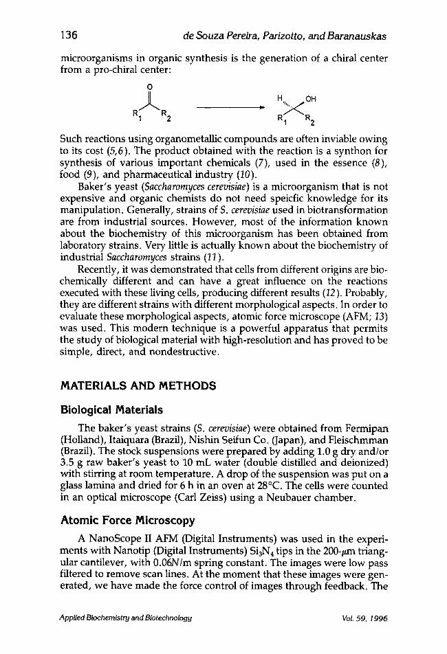
microorganisms in organic synthesis is the generation of a chiral center from a pro-chiral center:
O
R I ~ R 2
H OH
R 1 R 2
Such reactions using organometallic compounds are often inviable owing to its cost (5,6). The product obtained with the reaction is a synthon for synthesis of various important chemicals (7), used in the essence (8), food (9), and pharmaceutical industry (10).
Baker's yeast (Saccharomyces cerevisiae) is a microorganism that is not expensive and organic chemists do not need speicfic knowledge for its manipulation. Generally, strains of S. cerevisiae used in biotransformation are from industrial sources. However, most of the information known about the biochemistry of this microorganism has been obtained from laboratory strains. Very little is actually known about the biochemistry of industrial Saccharomyces strains (11).
Recently, it was demonstrated that cells from different origins are bio- chemically different and can have a great influence on the reactions executed with these living cells, producing different results (12). Probably, they are different strains with different morphological aspects. In order to evaluate these morphological aspects, atomic force microscope (AFM; 13) was used. This modern technique is a powerful apparatus that permits the study of biological material with high-resolution and has proved to be simple, direct, and nondestructive.
MATERIALS AND METHODS
Biological Materials The baker's yeast strains (S. cerevisiae) were obtained from Fermipan
(Holland), Itaiquara (Brazil), Nishin Seifun Co. (Japan), and Fleischmman (Brazil). The stock suspensions were prepared by adding 1.0 g dry and/or 3.5 g raw baker's yeast to 10 mL water (double distilled and deionized) with stirring at room temperature. A drop of the suspension was put on a glass lamina and dried for 6 h in an oven at 28~ The cells were counted in an optical microscope (Carl Zeiss) using a Neubauer chamber.
Atomic Force Microscopy
A NanoScope II AFM (Digital Instruments) was used in the experi- ments with Nanotip (Digital Instruments) Si3N4 tips in the 200-#m triang- ular cantilever, with 0.06N/m spring constant. The images were low pass filtered to remove scan lines. At the moment that these images were gen- erated, we have made the force control of images through feedback. The
Applied Biochemistry and Biotechnology Vol. 59, 1996

Structural Aspects of Baker's Yeast 13 7
Fig. 1. Atomic force micrographs showing different morphological aspects between commercial strains of S. cerevisiae. (A) Fermipan (Holland); (B) Itaiquara (Brazil); (C) Nishin Seifun Co. (Japan); (D) Fleischmman (Brazil).
AFM was operated with a typical scan speed of 8.68 or 19.53 Hz. All imag- ing was done in air at room temperature. The AFM was operated in con- tact mode in the repulsive form. The values of force of the scans were varied between 7 and 14 nN.
The AFM works by moving a microfabricated tip with a laser beam. Deflections in the tip, which correspond to the surface topography, are sensed by a photodetector (14).
RESULTS AND DISCUSSION
Figures I to 5 present micrographs of four strains of baker's yeast (S. cerevisiae) generated with an AFM. These images were reproducible (even in another AFM--Topometrix TMX 2000--data not shown) and they were the same when the scan was repeated. This instrument was chosen because
Applied Biochemistry and Biotechnology Vol. 59, 1996

138 de Souza Pereira, Parizotto, and Baranauskas
Fig. 2. Atomic force micrographs showing different morphological aspects between commercial strains of S. cerevisiae. (A) Fermipan (Holland); (B) Itaiquara (Brazil); (C) Nishin Seifun Co. (Japan); (D) Fleischmman (Brazil).
these types of cells can be seen without using conventional methods com- monly used in electronic microscopy (EM). One of the most common methods used for preparing biomolecules for AFM is immobilization of the biomaterial (15). Although the cells could be immobilized through electrostatic attraction (15) on inexpensive support, such as chrysotile (16), we preferred to use none of the methods and we have only dried the cells for 6 h in an oven at 28~ One important aspect is that the cells are not killed during the observation process as usually occurs in EM. Another aspect to emphasize is that with AFM, we can generate photo- graphs easily at the atomic level. In spite of all these advantages, the AFM has some problems; images from intact whole cells may be distorted, owing to the deformations in the cell membrane, caused by the tip of AFM (14). This issue is overcome when methods are used that give cer- tain rigidity to the membrane of the living cells (sample) (14), or when the cells have a cell wall. This last one gives a natural rigidity to sample (17), consequently it is not necessary to use methods that make it rigid (14).
Applied Biochemistry and Biotechnology Vol. 59, 1996

Structural Aspects of Baker's Yeast 139
Fig. 3. Atomic force micrographs showing pores in the polysaccharide membrane from commercial strains of S. cerevisiae. (A) Fermipan (Holland); (B) Nishin Seifun Co. (Japan); (C) Fleishmman (Brazil).
Since S. cerevisiae have a cell wall it was not necessary to use such methods, making it possible to obtain images of high resolution as shown in this paper.
The images of S. cerevisiae shown here are consistent with those made with electronic microscopes, including size and shape of the cells (18). Figures 1 (A-D) and 2 (A-D) show that these cells have morphological differences. In all photographs, panels A and C are Dutch and Japanese strains, respectively, and panel B and D are Brazilian strains.
The external surface (cell wall) of those microorganisms are made up of polysaccharides such as manan (31%), glucan (28.8%), chitosan, and chitin (2%) (17,19), which do not permit the passage of molecules from outside. The influx of substrate occurs through pores in the polysaccha- ride wall (see Fig. 3A-C). The diameter of these pores vary from cell to cell and this can exert an important influence on the reactions rates in bio- transformations. The pore diameters of the Japanese and Dutch cells are about three and four times larger, respectively, than those of the Brazilian strain. Probably, the influx of the substrate should be in the same propor- tion (see Fig. 3A-C). This may be one of the factors, beyond NAD(P)H pro- duction, that contributes to Dutch and Japanese strains having more reduction power than Brazillian strains as observed before (12).
Applied Biochemistry and Biotechnology Vol. 59, 1996
140 de $ouza Pereira, Parizotto, and Baranauskas
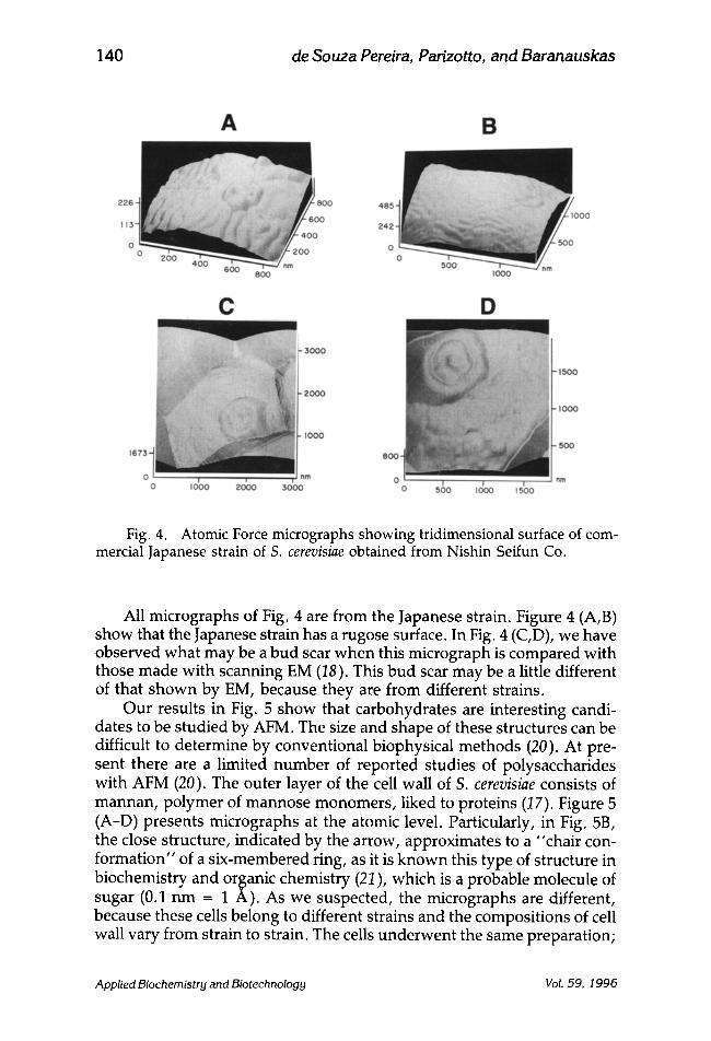
Fig. 4. Atomic Force micrographs showing tridimensional surface of com- mercial Japanese strain of S. cerevisiae obtained from Nishin Seifun Co.
All micrographs of Fig. 4 are from the Japanese strain. Figure 4 (A,B) show that the Japanese strain has a rugose surface. In Fig. 4 (C,D), we have observed what may be a bud scar when this micrograph is compared with those made with scanning EM (18). This bud scar may be a little different of that shown by EM, because they are from different strains.
Our results in Fig. 5 show that carbohydrates are interesting candi- dates to be studied by AFM. The size and shape of these structures can be difficult to determine by conventional biophysical methods (20). At pre- sent there are a limited number of reported studies of polysaccharides with AFM (20). The outer layer of the cell wall of S. cerevisiae consists of mannan, polymer of mannose monomers, liked to proteins (17). Figure 5 (A-D) presents micrographs at the atomic level. Particularly, in Fig. 5B, the close structure, indicated by the arrow, approximates to a "chair con- formation" of a six-membered ring, as it is known this type of structure in biochemistry and organic chemistry (21), which is a probable molecule of sugar (0.1 nm -- 1 A). As we suspected, the micrographs are different, because these cells belong to different strains and the compositions of cell wall vary from strain to strain. The cells underwent the same preparation;
Applied Biochemistry and Biotechnology Vol. 59, 1996

Structural Aspects of Baker's Yeast 141
Fig. 5. Atomic force micrographs showing molecules of polysaccharide in the membrane from commercial strains of S. cerevisiae. (A) Fermipan (Holland); (B) Itaiquara (Brazil); (C) Nishin Seifun Co. (Japan); (D) Fleischmman (Brazil).
if there are modifications between them, they are owing to the character- istics of each strain.
One of the most serious problems in generating high-resolution images in air, as those presented here, is tip contamination (14). This issue is resolved when imaging in liquids. The tip contamination was not present in these experiments because the samples tend to retain some water owing to the hydroxyls from polysaccharides of the cell wall, the temperature used for drying the samples is not high (28~ and the dry- ing time was not too long (about 6 h). The cells were hydrated and con- tinued to grow and divide. This was noticed when we withdrew the sample from AFM after 24 h and observed that the cantilever was lost because it was adhered among cells of the sample, owing to the raised height of the sample. With an optical microscope, we could see that the sample had also increased in size proving that the cells were dividing. In Figs. 6A-D, the distance was measured between two adjacent atoms present on the cell wall of these microbes. An average distance of 0.16 nm (1.6 A), which is coherent with the results of X-ray difraction for simple bond carbon- carbon that is 1.53 A (21), was found. The lines where these scans were obtained are marked in Fig. 5.
Applied Biochemistry and Biotechnology Vol. 59, 1996

142 de Souza Pereira, Parizotto, and Baranauskas
0.16 0.16 0.16
0 . 2
0.1
A I I I I
0 0.4 0 .8 I .Z 1.6 n m
O.t6 O,IT
B I I I t
0 .4 0 .8 1.2 1.6
o.!
n m
0.16 0.16 0.16
C I I 1 I
0.4 0 . 8 1.2 1.6
0.8
0 .4
r i m
0.16 O. t6 ~ " ~ " - ' ~ 0.14
D
0.2
0.1
I I I I 0 .4 0 .8 i . 2 1.6 nrn
Fig. 6. Measuring of distance between atoms in the cellular wall of S. cere- visiae, using an AFM. (A) Fermipan (Holland), (B) Itaiquara (Brazil); (C) Nishin Seifun Co. (Japan); (D) Fleischmman (Brazil).
CONCLUSIONS
Those results confirm the initial hypothesis that these cells belong to different strains and have different morphological aspects. Some of them, as diameter of pores, can exert direct influence at reactions rate in bio- transformations. AFM technique associated with biochemical measurings made before (12) can be used to identify one type of cell that is more ade- quate to use in biotransformations, which will get a more reproducible result.
Applied Biochemistry and Biotechnology Vol. 59, 1996

Structural Aspects of Baker's Yeast 143
ACKNOWLEDGMENTS
This work was supported by grants from Fundacao de Amparo a Pesquisa do Estado de Sao Paulo (FAPESP) and Conselho Nacional de De- senvolvimento Cientifico e Tecnol6gico (CNPq). We thank Nelson Duran for has critical reading of the manuscript, Iveraldo Ribeiro for the draw- ings, C61ia Maria Ribeiro for techinical support, and Yoshiko Yamauchi for the commercial Japanese strain.
REFERENCES
1. Leuenberger, H. G. W. and Kieslich, K. (1987), in Biotransformations, PrOve, P., Faust, U., Sittig, W., and Sukatsch, D. A., eds., VCH, Weinheim, pp. 563-600.
2. Pollack, J. W., Jacobs, J. W., and Schultz, P. G. (1986), Science 234, 1570-1573. 3. Schultz, P. G. (1989), Angew. Chem. Int. Ed. Engl. 28, 1283. 4. Prudent, J. R., Uno T., and Schultz, P. G. (1994), Science 264, 1924-1927. 5. Bosnick, B. and Fryzuk, M. D. (1981), in Topics in Inorganic and Organometallic Stereo-
chemistry, Geoffroy, G. L., ed., Wiley, New York, p. 119. 6. Kagan, H., Wilkinson, G., and F. G. A. (1982), in Comprehensive Organometallic Chem-
istry, vol. 8, Abel, E. W., ed., Pergamon, Oxford, p. 463. 7. Mori, K. (1989), Tetrahedron 45, 3233-3298. 8. Young, C. S. and Ward, O. P. (1991), Biotechnol. Bioeng. 38, 1280-1284. 9. Van der Shaft, P. H., ter Burg, N., Van der Bosch, S., and Cohen, A. M. (1992), Appl.
Microbiol. Biotechnol. 36, 712-716. 10. Bare, G., Jacques, P. H., Hubert, J. B., Rikir, R., and Thonart, P. H. (1991), Appl. Bio-
chem. Biotechnol 28/29, 445-456. 11. Russell, I., Jones, R., and Stewart, G. (1986), in Biotechnology in Food Processing,
Harlander, S. K., and Labuza, T. P., eds., Noyes, Park Ridge, NJ, pp. 172-185. 12. Pereira, R. S. (1995), Appl. Biochem. Biotechnol. 55, 123-132. 13. Binning, G., Quate, C. F., and Gerber, Ch. (1986), Phys. Rev. Lett. 56, 930-933. 14. Yang, J., Tamm, L. K., Somlyo, A. P., and Shao, Z. (1993), J. Microscopy 171, 183-198. 15. Roberts, C. J., Williams, P. M., Davies, M. C., Jackson, D. E., and Tendler, S. J. B.
(1994), Tibtech 12, 127-132. 16. Sorrilha, A. E. P. M., Marques, M., Joekes, I., Moran P. J. S., and Rodrigues, J. A. R.
(1992), Bioorg. Med. Chem. Lett. 2, 191-196. 17. Brady, D., Stoll, A. D., Starke, L., and Duncan, J. R. (1994), Biotechnol. Bioeng. 44,
297-302. 18. Dziezak, J. D. (1987), Food Technol. 41, 104-121. 19. Phaff, H. J. (1977), Adv. Chem. Ser. 160, 244-245. 20. Morris, V. J. (1994), Prog. Biophys. Molec. Biol. 61, 131-185. 21. Nyburg, S. C. (1961), in Fibrous Macromolecular Substances, Academic, New York,
pp. 338-393.
Applied Biochemistry and Biotechnology Vol. 59, 1996