Nucleotide and phylogenetic analysis of human papillomavirus types 6 and 11 isolated from recurrent...
8
Nucleotide and phylogenetic analysis of human papillomavirus types 6 and 11 isolated from recurrent respiratory papillomatosis in Brazil Renata Prandini Adum de Matos a , Laura Sichero d , Isabela Mazuco Mansur a , Caroline Measso do Bonfim a , Cíntia Bittar a , Rodrigo Lacerda Nogueira b , Daniel Salgado Küpper b , Fabiana Cardoso Pereira Valera b , Maurício Lacerda Nogueira c , Luisa Lina Villa d,e,f , Marilia Freitas Calmon a,1 , Paula Rahal a,⇑,1 a UNESP – São Paulo State University, IBILCE, Institute of Bioscience, Language & Literature and Exact Science, Department of Biology, Rua Cristóvão Colombo 2265, Bairro Jardim Nazareth, CEP 15054-010, São José do Rio Preto, São Paulo, Brazil b USP – University of Sao Paulo, School of Medicine of Ribeirão Preto, Department of Ophthalmology, Otorhinolaryngology and Surgery Head/Neck, Av. Bandeirantes, 3900 Bairro Monte Alegre, CEP 14049-900, Ribeirão Preto, SP, Brazil c FAMERP, School of Medicine of Sao Jose do Rio Preto, Laboratory of Research in Virology, Av. Brigadeiro Faria Lima, 5416 Bairro Vila São Pedro, CEP 15090-000, São José do Rio Preto, SP, Brazil d ICESP, Center for Translational Research in Oncology at the Cancer Institute of the State of Sao Paulo, Av. Dr. Arnaldo, 251, 8 o andar, Bairro Cerqueira César, CEP 01246-000, São Paulo, Brazil e Departament of Radiology and Oncology, School of Medicine, University of São Paulo, USP., Av. Dr. Enéas de Carvalho Aguiar, s/n° – Rua 1, Cerqueira César, São Paulo, SP, CEP 05403-900, São Paulo, Brazil f HPV Institute, Santa Casa College of Medical Sciences, São Paulo, Rua Marquês de Itú, 381, Santa Cecília, CEP 01223-001, São Paulo, Brazil article info Article history: Received 5 November 2012 Received in revised form 28 December 2012 Accepted 29 December 2012 Available online 1 March 2013 Keywords: Recurrent respiratory papillomatosis HPV variants HPV-11 HPV-6 Phylogenetic analysis abstract There are few studies about the distribution of natural molecular variants of low-risk HPVs. Our aim was to evaluate the E6 early gene variability among HPV-6 and HPV-11 isolates detected in recurrent respi- ratory papillomatosis (RRP) samples obtained in a cohort of Brazilian patients. We also performed a phy- logenetic analysis in order to compare nucleotide sequences identified in our study with previously reported isolates from different anatomic sites (laryngeal papillomas, genital warts, cervical cancer and anal swabs) obtained from other parts of the world to determine the phylogenetic relationships of vari- ants detected in Brazil. The complete coding region of the E6 gene of 25 samples was cloned and sequenced: 18 isolates of HPV-6 (72%) and 7 isolates of HPV-11 (28%). A total of four different HPV-6 genomic variants and two HPV-11 genomic variants was identified. It was not possible to correlate spe- cific variants with disease severity. Phylogenetic trees for both HPV types were constructed enclosing both E6 sequences detected in our study and formerly published sequences. In both phylogenetic trees, the sequences from Brazil did not group together. We could not establish a geographical association between HPV-6 or HPV-11 variants, unlike HPV-16 and HPV-18. Ó 2013 Elsevier B.V. All rights reserved. 1. Introduction Recurrent respiratory papillomatosis (RRP) is a benign disease of the upper respiratory tract (usually the larynx), which affects both pediatric and adult populations. RRP is characterized by soli- tary or multiple benign tumors in the respiratory tract, and is asso- ciated with infections with human papillomavirus (HPV) types 6 and 11 (Mounts and Kashima, 1984). Despite the benign nature of RRP, lesions tend to grow and extend throughout the entire respiratory tract, causing severe airway obstruction. Recurrence can also occur after surgical resection. For these reasons, RRP can result in considerable morbidity and mortality rates, which in- crease in rare cases in which malignant transformation occurs (Gallagher and Derkay, 2008). The HPV genome consists of approximately 8,000 bp of double- stranded circular DNA encapsulated within an icosahedral protein 1567-1348/$ - see front matter Ó 2013 Elsevier B.V. All rights reserved. http://dx.doi.org/10.1016/j.meegid.2012.12.033 ⇑ Corresponding author. Address: UNESP – São Paulo State University, IBILCE – Instituto de Biociências Letras e Ciências Exatas, Departamento de Biologia, Laboratório de Estudos Genômicos, Rua Cristóvão Colombo 2265, Bairro Jardim Nazareth, CEP 15054-010, São José do Rio Preto, São Paulo, Brazil. Tel.: +55 17 3221 2379. E-mail addresses: [email protected] (R.P.A. de Matos), lsichero@gmail. com (L. Sichero), [email protected] (I.M. Mansur), carol.unespbio@ yahoo.com.br (C.M. do Bonfim), [email protected] (C. Bittar), rodrigoln@ unimedjaboticabal.com.br (R.L. Nogueira), [email protected] (D.S. Küpper), [email protected] (F.C.P. Valera), [email protected] (M.L. Nogueira), [email protected], [email protected] (L.L. Villa), [email protected] (M.F. Calmon), [email protected] (P. Rahal). 1 All the two authors equally contributed to this work. Infection, Genetics and Evolution 16 (2013) 282–289 Contents lists available at SciVerse ScienceDirect Infection, Genetics and Evolution journal homepage: www.elsevier.com/locate/meegid
Transcript of Nucleotide and phylogenetic analysis of human papillomavirus types 6 and 11 isolated from recurrent...
Infection, Genetics and Evolution 16 (2013) 282–289
Contents lists available at SciVerse ScienceDirect
Infection, Genetics and Evolution
journal homepage: www.elsevier .com/locate /meegid
Nucleotide and phylogenetic analysis of human papillomavirus types 6 and 11isolated from recurrent respiratory papillomatosis in Brazil
Renata Prandini Adum de Matos a, Laura Sichero d, Isabela Mazuco Mansur a, Caroline Measso do Bonfim a,Cíntia Bittar a, Rodrigo Lacerda Nogueira b, Daniel Salgado Küpper b, Fabiana Cardoso Pereira Valera b,Maurício Lacerda Nogueira c, Luisa Lina Villa d,e,f, Marilia Freitas Calmon a,1, Paula Rahal a,⇑,1
a UNESP – São Paulo State University, IBILCE, Institute of Bioscience, Language & Literature and Exact Science, Department of Biology, Rua Cristóvão Colombo 2265,Bairro Jardim Nazareth, CEP 15054-010, São José do Rio Preto, São Paulo, Brazilb USP – University of Sao Paulo, School of Medicine of Ribeirão Preto, Department of Ophthalmology, Otorhinolaryngology and Surgery Head/Neck, Av. Bandeirantes,3900 Bairro Monte Alegre, CEP 14049-900, Ribeirão Preto, SP, Brazilc FAMERP, School of Medicine of Sao Jose do Rio Preto, Laboratory of Research in Virology, Av. Brigadeiro Faria Lima, 5416 Bairro Vila São Pedro, CEP 15090-000, São José do Rio Preto,SP, Brazild ICESP, Center for Translational Research in Oncology at the Cancer Institute of the State of Sao Paulo, Av. Dr. Arnaldo, 251, 8o andar, Bairro Cerqueira César, CEP 01246-000,São Paulo, Brazile Departament of Radiology and Oncology, School of Medicine, University of São Paulo, USP., Av. Dr. Enéas de Carvalho Aguiar, s/n� – Rua 1, Cerqueira César, São Paulo, SP,CEP 05403-900, São Paulo, Brazilf HPV Institute, Santa Casa College of Medical Sciences, São Paulo, Rua Marquês de Itú, 381, Santa Cecília, CEP 01223-001, São Paulo, Brazil
a r t i c l e i n f o a b s t r a c t
Article history:Received 5 November 2012Received in revised form 28 December 2012Accepted 29 December 2012Available online 1 March 2013
Keywords:Recurrent respiratory papillomatosisHPV variantsHPV-11HPV-6Phylogenetic analysis
1567-1348/$ - see front matter � 2013 Elsevier B.V. Ahttp://dx.doi.org/10.1016/j.meegid.2012.12.033
⇑ Corresponding author. Address: UNESP – São PauInstituto de Biociências Letras e Ciências Exatas,Laboratório de Estudos Genômicos, Rua Cristóvão CNazareth, CEP 15054-010, São José do Rio Preto, São P2379.
E-mail addresses: [email protected] (R.P.Acom (L. Sichero), [email protected] (I.Myahoo.com.br (C.M. do Bonfim), [email protected] (R.L. Nogueira), dskup@[email protected] (F.C.P. Valera), [email protected]@icesp.org.br, [email protected] (L.L.(M.F. Calmon), [email protected] (P. Rahal).
1 All the two authors equally contributed to this wo
There are few studies about the distribution of natural molecular variants of low-risk HPVs. Our aim wasto evaluate the E6 early gene variability among HPV-6 and HPV-11 isolates detected in recurrent respi-ratory papillomatosis (RRP) samples obtained in a cohort of Brazilian patients. We also performed a phy-logenetic analysis in order to compare nucleotide sequences identified in our study with previouslyreported isolates from different anatomic sites (laryngeal papillomas, genital warts, cervical cancer andanal swabs) obtained from other parts of the world to determine the phylogenetic relationships of vari-ants detected in Brazil. The complete coding region of the E6 gene of 25 samples was cloned andsequenced: 18 isolates of HPV-6 (72%) and 7 isolates of HPV-11 (28%). A total of four different HPV-6genomic variants and two HPV-11 genomic variants was identified. It was not possible to correlate spe-cific variants with disease severity. Phylogenetic trees for both HPV types were constructed enclosingboth E6 sequences detected in our study and formerly published sequences. In both phylogenetic trees,the sequences from Brazil did not group together. We could not establish a geographical associationbetween HPV-6 or HPV-11 variants, unlike HPV-16 and HPV-18.
� 2013 Elsevier B.V. All rights reserved.
ll rights reserved.
lo State University, IBILCE –Departamento de Biologia,
olombo 2265, Bairro Jardimaulo, Brazil. Tel.: +55 17 3221
. de Matos), lsichero@gmail.. Mansur), carol.unespbio@
om (C. Bittar), [email protected] (D.S. Küpper),famerp.br (M.L. Nogueira),
Villa), [email protected]
rk.
1. Introduction
Recurrent respiratory papillomatosis (RRP) is a benign diseaseof the upper respiratory tract (usually the larynx), which affectsboth pediatric and adult populations. RRP is characterized by soli-tary or multiple benign tumors in the respiratory tract, and is asso-ciated with infections with human papillomavirus (HPV) types 6and 11 (Mounts and Kashima, 1984). Despite the benign natureof RRP, lesions tend to grow and extend throughout the entirerespiratory tract, causing severe airway obstruction. Recurrencecan also occur after surgical resection. For these reasons, RRP canresult in considerable morbidity and mortality rates, which in-crease in rare cases in which malignant transformation occurs(Gallagher and Derkay, 2008).
The HPV genome consists of approximately 8,000 bp of double-stranded circular DNA encapsulated within an icosahedral protein
R.P.A. de Matos et al. / Infection, Genetics and Evolution 16 (2013) 282–289 283
capsid that is 55 nm in diameter (Nebesio et al., 2001). HPVs infectcells from the basal layer of the stratified epithelium, and viralgene expression is closely linked to the differentiation programof the host cells (McMurray et al., 2001). The upstream regulatoryregion (URR) or long control region (LCR) consists of a non-codingsegment of the genome that encloses cis-regulatory elements thatare important to viral replication and gene expression control(Bernard, 2002; Chow et al., 2010). Functionally, the coding partof the viral genome is divided into early (E) and late (L) regions.The late region encodes the viral capsid proteins L1 and L2. Incontrast, the early region contains genes designated E1–E7 thatencode proteins involved in the regulation of viral DNAreplication and transcription (Tyring, 2000). E6 and E7 have beenfound to be the most important pathogenic HPV proteins. Thegenes E6 and E7 from high-risk HPVs induce cellular immortaliza-tion by functionally interfering with proteins involved in cellcycle regulation (Longworth and Laimins, 2004). E6 and E7 havealso been shown to be necessary for the maintenance of extrachro-mosomal forms of HPV in undifferentiated basal cells (Thomaset al., 1999).
More than 150 HPV types have been identified to date (de Vil-liers et al., 2004). HPVs can be categorized into low- and high-risktypes based on the risk that the virus will cause squamous cell cer-vical carcinomas (IARC, 2007). The most often detected low-risktypes are HPV-6 and HPV-11, which are causally associated withthe development of benign lesions such as laryngeal papillomato-sis and genital or perianal warts (Klozar et al., 2010). A distinct HPVtype is established when the DNA sequence of the L1 gene differsfrom that of any other characterized type by at least 10%. In addi-tion, isolates of the same HPV type are referred to as variants orsubtypes when the nucleotide sequences differ by less than 10%(Bernard et al., 2006).
HPV-6 isolates are classified into HPV-6b-related (prototypic)and HPV-6a/6vc-related (non-prototypic) genomic variants(Heinzel et al., 1995). HPV-6b, the reference or prototype se-quence, was originally isolated from a condylomata acuminatum(de Villiers et al., 1981; Gissmann and zur Hausen, 1980;Kovelman et al., 1999). The HPV-11 prototype isolate wasdetected in a laryngeal papilloma specimen in 1982 throughSouthern blot hybridization and in 1986, the complete nucleotidesequence of this clone was fully characterized (Dartmann et al.,1986; Gissmann et al., 1982).
Genetic variability analysis has proven essential to a betterunderstanding of the natural and evolutionary history of papillo-mavirus. HPV nucleotide diversity has been investigated moreextensively among high-risk HPV types. In contrast, few studiesof this kind have been performed involving natural molecular vari-ants of low-risk HPVs. Our aim was to evaluate E6 early gene var-iability among HPV-6 and HPV-11 detected in laryngeal papillomassamples obtained in a cohort of Brazilian patients. We also con-ducted phylogenetic analysis in order to compare nucleotide se-quences identified in our study with isolates previouslydescribed from other parts of world.
Table 1Sequence of specific primers used for the amplification of the HPV E6 gene.
Primer Sequence (50 ? 30)
HPV6-E6F GGGGGATCCGAATTCATGGAAAGTGCAAATGCHPV6-E6R GTGAAGCTTGGATCCTAGGGTAACATGTCTTCCHPV11-E6F AAAATTAGCAGACGAGGCATTHPV11-E6R CCACCTTGTCCACCTCATCT
2. Materials and methods
2.1. Clinical samples
Biopsy specimens were obtained from 25 patients between 3and 52 years of age who had been treated at the Laryngology Clinicof the Department of Otorhinolaryngology of the School of Medi-cine of the University of São Paulo, in Ribeirão Preto, Brazil, andwho had been diagnosed with RRP. Disease severity was deter-mined at each surgical intervention using the Derkay staging sys-tem (Derkay et al., 1998).
2.2. HPV genotyping
Total DNA was extracted from biopsies of the lesions using theQIAmp DNA Micro Kit (Qiagen) according to the manufacturer’sinstructions. DNA quality assessment was performed using a PCRof endogenous gene GSTP1.
HPV positivity was accessed by a PCR protocol (PolymeraseChain Reaction) using the PGMY09/11 primer set which amplifiesa 450 bp segment of the L1 gene from a broad range of mucosalHPV types (Gravitt et al., 2000). The amplification mix was com-prised of 2.5 U of a proofreading polymerase (High Fidelity EnzymeMix, Fermentas), 2.5 ll of 10X High Fidelity PCR Buffer, 4.0 mMMgCl2, 0.2 mM dNTPs, 0.32 mM of each primer, 80 ng of extractedDNA to a final volume of 25 ll in nuclease free water. The reactionwas submitted to an initial denaturation step at 95 �C for 13 min,40 cycles at 95 �C for 1 min, 55 �C for 1 min and 72 �C for 1 min,and then to a final extension step at 72 �C for 5 min.
HPV positive samples were genotyped using RFLP (RestrictionFragment Length Polymorphism). In brief, samples were reampli-fied as described though in a final reaction volume of 100 ll. PCRproducts were independently digested by seven different restric-tion enzymes (BamHI, DdeI, HaeIII, HinfI, PstI, RsaI and Sau3AI).Digestion profiles were compared to previously described restric-tion patterns (Bernard et al., 1994).
2.3. Molecular variants identification
For the nucleotide variability analysis, the complete E6 gene se-quence of HPV-6 and HPV-11 positive samples was amplified usingPCR with specific primers (Table 1). The amplification mix con-sisted of 6.0 U of a proofreading polymerase (High Fidelity EnzymeMix, Fermentas), 5.0 ll of 10X High Fidelity PCR Buffer, 1.5 mMMgCl2, 0.24 mM dNTPs, 0.4 mM of each primer, 500 ng of DNA,and nuclease free water, all of which addedup to a final volumeof 50.0 ll. Initial denaturation step at 95 �C for 5 min was con-ducted, followed by 35 cycles at 95 �C for 1 min, 55 �C for 1 minand 72 �C for 2 min, and a final extension at 72 �C for 8 min.
Amplified fragments were cloned using the TOPO XL Cloning™Kit (Invitrogen). Five clones from each patient were purified usingthe GeneJET Plasmid Purification Kit (Fermentas). Sequencing reac-tions were performed using the BigDye Terminator kit (AppliedBiosystems) and products were sequenced in an ABI 3130XL se-quencer (Applied Biosystems). The sequencing reaction mixtureconsisted of 2.0 ll of 5X Sequencing Buffer (Applied Biosystems),0.4 mM of specific E6 primers, 2.0 ll of Big ET Dye Terminator,and 5.0 ll of the sample. Sequencing reactions consisted of a hotstart step of 10 min at 95 �C, followed by 30 cycles of 96 �C for15 s, 50 �C for 15 s and 60 �C for 4 min. To control for nucleotidechanges introduced by the polymerase, two different PCR productswere cloned and sequenced.
The quality of the sequences obtained was evaluated using theEletropherogram Quality Analysis, available online at <http://www.biomol.unb.br/phph>. Comparisons between the sequencesacquired and those previously added to GenBank were conductedusing BLAST (Basic Local Alignment Search Tool, available at<http://www.ncbi.nlm.nih.gov/BLAST>.) The alignment between
284 R.P.A. de Matos et al. / Infection, Genetics and Evolution 16 (2013) 282–289
the prototype and all sequences obtained was performed using theCLUSTAL W software nested in the Bioedit 7.0.9.0 package (Hall,1999; Thompson et al., 1997). All sequences were edited usingBio Edit (Hall, 1999) in order to remove vector fragments and ana-lyze solely the complete sequence of the E6 gene. HPV-6 and HPV-11 nucleotide sequences generated in this study were submitted toGenBank (accession numbers: KC285838-KC285862).
2.4. In silico secondary structure analysis
The Predict Protein Server <http://www.predictprotein.org>(Rost et al., 2004) was used to compare the secondary structuresof the E6 protein of HPV-6vc and the HPV-6vc variant with theamino acid change.
2.5. Phylogenetic analysis
Phylogenetic analysis was performed using the PhyML toolavailable online at South of France Bioinformatics Platform(<http://www.atgc-montpellier.fr/phyml/>) (Guindon et al., 2010;Guindon and Gascuel, 2003). Maximum-Likelihood trees were con-structed using the TrN model for HPV-6 sequences and the HKY85model for HPV-11 sequences. Models were determined using Mod-eltest 3.7 (Posada and Crandall, 1998) run in PAUP 4.0 beta10(Swofford, 2002). Bootstrap was calculated based on 1,000 repli-cates, and values above 70% were considered significant.
The phylogenetic analysis included all 25 E6 sequences ob-tained in this study in addition to other full length E6 sequencesreported previously: 77 and 12 HPV-6 samples isolated in Slovenia(Kocjan et al., 2009) and in South Africa (Combrinck et al., 2011),respectively, and 63 HPV-11 samples obtained in Slovenia (Maveret al., 2011). Sequences described by others were isolated from dif-ferent anatomic sites: laryngeal papillomas, genital warts, cervicalcancer and anal swabs.
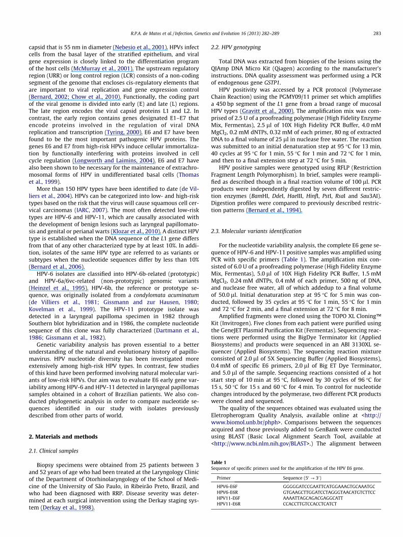
Table 2Characterization of samples and clinical presentation of patients with recurrent respirator
HPV-Type
IDSample
Gender Age at diagnosis(years)
Age at last follow up(years)
Td
HPV-6 BR_LP1 F 51 51 1BR_LP2 F 4 4 3BR_LP3 F 3 5 9BR_LP4 ⁄⁄⁄ ⁄⁄⁄ ⁄⁄⁄ ⁄BR_LP5 M 1 6 1BR_LP6 F 5 8 6BR_LP7 F 5 6 5BR_LP8 M 23 25 2BR_LP9 M 30 36 4BR_LP10 M 32 33 2BR_LP11 M newborn 1 4BR_LP12 M 28 28 2BR_LP13 M 32 32 1BR_LP14 F 8 8 1BR_LP15 M 41 42 3BR_LP16 M 47 47 1BR_LP17 M 31 31 3BR_LP18 M 45 46 3
HPV-11 BR_LP19 M 7 13 2BR_LP20 F 2 3 6BR_LP21 F 2 5 9BR_LP22 M 2 5 1BR_LP23 M 3 3 2BR_LP24 M 29 31 5BR_LP25 M 33 33 4
⁄⁄⁄Data unavailable.
3. Results
Our study used data from 25 patients diagnosed with RRP. De-tails from the patients, including clinical status and the number ofsurgical procedures performed, are presented in Table 2. The dis-ease was classified as severe when more than three proceduresper year were required with Derkay scores higher than 20. Only pa-tients BR_LP19 and BR_LP 20 presented with severe disease.
All 25 biopsy specimens were positive for HPV DNA (100%).HPV-6 was detected in 18 (72%) cases, and HPV-11 was detectedin 7 (28%) cases. All 25 isolates were successfully amplified and se-quenced across the entire E6 genomic region (nt positions 102-554). HPV-6 E6 molecular variant classification was conductedthrough a comparison with the prototype HPV-6b (GenBank acces-sion number X00203) and the non-prototypes HPV-6a (GenBankaccession number L41216) and HPV-6vc sequences (GenBankaccession no. AF092932). HPV-11 E6 variants were characterizedby comparing obtained sequences with the HPV-11 prototype gen-ome (GenBank accession number M14119).
3.1. HPV-6 and HPV-11 E6 molecular variants
Among the 18 HPV-6 samples analyzed, 17 isolates (94.4%)were found to possess a high identity to non-prototypic sequences(HPV-6a and HPV-6vc). A total of four E6 genomic variants wereidentified: BR_LP12, BR_LP8, Ref 6vc, and BR_LP14 (Fig. 1A). TheRef 6vc variant was the most prevalent – it was detected in 48%of the samples. Sequence analysis of the entire E6 gene revealedtwo synonymous point mutations compared to the prototypeHPV-6b genome: A120T and G372A. Five isolates were identicalto the non-prototype HPV-6a sequence with the exception of theG327A silent mutation detected in all samples. The nucleotide sub-stitution A296G was detected in 2/12 HPV-6vc-related isolates(BR_LP14 and BR_LP16). This substitution resulted in an aminoacid change from aspartate to glycine, a change which corre-sponded to an alteration rate of 0.66% within the entire E6 protein.
y papillomatosis.
otal procedures sinceiagnosis
Average procedures/year
Derkayscore
Diseaseseverity
1 08 Mild3 13 Mild4.5 17 Mild
⁄⁄ ⁄⁄⁄ ⁄⁄⁄ ⁄⁄⁄0 2 16 Mild
2 26 Mild5 15 Mild1 07 Mild0.7 07 Mild2 07 Mild4 16 Mild2 13 Mild1 10 Mild1 13 Mild3 11 Mild1 13 Mild3 10 Mild3 05 Mild
2 3.7 22 Severe6 21 Severe3 15 Mild0.3 06 Mild2 20 Mild2.5 31 Mild1 04 Mild
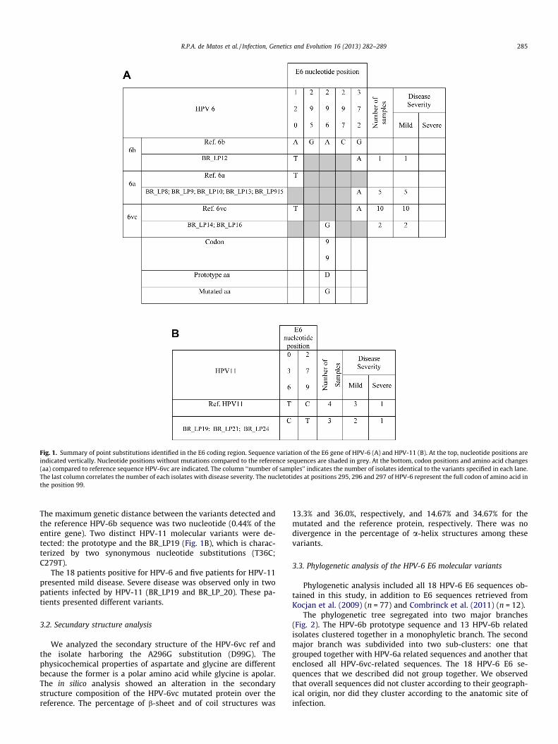
Fig. 1. Summary of point substitutions identified in the E6 coding region. Sequence variation of the E6 gene of HPV-6 (A) and HPV-11 (B). At the top, nucleotide positions areindicated vertically. Nucleotide positions without mutations compared to the reference sequences are shaded in grey. At the bottom, codon positions and amino acid changes(aa) compared to reference sequence HPV-6vc are indicated. The column ‘‘number of samples’’ indicates the number of isolates identical to the variants specified in each lane.The last column correlates the number of each isolates with disease severity. The nucletotides at positions 295, 296 and 297 of HPV-6 represent the full codon of amino acid inthe position 99.
R.P.A. de Matos et al. / Infection, Genetics and Evolution 16 (2013) 282–289 285
The maximum genetic distance between the variants detected andthe reference HPV-6b sequence was two nucleotide (0.44% of theentire gene). Two distinct HPV-11 molecular variants were de-tected: the prototype and the BR_LP19 (Fig. 1B), which is charac-terized by two synonymous nucleotide substitutions (T36C;C279T).
The 18 patients positive for HPV-6 and five patients for HPV-11presented mild disease. Severe disease was observed only in twopatients infected by HPV-11 (BR_LP19 and BR_LP_20). These pa-tients presented different variants.
3.2. Secundary structure analysis
We analyzed the secondary structure of the HPV-6vc ref andthe isolate harboring the A296G substitution (D99G). Thephysicochemical properties of aspartate and glycine are differentbecause the former is a polar amino acid while glycine is apolar.The in silico analysis showed an alteration in the secondarystructure composition of the HPV-6vc mutated protein over thereference. The percentage of b-sheet and of coil structures was
13.3% and 36.0%, respectively, and 14.67% and 34.67% for themutated and the reference protein, respectively. There was nodivergence in the percentage of a-helix structures among thesevariants.
3.3. Phylogenetic analysis of the HPV-6 E6 molecular variants
Phylogenetic analysis included all 18 HPV-6 E6 sequences ob-tained in this study, in addition to E6 sequences retrieved fromKocjan et al. (2009) (n = 77) and Combrinck et al. (2011) (n = 12).
The phylogenetic tree segregated into two major branches(Fig. 2). The HPV-6b prototype sequence and 13 HPV-6b relatedisolates clustered together in a monophyletic branch. The secondmajor branch was subdivided into two sub-clusters: one thatgrouped together with HPV-6a related sequences and another thatenclosed all HPV-6vc-related sequences. The 18 HPV-6 E6 se-quences that we described did not group together. We observedthat overall sequences did not cluster according to their geograph-ical origin, nor did they cluster according to the anatomic site ofinfection.
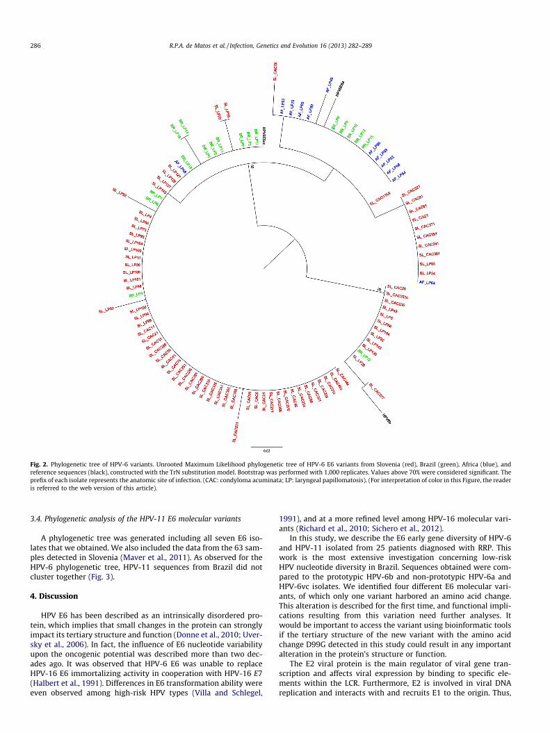
Fig. 2. Phylogenetic tree of HPV-6 variants. Unrooted Maximum Likelihood phylogenetic tree of HPV-6 E6 variants from Slovenia (red), Brazil (green), Africa (blue), andreference sequences (black), constructed with the TrN substitution model. Bootstrap was performed with 1,000 replicates. Values above 70% were considered significant. Theprefix of each isolate represents the anatomic site of infection. (CAC: condyloma acuminata; LP: laryngeal papillomatosis). (For interpretation of color in this Figure, the readeris referred to the web version of this article).
286 R.P.A. de Matos et al. / Infection, Genetics and Evolution 16 (2013) 282–289
3.4. Phylogenetic analysis of the HPV-11 E6 molecular variants
A phylogenetic tree was generated including all seven E6 iso-lates that we obtained. We also included the data from the 63 sam-ples detected in Slovenia (Maver et al., 2011). As observed for theHPV-6 phylogenetic tree, HPV-11 sequences from Brazil did notcluster together (Fig. 3).
4. Discussion
HPV E6 has been described as an intrinsically disordered pro-tein, which implies that small changes in the protein can stronglyimpact its tertiary structure and function (Donne et al., 2010; Uver-sky et al., 2006). In fact, the influence of E6 nucleotide variabilityupon the oncogenic potential was described more than two dec-ades ago. It was observed that HPV-6 E6 was unable to replaceHPV-16 E6 immortalizing activity in cooperation with HPV-16 E7(Halbert et al., 1991). Differences in E6 transformation ability wereeven observed among high-risk HPV types (Villa and Schlegel,
1991), and at a more refined level among HPV-16 molecular vari-ants (Richard et al., 2010; Sichero et al., 2012).
In this study, we describe the E6 early gene diversity of HPV-6and HPV-11 isolated from 25 patients diagnosed with RRP. Thiswork is the most extensive investigation concerning low-riskHPV nucleotide diversity in Brazil. Sequences obtained were com-pared to the prototypic HPV-6b and non-prototypic HPV-6a andHPV-6vc isolates. We identified four different E6 molecular vari-ants, of which only one variant harbored an amino acid change.This alteration is described for the first time, and functional impli-cations resulting from this variation need further analyses. Itwould be important to access the variant using bioinformatic toolsif the tertiary structure of the new variant with the amino acidchange D99G detected in this study could result in any importantalteration in the protein’s structure or function.
The E2 viral protein is the main regulator of viral gene tran-scription and affects viral expression by binding to specific ele-ments within the LCR. Furthermore, E2 is involved in viral DNAreplication and interacts with and recruits E1 to the origin. Thus,

Fig. 3. Phylogenetic tree of HPV-6 variants. Unrooted Maximum Likelihood phylogenetic tree of HPV-11 E6 variants from Slovenia (red), Brazil (green), and referencesequences (black). Constructed with the HKY85 substitution model. Bootstrap was performed with 1,000 replicates. Values above 70% were considered significant. The prefixof each isolate represents the anatomic site of infection. (CAC: condyloma acuminata; LP: laryngeal papillomatosis; CS: Swab Cervical; A: Swab Anal). (For interpretation ofcolor in this Figure, the reader is referred to the web version of this article).
R.P.A. de Matos et al. / Infection, Genetics and Evolution 16 (2013) 282–289 287
it is reasonable to suppose that mutations in these regions may af-fect the clinical outcome of the infection. Among HPV-16 molecu-lar variants, the influence of E2 variability upon the transcriptionaltransactivation potential has been described. We also observedthat specific HPV-16 variants exhibit lower replication efficiencythan others.
Although we have only sequenced the E6 gene of the HPV-6 and-11 variants analyzed in this study, the E2 and E1 viral nucleotidesequences of these isolates could be presumed to be a strong inter-gene sequence co-variation observed for HPV and the complete vir-al genome sequences of several variants of these viral types areavailable (Burk et al., 2011). However, no functional analyses ofthese proteins have been conducted, which, as of yet, preventsany further conclusions concerning the impact of the variabilitythat we observed upon viral transcription and replication.
Among the individuals analyzed, only two were diagnosed withsevere disease: BR_LP19 and BR_LP20. Both of these patients werepositive for HPV-11, results which are in line with the opinion ofseveral otolaryngologists who believe that HPV-11-associated dis-ease is more aggressive than cases harboring HPV-6 (Wiatrak et al.,2004). However, the small number of samples analyzed preventsthe correlation of disease severity with any particular E6 sequence.
In our results, we found four different nucleotide substitutionsthat did not result in any amino acid changes. Although synony-mous changes are not reflected in the amino acid composition of
the protein, they represent an important aspect of papillomavirus(PV) evolution due to a strong codon usage bias (Zhao and Chen,2011; Zhao et al., 2003, 1999). The codon preference in virus is dri-ven by co-evolution with hosts by adapting to immune responseand tissue tropism (Zhao and Chen, 2011). Interestingly, the prefer-ence of codon observed in PV is significantly different from the oneseen in mammalian proteins, which allows the virus to use tRNAsthat are being used less (Wada et al., 1990; Zhou et al., 1999).Though this strategy relies on using a less advantageous codon, italso avoids impairing host cell protein synthesis, which is impor-tant for the production of viral proteins (Aragones et al., 2010).Two synonymous mutations found in this study, one for HPV-6and one for HPV-11, changed a codon preferred by humans toone chosen by papillomavirus (HPV-6 - A120T and HPV-11 –C279T). Conversely, a synonymous change found in HPV-11 altereda codon found in higher frequencies in PV to one used more oftenduring human protein synthesis (HPV-11 – T36C). Studies convert-ing rare codons to those more frequently used in mammals showedup to 100-fold increases in protein expression levels while keepingthe levels of mRNA constant (Cid-Arregui et al., 2003; Disbrowet al., 2003; Mossadegh et al., 2004). Further studies showed thatexpression levels are related to tRNA abundance (Gu et al., 2004).To determine whether the synonymous mutations that we de-scribed have any impact in E6 protein expression, additional anal-ysis is required.
288 R.P.A. de Matos et al. / Infection, Genetics and Evolution 16 (2013) 282–289
Most of the HPV-6 isolates identified in this study have identicalsequences to those previously described among laryngeal papillo-matosis and condyloma acuminata specimens obtained in Sloveniaand Africa (Combrinck et al., 2011; Kocjan et al., 2009). Compara-tive analysis revealed that 12.35% and 10.1% of the HPV-6 isolatesdescribed by Kocjan et al. (2009) and Combrinck et al. (2011) wereidentical to variants BR_LP 12 and BR_LP8, respectively. Addition-ally, among the 63 HPV-11 sequences characterized by Maveret al. (2011), 82.5% were identical to the BR_LP19 variant that wedescribed. Maver et al. (2011) evaluated the nucleotide diversityof HPV-11 among 63 isolates and identified six E6 molecular vari-ants, of which only one resulted in amino acid substitutions. Wealso consistently observed low diversity of the HPV-11 E6 protein.No geographical clustering was observed in the phylogenetic anal-ysis of HPV-11 isolates. This same pattern was described by Kocjanet al. (2009), results which strongly suggest that HPV-6 and HPV-11 variants are detected with similar frequencies around theworld. Furthermore, phylogenetic analysis of a 252 bp segment ofthe LCR of 62 isolates collected worldwide (Brazil, Germany, India,Italy, Japan, New York, Senegal and Singapore) revealed that HPV-6and HPV-11 variant distribution show only minor correlations be-tween geographic origin and ethnicity of the population studied(Heinzel et al., 1995).
Nucleotide analysis of worldwide collections of HPV-18 andHPV-16 isolates points toward co-evolution of these viruses withthe three major ethnic groups: Africans, Caucasians, and EastAsians. In contrast to the phylogenetic analysis of HPV-6 andHPV-11, HPV-16 and -18 LCR molecular variants robustly segregateinto five major variant lineages: European (E), Asian (As), Asian-American (AA), and two African lineages (Af1 and Af2) (Ho et al.,1993; Ong et al., 1993). The distribution of HPV-16 variants world-wide varies significantly and correlates with the intrinsic admix-ture level of each population. In fact, the results of the analysis ofHPV-16 and HPV-18 isolates detected in Brazil reflect the historiccolonization of Brazil by European and Africans (Sichero et al.,2007).
Unfortunately, the small number of samples analyzed preventsthe evaluation of the association between specific molecular vari-ants and the anatomical site of infection. In contrast, an unevendistribution of HPV-16 variants among cervical and tonsillar cancerwas observed in Stockholm (Du et al., 2012).
We observed a 25% amino acid variation rate (1/4 nt substitu-tion resulted in amino acid substitutions) for HPV-6 samples, andno amino acid variation was found in the case of HPV-11 samples.However, Vrtacnik Bokal et al. (2010) and Shang et al. (2011) stud-ied HPV-16 E6 found an amino acid variation rate of 62.5% (10/16)and 57.69%, (30/52) respectively. We do not know the reason forthis divergence.
5. Conclusions
In this study, we demonstrated the low genomic diversity ofHPV-6 and HPV-11 E6 sequences. A total of four different HPV-6genomic variants and two HPV-11 genomic variants were identi-fied. However, it was not possible to correlate disease severity withany particular variant. We could not establish an association be-tween geographical location HPV-6 or HPV-11 variants. In silicoanalysis showed that the amino acid mutation detected in ourstudy has an impact on the protein’s secondary structure. Func-tional studies are required to evaluate the influence of this muta-tion on replication and protein expression as well as on theimpact on the tertiary structure. Additionally, LCR variability maybe linked to alterations in coding regions of the genome, whichcould, in turn, influence the risk of disease recurrence and maypotentially indicate a more severe disease process in RRP.
Acknowledgements
This work was financially supported by the Brazilian agenciesFAPESP 08/57889-1, CAPES, and CNPq 573799/2008-3.
References
Aragones, L., Guix, S., Ribes, E., Bosch, A., Pinto, R.M., 2010. Fine-tuning translationkinetics selection as the driving force of codon usage bias in the hepatitis Avirus capsid. PLoS Pathogens 6, e1000797.
Bernard, H.U., 2002. Gene expression of genital human papillomaviruses andconsiderations on potential antiviral approaches. Antiviral Therapy 7, 219–237.
Bernard, H.U., Chan, S.Y., Manos, M.M., Ong, C.K., Villa, L.L., Delius, H., Peyton, C.L.,Bauer, H.M., Wheeler, C.M., 1994. Identification and assessment of known andnovel human papillomaviruses by polymerase chain reaction amplification,restriction fragment length polymorphisms, nucleotide sequence, andphylogenetic algorithms. The Journal of Infectious Diseases 170, 1077–1085.
Bernard, H.U., Calleja-Macias, I.E., Dunn, S.T., 2006. Genome variation of humanpapillomavirus types: phylogenetic and medical implications. InternationalJournal of Cancer. Journal International du Cancer 118, 1071–1076.
Burk, R.D., Chen, Z., Harari, A., Smith, B.C., Kocjan, B.J., Maver, P.J., Poljak, M., 2011.Classification and nomenclature system for human Alphapapillomavirusvariants: general features, nucleotide landmarks and assignment of HPV6 andHPV11 isolates to variant lineages. Acta dermatovenerologica Alpina, Panonica,et Adriatica 20, 113–123.
Chow, L.T., Broker, T.R., Steinberg, B.M., 2010. The natural history of humanpapillomavirus infections of the mucosal epithelia. APMIS: acta pathologica,microbiologica, et immunologica Scandinavica 118, 422–449.
Cid-Arregui, A., Juarez, V., zur Hausen, H., 2003. A synthetic E7 gene of humanpapillomavirus type 16 that yields enhanced expression of the protein inmammalian cells and is useful for DNA immunization studies. Journal ofVirology 77, 4928–4937.
Combrinck, C.E., Seedat, R.Y., Randall, C., Roodt, Y., Burt, F.J., 2011. Novel HPV-6variants of human papillomavirus causing recurrent respiratory papillomatosisin Southern Africa. Epidemiology and Infection 140, 1–7.
Dartmann, K., Schwarz, E., Gissmann, L., zur Hausen, H., 1986. The nucleotidesequence and genome organization of human papilloma virus type 11. Virology151, 124–130.
de Villiers, E.M., Gissmann, L., zur Hausen, H., 1981. Molecular cloning of viral DNAfrom human genital warts. Journal of Virology 40, 932–935.
de Villiers, E.M., Gunst, K., Stein, H., Scherubl, H., 2004. Esophageal squamous cellcancer in patients with head and neck cancer: prevalence of humanpapillomavirus DNA sequences. International Journal of Cancer. JournalInternational du Cancer 109, 253–258.
Derkay, C.S., Malis, D.J., Zalzal, G., Wiatrak, B.J., Kashima, H.K., Coltrera, M.D., 1998. Astaging system for assessing severity of disease and response to therapy inrecurrent respiratory papillomatosis. The Laryngoscope 108, 935–937.
Disbrow, G.L., Sunitha, I., Baker, C.C., Hanover, J., Schlegel, R., 2003. Codonoptimization of the HPV-16 E5 gene enhances protein expression. Virology311, 105–114.
Donne, A.J., Hampson, L., Homer, J.J., Hampson, I.N., 2010. The role of HPV type inrecurrent respiratory papillomatosis. International Journal of PediatricOtorhinolaryngology 74, 7–14.
Du, J., Nordfors, C., Nasman, A., Sobkowiak, M., Romanitan, M., Dalianis, T.,Ramqvist, T., 2012. Human papillomavirus (HPV) 16 E6 variants in tonsillarcancer in comparison to those in cervical cancer in Stockholm, Sweden. PLoSOne 7, e36239.
Gallagher, T.Q., Derkay, C.S., 2008. Recurrent respiratory papillomatosis: update2008. Current Opinion in Otolaryngology & Head and Neck Surgery 16, 536–542.
Gissmann, L., zur Hausen, H., 1980. Partial characterization of viral DNA fromhuman genital warts (Condylomata acuminata). International Journal of Cancer.Journal International du Cancer 25, 605–609.
Gissmann, L., Diehl, V., Schultz-Coulon, H.J., zur Hausen, H., 1982. Molecular cloningand characterization of human papilloma virus DNA derived from a laryngealpapilloma. Journal of Virology 44, 393–400.
Gravitt, P.E., Peyton, C.L., Alessi, T.Q., Wheeler, C.M., Coutlee, F., Hildesheim, A.,Schiffman, M.H., Scott, D.R., Apple, R.J., 2000. Improved amplification of genitalhuman papillomaviruses. Journal of Clinical Microbiology 38, 357–361.
Gu, W., Li, M., Zhao, W.M., Fang, N.X., Bu, S., Frazer, I.H., Zhao, K.N., 2004.TRNASer(CGA) differentially regulates expression of wild-type and codon-modified papillomavirus L1 genes. Nucleic Acids Research 32, 4448–4461.
Guindon, S., Gascuel, O., 2003. A simple, fast, and accurate algorithm to estimatelarge phylogenies by maximum likelihood. Systematic Biology 52, 696–704.
Guindon, S., Dufayard, J.F., Lefort, V., Anisimova, M., Hordijk, W., Gascuel, O., 2010.New algorithms and methods to estimate maximum-likelihood phylogenies:assessing the performance of PhyML 3.0. Systematic Biology 59, 307–321.
Halbert, C.L., Demers, G.W., Galloway, D.A., 1991. The E7 gene of humanpapillomavirus type 16 is sufficient for immortalization of human epithelialcells. Journal of Virology 65, 473–478.
Hall, T.A., 1999. BioEdit: a user-friendly biological sequence alignment editor andanalysis program for Windows 95/98/NT. Nucleic Acids Symposium Series 41,95–98.
R.P.A. de Matos et al. / Infection, Genetics and Evolution 16 (2013) 282–289 289
Heinzel, P.A., Chan, S.Y., Ho, L., O’Connor, M., Balaram, P., Campo, M.S., Fujinaga, K.,Kiviat, N., Kuypers, J., Pfister, H., et al., 1995. Variation of human papillomavirustype 6 (HPV-6) and HPV-11 genomes sampled throughout the world. Journal ofClinical Microbiology 33, 1746–1754.
Ho, L., Chan, S.Y., Burk, R.D., Das, B.C., Fujinaga, K., Icenogle, J.P., Kahn, T., Kiviat, N.,Lancaster, W., Mavromara-Nazos, P., et al., 1993. The genetic drift of humanpapillomavirus type 16 is a means of reconstructing prehistoric viral spread andthe movement of ancient human populations. Journal of Virology 67, 6413–6423.
IARC, 2007. Human papillomaviruses. IARC Monographs on the Evaluation ofCarcinogenic Risks to Humans/World Health Organization, InternationalAgency for Research on Cancer 90, 1–636.
Klozar, J., Tachezy, R., Rotnaglova, E., Koslabova, E., Salakova, M., Hamsikova, E.,2010. Human papillomavirus in head and neck tumors: epidemiological,molecular and clinical aspects. Wien Med Wochenschr 160, 305–309.
Kocjan, B.J., Poljak, M., Cimerman, M., Gale, N., Potocnik, M., Bogovac, Z., Seme, K.,2009. Prevaccination genomic diversity of human papillomavirus genotype 6(HPV 6). Virology 391, 274–283.
Kovelman, R., Bilter, G.K., Roman, A., Brown, D.R., Barbosa, M.S., 1999. Humanpapillomavirus type 6: classification of clinical isolates and functional analysisof E2 proteins. The Journal of General Virology 80 (Pt 9), 2445–2451.
Longworth, M.S., Laimins, L.A., 2004. Pathogenesis of human papillomaviruses indifferentiating epithelia. Microbiology and Molecular Biology Reviews: MMBR68, 362–372.
Maver, P.J., Kocjan, B.J., Seme, K., Potocnik, M., Gale, N., Poljak, M., 2011.Prevaccination genomic diversity of human papillomavirus genotype 11: astudy on 63 clinical isolates and 10 full-length genome sequences. Journal ofMedical Virology 83, 461–470.
McMurray, H.R., Nguyen, D., Westbrook, T.F., McAnce, D.J., 2001. Biology of humanpapillomaviruses. International Journal of Experimental Pathology 82, 15–33.
Mossadegh, N., Gissmann, L., Muller, M., Zentgraf, H., Alonso, A., Tomakidi, P., 2004.Codon optimization of the human papillomavirus 11 (HPV 11) L1 gene leads toincreased gene expression and formation of virus-like particles in mammalianepithelial cells. Virology 326, 57–66.
Mounts, P., Kashima, H., 1984. Association of human papillomavirus subtype andclinical course in respiratory papillomatosis. The Laryngoscope 94, 28–33.
Nebesio, C.L., Mirowski, G.W., Chuang, T.Y., 2001. Human papillomavirus: clinicalsignificance and malignant potential. International Journal of Dermatology 40,373–379.
Ong, C.K., Chan, S.Y., Campo, M.S., Fujinaga, K., Mavromara-Nazos, P., Labropoulou,V., Pfister, H., Tay, S.K., ter Meulen, J., Villa, L.L., et al., 1993. Evolution of humanpapillomavirus type 18: an ancient phylogenetic root in Africa and intratypediversity reflect coevolution with human ethnic groups. Journal of Virology 67,6424–6431.
Posada, D., Crandall, K.A., 1998. MODELTEST: testing the model of DNA substitution.Bioinformatics 14, 817–818.
Richard, C., Lanner, C., Naryzhny, S.N., Sherman, L., Lee, H., Lambert, P.F., Zehbe, I.,2010. The immortalizing and transforming ability of two common humanpapillomavirus 16 E6 variants with different prevalences in cervical cancer.Oncogene 29, 3435–3445.
Rost, B., Yachdav, G., Liu, J., 2004. The Predict Protein Server. Nucleic Acids Research32, W321–326.
Shang, Q., Wang, Y., Fang, Y., Wei, L., Chen, S., Sun, Y., Li, B., Zhang, F., Gu, H., 2011.Human papillomavirus type 16 variant analysis of E6, E7, and L1 [corrected]genes and long control region in [corrected] cervical carcinomas in patients innortheast China. Journal of Clinical Microbiology 49, 2656–2663.
Sichero, L., Ferreira, S., Trottier, H., Duarte-Franco, E., Ferenczy, A., Franco, E.L., Villa,L.L., 2007. High grade cervical lesions are caused preferentially by non-European variants of HPVs 16 and 18. International Journal of Cancer. JournalInternational du Cancer 120, 1763–1768.
Sichero, L., Simao Sobrinho, J., Lina Villa, L., 2012. Oncogenic potential divergeamong human papillomavirus type 16 natural variants. Virology 432, 127–132.
Swofford, D.L., 2002. Phylogenetic Analysis Using Parsimony (⁄and Other Methods)Version 4. Sinauer Associates, Sunderland, Massachusetts.
Thomas, J.T., Hubert, W.G., Ruesch, M.N., Laimins, L.A., 1999. Human papillomavirustype 31 oncoproteins E6 and E7 are required for the maintenance of episomesduring the viral life cycle in normal human keratinocytes. Proceedings of theNational Academy of Sciences of the United States of America 96, 8449–8454.
Thompson, J.D., Gibson, T.J., Plewniak, F., Jeanmougin, F., Higgins, D.G., 1997. TheCLUSTAL_X windows interface. flexible strategies for multiple sequencealignment aided by quality analysis tools. Nucleic Acids Research 25, 4876–4882.
Tyring, S.K., 2000. Human papillomavirus infections: epidemiology, pathogenesis,and host immune response. Journal of the American Academy of Dermatology43, S18–26.
Uversky, V.N., Roman, A., Oldfield, C.J., Dunker, A.K., 2006. Protein intrinsic disorderand human papillomaviruses: increased amount of disorder in E6 and E7oncoproteins from high risk HPVs. Journal of Proteome Research 5, 1829–1842.
Villa, L.L., Schlegel, R., 1991. Differences in transformation activity between HPV-18and HPV-16 map to the viral LCR-E6-E7 region. Virology 181, 374–377.
Vrtacnik Bokal, E., Kocjan, B.J., Poljak, M., Bogovac, Z., Jancar, N., 2010. Genomicvariants of human papillomavirus genotypes 16, 18, and 33 in women withcervical cancer in Slovenia. The Journal of Obstetrics and Gynaecology Research36, 1204–1213.
Wada, K., Aota, S., Tsuchiya, R., Ishibashi, F., Gojobori, T., Ikemura, T., 1990. Codonusage tabulated from the GenBank genetic sequence data. Nucleic AcidsResearch 18 (Suppl), 2367–2411.
Wiatrak, B.J., Wiatrak, D.W., Broker, T.R., Lewis, L., 2004. Recurrent respiratorypapillomatosis: a longitudinal study comparing severity associated with humanpapilloma viral types 6 and 11 and other risk factors in a large pediatricpopulation. The Laryngoscope 114, 1–23.
Zhao, K.N., Chen, J., 2011. Codon usage roles in human papillomavirus. Reviews inMedical Virology 21, 397–411.
Zhao, K.N., Liu, W.J., Frazer, I.H., 2003. Codon usage bias and A+T content variation inhuman papillomavirus genomes. Virus Research 98, 95–104.
Zhou, J., Liu, W.J., Peng, S.W., Sun, X.Y., Frazer, I., 1999. Papillomavirus capsid proteinexpression level depends on the match between codon usage and tRNAavailability. Journal of Virology 73, 4972–4982.