Notochord induction of zebrafish slow muscle mediated by Sonic hedgehog
14
doi:10.1101/gad.11.17.2163 1997 11: 2163-2175 Genes & Dev. Chris S. Blagden, Peter D. Currie, Philip W. Ingham and Simon M. Hughes Sonic hedgehog Notochord induction of zebrafish slow muscle mediated by References http://www.genesdev.org/cgi/content/full/11/17/2163#otherarticles Article cited in: http://www.genesdev.org/cgi/content/full/11/17/2163#References This article cites 76 articles, 32 of which can be accessed free at: service Email alerting click here top right corner of the article or Receive free email alerts when new articles cite this article - sign up in the box at the Notes http://www.genesdev.org/subscriptions/ go to: Genes and Development To subscribe to © 1997 Cold Spring Harbor Laboratory Press on April 22, 2006 www.genesdev.org Downloaded from
Transcript of Notochord induction of zebrafish slow muscle mediated by Sonic hedgehog

doi:10.1101/gad.11.17.2163 1997 11: 2163-2175 Genes & Dev.
Chris S. Blagden, Peter D. Currie, Philip W. Ingham and Simon M. Hughes
Sonic hedgehogNotochord induction of zebrafish slow muscle mediated by
References
http://www.genesdev.org/cgi/content/full/11/17/2163#otherarticlesArticle cited in:
http://www.genesdev.org/cgi/content/full/11/17/2163#ReferencesThis article cites 76 articles, 32 of which can be accessed free at:
serviceEmail alerting
click heretop right corner of the article or Receive free email alerts when new articles cite this article - sign up in the box at the
Notes
http://www.genesdev.org/subscriptions/ go to: Genes and DevelopmentTo subscribe to
© 1997 Cold Spring Harbor Laboratory Press
on April 22, 2006 www.genesdev.orgDownloaded from
Notochord induction of zebrafish slowmuscle mediated by Sonic hedgehogChris S. Blagden,1,3 Peter D. Currie,2,3 Philip W. Ingham,2,4 and Simon M. Hughes1,5
1Developmental Biology Research Centre and Medical Research Council (MRC) Muscle and Cell Motility Unit, The RandallInstitute, King’s College London, London WC2B 5RL, UK; 2Molecular Embryology Laboratory, Imperial Cancer ResearchFund, London WC2A 3PX, UK
The patterning of vertebrate somitic muscle is regulated by signals from neighboring tissues. We examined thegeneration of slow and fast muscle in zebrafish embryos and show that Sonic hedgehog (Shh) secreted fromthe notochord can induce slow muscle from medial cells of the somite. Slow muscle derives from medialadaxial myoblasts that differentiate early, whereas fast muscle arises later from a separate myoblast pool.Mutant fish lacking shh expression fail to form slow muscle but do form fast muscle. Ectopic expression ofshh, either in wild-type or mutant embryos, leads to ectopic slow muscle at the expense of fast. We suggestthat Shh acts to induce myoblasts committed to slow muscle differentiation from uncommitted presomiticmesoderm.
[Key Words: Zebrafish; muscle; fiber type; adaxial cells; sonic hedgehog; myoblast]
Received April 3, 1997; revised version accepted July 4, 1997.
All vertebrates have two classes of muscle fibers: slowand fast. Slow fibers have low-force, long-duration con-tractions because they express myosin isoforms that arespecialized for slow contraction and an oxidative me-tabolism. Fast fibers have distinct fast myosins and gly-colytic metabolism, ideal for high-force, short-durationcontractions. Each muscle has a specific mix and spatialarray of fast and slow fibers from its formation early indevelopment. How such arrays are patterned is un-known, but evidence for two contrasting models exists.In one view, proliferative myoblasts are intrinsicallycommitted to form either fast or slow fibers and accu-mulate in appropriate regions of the embryo. Clonal cellanalysis in chicks shows that myoblasts are hetero-geneous before their differentiation: Some are special-ized to form slow muscle, whereas others form fast(Miller and Stockdale 1986; Schafer et al. 1987; DiMarioet al. 1993). Alternatively, naive myoblasts could be in-structed by their environment to express specific iso-forms of muscle proteins at the time of differentiation, asoccurs in postnatal rodent muscles (Hughes and Blau1992). A resolution of this issue is suggested by studiesin Drosophila where local extrinsic signals induce com-mitment of muscle founder myoblasts to the formationof a particular type of muscle in each location (Baylies etal. 1995). In this paper, we examine vertebrate musclepatterning in the zebrafish somite. We show that the
secreted glycoprotein Sonic hedgehog (Shh) regulates thedecision between fast and slow muscle formation and wesuggest this decision involves induction of a specificallyslow myoblast type.
Muscles are formed by the differentiation of mono-nucleate proliferative myoblasts into post-mitotic myo-cytes that subsequently fuse to form multinucleatemuscle fibers. In amniotes, muscle fibers differentiate intwo waves: The first-formed primary fibers are generallyslow, whereas later secondary fibers, which form in closeassociation with primary fibers, are fast (Kelly and Ru-binstein 1980). These two fiber types are not separatedspatially and, as they are formed over a considerable timeperiod, the fate of individual cells as they mature has notbeen followed. In zebrafish, by contrast, somitic musclefibers form in two temporally separated waves. The earlydifferentiating cells are formed medially near the noto-chord and migrate laterally during late somitogenesis tobecome slow muscle (van Raamsdonk et al. 1978; De-voto et al. 1996). However, most somitic cells differen-tiate later and become fast muscle. How spatial informa-tion in the early somite generates this pattern is unclear.
The differentiation of somites is central to vertebratemesoderm development. Somites are epithelial balls ofmesoderm that arise from a mesenchymal mass of pro-liferative paraxial tissue in a rostro–caudal order. Onceformed, somites differentiate rapidly into a ventralsclerotomal mesenchymal compartment and a dorsal ep-ithelial structure, the dermomyotome. In lower verte-brates, such as fish, in which the sclerotome is small(Morin-Kensicki and Eisen 1997), the somite mainlygives rise to muscle. In amniotes, the dermomyotomecontributes to trunk dermis and to several distinct popu-
3The first two authors have contributed equally to this work.4Present address: Developmental Genetics Programme, The Krebs Insti-tute, University of Sheffield, Sheffield S10 2TN, UK.5Corresponding author.E-MAIL [email protected]; FAX 44-171 497 9078.
GENES & DEVELOPMENT 11:2163–2175 © 1997 by Cold Spring Harbor Laboratory Press ISSN 0890-9369/97 $5.00 2163
on April 22, 2006 www.genesdev.orgDownloaded from

lations of muscle cells. The dorsomedial lip of the der-momyotome, which is located next to the neural tube,forms differentiated muscle of the myotome that arisesbetween the sclerotome and dermomyotome.
Signals from adjacent tissues regulate somitic muscledifferentiation (for review, see Lassar and Munsterberg1996). However, the precise source, nature, and roles ofthese signals is unclear. Axial structures (neural tube andnotochord) are important as their removal leads to celldeath and somite regression (Teillet and Le Douarin1983; Rong et al. 1992), and they can enhance both myo-genesis and chondrogenesis (Kenny-Mobbs and Thoro-good 1987). Notochord can induce myogenesis in someassays of myogenic induction (Buffinger and Stockdale1995; Gamel et al. 1995; Stern et al. 1995; Pownall et al.1996), although ectopically positioned notochords inchick embryos can induce sclerotome at the expense ofmyogenic tissue (Pourquie et al. 1993; Bober et al. 1994;Fan and Tessier-Lavigne 1994; Goulding et al. 1994). Shhis a signaling molecule expressed in notochord at timeswhen this tissue can influence muscle differentiation(Echelard et al. 1993; Krauss et al. 1993; Johnson et al.1994; Roelink et al. 1994; Marti et al 1995). Shh cansubstitute for notochord in various assays of both sclero-tome and muscle induction (Fan et al. 1995; Munsterberget al. 1995), and induce ectopic muscle markers in vivo(Johnson et al. 1994; Concordet et al. 1996; Hammer-schmidt et al. 1996; Weinberg et al. 1996). Moreover,mice homozygous for a targeted deletion of the shh genehave deficits in sclerotome and myotome precursor cellmarkers (Chiang et al. 1996). These data suggest that Shhmay mediate notochord-dependent signals that inducemyogenesis. However, two lines of evidence argueagainst this simple view. First, both the MyoD andMyf-5 muscle-specific transcription factors are still ex-pressed in shh−/− mouse somites, although Myf-5 mRNAis reduced (Chiang et al. 1996). As Myf-5 and MyoD aremyoblast markers in amniotes, this suggests that themyogenic program can be initiated in the absence of Shh.Second, ablation of all axial structures has little effect onlimb and body wall muscle development althoughsomitic myogenesis is reduced, partly attributable to re-gression of the somite (Rong et al. 1992). Therefore, al-though notochord-derived Shh is a strong candidate for aregulator of myotomal muscle formation, its precise rolein myogenesis remains enigmatic.
A confounding factor in understanding myotomalmuscle induction is the heterogeneity of myogenic cellpopulations within the somite (for review, see Cossu etal. 1996b). The phenotypes of mice with null mutationsin members of the MyoD family of myogenic regulatorytranscription factors (MRFs) suggest that several distinctpopulations of myogenic cells exist in different parts ofthe developing murine dermomyotome (Rudnicki et al.1993; Tajbakhsh et al. 1997) and these populations ap-pear to differ in their sensitivity to loss of Shh (Chiang etal. 1996). In addition to notochord, neural tube also con-tains inductive signals that can support somitic myogen-esis (Teillet and Le Douarin 1983; Rong et al. 1992;Goulding et al. 1994; Buffinger and Stockdale 1995; Stern
and Hauschka 1995), and dorsal neural tube can inducemyogenesis, an effect that can be mimicked by someWnt proteins (Gamel et al. 1995; Munsterberg et al.1995; Stern et al. 1995). Moreover, inhibitory signalsfrom lateral plate mesoderm and surface ectoderm havebeen suggested to influence myogenesis (Fan andTessier-Lavigne 1994; Pourquie et al. 1996). Therefore,although several distinct signals and muscle cell popu-lations exist, what signals induce each cell population invivo is unclear.
In the zebrafish, the somite gives rise mainly tomuscle, which is probably the primary fate of paraxialmesoderm during early chordate evolution (Holland etal. 1995). Even in this simple system, however, threemuscle cell populations can be resolved. Adaxial cellsform next to the notochord, eventually giving rise toslow muscle (van Raamsdonk et al. 1978; Devoto et al.1996) and are the first cells in the embryo to express themuscle transcription factors myoD and mef2D, mef2A,and mef2C (Ticho et al. 1996; Weinberg et al. 1996). Aspecialized subpopulation of adaxial cells, the musclepioneers, form at the dorsoventral midline of each so-mite (Felsenfeld et al. 1991), express engrailed proteins(Hatta et al. 1991), and appear to be induced by two se-quential signals from outside the somite (Currie and Ing-ham 1996). The majority of the somite forms the thirdmuscle cell population that both expresses myoD anddifferentiates later (Devoto et al. 1996; Weinberg et al.1996).
Here, we describe the development of slow and fastmuscle in the zebrafish embryo. We show that, as inamniotes, slow muscle differentiates first and that fastmuscle is formed later in close association with differ-entiated slow fibers. We demonstrate that notochord-de-rived signals are required for formation of slow, but notfast, muscle. We find that Shh, a molecule expressed andsecreted by early notochord, can induce slow muscle ec-topically at the expense of fast muscle. Taken together,our results suggest that slow primary muscle in zebrafishsomites is induced by Shh from the notochord, but thatneither notochord nor primary slow muscle is requiredto control the timing of secondary fast fiber differentia-tion.
Results
Early zebrafish embryos have distinct fast and slowmuscle cell populations
To examine the patterning of muscle in zebrafishsomites, we screened a series of anti-MyHC monoclonalantibodies for reactivity with 1- to 2-day zebrafishmuscle tissue. Two antibodies detected all differentiatedskeletal and heart muscle, whereas three antibodies de-tected specific subpopulations of cells within thesomites of 24-hr (prim-5) embryos (Fig. 1). BA-D5, anantibody that specifically detects slow MyHC in musclefibers of all ages of mammals and chicks examined (Schi-affino et al. 1989; C.S. Blagden and S.M. Hughes, un-publ.), detects a single layer of cells in the superficial
Blagden et al.
2164 GENES & DEVELOPMENT
on April 22, 2006 www.genesdev.orgDownloaded from

region of 24-hr zebrafish somites at all anteroposteriorpositions within the body axis (Fig. 1B,C,E,F). In con-trast, EB165, an antibody that detects fast fibers in em-bryonic and adult chicken muscle (Gardahaut et al.1992), detects an adjacent nonoverlapping population ofmedial somitic muscle fibers in the 1-day zebrafish em-bryo (Fig. 1D,F). A third monoclonal antibody, A4.1025,which reacts with a conserved epitope near the ATP-binding site of all striated muscle MyHC isoforms exam-ined in a wide variety of species (Dan-Goor et al. 1990),detects both the BA-D5+ and EB165+ populations of cells(Fig. 1A,C). All three antibodies reacted with muscle fi-bers in a striated pattern typical of sarcomeric myosinand Western analysis of 24-hr zebrafish extracts sepa-rated by SDS-PAGE demonstrated that all three antibod-ies detect protein bands at or just under Mr 200,000, thesize of MyHC isoforms (Fig. 1G). Therefore, these anti-MyHC antibodies distinguish slow and fast differenti-ated muscle cells in the zebrafish embryo.
Slow muscle differentiates before fast musclein zebrafish embryos
We determined the timing and location of slow and fastmuscle differentiation throughout zebrafish somite de-
velopment. New somites separate from the presomiticparaxial mesoderm in an anterior to posterior orderabout every half hour between 10.5 and 26 hrs of devel-opment at 28°C (Westerfield 1995). We observed thatMyHC+ adaxial cells appeared on each side of the noto-chord in an anterior-to-posterior order in each somite asit formed (Fig. 2A). Most, if not all, adaxial MyHC+ cellsalso express slow MyHC (Fig. 2B). Fast MyHC was un-detectable in 15 somite embryos (Fig. 2C). Therefore, thefirst population of muscle cells to differentiate in thezebrafish somite are the adaxial cells, and these cellsexpress slow, but not fast, characteristics from their in-ception.
A recent study by Devoto et al. (1996) has elegantlyshown that the differentiated adaxial cells of somites16–20 in the gut extension region of zebrafish embryosmigrate laterally through the somite 3–4 hr postsomito-genesis. Consistent with the results of Devoto et al.(1996), we find that before the 20-somite stage, adaxialslow MyHC+ cells in all somites remain medial, but thatthe adaxial slow MyHC+ cells of older somites appear tospread dorsally around the sides of the neural tube andventrally past the hypochord to form a single layer ofmedial cells. At about the 20-somite stage the adaxialslow MyHC+ cells of the most anterior somites appear to
Figure 1. Slow and fast MyHC isoform-specific antibodies recognize discrete muscle fiber populations in developing embryonic 24-hrzebrafish. Slow BA-D5-reactive MyHC (green; B,C) is present in the outermost layer of A4.1025-reactive cells containing skeletalmuscle MyHC (red; A,C) in a transverse cryosection through the trunk (A–C). A tail section (D–F) shows that fast EB165-reactiveMyHC (red; D,F) was detected in medial fibers that do not express slow MyHC (green; E,F) so that muscle fibers at this stage can bedivided into two subpopulations of slow lateral and fast medial cells (C,F). Western analysis (G) using protein extracted from 24-hrzebrafish embryos shows that A4.1025 and BA-D5 recognize major bands that migrate at the same rate as purified bovine cardiacmuscle MyHC (∼220 kD). EB165 detects two bands ∼170 kD that could either reflect degradation of the fast MyHC or an unusual sizedMyHC in zebrafish fast muscle. The lower ∼50-kD band is nonspecific because it is also detected with antibody F1.652 to mouseembryonic MyHC that does not react with fish sections. The ∼75-kD band in the A4.1025 lane reflects some degradation. (n)Notochord; (nt) neural tube.
Sonic hedgehog and zebrafish slow muscle
GENES & DEVELOPMENT 2165
on April 22, 2006 www.genesdev.orgDownloaded from
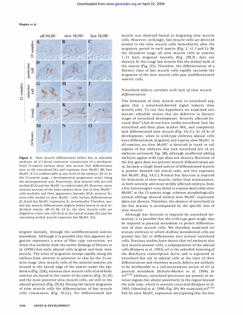
migrate laterally, through the undifferentiated somiticmesoderm. Although it is possible that this apparent mi-gration represents a wave of fiber type conversion, wethink this unlikely from the earlier findings of Devoto etal. (1996) that early adaxial cells migrate and form slowmuscle. The wave of migration sweeps rapidly along theembryo from anterior to posterior so that by the 21-so-mite stage, slow muscle cells of the anterior somites arelocated at the lateral edge of the somite under the epi-dermis (Fig. 2J,K), whereas slow muscle cells of mid-bodysomites are found in the center of the somite (Fig. 2G,H)and the most posterior slow muscle cells, are still in theadaxial position (Fig. 2D,E). During the lateral migrationof slow muscle cells the differentiation of fast musclecells commences (Fig. 2F,I,L). No differentiated fast
muscle was observed lateral to migrating slow musclecells. However, strikingly, fast muscle cells are detectedmedial to the slow muscle cells immediately after themigratory period in each somite (Fig. 2, cf. I and L). Bythe 26-somite stage, all slow muscle cells in somites1–21 have migrated laterally (Fig. 2M,N; data notshown). At this stage fast muscle fills the medial bulk ofthe somite (Fig. 2O). Therefore, the differentiation of adistinct class of fast muscle cells rapidly succeeds themigration of the slow muscle cells past undifferentiatedsomitic cells.
Notochord defects correlate with lack of slow muscledifferentiation
The formation of slow muscle next to notochord sug-gests that a notochord-derived signal induces slowmuscle cells. To test this hypothesis we examined twomutant zebrafish strains that are defective in distinctstages of notochord development. Severely affected bo-zozok (bozi2) fish do not have visible notochord, lack thenotochord and floor plate marker Shh, and completelylack differentiated slow muscle (Fig. 3A–C). At 24 hr ofdevelopment, when in wild-type embryos adaxial cellshave differentiated, migrated, and express slow MyHC inall somites, no slow MyHC is detected in trunk or tailregions of boz embryos that lack notochord (six of sixembryos sectioned; Fig. 3B), although unaffected siblingembryos appear wild type (data not shown). Mutation ofthe boz gene does not prevent muscle differentiation perse, because a single fused somite of differentiated muscleis present beneath the neural tube, and this expressesfast MyHC (Fig. 3A,C). Normal boz function is requiredfor formation of slow muscle, rather than maintenance,as both severely and more mildly affected embryos froma boz heterozygote cross failed to express detectable slowMyHC at the 15-somite stage, whereas morphologicallynormal siblings showed normal slow MyHC expression(data not shown). Therefore, the absence of notochord inthe boz mutant is accompanied by the specific loss ofslow muscle.
Although boz function is required for notochord for-mation, it is possible that the wild-type gene might alsobe required in paraxial mesoderm to permit differentia-tion of slow muscle cells. We therefore examined ntlmutant embryos in which midline mesodermal cells arepresent but fail to differentiate into mature notochordcells. Previous studies have shown that ntl embryos alsolack muscle pioneer cells, a subpopulation of the adaxialcells (Halpern et al. 1993). ntl is the zebrafish homolog ofthe Brachyury transcription factor and is expressed innotochord but not in adaxial cells at the time of theirdifferentiation and therefore muscle defects are unlikelyto be attributable to a cell-autonomous action of ntl inparaxial mesoderm (Schulte-Merker et al. 1994). Inntlb160 embryos, notochord precursors are present in an-terior regions but absent posteriorly in the region beyondthe yolk tube, which is severely truncated (Halpern et al.1993; Odenthal et al. 1996; Fig. 3P). We examined ntlb160
fish for slow MyHC expression anticipating that the loss
Figure 2. Slow muscle differentiates before fast in zebrafishembryos. (A–C) Serial transverse cryosections of a methanol-fixed 15-somite embryo show that muscle first differentiatesnext to the notochord (A), and expresses slow MyHC (B). FastMyHC (C) is undetectable at any level of the embryo. (D–L) Atthe 21-somite stage, a developmental progression exists alongthe anteroposterior axis. Posteriorly, slow muscle cells are stillmedial (D,E) and fast MyHC is undetectable (F). However, moreanterior sections of the same embryo show loss of slow MyHC+
cells medially and their appearance laterally (H,K; arrows). So-mite cells medial to slow MyHC+ cells initiate differentiation(G,J) and fast MyHC expression (L; arrowheads). Therefore, me-dial fast muscle differentiates slightly before lateral in each in-dividual somite. (M–O) By 24 hr, the slow muscle cells arealigned in a layer one cell thick at the lateral margin (N), and theremaining medial muscle expresses fast MyHC (O).
Blagden et al.
2166 GENES & DEVELOPMENT
on April 22, 2006 www.genesdev.orgDownloaded from

of notochordal maturation might prevent slow muscleformation. Despite the absence of muscle pioneer cells atthe dorsoventral midline, slow and fast muscle in ante-rior regions of ntl embryos appeared normal (Fig. 3D–F).Therefore, mature notochord is not required for slowmuscle differentiation. However, more posterior regionsof ntl embryos, in which axial mesoderm defects aremore severe (Halpern et al. 1993), showed reduced slow
muscle formation and aberrant positioning (Fig. 3G–I). Inrare embryos (1/16 serially sectioned) a complete ab-sence of slow muscle was observed in the most posteriorsomite at 24 hr of development, even though extensivedifferentiated fast muscle was present (Fig. 3J–L). Theremaining ntl embryos (15/16) showed regional slowdeficits. Therefore, ntl mutant fish demonstrate that al-though mature notochord is not necessary for slowmuscle formation, severe defects in notochord establish-ment in the tail correlate with loss of slow muscle dif-ferentiation.
Shh induces ectopic slow muscle differentiation
Examination of the muscle phenotype of boz and ntlmutant embryos suggested that notochord-derived sig-nals may determine the slow muscle fate, reminiscent ofthe induction of floorplate and motoneurons by noto-chord-derived Shh protein (Ericson et al. 1996) and ofmuscle pioneer cells by notochord-derived hedgehogs(Currie and Ingham 1996). Consistent with this, shhmRNA is absent from those regions of both boz and ntlembryos that show defects in slow muscle formation(Fig. 3M–Q). To test the possibility that Shh might be anotochord-derived inducer of the slow muscle fate, weinjected shh mRNA into two- or four-cell zebrafish em-bryos to create animals chimeric for shh overexpressingcells. Such injections lead to an easily detectable reducedretina phenotype (Krauss et al. 1993). In animals affectedfor retinal development, we observed an induction ofslow MyHC expression across the entire width of thesomite in each of 12 serially-sectioned 24-hr embryos(Fig. 4A–C). Strikingly, this expansion occurs at the ex-pense of differentiated fast muscle (Fig. 4C). In those ani-mals in which mosaic segregation of shh mRNA causespartial slow muscle induction, residual fast muscle isobserved in regions not expressing slow MyHC (data notshown). When the same experiment was repeated usingan equivalent amount of echidna hedgehog (ehh)mRNA, no defect was detected in any part of 10 embryosserially sectioned (data not shown). Therefore, Shh is anotochord-derived signal capable of inducing slowmuscle at the expense of fast.
The wholesale conversion of large areas of somite toslow muscle by Shh has two possible explanations. Shhcould induce somitic cells to differentiate as slowmuscle prematurely. Alternatively, Shh might not affectthe decision of when to differentiate, but simply deter-mine what type of muscle is formed. To address thisissue, we examined the effect of ectopic Shh on earlierstage zebrafish embryos. In 15-somite zebrafish embryos,Shh induces a wide region of ectopic lateral differenti-ated muscle within the somite (46/53 unselected in-jected embryos, Fig. 4G,H). Ectopic slow muscle differ-entiation occurred without premature induction of fastmuscle tissue (Fig. 4D–F). The premature differentiationof lateral muscle tissue suggested that Shh might inducepresomitic mesoderm to differentiate early. However,premature slow muscle differentiation before the normaltime of adaxial cell differentiation was not observed in
Figure 3. Severe midline defects correlate with loss of slowmuscle formation. (A–C) Serial transverse sections of severelyaffected 24-hr boz embryos, which have no axial mesodermfrom gastrulation, have no detectable slow MyHC (B). A singlefused somite (A) expressing fast MyHC (C) extends under theneural tube (cf. Fig. 2, M–O). (D–L) Successive transverse sec-tions of a 24-hr ntlb160 embryo. Anterior tail shows normal dis-tribution of muscle fiber types (D–F), except for the lack ofmuscle pioneers at the dorsoventral midline (E). More posteri-orly (G–I), slow cells are missing or migrate abnormally (arrowsin H,K). Fast MyHC appeared unaffected (F,I,L). (M–Q) Whole-mount in situ mRNA hybridization for shh at 15 somites (M–O,Q) or 24 hr (P). Notochord and floorplate shh expression iscompletely absent in severe boz embryos (N,Q), compared withwild type (M,O). In contrast, only the most posterior shh ex-pression is lacking in ntl mutants (P). (M,N) Dorsal views of thetail; (O–Q) lateral views of the tail.
Sonic hedgehog and zebrafish slow muscle
GENES & DEVELOPMENT 2167
on April 22, 2006 www.genesdev.orgDownloaded from

either presomitic mesoderm of any of 36 embryos exam-ined at the 15-somite stage (Fig. 4H) or in any region of22 embryos at tailbud stage (data not shown). Therefore,the earliest time somitic cells are competent to becomeslow muscle in response to Shh is when adaxial cellsnormally differentiate. However, at this stage, cells in allregions of the somite become competent.
The ability of Shh to induce slow muscle is consistentwith the lack of slow muscle in regions of zebrafish mu-tants that lack midline shh expression. This correlationsuggests strongly that the reason for the lack of slowmuscle in the bozi2 and the tail of ntlb160 mutants is lack
of notochord-derived Shh (Concordet et al. 1996). How-ever, the boz gene has not been cloned, so its expressionis unknown, and the ntl gene is expressed transiently inpresomitic mesoderm, as well as in notochord (Odenthalet al. 1996). This raises the possibility that the lack ofslow muscle in these mutants reflects a need for a cellautonomous action of the respective genes in paraxialmesoderm. To address this issue, we overexpressed shhin embryos from bozi2 mutant crosses and examined theresultant animals for slow MyHC expression. Five out ofsix severely affected boz mutants injected with shhmRNA showed induction of slow MyHC, and suppres-sion of fast MyHC (Fig. 4I,J). Therefore, the bozi2 muta-tion does not affect the ability of somitic tissue to re-spond to Shh and form slow muscle. Moreover, even inthe complete absence of notochord Shh is sufficient forthe formation of slow muscle.
A limited source of Shh is sufficient to rescueslow muscle
One limitation of overexpression of Shh by mRNA in-jection is that ectopic Shh is expressed in regions of theanimal never normally exposed to Shh and at above nor-mal physiological levels. This might perturb signals nec-essary for muscle development from other embryonictissues. To examine slow muscle differentiation in re-sponse to localized lower levels of shh expression wetook advantage of the floating head (flh) mutation. Ani-mals homozygous for flh are defective in notochordmaturation because of mutation in a homeobox-contain-ing gene expressed chiefly in the notochord (Halpern etal. 1995; Talbot et al. 1995) and, like ntl embryos, lacknotochords and muscle pioneers. However, unlike ntl,flh exhibits transdifferentiation of notochord tissue intomuscle (Halpern et al. 1995). We examined flh embryosfor muscle differentiation and found that it occurs in analtered location. Cells in the embryonic midline, notthose in the adaxial position, are the first to differentiatein flh embryos (Fig. 5A,B). This differentiation is imme-diately beneath the presumptive floorplate that ex-presses Shh sporadically (Fig. 5C). Despite the unusuallocation of these muscle cells, they express slow, but notfast, MyHC (Fig. 5D–F), spread dorsally around the neu-ral tube and ventrally in the midline, and appear able toundergo lateral migration to take up a normal positionbeneath the ectoderm by 24 hr of development (Fig. 5G–I). Therefore, in flh embryos, apparently normal slowmuscle cells differentiate beneath the residual floor-plate—the sole remaining location where somitic meso-derm abuts shh-expressing tissue.
The discontinuous location of shh-expressing cells inthe floorplate of flh mutants (Fig. 5C) allows an exami-nation of the relationship between the location of Shhand slow muscle differentiation. At the posterior limit ofMyHC-containing cells in flh we found no correlationbetween the location of remaining floorplate shh expres-sion and medial slow myoblast differentiation. Muscledifferentiates both immediately beneath and between is-lands of shh-expression (Fig. 5C). This data suggested
Figure 4. Ectopic overexpression of shh induces extra slowmuscle cells in the myotome of wild-type and mutant embryos.(A–C) shh mRNA was microinjected into the zebrafish blastulaeat the 2- or 4-cell stage and 24-hr embryos examined in trans-verse serial cryosections. All differentiated cells in both myo-tomes contained slow MyHC (A,B) and did not express fastMyHC (C). Some injected embryos showed ectopic slow MyHCand fast MyHC suppression on only one side, probably causedby mosaic expression of injected mRNA (data not shown). (D–H)Embryos injected with shh mRNA were analyzed at the 15-somite stage by whole-mount or cryosection immunohisto-chemistry. Compared with wild type (G and Fig. 2A), injectedembryos showed bilateral (H) or unilateral (D, arrow) expansionof muscle differentiation. Ectopic muscle contained slow (E,arrow), but not fast, MyHC (F). Note that although ectopic lat-eral muscle is present in the anterior portion of shh-injectedembryos (H, left), premature differentiation is not observed inpresomitic mesoderm (H, right). (I,J) Embryos from boz mutantcrosses were injected with shh mRNA and severely affected bozembryos (lacking eyes and notochord), were examined by serialcryosection immunohistochemistry. Widespread slow MyHCrescue (I) and supression of fast MyHC (J) was observed.
Blagden et al.
2168 GENES & DEVELOPMENT
on April 22, 2006 www.genesdev.orgDownloaded from

that terminal differentiation of slow muscle is not in-duced directly by Shh. Further evidence that muscle dif-ferentiation, per se, is not induced by Shh came fromexamining the up-regulation of patched1 (ptc1) mRNAin flh mutant embryos. ptc1, a zebrafish homolog of Dro-sophila patched, is a Shh receptor (Stone et al. 1996), andis up-regulated adjacent to residual shh expression in flhembryos both at somitic and presomitic anteroposteriorpositions (Concordet et al. 1996). Therefore, mesodermalcells are first exposed to Shh long before muscle differ-entiation commences. Moreover, even the most recentlydifferentiated muscle cells in flh embryos frequently donot express high levels of ptc1 mRNA, despite adjacentmesoderm expressing ptc1 abundantly (Fig. 5J,K). There-
fore, although Shh induces ptc1 locally along the entirelength of the flh embryo, there is a delay after Shh expo-sure before the appearance of differentiated slow musclecells. In addition, by the time slow muscle differentiates,any spatial correlation between shh expression and myo-genic cells has been lost. Taken together, these data sug-gest that Shh may initiate slow myoblast formation, butthat continued exposure is not required to trigger theterminal differentiation of slow muscle fibers.
Discussion
Shh and slow muscle induction
Several lines of evidence show that adaxial slow muscleformation in zebrafish embryos is controlled by Shh.First, slow muscle differentiation occurs next to the no-tochord, which expresses shh. This observation confirmsand extends the results of Devoto et al. (1996), who ob-served that adaxial cells give rise to slow muscle markersafter they migrate laterally through the somite. Our datashow that adaxial cells are already determined to formslow muscle as soon as they differentiate into skeletalmuscle myosin-expressing myocytes. Second, a lack ofslow muscle correlates with a lack of shh expression inmutant fish. Third, ectopic expression of shh can induceconversion of most, if not all, somitic cells to slowmuscle, at the expense of fast muscle. Fourth, even inthe absence of notochord, injection of shh mRNA canrescue formation of slow muscle. Fifth, in the absence ofthe normal Shh signal from notochord, a localized sourceof Shh correlates with induction of ectopic slow musclecells, which then migrate in a similar fashion to slowmuscle cells in wild-type embryos. These data are sup-ported by the wild-type expression of a Shh receptor,ptc1, that is up-regulated in adaxial cells within pre-somitic mesoderm indicating that these cells are re-sponding to hedgehog (hh) signaling (Concordet et al.1996). Taken together, these findings make a strong casefor notochord-derived Shh being the normal inducer ofthe differentiated slow adaxial muscle cell fate in thezebrafish.
Shh induces adaxial slow myoblasts
How might Shh induce the slow muscle fate in ze-brafish? Muscle is formed in two steps—mesodermalcommitment to the proliferative myoblast, followed byterminal differentiation into the postmitotic muscle fi-ber. Several lines of evidence suggest that Shh is respon-sible for induction of slow muscle precursor cells, ratherthan the terminal differentiation of slow muscle, per se.First, the muscle-specific transcription factor myoD isinitially detectable in adaxial precursors located adjacentto shh-expressing cells within the embryonic shield sev-eral hours before their terminal differentiation at aroundthe time of somitogenesis (Weinberg et al. 1996). Thisexpression of myoD before terminal differentiation isalso detected in adaxial cells at later stages when poste-rior somites arise from the tail bud. Second, zebrafishmutants like boz and ntl that lack slow muscle, also lack
Figure 5. Slow muscle forms beneath the floorplate in flh em-bryos. (A–C) Differentiated muscle visualized by MyHC-stain-ing in whole-mount 15-somite wild-type (A) or flh (B,C) em-bryos observed after flat-mounting. Dual labeling for shhmRNA (red; C) and MyHC (green; C) confirmed that the mostposterior differentiated muscle was at the midline (arrow, C).(D–I) Transverse cryosections show that differentiated musclein flh midline mesoderm expresses slow but not fast MyHC at15 somites (D–F). By 24 hr, flh embryos have a single fusedmyotome (G), with lateral slow and medial fast muscle (H,I).Despite the ectopic location of formation of slow muscle cellsin the midline the slow muscle cells appear to migrate normallyso that the only detectable residual slow muscle defect is lack ofmuscle pioneers. (J,K) Dual labeling for MyHC (green) and ptc1mRNA (red) in the two most posterior MyHC-containing serialsections of a 15-somite flh embryo. Newly differentiatingmuscle cells close to the midline do not always express detect-able ptc1 (arrows), even though ptc1 is expressed abundantly inthe mesoderm underlying residual floor plate (K).
Sonic hedgehog and zebrafish slow muscle
GENES & DEVELOPMENT 2169
on April 22, 2006 www.genesdev.orgDownloaded from

the early adaxial myoD expression, and this correlateswith a lack of axial Shh (Fig. 3; Concordet et al. 1996;Odenthal et al. 1996; Weinberg et al. 1996; Schier et al.1997). Third, Shh signaling can induce premature myoDin lateral presomitic cells (Concordet et al. 1996; Ham-merschmidt et al. 1996; Weinberg et al. 1996). We showthat these ectopic myoD-expressing cells in lateralsomites have other features, such as the direction andtiming of their differentiation and sensitivity to addi-tional hh signals (Currie and Ingham 1996), suggestingthat the terminal differentiation of these cells into slowmuscle is prefigured at the myoblast level. Fourth, ourexamination of the flh mutant suggests that adaxialmyoblasts differentiate into slow muscle fibers indepen-dent of their proximity to residual shh-expressing floorplate cells, and independent of their exposure to Shh dur-ing the period of terminal differentiation, as assayed byptc1 expression (Concordet et al. 1996; Marigo and Tabin1996). Therefore, at early stages MyoD may mark cellsthat, although not yet differentiated, have become com-mitted to a slow myoblast lineage.
Previous data have suggested that the combined actionof notochord-derived Shh and Ehh induces zebrafishmuscle pioneer subset of the adaxial slow muscle cells(Currie and Ingham 1996). The data in the present paperdemonstrate that Ehh is not required for production ofthe nonpioneer adaxial slow muscle cells. Ehh is notexpressed in notochord of ntlb160 (Currie and Ingham1996), yet nonpioneer slow muscle cells form and mi-grate normally in the anterior of ntlb160 embryos whereShh alone is expressed (Fig. 3). Similarly, in flh mutants,which lack notochord and ehh expression (Currie andIngham 1996), apparently normal nonpioneer adaxialcells are formed ectopically close to residual floorplate
Shh. Moreover, Ehh does not appear able to substitute forShh in the induction of nonpioneer adaxial cells as in-jection of ehh mRNA into wild-type embryos did notinduce ectopic slow MyHC. These data support the hy-pothesis that in vivo Shh and Ehh serve distinct roles.
The finding that Shh induces slow myoblasts suggestsa new view of the steps of muscle differentiation thatcontrasts with the traditional model in which somiticcells first become myoblasts and only subsequently spe-cialize into one particular myoblast subclass. We sug-gest, as summarized in Figure 6, that the decisionwhether to form one type of muscle or another is madeconcurrently with myoblast commitment to the musclelineage. This scheme concurs with conclusions fromanalysis of the you-type zebrafish mutants (van Eeden etal. 1996). Such a view also fits well with studies in Dro-sophila demonstrating that distinct extracellular signalsserve to commit each founder myoblast to a particularmuscle type (Baylies et al. 1995). However, it is possiblethat presomitic cells could be committed to myogenesisbefore myoD expression. Although MyoD is the firstMRF to be expressed in birds, Myf-5 is the earliest MRFto appear at high levels in mammalian somites (Ott et al.1991), and Pax-3 can induce myogenesis (Maroto et al.1997; Tajbakhsh et al. 1997). Furthermore, whether allmyoblasts are committed to form particular types ofmuscle from their inception is unclear. Whatever thecase, our data show that Shh induces adaxial myoblaststhat adopt a slow muscle fate.
Zebrafish fast muscle formation
Shh is not necessary for fast muscle formation. boz fishthat lack Shh produce abundant fast muscle throughout
Figure 6. Three phases of zebrafishmuscle development. A model in whichpresomitic mesoderm gains competence torespond to Shh early in development inwild-type zebrafish. The result of exposureto notochord-derived Shh at this stage is aslow myoblast phenotype involving myoDexpression and up-regulation of ptc1 (or-ange). However, a second event, either in-trinsically timed within the slow myo-blast, or in the form of a signal from out-side of the somite (indicated by ? on thediagram, but unlikely to be either Shh ornotochord-derived), is required for the slowmyoblast to differentiate into a fiber (red).Simultaneously, the MyoD-positive myo-blast state in lateral somitic cells arises in-dependent of notochord-derived signals(yellow). Later, adaxial slow fiber migra-tion and concurrent differentiation of lat-eral myoblasts into fast muscle (green) istimed by a third event(s). Fast fiber differ-entiation is independent of slow fiber mi-gration, although the converse may not betrue.
Blagden et al.
2170 GENES & DEVELOPMENT
on April 22, 2006 www.genesdev.orgDownloaded from
the somite. Moreover, the normal myoD-expressingmyoblasts stripes across the posterior border of the so-mite form at the normal time just before somitogenesisin embryos that lack shh expression (Odenthal et al.1996). Therefore, in zebrafish, MyoD may mark commit-ment to a myoblast fate irrespective of the type of myo-blast formed. We find that cells that are initially lateralwithin the somite differentiate into fast muscle. Theymay be committed to formation of fast muscle from theinception of myoD expression.
Timing of myoblast differentiation
We show that ectopic Shh can induce premature muscledifferentiation in the lateral somite. However, prema-ture differentiation was slow, rather than fast, and wasonly observed at the normal time of slow adaxial celldifferentiation. Therefore, no somite cells are competentto differentiate in response to Shh until around the timeof somitogenesis, even though myoD is expressed ear-lier. This may explain why slow muscle does not appearearlier in development even though shh is expressed inthe developing notochord from gastrulation onwards(Krauss et al. 1993), and is presumably secreted becauseptc1, a marker of Shh exposure, is expressed highly inadjacent presomitic cells (Concordet et al. 1996). Twoalternative models could explain the delay betweenMyoD and slow myosin expression (Fig. 6). In one model,the delay is caused by an intrinsically timed maturationof the somitic cells. Although cell division is not exten-sive in zebrafish somites (Kimmel and Warga 1987), Shhmight induce myoblasts committed to division followedby differentiation as it can be a somitic mitogen (Fan etal. 1995). Mammalian myoblasts show such behavior invitro (Quinn et al. 1985), which is reminiscent of theinduction of division followed by terminal differentia-tion in Drosophila lamina ganglia neurons in response toretinal neuron-derived hh (Huang and Kunes 1996). Al-ternatively, in the second model, extracellular signalsmay control terminal differentiation. In amniotes, othersignals can cooperate with Shh to regulate myogenesis(Munsterberg et al. 1995; Stern et al. 1995; Pourquie et al.1996), and a variety of growth factors repress myoblastdifferentiation in culture. Ventral axial structures areunlikely sources of such signals as Shh is sufficient toinduce slow MyHC in boz embryos that fail to formnotochord or floor plate. Regardless of the mechanism bywhich the timing of terminal adaxial slow muscle differ-entiation is controlled, our data show that similarmechanisms can operate in the lateral somite to controlectopic slow muscle differentiation in response to Shh.
Evolutionary conservation of muscle patterning
We found that in fish embryos the first skeletal musclefibers to form are slow from the time of their inception.Later, a second wave of fibers, which ultimately consti-tute the majority of all fibers, differentiate as fastmuscle. In amniote limbs muscle fibers also form in two
waves, an early primary population that express slow(and embryonic) myosin and later secondary cells that,forming in close association with primary fibers, expressfast (and embryonic) myosin from their inception (Kellyand Rubinstein 1980; Vivarelli et al. 1988; Cho et al.1994). We suspect that zebrafish embryos may also ex-press an embryonic myosin in both slow and fast fibersas the immunoreaction with our all-myosin antibodywas stronger than with the specific slow and fast anti-bodies. Moreover, both adaxial and nonadaxial cells reactfrom their inception with an antibody that detects em-bryonic myosin (Devoto et al. 1996). These analogiessuggest that adaxial and nonadaxial somitic muscle cellsin the zebrafish may be evolutionary homologs of amni-ote primary and secondary muscle fiber generations.Amniote secondary fibers form overlying the neuromus-cular junctions of primary fibers and it has been sug-gested that signals from the forming neuromuscularjunction region may be required to initiate secondaryfiber formation (Duxson et al. 1989). This is not the casein the zebrafish as absence of differentiated slow primaryfibers does not prevent differentiation of fast muscle de-spite the striking correlation between the lateral migra-tion of slow fibers and the differentiation of fast fibers.The converse relationship, that fast fiber differentiationmight cause slow fiber migration, remains a possibility.Nevertheless, the close similarities between fish andamniote fiber generation suggest that the common an-cestor had two steps of muscle patterning: early fibersbeing slow and later fast.
There are further analogies between amniote and fishmyogenesis. Amniote primary fibers are of several dis-tinct fiber types that prefigure later muscle characteris-tics (Crow and Stockdale 1986), even though all expresssome form of slow MyHC (Kelly and Rubinstein 1980;Vivarelli et al. 1988; Page et al. 1992; Hughes et al. 1993).Slow adaxial cells in the zebrafish are also composed oftwo subpopulations, the muscle pioneer cells which ex-press engrailed, and the nonpioneer adaxials. Engrailedproteins also mark a subpopulation of muscle cells in thejaw muscle of the zebrafish (Hatta et al. 1990). The datareported in the present paper, together with the previousfindings that a second notochord-derived signal (Halpernet al. 1993), provided by Ehh (Currie and Ingham 1996), isresponsible for regulating the formation of muscle pio-neer cells, suggest that hh signaling molecules may regu-late the diversity of muscle fiber types formed in theearly fish embryo. Banded hedgehog is also expressed inparticular regions of the Xenopus somite (Ekker et al.1995). Whether similar signals control muscle patterningin amniotes remains to be determined.
Hedgehogs and vertebrate myogenesis
Secretion of Shh from notochord has been shown to in-duce floorplate markers in anterior (although not poste-rior) zebrafish central nervous system (CNS) and bothfloorplate and motoneurons in amniote neural tube (Ech-elard et al. 1993; Krauss et al. 1993; Roelink et al. 1994;
Sonic hedgehog and zebrafish slow muscle
GENES & DEVELOPMENT 2171
on April 22, 2006 www.genesdev.orgDownloaded from
Ericson et al. 1996). The role of Shh in somite patterninghas been less clear. In amniotes, in which much of thesomite becomes sclerotome, either ectopic notochord orshh-expressing cells can induce extra sclerotome at theexpense of dermomyotome markers (Fan and Tessier-Lavigne 1994). Conversely, shh−/− mice have deficits insclerotomal derivatives (Chiang et al. 1996). On theother hand, in both chick and zebrafish, overexpressionof shh induces ectopic myoD expression, suggesting amyogenic action (Johnson et al. 1994; Concordet et al.1996; Weinberg et al. 1996). Moreover, shh−/− mice showdefects in medial muscle formation (Chiang et al. 1996)and notochord can induce avian myogenesis (Pownall etal. 1996). So Shh may regulate formation of both ventraland more dorsal somitic tissues. Action of Shh at dis-tinct concentrations or times (Ericson et al. 1996), or incollaboration with other factors (Munsterberg et al.1995; Stern et al. 1995; Pourquie et al. 1996), could de-termine the outcome of Shh signaling.
Induction of distinct myoblast types and the subse-quent control of their terminal differentiation may ac-count for the numerous signals capable of influencingsomite myogenesis. If equivalents of Shh-dependent ad-axial cells exist in amniotes, we would expect that par-ticular muscle markers are not distributed uniformly be-tween distinct muscle cell types in the developing der-momyotome. In amniotes, MRFs are the earliest knowndefinitive myogenic markers. Expression of at least oneMRF is obligatory for myogenesis in mice (Rudnicki etal. 1993). MRFs are expressed at low levels in presomiticmesoderm, which has the capacity to form muscle indissociated cell culture (George-Weinstein et al. 1994;Lin-Jones and Hauschka 1996). However, two myoblastpopulations arise with distinct temporal and spatial pat-terns within the dermomyotome: The first initially ex-presses myf-5 in medial regions and the second myoD inlateral regions (Cossu et al. 1996a; Maroto et al. 1997;Tajbakhsh et al. 1997). That shh−/− mice have reducedexpression of medial myf-5 but no detectable change inlateral myoD expression (Chiang et al. 1996) suggests arole for Shh in induction of the medial population. In-hibitory signals, such as BMP4 (Fan and Tessier-Lavigne1994; Cossu et al. 1996b; Pourquie et al. 1996), may func-tion in vivo to suppress overt myogenic phenotypes inthe lateral compartment that generates limb and bodywall muscle and may have no homologous process inmost zebrafish somites. So generation of further diver-sity within the dorsomedial myogenic compartmentcould be a role of Shh in amniote myogenesis. Distinctpopulations of slow and fast fibers may be present inamniote myotome (Dhoot 1994). In this paper, we haveshown that in zebrafish, Shh regulates formation of myo-tomal slow muscle. Much slow muscle in amniote limbsis located near developing bone that expresses indianhedgehog (Bitgood and McMahon 1995; Vortkamp et al.1996). Moreover, motoneurons, which strongly influ-ence muscle development, can express shh (Bitgood andMcMahon 1995; Stone et al. 1996), raising the possibilitythat diverse hh proteins may regulate muscle fiber diver-sification.
Materials and methods
Zebrafish lines and maintenance
Wild-type and heterozygote mutant breeding fish were main-tained at 28.5°C on a 14-hr/10-hr light cycle. We obtained flhn1
from the University of Newcastle-upon-Tyne, ntlb160 from theUniversity of Oregon, and bozi2 was isolated in the Inghamlaboratory (P.D. Currie., T. Schilling, G. Bergemann, and P.W.Ingham, unpubl.). bozi2 fish exhibit a variable phenotype withdefects ranging from reduced notochords to a severe lack of axialmesoderm at all rostro–caudal levels. Of 142 progeny of a het-erozygous cross examined at F2–F4, 20 (13%) showed completeabsence of eyes and notochord. A further 25 (17%) showed apartial phenotype with variable eyes and the anterior half of thenotochord missing. These, and a number of other aspects of thephenotype, are strongly reminiscent of boz mutant fish (Sol-nica-Krezel et al. 1996). Complementation analysis by a cross ofheterozygous bozi2 with bozm168 has shown reduced eyes andnotochord in 3 out of 30 progeny. We therefore tentatively con-clude that these genes are allelic. However, because of the in-complete penetrance of boz, definitive demonstration of allel-ism awaits the mapping of the mutation. Embryos were col-lected by natural spawning and staged by anatomical markersaccording to Westerfield (1995). Prim-5 stage embryos are re-ferred to as 24 hr.
RNA injection
RNA injections were performed as described (Currie and Ing-ham 1996).
Immunohistochemistry
The slow and fast MyHC antigens are destroyed by aldehydefixatives, so embryos were fixed by incubating for 5 min each ingraded methanols, rehydrated in 0.1% Tween 20, serially cryo-sectioned, and stained. However, preservation of younger em-bryos was better after staining in whole-mount, followed bypost-fixation in 4% paraformaldehyde for 4 hr at 4°C beforecryosectioning. Primary monoclonal antibody supernatants ofA4.1025 (Dan-Goor et al. 1990) and BA-D5 (Schiaffino et al.1989) were diluted 1:10. EB165 monoclonal ascites was used at1:5000 (Gardahaut et al. 1992). First antibodies were detectedwith biotin-conjugated horse-derived anti-mouse IgG (Vector),Vectastain ABC Elite Peroxidase kit (Vector), and visualized us-ing 0.5 mg/ml of diaminobenzidine with (black stains) or with-out (brown stains) 0.03% CoCl2 enhancement. Cryosections fordual immunofluorescence had IgG first antibodies detectedwith Cappell goat anti-mouse IgG (g-specific) Texas red. After amouse IgG block, biotinylated BA-D5, prepared using PierceNHS-Biotin reagent, was detected with Dako streptavidin-FITC. Sections were mounted in 150 mg/ml of polyvinyl alco-hol, 30% glycerol PBS with DABCO antifade, and photographedby confocal microscopy.
Western blots
Embryos were dechorionated, deyolked, and homogenizedmanually on ice for 10 min in 63 mM Tris-HCl (pH 6.8), 10%glycerol, 5% b-mercaptoethanol, 3.5% SDS, 0.2 mM PMSF, 0.5µM aprotinin, and 0.5 µM leupeptin. Samples were microcentri-fuged for 5 min at 4°C, 0.01% bromophenol blue added to thesupernatant, the equivalent of 10 embryos run on each lane of a7.5% acrylamide denaturing gel at 200 mV for 30 min, andelectroblotted onto nitrocellulose (Amersham). Purified bovinecardiac myosin was a kind gift of Dr. John Sleep (The Randall
Blagden et al.
2172 GENES & DEVELOPMENT
on April 22, 2006 www.genesdev.orgDownloaded from
Institute, London, UK). Nitrocellulose strips were blocked in5% milk powder PBS/Az overnight, washed, and incubatedwith A4.1025 (1:10), BA-D5 (1:10), F1.652 [1:10; Webster et al.1988)], or EB165 (1:250) for 2 hr at room temperature. Afterwashing, primary antibody was detected with horseradish per-oxidase-conjugated sheep anti-mouse IgG F(ab)2 and an ECL kit(Amersham).
Acknowledgments
We thank Steve Wilson and Nigel Holder for fish from theirBBSRC-funded facility, Everett Bandman for EB165 ascites, JohnSleep for bovine cardiac myosin, and Steve Devoto, Monte Wes-terfield, and Wolfgang Driever for showing us manuscripts be-fore publication. We thank Steve Wilson, Patricia Salinas, andmembers of our laboratories for helpful comments on the manu-script, and T. Schilling and G. Bergemann for participation inthe mutant screen. This work is supported by the Medical Re-search Council (S.M.H. and C.S.B.), the Imperial Cancer Re-search Fund, and a Human Frontier Science Program grant(P.W.I.).
The publication costs of this article were defrayed in part bypayment of page charges. This article must therefore be herebymarked ‘‘advertisement’’ in accordance with 18 USC section1734 solely to indicate this fact.
References
Baylies, M.K., A. Martinez-Arias, and M. Bate. 1995. wingless isrequired for the formation of a subset of muscle founder cellsduring Drosophila embryogenesis. Development 121: 3829–3837.
Bitgood, M.J. and A.P. McMahon. 1995. Hedgehog and Bmpgenes are coexpressed at many diverse sites of cell-cell in-teraction in the mouse embryo. Dev. Biol. 172: 126–138.
Bober, E., B. Brand-Saberi, C. Ebensperger, J. Wilting, R. Balling,B.M. Paterson, H.-H. Arnold, and B. Christ. 1994. Initialsteps of myogenesis in somites are independant of influencefrom axial structures. Development 120: 3073–3082.
Buffinger, N. and F.E. Stockdale. 1995. Myogenic specificationof somites is mediated by diffusible factors. Dev. Biol.169: 96–108.
Chiang, C., Y. Litingtung, E. Lee, K.E. Young, J.L. Corden, H.Westphal, and P.A. Beachy. 1996. Cyclopia and defectiveaxial patterning in mice lacking Sonic hedgehog gene func-tion. Nature 383: 407–413.
Cho, M., S.M. Hughes, I. Karsch-Mizrachi, M. Travis, L.A. Lein-wand, and H.M. Blau. 1994. Fast myosin heavy chains ex-pressed in secondary mammalian muscle fibres at the timeof their inception. J. Cell Sci. 107: 2361–2371.
Concordet, J.-P., K.E. Lewis, J.W. Moore, L.V. Goodrich, R.L.Johnson, M.P. Scott, and P.W. Ingham. 1996. Spatial regula-tion of a zebrafish patched homologue reflects the roles ofsonic hedgehog and protein kinase A in neural tube and so-mite patterning. Development 122: 2835–2846.
Cossu, G., R. Kelly, S. Tajbakhsh, S. Di Donna, E. Vivarelli, andM. Buckingham. 1996a. Activation of different myogenicpathways: myf-5 is induced by the neural tube and MyoD bythe dorsal ectoderm in mouse paraxial mesoderm. Develop-ment 122: 429–437.
Cossu, G., S. Tajbakhsh, and M. Buckingham. 1996b. How ismyogenesis initiated in the embryo? Trends Genet. 12: 218–222.
Crow, M.T. and F.E. Stockdale. 1986. Myosin expression andspecialization among the earliest muscle fibres of the devel-
oping avian limb. Dev. Biol. 113: 238–254.Currie, P.D. and P.W. Ingham. 1996. Induction of a specific
muscle cell type by a hedgehog-like protein in zebrafish.Nature 382: 452–455.
Dan-Goor, M., L. Silberstein, M. Kessel, and A. Muhlrad. 1990.Localization of epitopes and functional effects of two novelmonoclonal antibodies against skeletal muscle myosin. J.Muscle Res. Cell Motil. 11: 216–226.
Devoto, S.H., E. Melancon, J.S. Eisen, and M. Westerfield. 1996.Identification of separate slow and fast muscle precursorcells in vivo, prior to somite formation. Development122: 3371–3380.
Dhoot, G.K. 1994. Mammalian myoblasts become fast or slowmyocytes within the somitic myotome. J. Muscle Res. CellMotil. 15: 617–622.
DiMario, J.X., S.E. Fernyak, and F.E. Stockdale. 1993. Myoblaststransferred to the limbs of embryos are committed to spe-cific fibre fates. Nature 362: 165–167.
Duxson, M.J., Y. Usson, and A.J. Harris. 1989. The origin ofsecondary myotubes in mammalian skeletal muscles: Ultra-structural studies. Development 107: 743–750.
Echelard, Y., D.J. Epstein, B. St.-Jacques, L. Shen, J. Mohler, J.A.McMahon, and A.P. McMahon. 1993. Sonic hedgehog, amember of a family of putative signalling molecules impli-cated in regulation of CNS and limb polarity. Cell 75: 1417–1430.
Ekker, S.C., L.L. McGrew, C.J. Lai, J.J. Lee, D.P. Von Kessler,R.T. Moon, and P.A. Beachy. 1995. Distinct expression andshared activities of members of the hedgehog gene family ofXenopus laevis. Development 121: 2337–2347.
Ericson, J., S. Morton, A. Kawakami, H. Roelink, and T.M. Jes-sell. 1996. Two critical periods of Sonic Hedgehog signalingrequired for the specification of motor neuron identity. Cell87: 661–673.
Fan, C.-M. and M. Tessier-Lavigne. 1994. Patterning of mam-malian somites by surface ectoderm and notochord: Evi-dence for sclerotome induction by a hedgehog homolog. Cell79: 1175–1186.
Fan, C.-M., J.A. Porter, C. Chiang, D.T. Chang, P.A. Beachy, andM. Tessier-Lavigne. 1995. Long-range sclerotome inductionby sonic hedgehog: Direct role of the amino-terminal cleav-age product and modulation by the cAMP signalling path-way. Cell 81: 457–465.
Felsenfeld, A.L., M. Curry, and C.B. Kimmel. 1991. The fub-1mutation blocks initial myofibril formation in zebrafishmuscle pioneer cells. Dev. Biol. 148: 23–30.
Gamel, A.J., B. Brand-Saberi, and B. Christ. 1995. Halves of ep-ithelial somites and segmental plate show distinct muscledifferentiation behavior in vitro compared to entire somitesand segmental plate. Dev. Biol. 172: 625–639.
Gardahaut, M.F., J. Fontaine-Perus, T. Rouaud, E. Bandman, andR. Ferrand. 1992. Developmental modulation of myosin ex-pression by thyroid hormone in avian skeletal muscle. De-velopment 115: 1121–1131.
George-Weinstein, M., J.V. Gerhart, G.J. Foti, and J.W. Lash.1994. Maturation of myogenic and chondrogenic cells in thepresomitic mesoderm of the chick embryo. Exp. Cell Res.211: 263–274.
Goulding, M., A. Lumsden, and A.J. Paquette. 1994. Regulationof Pax-3 expression in the dermamyotome and its role inmuscle development. Development 120: 957–971.
Halpern, M.E., R.K. Ho, C. Walker, and C.B. Kimmel. 1993.Induction of muscle pioneers and floor plate is distinguishedby the zebrafish no tail mutation. Cell 75: 99–111.
Halpern, M.E., C. Thisse, R.K. Ho, B. Thisse, B. Riggleman, B.Trevarrow, E.S. Weinberg, J.H. Postlethwait, and C.B. Kim-
Sonic hedgehog and zebrafish slow muscle
GENES & DEVELOPMENT 2173
on April 22, 2006 www.genesdev.orgDownloaded from
mel. 1995. Cell-autonomous shift from axial to paraxial me-sodermal development in zebrafish floating head mutants.Development 121: 4257–4264.
Hammerschmidt, M., M.J. Bitgood, and A.P. McMahon. 1996.Protein kinase A is a common negative regulator of hedge-hog signalling in the vertebrate embryo. Genes & Dev.10: 647–658.
Hatta, K., T.F. Schilling, R.A. BreMiller, and C.B. Kimmel. 1990.Specification of jaw muscle identity in zebrafish: Correlationwith engrailed-homeoprotein expression. Science 250: 802–805.
Hatta, K., R. Bremiller, M. Westerfield, and C.B. Kimmel. 1991.Diversity of expression of engrailed-like antigens in ze-brafish. Development 112: 821–832.
Holland, L.Z., D.A. Pace, M.L. Blink, M. Kene, and N.D. Hol-land. 1995. Sequence and expression of amphioxus alkalimyosin light chain (AmphiMLC-alk) throughout develop-ment: Implications for vertebrate myogenesis. Dev. Biol.171: 665–676.
Huang, Z. and S. Kunes. 1996. Hedgehog, transmitted alongretinal axons, triggers neurogenesis in the developing visualcenters of the Drosophila brain. Cell 86: 411–422.
Hughes, S.M. and H.M. Blau. 1992. Muscle fiber pattern is in-dependent of cell lineage in postnatal rodent development.Cell 68: 659–671.
Hughes, S.M., M. Cho, I. Karsch-Mizrachi, M. Travis, L. Silber-stein, L.A. Leinwand, and H.M. Blau. 1993. Three slow myo-sin heavy chains sequentially expressed in developing mam-malian skeletal muscle. Dev. Biol. 158: 183–199.
Johnson, R.L., E. Laufer, R.D. Riddle, and C. Tabin. 1994. Ecto-pic expression of Sonic hedgehog alters dorsal-ventral pat-terning of somites. Cell 79: 1165–1173.
Kelly, A.M. and N.A. Rubinstein. 1980. Why are fetal musclesslow? Nature 288: 266–269.
Kenny-Mobbs, T. and P. Thorogood. 1987. Autonomy of differ-entiation in avian brachial somites and the influence of ad-jacent tissues. Development 100: 449–462.
Kimmel, C.B. and R.M. Warga. 1987. Cell lineages generatingaxial muscle in the zebrafish embryo. Nature 327: 234–237.
Krauss, S., J.-P. Concordet, and P.W. Ingham. 1993. A function-ally conserved homologue of the Drosophila segment polar-ity gene hh is expressed in tissues with polarizing activity inzebrafish embryos. Cell 75: 1431–1444.
Lassar, A.B. and A.E. Munsterberg. 1996. The role of positiveand negative signals in somite patterning. Curr. Opin. Neu-robiol. 6: 57–63.
Lin-Jones, J. and S.D. Hauschka. 1996. Myogenic determinationfactor expression in the developing avian limb bud: An RT-PCR analysis. Dev. Biol 174: 407–422.
Marigo, V. and C.J. Tabin. 1996. Regulation of patched by sonichedgehog in the developing neural tube. Proc. Natl. Acad.Sci. 93: 9346–9351.
Maroto, M., R. Reshef, A.E. Munsterberg, S. Koester, M. Gould-ing, and A.B. Lassar. 1997. Ectopic Pax-3 activates MyoD andMyf-5 expression in embryonic mesoderm and neural tissue.Cell 89: 139–148.
Marti, E., R. Takada, D.A. Bumcrot, H. Sasaki, and A.P. McMa-hon. 1995. Distribution of Sonic hedgehog peptides in thedeveloping chick and mouse embryo. Development 121:2537–2547.
Miller, J.B. and F.E. Stockdale. 1986. Developmental origins ofskeletal muscle fibres: Clonal analysis of myogenic cell lin-eages based on expression of fast and slow myosin heavychains. Proc. Natl. Acad. Sci. 83: 3860–3864.
Morin-Kensicki, E.M. and J.S. Eisen. 1997. Sclerotome develop-ment and peripheral nervous system segmentation in em-
bryonic zebrafish. Development 124: 159–167.Munsterberg, A.E., J. Kitajewski, D.A. Bumcrot, A.P. McMahon,
and A.B. Lassar. 1995. Combinatorial signaling by sonichedgehog and Wnt family members induces myogenic bHLHgene expression in the somite. Genes & Dev. 9: 2911–2922.
Odenthal, J., P. Haffter, E. Vogelsang, M. Brand, F.J.M. Van Ee-den, M. Furutani-Seiki, M. Granato, M. Hammerschmidt,C.P. Heisenberg, Y.J. Jiang, D.A. Kane, R.N. Kelsh, M.C.Mullins, R.M. Warga, M.L. Allende, E.S. Weinberg, and C.Nusslein-Volhard. 1996. Mutations affecting the formationof the notochord in the zebrafish, Danio rerio. Development123: 103–115.
Ott, M.-O., E. Bober, G. Lyons, H. Arnold, and M. Buckingham.1991. Early expression of the myogenic regulatory gene, myf-5, in precursor cells of skeletal muscle in the mouse embryo.Development 111: 1097–1107.
Page, S., J.B. Miller, J.X. DiMario, E.J. Hager, A. Moser, and F.E.Stockdale. 1992. Developmentally regulated expression ofthree slow isoforms of myosin heavy chain: Diversity amongthe first fibers to form in avian muscle. Dev. Biol. 154: 118–128.
Pownall, M.E., K.E. Strunk, and C.P. Emerson, Jr. 1996. Noto-chord signals control the transcriptional cascade of myo-genic bHLH genes in somites of quail embryos. Develop-ment 122: 1475–1488.
Pourquie, O., M. Coltey, M.-A. Teillet, C. Ordahl, and N.M. LeDouarin. 1993. Control of dorsoventral patterning of somiticderivatives by notochord and floor plate. Proc. Natl. Acad.Sci. 90: 5242–5246.
Pourquie, O., C.-M. Fan, M. Coltey, E. Hirsinger, Y. Watanabe,C. Breant, P. Francis-West, P. Brickell, M. Tessier-Lavigne,and N.M. Le Douarin. 1996. Lateral and axial signals in-volved in avian somite patterning: A role for BMP4. Cell84: 461–471.
Quinn, L.S., H. Holtzer, and M. Nameroff. 1985. Generation ofchick skeletal muscle cells in groups of 16 from stem cells.Nature 313: 692–694.
Roelink, H., A. Augsburger, J. Heemskerk, V. Korzh, S. Norlin,A. Ruiz i Altaba, Y. Tanabe, M. Placzek, T. Edlund, T.M.Jessell, and J. Dodd. 1994. Floor plate and motor neuron in-duction by vhh-1, a vertebrate homologue of hedgehog ex-pressed by the notochord. Cell 76: 761–775.
Rong, P.M., A.-M. Teillet, C. Ziller, and N.M. Le Douarin. 1992.The neural tube/notochord complex is necessary for verte-bral but not limb and body wall striated muscle differentia-tion. Development 115: 657–672.
Rudnicki, M.A., P.N.J. Schnegelsberg, R.H. Stead, T. Braun,H.-H. Arnold, and R. Jaenisch. 1993. MyoD or Myf-5 is re-quired for the formation of skeletal muscle. Cell 75: 1351–1359.
Schafer, D.A., J.B. Miller, and F.E. Stockdale. 1987. Cell diver-sification within the myogenic lineage: In vitro generation oftwo types of myoblasts from a single myogenic progenitorcell. Cell 48: 659–670.
Schier, A.F., S.C.F. Neuhauss, K.A. Helde, W.S. Talbot, and W.Driever. 1997. The one-eyed pinhead gene functions in me-soderm and endoderm formation in zebrafish and interactswith no tail. Development 124: 327–342.
Schiaffino, S., L. Gorza, S. Sartore, L. Saggin, S. Ausoni, M.Vianello, K. Gundersen, and T. Lomo. 1989. Three myosinheavy chain isoforms in type 2 skeletal muscle fibres. J.Muscle Res. Cell Motil. 10: 197–205.
Schulte-Merker, S., F.J.M. van Eeden, M.E. Halpern, C.B. Kim-mel, and C. Nusslein-Volhard. 1994. no tail (ntl) is the ze-brafish homologue of the mouse T (Brachyury) gene. Devel-opment 120: 1009–1015.
Blagden et al.
2174 GENES & DEVELOPMENT
on April 22, 2006 www.genesdev.orgDownloaded from

Solnica-Krezel, L., D.L. Stemple, E. Mountcastle-Shah, Z.Rangini, S.C.F. Neuhauss, J. Malicki, A.F. Schier, D.Y.R.Stainier, F. Zwartkruis, S. Abdelilah, and W. Driever. 1996.Mutations affecting cell fates and cellular rearrangementsduring gastrulation in zebrafish. Development 123: 67–80.
Stern, H.M. and S.D. Hauschka. 1995. Neural tube and noto-chord promote in vitro myogenesis in single somite ex-plants. Dev. Biol. 167: 87–103.
Stern, H.M., A.M.C. Brown, and S.D. Hauschka. 1995. Myogen-esis in paraxial mesoderm: Preferential induction by dorsalneural tube and by cells expressing Wnt-1. Development121: 3675–3686.
Stone, D.M., M. Hynes, M. Armanini, T.A. Swanson, Q. Gu,R.L. Johnson, M.P. Scott, D. Pennica, A. Goddard, H. Phil-lips, M. Noll, J.E. Hooper, F. De Sauvage, and A. Rosenthal.1996. The tumour-suppressor gene patched encodes a candi-date receptor for Sonic hedgehog. Nature 384: 129–134.
Tajbakhsh, S., D. Rocancourt, G. Cossu, and M. Buckingham.1997. Redefining the genetic hierarchies controlling skeletalmyogenesis: Pax-3 and Myf-5 act upstream of MyoD. Cell89: 127–138.
Talbot, W.B., B. Trevarrow, M.E. Halpern, A.E. Melby, G. Farr,T.J. Postlethwait, C.B. Kimmel, and D. Kimmelman. 1995. Ahomeobox gene essential for zebrafish notochord develop-ment. Nature 378: 150–157.
Teillet, M.-A. and N.M. Le Douarin. 1983. Consequences ofneural tube and notochord excision on the development ofthe peripheral nervous system in chick embryos. Dev. Biol.98: 192–211.
Ticho, B.S., D.Y.R. Stainier, and M.C. Fishman. 1996. Threezebrafish MEF2 genes delineate somitic and cardiac muscledevelopment in wild-type and mutant embryos. Mech. Dev.59: 205–218.
van Eeden, F.J.M., M. Granato, U. Schach, M. Brand, M. Furu-tani-Seiki, P. Haffter, M. Hammerschmidt, C.P. Heisenberg,Y.-J. Jiang, D.A. Kane, R.N. Kelsh, M.C. Mullins, J. Oden-thal, R.M. Warga, M.L. Allende, E.S. Weinberg, and C. Nus-slein-Volhard. 1996. Mutations affecting somite formationand patterning in the zebrafish, Danio rerio. Development123: 153–164.
van Raamsdonk, W., C.W. Pool, and G. te Kronnie. 1978. Dif-ferentiation of muscle fibre types in the teleost Brachydaniorerio. Anat. Embryol. 153: 137–155.
Vivarelli, E., W.E. Brown, R.G. Whalen, and G. Cossu. 1988. Theexpression of slow myosin during mammalian somitogen-esis and limb bud differentiation. J. Cell Biol. 107: 2191–2197.
Vortkamp, A., K. Lee, B. Lanske, G.V. Segre, H.M. Kronenberg,and C.J. Tabin. 1996. Regulation of rate of cartilage differen-tiation by Indian Hedgehog and PTH-related protein. Science273: 613–622.
Webster, C., L. Silberstein, A.P. Hays, and H.M. Blau. 1988. Fastmuscle fibers are preferentially affected in Duchenne mus-cular dystrophy. Cell 52: 503–513.
Weinberg, E.S., M.L. Allende, C.S. Kelly, A. Abdelhamid, T.Murakami, P. Andermann, O.G. Doerre, D.J. Grunwald, andB. Riggleman. 1996. Developmental regulation of zebrafishMyoD in wild-type, no tail, and spadetail embryos. Devel-opment 122: 271–280.
Westerfield, M. 1995. The zebrafish book. University of OregonPress. Eugene, OR.
Sonic hedgehog and zebrafish slow muscle
GENES & DEVELOPMENT 2175
on April 22, 2006 www.genesdev.orgDownloaded from